Conformational changes and catalytic competency of hydrolases adsorbing on fumed silica...
39
1 Conformational Changes and Catalytic Competency of Hydrolases Adsorbing 1 on Fumed Silica Nanoparticles: I. Tertiary Structure 2 Juan C. Cruz 1 , Peter H. Pfromm 1 *, John M. Tomich 2 , Mary E. Rezac 1 3 (1) Department of Chemical Engineering, Kansas State University, 1005 Durland Hall, 4 Manhattan, KS 66506 - 5106, USA. 5 (2) Department of Biochemistry and Biotechnology/Proteomics Core Facility, Kansas State 6 University, Burt Hall, Manhattan, KS 66506 - 5106, USA. 7 Please cite this article as: J.C. Cruz et al., Conformational changes and catalytic competency of 8 hydrolases adsorbing on fumed silica nanoparticles: I. Tertiary structure, Colloids Surf. B. 9 Biointerfaces 79 (2010), 97-104. 10 Abstract 11 We have recently introduced an immobilization protocol for preparations of enzymes on 12 fumed silica for catalysis in organic solvents. The observation of a maximum in apparent 13 catalytic activity at intermediate surface coverage for one enzyme while another enzyme showed 14 continuously increasing apparent catalytic activity with decreasing surface coverage led to 15 speculation on the impact of surface coverage on apparent catalytic activity through different 16 relative surface-protein and protein-protein interactions, combined with different “hardness” or 17 resistance towards unfolding by the enzymes. The kinetics of tertiary unfolding of Candida 18 antarctica Lipase B (CALB), subtilisin Carlsberg, and the Lipase from Thermomyces 19 lanuginosus (TLL) adsorbing on fumed silica nanoparticles were inferred here from tryptophan 20 fluorescence for 2%SC to 1250%SC, 0.5 mg/mL to 4.70 mg/mL enzyme concentration in 21 aqueous buffer solution, and in the presence of the structural modifiers 2,2,2-trifluoroethanol 22 (TFE) and Dithiothreitol (DTT). The results shown here confirm the earlier speculation that 23
-
Upload
independent -
Category
Documents
-
view
1 -
download
0
Transcript of Conformational changes and catalytic competency of hydrolases adsorbing on fumed silica...

1
Conformational Changes and Catalytic Competency of Hydrolases Adsorbing 1
on Fumed Silica Nanoparticles: I. Tertiary Structure 2
Juan C. Cruz1, Peter H. Pfromm1*, John M. Tomich2, Mary E. Rezac1 3
(1) Department of Chemical Engineering, Kansas State University, 1005 Durland Hall, 4
Manhattan, KS 66506 - 5106, USA. 5
(2) Department of Biochemistry and Biotechnology/Proteomics Core Facility, Kansas State 6
University, Burt Hall, Manhattan, KS 66506 - 5106, USA. 7
Please cite this article as: J.C. Cruz et al., Conformational changes and catalytic competency of 8 hydrolases adsorbing on fumed silica nanoparticles: I. Tertiary structure, Colloids Surf. B. 9 Biointerfaces 79 (2010), 97-104. 10
Abstract 11
We have recently introduced an immobilization protocol for preparations of enzymes on 12
fumed silica for catalysis in organic solvents. The observation of a maximum in apparent 13
catalytic activity at intermediate surface coverage for one enzyme while another enzyme showed 14
continuously increasing apparent catalytic activity with decreasing surface coverage led to 15
speculation on the impact of surface coverage on apparent catalytic activity through different 16
relative surface-protein and protein-protein interactions, combined with different “hardness” or 17
resistance towards unfolding by the enzymes. The kinetics of tertiary unfolding of Candida 18
antarctica Lipase B (CALB), subtilisin Carlsberg, and the Lipase from Thermomyces 19
lanuginosus (TLL) adsorbing on fumed silica nanoparticles were inferred here from tryptophan 20
fluorescence for 2%SC to 1250%SC, 0.5 mg/mL to 4.70 mg/mL enzyme concentration in 21
aqueous buffer solution, and in the presence of the structural modifiers 2,2,2-trifluoroethanol 22
(TFE) and Dithiothreitol (DTT). The results shown here confirm the earlier speculation that 23

2
“hard” enzymes can perform well at low and intermediate surface coverage of the solid fumed 24
silica particles until multi-layer packing imposes mass transfer limitations, while “soft” enzymes 25
unfold at low surface coverage and therefore show a maximum in catalytic competency at 26
intermediate surface coverage before declining apparent activity is caused by multi-layer 27
packing. 28
29
Keywords: Conformational Stability; Adsorption; Fumed Silica; CALB; TFE; DTT. 30
31
*Corresponding author: Tel: +1 785 532 4312; fax: +1 785 532 7372 32
E-mail address: [email protected] 33
34

3
Introduction 35
The synthesis of organic compounds often requires non-aqueous media due to solubility and 36
stability issues, and undesirable side reactions in the presence of water [1-7]. Many chemical 37
synthesis schemes require multiple steps and may involve toxic inorganic catalysts as well as 38
generate undesirable by-products [8, 9]. One avenue to overcome the issues with chemical 39
synthesis has been the extension of enzymatic catalysis in aqueous media to non-aqueous media 40
exploiting the exquisite regio- and stereo- selectivity of enzymes [2, 5, 10] in addition to the fact 41
that enzymatic catalysts are generally renewable and non-toxic. However, poor enzyme 42
solubility in solvents [11] generally requires to either solubilize the enzyme or immobilize it on a 43
solid support [12-18] to avoid mass transfer limitations for the desired reaction. There has been 44
an increasing interest in the immobilization of enzymes on nanoparticles due to their unique 45
properties including the extremely large surface area per mass [19]. 46
We have recently introduced an immobilization protocol with an inexpensive nanostructured 47
support [16-18]. Fumed silica is a fractal aggregate that consists of individual spherical non-48
porous nanoparticles linked in necklace-like structures [20-22]. This material has proven to be an 49
exceptional adsorbent for proteins and polymers [23-25]. In our two-step immobilization 50
protocol, the enzyme molecules are first physically adsorbed on fumed silica in an aqueous 51
buffer. The adsorbate is then lyophilized. We have previously considered subtilisin Carlsberg 52
and Candida antarctica Lipase B (CALB) to assess our immobilization protocol, alterations in 53
physical properties, and the impact on the catalytic competency. These enzymes were chosen due 54
to their wide range of applications in nonaqueous media. The maximum observed catalytic 55
activity in hexane reached or even exceeded commercial immobilizates of CALB [13-15] and 56
what has been termed “salt-activated” preparations for s. Carlsberg [26, 27]. CALB is a 57

4
monomeric 317 residue protein [28] with five tryptophan (Trp52, Trp65, Trp104, Trp113 and 58
Trp155) nine tyrosine, and ten phenylalanine residues, which can be monitored to follow 59
conformational changes [28]. CALB’s structure is stabilized by four disulfide bridges [28]. The 60
s. Carlsberg protein is a single polypeptide chain consisting of 274 amino acid residues [29] 61
including one surface exposed tryptophan (Trp117) and thirteen tyrosine residues [30, 31]. The 62
Trp fluorescence of s. Carlsberg, however, dominates mainly due to quenching and energy 63
transfer mechanisms [30, 31]. 64
We have previously reported on the relationship of surface coverage to apparent catalytic 65
activity of enzymes immobilized on fumed silica [16-18]. The s. Carlsberg exhibited a constant 66
increase in apparent catalytic activity as more surface area is provided per enzyme molecule for 67
immobilization. This enzyme increase levels off after sufficient area is available for a nominally 68
“sparse” surface population. CALB, however, showed a maximum in apparent catalytic activity 69
with steep decreases at both higher and lower surface coverage. In accordance with previously 70
reported observations [12-15], we postulated that this maximum in apparent catalytic activity 71
corresponds to a surface loading regime at intermediate surface coverage that results from the 72
overlapping of two other surface regimes at high and low coverages: I. low surface coverage 73
with opportunities for multi-point attachment of the enzyme on the support promoting 74
detrimental conformational changes, II. an intermediate surface coverage where some 75
interactions with neighboring enzyme molecules in addition to interactions with the support 76
surface help to maintain a higher population of catalytically competent enzyme molecules, and 77
III. increasingly multi-layer coverage where mass transfer limitations reduce the apparent 78
catalytic activity with increasing enzyme loading per support area. 79
5
The multi-point attachment and deformation under regime I. appears to promote substantial 80
conformational changes and consequently loss of catalytic activity for CALB. Previous 81
investigations have demonstrated that proteins can either deform or maintain their structure upon 82
adsorption on nanomaterials [32-35] depending on the chemistry, size, and curvature of the 83
nanomaterials [36], the intricate intermolecular forces between the surface and the protein [37], 84
and the conformational stability of the protein [38, 39]. A more complete understanding of these 85
phenomena appears crucial not only for enzyme based biocatalysis but for a number of emerging 86
fields including nanobiophotonics [40, 41] and nanobioelectronics [42, 43]. 87
Hitherto, information regarding in-situ conformation/structure of immobilized enzyme 88
molecules remains scarce [44]. A characterization of the folding/unfolding tendencies for 89
enzymes interacting with surfaces will help to engineer strategies for the development of 90
structurally stable and highly active biocatalysts for use in solvents [39, 44-46]. Fourier 91
Transform Infrared- (FTIR), Circular Dichroism- (CD), Intrinsic Fluorescence-, Raman optical 92
activity- and Nuclear Magnetic Resonance (NMR) spectroscopy techniques are the preferred 93
techniques for conformational assessment of proteins [39, 44-49]. The intrinsic tryptophan 94
fluorescence has been described as particularly well suited for characterizing tertiary 95
conformational changes of adsorbed proteins on nanomaterials [39, 45, 46, 50]. Fluorescence 96
spectroscopy is applied here to probe the kinetics of tertiary conformational changes of enzymes 97
adsorbing on fumed silica nanoparticles from the aqueous phase for CALB, s. Carlsberg and the 98
Lipase from Thermomyces lanuginosus (TLL). TLL was added as an external control 99
representing high intrinsic conformational stability. 100
We show evidence that for CALB in hexane the previously observed maximum in apparent 101
catalytic activity, at an intermediate surface coverage of fumed silica nanoparticles, is most 102

6
likely related to the low conformational stability of this enzyme. Conversely, the s. Carlsberg 103
and TLL enzymes appear to be conformationally more stable. The time-course of unfolding 104
during and after adsorption on fumed silica followed for about 40 minutes clearly shows that 105
CALB undergoes some unfolding and refolding until a equilibrium is reached as would be 106
expected for a “soft” enzyme [51]. Structurally “hard” s. Carlsberg and TLL, however, unfold 107
gradually before reaching equilibrium on fumed silica at both low and high surface coverages. At 108
equilibrium, all three enzymes exhibited distinctly different regions of conformational stability. 109
Comparison with the apparent catalytic activity of the lyophilized enzyme adsorbates in hexane 110
demonstrates that the postulated impact of changing surface coverage on the stabilization of 111
adsorbed molecules is corroborated by the spectroscopic unfolding measurements. We will also 112
discuss the impact of structure modifiers on the conformational stability conditions and thereby 113
on immobilization on fumed silica. 114
To our knowledge the data presented here represents the first attempt to understand the 115
impact of surface packing density changes on the conformational stability of enzymes adsorbed 116
on fumed silica nanoparticles. Previous reports [39, 45, 46] have considered colloidal silica 117
nanoparticles which have bigger hydration layers and narrower particle size distributions 118
compared to fumed silica. This study also explores a wide range of surface coverages which 119
allows detection of optimum conditions for preparations of the immobilizates. More narrowly 120
focused studies in regard to surface coverage [39, 45, 46] may not allow appreciation of the 121
intricate impact of enzyme properties, surface coverage, and support surface properties on the 122
catalytic competency of the immobilizate. We introduce 3D contour plots to comprehensively 123
correlate visually represent the folding state of inherently different enzyme molecules and the 124
catalytic competency under various regimes of surface coverage. This approach is expected to be 125

7
extremely useful for emerging applications where subtle changes in the physical or chemical 126
properties of the support need to be evaluated to assure optimal functionality. 127
Materials and Methods 128
Materials 129
Crude CALB (lyophilized; specific activity of 28U/mg solid) and TLL (lyophilized; specific 130
activity of 1400U/mg solid) were obtained from Codexis, Inc. (Pasadena, CA), stored at 4ºC, and 131
used as-received. TLL is a glycosylated monomeric protein with 269 amino acid residues, among 132
them four tryptophans (Trp89, Trp117, Trp221, Trp260) [52-55]. TLL is used for the 133
interesterification and hydrolysis of vegetable oils and animal fats [56]. Subtilisin Carlsberg (EC 134
3.4.21.14; proteinase from Bacillus licheniformis; specific activity of 8 U/mg solid), fumed silica 135
(purity of 99.8 wt.%, specific surface area 255 m2/g, primary particle diameter ~7-50 nm, as 136
reported by the manufacturer), ultrapure Guanidine Hydrochloride (GdmCl), 2,2,2- 137
trifluoroethanol (TFE), and dithiothreitol (DTT) were from Sigma-Aldrich (St. Louis, MO), and 138
used as received. Glass vials (24 mL screw-capped, flat-bottom) were used to prepare the 139
enzyme-fumed silica suspensions. 140
Unfolding of Enzymes by GdmCl in Aqueous Buffer Solution 141
The intrinsic fluorescence spectroscopy method to detect unfolding was tested for reference 142
with the powerful denaturant GdmCl in aqueous buffer solution. Unfolding relocates Trp 143
residues [45, 46, 55, 57] which is detectable by fluorescence spectroscopy. Crude enzyme was 144
weighed in a glass vial and 10 mM monobasic phosphate buffer (0.5 to 4.7 mg enzyme/mL, 145
adjusted to pH 7.8 by KOH) containing GdmCl with final concentrations in the range of 1M to 146
8M was added followed by vortexing for about 30 seconds. Trp is excited at 288 nm. At least 147

8
five scans were performed and averaged for each sample. Unfolding was monitored in a quartz 148
cuvette (3mm pathlength; Starna Cells, Atascadero CA) by collecting the emission spectrum 149
from 300 to 500 nm (Cary Eclipse spectrofluorimeter, Varian, Cary, NC, 25°C). Excitation and 150
emission bandpasses were set at 5 nm. Maximum emission intensity (Imax/em) and maximal 151
emission wavelength (λmax/em) were extracted from the spectra. 152
Two phenomena have been identified for globular proteins undergoing unfolding initiated by 153
changing from low to high concentrations of GdmCl: 1. Decrease in Imax/em and 2. shifting of 154
λmax/em towards the red through exposure of the Trp residues to the buffer [54, 55, 58]. Fig. 1 155
shows typical tryptophan emission spectra for the unfolding of CALB in aqueous buffer solution 156
by GdmCl from zero to 8M about 30 seconds after the denaturant was added to the enzyme 157
solution. The spectra are assumed to be at equilibrium since no further changes after this 158
measurement were detected over 40 minutes. The decrease in the maximum intensity is 159
attributed to a significant reduction in the quantum yield of the now increasingly exposed Trp 160
residues [54, 55, 58], while the red (right) shifting is attributed to the increased hydrophilicity of 161
the environment surrounding the Trp residues [54, 55, 58]. Inset in Fig. 1 shows the decrease in 162
Imax/em for CALB as the chemically-induced unfolding proceeds. Similar trends were obtained for 163
s. Carlsberg and TLL, however, with a less pronounced reduction in intensity most likely due to 164
their rigid structures (data not shown). The enzymes were then ranked in order of increasing 165
conformational stability: CALB, s. Carlsberg, TLL. This is corroborated by CALB’s plasticity 166
and molecular dynamism [59] as opposed to the rigidity of structurally “hard” s. Carlsberg and 167
TLL [31, 60]. 168

9
Unfolded Fraction Inference for GdmCl Unfolding in Aqueous Buffer Solution 169
The unfolding data in the presence of GdmCl can be normalized to define an unfolded fraction 170
(φ) as follows [45, 46]: 171
φ𝐺𝑑𝑚𝐶𝑙 = 1 − �ISλ − I8M GdmCl
λ
INλ − I8M GdmClλ�
Equation 1
where φGdmCl is the unfolded fraction (0, native; 1, completely unfolded). 172
IS λ is the emission intensity at a fixed wavelength for enzyme molecule ensembles at any state of 173
unfolding, IN λ is the emission intensity at a fixed wavelength for ensembles of native enzyme 174
molecules, and I8M GdmCl λ is the emission intensity at a fixed wavelength for fully unfolded 175
enzyme ensembles all in aqueous buffer and in arbitrary units (a.u.). 176
The wavelength was selected as 330 nm for CALB and TLL and as 305 nm for s. Carlsberg. 177
These wavelengths were found to produce the best resolution for detecting different unfolding 178
levels. The φGdmCl values showed a linear dependency on the GdmCl concentration [45, 46] and 179
served as reference to determine the extent of tertiary unfolding in the presence of fumed silica 180
nanoparticles (Fig. 2). Similar calibration curves were prepared for other enzyme concentrations 181
as well as for the other enzymes under study. 182
No significant changes in λmax/em were detected for s. Carlsberg, which limits the application 183
of this approach for monitoring unfolding under the conditions of our buffer solutions (data not 184
shown). 185

10
Baseline for Kinetic Experiments 186
Buffer only (no enzyme) and nanoparticles in buffer (no enzyme), as well as enzymes in buffer 187
were served as the controls at typical concentrations. These solutions showed no time-dependent 188
change in intensity over 40 minutes as expected [45, 46] leading to the conclusion that all 189
changes with time were attributable to changes in the enzyme molecules introduced by 190
interaction of enzymes, nanoparticles, and added chemicals. 191
Kinetics of Tertiary Conformational Changes of Enzymes Interacting with Fumed Silica 192
The time evolution of the tertiary conformational changes of enzymes interacting with fumed 193
silica nanoparticles were monitored by following the emission intensity of Trp residues at a fixed 194
wavelength. Crude enzymes (i.e., CALB, s. Carlsberg and TLL) were weighed in a glass vial 195
and 10 mM monobasic phosphate (adjusted to pH 7.8 by KOH) was added followed by vortexing 196
for about 30 seconds. Fumed silica was then added and vortexed until visually homogeneous 197
suspensions were formed (about 30 seconds) as described elsewhere [16-18]. Table 1 shows a 198
summary of the amounts of fumed silica and enzyme used to form the suspensions at the various 199
percentages of nominal surface coverages %SC of enzyme in the final enzyme/fumed silica 200
adsorbates. 201
The suspensions were transferred to quartz cuvettes and placed in the spectrofluorimeter 202
maintained at 25°C. The excitation was set at 288 nm and the emission recorded at 330 nm for 203
CALB and TLL and at 305 nm for s. Carlsberg. 204
Unfolded Fraction Inference for Kinetic Experiments with Fumed Silica in Aqueous Solution 205
The kinetics of tertiary deformation of the enzymes in the presence of fumed silica were 206
normalized according to Equation 2 [45, 46]: 207

11
𝜙𝐹𝑆 = 1 − �IFSSλ − I8M GdmCl
λ
INλ − I8M GdmClλ�
Equation 2
where φFS is the instantaneous unfolded fraction for enzymes in the presence of fumed silica 208
nanoparticles (0, native; 1, completely unfolded). 209
IFSS λ is the net average emission intensity at a fixed wavelength of enzyme molecule ensembles 210
interacting with fumed silica at any instantaneous state of unfolding (obtained by subtracting the 211
corresponding instantaneous contribution of buffer and nanoparticles), IN λ is the average 212
emission intensity at a fixed wavelength for the ensembles of native enzyme molecules, and I8M 213
GdmCl λ is the average emission intensity at a fixed wavelength for fully unfolded enzyme 214
ensembles all in aqueous buffer and in arbitrary units (a.u.). 215
Regions of Tertiary Conformational Stability: 3D Filled Contour Plots 216
Enzyme solutions of CALB and s. Carlsberg at concentrations of 0.5, 0.7, 3.30, and 4.70 217
mg/mL were mixed with nanoparticles to form adsorbates with different %SC according to Table 218
1. For TLL 0.5, 0.7, and 3.30 mg/mL were explored. Each suspension was monitored over about 219
40 minutes for changes in the tertiary structure via spectrofluorimetry (above). The final 220
equilibrium values for deformation (as a function of total enzyme molecules present) for each 221
enzyme are plotted as the elevation (z-direction) of contour plots where the y-axis is the 222
concentration of enzyme in the solution at the beginning of preparing the adsorbate and the 223
horizontal x-axis represents the expected %SC by the enzyme molecules in the obtained 224
adsorbates (Table 1). Fig. 3 shows the deformation data for CALB on fumed silica as an example 225
to introduce this type of plot. A total of 20 data points for CALB and s. Carlsberg, and 15 points 226
for TLL were used to develop contour plots (see below). An inverse-distance algorithm 227
(SigmaPlot) was used to interpolate. Fig. 3 indicates that at low surface coverage deformation 228

12
becomes very significant, no matter what the initial concentration to prepare the adsorbate was. 229
Detailed discussions follow below. 230
Results and Discussion 231
Deformation of Enzymes Interacting with Fumed Silica in Aqueous Solution 232
Time-dependent fluorescence intensity changes from the Trp residues were used to monitor the 233
structural alteration kinetics of enzymes interacting with fumed silica. Only events after ~30 s 234
could be monitored due to the time needed to mix and vortex suspensions. Baselines for 235
nanoparticle suspensions as well as for the enzyme solutions remained approximately constant as 236
a function of time with a combined error of 7%. Error propagation analysis for the unfolded 237
fraction function (φFS) (Equation 2) indicates an error of ~2% or equivalently φFS ± 0.02. 238
Deviations from the linear behavior and beyond this error limit can be, therefore, attributed to 239
structural changes. 240
Fig. 4 compares the time evolution of the unfolding of CALB, s. Carlsberg and TLL in the 241
presence of fumed silica. Here, we explore tertiary structural changes of enzymes adsorbing on 242
fumed silica that occur at long time scales (order of several minutes). This time framework 243
encompasses adsorption events that lead to surface saturation as reported previously [16]. In this 244
case, adsorbates forming a low surface coverage of 2%SC are shown. The three enzymes 245
underwent an initial rapid structural change (high initial φFS values). Those initial events are 246
thought to occur within a very short time scale on the order of milliseconds [46] and 247
consequently cannot be tracked with the techniques used in this study. 248
Unfolding studies by others for proteins adsorbing on colloidal silica nanoparticles revealed 249
rapid initial unfolding for both β lactoglobulin (1200 min) [45] and Cytochrome c (10 min) [39] 250

13
followed by slower unfolding. For lysozyme (600 min) [46], on the other hand, three events were 251
detected: an initial rapid unfolding followed by a slower refolding, and finally a slow unfolding 252
leading to apparent equilibrium. 253
Over the initial 20 minutes, CALB molecules undergo both unfolding and refolding until an 254
apparent equilibrium is reached. This might be attributed to the already demonstrated 255
plasticity/dynamism of this enzyme [59]. CALB’s structure is composed of seven central β 256
strands flanked by ten α helices [59]. The α5 and α10 are highly mobile regions thought to be 257
involved in CALB’s molecular dynamism also demonstrated by CALB’s catalytic promiscuity 258
[61]. Trp 113 (detected by our technique) is in close proximity to a mobile α5 region. 259
An alternative explanation of the unfolding/refolding of CALB is reorientation of CALB 260
molecules on the surface of the nanoparticles towards a low energy state as suggested in the 261
literature ([46] [51]). As opposed to CALB, both TLL and s. Carlsberg, showed no significant 262
further conformational changes as indicated by essentially constant φFS values. This is most 263
likely a consequence of the stable structures of these two enzymes. In all cases, no significant net 264
conformational change was observed beyond the initial value. 265
As shown in Fig. S1 in Supporting Information, decreasing the amount of nanoparticles to 266
form adsorbates with 100%SC leads to both lower initial and equilibrium deformation was 267
observed for all three enzymes as reported by others [46] for lysozyme adsorbed on colloidal 268
silica nanoparticles at high protein surface concentrations. The lower extent of denaturation was 269
attributed to the higher crowding regime where protein-protein interactions become more 270
dominant and help to stabilize the protein conformation. CALB showed a rapid initial unfolding 271
followed by a slower phase as reported for β lactoglobulin [45]. A significant net unfolding 272
above the initial value was also observed for s. Carlsberg. No significant net unfolding for TLL 273
14
was observed. The equilibrium values of unfolding correlated with the conformational stability 274
or “hardness” of the enzymes. “Hard” TLL unfolded the least while “soft” CALB unfolded the 275
most. 276
The amount of fumed silica added was decreased even further to form adsorbates with 277
400%SC (Fig. S2 in Supporting Information). The equilibrium unfolding values for the three 278
enzymes are quite similar. This further supports the notion that increased protein-protein 279
interaction precludes structural perturbations. The unfolding pathways for CALB and s. 280
Carlsberg show logarithmic trends over time. This can be attributed to a two-stage model of 281
unfolding [39]. The unfolding trace for TLL is essentially horizontal and consequently reflects 282
no significant net unfolding with respect to the initial value. 283
Regions of Tertiary Conformational Stability for Enzyme/Fumed Silica Adsorbates 284
A general view of tertiary conformational stability for the three hydrolases adsorbing on fumed 285
silica was obtained by conducting multiple unfolding experiments for various surface coverages 286
and enzyme concentrations according to Table 1. Fig. 5 combines the equilibrium values of 287
unfolding for CALB. Two different regions of conformational stability are observed at surface 288
coverage values above and below approximately 200%SC (vertical dotted line). Adsorbates 289
obtained at high surface coverages (i.e., above 200%SC) exhibit highly stable conformations 290
(region III). The tertiary structure remains essentially intact in this region (lighter shading). 291
Below 200%SC (region I), however, the adsorbed enzymes deform significantly (darker areas). 292
Some may even appear to be completely denatured at very low surface coverages as indicated by 293
φFS values near unity. Region II is a transitional region. Adsorbates with different %SC have 294
been previously obtained along the diagonal line (Fig. 5). The catalytic competency of these 295
lyophilized adsorbates in hexane [16] is schematically shown in the inset (Fig. 5). The three 296

15
regimes of surface loading and the catalytic competency can be linked: 1. (enzyme conformation 297
controlled, region I) low surface coverages where the enzyme molecules have enough space to 298
maximize their contact with the surface. This can alter the native conformation especially for 299
“soft” enzymes thereby leading to reduced catalytic activity while “hard” enzymes suffer less 300
impact [17], 2. intermediate surface coverages (region II) have active enzyme conformations 301
where enzyme-enzyme interactions stabilize even “soft” enzymes and prevent excessive surface 302
interactions, and 3. (reactions are diffusion controlled, region III) high surface coverages where 303
the enzyme molecules are densely packed in multi-layers on the surface leading to mass transfer 304
limitation of catalysis. [62]. 305
The usefulness of the above introduced 3D contour diagrams to correlate conformational 306
stability and catalytic competency was further exploited with “hard” s. Carlsberg and TLL. As 307
shown in Fig. S3 in Supporting Information, s. Carlsberg also shows three regions of 308
conformational stability but s. Carlsberg shows more resilience against denaturation in region I 309
where the “soft” CALB denatures strongly. Catalytic competency of lyophilized preparations of 310
s. Carlsberg along the diagonal in Fig. S3 is shown in the inset (Fig. S3), corroborating that s. 311
Carlsberg shows high catalytic competency at low surface coverage which would denature 312
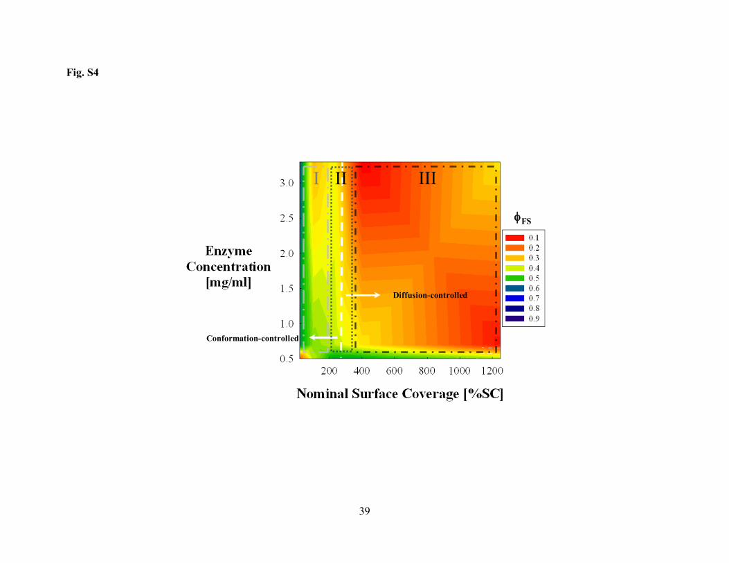
CALB. A similar diagram for TLL is shown in Fig. S4 in Supporting Information. TLL exhibits 313
three regions of conformational stability similar to the enzymes discussed above. Region I, 314
however, has only partially altered enzyme conformations as evident from a maximum φFS value 315
of 0.6. This is perhaps not surprising for the enzyme with the least tendency to denature of the 316
three considered here. This finding also further supports the proposed relationship between the 317
inherent conformational stability of enzyme molecules and their tendency to undergo 318
conformational changes upon contact with solid surfaces. While “soft” enzymes require 319

16
intermediate surface coverage to stabilize them, “hard” enzymes exhibit well maintained 320
structures already at low surface coverage. 321
In summary, our findings confirm the critical role that the surface area availability plays by 322
defining the physical arrangement of the enzyme molecules on the surface of the nanoparticles. 323
Additionally, visualizing conformational stability regions as a function of the surface packing 324
density and further correlation with activity data can be facilitated with the aid of the 3D contour 325
diagrams introduced here. 326
Impact of Tertiary Structure Modifiers for Enzymes Interacting with Fumed Silica in Aqueous 327
Solution 328
Tertiary structure modifiers (2,2,2-trifluoroethanol (TFE) and Dithiothreitol (DTT)) can be 329
added prior to adsorption on fumed silica to gain more insight into the type of intermolecular 330
interactions predominating during the adsorption process. TFE induces structural changes in 331
hydrophobic protein segments while DTT reduces disulfide bonds to the unbounded thiols. The 332
time dependent conformational changes of the three enzymes in the presence of 30% (v/v) TFE 333
for adsorbates with a 100%SC is shown in Fig. 6 panel A. Addition of TFE to both s. Carlsberg 334
and TLL resulted in increased initial values of deformation (data in the absence of TFE is 335
superimposed in Fig. 6 panel A). The shape of the absorbance changes for these two enzymes 336
were similar to those in the absence of TFE, i.e., logarithmic for s. Carlsberg and linear for TLL. 337
Overall, the extent of denaturation substantially increased over the initial values (40% and 30% 338
for s. Carlsberg and TLL, respectively). This most likely indicates that the disrupting 339
intramolecular contacts hasten denaturation, suggesting that these contact regions are responsible 340
for stabilizing the structures of these two enzymes. 341
17
The initial impact of TFE on CALB unfolding was essentially negligible and some refolding is 342
observed, likely due to the different content of groups in CALB that interact strongly with TFE. 343
The presence of DTT showed no significant impact on the time-course and extent of unfolding 344
for s. Carlsberg and TLL (Fig. 6 panel B). This is most likely due to the absence of thiol 345
sensitive disulfide bonds. CALB, on the other hand, contains DTT-sensitive disulfide bridges 346
and showed decreased initial and equilibrium unfolding values (Fig. 6 panel B). The exposed 347
residues are then available for interaction with the nanoparticles. This further supports the notion 348
that increased interactions lead to repression of dynamism and ultimately to less opportunities for 349
continued unfolding. This could also explain why the observed DTT-disrupted CALB unfolding 350
pathway resembles those of the “hard” enzymes. The exposure of regions suitable for interaction 351
with the surface is therefore likely to occur more gradually in the presence of DTT. 352
Regions of Conformational Stability for Enzyme/Fumed Silica Adsorbates in the Presence of 353
Structure Modifiers 354
Fig. 7 panel A shows that the region III originally observed for CALB was destabilized by 355
TFE. It is likely that groups exposed by TFE facilitated additional opportunities for unfolding by 356
increasing the affinity towards the hydrophilic solid surface. The unfolding extent in this region 357
increased by nearly 50% with respect to the absence of TFE (Fig. 5). 358
Fig. 7 panel B summarizes the impact of DTT on CALB’s tertiary structure. Once disulfide 359
bridges are disrupted, CALB’s structure is destabilized and some previously occluded regions 360
are exposed. The extent of unfolding in the lower part of regions II and III (i.e., below 2.5 361
mg/mL and surface coverages above 100%SC) remains low. This could support the argument 362
that the DTT-induced destabilization of CALB may possibly be compensated by interactions 363

18
with the neighboring molecules. This further supports the idea that protein-protein interactions 364
outweigh protein-surface interactions in region III. 365
As the enzyme concentration increases, the extent of unfolding also increases at both low and 366
high surface coverages (upper part of regions I and III in Fig. 7 panel B). The accessibility of 367
DTT to the enzyme at high enzyme concentrations is likely to be compromised by the crowding 368
of enzyme molecules. This leads to a behavior resembling that of the enzyme molecules in the 369
absence of DTT. 370
A similar study with structural modifiers was conducted for s. Carlsberg. As in the case of 371
CALB, the exposure of hydrophobic groups leads to a substantial loss of conformational stability 372
at high surface coverages (data not shown). Due to the tightly packed structure of s. Carlsberg, 373
the exposure of hydrophobic groups is rapid. This led to a relatively higher net unfolding than 374
that observed for CALB in this region. Below 100%SC, unfolding was reduced when compared 375
with the enzyme in the absence of TFE. This could be also a direct consequence of the rapid 376
initial unfolding. Once fully extended binding is promoted at the outset of the experiment, the 377
unfolded enzyme molecule’s structure is rapidly attached to the abundant solid surface area 378
provided in this region. No additional opportunities are present that would allow for further 379
unfolding. The case of DTT-induced conformational changes for s. Carlsberg is very different. 380
Due to the lack of open moieties on the structure suitable for reduction, no major changes were 381
observed and the regions of stability remained essentially unaffected (data not shown). 382
383

19
Conclusions 384
We have shown evidence that the extent of tertiary conformational changes of three hydrolases 385
adsorbed on fumed silica is mainly dependent on the density of the surface covered by the 386
enzyme molecules, which is described here as a nominal surface coverage (%SC), and the 387
inherent conformational stability and dynamics of the enzyme molecules (“hardness”). At low 388
%SC, the unfolding pathway of CALB, a “soft” enzyme, on fumed silica showed continuous 389
unfolding/refolding events. This was attributed to CALB’s conformationally dynamic and 390
fluctuating structure. “Hard” s. Carlsberg and TLL, in contrast, exhibited approximately linear 391
unfolding with time which was attributed to their higher inherent conformational stabilities. 392
When moving to high %SC, the unfolding pathways exhibited fewer fluctuations due to 393
stabilizing protein-protein interactions. 394
Three regions of conformational stability were identified for the three hydrolases at 395
equilibrium with the aid of 3D conformational diagrams: I. a region at low % SC where 396
adsorbates of “soft” CALB exhibited low conformational stability most likely due to multi-point 397
attachment to the surface, II. a region of transitional stability at an intermediate %SC 398
(~200%SC), and III. a region of high stability at high %SC where protein-protein interactions 399
exert a stabilizing effect. These regions correlated well the observed apparent catalytic activity in 400
hexane. The low stability region correlated well with poorly active preparations due to 401
denaturation for “soft” CALB, the transitional region with an optimal in activity, and the high 402
stability region with low CALB catalytic activity due to mass-transfer limitations. In the case of 403
“hard” s. Carlsberg, resilience to denaturation at low %SC when compared to CALB explained 404
the constantly increasing apparent catalytic activity observed in this surface loading regime. 405

20
Details of the structural interactions were further resolved by adding TFE and DTT to the 406
adsorbing mixtures. TFE exposed hydrophobic segments that for all enzymes appeared to help to 407
stabilize the structure in the “crowded surface” Region III. In the sparsely populated surface 408
Region I, the maximum extent of unfolding for both CALB and s. Carlsberg was reduced. This 409
was explained by lower content of interacting groups for CALB and by a rapid attachment of the 410
rigid unfolded state for s. Carlsberg. The addition of DTT to CALB’s adsorbing mixtures 411
showed no significant impact in the unfolding levels in the lower part of region III, which 412
confirmed that protein-protein interactions outweigh surface-protein interactions in this region. 413
Upon DTT-induced disruption, Region I showed less unfolding most likely due to a gradual 414
attachment of the very flexible unfolded state of CALB. This was corroborated with unfolding 415
pathways that resemble those of “hard” enzymes. 416
The complex enzyme/solid interactions in regard to catalytic activity that were investigated 417
here can be used to rationalize and optimize catalysis with enzymes in solvents. The conclusion 418
that for “soft” enzymes there is an optimum enzyme/surface area ratio for immobilization while 419
there is not for “hard” enzymes appears not obvious. 420
421

21
References 422
[1] M.N. Gupta, Enzyme Function in Organic-Solvents, Eur. J. Biochem., 203 (1992) 25-32. 423 [2] A. Zaks and A.J. Russell, Enzymes in Organic-Solvents - Properties and Applications, J. 424 Biotechnol., 8 (1988) 259-270. 425 [3] H. Kise, Ester and Peptide-Synthesis by Proteases in Organic-Solvents, Journal of Synthetic 426 Organic Chemistry Japan, 49 (1991) 42-51. 427 [4] R. Fernandezlafuente, C.M. Rosell and J.M. Guisan, Enzyme Reaction-Engineering - 428 Synthesis of Antibiotics Catalyzed by Stabilized Penicillin-G Acylase in the Presence of Organic 429 Cosolvents, Enzyme Microb. Technol., 13 (1991) 898-905. 430 [5] Y.L. Khmelnitsky and J.O. Rich, Biocatalysis in nonaqueous solvents, Curr. Opin. Chem. 431 Biol., 3 (1999) 47-53. 432 [6] J.O. Rich and Y.L. Khmelnitsky, Phospholipase D-catalyzed transphosphatidylation in 433 anhydrous organic solvents, Biotechnol. Bioeng., 72 (2001) 374-377. 434 [7] A.M. Klibanov, Asymmetric enzymatic oxidoreductions in organic solvents, Curr. Opin. 435 Biotechnol., 14 (2003) 427-431. 436 [8] M.S. Antczak, A. Kubiak, T. Antczak and S. Bielecki, Enzymatic biodiesel synthesis - Key 437 factors affecting efficiency of the process, Renew. Energy, 34 (2009) 1185-1194. 438 [9] J.E. Puskas, M.Y. Sen and K.S. Seo, Green Polymer Chemistry Using Nature's Catalysts, 439 Enzymes, J. Polym. Sci. Pol. Chem., 47 (2009) 2959-2976. 440 [10] A.M. Klibanov, Improving enzymes by using them in organic solvents, Nature, 409 (2001) 441 241-246. 442 [11] A.M. Klibanov, Why are enzymes less active in organic solvents than in water?, Trends 443 Biotechnol., 15 (1997) 97-101. 444 [12] J.A. Bosley and A.D. Peilow, Immobilization of lipases on porous polypropylene: 445 Reduction in esterification efficiency at low loading, J. Am. Oil Chem. Soc., 74 (1997) 107-111. 446 [13] B. Chen, J. Hu, E.M. Miller, W.C. Xie, M.M. Cai and R.A. Gross, Candida antarctica lipase 447 B chemically immobilized on epoxy-activated micro- and nanobeads: Catalysts for polyester 448 synthesis, Biomacromolecules, 9 (2008) 463-471. 449 [14] B. Chen, E.M. Miller, L. Miller, J.J. Maikner and R.A. Gross, Effects of macroporous resin 450 size on Candida antarctica lipase B adsorption, fraction of active molecules, and catalytic activity 451 for polyester synthesis, Langmuir, 23 (2007) 1381-1387. 452 [15] B. Chen, M.E. Miller and R.A. Gross, Effects of porous polystyrene resin parameters on 453 Candida antarctica Lipase B adsorption, distribution, and polyester synthesis activity, Langmuir, 454 23 (2007) 6467-6474. 455 [16] J.C. Cruz, P.H. Pfromm and M.E. Rezac, Immobilization of Candida antarctica Lipase B on 456 fumed silica, Process Biochem., 44 (2009) 62-69. 457 [17] K. Wurges, P.H. Pfromm, M.E. Rezac and P. Czermak, Activation of subtilisin Carlsberg in 458 hexane by lyophilization in the presence of fumed silica, J. Mol. Catal. B: Enzym., 34 (2005) 18-459 24. 460 [18] P.H. Pfromm, M.E. Rezac, K. Wurges and P. Czermak, Fumed silica activated subtilisin 461 Carlsberg in hexane in a packed-bed reactor, Aiche J., 53 (2007) 237-242. 462 [19] P. Wang, Nanoscale biocatalyst systems, Curr. Opin. Biotechnol., 17 (2006) 574-579. 463 [20] R.K. Iler, The Chemistry of Silica, John Wiley & Sons, Inc., 1979. 464 [21] V.M. Gun'ko, I.F. Mironyuk, V.I. Zarko, V.V. Turov, E.F. Voronin, E.M. Pakhlov, E.V. 465 Goncharuk, R. Leboda, J. Skubiszewska-Zieba, W. Janusz, S. Chibowski, Y.N. Levchuk and 466
22
A.V. Klyueva, Fumed silicas possessing different morphology and hydrophilicity, J. Colloid 467 Interface Sci., 242 (2001) 90-103. 468 [22] V.M. Gun'ko, I.F. Mironyuk, V.I. Zarko, E.F. Voronin, V.V. Turov, E.M. Pakhlov, E.V. 469 Goncharuk, Y.M. Nychiporuk, N.N. Vlasova, P.P. Gorbik, O.A. Mishchuk, O.A. Mishchuk, 470 A.A. Chuiko, T.V. Kulik, B.B. Palyanytsya, S.V. Pakhovchishin, J. Skubiszewska-Zieba, W. 471 Janusz, A.V. Turov and R. Leboda, Morphology and surface properties of fumed silicas, J. 472 Colloid Interface Sci., 289 (2005) 427-445. 473 [23] V.M. Gun'ko, E.F. Voronin, L.V. Nosach, E.M. Pakhlov, N.V. Guzenko, R. Leboda and J. 474 Skubiszewska-Zieba, Adsorption and migration of poly(vinyl pyrrolidone) at a fumed silica 475 surface, Adsorpt. Sci. Technol., 24 (2006) 143-157. 476 [24] V.M. Gun'ko, V.I. Zarko, E.F. Voronin, E.V. Goncharuk, L.S. Andriyko, N.V. Guzenko, 477 L.V. Nosach and W. Janusz, Successive interaction of pairs of soluble organics with nanosilica 478 in aqueous media, J. Colloid Interface Sci., 300 (2006) 20-32. 479 [25] V.M. Gun'ko, V.I. Zarko, E.F. Voronin, V.V. Turov, I.F. Mironyuk, Gerashchenko, II, E.V. 480 Goncharuk, E.M. Pakhlov, N.V. Guzenko, R. Leboda, J. Skubiszewska-Zieba, W. Janusz, S. 481 Chibowski, Y.N. Levchuk and A.V. Klyueva, Impact of some organics on structural and 482 adsorptive characteristics of fumed silica in different media, Langmuir, 18 (2002) 581-596. 483 [26] M.T. Ru, S.Y. Hirokane, A.S. Lo, J.S. Dordick, J.A. Reimer and D.S. Clark, On the salt-484 induced activation of lyophilized enzymes in organic solvents: Effect of salt kosmotropicity on 485 enzyme activity, J. Am. Chem. Soc., 122 (2000) 1565-1571. 486 [27] M.T. Ru, K.C. Wu, J.P. Lindsay, J.S. Dordick, J.A. Reimer and D.S. Clark, Towards more 487 active biocatalysts in organic media: Increasing the activity of salt-activated enzymes, 488 Biotechnol. Bioeng., 75 (2001) 187-196. 489 [28] J. Uppenberg, M.T. Hansen, S. Patkar and T.A. Jones, Sequence, Crystal-Structure 490 Determination and Refinement of 2 Crystal Forms of Lipase-B From Candida antarctica, 491 Structure, 2 (1994) 293-308. 492 [29] D.J. Neidhart and G.A. Petsko, The Refined Crystal-Structure of Subtilisin Carlsberg at 2.5 493 A Resolution, Protein Eng., 2 (1988) 271-276. 494 [30] J.K.A. Kamal, T.B. Xia, S.K. Pal, L. Zhao and A.H. Zewail, Enzyme functionality and 495 solvation of Subtilisin Carlsberg: from hours to femtoseconds, Chem. Phys. Lett., 387 (2004) 496 209-215. 497 [31] A.K. Shaw and S.K. Pal, Activity of Subtilisin Carlsberg in macromolecular crowding, J. 498 Photochem. Photobiol., B, 86 (2007) 199-206. 499 [32] P. Asuri, S.S. Bale, S.S. Karajanagi and R.S. Kane, The protein-nanomaterial interface, 500 Curr. Opin. Biotechnol., 17 (2006) 562-568. 501 [33] P. Asuri, S.S. Bale, R.C. Pangule, D.A. Shah, R.S. Kane and J.S. Dordick, Structure, 502 function, and stability of enzymes covalently attached to single-walled carbon nanotubes, 503 Langmuir, 23 (2007) 12318-12321. 504 [34] P. Asuri, S.S. Karajanagi, A.A. Vertegel, J.S. Dordick and R.S. Kane, Enhanced stability of 505 enzymes adsorbed onto nanoparticles, J. Nanosci. Nanotechnol., 7 (2007) 1675-1678. 506 [35] P. Singh, A. Rao, V. Dutta, M. Gupta and S. Raghava, Nanoparticles of unmodified titanium 507 dioxide facilitate protein refolding, J. Mater. Chem., 19 (2009) 2830-2834. 508 [36] R.S. Kane and A.D. Stroock, Nanobiotechnology: Protein-nanomaterial interactions, 509 Biotechnol. Prog., 23 (2007) 316-319. 510 [37] W. Norde and J.P. Favier, Structure of Adsorbed and Desorbed Proteins, Colloids Surf., B 511 64 (1992) 87-93. 512

23
[38] W. Shang, J.H. Nuffer, J.S. Dordick and R.W. Siegel, Unfolding of ribonuclease A on silica 513 nanoparticle surfaces, Nano Lett., 7 (2007) 1991-1995. 514 [39] W. Shang, J.H. Nuffer, V.A. Muniz-Papandrea, W. Colon, R.W. Siegel and J.S. Dordick, 515 Cytochrome c on Silica Nanoparticles: Influence of Nanoparticle Size on Protein Structure, 516 Stability, and Activity, Small, 5 (2009) 470-476. 517 [40] J.L. West and N.J. Halas, Engineered nanomaterials for biophotonics applications: 518 Improving sensing, imaging, and therapeutics, Annu. Rev. Biomed. Eng., 5 (2003) 285-292. 519 [41] H.J. Lewerenz, Enzyme-semiconductor interactions: Routes from fundamental aspects to 520 photoactive devices, Phys. Status Solidi B-Basic Solid State Phys., 245 (2008) 1884-1898. 521 [42] H.L. Su, R. Yuan, Y.Q. Chai, Y. Zhuo, C.L. Hong, Z.Y. Liu and X. Yang, Multilayer 522 structured amperometric immunosensor built by self-assembly of a redox multi-wall carbon 523 nanotube composite, Electrochim. Acta, 54 (2009) 4149-4154. 524 [43] N. Chopra, V.G. Gavalas, B.J. Hinds and L.G. Bachas, Functional one-dimensional 525 nanomaterials: Applications in nanoscale biosensors, Anal. Lett., 40 (2007) 2067-2096. 526 [44] A. Ganesan, B.D. Moore, S.M. Kelly, N.C. Price, O.J. Rolinski, D.J.S. Birch, I.R. Dunkin 527 and P.J. Halling, Optical Spectroscopic Methods for Probing the Conformational Stability of 528 Immobilised Enzymes, Chemphyschem, 10 (2009) 1492-1499. 529 [45] X. Wu and G. Narsimhan, Characterization of secondary and tertiary conformational 530 changes of beta-lactoglobulin adsorbed on silica nanoparticle surfaces, Langmuir, 24 (2008) 531 4989-4998. 532 [46] X.Y. Wu and G. Narsimhan, Effect of surface concentration on secondary and tertiary 533 conformational changes of lysozyme adsorbed on silica nanoparticles, Biochim. Biophys. Acta, 534 Proteins Proteomics, 1784 (2008) 1694-1701. 535 [47] A. Ganesan, N.C. Price, S.M. Kelly, I. Petry, B.D. Moore and P.J. Halling, Circular 536 dichroism studies of subtilisin Carlsberg immobilised on micron sized silica particles, Biochim. 537 Biophys. Acta, Proteins Proteomics, 1764 (2006) 1119-1125. 538 [48] K. Hidajat, M. Uddin and Z. Peng, Conformational change of adsorbed and desorbed 539 bovine serum albumin on nano-sized magnetic particles, Colloids Surf., B, 33 (2004) 15-21. 540 [49] N. Shamim, H. Liang, K. Hidajat and M.S. Uddin, Adsorption, desorption, and 541 conformational changes of lysozyme from thermosensitive nanomagnetic particles, J. Colloid 542 Interface Sci., 320 (2008) 15-21. 543 [50] A. Kondo, S. Oku and K. Higashitani, Structural-Changes in Protein Molecules Adsorbed 544 on Ultrafine Silica Particles, J. Colloid Interface Sci., 143 (1991) 214-221. 545 [51] N. Tokuriki and D.S. Tawfik, Protein Dynamism and Evolvability, Science, 324 (2009) 546 203-207. 547 [52] P. Hoyrup, S. Patkar, J. Vind, A. Svendsen, K. Hult and E. Hedin, Implications of surface 548 charge and curvature for the binding orientation of Thermomyces lanuginosus lipase on 549 negatively charged or zwitterionic phospholipid vesicles as studied by ESR spectroscopy, 550 Biochemistry, 44 (2005) 16658-16671. 551 [53] A. Jutila, K. Zhu, S.A. Patkar, J. Vind, A. Svendsen and P.K.J. Kinnunen, Detergent-552 induced conformational changes of Humicola lanuginosa lipase studied by fluorescence 553 spectroscopy, Biophys. J., 78 (2000) 1634-1642. 554 [54] A. Jutila, K. Zhu, E.K.J. Tuominen and P.K.J. Kinnunen, Fluorescence spectroscopic 555 characterization of Humicola lanuginosa lipase dissolved in its substrate, Biochim. Biophys. 556 Acta, Proteins Proteomics, 1702 (2004) 181-189. 557

24
[55] K. Zhu, A. Jutila and P.K.J. Kinnunen, Steady state and time resolved effects of guanidine 558 hydrochloride on the structure of Humicola lanuginosa lipase revealed by fluorescence 559 spectroscopy, Protein Sci., 9 (2000) 598-609. 560 [56] T. Schafer, T.W. Borchert, V.S. Nielsen, P. Skagerlind, K. Gibson, K. Wenger, F. Hatzack, 561 L.D. Nilsson, S. Salmons, S. Pedersen, H.P. Heldt-Hansen, P.B. Poulsen, H. Lund, K.M. 562 Oxenboll, G.F. Wu, H.H. Pedersen and H. Xu, Industrial enzymes, in: White Biotechnology, 563 Vol 105, Springer-Verlag Berlin, Berlin, 2007, pp. 59-131. 564 [57] P. Acharya and N.M. Rao, Stability studies on a lipase from Bacillus subtilis in 565 guanidinium chloride, J. Protein Chem., 22 (2003) 51-60. 566 [58] T. De Diego, P. Lozano, S. Gmouh, M. Vaultier and J.L. Iborra, Understanding structure - 567 Stability relationships of Candida antartica lipase B in ionic liquids, Biomacromolecules, 6 568 (2005) 1457-1464. 569 [59] M. Skjot, L. De Maria, R. Chatterjee, A. Svendsen, S.A. Patkar, P.R. Ostergaard and J. 570 Brask, Understanding the Plasticity of the alpha/beta Hydrolase Fold: Lid Swapping on the 571 Candida antarctica Lipase B Results in Chimeras with Interesting Biocatalytic Properties, 572 ChemBioChem, 10 (2009) 520-527. 573 [60] A.W. Sonesson, T.H. Callisen, H. Brismar and U.M. Elofsson, Lipase surface diffusion 574 studied by fluorescence recovery after photobleaching, Langmuir, 21 (2005) 11949-11956. 575 [61] C. Carboni-Oerlemans, P.D. de Maria, B. Tuin, G. Bargeman, A. van der Meer and R. van 576 Gemert, Hydrolase-catalysed synthesis of peroxycarboxylic acids: Biocatalytic promiscuity for 577 practical applications, J. Biotechnol., 126 (2006) 140-151. 578 [62] L. Cao, Adsorption-based Immobilization, in: Carrier-bound Immobilized Enzymes: 579 Principles, Applications and Design, Vol 1, Wiley-VCH Verlag GmbH & Co. KGaA, Germany, 580 Weinheim, 2005, pp. 53-168. 581 [63] U. Derewenda, L. Swenson, R. Green, Y. Wei, S. Yamaguchi, R. Joerger, M.J. Haas and 582 Z.S. Derewenda, Current Progress in Crystallographic Studies of New Lipases from Filamentous 583 Fungi, Protein Eng., 7 (1994) 551-557. 584 585
586

25
Tables 587
Table 1 588
Summary of the amounts of fumed silica and enzyme employed to form the suspensions with 589 different nominal surface coverages. The enzyme concentration for each suspension was varied 590 from 0.5 mg/mL to 4.70 mg/mL. 591
Mass fumed silica (g) Enzyme Enzyme mass (mg) 2%SC* 17%SC 100%SC 230%SC 400%SC 1250%SC CALB 7 0.718 0.092 0.016 0.0068 0.0039 0.0013 TLL 5 0.350 0.040 0.010 0.0030 0.0018 0.0006
s. Carlsberg 5 0.315 0.036 0.009 0.0027 0.0016 0.0005 * The Nominal Surface Coverage (% SC) was calculated as follows: 592
%𝑆𝐶 = 𝑃𝑟𝑜𝑗𝑒𝑐𝑡𝑒𝑑 𝑎𝑟𝑒𝑎 𝑜𝑓 𝑒𝑛𝑧𝑦𝑚𝑒 𝑚𝑜𝑙𝑒𝑐𝑢𝑙𝑒𝑁𝑜𝑚𝑖𝑛𝑎𝑙 𝑠𝑢𝑟𝑓𝑎𝑐𝑒 𝑎𝑟𝑒𝑎 𝑜𝑓 𝐹𝑢𝑚𝑒𝑑 𝑆𝑖𝑙𝑖𝑐𝑎
∗ 100 Equation 3 593
The projected area of enzyme is calculated assuming a spherical shape for the enzyme molecules. The diameter of 594 the enzyme molecules from crystallographic data were 6.4 nm [28], 5.0 nm [63], and 4.2 nm [29] for CALB, TLL 595 and s. Carlsberg, respectively. The nominal surface area of fumed silica is as provided by the manufacturer: 596 255m2/g. 597
598

26
Figure Captions 599
Fig. 1. Tryptophan fluorescence spectrum of the unfolding of CALB induced by GdmCl in 600 aqueous buffer solution. (○) Native, (□) 1M GdmCl, (∆) 2M GdmCl, (×) 4M GdmCl and (*) 8M 601 GdmCl. Decrease in the maximum emission intensity (Imax/em) of Trp residues due to GdmCl-602 induced denaturation is shown in the inset. y-error bars represent the standard error of multiple 603 analyses of identical samples. Enzyme concentration was maintained at 0.5 mg/ml and pH 7.8. 604
Fig. 2. Normalized fluorescence intensity for the GdmCl induced unfolding of CALB in aqueous 605 buffer. The tryptophan emission was monitored at 330 nm. Measurements were carried out at 606 enzyme concentration of 0.5 mg/mL and pH 7.8. Similar calibration curves were obtained for s. 607 Carlsberg and TLL (data not shown). 608
Fig. 3. Unfolding data for CALB on fumed silica (z-axis) as a function of initial enzyme 609 concentration (y-axis) and nominal surface coverage (x-axis). These data will be shown in 3D 610 contour plots to identify regions of conformational stability and to subsequently correlate them 611 with the surface loading regimes previously postulated for the lyophilized adsorbates. 612
Fig. 4. Unfolding kinetics of (●) CALB, (□) s. Carlsberg and (∆) TLL adsorbing on fumed silica 613 nanoparticles to form adsorbates with a nominal surface coverage of 2%SC. Data is normalized 614 with the corresponding calibration curves and subsequently expressed as unfolded fraction. 615 Enzyme concentration was maintained at 0.5 mg/mL and pH at 7.8. 616
Fig. 5. Regions of conformational stability for CALB/Fumed Silica adsorbates. The dotted 617 vertical line at ~200%SC separates two different regions of conformational stability: Region I 618 delimited by a long-dash-dot line where adsorbates exhibit low conformational stability, and 619 Region III delimited by a short-dash-dot line where adsorbates have highly stable 620 conformations. The presence of these two regions is likely to be responsible for the observed 621 catalytic activity (r0) of the lyophilized adsorbates in hexane (inset). The low catalytic activity 622 observed at low %SC can be linked to Region I. Even though the structure is well preserved in 623 Region III, multi-layer packing is likely responsible for diffusional limitations of catalysis. The 624 maximum in activity between those two regions can be attributed to an optimal arrangement on 625 the surface where the structure is relatively well maintained without excessive clustering 626 (Region II, delimited by a dotted line). 627
Fig. 6. Unfolding kinetics of hydrolases adsorbing on fumed silica nanoparticles in the presence 628 of tertiary structure modifiers: (A) 30% (v/v) TFE and (B) 0.5 mg/mL DTT: (O) CALB, () s. 629 Carlsberg and () TLL. Data for unfolding in the absence of modifiers is superimposed for 630 comparison: (●) CALB, (□) s. Carlsberg and (∆) TLL. Components were mixed to form a 631 nominal surface coverage of 100%SC. Data is normalized with the corresponding calibration 632 curves and subsequently expressed as unfolded fraction. Enzyme concentration was maintained 633 at 0.7 mg/mL and pH at 7.8. 634
Fig. 7. Impact of tertiary structure modifiers on conformational regions of CALB/FS adsorbates. 635 This was tested by forming buffered suspensions with (A) 30%(v/v) TFE and (B) 0.5 mg/mL 636 DTT. The previously identified Region III exhibits a higher unfolding in the presence of TFE. 637 The extent of unfolding in Region I decreased in the presence of TFE. The addition of DTT 638

27
resulted in a substantially large Region III for low %SC and low concentrations. The same type 639 of lines as those in Fig. 5 were used to delimit the regions of conformational stability. 640
641
28
Supporting Information 642
Conformational Changes and Catalytic Competency of Hydrolases Adsorbing on Fumed Silica 643
Nanoparticles: I. Tertiary Structure 644
Juan C. Cruz1, Peter H. Pfromm1*, John M. Tomich2, Mary E. Rezac1 645
(1) Department of Chemical Engineering, Kansas State University, 1005 Durland Hall, 646
Manhattan, KS 66506 - 5106, USA. 647
(2) Department of Biochemistry and Biotechnology/Proteomics Core Facility, Kansas State 648
University, Burt Hall, KS 66506 - 5106, USA. 649
650
Fig. S1. Unfolding kinetics of (●) CALB, (□) s. Carlsberg and (∆) TLL adsorbing of fumed 651 silica nanoparticles to form adsorbates with a nominal surface coverage of 100%SC. Data is 652 normalized with the corresponding calibration curves and subsequently expressed as unfolded 653 fraction using Equation 2. Enzyme concentration was maintained at 0.5 mg/mL and pH at 7.8. 654
Fig. S2. Unfolding kinetics of (●) CALB, (□) s. Carlsberg and (∆) TLL adsorbed of fumed silica 655 nanoparticles to obtain a nominal surface coverage of 400%SC. Data is normalized with the 656 corresponding calibration curves and subsequently expressed as unfolded fraction using Equation 657 2. Enzyme concentration was maintained at 0.5 mg/mL and pH at 7.8. 658
Fig. S3. Regions of conformational stability for s. Carlsberg/Fumed Silica adsorbates. The 659 dotted vertical line at ~200%SC separates two different regions of conformational stability: 660 Region I and III of low and high conformational stability, respectively. In this case, the catalytic 661 activity (r0) of the lyophilized adsorbates in hexane (inset) is constantly increasing. As opposed 662 to CALB, only partially unfolded enzyme molecules are present in the lower part of Region I at 663 low initial enzyme concentrations. This resilience to denaturation could explain the higher 664 activities observed for lyophilized adsorbates of s. Carlsberg at low surface coverages. The same 665 type of lines as those in Fig. 5 were used to delimit the regions of conformational stability. 666
Fig. S4. Regions of conformational stability for TLL/Fumed Silica adsorbates. The dotted 667 vertical line at ~250%SC separates two different regimes of conformational stability: Region I 668 and III of low and high conformational stability, respectively. Across the whole Region I only 669 partially unfolded conformations (φFS ~0.6) were identified, which confirms the resilience of 670 highly stable enzymes to denature at low surface coverages. The same type of lines as those in 671 Fig. 5 were used to delimit the regions of conformational stability. 672
673

29
Fig. 1
0
10
20
30
40
50
60
70
80
90
100
300 320 340 360 380 400 420 440
Intensity [a.u.]
Wavelength [nm]
0
10
20
30
40
50
60
70
80
90
100
Native 1M GdmCl
2M GdmCl
4M GdmCl
6M GdmCl
8M GdmCl
CALB
I max/em [a.u.]

30
Fig. 2
0
0.2
0.4
0.6
0.8
1
1.2
0 2 4 6 8 10
φ GdmCl[ ]
GdmCl [M]

31
Fig. 3
Denatured
Native

32
Fig. 4
0.6
0.7
0.8
0.9
1
0 5 10 15 20 25 30 35 40 45
φFS [ ]
Time (min)

33
Fig. 5
Conformation-controlled
φFS
Maximum activity
Current operation line
Diffusion-controlled
IIIIII

34
Fig. 6
0.1
0.2
0.3
0.4
0.5
0.6
0.7
0.8
0 5 10 15 20 25 30 35 40 45
φFS [ ]
Time [min]
0
0.1
0.2
0.3
0.4
0.5
0.6
0 5 10 15 20 25 30 35 40 45
φFS [ ]
Time [min]
A
B

35
Fig. 7
IIIIII
A
φFS
IIIIII
B
φFS

36
Fig. S1
0.3
0.4
0.5
0.6
0.7
0 5 10 15 20 25 30 35 40 45
φFS [ ]
Time (min)

37
Fig. S2
0.3
0.4
0.5
0.6
0.7
0 5 10 15 20 25 30 35 40 45
φFS [ ]
Time (min)

38
Fig. S3
Diffusion-controlled
Conformation-controlled
φFS
Current operation line
IIIIII
39
Fig. S4
Diffusion-controlled
Conformation-controlled
φFS
IIIIII