Complementary DNA cloning of the alternatively expressed endothelial cell glycoprotein Ib beta (GPIb...
8
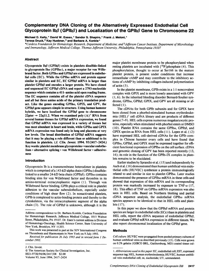
Complementary DNA Cloning of the Alternatively Expressed Endothelial Cell Glycoprotein lbfl (GPlbB) and Localization of the GPlbB Gene to Chromosome 22 Michael D. Kelly, * David W. Essex, * Sandor S. Shapiro, * Frank J. Meloni, * Teresa Druck,t Kay Huebner,* and Barbara A. Konkle* *Cardeza Foundation for Hematologic Research, Department ofMedicine; and t Jefferson Cancer Institute, Department ofMicrobiology and Immunology, Jefferson Medical College, Thomas Jefferson University, Philadelphia, Pennsylvania 19107 Abstract Glycoprotein Ib# (GPIb#) exists in platelets disulfide-linked to glycoprotein Iba (GPIba), a major receptor for von Wille- brand factor. Both GPIba and GPIb# are expressed in endothe- lial cells (EC). While the GPIba mRNA and protein appear similar in platelets and EC, EC GPIbft mRNA is larger than platelet GPIb#t and encodes a larger protein. We have cloned and sequenced EC GPIbfi cDNA and report a 2793-nucleotide sequence which contains a 41 1-amino acid open reading frame. The EC sequence contains all of the platelet cDNA sequence and all but three amino acids of the primary translation prod- uct. Like the genes encoding GPIba, GPIX, and GPV, the GPIb# gene appears simple in structure. Using human hamster hybrids, we have localized the GPIbfl gene to chromosome 22pter -- 22q11.2. When we examined poly (A)' RNA from several human tissues for GPIb,8 mRNA expression, we found that GPIbfl mRNA was expressed in a variety of tissues but was most abundant in heart and brain, while GPIba and GPIX mRNA expression was found only in lung and placenta at very low levels. The broad distribution of GPIb# mRNA suggests that it may be playing a role different than or additional to its function in platelets. (J. Clin. Invest. 1994. 93:2417-2424.) Key words: platelet membrane glycoproteins * vascular endothe- lium * alternative splicing , von Willebrand factor * adhesive- ness Introduction Glycoprotein lb is a transmembrane heterodimer in platelets which is comprised of a 143-kD alpha chain (GPIba) disulfide- linked to a smaller 24-kD beta chain (GPIbf.). GPIba contains binding sites for von Willebrand factor and thrombin in its amino-terminal extracytoplasmic region ( 1). Through von Willebrand factor binding, GPIb plays a critical role in platelet Adhesion to the vascular subendothelium, especially under conditions of high shear flow (2). In nonactivated platelets, GPIb is complexed to actin-binding protein and thereby to the cytoskeleton, via the intracytoplasmic segment of the alpha chain (3). The role of GPIb3 is unknown, although it is the Address correspondence to Dr. Barbara Konkle, Cardeza Foundation for Hematologic Research, Jefferson Medical College, 1015 Walnut Street, Philadelphia, PA 19107. Dr. Essex's current address is Division of Hematology-Oncology, Health Science Center, State University of New York, Brooklyn, NY 1203. This work was presented in part at the XIV International Congress on Thrombosis and Haemostasis in New York on 9 July 1993. Received for publication 26 July 1993 and in revised form I De- cember 1993. major platelet membrane protein to be phosphorylated when resting platelets are incubated with [32P]phosphate (4). This phosphorylation, thought to occur at Serl66 in the mature platelet protein, is present under conditions that increase intracellular cAMP and may contribute to the inhibitory ac- tions of cAMP by inhibiting collagen-induced polymerization of actin (5). In the platelet membrane, GPIb exists in a 1 :1 noncovalent complex with GPIX and is more loosely associated with GPV ( 1, 6). In the inherited bleeding disorder Bernard-Soulier syn- drome, GPIba, GPIbf3, GPIX, and GPV are all missing or af- fected ( 1). The cDNAs for both GPIb subunits and for GPIX have been cloned from a phorbol-stimulated human erythroleuke- mia (HEL)' cell cDNA library and are products of different genes (7-9). HEL cells express numerous megakaryocytic pro- teins, especially when stimulated with phorbol esters or DMSO ( 10). Platelet RNA contains the same GPIba, GPIb#, and GPIX species as RNA from HEL cells ( 11 ). Lopez et al. ( 12) have expressed HEL cell-derived cDNAs for the GPIb com- plex in Chinese hamster ovary cells and have found that GPIba, GPIb3, and GPIX must be expressed together for effi- cient functional expression of GPIba on the cell surface. cDNA and genomic cloning of GPV has been reported recently ( 13, 14); its role in the function of the GPIb-IX complex in plate- lets remains to be elucidated. Earlier studies by Sprandio et al. ( 15) and independently by Asch et al. ( 16) demonstrated than human umbilical vein endo- thelial cells (HUVEC) synthesize a protein immunologically related to and similar in size to platelet GPIba. Later studies demonstrated the presence of GPIba mRNA in these cells and showed that expression of this mRNA and its corresponding protein was markedly increased by exposure to TNF-a ( 17, 18). This effect of TNF on GPIba mRNA expression was also seen in HEL cells. Based on Northern blotting and partial cDNA sequence analysis, the endothelial GPIba mRNA species appears to be identical to that in HEL cells and plate- lets (17). In this paper we show that the GPIb, mRNA and protein species are larger in endothelial cells (ECs) than in platelets and HEL cells, report the cDNA sequence of endothelial GPIbfl, and evaluate GPIb3 mRNA expression in different tissues. We also report chromosomal localization of the GPIbB gene. Methods Cell culture. HUVEC were propagated from pooled primary cultures of human umbilical veins as described previously ( 15). Cells were grown on 0.2% gelatin (GIBCO BRL, Gaithersburg, MD) coated tissue cul- 1. Abbreviations used in this paper: EC, endothelial cell; EST, expressed sequence tag; HEL, human erythroleukemia; HUVEC, human umbili- cal vein endothelial cell; nt, nucleotide; UT, untranslated. Complementary DNA Cloning ofEndothelial Glycoprotein Ib,1 2417 J. Clin. Invest. © The American Society for Clinical Investigation, Inc. 0021-9738/94/06/2417/08 $2.00 Volume 93, June 1994, 2417-2424
-
Upload
independent -
Category
Documents
-
view
2 -
download
0
Transcript of Complementary DNA cloning of the alternatively expressed endothelial cell glycoprotein Ib beta (GPIb...
Complementary DNA Cloning of the Alternatively Expressed Endothelial CellGlycoprotein lbfl (GPlbB) and Localization of the GPlbB Gene to Chromosome 22Michael D. Kelly, * David W. Essex, * Sandor S. Shapiro, * Frank J. Meloni, *Teresa Druck,t Kay Huebner,* and Barbara A. Konkle**Cardeza Foundation for Hematologic Research, Department ofMedicine; and tJefferson Cancer Institute, Department ofMicrobiologyand Immunology, Jefferson Medical College, Thomas Jefferson University, Philadelphia, Pennsylvania 19107
Abstract
Glycoprotein Ib# (GPIb#) exists in platelets disulfide-linkedto glycoprotein Iba (GPIba), a major receptor for von Wille-brand factor. Both GPIba and GPIb# are expressed in endothe-lial cells (EC). While the GPIba mRNA and protein appearsimilar in platelets and EC, EC GPIbft mRNA is larger thanplatelet GPIb#t and encodes a larger protein. We have clonedand sequenced EC GPIbfi cDNA and report a 2793-nucleotidesequence which contains a 41 1-amino acid open reading frame.The EC sequence contains all of the platelet cDNA sequenceand all but three amino acids of the primary translation prod-uct. Like the genes encoding GPIba, GPIX, and GPV, theGPIb# gene appears simple in structure. Using human hamsterhybrids, we have localized the GPIbfl gene to chromosome22pter -- 22q11.2. When we examined poly (A)' RNA fromseveral human tissues for GPIb,8 mRNA expression, we foundthat GPIbfl mRNA was expressed in a variety of tissues butwas most abundant in heart and brain, while GPIba and GPIXmRNA expression was found only in lung and placenta at verylow levels. The broad distribution of GPIb# mRNA suggeststhat it may be playing a role different than or additional to itsfunction in platelets. (J. Clin. Invest. 1994. 93:2417-2424.)Key words: platelet membrane glycoproteins * vascular endothe-lium * alternative splicing , von Willebrand factor * adhesive-ness
Introduction
Glycoprotein lb is a transmembrane heterodimer in plateletswhich is comprised ofa 143-kD alpha chain (GPIba) disulfide-linked to a smaller 24-kD beta chain (GPIbf.). GPIba containsbinding sites for von Willebrand factor and thrombin in itsamino-terminal extracytoplasmic region (1). Through vonWillebrand factor binding, GPIb plays a critical role in plateletAdhesion to the vascular subendothelium, especially underconditions of high shear flow (2). In nonactivated platelets,GPIb is complexed to actin-binding protein and thereby to thecytoskeleton, via the intracytoplasmic segment of the alphachain (3). The role of GPIb3 is unknown, although it is the
Address correspondence to Dr. Barbara Konkle, Cardeza Foundationfor Hematologic Research, Jefferson Medical College, 1015 WalnutStreet, Philadelphia, PA 19107. Dr. Essex's current address is Divisionof Hematology-Oncology, Health Science Center, State University ofNew York, Brooklyn, NY 1203.
This work was presented in part at the XIV International Congresson Thrombosis and Haemostasis in New York on 9 July 1993.
Received for publication 26 July 1993 and in revisedform I De-cember 1993.
major platelet membrane protein to be phosphorylated whenresting platelets are incubated with [32P]phosphate (4). Thisphosphorylation, thought to occur at Serl66 in the matureplatelet protein, is present under conditions that increaseintracellular cAMP and may contribute to the inhibitory ac-tions ofcAMP by inhibiting collagen-induced polymerizationof actin (5).
In the platelet membrane, GPIb exists in a 1 :1 noncovalentcomplex with GPIX and is more loosely associated with GPV( 1, 6). In the inherited bleeding disorder Bernard-Soulier syn-drome, GPIba, GPIbf3, GPIX, and GPV are all missing or af-fected ( 1).
The cDNAs for both GPIb subunits and for GPIX havebeen cloned from a phorbol-stimulated human erythroleuke-mia (HEL)' cell cDNA library and are products of differentgenes (7-9). HEL cells express numerous megakaryocytic pro-teins, especially when stimulated with phorbol esters orDMSO( 10). Platelet RNA contains the same GPIba, GPIb#, andGPIX species as RNA from HEL cells ( 11 ). Lopez et al. ( 12)have expressed HEL cell-derived cDNAs for the GPIb com-plex in Chinese hamster ovary cells and have found thatGPIba, GPIb3, and GPIX must be expressed together for effi-cient functional expression ofGPIba on the cell surface. cDNAand genomic cloning ofGPV has been reported recently ( 13,14); its role in the function of the GPIb-IX complex in plate-lets remains to be elucidated.
Earlier studies by Sprandio et al. ( 15) and independently byAsch et al. ( 16) demonstrated than human umbilical vein endo-thelial cells (HUVEC) synthesize a protein immunologicallyrelated to and similar in size to platelet GPIba. Later studiesdemonstrated the presence ofGPIba mRNA in these cells andshowed that expression of this mRNA and its correspondingprotein was markedly increased by exposure to TNF-a ( 17,18). This effect ofTNF on GPIba mRNA expression was alsoseen in HEL cells. Based on Northern blotting and partialcDNA sequence analysis, the endothelial GPIba mRNAspecies appears to be identical to that in HEL cells and plate-lets (17).
In this paper we show that the GPIb, mRNA and proteinspecies are larger in endothelial cells (ECs) than in platelets andHEL cells, report the cDNA sequence of endothelial GPIbfl,and evaluate GPIb3 mRNA expression in different tissues. Wealso report chromosomal localization of the GPIbB gene.
Methods
Cell culture. HUVEC were propagated from pooled primary cultures ofhuman umbilical veins as described previously ( 15). Cells were grownon 0.2% gelatin (GIBCO BRL, Gaithersburg, MD) coated tissue cul-
1. Abbreviations used in this paper: EC, endothelial cell; EST, expressedsequence tag; HEL, human erythroleukemia; HUVEC, human umbili-cal vein endothelial cell; nt, nucleotide; UT, untranslated.
Complementary DNA Cloning ofEndothelial Glycoprotein Ib,1 2417
J. Clin. Invest.© The American Society for Clinical Investigation, Inc.0021-9738/94/06/2417/08 $2.00Volume 93, June 1994, 2417-2424

ture flasks (Corning Medical, Corning, NY) in Hepes-buffered M199(GIBCO BRL) with 10% FBS (Hyclone Laboratories, Logan, UT) in a5% CO2 humidified atmosphere at 370C. Culture media were supple-mented with crude EC growth factor ( 100 Ag/ml) and heparin ( 100,ug/ml). In a given experiment, all cells were grown in the same lot ofFBS. HUVEC were passaged as a 1:4 split, fed every 2-3 d, and used at2nd to 4th passage.
HEL cells were obtained from the American Type Culture Collec-tion (Rockville, MD) (ATCC TIB 180, HEL 92.1.7) and propagated inRPMI 1640 (GIBCO BRL) with 10% FBS. CHRF-288 cells, kindlyprovided by M. Lieberman (Children's Hospital Medical Center, Cin-cinnati, OH), were propagated in Fischer's medium (GIBCO BRL)with 20% horse serum (GIBCO BRL). All cultures were maintained ina 5% CO2 humidified atmosphere at 370C.
RNA andDNA analysis. Total cellular RNA was isolated by imme-diate solubilization of the cells in guanidine hydrochloride, as de-scribed previously (19). Poly (A)' RNA was isolated either by (a)incubation oftotal cellular RNA with oligo-dT cellulose (CollaborativeResearch Inc., Bedford, MA) followed by elution as described (20) orby (b) direct isolation from cells using the Mini-Ribosep kit (Collabora-tive Research Inc.). A Northern blot containing human megakaryocyteRNA was a kind gift of Dr. Paul Schick of the Cardeza Foundation. ANorthern blot containing poly (A)' RNA from various human tissueswas purchased from Clontech Laboratories, Inc. (Palo Alto, CA).
RNA was analyzed by Northern blotting. The RNA was fixed to themembrane with ultraviolet irradiation, prehybridized in 1 M NaCl,0.1% SDS, 1.5 mg/ml herring sperm DNA, and 10% dextran for 3 h at650C, and hybridized at 650C for 12-24 h in the prehybridizationsolution with the addition of 1.5 mg/ml sonicated herring sperm DNAand the appropriate radiolabeled probe. Probes used included (a) HELcell-derived GPIba cDNA, GPIb2.4 (7), (b) HEL cell-derived GPIbBcDNA (8), (c) HEL cell-derived GPIX cDNA (9), and (d) the endo-thelial GPIbj3 clone, EC,4. cDNA inserts were radiolabeled directly inlow melting agarose by random hexamer priming (21 ). The blots werewashed to high stringency (0.1 X SSC, 0.1% SDS, 1 mM EDTA, 10mM sodium phosphate [pH 6.8 ], at 65°C) and analyzed by autoradiog-raphy. Individual blots were rehybridized with a second radiolabeledprobe after incubation in 5 mM Tris/ HCl (pH 7.5), 2 mM EDTA, 0.1X Denhardts at 68°C for 3-4 h to remove the original probe. Successful"stripping" was confirmed by autoradiography. The Clontech multi-tissue blot was hybridized, washed, stripped of radiolabeled probe, andrehybridized according to the manufacturer's directions, except that itwas washed to the stringency noted above.
Human genomic DNA was isolated from white blood cells andanalyzed by Southern blotting as described previously (22), with hy-bridization and washing conditions identical to those described above.The PCR was performed on 250 ng ofhuman genomic DNA with 500ng of each primer using the Stoffel fragment of Taq DNA polymeraseand the buffer provided by the manufacturer (Perkin-Elmer Corp.,Norwalk, CT) except that 10% DMSO (final concentration) wasadded. Cycle times were 1 min at 94°C, 1 min at 48°C, and 1 min at72°C (plus an additional 3 s per cycle) for 30 cycles. All reactions werecompleted with 10 min at 72°C. Restriction enzyme digests were per-formed on 5 jil of the PCR products using the buffer supplied by theenzyme manufacturer (Promega Corp., Madison, WI). Reaction prod-ucts and digests were checked by electrophoresis on a 2% agarose gel.The genomic fragment (EC cDNA nucleotides [nt] 961-1446) wassubcloned into the vector pCRII (Invitrogen, San Diego, CA), andDNA sequence analysis was performed.
Isolation and sequence analysis of cDNA clones. The HUVEClambda gtl 1 random-primed cDNA library was kindly provided by Dr.David Ginsburg (University of Michigan, Ann Arbor, MI) and hasbeen reported previously (22). Greater than 5 x 10⁶ recombinantphage were screened with 32P-GPIbJI HEL cell-derived cDNA as de-scribed by Benton and Davis (23). Positive bacteriophage were plaquepurified through two to three additional rescreening steps. Phage DNApreparation was performed as described (20), and after digestion withEcoRl the inserts were purified from agarose gels by electroelution
(20) and subcloned into M 13mpl 8 and Ml 3mp19 and either pGEM-7Zf (Promega Corp.) or pBluescript II KS (Stratagene Cloning Sys-tems, La Jolla, CA). Sequence analysis was performed using both sin-gle-stranded and denatured double-stranded template by the dideoxychain termination method using modified T7 DNA polymerase (Se-quenase; United States Biochemical Corp., Cleveland, OH) and M13forward and reverse sequencing primers or synthetic primers comple-mentary to determined sequence within the clones. Sequences withinclone EC# 1 were also determined from nested deletions prepared usingexonuclease III (Erase-a-Base; Promega Corp.). Sequence compres-sions were resolved by sequencing the areas in question with Sequenaseusing dITP instead of dGTP, increasing the temperature for templateextension in the sequencing reactions to 450C, or by modifying theSequenase protocol by adding 0.5 Mg ofsingle-stranded binding protein(United States Biochemical Corp.) during the labeling reaction. Afteraddition ofthe stop solution, 0.1 Mg ofproteinase Kwas added, and thereaction was heated to 650C for 30 min before loading on the gel. Allclones were sequenced on both strands in their entirety at least once.Sequences were stored and analyzed using the IBI-Pustell softwarepackage (International Biotechnologies, Inc., New Haven, CT).
Western blotting of platelet and HUVEC proteins. Platelets wereprepared as described previously (24). After the final wash, the plateletpellet was solubilized in 1% SDS with 10% fl-mercaptoethanol. Con-fluent second passage HUVEC were detached by incubation for 20 minat 370C in a buffer composed of 150 mM NaCl, 2 mM Tris, 10 mMEDTA, 1 mM PMSF, 5 mM benzamidine, 200 kU/ml aprotinin, and200 tg/ml leupeptin, pH 7.4. The cells were centrifuged at 1,100 g for10 min, and an equal volume of2% SDS with 10% f,-mercaptoethanolwas added to solubilize the pellet. The equivalent of 1 X I0 HUVECor 1 X 10⁷ platelets was added per lane and subjected to SDS-PAGEusing a 4-20% linear gradient of acrylamide.
To concentrate EC GPIbf,, - 8 X 10⁸ HUVEC were lysed in PBScontaining 0.1% RIA-grade BSA, 10 mM Hepes, 0.5% Triton X-100,10mM EDTA, 5 mM EGTA, 0.5 mg/ml DNaseI, 80 ug/ml leupeptin,20 KIU/ml aprotinin, 10 mM benzamidine, and 2 mM PMSF. TheTriton-soluble fraction was subjected to wheat germ agglutinin Sepha-rose (Sigma Immunochemicals, St. Louis, MO) chromatography fol-lowed by ion exchange chromatography using Mono Q Superose(HR5 /5) (Pharmacia LKB Biotechnology Inc., Piscataway, NJ). TheGPIba-positive fraction was electrophoresed in the presence and ab-sence of 5% 2-mercaptoethanol using a 4-20% gradient gel. Proteinswere electrophoretically transferred to a nitrocellulose membrane(Bio-Rad Laboratories, Inc., Hercules, CA), incubated with a polyclo-nal rabbit antibody to human platelet GPIbB (a generous gift of Dr.Gerald Roth, University of Washington, Seattle, WA), and developedusing a horseradish peroxidase-conjugated goat antibody to rabbit IgGsupplied with the Bio-Rad Western blotting kit.
Rodent-human hybrids. Hybrid DNAs were from previously de-scribed rodent-human hybrid cell lines (25) or from the Human Ge-netic Mutant Cell Repository (Coriell Institute, Camden, NJ). Hybridsretaining partial chromosome 22 have also been described (26). Hy-brid DNAs were tested for the presence ofspecific human EcoRI or SstIrestriction fragments detected by the radiolabeled GPIbB probe usingstandard Southern hybridization methods.
Results
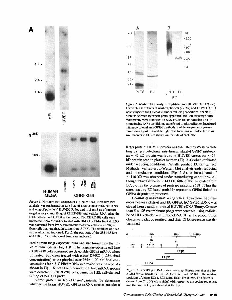
GPIb,3 mRNA expression in EC, HEL cells, megakaryocytes,and the megakaryoblastic cell line CHRF-288. Poly (A)' RNAfrom HUVEC was analyzed for GPIbB mRNA expression (Fig.1 A). Using a "full length" HEL cell-derived GPIb# cDNA as
the probe (8), Northern blot analysis showed an - 3.5-kbmRNA species in HUVEC RNA, while total cellular RNAfrom unstimulated HEL cells contained only the 1. 1-kb speciesthat has been reported previously by Lopez et al. (8) (Fig. 1 A).Platelet RNA contained only the 1.1-kb species ( 11 ). We evalu-
2418 Kelly, Essex, Shapiro, Meloni, Druck, Huebner, and Konkle
A
PLTS EC1.4-g
-J 0w w
>
:I
B 28S-AN".
VW;_ 4
18S -
0CO) . M
HUMAN 8MEGA CHRF-288
Figure 1. Northern blot analysis of GPlb,3 mRNA. Northern blotanalysis was performed on (A) 5 gg of total cellular HEL cell RNAand 4 ,g of poly (A)' HUVEC RNA, and in B on 3 ytg of humanmegakaryocyte and 10 g of CHRF-288 total cellular RNA using theHEL cell-derived GPIbf3 as the probe. The CHRF-288 cells wereuntreated (CONTROL) or treated with DMSO or PMA for 4 d. RNAwas harvested from PMA-treated cells that were adherent (ADH) or
from cells that remained in suspension (SUSP). The positions ofRNAsize markers are indicated. For B, the positions of the 28S (4.8 kb)and 18S ( 1.7 kb) ribosomal bands are indicated.
ated human megakaryocyte RNA and also found only the 1.1-kb mRNA species (Fig. 1 B). The megakaryoblastic cell lineCHRF-288 cells contained no detectable GPIb,# mRNA whenuntreated, but when treated with either DMSO (1.25% finalconcentration) or the phorbol ester PMA (100 nM final con-centration) for 4 d, GPIb# mRNA expression was induced. Asshown in Fig. 1 B, both the 3.5- and the 1. 1-kb mRNA specieswere detected in CHRF-288 cells, using the HEL cell-derivedGPIbfl cDNA as a probe.
GPIb# protein in HUVEC and platelets. To determinewhether the larger HUVEC GPIb, mRNA species encodes a
NR RIL.
EC
Figure 2. Western blot analysis of platelet and HUVEC GPIbI3. (A)Triton X-l00 extracts of washed platelets (PLTS) and HUVEC (EC)were subjected to SDS-PAGE under reducing conditions, or (B) ECproteins selected by wheat germ agglutinin and ion exchange chro-matography were subjected to SDS-PAGE under reducing (R) ornonreducing (NR) conditions, transferred to nitrocellulose, incubatedwith a polyclonal anti-GPIb3 antibody, and developed with peroxi-dase-labeled goat anti-rabbit IgG. The locations of molecular masssize markers in kD are shown on the side of each blot.
larger protein, HUVEC protein was evaluated by Western blot-ting. Using a polyclonal anti-human platelet GPIbJ3 antibody,an - 45-kD protein was found in HUVEC versus the - 24-kD protein seen in platelet extracts (Fig. 2 A) when evaluatedunder reducing conditions. Partially purified EC GPIbf3 (seeMethods) was subject to Western blot analysis under reducingand nonreducing conditions (Fig. 2 B). A broad band of- 116 kD was observed under nonreducing conditions. Al-though intact GPIba is - 143 kD, little of this is isolated fromEC, even in the presence of protease inhibitors ( 18 ). Thus thecross-reacting EC band probably represents GPIbI3 linked toGPIba degradation products.
Isolation ofendothelial GPIbf3 cDNA. To explore the differ-ences between platelet and EC GPIbB, EC GPIbfl cDNA wascloned from a random-primed HUVEC cDNA library. Greaterthan 5 X 10⁶ recombinant phage were screened using radiola-beled HEL cell-derived GPIbB cDNA (8) as the probe. Threeclones were plaque purified, and their DNA sequence was de-termined.
0 1 Kb 2Kb 2.792Kb
ScN PBP B P P P Si N
ii i I.
ECo1ECJ2
ECP4
Figure 3. EC GPIbB cDNA restriction map. Restriction sites are in-cluded for: B, BamHI; P, PstI; N, NcoI; Sc, Sacd; SI, SalI. The relativepositions ofclones ECB 1, ECB2, and ECf34 are shown. The figure isdrawn from 5' to 3' (left to right) with respect to the coding sequence,and the size, in kb, is indicated at the top.
Complementary DNA Cloning ofEndothelial Glycoprotein Ib4 2419
A 4,,.. kD
4.4 -
205 -
a'..
I
2.4 -
117 -
kD
- 200
-116- 97- 66
. 45
77 -
47 -
33 -
- 31
B

The three isolated clones were sequenced in their entirety The HEL cell cDNA sequence, as reported by Lopez et al. (8),and are illustrated in Fig. 3. The complete sequence and puta- is contained entirely within the EC sequence, as illustrated intive translation product is illustrated in Fig. 4. There is a long 5' bold type in Fig. 4. Our DNA sequence ofthis region was iden-untranslated region with stop codons in all three reading tical to that reported by Lopez et al. (8) except for nt 1275frames. This is followed by three "in frame" methionines at (HEL cell sequence nt 46) which is a "C" in our clones and anucleotides 637, 658, and 715. Using the first methionine as "G" in the HEL cell sequence and does not change the aminothe initiation codon, there is a 1233-nt open reading frame. acid encoded.
5'ctttattctcagcaccac cctcccaggtcattgtgtctgtttccgaggggcctggaccgtagcccgcccagctggcctctctgaccttgggggatc 101aggagcgaagttgggcgggacttcagagatccgcctcccttacccttc~cccgc~cccggacggtcacagcacccaaaccgcagg~cctgctctggcaggcag 204gcaaagctaggcagaagaggattcccaggatcctgggtctgttccctg~cccagtagctgcagaacggacttgggag~cctcctttgcctgctcecgcgggtc 307acccagcgagtgctgagaccccattttctgtcgaggcgggccgagtcttcccttatccccagacgcctagcgggcagggttgggctgaatcaaatgggagccc 410tccagacataaggaggccagaggctgcaaggagcggggtcgtgaccgcttacaccccttctccacagcccggcccgacctggagggcssccccggggcactgg 513gcggtgagccacctcctggcaactctcggtgccgtcccctgcc~tcgctcgaggcctcttctccccagcaccgctgtggtgtgccgggatcctgagcctaggc 616ctcccgatgttcccacccgc 636ATG ATC CCT TCC CGC CAC ACG ATG CTC CGT TTT CTT CCC GTT GTG AAT GCC GCG TCC TGT CCT GGT GAC AGG AGA ACA 714Met Ile Pro Ser Arg His Thr Met Leu Arg Phe Leu Pro Val Val Asn Ala Ala Ser Cys Pro Gly Asp Arg Arg Thr 26
ATG TTG GTG AAC GTC GCA GCG GGT GTC CGA GTG CTC CGT GTG CCC CTG AGA GCG GGT GGG AGC GGA AGC CTG AGC GGC 792Met Leu Val Asn Val Ala Ala Gly Val Arg Val Leu Arg Val Pro Leu Arg Ala Gly Gly Ser Gly Ser Leu Ser Gly 52
CTG CGG CCT CCG GCG ATA GTG TGC TAT CTG CCG CTG CAG CGC GCG TCC GCG GCC TCT GGG CTA TTT CTG GCC AGG CCG 870Leu Arg Pro Pro Ala Ile Val Cys Tyr Leu Pro Leu Gln Arg Ala Ser Ala Ala Ser Gly Leu Phe Leu Ala Arg Pro 78
CAG CAC TGT GGT CGG TGC GGG CGT GGC AGG GGC GGG GCG GCC TTA TCG CTC GGC TCT CCC GCC TAC GCC TCC CGC TGC 948Gln His Cys Gly Arg Cys Gly Arg Gly Arg Gly Gly Ala Ala Leu Ser Leu Gly Ser Pro Ala Tyr Ala Ser Arg Cys 104
AGA GTA AGC COG OCT oCC GTC TTC TCG CCA TOG OCT CCG GTG AGT CTG GAG TCC GGT CGG GCC CCC GGC TGC TCC CTA 1026Arg Val Ser Arg Ala Ala Val Phe Ser Pro Trp Ala Pro Val Ser Leu Glu Ser Gly Arg Ala Pro Gly Cys Ser Leu 130
GGC CGA CCC GGG TTG AGA GGA GCT CTG GTC GTT TGG CTG CAG CTG GGA GAG ACT TGG GTC AGA CTT AGA GGG GAC TTC 1104Gly Arg Pro Gly Leu Arg Gly Ala Leu Val Val Trp Leu Gln Leu Gly Glu Thr Trp Val Arg Leu Arg Gly Asp Phe 156
CAG CCG GCG TGC GGG GTG GTC AGG GTG GAG AGG CTG GCG GGC TAC CGG GAC GCC GGG CAT CAG GGG CTG GAT GGA GCC 1182Gln Pro Ala Cys Gly Val Val Arg Val Glu Arg Leu Ala Gly Tyr Arg Asp Ala Gly His Gln Gly Leu Asp Gly Ala 182
GGG CCG GCA GTC TGG GTA CTC AGA GAT GTC GCC CAG GTG CCC GCC GAC CGC TCG GCT TAC TGC GGC GCT TCC CTT GCA 1260Gly Pro Ala Val Trp Val Leu Arg Asp Val Ala Gln Val Pro Ala Asp Arg Ser Ala Tyr Cys Gly Ala Ser Leu Ala 208
GOG CCG CGC 00M 0=C CTG AGC TTA CTG CTC CTG CTG CTG 0CC CCG Cca AGC CC COG 0CC 0CG GOT TOC CCG GOC COC 1338Gly Pro Arg Gly Ala Lou Bor Leu Lou Lou Lou Lou Lou Ala Pro Pro Ser Arg Pro Ala Ala Gly Cys Pro Ala Pro 234
TOT AOC TOC OCG OOG ACG CTC GTG GkC TOC OOG COC CGC OOG CTG ACT TOGC TCO CTG CCG ACC 0Cc TTC CCT OTC 1416Cys Sor Cys Ala Gly Tbr Lou Val Asp Cys Gly Arg Arg Gly Lou Thr Trp Ala Ser Lou Pro Thr Ala Phe Pro Val 260
GAC ACA AC GAG CTG GTG CWG ACC GOC A&C AAC CTG ACG OCO CTG COG CoG G0 CTG CT GA GM CT CCC aOC CTG 1494Asp Thr Thr Glu Lou Val Lou Thr Gly Asn Asn Iu Thr Ala Lou Pro Pro Gly Lou Lou Asp A1a Lou Pro Ala Lou 286
caC ACC GCM aCC CG CC AAC CC TO COC TOC GAC TOC COC CTT GTG CCG CTG COC CCC TOG CTG 0CC GOC COC 1572Arg Thr Ala His Lou Gly Ala Asn Pro Trp Arg Cys Asp Cys Arg Lou Val Pro Lou Arg Ala Trp Lou Ala Gly Arg 312
CC GAG COT OCG CCC TAC CGC GAC CaG COT TOC GTG OCG CCC CCA OCG CTG CoC ooC CGC CTG CTG CCC TAT CTG 0cC 1650Pro Glu Arg Ala Pro Tyr Arg Asp Leu Arg Cys Val Ala Pro Pro Ala Lou Arg Gly Arg Lou Lou Pro Tyr Lou Ala 338
GAG GAC GAG CTG Coc 0cc OCT TOC OCT CCC GOC CCG CTC TOC TOG GOG OCO CTG OCO OCo CAG CTT OG CTG CTG ooc 1728Glu Asp Glu Lou Arg Ala Ala Cys Ala Pro Gly Pro Lou Cys TZp Gly Ala Leu Ala Ala Gln Leu Ala Lou Lou Gly 364
CTT GOG CTG CTG ChC OCG TTG CTo CTG GTG CTG CTG CTG TOC COC CTG COG AOG CTG COG 0CC COG 0CC CoC OT CoC 1806Lou Gly Lou Lou His Ala Lou Lou Lou Val Lou Lou LOu Cys Arg Lou Arg Arg Lou Arg Ala Arg Ala Arg Ala Arg 390
OCC OCG 0CC COG CT0 TCG CTG ACC GAC COG CTG OTO 0CC GAG CO 0CC OGA ACC GAC GAG TCC tgaggagagaaooggtog 1888Ala Ala Ala Arg Lou Ser Lou Thr Asp Pro Lou Val Ala Glu Arg Ala Gly Thr Asp Glu Sor 411
tcctgaggagagaacaggcgctggggaaaacgggcctgcaaactegaeagaccctgae g ecateg~egecaacotggacoggtacoxegetctcog 1991ctgcccaatetetcagacccaccccacctgcaggecagaccaegt ggacagaactcctgcacaccctac cegggSSS~ggcgaaccagcacttcaggett 2094gggagg ecatgggg--- atgoggtccagaccctgctgcgtetccttccaaactctggtgctgaat!%!-cccttctgatctggtcttctctgcacgactga 21 97cctggaaattccc~tcgccaaactcaggaaaagg~cccaggaggcggttcaaggggacacaaacacatcctcacatgcccaccctaacctcaggggcagacag 2300gagtcagtgggtgacctctc~ccttccagacacactcccaaaaggccacatgggaaggcatgagagagatgctaagtgataacgtgggggccagtgaggacgt 2403tgtggggacactgaggaccctgtgggggcagtgaggatgctgtggggatgctgtggggacagtaaggacactgggggcagtgaggacgctgtggacactgcgg 2506acgctatggggacaatgacgctgtggacagtgaggacgctgtgggggcggtgaggacactgtggagtgaggacactgggggcagtgaggacgctgtggacagt 2609ggggcgctgtgggggcggtgaggacgctgtaggggccgtggaggacgctgtggacggtgagaacgctgtggggacactgaggatgctgtgaggacgctgtggg 22712ggcagtgaggacgctgtggaccgtgaggacgctgtgggggcagtgaggacgctgtggaccgtgaggacgctgtggggatgg 3' 2793
Figure 4. EC GPIb3 cDNA sequence. The cDNA sequence is shown. Nucleotide sequence is indicated at the right at the end of each line, andamino acid residue numbers, beginning with the putative initiator methionine at nt 637, are indicated at the right immediately below the nu-
cleotide number. Nucleotides and amino acids highlighted in bold are identical to the reported HEL cell sequence. The HEL cell signal peptidecleavage point is indicated by A. The putative transmembrane domain for the EC and HEL peptides (8) is underlined. The putative phosphor-ylation site (5) is indicated by *. The sequence has been submitted to GenBank, accession number 20860.
2420 Kelly, Essex, Shapiro, Meloni, Druck, Huebner, and Konkle

The first 32 nt of the HEL cell sequence correspond to ECsequence nt 956-987, however the EC sequence is not in thesame reading frame as in the HEL cell sequence. In the ECsequence these 32 nt are followed by 274 nt not found in theHEL cell cDNA (nt 988-1261 ), and then the sequence is againidentical, resuming with HEL cell nt 33 (EC nt 1262) in thesame reading frame as found in the HEL cell sequence.
Using the first in frame methionine as the initiation codon,the EC sequence gives a predicted translation product of 411amino acids with a calculated molecular mass of 43 kD. Fromamino acid 209 through the carboxy terminus of the EC pro-tein, it is identical to the HEL cell translation product, aminoacids 4-206. The HEL cell translation product contains a 25-amino acid signal peptide at its NH2 terminus, which is cleavedto produce the mature protein. There are no sequences sugges-tive of a signal peptide at the 5' end of the EC protein (27).Much of the signal peptide sequence ofthe HEL cell protein isretained within the EC sequence and may serve as an internalsignal peptide. Computer assisted analysis (28, 29) predictsonly one transmembrane region, identical to the one in theHEL cell sequence.
Analysis of the GPIbJ gene. The genes encoding GPIba,GPIX, and GPV are simple in structure, with the coding re-gions contained within a single exon ( 13, 14, 30, 31 ). To evalu-ate the GPIb,3 gene, human genomic DNA was cut with therestriction enzymes BamHI, NcoI, and PstI, which all cutwithin the EC GPIb,3 cDNA (see Fig. 3), and analyzed in du-plicate by Southern blotting using both ECj34 and the HELcell-derived GPIbf3 cDNA as probes. The size ofthe fragmentspredicted from the cDNA were detected in genomic DNA asillustrated in Fig. 5. We also amplified nt 715-1218 and 1276-2168 using PCR to amplify genomic DNA. The sizes predictedfrom the cDNA were found in genomic DNA and are illus-trated in Fig. 5. To confirm that the amplified bands wereGPIb,3, the PCR products were cut with the restriction en-zymes Sad and Sal, respectively, and the appropriate-sizedproducts were obtained. Taken together these data suggest thatat least nt 266-2102 are contained within one exon.
To confirm that thecDNA sequence present in the endothe-lial transcripts, but apparently spliced out in megakaryocyte
HEL GplbP
ECP4
EC Gp~bP rcDNA
Figure 5. Evaluation of the GPIb# gene using Southern blot analysesand PCR. 10 ,g ofgenomic DNA was cut with BamHI, NcoI, or PstIand analyzed in duplicate by Southern blotting using either the HELcell-derived or the EC GPIbfl clone ECB4 as the probe. A drawing ofthe EC GPlbfl cDNA is shown in the middle of the figure. Abovethis the areas included in the probes are indicated. The fragmentspredicted by restriction enzyme analysis of the cDNA sequence,which were the same size detected by Southern blot analysis, are des-ignated in A. For B, PCR primers were devised from GPIbB cDNAsequence and used to amplify genomic DNA. The size fragmentspredicted from the cDNA were found in genomic DNA and their lo-cations are indicated.
and HEL cell transcripts, is present in genomic DNA, we am-plified by PCR nt 961-1446 from genomic DNA preparedfrom peripheral blood white blood cells. DNA sequence analy-sis through both presumed intron-exon boundaries confirmedtheir presence in genomic DNA.
The GPIbf3 gene maps to chromosome region 22pter -22q11.2. The radiolabeled human cDNA probe ECB1 detectsone human EcoRI restriction fragment of - 25 kb. 14 rodent-human hybrid DNAs, each carrying a few or many humanchromosomes so that most human chromosome regions wererepresented, were tested for the presence ofthe GPIbB-specifichuman EcoRI fragment by filter hybridization to the radiola-beled cDNA probe. All hybrids carrying human chromosome22 exhibited the strongly hybridizing 25-kb human EcoRIband, while all hybrids without human chromosome 22 werenegative for the human fragment. Thus the human GPIbf3 geneis located on chromosome 22 (data summarized in Fig. 6). Achromosome 22 hybrid mapping panel, in which each hybridDNA carries a specific portion of chromosome 22 (26), wastested similarly for the presence of the GPIbf3 fragment. Onlyhybrids that retained 22pter -o 22q 11.2 tested positive for theGPIbB gene; a drawing ofthe most relevant hybrids is shown inFig. 7.
GPIbB mRNA expression in different tissues. To evaluatethe pattern of GPIba, GPIbf3, and GPIX mRNA expression,we probed a Northern blot containing poly (A)+ RNA fromhuman heart, brain, placenta, lung, liver, skeletal muscle, kid-ney, and pancreas with cDNA probes for GPIba, GPIbf3, andGPIX as illustrated in Fig. 8. When probed with full lengthHEL cell-derived GPIX cDNA, a faint 1-kb mRNA specieswas seen only in the RNA from lung and placenta. When re-probed with full length HEL cell-derived GPIba cDNA, a 2.5-kb mRNA species was detected also in lung and placentalRNA, and upon longer exposure, faintly in liver. However,when the blot was probed with full length HEL cell-derivedGPIbI3 cDNA (EC nt 956-988; 1262-2179), the 3.5- and the1.1-kb mRNA species were seen in heart, and the 3.5-kbmRNA species was seen in brain, both at a much greater rela-tive abundance than the levels of GPIba and GPIX mRNAdetected in lung and placental RNA. The two GPIb,3 mRNAspecies, in varying relative abundance, were also detected atlower levels in the other tissues. When the blot was reprobedwith an EC-specific probe (the 367-bp BamHI fragment ofEC34, nt 233-600), the 3.5-kb mRNA species and, in addi-tion, an 2.3-kb species were detected predominantly in heartand brain, but also at a lower relative abundance in other tis-sues (Fig. 8).
Discussion
GPIbf is part of a four protein complex in platelets that,through the GPIba chain, serves as a receptor for von Wille-brand factor and thrombin. This platelet complex plays a ma-jor role in platelet-subendothelial interactions. Patients lackingthis complex have a moderate to severe bleeding disorder, theBernard-Soulier syndrome. However, the expression ofGPIba,GPIbf3, and GPIX is not limited to platelets and megakaryo-cytes. GPIba and GPIX mRNA and protein are expressed inEC and appear to be identical in EC and platelets ( 17, 18, andKonkle, B. A., F. J. Meloni, and S. S. Shapiro, unpublishedobservations). GPIb, mRNA and protein species are differentin EC from those found in platelets and HEL cells, as we haveillustrated in this paper.
Complementary DNA Cloning ofEndothelial Glycoprotein Ibhi 2421

Air an Venom osomes
It 13141516 17 1 8 19 ItO}I Ill2113l1411fll6l17118X0121HX I
1 1 1 1 1 1 11 I T 1S1_ 1 1 I T 1 1 1 1 1F _ _ rue SE I - 3 - , _ _ _1] r - - _ | 1 ! w _I1 w w w - lily_7_|_ _ _ I T I _7 _____-S r__ _ -]-EI E _ _ _ __ _ _ -. 1..Ji-_ __ _ - _, , - - - __t --_- ----E ___ _ _ _I__ 7 -W__ __ _ _ _ I I_ _ *IFI __ E _ _ d _ d__ _ AT_ 5___ _ _,_ _c he_ _I__I___ _ __ _ _ _ sC _. L _ E L i __ ___ F 01X _ _s T --9 -1 1-, _ _I__T___ - --- | --__ _I__I___ _ F __ _ _ _ CA
Figure 6. Presence of the GPIbf3 locus in a panel of 14rodent-human hybrids. * indicates that the hybridnamed in the left column contains the chromosome in-dicated in the upper row; w indicates presence of the longarm of the chromosome (or part ofthe long arm repre-sented by a smaller fraction of stippling); e indicatespresence of the short arm (or partial short arm) of thechromosome; o indicates the absence of the chromosomelisted above the column. The column for chromosome22 is boldly outlined and stippled to highlight correlationof presence of this chromosome with presence of theGPIbfi gene. The pattern of retention of the gene in thepanel is shown to the right ofthe figure where presenceof the locus in a hybrid is indicated by a stippled box witha plus sign, and absence of the locus is indicated by anopen box enclosing a minus sign.
EC GPIbfl cDNA is approximately three times the size ofHEL cell and platelet species. Some of this difference is due toadditional 5' and 3' untranslated (UT) regions not present inHEL cell cDNA. Our clones encompass 2.8 kb ofcDNA, andthus additional 5' and 3' UT sequences are likely present in thefull length cDNA. The EC coding region is approximatelytwice that ofHEL cells. The predicted EC translation productof 43 kD fits well with the protein size found on Western blotanalysis ofHUVEC proteins.
We are not certain which ofthe three in frame methioninespresent at the beginning ofthe coding region serves as the trans-lation initiation site. Translation does not initiate with theATG used for the HEL cell sequence (located at EC nt 956),since, with the unique EC sequence that follows that ATG,there would be no long open reading frame. In addition, since apolyclonal anti-GPIbf antibody recognizes both the EC andplatelet proteins, they must share a common region, and thishomology can only occur ifone ofthe upstream initiation sitesis used. Studies are currently underway to obtain amino-ter-minal protein sequence to confirm the translation initiationsite. We have used the first methionine as the assumed initia-tion site since this is true for 95% ofmRNAs reviewed by Ko-zak (32). However, the second methionine at nt 658 fits theconsensus sequence proposed by Kozak (33) for translation
05
co_ ,0
Lfl
(2)
22 + -
07N1~ <rm C
Nrrrn~
N _
- - + + GplbB
Figure 7. Regional localization of the GPIbfl gene in rodent-humanhybrids carrying partial chromosome 22. Hybrids carrying chromo-some 22 or a region of 22 are illustrated to the right of the 22 ideo-gram, with results of filter hybridization to the GPIb#-specific probeindicated below the lines representing specific hybrids; the GPIbf3locus is present in hybrids which retain the region 22pter -L 22q 1 1.2in common. Hybrid GL5 retains a derivative 17 chromosome froma constitutional t( 17;22)(pl3;ql 1.2) translocation break. All thesehybrids were described in detail previously (26).
initiation better than the first and may serve as the translationinitiation site.
EC GPIbf contains an unusually long 5' UT region.cDNAs with long leader sequences and multiple ATG codonshave been recognized most commonly in oncogenes, growthfactors, transcription factors, signal transduction components,and receptor proteins (34, 35). These frequently have G-C-rich leader sequences (70-90% GC), implying considerable sec-ondary structure. The EC GPIbj3 5' UT contains 66% G + C.The entire cDNA for the HEL cell and the EC species is rela-tively G-C rich, containing 73 and 68% G + C, respectively.
GpIX
Gp Iba
HEL cellGplbp
ECGplbp
(nt 233-600)
IZIIz <r
a:zIm C
-J
a-
-f
z W W <D >-J Z W
w L
(n
Figure 8. Northern blot analysis of various human tissues for GPIba,GPIX, and GPIbI3 mRNA expression. A Northern blot containing2 gg of poly (A)' RNA from the tissues indicated was probed,stripped of radiolabeled probe, then reprobed with the cDNAs indi-cated on the left side of the figure. For the EC-specific GPIbfi probe,the BamHI fragment (nt 233-600) of ECfl4 was used.
2422 Kelly, Essex, Shapiro, Meloni, Druck, Huebner, and Konkle
Hybrids
M44J14-2
9142730077-30
734GL511102
10027885410888
PB5-5Nu9
1113010664
131211.211.111.21213 .113.3

There are no sequences suggestive of a signal peptide at the5' end of the GPIbf coding sequence. Much of the coding se-quence which serves as the signal peptide for platelet and HELcell GPIbo is present in the middle of the EC GPIb3 cDNAsequence. This region may serve as an internal signal peptidefor EC GPIb3. Internal signal peptides are common in proteinsthat have multiple transmembrane regions, but may also beseen in proteins with only one membrane-spanning region (36).
EC GPIb3 appears to have the same transmembrane regionas that found in platelets and HEL cells. The cysteines externalto the membrane in platelet GPIbf, one of which is probablyinvolved in the disulfide-mediated linking of GPIb,3 to GPIba(8), are present in the EC sequence. Western blot analysis ofunreduced HUVEC protein reveals a larger species than thatobtained in reduced gels, and we have successfully purified ECGPIb3 from HUVEC protein using an affinity column con-taining the monoclonal antibody to GPIba, AP- 1, suggestingthat at least some GPIbf3 is disulfide linked to GPIba in ECalso.
GPIba, GPIbB, GPIX, and GPV are known to be membersof the leucine-rich glycoprotein family. GPIba contains 7 tan-dem LRG repeats of 24 amino acids each, while HEL cellGPIbf and GPIX each contain 1, and GPV contains 15 LRGrepeats (2, 13, 14). EC GPIbf3 contains the one repeat commonto the HEL cell species, but no additional ones in the uniqueamino-terminal region. The function ofLRG repeats in theseproteins as well as in the other proteins in which they are foundis unknown, although they may play a role in adhesion (2).
When the EC cDNA sequence was compared with othersequences accessible through GenBank and EMBL databases,sequence similarity was found to other cDNAs encoding pro-teins with LRG repeats. These similarities were within the re-gions common to HEL cell and EC cDNA. Sequence similaritywith the cDNA unique to EC was found with two previouslyreported expressed sequence tags (EST), both cloned frombrain cDNA (37, 38). Both were obtained by rapid, single passsequencing methods. One 248-bp clone (EST 01258) was ob-tained from a human hippocampal library (37) and is nearlyidentical to EC GPIbo nt 2467-2714, located in the 3' UTregion. The other clone (IB616) was obtained from an infantbrain cDNA library (38), is 132 nt in length, and is nearlyidentical to EC GPIbB nt 1174-1301. This is located within theEC coding sequence. These ESTs most likely represent thesame transcript as the EC GPIb3 cDNA, as both were reportedwith a number of undetermined nucleotides (six each in EST01258 and IB616), and the differences in sequence between theESTs and EC GPIbf cDNA (2/248 in EST 01258 and 5/132in IB616) may represent sequencing errors or possibly poly-morphisms.
The GPIbf3 gene, with two or possibly three alternativelyprocessed mRNA species, still appears to be quite simple instructure, as are genes for GPIb, GPIX, and GPV. The GPIba,GPIX, and GPV genes have been cloned and, in each case, thecoding region is contained within one exon (13, 14, 30, 31).The EC GPIbf cDNA is also contained within one exon, andthe HEL cell and platelet species are produced by splicing outthe 274-nt EC sequence, nt 987-1261. This EC sequence hasperfect splice acceptor and donor sequences at its 5' and 3'ends (39).
ECs appear to contain only the larger 3.5-kb GPIbB mRNAspecies, while HEL cells and platelets contain only the smaller1. 1-kb mRNA species. We do not know whether the multiplemRNA species seen in the tissues reflect alternative expression
in different cells or within a single cell type. However, it isinteresting that the megakaryoblastic cell line CHRF-288 ex-presses both mRNA species. CHRF-288 cells were derivedfrom a solid tumor in an infant with megakaryoblastic leuke-mia and have retained some characteristics ofthe original solidtumor, including production of basic fibroblast growth factorand TGF-3 (40). The larger mRNA species seen in these cells,but not in normal megakaryocytes, may be due either to thissolid tumor phenotype or, possibly, to the malignant pheno-type of these cells. Of note, HEL cells derived from a fluidphase leukemia (41 ) do not express the larger mRNA species.
When we probed the human tissue blot with the 5' end ofthe endothelial cDNA, we observed a third mRNA species of- 2.3 kb. This was not detected using the HEL cell-derivedprobe, even under conditions oflow stringency, suggesting thatthe gene extends 5' from our sequence. Part or all ofthe lung 5'UT region of the EC GPIbf3 cDNA may contain part of thismRNA species, although additional processing would beneeded as there is not a long open reading frame in this area.We have not detected the 2.3-kb mRNA species in EC, HELcell, or CHRF-288 cell RNA (data not shown).
In patients with the Bernard-Soulier syndrome, GPIba,GPIbf3, GPIX, and GPV are all missing or affected. We nowknow that they are products of separate genes. GPIba andGPIX are located on chromosomes 17 and 3, respectively (30,31 ), and, as we have shown, GPIbo is located on chromosome22. The molecular defects present in patients with Bernard-Soulier syndrome are now being elucidated. Three differentmutations in GPIba have been reported in three different pa-tients (42-44), and a family with two mutations in GPIX pro-ducing a double heterozygous state has been reported (45). Nomutations have been reported to date in GPIbfl.
It appears from our studies that GPIba and GPIX may notneed to be present for GPIbf3 expression, at least at the mRNAlevel, since we found relatively abundant GPIb3 mRNA ex-pression in heart and brain (and less abundant expression inother tissues), while GPIba and GPIX mRNA species weredetected only in placenta and lung. Considering that placentaand lung are the most vascular of those tested, this may repre-sent EC expression. In situ hybridization and immunohisto-chemistry on tissue sections will be needed to confirm this. Incollaboration with Asch and Nachman, we previously per-formed in situ hydrization and immunohistochemistry on ton-silar tissue and have found that GPIba appeared to be ex-pressed only in the ECs in that tissue (17). While GPIbfmRNA expression could represent endothelial expression indifferent vascular beds, this is unlikely given the pattern ofdistribution and the expression of both mRNA species.
The broad distribution ofGPIbo mRNA in different tissuessuggests that it may be playing a role different than or addi-tional to its function in platelets. Other than its presence beingrequired for stable expression ofGPIba, the function ofGPIbf3in platelets is not known. GPIbf protein is phosphorylatedunder conditions that increase cAMP and may play a role inmediating platelet shape change through GPIba which islinked to actin-binding protein (5). Whether GPIbf serves as asignal transduction molecule for other proteins or binds addi-tional ligands itself remains to be explored.
Acknowledgments
We thank Lu Zhang for technical assistance; Dr. David Ginsburg forproviding the HUVEC cDNA library; Dr. Jose Lopez and Dr. Gerald
Complementary DNA Cloning ofEndothelial Glycoprotein Ibfl 2423
Roth for providing the HEL cell GPIbfB cDNA clone and GPIbl# anti-body; Dr. Michael Lieberman for providing the CHRF-288 cells; Dr.Robert Montgomery for providing the monoclonal antibody, AP-l;and James Averback and Dr. Devjani Chatteijee for assistance withDNA/protein computer database access. Chromosome drawings werereproduced from Idiogram Albums, 1993, by copyright permission ofDavid Adler, University of Washington, Seattle.
This work was supported in part by grants HL-09163 (S. S. Sha-piro), HL-449566 (B. A. Konkle), and CA-5 1083 (K. Huebner), andtraining grant HL-0737 1 (D. W. Essex) from the National Institutes ofHealth. This work was performed during the tenure of an AmericanHeart Association-Squibb Corporation Clinician Scientist Award toB. A. Konkle.
References
1. Ruggeri, Z. M. 1991. The platelet glycoprotein Ib-IX complex. Prog. Hemo-stasis Thromb. 10:35-68.
2. Roth, G. J. 1991. Developing relationships: arterial platelet adhesion, gly-coprotein Ib, and leucine-rich glycoproteins. Blood. 77:5-19.
3. Andrews, R. K. 1991. Interaction ofpurified actin-binding protein with theplatelet membrane GPIb-IX complex. J. Biol. Chem. 266:7144-7147.
4. Wyler, B., D. Bienz, K. J. Clemtson, and K. J. Luscher. 1986. GPIb,5 is theonly phosphorylated major membrane glycoprotein in human platelets. Biochem-istry. 234:373-379.
5. Wardell, M. R., C. C. Reynolds, M. C. Berndt, R. W. Wallace, and J. E. B.Fox. 1989. Platelet GPIbl is phosphorylated on serine 166 by cAMP-dependentprotein kinase. J. Biol. Chem. 264:15656-15661.
6. Modderman, P. W., L. G. Admiral, A. Sonnenberg, and A. E. G. Kr vondem Borne. 1991. Glycoproteins V and Ib-IX form a noncovalent complex in theplatelet membrane. J. Biol. Chem. 267:364-369.
7. Lopez, J. A., D. W. Chung, K. Fujikawa, F. S. Hagen, T. Papayannopou-lou, and G. J. Roth. 1987. Cloning of the a chain of human platelet GPIb: atransmembrane protein with homology to leucine rich a2-glycoprotein. Proc.Natl. Acad. Sci. USA. 84:5615-5619.
8. Lopez, J. A., D. W. Chung, K. Fujikawa, F. S. Hagen, E. W. Davie, andG. J. Roth. 1988. The a and ft chains ofhuman platelet GPIb are both transmem-brane proteins containing a leucine-rich amino acid sequence. Proc. Natl. Acad.Sci. USA. 85:2135-2139.
9. Hickey, M. J., L. L. Deaven, and G. J. Roth. 1990. Human platelet GPIX:characterization of"full-length" cDNA and localization to chromosome 3. FEBS(Fed. Eur. Biochem. Soc) Lett. 274:189-192.
10. Kieffer, N., N. Debili, A. Wicki, M. Titeux, A. Henri, Z. Mishal, J. Breton-Gorius, W. Vainschenker, and K. J. Clemetson. 1986. Expression of GPIba inHEL cells. J. Biol. Chem. 261:15854-15862.
11. Roth, J. G., M. J. Hickey, D. W. Chung, and D. D. Hickstein. 1989.Circulating human blood platelets retain appreciable amount ofpoly (A)' RNA.Biochem. Biophy. Res. Commun. 160:705-7 10.
12. Lopez, J. A., B. Leung, C. C. Reynolds, C. Q. Li, and J. E. B. Fox. 1992.Efficient plasma membrane expression ofa functional platelet glycoprotein Ib-IXcomplex requires the presence of its three subunits. J. Biol. Chem. 267:1285 1-12859.
13. Hickey, M. J., F. S. Hagen, M. Yagi, and G. J. Roth. 1993. Human plateletglycoprotein V: characterization of the polypeptide and the related Ib-V-IX re-ceptor system ofadhesive, leucine-rich glycoproteins. Proc. Natl. Acad. Sci. USA.90:8327-8331.
14. Lanza, F., M. Morales, C. deLa Salle, J.-P. Cazenave, K. J. Clemetson, T.Shimomura, and D. R. Phillips. 1993. Cloning and characterization of the geneencoding the human platelet glycoprotein V. J. Biol. Chem. 268:20801-20807.
15. Sprandio, J. D., S. S. Shapiro, P. Thiagarajan, and S. McCord. 1988.Cultured human umbilical vein endothelial cells contain a membrane glycopro-tein immunologically related to glycoprotein lb. Blood. 71:234-237.
16. Asch, A. S., B. Adelman, M. Fujimoto, and R. L. Nachman. 1988. Identi-fication and isolation ofplatelet GPIb-like protein in human umbilical vein endo-thelial cells and bovine aortic smooth muscle cells. J. Clin. Invest. 81:1600-1607.
17. Konkle, B. A., S. S. Shapiro, A. S. Asch, and R. L. Nachman. 1990.Cytokine enhanced expression ofGPIba in human endothelium. J. Biol. Chem.265: 1983-1938.
18. Rajagopalan, V., D. W. Essex, S. S. Shapiro, and B. A. Konkle. 1992.Tumor necrosis factor-a modulation of glycoprotein Iba expression in humanendothelial and erythroleukemia cells. Blood. 80:153-16 1.
19. Konkle, B. A., and D. Ginsburg. 1988. The addition of endothelial cellgrowth factor and heparin to human umbilical vein endothelial cell cultures
decreases plasminogen activator inhibitor-l expression. J. Clin. Invest. 82:579-585.
20. Sambrook, J., E. F. Fritsch, and T. Maniatis. 1989. Molecular Cloning: ALaboratory Manual. Cold Spring Harbor Laboratory, Cold Spring Harbor, NY.545 pp.
21. Feinberg, A. P., and B. Vogelstein. 1983. A technique for radiolabelingDNA restriction endonuclear fragments to high specific activity. Anal. Biochem.132:6-13.
22. Ginsburg, D., R. I. Handin, D. T. Bonthron, T. A. Donlon, G. A. P. Burns,S. A. Latt, and S. H. Orkin. 1985. Human von Willebrand factor (vWF): isola-tion of complementary DNA clones and chromosomal localization. Science(Wash. DC). 228:1401-1406.
23. Benton, W. D., and R. W. Davis. 1977. Screening lambda gt recombinantclones by hybridization to single plaques in situ. Science (Wash. DC). 196:180-182.
24. Thiagarajan, P., S. S. Shapiro, E. Levine, L. DeMarco, and A. Yalcin.1985. A monoclonal antibody to human platelet GPIIIa detects a related proteinin cultured human endothelial cells. J. Clin. Invest. 75:896-901.
25. Huebner, K., T. Druck, C. M. Croce, and H. J. Thiesen. 1991. Twenty-se-ven non-overlapping cDNAs from T-cells encoding zinc finger structures map tonine human chromosomes with apparent clustering. Am. J. Hum. Genet. 48:726-740.
26. Bauer, S. K., K. Huebner, M. Budarf, J. Finan, J. Erikson, B. Emanual,P. C. Nowell, C. M. Croce, and F. Melchers. 1988. Localization of the humanVpre B gene to the 22q 1 1.2 region ofchromosome 22 near a cluster of VI, genesegments. Immunogenetics. 28:328-333.
27. von Heijne, G. 1985. Signal sequences: the limits of variation. J. Mol.Biol. 184:99-105.
28. Chou, P. Y., andG. D. Fasman. 1978. Prediction ofsecondary structure ofproteins from their amino acid sequence. Adv. Enzymol. 47:45-148.
29. Garnier, J., D. J. Osguthorpe, and B. Robson. 1978. Analysis ofthe accu-racy and implications ofsimple methods for predicting the secondary structure ofglobular proteins. J. Mol. Biol. 120:97-120.
30. Wenger, R. H., A. N. Wicki, N. Kieffer, S. Adolph, S. Hameister, and K. J.Clemetson. 1989. The 5' flanking region and chromosomal localization of thegene encoding human platelet membrane GPIba. Gene (Amst.). 85:517-524.
31. Hickey, M. J., and G. J. Roth. 1993. Characterization ofthe gene encodinghuman platelet glycoprotein IX. J. Biol. Chem. 268:3438-3443.
32. Kozak, M. 1987. An analysis of 5'-noncoding sequences from 699 verte-brate messenger RNAs. Nucleic Acids Res. 15:8125-8148.
33. Kozak, M. 1986. Point mutations define a sequence flanking the AUGcodon that modulates translation of eukaryotic ribosomes. Cell. 44:283-292.
34. Kozak, M. 1991. An analysis of vertebrate mRNA sequences: intimationsof translational control. J. Cell Biol. 1 5:887-903.
35. Kozak, M. 1992. Regulation of translation in eukaryotic systems. Annu.Rev. Cell Biol. 8:197-225.
36. von Heijne, G. 1990. The signal peptide. J. Membr. Biol. 115:195-201.37. Abrams, J., M. Dubnick, A. R. Kerlavage, R. Moreno, J. M. Kelley, T. R.
Utterback, J. W. Nagle, C. Fields, and J. C. Venter. 1992. Sequence identificationof 2,375 human brain genes. Nature (Lond.). 355:632-634.
38. Khan, A. S., A. S. Wilcox, M. H. Polymeropoulos, J. A. Hopkins, T. J.Stevens, M. Robinson, A. K. Orpana, and J. M. Sikela. 1992. Single pass sequenc-ing and physical and genetic mapping ofhuman brain cDNAs. Nature Genetics.2:180-185.
39. McKeown, M. 1992. Alternative mRNA splicing. Annu. Rev. Cell Biol.8:133-155.
40. Fugman, D. A., D. P. Witte, C. L. Jones, B. J. Aionow, and M. A. Lieber-man. 1990. In vitro establishment and characterization of a human megakaryo-blastic cell line. Blood. 75:1252-1261.
41. Martin, P., and T. Papayannopoulou. 1992. HEL cells: a new humanerythroleukemia cell line with spontaneous and induced globin expression.Science (Wash. DC). 216:1233-1234.
42. Ware, J., S. R. Russell, V. Vincente, R. E. Scharf, A. Tomer, R. McMillan,and Z. M. Ruggeri. 1990. Nonsense mutation in the glycoprotein Iba codingsequence associated with Bernard-Soulier syndrome. Proc. Natl. Acad. Sci. USA.87:2026-2030.
43. Ware, J., S. Russell, M. Murata, M. Mazzucato, L. DeMarco, and Z. M.Ruggeri. 1992. Ala,1-Val substitution in platelet glycoprotein Iba impairs von
Willebrand factor binding and is the molecular basis of Bernard-Soulier syn-drome. Blood. 79:278a. (Abstr.)
44. Miller, J. L., V. A. Lyle, and D. Cunningham. 1992. Mutation ofleucine-57 to phenylalanine in a platelet glycoprotein Iba leucine tandem repeat occur-
ring in patients with an autosomal dominant variant of Bernard-Soulier disease.Blood. 79:439-446.
45. Wright, S. D., K. Michaelides, D. J. D. Johnson, N. C. West, and E. G. D.Tuddenham. 1993. Double heterozygosity for mutations in the platelet glycopro-tein IX gene in three siblings with Bernard-Soulier syndrome. Blood. 81:2339-2347.
2424 Kelly, Essex, Shapiro, Meloni, Druck, Huebner, and Konkle