Complement activation cascade triggered by PEG–PL engineered nanomedicines and carbon nanotubes:...
7
Complement activation cascade triggered by PEG–PL engineered nanomedicines and carbon nanotubes: The challenges ahead S.M. Moghimi a, ⁎, A.J. Andersen a , S.H. Hashemi a , B. Lettiero a , D. Ahmadvand a , A.C. Hunter b , T.L. Andresen c , I. Hamad d , J. Szebeni e a Centre for Pharmaceutical Nanotechnology and Nanotoxicology, Department of Pharmaceutics and Analytical Chemistry, University of Copenhagen, Universitetsparken 2, DK-2100 Copenhagen Ø, Denmark b Molecular Targeting and Polymer Toxicology Group, School of Pharmacy, University of Brighton, Brighton BN2 4GJ, UK c Department of Micro- and Nano-Technology, DTU Nanotech, Technical University of Denmark, Frederiksborgvej 399, DK-4000 Roskilde, Denmark d Department of Pharmaceutical Sciences and Pharmaceutics, Faculty of Pharmacy, Applied Sciences University, Amman, Jordan e Nanomedicine Research Centre, Bay Zoltán Foundation for Applied Research, Semmelweis Medical University and Seroscience Kft, Budapest, Hungary abstract article info Article history: Received 2 February 2010 Accepted 4 April 2010 Available online 11 April 2010 Keywords: Carbon nanotube Complement activation Liposome Micelle Poly(ethylene glycol) Pseudoallergy Since their introduction, poly(ethylene glycol)–phospholipid (PEG–PL) conjugates have found many applications in design and engineering of nanosized delivery systems for controlled delivery of pharmaceuticals especially to non-macrophage targets. However, there are reports of idiosyncratic reactions to certain PEG–PL engineered nanomedicines in both experimental animals and man. These reactions are classified as pseudoallergy and may be associated with cardiopulmonary disturbance and other related symptoms of anaphylaxis. Recent studies suggest that complement activation may be a contributing, but not a rate limiting factor, in eliciting hypersensitivity reactions to such nanomedicines in sensitive individuals. This is rather surprising since PEGylated structures are generally assumed to suppress protein adsorption and blood opsonization events including complement. Here, we examine the molecular basis of complement activation by PEG–PL engineered nanomedicines and carbon nanotubes and discuss the challenges ahead. © 2010 Elsevier B.V. All rights reserved. 1. Introduction MethoxyPEG 2000–5000 –phosholipid (mPEG–PL) conjugates are versatile molecules with numerous biomedical applications. These conjugates were initially incorporated into the liposomal bilayer for conferring longevity to vesicles in the systemic circulation; this eventually led to the development of Doxil ® approved for treatment of HIV-related Kaposi's sarcoma and refractory ovarian carcinoma [1–3]. PEGylated vesicles, through prolonged circulation times in the blood, can ultimately target many vascular elements following conjugation of targeting ligands to the distal end of the projected PEG chains bearing a reactive functional group [1,2,4]. Other attempts include coupling of contrast agents and bio-sensors to the distal end of the PEG chain for engineering of multifunctional vesicles applicable to a wide range of pathologies [2,4–6]. PEG–phospholipid conjugates exhibit low critical micelle concen- tration values and form stable micellar structures of approximately 30 nm in size, allowing drug solubilization and are amenable for additional functionalization and parenteral administration [7]. The application of PEG–phospholipid conjugates in steric stabilization and generation of long circulating oil-in-water nanoemulsions is also noteworthy [8]. Finally, apart from nanomedicine design and development, PEG–phospholipids can further stabilize entities such as carbon nanotubes through surface adsorption and enhance their dispersion in aqueous media; such modifications also affect nanotube pharmacokinetics following intravenous injection for biological and mechanistic studies [9]. There are indications that certain PEG–phospholipid bearing nanomedicines may be Janus-faced; today, there are clinical reports of acute hypersensitivity reactions to infusion of long circulating regulatory-approved PEGylated liposomes in sensitive individuals [10–12]. These reactions, classified as pseudoallergy, are often associated with flushing and circulatory disturbances [13]. Recent studies in pigs have suggested that the cardiopulmonary distress caused by Doxil ® and other liposomes strongly correlate with complement activation [14]. The complement system (Fig. 1), con- sisting of over 30 soluble plasma and cell-surface bound proteins, serves as an important effector of both innate and acquired immunity [15]. Accordingly, complement activation by PEGylated vesicles is an unexpected phenomenon since surface mPEG coverage is generally believed to dramatically suppress particle–protein interaction and blood opsonization events, including complement fixation [1,16]. Similar to the binding of allergens to IgE on the surface of mast cells and basophils, complement anaphylatoxins (and particularly C5a) can Journal of Controlled Release 146 (2010) 175–181 ⁎ Corresponding author. Tel.: + 45 35336528. E-mail address: [email protected] (S.M. Moghimi). 0168-3659/$ – see front matter © 2010 Elsevier B.V. All rights reserved. doi:10.1016/j.jconrel.2010.04.003 Contents lists available at ScienceDirect Journal of Controlled Release journal homepage: www.elsevier.com/locate/jconrel
Transcript of Complement activation cascade triggered by PEG–PL engineered nanomedicines and carbon nanotubes:...

Journal of Controlled Release 146 (2010) 175–181
Contents lists available at ScienceDirect
Journal of Controlled Release
j ourna l homepage: www.e lsev ie r.com/ locate / jconre l
Complement activation cascade triggered by PEG–PL engineered nanomedicines andcarbon nanotubes: The challenges ahead
S.M. Moghimi a,⁎, A.J. Andersen a, S.H. Hashemi a, B. Lettiero a, D. Ahmadvand a, A.C. Hunter b, T.L. Andresen c,I. Hamad d, J. Szebeni e
a Centre for Pharmaceutical Nanotechnology and Nanotoxicology, Department of Pharmaceutics and Analytical Chemistry, University of Copenhagen, Universitetsparken 2,DK-2100 Copenhagen Ø, Denmarkb Molecular Targeting and Polymer Toxicology Group, School of Pharmacy, University of Brighton, Brighton BN2 4GJ, UKc Department of Micro- and Nano-Technology, DTU Nanotech, Technical University of Denmark, Frederiksborgvej 399, DK-4000 Roskilde, Denmarkd Department of Pharmaceutical Sciences and Pharmaceutics, Faculty of Pharmacy, Applied Sciences University, Amman, Jordane Nanomedicine Research Centre, Bay Zoltán Foundation for Applied Research, Semmelweis Medical University and Seroscience Kft, Budapest, Hungary
⁎ Corresponding author. Tel.: +45 35336528.E-mail address: [email protected] (S.M. Moghimi)
0168-3659/$ – see front matter © 2010 Elsevier B.V. Aldoi:10.1016/j.jconrel.2010.04.003
a b s t r a c t
a r t i c l e i n f oArticle history:Received 2 February 2010Accepted 4 April 2010Available online 11 April 2010
Keywords:Carbon nanotubeComplement activationLiposomeMicellePoly(ethylene glycol)Pseudoallergy
Since their introduction, poly(ethylene glycol)–phospholipid (PEG–PL) conjugates have found manyapplications in design and engineering of nanosized delivery systems for controlled delivery ofpharmaceuticals especially to non-macrophage targets. However, there are reports of idiosyncratic reactionsto certain PEG–PL engineered nanomedicines in both experimental animals and man. These reactions areclassified as pseudoallergy and may be associated with cardiopulmonary disturbance and other relatedsymptoms of anaphylaxis. Recent studies suggest that complement activation may be a contributing, but nota rate limiting factor, in eliciting hypersensitivity reactions to such nanomedicines in sensitive individuals.This is rather surprising since PEGylated structures are generally assumed to suppress protein adsorptionand blood opsonization events including complement. Here, we examine the molecular basis of complementactivation by PEG–PL engineered nanomedicines and carbon nanotubes and discuss the challenges ahead.
.
l rights reserved.
© 2010 Elsevier B.V. All rights reserved.
1. Introduction
MethoxyPEG2000–5000–phosholipid (mPEG–PL) conjugates areversatile molecules with numerous biomedical applications. Theseconjugates were initially incorporated into the liposomal bilayer forconferring longevity to vesicles in the systemic circulation; thiseventually led to the development of Doxil® approved for treatment ofHIV-related Kaposi's sarcoma and refractory ovarian carcinoma [1–3].PEGylated vesicles, through prolonged circulation times in the blood,can ultimately target many vascular elements following conjugationof targeting ligands to the distal end of the projected PEG chainsbearing a reactive functional group [1,2,4]. Other attempts includecoupling of contrast agents and bio-sensors to the distal end of thePEG chain for engineering of multifunctional vesicles applicable to awide range of pathologies [2,4–6].
PEG–phospholipid conjugates exhibit low critical micelle concen-tration values and form stable micellar structures of approximately30 nm in size, allowing drug solubilization and are amenable foradditional functionalization and parenteral administration [7]. Theapplication of PEG–phospholipid conjugates in steric stabilization and
generation of long circulating oil-in-water nanoemulsions is alsonoteworthy [8]. Finally, apart from nanomedicine design anddevelopment, PEG–phospholipids can further stabilize entities suchas carbon nanotubes through surface adsorption and enhance theirdispersion in aqueous media; such modifications also affect nanotubepharmacokinetics following intravenous injection for biological andmechanistic studies [9].
There are indications that certain PEG–phospholipid bearingnanomedicines may be Janus-faced; today, there are clinical reportsof acute hypersensitivity reactions to infusion of long circulatingregulatory-approved PEGylated liposomes in sensitive individuals[10–12]. These reactions, classified as pseudoallergy, are oftenassociated with flushing and circulatory disturbances [13]. Recentstudies in pigs have suggested that the cardiopulmonary distresscaused by Doxil® and other liposomes strongly correlate withcomplement activation [14]. The complement system (Fig. 1), con-sisting of over 30 soluble plasma and cell-surface bound proteins,serves as an important effector of both innate and acquired immunity[15]. Accordingly, complement activation by PEGylated vesicles is anunexpected phenomenon since surface mPEG coverage is generallybelieved to dramatically suppress particle–protein interaction andblood opsonization events, including complement fixation [1,16].Similar to the binding of allergens to IgE on the surface of mast cellsand basophils, complement anaphylatoxins (and particularly C5a) can
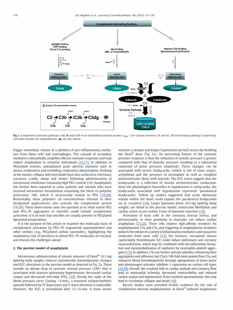
Fig. 1. Complement activation pathways. C4d, Bb and SC5b-9 are established activation markers ( ) for calcium-sensitive (CP and LP), AP and terminal pathways, respectively.Activation markers for anaphylatoxins ( ) are also shown.
176 S.M. Moghimi et al. / Journal of Controlled Release 146 (2010) 175–181
trigger immediate release of a plethora of pro-inflammatory media-tors from these cells and macrophages. This cascade of secondarymediators substantially amplifies effector immune responses andmayinduce anaphylaxis in sensitive individuals [13,17]. In addition toPEGylated vesicles, unexplained acute adverse reactions such asataxia, restlessness and trembling, respiratory abnormalities, frothingat themouth, collapse and even death have also surfaced in veterinaryscenarios (cattle, sheep and swine) following administration ofintravenous medicines containing high PEG content [18]. Anaphylaxishas further been reported in some patients and animals who havereceived intravenous formulations containing the block co-polymerpoloxamer 188, which is structurally similar to PEG [19,20].Remarkably, these polymers (at concentrations relevant to theirdesignated applications) also activate the complement system[18,20]. These observations raise the question as to what extent PEGand PEG–PL aggregates or micelles could initiate complementactivation; it is of note that micelles are usually present in PEGylatedliposomal preparations.
It is the purpose of this article to examine the molecular basis ofcomplement activation by PEG–PL engineered nanomedicines andother entities (e.g., PEGylated carbon nanotubes), highlighting themodulatory role of interfaces in which PEG–PL molecules are located,and discuss the challenges ahead.
2. The porcine model of anaphylaxis
Intravenous administration of minute amounts of Doxil® (0.1 mglipid/kg body weight) induces reproducible haemodynamic changesand ECG alterations in the porcine model as depicted in Fig. 2a. Theseinclude an abrupt drop in systemic arterial pressure (SAP) that isassociated with massive pulmonary hypertension, decreased cardiacoutput and decreased end-tidal PCO2 [14]. During the nadir of theblood pressure curve (lasting ∼4 min), a transient tachyarrhythmicepisode followed by ST depression and T-wave elevation is noticeable.However, the ECG is normalized after 12–15 min. A more severe
reaction (a deeper and longer hypotensive period) occurs by doublingthe Doxil® dose (Fig. 2a). An interesting feature of the systemicpressure response is that the reduction of systolic pressure is greatercompared with that of diastolic pressure, resulting in a substantialreduction of pulse pressure amplitude. These changes can beassociated with severe bradycardia (which is not of sinus origin),arrhythmia and the presence of incomplete as well as completeatrioventricular block with asystole. The ECG traces suggest that thebradycardia is a reflection of slowed atrioventricular conduction.Since the physiological baroreflex to hypotension is tachycardia, thebradycardia associated with hypotension represents ‘paradoxicalbradycardia’. Follow up studies suggested that acute adenosinerelease within the heart could explain the paradoxical bradycardiavia A1 receptors [14]. Larger liposome doses (0.5 mg lipid/kg bodyweight) are lethal in the porcine model; ventricular fibrillation andcardiac arrest occurs within 3 min of liposome injection [14].
Activation of mast cells in the coronary arterial intima, andperivascularly, in close proximity to myocytes can induce cardiacanaphylaxis [21,22]. These cells express high-affinity receptors foranaphylatoxins C3a and C5a, and triggering of anaphylatoxin receptorsinduces the releaseof a varietyof inflammatorymediators andvasoactivemolecules from mast cells [13]. For instance, eicosanoid release(particularly thromboxane A2) could induce pulmonary and coronaryvasoconstriction, which may be combined with microthrombus forma-tion and microembolization of capillaries by neutrophil–platelet aggre-gates [13]. In addition, C3a can further activate platelets, enhancing theiraggregation and adhesion, but C5a is 100-foldmore potent than C3a, andenhances blood thrombogenicity through upregulation of tissue factorand plasminogen activator inhibitor-1 expression on various cell types[23,24]. Overall, the resultant falls in cardiac preload and coronary flowlead to myocardial ischemia, decreased contractibility and reducedcardiac output and hypotension. If not resolved spontaneously, this maylead to circulatory collapse and death [14].
Recent studies have provided further evidence for the role ofcomplement-derived anaphylatoxins in Doxil®-induced anaphylaxis
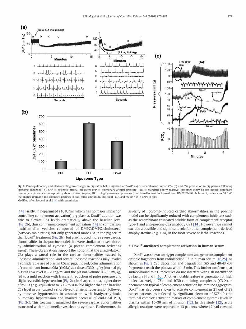
Fig. 2. Cardiopulmonary and electrocardiogram changes in pigs after bolus injection of Doxil® (a) or recombinant human C5a (c) and C5a production in pig plasma followingliposome challenge (b). SAP = systemic arterial pressure; PAP = pulmonary arterial pressure; PRL = standard poorly reactive liposomes (they do not induce significanthaemodynamic and cardiorespiratory abnormalities) in pigs; HRL = highly reactive liposomes (multilamellar vesicles formed from DMPC:DMPG:cholesterol, mole ratios 50:5:45that induce dramatic and extended declines in SAP, pulse amplitude, end-tidal PCO2 and major rise in PAP) in pigs.Modified after Szebeni et al. [14] with permission.
177S.M. Moghimi et al. / Journal of Controlled Release 146 (2010) 175–181
[14]. Firstly, in heparinized (10 IU/ml, which has no major impact oncontrolling complement activation) pig plasma, Doxil® addition wasable to elevate C5a levels dramatically above the baseline level(Fig. 2b), thus confirming complement activation [14]. In comparison,multilamellar vesicles composed of DMPC:DMPG:cholesterol(50:5:45 mole ratios) not only generated more C5a in the pig serumthan Doxil® treatment (Fig. 2b), but also induced more severe cardiacabnormalities in the porcinemodel that were similar to those inducedby administration of zymosan (a potent complement-activatingagent). These observations support the notion that the anaphylatoxinC5a plays a causal role in the cardiac abnormalities caused byliposome administration, and severe liposome reactions may involvea considerable rise of plasma C5a in pigs. Indeed, bolus administrationof recombinant human C5a (rhC5a) at a dose of 330 ng/kg (normal pigplasma C5a level is ∼20 ng/ml and the plasma volume is ∼33 ml/kg)led to a mild reaction with transient reduction of pulse pressure andslight reversible hypertension (Fig. 2c). In sharp contrast, higher dosesof rhC5a (e.g., equivalent to 600- to 700-fold higher than the baselineC5a level in pig) caused a short-lived transient hypertension followedby massive hypotension in association with bradyarrhythmia,pulmonary hypertension and marked decrease of end-tidal PCO2
(Fig. 2c). This treatment mimicked the severe cardiac abnormalitiesassociated withmultilamellar vesicles and zymosan. Furthermore, the
severity of liposome-induced cardiac abnormalities in the porcinemodel can be significantly reduced with complement inhibitors suchas the recombinant truncated soluble form of complement receptortype-1 and anti-porcine C5a antibody GS1 [14]. However, we cannotexclude a possible and significant role for other complement-derivedanaphylatoxins (e.g., C3a) in the most severe or lethal reactions.
3. Doxil®-mediated complement activation in human serum
Doxil®was shown to trigger complement and generate complementopsonic fragments from radiolabelled C3 in human serum [16,25]. Asshown in Fig. 3 C3b deposition and degradation (65 and 40/43 kDafragments) reach the plateau within 5 min. This further confirms thatsurface-bound mPEG molecules do not interfere with C3b inactivationby factors H and I [16]. Another notable feature is generation of highmolecular weight C3b- and iC3b-containing complexes (C3-X), aphenomenon typical of complement activation by immune aggregates.Doxil® has also been shown to activate complement in 21 out of 29cancer patients, as reflected by significant elevation of SC5b-9 (theterminal complex activation marker of complement system) levels inplasma within 10–30 min of infusion [12]. In this study [12], acuteallergic reactions were reported in 13 patients, where 12 had elevated
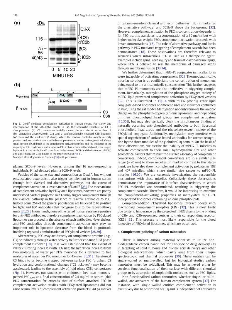
Fig. 3. Doxil®-mediated complement activation in human serum. For clarity andinterpretation of the SDS-PAGE profile in (a), the schematic structure of C3 isalso presented (b). C3 convertases initially cleave the α chain at arrow head 1(b), generating anaphylatoxin C3a and a conformationally changed C3b fragment(α' chain and the uncleaved β chain), where the reactive thiolester moiety in C3dgportion can formcovalentbondswith the complement-activating surface/particle P. Only asmall portion of C3b binds to the complement-activating surface and the thiolester of themajority of C3b reacts with water to form iC3b. C3b is sequentially catalyzed (two stages)by factor I (arrowheads 2 and3), resulting in the release of C3f (and the formation of iC3b)and C3c. This leaves C3dg bound to the target (see also Fig. 1).Modified after Moghimi and Szebeni [16] with permission.
178 S.M. Moghimi et al. / Journal of Controlled Release 146 (2010) 175–181
plasma SC5b-9 levels. However, among the 16 non-respondingindividuals, 9 had elevated plasma SC5b-9 levels.
Vesicles of the same size and composition as Doxil®, but withoutencapsulated doxorubicin, also trigger complement in human serumthrough both classical and alternative pathways, but the extent ofcomplement activation is less than that of Doxil® [25]. Themechanismsof complement activation by PEGylated liposomes, however, are poorlyunderstood. Surface projectedmPEGsmay trigger complement throughthe classical pathway in the presence of reactive antibodies to PEG.Indeed, some 25% of the general populations are believed to be positivefor IgG2 and IgM antibodies that recognize four to five repeat ethoxyunits [26,27]. In our hands, none of the tested human sera were positivefor anti-PEG antibodies, therefore complement activation by PEGylatedliposomes can proceed in the absence of such antibodies. Nevertheless,anti-PEG antibodies through complement activation may play animportant role in liposome clearance from the blood in protocolsinvolving repeated administration of PEGylated vesicles [28,29].
Alternatively, PEG may act directly on complement proteins (e.g.,C3) or indirectly throughwater activity to further enhance fluid phasecomplement turnover [18]. It is well established that the extent ofwater clustering increaseswith PEG size; the hydration increases fromtwo molecules of water per PEG monomer for a tetramer to fivemolecules of water per PEGmonomer for 45-mer [30,31]. Therefore, ifC3 binds to or become trapped between surface PEG ‘brushes’, C3hydration and conformational changes (“C3 tickover”) may becomeaccelerated, leading to the assembly of fluid phase C3Bb convertases(Fig. 1). However, our studies with endotoxin free near monodis-persed PEG2000 at a final concentration of 2.5 mg/ml in serum (thisPEG concentration far exceeds that of surface attached PEG incomplement activation studies with PEGylated liposomes) did notraise serum levels of complement activation products C4d (a marker
of calcium-sensitive classical and lectin pathways), Bb (a marker ofthe alternative pathway) and SC5b-9 above the background [32].However, complement activation by PEG is concentration-dependent;for PEG2000 this translates to a concentration of ≥10 mg/ml but withhigher molecular weight PEGs complement activation proceeds withlower concentrations [18]. The role of alternative pathway and lectinpathway in PEG-mediated triggering of complement cascade has beendemonstrated [18]. These observations are therefore relevant toscenarios where intravenous PEG is used as a therapeutic agent;examples include spinal cord injury and traumatic axonal brain injury,where PEG is believed to seal the membrane of damaged axonsthrough membrane fusion [33,34].
We further determined that mPEG–PL conjugates in micellar formwere incapable of activating complement [32]. Thermodynamically,micellar solution is at equilibrium, the concentration of monomersbeing equal to the critical micelle concentration. This further suggeststhat mPEG–PL monomers are also ineffective in triggering comple-ment. Remarkably, methylation of the phosphate-oxygen moiety ofmPEG–lipid prevented complement activation by PEGylated vesicles[32]. This is illustrated in Fig. 4 with mPEG–prodrug ether lipidconjugate-based liposomes of different sizes and is further confirmedin vivo, using the rat model. Methylation not only removes the anioniccharge on the phosphate-oxygen (anionic liposomes, and dependingon their phospholipid head group, are complement activators[15,35]), but may also sterically block the simultaneous binding ofnaturally occurring anti-phospholipid antibodies to both liposomalphospholipid head group and the phosphate-oxygen moiety of thePEGylated conjugate. Additionally, methylation may interfere withspatial organization of surface-bound antibodies for correct recogni-tion by the three modules of the globular C1q domain. On the basis ofthese observations, we ascribe the inability of mPEG–PL micelles toactivate complement to their small hydrodynamic size and othergeometrical factors that restrict the surface assembly of complementconvertases. Indeed, complement convertases are in a similar sizerange (∼20 nm) to these micelles. In marked contrast to this state-ment, we have also shown complement activation by poloxamer 188and 407 micelles, which share similar size ranges to mPEG–PLmicelles [16,20]. We are currently investigating the responsiblemechanisms with these micelles. Collectively, these observationsdemonstrate the importance of interface characteristics, in whichPEG–PL molecules are accumulated, resulting in triggering thecomplement cascade. Therefore, it would be interesting to examinethe complement-activating properties of methylated mPEG–PLincorporated liposomes containing anionic phospholipids.
Complement-fixed PEGylated liposomes interact poorly withmacrophage complement receptors (CRs) [32]. This is most likelydue to steric hinderance by the projected mPEG chains to the bindingof C3b- and iC3b-opsonized vesicles to their corresponding receptor(CR3) [32]. This process is most likely responsible for the bloodlongevity of PEGylated liposomes, which are opsonized.
4. Complement policing of carbon nanotubes
There is interest among material scientists to utilize non-biodegradable carbon nanotubes for site-specific drug delivery (asin targeting of solid tumours and nucleic acid delivery) and otherbiological interventions, which partly arise from their uniquespectroscopic and thermal properties [36]. These entities can besingle-walled or multi-walled, but for biological studies carbonnanotubes must be solubilized. This may be achieved either bycovalent functionalization of their surface with different chemicalgroups or by adsorption of amphiphilic molecules, such as PEG–lipids.
Non-functionalized carbon nanotubes, whether single- or multi-walled, are activators of the human complement system [37]. Forinstance, with single-walled entities complement activation isexclusively due to adsorption of C1q and is independent of antibodies
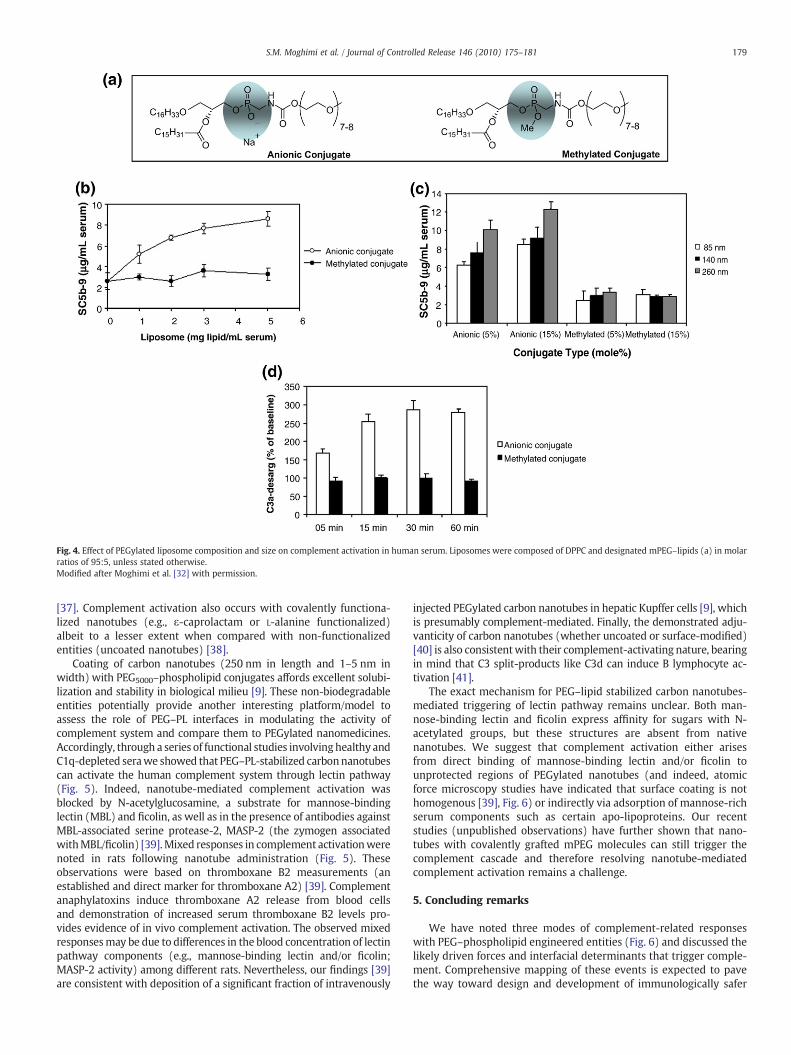
Fig. 4. Effect of PEGylated liposome composition and size on complement activation in human serum. Liposomes were composed of DPPC and designated mPEG–lipids (a) in molarratios of 95:5, unless stated otherwise.Modified after Moghimi et al. [32] with permission.
179S.M. Moghimi et al. / Journal of Controlled Release 146 (2010) 175–181
[37]. Complement activation also occurs with covalently functiona-lized nanotubes (e.g., ε-caprolactam or L-alanine functionalized)albeit to a lesser extent when compared with non-functionalizedentities (uncoated nanotubes) [38].
Coating of carbon nanotubes (250 nm in length and 1–5 nm inwidth) with PEG5000–phospholipid conjugates affords excellent solubi-lization and stability in biological milieu [9]. These non-biodegradableentities potentially provide another interesting platform/model toassess the role of PEG–PL interfaces in modulating the activity ofcomplement system and compare them to PEGylated nanomedicines.Accordingly, through a series of functional studies involvinghealthy andC1q-depleted serawe showed that PEG–PL-stabilized carbonnanotubescan activate the human complement system through lectin pathway(Fig. 5). Indeed, nanotube-mediated complement activation wasblocked by N-acetylglucosamine, a substrate for mannose-bindinglectin (MBL) and ficolin, as well as in the presence of antibodies againstMBL-associated serine protease-2, MASP-2 (the zymogen associatedwithMBL/ficolin) [39].Mixed responses in complement activationwerenoted in rats following nanotube administration (Fig. 5). Theseobservations were based on thromboxane B2 measurements (anestablished and direct marker for thromboxane A2) [39]. Complementanaphylatoxins induce thromboxane A2 release from blood cellsand demonstration of increased serum thromboxane B2 levels pro-vides evidence of in vivo complement activation. The observed mixedresponsesmay be due to differences in the blood concentration of lectinpathway components (e.g., mannose-binding lectin and/or ficolin;MASP-2 activity) among different rats. Nevertheless, our findings [39]are consistent with deposition of a significant fraction of intravenously
injected PEGylated carbon nanotubes in hepatic Kupffer cells [9], whichis presumably complement-mediated. Finally, the demonstrated adju-vanticity of carbon nanotubes (whether uncoated or surface-modified)[40] is also consistentwith their complement-activating nature, bearingin mind that C3 split-products like C3d can induce B lymphocyte ac-tivation [41].
The exact mechanism for PEG–lipid stabilized carbon nanotubes-mediated triggering of lectin pathway remains unclear. Both man-nose-binding lectin and ficolin express affinity for sugars with N-acetylated groups, but these structures are absent from nativenanotubes. We suggest that complement activation either arisesfrom direct binding of mannose-binding lectin and/or ficolin tounprotected regions of PEGylated nanotubes (and indeed, atomicforce microscopy studies have indicated that surface coating is nothomogenous [39], Fig. 6) or indirectly via adsorption of mannose-richserum components such as certain apo-lipoproteins. Our recentstudies (unpublished observations) have further shown that nano-tubes with covalently grafted mPEG molecules can still trigger thecomplement cascade and therefore resolving nanotube-mediatedcomplement activation remains a challenge.
5. Concluding remarks
We have noted three modes of complement-related responseswith PEG–phospholipid engineered entities (Fig. 6) and discussed thelikely driven forces and interfacial determinants that trigger comple-ment. Comprehensive mapping of these events is expected to pavethe way toward design and development of immunologically safer
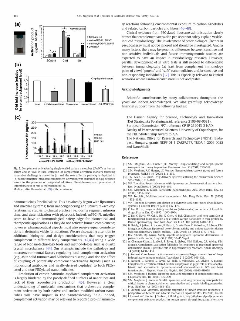
Fig. 5. Complement activation by single-walled carbon nanotubes (SWNT) in humanserum and in vivo in rats. Detection of complement activation markers followingnanotubes challenge is shown in (a) and the role of lectin pathway is depicted in(b) where nanotube-mediated complement activation was examined in C1q-depletedserum in the presence of designated additives. Nanotube-mediated generation ofthromboxane B in rats is represented in (c).Modified after Hamad et al. [39] with permission.
180 S.M. Moghimi et al. / Journal of Controlled Release 146 (2010) 175–181
nanomedicines for clinical use. This has already begunwith liposomesand micellar systems; from nanoengineering and ‘structure–activity’relationship studies to clinical practice (i.e., dosing regimen, infusiontime, and desensitization with placebos). Indeed, mPEG–PL micellesseem to have an immunological safety edge for biomedical andtherapeutic applications as they do not activate human complement;however, pharmaceutical aspects must also receive equal considera-tions in designing viable formulations. We are also paying attention toadditional biological and design considerations that may triggercomplement in different body compartments [42,43] using a widerange of bionanotechnology tools and methodologies such as quartzcrystal microbalance [44]. Our attempts include the pathology andmicroenvironmental factors regulating local complement activation(e.g., as in solid tumours and Alzheimer's disease), and also the effectof coupling of potentially complement-activating ligands (such asmonoclonal antibodies and virally-derived peptides) to both PEGy-lated and non-PEGylated nanomedicines.
Resolution of carbon nanotube-mediated complement activationis largely hindered by the poorly-defined surfaces of nanotubes andlack of their reproducible production [45]. However, a clearunderstanding of molecular mechanisms that orchestrate comple-ment activation by both native and surface-modified carbon nano-tubes will have impact in the nanotoxicology field. Indeed,complement activation may be relevant to reported pro-inflammato-
ry reactions following environmental exposure to carbon nanotubesand related carbon particles and fibers [46–48].
Clinical evidence from PEGylated liposome administration clearlyattests that complement activation per se cannot solely explain vesicle-induced pseudoallergy. The involvement of other biological factors inpseudoallergy must not be ignored and should be investigated. Amongmany factors, there may be genomic differences between sensitive andnon-sensitive individuals and future immunogenomic studies areexpected to have an impact in pseudoallergy research. However,parallel development of in vitro tests is still needed to differentiatebetween immunologically (at least from complement immunologypoint of view) “potent” and “safe” nanomedicines and/or sensitive andnon-responding individuals [17]. This is especially relevant to clinicalscenarios where cardiovascular stress is not acceptable.
Acknowledgments
Scientific contributions by many collaborators throughout theyears are indeed acknowledged. We also gratefully acknowledgefinancial support from the following bodies:
The Danish Agency for Science, Technology and Innovation(Det Strategiske Forskningsråd, reference 2106-08-0081).European Commission FP7, reference CP-IP 212043-2 NAD.Faculty of Pharmaceutical Sciences, University of Copenhagen, forthe PhD Studentship Award to AJA.The National Office for Research and Technology (NKTH), Buda-pest, Hungary, grants NKFP 01 1-CARPA777, TUDA-1-2006-0035and NanoMedi.
References
[1] S.M. Moghimi, A.C. Hunter, J.C. Murray, Long-circulating and target-specificnanoparticles: theory to practice, Pharmacol. Rev. 53 (2001) 283–318.
[2] S.M. Moghimi, A.C. Hunter, J.C. Murray, Nanomedicine: current status and futureprospects, FASEB J. 19 (2005) 311–330.
[3] T.M. Allen, P.R. Cullis, Drug delivery systems: entering the mainstream, Science303 (2004) 1818–1822.
[4] V.P. Torchilin, Recent advances with liposomes as pharmaceutical carriers, Nat.Rev. Drug Discov. 4 (2005) 145–160.
[5] S.M. Moghimi, T. Kissel, Particulate nanomedicines, Adv. Drug Deliv. Rev. 58(2006) 1451–1455.
[6] V.P. Torchilin, Multifunctional nanocarriers, Adv. Drug Deliv. Rev. 58 (2006)1532–1555.
[7] V.P. Torchilin, Structure and design of polymeric surfactant-based drug deliverysystems, J. Control. Rel. 73 (2001) 137–172.
[8] F. Liu, D. Liu, Long-circulating emulsions (oil-in-water) as carriers of lipophilicdrugs, Pharm. Res. 12 (1995) 1060–1064.
[9] Z. Liu, C. Davis, W. Cai, L. He, X. Chen, H. Dai, Circulation and long-term fate offunctionalized, biocompatible single-walled carbon nanotubes in mice probed byRaman spectroscopy, Proc. Natl. Acad. Sci. U.S.A. 105 (2008) 1410–1415.
[10] B. Uziely, S. Jeffers, R. Isacson, K. Kutsch, D. Wei-Tsao, Z. Yehoshua, E. Libson, F.M.Muggia, A. Gabizon, Liposomal doxorubicin: activity and unique toxicities duringtwo complementary phase I studies, J. Clin. Oncol. 13 (1995) 1777–1785.
[11] D.S. Alberts, D.J. Garcia, Safety aspects of pegylated liposomal doxorubicin inpatients with cancer, Drugs 54 (1997) 30–45 Suppl.
[12] A. Channan-Khan, J. Szebeni, S. Savay, L. Liebes, N.M. Rafique, C.R. Alving, F.M.Muggia, Complement activation following first exposure to pegylated liposomaldoxorubicin (Doxil): possible role in hypersensitivity reactions, Annal. Oncology14 (2003) 1430–1437.
[13] J. Szebeni, Complement activation-related pseudoallergy: a new class of drug-induced acute immune toxicity, Toxicology 216 (2005) 106–121.
[14] J. Szebeni, L. Baranyi, S. Savay, M. Bodo, J. Milosevits, C.R. Alving, R. Bunger,Complement activation-related cardiac anaphylaxis in pigs: role of C5a anaphy-latoxin and adenosine in liposome-induced abnormalities in ECG and heartfunction, Am. J. Physiol. Heart Cir. Physiol. 290 (2006) H1050–H1058.
[15] S.M. Moghimi, I. Hamad, Liposome-mediated triggering of complement cascade,J. Liposome Res. 18 (2008) 195–209.
[16] S.M. Moghimi, J. Szebeni, Stealth liposomes and long circulating nanoparticles:critical issues in pharmacokinetics, opsonization and protein-binding properties,Prog. Lipid Res. 42 (2003) 463–478.
[17] J. Szebeni, S.M. Moghimi, Liposome triggering of innate immune responses: aperspective on benefits and adverse reactions, J. Liposome. Res. 19 (2009) 85–90.
[18] I. Hamad, A.C. Hunter, J. Szebeni, S.M. Moghimi, poly(ethylene glycol)s generatecomplement activation products in human serum through increased alternative
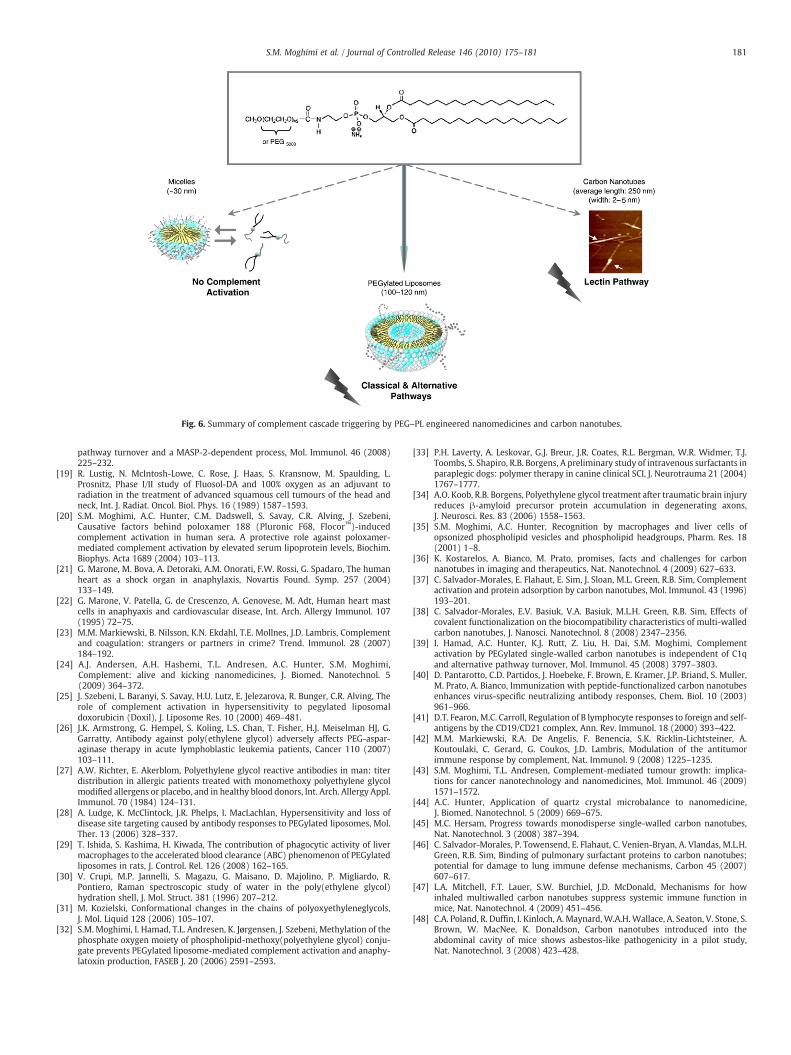
Fig. 6. Summary of complement cascade triggering by PEG–PL engineered nanomedicines and carbon nanotubes.
181S.M. Moghimi et al. / Journal of Controlled Release 146 (2010) 175–181
pathway turnover and a MASP-2-dependent process, Mol. Immunol. 46 (2008)225–232.
[19] R. Lustig, N. McIntosh-Lowe, C. Rose, J. Haas, S. Kransnow, M. Spaulding, L.Prosnitz, Phase I/II study of Fluosol-DA and 100% oxygen as an adjuvant toradiation in the treatment of advanced squamous cell tumours of the head andneck, Int. J. Radiat. Oncol. Biol. Phys. 16 (1989) 1587–1593.
[20] S.M. Moghimi, A.C. Hunter, C.M. Dadswell, S. Savay, C.R. Alving, J. Szebeni,Causative factors behind poloxamer 188 (Pluronic F68, Flocor™)-inducedcomplement activation in human sera. A protective role against poloxamer-mediated complement activation by elevated serum lipoprotein levels, Biochim.Biophys. Acta 1689 (2004) 103–113.
[21] G. Marone, M. Bova, A. Detoraki, A.M. Onorati, F.W. Rossi, G. Spadaro, The humanheart as a shock organ in anaphylaxis, Novartis Found. Symp. 257 (2004)133–149.
[22] G. Marone, V. Patella, G. de Crescenzo, A. Genovese, M. Adt, Human heart mastcells in anaphyaxis and cardiovascular disease, Int. Arch. Allergy Immunol. 107(1995) 72–75.
[23] M.M. Markiewski, B. Nilsson, K.N. Ekdahl, T.E. Mollnes, J.D. Lambris, Complementand coagulation: strangers or partners in crime? Trend. Immunol. 28 (2007)184–192.
[24] A.J. Andersen, A.H. Hashemi, T.L. Andresen, A.C. Hunter, S.M. Moghimi,Complement: alive and kicking nanomedicines, J. Biomed. Nanotechnol. 5(2009) 364–372.
[25] J. Szebeni, L. Baranyi, S. Savay, H.U. Lutz, E. Jelezarova, R. Bunger, C.R. Alving, Therole of complement activation in hypersensitivity to pegylated liposomaldoxorubicin (Doxil), J. Liposome Res. 10 (2000) 469–481.
[26] J.K. Armstrong, G. Hempel, S. Koling, L.S. Chan, T. Fisher, H.J. Meiselman HJ, G.Garratty, Antibody against poly(ethylene glycol) adversely affects PEG-aspar-aginase therapy in acute lymphoblastic leukemia patients, Cancer 110 (2007)103–111.
[27] A.W. Richter, E. Akerblom, Polyethylene glycol reactive antibodies in man: titerdistribution in allergic patients treated with monomethoxy polyethylene glycolmodified allergens or placebo, and in healthy blood donors, Int. Arch. Allergy Appl.Immunol. 70 (1984) 124–131.
[28] A. Ludge, K. McClintock, J.R. Phelps, I. MacLachlan, Hypersensitivity and loss ofdisease site targeting caused by antibody responses to PEGylated liposomes, Mol.Ther. 13 (2006) 328–337.
[29] T. Ishida, S. Kashima, H. Kiwada, The contribution of phagocytic activity of livermacrophages to the accelerated blood clearance (ABC) phenomenon of PEGylatedliposomes in rats, J. Control. Rel. 126 (2008) 162–165.
[30] V. Crupi, M.P. Jannelli, S. Magazu, G. Maisano, D. Majolino, P. Migliardo, R.Pontiero, Raman spectroscopic study of water in the poly(ethylene glycol)hydration shell, J. Mol. Struct. 381 (1996) 207–212.
[31] M. Kozielski, Conformational changes in the chains of polyoxyethyleneglycols,J. Mol. Liquid 128 (2006) 105–107.
[32] S.M. Moghimi, I. Hamad, T.L. Andresen, K. Jørgensen, J. Szebeni, Methylation of thephosphate oxygen moiety of phospholipid-methoxy(polyethylene glycol) conju-gate prevents PEGylated liposome-mediated complement activation and anaphy-latoxin production, FASEB J. 20 (2006) 2591–2593.
[33] P.H. Laverty, A. Leskovar, G.J. Breur, J.R. Coates, R.L. Bergman, W.R. Widmer, T.J.Toombs, S. Shapiro, R.B. Borgens, A preliminary study of intravenous surfactants inparaplegic dogs: polymer therapy in canine clinical SCI, J. Neurotrauma 21 (2004)1767–1777.
[34] A.O. Koob, R.B. Borgens, Polyethylene glycol treatment after traumatic brain injuryreduces β-amyloid precursor protein accumulation in degenerating axons,J. Neurosci. Res. 83 (2006) 1558–1563.
[35] S.M. Moghimi, A.C. Hunter, Recognition by macrophages and liver cells ofopsonized phospholipid vesicles and phospholipid headgroups, Pharm. Res. 18(2001) 1–8.
[36] K. Kostarelos, A. Bianco, M. Prato, promises, facts and challenges for carbonnanotubes in imaging and therapeutics, Nat. Nanotechnol. 4 (2009) 627–633.
[37] C. Salvador-Morales, E. Flahaut, E. Sim, J. Sloan, M.L. Green, R.B. Sim, Complementactivation and protein adsorption by carbon nanotubes, Mol. Immunol. 43 (1996)193–201.
[38] C. Salvador-Morales, E.V. Basiuk, V.A. Basiuk, M.L.H. Green, R.B. Sim, Effects ofcovalent functionalization on the biocompatibility characteristics of multi-walledcarbon nanotubes, J. Nanosci. Nanotechnol. 8 (2008) 2347–2356.
[39] I. Hamad, A.C. Hunter, K.J. Rutt, Z. Liu, H. Dai, S.M. Moghimi, Complementactivation by PEGylated single-walled carbon nanotubes is independent of C1qand alternative pathway turnover, Mol. Immunol. 45 (2008) 3797–3803.
[40] D. Pantarotto, C.D. Partidos, J. Hoebeke, F. Brown, E. Kramer, J.P. Briand, S. Muller,M. Prato, A. Bianco, Immunization with peptide-functionalized carbon nanotubesenhances virus-specific neutralizing antibody responses, Chem. Biol. 10 (2003)961–966.
[41] D.T. Fearon, M.C. Carroll, Regulation of B lymphocyte responses to foreign and self-antigens by the CD19/CD21 complex, Ann. Rev. Immunol. 18 (2000) 393–422.
[42] M.M. Markiewski, R.A. De Angelis, F. Benencia, S.K. Ricklin-Lichtsteiner, A.Koutoulaki, C. Gerard, G. Coukos, J.D. Lambris, Modulation of the antitumorimmune response by complement, Nat. Immunol. 9 (2008) 1225–1235.
[43] S.M. Moghimi, T.L. Andresen, Complement-mediated tumour growth: implica-tions for cancer nanotechnology and nanomedicines, Mol. Immunol. 46 (2009)1571–1572.
[44] A.C. Hunter, Application of quartz crystal microbalance to nanomedicine,J. Biomed. Nanotechnol. 5 (2009) 669–675.
[45] M.C. Hersam, Progress towards monodisperse single-walled carbon nanotubes,Nat. Nanotechnol. 3 (2008) 387–394.
[46] C. Salvador-Morales, P. Towensend, E. Flahaut, C. Venien-Bryan, A. Vlandas, M.L.H.Green, R.B. Sim, Binding of pulmonary surfactant proteins to carbon nanotubes;potential for damage to lung immune defense mechanisms, Carbon 45 (2007)607–617.
[47] L.A. Mitchell, F.T. Lauer, S.W. Burchiel, J.D. McDonald, Mechanisms for howinhaled multiwalled carbon nanotubes suppress systemic immune function inmice, Nat. Nanotechnol. 4 (2009) 451–456.
[48] C.A. Poland, R. Duffin, I. Kinloch, A. Maynard,W.A.H.Wallace, A. Seaton, V. Stone, S.Brown, W. MacNee, K. Donaldson, Carbon nanotubes introduced into theabdominal cavity of mice shows asbestos-like pathogenicity in a pilot study,Nat. Nanotechnol. 3 (2008) 423–428.