Lesion scoring technique for assessing the virulence and ...
Upload
independentCategory
view
1download
0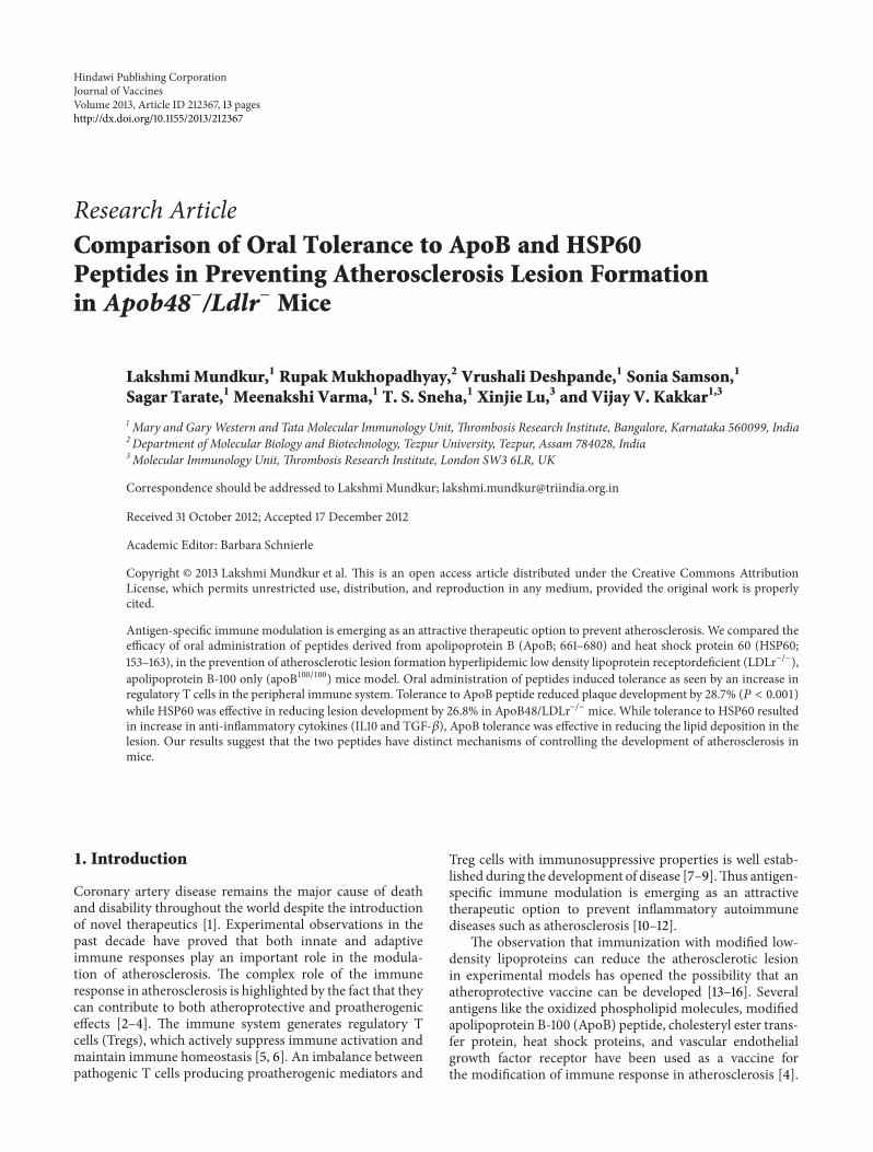
Hindawi Publishing CorporationJournal of VaccinesVolume 2013, Article ID 212367, 13 pageshttp://dx.doi.org/10.1155/2013/212367
Research ArticleComparison of Oral Tolerance to ApoB and HSP60Peptides in Preventing Atherosclerosis Lesion Formationin Apob48−/Ldlr− Mice
Lakshmi Mundkur,1 Rupak Mukhopadhyay,2 Vrushali Deshpande,1 Sonia Samson,1
Sagar Tarate,1 Meenakshi Varma,1 T. S. Sneha,1 Xinjie Lu,3 and Vijay V. Kakkar1,3
1 Mary and Gary Western and Tata Molecular Immunology Unit, Thrombosis Research Institute, Bangalore, Karnataka 560099, India2Department of Molecular Biology and Biotechnology, Tezpur University, Tezpur, Assam 784028, India3Molecular Immunology Unit, Thrombosis Research Institute, London SW3 6LR, UK
Correspondence should be addressed to Lakshmi Mundkur; [email protected]
Received 31 October 2012; Accepted 17 December 2012
Academic Editor: Barbara Schnierle
Copyright © 2013 Lakshmi Mundkur et al. This is an open access article distributed under the Creative Commons AttributionLicense, which permits unrestricted use, distribution, and reproduction in any medium, provided the original work is properlycited.
Antigen-specific immune modulation is emerging as an attractive therapeutic option to prevent atherosclerosis. We compared theefficacy of oral administration of peptides derived from apolipoprotein B (ApoB; 661–680) and heat shock protein 60 (HSP60;153–163), in the prevention of atherosclerotic lesion formation hyperlipidemic low density lipoprotein receptordeficient (LDLr−/−),apolipoprotein B-100 only (apoB100/100) mice model. Oral administration of peptides induced tolerance as seen by an increase inregulatory T cells in the peripheral immune system. Tolerance to ApoB peptide reduced plaque development by 28.7% (𝑃 < 0.001)while HSP60 was effective in reducing lesion development by 26.8% in ApoB48/LDLr−/− mice. While tolerance to HSP60 resultedin increase in anti-inflammatory cytokines (IL10 and TGF-𝛽), ApoB tolerance was effective in reducing the lipid deposition in thelesion. Our results suggest that the two peptides have distinct mechanisms of controlling the development of atherosclerosis inmice.
1. Introduction
Coronary artery disease remains the major cause of deathand disability throughout the world despite the introductionof novel therapeutics [1]. Experimental observations in thepast decade have proved that both innate and adaptiveimmune responses play an important role in the modula-tion of atherosclerosis. The complex role of the immuneresponse in atherosclerosis is highlighted by the fact that theycan contribute to both atheroprotective and proatherogeniceffects [2–4]. The immune system generates regulatory Tcells (Tregs), which actively suppress immune activation andmaintain immune homeostasis [5, 6]. An imbalance betweenpathogenic T cells producing proatherogenic mediators and
Treg cells with immunosuppressive properties is well estab-lished during the development of disease [7–9].Thus antigen-specific immune modulation is emerging as an attractivetherapeutic option to prevent inflammatory autoimmunediseases such as atherosclerosis [10–12].
The observation that immunization with modified low-density lipoproteins can reduce the atherosclerotic lesionin experimental models has opened the possibility that anatheroprotective vaccine can be developed [13–16]. Severalantigens like the oxidized phospholipid molecules, modifiedapolipoprotein B-100 (ApoB) peptide, cholesteryl ester trans-fer protein, heat shock proteins, and vascular endothelialgrowth factor receptor have been used as a vaccine forthe modification of immune response in atherosclerosis [4].
2 Journal of Vaccines
Immunotherapy is directed toward inducing tolerance to self-antigens mediated by protective antibodies or increasing theantigen-specific Treg cells, which can suppress the proathero-genic T-effector cells [11–16].
Themucosal (intranasal or oral) route of administration isan attractive method of inducing antigen-specific peripheraltolerance to treat autoimmune diseases [17]. Oral administra-tion suppresses the initiation of autoimmune diseases in ani-mal models of experimental autoimmune encephalomyelitis,uveitis, colitis, and atherosclerosis [18]. The efficiency oforal tolerance is dependent on the type of antigen and itsdose. While treatment with a high antigen dose results indeletion or anergy of peripheral T cells, low-dose treatmentinduces antigen-specific Treg cells [19, 20]. Recent reportshave demonstrated the atheroprotective role of natural Treg(nTreg) cells expressing CD25 and the transcription factor foxhead box p3 (Foxp3) and suppress proliferation of effectorcells by a contact-dependent mechanism [17]. Several studieshave established effective early reduction of atherosclerosisin hyperlipidemic mouse models by inducing tolerance tomodified lipids and peptides derived from apolipoprotein B(ApoB) 100, HSPs 60/65, and 𝛽2-glycoprotein [20–22].
We believe that each of these self antigens has a distinctrole to play in the development of the disease and usingmultiple peptides from these atherogenic proteins wouldbe more effective than using either of them alone. In thisstudy we compared the immune tolerance mediated bytwo peptides derived from ApoB 100 and HSP60 moleculeand its effect on atheroprotection in a double-gene knock-out (ApoBtm25gy/LDLrtm1Her) mouse model, which expressesonly ApoB100 and is deficient in low-density lipoproteinreceptor.The specific strain of mice was used for this study asmajority of cholesterol is transported in ApoB100-containinglipoproteins (i.e., LDL fraction), and also it is reported tomore closelymimic human atherosclerosis than othermodels[17].
2. Methods
2.1. Animals. ApoBtm2Sgy/Ldlrtm1Her/J knockout mice on aC57BL/6 background (ApoB48−/Ldlr−) were donated by theThrombosis Research Institute, London, UK. All animalexperiments were approved by the institutional animal ethicscommittee and in compliance with Government of Indiaguidelines and conform to the Guide for the Care andUse of Laboratory Animals published by the US NationalInstitutes of Health (NIH Publication, 8th Edition, 2011).Mice were kept under standard laboratory conditions andfed a normal chow diet (Nutrilab, India) or a high-fat diet(Harlan, TD 96121, Indianapolis, IN, USA). Food and waterwere administered ad libitum. 6–8 animals were used pergroup.
2.2. Antigens. The antigens used were apolipoproteinB-keyhole limpet hemocyanin (ApoB-KLH) peptide (ApoB-100 amino acids 661–680 conjugated to KLH, used at1mg/mL, dissolved in phosphate-buffered saline [PBS]) andheat shock protein 60-KLH (HSP60-KLH) peptide (HSP60
amino acids 153–163 conjugated to KLH, used at 1mg/mL,dissolved in PBS) (Severn Biotech, Worcester, UK). Thepeptides were administered orally, (20𝜇g per animal, perdose). PBS dosing was carried out as control.
2.3. Lipid Analysis. Blood from individualmice was collectedby retroorbital venous plexus under 3% Isoflurane inhalantanesthesia in oxygen as per the American VeterinaryMedicalAssociation guidelines (June 2007). Plasma was obtained bycentrifugation and was stored at −80∘C until the assay wasperformed. Concentrations of plasma total cholesterol andtriglycerides were determined by Siemens Dimension Flexreagent cartridge (Siemens Healthcare Diagnostics Ltd, UK)using the Cobas Fara II Clinical Chemistry auto analyzer (F.Hoffman La Roche Ltd., Basel, Switzerland), following themanufacturer’s instructions.
2.4. Antibody Response Measurement. Blood samples werecollected in heparinized capillaries by retro-orbital bleeding2 and 10 weeks after the first oral dose for assessment ofantibody production. Free ApoB-100 and human HSP-60peptides containing an N-terminal cysteine were synthe-sized by Severn Biotech (Worcester, UK) and were coatedon Maleimide-activated 96-well plates (Pierce, ThermoFisher Scientific Inc., USA). Peptide-specific immunoglob-ulin IgG or IgA was measured in the serum of immu-nized mice using horseradish peroxidase-conjugated 𝛼-mouse IgG and horseradish peroxidase-conjugated 𝛼-mouseIgA (Sigma chemicals, St. Louis, MO, USA) as secondaryantibodies.
2.5. Assessment of Atherosclerotic Lesions. Quantification ofatherosclerotic lesions was carried out as per the protocolapproved by the Animal Models of Diabetic ComplicationsConsortium (http://www.diacomp.org/). Mice were eutha-nized humanely using an overdose of isoflurane inhalantanesthetic (15%). Mouse hearts were perfused with 10mL ofPBS. Hearts were collected in two mediums: optimal cuttingtemperature (OCT) medium (Tissue Tek, Leica, Germany)and 10% buffered formalin. Aortic root sections (10 𝜇m)were cut from the hearts embedded in an OCT mediumin frozen conditions using a cryotome (Leica CM 1900UV Cryotome) and used for either immunofluorescence orimmunohistochemical studies. Similarly, aortic roots weresectioned from hearts in neutral buffered phenol (NBF) afterembedding in paraffin blocks. For lesion analysis in eachmouse, five sections 80𝜇m apart were stained with Elasticavan Geison (EVG). Several sections were measured and themean is calculated and expressed asmicrometer square, usingImage-Pro Plus software (Media Cybernetics, Bethesda, MD,USA).
2.6. Immunohistochemical Analysis of Atherosclerotic Lesions.Immunofluorescence on frozen sections was carried outusing an indirect immunofluorescence technique. Mice heartwas collected in OCT (JUNG tissue freezing media). Sec-tions (10 micron) were cut using a Leica CM 1900 UVcryotome. The frozen sections on poly-l-lysine-coated slides
Journal of Vaccines 3
(poly-prep-slides sigma) were permeabilized using 0.2%of triton X 100 for 30min, fixed with ice-cold acetone,and blocked with 5% goat serum or 5% rabbit serumdepending on the secondary antibody used. Further, thesections were incubated with primary antibodies (rat anti-mouse CD68 (AbD Serotech, Oxford, UK) anti-mouseCD4 (Abcam, Cambridge, UK), antimouse 𝛼-actin (Abcam,Cambridge, UK) for 2 h followed by incubation with anappropriate secondary antibody (Alexa-633 tagged Invitro-gen), diluted 10 times that of the primary antibody for1 h). Tissue sections were treated with Bouin’s solution tointensify the final color. Nuclei were stained with Weigert’siron hematoxylin, and cytoplasm and muscle were thenstained with Beibrich Scarlet-Acid Fuchsin after treatmentwith phosphotungstic/phosphomolybdic acid. The presenceof collagen was demonstrated by staining with anilineblue.
Sections were mounted with Vector Shield. Images werecaptured using a Leica DMI 4000 B confocal microscope andthe analysis was done using Image-Pro software, and percent-age areas of fluorescence of specific antigens of interest in theplaque were calculated.
2.7. Cell Proliferation and Cytokine Assays. Cell cultureexperiments were performed in Roswell Park MemorialInstitute (RPMI) 1640 medium (Bio Whittaker, Walkersville,MD, USA) supplemented with 10% Fetal bovine serum,2mM glutamine, 10mM HEPES (4-(2-hydroxyethyl)-1-piperazineethanesulfonic acid), sodium pyruvate, andantibiotics. X vivo 20 (Lonza, Basel, Switzerland) was usedfor the assays whenever the supernatants were collectedfor cytokine analysis. Splenocytes and lymph node cellspassed through a sterile cell strainer, washed twice withHanks balanced salt solution and plated in culture dishes ata concentration of 1 × 10⁵ cells/mL in RPMI medium, andstimulated with 10𝜇g/mL of concavalin A (ConA; Merck,New Jersey, USA) or ApoB and HSP60 peptides at 10 𝜇g/mLin triplicates for 72 hours. To measure cell proliferation,Alamar blue (AbD Serotec, Raleigh, NC, USA) was addedto the wells at a dilution of 1 : 10. Reduction of Alamar bluewas quantified by measuring fluorescence after 24 hoursusing filters set at 560 nm/590 nm (excitation/emission) [18].Culture supernatants were collected at 72 h, acidified by theaddition of HCl, and neutralized with NaOH. Transforminggrowth factor (TGF)-𝛽 concentrations was measured byenzyme-linked immunosorbent assay (ELISA) kits, as perthe manufacturer’s instructions (eBiosciences, CA, USA).The concentrations of interferon (IFN)-𝛾 and interleukin(IL) 10 were also measured in the supernatant by ELISAas per the manufacturer’s instructions (eBiosciences, CA,USA).
2.8. Flow Cytometry Analysis. For fluorescent activated cellscanner (FACS) analyses, splenocytes were isolated fromcontrol and peptide-treated mice at the end of the study. Tostudy oral tolerance, splenocytes were isolated 1 week afterthe final dose. Cells were stained in 2% serum containingphosphate buffered saline. Flow cytometry analyses were per-formed by FACSCanto II using FACSDIVA software (Becton
Dickinson, NJ, USA) and FLOWJO software (Tree star Ltd.,OR, USA). The antibodies used were as follows: fluoresceinisothiocyanate (FITC)-conjugated CD4 (clone RM4-5; eBio-sciences, San Diego, CA, USA), allophycocyanin- (APC-)anti CD25 (clonePC61.5; eBiosciences), phycoerythrin- (PE-)antifork head box p3 (Foxp3) (cloneNRRF-30, eBiosciences),allophycocyanin- (APC-) anti-TGF-𝛽1 (R&D systems, Min-neapolis, MN, USA) PE-anti-CD152 (clone UC10-4F10-11),and isotype-matched control antibodies. Intracellular stain-ing of Foxp3 was performed using the Foxp3-staining bufferset (eBiosciences) according to the manufacturer’s instruc-tions. Surface staining was performed according to standardprocedures at a density of 1 × 10⁵ cells/100𝜇L, and volumesscaled up accordingly.
2.9. Functional Immunoassays. To generate effector T cells,groups of six mice were immunized with either ApoB-KLH peptide or HSP60-KLH peptide (50 𝜇g/animal) via thesubcutaneous route with complete Freund’s adjuvant. Theanimals were given two booster doses of the same antigen(25 𝜇g/animal) in incomplete Freund’s adjuvant 3 weeksapart. Six days after the last immunization, the splenocyteswere collected and used as effector cells. Oral tolerance wasinduced in a second set of mice as described earlier. Thespleen cells were collected from tolerizedmice and regulatoryT cells were isolated using a CD4+CD25+ regulatory T-Cell Isolation Kit (Miltenyi Biotech, Teterow, Germany). TheCD4+CD25+ regulatory cells were labeled with 6𝜇MPKH26(Sigma chemicals, St. Louis, MO, USA) to discriminatethe effector and regulatory CD4 population. The effectorcells were labeled with 10𝜇M CFSE (Sigma chemicals).The effector cells (1 × 10⁵) and regulatory cells were takenin different ratios and activated with 10𝜇g/mL of antigen(ApoB100 peptide and HSP60 peptide). After 5 days of incu-bation, cells were stained with CD4-APC (eBiosciences, CA,USA) [3].
The lymphocytes were gated using forward and sidescat-ter plots. PKH26 (Sigma, USA)-stained CD4 cells wereexcluded from the analysis. Proliferation of CD4 effector cellswas measured by 5, 6-carboxyfluorescein diacetatesuccin-imidyl ester (CFSE) dilution using FACS CANTO II (BectonDickinson, NJ, USA) and analyzed using FLOWJO software.The proliferation index of T cells was calculated as describedpreviously [4].
2.10. Plasma Cytokine Concentrations. Cytokine concentra-tions in the plasma such as IL-10, TGF-𝛽, IFN-𝛾, and TNF-𝛼were measured using paired antibodies specific for the corre-sponding cytokines by ELISA kits, as per the manufacturer’sinstructions (eBiosciences).
2.11. Statistical Analysis. Results are expressed as Mean ±SEM. Statistical significance was determined using Student’s𝑡-test or theMann-Whitney test using Prism software version5.01 (CA, USA). 𝑃 value < 0.05 was considered to bestatistically significant.
4 Journal of Vaccines
3. Results
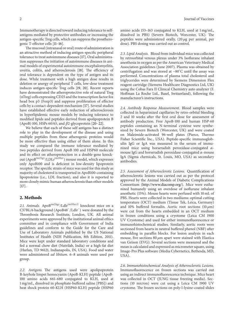
3.1. Oral Administration of ApoB andHSP60Peptides Increasesthe Number of Treg Cells in Lymphoid Organs. To evalu-ate whether oral tolerance induction to the peptides wasassociated with an effect on Tregs, flow Cytometry analysiswas performed. (Gating strategy for the analysis is presentedin Supplementary Figure 1 in supplementary Material avail-able online at http://dx.doi.org/10.1155/2013/212367). HSP60andApoB peptide-treated ApoBtm2Sgy/Ldlrtm1Her/J mice wereeuthanized 3, 8 days and 10 weeks after last oral dose.During this period mice were given a diet rich in cholesterolto induce the development of atherosclerosis. Three daysafter oral dosing the number of CD4+CD25+ Foxp3+ Tcells increased significantly (𝑃 < 0.001) in payers patches(37.62% ± 3.09 versus 15.9% ± 1.54), spleen (28.4% ± 1.80versus 9.44% ± 0.41), and in blood (26.23% ± 5.79 ver-sus 4.50% ± 0.62) of HSP60-treated animals compared tocontrol. The increase in Treg cells in ApoB-treated ani-mals was lower compared to HSP60-treated mice but werestill significantly higher (𝑃 < 0.05) than the control.Increase was seen in payers patches (22.75% ± 0.96 versus15.9% ± 1.54), spleen (13.8% ± 0.99 versus 9.44 ± 0.41), andin blood (14.52% ± 2.10 versus 4.50% ± 0.62). The numberof Treg cells was not significantly different in the mesentericlymph nodes. Eight days after the last dose, the numberof Treg cells decreased in all the organs but were stillsignificantly higher (𝑃 < 0.05) only in HSP60-treatedanimals compared to control. The increase in FoxP3 positivecells compared to control was maintained for 10 weeks afterthe oral dosing and was found to be significantly higher(𝑃 < 0.05) in the spleen (5.18% ± 0.91 and 4.48% ± 0.4 ver-sus 2.25 ± 0.29) and blood (7.56% ± 0.68 and 5.11% ± 0.56versus 2.28% ± 0.22) of ApoB- and HSP60-treated ani-mals, respectively (Figure 1 and Supplementary Figure 2).The percentage of Treg cells was consistently higher withHSP60 treatment compared to ApoB. Antibodies specific forApoB and HSP60 peptides were not detected in the serumafter oral administration of peptides, further supporting theinduction of tolerance to individual peptides (SupplementaryTable 1).
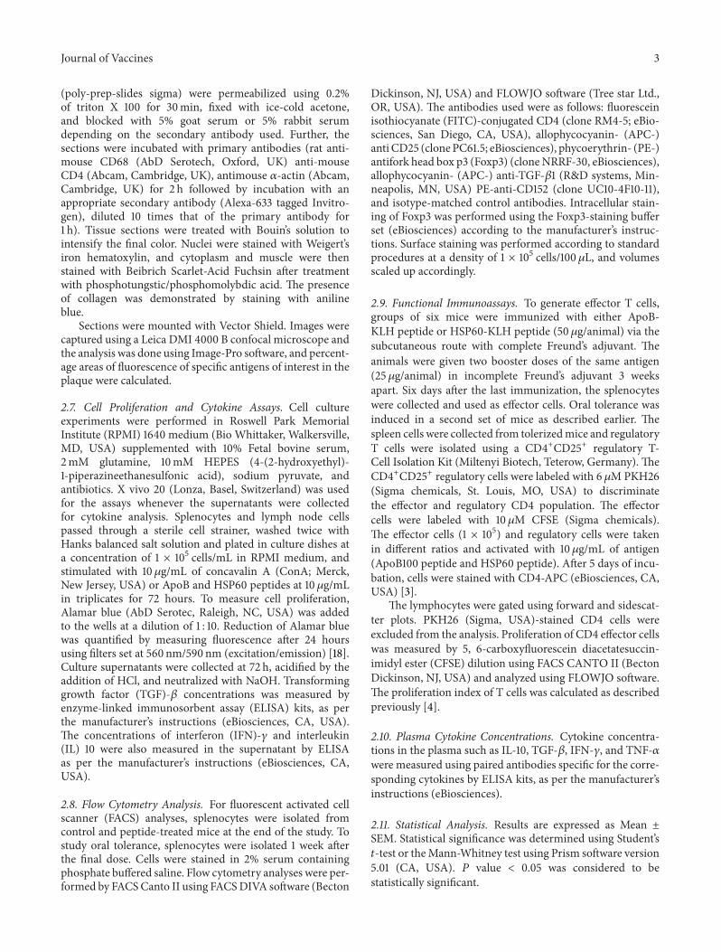
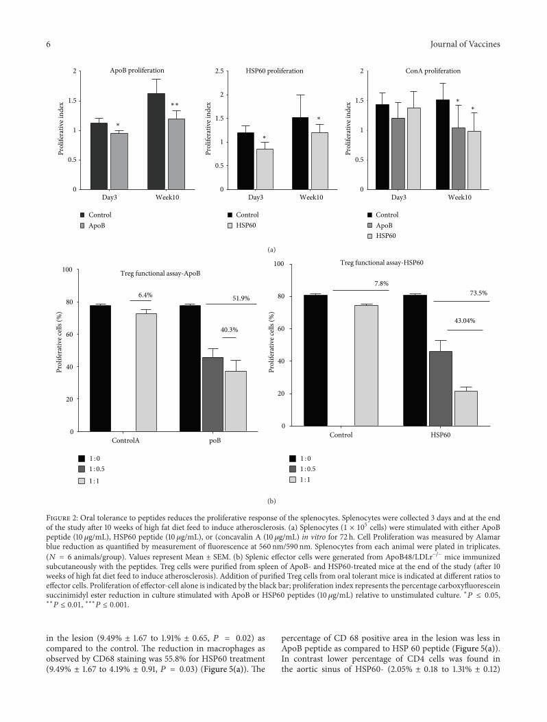
3.2. Oral Tolerance to Peptides Reduces the ProliferativeResponse of the Splenocytes. To determine the effect oforal tolerance induction on the proliferative response ofsplenocytes, mice were orally dosed with peptides and PBS.Splenocytes were collected 3 days and 10 weeks after the lastdose and cultured with ApoB peptide, HSP60 peptide, andConcavalin A in vitro. Control mice showed a proliferationindex of 1.12 ± 0.028 to ApoB peptide 3 days after last dosewhich increased to 1.65 ± 0.078 by the end of 10 weeks,suggesting that ApoB peptide reactive T cells accumulate inthe lymphoid organs as the lesion develops. In the ApoB-treated mice the proliferative index moderately increasedfrom 0.95 ± 0.016 to 1.19 ± 0.047 on day 3 and 10 weeks afterthe last dose, respectively. The reduction in ApoB-specificproliferation of splenocytes was significant (𝑃 < 0.05) for
3 days and (𝑃 < 0.01) for 10 weeks after the last dose.Similar reduction in HSP60 peptide-specific proliferationwas observed in the splenocytes of HSP60-treated micecompared to control (0.95 ± 0.02 versus 1.12 ± 0.028 on day 3and 1.19 ± 0.04 versus 1.62 ± 0.07 by 10 weeks). Splenocytescollected 3 days after last dose were comparable for theirproliferative response to Concavalin A but a significantreduction was observed in those taken after 10 weeks (1.43 ±0.07 for control, 1.21 ± 0.09 for ApoB, and 1.38 ± 0.09 forHSP60 treatment on day 3 and 1.51 ± 0.01, 1.04 ± 0.14 and0.98 ± 0.11 after 10 weeks) (Figure 2(a)).
We then assessed the ability of Treg cells recoveredfrom either HSP60 or ApoB-tolerized mice to suppress theproliferation of T effector CD4+CD25− cells recovered fromHSP60 or ApoB-sensitized (immunization with adjuvant)mice (Figure 2(b)). The suppression of effector cell prolifer-ation was more pronounced in the HSP60 treatment group.The Treg cells from HSP60-treated mice reduced the effectorcell proliferation by 73.5% at a ratio of 1 : 1, while thosefrom control mice suppressed proliferation by 7.8%. Thesuppression of T effector cells by regulatoryT cells formApoBtreated mice was 51.9% compared to 6.4% by control. Thefunctional activity of Treg cells isolated from HSP60-treatedmice gave higher degree of suppression compared to ApoBtreatment.
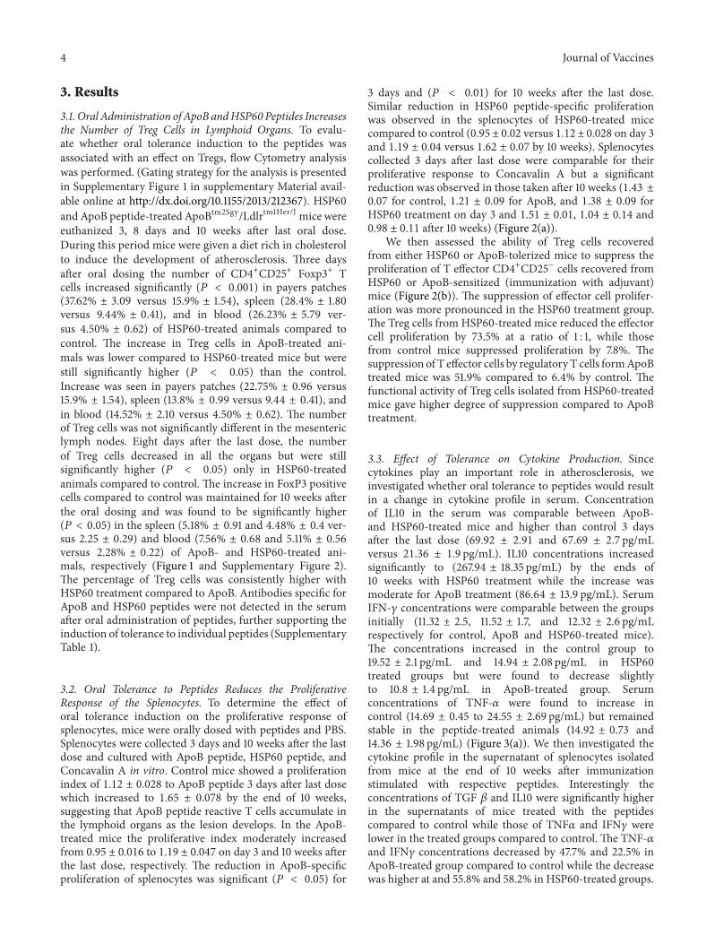
3.3. Effect of Tolerance on Cytokine Production. Sincecytokines play an important role in atherosclerosis, weinvestigated whether oral tolerance to peptides would resultin a change in cytokine profile in serum. Concentrationof IL10 in the serum was comparable between ApoB-and HSP60-treated mice and higher than control 3 daysafter the last dose (69.92 ± 2.91 and 67.69 ± 2.7 pg/mLversus 21.36 ± 1.9 pg/mL). IL10 concentrations increasedsignificantly to (267.94 ± 18.35 pg/mL) by the ends of10 weeks with HSP60 treatment while the increase wasmoderate for ApoB treatment (86.64 ± 13.9 pg/mL). SerumIFN-𝛾 concentrations were comparable between the groupsinitially (11.32 ± 2.5, 11.52 ± 1.7, and 12.32 ± 2.6 pg/mLrespectively for control, ApoB and HSP60-treated mice).The concentrations increased in the control group to19.52 ± 2.1 pg/mL and 14.94 ± 2.08 pg/mL in HSP60treated groups but were found to decrease slightlyto 10.8 ± 1.4 pg/mL in ApoB-treated group. Serumconcentrations of TNF-𝛼 were found to increase incontrol (14.69 ± 0.45 to 24.55 ± 2.69 pg/mL) but remainedstable in the peptide-treated animals (14.92 ± 0.73 and14.36 ± 1.98 pg/mL) (Figure 3(a)). We then investigated thecytokine profile in the supernatant of splenocytes isolatedfrom mice at the end of 10 weeks after immunizationstimulated with respective peptides. Interestingly theconcentrations of TGF 𝛽 and IL10 were significantly higherin the supernatants of mice treated with the peptidescompared to control while those of TNF𝛼 and IFN𝛾 werelower in the treated groups compared to control. The TNF-𝛼and IFN𝛾 concentrations decreased by 47.7% and 22.5% inApoB-treated group compared to control while the decreasewas higher at and 55.8% and 58.2% in HSP60-treated groups.
Journal of Vaccines 5
Day3
Lymph node Spleen Payers patches PBMC0
10
20
30
40
50
∗
∗
∗
∗
∗∗
∗∗
Treg
cells
(%)
(a)
Lymph node Spleen Payers patches PBMC
Day8
0
2
4
6
8
10
∗
∗
∗
∗∗∗
Treg
cells
(%)
(b)
Lymph node Spleen PBMC
6
8
0
2
4
10 Week10
∗
∗
∗
∗
∗
ControlApoBHSP60
Treg
cells
(%)
(c)
Figure 1: Oral Administration of ApoB and HSP60 peptides increases the number of Treg cells in lymphoid organs. Flow cytometry analysis:(a) percentage of CD4+CD25+ FoxP3+ cells within the CD4 population (total Treg cells) in lymph nodes, spleen, Payer’s patches, and PBMCin ApoB peptide and Hsp60 peptide treated compared to control (PBS) three days after the last dose (𝑁 = 6). (b) Percentage of CD4+CD25+FoxP3+ cells within the CD4 population (total Treg cells) in lymph nodes, spleen, Payer’s patches and PBMC in ApoB peptide and Hsp60peptide treated compared to control (PBS) eight days after the last dose (𝑁 = 6). (c) Percentage of CD4+CD25+ FoxP3+ cells within the CD4population (total Treg cells) in lymph nodes, spleen, Payer’s patches, and PBMC in ApoB peptide and Hsp60 peptide treated compared tocontrol (PBS) at the end of the study after 10 weeks of high fat diet feed to induce atherosclerosis (𝑁 = 6). ∗𝑃 ≤ 0.05, ∗∗𝑃 ≤ 0.01, ∗∗∗𝑃 ≤ 0.001.
The concentration of IL10 and TGF-𝛽 increased by 23.9% and24.3% in the supernatants of ApoB-treated splenocytes whileit was 24.3% and 38.7% in that of HSP60-treated splenocytes(Figure 3(b)). These results further reiterate that toleranceto HSP60 results in a relatively higher anti-inflammatoryprofile compared to ApoB tolerance.
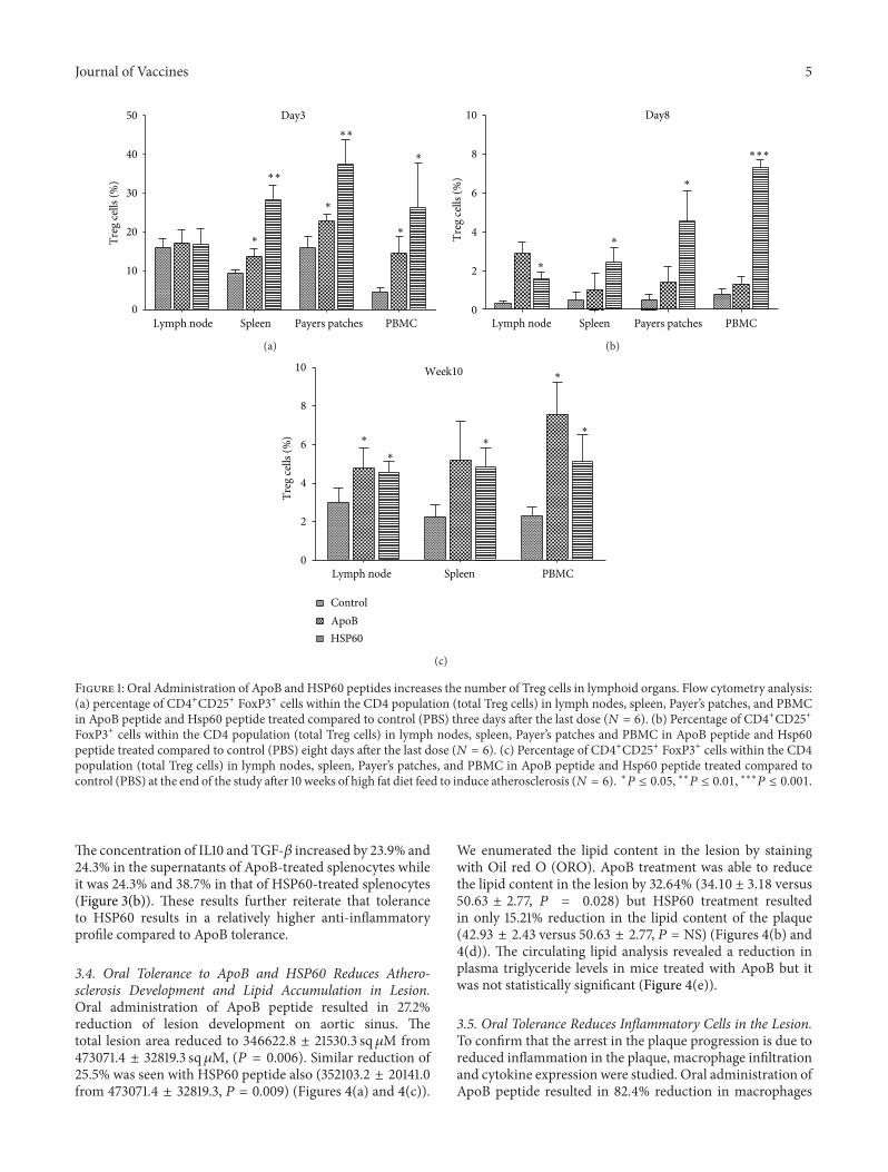
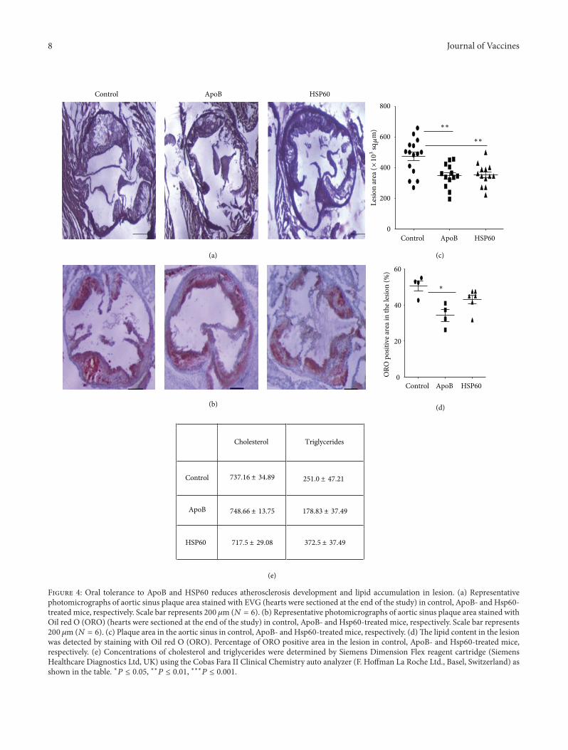
3.4. Oral Tolerance to ApoB and HSP60 Reduces Athero-sclerosis Development and Lipid Accumulation in Lesion.Oral administration of ApoB peptide resulted in 27.2%reduction of lesion development on aortic sinus. Thetotal lesion area reduced to 346622.8 ± 21530.3 sq𝜇M from473071.4 ± 32819.3 sq 𝜇M, (𝑃 = 0.006). Similar reduction of25.5% was seen with HSP60 peptide also (352103.2 ± 20141.0from 473071.4 ± 32819.3, 𝑃 = 0.009) (Figures 4(a) and 4(c)).
We enumerated the lipid content in the lesion by stainingwith Oil red O (ORO). ApoB treatment was able to reducethe lipid content in the lesion by 32.64% (34.10 ± 3.18 versus50.63 ± 2.77, 𝑃 = 0.028) but HSP60 treatment resultedin only 15.21% reduction in the lipid content of the plaque(42.93 ± 2.43 versus 50.63 ± 2.77, 𝑃 = NS) (Figures 4(b) and4(d)). The circulating lipid analysis revealed a reduction inplasma triglyceride levels in mice treated with ApoB but itwas not statistically significant (Figure 4(e)).
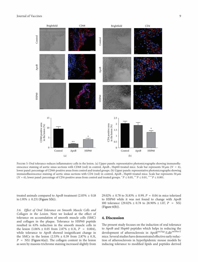
3.5. Oral Tolerance Reduces Inflammatory Cells in the Lesion.To confirm that the arrest in the plaque progression is due toreduced inflammation in the plaque, macrophage infiltrationand cytokine expression were studied. Oral administration ofApoB peptide resulted in 82.4% reduction in macrophages
6 Journal of Vaccines
ApoB proliferation
0
0.5
1
1.5
2
Control Control
Day3 Week10 Day3 Week10 Day3 Week10
ApoB ApoBControl
Prol
ifera
tive i
ndex
0
0.5
1
1.5
2
Prol
ifera
tive i
ndex
0
0.5
1
1.5
2.5
2
Prol
ifera
tive i
ndex
HSP60 proliferation
HSP60HSP60
ConA proliferation
∗
∗
∗
∗
∗
∗∗
(a)
ControlA poBControl HSP60
Treg functional assay-ApoBTreg functional assay-HSP60
0
20
40
60
80
100
0
20
40
60
80
100
6.4% 51.9%
40.3%
7.8%73.5%
43.04%
1 : 01 : 0.51 : 1
1 : 01 : 0.51 : 1
Prol
ifera
tive c
ells (
%)
Prol
ifera
tive c
ells (
%)
(b)
Figure 2: Oral tolerance to peptides reduces the proliferative response of the splenocytes. Splenocytes were collected 3 days and at the endof the study after 10 weeks of high fat diet feed to induce atherosclerosis. (a) Splenocytes (1 × 10⁵ cells) were stimulated with either ApoBpeptide (10𝜇g/mL), HSP60 peptide (10𝜇g/mL), or (concavalin A (10 𝜇g/mL) in vitro for 72 h. Cell Proliferation was measured by Alamarblue reduction as quantified by measurement of fluorescence at 560 nm/590 nm. Splenocytes from each animal were plated in triplicates.(𝑁 = 6 animals/group). Values represent Mean ± SEM. (b) Splenic effector cells were generated from ApoB48/LDLr−/− mice immunizedsubcutaneously with the peptides. Treg cells were purified from spleen of ApoB- and HSP60-treated mice at the end of the study (after 10weeks of high fat diet feed to induce atherosclerosis). Addition of purified Treg cells from oral tolerant mice is indicated at different ratios toeffector cells. Proliferation of effector-cell alone is indicated by the black bar; proliferation index represents the percentage carboxyfluoresceinsuccinimidyl ester reduction in culture stimulated with ApoB or HSP60 peptides (10𝜇g/mL) relative to unstimulated culture. ∗𝑃 ≤ 0.05,∗∗
𝑃 ≤ 0.01, ∗∗∗𝑃 ≤ 0.001.
in the lesion (9.49% ± 1.67 to 1.91% ± 0.65, 𝑃 = 0.02) ascompared to the control. The reduction in macrophages asobserved by CD68 staining was 55.8% for HSP60 treatment(9.49% ± 1.67 to 4.19% ± 0.91, 𝑃 = 0.03) (Figure 5(a)). The
percentage of CD 68 positive area in the lesion was less inApoB peptide as compared to HSP 60 peptide (Figure 5(a)).In contrast lower percentage of CD4 cells was found inthe aortic sinus of HSP60- (2.05% ± 0.18 to 1.31% ± 0.12)

Journal of Vaccines 7
Conc
entra
tion
(pg/
mL)
Serum conc of IL10
0
100
200
300
Day3 Week10
∗∗∗
∗∗
∗∗∗∗
0
10
20
30
Day3 Week10
ControlApoBHSP60
Conc
entra
tion
(pg/
mL)
Serum conc of TNF-
∗
∗
Conc
entra
tion
(pg/
mL)
0
5
10
15
20
25
Day3 Week10
Serum conc of IFN-
∗
ControlApoBHSP60
ControlApoBHSP60
(a)
Control
ApoB HSP60
Conc
entra
tion
(pg/
mL)
0
5
10
15
20
Antigen
∗∗
∗∗
TNF- concentration
ApoB HSP60
Conc
entra
tion
(pg/
mL)
ControlAntigen
0
10
20
30
40
50 IFN- concentration
Conc
entra
tion
(pg/
mL)
Control
ApoB HSP60
Antigen
IL10 concentration
0
100
200
300
400
500
∗∗
∗
Conc
entra
tion
(pg/
mL)
Control
ApoB HSP60
Antigen
0
200
400
600
800 TGF- concentration
∗
(b)
Figure 3: Effect of tolerance on cytokine production. (a) Concentration of the cytokines (TNF-𝛼, IFN-𝛾, and IL-10) in the plasma wasdetermined by ELISA as per the manufacturers instructions, three days after the last dose and at the end of the study after 10 weeks of high fatdiet feed to induce atherosclerosis following oral administration of ApoB and Hsp60 peptides (𝑁 = 6). (b) Splenocytes isolated from orallytreated animals at the end of the study were stimulated with ApoB or Hsp60 peptide (10 𝜇g/mL) in vitro for 72 hrs. Cytokine concentrations(TNF-𝛼, IFN-𝛾 TGF-𝛽, and IL-10) in the supernatant were estimated by ELISA. (𝑁 = 6) ∗𝑃 ≤ 0.05, ∗∗𝑃 ≤ 0.01, ∗∗∗𝑃 ≤ 0.001.
8 Journal of Vaccines
0
200
400
600
800
0
20
40
60
Cholesterol Triglycerides
Control
Control
Control
(a)
(b)
(c)
(d)
(e)
ApoB
ApoB
ApoB
HSP60
HSP60
Control ApoB HSP60
HSP60
Lesio
n ar
ea (×
103
sq
m)
737.16 ± 34.89
748.66 ± 13.75 178.83 ± 37.49
717.5 ± 29.08 372.5 ± 37.49
251.0 ± 47.21
∗
∗∗
∗∗
ORO
pos
itive
area
in th
e les
ion
(%)
Figure 4: Oral tolerance to ApoB and HSP60 reduces atherosclerosis development and lipid accumulation in lesion. (a) Representativephotomicrographs of aortic sinus plaque area stained with EVG (hearts were sectioned at the end of the study) in control, ApoB- and Hsp60-treated mice, respectively. Scale bar represents 200𝜇m (𝑁 = 6). (b) Representative photomicrographs of aortic sinus plaque area stained withOil red O (ORO) (hearts were sectioned at the end of the study) in control, ApoB- and Hsp60-treated mice, respectively. Scale bar represents200 𝜇m (𝑁 = 6). (c) Plaque area in the aortic sinus in control, ApoB- and Hsp60-treated mice, respectively. (d)The lipid content in the lesionwas detected by staining with Oil red O (ORO). Percentage of ORO positive area in the lesion in control, ApoB- and Hsp60-treated mice,respectively. (e) Concentrations of cholesterol and triglycerides were determined by Siemens Dimension Flex reagent cartridge (SiemensHealthcare Diagnostics Ltd, UK) using the Cobas Fara II Clinical Chemistry auto analyzer (F. Hoffman La Roche Ltd., Basel, Switzerland) asshown in the table. ∗𝑃 ≤ 0.05, ∗∗𝑃 ≤ 0.01, ∗∗∗𝑃 ≤ 0.001.
Journal of Vaccines 9
0
5
10
15
Control ApoB
Apo
B
HSP60
HSP
60Co
ntro
l
∗
∗∗
Brightfield CD68
CD68
pos
itive
area
in th
e les
ion
(%)
(a)
0
0.5
1
1.5
2
2.5
Control
Apo
BH
SP60
Cont
rol
ApoB HSP60
∗
Brightfield CD4
CD4
posit
ive a
rea
in th
e les
ion
(%)
(b)
Figure 5: Oral tolerance reduces inflammatory cells in the lesion. (a) Upper panels: representative photomicrographs showing immunoflu-orescence staining of aortic sinus sections with CD68 (red) in control, ApoB-, Hsp60-treated mice. Scale bar represents 50 𝜇m (𝑁 = 6),lower panel: percentage of CD68-positive areas from control and treated groups. (b) Upper panels: representative photomicrographs showingimmunofluorescence staining of aortic sinus sections with CD4 (red) in control, ApoB-, Hsp60-treated mice. Scale bar represents 50𝜇m(𝑁 = 6), lower panel: percentage of CD4-positive areas from control and treated groups. ∗𝑃 ≤ 0.05, ∗∗𝑃 ≤ 0.01, ∗∗∗𝑃 ≤ 0.001.
treated animals compared to ApoB treatment (2.05% ± 0.18to 1.91% ± 0.23) (Figure 5(b)).
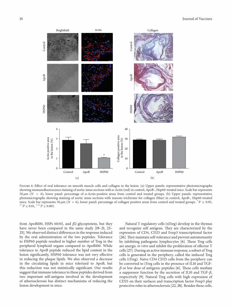
3.6. Effect of Oral Tolerance on Smooth Muscle Cells andCollagen in the Lesion. Next we looked at the effect oftolerance on accumulation of smooth muscle cells (SMC)and collagen in the plaque. Tolerance to HSP60 peptideresulted in 63% reduction in the smooth muscle cells inthe lesion (1.06% ± 0.05 from 2.87% ± 0.31, 𝑃 = 0.004),while tolerance to ApoB showed insignificant change inthe SMCs in the lesion (2.53% ± 0.39 from 2.87% ± 0.31,𝑃 = NS) (Figure 6(a)). The collagen content in the lesionas seen by masons trichrome staining increased slightly from
29.02% ± 0.78 to 31.83% ± 0.99, 𝑃 = 0.04 in mice tolerizedto HSP60 while it was not found to change with ApoB100 tolerance (29.02% ± 0.78 to 28.90% ± 1.07, 𝑃 = NS)(Figure 6(b)).
4. Discussion
The present study focuses on the induction of oral toleranceto ApoB and Hsp60 peptides which helps in reducing thedevelopment of atherosclerosis in ApoBtm2Sgy/Ldlrtm1Her/J
mice. Several studies have demonstrated effective early reduc-tion of atherosclerosis in hyperlipidemic mouse models byinducing tolerance to modified lipids and peptides derived
10 Journal of Vaccines
0
1
2
3
4
Control ApoB HSP60
Cont
rol
Brightfield
Apo
BH
SP60
∗
Actin
Act
in p
ositi
ve ar
eain
the l
esio
n (%
)
(a)
0
10
20
30
40
Control
Cont
rol
Collagen
ApoB
Apo
B
HSP60
HSP
60
Colla
gen
posit
ive a
rea
in th
e les
ion
(%)
(b)
Figure 6: Effect of oral tolerance on smooth muscle cells and collagen in the lesion. (a) Upper panels: representative photomicrographsshowing immunofluorescence staining of aortic sinus sections with 𝛼-Actin (red) in control, ApoB-, Hsp60-treatedmice. Scale bar represents50 𝜇m (𝑁 = 6), lower panel: percentage of 𝛼-Actin-positive areas from control and treated groups. (b) Upper panels: representativephotomicrographs showing staining of aortic sinus sections with masons trichrome for collagen (blue) in control, ApoB-, Hsp60-treatedmice. Scale bar represents 50 𝜇m (𝑁 = 6), lower panel: percentage of collagen positive areas from control and treated groups. ∗𝑃 ≤ 0.05,∗∗
𝑃 ≤ 0.01, ∗∗∗𝑃 ≤ 0.001.
from ApoB100, HSPs 60/65, and 𝛽2-glycoprotein, but theyhave never been compared in the same study [19–21, 23–25].We observed distinct differences in the response inducedby the oral administration of the two peptides. Toleranceto HSP60 peptide resulted in higher number of Treg in theperipheral lymphoid organs compared to ApoB100. Whiletolerance to ApoB peptide reduced the lipid content in thelesion significantly, HSP60 tolerance was not very effectivein reducing the plaque lipids. We also observed a decreasein the circulating lipids in mice tolerized to ApoB, butthis reduction was not statistically significant. Our resultssuggest that immune tolerance to these peptides derived fromtwo important self-antigens involved in the developmentof atherosclerosis has distinct mechanisms of reducing thelesion development in mice.
Natural T regulatory cells (nTreg) develop in the thymusand recognize self antigens. They are characterized by theexpression of CD4, CD25 and Foxp3 transcriptional factor[26].Theymaintain self-tolerance and prevent autoimmunityby inhibiting pathogenic lymphocytes [6]. These Treg cellsare anergic in vitro and inhibit the proliferation of effector Tcells [27]. During an active immune response, a subset of Tregcells is generated in the periphery, called the induced Tregcells (iTreg). Naive CD4 CD25 cells from the periphery canbe converted to iTreg cells in the presence of IL10 and TGF-𝛽 or low dose of antigenic peptides [6]. These cells mediatea suppressor function by the secretion of IL10 and TGF-𝛽,respectively [9]. Natural Treg cells with high expression ofCD25 on their surfaces and transcription factor Foxp3 playprotective roles in atherosclerosis [22, 28]. Besides these cells,
Journal of Vaccines 11
adaptive regulatory cells, Tr1 andTh3 cells secreting IL-10 andTGF-𝛽, respectively, have also been implicated in protectionagainst atherogenesis [20, 24, 25].
Three days after the oral HSP60-peptide (AA 153–163) treatment, the number of CD4+CD25+ Foxp3+ Tregsincreased significantly in Payer’s patches, spleen, and blood.After a week, their number reduced in all the organs butremained significantly higher compared to control. Tregcells were found to be maintained at higher level in thelymphoid organs during development of atherosclerosis(observed after 10 weeks of high fat diet feeding). Weobserved an increase in Treg cells following ApoB (AA 661–680) treatment also but their numbers were distinctly lowerthan HSP60-treated mice. The decrease in the number ofTreg cells after a week of dosing could be attributed to themigration of the activated Treg cells to the site of inflam-mation (atherosclerotic lesions) where they may recognizeself-antigens. Similar results were reported for a differentpeptide epitope fromHSP60 (253–268) by van Puijvelde et al.[23].
Increase in Treg cells in lymphoid organs of thepeptide-treated animals does not ensure their antigen speci-ficity. However, we observed that these Treg cells recov-ered from lymph nodes and spleens of peptide-treatedApoBtm2Sgy/Ldlrtm1Her mice significantly suppressed antigen-specific T effector cell proliferation in vitro, the suppres-sive function being higher for HSP60 tolerance comparedto ApoB100. Plasma concentration of IL10 increased sig-nificantly in HSP60-tolerized mice suggesting the role ofinduced Treg cells in atheroprotection. The increase inIL10 concentrations was moderate in ApoB tolerized mice,while the concentrations of inflammatory cytokines werereduced in the plasma. In addition, oral treatment withHSP60 and ApoB peptides also reduced the proliferativeresponse of splenocytes to the respective peptides. Reduc-tion in splenocytes proliferative response to HSP65 proteinfollowing oral administration was also reported by Maronet al. and Harats et al. [19, 29]. Furthermore, spleno-cytes of HSP60 and ApoB treated mice also producedincreased levels of TGF-𝛽 and IL-10 after in vitro restim-ulation with the respective peptides. All these data sug-gest that oral administration of HSP60- and ApoB-peptidesinduced Tregs, which can dampen the inflammatory immuneresponse to HSP60 and ApoB100 proteins in atheroscle-rosis prone mice and thus reduce the development ofatherosclerosis.
We observed significant reduction in lipid deposition inthe lesion in mice orally treated with ApoB peptide. Thisfinding was further corroborated by the decrease in numberof CD68 positive macrophages in the lesion. These resultssuggest that tolerance to ApoB peptide has an effect onthe macrophage migration into the lesion and consequentlythe foam cell formation and lipid deposition. Reduction inlipid content in the lesion was not significant with HSP60peptide treatment. Although the number of CD68 positivemacrophages in the lesion was lower than control, it wasmoderate compared to ApoB peptide treatment. Oral toler-ance to HS60 peptide resulted in reduction in CD4 positive T
cells, smoothmuscle cells, and an increase in collagen contentin the lesion, which could be correlated to the effectiveTreg activity seen in these mice. Treg cells producing TGF-𝛽have been shown to inhibit smooth muscle proliferation andpromote extracellular matrix formation, thus stabilizing theplaque [30, 31].
Atherosclerosis is a multifactorial, chronic inflammatorydisease. Inflammation mediated by a pathogenic immuneresponse to endogenous antigens such as HSP, modifiedlipids, as well as exogenous antigens from pathogens, havebeen implicated in the initiation of immune response duringatherogenesis [3]. Each molecule has a distinct role to playin the initiation and progression of the disease. It is likelythat these molecules also induce a different mechanism ofatheroprotection. We believe that to address a multifactorialdisease, a combination of multiple epitopes would have a bet-ter efficacy compared to single peptides. Based on our resultswe think that tolerance to two different proteins inducesatheroprotection by diversemechanisms.WhileHSP60 toler-ance resulted in increase in Treg cells and anti inflammatorycytokine secretion ApoB tolerance was effective in reducingthe lipid deposition in the lesion. Both were equally effectivein controlling the disease development. We have earlierreported that repeated subcutaneous immunization with acombination of peptide epitopes from ApoB100 and HSP60had a synergistic effect in reducing the development ofatherosclerosis in mice [32]. Similar increase in protectionwas observed by administration of the combination of thesetwo peptides by mucosal route also (unpublished observa-tion). It is likely that the combination of peptides wouldcontrol different aspects of disease development and result ina synergistic increase in protection compared to individualpeptides.
5. Conclusions
In conclusion, based on our present and earlier results webelieve that using multiple antigenic epitopes from differentmolecules involved in atherogenesis would be a novel andattractive strategy for immunoprotection against atheroscle-rosis. Additional studies will be required to completely delin-eate the mechanisms of protection mediated by tolerance tothese individual peptides and the mechanisms that controltheir maintenance in vivo.
Acknowledgments
The authors gratefully acknowledge the support of thetrustees of Thrombosis Research Institute, London andBangalore, the Tata Social Welfare Trust, India (TSWT/IG/SNB/JP/Sdm), and the Department of Biotechnology, Min-istry of Science and Technology, Government of India(BT/01/CDE/08/07). The sponsors did not participate inthe design, conduct, sample collection, analysis and inter-pretation of the data, or in the preparation, review, orapproval of the paper. The authors thank Ms. SheenaPhilip for her contribution to the immunohistochemicalanalysis.
12 Journal of Vaccines
References
[1] C. Baigent, A. Keech, P. M. Kearney et al., “Efficacy and safetyof cholesterollowering treatment: prospective meta-analysis ofdata from 90,056 participants in 14 randomised trials of statins,”Lancet, vol. 366, no. 9493, pp. 1267–1278, 2005.
[2] J. Andersson, P. Libby, and G. K. Hansson, “Adaptive immunityand atherosclerosis,”Clinical Immunology, vol. 134, no. 1, pp. 33–46, 2010.
[3] G. K. Hansson, “Immune mechanisms in atherosclerosis,”Arteriosclerosis, Thrombosis, and Vascular Biology, vol. 21, pp.1876–1890, 2001.
[4] G. K. Hansson, “Mechanisms of disease: inflammation,atherosclerosis, and coronary artery disease,” New EnglandJournal of Medicine, vol. 352, no. 16, pp. 1685–1626, 2005.
[5] S. Sakaguchi, “Naturally arising Foxp3-expressing CD25+CD4+ regulatory T cells in immunological tolerance to self andnon-self,” Nature Immunology, vol. 6, no. 4, pp. 345–352, 2005.
[6] H. von Boehmer, “Mechanisms of suppression by suppressor Tcells,” Nature Immunology, vol. 6, no. 4, pp. 338–344, 2005.
[7] Z. Mallat and A. Tedgui, “Immunomodulation to combatatherosclerosis: the potential role of immune regulatory cells,”Expert Opinion on Biological Therapy, vol. 4, no. 9, pp. 1387–1393, 2004.
[8] G. K. Hansson, “Atherosclerosis—an immune disease. Theanitschkov lecture 2007,” Atherosclerosis, vol. 202, no. 1, pp. 2–10, 2009.
[9] Z. Mallat, H. Ait-Oufella, and A. Tedgui, “Regulatory T-cell immunity in atherosclerosis,” Trends in CardiovascularMedicine, vol. 17, no. 4, pp. 113–118, 2007.
[10] G. K. Hansson and J. Nilsson, “Vaccination against atheroscle-rosis? Induction of atheroprotective immunity,” Seminars inImmunopathology, vol. 31, no. 1, pp. 95–101, 2009.
[11] L. A. Mundkur and V. V. Kakkar, “Immune modulation as atherapeutic strategy for atherosclerosis,” Current DrugTherapy,vol. 5, no. 4, pp. 288–300, 2010.
[12] J. Nilsson, G. N. Fredrikson, H. Bjorkbacka, K. Y. Chyu, andP. K. Shah, “Vaccines modulating lipoprotein autoimmunity asa possible future therapy for cardiovascular disease,” Journal ofInternal Medicine, vol. 266, no. 3, pp. 221–231, 2009.
[13] S. Ameli, A. Hultgardh-Nilsson, J. Regnstrom et al., “Effectof immunization with homologous LDL and oxidized LDLon early atherosclerosis in hypercholesterolemic rabbits,” Arte-riosclerosis, Thrombosis, and Vascular Biology, vol. 16, no. 8, pp.1074–1079, 1996.
[14] C. J. Binder, S. Horkko, A. Dewan et al., “Pneumococcalvaccination decreases atherosclerotic lesion formation: molec-ular mimicry between Streptococcus pneumoniae and oxidizedLDL,” Nature Medicine, vol. 9, no. 6, pp. 736–743, 2003.
[15] W. Palinski, E. Miller, and J. L. Witztum, “Immunization oflow density lipoprotein (LDL) receptor-deficient rabbits withhomologous malondialdehyde-modified LDL reduces athero-genesis,” Proceedings of the National Academy of Sciences of theUnited States of America, vol. 92, no. 3, pp. 821–825, 1995.
[16] X. Zhou, G. Caligiuri, A. Hamsten, A. K. Lefvert, and G.K. Hansson, “LDL immunization induces T-cell-dependentantibody formation and protection against atherosclerosis,”Arteriosclerosis, Thrombosis, and Vascular Biology, vol. 21, no.1, pp. 108–114, 2001.
[17] R. V. Farese Jr., M. M. Veniant, C. M. Cham et al., “Phenotypicanalysis of mice expressing exclusively apolipoprotein B48 orapolipoprotein B100,” Proceedings of the National Academy ofSciences of the United States of America, vol. 93, no. 13, pp. 6393–6398, 1996.
[18] S. A. Ahmed, R. M. Gogal Jr., and J. E. Walsh, “A new rapidand simple non-radioactive assay tomonitor and determine theproliferation of lymphocytes: an alternative to [3H]thymidineincorporation assay,” Journal of Immunological Methods, vol.170, no. 2, pp. 211–224, 1994.
[19] R. Maron, G. Sukhova, A. M. Faria et al., “Mucosal adminis-tration of heat shock protein-65 decreases atherosclerosis andinflammation in aortic arch of low-density lipoprotein receptor-deficient mice,” Circulation, vol. 106, no. 13, pp. 1708–1715, 2002.
[20] G. H. M. van Puijvelde, A. D. Hauer, P. de Vos et al., “Inductionof oral tolerance to oxidized low-density lipoprotein amelioratesatherosclerosis,” Circulation, vol. 114, no. 18, pp. 1968–1976,2006.
[21] J. George, N. Yacov, E. Breitbart et al., “Suppression of earlyatherosclerosis in LDL-receptor deficient mice by oral tolerancewith 𝛽2-glycoprotein I,” Cardiovascular Research, vol. 62, no. 3,pp. 603–609, 2004.
[22] H. Ait-Oufella, B. L. Salomon, S. Potteaux et al., “Naturalregulatory T cells control the development of atherosclerosis inmice,” Nature Medicine, vol. 12, no. 2, pp. 178–180, 2006.
[23] G. H. M. van Puijvelde, T. van Es, E. J. A. van Wanrooijet al., “Induction of oral tolerance to HSP60 or an HSP60-peptide activates t cell regulation and reduces atherosclerosis,”Arteriosclerosis,Thrombosis, andVascular Biology, vol. 27, no. 12,pp. 2677–2683, 2007.
[24] N. Sasaki, T. Yamashita, M. Takeda et al., “Oral anti-CD3antibody treatment induces regulatory t cells and inhibits thedevelopment of atherosclerosis in mice,” Circulation, vol. 120,no. 20, pp. 1996–2005, 2009.
[25] R. Klingenberg, M. Lebens, A. Hermansson et al., “Intranasalimmunization with an apolipoprotein B-100 fusion pro-tein induces antigen-specific regulatory T cells and reducesatherosclerosis,”Arteriosclerosis,Thrombosis, and Vascular Biol-ogy, vol. 30, no. 5, pp. 946–952, 2010.
[26] G. L. Stephens and E. M. Shevach, “Foxp3+ regulatory T cells:selfishness under scrutiny,” Immunity, vol. 27, no. 3, pp. 417–419,2007.
[27] Z. Mallat, S. Taleb, H. Ait-Oufella, and A. Tedgui, “The role ofadaptive T cell immunity in atherosclerosis,” Journal of LipidResearch, vol. 50, pp. S364–S369, 2009.
[28] A. Mor, D. Planer, G. Luboshits et al., “Role of naturallyoccurring CD4+CD25+ regulatory T cells in experimentalatherosclerosis,”Arteriosclerosis,Thrombosis, and Vascular Biol-ogy, vol. 27, no. 4, pp. 893–900, 2007.
[29] D. Harats, N. Yacov, B. Gilburd, Y. Shoenfeld, and J. George,“Oral tolerance with heat shock protein 65 attenuatesMycobacterium tuberculosis-induced and high-fat-diet-driven atherosclerotic lesions,” Journal of the American Collegeof Cardiology, vol. 40, no. 7, pp. 1333–1338, 2002.
[30] D. J. Grainger, “Transforming growth factor 𝛽 and atheroscle-rosis: so far, so good for the protective cytokine hypothesis,”Arteriosclerosis, Thrombosis, and Vascular Biology, vol. 24, no.3, pp. 399–404, 2004.

Journal of Vaccines 13
[31] A. C. Doran, N. Meller, and C. A. McNamara, “Role ofsmooth muscle cells in the initiation and early progressionof atherosclerosis,” Arteriosclerosis, Thrombosis, and VascularBiology, vol. 28, no. 5, pp. 812–819, 2008.
[32] X. Lu, D. Chen, V. Endresz et al., “Immunization with acombination of ApoB andHSP60 epitopes significantly reducesearly atherosclerotic lesion in Apobtm2SgyLdlrtm1Her/J mice,”Atherosclerosis, vol. 212, no. 2, pp. 472–480, 2010.
Copyright © 2022 FDOKUMEN