Comparative genomics defines the core genome of the growing N4-like phage genus and identifies...
14
ORIGINAL RESEARCH ARTICLE published: 10 October 2014 doi: 10.3389/fmicb.2014.00506 Comparative genomics defines the core genome of the growing N4-like phage genus and identifies N4-like Roseophage specific genes Jacqueline Z.-M. Chan 1 *, Andrew D. Millard 2 , Nicholas H. Mann 3 and Hendrik Schäfer 3 1 Oxford Gene Technologies, Begbroke, UK 2 Division of Microbiology and Infection, Warwick Medical School, University of Warwick, Coventry, UK 3 School of Life Sciences, University of Warwick, Coventry, UK Edited by: Brian Palenik, Scripps Instituion of Oceanography, USA Reviewed by: Alison Buchan, University of Tenessee-Knoxville, USA Lisa Zeigler Allen, J. Craig Venter Institute, USA *Correspondence: Jacqueline Z.-M. Chan, Oxford Gene Technologies, Begbroke Science Park, Begbroke Hill, Woodstock Road, Begbroke, Oxfordshire, OX5 1PF, UK e-mail: [email protected] Two bacteriophages, RPP1 and RLP1, infecting members of the marine Roseobacter clade were isolated from seawater. Their linear genomes are 74.7 and 74.6kb and encode 91 and 92 coding DNA sequences, respectively. Around 30% of these are homologous to genes found in Enterobacter phage N4. Comparative genomics of these two new Roseobacter phages and 23 other sequenced N4-like phages (three infecting members of the Roseobacter lineage and 20 infecting other Gammaproteobacteria) revealed that N4-like phages share a core genome of 14 genes responsible for control of gene expression, replication and virion proteins. Phylogenetic analysis of these genes placed the five N4-like roseophages (RN4) into a distinct subclade. Analysis of the RN4 phage genomes revealed they share a further 19 genes of which nine are found exclusively in RN4 phages and four appear to have been acquired from their bacterial hosts. Proteomic analysis of the RPP1 and RLP1 virions identified a second structural module present in the RN4 phages similar to that found in the Pseudomonas N4-like phage LIT1. Searches of various metagenomic databases, including the GOS database, using CDS sequences from RPP1 suggests these phages are widely distributed in marine environments in particular in the open ocean environment. Keywords: N4 bacteriophage, Roseobacter, comparative genomics, core genes, auxiliary metabolic genes INTRODUCTION Phages (viruses that infect bacteria) are the most prevalent enti- ties in the biosphere; they harbor a vast, untapped reservoir of genomic diversity and are important in driving the evolution of bacteria (Rohwer, 2003; Paul and Sullivan, 2005; Angly et al., 2006). They are also a significant component of the microbial food web and have major influence on fluxes of organic and inorganic matter, in particular in the oceans (Fuhrman, 1999; Wilhelm and Suttle, 1999; Weinbauer and Rassoulzadegan, 2004; Suttle, 2005, 2007; Breitbart et al., 2007). Metagenomic surveys suggest that the true diversity of marine phages exceeds that rep- resented by isolated phages (Breitbart and Rohwer, 2005; Angly et al., 2006; Hurwitz and Sullivan, 2013) and there remain major gaps in understanding which hosts are infected by the wide diversity of phage observed in the environment. One of the major groups of bacteria found in the marine environment is the so-called Roseobacter clade. Its members rep- resent a taxonomically and metabolically diverse group of bacteria found in pelagic and benthic habitats where they play key roles in a wide range of biogeochemically important transformations (Buchan et al., 2005). Processes affecting their abundance and activity, such as viral lysis, are of biogeochemical significance but are currently poorly understood as only a small number of bacteriophages interacting with Roseobacters (roseophages) have previously been described. The first isolated roseophage was SIO1 (Rohwer et al., 2000), but since then four lytic roseophages infect- ing Roseobacter denitrificans (phage RDJL1), Ruegeria pomeroyi (phage DSS32), Sulfitobacter strain EE36 (phage EE361) and Sulfitobacter strain 2047 (phage pCB2047-B) have been described (Zhang and Jiao, 2009; Zhao et al., 2009; Ankrah and Budinoff, 2014). The latter three are closely related to Enterobacteria phage N4, which, for over 40 years, was the sole representative of the N4-like genus, a genetic orphan among the tailed phages (Schito et al., 1965; Ceyssens et al., 2010). N4 was unique in the phage world due to its use of three distinct RNA polymerases and single-stranded DNA protein/activators to control gene expres- sion (Choi et al., 2008). In recent years a further 25 N4-like phages have been isolated and genome sequenced (Table 1) all of which share these features. The aim of this study was to isolate and characterize lytic phages infecting members of the Roseobacter clade using a number of different Roseobacter host strains and samples of coastal seawater from the United Kingdom. We isolated two new Roseobacter N4-like phages (RN4-phages) that infect Roseovarius nubinhibens and Roseovarius sp. 217. Here, we report the sequenc- ing of their genomes and the identification of phage-particle asso- ciated proteins by mass spectrometry. With the increased number of genome sequences available for N4-like phages it was possible www.frontiersin.org October 2014 | Volume 5 | Article 506 | 1
Transcript of Comparative genomics defines the core genome of the growing N4-like phage genus and identifies...

ORIGINAL RESEARCH ARTICLEpublished: 10 October 2014
doi: 10.3389/fmicb.2014.00506
Comparative genomics defines the core genome of thegrowing N4-like phage genus and identifies N4-likeRoseophage specific genesJacqueline Z.-M. Chan1*, Andrew D. Millard2, Nicholas H. Mann3 and Hendrik Schäfer3
1 Oxford Gene Technologies, Begbroke, UK2 Division of Microbiology and Infection, Warwick Medical School, University of Warwick, Coventry, UK3 School of Life Sciences, University of Warwick, Coventry, UK
Edited by:
Brian Palenik, Scripps Instituion ofOceanography, USA
Reviewed by:
Alison Buchan, University ofTenessee-Knoxville, USALisa Zeigler Allen, J. Craig VenterInstitute, USA
*Correspondence:
Jacqueline Z.-M. Chan, Oxford GeneTechnologies, Begbroke SciencePark, Begbroke Hill, WoodstockRoad, Begbroke, Oxfordshire, OX51PF, UKe-mail: [email protected]
Two bacteriophages, RPP1 and RLP1, infecting members of the marine Roseobacterclade were isolated from seawater. Their linear genomes are 74.7 and 74.6 kb and encode91 and 92 coding DNA sequences, respectively. Around 30% of these are homologousto genes found in Enterobacter phage N4. Comparative genomics of these two newRoseobacter phages and 23 other sequenced N4-like phages (three infecting membersof the Roseobacter lineage and 20 infecting other Gammaproteobacteria) revealed thatN4-like phages share a core genome of 14 genes responsible for control of geneexpression, replication and virion proteins. Phylogenetic analysis of these genes placedthe five N4-like roseophages (RN4) into a distinct subclade. Analysis of the RN4 phagegenomes revealed they share a further 19 genes of which nine are found exclusively inRN4 phages and four appear to have been acquired from their bacterial hosts. Proteomicanalysis of the RPP1 and RLP1 virions identified a second structural module present inthe RN4 phages similar to that found in the Pseudomonas N4-like phage LIT1. Searches ofvarious metagenomic databases, including the GOS database, using CDS sequences fromRPP1 suggests these phages are widely distributed in marine environments in particularin the open ocean environment.
Keywords: N4 bacteriophage, Roseobacter, comparative genomics, core genes, auxiliary metabolic genes
INTRODUCTIONPhages (viruses that infect bacteria) are the most prevalent enti-ties in the biosphere; they harbor a vast, untapped reservoir ofgenomic diversity and are important in driving the evolution ofbacteria (Rohwer, 2003; Paul and Sullivan, 2005; Angly et al.,2006). They are also a significant component of the microbialfood web and have major influence on fluxes of organic andinorganic matter, in particular in the oceans (Fuhrman, 1999;Wilhelm and Suttle, 1999; Weinbauer and Rassoulzadegan, 2004;Suttle, 2005, 2007; Breitbart et al., 2007). Metagenomic surveyssuggest that the true diversity of marine phages exceeds that rep-resented by isolated phages (Breitbart and Rohwer, 2005; Anglyet al., 2006; Hurwitz and Sullivan, 2013) and there remain majorgaps in understanding which hosts are infected by the widediversity of phage observed in the environment.
One of the major groups of bacteria found in the marineenvironment is the so-called Roseobacter clade. Its members rep-resent a taxonomically and metabolically diverse group of bacteriafound in pelagic and benthic habitats where they play key rolesin a wide range of biogeochemically important transformations(Buchan et al., 2005). Processes affecting their abundance andactivity, such as viral lysis, are of biogeochemical significancebut are currently poorly understood as only a small number ofbacteriophages interacting with Roseobacters (roseophages) have
previously been described. The first isolated roseophage was SIO1(Rohwer et al., 2000), but since then four lytic roseophages infect-ing Roseobacter denitrificans (phage RDJL�1), Ruegeria pomeroyi(phage DSS3�2), Sulfitobacter strain EE36 (phage EE36�1) andSulfitobacter strain 2047 (phage pCB2047-B) have been described(Zhang and Jiao, 2009; Zhao et al., 2009; Ankrah and Budinoff,2014). The latter three are closely related to Enterobacteria phageN4, which, for over 40 years, was the sole representative of theN4-like genus, a genetic orphan among the tailed phages (Schitoet al., 1965; Ceyssens et al., 2010). N4 was unique in the phageworld due to its use of three distinct RNA polymerases andsingle-stranded DNA protein/activators to control gene expres-sion (Choi et al., 2008). In recent years a further 25 N4-like phageshave been isolated and genome sequenced (Table 1) all of whichshare these features.
The aim of this study was to isolate and characterize lyticphages infecting members of the Roseobacter clade using anumber of different Roseobacter host strains and samples ofcoastal seawater from the United Kingdom. We isolated two newRoseobacter N4-like phages (RN4-phages) that infect Roseovariusnubinhibens and Roseovarius sp. 217. Here, we report the sequenc-ing of their genomes and the identification of phage-particle asso-ciated proteins by mass spectrometry. With the increased numberof genome sequences available for N4-like phages it was possible
www.frontiersin.org October 2014 | Volume 5 | Article 506 | 1

Chan et al. Comparative genomics of N4-like phages
Table 1 | N4-like bacteriophages for which genome sequences are available.
Phage Host Isolation location Genome Accession References
size (kb) number
N4 Escherichia coli K12 Sewage water, Genoa, Italy 70.2 EF056009 Schito et al.,1967
DSS3F2 Ruegeria pomeroyi DSS-3 Baltimore Inner Harbor water, USA 74.6 FJ591093 Zhao et al., 2009EE36F1 Sulfitobacter sp. EE-36 Baltimore Inner Harbor water, USA 73.3 FJ591094 Zhao et al., 2009LIT1 Pseudomonas aeruginosa US449 Belgian hospital sewage, Belgium 72.5 NC_013692 Ceyssens et al.,
2010LUZ7 Pseudomonas aeruginosa Br257 Belgian hospital sewage, Belgium 74.9 NC_013691 Ceyssens et al.,
2010PEV2 Pseudomonas aeruginosa PAV237 Sewage water, Olympia, WA, USA 72.7 n/a Ceyssens et al.,
2010S6 Erwinia amylovora Fruit production environment, Switzerland 74.7 HQ728266 Born et al., 2011KBNP21 Escherichia coli KBP21 Chicken farm, in Yesan, South Korea 69.9 JX415535 Nho et al., 2012PA26 Pseudomonas aeruginosa ATCC 27853 Reservoir water, Naju City, South Korea 72.3 JX194238 Kim et al., 2012G7C Escherichia coli strain 4s Horse feces 71.8 HQ259105 Kulikov et al.,
2012IME11 Escherichia coli Sewage of the no. 307 hospital in Beijing, China 72.6 JX880034 Fan et al., 2012EC1-UPM Escherichia coli O78:K80 Chicken feces 70.9 KC206276 Gan et al., 2013FSL SP-058 Salmonella serova Dublin Dairy farm 72 KC139517 Moreno Switt
et al., 2013FSL SP-076 Salmonella serova Dublin Dairy farm 72 KC139520 Moreno Switt
et al., 2013JA1 Vibrio cholerae O139 Stool of a Vibrio cholerae O139 Bengal-infected
patient69.3 KC438282 Fouts et al., 2013
Presely Acinetobacter baumannii M2 sewage sample collected in College Station,TX, USA
77.2 KF669658 Farmer et al.,2013
VCO139 Vibrio cholerae O139 Bengal Sewage effluent from the International Centrefor DiarrhealDisease Research, Bangladesh
68.9 KC438283 Fouts et al., 2013
JW Alpha Achromobacter xylosoxidans DSM 11852 Waste water treatment plant in Werl, Germany 72.3 KF787095 Wittmann et al.,2014
JW Delta Achromobacter xylosoxidans DSM 11852 Waste water treatment plant in Braunschweig,Germany
73.7 KF787094 Wittmann et al.,2014
pCB2047-B Sulfitobacter sp. strain 2047 Mesocosm study, Raunefjorden, Norway 74.5 HQ317387 Ankrah andBudinoff, 2014
EcP1 Escherichia coli strain 285 Hospital raw sewage, China 59.1 HQ641380 unpublishedpYD6-A Pseudoalteromonas sp. YD6 Surface coastal water, South China Sea 76.8 NC_020849 unpublishedVBP32 Vibrio parahaemolyticus RIMD2210633 Lobster Hatchery Stonington, ME 76.7 HQ634196 unpublishedVCP47 Vibrio parahaemolyticus RIMD2210633 Lobster Hatchery Stonington, ME, USA 76.7 HQ634194 unpublishedRLP1 Roseovarius sp. 217 Langstone Harbour, Hampshire, UK 74.6 FR682616 This studyRPP1 Roseovarius nubinhibens L4 sampling station, Plymouth, UK 74.7 FR719956 This study
to address questions regarding the structure and evolution of thegenomes of this growing group of phages.
MATERIALS AND METHODSGROWTH OF BACTERIAL STRAINSCultures of Rsv. nubinhibens (Gonzalez et al., 2003) and Rsv.sp. 217 (Schäfer et al., 2005) were routinely grown in MarineAmmonium Mineral Salts amended with 10 g L−1 peptone and5 g L−1 yeast extract (MAMS-PY).
PHAGE ISOLATIONPhages were isolated from seawater samples collected fromthe English Channel at the L4 sampling station situated
approx. 10 nautical miles south of Plymouth, Devon, UK,50◦15′N, 04◦13′W (http://www.westernchannelobservatory.org.uk/) on 24-11-1998 and Langstone Harbour on 17-09-2005 (Hampshire, UK). Seawater samples, supplemented withYeast/Peptone (1 g L−1/5 g L−1 respectively), were inoculatedwith Ruegeria sp. 198, Rhodobacteraceae bacterium 176, Rsv.nubinhibens, Rsv. sp. 257 and Rsv. sp. 217 (Gonzalez et al.,2003; Schäfer et al., 2005) to enrich any Roseobacter phagespresent. After incubation for 7 days, cells and large cellu-lar debris were removed by centrifugation and the super-natant used in plaque assays against the species in theoriginal inoculum. Clear plaques could be observed on bacteriallawns of Rsv. nubinhibens and Rsv sp. 217 after 24–48 h
Frontiers in Microbiology | Evolutionary and Genomic Microbiology October 2014 | Volume 5 | Article 506 | 2

Chan et al. Comparative genomics of N4-like phages
incubation at 25◦C. The plaques were then picked and madeclonal.
PRODUCTION OF PHAGE STOCKSThe clonal phage samples made from agar plugs were usedin plaque assays to produce plates with confluent lysis of theRoseovarius lawn. The top agar layer was removed using a flame-sterilized glass microscope slide and mixed with 3 ml (per plate)of artificial seawater (ASW) modified as described in Wilson et al.(1996). Chloroform was added to a final concentration of 25%(v/v) to lyse remaining host cells. The resulting slurry was mixedthoroughly for at least 1 min and incubated for at least 30 min atroom temperature in the dark. The top agar and chloroform wasremoved by centrifugation at 1780 × g for 10 min at 4◦C. Thistypically produced stocks of 1 × 10⁸ plaque-forming units (PFU)ml−1. Phages were further purified using CsCl gradient centrifu-gation for subsequent electron microscopy, DNA extraction andvirion proteomic analyses (Sambrook and Russell, 2001).
MODIFIED BACTERIOPHAGE ONE-STEP GROWTH CURVEBacterial host cells grown in MAMS-PY in early exponentialphase were harvested by centrifugation (4000 rpm/1300 × g,15◦C for 10 min). The cells were then washed in Marine Broth(Pronadisa, Conda, Madrid) and centrifuged again at 16000 × gat room temperature for 10 min. The pellet was resuspended insterile Marine Broth containing enough phage to have a multi-plicity of infection of 0.001. Prior to addition of bacterial hostcells, aliquots of the Marine Broth + phage solution had beenremoved to act as control samples. Both “bacteria + phage” and“phage-only” samples were then plated using the top agar over-lay technique and the time noted for each plate. The plates werethen transferred to a dark, 20◦C incubator for the duration of theexperiment.
At appropriate intervals plates were removed and the topagar layer removed with a flame-sterilized glass slide. Thiswas mixed with 3 ml ASW and 3 ml chloroform or cold 3 mlASW. The period of time between plating and mixing with theASW:chloroform or cold ASW only solution was taken as time ofincubation.
All samples were left at 4◦C in the dark overnight then cen-trifuged at 1300 × g at 4◦C for 10 min to separate the agarand chloroform. The number of free plaque forming units inthe supernatant was then analyzed by appropriate dilution andplaque assays. Each time point for bacterial/phage samples wasassayed in triplicate, control samples in duplicate and each growthcurve was repeated three times.
PHAGE GENOMIC DNA DIGESTION WITH Bal31CsCl-purified phage stocks were dialysed twice using size3/MWCO 12-14,000 Da, dialysis tubing for at least 2 h in ASW at4◦C. DNA was isolated and purified using a phenol-chloroformextraction as described previously (Sambrook and Russell, 2001).To determine the physical structure of the genome of the twophages (linear or circular), around 40 μg of phage DNA wasdigested with Bal31 at 30◦C as described elsewhere (Loessneret al., 2000). Briefly, samples were removed 0, 5, 10, 20, 40, and60 min after the addition of the enzyme and the digest stopped
by incubation at 65◦C for 10 min. All samples were purified byphenol-chloroform extraction, precipitated with sodium acetateand ethanol which was followed by digestion with Nde1 fast digest(Fermentas) according to manufacturer’s instructions. The digestpatterns were analyzed by pulsed field gel electrophoresis using a1% PFGE grade agarose gel run in a CHEF Mapper (BioRad).
PHAGE GENOME SEQUENCINGRLP1 and RPP1 phage DNA was extracted from CsCl stocksand dissolved in 10 mM Tris 1 mM EDTA buffer pH 8 (TE).The genomes were sequenced by the GenePool at the Universityof Edinburgh using Illumina for RPP1 and a combination ofIllumina and Roche 454 shotgun sequencing for RLP1. Short-readIllumina data from RPP1 were assembled using Velvet (Zerbinoand Birney, 2008), whereas the mixture of 454 and Illumina readsfrom RLP1 was assembled using Minimus (Sommer et al., 2007).
RPP1 assembled into a single contig whilst RLP1 assembledinto 10 contigs; initial annotation of the largest contig sug-gested a high degree of gene synteny between RLP1 and RPP1.Consequently, RPP1 was used as a scaffold for RLP1 and theorder of contigs was confirmed by PCR. Sequencing of the PCRproducts (by Sanger sequencing) resulted in complete assemblyof RLP1. Whole-genome sequence data was submitted to EBMLunder accession numbers FR682616 and FR719956 for RLP1 andRPP1 respectively.
IDENTIFICATION OF CODING SEQUENCESCoding sequences (CDSs) were predicted using the freely avail-able gene prediction programs GeneMark™, heuristic approach(Besemer and Borodovsky, 1999) and GLIMMER 3.01 (NCBI)(Delcher et al., 1999). The final set of predicted CDSs for eachgenome was created by amalgamation of the two sets of resultsfrom GeneMark and GLIMMER. For predicted CDSs with dis-cordant start codons between the two programs, the longer of thetwo predictions was kept.
DATABASE SEARCHESBasic Local Alignment Search Tool (BLAST) comparisons werecarried out on the predicted CDSs using different custom-made databases (Altschul et al., 1990). Initially, a search usingthe BLASTp algorithm of the predicted protein sequencesfrom the two Roseovarius phages to a database containing allbacteriophage protein sequences freely available in July 2008 wasperformed. This was then repeated using BLASTp against thenon-redundant protein sequences database at the National Centrefor Biotechnology Information (NCBI). In addition, HMMERwas used to search the SWISS-PROT database. The results fromthe three searches were compared to assign putative function toeach predicted CDS in RLP1 and RPP1.
To examine the environmental distribution of RN4phages CDS sequences from RPP1 were used as querysequences for the BLAST algorithm against the environmentalmetagenomes downloaded from CAMERA (accession num-bers CAM_PROJ_HumanGut, CAM_PROJ_AntarcticAquatic,CAM_PROJ_BotanyBay, CAM_P_0000545, CAM_P_0000915,CAM_PROJ_GOS, CAM_PROJ_SalternMetagenome) and EBIfor metagenomes from freshwater lakes Bourget (MET6) and
www.frontiersin.org October 2014 | Volume 5 | Article 506 | 3
Chan et al. Comparative genomics of N4-like phages
Pavin (MET7) (accession ERS015568 and ERS015567 respec-tively). tBLASTx analysis was carried out with the followingparameters modified from default settings –F F –b 100000 –v100000 –e 0.0001. A reciprocal blastp analysis was then carriedout against a custom database of viral sequences. This wasconstructed from all complete viral genomes available fromhttp://ftp.ncbi.nlm.nih.gov/genomes/Viruses as of February2013. RPP1 was chosen as a representative of RN4 phages as ithas the same complement of genes as RLP1, and an additionalthree genes. A sequence identified in a metagenome was onlyconsidered to be of RN4-like origin if RPP1 was one the top fourresults in a BLAST search against the viral database describedabove. The top four were considered as there is significantsimilarity between the proteins of the RN4 phages DSS3F2,EE36F1, RLP1 and RPP1 that were also in the blast database.
To account for the difference in size between genes andbetween metagenomic libraries a similar approach to that takenby Zhao et al. was employed (Zhao et al., 2013). The numberof hits for each gene was divided by the number of sequences inthe database, this was then divided by the size of the gene prod-uct. Samples were then scaled using the mean of all samples, toreduce the number of significant figures. Counts are presented asnormalized relative abundance of each gene.
To determine how RN4 phage abundance changes within thedefined environmental sites of the Global Ocean Survey (Venteret al., 2004) the same approach was carried out for individualsampling station using ORFs 24, 36, and 51 (the three mostabundant ORFs in the eight metagenome examined) as queries.
CDS/GENOME COMPARISONSPhage genome comparisons of all the available N4-like phageswere carried out using Orthomcl (Li et al., 2003) which com-putes a bidirectional best hit search in the amino acid space (withan e-value Cutoff -1e−06, I = 1.5). The initial database was con-structed of the amino acid sequence of all predicted proteinsextracted from publically available files in Genbank.
PHYLOGENETIC ANALYSESThe evolutionary history of selected genes encoding thioredoxinsand the core N4-like genome was inferred using the Neighbor-Joining method (Saitou and Nei, 1987). The bootstrap consensustree inferred from 1000 replicates was taken to represent theevolutionary history of the taxa analyzed (Felsenstein, 1985).Branches corresponding to partitions reproduced in less than50% bootstrap replicates were collapsed. The evolutionary dis-tances were computed using the Poisson correction method(Zuckerkandl and Pauling, 1965) and all positions contain-ing gaps and missing data were eliminated from the dataset.Phylogenetic analyses were conducted in MEGA5 (Tamura et al.,2007).
EXTRACTION OF PHAGE STRUCTURAL PROTEINS ANDSODIUM-DODECYL-SULFATE POLYACRYLAMIDE GELELECTROPHORESISHigh titre suspensions of RLP1 and RPP1 roseophage stockswere purified twice on a CsCl step gradient to remove host cel-lular protein contaminants. 0.01 volume of 2% (w/v) sodium
deoxycholate was added to the phage sample and left on ice for30 min. Trichloracetic acid was added to the samples to a finalconcentration of 12% (w/v) and the sample was left on ice for30 min. The precipitated proteins were harvested by centrifuga-tion using a TLA-100.3 (Beckman Coulter) at 37200 × g at 4◦Cfor 20 min. The pellet was washed twice in cold acetone thenleft to air dry. The dry pellet was re-suspended in 1 × Laemmlibuffer (50 mM Tris-HCl pH 6.8, 2% (w/v) SDS, 10% (v/v) glyc-erol, 1% (v/v) β—mercaptoethanol, 12.5 mM EDTA, 0.02% (w/v)bromophenol blue). All samples were denatured at 100◦C for10 min prior to electrophoresis on a 10–20% sodium dodecyl-sulfate (SDS) gradient polyacrylamide gel using a dual slab gelkit (C.B.S. Scientific) run overnight at 100 V. Protein bands werevisualized using Coomassie stain.
MASS SPECTROMETRY ANALYSIS OF PHAGE PROTEINSProtein bands of interest were excised from SDS-PAGE gels andtryptically digested using the manufacturer’s recommended pro-tocol on the MassPrep robotic protein handling system (Waters).The extracted peptides from each sample were analyzed by meansof nanoLC-ESI-MS/MS using the NanoAcquity/Q-ToFUltimaGlobal instrumentation (Waters) using a 45-min LC gradient. AllMS data were corrected for mass drift using reference data col-lected from the [Glu1]-Fibrinopeptide B (human—F3261 Sigma)sampled each minute of data collection. The data were thenused to interrogate a database made up of the predicted pro-tein sequences from RLP1 or RPP1 appended with the commonRepository of Adventitious Proteins sequences (http://www.
thegpm.org/cRAP/index.html) using ProteinLynx Global Serverv2.3. All protein identification was carried out in the in-houseBiological Mass Spectrometry and Proteomics Facility of theSchool of Life Sciences at the University of Warwick.
RESULTS AND DISCUSSIONISOLATION AND CHARACTERIZATION OF PHAGES RPP1 AND RLP1Two lytic phages RLP1 and RPP1, infecting two strains ofRoseovarius were isolated from seawater collected from LangstoneHarbour, Hampshire, UK and from water collected from stationL4 in the English Channel, respectively. The phages were namedusing the nomenclature suggested by Kropinski et al. (2009);vB_Rsv217_RLP1 (RLP1, Roseovarius Langstone Podovirus)which infects Roseovarius (Rsv.) 217 (Schäfer et al., 2005) andvB_RsvN_RPP1 (RPP1, Roseovarius Plymouth Podovirus) whichinfects Rsv. nubinhibens (Gonzalez et al., 2003).
The phages did not infect a number of other Roseobactergroup isolates tested including Rsv. crassostreae, Rsv. mucosus,Ruegeria pomeroyi DSS-3, Ruegeria atlantica, Marinovum algi-cola, Sagittula stellata E-37, Leisingera methylohalidivorans MB2,Rhodobacteraceae bacterium 176, and Ruegeria sp. 198. The sus-ceptible hosts for which phage were isolated, Rsv. nubinibens andRsv. sp 217, are 93.5% identical in their 16S rRNA genes but thephage isolated from Rsv. nubinhibens was not able to lyse Rsv.sp. 217 and vice versa. Based on pairwise 16S rRNA identity thestrain most closely related to Rsv. sp. 217 is Rsv. mucosus with99% sequence identity, but that strain was not lysed by RLP1either, demonstrating a very narrow host range of phage RLP1.Interestingly, such a narrow host range has also been observed
Frontiers in Microbiology | Evolutionary and Genomic Microbiology October 2014 | Volume 5 | Article 506 | 4

Chan et al. Comparative genomics of N4-like phages
with other N4-like phages (Zhao et al., 2009; Ceyssens et al.,2010; Kulikov et al., 2012; Fouts et al., 2013) and appears to be aproperty of many podoviruses (Sullivan et al., 2003; Hess, 2008).
Infection using soft agar overlays with both phages pro-duced clear plaques around 0.5–2 mm in diameter after ca. 48 hincubation with susceptible hosts and infectivity was found tobe unaffected by chloroform treatment. Transmission electronmicroscopy (TEM) of purified virions revealed phages with icosa-hedral heads and short tails (Figure 1), characteristics typical ofthe family Podoviridae. RLP1 and RPP1 had capsid head sizes of72.4 ± 2 and 77.4 ± 5 nm respectively.
HOST-VIRUS INTERACTIONSIn laboratory conditions RLP1 and RPP1 only infected hostcells when in semi-solid agar matrix, but not in liquid culture.Therefore, it was not possible to carry out a standard liquid-basedone-step growth curve analysis and a modified assay was per-formed using infected hosts embedded in double-layer agar platesin order to characterize some basic properties of these phages (seeMaterials and Methods for details). In the modified assay, imme-diate processing of samples taken during infection (to determinenascent and mature/free phage) was not possible as both infectedand un-infected host cells and nascent and mature/free phageswere trapped within the top agar matrix and therefore not avail-able for plaque assay. Instead an additional overnight incubationof the top agar layer in phage buffer, to allow diffusion of phageparticles out of the matrix, was required prior to enumeration.To quench phage replication mid-cycle, chloroform was added tothe phage buffer. As a result only the total plaque forming units(PFU), comprised of both nascent and mature phage, could be
FIGURE 1 | TEM micrograph of RLP1 and RPP1 negatively stained with
uranyl acetate. RLP1—(A,B) and RPP1—(C,D). Based on their morphologyphages were classified into the Podoviridae family. Magnification: (A) ×120,000, (B) × 300,000 (C) × 75,000 and (D) × 200,000.
determined. The results suggest that the eclipse period for bothphages is between 2 and 3 h and the latent period is between 4and 6 h (Figure 2), however, without a free phage infection pro-file this cannot be verified. RLP1 appears to have a larger burst sizecompared to that of RPP1, ∼100 PFU cell−1 and ∼10 PFU cell−1,respectively. A precise number for burst size could not be calcu-lated as it is likely that the infected cells were not synchronizedand it is possible that multiple infections of a single bacteriumoccurred as infected cells were not diluted as occurs in a standardone-step growth assay. Compared to EE36�1 and DSS3�2, whichhad latent periods of 2 and 3 h respectively, the phages obtainedhere had slightly longer latent periods although data have to beinterpreted with caution due to the use of a modified one-stepexperiment.
GENOME SEQUENCE AND STRUCTURE OF PHAGES RPP1 AND RLP1The genome sizes of phages RPP1 and RLP1 determined bywhole-genome sequencing were 74.7 and 74.6 kb, respectively,which was in good agreement with estimates based on PFGE(Supplementary Material Figure 1). Both phages have a GC con-tent of 49% in contrast to their hosts, Roseovarius sp. 217 andRsv. nubinhibens, which have a GC content of 60 and 63%,respectively.
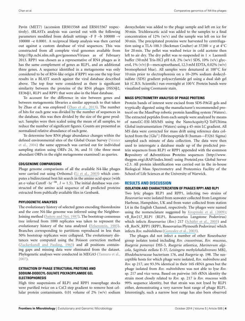
Both phage genomes were determined to be linear dsDNAthrough Bal31/Nde1 double digest treatment (Figure 3). Thepresence of two progressively shortening bands is indicative ofa linear genome with defined ends. Gene prediction identified92 and 91 putative CDSs in RLP1 and RPP1 respectively. MostCDSs (in both phages) appear to initiate at an ATG codonalthough around 10% use GTG or TTG as start codons. Threetransfer RNA genes were also identified in both phages for pro-line (CCA), isoleucine (ATC) and glutamine (CAA). The twoRoseovarius phages are highly related in almost all putative CDSs;
FIGURE 2 | Modified one-step growth cure for phages RLP1 and RPP1.
Host cells were infected with a MOI of 0.001. One step growth curve of RLP1on Rsv. 217 (�) and RPP1 on Rsv. nubinhibens (�). The number of phageincreases over time indicating infection has occurred. There is a markedincrease in phage between 2 and 3 h which suggests a burst event hasoccurred during this period. Each growth curve was performed in triplicate.
www.frontiersin.org October 2014 | Volume 5 | Article 506 | 5
Chan et al. Comparative genomics of N4-like phages
FIGURE 3 | Nde1 digested (A) RLP1 and (B) RPP1 genomic DNA after
treatment with Bal31 for the indicated time intervals. Solid arrowsindicate restriction fragment decreasing over time, dotted arrows indicatepossible second disappearing restriction fragment. The presence offragments reducing in size with time indicates the phage genome is linearnot circular. M, DNA marker (kb).
RLP1 has only three unique CDSs (gps 61, 83, 84) and RPP1also has three (gps 2, 3, 83) all of which have unknown func-tion. At the nucleotide level, gene homologs are 95–100% similar.Sequence comparison of the two phage genomes demonstratedthat there are no large-scale genomic re-arrangements. Overall,the genome structures of RPP1 and RLP1 are similar to thoseof RN4-phages DSS3�2 and EE36�1 but different to that ofpCB2047-B (Zhao et al., 2009; Ankrah and Budinoff, 2014)(Figure 4).
Twenty-eight (∼30%) of the predicted CDSs in RLP1/RPP1are related to those found in Enterobacteria phage N4 and afurther 19 CDSs are similar to genes found in roseophagesDSS3�2, EE36�1 and pCB2047-B (Table 2). Unlike N4 and N4-like Pseudomonas phages no promoter consensus sequences couldbe identified to assign the predicted CDSs to early, middle or lategenes.
The properties and genome sequences of these two novelphages are remarkably similar even though they were isolatedfrom samples obtained 7 years apart, from two locations in UKcoastal waters, and they infect different hosts (one isolated fromthe Caribbean the other from the English Channel). The hoststrains of these highly similar phage are only moderately close rel-atives at 93.5% 16S rRNA gene identity, and in case of RLP1, eventhe closest relative (Rsv mucosus, 99% 16S rRNA gene identitywith Rsv. Sp. 217) was not infected. Although relatively few lyticphages of Roseobacters had been reported previously, it is intrigu-ing that five of the seven lytic roseophages are closely relatedN4-like phages suggesting that similar phages may be commonin the marine environment.
PHYLOGENETIC ANALYSIS OF N4-LIKE CORE GENESAnalysis of the 25 sequenced N4-like phages identified 14 coregenes, examples of these genes in N4 are listed in Table 3 (seeSupplementary Material Table 1 for full list). This number ofcore is genes is similar to the 12 that were found for podovirusesinfecting marine Synechococcus and Prochlorococcus (Labrie et al.,
2013), however, the environments and hosts of the N4-like phagein this study are more diverse. Of these core genes five have noknown function (designated as gps 24, 25, 53, 55, 69 in N4), leav-ing only nine genes that have putative function that are core toN4-like phage. As might be expected these are involved in pro-cesses that all N4-like phage would undergo regardless of thehost they infect including DNA replication and packaging (gps45, 50 and 68), transcription (gp15 and gp16) and productionof structural proteins (gps 54, 55, 56 and 59). Interestingly, thehomolog of RNAP2 in the Achromobacter phages JWAlpha andJWDelta has been divided into two parts due to the insertion ofa 186 amino acid CDS similar to gp8 from Celetribacter phageP12053L (Wittmann et al., 2014). In N4, middle gene productsare transcribed by a heterodimeric RNA polymerase the subunitsof which are encoded by genes RNAP1 and RNAP2 (Willis et al.,2002). Though it is not clear if the RNAP2 homolog is functionalin JWAlpha and JWDelta, we believe that the function of the geneproduct is essential and hence warrants its inclusion in the list ofcore genes.
Gene order of the core genes is largely conserved across allN4-like phage isolates (Figure 4) with unique/clade-specific genestending to be toward the ends of the genomes. The insertionof genes specific to a subset of phage such as the RN4 phagesalso occurs at conserved positions as can be seen for rnr and trx(Figure 4). The high degree of synteny of the core genes involvedin control of gene expression, DNA replication and structuralproteins of 25 N4-like phages suggests that a stable associa-tion within each core module has been formed; conversely theareas between the blocks of core genes are likely hot-spots forrecombination.
Phylogenetic analysis of the N4-like phages based on an align-ment of concatenated core gene products showed that, with theexception of Escherichia phage EC1-UPM, phages that infectclosely related hosts cluster together on well supported branches(Figure 5). For example, the five RN4-phages which infect marineAlphaproteobacteria, form a distinct clade away from their rel-atives that target gammaproteobacterial hosts. Furthermore, thetwo phages which infect Roseovarius species, RLP1 and RPP1, arefurther delineated from the other three RN4-phages; however, thephages EE36F1 and pCB2047-B that infect Sulfitobacter strainsEE36 and 2047, respectively, did not form a distinct subclade.Overall the phylogeny based on concatenated core genes is con-cordant to that previously reported by Wittman et al. based onthe proteomes of 24 N4-like phages (Wittmann et al., 2014). Thedelineation of N4 phage into clades that infect specific hosts sug-gests that all N4 phage shared a common ancestor and have sincespecialized to infect a particular group of hosts.
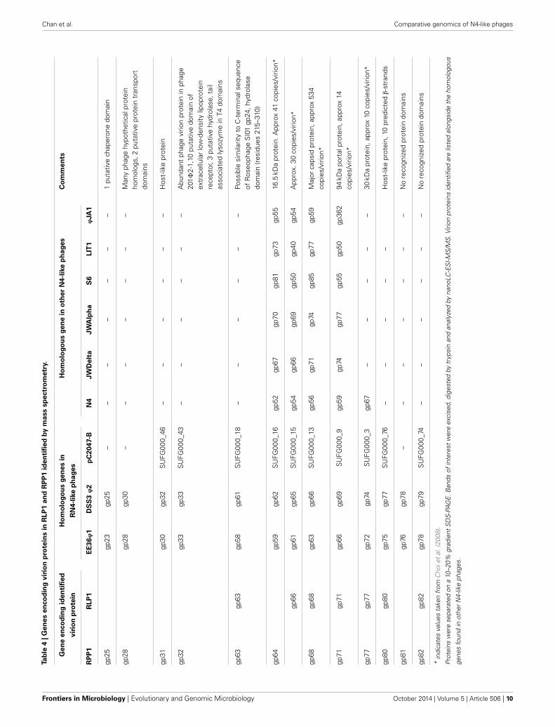
COMPARATIVE ANALYSIS OF RN4 PHAGES pCB2047-B, DSS3�2,EE36�1, RLP1 AND RPP1Analysis of the five RN4-phages identified 33 conserved CDSsof which 14 are N4 core genes, five have homologs in N4phage, five are found in other N4-like phages and nine areexclusive to the RN4 phages (Table 2). Interestingly one of theconserved RN4 phage genes, gp37 (in RPP1), is a host-likemetabolic gene (known as auxiliary metabolic genes, AMGs;highlighted in bold in Table 2). Gp37 encodes a thioredoxin
Frontiers in Microbiology | Evolutionary and Genomic Microbiology October 2014 | Volume 5 | Article 506 | 6

Chan et al. Comparative genomics of N4-like phages
FIGURE 4 | Comparison of 25 N4-like phage genomes. Arrowsrepresent the predicted ORFs and point in the direction of transcription.N4-like core genes are shaded in green and labeled with N4 phagehomolog ORF numbers, host-like genes found in Roseobacter N4-likephages are shaded in red, and finally experimentally determined
structural genes are outlined by dotted lines. The gray box in RPP1marks the putative second structural module containing experimentallyidentified virion proteins. The genomes of RLP1 and RPP1 weredeposited with EMBL under accession numbers FR682616 andFR719956, respectively.
which has also been found in the T7-like Roseophage SIO1(Rohwer et al., 2000). A homolog of this gene is also found inphages JWAlpha and JWDelta which were isolated from wastewater treatment plants. It is interesting to note that whilst thesephages infect Achromobacter xylosoxidans, a nosocomial pathogenwidely distributed in the natural environment (Wittmann et al.,2014), other members of the Achromobacter genus are found infreshwater and marine environments (Brenner et al., 2005).
Phages DSS3�2, EE36�1, RLP1 and RPP1 share a further22 CDSs (Supplementary Material Table 2) one of which, gp51(in RPP1), is another AMG. RPP1 gp51 encodes a class IIribonucleoside diphosphate reductase (rnr). A previous studyby Dwivedi et al., showed that the rnr genes in DSS3�2 andEE36�1 cluster together, with their bacterial host(s) forminga sister group (Dwivedi et al., 2013). A similar analysis usingtrx from the five RN4 phages, showed no clear relationship
www.frontiersin.org October 2014 | Volume 5 | Article 506 | 7

Chan et al. Comparative genomics of N4-like phages
Tab
le2
|R
oseo
bacte
rp
hag
eg
en
es.
Fo
un
din
N4
ph
ag
eFo
un
din
oth
er
N4-l
ike
ph
ag
es
Fo
un
din
RN
4-p
hag
es
on
ly
gp14inN4
rIIA-likeprotein
rIIB-likeprotein
DNAHelicase
16.5kDavirionprotein
Virionprotein
RoseobacterHost-likethioredoxin
Hypotheticalprotein
EndoribonucleaseRusA
Hypotheticalprotein
Hypotheticalprotein
Hypotheticalprotein
Host-likeprotein/virion
protein
Hypotheticalprotein
Host-likeprotein
Hypotheticalprotein
Virionprotein
Host-likeprotein/virion
protein
Virionprotein
RLP
1gp
43gp
39gp
40gp
52gp
64gp
31g
p36
gp47
gp59
gp70
gp2
gp17
gp30
gp35
gp74
gp76
gp77
gp80
gp82
RP
P1
gp44
gp40
gp41
gp53
gp64
gp32
gp
37
gp48
gp60
gp70
gp3
gp18
gp31
gp36
gp74
gp76
gp77
gp80
gp82
DS
S3F
2or
f45
orf4
1or
f42
orf5
0or
f62
orf3
5o
rf40
orf4
6or
f58
orf6
8or
f3or
f18
orf3
2or
f39
orf7
1or
f73
orf7
4or
f77
orf7
9
EE
36F
1or
f42
orf3
8or
f39
orf4
7or
f59
orf3
3o
rf37
orf4
3or
f55
orf6
5or
f4or
f16
orf3
0or
f36
orf6
9or
f71
gp72
orf7
5or
f78
pCB
2047
-Bor
f34
orf3
9or
f38
orf2
9or
f17
orf3
4o
rf40
orf3
3or
f22
orf1
0or
f66
orf5
7or
f46
orf4
1or
f06
orf0
4or
f03
orf7
6or
f74
N4
gp14
gp33
gp34
gp37
gp52
JWD
elta
orf1
2or
f47
orf4
8or
f51
orf6
7or
f44
JWA
lpha
orf1
2or
f50
orf5
1or
f54
orf7
0or
f44
IME
11or
f64
orf3
7or
f36
orf3
2or
f14
G7C
gp14
gp33
gp34
gp37
gp52
KB
NP
21or
f15
orf3
9or
f40
orf4
3or
f60
S6
gp59
gp81
gp66
gp76
gp87
EC
1-U
PM
gp29
gp33
gp34
gp37
gp52
LIT1
orf1
5or
f42
orf4
3or
f36
LUZ7
orf4
6or
f47
orf4
0or
f75
PA26
orf1
5or
f42
orf4
3or
f35
FSL
SP-
076
SP
076_
0013
5S
P07
6_00
220
SP
076_
0019
5
FSL
SP-
058
SP
058_
0022
0S
P05
8_00
305
SP
058_
0028
0
Pres
ely
Pres
ley_
34Pr
esle
y_81
Pres
ley_
17
EcP
1gp
37gp
52gp
48
JA1
JA1_
0038
VCO
139
VCO
139_
0038
VCO
139_
0052
VB
P32
VP
MG
_000
82
VCP
47V
PN
G_0
0047
pYD
6-A
PY
DG
_000
81
JA1
JA1_
0052
Hom
olog
sw
ere
iden
tified
byB
LAST
pse
arch
esan
d%
iden
tity
atth
enu
cleo
tide
leve
lcal
cula
ted
usin
gC
lust
alW
pair
wis
ean
alys
is.G
enes
inbo
lden
code
host
-like
AM
Gs.
Frontiers in Microbiology | Evolutionary and Genomic Microbiology October 2014 | Volume 5 | Article 506 | 8

Chan et al. Comparative genomics of N4-like phages
Table 3 | Conserved core genes of the N4-like phage genus.
Gene in N4 Gene description
15 RNAP1 (Transcriptional control)
16 RNAP2 (Transcriptional control)
24 Unknown
25 vWFA domain
39 DNA polymerase (DNA metabolism/replication)
45 SSB (DNA metabolism/replication)
50 vRNAP (DNA metabolism/replication)
53 Unknown
54 Structural protein (Structural)
55 Unknown
56 Major coat protein (Structural)
59 94 kDa portal protein (Structural)
68 Terminase, large subunit
69 Unknown
Homologs were identified using OrthoMCL which computes reciprocal best
blast hit. An e-value cutoff of 1e-6 and I = 1.5 was used to identify the 14 core
genes in the 25 publically available N4-like phage genomes.
FIGURE 5 | Phylogram of concatenated core genes of the 25 sequenced
N4-like phages. The neighbor-joining tree was based on a ClustalWalignment of the concatenated core genes amino acid sequences;bootstrap values were based on 1000 replicates. Apart from Escherichiaphage EC1-UPM, N4-like phages that infect closely related hosts clustertogether on well supported branches. The tree is rooted at mid-point andbranches with less than 50% bootstrap replicates were collapsed; scale barindicate expected changes per site.
between phage and host genes (Supplementary MaterialFigure 2).
The presence of the AMG trx in the five RN4 phages is likely torepresent an adaptation to the marine environment as it is com-mon to all N4-like phages that infect marine bacteria (Figure 4).Thioredoxin-encoding genes can also be found in T7-like phages
though it is also more common in viruses from the marine envi-ronment e.g., SIO1 and P60, than in enteric phages (Zhao et al.,2009). What the function of this gene might be is unclear; inbacteriophage T7 there is an increased rate of processing whenthioredoxin binds to T7 DNA polymerase (Huber et al., 1987).However, whilst trx is found in other marine phages it is notclear if it serves the same function as found in T7 as the cor-rect domain required for thioredoxin to bind may not be present(Hardies et al., 2003). Thioredoxin is known to have many otherroles, one of which is a hydrogen donor to ribonucleotide reduc-tase. This is possibly the most parsimonious function for trx,as four out of five RN4 phage also carry the rnr gene encod-ing for a ribonucleotide reductase. With rnr commonly foundin other marine phage (Angly et al., 2006) it is thought toprovide a mechanism of scavenging ribonucleotides in the olig-otrophic marine environment (Sullivan et al., 2005). Therefore,it could be speculated for RN4 phages ribonuclease reductase isexpressed to replicate the function of the host gene and the phageencoded thioredoxin acts in co-ordination as specific hydro-gen donor, in a similar fashion that occurs in T4 (Holmgren,1989).
IDENTIFICATION OF A SECOND STRUCTURAL MODULE IN RPP1 PHAGEWe identified, using mass spectrometry, 13 structural proteins inthe mature RPP1/RLP1 virions (Table 4, Supplementary MaterialFigure 3) including five which have been identified as N4 virionproteins (gps 52, 54, 56, 59, and 67 in N4 phage/ gps 64, 66,68, 71, and 77 in RPP1). Nine of the identified structural pro-teins in RPP1/RLP1 (gps 63, 64, 66, 68, 71 77, 80, 81, and 82in RPP1) are likely “late” gene products inferred through syn-teny with N4 phage and their localization after the vRNAP geneand other late genes in N4 (Kazmierczak and Rothman-Denes,2005). The remaining four (gps 25, 28, 31, and 32 in RPP1)are located near the N4 homologs of gp24 and 25 which in theEnterobacter phage N4 are middle gene transcripts (Kazmierczakand Rothman-Denes, 2005). This suggests there is a second struc-tural module (SSM) in RPP1 which is expressed during themid-phase of infection. Ceyssens et al. (2010) also identified asimilar additional cluster of structural genes not expressed withthe late genes in Pseudomonas phage LIT1 (Ceyssens et al., 2010).BLASTp analysis shows that the RPP1 gp32 gene product (a 650aa protein) shares similarity with gp230 in Pseudomonas myovrius201�2-1, which is a fusion of homologs of �KZ gp145 andgp146, both tail proteins. Interestingly, genes within the sec-ond structural cluster in LIT1 (gps 48–56) have strong similarityto Pseudomonas aeruginosa prophage proteins and tail proteinsfrom other Podoviridae (Ceyssens et al., 2010). Taken together,these observations suggest that the additional structural mod-ule encodes for and/or is associated with virion tail protein(s)production.
The gene products 25 and 28 in RPP1 found in the tail protein-linked SSM contain protein chaperone-like domains which couldbe associated with the translocation of the unfolded/semi-foldedvRNAP out of the virion head into the host cell during initialinfection. This is required as the virion polymerase is relativelylarge, 382.5 kDa, whilst the narrowest section of the tail tube inN4 is only 25 Å in diameter (Choi et al., 2008).
www.frontiersin.org October 2014 | Volume 5 | Article 506 | 9
Chan et al. Comparative genomics of N4-like phages
Tab
le4
|G
en
es
en
co
din
gvir
ion
pro
tein
sin
RLP
1an
dR
PP
1id
en
tifi
ed
by
mass
sp
ectr
om
etr
y.
Gen
een
co
din
gid
en
tifi
ed
Ho
mo
log
ou
sg
en
es
inH
om
olo
go
us
gen
ein
oth
er
N4-l
ike
ph
ag
es
Co
mm
en
ts
vir
ion
pro
tein
RN
4-l
ike
ph
ag
es
RP
P1
RLP
1E
E36ϕ
1D
SS
3ϕ
2p
C2047-B
N4
JW
Delt
aJW
Alp
ha
S6
LIT
1ϕ
JA
1
gp25
gp23
gp25
––
––
––
–1
puta
tive
chap
eron
edo
mai
n
gp28
gp28
gp30
––
––
––
–M
any
phag
ehy
poth
etic
alpr
otei
nho
mol
ogs,
2pu
tativ
epr
otei
ntr
ansp
ort
dom
ains
gp31
gp30
gp32
SU
FG00
0_46
––
––
––
Hos
t-lik
epr
otei
n
gp32
gp33
gp33
SU
FG00
0_43
––
––
––
Abu
ndan
tph
age
virio
npr
otei
nin
phag
e20
1�2-
1,10
puta
tive
dom
ain
ofex
trac
ellu
lar
low
-den
sity
lipop
rote
inre
cept
or,3
puta
tive
hydr
olas
e,ta
ilas
soci
ated
lyso
zym
ein
T4do
mai
ns
gp63
gp63
gp58
gp61
SU
FG00
0_18
––
––
––
Poss
ible
sim
ilarit
yto
C-t
erm
inal
sequ
ence
ofR
oseo
phag
eS
I01
gp24
,hyd
rola
sedo
mai
n(r
esid
ues
215–
310)
gp64
gp59
gp62
SU
FG00
0_16
gp52
gp67
gp70
gp81
gp73
gp55
16.5
kDa
prot
ein.
App
rox
41co
pies
/viri
on*
gp66
gp61
gp65
SU
FG00
0_15
gp54
gp66
gp69
gp50
gp40
gp54
App
rox.
30co
pies
/viri
on*
gp68
gp68
gp63
gp66
SU
FG00
0_13
gp56
gp71
gp74
gp85
gp77
gp59
Maj
orca
psid
prot
ein,
appr
ox53
4co
pies
/viri
on*
gp71
gp71
gp66
gp69
SU
FG00
0_9
gp59
gp74
gp77
gp55
gp50
gp36
294
kDa
port
alpr
otei
n,ap
prox
14co
pies
/viri
on*
gp77
gp77
gp72
gp74
SU
FG00
0_3
gp67
––
––
–30
kDa
prot
ein,
appr
ox10
copi
es/v
irion
*
gp80
gp80
gp75
gp77
SU
FG00
0_76
––
––
–H
ost-
like
prot
ein,
10pr
edic
ted
β-s
tran
ds
gp81
gp76
gp78
––
––
––
–N
ore
cogn
ized
prot
ein
dom
ains
gp82
gp82
gp78
gp79
SU
FG00
0_74
––
––
––
No
reco
gniz
edpr
otei
ndo
mai
ns
*in
dica
tes
valu
esta
ken
from
Cho
iet
al.(
2008
).
Prot
eins
wer
ese
para
ted
ona
10–2
0%gr
adie
ntS
DS
-PA
GE
.Ban
dsof
inte
rest
wer
eex
cise
d,di
gest
edby
tryp
sin
and
anal
yzed
byna
noLC
-ES
I-MS
/MS.
Virio
npr
otei
nsid
entifi
edar
elis
ted
alon
gsid
eth
eho
mol
ogou
s
gene
sfo
und
inot
her
N4-
like
phag
es.
Frontiers in Microbiology | Evolutionary and Genomic Microbiology October 2014 | Volume 5 | Article 506 | 10
Chan et al. Comparative genomics of N4-like phages
The location of these additional structural genes (upstream ofthe N4 gp45 homolog encoding an ssDNA-binding protein whichactivates transcription of late phage genes) suggests they are “mid-dle” genes, but the advantage of expressing such proteins priorto the capsid genes is not yet clear. It may point to a gene regu-lation requirement and/or a possibility that tail proteins requirematuration prior to assembly on the virion. In general, the con-stituent parts of phage virion particles (heads, tails and tail fibers)are made separately via subassembly pathways rather than a singlelinear pathway. Upon completion of the virion segment, the headsand tails combine first, forming complexes that are visible by elec-tron microscopy, then the distal tail fibers are added (Campbell,2007). It is possible that the assembly of the structurally complextail portion of the virion may involve multiple steps and requiresthe assistance of helper proteins whilst the head is relatively sim-ple to construct. Consequently, there might be an advantage inexpressing some tail structural genes earlier than the genes codingfor head, portal and other tail fiber genes.
Of the 13 structural proteins identified in RPP1/RLP1, 10 areconserved in all the sequenced RN4 phages. These include gps31 and 32 (in RPP1) from the SSM. Interestingly whilst gp31 isonly shared by the RN4 phages, a homolog of gp32 is also foundin Erwinia phage S6 (Born et al., 2011) as gp66. The aforemen-tioned gps 25 and 28 (in RPP1) are only found in phages DSS3�2,EE36�1 and RLP1 suggesting this module could be a determinantof host specificity whilst gene product 81 is only found in RLP1and RPP1.
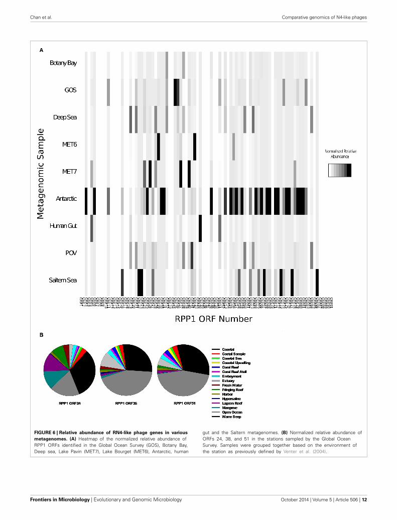
ENVIRONMENTAL DISTRIBUTION OF RN4-LIKE PHAGESUsing all the CDS sequences in RPP1 as blast query against arange of environmental metagenomic datasets downloaded fromCAMERA (Sun et al., 2011) we searched for RN4-like phagesequences. The number of hits were normalized for databasesize and gene size to allow comparison between metagenomes(see Materials and Methods for further details). Previous searchesof Global Ocean Survey (GOS) metagenomic data using RN4polymerase genes as well as the other N4-like genes as querysequences suggested that N4-like phage infecting Roseobactersare mainly found in coastal areas and may be rare in open oceanenvironments (Zhao et al., 2009). We found homologs of CDSsfrom RN4 phages are widespread in a number of environments(Figure 6A) with the highest frequency of counts in samples fromthe Antarctic, Saltern Sea and GOS metagenomes. As expected,given the known distribution of members of the Roseobacter lin-eage, we found very low detection rates in the metagenomes fromfreshwater lakes (MET6, MET7).
A more detailed analysis of the distribution of hits found inthe GOS metagenome was carried out based on the previouslydefined environments as reported by the Sorcerer II GOS expe-dition (Rusch et al., 2007). The distribution of three RPP1-likegenes for each GOS sampling site was carried out using the threemost abundant gene sequences identified previously, ORFs 24,38, and 51, as queries. A large proportion of matches were foundin locations characterized as a coastal environment (Figure 6B);this would be expected based on the distribution of Roseobacterhosts in costal environments. However, for some genes—ORF36and ORF51, a higher percentage of hits were found in samples
from open ocean environments (Figure 6B), thus suggesting thatthere are more RN4-like phage, and their corresponding hosts,present in the open ocean environment than previously thought.However, this finding should be considered with caution as wepresume the hosts of these phages belong to the Roseobacter lin-eage. There is the possibility that these are not RN4 phages andinstead belong to a different family of podoviruses that infectanother group of bacteria which have not yet been culturedand/or had their genome sequenced.
EVOLUTION OF THE N4-LIKE PHAGE GENUS AND BEYONDThe genome arrangement of core and variable genes withinthis phage genus bears striking similarity to the T4 superfam-ily in which the genomes have been defined as bipartite (Krischand Comeau, 2008); a conserved core comprised of the mini-mal essential genes required for viral multiplication and a larger,highly variable set of facultative genes which collectively create anoptimal environment, particular to that host, to enable successfulinfection. However, in the T4 superfamily most of the “core T4”genes encode either virus replication functions or virion struc-tural components. As N4 has such an unusual gene expressionmechanism (Kazmierczak and Rothman-Denes, 2005), it is per-haps not surprising to find genes involved in transcription controlto be conserved, such as the three RNA polymerase genes andthe single-stranded DNA-binding protein involved in late geneexpression.
In the T4 superfamily, the number of core genes varies accord-ing to the subset of phages considered. For example, there are75 common core genes when “true” T-even (T4), pseudo T-even (RB49) and schizo T-even (Aeh1) are compared (Sullivanet al., 2005; Clokie et al., 2010), but this falls to 38 when thecyanophages are included (Millard et al., 2009; Sullivan et al.,2010). With the N4-like phages, the subdivisions below genuslevel are not as clear but it appears that core genes from phageswhich infect closely related hosts bear more similarity to eachother than those from evolutionary distant hosts as seen by theclustering of the RN4, Pseudomonas, Enterobacter/Escherichia,and Vibrio phages (Figure 4). In addition to vertical gene trans-fer, horizontal gene exchanges could have occurred from bothphage (Pseudomonas tail proteins and the trx gene) and host(Roseobacter host-like proteins e.g., rnr) sources.
Phage biologists have long debated as to whether or not phagegenera actually exist or if instead there is a continuum of phagegenes in which all tailed-phages dip into, to find a “best-fit”genome. The mosaic model proposed by Hendrix et al., poses thebest compromise to this problem (Hendrix et al., 1999), propos-ing that early phages have exchanged large chunks of geneticinformation prior to the demarcation of the now accepted super-groups. Fine tuning of host/environmental specific genes betweenclose relatives then followed, the consequence of which are phageswith genomes created from a mixture of vertical and horizontalgene transfer events. The results from this study fit in well withthis theory. The 14 core genes, which encode and control gen-eral infectivity, appear to be derived from ancient phages thusaccounting for the homology and gene synteny found in theterrestrial and marine phages, whilst the plastic periphery is com-prised of genes such as rnr, trx and the tail/tail fiber structural
www.frontiersin.org October 2014 | Volume 5 | Article 506 | 11
Chan et al. Comparative genomics of N4-like phages
FIGURE 6 | Relative abundance of RN4-like phage genes in various
metagenomes. (A) Heatmap of the normalized relative abundance ofRPP1 ORFs identified in the Global Ocean Survey (GOS), Botany Bay,Deep sea, Lake Pavin (MET7), Lake Bourget (MET6), Antarctic, human
gut and the Saltern metagenomes. (B) Normalized relative abundance ofORFs 24, 38, and 51 in the stations sampled by the Global OceanSurvey. Samples were grouped together based on the environment ofthe station as previously defined by Venter et al. (2004).
Frontiers in Microbiology | Evolutionary and Genomic Microbiology October 2014 | Volume 5 | Article 506 | 12
Chan et al. Comparative genomics of N4-like phages
proteins which provide environmental adaptations and deter-mine the host range. However, further analyses are required todetermine if the latter set of genes were horizontally or verti-cally acquired. Such studies and characterization of more N4-likephages, in particular those from the marine environment, willallow further population genetic type analyses of this diversephage group.
ACKNOWLEDGMENTThis work was supported by BBSRC and NERC (UK). JacquelineZ.-M. Chan. was supported through a BBSRC PhD studentship.Hendrik Schäfer was supported by a NERC Advanced Fellowship(NE/E01333/1) and phage genome sequencing was funded by agrant from the NERC (NE/F010044/1). Ms Susan Slade from theBiological Mass Spectrometry and Proteomics Facility, Universityof Warwick is thanked for performing mass spectrometry analy-ses. The GenePool facility, University of Edinburgh is thanked forperforming the genome sequencing.
SUPPLEMENTARY MATERIALThe Supplementary Material for this article can be foundonline at: http://www.frontiersin.org/journal/10.3389/fmicb.2014.00506/abstract
REFERENCESAltschul, S. F., Gish, W., Miller, W., Myers, E. W., and Lipman, D. J. (1990). Basic
local alignment search tool. J. Mol. Biol. 215, 403–410. doi: 10.1016/S0022-2836(05)80360-2
Angly, F. E., Felts, B., Breitbart, M., Salamon, P., Edwards, R. A., Carlson, C., et al.(2006). The marine viromes of four oceanic regions. PLoS Biol. 4:e368. doi:10.1371/journal.pbio.0040368
Ankrah, N., and Budinoff, C. (2014). Genome sequence of the Sulfitobactersp. strain 2047-infecting lytic phage �CB2047-B. Genome 2, 10–11. doi:10.1128/genomeA.00945-13
Besemer, J., and Borodovsky, M. (1999). Heuristic approach to deriving models forgene finding. Nucleic Acids Res. 27, 3911–3920. doi: 10.1093/nar/27.19.3911
Born, Y., Fieseler, L., Marazzi, J., Lurz, R., Duffy, B., and Loessner, M. J. (2011).Novel virulent and broad-host-range Erwinia amylovora bacteriophages reveala high degree of mosaicism and a relationship to Enterobacteriaceae phages.Appl. Environ. Microbiol. 77, 5945–5954. doi: 10.1128/AEM.03022-10
Breitbart, M., and Rohwer, F. (2005). Here a virus, there a virus, everywhere thesame virus? Trends Microbiol. 13, 278–284. doi: 10.1016/j.tim.2005.04.003
Breitbart, M., Thompson, L. R., Suttle, C. A., and Sullivan, M. B. (2007).Exploring the vast diversity of marine viruses. Oceanography 20, 135–139. doi:10.5670/oceanog.2007.58
Brenner, D. J., Krieg, N. R., and Staley, J. T. (eds.). (2005). Bergey’s Manual ofSystematic Bacteriology. New York, NY: Springer US.
Buchan, A., Gonzalez, J. M., and Moran, M. A. (2005). Overview of themarine Roseobacter lineage. Appl. Environ. Microbiol. 71, 5665–5677. doi:10.1128/AEM.71.10.5665-5677.2005
Campbell, A. M. (2007). “Bacteriophages,” in Fields Virology, 5th Edn., eds B.N. Fields, D. M. Knipe, P. M. Howley, and D. E. Griffin (Philadelphia, PA:Lippencott Williams & Wilkins), 769–791.
Ceyssens, P.-J., Brabban, A., Rogge, L., Lewis, M. S., Pickard, D., Goulding, D.,et al. (2010). Molecular and physiological analysis of three Pseudomonas aerug-inosa phages belonging to the “N4-like viruses.” Virology 405, 26–30. doi:10.1016/j.virol.2010.06.011
Choi, K. H., McPartland, J., Kaganman, I., Bowman, V. D., Rothman-Denes, L.B., and Rossmann, M. G. (2008). Insight into DNA and protein transport indouble-stranded DNA viruses: the structure of bacteriophage N4. J. Mol. Biol.378, 726–736. doi: 10.1016/j.jmb.2008.02.059
Clokie, M. R., Millard, A. D., and Mann, N. H. (2010). T4 genes in the marineecosystem: studies of the T4-like cyanophages and their role in marine ecology.Virol. J. 7:291. doi: 10.1186/1743-422X-7-291
Delcher, A. L., Harmon, D., Kasif, S., White, O., and Salzberg, S. L. (1999).Improved microbial gene identification with GLIMMER. Nucleic Acids Res. 27,4636–4641. doi: 10.1093/nar/27.23.4636
Dwivedi, B., Xue, B., Lundin, D., Edwards, R. A., and Breitbart, M. (2013).A bioinformatic analysis of ribonucleotide reductase genes in phagegenomes and metagenomes. BMC Evol. Biol. 13:33. doi: 10.1186/1471-2148-13-33
Fan, H. H., An, X., Huang, Y., Zhang, Z., Mi, Z., and Tong, Y. (2012). Completegenome sequence of IME11, a new N4-like bacteriophage. J. Virol. 86, 13861.doi: 10.1128/JVI.02684-12
Farmer, N. G., Wood, T. L., Chamakura, K. R., and Everett, G. F. K. (2013).Complete Genome of Acinetobacter baumannii N4-Like Podophage. GenomeAnnounc. 1, 6–7. doi: 10.1128/genomeA.00852-13
Felsenstein, J. (1985). Confidence limits on phylogenies: an approach using thebootstrap. Evolution (N. Y). 39, 783–791.
Fouts, D. E., Klumpp, J., Bishop-Lilly, K. A., Rajavel, M., Willner, K. M., Butani, A.,et al. (2013). Whole genome sequencing and comparative genomic analyses oftwo Vibrio cholerae O139 Bengal-specific Podoviruses to other N4-like phagesreveal extensive genetic diversity. Virol. J. 10:165. doi: 10.1186/1743-422X-10-165
Fuhrman, J. A. (1999). Marine viruses and their biogeochemical and ecologicaleffects. Nature 399, 541–548. doi: 10.1038/21119
Gan, H. M., Sieo, C. C., Tang, S. G. H., Omar, A. R., and Ho, Y. W. (2013).The complete genome sequence of EC1-UPM, a novel N4-like bacteriophagethat infects Escherichia coli O78:K80. Virol. J. 10:308. doi: 10.1186/1743-422X-10-308
Gonzalez, J. M., Covert, J. S., Whitman, W. B., Henriksen, J. R., Mayer, F., Scharf,B., et al. (2003). Silicibacter pomeroyi sp. nov. and Roseovarius nubinhibens sp.nov., dimethylsulfoniopropionate-demethylating bacteria from marine envi-ronments. Int. J. Syst. Evol. Microbiol. 53, 1261–1269. doi: 10.1099/ijs.0.02491-0
Hardies, S. C., Comeau, A. M., Serwer, P., and Suttle, C. A. (2003). The completesequence of marine bacteriophage VpV262 infecting Vibrio parahaemolyticusindicates that an ancestral component of a T7 viral supergroup is widespreadin the marine environment. Virology 310, 359–371. doi: 10.1016/S0042-6822(03)00172-7
Hendrix, R. W., Smith, M. C. M., Burns, R. N., Ford, M. E., and Hatfull,G. F. (1999). Evolutionary relationships among diverse bacteriophages andprophages: all the world’s a phage. Proc. Natl. Acad. Sci. U.S.A. 96, 2192–2197.doi: 10.1073/pnas.96.5.2192
Hess, W. R. (2008). “Comparative genomics of marine cyanobacteria and theirphages,” in The Cyanobacteria: Molecular Biology, Genomics and Evolution, edsA. Herrero and E. Flores (Norwich, UK: Caister Academic Press), 89–116.
Holmgren, A. (1989). Thioredoxin and glutaredoxin systems. J. Biol. Chem. 264,13963–13966.
Huber, H. E., Tabor, S., and Richardson, C. C. (1987). Escherichia coli thiore-doxin stabilizes complexes of bacteriophage T7 DNA polymerase and primedtemplates. J. Biol. Chem. 263, 16224–16232.
Hurwitz, B. L., and Sullivan, M. B. (2013). The Pacific Ocean virome (POV): amarine viral metagenomic dataset and associated protein clusters for quantita-tive viral ecology. PLoS ONE 8:e57355. doi: 10.1371/journal.pone.0057355
Kazmierczak, K. M., and Rothman-Denes, L. B. (2005). “Bacteriophage N4,” inThe Bacteriophages, 2nd Edn., ed R. Calendar (Oxford: Oxford University Press),302–314.
Kim, M. S., Cha, K. E., and Myung, H. (2012). Complete genome of Pseudomonasaeruginosa phage PA26. J. Virol. 86, 10244. doi: 10.1128/JVI.01630-12
Krisch, H. M., and Comeau, A. M. (2008). The immense journey of bacteriophageT4–from d’Hérelle to Delbrück and then to Darwin and beyond. Res. Microbiol.159, 314–324. doi: 10.1016/j.resmic.2008.04.014
Kropinski, A. M., Prangishvili, D., and Lavigne, R. (2009). Position paper:the creation of a rational scheme for the nomenclature of viruses ofBacteria and Archaea. Environ. Microbiol. 11, 2775–2777. doi: 10.1111/j.1462-2920.2009.01970.x
Kulikov, E., Kropinski, A. M., Goldmidova, A., Lingohr, E., Govorun, V.,Serebryakova, M., et al. (2012). Isolation and characterization of a novel indige-nous intestinal N4-related coliphage vB_EcoP_G7C. Virology 426, 93–99. doi:10.1016/j.virol.2012.01.027
Labrie, S. J., Frois-Moniz, K., Osburne, M. S., Kelly, L., Roggensack, S. E., Sullivan,M. B., et al. (2013). Genomes of marine cyanopodoviruses reveal multiple
www.frontiersin.org October 2014 | Volume 5 | Article 506 | 13
Chan et al. Comparative genomics of N4-like phages
origins of diversity. Environ. Microbiol. 15, 1356–1376. doi: 10.1111/1462-2920.12053
Li, L., Stoeckert, C. J., and Roos, D. S. (2003). OrthoMCL: identification ofortholog groups for eukaryotic genomes. Genome Res. 13, 2178–2189. doi:10.1101/gr.1224503
Loessner, M. J., Inman, R. B., Lauer, P., and Calendar, R. (2000). Completenucleotide sequence, molecular analysis and genome structure of bacteriophageA118 of Listeria monocytogenes: implications for phage evolution. Mol.Microbiol. 35, 324–340. doi: 10.1046/j.1365-2958.2000.01720.x
Millard, A. D., Zwirglmaier, K., Downey, M. J., Mann, N. H., and Scanlan,D. J. (2009). Comparative genomics of marine cyanomyoviruses revealsthe widespread occurrence of Synechococcus host genes localized to ahyperplastic region: implications for mechanisms of cyanophage evolu-tion. Environ. Microbiol. 11, 2370–2387. doi: 10.1111/j.1462-2920.2009.01966.x
Moreno Switt, A. I., Orsi, R. H., den Bakker, H. C., Vongkamjan, K., Altier, C.,and Wiedmann, M. (2013). Genomic characterization provides new insight intoSalmonella phage diversity. BMC Genomics 14:481. doi: 10.1186/1471-2164-14-481
Nho, S.-W., Ha, M.-A., Kim, K.-S., Kim, T.-H., Jang, H.-B., Cha, I.-S., et al. (2012).Complete genome sequence of the bacteriophages ECBP1 and ECBP2 iso-lated from two different Escherichia coli strains. J. Virol. 86, 12439–12440. doi:10.1128/JVI.02141-12
Paul, J. H., and Sullivan, M. B. (2005). Marine phage genomics: what havewe learned? Curr. Opin. Biotechnol. 16, 299–307. doi: 10.1016/j.copbio.2005.03.007
Rohwer, F. (2003). Global phage diversity. Cell 113, 141. doi: 10.1016/S0092-8674(03)00276-9
Rohwer, F., Segall, A., Steward, G., Seguritan, V., Breitbart, M., Wolven, F., et al.(2000). The complete genomic sequence of the marine phage Roseophage SIO1shares homology with nonmarine phages. Limnol. Ocean. 45, 408–418. doi:10.4319/lo.2000.45.2.0408
Rusch, D. B., Halpern, A. L., Sutton, G., Heidelberg, K. B., Williamson, S., Yooseph,S., et al. (2007). The Sorcerer II global ocean sampling expedition: northwestAtlantic through eastern tropical Pacific. PLoS Biol. 5:e77. doi: 10.1371/jour-nal.pbio.0050077
Saitou, N., and Nei, M. (1987). The neighbor-joining method: a new method forreconstructing phylogenetic trees. Mol. Biol. Evol. 4, 406–425.
Sambrook, J., and Russell, D. W. (2001). Molecular Cloning: A Laboratory Manual.New York, NY: Cold Spring Harbour Laboratory Press.
Schäfer, H., McDonald, I. R., Nightingale, P. D., and Murrell, J. C. (2005). Evidencefor the presence of a CmuA methyltransferase pathway in novel marine methylhalide-oxidizing bacteria. Environ. Microbiol. 7, 839–852. doi: 10.1111/j.1462-2920.2005.00757.x
Schito, G. C., Molina, A. M., and Pesce, A. (1965). Un nuovo batteriofago attivo sulceppo K12 di E. coli. I. Caratteristiche biologiche. Boll. Inst. Sieroter. Milanese44, 329–332.
Schito, G. C., Molina, A. M., and Pesce, A. (1967). Lysis and lysis inhibition withN4 coliphage. Giorn. Microbiol. 15, 229–244.
Sommer, D., Delcher, A., Salzberg, S., and Pop, M. (2007). Minimus: a fast,lightweight genome assembler. BMC Bioinformatics 8:64. doi: 10.1186/1471-2105-8-64
Sullivan, M. B., Coleman, M. L., Weigele, P., Rohwer, F., and Chisholm, S.W. (2005). Three Prochlorococcus cyanophage genomes: signature featuresand ecological interpretations. PLoS Biol. 3:e144. doi: 10.1371/journal.pbio.0030144
Sullivan, M. B., Huang, K. H., Ignacio-Espinoza, J. C., Berlin, A. M., Kelly,L., Weigele, P. R., et al. (2010). Genomic analysis of oceanic cyanobac-terial myoviruses compared with T4-like myoviruses from diverse hostsand environments. Environ. Microbiol 12, 3035–3056. doi: 10.1111/j.1462-2920.2010.02280.x
Sullivan, M. B., Waterbury, J. B., and Chisholm, S. W. (2003). Cyanophages infect-ing the oceanic cyanobacterium Prochlorococcus. Nature 424, 1047–1051. doi:10.1038/nature01929
Sun, S., Chen, J., Li, W., Altintas, I., Lin, A., Peltier, S., et al. (2011).Community cyberinfrastructure for advanced microbial ecology research andanalysis: the CAMERA resource. Nucleic Acids Res. 39, D546–D551. doi:10.1093/nar/gkq1102
Suttle, C. A. (2005). Viruses in the sea. Nature 437, 356–361. doi:10.1038/nature04160
Suttle, C. A. (2007). Marine viruses–major players in the global ecosystem. Nat.Rev. Microbiol. 5, 801–812. doi: 10.1038/nrmicro1750
Tamura, K., Dudley, J., Nei, M., and Kumar, S. (2007). MEGA4: molecular evo-lutionary genetics analysis (MEGA) software version 4.0. Mol. Biol. Evol. 24,1596–1599. doi: 10.1093/molbev/msm092
Venter, J. C., Remington, K., Heidelberg, J. F., Halpern, A. L., Rusch, D., Eisen, J. A.,et al. (2004). Environmental genome shotgun sequencing of the Sargasso Sea.Science 304, 66–74. doi: 10.1126/science.1093857
Weinbauer, M. G., and Rassoulzadegan, F. (2004). Are viruses driving microbialdiversification and diversity? Environ. Microbiol. 6, 1–11. doi: 10.1046/j.1462-2920.2003.00539.x
Wilhelm, S. W., and Suttle, C. A. (1999). Viruses and nutrient cycles in the sea.Bioscience 49, 781–788. doi: 10.2307/1313569
Willis, S. H., Kazmierczak, K. M., Carter, R. H., and Rothman-Denes, L. B. (2002).N4 RNA Polymerase II, a heterodimeric RNA polymerase with homology to thesingle-subunit family of RNA polymerases. J. Bacteriol. 184, 4952–4961. doi:10.1128/JB.184.18.4952-4961.2002
Wilson, W. H., Carr, N. G., and Mann, N. H. (1996). The effect of phosphatestatus on the kinetics of cyanophage infection in the oceanic cyanobac-terium Synechococcus sp. WH7803. J. Phycol. 32, 506–516. doi: 10.1111/j.0022-3646.1996.00506.x
Wittmann, J., Dreiseikelmann, B., Rohde, M., Meier-Kolthoff, J. P., Bunk, B., andRohde, C. (2014). First genome sequences of Achromobacter phages reveal newmembers of the N4 family. Virol. J. 11:14. doi: 10.1186/1743-422X-11-14
Zerbino, D. R., and Birney, E. (2008). Velvet: algorithms for de novo shortread assembly using de Bruijn graphs. Genome Res. 18, 821–829. doi:10.1101/gr.074492.107
Zhang, Y. Y., and Jiao, N. Z. (2009). Roseophage RDJL Phi 1, infecting the aero-bic anoxygenic phototrophic bacterium Roseobacter denitrificans OCh114. Appl.Environ. Microbiol. 75, 1745–1749. doi: 10.1128/AEM.02131-08
Zhao, Y. L., Wang, K., Jiao, N. Z., and Chen, F. (2009). Genome sequences of twonovel phages infecting marine roseobacters. Environ. Microbiol. 11, 2055–2064.doi: 10.1111/j.1462-2920.2009.01927.x
Zhao, Y., Temperton, B., Thrash, J. C., Schwalbach, M. S., Vergin, K. L., Landry, Z.C., et al. (2013). Abundant SAR11 viruses in the ocean. Nature 494, 357–360.doi: 10.1038/nature11921
Zuckerkandl, E., and Pauling, L. (1965). “Evolutionary divergence and convergencein proteins,” in Evolving Genes and Proteins, eds V. Bryson and H. J. Vogel (NewYork, NY: Academic Press), 97–165.
Conflict of Interest Statement: The authors declare that the research was con-ducted in the absence of any commercial or financial relationships that could beconstrued as a potential conflict of interest.
Received: 06 June 2014; accepted: 08 September 2014; published online: 10 October2014.Citation: Chan JZ-M, Millard AD, Mann NH and Schäfer H (2014) Comparativegenomics defines the core genome of the growing N4-like phage genus and identi-fies N4-like Roseophage specific genes. Front. Microbiol. 5:506. doi: 10.3389/fmicb.2014.00506This article was submitted to Evolutionary and Genomic Microbiology, a section of thejournal Frontiers in Microbiology.Copyright © 2014 Chan, Millard, Mann and Schäfer. This is an open-access articledistributed under the terms of the Creative Commons Attribution License (CC BY).The use, distribution or reproduction in other forums is permitted, provided theoriginal author(s) or licensor are credited and that the original publication in thisjournal is cited, in accordance with accepted academic practice. No use, distribution orreproduction is permitted which does not comply with these terms.
Frontiers in Microbiology | Evolutionary and Genomic Microbiology October 2014 | Volume 5 | Article 506 | 14