Comparative Evaluation of Three JAK2V617F Mutation Detection Methods
10
Am J Clin Pathol 2007;128:865-874 865 865 DOI: 10.1309/LW7Q3739RBRMBXXP 865 © American Society for Clinical Pathology Hematopathology / JAK2 V617F DETECTION BY AS-PCR AND RFLP Comparative Evaluation of Three JAK2 V617F Mutation Detection Methods Christine Frantz, MS, 1 Donna M. Sekora, MT, 1 Donald C. Henley, MS, 2 Chih-Kang Huang, MS, 3 Qiulu Pan, MD, PhD, 3 Neil B. Quigley, PhD, 2 Eric Gorman, MD, 3 Roger A. Hubbard, PhD, 2 and Imran Mirza, MD 1 Key Words: JAK2 mutation; Allele-specific PCR; AS-PCR; Restriction fragment length polymorphism; RFLP; Capillary electrophoresis; Polyacrylamide gel electrophoresis DOI: 10.1309/LW7Q3739RBRMBXXP Abstract The correlation of JAK2 V617F with a proportion of chronic myeloproliferative disorders has generated numerous studies focused on the development of molecular-based assays for JAK2 V617F detection. The current parallel study comparatively evaluated 3 JAK2 V617F molecular detection methods. Genomic DNA from blood or bone marrow was assayed by 3 laboratories using allele-specific polymerase chain reaction (AS-PCR) or kit-based restriction fragment length polymorphism methods, which used polyacrylamide gel or capillary electrophoresis analysis. In addition, samples were sequenced in 2 of the laboratories. Results found 100% concordance among the 3 methods, with analytic sensitivities of 5% for both kit methods and 0.01% for AS-PCR. The kit- based assays detect JAK2 V617F with equal sensitivity regardless of analysis method, and, despite greater sensitivity of AS-PCR, all 3 methods yielded 100% concordant results for this 36-sample set. Consistent with other reports, direct sequencing was insufficiently sensitive to serve as an initial diagnostic tool for JAK2 V617F detection. Chronic myeloproliferative disorders (CMPDs) are clon- al stem cell diseases characterized by unrestricted prolifera- tion of granulocytes, RBCs, or platelets. The 3 primary Philadelphia chromosome (Ph)– CMPDs, polycythemia vera (PV), essential thrombocythemia (ET), and chronic idiopathic myelofibrosis (CIMF), demonstrate distinct clinical, hemato- logic, biologic, and molecular features. 1-3 Until recently, diag- nosis of CMPDs was facilitated by assays including serum erythropoietin levels, endogenous erythroid colony formation, polycythemia rubra vera-1 transcript levels, and leukocyte alkaline phosphatase score. 1 In 2004 and 2005, several studies first correlated a proportion of the Ph– CMPDs with the pres- ence of a G-to-T transversion mutation at nucleotide 1849 of the JAK2 gene. 4-8 This mutation, designated JAK2 V617F , result- ed in a valine-to-phenylalanine substitution at codon 617 and conferred constitutive tyrosine kinase activity. Although JAK2 V617F has been detected in a proportion of Ph– CMPDs, it has not been detected in Ph+ chronic myelogenous leukemia, and these are currently considered exclusive mutations. 9 The identification of the JAK2 V617F mutation has generated a grow- ing body of research focused on the characterization of distin- guishing clinical features of JAK2 V617F -associated CMPDs and on the development of various molecular assays for detection of the mutation. 1,10,11 The molecular assays described in the literature include a variety of techniques such as allele-specific polymerase chain reaction (AS-PCR, or amplification refractory mutation sys- tem PCR), direct sequencing and pyrosequencing, single- nucleotide polymorphism array, quantitative PCR, denaturing high-performance liquid chromatography, and restriction frag- ment length polymorphism (RFLP) analysis. 12-19 These assays are performed in combination with detection methods including
Transcript of Comparative Evaluation of Three JAK2V617F Mutation Detection Methods

Am J Clin Pathol 2007;128:865-874 865865 DOI: 10.1309/LW7Q3739RBRMBXXP 865
© American Society for Clinical Pathology
Hematopathology / JAK2V617F DETECTION BY AS-PCR AND RFLP
Comparative Evaluation of Three JAK2V617F MutationDetection Methods
Christine Frantz, MS,1 Donna M. Sekora, MT,1 Donald C. Henley, MS,2 Chih-Kang Huang, MS,3
Qiulu Pan, MD, PhD,3 Neil B. Quigley, PhD,2 Eric Gorman, MD,3 Roger A. Hubbard, PhD,2
and Imran Mirza, MD1
Key Words: JAK2 mutation; Allele-specific PCR; AS-PCR; Restriction fragment length polymorphism; RFLP; Capillary electrophoresis;Polyacrylamide gel electrophoresis
DOI: 10.1309/LW7Q3739RBRMBXXP
A b s t r a c t
The correlation of JAK2V617F with a proportion ofchronic myeloproliferative disorders has generatednumerous studies focused on the development ofmolecular-based assays for JAK2V617F detection. Thecurrent parallel study comparatively evaluated 3JAK2V617F molecular detection methods. Genomic DNAfrom blood or bone marrow was assayed by 3laboratories using allele-specific polymerase chainreaction (AS-PCR) or kit-based restriction fragmentlength polymorphism methods, which usedpolyacrylamide gel or capillary electrophoresisanalysis. In addition, samples were sequenced in 2 ofthe laboratories. Results found 100% concordanceamong the 3 methods, with analytic sensitivities of 5%for both kit methods and 0.01% for AS-PCR. The kit-based assays detect JAK2V617F with equal sensitivityregardless of analysis method, and, despite greatersensitivity of AS-PCR, all 3 methods yielded 100%concordant results for this 36-sample set. Consistentwith other reports, direct sequencing was insufficientlysensitive to serve as an initial diagnostic tool forJAK2V617F detection.
Chronic myeloproliferative disorders (CMPDs) are clon-al stem cell diseases characterized by unrestricted prolifera-tion of granulocytes, RBCs, or platelets. The 3 primaryPhiladelphia chromosome (Ph)– CMPDs, polycythemia vera(PV), essential thrombocythemia (ET), and chronic idiopathicmyelofibrosis (CIMF), demonstrate distinct clinical, hemato-logic, biologic, and molecular features.1-3 Until recently, diag-nosis of CMPDs was facilitated by assays including serumerythropoietin levels, endogenous erythroid colony formation,polycythemia rubra vera-1 transcript levels, and leukocytealkaline phosphatase score.1 In 2004 and 2005, several studiesfirst correlated a proportion of the Ph– CMPDs with the pres-ence of a G-to-T transversion mutation at nucleotide 1849 ofthe JAK2 gene.4-8 This mutation, designated JAK2V617F, result-ed in a valine-to-phenylalanine substitution at codon 617 andconferred constitutive tyrosine kinase activity. AlthoughJAK2V617F has been detected in a proportion of Ph– CMPDs, ithas not been detected in Ph+ chronic myelogenous leukemia,and these are currently considered exclusive mutations.9 Theidentification of the JAK2V617F mutation has generated a grow-ing body of research focused on the characterization of distin-guishing clinical features of JAK2V617F-associated CMPDs andon the development of various molecular assays for detectionof the mutation.1,10,11
The molecular assays described in the literature include avariety of techniques such as allele-specific polymerase chainreaction (AS-PCR, or amplification refractory mutation sys-tem PCR), direct sequencing and pyrosequencing, single-nucleotide polymorphism array, quantitative PCR, denaturinghigh-performance liquid chromatography, and restriction frag-ment length polymorphism (RFLP) analysis.12-19 These assaysare performed in combination with detection methods including
866 Am J Clin Pathol 2007;128:865-874866 DOI: 10.1309/LW7Q3739RBRMBXXP
© American Society for Clinical Pathology
Frantz et al / JAK2V617F DETECTION BY AS-PCR AND RFLP
polyacrylamide gel electrophoresis (PAGE), capillary elec-trophoresis (CE), and probe dissociation (melting curve)analysis using real-time instrumentation. The analytic sensi-tivities of the different methods vary, with AS-PCR methodsreporting the highest sensitivity and direct sequencing report-ing the lowest.12-19 The variety of available techniques pro-vides flexibility of method design, which prompts a need forcomparative analysis of these methods.10
The present interlaboratory study comparatively evaluat-ed the detection of JAK2V617F in 36 clinical samples using 3detection methods: 2 kit-based RFLP assays using PAGE orCE detection and an AS-PCR using CE detection. In addition,samples were sequenced in parallel by 2 of the 3 participatinglaboratories. The aim was to compare the performance of theconvenient kit-based RFLP assays with the more sensitive in-house AS-PCR for a clinical sample set.
Materials and Methods
DNA Isolation and Quantification
Genomic DNA was isolated from 0.3 mL of fresh EDTAperipheral blood or bone marrow or from archived WBC pelletsusing the KingFisher mL automated extractor (Thermo Electron,Vantaa, Finland) and BioSprint DNA Blood Kit (Qiagen,Valencia, CA). Archived WBC pellets were prepared for auto-mated extraction by initial resuspension in 0.3 mL of buffer AEprovided with the BioSprint kit. DNA was eluted in 100 µL ofTE buffer (10 mmol/L tris(hydroxymethyl)aminomethane, 0.1mmol/L EDTA, pH 8.0) and quantified on the FluoroskanAscent FL microplate instrument (Thermo Electron) usingPicoGreen dye (Molecular Probes, Eugene, OR).
JAK2V617F Assay and AnalysisAll samples were coded and assayed blindly for the
JAK2V617F mutation. The sequences of all primers used in thisstudy are provided in ❚Table 1❚.
Allele-Specific PCR
AS-PCR test primers were designed at the MolecularPathology Laboratory Network (MPLN), Maryville, TN,and synthesized commercially (Sigma-Proligo and Sigma-Genosys, St Louis, MO). The AS-PCR forward primer(JAK2 LNA T) carried a single 3'-end allele-specificlocked nucleic acid (LNA) residue, and the reverse primer(JAK2 Rev3FAM) was 5'-labeled with 6-FAM. Separatecontrol reactions used the same reverse primer but a differ-ent forward primer (JAK2 IntCont), which binds upstreamof the 1869 G>T mutation to generate a 364-base-pair(bp) amplicon. The 25-µL JAK2 AS-PCR reaction volumeincorporated 50 to 100 ng of template DNA, 1 U ofAmpliTaq Gold DNA polymerase (Applied Biosystems,Foster City, CA), 1× ABI PCR buffer (containing 1.5mmol/L of magnesium chloride [MgCl2]) (AppliedBiosystems), 2.5 mmol/L of MgCl2, and 0.2 µmol/L ofeach primer. The internal control reactions were identicalexcept for the forward primer used and the omission of theadditional 2.5 mmol/L of MgCl2.
The PCR parameters were as follows: hold for 10 min-utes at 95°C; 36 cycles of 94°C for 15 seconds, 65°C for 30seconds, and 72°C for 30 seconds; hold for 5 minutes at72°C; and infinite hold at 4°C. Analysis was performed byCE on the 3100 Genetic Analyzer from Applied Biosystemsusing ABI ROX-500 size standards. Amplicons were dilutedto 1:20 in a mixture of formamide and ROX size standard forCE analysis.
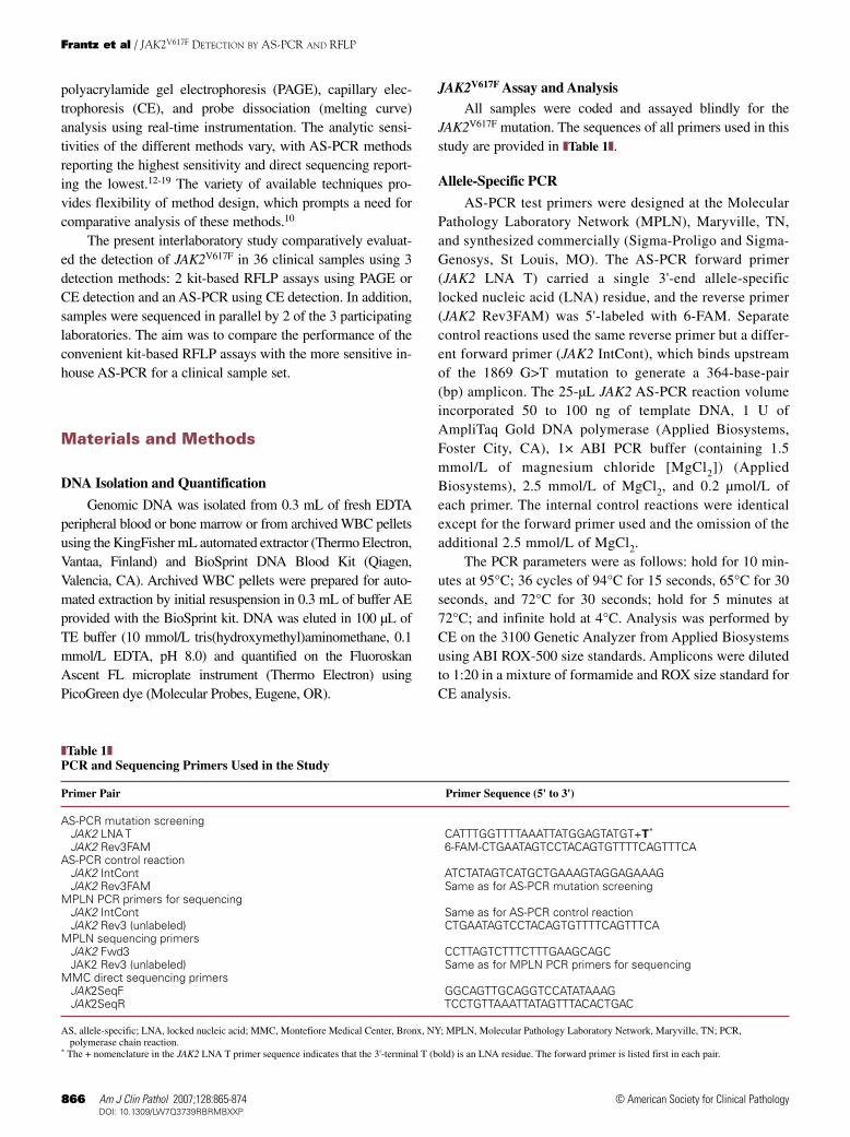
❚Table 1❚PCR and Sequencing Primers Used in the Study
Primer Pair Primer Sequence (5' to 3')
AS-PCR mutation screeningJAK2 LNA T CATTTGGTTTTAAATTATGGAGTATGT+T*
JAK2 Rev3FAM 6-FAM-CTGAATAGTCCTACAGTGTTTTCAGTTTCAAS-PCR control reaction
JAK2 IntCont ATCTATAGTCATGCTGAAAGTAGGAGAAAGJAK2 Rev3FAM Same as for AS-PCR mutation screening
MPLN PCR primers for sequencingJAK2 IntCont Same as for AS-PCR control reactionJAK2 Rev3 (unlabeled) CTGAATAGTCCTACAGTGTTTTCAGTTTCA
MPLN sequencing primersJAK2 Fwd3 CCTTAGTCTTTCTTTGAAGCAGCJAK2 Rev3 (unlabeled) Same as for MPLN PCR primers for sequencing
MMC direct sequencing primersJAK2SeqF GGCAGTTGCAGGTCCATATAAAGJAK2SeqR TCCTGTTAAATTATAGTTTACACTGAC
AS, allele-specific; LNA, locked nucleic acid; MMC, Montefiore Medical Center, Bronx, NY; MPLN, Molecular Pathology Laboratory Network, Maryville, TN; PCR,polymerase chain reaction.
* The + nomenclature in the JAK2 LNA T primer sequence indicates that the 3'-terminal T (bold) is an LNA residue. The forward primer is listed first in each pair.

Am J Clin Pathol 2007;128:865-874 867867 DOI: 10.1309/LW7Q3739RBRMBXXP 867
© American Society for Clinical Pathology
RFLP-PAGE and RFLP-CEThe RFLP assay kits for polyacrylamide gel (RFLP-
PAGE) or CE (RFLP-CE) detection were purchased fromInvivoScribe Technologies (IVS; San Diego, CA) and useidentical chemistry except that the proprietary PCR primersare unlabeled (for RFLP-PAGE) or labeled with 6-FAM (for-ward primer) or HEX (reverse primer) (for RFLP-CE) ❚Figure
1❚. The PCR setup and cycling parameters were performed asdescribed in the respective kit handbooks. The recommendedDNA input for both assays was 0.5 to 2 µg in a total volumeof 5 µL (RFLP-PAGE) or 2.5 µL (RFLP-CE). Control reac-tions for sample fidelity were performed using a separate mas-ter mix supplied with the RFLP kits. Following amplification,JAK2 amplicons were digested for 16 hours at 37°C withBsaXI endonuclease (New England BioLabs, Ipswich, MA)essentially as recommended in the respective kit handbooksexcept that the digestion volume for the RFLP-CE assay was20 µL and incorporated 5 µL of JAK2 amplicon, 1 µL ofBsaXI endonuclease (2U/µL), 2 µL of NEBuffer 4 (providedwith BsaXI), and 12 µL of molecular grade water.
RFLP-CE analysis of the digested JAK2 amplicons wasperformed on the ABI 3130XL Genetic Analyzer according tothe RFLP kit handbook. For RFLP-PAGE analysis, a 20-µLmixture consisting of 4 µL of 5× nucleic acid sample loadingbuffer (Bio-Rad, Hercules, CA) and 16 µL of BsaX1-digestedJAK2 amplicon was separated by 6% Tris Borate EDTA(TBE) PAGE and visualized by staining with SYBR Green(Molecular Probes) at 1:10,000 in TBE buffer, pH 8.0, for 15minutes, followed by imaging using the AlphaImager 2200(PerkinElmer, Wellesley, MA).
Zygosity Determination
The AS-PCR was not designed to distinguish zygosityand generated a 201-bp mutation-specific amplicon onlywhen the V617F allele was present; JAK2V617F– samples pro-duced only a 364-bp internal control amplicon.
Both RFLP kits distinguish normal and V617F JAK2 al-leles according to distinct fragment patterns generated onamplicon digestion by BsaXI (Figure 1). Amplification ofJAK2 yields a 267-bp amplicon that includes the G>T 1869(V617F) mutation site. The mutation site itself is found with-in the BsaXI recognition sequence. The normal JAK2 ampli-con contains an intact BsaXI recognition sequence, whereasthe V617F mutation changes this sequence and preventsdigestion.
Zygosity determination by RFLP-PAGE was based onsample interpretation guidelines in the original kit handbook.Digestion of the normal JAK2 amplicon yields 3 detectablefragments of 170, 140, and 97 bp; the 30-bp fragment is unde-tectable (Figure 1). The 140- and 30-bp fragments result fromcomplete digestion of the 170-bp fragment. Therefore, thepresence of the 170-bp fragment is indicative of incomplete
digestion. The 267-bp amplicon remains undigested inhomozygous JAK2V617F samples, whereas heterozygousJAK2V617F samples (those comprising mixtures of normal andheterozygous or homozygous mutant cells) generate a com-posite digestion pattern consisting of the 267-, 170-, 140-, and97-bp fragments, representing the normal and V617F alleles.
Zygosity determination by RFLP-CE was similarly basedon restriction digestion patterns but additionally included anallelic ratio calculation based on the peak height ratio of undi-gested and digested JAK2 fragments (undigested peakheight/undigested + digested peak height). Samples generat-ing ratios greater than 50% were considered homozygous-positive and those generating ratios equal to or less than 50%were considered heterozygous-positive for JAK2V617F.
Direct Sequencing
The Montefiore Medical Center (MMC) laboratory(Bronx, NY) performed direct sequencing on all 36 DNAsamples using the ABI 3130XL Genetic Analyzer accordingto the method described by Pan et al20 and using the MMCdirect sequencing primers listed in Table 1.
Hematopathology / ORIGINAL ARTICLE
BsaXI recognitionsequence
5'…(N)7GGAG(N)5GT(N)12…3'
Normal JAK2DNA sequence
JAK2 amplicon 267 bp
170 bp
140 bp 30 bp
97 bp 267 bp
BsaXINormal V617F
5'…AAATTATGGAGTATGTGTCTGTGGAGACGA…3'
❚❚Figure 1❚❚ Schematic of BsaXI restriction digestion patternsfor normal and V617F JAK2 alleles. Amplification generates a267-base-pair (bp) amplicon encompassing the site of theJAK2 G>T mutation at nucleotide 1849 (large bold G) and theBsaXI recognition sequence (core sequence is underlined).For polyacrylamide gel electrophoresis (PAGE) detection, bothprimers are unlabeled. For capillary electrophoresis (CE)detection, the forward primer is labeled with 6-FAM dye andthe reverse with HEX dye, generating a primary ampliconlabeled with both dyes. Digestion of the normal sequenceyields 4 fragments of 170, 140, 97, and 30 bp, which retain 6-FAM (170 and 140 bp) or HEX (97 bp); the 30-bp fragmentexcised by BsaXI is unlabeled and undetectable by PAGE andCE. The V617F mutation abolishes the BsaXI site andprevents digestion; the full-length 267-bp amplicon remainsintact and is labeled with 6-FAM and HEX, generating 2alternately labeled 267-bp peaks in the restriction fragmentlength polymorphism–CE method.

868 Am J Clin Pathol 2007;128:865-874868 DOI: 10.1309/LW7Q3739RBRMBXXP
© American Society for Clinical Pathology
Frantz et al / JAK2V617F DETECTION BY AS-PCR AND RFLP
The MPLN laboratory sequenced only the 15 JAK2V617F+DNA samples using the primers listed in Table 1. Sampleswere amplified before sequencing using the MPLN PCRprimers for sequencing. Amplicons were purified using theQiagen Minelute PCR Purification Kit and sequenced usingMPLN sequencing primers. Sequencing was performed usingthe Big Dye Terminator DNA Sequencing Kit v3.1 (ABI) andthe ABI 3100 Genetic Analyzer.
Analytic Sensitivity Determination
For sensitivity determinations, the RFLP-PAGE and AS-PCR methods used the IVS JAK2V617F Sensitivity Panel,which consists of DNA from the homozygous JAK2V617F cellline HEL diluted to 1%, 5%, 10%, 20%, or 30% (vol/vol) intonormal JAK2 DNA (from human tonsil), which served as dilu-ent and JAK2V617F– standard/control. The RFLP-CE methodused similar sensitivity controls, which were prepared inhouse by diluting the same JAK2V617F+ DNA into the samenormal JAK2 DNA (both supplied with the IVS RFLP-CEkit). The analytic sensitivity of each method was based on thelowest percentage sensitivity standard that generated anunequivocal and consistently positive JAK2V617F result. Forthe AS-PCR, this required serial 10-fold dilution of the 1%IVS standard into normal JAK2 DNA from the sensitivitypanel to generate the 0.01% standard.
Results
JAK2V617F Assay and Analysis
The PCR for each JAK2V617F assay required variableDNA input, with both RFLP assays requiring a minimumof 500 ng and the AS-PCR requiring 50 to 100 ng. Becausethe automated DNA extraction method sometimes pro-duced extracts of relatively low concentration, all sampleswere assayed undiluted by both RFLP methods, and, con-sequently, a proportion was assayed using less-than-recom-mended DNA input ❚Table 2❚; the lowest input for aJAK2V617F– sample was 33 ng, and the lowest input for aJAK2V617F+ sample was 55 ng. The AS-PCR methodrequired dilution of only samples with concentrations greaterthan 100 µg/mL to maintain consistent input. Despite subop-timal DNA input for the RFLP assays, the results indicated100% concordance for all 36 samples among the 3 detectionmethods; 21 samples were JAK2V617F–/undetectable and 15samples were JAK2V617F+ (5 homozygous- and 10 het-erozygous-positive by both RFLP methods). Successfulcontrol reactions were generated by all samples in all 3methods (data not shown). None of the JAK2V617F+ sam-ples were also positive for the presence of the BCR/ABL1transcript (data not shown).
The 21 JAK2V617F–/undetectable samples generated anRFLP-PAGE digestion pattern consisting of 3 bands of nearlyequal intensity (170, 140, and 97 bp); a small but detectableamount of the 267-bp band often remained undigested ❚Figure
2❚. Parallel analysis of these samples by RFLP-CE revealedthe same 170-, 140-, and 97-bp peaks and residual 267-bpamplicon. None of these samples produced detectableJAK2V617F amplicon by AS-PCR.
All 15 JAK2V617F+ samples generated a single 201-bpamplicon by AS-PCR (Figure 2). Of these samples, bothRFLP assays distinguished the same 5 homozygous and 10heterozygous samples based on the BsaXI digestion patternsand allelic ratios calculated for all positive samples (Table 2and Figure 2). Heterozygous samples consistently produced267-, 170-, 140-, and 97-bp peaks by CE; the same 4 bands ofnearly equal intensity were also evident by PAGE. Calculationof allelic ratios using CE data generated values less than 50%for these samples (Table 2). Of the 5 homozygous-positivesamples, only 1 (sample 18) produced the expected single267-bp peak/band. The other 4 samples (samples 17, 21, andpaired sample 29) generated an alternative digestion patternby PAGE that included prominent bands of 267, 170, and 97bp but a very faint 140-bp band. The corresponding RFLP-CEdetection of these samples similarly generated the 267-, 170-,and 97-bp peaks, but the 140-bp peak was undetectable.Allelic ratio calculation generated values of more than 50%for all 5 samples, with 3 (samples 17, 18, and 21) generatingratios of or near 100%, resulting in the classification of theseas homozygous-positive. Although samples 17 and 21 gener-ated very small digestion fragments detectable by CE, theheight of these peaks was too low to quantify; therefore, thecorresponding allelic ratio for these samples is given as“~100%” in Table 2.
Four sets of paired blood and bone marrow samples wereincluded in the sample set (Table 2). Despite relative differ-ences in the DNA concentration of each sample within therespective pairs, all 4 sets generated corresponding results forboth sample types by all 3 detection methods, with 2 sets gen-erating positive results and 2 sets generating negative/unde-tectable results. The allelic ratios within each pairedJAK2V617F+ sample were comparable for peripheral blood andbone marrow.
The clinical features of all cases (samples) included in thestudy are detailed in ❚Table 3❚. Of the 15 JAK2V617F+ cases, 13were previously diagnosed as or showed features of CMPD,based on bone marrow biopsy, cell counts, endogenous ery-throid colony formation, and clinical manifestations. The 2remaining JAK2V617F+ cases did not have bone marrow biop-sy samples available for diagnosis (cases 23 and 25). Theabsence of JAK2V617F was also well correlated with negativeCMPD diagnosis despite suggestive clinical features. All but 2of these cases, including 4 lacking bone marrow biopsy, were

Am J Clin Pathol 2007;128:865-874 869869 DOI: 10.1309/LW7Q3739RBRMBXXP 869
© American Society for Clinical Pathology
diagnosed as non-CMPD, leukemia (including Ph+ leukemia),or reactive thrombocytosis. However, 2 JAK2V617F– caseswere diagnosed as CMPD (cases 8 and 17, CIMF and ET,respectively).
JAK2V617F Analytic Sensitivity Determination
Each of the participating laboratories independentlydetermined the sensitivity of its JAK2V617F detection method❚Figure 3❚. Both RFLP methods had an analytic sensitivity of5% because this was the lowest standard that produced themost consistent and unambiguous positive result. The 1% sen-sitivity standard was less consistent, sometimes generating adigestion pattern more similar to the JAK2V617F– control sam-ple, with traces of the 267-bp fragment, but at other times gen-erating a pattern more similar to the heterozygous JAK2V617F
pattern, with a more prominent 267-bp fragment. The AS-PCR assay demonstrated an analytic sensitivity of 0.01%,based on 10-fold serial dilution of the 1% sensitivity standard(not shown), which represents a 500-fold increase in sensitiv-ity over the RFLP methods. The 0.01% sensitivity standardwas not evaluated by either RFLP method.
JAK2V617F Direct Sequencing
Samples were sequenced in parallel by the MMC andMPLN laboratories using different sequencing primers. Of the15 JAK2V617F+ samples, the MMC laboratory was unable todetect the mutation in 1 heterozygous-positive sample and theMPLN laboratory was unable to detect the mutation in thatsame sample and in 4 additional heterozygous-positive sam-ples (Table 2; sequencing data not shown). All 5 samples that
Hematopathology / ORIGINAL ARTICLE
❚Table 2❚Summary of Parallel Results
Total DNA Input (ng) JAK2V617F Result Sequencing Results
Sample No. Sample Type PAGE CE PAGE CE AS-PCR Allelic Ratio (%) MMC MPLN
1 BM 755 378 N N N — – ND2 PB 340 170 N N N — – ND3 BM 720 360 N N N — – ND4 PB 75 38 N N N — – ND5 PB 395 198 N N N — – ND6 PB 240 120 N N N — – ND7* PB 110 55 h h P 10.45 – –8 PB 65 33 N N N — – ND9 PB 100 50 N N N — – ND10 PB 190 95 N N N — – ND11 BM 520 260 N N N — – ND12 PB 345 173 N N N — – ND13 PB 470 235 N N N — – ND14 PB 255 128 h h P 39.80 + +15 PB 255 128 N N N — – ND16* PB 325 163 h h P 14.60 + –17 PB 315 158 H H P ~100 + +18 PB 385 193 H H P 100 + +19* PB 290 145 h h P 14.80 + –20 PB 285 143 N N N — – ND21 PB 525 263 H H P ~100 + +22 BM 355 178 N N N — – ND23 PB 220 110 h h P 23.20 + +24 WBC/BM 565 283 h h P 30.00 + +25 WBC/PB 315 158 h h P 43.15 + +26 WBC/BM 325 163 h h P 42.85 + +27 WBC/PB 195 98 N N N — – ND28 WBC/PB 245 123 N N N — – ND29† PB 690 345 H H P 70.20 + +29† BM 250 125 H H P 68.45 + +30† PB 140 70 N N N — – ND30† BM 300 150 N N N — – ND31† PB 375 188 N N N — – ND31† BM 310 155 N N N — – ND32*† PB 375 188 h h P 13.55 + –32*† BM 270 135 h h P 14.40 + –
AS-PCR, allele-specific polymerase chain reaction; BM, bone marrow; CE, capillary electrophoresis; h, heterozygous positive; H, homozygous positive; MMC, MontefioreMedical Center, Bronx, NY; MPLN, Molecular Pathology Laboratory Network, Maryville, TN; N, negative or undetectable; ND, not done; P, positive; PAGE, polyacrylamidegel electrophoresis; PB, peripheral blood; WBC/BM, archived frozen WBC pellet from BM; WBC/PB, archived frozen WBC pellet from PB; –, JAK2V617F mutation notdetected by sequencing; +, JAK2V617F mutation detected by sequencing.
* JAK2V617F+ samples not detected by sequencing.† Paired sets of blood and bone marrow samples.

870 Am J Clin Pathol 2007;128:865-874870 DOI: 10.1309/LW7Q3739RBRMBXXP
© American Society for Clinical Pathology
Frantz et al / JAK2V617F DETECTION BY AS-PCR AND RFLP
were undetectable by sequencing had allelic ratios of lessthan 20%; the single sample that was undetectable by bothsequencing methods (sample 7) had the lowest allelic ratio ofany positive sample at 10.45%, as well as the lowest DNAconcentration of any positive sample. The mutation wasdetected in all 5 homozygous-positive samples in bothsequencing laboratories. Although the sequencing datarevealed varying relative amounts of normal and V617FJAK2 alleles in all but 1 of 15 positive samples (sample 18,no normal allele detectable), 3 of the homozygous-positivesamples generated allelic ratios of 100% (samples 17, 18, and21), indicating the normal allele to be minimally present inthese samples.
Discussion
Although the RFLP assay kits offer the simplicity andconvenience of master mix solutions and supplied controlsamples, this method presents unique technical considerations
not common to AS-PCR.18 The RFLP method requires signif-icant DNA input, which may not be achievable, and addition-al processing time for the restriction digestion. In addition,incomplete digestion of amplicons could potentially generatefalse-positive results, and weakly positive samples containingless than approximately 5% JAK2V617F+ DNA may be difficultto interpret unequivocally by PAGE or CE.
Despite these potential issues, the current parallel evalu-ation of the RFLP and AS-PCR methods revealed that theserepresent comparable methods of JAK2V617F mutation detec-tion for this clinical sample set because all tested sampleswere similarly characterized by the 3 detection methods.With an analytic sensitivity of 0.01%, the AS-PCR methoddescribed herein represents the most sensitive method evalu-ated, and the 5% analytic sensitivity reported for the RFLPmethods is similar to reported sensitivities for pyrosequenc-ing, denaturing high-performance liquid chromatography,and melting curve analysis, suggesting that these methods offerno significant diagnostic advantage compared with RFLP.10,12-16
Although the analytic sensitivities of the methods compared in
1,200
800
400
50
1,000800600400200
0
502,0001,6001,200
800400
0
50 100 150 200 250 300 350
700560420280140
0
50
7,200
3,600
0
7,200
3,600
0
100 150 200 250 300 350
100 150 200 250 300 350
8,0006,0004,0002,000
0
60 90 120 150 180 210 240 270 300 330 360 390 420 450
8,0006,0004,0002,000
0
60 90 120 150 180 210 240 270 300 330 360 390 420 450
100 150 200
97 bp
201 bp
97 bp140 bp
170 bp267 bp
a267 bp
97 bp
267 bp
201 bp
201 bp
a
b
170 bp
b
140 bp
170 bp 267 bp
250 300
Negative/Undetectable
RFLP-PAGE
267 bp
170 bp140 bp
97 bp
267 bp
170 bp
140 bp
97 bp
267 bp
267 bponly
Weak140 bp
a b
170 bp140 bp
97 bp
RFLP-CE
AS-PCR
Heterozygous Positive Homozygous Positive
350
0
❚❚Figure 2❚❚ Polyacrylamide gel electrophoresis (PAGE) and capillary electrophoresis (CE) profiles of JAK2V617F+ and JAK2V617F–samples. Representative data for each method are shown. Representative negative or undetectable data are derived fromsample 15, heterozygous positive data from sample 14, and homozygous positive data from sample 18 (a; 267-base-pair [bp]fragment only) or from sample 17 (b; alternate digestion pattern with weak 140-bp fragment). The red peaks in the allele-specificpolymerase chain reaction (AS-PCR) electropherograms represent ROX-500 size standard peaks. Note the different scales forthe restriction fragment length polymorphism (RFLP)-CE and AS-PCR electropherogram data.

Am J Clin Pathol 2007;128:865-874 871871 DOI: 10.1309/LW7Q3739RBRMBXXP 871
© American Society for Clinical Pathology
the present study differed 500-fold, the completely concor-dant characterization of JAK2V617F– samples indicated that ifthe mutation was, in fact, present in any of the samples, it waspresent at less than 0.01%, making it undetectable even byAS-PCR. Moreover, the identification of all positive samplesby the RFLP methods, even when DNA input was less thanrecommended, is also indicative of the robustness of thesemethods. Therefore, for this sample set, the RFLP assaysseem functionally comparable to AS-PCR, and the resultsindicate that the technical considerations associated with the
RFLP methods may be largely managed and controlled. Asimilar conclusion about the diagnostic advantage of AS-PCR as compared with other methods is offered in compara-ble studies.13,15
Had this study, however, included samples weakly posi-tive for JAK2V617F (<~5%), the mutation may have been unde-tectable by the RFLP methods but detectable by the AS-PCRmethod. The literature is unclear regarding the proportion ofprimary Ph– CMPD samples that might contain less than 5%JAK2V617F DNA, possibly making them detectable only by a
Hematopathology / ORIGINAL ARTICLE
❚Table 3❚Clinical Features of Patients Whose Samples Were Included in the Study
Case No./ Hemoglobin, Platelets, WBCs (Neutrophils), Bone Marrow Sex/Age, y g/L* × 109/L* × 109/L* Diagnosis Cytogenetics Other Findings
1/M/44 154 473 4.4 (2.6) No CMPD 46,XY Previous diagnosis of ET; taking hydroxyurea for 2 y
2/F/56 121 1,012 187.6 (90.1) CML No results BCR/ABL1+ (b3a2)3/M/51 122 646 10.4 (8.2) No CMPD ND Stroke4/F/86 130 1,159 5.9 (3.7) ND ND5/M/51 149 556 17.7 (10) No CMPD 46,XY6/M/9 126 1,088 7.7 (4.1) Reactive 46,XY No HSM on US
thrombocytosis7/M/57† 160 808 9.5 (6.3) CMPD ND No HSM on US8/F/62 103 85 4.0 (1.7) CIMF 47,XX,der(7)t(1;7) Splenomegaly
(q21;q22),+89/F/39 130 623 8.0 (6.5) ND ND No HSM on US10/F/84 117 255 24.8 (21.1) ND ND Deceased11/F/53 159 175 6.2 (4.9) No CMPD 46,XX12/F/58 139 460 28 (19) Nondiagnostic ND No HSM on US13/F/46 74 14 209 (1.7) Pre-B ALL 46,XX BCR/ABL1+ (e1a2)14/F/84† 96 297 14.1 (7.8) CIMF No results Deceased15/F/88 92 224 62.7 (39.5) AML 46,XX Prior diagnosis of CMML16/F/55† 142 1,116 13.5 (9.2) ET 46,XX No HSM on US17/M/62 127 967 14.3 (8.9) ET 46,XY Splenomegaly18/M/61† 132 611 30.6 (26.1) PV 46,XYdel(20)
(q11.2q13.1)/46,XY,der(15)t(1;15)(q10;q10)
19/M/37† 165 520 9.8 (5.8) ET 46,XY Mild splenomegaly on US20/F/62 138 643 8.5 (3.8) Features of CMPD 46,XX No HSM on US21/M/79† 144 485 48.8 (44.8) CIMF 45,X,-Y Splenomegaly22/F/29 137 488 7.3 (4.1) Reactive 46,XX No HSM on US
thrombocytosis23/F/67† 146 998 9.3 (7.0) ND ND Splenomegaly24/F/33† 161 712 8.9 (6.4) PV 46,XX Increased RBC mass; decreased
serum EPO; EECF25/F/77† 164 469 13.9 (7.4) ND ND Increased serum EPO level; EECF;
deceased26/F/15† 141 213 32.3 (25.2) CMPD 46,XX Budd-Chiari syndrome; increased
serum EPO level; EECF27/M/52 192 178 6.9 (4.7) No CMPD 46,XY No HSM on US; increased RBC
mass; increased serum EPO level28/M/83 169 199 8.4 (5.4) No CMPD 46,XY Increased RBC mass; normal serum
EPO level; no EECF29/F/51† 92 NA 26.3 (15.1) ET 46,XX30/F/58 88 94 39.4 (27.5) CMML 46,XX31/F/37 143 200 10.1 (7.1) No definitive CMPD 46,XX Portal vein thrombosis; increased
serum EPO level; no EECF32/F/44† 146 763 9.5 (6.7) CMPD 46,XX,t(1;2)(p34;q37)c
ALL, acute lymphoblastic leukemia; AML, acute myeloid leukemia; CIMF, chronic idiopathic myelofibrosis; CML, chronic myelogenous leukemia; CMML, chronicmyelomonocytic leukemia; CMPD, chronic myeloproliferative disorder; EECF, endogenous erythroid colony formation; EPO, erythropoietin; ET, essential thrombocythemia;HSM, hepatosplenomegaly; NA, not available due to platelet clumping; ND, not done; PV, polycythemia vera; US, ultrasound.
* Bold values indicate an increased level relative to the reference range. Values are given in Système International units; conversions to conventional units are as follows:hemoglobin (g/dL), divide by 10.0; WBC and neutrophil counts (/µL), divide by 0.001; platelet count (× 103/µL), divide by 1.0.
† JAK2V617F mutation–positive cases.

872 Am J Clin Pathol 2007;128:865-874872 DOI: 10.1309/LW7Q3739RBRMBXXP
© American Society for Clinical Pathology
Frantz et al / JAK2V617F DETECTION BY AS-PCR AND RFLP
high-sensitivity assay such as AS-PCR. A recent studyaddressing this issue reported allelic ratios ranging from 8% to98% in PV and 3% to 50% in ET, which, in theory, would bedetectable by RFLP or comparable intermediate-sensitivitymethods.21 However, these ranges were derived by using puri-fied granulocytes and not whole blood or bone marrow, andadjustment of these ranges to account for this might shift thevalues close to or below the limit of detection of the RFLPmethods. Therefore, the convenient and simple RFLP meth-ods may be used for JAK2V617F mutation screening for themajority of clinical samples and may be most suitable for low-volume laboratories. If greater analytic sensitivity or samplethroughput is required, the AS-PCR method represents themost sensitive method currently available.
An examination of the clinical features associated witheach case in the study revealed a prevalence of JAK2V617F
among cases previously diagnosed as CMPD based on estab-lished diagnostic criteria.1 Of the 15 JAK2V617F+ cases, 2 werenot previously diagnosed as CMPD. These 2 cases had fea-tures suggestive of CMPD (cases 23 and 25), but unlike theremaining 13 JAK2V617F+ cases, no bone marrow biopsy spec-imen was available to aid in diagnosis. This may have beensignificant in these 2 cases because the patients had nonspecif-ic clinical features also common to several JAK2V617F– cases.Of the JAK2V617F– cases, 2 were previously diagnosed asCMPD (CIMF and ET) based on diagnostic criteria includingbone marrow biopsy. The absence of JAK2V617F in these sam-ples would not have excluded a diagnosis of CMPD becauseit has been reported that approximately half of CIMF and ETcases will not harbor the JAK2V617F mutation.11 Althoughmolecular JAK2V617F analysis complements clinical find-ings in the diagnosis of CMPD, the absence of JAK2V617F
does not preclude a CMPD diagnosis, particularly in CIMFand ET. Conversely, the presence of JAK2V617F is not diag-nostic for CMPD because studies have revealed the pres-ence of JAK2V617F in a percentage of asymptomatic patients,
analogous to the presence of BCR/ABL1 reported in a propor-tion of healthy people.22-25
Although the RFLP methods required additional process-ing time owing to the overnight restriction digestion step, itwas this step that enabled zygosity determination of the sam-ples. In this context, the term zygosity referred to the entiremyeloid population and not to individual JAK2V617F+ clones.The application of classic zygosity terminology to JAK2V617F
mutation status is historical and persists in current litera-ture.17,26-31 Although this terminology has been retained here-in, no claims are made with respect to the actual JAK2 allelespresent within individual clones based on the data presented.Of the 5 homozygous samples identified by allelic ratio in thisstudy, 4 generated an alternative digestion pattern that includ-ed the 267-bp band and additional robust bands of 170 and 97bp. These were distinguished from heterozygous samples onlyby the weakness of the 140-bp band, which, when consideredwithin the context of the corresponding sequencing and allelicratio data, indicates that the normal allele is minimally presentin these samples. Although the presence of a significant 170-bp band may suggest incomplete digestion, all but 1 of the 36tested samples generated a distinct 170-bp band or peak on16-hour digestion, indicating that this fragment represents aconsistent product of BsaXI digestion.
The concordant identification of all samples in this studyby all 3 detection methods indicates that the persistence of the170-bp fragment does not invalidate the RFLP data presentedherein. In addition, direct sequencing of the positive samplesultimately confirmed the presence of the JAK2V617F allele inall but 1 undetectable heterozygous-positive sample. Althoughthe clinical usefulness of JAK2V617F zygosity reporting is notfully supported by the literature, a mutant allele dose effecthas been reported in JAK2V617F+ PV and CIMF, and it hasbeen suggested that individual laboratories may find zygositydetermination useful for assessing disease progression orresponse to treatment.8,12,30 To more accurately reflect the
1,500
1,000
01,8003,600
01,8003,600
500
0
501,200
800400
0
50 100 150 200 250 300 350100 150 200
97 bp
140 bp
170 bp267 bp
201 bp
97 bp140 bp
170 bp267 bp
201 bp
250 300 350
267 bpRFLP-PAGE
RFLP-CE
AS-PCR
1% Sensitivity Standard 5% Sensitivity Standard
170 bp140 bp
97 bp
267 bp
170 bp140 bp
97 bp
❚❚Figure 3❚❚ Comparative analytic sensitivitydata for the restriction fragment lengthpolymorphism (RFLP) and allele-specificpolymerase chain reaction (AS-PCR)assays. Representative polyacrylamide gelelectrophoresis (PAGE) and capillaryelectrophoresis (CE) for the 1% and 5%sensitivity controls are shown for eachmethod. The red peaks in the AS-PCRelectropherograms represent ROX-500size standard peaks. Note the differentscales for the RFLP-CE and AS-PCRelectropherogram data. bp, base pairs.

Am J Clin Pathol 2007;128:865-874 873873 DOI: 10.1309/LW7Q3739RBRMBXXP 873
© American Society for Clinical Pathology
somatic nature of the JAK2V617F mutation, it may be prefer-able to replace the classic zygosity terminology applied toJAK2V617F mutation with alternative descriptors of allelic pro-file by laboratories that find usefulness in this distinction.
The direct sequencing data confirmed that sequencingrepresents the least sensitive or reliable method of JAK2V617F
detection because samples with an allelic ratio of less than20% were undetectable by one or both sequencing laborato-ries.5,13 Successful detection of the JAK2V617F mutation bydirect sequencing varied in this study, with one laboratoryidentifying the JAK2V617F mutation in 93% of the samples(14/15) and the other identifying the mutation in 67% (10/15).These results underscore the limited usefulness of directsequencing and the advantage of more sensitive PCR methodsin the detection of this mutation. Direct sequencing may,therefore, be useful primarily as an adjunct analysis to themore sensitive JAK2 mutation analysis methods.
In light of the range of assay sensitivities reported for var-ious JAK2V617F detection methods, the question remains:“How sensitive is sensitive enough?”11,15 The JAK2V617F
mutation was recently detected at a rate of 10% in a cohort ofhealthy donors when analyzed by quantitative AS-PCR.22 Thismethod reported an analytic sensitivity of 0.01%, which sug-gests that this may represent a suitable analytic sensitivity forJAK2V617F detection assays. The monitoring of asymptomaticJAK2V617F molecular-positive people may allow early inter-vention in the event of eventual CMPD development.11
From the 1Department of Laboratory Medicine and Pathology,University of Alberta Hospital, Edmonton, Canada; 2MolecularPathology Laboratory Network, Maryville, TN; and 3MolecularPathology Laboratory, Department of Pathology, MontefioreMedical Center, Bronx, New York.
Address reprint requests to Dr Mirza: Dept of LaboratoryMedicine and Pathology, University of Alberta Hospital, 8440-112Street, Edmonton, AB, Canada T6G 2B7.
References1. Michiels JJ, De Raeve H, Berneman Z, et al. The 2001 World
Health Organization and Updated European Clinical andPathological Criteria for the Diagnosis, Classification, andStaging of the Philadelphia Chromosome–Negative ChronicMyeloproliferative Disorders. Semin Thromb Hemost.2006;32:307-340.
2. Ugo V, Marzac C, Teyssandier I, et al. Multiple signalingpathways are involved in erythropoietin-independentdifferentiation of erythroid progenitors in polycythemia vera.Exp Hematol. 2004;32:179-187.
3. Hinshelwood S, Bench AJ, Green AR. Pathogenesis ofpolycythaemia vera. Blood Rev. 1997;11:224-232.
4. Radosevic N, Winterstein D, Keller JR, et al. JAK2contributes to the intrinsic capacity of primary hematopoieticcells to respond to stem cell factor. Exp Hematol. 2004;32:149-156.
5. Baxter EJ, Scott LM, Campbell PJ, et al. Acquired mutation of the tyrosine kinase JAK2 in human myeloproliferativedisorders. Lancet. 2005;365:1054-1061.
6. James C, Ugo V, Le Couedic J-P, et al. A unique clonal JAK2mutation leading to constitutive signaling causes polycythemiavera. Nature. 2005;434:1144-1148.
7. Levine RL, Wadleigh M, Cools J, et al. Activating mutation inthe tyrosine kinase JAK2 in polycythemia vera, essentialthrombocythemia, and myeloid metaplasia with myelofibrosis.Cancer Cell. 2005;7:387-397.
8. Kralovics R, Passamonti F, Buser AS, et al. A gain-of-functionmutation of JAK2 in myeloproliferative disorders. N Engl JMed. 2005;352:1779-1790.
9. Jelinek J, Oki Y, Gharibyan V, et al. JAK2 mutation 1849G>Tis rare in acute leukemias but can be found in CMML,Philadelphia chromosome–negative CML, andmegakaryocytic leukemia. Blood. 2005;106:3370-3373.
10. Greiner TC. Diagnostic assays for the JAK2 V617F mutationin chronic myeloproliferative disorders. Am J Clin Pathol.2006;125:651-653.
11. Steensma D. JAK2 V617F in myeloid disorders: moleculardiagnostic techniques and their clinical utility. J Mol Diagn.2006;8:397-411.
12. Olsen RJ, Tang Z, Farkas DH, et al. Detection of theJAK2V617F mutation in myeloproliferative disorders by meltingcurve analysis using the LightCycler system. Arch Pathol LabMed. 2006;130:997-1003.
13. Stevenson WS, Hoyt R, Bell A, et al. Genetic heterogeneityof granulocytes for the JAK2 V617F mutation in essentialthrombocythaemia: implications for mutation detection inperipheral blood. Pathology. 2006;38:336-342.
14. Murugesan G, Aboudola S, Szpurka H, et al. Identification of the JAK2 V617F mutation in chronic myeloproliferativedisorders using FRET probes and melting curve analysis. Am J Clin Pathol. 2006;125:625-633.
15. McClure R, Mai M, Lasho T. Validation of two clinicallyuseful assays for evaluation of JAK2 V617F mutation inchronic myeloproliferative disorders. Leukemia. 2006;20:168-171.
16. Jones AV, Kreil S, Zoi K, et al. Widespread occurrence of theJAK2 V617F mutation in chronic myeloproliferative disorders.Blood. 2005;106:2162-2168.
17. Vannucchi AM, Pancrazzi A, Bogani C, et al. A quantitativeassay for JAK2V617F mutation in myeloproliferative disorders by ARMS-PCR and capillary electrophoresis. Leukemia.2006;20:1055-1060.
18. Lay M, Mariappan R, Gotlib J, et al. Detection of the JAK2V617F mutation by LightCycler PCR and probe dissociationanalysis. J Mol Diagn. 2006;8:330-334.
19. James C, Delhommeau F, Marzac C, et al. Detection of JAK2V617F as a first intention diagnostic test for erythrocytosis.Leukemia. 2006;20:350-353.
20. Pan Q, Pao W, Ladanyi M. Rapid polymerase chainreaction–based detection of epidermal growth factor receptorgene mutations in lung adenocarcinomas. J Mol Diagn.2005;7:396-403.
21. Lippert E, Boissinot M, Kralovics R, et al. The JAK2-V617Fmutation is frequently present at diagnosis in patients withessential thrombocythemia and polycythemia vera. Blood.2006;108:1865-1867.
22. Sidon P, El Housni EI, Dessars B, et al. The JAK2V617Fmutation is detectable at a very low level in peripheral bloodof healthy donors [letter]. Leukemia. 2006;20:1622.
Hematopathology / ORIGINAL ARTICLE

874 Am J Clin Pathol 2007;128:865-874874 DOI: 10.1309/LW7Q3739RBRMBXXP
© American Society for Clinical Pathology
Frantz et al / JAK2V617F DETECTION BY AS-PCR AND RFLP
23. Xu X, Luo J, Xing S, et al. JAK2V617F: prevalence in a largeChinese hospital population. Blood. 2007;109:339-342.
24. Biernaux C, Loos M, Sels A, et al. Detection of major bcr-ablgene expression at a very low level in blood cells of somehealthy individuals. Blood. 1995;86:3118-3122.
25. Bose S, Deininger M, Gora-Tybor J, et al. The presence of typical and atypical bcr-abl fusion genes in leukocytes ofnormal individuals: biologic significance and implications for the assessment of minimal residual disease. Blood.1998;9:3362-3367.
26. Khwaja A. The role of Janus kinases in haemopoiesis andhaematological malignancy. Br J Haematol. 2006;134:366-384.
27. Ma W, Kantarjian H, Verstovsek S, et al.Hemizygous/homozygous and heterozygous JAK2 mutationdetected in plasma of patients with myeloproliferative diseases:correlation with clinical behaviour. Br J Haematol.2006;134:340-350.
28. McLornan D, Percy M, McMullin MF. JAK2 V617F: a singlemutation in the myeloproliferative group of disorders. UlsterMed J. 2006;75:112-119.
29. Tefferi A, Strand JJ, Lasho TL, et al. Respective clustering ofunfavorable and favorable cytogenetic clones in myelofibrosiswith myeloid metaplasia with homozygosity for JAK2V617F andresponse to erythropoietin therapy. Cancer. 2006;106:1739-1743.
30. Tefferi A, Lasho TL, Schwager SM, et al. The clinicalphenotype of wildtype, heterozygous, and homozygousJAK2V617F in polycythemia vera. Cancer. 2006;106:631-635.
31. Villeval J-L, James C, Pisani DF, et al. New insights into thepathogenesis of JAK2 V617F-positive myeloproliferativedisorders and consequences for the management of patients.Semin Thromb Hemost. 2006;32:341-351.