
Common Conditions Requiring Emergency Life Support
13
Common Conditions Requiring Emergency Life Support Kelsey Fawcett, MD,* † Nicole Gerber, MD,* † Shweta Iyer, MD,* † Guillermo De Angulo, MD, MPH,* † Martin Pusic, MD, PhD,* Michael Mojica, MD* †‡ *Department of Emergency Medicine and † Department of Pediatrics, New York University School of Medicine, New York, NY ‡ Department of Emergency Medicine, Bellevue Hospital Center, New York, NY Education Gaps Providers should be up to date on the evidence-based current guidelines for pediatric resuscitation and be able to identify and manage life- threatening illness and injury. Objectives After completing this article, readers should be able to: 1. Understand the major changes in the 2015 American Heart Association guidelines for pediatric basic life support, advanced life support, and postresuscitation care. 2. Initiate the management and identify prognostic factors associated with near drowning. 3. Identify the signs and symptoms of life-threatening thoracic injuries, including pneumothorax, hemothorax, flail chest, and cardiac tamponade. 4. Identify and manage cerebral edema in the asphyxiated patient. OVERVIEW There are common emergencies in pediatrics that any pediatrician needs to be equipped to handle. Common office emergencies include respiratory distress, dehydration, seizures, and anaphylaxis. However, pediatricians also need to be prepared for common conditions that require emergency life support. These include, but are not limited to, cardiac arrest, near drowning, thoracic injuries, and cerebral edema. This article focuses on these common conditions that require emergency life support. PEDIATRIC RESUSCITATION Introduction Cardiac arrest is rare in the pediatric population. In 2015, the American Heart Association (AHA) released updated recommendations for pediatric ad- vanced life support. (1) These updated guidelines, incorporating the available AUTHOR DISCLOSURE Drs Fawcett, Gerber, Iyer, De Angulo, and Mojica have disclosed no financial relationships relevant to this article. Dr Pusic has disclosed that he receives research study funding from the Department of Defense and the American Medical Association. This commentary does not contain a discussion of an unapproved/ investigative use of a commercial product/ device. ABBREVIATIONS AHA American Heart Association CPR cardiopulmonary resuscitation CT computed tomography ED emergency department ICP intracranial pressure ROSC return of spontaneous circulation Vol. 40 No. 6 JUNE 2019 291 by guest on August 6, 2019 http://pedsinreview.aappublications.org/ Downloaded from
-
Upload
khangminh22 -
Category
Documents
-
view
2 -
download
0
Transcript of Common Conditions Requiring Emergency Life Support
Common Conditions RequiringEmergency Life Support
Kelsey Fawcett, MD,*† Nicole Gerber, MD,*† Shweta Iyer, MD,*† Guillermo De Angulo, MD, MPH,*†
Martin Pusic, MD, PhD,* Michael Mojica, MD*†‡
*Department of Emergency Medicine and †Department of Pediatrics, New York University School of Medicine, New York, NY‡Department of Emergency Medicine, Bellevue Hospital Center, New York, NY
Education Gaps
Providers should be up to date on the evidence-based current guidelines
for pediatric resuscitation and be able to identify and manage life-
threatening illness and injury.
Objectives After completing this article, readers should be able to:
1. Understand themajor changes in the 2015 American Heart Association
guidelines for pediatric basic life support, advanced life support, and
postresuscitation care.
2. Initiate the management and identify prognostic factors associated
with near drowning.
3. Identify the signs and symptoms of life-threatening thoracic injuries,
including pneumothorax, hemothorax, flail chest, and cardiac
tamponade.
4. Identify and manage cerebral edema in the asphyxiated patient.
OVERVIEW
There are common emergencies in pediatrics that any pediatrician needs to be
equipped to handle. Common office emergencies include respiratory distress,
dehydration, seizures, and anaphylaxis. However, pediatricians also need to be
prepared for common conditions that require emergency life support. These
include, but are not limited to, cardiac arrest, near drowning, thoracic injuries,
and cerebral edema. This article focuses on these common conditions that require
emergency life support.
PEDIATRIC RESUSCITATION
IntroductionCardiac arrest is rare in the pediatric population. In 2015, the American Heart
Association (AHA) released updated recommendations for pediatric ad-
vanced life support. (1) These updated guidelines, incorporating the available
AUTHOR DISCLOSURE Drs Fawcett, Gerber,Iyer, De Angulo, and Mojica have disclosed nofinancial relationships relevant to this article.Dr Pusic has disclosed that he receivesresearch study funding from the Departmentof Defense and the American MedicalAssociation. This commentary does notcontain a discussion of an unapproved/investigative use of a commercial product/device.
ABBREVIATIONS
AHA American Heart Association
CPR cardiopulmonary resuscitation
CT computed tomography
ED emergency department
ICP intracranial pressure
ROSC return of spontaneous circulation
Vol. 40 No. 6 JUNE 2019 291 by guest on August 6, 2019http://pedsinreview.aappublications.org/Downloaded from
evidence, can guide resuscitation efforts to continue to
improve overall survival after pediatric cardiac arrest and,
in particular, improve survival with a favorable neurologic
outcome.
Basic Life SupportAn estimated 6,000 children experience out-of-hospital
cardiac arrest per year in the United States. (2) The survival
to hospital discharge is 11.3%, and survival with a neuro-
logically favorable outcome is 9.1%. (3) The promotion of
bystander cardiopulmonary resuscitation (CPR) remains
important. Bystander CPR is performed in out-of-hospital
pediatric arrest at an abysmally low rate of 46.5%, with the
lowest rates in African American and Hispanic children. (3)
Bystander CPR in children increases survival to hospital
discharge by 3.7% and increases neurologically favorable
outcomes by 2.7%. (4)
The sequence of interventions in cardiopulmonary arrest
are different in pediatrics. In 2010, the general AHA guide-
lines changed from an airway-breathing-circulation (A-B-C)
intervention sequence to a circulation-airway-breathing
(C-A-B) sequence in an attempt to reduce the time to the
first compression. (5) This recommendation was reaffirmed
in 2015. However, evidence suggests that the focus on
compression-only CPRmay not be beneficial in the pediatric
population. In infants (<1 year of age), there was no
improvement in survival with compression-only CPR com-
pared with no CPR. (3) For older children, the provision of
CPR, including rescue breathing and chest compressions,
resulted in improved neurologically favorable survival. (3)
Given the high rates of asphyxial arrest in the pediatric
population, conventional CPR with rescue breathing is
recommended. However, starting resuscitation with com-
pressions will not delay the initiation of ventilations bymore
than 20 seconds. In addition, the sequence of interventions
is of less importance in the health-care setting, where more
than 1 provider is available and tasks can be accomplished
concurrently.
Advanced Life SupportThe 2015 updates have been changed to reflect the lack of
clear evidence favoring either amiodarone or lidocaine,
allowing for the use of either during pediatric cardiac arrest
due to ventricular fibrillation and ventricular tachycardia
with a pulse. (1) A recent pediatric study demonstrated
improvement in the rate of return of spontaneous circula-
tion (ROSC) and 24-hour survival with lidocaine use but no
difference in survival to hospital discharge compared with
amiodarone use. (6) In 2016, an adult study compared
lidocaine with amiodarone and placebo in out-of-hospital
cardiac arrest and demonstrated no benefit to either med-
ication over placebo. (7)
There have also been minor modifications to the recom-
mendations for both depth and rate of compressions, with
maximums now being added. For depth, the recommenda-
tion is that compressions be between 5 and 6 cmdue to adult
studies showing harm with deeper compressions. (8) There
have been no changes to the depth recommendation of 4 cm
in infants and 5 cm in children. For rate, the recommen-
dation is now 100 to 120/min based on increased survival
with compression rates in that range. (9)
There also continues to be conflicting evidence surround-
ing the benefits of extracorporeal CPR (extracorporeal mem-
brane oxygenation) in the noncardiac patient. Extracorporeal
CPR involves attaching the patient to a machine that exter-
nally oxygenates and circulates the blood. There does seem to
be clear evidence of improved survival for cardiac patients,
both in the postoperative period and otherwise. (10)(11)(12)
However, a recent article suggests that it may have general
benefits, with the caveat that more patients in the extracor-
poreal CPR group were patients with a cardiac history. (13)
Postresuscitation CareOnce ROSC is achieved there are evolving recommendations
on how to maximize survival to discharge and improve neu-
rologic outcomes. The newest literature focuses on the manip-
ulation of temperature after arrest. There is a clear benefit to
avoiding hyperthermia. (14) However, the 2 largest trials on
therapeutic hypothermia after in-hospital (15) and out-of-hos-
pital (16) pediatric cardiac arrest did not demonstrate a benefit
of hypothermia over therapeutic normothermia.
Hypotension, defined as “a systolic blood pressure less
than the fifth percentile for age,” in patients with ROSC
should be avoided because it is associated with an increased
odds of in-hospital mortality. (17) The recommendations for
oxygenation and ventilation goals are less clear. In the
pediatric literature, it is uncertain whether hyperoxia (18)
(19)(20)(21) or hypercarbia (19)(20) after ROSC have detri-
mental effects on outcomes. Current recommendations are
to avoid extremes and target an oxygen saturation greater
than or equal to 94%, but they do not include specific
ventilation targets. (1)
The question of when to initiate, or terminate, CPR can
be difficult. The AHA defines the goal of resuscitation as “to
preserve life; restore health; relieve suffering; limit disabil-
ity, and respect individuals’ decisions, rights and privacy.”
(22) There is currently no single factor or combination
of factors that reliably predict outcomes sufficiently to
guide recommendations for initiation or termination of
CPR. In addition, after ROSC, there are few well-studied
292 Pediatrics in Review by guest on August 6, 2019http://pedsinreview.aappublications.org/Downloaded from
factors that reliably predict neurologically favorable out-
comes. Given that children’s brains have a higher potential
for recovery, the decisions surrounding starting and stop-
ping CPR continue to be challenging and should bemade in
a discussion between the treating physicians and family
members. Several studies have shown that it is beneficial
to provide families the opportunity to be physically present
at the resuscitation because this has been shown to
decrease posttraumatic stress disorder–related symptoms
(23) and lessen the pain of death (24) without being
disruptive to the resuscitative efforts. (25) Early consultation
with a palliative care team, even in cases of prognostic
uncertainty, can be beneficial becausemany physicians have
difficulty initiating and leading timely discussions on end-
of-life care in pediatric patients. (26)(27)
For cardiac arrest in pediatric patients, based on strong
research evidence, pediatricians can:• Continue to encourage conventional CPR with rescue
breathing in pediatric patients (3)• Promote bystander CPR (4)
• Know the role of extracorporeal membrane oxygena-
tion, especially for cardiac patients (10)(11)(12)
• Pay close attention to blood pressure and temperature
in the post-ROSC period (16)• Promote initiatives for the physical presence of families
during resuscitation (23)
DROWNING
IntroductionDrowning accounts for 6,000 to 8,000 deaths in the
United States (28) and at least 450,000 deaths worldwide
annually. (29) Drowning rates are higher in low-income
countries and, predictably, in countries with large accessible
bodies of water. (30) In the United States, drowning and near
drowning commonly occur in homes with pools and near
bodies of fresh water. (28) Drowning ismore frequent during
the summer months and more common in males and non-
Hispanic minorities. (31) There is a bimodal distribution in
age, with peaks in the toddler and adolescent age groups. (31)
There are multiple individual risk factors for drowning
and near drowning. For younger children, themain factor in
drowning events is inadequate supervision by adults. (28)
Alcohol use is a frequent trigger for these events and is
involved in most adolescent drowning events. (28) Over-
estimating swimming abilities, risk-taking behaviors such
as hyperventilating before submerging, and injuries such as
cervical spine injuries also contribute to increased drowning
rates. (28)
Several studies indicate an increased risk of drowning in
children with epilepsy. (32)(33) A meta-analysis of patients
with epilepsy demonstrated a 15-fold greater rate of drown-
ing in this population. (32) Adequate anticonvulsant levels
may not be protective, and these patients should be specif-
ically counseled regarding water safety.
Apnea and aspiration lead to pulmonary complications
and hypoxemia. (34)(35) Systemic hypoxia involves every
organ, with poor outcomes attributed primarily to cerebral
hypoxia. (36)(37) The primary systems affected are the
pulmonary, neurologic, and cardiovascular.
Near DrowningNear drowning is distinct from drowning in that the victim
survives the drowning episode for at least some period. (31)
Near drowning rates are uncertain, with an approximate
estimate of being 3 to 4 times as common as drowning
events. (38) Loss of consciousness is usually included in
the definition of near drowning. (28) There are various
subcategories of near drowning, includingwet near drowning
with pulmonary aspiration of fluids as well as dry near
drowning with laryngospasm leading to hypoxia without
significant aspiration. (39)(40) The clinical importance of
these distinctions has not been established. Children who
survive drowning may be left with long-term adverse health
outcomes, which can increase health-care costs and create
difficulty for families. (30)
Prehospital CareThe most important phase of treatment is the prehospital
care phase. The most critical factor is immediate resusci-
tation, including CPR, by witnesses and bystanders. (37)
CPR by laypersons has been demonstrated to improve
survival. (37) If resuscitation is delayed until emergency
medical services arrive, there is a poor chance of survival or
survival with good neurologic outcome. (41) Because the
main concern in drowning is hypoxia, rescue breathing
alone, and compressions if necessary, can improve the rates
of morbidity and mortality in near drowning. (37) If sup-
plemental oxygen is available, it should be used to mitigate
organ damage due to hypoxia. (37) Rewarming measures
should be initiated immediately, especially for a core tem-
perature less than 91.4°F (<33°C). (37) Because these
patients have typically swallowed large amounts of water,
the high risk of emesis and aspiration can be diminished by
placing breathing patients in a lateral decubitus position if
CPR is not being performed. (34)
Routine cervical spine immobilization is not recom-
mended for drowning victims without risk factors (such
as a shallow dive or signs of trauma) and may actually
Vol. 40 No. 6 JUNE 2019 293 by guest on August 6, 2019http://pedsinreview.aappublications.org/Downloaded from
increase the chances of aspiration. (37) The Heimlich
maneuver is not recommended as a method of water
expulsion as water in the lungs is rapidly reabsorbed and
abdominal compressions increase the risk of regurgitation.
(37) Arrhythmias requiring an automated external defibril-
lator, such as ventricular fibrillation, are rare, and manage-
ment should not delay CPR. (42)
Emergency Department CareIn the emergency department (ED), early intubation and
ventilation, or providing supplemental oxygen as needed,
should be considered. (43) The patient should be monitored
serially, with cardiac monitoring, end-tidal carbon dioxide
monitoring, and oxygen saturation measurements. (34)
Patients should be monitored with frequent blood gases
and a baseline chest radiograph. (34) Acute respiratory
distress syndrome is common; as a result, ventilation with
high pressures may be necessary. (44) If there is concern for
infection (ie, contaminated water), antibiotics should be
administered. (45) An electrocardiogram and serum elec-
trolyte measurements should be performed in near drown-
ing victims, but these rarely show life-threatening or
severely abnormal values, although metabolic acidosis is
usually present. (46) Euglycemia should be maintained for
optimal cerebral perfusion. (37)
Resuscitation and rewarming measures should continue
once the patient is brought to the hospital. Multiple passive
and active rewarming measures can be used in the ED,
including removal of wet clothing, basic drying, protective
warmed clothing or blankets, warmed intravenous fluids,
inhalation of warmed oxygen, and lavage of body cavities
with warmed fluids. (34)(37) Prolonged resuscitative efforts
may be necessary in hypothermic patients until core tem-
peratures are increased beyond 91.4°F (33°C). Cases of
complete recovery have been observed in hypothermic
patients despite cardiac arrest. (47) Therapeutic hypother-
mia has not proved beneficial, (41) although hyperthermia
should be avoided.
Although a variety of ED interventions have been put
forth, no specific intervention has demonstrated clear
superiority to goodmedical management alone. Candidate
interventions not shown to be clearly beneficial include
high-dose barbiturate treatment, intracranial pressure
(ICP) monitoring, and protective hypothermia. (42)
DispositionHospital admission decisions can be guided by the patient’s
clinical status. Patients who are initially asymptomatic may
develop delayed pulmonary edema as a result of ongoing
damage to the alveolar lining. In a retrospective study of 75
pediatric near drowning patients, 98% of patients who had
delayed onset of symptoms occurred within 4.5 hours of
submersion, and all developed symptoms within 7 hours.
(48) Another study confirmed that pediatric near drowning
patients could be safely discharged after a 4- to 6-hour
observation period if their initial Glasgow Coma Scale score
was greater than or equal to 13, they required no advanced
life support measures, and they had normal oxygen satu-
rations and pulmonary examination findings after the obser-
vation period. (49) A normal chest radiograph in a patient
who remains asymptomatic after a period of observation can
guide the disposition decision, although chest radiographs
were not found to be predictive of the clinical course. (48)
Prognostic FactorsPrognosis mainly depends on prehospital factors. Submer-
sion times play a critical role, with submersions longer than
5 minutes having a poorer prognosis. (50)(51)(52) However,
there is no clear submersion timeframe during which a near
drowning event has a safer outcome.
Hypothermia is typically thought to result in a better
outcome than warm water drowning because hypothermia
has been postulated to be neuroprotective, especially if it
occurs rapidly. (34)(42)(53) However, this has been ques-
tioned in a recent study, with cold water showing no pro-
tective effect regarding neurologic outcome. (52)
As mentioned previously herein, early resuscitative
efforts are the most important factor in predicting a favor-
able outcome. (51) Previous studies have demonstrated that
resuscitation efforts started within 10minutes of the drown-
ing episode are more likely to be successful, (51) and
resuscitation efforts for longer than 25 minutes are typically
associated with poor outcomes. (54)(55) However, there have
been some patients with favorable outcomes even with
prolonged aggressive resuscitation efforts or prolonged
submersion times because neurologic recovery can be dif-
ficult to predict in the initial period. (43) In a study of 274
pediatric drowning victims, 17% who continued to be resus-
citated after arrival at the ED had functional recovery. (43)
This study found 3 predictors of poor clinical outcome: a
continuing need for CPR and resuscitative efforts in the ED,
a pH less than 7, and coma or apnea in the ED. (43) These
results have been supported by other studies. (51) Many
prognostic scoring systems have been proposed, but none
have been universally accepted.
After 48 hours, if neurologic recovery or improvement is
not seen, it would be appropriate to initiate a discussion with
the family on possible withdrawal of care. (43) In a study of
44 pediatric patients with near drowning, all of the patients
who did not have recovery of spontaneous, purposeful
294 Pediatrics in Review by guest on August 6, 2019http://pedsinreview.aappublications.org/Downloaded from
movements within 24 hours had very poor neurologic out-
comes or died. (56) In addition, a Glasgow Coma Scale score
of less than or equal to 5 and unreactive pupils have been
shown to be predictors of poor neurologic outcome. (57)
PreventionIt is hypothesized that 80% of drownings can be prevented.
(37) Prevention strategies include appropriate supervision,
avoidance of intoxicants such as alcohol, knowledge of
swimming and safety, use of appropriate flotation devices
during water sports and activities, and the presence of
lifeguards. (37)(45) The use of fencing and self-latching
gates around pools has been shown to substantially decrease
drowning incidents among children. (31)(58) Caregivers
should be counseled on appropriate supervision of infants
and toddlers in any area of shallow water, including bath-
tubs. (59)
Infant swimming programs may provide caregivers a
false sense of security and should not replace close super-
vision. In a 2010 policy statement on drowning prevention,
the American Academy of Pediatrics reevaluated the age at
which swimming lessons can be considered protective
based on new evidence. (60) The American Academy of
Pediatrics continues to support swimming instruction for
children older than 4 years. Several recent studies have
suggested that children 1 to 4 years of age may also benefit
from lessons, although lessons for this age group are not
recommended. (61)(62)
In drowning prevention and near drowning, based on
strong research evidence, it is important for pediatricians to:
• Understand the factors that influence prognosis in near
drowning (50)(51)(52)
• Provide anticipatory guidance to parents of young chil-
dren and those with epilepsy regarding water safety (60)
LIFE-THREATENING THORACIC INJURIES
IntroductionAlthough thoracic trauma occurs infrequently in pediatric
patients, it is essential to identify potentially life-threatening
injuries to provide prompt medical or surgical interventions.
The incidence of thoracic trauma in children has been
demonstrated to be between 4% and 8%, with blunt and
penetrating trauma accounting for 85% and 15% of cases,
respectively. (63)(64) The most common mechanisms of
injury in patients sustaining blunt trauma are motor vehicle
accidents, intentional injury, and falls. The most common
thoracic injuries due to blunt trauma include pulmonary
contusions, pneumothorax, hemothorax, and rib fractures.
(65) Gunshot wounds, stabbings, and impalement are the
most common etiologies of penetrating thoracic trauma
resulting in pneumothorax, hemothorax, pulmonary contu-
sion, pulmonary laceration, and blood vessel injury. (65) The
mortality rate for thoracic injury is approximately 20%, with a
higher rate in penetrating trauma than in blunt trauma. (65)
(66) This review focuses on the identification (Table 1) and
management (Table 2) of 4 acutely life-threatening thoracic
injuries: pneumothorax, hemothorax, flail chest, and cardiac
tamponade.
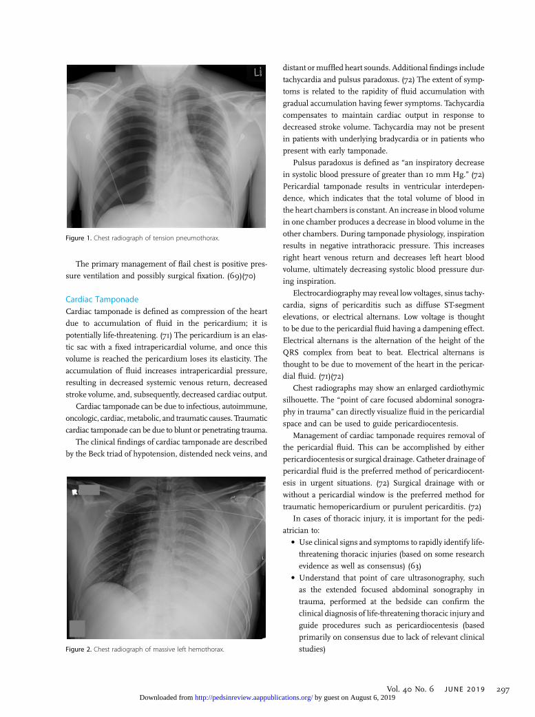
PneumothoraxPneumothorax is defined as the accumulation of air in the
pleural cavity. Tension pneumothorax occurs when air in the
pleural space causes a contralateral shift of themediastinum
(Fig 1). This causes compression of the thoracic vasculature,
reducing systemic venous return to the heart and, therefore,
reducing cardiac output. Compression of the contralateral
lung restricts lung expansion. Children have a higher rate of
tension pneumothorax due to greater compliance of the
mediastinal structures. Tension pneumothorax is a diagno-
sis that should be made clinically, and management should
not be delayed for radiologic confirmation. Common symp-
toms include shortness of breath and chest pain. Vital sign
abnormalities may include tachypnea, tachycardia, hypoten-
sion, and hypoxia. Examination findings may include cya-
nosis or pallor, contralateral tracheal deviation, ipsilateral
decreased breath sounds, and neck vein distention. The
absence of lung sliding on a “point of care focused abdom-
inal sonography in trauma” can provide immediate bedside
confirmation. Management includes immediate needle
decompression in the midclavicular line at the second inter-
costal space followed by chest tube insertion.
An open pneumothorax, also known as a sucking chest
wound, typically results from penetrating trauma. Air pref-
erentially enters during inspiration through the chest wall
defect, compromising lung expansion. A sterile dressing
that is occlusive on 3 sides (applied when the patient is in full
expiration) will prevent air entry through the chest wall
defect on inspiration while allowing air to escape from the
nonocclusive fourth side during expiration. Definitive man-
agement incudes chest tube placement and closure of the
chest wall defect.
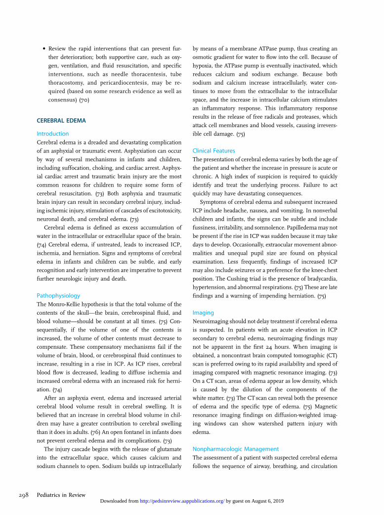
HemothoraxHemothorax is defined as the accumulation of blood in the
pleural space. Hemothorax occurs 38% and 64% of the time
in pediatric patients with blunt and penetrating thoracic
trauma, respectively. (65) A hemothorax is most commonly
Vol. 40 No. 6 JUNE 2019 295 by guest on August 6, 2019http://pedsinreview.aappublications.org/Downloaded from
caused by penetrating injury through injury to systemic or
hilar vessels but may also occur from blunt trauma from rib
fractures lacerating the underlying lung or other vascular
structures. In the adult-sized patient, a massive hemothorax
is defined as more than 1,500 mL of blood in the pleural
cavity. In pediatric patients, it is defined as greater than or
equal to one-third of a child’s blood volume (based on an
estimated total blood volume of approximately 80 mL/kg).
Clinical signs and symptoms include tachypnea, hypo-
xemia, increased work of breathing, ipsilateral absent or
decreased breath sounds, tachycardia, and hypotension.
Hemothorax can be identified with point of care lung
ultrasonography or chest radiography (Fig 2). Blood in
the pleural cavity may tamponade bleeding, and, therefore,
rapid evacuationmay actually cause additional bleeding. For
this reason, the patient should be adequately fluid resusci-
tated before thoracostomy tube placement if time permits.
Ongoing bleeding may require packed red blood cell trans-
fusion, activation of a massive transfusion protocol, tranexa-
mic acid, or an operative thoracotomy.
Flail ChestFlail chest is defined as the fracture of 3 or more consecutive
ribs in more than 2 locations. This creates a mechanically
unstable floating or flail segment. A review of the National
Trauma Data Bank revealed a 1% rate of flail chest in
admitted patients at level 1 and 2 trauma centers. (67) In
the Israeli National Trauma Registry, 2% and 9% of the
documented flail chest injuries occurred in patients aged
0 to 14 years and 15 to 24 years, respectively. (68) The same
Israeli study demonstrated a mortality rate of 20%, with the
risk of mortality higher with increasing age. (68)
Flail chest is typically due to a blunt mechanism, such as
a motor vehicle accident, a vehicle versus pedestrian or
bicycle accident, or a fall from a height. It can occur over the
anterior, posterior, and lateral thoracic chest wall. The
degree of force required to cause a flail chest increases
the risk of associated underlying pulmonary injury.
Clinically, external signs of chest trauma are frequent.
The unstable floating segment leads to paradoxical chest
motion. (69) Normally, contraction of the diaphragm and
chest wall expansion outward occurs during inspiration to
create the negative intrathoracic pressure required for lung
expansion. Paradoxical chest wall motion is observed when
the flail chest segment is pulled inward from the negative
intrathoracic pressure generated during inspiration. This
causes a decrease in lung volumes, atelectasis, chest tight-
ness, and dyspnea. (67)
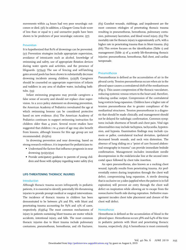
TABLE 1. Life-threatening Thoracic Injuries: Clinical Signs
SIGN TENSION PNEUMOTHORAX MASSIVE HEMOTHORAX CARDIAC TAMPONADE
Breath sounds Decreased (ipsilateral) Decreased (ipsilateral) Normal
Lung percussion Hyperresonant (ipilateral) Dull (ipsilateral) Normal
Tracheal position Deviation (contralateral) Midline Midline
Neck veins Distended Flat Distended
Heart sounds Normal Normal Muffled
The pulmonary component of the extended focused abdominal sonography in trauma examination can assist in rapidly identifying many of the injuriesrequiring urgent treatment at the bedside.
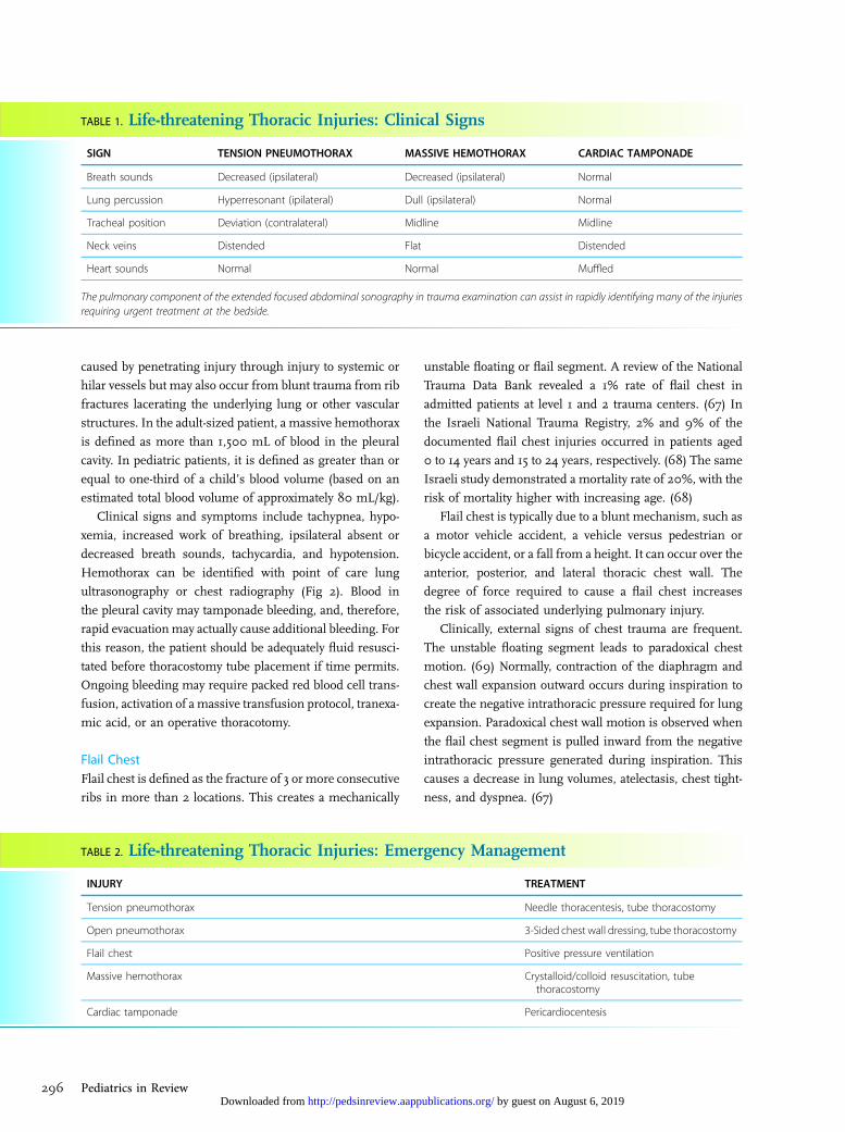
TABLE 2. Life-threatening Thoracic Injuries: Emergency Management
INJURY TREATMENT
Tension pneumothorax Needle thoracentesis, tube thoracostomy
Open pneumothorax 3-Sided chest wall dressing, tube thoracostomy
Flail chest Positive pressure ventilation
Massive hemothorax Crystalloid/colloid resuscitation, tubethoracostomy
Cardiac tamponade Pericardiocentesis
296 Pediatrics in Review by guest on August 6, 2019http://pedsinreview.aappublications.org/Downloaded from
The primary management of flail chest is positive pres-
sure ventilation and possibly surgical fixation. (69)(70)
Cardiac TamponadeCardiac tamponade is defined as compression of the heart
due to accumulation of fluid in the pericardium; it is
potentially life-threatening. (71) The pericardium is an elas-
tic sac with a fixed intrapericardial volume, and once this
volume is reached the pericardium loses its elasticity. The
accumulation of fluid increases intrapericardial pressure,
resulting in decreased systemic venous return, decreased
stroke volume, and, subsequently, decreased cardiac output.
Cardiac tamponade can be due to infectious, autoimmune,
oncologic, cardiac,metabolic, and traumatic causes. Traumatic
cardiac tamponade can be due to blunt or penetrating trauma.
The clinical findings of cardiac tamponade are described
by the Beck triad of hypotension, distended neck veins, and
distant ormuffled heart sounds. Additional findings include
tachycardia and pulsus paradoxus. (72) The extent of symp-
toms is related to the rapidity of fluid accumulation with
gradual accumulation having fewer symptoms. Tachycardia
compensates to maintain cardiac output in response to
decreased stroke volume. Tachycardia may not be present
in patients with underlying bradycardia or in patients who
present with early tamponade.
Pulsus paradoxus is defined as “an inspiratory decrease
in systolic blood pressure of greater than 10 mm Hg.” (72)
Pericardial tamponade results in ventricular interdepen-
dence, which indicates that the total volume of blood in
the heart chambers is constant. An increase in blood volume
in one chamber produces a decrease in blood volume in the
other chambers. During tamponade physiology, inspiration
results in negative intrathoracic pressure. This increases
right heart venous return and decreases left heart blood
volume, ultimately decreasing systolic blood pressure dur-
ing inspiration.
Electrocardiographymay reveal low voltages, sinus tachy-
cardia, signs of pericarditis such as diffuse ST-segment
elevations, or electrical alternans. Low voltage is thought
to be due to the pericardial fluid having a dampening effect.
Electrical alternans is the alternation of the height of the
QRS complex from beat to beat. Electrical alternans is
thought to be due to movement of the heart in the pericar-
dial fluid. (71)(72)
Chest radiographs may show an enlarged cardiothymic
silhouette. The “point of care focused abdominal sonogra-
phy in trauma” can directly visualize fluid in the pericardial
space and can be used to guide pericardiocentesis.
Management of cardiac tamponade requires removal of
the pericardial fluid. This can be accomplished by either
pericardiocentesis or surgical drainage. Catheter drainage of
pericardial fluid is the preferred method of pericardiocent-
esis in urgent situations. (72) Surgical drainage with or
without a pericardial window is the preferred method for
traumatic hemopericardium or purulent pericarditis. (72)
In cases of thoracic injury, it is important for the pedi-
atrician to:
• Use clinical signs and symptoms to rapidly identify life-
threatening thoracic injuries (based on some research
evidence as well as consensus) (63)
• Understand that point of care ultrasonography, such
as the extended focused abdominal sonography in
trauma, performed at the bedside can confirm the
clinical diagnosis of life-threatening thoracic injury and
guide procedures such as pericardiocentesis (based
primarily on consensus due to lack of relevant clinical
studies)
Figure 1. Chest radiograph of tension pneumothorax.
Figure 2. Chest radiograph of massive left hemothorax.
Vol. 40 No. 6 JUNE 2019 297 by guest on August 6, 2019http://pedsinreview.aappublications.org/Downloaded from
• Review the rapid interventions that can prevent fur-
ther deterioration; both supportive care, such as oxy-
gen, ventilation, and fluid resuscitation, and specific
interventions, such as needle thoracentesis, tube
thoracostomy, and pericardiocentesis, may be re-
quired (based on some research evidence as well as
consensus) (70)
CEREBRAL EDEMA
IntroductionCerebral edema is a dreaded and devastating complication
of an asphyxial or traumatic event. Asphyxiation can occur
by way of several mechanisms in infants and children,
including suffocation, choking, and cardiac arrest. Asphyx-
ial cardiac arrest and traumatic brain injury are the most
common reasons for children to require some form of
cerebral resuscitation. (73) Both asphyxia and traumatic
brain injury can result in secondary cerebral injury, includ-
ing ischemic injury, stimulation of cascades of excitotoxicity,
neuronal death, and cerebral edema. (73)
Cerebral edema is defined as excess accumulation of
water in the intracellular or extracellular space of the brain.
(74) Cerebral edema, if untreated, leads to increased ICP,
ischemia, and herniation. Signs and symptoms of cerebral
edema in infants and children can be subtle, and early
recognition and early intervention are imperative to prevent
further neurologic injury and death.
PathophysiologyThe Monro-Kellie hypothesis is that the total volume of the
contents of the skull—the brain, cerebrospinal fluid, and
blood volume—should be constant at all times. (75) Con-
sequentially, if the volume of one of the contents is
increased, the volume of other contents must decrease to
compensate. These compensatory mechanisms fail if the
volume of brain, blood, or cerebrospinal fluid continues to
increase, resulting in a rise in ICP. As ICP rises, cerebral
blood flow is decreased, leading to diffuse ischemia and
increased cerebral edema with an increased risk for herni-
ation. (74)
After an asphyxia event, edema and increased arterial
cerebral blood volume result in cerebral swelling. It is
believed that an increase in cerebral blood volume in chil-
dren may have a greater contribution to cerebral swelling
than it does in adults. (76) An open fontanel in infants does
not prevent cerebral edema and its complications. (73)
The injury cascade begins with the release of glutamate
into the extracellular space, which causes calcium and
sodium channels to open. Sodium builds up intracellularly
by means of a membrane ATPase pump, thus creating an
osmotic gradient for water to flow into the cell. Because of
hypoxia, the ATPase pump is eventually inactivated, which
reduces calcium and sodium exchange. Because both
sodium and calcium increase intracellularly, water con-
tinues to move from the extracellular to the intracellular
space, and the increase in intracellular calcium stimulates
an inflammatory response. This inflammatory response
results in the release of free radicals and proteases, which
attack cell membranes and blood vessels, causing irrevers-
ible cell damage. (75)
Clinical FeaturesThe presentation of cerebral edema varies by both the age of
the patient and whether the increase in pressure is acute or
chronic. A high index of suspicion is required to quickly
identify and treat the underlying process. Failure to act
quickly may have devastating consequences.
Symptoms of cerebral edema and subsequent increased
ICP include headache, nausea, and vomiting. In nonverbal
children and infants, the signs can be subtle and include
fussiness, irritability, and somnolence. Papilledemamay not
be present if the rise in ICP was sudden because it may take
days to develop. Occasionally, extraocular movement abnor-
malities and unequal pupil size are found on physical
examination. Less frequently, findings of increased ICP
may also include seizures or a preference for the knee-chest
position. The Cushing triad is the presence of bradycardia,
hypertension, and abnormal respirations. (75) These are late
findings and a warning of impending herniation. (75)
ImagingNeuroimaging should not delay treatment if cerebral edema
is suspected. In patients with an acute elevation in ICP
secondary to cerebral edema, neuroimaging findings may
not be apparent in the first 24 hours. When imaging is
obtained, a noncontrast brain computed tomographic (CT)
scan is preferred owing to its rapid availability and speed of
imaging compared with magnetic resonance imaging. (73)
On a CT scan, areas of edema appear as low density, which
is caused by the dilution of the components of the
white matter. (73) The CT scan can reveal both the presence
of edema and the specific type of edema. (75) Magnetic
resonance imaging findings on diffusion-weighted imag-
ing windows can show watershed pattern injury with
edema.
Nonpharmacologic ManagementThe assessment of a patient with suspected cerebral edema
follows the sequence of airway, breathing, and circulation
298 Pediatrics in Review by guest on August 6, 2019http://pedsinreview.aappublications.org/Downloaded from
and the immediate management of conditions identified
before assessing neurologic status. If impending respiratory
failure, herniation, or loss of airway protection is suspected,
the airway should be secured bymeans of intubation using a
cerebroprotective approach to sedation and paralysis. Accu-
mulation of carbon dioxide results in vasodilation of cere-
bral vasculature and, thus, it is recommended to maintain
a PaCO2 of 30 to 35 mmHg to help prevent the development
of intracranial hypertension. (73) A PaCO2 less than 25 mm
Hg, an end point that at one time was used to guide
ventilator settings, has been found to instead cause cere-
bral vasoconstriction, leading to worsening hypoxia and
cerebral ischemia. (75) Continuous end-tidal carbon
dioxide monitoring can be used to assess the degree of
hyperventilation.
Elevation of the head of the bed to nomore than 15° to 30°
allows for venous drainage by gravity. In addition, the
patient’s head should be kept midline to limit neck vein
compression and impaired venous return. (74)
In a patient with cerebral edema, close blood pressure
monitoring is imperative. (74) Fluid restriction has a small
effect on cerebral edema and, if practiced too aggressively,
can lead to hypotension, which may conversely increase
ICP. (75) Hypertension can be a compensatory mechanism
to maintain cerebral blood flow in the setting of elevated
ICP. Rapidly decreasing an elevated blood pressure or
failing to address hypotension in a patient with cerebral
edema can result in cerebral ischemia and worse neurologic
outcomes. (75)
Tight temperature and glucose control in a patient with
cerebral edema can have a neuroprotective effect. Previous
studies have demonstrated that body temperature higher
than 99.5°F (37.5°C) and blood glucose level greater
than 150mg/dL (8.3mmol/L) are associated with worsening
cerebral edema. (75) Therefore, hyperthermia and glucose-
containing substances should be avoided in the patient with
cerebral edema. Trials of therapeutic hypothermia after in-
hospital (15) and out-of-hospital (16) cardiac arrest failed to
show a benefit to hypothermia over therapeutic normother-
mia in pediatric patients.
Pharmacologic ManagementHyperosmolar therapy with 3% hypertonic saline or man-
nitol is administered to patients with suspected cerebral
edema with ICP elevations greater than 20 mm Hg or with
clinical signs of impending herniation. (75) The recom-
mended mannitol dose is 0.5 to 1.0 g/kg. This dose can
be repeated every 4 hours for sustained ICP greater than
20 mm Hg. Close monitoring of urine output and blood
pressure is essential because mannitol promotes diuresis.
(75)
Unlike mannitol, hypertonic (3%) saline does not result
in diuresis and may maintain hemodynamic stability by
maintaining intravascular volume. Hypertonic saline can be
administered as an initial bolus of 5 to 10 mL/kg, followed by
an infusion of 0.5 to 1.5 mL/kg per hour. (75) Serum sodium
levels should bemonitored. Serumsodium levels greater than
160 to 165 mEq/L (>160-165 mmol/L) do not provide an
additional reduction in ICP. (75)
Additional therapies to consider in patients with raised
ICP include antiepileptics and corticosteroids. The role of
corticosteroids in head trauma, however, is not well defined.
(74)
For patients with cerebral edema, based on some
research evidence as well as consensus, it is important
for pediatricians to:• Understand that signs and symptoms of cerebral
edema may be subtle in young children and when
readily apparent often indicate advanced disease and
impending herniation. (73)(74)(75)• Use both nonpharmacologic methods, such as eleva-
tion of the head of the bed, maintenance of normal
blood pressure, and control of hyperventilation, and
pharmacologic treatment with hyperosmolar therapy,
such asmannitol or hypertonic saline, to rapidly reduce
cerebral edema if impending herniation is suspected.
(73)(74)(75)
References for this article are at http://pedsinreview.aappubli-
cations.org/content/40/6/291.
Vol. 40 No. 6 JUNE 2019 299 by guest on August 6, 2019http://pedsinreview.aappublications.org/Downloaded from
PIR QuizThere are two ways to access the journal CME quizzes:
1. Individual CME quizzes are available via the blue CME link under the article title in the Table of Contents of any issue.
2. To access all CME articles, click “Journal CME” from Gateway’s main menu or go directly to: http://www.aappublications.
org/content/journal-cme.
3. To learn how to claim MOC points, go to: http://www.aappublications.org/content/moc-credit.
REQUIREMENTS: Learnerscan take Pediatrics in Reviewquizzes and claim creditonline only at: http://pedsinreview.org.
To successfully complete2019 Pediatrics in Reviewarticles for AMA PRACategory 1 CreditTM, learnersmustdemonstrate aminimumperformance level of 60% orhigher on this assessment.If you score less than 60%on the assessment, youwill be given additionalopportunities to answerquestions until an overall 60%or greater score is achieved.
This journal-based CMEactivity is available throughDec. 31, 2021, however, creditwill be recorded in the year inwhich the learner completesthe quiz.
2019 Pediatrics in Review nowis approved for a total of 30Maintenance of Certification(MOC) Part 2 credits by theAmerican Board of Pediatricsthrough the AAP MOCPortfolio Program. Completethe first 10 issues or a total of30 quizzes of journal CMEcredits, achieve a 60% passingscore on each, and startclaiming MOC credits as earlyas October 2019. To learn howto claim MOC points, go to:http://www.aappublications.org/content/moc-credit.
1. A 9-month-old infant presents to the emergency department after asphyxial cardiac arrest.Which of the following is most likely to increase this child’s chances of neurologicallyfavorable survival?
A. Bystander cardiopulmonary resuscitation (CPR).B. Compression depth of 2 cm.C. Compression rate of 80/min.D. Use of amiodarone.E. Use of compression-only CPR.
2. Return of spontaneous circulation is achieved in the emergency department in a 2-year-old child who had a witnessed cardiac arrest. Which of the following is currentlyrecommended for postresuscitation care?
A. Broad spectrum antibiotics.B. Permissive hypercapnia.C. Target oxygen saturations of 94% or greater.D. 24-hour therapeutic hypotension.E. 24-hour therapeutic hypothermia.
3. A 3-year-old child was found underwater in the family swimming pool in full arrest.Bystander CPR was administered. On arrival at the emergency department, the child iscomatose, has a palpable pulse, and is breathing spontaneously, although respirationsare labored. Which of the following is the most important next step in the managementof this patient?
A. Heimlich maneuver to empty the stomach of swallowed water.B. Immediate endotracheal intubation.C. Initiation of broad spectrum antibiotics.D. Maintenance of cervical spine immobilization.E. Measurement of serum electrolytes.
4. A 6-year-old child has hypotension, distended neck veins, and muffled heart sounds aftersuffering blunt chest trauma in a motor vehicle collision. Breath sounds are equalbilaterally. The nurse notes a pulsus paradoxus of 25 mm Hg. Which of the followingis the most appropriate physiologic explanation for the nurse’s finding?
A. The patient has a flail chest. Outward motion of the flail chest segment duringinspiration causes the pulsus paradoxus.
B. The patient has an isolated pneumothorax. Hypotension leads directly to thepulsus paradoxus.
C. The patient has an isolated pneumothorax. Hypoxia itself causes the pulsusparadoxus.
D. The patient has pericardial tamponade. Tachycardia and restriction in ventilationcause the pulsus paradoxus.
E. The patient has pericardial tamponade. Ventricular interdependence and increasedright heart venous return during inspiration cause the pulsus paradoxus.
300 Pediatrics in Review by guest on August 6, 2019http://pedsinreview.aappublications.org/Downloaded from

5. A 10-year-old child has had a ventriculoperitoneal shunt in place for several years due tohydrocephalus after surgery for a central nervous system malignancy. The mother notesthat the child has been increasingly sleepy during the past 24 hours, has vomited 5 times inthe past 8 hours, and complains of a severe headache. On physical examination his heartrate is 50 beats/min and blood pressure is 135/75 mm Hg. He has unequal pupil sizes. Hisrespirations are irregular. On neurologic examination, the child is quite somnolent butdoes respond slowly to questions. Which of the following is the most appropriate nextstep in the management of this patient?
A. Administer intravenous fluids with a high concentration of glucose.B. Administer labetalol with a target systolic blood pressure of 110 mm Hg.C. Administer mannitol, 0.5 g/kg.D. Administer normal saline, 20 mL/kg.E. Lower the head of the bed.
Vol. 40 No. 6 JUNE 2019 301 by guest on August 6, 2019http://pedsinreview.aappublications.org/Downloaded from
DOI: 10.1542/pir.2017-03312019;40;291Pediatrics in Review
Michael MojicaKelsey Fawcett, Nicole Gerber, Shweta Iyer, Guillermo De Angulo, Martin Pusic and
Common Conditions Requiring Emergency Life Support
ServicesUpdated Information &
http://pedsinreview.aappublications.org/content/40/6/291including high resolution figures, can be found at:
References
-1http://pedsinreview.aappublications.org/content/40/6/291.full#ref-listThis article cites 70 articles, 20 of which you can access for free at:
Subspecialty Collections
_subhttp://classic.pedsinreview.aappublications.org/cgi/collection/traumaTraumancy_medicine_subhttp://classic.pedsinreview.aappublications.org/cgi/collection/emergeEmergency Medicine_care_subhttp://classic.pedsinreview.aappublications.org/cgi/collection/criticalCritical Carefollowing collection(s): This article, along with others on similar topics, appears in the
Permissions & Licensing
https://shop.aap.org/licensing-permissions/in its entirety can be found online at: Information about reproducing this article in parts (figures, tables) or
Reprintshttp://classic.pedsinreview.aappublications.org/content/reprintsInformation about ordering reprints can be found online:
by guest on August 6, 2019http://pedsinreview.aappublications.org/Downloaded from
DOI: 10.1542/pir.2017-03312019;40;291Pediatrics in Review
Michael MojicaKelsey Fawcett, Nicole Gerber, Shweta Iyer, Guillermo De Angulo, Martin Pusic and
Common Conditions Requiring Emergency Life Support
http://pedsinreview.aappublications.org/content/40/6/291located on the World Wide Web at:
The online version of this article, along with updated information and services, is
Print ISSN: 0191-9601. Illinois, 60143. Copyright © 2019 by the American Academy of Pediatrics. All rights reserved. published, and trademarked by the American Academy of Pediatrics, 345 Park Avenue, Itasca,publication, it has been published continuously since 1979. Pediatrics in Review is owned, Pediatrics in Review is the official journal of the American Academy of Pediatrics. A monthly
by guest on August 6, 2019http://pedsinreview.aappublications.org/Downloaded from