Combined Raman spectroscopic and Rietveld analyses as a useful and nondestructive approach to...
11
1 23 Archaeological and Anthropological Sciences ISSN 1866-9557 Archaeol Anthropol Sci DOI 10.1007/s12520-014-0189-0 Combined Raman spectroscopic and Rietveld analyses as a useful and nondestructive approach to studying flint raw materials at prehistoric archaeological sites C. Capel Ferrón, L. León-Reina, S. Jorge- Villar, J. M. Compaña, M. A. G. Aranda, J. T. López Navarrete, V. Hernández, et al.
Transcript of Combined Raman spectroscopic and Rietveld analyses as a useful and nondestructive approach to...

1 23
Archaeological and AnthropologicalSciences ISSN 1866-9557 Archaeol Anthropol SciDOI 10.1007/s12520-014-0189-0
Combined Raman spectroscopic andRietveld analyses as a useful andnondestructive approach to studying flintraw materials at prehistoric archaeologicalsitesC. Capel Ferrón, L. León-Reina, S. Jorge-Villar, J. M. Compaña, M. A. G. Aranda,J. T. López Navarrete, V. Hernández, etal.

1 23
Your article is protected by copyright and
all rights are held exclusively by Springer-
Verlag Berlin Heidelberg. This e-offprint is
for personal use only and shall not be self-
archived in electronic repositories. If you wish
to self-archive your article, please use the
accepted manuscript version for posting on
your own website. You may further deposit
the accepted manuscript version in any
repository, provided it is only made publicly
available 12 months after official publication
or later and provided acknowledgement is
given to the original source of publication
and a link is inserted to the published article
on Springer's website. The link must be
accompanied by the following text: "The final
publication is available at link.springer.com”.
ORIGINAL PAPER
Combined Raman spectroscopic and Rietveld analyses as a usefuland nondestructive approach to studying flint raw materialsat prehistoric archaeological sites
C. Capel Ferrón & L. León-Reina & S. Jorge-Villar & J. M. Compaña & M. A. G. Aranda &
J. T. López Navarrete & V. Hernández & F. J. Medianero & J. Ramos & G.-C. Weniger &
S. Domínguez-Bella & J. Linstaedter & P. Cantalejo & M. Espejo & J. J. Durán Valsero
Received: 7 October 2013 /Accepted: 24 February 2014# Springer-Verlag Berlin Heidelberg 2014
Abstract A set of six lithic tools, unearthed along 2011 intwo karst sites of the Guadalteba County (Málaga, Spain), hasbeen nondestructively investigated by Raman spectroscopyand laboratory x-ray diffraction. From a chemist’s point ofview, our goal is to develop a systematic screening protocolfor a quick, easy, low cost and nondestructive characterizationof archaeological flints based on Raman spectroscopy, x-raydiffraction and Rietveld refinement. In this paper, we firstlymade use of Raman spectroscopy to determine, in a semi-quantitative way, but with the generic advantage of a faster
data acquisition than x-ray diffraction, the surface content ofmoganite of each lithic tool, from the ratio between the rela-tive intensities of the two Raman-active symmetric stretchingvibrations (A1 modes) of α-quartz (465 cm−1) and moganite(501 cm−1). The precise bulk quartz/moganite weight contentwas then accurately quantified by means of high-quality x-raydiffraction, followed by Rietveld refinement. We found ageneral good correlation between Raman and x-ray data.Nonetheless, as recently reported in the scientific literatureby other authors, the vibrational spectroscopic quantification
C. Capel FerrónLaboratorio de Espectroscopía Vibracional, Servicios Centrales deApoyo a la Investigación, Universidad de Málaga, Campus deTeatinos, s/n, 29071 Málaga, Spain
L. León-ReinaLaboratorio de Difracción de rayos-X, Servicios Centrales de Apoyoa la Investigación, Universidad de Málaga, Campus de Teatinos, s/n,29071 Málaga, Spain
S. Jorge-VillarÁrea de Geodinámica Interna, Facultad de Humanidades yEducación, Universidad de Burgos, C/ Villadiego s/n, 09001 Burgos,Spain
S. Jorge-Villar (*)National Research Center on Human Evolution (CENIEH), Burgos,Spaine-mail: [email protected]
J. M. Compaña :M. A. G. ArandaDepartamento de Química Inorgánica, Cristalografía y Mineralogía,Facultad de Ciencias, Universidad de Málaga, Campus de Teatinos,s/n, 29071 Málaga, Spain
J. T. López Navarrete :V. Hernández (*)Departamento de Química Física, Facultad de Ciencias, Universidadde Málaga, Campus de Teatinos, s/n, 29071 Málaga, Spaine-mail: [email protected]
F. J. MedianeroEscuela Taller de Peñarrubia, Consorcio Guadalteba, Paseo dePeñarrubia, s/n, 29320, Campillos Málaga, Spain
J. RamosÁrea de Prehistoria, Facultad de Filosofía y Letras, Universidad deCádiz, Avda. Gómez Ulla, s/n, 11003 Cádiz, Spain
G.<C. WenigerStiftung Neanderthal Museum, Talstrasse, 300, 40822 Mettman,Germany
S. Domínguez-BellaDepartamento de Ciencias de la Tierra, Universidad de Cádiz,Campus Rio San Pedro, 11510, Puerto Real Cádiz, Spain
J. LinstaedterInstitute of Prehistoric Archaeology, University of Cologne,Weyertal125, 50923 Cologne, Germany
P. Cantalejo :M. EspejoRed Patrimonio Guadalteba, Consorcio Guadalteba, Paseo dePeñarrubia, s/n, 29320, Campillos Málaga, Spain
J. J. D. ValseroInstituto Geológico y Minero de España (IGME), C/ Ríos Rosas, 23,28003 Madrid, Spain
Archaeol Anthropol SciDOI 10.1007/s12520-014-0189-0
Author's personal copy
of moganite in silica rocks like flint and chert have to beperformed very cautiously, to avoid undesired interferencesfrom other Raman features due to the eventual presence ofsilanol (SiOH) groups, which could finally lead to an overes-timation of the surface moganite concentration. As reported insuch a recent article, a useful treatment to reduce the interfer-ence from silanol-bands is to heat the samples prior to theirRaman analysis at a minimum of 600ºC (but better at 700 or800ºC) for silanol “dehydration”. This, in our opinion, may befor sure a satisfactory procedure when studying flint or chertsamples of a “geological origin”. But not of practical use whenstudying lithic tools which were manufactured by men thou-sands and thousands of years ago.
Keywords Raman spectroscopy . X-ray diffraction . Rietveldanalysis . Quartz . Moganite . Archaeometry . Lithic tools .
Flint
Introduction
A very abundant and continuous human occupation has beendocumented in the Guadalteba County (Málaga, Spain), witha chronologic sequence extending from the Middle Pleisto-cene to modern times (Fig. 1). The archaeological studiesperformed so far in this Andalusian area have been mainlyfocused on the cleaning, prospecting and excavation worksconducted in the karst site of Las Palomas cave in Teba(Medianero et al. 2011), as well as at the nearby Ardales caveover the past 25 years. Recently, all these on-site archaeolog-ical works have allowed the awarding of the research project,“The Guadalteba Project”, by the Junta de Andalucía regionalgovernment, one of whose main aim is the routinary use ofmodern and nondestructive archaeometric techniques as ahelp to the scientific and archaeological research of the pre-historic heritage of the Guadalteba County.
In a previous investigation, we analysed by Raman spec-troscopy the bulks and patinas of a series of flint-manufactured artefacts from the Guadalteba County(Hernández et al. 2012). The final goal of that previous workwas checking if this fast, nondestructive and samplepreparation-free vibrational spectroscopic technique couldprovide useful information about the mineral composition ofthe chert tools as well as some information about their assign-ment to distinctive groups of raw materials of a particularprovenance. On the basis of the vibrational data reported inthat archaeometric Raman study, it was confirmed that α-quartz was the main phase in the flints under study, althougha small amount of moganite was also evidenced as a distinc-tive fingerprint in some specimens (Hernández et al. 2012).
On the other hand, x-ray diffraction studies of microcrys-talline quartz have been also addressed in the archaeometricliterature for lithic sourcing. Based on earlier works, some
authors have applied the Rietveld method to quantify the silicapolymorph ratios for sourcing chert and chalcedony lithicmaterials (Heaney and Post 1992; Pretola 2001). Initially,diffraction data on powdered samples were taken for a betterprofile shape and particle averaging. Advances in the opticsetup(s) of modern diffractometers allow collection of x-raydiffraction data directly from the bulk of the flint(polycrystalline) samples if at least one pseudo-flat surface ispresent. In this regard, we can highlight the nondestructiveanalysis of the quartz/moganite content ratio of Roman silicagemstones, on the own bulk samples (Gliozzo et al. 2011). X-ray diffraction has also been recently used for analysing flintsand other raw lithic materials, with data directly collected onflint bulk specimens (Graetsch and Grünberg 2012).
Flints and cherts have extensively been used as raw mate-rials for production of lithic industry since the Palaeolithic.However, to nonexperts, there is no sharp mineralogical dis-tinction between them: both consist of microfibrous chalce-donic silica. Flint usually occurs in the form of macroscopicnodules, several centimetres thick and forming discontinuouslayers in chalk or limestone. Geologically, the name is restrict-ed to Cretaceous European formations. On the other hand,chert is often found as bedded massy, sometimes layereddeposits (Frondel, 1962), but also in nodular form. The term“chert” is used more broadly and may include rocks with amicrogranular rather than a microfibrous microstructure aswell as flint.
Moganite is a naturally abundant silica polymorph whichhas been found in silica rocks all over the world, mainlyintimately intergrown with chalcedony (Heaney and Post1992; Heaney 1995), and which was identified in pure form onlyon Gran Canaria Island, Spain (Flörke et al. 1976; Jambor et al.1993; Kingma and Hemley 1994; Götze et al. 1998), where itoccurs in fractures and voids within the rhyolite ignimbrite flowsof the Mogán Formation. Moganite was definitively accepted asa new mineral member of the silica system in 1999 (IMA N°99-035). Moganite can be distinguished from α-quartz byLXRD on the basis of its crystal structure (Heaney and Post2001) and, hence, its unique diffraction pattern (lattice param-eters at room temperature: a=8.737 Å, b=4.869 Å,c=10.722 Å and θ=90.19) (Heaney and Post 2001). Moganiteis nowadays recognised as a common intergrowth in manytypes of cherts used as raw materials during prehistoric timesin different locations of the Iberian Peninsula (Gutiérrez et al.1991; Olivares et al. 2009; Bustillo et al. 2012; Bustillo 2001;Tarriño et al. 2007).
In this work, we analyse by means of nondestructivearchaeometric techniques such as Raman spectroscopy andRietveld methodology six lithic tools collected from twodifferent karst sites of the Guadalteba County (Málaga,Spain). Our main goal is to develop a systematic protocolfor a fast and non-destructive characterization of archaeolog-ical flints, with the hope that it might provide a scientific basis
Archaeol Anthropol Sci
Author's personal copy

for future works on raw material procurement patternanalyses.
Archaeological context and geological setting
Over the past 10 years, around 70,000 lithic tools have beenfound during the systematic and exhaustive stratigraphic studyof the low terraces of the Guadalteba river. The specimens canbe attributed to different chronologic periods including: (1)Acheulean industry (cores, flakes, bifaces and retouched pro-ducts) (Carbonell et al. 1999; Clark 1998; Laplace 1975), whichwere also documented as belonging to Mode II-Old IberianAcheulean and Plenum Iberian Acheulean (Vallespí 1986a;Vallespí 1986b; Vallespí 1992) and (2) Mode III-Mousteriantechnology (cores, Levallois flakes, and among the retouchedproducts: scrapers, notches and points). The huge number ofstone tools and other archaeological objects already found inthese privileged lands (such as human bones, wall paintingsand engravings in caves, stone lamps, remains of fire-linkedactivities, etc.) are indicative of an abundant and continuousoccupation of this territory by pre-Neanderthal hunter-gatherer societies and Neanderthals, who lived in the middleand high Guadalhorce river areas, hunting large mammals.
The Ardales cave, which is known worldwide for its nu-merous manifestations of Palaeolithic art, has a great cone ofsediments of high archaeological interest located just at its
current entry. The nearby karst complex of Las Palomas deTeba contains another vertical stratigraphic deposit of nearly8 m in depth (in a place known as “Sima de las Palomas”)which is particularly rich in bones, manufactured stone toolsand some other evidences of Pleistocene human occupation,characteristic of Neanderthal occupation.
The karst complex of Las Palomas de Teba is located in theWest Peñarrubia Mountains, in a gorge known as Tajo delMolino (Fig. 1). This karst complex is developed in lime-stones sediments of the inner Subbetic, showing a thick cal-careous series of Jurassic age composed by red colourednodular and brechoid Malm limestones, followed by Creta-ceous (Coniacian-Maastrichtian) “red beds” characteristic ofthe Betic Chain. The cave was formed by the karstification ofSierra de Teba and includes a collapsed doline as well asseveral galleries that are not particularly rich in speleothems.Above the speleothem horizon, there is a horizon of clays anddebris, followed by cemented limestone breccias that aresurmounted by large fallen blocks (Medianero et al. 2011).
The Ardales Cave is located around 20 km from LasPalomas de Teba Cave and 2 km from the Ardales village,in the Cerro de la Calinoria, part of the Serrezuela mountainrange. The cave was sealed by an earthquake around8,000 years ago and rediscovered after another earthquake in1821, which revealed its current mouth. The cave is 1,577 mlong and is formed by Triassic calcareous rocks of uncertain
Fig. 1 Geographic setting of theGuadalteba County (Málaga,Spain). Photographs on the leftshow the scaffold at the entry ofLas Palomas de Teba Cave as wellas some views of Tajo del Molinoand La Venta River. Photographsat the centre display a view of thelarge fallen blocks inside of LasPalomas de Teba Cave as well asother five views from the top andbottom of Sima de las Palomasbefore and after the installation ofthe stable scaffold and the grille atits entry. Finally, photographs atthe right show other two views ofthe great cone of sediments just atthe current mouth of the ArdalesCave (opened after the earthquakeof 1821) and some representativeexamples of its severalmanifestations of prehistoric art(such as negative hand stencilsand an engraved hind)
Archaeol Anthropol Sci
Author's personal copy
paleogeographic attribution, although they belong to the in-ternal zone of the mountain chain of Andalucía, in one of thenatural paths between the Málaga Bay (south) and the Gua-dalquivir Depression (northwest).
Material and methods
Specimens
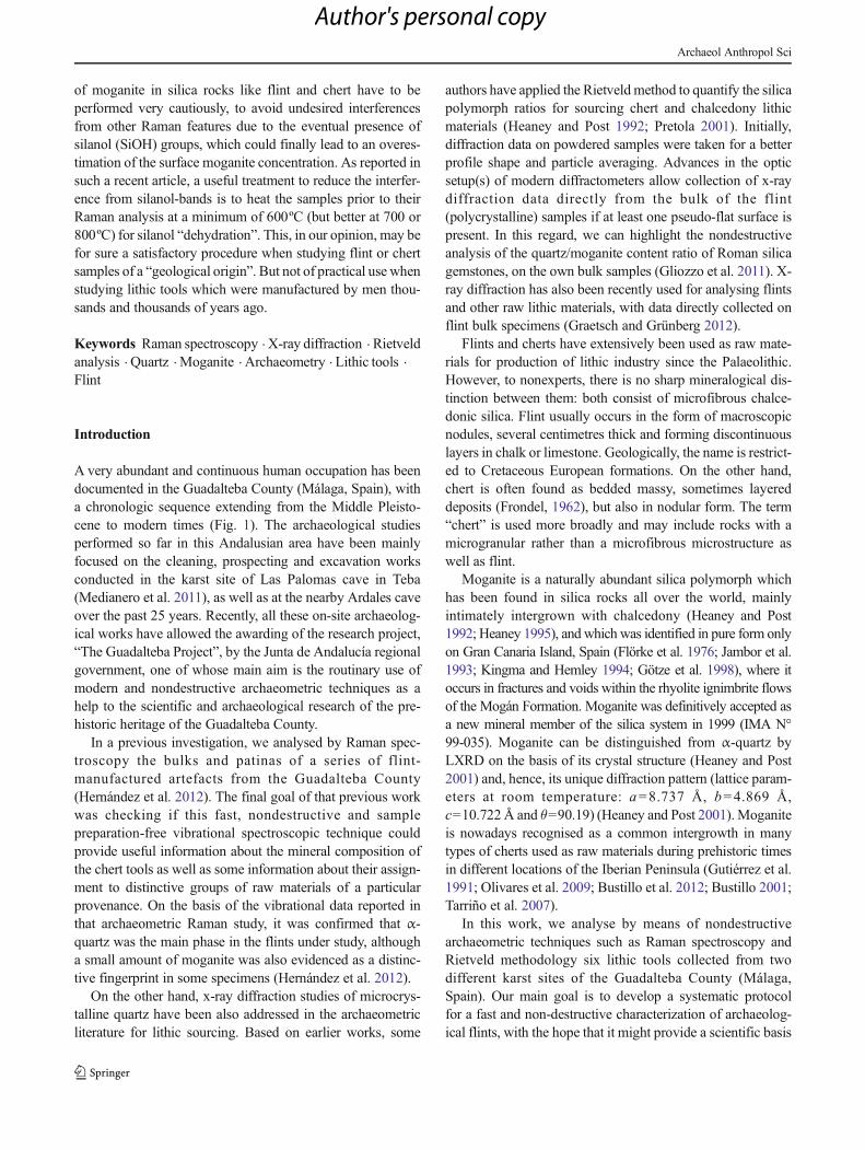
Lithic tools numbered as 2 and 5 were collected during thepreliminary prospection works carried out during the 2011–2012 season at the great cone of sediments located just belowthe current entry of the Ardales Cave (Fig. 1 and 2). Specimen2 is a chisel (Upper Palaeolithic) made in massive white chert,and showing small white microlithic inclusions or spicules,whereas specimen 5 is a Mousterian flake of cream flint withclear microsphere inclusions.
The other lithic tools (1, 3, 4 and 6), all of them ascribed toMousterian technology, were unearthed from the deepest level(VIII) in Sima de Las Palomas (Fig. 1 and 2). They weredocumented, together with many others tools, in a rich, stillunpublished, continuous sequence of Middle Palaeolithic oc-cupations, starting from the first half of the last glacial cycle.Lithic tools 3 and 4 (both of them flakes) and 6 (core) aremade of white and grey flints; while specimen 1 (core) is madeof a reddish brown radiolarite.
Raman spectroscopy
Raman data were collected on a Bruker RamII FT-Ramanmodule attached to a Vertex FT-IR spectrometer at the SCAIof the University of Málaga. The 1,064 nm laser power waskept fixed to the lowest value to avoid mineralogical changes.Typically, 3,000 Raman scans were averaged in each case toimprove the signal-to-noise ratio, with a spectral resolution of1 cm−1, and the 100 to 1,800 cm−1 spectral range was record-ed. Typically, the spot size on the lithic tools was around50 μm. Different spots were analysed for each specimen, withthe aim of analysing the homogeneity of the moganite distri-bution on the surface of each lithic tool. Cortex and patinaswere avoided, and only fresh surfaces were analysed.
X-ray diffraction
Laboratory x-ray diffraction (LXRD) patterns for the bulk(fresh rock from each specimen) lithic tools were recordedon a PANalytical X'Pert PROMPD diffractometer working inreflection geometry (θ/2θ) and using the X'Celerator RealTime Multiple Strip (RTMS) detector with an active lengthof 2.122º. The flints were loaded in a multipurpose holderwhich allows the micrometric controlled alignment of sampleswith an exposed surface as flat as possible. Cu-patterns were
collected with a long fine focus Cu tube working at 45 kVand40 mA. The incident beam optic path contained a hybridmonochromator (composed of a W/Si parabolic x-ray-graded mirror and a 2-crystals Ge (220) 2-bounce monochro-mator) which yielded a monochromatic, λ=1.54059 Å paral-lel x-ray beam, and a fixed 1/8° divergence slit. The diffractedbeam optic path contained a fixed 1/4° anti-scatter slit. Bothincident and diffracted beams were equipped with 0.04 radSoller slits. A typical scan range was from 14.0 to 90.0° 2θwith a step size of 0.0167° 2 θ and an overall recording time ofapproximately 4 h. We recall that the diffraction data weretaken on bulk lithic tools, so we can describe the setup aspolycrystalline x-ray data collection.
Polycrystalline patterns for the six lithic tools were furtheranalysed by the Rietveld method as implemented in theGSAS-EXPGUI software package (Larson and Dreele 2000)by using a pseudo-Voigt peak shape function (Thompson et al.1987) to obtain Rietveld Quantitative Phase Analysis(RQPA). The refined overall parameters were: phase scalefactors, background coefficients, unit cell parameters, zero-shift error and peak shape parameters. Structural parameters atroom temperature were taken: Will et al. (1988) for α-quartz,Heaney and Post (2001) for moganite andMaslen et al. (1995)for calcite, and not refined further.
Results and discussion
Flint usually forms irregular nodules in sedimentary rocks. Athin white layer can surround the flint nodules. The outerlayer, generally white, is sometimes denoted as cortex. Quiteoften, the cortex shows a powder-like appearance. Remains ofthis cortex layer can be seen in some of the specimens which
Fig. 2 Photographs of the six lithic tools subject of FT-Raman andRietveld analysis: (1) number 1:1628-Silex-LP-perfil-2011-bulk (core);(2) number 2: 1765-AD-2-11-bulk (flake); (3) number 3: 225-230-CP-3-46-artefact-3-bulk (flake); (4) number 4: 1431-2011-CP-R-S-P1-bulk(flake); (5) number 5: Point-492-AD-4-11-2-CapaR-bulk (scraper); (6)number 6:1430-2011-CP-R-S-P1-bulk (waste knapping)
Archaeol Anthropol Sci
Author's personal copy

are the subject of this study (see Fig. 2). Flint (Fig 2. (2–6))and radiolarite (Fig. 2. (1)) patinas are also present in some ofthe studied samples; but in general, the areas of the lithic toolswith a knapping display fresh and clean surfaces. Patinas arethe result of different physico-chemical transformations on theouter surface of siliceous material stone tools after their knap-ping process due to exposition to weathering and burial duringlong periods of time.
Thickness and compositional features of the surfaces ofstone tools will depend on the type and composition of theoriginal bulk raw materials (flint, radiolarite, sandstone) andalso on the physico-chemical environment(s) to which they
were subjected during their transport, sedimentation or burial.Quite often, a decrease of the rock density, lost or re-precipitation of silica with different morphologies or a dehy-dration process can occur. Furthermore, these processes can berather heterogeneous, so slightly different data may be derivedfrom different points on a given surface (Bustillo et al. 2007).
As bulk mineral composition is dependent on physical andchemical conditions in sedimentary areas, mineralogical char-acterization and phase rates of flints can be specific for eachoutcrop and, then, provide information about the source of theraw material.
Raman spectroscopy clearly shows that α-quartz is themain mineral which constitutes the bulks of the six flint-radiolarite specimens. This result is derived from its
Fig. 3 FT-Raman spectra of thewhole series of lithic tools subjectof study in the 400–550 cm−1
spectral range. Raman spectrahave been shifted vertically, andscaled in intensity relative to thestrongest Raman feature of α-quartz at 465 cm−1, to make clearthe variable intensity of themoganite Raman feature at502 cm−1; and comparisonbetween the FT-Raman profilescollected for the number 1 andnumber 2 specimens, at differentspots on their surface
Fig. 4 FT-Raman spectra of α-quartz (bottom) and a “moganite” samplefrom Mogán (Canary Islands), containing nearly 75 % moganite and25 % α-quartz (top) as determined from a Rietveld analysis of powderdiffraction data
Fig. 5 FT-Raman spectra of specimen number 6 (top) and pure calcite(bottom)
Archaeol Anthropol Sci
Author's personal copy
characteristic strong Raman scattering at 465 cm−1, due to theSiO2 symmetric stretching vibration, together with other twoweaker features at 206 and 128 cm−1 (see Figs. 3, 4 and top of5). The bulks of the six lithic tools also showed anotherRaman scattering at 501 cm−1, due to moganite (Table 1).
Figure 3 displays the FT-Raman profiles of the six lithictools in the 400–550 cm−1 spectral range. The changes of therelative Raman intensity associated to the moganite vibrationat 501 cm−1 as compared with the strong Raman feature of α-quartz at 465 cm−1 are rather conspicuous. The FT-Ramanspectra of pureα-quartz and that of a nearly “75%moganite+25 % α-quartz” sample from Mogán (Canary Islands, Spain)are displayed in Fig. 4 for the sake of comparison. Figure 3also shows a comparison between the FT-Raman profilescollected for specimens 1 and 2 at different spots on theirsurface. The resemblance between the various Raman spectralpatterns recorded for each lithic tool evidences the homoge-neity of the surface moganite distribution.
We recall however that some authors (Schmidt et al. 2012;Schmidt et al. 2013) have recently reported on a secondRaman feature, at 503 cm−1, commonly observed in chalce-dony samples and resulting from silanol (SiOH) groups,
which interferes with the main moganite band at 501 cm−1.This overlap can make the Raman spectroscopic quantifica-tion in the presence of silanol groups difficult. These authorsrecommended that silica rocks are heat-treated prior to theirRaman analysis at a minimum of 600ºC (but better at 700 or800ºC) for silanol “dehydration”. This heat-treatment, in ouropinion, may be for sure a satisfactory procedure when study-ing flint or chert samples of a “geological origin”. But it is notof practical use when studying lithic tools which weremanufactured by men several thousands of years ago.
On the other hand, Fig. 5 display the FT-Raman spectrumof specimen number 6, in the 100–1,800 cm−1 spectral range,together with that of pure calcite. The observation of a weakRaman scattering at 1,086 cm−1 in some of these lithic tools,and a second weaker Raman feature at 281 cm−1, is indicativeof the presence of a calcite crust.
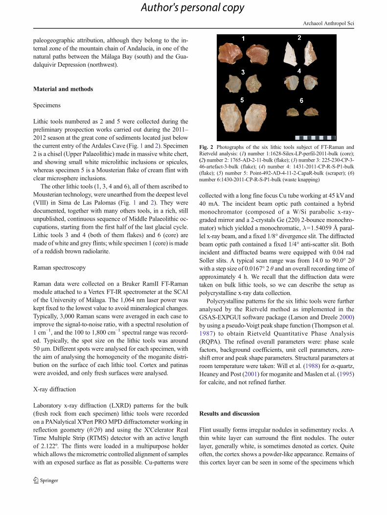
Advanced methodology for x-ray diffraction allows thestudy/analysis of polycrystalline materials if there is at leastone relatively flat surface. The polycrystalline patterns werecollected as described in the experimental section, and the datawere further treated by the Rietveld method to obtain themineralogical quantitative phase analyses. Figures 6 and 7
Fig. 6 Rietveld plot of number 1as an example of a high-contentmoganite lithic tool. The insethighlights the 2θ region wheremoganite diffraction peaks aremore intense and unoverlapped
Table 1 SamplesSample no. Sample name Description Site
1 1628-Radiolarite-LP-profile-2011 bulk (core) (Palomas de Teba)
2 1765-AD-2-11 bulk (burin) (Cave of Ardales)
3 225-230-CP-3-46 artefact-3-bulk (flake) (Palomas de Teba)
4 1431-2011-CP-R-S-P1 bulk (flake) (Palomas de Teba)
5 Point-492-AD-4-11-2 Rstrata-bulk (scraper) (Cave of Ardales)
6 1430-2011-CP-R-S-P1 bulk (core) (Palomas de Teba)
Archaeol Anthropol Sci
Author's personal copy

display the Rietveld plots of two lithic tools as examples ofhigh- and low-content moganite objects, respectively. Table 2reports the phase analyses of the six studied lithic tools. It canbe seen than two lithic tools (numbers 1 and 2) have relativelyhigh moganite contents, close to 15 wt%. This result is in verygood agreement with the Raman study as these objectsdisplayed the strongest Raman scattering at 501 cm−1 (seeFig. 3). On the other hand, the remaining lithic tools have amuch smaller moganite content, ranging between 1.4 and4.0 wt%, in close agreement with their Raman spectra wherethe 501 cm−1 scattering is much weaker. Furthermore, five ofthe six flints are determined to be pure silicon dioxide objects.However, number 6 lithic tool also contains 2.4 wt% of calcitewhich may be due to a small amount of surface depositedcalcite.
The diffraction peak shape for α-quartz was found to beanisotropic (i.e. hkl-dependent). This was modelled by thepseudo-Voigt function by refining GW (the Gaussian part),LY (the Lorentzian part) and STEC (the anisotropic correctionof the Lorentzian width) along [001] direction. Moganite
phase is in low contents, and their diffraction peaks arestrongly overlapped with those of α-quartz. Thus, its peakshape was isotropically modelled. Table 2 also reports thefinal values used in the Rietveld analysis. As expected, thetwo samples with higher moganite contents showed the largeanisotropic broadening for α-quartz, i.e. larger STEC values(see Table 2). This result could be explained because moganiteis present as intergrowths between the α-quartz domains. So,higher moganite contents increase the quartz microstrain anddecrease the quartz coherent diffraction domain sizes. Thesetwo features make the x-ray powder diffraction peaks of α-quartz anisotropically broader.
Finally, it should be mentioned that polycrystalline diffrac-tion data collection followed by Rietveld analysis is muchmore time consuming (between 5 and 6 h each sample) thanRaman spectroscopy. Therefore, an experimental approachcan be established for the methodological study of the flinttools in the Guadalteba County in which Raman data arecollected in a first step on many lithic tools (ideally in situ,by screening the samples in the field through a suited portable
Table 2 Summary of the Rietveld analysis results for the six lithic tools under study
Sampleno.
Quartz/wt%
Moganite/wt%
Quartz unit cellvolume/Å3
Moganite unitcell/Å3
GWqtz/0.01°2
LYqtz/0.01°
STECqtz/0.01°
GWmog/0.01°2
LYmog/0.01°
1 86.7 (5) 13.2 (3) 113.40 (1) 455.8 (1) 31.5 (8) 74 (1) −50 (1) 150 (–) 34 (3)
2 84.8 (7) 15.2 (4) 113.56 (1) 452.0 (4) 252 (3) 64 (2) −62 (2) 150 (–) 155 (7)
3 95.9 (2) 4.1 (2) 113.19 (1) 454.0 (–) 23.8 (5) 44.5 (6) −26 (1) 150 (–) 20 (–)
4 98.0 (1) 2.0 (1) 113.34 (1) 454.0 (–) 23.9 (4) 50.9 (5) −30 (1) 150 (–) 20 (–)
5 97.9 (1) 2.1 (2) 113.17 (1) 454.0 (–) 40.8 (6) 38.0 (6) −22 (1) 150 (–) 20 (–)
6* 94.5 (2) 3.1 (2) 113.12 (1) 454.0 (–) 123.6 (1) 16.5 (8) −15 (1) 150 (–) 20 (–)
* This sample also contains 2.4 (1) wt% of calcite
Fig. 7 Rietveld plot of number 5as an example of a low-contentmoganite lithic tool. The insethighlights the 2θ region wheremoganite diffraction peaks aremore intense and unoverlapped
Archaeol Anthropol Sci
Author's personal copy
Raman spectrometer) and then selected specimens are subse-quently analysed in the laboratory by means of Rietveldmethodology for crosschecking. The final goal of such asystematic archaeometric study of the manufactured lithictools is to establish a broad database including visual appear-ance, Raman spectroscopic data, moganite weight content andmicrostructure information for the future studies of the possi-ble source areas of raw materials.
Conclusions
Raman spectroscopy allowed us to clearly distinguish be-tween α-quartz and moganite in a series of Palaeolithicchert-manufactured specimens, revealing noticeable differ-ences in the qualitative content ratio of these two microcrys-talline SiO2 polymorphic phases among the various lithic toolsunder analysis.
X-ray diffraction data were also nondestructively taken onthe same lithic tools. Further, Rietveld quantitative phaseanalyses allowed determining the precise moganite weightcontent in each chert-manufactured tool. The variablemoganite/α-quartz ratio in manufactured flint and radiolariteprehistoric artefacts unearthed in different archaeological sitesof the Guadalteba County may provide valuable informationto assess the possible geological sources of the correspondingraw materials. As for the six studied samples, the weightcontent of moganite ranged between 2 and 15 wt%.
In summary, the combined use of FT-Raman spectroscopyand Rietveld refinement of high-quality x-ray diffraction ofbulk lithic tools constitutes a useful and nondestructive exper-imental strategy for the methodological study of siliceousstone industries manufactured by men in the prehistory, andmay help in determining the source areas of raw materials.
Acknowledgments Research at the Universities of Málaga and Cádizwas supported by Junta de Andalucía, through funding the FQM-159,FQM-113 and HUM-440 scientific groups. We also acknowledge thehelp by part of the students of the “Escuela Taller de Peñarrubia”,sponsored by the “Guadalteba County Consortium”, both in regards tothe on-site works and the subsequent classification of the findings in thearchaeology workroom. Financial and technical support from the StiftungNeanderthal Museum is also acknowledged. We finally thank José Bellofor providing us with moganite-rich samples from Mogán (CanaryIslands, Spain).
References
Bustillo MA (2001) Aparición y significado de la moganita en las rocasde la sílice: una revisión. J Sediment Res 71:436–443
Bustillo MA, Pérez-Jiménez JL, Fort R (2007) Deterioro de Sílex enCanteras Históricas y Muestras Líticas. MACLA 7:18
Bustillo MA, Pérez-Jiménez JL, Alonso-Zarza AM, Furio M (2012)Moganite in the Chalcedony varieties of continental cherts(Miocene, Madrid Basin, Spain). Spectrosc Lett 45:109–113
Carbonell E, Márquez B, Mosquera M, Olí A, Rodríguez XP, SalaR, Vergés JM (1999) “El Modo 2 en Galería. Análisis de laindustria lítica y sus procesos técnicos” in Atapuerca:ocupaciones humanas y paleoecología del yacimiento deGalería, ed. Junta de Castilla y León, Consejería deEducación y Cultura, Valladolid, 1st edn., 390
ClarkG (1998)World prehistory: in new perspective, 3rd edn. CambridgeUniversity Press, New York, ISBN: 052129178X
Flörke OW, Jones JB, Schmincke HU (1976) A new microcrystallinesilica from Gran Canaria. Z Kristallogr 143:156–165
Frondel C (1962) The system of mineralogy, vol. III: silica minerals.Wiley, New York
Gliozzo E, Grassi N, Bonanni P, Meneghini C, Tomei MA (2011)Gemstones from Vigna Barberini at the Palatine Hill (Rome,Italy). Archaeometry 53:469–489
Götze J, Nasdala L, Kleeberg R, Wenzel RM (1998) Occurrence anddistribution of “moganite” in agate/chalcedony: a combined micro-Raman, Rietveld and cathodoluminescence study. Contrib MineralPetrol 133:96–105
Graetsch HA, Grünberg JM (2012) Microstructure of flint and other chertraw materials. Archaeometry 54:18–36
Gutiérrez Mas JM, Martín Algarra A, Domínguez-Bella S, Moral JP(1991), Introducción a la Geología de la provincia de Cádiz, Ed.University of Cádiz, ISBN: 978-84-7786-050-1
Heaney PJ (1995) Moganite as an indicator for vanished evaporites: atestament reborn? J Sediment Res A65:633–638
Heaney PJ, Post JE (1992) The widespread distribution of a novel silicapolymorph in microcrystalline quartz varieties. Science 255:441–443
Heaney PJ, Post JE (2001) Evidence for an I2/a to Imab phase transitionin the silica polymorph moganite at 570 K. Am Mineral 86:1358–1366
Hernández V, Jorge-Villar SE, Capel Ferrón C, Medianero FJ, Ramos J,Weniger GC, Domínguez-Bella S, Linstaedter J, Cantalejo P, EspejoM, Durán Valsero JJ (2012) Raman spectroscopy analysis ofPalaeolithic industry from Guadalteba terrace river, Campillos(Guadalteba county Southern of Iberian Peninsula. J Raman Spec43:1651–1657
Jambor JL, Burke EAJ, Grew ES, Puziewicz J (1993) New mineralnames. Am Mineral 78:672–678
Kingma KJ, Hemley RJ (1994) Raman spectroscopic study of microcrys-talline silica. Am Mineral 79:269–273
Laplace G (1975) La typologie analytique et structurale: Base rationnelled’étude des industries lithiques et osseuses. Colloques Nationaux duCentre National de la Reserche Scientifique, Ed. CNRS, Marseille,932: 91-141
Larson AC, Von Dreele RB (2000) General Structure Analysis System(GSAS), Los Alamos National Laboratory Report LAUR
Maslen EN, Streltsov VA, Streltsova NR, Ishizawa N (1995) Electrondensity and optical anisotropy in rhombohedral carbonates. III.Synchrotron X-ray studies of CaCO3, MgCO3 and MnCO3. ActaCryst B51:929–939
Medianero FJ, Ramos J, Palmqvist P,Weniger GC, JRiquelme JA, EspejoM, Cantalejo P, Aranda A, Pérez-Claros JA, Figueirido B, EspigaresP, Ros-Montoya S, Torregrosa V, Linstädter L, Cabello L, Becerra S,Ledesma P, Mevdev I, Castro A, Romero M, Martínez-Navarro B(2011) The karst site of Las Palomas (Guadalteba County, Málaga,Spain): a preliminary study of its Middle–Late Pleistocenearchaeopaleontological record. Quatern Int 243:127–136
Olivares M, Tarriño A, Murelaga X, Baceta JI, Castro K, Etxebarria N(2009) Non-destructive spectrometry methods to study the distribu-tion of archaeological and geological chert samples. SpectrochimActa A 73:492–497
Archaeol Anthropol Sci
Author's personal copy
Pretola JP (2001) A feasibility study using silica polymorph ratios forsourcing chert and chalcedony lithic materials. J Archaeol Sci 28:721–739
Schmidt P, Bellot-Gurlet L, Slodczyk A, Fröhlich F (2012) A hithertounrecognised band in the Raman spectra of silica rocks: influence ofhydroxylated Si–O bonds (silanole) on the Raman moganite band inchalcedony and flint (SiO2). Phys Chem Minerals 39:455–464
Schmidt P, Bellot-Gurlet L, Léa V, Sciau P (2013) Moganite detection insilica rocks using Raman and infrared spectrocopy. Eur J Mineral25(5):797–805
Tarriño A, Olivares M, Etxebarria N, Baceta JI, Larrasoaña JC, Yusta I,Pizarro JL, Cava A, Barandiarán I, Murelaga X (2007) El sílex detipo “Urbasa” Caracterización petrológica y geoquímica de unmarcador litológico en yacimientos arqueológicos del Suroesteeuropeo durante el Pleistoceno superior y Holoceno inicial.Geogaceta 43:127–130
Thompson P, Cox DE, Hastings JB (1987) Rietveld refinement ofDebye-Scherrer synchrotron X-ray data from Al2O3. J ApplCryst 20:79–83
Vallespí E (1986), El Paleolítico inferior y medio en Andalucía inHomenaje a Luis Siret (1934–1984). Ed. Junta de Andalucía,Consejería de Cultura, pp. 59-66. ISBN: 84-505-3511-5
Vallespí E (1986), Cultura de las graveras y comienzos del AchelenseIbérico in Estudios en homenaje al Dr. Antonio Beltrán Martínez,Ed. University of Zaragoza, Zaragoza, pp. 147–157. ISBN: 84-600-4366-5
Vallespí E (1992) Las industrias Achelenses de Andalucía: Ordenación yComentarios. SPAL 1:61–78
Will G, Bellotto M, Parrish W, Hart M (1988) Crystal structuresof quartz and magnesium germanate by profile analysis ofsynchrotron-radiation high-resolution powder data. J ApplCryst 21:182–191
Archaeol Anthropol Sci
Author's personal copy