Collenchyma: a versatile mechanical tissue with dynamic cell ...
16
INVITED REVIEW Collenchyma: a versatile mechanical tissue with dynamic cell walls Olivier Leroux 1,2, * 1 Botany and Plant Science and Ryan Institute, School of Natural Sciences, National University of Ireland Galway, University Road, Galway, Ireland and 2 Pteridology, Department of Biology, Ghent University, KL Ledeganckstraat 35, B-9000 Ghent, Belgium * E-mail: [email protected] Received: 29 April 2012 Returned for revision: 11 June 2012 Accepted: 6 July 2012 Published electronically: 29 August 2012 † Background Collenchyma has remained in the shadow of commercially exploited mechanical tissues such as wood and fibres, and therefore has received little attention since it was first described. However, collenchyma is highly dynamic, especially compared with sclerenchyma. It is the main supporting tissue of growing organs with walls thickening during and after elongation. In older organs, collenchyma may become more rigid due to changes in cell wall composition or may undergo sclerification through lignification of newly deposited cell wall material. While much is known about the systematic and organographic distribution of collenchyma, there is rather less information regarding the molecular architecture and properties of its cell walls. † Scope and conclusions This review summarizes several aspects that have not previously been extensively dis- cussed including the origin of the term ‘collenchyma’ and the history of its typology. As the cell walls of col- lenchyma largely determine the dynamic characteristics of this tissue, I summarize the current state of knowledge regarding their structure and molecular composition. Unfortunately, to date, detailed studies specif- ically focusing on collenchyma cell walls have not been undertaken. However, generating a more detailed under- standing of the structural and compositional modifications associated with the transition from plastic to elastic collenchyma cell wall properties is likely to provide significant insights into how specific configurations of cell wall polymers result in specific functional properties. This approach, focusing on architecture and functional properties, is likely to provide improved clarity on the controversial definition of collenchyma. Key words: Collenchyma, histology, plant anatomy, mechanical tissue, plant cell wall, primary and secondary cell walls, plant biomechanics. INTRODUCTION The emergence of mechanical tissues was a key innovation in the evolution of land plants and a prerequisite for the appear- ance of large terrestrial species. By the middle Devonian, many plant species developed a hypodermal sterome consist- ing of heavily thickened sclerenchyma cells (Rowe and Speck, 2004). Biomechanical investigations indicated that the sterome significantly contributed to the stiffness of stems and allowed them to reach great heights and evolve diverse branched architectures compared with plants with turgor-based support systems (Rowe and Speck, 2004). While sclerenchyma tissues confer rigidity and tensile and shear strength to many plant organs (Niklas, 1992; Jarvis, 2007), their properties are incapable of supporting growing plant organs which undergo extensive turgor-driven elong- ation. Indeed, sclerified tissues generally consist of dead cells with non-extensible rigid cell walls which are unable to undergo mitotic divisions. In small slowly growing plant organs, turgor pressure generated in parenchyma cells may provide sufficient support, but many plant stems grow fast and are fragile, and therefore they cannot fully rely on turgor pressure for support. Partly because they are non- sclerified and only minimally lignified, young plant tissues are preferentially selected by grazing animals and plant bugs. For this reason, supporting tissues in these regions should not be terminally differentiated but capable of under- going wound healing or tissue regeneration. Moreover, as sec- ondary growth increases the diameter of stems, the ability to transdifferentiate and initiate periderm formation is an add- itional advantage. Finally, young above-ground organs are photosynthetic and the reinforcing tissues should ideally be as translucent as possible to enable light to reach the chloro- plasts in tissues deeper in the plant. To meet most of the above-mentioned requirements, and to provide support without preventing cell elongation, many plants – eudicotyle- dons in particular – develop collenchyma: a mechanical tissue composed of elongated cells with thick flexible and translucent cell walls and with protoplasts capable of resum- ing meristematic activity. In this review, I describe the remarkable origin of the term ‘collenchyma’ and discuss some of the controversies asso- ciated with the description of this tissue. In contrast to scleri- fied mechanical tissues such as wood and fibres, which are economically important raw materials, collenchyma tissues have received little attention. Consequently, a clear definition of collenchyma has never been given. It is not surprising that this has resulted in some confusion with respect to its des- ignation in certain cases. However, it needs to be highlighted that one of the factors that makes collenchyma so unique is also the reason why it is difficult to define: the dynamic nature of its cell walls. # The Author 2012. Published by Oxford University Press on behalf of the Annals of Botany Company. All rights reserved. For Permissions, please email: [email protected] Annals of Botany 110: 1083–1098, 2012 doi:10.1093/aob/mcs186, available online at www.aob.oxfordjournals.org by guest on December 6, 2012 http://aob.oxfordjournals.org/ Downloaded from
-
Upload
khangminh22 -
Category
Documents
-
view
2 -
download
0
Transcript of Collenchyma: a versatile mechanical tissue with dynamic cell ...
INVITED REVIEW
Collenchyma a versatile mechanical tissue with dynamic cell walls
Olivier Leroux121Botany and Plant Science and Ryan Institute School of Natural Sciences National University of Ireland Galway
University Road Galway Ireland and 2Pteridology Department of Biology Ghent University KL Ledeganckstraat 35B-9000 Ghent Belgium
E-mail OlivierLerouxUGentbe
Received 29 April 2012 Returned for revision 11 June 2012 Accepted 6 July 2012 Published electronically 29 August 2012
dagger Background Collenchyma has remained in the shadow of commercially exploited mechanical tissues such aswood and fibres and therefore has received little attention since it was first described However collenchymais highly dynamic especially compared with sclerenchyma It is the main supporting tissue of growing organswith walls thickening during and after elongation In older organs collenchyma may become more rigid dueto changes in cell wall composition or may undergo sclerification through lignification of newly depositedcell wall material While much is known about the systematic and organographic distribution of collenchymathere is rather less information regarding the molecular architecture and properties of its cell wallsdagger Scope and conclusions This review summarizes several aspects that have not previously been extensively dis-cussed including the origin of the term lsquocollenchymarsquo and the history of its typology As the cell walls of col-lenchyma largely determine the dynamic characteristics of this tissue I summarize the current state ofknowledge regarding their structure and molecular composition Unfortunately to date detailed studies specif-ically focusing on collenchyma cell walls have not been undertaken However generating a more detailed under-standing of the structural and compositional modifications associated with the transition from plastic to elasticcollenchyma cell wall properties is likely to provide significant insights into how specific configurations ofcell wall polymers result in specific functional properties This approach focusing on architecture and functionalproperties is likely to provide improved clarity on the controversial definition of collenchyma
Key words Collenchyma histology plant anatomy mechanical tissue plant cell wall primary and secondarycell walls plant biomechanics
INTRODUCTION
The emergence of mechanical tissues was a key innovation inthe evolution of land plants and a prerequisite for the appear-ance of large terrestrial species By the middle Devonianmany plant species developed a hypodermal sterome consist-ing of heavily thickened sclerenchyma cells (Rowe andSpeck 2004) Biomechanical investigations indicated thatthe sterome significantly contributed to the stiffness ofstems and allowed them to reach great heights and evolvediverse branched architectures compared with plants withturgor-based support systems (Rowe and Speck 2004)While sclerenchyma tissues confer rigidity and tensile andshear strength to many plant organs (Niklas 1992 Jarvis2007) their properties are incapable of supporting growingplant organs which undergo extensive turgor-driven elong-ation Indeed sclerified tissues generally consist of deadcells with non-extensible rigid cell walls which are unableto undergo mitotic divisions In small slowly growing plantorgans turgor pressure generated in parenchyma cells mayprovide sufficient support but many plant stems grow fastand are fragile and therefore they cannot fully rely onturgor pressure for support Partly because they are non-sclerified and only minimally lignified young plant tissuesare preferentially selected by grazing animals and plantbugs For this reason supporting tissues in these regions
should not be terminally differentiated but capable of under-going wound healing or tissue regeneration Moreover as sec-ondary growth increases the diameter of stems the ability totransdifferentiate and initiate periderm formation is an add-itional advantage Finally young above-ground organs arephotosynthetic and the reinforcing tissues should ideally beas translucent as possible to enable light to reach the chloro-plasts in tissues deeper in the plant To meet most of theabove-mentioned requirements and to provide supportwithout preventing cell elongation many plants ndash eudicotyle-dons in particular ndash develop collenchyma a mechanicaltissue composed of elongated cells with thick flexible andtranslucent cell walls and with protoplasts capable of resum-ing meristematic activity
In this review I describe the remarkable origin of the termlsquocollenchymarsquo and discuss some of the controversies asso-ciated with the description of this tissue In contrast to scleri-fied mechanical tissues such as wood and fibres which areeconomically important raw materials collenchyma tissueshave received little attention Consequently a clear definitionof collenchyma has never been given It is not surprisingthat this has resulted in some confusion with respect to its des-ignation in certain cases However it needs to be highlightedthat one of the factors that makes collenchyma so unique isalso the reason why it is difficult to define the dynamicnature of its cell walls
The Author 2012 Published by Oxford University Press on behalf of the Annals of Botany Company All rights reserved
For Permissions please email journalspermissionsoupcom
Annals of Botany 110 1083ndash1098 2012
doi101093aobmcs186 available online at wwwaoboxfordjournalsorg
by guest on Decem
ber 6 2012httpaoboxfordjournalsorg
Dow
nloaded from
HISTORY
Several textbooks (eg Esau 1965 Fahn 1990) report thatlsquocollenchymarsquo is derived from the Greek word lsquokollarsquomeaning glue and referring to the thick glistening appearanceof unstained collenchyma cell walls Although this explanationseems perfectly acceptable confusion exists because the firstuse of lsquocollenchymarsquo was by Link (1837) who used it to de-scribe the sticky substance on Bletia (Orchidaceae monocots)pollen Two years later in an anatomical survey of Cactaceae(eudicots) Schleiden (1839) criticized Linkrsquos (1837) excessivenomenclature and stated mockingly that the term lsquocollen-chymarsquo could have more easily been used to describe elon-gated sub-epidermal cells with unevenly thickened cellsAlthough Schleiden (1839) himself used lsquoaussere Rinden-lagersquo or lsquoZellen der aussere Rindenschichtrsquo rather than lsquocollen-chymarsquo the term seems to have stuck as a way to describeelongated and thickened sub-epidermal cells similarly to cur-rently accepted usage Others such as Meyen (1830) usedlsquoprosenchymarsquo to describe elongated cells with taperingends without distinguishing between vascularground tissueand even between sclerenchyma-like and collenchyma-liketissues Common usage of lsquocollenchymarsquo can perhaps beattributed to Harting (1844) as he repetitively used lsquocollen-chymarsquo sensu Schleiden in his anatomical survey of annualdicotyledonous angiosperms French and English translationsof his work soon followed (Giltay 1882) spreading the newdefinition or appropriation of lsquocollenchymarsquo That collen-chyma was not in common use in the mid-19th century isperhaps suggested by von Mohl (1844) who described collen-chyma tissues as lsquojelly-like subepidermal cellsrsquo adding paren-thetically lsquothe so-called collenchyma cellsrsquo By the end of the19th century the term lsquocollenchymarsquo was incorporated insome prominent and influential plant anatomy text books andpublications (eg Sachs 1868 de Bary 1877 Ambronn1881 Giltay 1882 van Tieghem 1886ndash1888) and becamemore widely accepted
GENERAL MORPHOLOGY AND ONTOGENY
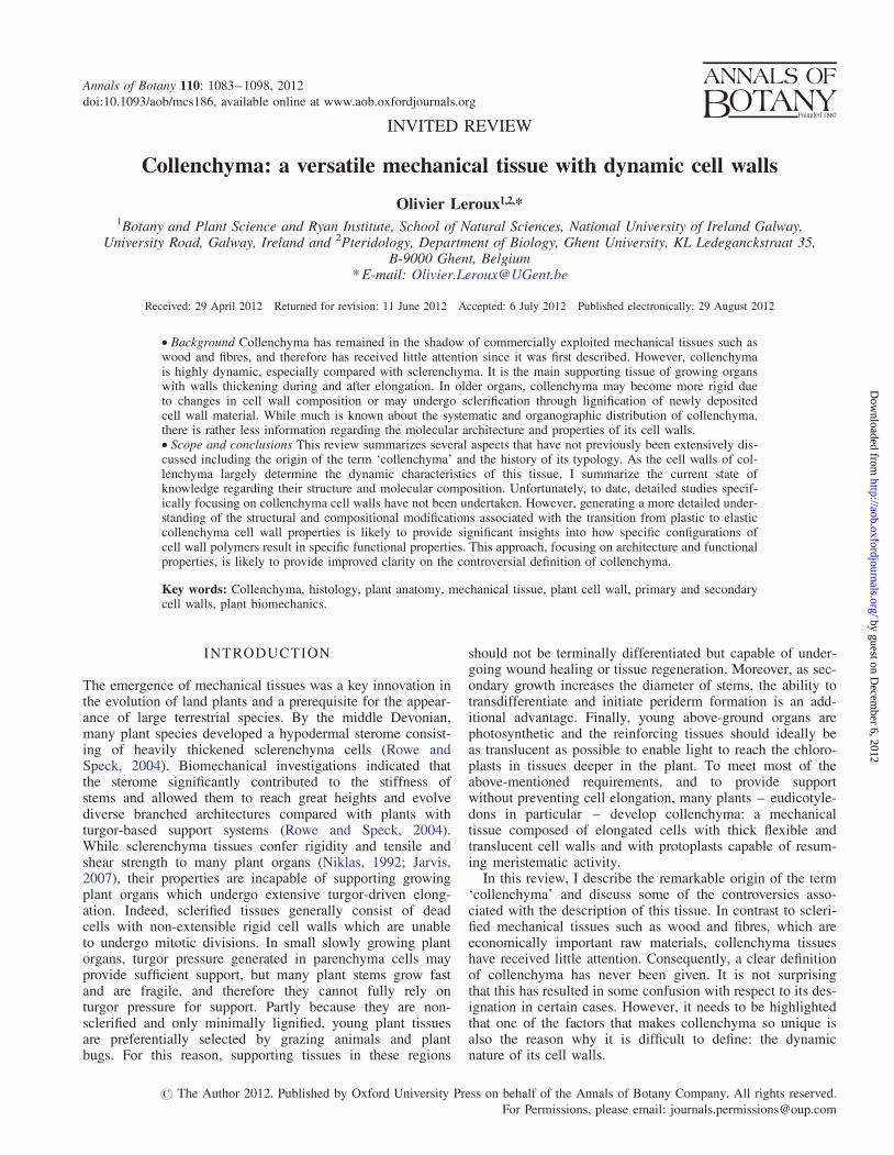
The three most characteristic morphological features of collen-chyma are (i) their axially elongated cells (2) their cell wallthickenings and (3) their living protoplasts (Fig 1AndashD)During elongation collenchyma cells do not divide as muchas the surrounding parenchyma cells which explains their pro-senchymatic nature However cell size and shape still can varyfrom short isodiametric and prismatic cells to long fibre-likecells with tapering ends The latter may even reach lengthsof up to 25 mm in Heracleum sphondylium (Apiaceae eudi-cots) (Majumdar and Preston 1941) In some cases transversedivisions take place after or during elongation and the result-ing daughter cells often remain together enclosed by a sharedcell wall derived from the mother cell giving it the appearanceof a septate fibre with non-thickened cross walls (Fig 1D)Nonetheless collenchyma shares more morphological andphysical characteristics with parenchyma tissues and thereforeintermediate types are not uncommon The similaritiesbetween both tissues even led several researchers to categorizecollenchyma as thick-walled parenchyma (eg de Bary 1877)Collenchyma and parenchyma cell walls both have the ability
to stretch andor grow during differentiation but in the case ofcollenchyma the walls thicken throughout elongation and oftenpost-elongation (Jarvis 2007) Cell wall material is generallynot distributed equally so that most collenchyma cells have ir-regular thickenings (see Histological typology) Similarly toparenchyma collenchyma cells have living protoplasts essen-tial for controlling the hydration state of the cell wall but alsoto enable transdifferentiation and cell wall thickening andmodification Many textbooks (eg Esau 1965 Fahn 1990)mention that chloroplasts are present in collenchyma but intypical collenchyma tissue with a clear mechanical functionchloroplasts are rarely found (Evert 2006) However toallow photosynthesis collenchyma cell walls are generallytranslucent enabling light to be transmitted to the chloroplastsin tissues below
Controversy remains regarding the ontogeny of collenchymaas it has been the focus of very few studies (Ambronn 1881van Wisselingh 1882 Esau 1936 Majumdar 1941) Acc-ording to Esau (1936) collenchyma of celery (Apium graveo-lens Apiaceae eudicots) originates in the ground meristemclose to or against the protoderm Periclinal divisions initiallypredominate but are soon followed by anticlinal longitudinalsections As divisions rapidly follow each other cellsenlarge only moderately appearing smaller than the surround-ing ground meristem cells The rapid succession of divisionsgenerally prevents the formation of intercellular spaceswhich are numerous in the ground tissue at that stageAmbronn (1881) on the other hand observed that in theApiaceae collenchyma and vascular tissue arise from thesame procambium strand while in most other families bothtissues arise independently from each other van Wisselingh(1882) who did not study Apiaceae never found a commonorigin for collenchyma and vascular tissue in any of thespecies he studied (including Aucuba Euonymus andLamium) A later investigation of Heracleum (Majumdar1941) failed to provide further clarity as it was reported thatthe inner parts of each collenchyma strand are derived in theearliest stages from the same meristem as the vascularbundles whereas the outer parts are derived from the groundmeristem It needs to be noted that Esau (1936) Majumdar(1941) and Ambronn (1881) did not study the same speciesand variation at species level may occur Esau (1936) alsostudied the ontogeny and origin of collenchyma tissue asso-ciated with the vascular bundles in celery and showed thatthey are composed of phloem parenchyma cells that haveenlarged and thickened their cell walls subsequent to the oblit-eration of sieve tubes and companion cells (Esau 1936)
SYSTEMATIC AND ORGANOGRAPHICDISTRIBUTION IN THE PLANT
Position in the plant
Collenchyma is a supporting tissue characteristic of thegrowing organs of many herbaceous and woody plants andit is also found in stems and leaves of mature herbaceousplants including those that are only slightly modified by sec-ondary growth Although the localization of collenchyma hasbeen described by many authors only Duchaigne (1955)proposed a typology which has been adopted here (Table 1)
Leroux mdash Collenchyma a review1084
by guest on Decem
ber 6 2012httpaoboxfordjournalsorg
Dow
nloaded from
In stems and petioles collenchyma typically occurs in a per-ipheral position and can be found immediately beneath the epi-dermis or separated from it by one to several layers ofparenchyma If collenchyma is located adjacent to the epider-mis its inner tangential walls may be collenchymatouslythickened or in some cases all epidermal walls may developthickenings It is not uncommon to observe one cell layer ofcollenchyma under the epidermis where only the cell wallfacing the epidermis is thickened Collenchyma can occur asa continuous peripheral layer (Fig 2A) but may occasionallybe interspersed by intercellular space-rich andor parenchy-matous tissues opposite stomata In other cases collenchymais organized in discrete axial strands and in stems or petioleswith protruding ribs it is usually well developed in the ribs
(Fig 2B) Apart from its peripheral distribution collenchymais sometimes associated with vascular bundles (fascicular col-lenchyma) occurring at the phloem side (supracribral)(Fig 2C) xylem side (infraxylary) or surrounding the vascularbundle completely (circumfascicular) Whereas most research-ers recognize both peripheral and fascicular collenchymaEsau (1965) suggested that only cells in a peripheral locationin the plant should be called collenchyma As mentionedearlier celery collenchyma bundle caps differ in their on-togeny (Esau 1936) Other observations such as differencesin their biomechanical properties (Esau 1936) and responseto boron deficiency (Spurr 1957) led Esau (1936) to concludethat this tissue should be referred to as lsquocollenchymatousrsquo aterm she also suggested to apply to any parenchyma in a
BA
DC
FI G 1 General morphology of celery collenchyma (Apium graveolens eudicot Apiaceae) (A) Transverse vibratome-cut section of a fresh petiole triple-stainedwith acridine red chrysoidine and astra blue showing collenchyma strands in prominent abaxial ribs Vascular bundles are positioned opposite the collenchymastrands (B) Detail of a collenchyma strand indicated in A (C) Transverse section of a resin-embedded petiole stained with toluidine blue showing that collen-chyma accompanies the vascular bundles at the phloem side Note that dehydration which is required for resin embedding resulted in a decreased thickness ofthe collenchyma cell walls (D) Longitudinal resin section stained with toluidine blue showing elongated collenchyma cells and isodiametric ground parenchymaand epidermis cells Note that most collenchyma cells are septate with thin cross-walls (inset arrowhead) Abbreviations c collenchyma p parenchyma e
epidermis ph phloem x xylem Scale bars (A) frac14 500 mm BndashD D inset) frac14 100 mm
Leroux mdash Collenchyma a review 1085
by guest on Decem
ber 6 2012httpaoboxfordjournalsorg
Dow
nloaded from
non-peripheral position resembling collenchyma (Esau 1965)Although this logic might be acceptable few have adopted itTherefore I refer to the collenchymatous bundle caps ascollenchyma as they are composed of elongated cells withcollenchymatous thickenings
In the lamina collenchyma occurs in the ribs associatedwith the major veins where it can be found under the epidermisor as a cap at the phloem side of the vascular bundle andoralong the leaf margins Some leaves such as these ofRobinia pseudoacacia (Fabaceae eudicots) have the abilityto move due to the presence of joint-like thickenings at thebase of the petiole These structures called pulvini cancontain a central perivascular collenchyma ring surroundedby cortical motor cells that swell asymmetrically to bend thepetiole (Moysset and Simon 1991) Sclerenchyma is alsooften replaced by collenchyma at the transition from blade to
sheath in grass leaves (Dayanandan et al 1976 1977 Paivaand Machado 2003 Evert 2006)
Collenchyma has been reported in roots (Kroemer 1903Basecke 1908 von Alten 1909 Turner 1934 van Fleet1946) and although this appears anomalous as roots are un-likely to require the type of support that collenchyma offersvon Guttenberg (1940) and van Fleet (1950) highlighted thatcollenchyma was especially apparent in aerial roots
Systematic distribution
Collenchyma is most commonly observed in eudicots (foran exhaustive overview I refer to Metcalfe and Chalk 19501979) Interestingly collenchyma is absent in stems andorleaves of many of the ferns and monocots (grasses includingcereals) that develop sclerenchyma early (Falkenberg 1876Giltay 1882 Metcalfe and Chalk 1979) Tissues withsimilar properties either in appearance or in function havebeen reported to occur in representatives of other plantgroups However some of these tissues have not beenstudied in detail and while they may share some characteris-tics it is unclear if they are homologous to collenchymadescribed from angiosperms In bryophytes collenchyma-likecells have been reported in Dendroligotrichum (Polytricha-ceae mosses) (Scheirer 1977) and Physcomitrium collenchy-matum (Funariaceae mosses) was categorized on the basis ofthe collenchymatous nature of the exothecial cells of its cap-sules (Gier 1955) However Crum and Anderson (1964) didnot observe these thickenings in a more mature sample andconcluded that they are an unimportant expression of develop-ment Rolleri and Prada (2007) observed collenchyma tissue inthe lycophyte Isoetes (Isoetaceae lycophytes) but only pro-vided drawings and no photographic evidence In ferns collen-chyma has only been infrequently reported Russow (1872)reported collenchyma in the petiole of the eusporangiate fernMarattia (Marattiaceae) but this observation was not comple-mented with drawings In Equisetum (Equisetaceae) strength-ening tissue under the ridges has been described as (annular)collenchyma by some authors (eg Hauke 1963 Brown1976) while others referred to it as sclerenchyma (egde Bary 1877 Ogura 1972 Johnson 1933 Soslashrensen et al2008) To avoid confusion some preferred more neutralterms such as hypodermis (Brown 1976) hypodermalsterome (Gierlinger et al 2008) or strengthening tissue(Spatz et al 1998 Speck et al 1998) In the more advancedleptosporangiate ferns collenchymatous tissues have beenobserved in Asplenium rutifolium (Aspleniaceae) (O Lerouxet al unpubl res) This fern contains annular collenchyma-tous tissues which at maturity sclerify and become impreg-nated with brown phenolic compounds Chaerle and Viane(2004) described false veins in Asplenium (Aspleniaceae)composed of moderately thickened annular collenchymacells Nayar and Bajpai (1970) and Nayar (1965) reportedcollenchyma-like thickenings in the corners of wing cells inthe prothalli of Hypodematium crenatum (Hypodematiaceae)and drynarioid (Polypodiaceae) ferns respectively Unfortu-nately neither study provided photographic evidence so theirobservations are questionable Moreover some reportsincluding Alston (1956) who stated that collenchyma is welldeveloped in the black stipes of Adiantaceae incorrectly
TABLE 1 Distribution and histological types of collenchyma
Position of collenchyma in plant stems and petioles
1 Peripheral collenchyma (lsquocollenchyme peripherique ou corticalrsquoDuchaigne 1955) immediately beneath the epidermis or separated fromit by one or more layers of parenchyma
(a) Continuous collenchyma (lsquocylindre continursquo Duchaigne 1955)occurring as a continuous layer (although parenchymatous interruptionscan occur below the stomata)
(b) Strand collenchyma (lsquocordon distinctrsquo Duchaigne 1955)occurring as axial strands separated from one another by parenchymaoften in externally visible stem ridges2 Fascicular collenchyma (lsquocollenchyme profound ou fasciculairersquoDuchaigne 1955 lsquoperivascular collenchymarsquo Metcalfe 1979)
(a) Supracribral (lsquosupraliberiensrsquo Duchaigne 1955) bordering thevascular bundle at the phloem pole
(b) Infraxylary (lsquoinfralignieuxrsquo Duchaigne 1955) bordering thevascular bundle at the xylem pole
(c) Circumfascicular (lsquocircumfasciculairersquo Duchaigne 1955)completely surrounding the vascular bundle
Histological types of collenchyma
1 Angular collenchyma (Esau 1965 Metcalfe 1979 Mauseth 1988Fahn 1990) (syn lsquoEckencollenchymrsquo Muller 1890 lsquocollenchymeangulairersquo Duchaigne 1955) extra wall material is deposited on thevertical walls where cells meet2 Tangential collenchyma (Metcalfe 1979) (syn lsquoPlattencollenchymrsquoMuller 1890 lsquocollenchyme tangentielrsquo Duchaigne 1955 lsquolamellarcollenchymarsquo Esau 1965 Mauseth 1988 Fahn 1990) thickeningsmainly located on the inner and outer tangential cell walls3 Annular collenchymasdagger (Metcalfe 1979 Mauseth 1988) (synlsquocollenchyma annulairersquo Duchaigne 1955) uniformly thickened cellwalls4 Lacunar collenchyma (Esau 1965 Mauseth 1988 Fahn 1990)(lsquoLuckencollenchymrsquo Muller 1890 lsquoLacunate collenchymarsquo Metcalfe1979) walls facing the intercellular spaces are thickened5 Collenchymatous thickenings (Esau 1936 1965) collenchyma-likecell wall thickenings which cannot be categorized in the four typesmentioned above [eg thickened radial cell walls of sub-epidermal cellsin Mamillaria magnimamma (Mauseth 1988) or epidermal cell wallswith thickened inner tangential walls] By using this term it is impliedneither that the cells are prosenchymatous nor that they contribute to themechanical support of the organs in which they occur
Some authors do not recognize perivascular collenchyma and refer tothis tissue as lsquocollenchymatous tissuersquo
dagger The distinction between angular and annular collenchyma is oftendifficult especially when massive thickening occurs causing the lumen tolose its angular appearance
Leroux mdash Collenchyma a review1086
by guest on Decem
ber 6 2012httpaoboxfordjournalsorg
Dow
nloaded from
attributed collenchyma to ferns In Gymnosperms collen-chyma cells have been reported in the leaves of Chigua restre-poi (Zamiaceae) (Stevenson et al 1996) and Abies grandis(Pinaceae) (Larsen 1927) Although some reports were sup-ported with microphotographs most are unclear It is yet tobe determined if these collenchyma-like tissues in non-flowering plants are homologous or analogous to the collen-chyma tissues commonly found in flowering plants
HISTOLOGICAL TYPOLOGY
Since the late 19th century collenchyma tissues have receivedmore attention and several typologies have been presented Asdifferent names were often applied to the same tissue type Ihave adopted the typology of Metcalfe (1979) and mentionsome alternative names reported in other publications(Muller 1890 Duchaigne 1955) or in commonly used plantanatomy text books in which collenchyma is extensively dis-cussed (Esau 1965 Mauseth 1988 Fahn 1990 Table 1)
In the section on the history of the term collenchyma I men-tioned that by the end of the 19th century lsquocollenchymarsquo wasincluded in most plant anatomy text books However in Sachsrsquopopular lsquoLehrbuch der Botanikrsquo(Sachs 1870) lsquocollenchymarsquo
was mentioned solely in the figure legend for a drawing of atransverse section through a Begonia (Begoniaceae eudicots)petiole reporting that lsquocollenchyma adjoining the epidermisconsists of cell thickenings where three cells adjoin eachotherrsquo In later editions Sachs promoted this description tothe main text further explaining that collenchyma cells areprosenchymatous but different types were not distinguishedVesque (1876) defined lsquotypicalrsquo collenchyma as a prosenchy-matous tissue devoid of intercellular spaces with thickeningsmost apparent in the cell corners He described this type aslsquoconvexrsquo collenchyma and distinguished it from lsquoconcaversquo col-lenchyma with the latter type giving the lumen a ratherrounded appearance de Bary (1877) described collenchymain more detail as a specialized type of thick-walled paren-chyma reproducing Sachsrsquo image of Begonia collenchymaAlthough he reported similar patterns to Vesque (1876) hedid not distinguish different types Haberlandt (1879) pro-posed a different typology by discriminating lsquoprovisorischescollenchymgewebersquo from lsquodauercollenchymrsquo with the latterconsisting of cells with thickenings mainly located in thecell corners and the first with all walls moderately thickenedIn a comprehensive study Giltay (1882) reported many differ-ent patterns of thickening in collenchyma tissues but did not
DA
EB
FC
POSITION IN THE STEM HISTOLOGICAL TYPES
FI G 2 Collenchyma diversity position in the stem (AndashC) and histological types (DndashF) Vibratome sections triple-stained with acridine red chrysoidine andastra blue (A) Coprosma repens (Rubiaceae eudicots) with a continuous peripheral layer of collenchyma (B C) Levisticum officinale (Apiaceae eudicots) withcollenchyma in the ribs (B) and at the phloem side of the vascular bundles (C) (D) Angular collenchyma in Plectranthus fruticosus (Lamiaceae eudicots) Notethe sub-epidermal periderm tissue (E) Intermediate type between tangential and lacunar collenchyma in Geranium sobolifolium (Geraniaceae eudicots) Note themany intercellular spaces (arrows) (F) Peperomia sp (Piperaceae basal angiosperms) with annular collenchyma Abbreviations c collenchyma p parenchyma
pe periderm ph phloem Scale bars frac14 50 mm
Leroux mdash Collenchyma a review 1087
by guest on Decem
ber 6 2012httpaoboxfordjournalsorg
Dow
nloaded from
propose a typology Instead he highlighted that collenchymadisplays a natural gradient of shape and form towards both par-enchyma and sclerenchyma
The first exhaustive overview of the different types of col-lenchyma was published by Muller (1890) He distinguishedseveral types based on the pattern of cellular thickeninglsquoEckencollenchymrsquo (angular collenchyma) with more pro-nounced wall thickening in the cell corners lsquoLuckencollen-chymrsquo (lacunar or lacunate collenchyma) with only thatportion of the wall thickened which borders intercellularspaces lsquoBastcollenchymrsquo (lsquobast collenchymarsquo) characterizedby cells grouped in sub-epidermal strands with no intercellularspaces and cells thickened all around lsquoKnorpelcollenchymrsquo(lsquocartilage collenchymarsquo) with walls thickened strongly allaround and with a sharply distinguishable inner lamellagiving the tissue the appearance of a transverse section of car-tilage with separate tubes imbedded in a homogeneous matrixlsquoPlattencollenchymrsquo (tangential plate or lamellar collen-chyma) with the thickenings on the tangential wallslsquoMetacollenchymrsquo formed very late in the differentiation oforgans by the obliteration of primary phloem and xylemcells (called lsquokeratenchymrsquo by Wigand 1863) and finally atype which resembles sclerenchyma in shape lsquoProtoscleren-chymrsquo a transitionary collenchymatous phase prior to the de-velopment of sclerenchyma (cf Haberlandtrsquos lsquoProvisorischesCollenchymrsquo) Later Duchaigne (1955) simplified earlier clas-sifications by recognizing only the three types of collenchymawhich are still distinguished in most contemporary plantanatomy text books (eg Esau 1965 Fahn 1990 Mauseth1988 Dickison 2000 Beck 2005 Evert 2006) The firsttype lsquoangular collenchymarsquo (lsquocollenchyme angulairersquo)(Figs 2D and 3A) is the common classical type of collen-chyma where the cell corners appear more heavily thickenedThis type is seen most as sub-epidermal tissue in manystems and petioles of herbaceous dicots The second type lsquotan-gential collenchymarsquo (lsquocollenchyme tangentialrsquo) (Figs 2E and3B) also known as lamellar or plate collenchyma is character-ized by thickening of the inner and outer tangential cell wallsI favour the term lsquotangential collenchymarsquo as it best suits theactual distribution of the thickenings and avoids confusionwith the lamellar structure of collenchyma cell walls (seeCell wall structure) Finally the third type lsquoannular collen-chymarsquo (lsquocollenchyme annulairersquo) (Figs 2F and 3C) is distin-guished by having uniformly thickened walls Although thesetypes show clear-cut differences in reality there appears to bea continuum and separation of these types is not always clearFor example the distinction between angular and annular col-lenchyma is often difficult especially when massive thicken-ing occurs causing the lumen to lose its angular appearance(Fig 2F) Therefore some authors (Esau 1965 Fahn 1990Beck 2005) do not recognize this type Several textbooksalso distinguish lacunar (or lacunate) collenchyma (MullerrsquoslsquoLuckencollenchymrsquo) (Fig 3D) when thickened cell wallsoccur adjacent to intercellular spaces (Esau 1965 Mauseth1988 Dickison 2000 Beck 2005 Evert 2006) Duchaigne(1955) and Fahn (1990) did not distinguish this type as theystate that intercellular spaces often occur in other collenchymatypes Therefore intermediate forms occur where forexample tangential collenchyma can be lacunate (Fig 2E)and these are often found at the interface with parenchyma
tissues As mentioned in the previous section the term lsquocol-lenchymatous tissuersquo was introduced by Esau (1936) to de-scribe the bundle caps composed of collenchyma-like cellsAlthough this terminology is not adopted by many authorsit is still applicable to parenchyma resembling collenchymain any location in the plant eg collenchymatous thickeningsoccurring in epidermal and secretory cells (Evert 2006) Oftenparenchymatous cell types can have thickened walls which canbe referred to as lsquocollenchymatous thickeningsrsquo By using thisterm it is implied neither that the cells are prosenchymatousnor that they contribute to the mechanical support of theorgans in which they occur
In tissues lacking intercellular spaces cell wall material isoften accumulated in the cell junctions and therefore theyhave frequently been mistakenly referred to as collenchymaDuring the formation of intercellular spaces these accumula-tions are generally reorganized as filamentous or wart-likeprotrusions and have been named intercellular pectic pro-tuberances (Carlquist 1956 Potgieter and Van Wyck 1992Leroux et al 2007) Carlquist (1956) observed such structuresin the intercellular spaces of the peripheral collenchyma ofFitchia speciosa (Asteraceae eudicots) explaining that someintercellular spaces were occluded by lsquocentrifugal extrusionof pectic materialsrsquo These tissues with large occluded inter-cellular spaces resemble but may not be referred to ascollenchyma
COLLENCHYMA A PRIMARY OR SECONDARYCELL WALL
Collenchyma cell walls are generally described as beingprimary walls which they resemble in properties and compos-ition However the terms lsquoprimary wallrsquo and lsquosecondary wallrsquohave been employed in several fundamentally different sensesoften designating different structures or cell wall layers Jarvis(2007) pointed out that collenchyma does not fit comfortablyin most definitions as it is unclear how much of the thickeningis deposited after cells have ceased elongation Kerr and Bailey(1934) described a terminology based on morphology reserv-ing the term lsquoprimary cell wallrsquo for the original wall of the cellwhich is formed in the meristematic region after cytokinesisand the lsquosecondary cell wallrsquo for all subsequent layers depos-ited during differentiation According to these definitions col-lenchyma cell walls are secondary This terminology wasbased on investigations of tracheids and fibres and did notconsider cell walls such as those of collenchyma which in-crease simultaneously in surface area and in thickness duringthe growth of young tissues Wardrop et al (1979) proposedan alternative concept and recognized a primary cell wall inthe sense of a meristematic one a growing cell wall presentduring elongation and a secondary cell wall representingthe wall material deposited after surface expansion hasceased In this case a part of the collenchyma cell wall is sec-ondary A more widely adopted concept (Fry 2008) defines aprimary cell wall as lsquoa wall in which microfibrils were laiddown while it was still capable of growing in arearsquo and sec-ondary cell walls as being composed of lsquoany additional micro-fibrils deposited after the cell has stopped growingrsquo Thismeans that all growing cell walls including collenchyma cellwalls are primary However it is not clear if collenchyma
Leroux mdash Collenchyma a review1088
by guest on Decem
ber 6 2012httpaoboxfordjournalsorg
Dow
nloaded from
cell walls lose their capability to grow in area after cell elong-ation has ceased and how much if any cell wall material isdeposited after termination of cell wall expansion Thereforea part of the collenchyma cell wall might be referred to as sec-ondary Moreover this concept is problematic when describingthickened xylem cell walls Lignified cell walls of protoxylemelements organized in ring or helix patterns are depositedduring elongation and should therefore be called primarywhereas the lignified metaxylem thickenings are depositedwhen elongation has ceased and hence should be referred toas secondary The latter problem can be solved by definingcell walls in terms of their extensibility (Lee et al 2011) ndashwith primary cell walls being extensible and secondary cellwalls being non-extensible ndash and allowing application ofthis concept either locally or over the entire cell surface Forexample the cell walls between the rings or helix structureof protoxylem elements are extensible and should be referredto as primary whereas the locally lignified cell wall layersare non-extensible and therefore should be called secondaryNonetheless the confusion with regards to the nature (ieprimary andor secondary) of collenchyma cell walls remainsas there is no clear view on the architecture and propertiesof the collenchyma cell wall layers that have been depositedafter elongation has ceased
Regardless of which concept is preferred one needs to bearin mind that cell walls are complex biomaterials While manyconcepts attempt to define boundaries in reality there is moreof a gradient of architectures and properties between primaryand secondary cell walls (Lee et al 2011)
CELL WALL STRUCTURE
To the best of my knowledge Giltay (1882) was the first toreport the lamellation of collenchymatous cell walls andAnderson (1927) the first to have documented it The latter
researcher also suggested that pectin-rich and cellulose-poorlamellae alternated with pectin-poor and cellulose-rich lamel-lae While some researchers (Majumdar 1941 Majumdar andPreston 1941) presented similar results for the collenchymacell walls in Heracleum sphondyllum (Apiaceae eudicots)others including Preston and Duckworth (1946) reported auniform distribution of cellulose in the collenchyma cellwalls of Petasites vulgaris (Asteraceae eudicots) It was notuntil the development of transmission electron microscopyand its subsequent use in biological disciplines in the mid1950s that detailed studies were possible and after investigat-ing the ultrastructure of collenchyma cell walls Beer andSetterfield (1958) and Roland (1964 1965 1966) reportedthat the cellulose microfibril orientation in collenchyma cellwalls was predominantly longitudinal However both Beerand Setterfield (1958) and Roelofsen (1951 1958 1965)found that in elongating cell walls such as these of collen-chyma tissue the microfibrils adjacent to the cell membranehad a more or less transverse orientation These observationsled to the lsquomulti-net growthrsquo hypothesis (Roelofsen andHouwing 1953 Roelofsen 1959) which states that microfi-brils deposited transversely (or as transverse helices with aflat shallow pitch Lloyd 2011) adjacent to the cell membranechange in orientation to a degree depending on the extent andpolarity of wall growth As well as disagreement on the distri-bution of cellulose and pectins in each of the lamellae somereported that the lamellae were continuous (Majumdar andPreston 1941) while others believed they were discontinuous(Beer and Setterfield 1958 Roland 1965) as extra layersappeared to arise at the corners inside and outside continuouslamellae These controversies led Chafe (1970) to undertake acomparative study of different types of collenchyma some ofwhich had previously been studied such as celery andPetasites Instead of providing improved clarity he observedthat the transverse orientation of fibrils that appeared to be
A B
C D
FI G 3 Schematic drawings of the most common types of collenchyma (A) Angular collenchyma (B) Tangential collenchyma (C) Annular collenchyma (D)Lacunar collenchyma This type often occurs as an intermediate type with angular and lamellar collenchyma in which the size of the intercellular spaces can vary
from minute spaces (1) to large cavities surrounded by collenchymatous walls (2)
Leroux mdash Collenchyma a review 1089
by guest on Decem
ber 6 2012httpaoboxfordjournalsorg
Dow
nloaded from
restricted to the innermost layer in earlier works occurredthroughout the cell wall alternating with lamellae in whichthe orientation was longitudinal the so-called crossed polyla-mellated wall In addition he showed that the lamellae werecontinuous Roland et al (1975 1977) later mentioned thatthe observations that served as a basis for the multi-netgrowth hypothesis were made on macerated material (partialremoval of matrix components) The cell walls in which thecrossed polylamellation was shown on the other hand hadeither been stained with histochemical dyes or were investi-gated by applying shadow-casting techniques The removalof matrix material probably disrupted the orientation of themicrofibrils and may have caused the differences in observa-tions The crossed-polylamellated cell walls in collenchyma(Wardrop 1969 Chafe 1970 Wardrop et al 1979) chal-lenged the multi-net growth hypothesis Moreover Rolandet al (1975) showed that orientation of microfibrils near thecell membrane could be either transverse or parallel to thelong axis of the cell These observations led to the lsquoorderedfibril hypothesisrsquo (Roland et al 1975) in which microfibrilsare considered to be deposited in alternating transverse and
longitudinal orientations (Lloyd 2011) During elongationpreviously formed layers decrease in thickness due to stretch-ing (Chafe and Wardrop 1972) In fact the crossed-polylamellated structure represents helices of shallow andsteep pitch respectively as shown by the results obtained byVian et al (1993) (Fig 4A B) The pattern of depositionmight thus not alternate discontinuously between left-handoblique and right-hand oblique but change more progressive-ly with the angle of each layer being regularly offset from itspredecessor During cell expansion or elongation this helicoid-al organization of the expanding wall may become partly dis-persed When extension of collenchyma cells has ceased thehelicoidal pattern of deposition continues to thicken the wall(Vian et al 1993) and the transitional strata may becomeclearly observable resulting in a cell wall structure consistingof consecutive bow-shaped arcs (Fig 4B) In some cases thesetransitional layers are very thin and only a criss-crossed poly-lamellated pattern can be observed
Some authors reported that the outermost layer of collen-chyma cell walls shows a more distorted and random orienta-tion of microfibrils (Wardrop 1956a b 1969 Roland 1965Deshpande 1976a b c Matar and Catesson 1988) and sug-gested that this layer represents the cell wall formed duringcytokinesis Furthermore Beer and Setterfield (1958) observeda distinct inner layer in which the cellulose microfibrils have atransverse orientation They suggested that this layer is depos-ited after cell elongation has ceased and therefore might repre-sent a thin secondary cell wall
CELL WALL COMPOSITION
Early studies focused on the heterogeneity in collenchyma cellwall composition
Apart from observations that collenchyma cell walls swell inwater most early researchers including de Bary (1877) andGiltay (1882) did not mention the chemical nature of collen-chyma cell walls After performing only a few histochemicaltests Ambronn (1881) concluded that they were composedof cellulose and lacked lignins As late as the early 1900splant anatomy books (eg Haberland 1914) only specified cel-lulose as the main constituent of collenchyma cell wallsHowever Giltay (1882) mentioned that some researchers in-cluding Harting Mulder and Schacht reported that collenchymacells were not entirely composed of cellulose Vesque on theother hand suggested that cellulose in older collenchymawalls is modified into a gum-like substance (Giltay 1882)
To the best of my knowledge Anderson (1927) was the firstto report that collenchyma cell walls contain pectins in add-ition to cellulose He suggested that the wall consists of alter-nating and closely packed cellulose and pectin lamellae Afterretting (a process employing the action of moist and decay-producing bacteria to dissolve cellular components) and treat-ment with chromic acid to remove pectic substancesMajumdar and Preston (1941) concluded that pectin-rich la-mellae alternated with cellulose-rich lamellae This compos-itional heterogeneity was later affirmed (Beer and Setterfield1958 Roland 1964 1965 1966) Deshpande (1976b) foundthat pectinase treatment of Curcurbita petiole collenchymacaused cells to separate at the middle lamellae and layers of
A
B
FI G 4 Structure of celery collenchyma cell walls revealed by transmissionelectron microscopy (A) Transverse section of collenchyma tissue treatedwith dimethylsulfoxide (DMSO) to extract matrix polysaccharides showingunevenly thickened cell walls (B) Detail of a thickened cell wall showingits lamellated structure While the helicoidal pattern is obvious near theplasma membrane it is dispersed in the outward direction (arrow) Note thelateral thinning of the lamellae (squared area) Abbreviations ct collenchymathickenings n nucleus pm plasma membrane pp periplasmatic spaceReproduced with minor modifications from Vian et al (1993) with permission
from The University of Chicago Press
Leroux mdash Collenchyma a review1090
by guest on Decem
ber 6 2012httpaoboxfordjournalsorg
Dow
nloaded from
microfibrils to separate within the cell walls which led him tosuggest that pectins serve as lsquogluersquo between adjacent cells aswell as lamellae However in the collenchyma walls ofPetasites vulgaris cellulose was found to be distributedevenly within the cell wall while pectin was found in alternat-ing lamellae (Preston and Duckworth 1946) To obtain moreinsight into the distribution of pectins in collenchyma cellwalls Chafe (1970) stained collenchyma of different specieswith pectin-specific dyes and showed that the distribution ofpectic substances within the collenchymatous cell wall wasrelatively continuous in some species but associated with spe-cific lamellae in other species In the latter case he found thepectins in the lamellae with microfibrils in longitudinal orien-tation The lack of a pronounced heterogeneity in pectin distri-bution in some species led Chafe and Wardrop (1972) tosuggest that the lamellate appearance of the walls could bean optical effect caused by differences in microfibrillarorientation
Majumdar and Preston (1941) reported that the inner layerof collenchyma cell walls in Heracleum is chemically distinctbeing composed of cellulose only Later Preston (1952) men-tioned that histochemical tests failed to provide evidence forthe presence of lignins pectins cellulose and lipids in thislayer To date no detailed study has been undertaken toconfirm or complete these observations
Recent studies provided more insight into the molecularcomposition of collenchyma cell walls
Since the 1990s more detailed compositional analyses of thecell walls of specific tissues have been undertaken and alsoprovided greater insight into the molecular composition of col-lenchyma This was largely made possible by the increased useand number of available cell wall-directed monoclonal anti-bodies and carbohydrate-binding modules (CBMs Biosupp-lies Australia CarboSource Services Complex CarbohydrateResearch Center University of Georgia USA andPlantProbes University of Leeds UK) which have facilitatedimproved knowledge of in situ cell wall composition (Herveet al 2011 Lee et al 2011) Unfortunately to date nodetailed immunocytochemical study specifically focused oncollenchyma cell walls has been undertaken However as col-lenchyma occurs in the stems of many dicots such as tobaccowhich has been included in detailed analyses of cell wall com-position some data are available and are summarized belowand shown in Fig 5 Details of the molecular composition ofcollenchyma walls obtained by methods other than immuno-cytochemistry are also discussed
Collenchyma walls have a similar composition to primarycell walls (Jarvis 1992) These cell walls surround growingcells and are made up of cellulose microfibrils embedded ina hydrated matrix of complex polysaccharides classified ashemicelluloses and pectins (Cosgrove 2005) Hemicellulosesare cellulose-binding polysaccharides tethering cellulosemicrofibrils together in order to form a strong networkPectins on the other hand are complex polysaccharidesforming hydrated gels that could affect the physical propertiesof the cell wall They are also important factors for controllingwall porosity and wall thickness and they are the main compo-nent of the middle lamella (Albersheim et al 2010)
In addition to these polysaccharides cell walls also containsmall amounts of structural proteins Secondary cell wallswhich are thick and rigid contain larger proportions of hemi-celluloses lower amounts of pectins and are generally ligni-fied (Albersheim et al 2010)
Cellulose Cellulose was one of the first components reportedto be present in collenchyma cell walls (Giltay 1882) It isfound in the form of linear insoluble microfibrils occurringin highly ordered crystalline semi-ordered para-crystallineand disordered amorphous states (OrsquoSullivan 1997) Using13C nuclear magnetic resonance (NMR) spectroscopy onliving tissues Jarvis and Apperley (1990) found high amo-unts (in comparison with cotton wood and other secondarycell walls) of amorphous (possible crystallite-surface) cellu-lose relative to cellulose I in celery collenchyma Recentlythe repertoire of cell wall-directed probes has been extendedby the development of CBMs obtained from microbial plantcell wall hydrolases (Boraston et al 2004 Shoseyov et al2006) (Fig 5A) The variation in binding patterns of somecellulose-directed CBMs (Blake et al 2006) showed that cel-lulose chains in collenchymatous cell walls in celery petiolesdo not form highly ordered crystalline structures as CBM17binding to internal regions of amorphous cellulose labelledthe collenchymatous thickenings especially after enzymaticremoval of pectic homogalacturonan
Pectins Pectic polysaccharides are abundant in primary cellwalls and include homogalacturonans rhamnogalacturonansxylogalacturonans galactans arabinans and arabinogalactans(Harholt et al 2010) Pectic polymers display variation interms of both glycosyl structure and polysaccharide modifica-tions such as methyl-esterification and acetylation and thesemay vary within tissues and even single cell walls and areoften developmentally regulated (Albersheim et al 2010)Jones et al (1997) showed that JIM5 and JIM7 binding topectic homogalacturonan with low and high degrees ofmethyl-esterification respectively bound to collenchymatousthickenings in tomato petioles with JIM5 displaying a strongerbinding JIM5 also labelled collenchyma in elderberry(Sambucus Adoxaceae eudicots) (Fig 5C) and tobacco(Fig 5G) Pectins with low degrees of methyl-esterificationhave the ability to form gels (Willats et al 2001) For in-stance in parenchyma high-esterified pectins are generallypresent throughout the cell wall while low-esterified homoga-lacturonans are generally found in the middle lamellae wherethey can participate in calcium cross-linking and gel formation(Albersheim et al 2010) In a study estimating the polymerrigidity of growing and non-growing celery collenchymawalls through in vivo solid-state NMR Fenwick et al (1997)showed a decrease in the proportion of methyl-esterifiedpectin in the collenchyma cell walls when growth ceased Ifthe pectin matrix in which the layers of cellulose microfibrilsare embedded are rich in methyl-esterified pectins the lattermay facilitate a degree of shear between the lamellae andenable elongation Jarvis (1992) reported that pectic polysac-charides might control the thickness of collenchyma cellwalls by tethering the lamellae in its walls He also suggestedthat the cell wall pectins in the collenchymatous thickeningsform a gel continuous with that of the middle lamellae pre-venting the layers from delaminating
Leroux mdash Collenchyma a review 1091
by guest on Decem
ber 6 2012httpaoboxfordjournalsorg
Dow
nloaded from
In contrast to the abundance of pectic homogalacturonanthroughout collenchyma cell walls of tomato petioles LM5an antibody recognizing (1 4)-b-D-galactan which occursas side chains on pectic rhamnogalacturonan-I (RG-I) wasspecifically detected in the inner cell wall layer of collenchyma(Jones et al 1997) as shown for elderberry in Fig 5E(Sambucus Adoxaceae eudicots) It is possible that this
layer corresponds to the distinct inner layer observed in collen-chyma walls by Majumdar and Preston (1941) Preston (1952)and Beer and Setterfield (1958) The side chains of RG-I alsoinclude arabinan polymers Weak labelling of collenchymacell walls by the LM6 antibody which recognizes pectic(1 5)-b-D-arabinan was shown in tobacco by Marcuset al (2008) (Fig 5J) Willats et al (2001) suggested that
AE
lder
berr
yTo
bacc
oB
C D
E
G
L M
H I J K
F
FI G 5 Indirect immunolabelling of cell wall polysaccharides in collenchyma of elderberry (Sambucus nigra Adoxaceae eudicots) and tobacco (Nicotianatabacum Solanaceae eudicots) with monoclonal antibodies and carbohydrate-binding modules (CBMs) (A) CBM3a targeting crystalline cellulose bindsstrongly to the collenchyma cell walls (B) Equivalent section to (A) stained with Calcofluor White to show the full extent of cell walls (C) Binding of thepectic homogalacturonan antibody JIM5 to all cell walls Note stronger labelling of the inner cell wall layers of the collenchyma tissue (arrow) (D)Equivalent section to (C) stained with Calcofluor White to show the full extent of cell walls (E) LM5 directed against pectic galactan labels the inner layerof the collenchyma cell walls (arrow) (F) Equivalent section to (E) stained with Calcofluor White to show the full extent of cell walls (G) Section immuno-labelled with the pectic homogalacturonan-directed probe JIM5 (H I) Weak recognition of the xyloglucan LM15 epitope (H) and its increased detectionafter pectate lyase pre-treatment (I) (J) Section immunolabelled with the pectic arabinan probe LM6 after pectate lyase pre-treatment (K) Calcofluor Whitestaining of equivalent section to (GndashJ) showing the full extent of cell walls (L M) The anti-xylan monoclonal antibodies LM10 and LM11 bind to theinner regions (arrow) of cell wall thickenings and to the outer regions (arrow) of the cell wall near the cell junctions respectively (combined with CalcofluorWhite fluorescence in the insets) Abbreviations e epidermis c collenchyma p parenchyma Scale bars (AndashK) frac14 100 mm (L M) frac14 10 mm (AndashF) are repro-duced from unpublished results with kind permission of P Knox (University of Leeds UK) (GndashK) are reproduced from Marcus et al (2008) and (L M) are
reproduced from Herve et al (2009) with permission from John Wiley and Sons
Leroux mdash Collenchyma a review1092
by guest on Decem
ber 6 2012httpaoboxfordjournalsorg
Dow
nloaded from
the occurrence of RG-I and its structural variants may berelated to mechanical properties For instance the appearanceof galactan is correlated with an increase in firmness of peacotyledons (McCartney et al 2000) McCartney et al(2003) found that the incorporation of the pectic (1 4)-b-D-galactan epitope in cell walls preceded the mainphase of cell elongation in arabidopsis roots Moreover galac-tans are enriched in primary cell walls of elongating cells ofpotato stolons (Bush et al 2001) as well as in elongatingcarrot suspension cells (Willats et al 1999) Arend (2008)showed that in tension wood fibres the distribution of theLM5 epitope is restricted to a narrow cell wall area betweenthe gelatinous G-layer and the secondary cell wall furtherstrengthening the view that galactan-rich pectins may play animportant role in mechanically stressed tissues Pectic galac-tans are also known to be some of the most flexible cell wallpolymers (Ha et al 1996) and the presence of these polymersdecreases the ability of pectin molecules to cross-link and forma coherent gel network (Hwang and Kokini 1991) Jones et al(2003) showed that enzymic treatments with arabinase pre-vented either stomatal opening or closing suggesting thatremoval of arabinans also occurring as side chains on RG-Imight induce stiffening of the cell wall by preventing the for-mation of calcium-mediated interactions between homogalac-turonan domains (Harholt et al 2010) However at presentthere is no clear view on the function of these arabinan sidechains Other studies showed that they might have importantfunctions in relation to cell wall hydration (Moore et al 2008)
Spurr (1957) showed that the amount of boron in celery hasa pronounced effect on the cell wall thickness In boron-deficient plants the cell walls of different tissues showed con-trasting responses While cortical and phloem parenchyma cellwalls were thicker collenchyma cell walls became markedlythinner This effect was not (or not entirely) caused by swel-ling as Spurr (1957) showed that boron-deficient celery collen-chyma cell walls contained fewer lamellae An early symptomof boron deficiency in flowering plants is the formation ofprimary walls with abnormal morphology and mechanicalproperties causing stems to become more rigid sometimes re-ferred to as the lsquocracked-stemrsquo symptom (Purvis and Ruprecht1937) Kaneko et al (1997) showed that borate diesters cova-lently cross-link rhamnogalacturonan-II (RG-II) dimers RG-IIis a structurally complex pectic polysaccharide present in theprimary walls of lycophytes ferns gymnosperms and angios-perms and its cross-linking is required for the formation of athree-dimensional pectic network in muro (OrsquoNeill et al2004) This network contributes to the mechanical propertiesof the primary wall and is required for normal plant growthand development (OrsquoNeill et al 2004) The changes in wallproperties that result from decreased borate cross-linking ofpectin may have led to some of the symptoms reported bySpurr (1957)
Since pectins are hydrophilic collenchyma cell wallsare rich in water The amount of water may reach 60 based on fresh weight (Cohn 1892) Dehydration of col-lenchyma generally results in shrinkage especially in theradial direction By comparing vibratome sections whichare made without performing chemical fixation or dehy-dration and sections of resin-embedded samples whichare chemically fixed and dehydrated a notable shrinking
of collenchyma cell walls can be observed in the latter(Fig 1B C)
Hemicelluloses Hemicelluloses include xyloglucans xylansmannans and glucomannans and their most important bio-logical role is their contribution to strengthening the cellwall by interaction with cellulose (Scheller and Ulvskov2010) Although cellulose and pectins have long been seenas the sole constituents of collenchymatous cell wallsPreston (1952) reported that collenchyma cell walls alsocontain hemicelluloses In the collenchyma of PetasitesRoelofsen (1959) detected that the cell walls were composedof 45 pectin and 35 hemicelluloses meaning that cellu-lose only accounts for a maximum of 20 of the wall polysac-charides Xyloglucan is one of the most abundanthemicelluloses found in primary cell walls of non-graminaceous flowering plants and is proposed to have a func-tional role in tethering cellulose microfibrils Moreover thereis evidence suggesting that hemicelluloses can be linked topectins (Popper and Fry 2008) and therefore might take partin a complex mechanism for the tethering and spacing of cel-lulose microfibrils The LM15 monoclonal antibody bindingto the XXXG-motif of xyloglucan labelled tobacco collen-chyma cell walls especially the inner cell wall layers adjacentto the middle lamella However this distribution pattern wasonly observed after pectate lyase pre-treatment (Marcuset al 2008) (Fig 5HndashI) In young celery petioles xyloglucanendotransglycosylase action was found to be particularly highin the thick-walled collenchyma cells (Vissenberg et al2000) As collenchyma tissues support the stem while itscells are still elongating the increased XET action suggeststhat endotransglycosylasehydrolase-mediated wall modificationmay contribute to the structural integrity of the plant body andor that it plays an important role during cell expansion
Although xylans are one of the major hemicelluloses foundin most secondary cell walls (Carpita and Gibeaut 1993Harris 2005) they have been detected in collenchyma cellwalls especially after enzymatic removal of pectic homogalac-turonan (Herve et al 2009) Herve et al (2009) showed thatthe xylan probes CBM15 and LM11 bound to the collenchy-matous cell thickenings in tobacco stems while LM10 labelledthe complementary inner cell wall regions (Fig 5LndashM) Theoccurrence of xylans in thickened primary cell walls of collen-chyma and the epidermis and not in all primary cell wallsmay indicate some similarities in the thickening processes ofprimary and secondary cell walls
Mannans are another group of hemicellulosic polysacchar-ides associated with both storage and structural properties ofcell walls (Scheller and Ulvskov 2010) They are proposedto cross-link cellulose by means of hydrogen bonds actingin similar ways to other hemicelluloses Recently mannan epi-topes have been unmasked in collenchyma cell walls oftobacco after removal of pectic homogalacturonan (Marcuset al 2010) Although a link between pectins and mannanshas not been proven it is possible that this association maybe implicated in cell wall structure andor modification
Even though the knowledge of the in situ distribution of cellwall polymers in collenchyma is fragmentary the above exam-ples serve to illustrate that much is to be discovered in futurestudies focused on collenchyma
Leroux mdash Collenchyma a review 1093
by guest on Decem
ber 6 2012httpaoboxfordjournalsorg
Dow
nloaded from
SCLERIFICATION AND PERIDERM FORMATION
Although collenchyma tissues are especially suited to providesupport to young plant organs they may also serve as the mainsupporting tissue in stems that are only moderately strength-ened by secondary growth or in herbaceous stems that lacksuch growth altogether In some (flowering) plant speciesbelonging to the Acanthaceae Apiaceae BignoniaceaeFabaceae Lamiaceae Piperaceae and Polemoniaceae periph-eral collenchyma tissues are known to undergo sclerificationby deposition of lignified secondary cell walls after elongationhas ceased (eg Born 1886 Went 1924 Lemesle 1929Nobecourt and Papier 1951 Duchaigne 1953 1954a b1955 Roland 1965 Wardrop 1969 Calvin and Null 1977)(Fig 6) While most of the researchers listed above agreethat sclerification implies the addition of extra lignified cellwall layers much controversy remains when it comes towhat extent if at all the original collenchyma wall is lignifiedSome report that the original collenchyma wall remains non-lignified (Nobecourt and Papier 1951) while others believethat it becomes lignified or that the wall is reabsorbed(Went 1924) Duchaigne (1953 1955) reports a double trans-formation process in which secondary cell wall material isdeposited in a centripetal direction while the collenchymawall is modified through deposition of new material andorsubstitution in a transfugal direction The addition of second-ary cell wall material reduces the size of the cell lumengiving the cell the appearance of a thick-walled sclerenchymacell Roland (1965) and Wardrop (1969) both confirmed thedouble transformation system of Duchaigne (1953) whichmakes lignified collenchyma indistinguishable from scleren-chyma A reduction in thickness of the original collenchymacell walls does not necessarily imply that parts of it becomelignified as sclerification could cause the original collenchymacell walls to shrink as a result of dehydration Moreover thecontroversy regarding whether or not the original collenchymacell wall undergoes lignification should at least take intoaccount that variation may exist between species For compari-son when considering sclerenchyma tissues in some xylemelements lignin is first deposited in the middle lamellae some-times even before elongation has ceased while in other scler-ified cells lignin is not present in the primary cell walls (Evert
2006) Both Wardrop (1969) and Calvin and Null (1977)
reported that collenchyma tissues at the petiole base ofcarrots did not sclerify while higher up in the petiole sclerifi-cation was observed Wardrop (1969) concluded that growthmainly took place in this region but as pointed out byCalvin and Null (1977) carrot leaves can change their anglewith respect to the horizontal and these adjustments mightbe facilitated by collenchyma tissues at the petiole base
The cases mentioned above are examples of mature collen-chyma undergoing sclerification However in some cases col-lenchyma could represent sclerenchyma in which collenchymais just a temporal developmental phase Slowness of matur-ation relative to initiation and early differentiation is not un-common in biological systems This could explain why suchtissues are present in some ferns species such as Aspleniumtheciferum (O Leroux et al unpubl res) In the latter casecell walls may thicken as long as elongation takes place Thecollenchymatous nature of these walls enables more light toreach the chloroplasts of the underlaying cortical tissue anddifferences in growth rate might also enable unfurling offern croziers Once elongation is completed the lamina takesover photosynthesis and the petiole can increase its stiffnessthrough sclerification of some of its tissues a process often ac-companied by the impregnation of cell walls with dark phen-olic compounds The collenchymatous tissue shown toreplace sclerenchyma at the transition from blade to sheathin grass leaves (Dayanandan et al 1976 1977 Paiva andMachado 2003 Evert 2006) is another example of non-lignified sclerenchyma mimicking the properties of collen-chyma in an early developmental phase
Although collenchyma is the main supporting tissue ingrowing stems it is also often implicated in the secondarygrowth process At first collenchyma cells enlarge in orderto keep up with the increasing diameter of stems during exten-sive secondary growth During this process the cell wallsbecome thinner It is not clear if this thinning results fromremoval of wall components or from wall stretching and dehy-dration (Esau 1965 Evert 2006) When stretching hasreached its limit the phellogen initiates in a single cell layerof the collenchyma through periclinal divisions While mostdivisions are periclinal periodic anticlinal longitudinal
A B C D
FI G 6 Sclerified collenchyma tissue in the petiole of Eryngium campestre (Apiaceae eudicots) (A) Vibratome section triple-stained with acridine red chry-soidine and astra blue showing gross anatomy (B) Detail of (A) showing sub-epidermal sclerified collenchyma (sc) and sclerenchyma (s) (C) Unstained vibra-tome section (D) Vibratome section stained with phloroglucinolHCl to indicate lignins (red) Only the inner layer of the collenchyma cell walls is lignified Notethe glistening nature of the non-lignified collenchyma cell walls under the epidermis (arrowheads) Abbreviations s sclerenchyma sc sclerified collenchyma
Scale bars frac14 50 mm
Leroux mdash Collenchyma a review1094
by guest on Decem
ber 6 2012httpaoboxfordjournalsorg
Dow
nloaded from
divisions are necessary to keep up with the increase in circum-ference of the stem It needs to be noted that the phellogen canbe initiated in any living and thus potentially meristematictissue (Evert 2006) Similar structures are initiated whencollenchyma responds to injuries through formation of awound-periderm
FUNCTIONAL ROLE
Although collenchyma was one of the first tissues that intri-gued botanists because of its mechanical role in growth anddevelopment (Ambronn 1881) few studies have focused onits biomechanical properties (Ambronn 1881 Esau 1936)By the end of the 19th century it became clear that plantsreact to gradually increasing strains by developing more mech-anical tissue (for a review see Hibbard 1907) Many experi-ments were carried out to investigate the impact ofmechanical stress on plant growth and development and theterm lsquothigmomorphogenesisrsquo was later coined by Jaffe(1973) to describe the response of plants to mechanical stimu-lation Venning (1949) and Walker (1957) studied the effect ofwind on celery collenchyma and found that while the numberof collenchyma bundles remained unchanged larger areas ofcollenchyma with heavier cell wall thickenings developed atan early stage Walker (1960) measured the length of collen-chyma cells in Datura stramonium (Solanaceae eudicots)and found that mechanical stimulation decreased their sizeand thus inhibited elongation He also examined if etiolationresulted in reutilization of collenchyma cell wall materialand showed a reduction of wall thickening which suggestedthat collenchyma wall material might be reabsorbed as a re-spiratory substrate during etiolation A number of detailedstudies have been performed on isolated collenchymastrands and their tensile strength has been estimated by meas-uring the weight required to break them Ambronn (1881) mea-sured the breaking stresses of isolated mechanical tissues ofdifferent species and found that collenchyma and scleren-chyma could support 10ndash12 and 10ndash15 kg mm22 respective-ly Collenchyma was shown to undergo plastic deformations atrelatively low stresses compared with fibres which regainedtheir original length after tensile stresses that were up to 18times higher (Ambronn 1881) Esau (1936) reported that theaverage tensile breaking stress for collenchyma of celery wasroughly 57 times higher than that of the primary xylem ele-ments in the same region but also showed that variation inbreaking stress was correlated with the age of the tissue Thelatter aspect was also shown by Jaccard and Pilet (1975)who reported that collenchyma exhibits less strain as it ageswith a decrease of the plastic to elastic strain ratios Theseexperiments indicate that young collenchyma tissues are char-acterized by their great tensile strength and plasticity whereassclerenchyma combines great tensile strengths with elasticityThe plastic deformation of young collenchyma walls at rela-tively low stress levels is important as most of the elongationoccurs after collenchyma cells have started to thicken theirwalls In some plant organs that do not develop sclerenchymaor secondary growth collenchyma often persists as the mainmechanical tissue and as mentioned above their wallsbecome more elastic Some collenchyma tissues sclerify andmodify their walls in such a way that they are able to withstand
tensile and compressive components of bending stresses(Jarvis 2007) In organs that undergo secondary growth thexylem becomes the main supporting tissue and therefore themechanical properties of collenchyma become less important
Flexibility of collenchymatous tissues is an advantage forany plant part that is subjected to mechanical stress thatwould result in damaging its tissues For example the col-lenchymatous tissue found at the sheathndashleaf blade transitionarea of the grass Panicum maximum (Paiva and Machado2003) enables greater flexibility reducing the risk of rupturesLikewise the central position of the collenchyma in somepulvini (Moysset and Simon 1991) facilitates bending andprevents damage to the vascular tissue
FUTURE PERSPECTIVES
Although there is a general consensus that collenchyma is amechanical tissue supporting growing organs there remainssome controversy about its precise definition Collenchymacell walls thicken during elongation but also after cell expan-sion has ceased and therefore they do not fit comfortably inthe definitions of either primary or secondary cell wallsMoreover as their shape varies from short prismatic-like toelongated cells with tapering ends and some tissues undergosclerification efforts for sharp distinction of collenchymamay lead to partial overlap with other tissues Regardless ofits definition many questions pertaining to collenchyma cellwalls remain and these should be addressed by employingtechniques that have become possible or more easily accessiblein recent years The distribution patterns of some cell wallglycan epitopes discussed in this review indicate that immuno-cytochemistry has significant potential to contribute to knowl-edge of collenchyma cell wall architecture Is the distributionof glycan polymers uniform or restricted to specific lamellaeTo what extent are polymers and their structural variants asso-ciated with specific developmental stages of collenchyma cellwalls How are the cell walls remodelled during and after cellelongation Detailed analysis of the chemical composition ofcollenchyma cell walls will also provide increased insightinto the structuralndashfunctional relationships of cell wall archi-tectures especially when its biomechanical properties are thesubject of concurrent investigations Which architectural mod-ifications are associated with the transition from plastic toelastic properties As mechanical properties are determinednot only by collenchyma cell wall architecture but also bythe shape and arrangement of cells in the tissue a combinator-ial approach is preferred correlating structural and compos-itional information with biomechanical properties at boththe tissue and cell level
ACKNOWLEDGEMENTS
This work was supported by an IRCSET-EMPOWER post-doctoral fellowship grant at the National University ofIreland Galway (NUI Galway) as well as by the SystematicsResearch Fund (The Linnean Society of London) awarded tothe author The NUI Galway James Hardiman Library pro-vided support through its interlibrary loan service handlingmany requests for literature quickly and efficiently I thankZ A Popper for critically reading the manuscript
Leroux mdash Collenchyma a review 1095
by guest on Decem
ber 6 2012httpaoboxfordjournalsorg
Dow
nloaded from
LITERATURE CITED
Albersheim P Darvill A Roberts K Sederoff R Staehelin A 2010 Plantcell walls from chemistry to biology New York Garland Science
Alston HG 1956 The subdivision of the Polypodiaceae Taxon 5 23ndash25von Alten H 1909 Wurzelstudien Botanische Zeitung 67 175ndash198Ambronn H 1881 Uber die Entwickelungsgeschichte und die mechanishen
Eigenschaften des Collenchyms Jahrbuch fur WissenschaftlicheBotanik 12 473ndash541
Anderson D 1927 Uber die Struktur der Kollenchymzellwand auf Grundmikrochemischer Untersuchungen Sitzungsberichte der Akademie derWissenschaften in Wien Mathematisch-Naturwissenschaftliche Klasse136 429ndash440
Arend M 2008 Immunolocalization of (14)-b-galactan in tension woodfibers of poplar Tree Physiology 28 1263ndash1267
de Bary A 1877 Vergleichende Anatomie der Vegetationsorgane derPhanerogamen und Farne Leipzig Engelmann
Basecke P 1908 Beitrage zur lenntnis der physiologischen Scheiden derFilicinen-Achsen und -Wedel sowie uber den Ersatz des Korkes Bei denFilicinen Dissertation Marburg
Beck CB 2005 An introduction to plant structure and developmentCambridge Cambridge University Press
Beer M Setterfield G 1958 Fine structure in thickened primary walls of col-lenchyma cells of celery petioles American Journal of Botany 45571ndash580
Blake AW McCartney L Flint JE et al 2006 Understanding the biologicalrationale for the diversity of cellulose-directed carbohydrate-bindingmodules in prokaryotic enzymes Journal of Biological Chemistry 28129321ndash29329
Boraston AB Bolam DN Gilbert HJ Davies GJ 2004 Carbohydratebinding modules fine-tuning polysaccharide recognition BiochemicalJournal 382769ndash781
Born A 1886 Vergleichende systematische Anatomie des Stengels derLabiaten und Scrophulariaceen Dissertation Berlin
Brown JT 1976 Observations on the hypodermis of Equisetum SouthAfrican Journal of Science 72 303ndash305
Bush MS Marry M Huxham IM Jarvis MJ McCann MC 2001Developmental regulation of pectic epitopes during potato tuberisationPlanta 213 869ndash880
Calvin CL Null RL 1977 On the development of collenchyma in carrotPhytomorphology 27 323ndash331
Carlquist S 1956 On the occurrence of intercellular pectic warts inCompositae American Journal of Botany 43 425ndash429
Carpita NC Gibeaut DM 1993 Structural models of primary-cell walls inflowering land plants ndash consistency of molecular structure with the phys-ical properties of the walls during growth The Plant Journal 3 1ndash30
Chaerle P Viane RLL 2004 Leaf anatomy and the occurrence of false veinsin Asplenium (Aspleniaceae Pteridophyta) Botanical Journal of theLinnean Society 145 187ndash194
Chafe SC Wardrop B 1972 Fine structural observations on theepidermis 1 The epidermal cell wall Planta 92 13ndash24
Chafe SC 1970 The fine structure of the collenchyma cell wall Planta 9012ndash21
Cohn J 1892 Beitrage zur Physiologie des Collenchyms Jahrbucher fur wis-senschaftliche Botanik 24 144ndash172
Cosgrove DJ 2005 Growth of the plant cell wall Nature Reviews MolecularCell Biology 6 850ndash861
Crum HA Anderson LE 1964 Notes on Physcomitrium collenchymatumBryologist 67 350ndash355
Dayanandan P Herbard FV Kaufman PB 1976 Cell elongation in thegrass pulvinus in response to geotropic stimulation and auxin applicationPlanta 132 245ndash252
Dayanandan P Hebard FV Baldwin Van D Kaufman PB 1977 Structureof gravity-sensitive sheath and internodal pulvini in grass shootsAmerican Journal of Botany 64 1189ndash1199
Deshpande BP 1976a Observations on the fine structure of plant cellwalls I Use of permanganate staining Annals of Botany 40 433ndash437
Deshpande BP 1976b Observations on the fine structure of plant cell wallsII The microfibrillar framework of the parenchymatous cell wall inCucurbita Annals of Botany 40 439ndash442
Deshpande BP 1976c Observations on the fine structure of plant cell wallsIII The sieve tube wall in Cucurbita Annals of Botany 40 443ndash446
Dickison WC 2000 Integrative plant anatomy New York HarcourtAcademic Press
Duchaigne A 1953 Sur la transformation du collenchyme en sclerenchymechez certaines Ombelliferes Comptes Rendus Hebdomadaires desSeances de lrsquoAcademie des Sciences Paris 236 839ndash841
Duchaigne A 1954a Nouvelles observations sur la sclerification du collench-ymes chez les Ombelliferes Comptes Rendus Hebdomadaires desSeances de lrsquoAcademie des Sciences Paris 238 375ndash377
Duchaigne A 1954b La sclerification du collenchymes chez les labieesBulletin de la Societe Botanique de France 101 235ndash237
Duchaigne A 1955 Les divers types de collenchymes chez les dicotyledonesleur ontogenie et leur lignification Annales des SciencesNaturelles-Botanique Biologie Vegetales 16 455ndash479
Esau K 1936 Ontogeny and structure of collenchyma and of vascular tissuesin celery petioles Hilgardia 10 431ndash476
Esau K 1965 Plant anatomy New York John Wiley
Evert RF 2006 Esaursquos plant anatomy Meristems cells and tissues of theplant body their structure function and development 3rd edn NewJersey John Wiley and Sons Inc
Fahn A 1990 Plant anatomy 4th edn New York Pergamon Press
Falkenberg P 1876 Vergleichende Untersuchungen uber den Bau derVegetationsorgane der Monocotyledonen Stuttgart Ferdinand Enke
Fenwick KM Jarvis MC Apperley DC 1997 Estimation of polymer rigid-ity in cell walls of growing and nongrowing celery collenchyma by solid-state nuclear magnetic resonance in vivo Plant Physiology 115 587ndash592
van Fleet DS 1946 An oxidation and absorption reaction for differentiatingthe endodermis and the collenchyma Stain Technology 21 95ndash98
van Fleet DS 1950 A comparison of histochemical and anatomical character-istics of the hypodermis with the endodermis in vascular plants AmericanJournal of Botany 37 721ndash725
Fry SC 2008 Plant cell walls In Roberts K ed Handbook of plant scienceNew York John Wiley 266ndash276
Gier LJ 1955 Physcomitrium collenchymatum Transactions of the KansasAcademy of Science 58 330ndash333
Gierlinger N Sapei L Paris O 2008 Insights into the chemical compositionof Equisetum hyemale by high resolution Raman imaging Planta 227969ndash980
Giltay E 1882 Het collenchym PhD dissertation University of Leiden
von Guttenberg H 1940 Der primare Bau der Angiospermenwurzel In VonLindbauer K ed Handbuch der Pflanzenanatomie Berlin GebruderBorntraeger
Ha MA Evans BW Jarvis MC Apperley DC Kenwright AM 1996CP-MAS NMR of highly mobile hydrated biopolymers polysaccharidesof Allium cell walls Carbohydrate Research 288 15ndash23
Haberland H 1914 Physiological plant anatomy London MacMillan
Haberlandt G 1879 Die Entwicklungsgeschichte des mechanischenGewebesystems der Pflanzen Leipzig Verlag Von Wilhelm Engelmann
Harholt J Suttangkakul A Scheller HV 2010 Biosynthesis of pectin PlantPhysiology 153 384ndash395
Harris PJ 2005 Diversity in plant cell walls In Henry RJ ed Plant diversityand evolution genotypic and phenotypic variation in higher plantsWallingford UK CAB International 201ndash227
Harting P 1844 Over de ontwikkeling der elementaire weefsels gedurendeden groei Van den eenjarigen dicotyledonischen stengel Tijdschriftvoor Natuurlijke Geschiedenis en Physiologie 11 229ndash335
Hauke RL 1963 A taxonomic monograph of the genus Equisetum subgenusHippochaete Beihefte Nova Hedwigia 8 1ndash123
Herve C Rogowski A Gilbert HJ Knox JP 2009 Enzymatic treatmentsreveal differential capacities for xylan recognition and degradation inprimary and secondary plant cell walls The Plant Journal 58 413ndash422
Herve C Marcus SE Knox JP 2011 Monoclonal antibodies carbohydrate-binding modules and the detection of polysaccharides in plant cell wallsMethods in Molecular Biology 715 103ndash113
Hibbard RP 1907 The influence of tension on the formation of mechanicaltissue in plants Botanical Gazette 43 361ndash382
Hwang JW Kokini JL 1991 Structure and rheological function of sidebranches of carbohydrate polymers Journal of Texture Studies 22123ndash167
Jaccard M Pilet PE 1975 Extensibility and rheology of collenchymacells I Creep relaxation and viscoelasticity of young and senescentcells Plant and Cell Physiology 16 113ndash120
Leroux mdash Collenchyma a review1096
by guest on Decem
ber 6 2012httpaoboxfordjournalsorg
Dow
nloaded from
Jaffe MJ 1973 Thigmomorphogenesis the response of plant growth and de-velopment to mechanical stimulation With special reference to Bryoniadioica Planta 114 143ndash157
Jarvis MC 1992 Control of thickness of collenchyma cell walls by pectinsPlanta 187 218ndash220
Jarvis MC 2007 Collenchyma In Roberts K ed Handbook of plant scienceChichester UK Wiley 187ndash189
Jarvis MC Apperley DC 1990 Direct observation of cell wall structure inliving plant tissues by solid-state 13C NMR spectroscopy PlantPhysiology 92 61ndash65
Johnson MA 1933 Origin and development of tissues in Equisetum scir-poides Botanical Gazette 94 469ndash494
Jones L Seymour GB Knox JP 1997 Localization of pectic galactan intomato cell walls using a monoclonal antibody specific to(14)-b-D-galactan Plant Physiology 113 1405ndash1412
Jones L Milne JL Ashford D McQueen-Mason SJ 2003 Cell wall arabi-nan is essential for guard cell function Proceedings of the NationalAcademy of Sciences USA 100 11783ndash11788
Kaneko S Ishii T Matsunaga T 1997 A boronndashrhamnogalacturonan-IIcomplex from bamboo shoot cell walls Phytochemistry 44 243ndash248
Kerr T Bailey IW 1934 The cambium and its derivativetissues X Structure optical properties and chemical composition ofthe so-called middle lamella Journal of the Arnold Arboretum 15327ndash349
Kroemer K 1903 Wurzelhaut Hypodermis und Endodermis derAngiospermenwurzel Bibliotheca Botanica 59 1ndash151
Larsen JA 1927 Relation of leaf structure of conifers to light and moistureEcology 8 371ndash377
Lee KJD Marcus SE Knox JP 2011 Plant cell wall biology perspectivesfrom cell wall imaging Molecular Plant 4 212ndash219
Lemesle R 1929 Contribution a lrsquoetude structural des Labiees endemique desıles Canaries Bulletin de la Societe Botanique de France 75 19ndash20
Leroux O Knox JP Leroux F et al 2007 Intercellular pectic protuberancesin Asplenium new data on their composition and origin Annals of Botany100 1165ndash1173
Link JHF 1837 Grundlehren der Krauterkunde Berlin Haude und SpenerLloyd C 2011 Dynamic microtubules and the texture of plant cell walls
International Review of Cell and Molecular Biology 287 287ndash329Majumdar GP 1941 The collenchyma of Heracleum sphondylium L
Proceedings of the Leeds Philosophical and Literary Society 4 25ndash41Majumdar GP Preston RD 1941 The fine structure of collenchyma of
Heracleum sphondylium L Proceedings of the Royal Society BBiological Sciences 130 250ndash217
Marcus SE Blake AW Benians TAS et al 2010 Restricted access of pro-teins to mannan polysaccharides in intact plant cell walls The PlantJournal 64 191ndash203
Marcus SE Verhertbruggen Y Herve C et al 2008 Pectic homogalactur-onan masks abundant sets of xyloglucan epitopes in plant cell walls BMCPlant Biology 8 60 httpdxdoi1011861471-2229-8-60
Matar D Catesson AM 1988 Cell plate development and delayed formationof the pectic middle lamella in root meristems Protoplasma 146 10ndash17
Mauseth JD 1988 Plant anatomy Menlo Park CA BenjaminCummingsMcCartney L Ormerod AP Gidley MJ Knox JP 2000 Temporal and
spatial regulation of pectic (14)-b-D-galactan in cell walls of develop-ing pea cotyledons implications for mechanical properties The PlantJournal 22 105ndash113
McCartney L Steele-King CG Jordan E Knox JP 2003 Cell wall pectic(14)-b-D-galactan marks the acceleration of cell elongation in theArabidopsis seedling root meristem The Plant Journal 33 447ndash454
Metcalfe CR 1979 Some basic types of cells and tissues In Metcalfe CRChalk L eds Anatomy of the dicotyledons vol 1 2nd edn OxfordClarendon Press 54ndash56
Metcalfe CR Chalk L 1950 Anatomy of the dicotyledons OxfordClarendon Press
Metcalfe CR Chalk L 1979 Anatomy of the dicotyledons 2nd edn OxfordClarendon Press
Meyen FJF 1830 Phytotomie Berlin Haude and Spenervon Mohl H 1844 Einige Bemerkungen uber den Bau der vegetabilischen
Zelle Botanische Zeitung 2 273ndash277 298ndash294 305ndash310 321ndash326337ndash342
Moore JP Farrant JM Driouich A 2008 A role for pectin-associated ara-binans in maintaining the flexibility of the plant cell wall during waterdeficit stress Plant Signaling Behavior 3 102ndash104
Moysset L Simon E 1991 Secondary pulvinus of Robinia pseudoacacia(Leguminosae) ndash structural and ultrastructural features AmericanJournal of Botany 78 1467ndash1486
Muller C 1890 Ein beitrag zur Kenntniss der Formen des CollenchymsBerichte der Deutschen Botanischen Gesellschaft 8 150ndash166
Nayar BK Bajpai N 1970 A reinvestigation of the morphology ofHypodematium crenatum American Fern Journal 60 107ndash118
Nayar BK 1965 Gametophytes and juvenile leaves of drynarioid fernsBotanical Gazette 126 46ndash52
Niklas KJ 1992 Plant biomechanics an engineering approach to plant formand function Chicago University of Chicago Press
Nobecourt P Papier M-T 1951 Sur la lignification du collenchyme dans lepetiole de carotte Comptes Rendus Hebdomadaires des Seances delrsquoAcademie des Sciences Paris 233 1672ndash1673
Ogura Y 1972 Comparative anatomy of vegetative organs of the pterido-phytes Berlin Gebruder Borntraeger
OrsquoNeill MA Ishii T Albersheim P Darvill AG 2004 RhamnogalacturonanII structure and function of a borate cross-linked cell wall pectic polysac-charide Annual Review of Plant Biology 55 109ndash139
OrsquoSullivan AC 1997 Cellulose the structure slowly unravels Cellulose 4173ndash207
Paiva EAS Machado SR 2003 Collenchyma in Panicum maximum(Poaceae) localisation and possible role Australian Journal of Botany51 69ndash73
Popper ZA Fry SC 2008 Xyloglucanndashpectin linkages are formed intra-protoplasmically contribute to wall assembly and remain stable in thecell wall Planta 227 781ndash794
Potgieter MJ Van Wyk AE 1992 Intercellular pectic protuberances inplants their structure and taxonomic significance Botanical Bulletin ofAcademia Sinica 33 295ndash316
Preston RD 1952 The molecular architecture of plant cell walls LondonChapman and Hall
Preston RD Duckworth RB 1946 The fine structure of the walls of collen-chyma cells of Petasites vulgaris L Proceedings of the LeedsPhilosophical and Literary Society 4 343ndash351
Purvis ER Ruprecht RW 1937 Cracked stem of celery caused by a borondeficiency in the soil Florida Agricultural Experiment Stations Bulletin307 1ndash16
Roelofsen PA 1951 Orientation of cellulose microfibrils in the cell wall ofgrowing cotton hairs and its bearing on the physiology of cell wallgrowth Biochimica et Biophysica Acta 7 45ndash53
Roelofsen PA 1958 Cell-wall structure as related to surface growth ActaBotanica Neerlandica 7 77ndash89
Roelofsen PA 1959 The plant cell wall In Von Lindbauer K ed Handbuchder Pflanzenanatomie Berlin Gebruders Borntraeger
Roelofsen PA 1965 Ultrastructure of the wall in growing cells and its relationto the direction of the growth Advances in Botanical Research 269ndash149
Roelofsen PA Houwink AL 1953 Architecture and growth of the primarycell wall in some plant hairs and in the Phycomyces sporangiophoreActa Botanica Neerlandica 2 218ndash225
Roland JC 1964 Infrastructure des membranes du collenchyme ComptesRendus de lrsquoAcademie des Sciences Paris 259 4331ndash4334
Roland JC 1965 Edification et intrastructure de la membrane collenchyma-teuse Son remaniement lors de la sclerification Comptes RendusHebdomadaires des Seances de lrsquoAcademie des Sciences Paris 260950ndash953
Roland JC 1966 Organization de la membrane paraplasmique du collench-yme Journal de Microscopie 5 323ndash348
Roland JC Vian B Reis D 1975 Observations with cytochemistry and ultra-cryotomy on the fine structure of the expanding wall in actively elongat-ing plant cells Journal of Cell Science 19 239ndash259
Roland JC Vian B Reis D 1977 Further observations on cell wall morpho-genesis and polysaccharide arrangement during plant growthProtoplasma 91 125ndash141
Rolleri CH Prada C 2007 Caracteres diagnosticos foliares en Isoetes(Pteridophyta Isoetaceae) Annals of the Missouri Botanical Garden94 202ndash235
Rowe NP Speck T 2004 Hydraulics and mechanics of plants novelty innov-ation and evolution In Poole I Hemsley AR eds The evolution of plantphysiology Kew London Elsevier Academic Press 297ndash325
Russow E 1872 Vergleichende Untersuchungen betreffend die Histologie dervegetativen und sporenbildenden Organe und die Entwicklung der der
Leroux mdash Collenchyma a review 1097
by guest on Decem
ber 6 2012httpaoboxfordjournalsorg
Dow
nloaded from
Sporen der Leitbundel-Kryptogamen Memoires de lrsquoAcademie Imperialedes Sciences de St Petersbourg serie 7 19 1ndash207
Sachs J 1868 Lehrbuch der Botanik Leipzig EngelmannSachs J 1870 Lehrbuch der Botanik 2nd edn Leipzig EngelmannScheller HV Ulvskov P 2010 Hemicelluloses Annual Review of Plant
Biology 61 263ndash289Schleiden MJ 1839 Beitrage zur Anatomie der Cacteen Memoires de
lrsquoAcademie Imperiale des Sciences (St Petersbourg) 4 335ndash380Schreirer DC 1977 The thickened leptoid (sieve element) wall of
Dendroligotrichum (Bryophyta) cytochemistry and fine structureAmerican Journal of Botany 64 396ndash376
Shoseyov O Shani Z Levy I 2006 Carbohydrate binding modules biochem-ical properties and novel applications Microbiology and MolecularBiology Reviews 70 283ndash295
Soslashrensen I Pettolino FA Wilson SM et al 2008 Mixed-linkage (13)(14)-b-D-glucan is not unique to the poales and is an abundant compo-nent of Equisetum arvense cell walls The Plant Journal 54 510ndash521
Spatz HC Kohler L Speck T 1998 Biomechanics and functional anatomyof hollow stemmed sphenopsids I Equisetum giganteum (Equisetaceae)American Journal of Botany 85 305ndash314
Speck T Speck O Emmans A Spatz HC 1998 Biomechanics and function-al anatomy of hollow stemmed sphenopsids III Equisetum hyemaleBotanica Acta 111 366ndash376
Spurr AR 1957 The effect of boron on cell-wall structure in celeryAmerican Journal of Botany 44 637ndash650
Stevenson DW Norstog KJ Molsen DV 1996 Midribs of Cycad PinnaeBrittonia 48 67ndash74
van Tieghem P 1886ndash1888 Traite de botanique Paris G MassonTurner LM 1934 Anatomy of the aerial roots of Vitis rotundifolia Botanical
Gazette 96 367ndash371Venning FD 1949 Stimulation by wind motion of collenchyma formation in
celery petioles Botanical Gazette 110 511ndash514Vesque J 1876 Memoires sur lrsquoanatomie comparee de lrsquoecorce Paris G
Masson
Vian B Roland JC Reis D 1993 Primary cell wall texture and its relation tosurface expansion International Journal of Plant Science 154 1ndash9
Vissenberg K Martinez-Vilchez IM Verbelen J-P Miller JG Fry SC2000 In vivo colocalization of xyloglucan endotransglycosylase activityand its donor substrate in the elongation zone of Arabidopsis roots ThePlant Cell 12 1229ndash1237
Walker WS 1957 The effect of mechanical stimulation on the collenchymaof Apium graveolens L Proceedings of the Iowa Academy of Science 64177ndash186
Walker WS 1960 The effects of mechanical stimulation and etiolation on thecollenchyma of Datura stramonium American Journal of Botany 47717ndash724
Wardrop AB 1956a Mechanism of surface growth in the parenchyma ofAvena coleoptiles Biochimica et Biophysica Acta 21 200ndash201
Wardrop AB 1956b Nature of surface growth in plant cells AustralianJournal of Botany 4 193ndash199
Wardrop AB 1969 The structure of the cell wall in lignified collenchyma ofEryngium sp (Umbelliferae) Australian Journal of Botany 17 229ndash240
Wardrop AB Wolters-Arts M Sassen MMA 1979 Changes in microfibrilorientation in the walls of elongating plant cells Acta BotanicaNeerlandica 28 313ndash333
Went FA 1924 Sur la transformation du collenchyme en sclerenchyme chezles podostemonacees Recueil des Travaux Botaniques Neerlandais 21513ndash526
Wigand A 1863 Zur Morphologie und Systematik der Gattungen Trichia undArcyria Jahrbucher fur wissenschaftliche Botanik 1ndash58
Willats WGT McCartney L Mackie W Knox JP 2001 Pectin cell biologyand prospects for functional analysis Plant Molecular Biology 47 9ndash27
Willats WGT Steele-King CG Marcus SE Knox JP 1999 Side chains ofpectic polysaccharides are regulated in relation to cell proliferation andcell differentiation The Plant Journal 20 619ndash628
van Wisselingh C 1882 Contribution a la connaissance du collenchymeArchives Neerlandaises des Sciences exactes et naturelles 17 23ndash58
Leroux mdash Collenchyma a review1098
by guest on Decem
ber 6 2012httpaoboxfordjournalsorg
Dow
nloaded from

HISTORY
Several textbooks (eg Esau 1965 Fahn 1990) report thatlsquocollenchymarsquo is derived from the Greek word lsquokollarsquomeaning glue and referring to the thick glistening appearanceof unstained collenchyma cell walls Although this explanationseems perfectly acceptable confusion exists because the firstuse of lsquocollenchymarsquo was by Link (1837) who used it to de-scribe the sticky substance on Bletia (Orchidaceae monocots)pollen Two years later in an anatomical survey of Cactaceae(eudicots) Schleiden (1839) criticized Linkrsquos (1837) excessivenomenclature and stated mockingly that the term lsquocollen-chymarsquo could have more easily been used to describe elon-gated sub-epidermal cells with unevenly thickened cellsAlthough Schleiden (1839) himself used lsquoaussere Rinden-lagersquo or lsquoZellen der aussere Rindenschichtrsquo rather than lsquocollen-chymarsquo the term seems to have stuck as a way to describeelongated and thickened sub-epidermal cells similarly to cur-rently accepted usage Others such as Meyen (1830) usedlsquoprosenchymarsquo to describe elongated cells with taperingends without distinguishing between vascularground tissueand even between sclerenchyma-like and collenchyma-liketissues Common usage of lsquocollenchymarsquo can perhaps beattributed to Harting (1844) as he repetitively used lsquocollen-chymarsquo sensu Schleiden in his anatomical survey of annualdicotyledonous angiosperms French and English translationsof his work soon followed (Giltay 1882) spreading the newdefinition or appropriation of lsquocollenchymarsquo That collen-chyma was not in common use in the mid-19th century isperhaps suggested by von Mohl (1844) who described collen-chyma tissues as lsquojelly-like subepidermal cellsrsquo adding paren-thetically lsquothe so-called collenchyma cellsrsquo By the end of the19th century the term lsquocollenchymarsquo was incorporated insome prominent and influential plant anatomy text books andpublications (eg Sachs 1868 de Bary 1877 Ambronn1881 Giltay 1882 van Tieghem 1886ndash1888) and becamemore widely accepted
GENERAL MORPHOLOGY AND ONTOGENY
The three most characteristic morphological features of collen-chyma are (i) their axially elongated cells (2) their cell wallthickenings and (3) their living protoplasts (Fig 1AndashD)During elongation collenchyma cells do not divide as muchas the surrounding parenchyma cells which explains their pro-senchymatic nature However cell size and shape still can varyfrom short isodiametric and prismatic cells to long fibre-likecells with tapering ends The latter may even reach lengthsof up to 25 mm in Heracleum sphondylium (Apiaceae eudi-cots) (Majumdar and Preston 1941) In some cases transversedivisions take place after or during elongation and the result-ing daughter cells often remain together enclosed by a sharedcell wall derived from the mother cell giving it the appearanceof a septate fibre with non-thickened cross walls (Fig 1D)Nonetheless collenchyma shares more morphological andphysical characteristics with parenchyma tissues and thereforeintermediate types are not uncommon The similaritiesbetween both tissues even led several researchers to categorizecollenchyma as thick-walled parenchyma (eg de Bary 1877)Collenchyma and parenchyma cell walls both have the ability
to stretch andor grow during differentiation but in the case ofcollenchyma the walls thicken throughout elongation and oftenpost-elongation (Jarvis 2007) Cell wall material is generallynot distributed equally so that most collenchyma cells have ir-regular thickenings (see Histological typology) Similarly toparenchyma collenchyma cells have living protoplasts essen-tial for controlling the hydration state of the cell wall but alsoto enable transdifferentiation and cell wall thickening andmodification Many textbooks (eg Esau 1965 Fahn 1990)mention that chloroplasts are present in collenchyma but intypical collenchyma tissue with a clear mechanical functionchloroplasts are rarely found (Evert 2006) However toallow photosynthesis collenchyma cell walls are generallytranslucent enabling light to be transmitted to the chloroplastsin tissues below
Controversy remains regarding the ontogeny of collenchymaas it has been the focus of very few studies (Ambronn 1881van Wisselingh 1882 Esau 1936 Majumdar 1941) Acc-ording to Esau (1936) collenchyma of celery (Apium graveo-lens Apiaceae eudicots) originates in the ground meristemclose to or against the protoderm Periclinal divisions initiallypredominate but are soon followed by anticlinal longitudinalsections As divisions rapidly follow each other cellsenlarge only moderately appearing smaller than the surround-ing ground meristem cells The rapid succession of divisionsgenerally prevents the formation of intercellular spaceswhich are numerous in the ground tissue at that stageAmbronn (1881) on the other hand observed that in theApiaceae collenchyma and vascular tissue arise from thesame procambium strand while in most other families bothtissues arise independently from each other van Wisselingh(1882) who did not study Apiaceae never found a commonorigin for collenchyma and vascular tissue in any of thespecies he studied (including Aucuba Euonymus andLamium) A later investigation of Heracleum (Majumdar1941) failed to provide further clarity as it was reported thatthe inner parts of each collenchyma strand are derived in theearliest stages from the same meristem as the vascularbundles whereas the outer parts are derived from the groundmeristem It needs to be noted that Esau (1936) Majumdar(1941) and Ambronn (1881) did not study the same speciesand variation at species level may occur Esau (1936) alsostudied the ontogeny and origin of collenchyma tissue asso-ciated with the vascular bundles in celery and showed thatthey are composed of phloem parenchyma cells that haveenlarged and thickened their cell walls subsequent to the oblit-eration of sieve tubes and companion cells (Esau 1936)
SYSTEMATIC AND ORGANOGRAPHICDISTRIBUTION IN THE PLANT
Position in the plant
Collenchyma is a supporting tissue characteristic of thegrowing organs of many herbaceous and woody plants andit is also found in stems and leaves of mature herbaceousplants including those that are only slightly modified by sec-ondary growth Although the localization of collenchyma hasbeen described by many authors only Duchaigne (1955)proposed a typology which has been adopted here (Table 1)
Leroux mdash Collenchyma a review1084
by guest on Decem
ber 6 2012httpaoboxfordjournalsorg
Dow
nloaded from
In stems and petioles collenchyma typically occurs in a per-ipheral position and can be found immediately beneath the epi-dermis or separated from it by one to several layers ofparenchyma If collenchyma is located adjacent to the epider-mis its inner tangential walls may be collenchymatouslythickened or in some cases all epidermal walls may developthickenings It is not uncommon to observe one cell layer ofcollenchyma under the epidermis where only the cell wallfacing the epidermis is thickened Collenchyma can occur asa continuous peripheral layer (Fig 2A) but may occasionallybe interspersed by intercellular space-rich andor parenchy-matous tissues opposite stomata In other cases collenchymais organized in discrete axial strands and in stems or petioleswith protruding ribs it is usually well developed in the ribs
(Fig 2B) Apart from its peripheral distribution collenchymais sometimes associated with vascular bundles (fascicular col-lenchyma) occurring at the phloem side (supracribral)(Fig 2C) xylem side (infraxylary) or surrounding the vascularbundle completely (circumfascicular) Whereas most research-ers recognize both peripheral and fascicular collenchymaEsau (1965) suggested that only cells in a peripheral locationin the plant should be called collenchyma As mentionedearlier celery collenchyma bundle caps differ in their on-togeny (Esau 1936) Other observations such as differencesin their biomechanical properties (Esau 1936) and responseto boron deficiency (Spurr 1957) led Esau (1936) to concludethat this tissue should be referred to as lsquocollenchymatousrsquo aterm she also suggested to apply to any parenchyma in a
BA
DC
FI G 1 General morphology of celery collenchyma (Apium graveolens eudicot Apiaceae) (A) Transverse vibratome-cut section of a fresh petiole triple-stainedwith acridine red chrysoidine and astra blue showing collenchyma strands in prominent abaxial ribs Vascular bundles are positioned opposite the collenchymastrands (B) Detail of a collenchyma strand indicated in A (C) Transverse section of a resin-embedded petiole stained with toluidine blue showing that collen-chyma accompanies the vascular bundles at the phloem side Note that dehydration which is required for resin embedding resulted in a decreased thickness ofthe collenchyma cell walls (D) Longitudinal resin section stained with toluidine blue showing elongated collenchyma cells and isodiametric ground parenchymaand epidermis cells Note that most collenchyma cells are septate with thin cross-walls (inset arrowhead) Abbreviations c collenchyma p parenchyma e
epidermis ph phloem x xylem Scale bars (A) frac14 500 mm BndashD D inset) frac14 100 mm
Leroux mdash Collenchyma a review 1085
by guest on Decem
ber 6 2012httpaoboxfordjournalsorg
Dow
nloaded from
non-peripheral position resembling collenchyma (Esau 1965)Although this logic might be acceptable few have adopted itTherefore I refer to the collenchymatous bundle caps ascollenchyma as they are composed of elongated cells withcollenchymatous thickenings
In the lamina collenchyma occurs in the ribs associatedwith the major veins where it can be found under the epidermisor as a cap at the phloem side of the vascular bundle andoralong the leaf margins Some leaves such as these ofRobinia pseudoacacia (Fabaceae eudicots) have the abilityto move due to the presence of joint-like thickenings at thebase of the petiole These structures called pulvini cancontain a central perivascular collenchyma ring surroundedby cortical motor cells that swell asymmetrically to bend thepetiole (Moysset and Simon 1991) Sclerenchyma is alsooften replaced by collenchyma at the transition from blade to
sheath in grass leaves (Dayanandan et al 1976 1977 Paivaand Machado 2003 Evert 2006)
Collenchyma has been reported in roots (Kroemer 1903Basecke 1908 von Alten 1909 Turner 1934 van Fleet1946) and although this appears anomalous as roots are un-likely to require the type of support that collenchyma offersvon Guttenberg (1940) and van Fleet (1950) highlighted thatcollenchyma was especially apparent in aerial roots
Systematic distribution
Collenchyma is most commonly observed in eudicots (foran exhaustive overview I refer to Metcalfe and Chalk 19501979) Interestingly collenchyma is absent in stems andorleaves of many of the ferns and monocots (grasses includingcereals) that develop sclerenchyma early (Falkenberg 1876Giltay 1882 Metcalfe and Chalk 1979) Tissues withsimilar properties either in appearance or in function havebeen reported to occur in representatives of other plantgroups However some of these tissues have not beenstudied in detail and while they may share some characteris-tics it is unclear if they are homologous to collenchymadescribed from angiosperms In bryophytes collenchyma-likecells have been reported in Dendroligotrichum (Polytricha-ceae mosses) (Scheirer 1977) and Physcomitrium collenchy-matum (Funariaceae mosses) was categorized on the basis ofthe collenchymatous nature of the exothecial cells of its cap-sules (Gier 1955) However Crum and Anderson (1964) didnot observe these thickenings in a more mature sample andconcluded that they are an unimportant expression of develop-ment Rolleri and Prada (2007) observed collenchyma tissue inthe lycophyte Isoetes (Isoetaceae lycophytes) but only pro-vided drawings and no photographic evidence In ferns collen-chyma has only been infrequently reported Russow (1872)reported collenchyma in the petiole of the eusporangiate fernMarattia (Marattiaceae) but this observation was not comple-mented with drawings In Equisetum (Equisetaceae) strength-ening tissue under the ridges has been described as (annular)collenchyma by some authors (eg Hauke 1963 Brown1976) while others referred to it as sclerenchyma (egde Bary 1877 Ogura 1972 Johnson 1933 Soslashrensen et al2008) To avoid confusion some preferred more neutralterms such as hypodermis (Brown 1976) hypodermalsterome (Gierlinger et al 2008) or strengthening tissue(Spatz et al 1998 Speck et al 1998) In the more advancedleptosporangiate ferns collenchymatous tissues have beenobserved in Asplenium rutifolium (Aspleniaceae) (O Lerouxet al unpubl res) This fern contains annular collenchyma-tous tissues which at maturity sclerify and become impreg-nated with brown phenolic compounds Chaerle and Viane(2004) described false veins in Asplenium (Aspleniaceae)composed of moderately thickened annular collenchymacells Nayar and Bajpai (1970) and Nayar (1965) reportedcollenchyma-like thickenings in the corners of wing cells inthe prothalli of Hypodematium crenatum (Hypodematiaceae)and drynarioid (Polypodiaceae) ferns respectively Unfortu-nately neither study provided photographic evidence so theirobservations are questionable Moreover some reportsincluding Alston (1956) who stated that collenchyma is welldeveloped in the black stipes of Adiantaceae incorrectly
TABLE 1 Distribution and histological types of collenchyma
Position of collenchyma in plant stems and petioles
1 Peripheral collenchyma (lsquocollenchyme peripherique ou corticalrsquoDuchaigne 1955) immediately beneath the epidermis or separated fromit by one or more layers of parenchyma
(a) Continuous collenchyma (lsquocylindre continursquo Duchaigne 1955)occurring as a continuous layer (although parenchymatous interruptionscan occur below the stomata)
(b) Strand collenchyma (lsquocordon distinctrsquo Duchaigne 1955)occurring as axial strands separated from one another by parenchymaoften in externally visible stem ridges2 Fascicular collenchyma (lsquocollenchyme profound ou fasciculairersquoDuchaigne 1955 lsquoperivascular collenchymarsquo Metcalfe 1979)
(a) Supracribral (lsquosupraliberiensrsquo Duchaigne 1955) bordering thevascular bundle at the phloem pole
(b) Infraxylary (lsquoinfralignieuxrsquo Duchaigne 1955) bordering thevascular bundle at the xylem pole
(c) Circumfascicular (lsquocircumfasciculairersquo Duchaigne 1955)completely surrounding the vascular bundle
Histological types of collenchyma
1 Angular collenchyma (Esau 1965 Metcalfe 1979 Mauseth 1988Fahn 1990) (syn lsquoEckencollenchymrsquo Muller 1890 lsquocollenchymeangulairersquo Duchaigne 1955) extra wall material is deposited on thevertical walls where cells meet2 Tangential collenchyma (Metcalfe 1979) (syn lsquoPlattencollenchymrsquoMuller 1890 lsquocollenchyme tangentielrsquo Duchaigne 1955 lsquolamellarcollenchymarsquo Esau 1965 Mauseth 1988 Fahn 1990) thickeningsmainly located on the inner and outer tangential cell walls3 Annular collenchymasdagger (Metcalfe 1979 Mauseth 1988) (synlsquocollenchyma annulairersquo Duchaigne 1955) uniformly thickened cellwalls4 Lacunar collenchyma (Esau 1965 Mauseth 1988 Fahn 1990)(lsquoLuckencollenchymrsquo Muller 1890 lsquoLacunate collenchymarsquo Metcalfe1979) walls facing the intercellular spaces are thickened5 Collenchymatous thickenings (Esau 1936 1965) collenchyma-likecell wall thickenings which cannot be categorized in the four typesmentioned above [eg thickened radial cell walls of sub-epidermal cellsin Mamillaria magnimamma (Mauseth 1988) or epidermal cell wallswith thickened inner tangential walls] By using this term it is impliedneither that the cells are prosenchymatous nor that they contribute to themechanical support of the organs in which they occur
Some authors do not recognize perivascular collenchyma and refer tothis tissue as lsquocollenchymatous tissuersquo
dagger The distinction between angular and annular collenchyma is oftendifficult especially when massive thickening occurs causing the lumen tolose its angular appearance
Leroux mdash Collenchyma a review1086
by guest on Decem
ber 6 2012httpaoboxfordjournalsorg
Dow
nloaded from
attributed collenchyma to ferns In Gymnosperms collen-chyma cells have been reported in the leaves of Chigua restre-poi (Zamiaceae) (Stevenson et al 1996) and Abies grandis(Pinaceae) (Larsen 1927) Although some reports were sup-ported with microphotographs most are unclear It is yet tobe determined if these collenchyma-like tissues in non-flowering plants are homologous or analogous to the collen-chyma tissues commonly found in flowering plants
HISTOLOGICAL TYPOLOGY
Since the late 19th century collenchyma tissues have receivedmore attention and several typologies have been presented Asdifferent names were often applied to the same tissue type Ihave adopted the typology of Metcalfe (1979) and mentionsome alternative names reported in other publications(Muller 1890 Duchaigne 1955) or in commonly used plantanatomy text books in which collenchyma is extensively dis-cussed (Esau 1965 Mauseth 1988 Fahn 1990 Table 1)
In the section on the history of the term collenchyma I men-tioned that by the end of the 19th century lsquocollenchymarsquo wasincluded in most plant anatomy text books However in Sachsrsquopopular lsquoLehrbuch der Botanikrsquo(Sachs 1870) lsquocollenchymarsquo
was mentioned solely in the figure legend for a drawing of atransverse section through a Begonia (Begoniaceae eudicots)petiole reporting that lsquocollenchyma adjoining the epidermisconsists of cell thickenings where three cells adjoin eachotherrsquo In later editions Sachs promoted this description tothe main text further explaining that collenchyma cells areprosenchymatous but different types were not distinguishedVesque (1876) defined lsquotypicalrsquo collenchyma as a prosenchy-matous tissue devoid of intercellular spaces with thickeningsmost apparent in the cell corners He described this type aslsquoconvexrsquo collenchyma and distinguished it from lsquoconcaversquo col-lenchyma with the latter type giving the lumen a ratherrounded appearance de Bary (1877) described collenchymain more detail as a specialized type of thick-walled paren-chyma reproducing Sachsrsquo image of Begonia collenchymaAlthough he reported similar patterns to Vesque (1876) hedid not distinguish different types Haberlandt (1879) pro-posed a different typology by discriminating lsquoprovisorischescollenchymgewebersquo from lsquodauercollenchymrsquo with the latterconsisting of cells with thickenings mainly located in thecell corners and the first with all walls moderately thickenedIn a comprehensive study Giltay (1882) reported many differ-ent patterns of thickening in collenchyma tissues but did not
DA
EB
FC
POSITION IN THE STEM HISTOLOGICAL TYPES
FI G 2 Collenchyma diversity position in the stem (AndashC) and histological types (DndashF) Vibratome sections triple-stained with acridine red chrysoidine andastra blue (A) Coprosma repens (Rubiaceae eudicots) with a continuous peripheral layer of collenchyma (B C) Levisticum officinale (Apiaceae eudicots) withcollenchyma in the ribs (B) and at the phloem side of the vascular bundles (C) (D) Angular collenchyma in Plectranthus fruticosus (Lamiaceae eudicots) Notethe sub-epidermal periderm tissue (E) Intermediate type between tangential and lacunar collenchyma in Geranium sobolifolium (Geraniaceae eudicots) Note themany intercellular spaces (arrows) (F) Peperomia sp (Piperaceae basal angiosperms) with annular collenchyma Abbreviations c collenchyma p parenchyma
pe periderm ph phloem Scale bars frac14 50 mm
Leroux mdash Collenchyma a review 1087
by guest on Decem
ber 6 2012httpaoboxfordjournalsorg
Dow
nloaded from
propose a typology Instead he highlighted that collenchymadisplays a natural gradient of shape and form towards both par-enchyma and sclerenchyma
The first exhaustive overview of the different types of col-lenchyma was published by Muller (1890) He distinguishedseveral types based on the pattern of cellular thickeninglsquoEckencollenchymrsquo (angular collenchyma) with more pro-nounced wall thickening in the cell corners lsquoLuckencollen-chymrsquo (lacunar or lacunate collenchyma) with only thatportion of the wall thickened which borders intercellularspaces lsquoBastcollenchymrsquo (lsquobast collenchymarsquo) characterizedby cells grouped in sub-epidermal strands with no intercellularspaces and cells thickened all around lsquoKnorpelcollenchymrsquo(lsquocartilage collenchymarsquo) with walls thickened strongly allaround and with a sharply distinguishable inner lamellagiving the tissue the appearance of a transverse section of car-tilage with separate tubes imbedded in a homogeneous matrixlsquoPlattencollenchymrsquo (tangential plate or lamellar collen-chyma) with the thickenings on the tangential wallslsquoMetacollenchymrsquo formed very late in the differentiation oforgans by the obliteration of primary phloem and xylemcells (called lsquokeratenchymrsquo by Wigand 1863) and finally atype which resembles sclerenchyma in shape lsquoProtoscleren-chymrsquo a transitionary collenchymatous phase prior to the de-velopment of sclerenchyma (cf Haberlandtrsquos lsquoProvisorischesCollenchymrsquo) Later Duchaigne (1955) simplified earlier clas-sifications by recognizing only the three types of collenchymawhich are still distinguished in most contemporary plantanatomy text books (eg Esau 1965 Fahn 1990 Mauseth1988 Dickison 2000 Beck 2005 Evert 2006) The firsttype lsquoangular collenchymarsquo (lsquocollenchyme angulairersquo)(Figs 2D and 3A) is the common classical type of collen-chyma where the cell corners appear more heavily thickenedThis type is seen most as sub-epidermal tissue in manystems and petioles of herbaceous dicots The second type lsquotan-gential collenchymarsquo (lsquocollenchyme tangentialrsquo) (Figs 2E and3B) also known as lamellar or plate collenchyma is character-ized by thickening of the inner and outer tangential cell wallsI favour the term lsquotangential collenchymarsquo as it best suits theactual distribution of the thickenings and avoids confusionwith the lamellar structure of collenchyma cell walls (seeCell wall structure) Finally the third type lsquoannular collen-chymarsquo (lsquocollenchyme annulairersquo) (Figs 2F and 3C) is distin-guished by having uniformly thickened walls Although thesetypes show clear-cut differences in reality there appears to bea continuum and separation of these types is not always clearFor example the distinction between angular and annular col-lenchyma is often difficult especially when massive thicken-ing occurs causing the lumen to lose its angular appearance(Fig 2F) Therefore some authors (Esau 1965 Fahn 1990Beck 2005) do not recognize this type Several textbooksalso distinguish lacunar (or lacunate) collenchyma (MullerrsquoslsquoLuckencollenchymrsquo) (Fig 3D) when thickened cell wallsoccur adjacent to intercellular spaces (Esau 1965 Mauseth1988 Dickison 2000 Beck 2005 Evert 2006) Duchaigne(1955) and Fahn (1990) did not distinguish this type as theystate that intercellular spaces often occur in other collenchymatypes Therefore intermediate forms occur where forexample tangential collenchyma can be lacunate (Fig 2E)and these are often found at the interface with parenchyma
tissues As mentioned in the previous section the term lsquocol-lenchymatous tissuersquo was introduced by Esau (1936) to de-scribe the bundle caps composed of collenchyma-like cellsAlthough this terminology is not adopted by many authorsit is still applicable to parenchyma resembling collenchymain any location in the plant eg collenchymatous thickeningsoccurring in epidermal and secretory cells (Evert 2006) Oftenparenchymatous cell types can have thickened walls which canbe referred to as lsquocollenchymatous thickeningsrsquo By using thisterm it is implied neither that the cells are prosenchymatousnor that they contribute to the mechanical support of theorgans in which they occur
In tissues lacking intercellular spaces cell wall material isoften accumulated in the cell junctions and therefore theyhave frequently been mistakenly referred to as collenchymaDuring the formation of intercellular spaces these accumula-tions are generally reorganized as filamentous or wart-likeprotrusions and have been named intercellular pectic pro-tuberances (Carlquist 1956 Potgieter and Van Wyck 1992Leroux et al 2007) Carlquist (1956) observed such structuresin the intercellular spaces of the peripheral collenchyma ofFitchia speciosa (Asteraceae eudicots) explaining that someintercellular spaces were occluded by lsquocentrifugal extrusionof pectic materialsrsquo These tissues with large occluded inter-cellular spaces resemble but may not be referred to ascollenchyma
COLLENCHYMA A PRIMARY OR SECONDARYCELL WALL
Collenchyma cell walls are generally described as beingprimary walls which they resemble in properties and compos-ition However the terms lsquoprimary wallrsquo and lsquosecondary wallrsquohave been employed in several fundamentally different sensesoften designating different structures or cell wall layers Jarvis(2007) pointed out that collenchyma does not fit comfortablyin most definitions as it is unclear how much of the thickeningis deposited after cells have ceased elongation Kerr and Bailey(1934) described a terminology based on morphology reserv-ing the term lsquoprimary cell wallrsquo for the original wall of the cellwhich is formed in the meristematic region after cytokinesisand the lsquosecondary cell wallrsquo for all subsequent layers depos-ited during differentiation According to these definitions col-lenchyma cell walls are secondary This terminology wasbased on investigations of tracheids and fibres and did notconsider cell walls such as those of collenchyma which in-crease simultaneously in surface area and in thickness duringthe growth of young tissues Wardrop et al (1979) proposedan alternative concept and recognized a primary cell wall inthe sense of a meristematic one a growing cell wall presentduring elongation and a secondary cell wall representingthe wall material deposited after surface expansion hasceased In this case a part of the collenchyma cell wall is sec-ondary A more widely adopted concept (Fry 2008) defines aprimary cell wall as lsquoa wall in which microfibrils were laiddown while it was still capable of growing in arearsquo and sec-ondary cell walls as being composed of lsquoany additional micro-fibrils deposited after the cell has stopped growingrsquo Thismeans that all growing cell walls including collenchyma cellwalls are primary However it is not clear if collenchyma
Leroux mdash Collenchyma a review1088
by guest on Decem
ber 6 2012httpaoboxfordjournalsorg
Dow
nloaded from
cell walls lose their capability to grow in area after cell elong-ation has ceased and how much if any cell wall material isdeposited after termination of cell wall expansion Thereforea part of the collenchyma cell wall might be referred to as sec-ondary Moreover this concept is problematic when describingthickened xylem cell walls Lignified cell walls of protoxylemelements organized in ring or helix patterns are depositedduring elongation and should therefore be called primarywhereas the lignified metaxylem thickenings are depositedwhen elongation has ceased and hence should be referred toas secondary The latter problem can be solved by definingcell walls in terms of their extensibility (Lee et al 2011) ndashwith primary cell walls being extensible and secondary cellwalls being non-extensible ndash and allowing application ofthis concept either locally or over the entire cell surface Forexample the cell walls between the rings or helix structureof protoxylem elements are extensible and should be referredto as primary whereas the locally lignified cell wall layersare non-extensible and therefore should be called secondaryNonetheless the confusion with regards to the nature (ieprimary andor secondary) of collenchyma cell walls remainsas there is no clear view on the architecture and propertiesof the collenchyma cell wall layers that have been depositedafter elongation has ceased
Regardless of which concept is preferred one needs to bearin mind that cell walls are complex biomaterials While manyconcepts attempt to define boundaries in reality there is moreof a gradient of architectures and properties between primaryand secondary cell walls (Lee et al 2011)
CELL WALL STRUCTURE
To the best of my knowledge Giltay (1882) was the first toreport the lamellation of collenchymatous cell walls andAnderson (1927) the first to have documented it The latter
researcher also suggested that pectin-rich and cellulose-poorlamellae alternated with pectin-poor and cellulose-rich lamel-lae While some researchers (Majumdar 1941 Majumdar andPreston 1941) presented similar results for the collenchymacell walls in Heracleum sphondyllum (Apiaceae eudicots)others including Preston and Duckworth (1946) reported auniform distribution of cellulose in the collenchyma cellwalls of Petasites vulgaris (Asteraceae eudicots) It was notuntil the development of transmission electron microscopyand its subsequent use in biological disciplines in the mid1950s that detailed studies were possible and after investigat-ing the ultrastructure of collenchyma cell walls Beer andSetterfield (1958) and Roland (1964 1965 1966) reportedthat the cellulose microfibril orientation in collenchyma cellwalls was predominantly longitudinal However both Beerand Setterfield (1958) and Roelofsen (1951 1958 1965)found that in elongating cell walls such as these of collen-chyma tissue the microfibrils adjacent to the cell membranehad a more or less transverse orientation These observationsled to the lsquomulti-net growthrsquo hypothesis (Roelofsen andHouwing 1953 Roelofsen 1959) which states that microfi-brils deposited transversely (or as transverse helices with aflat shallow pitch Lloyd 2011) adjacent to the cell membranechange in orientation to a degree depending on the extent andpolarity of wall growth As well as disagreement on the distri-bution of cellulose and pectins in each of the lamellae somereported that the lamellae were continuous (Majumdar andPreston 1941) while others believed they were discontinuous(Beer and Setterfield 1958 Roland 1965) as extra layersappeared to arise at the corners inside and outside continuouslamellae These controversies led Chafe (1970) to undertake acomparative study of different types of collenchyma some ofwhich had previously been studied such as celery andPetasites Instead of providing improved clarity he observedthat the transverse orientation of fibrils that appeared to be
A B
C D
FI G 3 Schematic drawings of the most common types of collenchyma (A) Angular collenchyma (B) Tangential collenchyma (C) Annular collenchyma (D)Lacunar collenchyma This type often occurs as an intermediate type with angular and lamellar collenchyma in which the size of the intercellular spaces can vary
from minute spaces (1) to large cavities surrounded by collenchymatous walls (2)
Leroux mdash Collenchyma a review 1089
by guest on Decem
ber 6 2012httpaoboxfordjournalsorg
Dow
nloaded from
restricted to the innermost layer in earlier works occurredthroughout the cell wall alternating with lamellae in whichthe orientation was longitudinal the so-called crossed polyla-mellated wall In addition he showed that the lamellae werecontinuous Roland et al (1975 1977) later mentioned thatthe observations that served as a basis for the multi-netgrowth hypothesis were made on macerated material (partialremoval of matrix components) The cell walls in which thecrossed polylamellation was shown on the other hand hadeither been stained with histochemical dyes or were investi-gated by applying shadow-casting techniques The removalof matrix material probably disrupted the orientation of themicrofibrils and may have caused the differences in observa-tions The crossed-polylamellated cell walls in collenchyma(Wardrop 1969 Chafe 1970 Wardrop et al 1979) chal-lenged the multi-net growth hypothesis Moreover Rolandet al (1975) showed that orientation of microfibrils near thecell membrane could be either transverse or parallel to thelong axis of the cell These observations led to the lsquoorderedfibril hypothesisrsquo (Roland et al 1975) in which microfibrilsare considered to be deposited in alternating transverse and
longitudinal orientations (Lloyd 2011) During elongationpreviously formed layers decrease in thickness due to stretch-ing (Chafe and Wardrop 1972) In fact the crossed-polylamellated structure represents helices of shallow andsteep pitch respectively as shown by the results obtained byVian et al (1993) (Fig 4A B) The pattern of depositionmight thus not alternate discontinuously between left-handoblique and right-hand oblique but change more progressive-ly with the angle of each layer being regularly offset from itspredecessor During cell expansion or elongation this helicoid-al organization of the expanding wall may become partly dis-persed When extension of collenchyma cells has ceased thehelicoidal pattern of deposition continues to thicken the wall(Vian et al 1993) and the transitional strata may becomeclearly observable resulting in a cell wall structure consistingof consecutive bow-shaped arcs (Fig 4B) In some cases thesetransitional layers are very thin and only a criss-crossed poly-lamellated pattern can be observed
Some authors reported that the outermost layer of collen-chyma cell walls shows a more distorted and random orienta-tion of microfibrils (Wardrop 1956a b 1969 Roland 1965Deshpande 1976a b c Matar and Catesson 1988) and sug-gested that this layer represents the cell wall formed duringcytokinesis Furthermore Beer and Setterfield (1958) observeda distinct inner layer in which the cellulose microfibrils have atransverse orientation They suggested that this layer is depos-ited after cell elongation has ceased and therefore might repre-sent a thin secondary cell wall
CELL WALL COMPOSITION
Early studies focused on the heterogeneity in collenchyma cellwall composition
Apart from observations that collenchyma cell walls swell inwater most early researchers including de Bary (1877) andGiltay (1882) did not mention the chemical nature of collen-chyma cell walls After performing only a few histochemicaltests Ambronn (1881) concluded that they were composedof cellulose and lacked lignins As late as the early 1900splant anatomy books (eg Haberland 1914) only specified cel-lulose as the main constituent of collenchyma cell wallsHowever Giltay (1882) mentioned that some researchers in-cluding Harting Mulder and Schacht reported that collenchymacells were not entirely composed of cellulose Vesque on theother hand suggested that cellulose in older collenchymawalls is modified into a gum-like substance (Giltay 1882)
To the best of my knowledge Anderson (1927) was the firstto report that collenchyma cell walls contain pectins in add-ition to cellulose He suggested that the wall consists of alter-nating and closely packed cellulose and pectin lamellae Afterretting (a process employing the action of moist and decay-producing bacteria to dissolve cellular components) and treat-ment with chromic acid to remove pectic substancesMajumdar and Preston (1941) concluded that pectin-rich la-mellae alternated with cellulose-rich lamellae This compos-itional heterogeneity was later affirmed (Beer and Setterfield1958 Roland 1964 1965 1966) Deshpande (1976b) foundthat pectinase treatment of Curcurbita petiole collenchymacaused cells to separate at the middle lamellae and layers of
A
B
FI G 4 Structure of celery collenchyma cell walls revealed by transmissionelectron microscopy (A) Transverse section of collenchyma tissue treatedwith dimethylsulfoxide (DMSO) to extract matrix polysaccharides showingunevenly thickened cell walls (B) Detail of a thickened cell wall showingits lamellated structure While the helicoidal pattern is obvious near theplasma membrane it is dispersed in the outward direction (arrow) Note thelateral thinning of the lamellae (squared area) Abbreviations ct collenchymathickenings n nucleus pm plasma membrane pp periplasmatic spaceReproduced with minor modifications from Vian et al (1993) with permission
from The University of Chicago Press
Leroux mdash Collenchyma a review1090
by guest on Decem
ber 6 2012httpaoboxfordjournalsorg
Dow
nloaded from
microfibrils to separate within the cell walls which led him tosuggest that pectins serve as lsquogluersquo between adjacent cells aswell as lamellae However in the collenchyma walls ofPetasites vulgaris cellulose was found to be distributedevenly within the cell wall while pectin was found in alternat-ing lamellae (Preston and Duckworth 1946) To obtain moreinsight into the distribution of pectins in collenchyma cellwalls Chafe (1970) stained collenchyma of different specieswith pectin-specific dyes and showed that the distribution ofpectic substances within the collenchymatous cell wall wasrelatively continuous in some species but associated with spe-cific lamellae in other species In the latter case he found thepectins in the lamellae with microfibrils in longitudinal orien-tation The lack of a pronounced heterogeneity in pectin distri-bution in some species led Chafe and Wardrop (1972) tosuggest that the lamellate appearance of the walls could bean optical effect caused by differences in microfibrillarorientation
Majumdar and Preston (1941) reported that the inner layerof collenchyma cell walls in Heracleum is chemically distinctbeing composed of cellulose only Later Preston (1952) men-tioned that histochemical tests failed to provide evidence forthe presence of lignins pectins cellulose and lipids in thislayer To date no detailed study has been undertaken toconfirm or complete these observations
Recent studies provided more insight into the molecularcomposition of collenchyma cell walls
Since the 1990s more detailed compositional analyses of thecell walls of specific tissues have been undertaken and alsoprovided greater insight into the molecular composition of col-lenchyma This was largely made possible by the increased useand number of available cell wall-directed monoclonal anti-bodies and carbohydrate-binding modules (CBMs Biosupp-lies Australia CarboSource Services Complex CarbohydrateResearch Center University of Georgia USA andPlantProbes University of Leeds UK) which have facilitatedimproved knowledge of in situ cell wall composition (Herveet al 2011 Lee et al 2011) Unfortunately to date nodetailed immunocytochemical study specifically focused oncollenchyma cell walls has been undertaken However as col-lenchyma occurs in the stems of many dicots such as tobaccowhich has been included in detailed analyses of cell wall com-position some data are available and are summarized belowand shown in Fig 5 Details of the molecular composition ofcollenchyma walls obtained by methods other than immuno-cytochemistry are also discussed
Collenchyma walls have a similar composition to primarycell walls (Jarvis 1992) These cell walls surround growingcells and are made up of cellulose microfibrils embedded ina hydrated matrix of complex polysaccharides classified ashemicelluloses and pectins (Cosgrove 2005) Hemicellulosesare cellulose-binding polysaccharides tethering cellulosemicrofibrils together in order to form a strong networkPectins on the other hand are complex polysaccharidesforming hydrated gels that could affect the physical propertiesof the cell wall They are also important factors for controllingwall porosity and wall thickness and they are the main compo-nent of the middle lamella (Albersheim et al 2010)
In addition to these polysaccharides cell walls also containsmall amounts of structural proteins Secondary cell wallswhich are thick and rigid contain larger proportions of hemi-celluloses lower amounts of pectins and are generally ligni-fied (Albersheim et al 2010)
Cellulose Cellulose was one of the first components reportedto be present in collenchyma cell walls (Giltay 1882) It isfound in the form of linear insoluble microfibrils occurringin highly ordered crystalline semi-ordered para-crystallineand disordered amorphous states (OrsquoSullivan 1997) Using13C nuclear magnetic resonance (NMR) spectroscopy onliving tissues Jarvis and Apperley (1990) found high amo-unts (in comparison with cotton wood and other secondarycell walls) of amorphous (possible crystallite-surface) cellu-lose relative to cellulose I in celery collenchyma Recentlythe repertoire of cell wall-directed probes has been extendedby the development of CBMs obtained from microbial plantcell wall hydrolases (Boraston et al 2004 Shoseyov et al2006) (Fig 5A) The variation in binding patterns of somecellulose-directed CBMs (Blake et al 2006) showed that cel-lulose chains in collenchymatous cell walls in celery petiolesdo not form highly ordered crystalline structures as CBM17binding to internal regions of amorphous cellulose labelledthe collenchymatous thickenings especially after enzymaticremoval of pectic homogalacturonan
Pectins Pectic polysaccharides are abundant in primary cellwalls and include homogalacturonans rhamnogalacturonansxylogalacturonans galactans arabinans and arabinogalactans(Harholt et al 2010) Pectic polymers display variation interms of both glycosyl structure and polysaccharide modifica-tions such as methyl-esterification and acetylation and thesemay vary within tissues and even single cell walls and areoften developmentally regulated (Albersheim et al 2010)Jones et al (1997) showed that JIM5 and JIM7 binding topectic homogalacturonan with low and high degrees ofmethyl-esterification respectively bound to collenchymatousthickenings in tomato petioles with JIM5 displaying a strongerbinding JIM5 also labelled collenchyma in elderberry(Sambucus Adoxaceae eudicots) (Fig 5C) and tobacco(Fig 5G) Pectins with low degrees of methyl-esterificationhave the ability to form gels (Willats et al 2001) For in-stance in parenchyma high-esterified pectins are generallypresent throughout the cell wall while low-esterified homoga-lacturonans are generally found in the middle lamellae wherethey can participate in calcium cross-linking and gel formation(Albersheim et al 2010) In a study estimating the polymerrigidity of growing and non-growing celery collenchymawalls through in vivo solid-state NMR Fenwick et al (1997)showed a decrease in the proportion of methyl-esterifiedpectin in the collenchyma cell walls when growth ceased Ifthe pectin matrix in which the layers of cellulose microfibrilsare embedded are rich in methyl-esterified pectins the lattermay facilitate a degree of shear between the lamellae andenable elongation Jarvis (1992) reported that pectic polysac-charides might control the thickness of collenchyma cellwalls by tethering the lamellae in its walls He also suggestedthat the cell wall pectins in the collenchymatous thickeningsform a gel continuous with that of the middle lamellae pre-venting the layers from delaminating
Leroux mdash Collenchyma a review 1091
by guest on Decem
ber 6 2012httpaoboxfordjournalsorg
Dow
nloaded from
In contrast to the abundance of pectic homogalacturonanthroughout collenchyma cell walls of tomato petioles LM5an antibody recognizing (1 4)-b-D-galactan which occursas side chains on pectic rhamnogalacturonan-I (RG-I) wasspecifically detected in the inner cell wall layer of collenchyma(Jones et al 1997) as shown for elderberry in Fig 5E(Sambucus Adoxaceae eudicots) It is possible that this
layer corresponds to the distinct inner layer observed in collen-chyma walls by Majumdar and Preston (1941) Preston (1952)and Beer and Setterfield (1958) The side chains of RG-I alsoinclude arabinan polymers Weak labelling of collenchymacell walls by the LM6 antibody which recognizes pectic(1 5)-b-D-arabinan was shown in tobacco by Marcuset al (2008) (Fig 5J) Willats et al (2001) suggested that
AE
lder
berr
yTo
bacc
oB
C D
E
G
L M
H I J K
F
FI G 5 Indirect immunolabelling of cell wall polysaccharides in collenchyma of elderberry (Sambucus nigra Adoxaceae eudicots) and tobacco (Nicotianatabacum Solanaceae eudicots) with monoclonal antibodies and carbohydrate-binding modules (CBMs) (A) CBM3a targeting crystalline cellulose bindsstrongly to the collenchyma cell walls (B) Equivalent section to (A) stained with Calcofluor White to show the full extent of cell walls (C) Binding of thepectic homogalacturonan antibody JIM5 to all cell walls Note stronger labelling of the inner cell wall layers of the collenchyma tissue (arrow) (D)Equivalent section to (C) stained with Calcofluor White to show the full extent of cell walls (E) LM5 directed against pectic galactan labels the inner layerof the collenchyma cell walls (arrow) (F) Equivalent section to (E) stained with Calcofluor White to show the full extent of cell walls (G) Section immuno-labelled with the pectic homogalacturonan-directed probe JIM5 (H I) Weak recognition of the xyloglucan LM15 epitope (H) and its increased detectionafter pectate lyase pre-treatment (I) (J) Section immunolabelled with the pectic arabinan probe LM6 after pectate lyase pre-treatment (K) Calcofluor Whitestaining of equivalent section to (GndashJ) showing the full extent of cell walls (L M) The anti-xylan monoclonal antibodies LM10 and LM11 bind to theinner regions (arrow) of cell wall thickenings and to the outer regions (arrow) of the cell wall near the cell junctions respectively (combined with CalcofluorWhite fluorescence in the insets) Abbreviations e epidermis c collenchyma p parenchyma Scale bars (AndashK) frac14 100 mm (L M) frac14 10 mm (AndashF) are repro-duced from unpublished results with kind permission of P Knox (University of Leeds UK) (GndashK) are reproduced from Marcus et al (2008) and (L M) are
reproduced from Herve et al (2009) with permission from John Wiley and Sons
Leroux mdash Collenchyma a review1092
by guest on Decem
ber 6 2012httpaoboxfordjournalsorg
Dow
nloaded from
the occurrence of RG-I and its structural variants may berelated to mechanical properties For instance the appearanceof galactan is correlated with an increase in firmness of peacotyledons (McCartney et al 2000) McCartney et al(2003) found that the incorporation of the pectic (1 4)-b-D-galactan epitope in cell walls preceded the mainphase of cell elongation in arabidopsis roots Moreover galac-tans are enriched in primary cell walls of elongating cells ofpotato stolons (Bush et al 2001) as well as in elongatingcarrot suspension cells (Willats et al 1999) Arend (2008)showed that in tension wood fibres the distribution of theLM5 epitope is restricted to a narrow cell wall area betweenthe gelatinous G-layer and the secondary cell wall furtherstrengthening the view that galactan-rich pectins may play animportant role in mechanically stressed tissues Pectic galac-tans are also known to be some of the most flexible cell wallpolymers (Ha et al 1996) and the presence of these polymersdecreases the ability of pectin molecules to cross-link and forma coherent gel network (Hwang and Kokini 1991) Jones et al(2003) showed that enzymic treatments with arabinase pre-vented either stomatal opening or closing suggesting thatremoval of arabinans also occurring as side chains on RG-Imight induce stiffening of the cell wall by preventing the for-mation of calcium-mediated interactions between homogalac-turonan domains (Harholt et al 2010) However at presentthere is no clear view on the function of these arabinan sidechains Other studies showed that they might have importantfunctions in relation to cell wall hydration (Moore et al 2008)
Spurr (1957) showed that the amount of boron in celery hasa pronounced effect on the cell wall thickness In boron-deficient plants the cell walls of different tissues showed con-trasting responses While cortical and phloem parenchyma cellwalls were thicker collenchyma cell walls became markedlythinner This effect was not (or not entirely) caused by swel-ling as Spurr (1957) showed that boron-deficient celery collen-chyma cell walls contained fewer lamellae An early symptomof boron deficiency in flowering plants is the formation ofprimary walls with abnormal morphology and mechanicalproperties causing stems to become more rigid sometimes re-ferred to as the lsquocracked-stemrsquo symptom (Purvis and Ruprecht1937) Kaneko et al (1997) showed that borate diesters cova-lently cross-link rhamnogalacturonan-II (RG-II) dimers RG-IIis a structurally complex pectic polysaccharide present in theprimary walls of lycophytes ferns gymnosperms and angios-perms and its cross-linking is required for the formation of athree-dimensional pectic network in muro (OrsquoNeill et al2004) This network contributes to the mechanical propertiesof the primary wall and is required for normal plant growthand development (OrsquoNeill et al 2004) The changes in wallproperties that result from decreased borate cross-linking ofpectin may have led to some of the symptoms reported bySpurr (1957)
Since pectins are hydrophilic collenchyma cell wallsare rich in water The amount of water may reach 60 based on fresh weight (Cohn 1892) Dehydration of col-lenchyma generally results in shrinkage especially in theradial direction By comparing vibratome sections whichare made without performing chemical fixation or dehy-dration and sections of resin-embedded samples whichare chemically fixed and dehydrated a notable shrinking
of collenchyma cell walls can be observed in the latter(Fig 1B C)
Hemicelluloses Hemicelluloses include xyloglucans xylansmannans and glucomannans and their most important bio-logical role is their contribution to strengthening the cellwall by interaction with cellulose (Scheller and Ulvskov2010) Although cellulose and pectins have long been seenas the sole constituents of collenchymatous cell wallsPreston (1952) reported that collenchyma cell walls alsocontain hemicelluloses In the collenchyma of PetasitesRoelofsen (1959) detected that the cell walls were composedof 45 pectin and 35 hemicelluloses meaning that cellu-lose only accounts for a maximum of 20 of the wall polysac-charides Xyloglucan is one of the most abundanthemicelluloses found in primary cell walls of non-graminaceous flowering plants and is proposed to have a func-tional role in tethering cellulose microfibrils Moreover thereis evidence suggesting that hemicelluloses can be linked topectins (Popper and Fry 2008) and therefore might take partin a complex mechanism for the tethering and spacing of cel-lulose microfibrils The LM15 monoclonal antibody bindingto the XXXG-motif of xyloglucan labelled tobacco collen-chyma cell walls especially the inner cell wall layers adjacentto the middle lamella However this distribution pattern wasonly observed after pectate lyase pre-treatment (Marcuset al 2008) (Fig 5HndashI) In young celery petioles xyloglucanendotransglycosylase action was found to be particularly highin the thick-walled collenchyma cells (Vissenberg et al2000) As collenchyma tissues support the stem while itscells are still elongating the increased XET action suggeststhat endotransglycosylasehydrolase-mediated wall modificationmay contribute to the structural integrity of the plant body andor that it plays an important role during cell expansion
Although xylans are one of the major hemicelluloses foundin most secondary cell walls (Carpita and Gibeaut 1993Harris 2005) they have been detected in collenchyma cellwalls especially after enzymatic removal of pectic homogalac-turonan (Herve et al 2009) Herve et al (2009) showed thatthe xylan probes CBM15 and LM11 bound to the collenchy-matous cell thickenings in tobacco stems while LM10 labelledthe complementary inner cell wall regions (Fig 5LndashM) Theoccurrence of xylans in thickened primary cell walls of collen-chyma and the epidermis and not in all primary cell wallsmay indicate some similarities in the thickening processes ofprimary and secondary cell walls
Mannans are another group of hemicellulosic polysacchar-ides associated with both storage and structural properties ofcell walls (Scheller and Ulvskov 2010) They are proposedto cross-link cellulose by means of hydrogen bonds actingin similar ways to other hemicelluloses Recently mannan epi-topes have been unmasked in collenchyma cell walls oftobacco after removal of pectic homogalacturonan (Marcuset al 2010) Although a link between pectins and mannanshas not been proven it is possible that this association maybe implicated in cell wall structure andor modification
Even though the knowledge of the in situ distribution of cellwall polymers in collenchyma is fragmentary the above exam-ples serve to illustrate that much is to be discovered in futurestudies focused on collenchyma
Leroux mdash Collenchyma a review 1093
by guest on Decem
ber 6 2012httpaoboxfordjournalsorg
Dow
nloaded from
SCLERIFICATION AND PERIDERM FORMATION
Although collenchyma tissues are especially suited to providesupport to young plant organs they may also serve as the mainsupporting tissue in stems that are only moderately strength-ened by secondary growth or in herbaceous stems that lacksuch growth altogether In some (flowering) plant speciesbelonging to the Acanthaceae Apiaceae BignoniaceaeFabaceae Lamiaceae Piperaceae and Polemoniaceae periph-eral collenchyma tissues are known to undergo sclerificationby deposition of lignified secondary cell walls after elongationhas ceased (eg Born 1886 Went 1924 Lemesle 1929Nobecourt and Papier 1951 Duchaigne 1953 1954a b1955 Roland 1965 Wardrop 1969 Calvin and Null 1977)(Fig 6) While most of the researchers listed above agreethat sclerification implies the addition of extra lignified cellwall layers much controversy remains when it comes towhat extent if at all the original collenchyma wall is lignifiedSome report that the original collenchyma wall remains non-lignified (Nobecourt and Papier 1951) while others believethat it becomes lignified or that the wall is reabsorbed(Went 1924) Duchaigne (1953 1955) reports a double trans-formation process in which secondary cell wall material isdeposited in a centripetal direction while the collenchymawall is modified through deposition of new material andorsubstitution in a transfugal direction The addition of second-ary cell wall material reduces the size of the cell lumengiving the cell the appearance of a thick-walled sclerenchymacell Roland (1965) and Wardrop (1969) both confirmed thedouble transformation system of Duchaigne (1953) whichmakes lignified collenchyma indistinguishable from scleren-chyma A reduction in thickness of the original collenchymacell walls does not necessarily imply that parts of it becomelignified as sclerification could cause the original collenchymacell walls to shrink as a result of dehydration Moreover thecontroversy regarding whether or not the original collenchymacell wall undergoes lignification should at least take intoaccount that variation may exist between species For compari-son when considering sclerenchyma tissues in some xylemelements lignin is first deposited in the middle lamellae some-times even before elongation has ceased while in other scler-ified cells lignin is not present in the primary cell walls (Evert
2006) Both Wardrop (1969) and Calvin and Null (1977)
reported that collenchyma tissues at the petiole base ofcarrots did not sclerify while higher up in the petiole sclerifi-cation was observed Wardrop (1969) concluded that growthmainly took place in this region but as pointed out byCalvin and Null (1977) carrot leaves can change their anglewith respect to the horizontal and these adjustments mightbe facilitated by collenchyma tissues at the petiole base
The cases mentioned above are examples of mature collen-chyma undergoing sclerification However in some cases col-lenchyma could represent sclerenchyma in which collenchymais just a temporal developmental phase Slowness of matur-ation relative to initiation and early differentiation is not un-common in biological systems This could explain why suchtissues are present in some ferns species such as Aspleniumtheciferum (O Leroux et al unpubl res) In the latter casecell walls may thicken as long as elongation takes place Thecollenchymatous nature of these walls enables more light toreach the chloroplasts of the underlaying cortical tissue anddifferences in growth rate might also enable unfurling offern croziers Once elongation is completed the lamina takesover photosynthesis and the petiole can increase its stiffnessthrough sclerification of some of its tissues a process often ac-companied by the impregnation of cell walls with dark phen-olic compounds The collenchymatous tissue shown toreplace sclerenchyma at the transition from blade to sheathin grass leaves (Dayanandan et al 1976 1977 Paiva andMachado 2003 Evert 2006) is another example of non-lignified sclerenchyma mimicking the properties of collen-chyma in an early developmental phase
Although collenchyma is the main supporting tissue ingrowing stems it is also often implicated in the secondarygrowth process At first collenchyma cells enlarge in orderto keep up with the increasing diameter of stems during exten-sive secondary growth During this process the cell wallsbecome thinner It is not clear if this thinning results fromremoval of wall components or from wall stretching and dehy-dration (Esau 1965 Evert 2006) When stretching hasreached its limit the phellogen initiates in a single cell layerof the collenchyma through periclinal divisions While mostdivisions are periclinal periodic anticlinal longitudinal
A B C D
FI G 6 Sclerified collenchyma tissue in the petiole of Eryngium campestre (Apiaceae eudicots) (A) Vibratome section triple-stained with acridine red chry-soidine and astra blue showing gross anatomy (B) Detail of (A) showing sub-epidermal sclerified collenchyma (sc) and sclerenchyma (s) (C) Unstained vibra-tome section (D) Vibratome section stained with phloroglucinolHCl to indicate lignins (red) Only the inner layer of the collenchyma cell walls is lignified Notethe glistening nature of the non-lignified collenchyma cell walls under the epidermis (arrowheads) Abbreviations s sclerenchyma sc sclerified collenchyma
Scale bars frac14 50 mm
Leroux mdash Collenchyma a review1094
by guest on Decem
ber 6 2012httpaoboxfordjournalsorg
Dow
nloaded from
divisions are necessary to keep up with the increase in circum-ference of the stem It needs to be noted that the phellogen canbe initiated in any living and thus potentially meristematictissue (Evert 2006) Similar structures are initiated whencollenchyma responds to injuries through formation of awound-periderm
FUNCTIONAL ROLE
Although collenchyma was one of the first tissues that intri-gued botanists because of its mechanical role in growth anddevelopment (Ambronn 1881) few studies have focused onits biomechanical properties (Ambronn 1881 Esau 1936)By the end of the 19th century it became clear that plantsreact to gradually increasing strains by developing more mech-anical tissue (for a review see Hibbard 1907) Many experi-ments were carried out to investigate the impact ofmechanical stress on plant growth and development and theterm lsquothigmomorphogenesisrsquo was later coined by Jaffe(1973) to describe the response of plants to mechanical stimu-lation Venning (1949) and Walker (1957) studied the effect ofwind on celery collenchyma and found that while the numberof collenchyma bundles remained unchanged larger areas ofcollenchyma with heavier cell wall thickenings developed atan early stage Walker (1960) measured the length of collen-chyma cells in Datura stramonium (Solanaceae eudicots)and found that mechanical stimulation decreased their sizeand thus inhibited elongation He also examined if etiolationresulted in reutilization of collenchyma cell wall materialand showed a reduction of wall thickening which suggestedthat collenchyma wall material might be reabsorbed as a re-spiratory substrate during etiolation A number of detailedstudies have been performed on isolated collenchymastrands and their tensile strength has been estimated by meas-uring the weight required to break them Ambronn (1881) mea-sured the breaking stresses of isolated mechanical tissues ofdifferent species and found that collenchyma and scleren-chyma could support 10ndash12 and 10ndash15 kg mm22 respective-ly Collenchyma was shown to undergo plastic deformations atrelatively low stresses compared with fibres which regainedtheir original length after tensile stresses that were up to 18times higher (Ambronn 1881) Esau (1936) reported that theaverage tensile breaking stress for collenchyma of celery wasroughly 57 times higher than that of the primary xylem ele-ments in the same region but also showed that variation inbreaking stress was correlated with the age of the tissue Thelatter aspect was also shown by Jaccard and Pilet (1975)who reported that collenchyma exhibits less strain as it ageswith a decrease of the plastic to elastic strain ratios Theseexperiments indicate that young collenchyma tissues are char-acterized by their great tensile strength and plasticity whereassclerenchyma combines great tensile strengths with elasticityThe plastic deformation of young collenchyma walls at rela-tively low stress levels is important as most of the elongationoccurs after collenchyma cells have started to thicken theirwalls In some plant organs that do not develop sclerenchymaor secondary growth collenchyma often persists as the mainmechanical tissue and as mentioned above their wallsbecome more elastic Some collenchyma tissues sclerify andmodify their walls in such a way that they are able to withstand
tensile and compressive components of bending stresses(Jarvis 2007) In organs that undergo secondary growth thexylem becomes the main supporting tissue and therefore themechanical properties of collenchyma become less important
Flexibility of collenchymatous tissues is an advantage forany plant part that is subjected to mechanical stress thatwould result in damaging its tissues For example the col-lenchymatous tissue found at the sheathndashleaf blade transitionarea of the grass Panicum maximum (Paiva and Machado2003) enables greater flexibility reducing the risk of rupturesLikewise the central position of the collenchyma in somepulvini (Moysset and Simon 1991) facilitates bending andprevents damage to the vascular tissue
FUTURE PERSPECTIVES
Although there is a general consensus that collenchyma is amechanical tissue supporting growing organs there remainssome controversy about its precise definition Collenchymacell walls thicken during elongation but also after cell expan-sion has ceased and therefore they do not fit comfortably inthe definitions of either primary or secondary cell wallsMoreover as their shape varies from short prismatic-like toelongated cells with tapering ends and some tissues undergosclerification efforts for sharp distinction of collenchymamay lead to partial overlap with other tissues Regardless ofits definition many questions pertaining to collenchyma cellwalls remain and these should be addressed by employingtechniques that have become possible or more easily accessiblein recent years The distribution patterns of some cell wallglycan epitopes discussed in this review indicate that immuno-cytochemistry has significant potential to contribute to knowl-edge of collenchyma cell wall architecture Is the distributionof glycan polymers uniform or restricted to specific lamellaeTo what extent are polymers and their structural variants asso-ciated with specific developmental stages of collenchyma cellwalls How are the cell walls remodelled during and after cellelongation Detailed analysis of the chemical composition ofcollenchyma cell walls will also provide increased insightinto the structuralndashfunctional relationships of cell wall archi-tectures especially when its biomechanical properties are thesubject of concurrent investigations Which architectural mod-ifications are associated with the transition from plastic toelastic properties As mechanical properties are determinednot only by collenchyma cell wall architecture but also bythe shape and arrangement of cells in the tissue a combinator-ial approach is preferred correlating structural and compos-itional information with biomechanical properties at boththe tissue and cell level
ACKNOWLEDGEMENTS
This work was supported by an IRCSET-EMPOWER post-doctoral fellowship grant at the National University ofIreland Galway (NUI Galway) as well as by the SystematicsResearch Fund (The Linnean Society of London) awarded tothe author The NUI Galway James Hardiman Library pro-vided support through its interlibrary loan service handlingmany requests for literature quickly and efficiently I thankZ A Popper for critically reading the manuscript
Leroux mdash Collenchyma a review 1095
by guest on Decem
ber 6 2012httpaoboxfordjournalsorg
Dow
nloaded from
LITERATURE CITED
Albersheim P Darvill A Roberts K Sederoff R Staehelin A 2010 Plantcell walls from chemistry to biology New York Garland Science
Alston HG 1956 The subdivision of the Polypodiaceae Taxon 5 23ndash25von Alten H 1909 Wurzelstudien Botanische Zeitung 67 175ndash198Ambronn H 1881 Uber die Entwickelungsgeschichte und die mechanishen
Eigenschaften des Collenchyms Jahrbuch fur WissenschaftlicheBotanik 12 473ndash541
Anderson D 1927 Uber die Struktur der Kollenchymzellwand auf Grundmikrochemischer Untersuchungen Sitzungsberichte der Akademie derWissenschaften in Wien Mathematisch-Naturwissenschaftliche Klasse136 429ndash440
Arend M 2008 Immunolocalization of (14)-b-galactan in tension woodfibers of poplar Tree Physiology 28 1263ndash1267
de Bary A 1877 Vergleichende Anatomie der Vegetationsorgane derPhanerogamen und Farne Leipzig Engelmann
Basecke P 1908 Beitrage zur lenntnis der physiologischen Scheiden derFilicinen-Achsen und -Wedel sowie uber den Ersatz des Korkes Bei denFilicinen Dissertation Marburg
Beck CB 2005 An introduction to plant structure and developmentCambridge Cambridge University Press
Beer M Setterfield G 1958 Fine structure in thickened primary walls of col-lenchyma cells of celery petioles American Journal of Botany 45571ndash580
Blake AW McCartney L Flint JE et al 2006 Understanding the biologicalrationale for the diversity of cellulose-directed carbohydrate-bindingmodules in prokaryotic enzymes Journal of Biological Chemistry 28129321ndash29329
Boraston AB Bolam DN Gilbert HJ Davies GJ 2004 Carbohydratebinding modules fine-tuning polysaccharide recognition BiochemicalJournal 382769ndash781
Born A 1886 Vergleichende systematische Anatomie des Stengels derLabiaten und Scrophulariaceen Dissertation Berlin
Brown JT 1976 Observations on the hypodermis of Equisetum SouthAfrican Journal of Science 72 303ndash305
Bush MS Marry M Huxham IM Jarvis MJ McCann MC 2001Developmental regulation of pectic epitopes during potato tuberisationPlanta 213 869ndash880
Calvin CL Null RL 1977 On the development of collenchyma in carrotPhytomorphology 27 323ndash331
Carlquist S 1956 On the occurrence of intercellular pectic warts inCompositae American Journal of Botany 43 425ndash429
Carpita NC Gibeaut DM 1993 Structural models of primary-cell walls inflowering land plants ndash consistency of molecular structure with the phys-ical properties of the walls during growth The Plant Journal 3 1ndash30
Chaerle P Viane RLL 2004 Leaf anatomy and the occurrence of false veinsin Asplenium (Aspleniaceae Pteridophyta) Botanical Journal of theLinnean Society 145 187ndash194
Chafe SC Wardrop B 1972 Fine structural observations on theepidermis 1 The epidermal cell wall Planta 92 13ndash24
Chafe SC 1970 The fine structure of the collenchyma cell wall Planta 9012ndash21
Cohn J 1892 Beitrage zur Physiologie des Collenchyms Jahrbucher fur wis-senschaftliche Botanik 24 144ndash172
Cosgrove DJ 2005 Growth of the plant cell wall Nature Reviews MolecularCell Biology 6 850ndash861
Crum HA Anderson LE 1964 Notes on Physcomitrium collenchymatumBryologist 67 350ndash355
Dayanandan P Herbard FV Kaufman PB 1976 Cell elongation in thegrass pulvinus in response to geotropic stimulation and auxin applicationPlanta 132 245ndash252
Dayanandan P Hebard FV Baldwin Van D Kaufman PB 1977 Structureof gravity-sensitive sheath and internodal pulvini in grass shootsAmerican Journal of Botany 64 1189ndash1199
Deshpande BP 1976a Observations on the fine structure of plant cellwalls I Use of permanganate staining Annals of Botany 40 433ndash437
Deshpande BP 1976b Observations on the fine structure of plant cell wallsII The microfibrillar framework of the parenchymatous cell wall inCucurbita Annals of Botany 40 439ndash442
Deshpande BP 1976c Observations on the fine structure of plant cell wallsIII The sieve tube wall in Cucurbita Annals of Botany 40 443ndash446
Dickison WC 2000 Integrative plant anatomy New York HarcourtAcademic Press
Duchaigne A 1953 Sur la transformation du collenchyme en sclerenchymechez certaines Ombelliferes Comptes Rendus Hebdomadaires desSeances de lrsquoAcademie des Sciences Paris 236 839ndash841
Duchaigne A 1954a Nouvelles observations sur la sclerification du collench-ymes chez les Ombelliferes Comptes Rendus Hebdomadaires desSeances de lrsquoAcademie des Sciences Paris 238 375ndash377
Duchaigne A 1954b La sclerification du collenchymes chez les labieesBulletin de la Societe Botanique de France 101 235ndash237
Duchaigne A 1955 Les divers types de collenchymes chez les dicotyledonesleur ontogenie et leur lignification Annales des SciencesNaturelles-Botanique Biologie Vegetales 16 455ndash479
Esau K 1936 Ontogeny and structure of collenchyma and of vascular tissuesin celery petioles Hilgardia 10 431ndash476
Esau K 1965 Plant anatomy New York John Wiley
Evert RF 2006 Esaursquos plant anatomy Meristems cells and tissues of theplant body their structure function and development 3rd edn NewJersey John Wiley and Sons Inc
Fahn A 1990 Plant anatomy 4th edn New York Pergamon Press
Falkenberg P 1876 Vergleichende Untersuchungen uber den Bau derVegetationsorgane der Monocotyledonen Stuttgart Ferdinand Enke
Fenwick KM Jarvis MC Apperley DC 1997 Estimation of polymer rigid-ity in cell walls of growing and nongrowing celery collenchyma by solid-state nuclear magnetic resonance in vivo Plant Physiology 115 587ndash592
van Fleet DS 1946 An oxidation and absorption reaction for differentiatingthe endodermis and the collenchyma Stain Technology 21 95ndash98
van Fleet DS 1950 A comparison of histochemical and anatomical character-istics of the hypodermis with the endodermis in vascular plants AmericanJournal of Botany 37 721ndash725
Fry SC 2008 Plant cell walls In Roberts K ed Handbook of plant scienceNew York John Wiley 266ndash276
Gier LJ 1955 Physcomitrium collenchymatum Transactions of the KansasAcademy of Science 58 330ndash333
Gierlinger N Sapei L Paris O 2008 Insights into the chemical compositionof Equisetum hyemale by high resolution Raman imaging Planta 227969ndash980
Giltay E 1882 Het collenchym PhD dissertation University of Leiden
von Guttenberg H 1940 Der primare Bau der Angiospermenwurzel In VonLindbauer K ed Handbuch der Pflanzenanatomie Berlin GebruderBorntraeger
Ha MA Evans BW Jarvis MC Apperley DC Kenwright AM 1996CP-MAS NMR of highly mobile hydrated biopolymers polysaccharidesof Allium cell walls Carbohydrate Research 288 15ndash23
Haberland H 1914 Physiological plant anatomy London MacMillan
Haberlandt G 1879 Die Entwicklungsgeschichte des mechanischenGewebesystems der Pflanzen Leipzig Verlag Von Wilhelm Engelmann
Harholt J Suttangkakul A Scheller HV 2010 Biosynthesis of pectin PlantPhysiology 153 384ndash395
Harris PJ 2005 Diversity in plant cell walls In Henry RJ ed Plant diversityand evolution genotypic and phenotypic variation in higher plantsWallingford UK CAB International 201ndash227
Harting P 1844 Over de ontwikkeling der elementaire weefsels gedurendeden groei Van den eenjarigen dicotyledonischen stengel Tijdschriftvoor Natuurlijke Geschiedenis en Physiologie 11 229ndash335
Hauke RL 1963 A taxonomic monograph of the genus Equisetum subgenusHippochaete Beihefte Nova Hedwigia 8 1ndash123
Herve C Rogowski A Gilbert HJ Knox JP 2009 Enzymatic treatmentsreveal differential capacities for xylan recognition and degradation inprimary and secondary plant cell walls The Plant Journal 58 413ndash422
Herve C Marcus SE Knox JP 2011 Monoclonal antibodies carbohydrate-binding modules and the detection of polysaccharides in plant cell wallsMethods in Molecular Biology 715 103ndash113
Hibbard RP 1907 The influence of tension on the formation of mechanicaltissue in plants Botanical Gazette 43 361ndash382
Hwang JW Kokini JL 1991 Structure and rheological function of sidebranches of carbohydrate polymers Journal of Texture Studies 22123ndash167
Jaccard M Pilet PE 1975 Extensibility and rheology of collenchymacells I Creep relaxation and viscoelasticity of young and senescentcells Plant and Cell Physiology 16 113ndash120
Leroux mdash Collenchyma a review1096
by guest on Decem
ber 6 2012httpaoboxfordjournalsorg
Dow
nloaded from
Jaffe MJ 1973 Thigmomorphogenesis the response of plant growth and de-velopment to mechanical stimulation With special reference to Bryoniadioica Planta 114 143ndash157
Jarvis MC 1992 Control of thickness of collenchyma cell walls by pectinsPlanta 187 218ndash220
Jarvis MC 2007 Collenchyma In Roberts K ed Handbook of plant scienceChichester UK Wiley 187ndash189
Jarvis MC Apperley DC 1990 Direct observation of cell wall structure inliving plant tissues by solid-state 13C NMR spectroscopy PlantPhysiology 92 61ndash65
Johnson MA 1933 Origin and development of tissues in Equisetum scir-poides Botanical Gazette 94 469ndash494
Jones L Seymour GB Knox JP 1997 Localization of pectic galactan intomato cell walls using a monoclonal antibody specific to(14)-b-D-galactan Plant Physiology 113 1405ndash1412
Jones L Milne JL Ashford D McQueen-Mason SJ 2003 Cell wall arabi-nan is essential for guard cell function Proceedings of the NationalAcademy of Sciences USA 100 11783ndash11788
Kaneko S Ishii T Matsunaga T 1997 A boronndashrhamnogalacturonan-IIcomplex from bamboo shoot cell walls Phytochemistry 44 243ndash248
Kerr T Bailey IW 1934 The cambium and its derivativetissues X Structure optical properties and chemical composition ofthe so-called middle lamella Journal of the Arnold Arboretum 15327ndash349
Kroemer K 1903 Wurzelhaut Hypodermis und Endodermis derAngiospermenwurzel Bibliotheca Botanica 59 1ndash151
Larsen JA 1927 Relation of leaf structure of conifers to light and moistureEcology 8 371ndash377
Lee KJD Marcus SE Knox JP 2011 Plant cell wall biology perspectivesfrom cell wall imaging Molecular Plant 4 212ndash219
Lemesle R 1929 Contribution a lrsquoetude structural des Labiees endemique desıles Canaries Bulletin de la Societe Botanique de France 75 19ndash20
Leroux O Knox JP Leroux F et al 2007 Intercellular pectic protuberancesin Asplenium new data on their composition and origin Annals of Botany100 1165ndash1173
Link JHF 1837 Grundlehren der Krauterkunde Berlin Haude und SpenerLloyd C 2011 Dynamic microtubules and the texture of plant cell walls
International Review of Cell and Molecular Biology 287 287ndash329Majumdar GP 1941 The collenchyma of Heracleum sphondylium L
Proceedings of the Leeds Philosophical and Literary Society 4 25ndash41Majumdar GP Preston RD 1941 The fine structure of collenchyma of
Heracleum sphondylium L Proceedings of the Royal Society BBiological Sciences 130 250ndash217
Marcus SE Blake AW Benians TAS et al 2010 Restricted access of pro-teins to mannan polysaccharides in intact plant cell walls The PlantJournal 64 191ndash203
Marcus SE Verhertbruggen Y Herve C et al 2008 Pectic homogalactur-onan masks abundant sets of xyloglucan epitopes in plant cell walls BMCPlant Biology 8 60 httpdxdoi1011861471-2229-8-60
Matar D Catesson AM 1988 Cell plate development and delayed formationof the pectic middle lamella in root meristems Protoplasma 146 10ndash17
Mauseth JD 1988 Plant anatomy Menlo Park CA BenjaminCummingsMcCartney L Ormerod AP Gidley MJ Knox JP 2000 Temporal and
spatial regulation of pectic (14)-b-D-galactan in cell walls of develop-ing pea cotyledons implications for mechanical properties The PlantJournal 22 105ndash113
McCartney L Steele-King CG Jordan E Knox JP 2003 Cell wall pectic(14)-b-D-galactan marks the acceleration of cell elongation in theArabidopsis seedling root meristem The Plant Journal 33 447ndash454
Metcalfe CR 1979 Some basic types of cells and tissues In Metcalfe CRChalk L eds Anatomy of the dicotyledons vol 1 2nd edn OxfordClarendon Press 54ndash56
Metcalfe CR Chalk L 1950 Anatomy of the dicotyledons OxfordClarendon Press
Metcalfe CR Chalk L 1979 Anatomy of the dicotyledons 2nd edn OxfordClarendon Press
Meyen FJF 1830 Phytotomie Berlin Haude and Spenervon Mohl H 1844 Einige Bemerkungen uber den Bau der vegetabilischen
Zelle Botanische Zeitung 2 273ndash277 298ndash294 305ndash310 321ndash326337ndash342
Moore JP Farrant JM Driouich A 2008 A role for pectin-associated ara-binans in maintaining the flexibility of the plant cell wall during waterdeficit stress Plant Signaling Behavior 3 102ndash104
Moysset L Simon E 1991 Secondary pulvinus of Robinia pseudoacacia(Leguminosae) ndash structural and ultrastructural features AmericanJournal of Botany 78 1467ndash1486
Muller C 1890 Ein beitrag zur Kenntniss der Formen des CollenchymsBerichte der Deutschen Botanischen Gesellschaft 8 150ndash166
Nayar BK Bajpai N 1970 A reinvestigation of the morphology ofHypodematium crenatum American Fern Journal 60 107ndash118
Nayar BK 1965 Gametophytes and juvenile leaves of drynarioid fernsBotanical Gazette 126 46ndash52
Niklas KJ 1992 Plant biomechanics an engineering approach to plant formand function Chicago University of Chicago Press
Nobecourt P Papier M-T 1951 Sur la lignification du collenchyme dans lepetiole de carotte Comptes Rendus Hebdomadaires des Seances delrsquoAcademie des Sciences Paris 233 1672ndash1673
Ogura Y 1972 Comparative anatomy of vegetative organs of the pterido-phytes Berlin Gebruder Borntraeger
OrsquoNeill MA Ishii T Albersheim P Darvill AG 2004 RhamnogalacturonanII structure and function of a borate cross-linked cell wall pectic polysac-charide Annual Review of Plant Biology 55 109ndash139
OrsquoSullivan AC 1997 Cellulose the structure slowly unravels Cellulose 4173ndash207
Paiva EAS Machado SR 2003 Collenchyma in Panicum maximum(Poaceae) localisation and possible role Australian Journal of Botany51 69ndash73
Popper ZA Fry SC 2008 Xyloglucanndashpectin linkages are formed intra-protoplasmically contribute to wall assembly and remain stable in thecell wall Planta 227 781ndash794
Potgieter MJ Van Wyk AE 1992 Intercellular pectic protuberances inplants their structure and taxonomic significance Botanical Bulletin ofAcademia Sinica 33 295ndash316
Preston RD 1952 The molecular architecture of plant cell walls LondonChapman and Hall
Preston RD Duckworth RB 1946 The fine structure of the walls of collen-chyma cells of Petasites vulgaris L Proceedings of the LeedsPhilosophical and Literary Society 4 343ndash351
Purvis ER Ruprecht RW 1937 Cracked stem of celery caused by a borondeficiency in the soil Florida Agricultural Experiment Stations Bulletin307 1ndash16
Roelofsen PA 1951 Orientation of cellulose microfibrils in the cell wall ofgrowing cotton hairs and its bearing on the physiology of cell wallgrowth Biochimica et Biophysica Acta 7 45ndash53
Roelofsen PA 1958 Cell-wall structure as related to surface growth ActaBotanica Neerlandica 7 77ndash89
Roelofsen PA 1959 The plant cell wall In Von Lindbauer K ed Handbuchder Pflanzenanatomie Berlin Gebruders Borntraeger
Roelofsen PA 1965 Ultrastructure of the wall in growing cells and its relationto the direction of the growth Advances in Botanical Research 269ndash149
Roelofsen PA Houwink AL 1953 Architecture and growth of the primarycell wall in some plant hairs and in the Phycomyces sporangiophoreActa Botanica Neerlandica 2 218ndash225
Roland JC 1964 Infrastructure des membranes du collenchyme ComptesRendus de lrsquoAcademie des Sciences Paris 259 4331ndash4334
Roland JC 1965 Edification et intrastructure de la membrane collenchyma-teuse Son remaniement lors de la sclerification Comptes RendusHebdomadaires des Seances de lrsquoAcademie des Sciences Paris 260950ndash953
Roland JC 1966 Organization de la membrane paraplasmique du collench-yme Journal de Microscopie 5 323ndash348
Roland JC Vian B Reis D 1975 Observations with cytochemistry and ultra-cryotomy on the fine structure of the expanding wall in actively elongat-ing plant cells Journal of Cell Science 19 239ndash259
Roland JC Vian B Reis D 1977 Further observations on cell wall morpho-genesis and polysaccharide arrangement during plant growthProtoplasma 91 125ndash141
Rolleri CH Prada C 2007 Caracteres diagnosticos foliares en Isoetes(Pteridophyta Isoetaceae) Annals of the Missouri Botanical Garden94 202ndash235
Rowe NP Speck T 2004 Hydraulics and mechanics of plants novelty innov-ation and evolution In Poole I Hemsley AR eds The evolution of plantphysiology Kew London Elsevier Academic Press 297ndash325
Russow E 1872 Vergleichende Untersuchungen betreffend die Histologie dervegetativen und sporenbildenden Organe und die Entwicklung der der
Leroux mdash Collenchyma a review 1097
by guest on Decem
ber 6 2012httpaoboxfordjournalsorg
Dow
nloaded from
Sporen der Leitbundel-Kryptogamen Memoires de lrsquoAcademie Imperialedes Sciences de St Petersbourg serie 7 19 1ndash207
Sachs J 1868 Lehrbuch der Botanik Leipzig EngelmannSachs J 1870 Lehrbuch der Botanik 2nd edn Leipzig EngelmannScheller HV Ulvskov P 2010 Hemicelluloses Annual Review of Plant
Biology 61 263ndash289Schleiden MJ 1839 Beitrage zur Anatomie der Cacteen Memoires de
lrsquoAcademie Imperiale des Sciences (St Petersbourg) 4 335ndash380Schreirer DC 1977 The thickened leptoid (sieve element) wall of
Dendroligotrichum (Bryophyta) cytochemistry and fine structureAmerican Journal of Botany 64 396ndash376
Shoseyov O Shani Z Levy I 2006 Carbohydrate binding modules biochem-ical properties and novel applications Microbiology and MolecularBiology Reviews 70 283ndash295
Soslashrensen I Pettolino FA Wilson SM et al 2008 Mixed-linkage (13)(14)-b-D-glucan is not unique to the poales and is an abundant compo-nent of Equisetum arvense cell walls The Plant Journal 54 510ndash521
Spatz HC Kohler L Speck T 1998 Biomechanics and functional anatomyof hollow stemmed sphenopsids I Equisetum giganteum (Equisetaceae)American Journal of Botany 85 305ndash314
Speck T Speck O Emmans A Spatz HC 1998 Biomechanics and function-al anatomy of hollow stemmed sphenopsids III Equisetum hyemaleBotanica Acta 111 366ndash376
Spurr AR 1957 The effect of boron on cell-wall structure in celeryAmerican Journal of Botany 44 637ndash650
Stevenson DW Norstog KJ Molsen DV 1996 Midribs of Cycad PinnaeBrittonia 48 67ndash74
van Tieghem P 1886ndash1888 Traite de botanique Paris G MassonTurner LM 1934 Anatomy of the aerial roots of Vitis rotundifolia Botanical
Gazette 96 367ndash371Venning FD 1949 Stimulation by wind motion of collenchyma formation in
celery petioles Botanical Gazette 110 511ndash514Vesque J 1876 Memoires sur lrsquoanatomie comparee de lrsquoecorce Paris G
Masson
Vian B Roland JC Reis D 1993 Primary cell wall texture and its relation tosurface expansion International Journal of Plant Science 154 1ndash9
Vissenberg K Martinez-Vilchez IM Verbelen J-P Miller JG Fry SC2000 In vivo colocalization of xyloglucan endotransglycosylase activityand its donor substrate in the elongation zone of Arabidopsis roots ThePlant Cell 12 1229ndash1237
Walker WS 1957 The effect of mechanical stimulation on the collenchymaof Apium graveolens L Proceedings of the Iowa Academy of Science 64177ndash186
Walker WS 1960 The effects of mechanical stimulation and etiolation on thecollenchyma of Datura stramonium American Journal of Botany 47717ndash724
Wardrop AB 1956a Mechanism of surface growth in the parenchyma ofAvena coleoptiles Biochimica et Biophysica Acta 21 200ndash201
Wardrop AB 1956b Nature of surface growth in plant cells AustralianJournal of Botany 4 193ndash199
Wardrop AB 1969 The structure of the cell wall in lignified collenchyma ofEryngium sp (Umbelliferae) Australian Journal of Botany 17 229ndash240
Wardrop AB Wolters-Arts M Sassen MMA 1979 Changes in microfibrilorientation in the walls of elongating plant cells Acta BotanicaNeerlandica 28 313ndash333
Went FA 1924 Sur la transformation du collenchyme en sclerenchyme chezles podostemonacees Recueil des Travaux Botaniques Neerlandais 21513ndash526
Wigand A 1863 Zur Morphologie und Systematik der Gattungen Trichia undArcyria Jahrbucher fur wissenschaftliche Botanik 1ndash58
Willats WGT McCartney L Mackie W Knox JP 2001 Pectin cell biologyand prospects for functional analysis Plant Molecular Biology 47 9ndash27
Willats WGT Steele-King CG Marcus SE Knox JP 1999 Side chains ofpectic polysaccharides are regulated in relation to cell proliferation andcell differentiation The Plant Journal 20 619ndash628
van Wisselingh C 1882 Contribution a la connaissance du collenchymeArchives Neerlandaises des Sciences exactes et naturelles 17 23ndash58
Leroux mdash Collenchyma a review1098
by guest on Decem
ber 6 2012httpaoboxfordjournalsorg
Dow
nloaded from
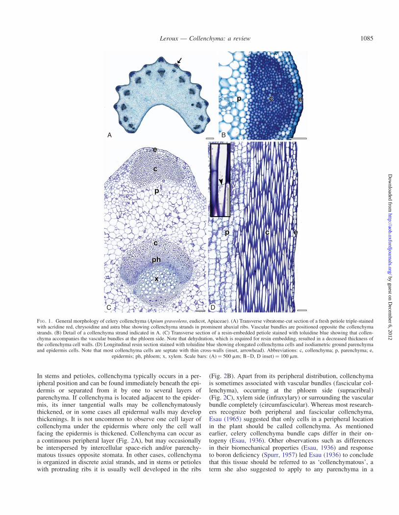
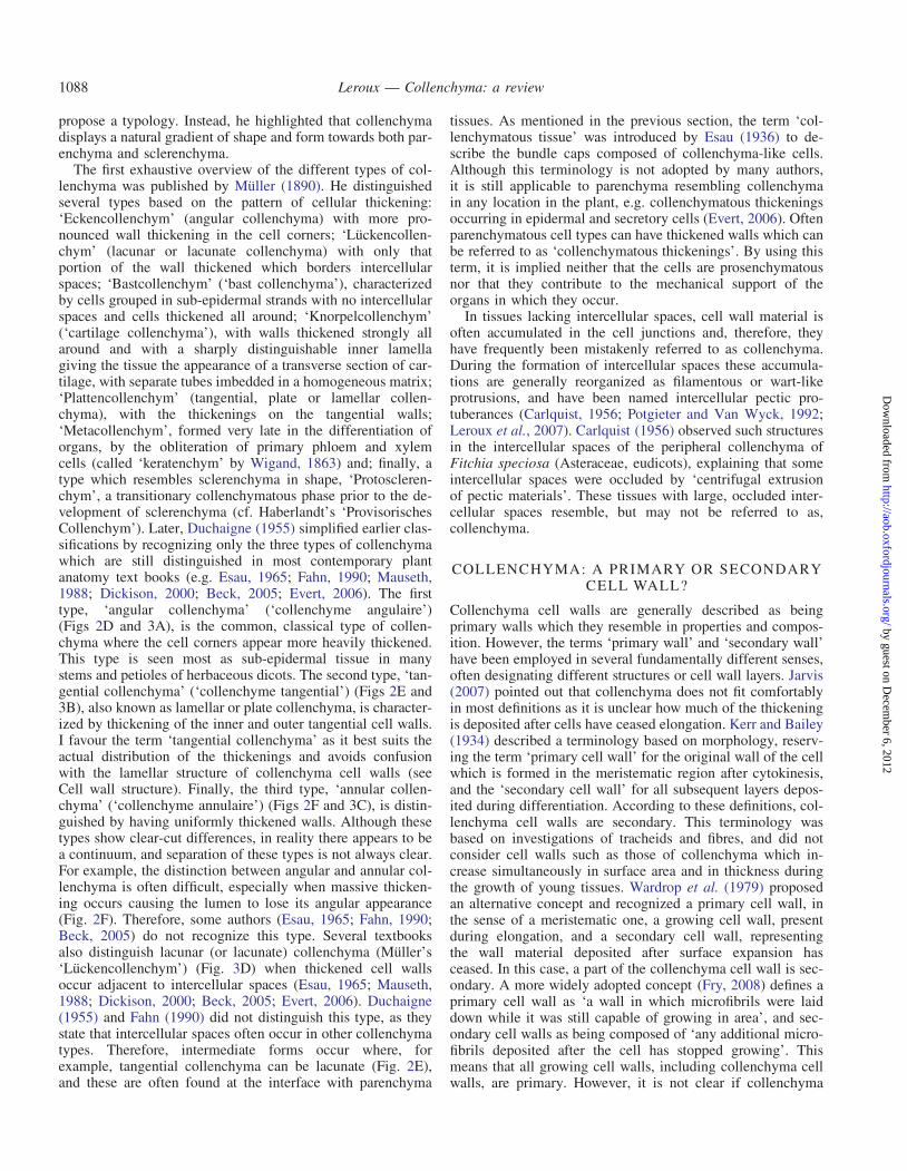
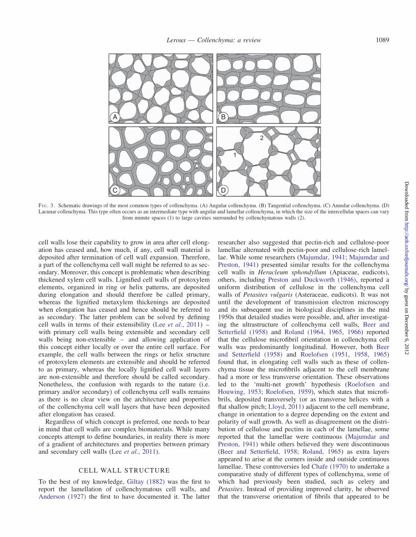
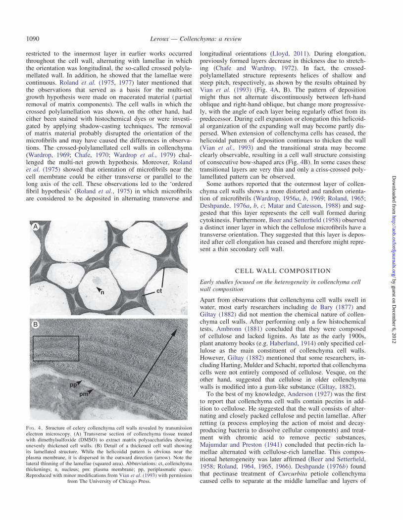
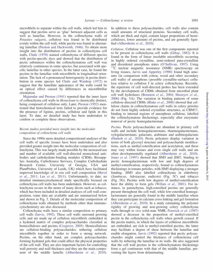
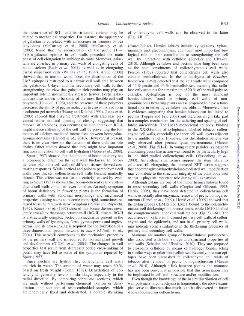
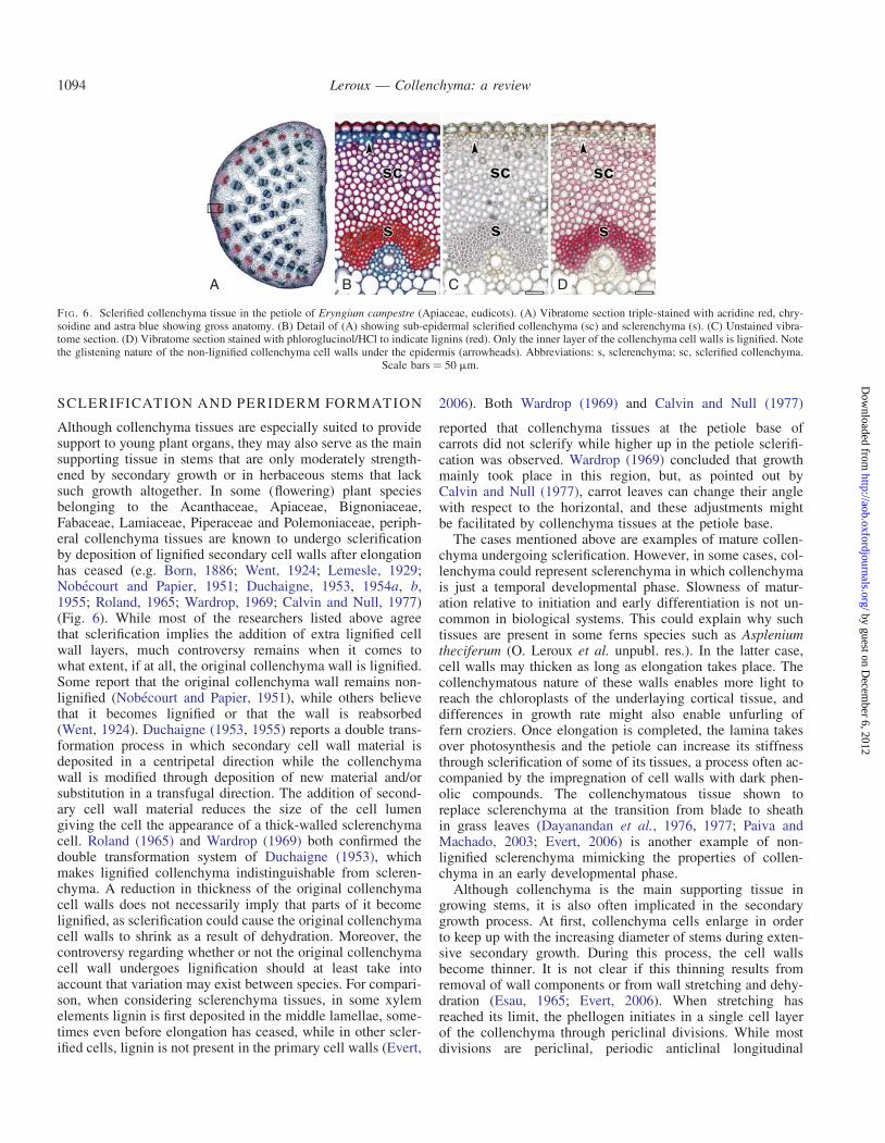
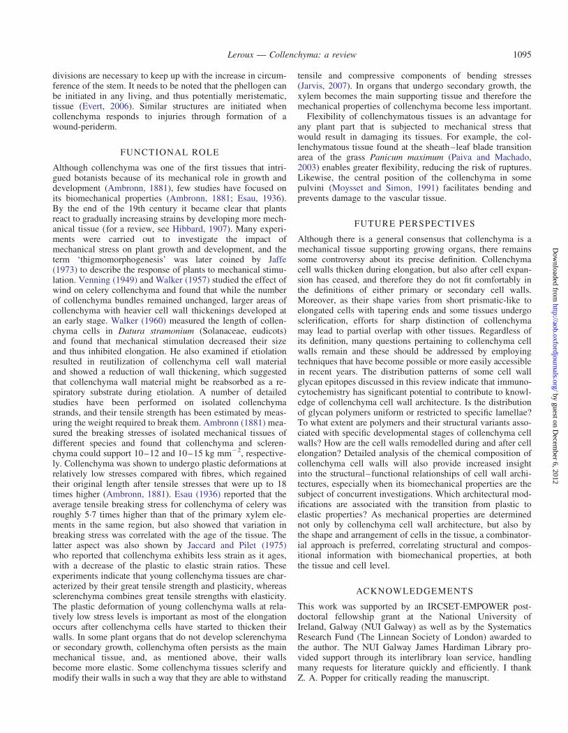
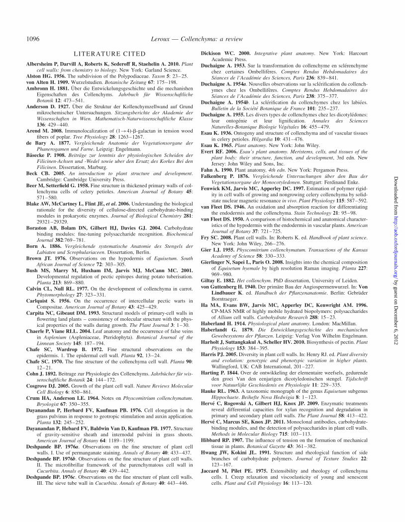
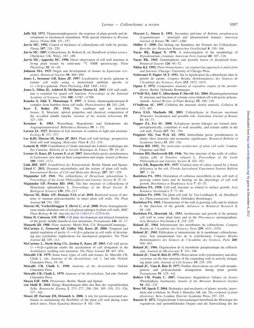
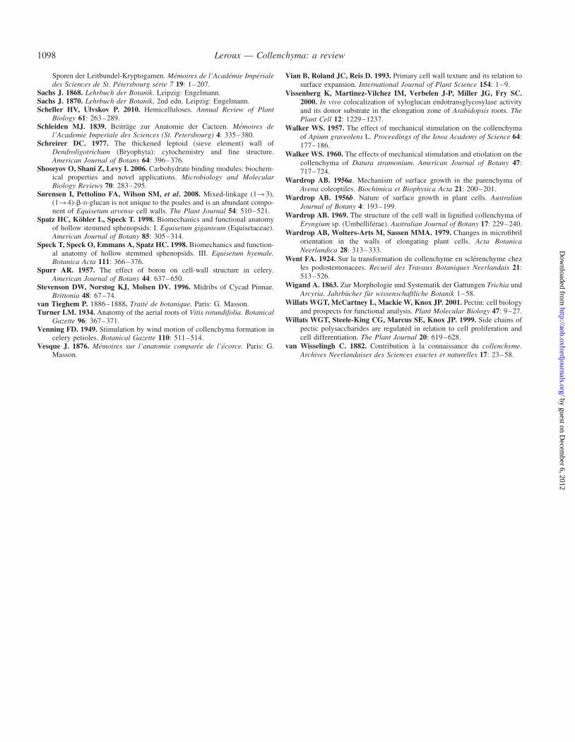