Collaboration of homologous recombination and nonhomologous end-joining factors for the survival and...
12
Collaboration of homologous recombination and nonhomologous end-joining factors for the survival and integrity of mice and cells Chrystelle Couëdel, 1 Kevin D. Mills, 4 Marco Barchi, 1 Lingbo Shen, 2 Adam Olshen, 3 Roger D. Johnson, 1,5 André Nussenzweig, 6 Jeroen Essers, 7 Roland Kanaar, 7 Gloria C. Li, 2 Frederick W. Alt, 4,8 and Maria Jasin 1,9 1 Molecular Biology Program, 2 Department of Medical Physics and Department of Radiation Oncology, and 3 Department of Epidemiology and Biostatistics, Memorial Sloan-Kettering Cancer Center, New York, New York 10021, USA; 4 CBR Institute for Biomedical Research, Boston, Massachusetts 02115, USA; 5 Cancer Center and Department of Cell Biology, University of Massachusetts Medical School, Worcester, Massachusetts 01605, USA; 6 Experimental Immunology Branch, National Cancer Institute, National Institutes of Health, Bethesda, Maryland 20892, USA; 7 Department of Cell Biology and Genetics, and Department of Radiation Oncology, Erasmus MC, 3000 DR Rotterdam, The Netherlands; 8 Howard Hughes Medical Institute, The Children’s Hospital, Boston, Massachusetts 02115, USA Homologous recombination (HR) and nonhomologous end-joining (NHEJ) are mechanistically distinct DNA repair pathways that contribute substantially to double-strand break (DSB) repair in mammalian cells. We have combined mutations in factors from both repair pathways, the HR protein Rad54 and the DNA-end-binding factor Ku80, which has a role in NHEJ. Rad54 −/− Ku80 −/− mice were severely compromised in their survival, such that fewer double mutants were born than expected, and only a small proportion of those born reached adulthood. However, double-mutant mice died at lower frequency from tumors than Ku80 single mutant mice, likely as a result of rapid demise at a young age from other causes. When challenged with an exogenous DNA damaging agent, ionizing radiation, double-mutant mice were exquisitely sensitive to low doses. Tissues and cells from double-mutant mice also showed indications of spontaneous DNA damage. Testes from some Rad54 −/− Ku80 −/− mice displayed enhanced apoptosis and reduced sperm production, and embryonic fibroblasts from Rad54 −/− Ku80 −/− animals accumulated foci of -H2AX, a marker for DSBs. The substantially increased DNA damage response in the double mutants implies a cooperation of the two DSB repair pathways for survival and genomic integrity in the animal. [Keywords: DNA double-strand break; homologous recombination; nonhomologous end-joining; Ku80, Rad54] Supplemental material is available at http://www.genesdev.org. Received April 5, 2004; revised version accepted April 23, 2004. Cells have evolved numerous repair pathways to con- tend with various types of DNA damage. One type of lesion, a DNA double-strand break (DSB), poses a par- ticular threat to genomic integrity, as misrepair of DSBs can lead to gross chromosomal rearrangements and other mutagenic events (van Gent et al. 2001; Mills et al. 2003). DSBs are initiators of genetically programmed processes, such as V(D)J recombination and meiotic ex- change, but they arise more generally in all cells from DNA replication, oxidative metabolism, and other en- dogenous processes. DSBs are also considered to be the relevant lesion for cell lethality after treatment with ion- izing radiation (IR). If left unrepaired or if misrepaired, DSBs can result in permanent cell cycle arrest, cell death, or tumorigenesis (Pierce et al. 2001b). Homologous recombination (HR) has long been known to be the predominant pathway for the repair of DSBs in yeast and bacteria (Paques and Haber 1999; Cro- mie et al. 2001), and direct examination of DSB repair supports a major role for HR in mammalian cells as well (Rouet et al. 1994; Liang et al. 1998). HR uses an unbro- ken, homologous sequence to template repair of the DSB, in a process involving members of the conserved Rad52 epistasis group (Symington 2002; West 2003), as well as other less-conserved factors, such as the tumor suppressor BRCA2 (Jasin 2002). Loss of most HR factors leads to early or mid-embryonic lethality in mice (Pierce 9 Corresponding author. E-MAIL [email protected]; FAX (212) 717-3317. Article and publication are at http://www.genesdev.org/cgi/doi/10.1101/ gad.1209204. GENES & DEVELOPMENT 18:1293–1304 © 2004 by Cold Spring Harbor Laboratory Press ISSN 0890-9369/04; www.genesdev.org 1293
-
Upload
independent -
Category
Documents
-
view
2 -
download
0
Transcript of Collaboration of homologous recombination and nonhomologous end-joining factors for the survival and...

Collaboration of homologousrecombination and nonhomologousend-joining factors for the survivaland integrity of mice and cellsChrystelle Couëdel,1 Kevin D. Mills,4 Marco Barchi,1 Lingbo Shen,2 Adam Olshen,3
Roger D. Johnson,1,5 André Nussenzweig,6 Jeroen Essers,7 Roland Kanaar,7 Gloria C. Li,2
Frederick W. Alt,4,8 and Maria Jasin1,9
1Molecular Biology Program, 2Department of Medical Physics and Department of Radiation Oncology, and 3Department ofEpidemiology and Biostatistics, Memorial Sloan-Kettering Cancer Center, New York, New York 10021, USA; 4CBR Institutefor Biomedical Research, Boston, Massachusetts 02115, USA; 5Cancer Center and Department of Cell Biology, University ofMassachusetts Medical School, Worcester, Massachusetts 01605, USA; 6Experimental Immunology Branch, National CancerInstitute, National Institutes of Health, Bethesda, Maryland 20892, USA; 7Department of Cell Biology and Genetics, andDepartment of Radiation Oncology, Erasmus MC, 3000 DR Rotterdam, The Netherlands; 8Howard Hughes MedicalInstitute, The Children’s Hospital, Boston, Massachusetts 02115, USA
Homologous recombination (HR) and nonhomologous end-joining (NHEJ) are mechanistically distinct DNArepair pathways that contribute substantially to double-strand break (DSB) repair in mammalian cells. Wehave combined mutations in factors from both repair pathways, the HR protein Rad54 and theDNA-end-binding factor Ku80, which has a role in NHEJ. Rad54−/−Ku80−/− mice were severely compromisedin their survival, such that fewer double mutants were born than expected, and only a small proportion ofthose born reached adulthood. However, double-mutant mice died at lower frequency from tumors than Ku80single mutant mice, likely as a result of rapid demise at a young age from other causes. When challenged withan exogenous DNA damaging agent, ionizing radiation, double-mutant mice were exquisitely sensitive to lowdoses. Tissues and cells from double-mutant mice also showed indications of spontaneous DNA damage.Testes from some Rad54−/−Ku80−/− mice displayed enhanced apoptosis and reduced sperm production, andembryonic fibroblasts from Rad54−/−Ku80−/− animals accumulated foci of �-H2AX, a marker for DSBs. Thesubstantially increased DNA damage response in the double mutants implies a cooperation of the two DSBrepair pathways for survival and genomic integrity in the animal.
[Keywords: DNA double-strand break; homologous recombination; nonhomologous end-joining; Ku80, Rad54]
Supplemental material is available at http://www.genesdev.org.
Received April 5, 2004; revised version accepted April 23, 2004.
Cells have evolved numerous repair pathways to con-tend with various types of DNA damage. One type oflesion, a DNA double-strand break (DSB), poses a par-ticular threat to genomic integrity, as misrepair of DSBscan lead to gross chromosomal rearrangements and othermutagenic events (van Gent et al. 2001; Mills et al.2003). DSBs are initiators of genetically programmedprocesses, such as V(D)J recombination and meiotic ex-change, but they arise more generally in all cells fromDNA replication, oxidative metabolism, and other en-dogenous processes. DSBs are also considered to be the
relevant lesion for cell lethality after treatment with ion-izing radiation (IR). If left unrepaired or if misrepaired,DSBs can result in permanent cell cycle arrest, celldeath, or tumorigenesis (Pierce et al. 2001b).
Homologous recombination (HR) has long beenknown to be the predominant pathway for the repair ofDSBs in yeast and bacteria (Paques and Haber 1999; Cro-mie et al. 2001), and direct examination of DSB repairsupports a major role for HR in mammalian cells as well(Rouet et al. 1994; Liang et al. 1998). HR uses an unbro-ken, homologous sequence to template repair of theDSB, in a process involving members of the conservedRad52 epistasis group (Symington 2002; West 2003), aswell as other less-conserved factors, such as the tumorsuppressor BRCA2 (Jasin 2002). Loss of most HR factorsleads to early or mid-embryonic lethality in mice (Pierce
9Corresponding author.E-MAIL [email protected]; FAX (212) 717-3317.Article and publication are at http://www.genesdev.org/cgi/doi/10.1101/gad.1209204.
GENES & DEVELOPMENT 18:1293–1304 © 2004 by Cold Spring Harbor Laboratory Press ISSN 0890-9369/04; www.genesdev.org 1293
et al. 2001b; van Gent et al. 2001), suggesting an essen-tial role for HR in development, presumably to repairspontaneously arising DNA damage. An exception is theRad54 protein, for which the mouse disruption shows noovert phenotype (Essers et al. 1997, 2000), likely becauseDSB-promoted HR is only mildly compromised inRad54−/− cells (Dronkert et al. 2000).
A second prominent pathway for DSB repair in mam-malian cells is nonhomologous end-joining (NHEJ), inwhich broken ends are healed without the requirementfor significant sequence homology (Pierce et al. 2001b;van Gent et al. 2001). NHEJ factors have been identifiedthat play important roles in DSB repair after IR damageand during V(D)J recombination, including the DNA-end-binding Ku70/Ku80 heterodimer, the protein kinaseDNA-PKcs, and the XRCC4/DNA ligase IV complex(Mills et al. 2003). Loss of NHEJ factors results in lateembryonic death for some factors (i.e., XRCC4 and DNAligase IV), but not for others (i.e., Ku80, Ku70, and DNA-PKcs; Pierce et al. 2001b; van Gent et al. 2001; Mills etal. 2003), although loss of Ku80 or Ku70 leads to a num-ber of phenotypes, including small size and early death,as well as immunodeficiency (Nussenzweig et al. 1996;Gu et al. 1997; Li et al. 1998). Thus, either NHEJ does notplay as important a role in development as HR, or otherNHEJ factors with key roles in development remain tobe identified.
DSBs that serve as initiators of genetically pro-grammed processes are directed into either NHEJ or HR;for example, DSBs introduced by the RAG proteins dur-ing V(D)J recombination are repaired by the NHEJ path-way (Mills et al. 2003), whereas those introduced by theSpo11 protein during meiosis are repaired by the HRpathway (Keeney 2001). Less is known about the relativecontribution of NHEJ and HR to the repair of DNA dam-age that is not genetically programmed, although onefactor that influences repair pathway choice is cell cyclephase. NHEJ is preferred during G1/early S phase,whereas HR is prominent in late S/G2 phase (Takata etal. 1998; Rothkamm et al. 2003). However, these prefer-ences do not appear to be absolute (Rothkamm et al.2003), and there is evidence that both pathways haveaccess to the same DSB. For example, HR and NHEJ canbe coupled for the repair of a single DSB (Richardson andJasin 2000). In addition, in NHEJ mutants, HR repair of aDSB is increased compared with wild-type cells, suggest-ing that HR can compensate when NHEJ is impaired(Pierce et al. 2001a; Allen et al. 2002; Delacote et al.2002). This increase is particularly striking in Ku mutantcells compared with other NHEJ mutants (Pierce et al.2001a).
To begin to understand the cooperation between HRand NHEJ in the mouse, we combined a mild HR muta-tion, that is, loss of Rad54, with a moderate NHEJ mu-tation, that is, loss of Ku80. We find that the survival ofRad54−/−Ku80−/− mice is severely compromised, evenwhen compared with Ku80−/− mice, revealing a vital rolefor Rad54 in the support of Ku80−/− mouse viability. Al-though Rad54−/− mice are not sensitive to IR (Essers etal. 2000), Rad54−/−Ku80−/− mice are exquisitely sensi-
tive, much more so than Ku80−/− mice. Consistent witha collaborative role for Rad54 and Ku in the repair ofspontaneously arising DNA damage, embryonic fibro-blasts from double-mutant animals accumulate largenumbers of �-H2AX foci, a marker for DSBs (Rogakou etal. 1998, 1999), as compared with single mutants. Apop-tosis is also elevated in testes, compromising sperm pro-duction. These results imply that NHEJ and HR factorscooperate to repair DNA damage in mammalian cells.Moreover, these findings uncover a key role for Rad54 inthe animal, suggesting that even a mild repair deficiencycan have profound effects in the context of other muta-tions.
Results
Rad54 mutation impairs the embryonic viabilityof Ku80−/− mice
To investigate the effect of combined Rad54 andKu80 mutation in mice, we set up crosses to generateRad54−/−Ku80−/− double-mutant mice. Because Rad54−/−
mice are fully fertile and viable (Essers et al. 1997), weused both Rad54−/− and Rad54+/− mice in the crosses;however, due to the small litter size of Ku80−/− mutantmice (Nussenzweig et al. 1996), we crossed only Ku80+/−
mice. From our initial crosses, we did not obtain anyRad54−/−Ku80−/− mice from litters genotyped at wean-ing. Therefore, we monitored animals starting at birth todetermine whether double mutants would be repre-sented among newborn animals. Rad54−/−Ku80−/− ani-mals were recovered among the newborns, although at asignificantly lower frequency than expected from theMendelian ratios (Table 1A). (Data from different crossesare available in Supplementary Table 1.) Some of thenewborns were dead at birth or died soon thereafter, butthe majority was recovered alive (see below). The recov-ery of Rad54+/−Ku80−/− newborns was also lower thanexpected from the Mendelian ratios, although to a lesserextent than Rad54−/−Ku80−/− newborns (81.7% and60.6%, respectively), suggesting that heterozygosity atthe Rad54 locus may have a small effect on the survivalof Ku80−/− mice to term. Ku80−/− newborns were ob-tained at a somewhat lower frequency than expected(86.2%), although this decrease was not significant. Im-portantly, the recovery of Rad54−/−Ku80−/− newborns, inaddition to being lower than expected among all prog-eny, was also significantly lower than either the Ku80−/−
or Rad54+/−Ku80−/− newborns (p < 0.01 for each; Table1A). These results imply that combined mutation of theKu80 and Rad54 DSB repair genes compromises the re-covery of mice at birth, although only partially.
Mice with NHEJ deficiency have increased cell deathin the developing nervous system, and a correlation hasbeen made between the extent of neuronal cell death andembryonic survival (Gu et al. 2000). To investigate thelethality observed for Rad54−/−Ku80−/− mutants, brains,and spinal cords from embryonic day 13.5 (E13.5) andE18.5 embryos were analyzed for cell death using a DNAfragmentation assay, and results compared with litter-
Couëdel et al.
1294 GENES & DEVELOPMENT

mate controls. TUNEL-positive nuclei were occasionallydetected in Rad54−/− embryos (Supplementary Fig. 1A,E;data not shown), although the number was only margin-ally increased—that is, approximately twofold for E13.5embryos—compared with wild-type embryos (data notshown). In contrast, in Ku80−/− E13.5 embryos, apoptoticcells were increased two orders of magnitude or more(Supplementary Fig. 1B,F; see also Gu et al. 2000). Wealso observed increased apoptosis in the brain of Ku80−/−
E18.5 embryos, although the level of apoptosis was notas dramatically increased as in E13.5 embryos (Supple-mentary Fig. 1J; data not shown). All Rad54−/−Ku80−/−
embryos at both E13.5 and E18.5 also had a large numberof TUNEL-positive nuclei in the developing brain andspinal cord (Supplementary Fig. 1D,H,L), similar to theKu80−/− embryos. However, variability between embryosprecluded us from determining whether the level ofapoptosis in the double mutant was significantly in-creased compared with the single mutant. Nevertheless,the lack of a substantially increased level of apoptosis inthe Rad54−/−Ku80−/− embryos is consistent with thecompletion of embryogenesis for the majority of embryos.
Rad54 mutation reduces the postnatal viabilityof Ku80−/− mice
Although combined mutation of the Ku80 and Rad54DSB repair genes compromised the recovery of mice at
birth, a substantial portion of double-mutant mice wasrecovered. To determine whether we could obtain adultanimals, we therefore expanded the number of crossesand allowed the dams to nurse their progeny until wean-ing at 3 wk of age, at which point the animals weregenotyped (Table 1B). Ku80 mutation alone led to a sig-nificant impairment in the survival of mice, as has beendescribed previously (Nussenzweig et al. 1996), such thatonly 27.3% of Ku80−/− mice expected from the Mendel-ian ratios were alive at weaning. Similar results wereobtained with the Rad54+/−Ku80−/− mice, with only26.5% of the expected number of mice recovered. How-ever, Rad54−/−Ku80−/− mice were even more severelycompromised in their survival, with only 7.3% of theexpected number of mice alive at weaning. The differ-ence in recovery of viable double-mutant mice washighly significant compared with either Ku80−/− orRad54+/−Ku80−/− mice (p < 0.001 for each; Table 1B), in-dicating that Rad54 mutation compromises the postna-tal viability of Ku80−/− mice.
Ku80−/− newborns are significantly smaller than wild-type and heterozygote littermates (Nussenzweig et al.1996). Similarly, we found that Rad54+/−Ku80−/− andRad54−/−Ku80−/− newborns are also dwarfs, not notice-ably different in size from their Ku80−/− littermates (seebelow). As a consequence, Ku80−/− mutants of anyRad54 genotype could be easily identified among thenewborns and placed with foster mothers to prevent pos-
Table 1. Compromised viability of Rad54−/−Ku80−/− mice
Genotype Expected Observed
Recovery
(p-value vs Rad54−/−Ku80−/−)
A) Birth (n = 2054)Ku80−/− 87 75 86.2% p = 0.20
(p = 0.008)Rad54+/−Ku80−/− 257 210 81.7% p = 0.001
(p = 0.003)Rad54−/−Ku80−/− 170 103 60.6% p < 0.001
B) Maternal Nursing (n = 854)Ku80−/− 22 6 27.3% p < 0.001
(p < 0.001)Rad54+/−Ku80−/− 68 18 26.5% p < 0.001
(p < 0.001)Rad54−/−Ku80−/− 123 9 7.3% p < 0.001
C) Fostering (n = 1813)Ku80−/− 77 33 42.9% p < 0.001
(p < 0.001)Rad54+/−Ku80−/− 227 90 39.6% p < 0.001
(p < 0.001)Rad54−/−Ku80−/− 150 22 14.7% p < 0.001
Mouse genotypes obtained from three types of crosses are summarized. (See Supplementary Table 1 for detailed counts from each typeof cross.) Recovery is the observed number of mice at birth (A) or 21 d of age at weaning from the birth (B) or foster mother (C), relativeto the number of mice expected from the Mendelian ratios. The p-value is indicated. The recovery of Ku80−/− or Rad54+/−Ku80−/− micewas also compared with the recovery of Rad54−/−Ku80−/− mice, and the p-value for this is indicated in parentheses. For Ku80, bothparents were heterozygous for the mutation, whereas for Rad54, parents were heterozygous, homozygous, or heterozygous andhomozygous. Because the Rad54 mutant allele was overrepresented in the parents relative to the wild-type allele, the expected numberof Rad54+/+ animals is low compared with the other genotypes.(A) Total animals recovered at birth, whether they were alive or stillborn.(B) Total animals alive at weaning, when nursed by their birth mother.(C) Total animals alive at weaning, when nursed by foster mothers.
Rad54 and Ku80 support genomic integrity
GENES & DEVELOPMENT 1295
sible death due to competition for resources with theirlarger littermates. For each mutant, we found that fos-tering improved survival to weaning approximatelytwofold, although the expected number of mice wasstill not obtained (Table 1C). Again, the survival ofRad54−/−Ku80−/− mice was still significantly compro-mised relative to either Rad54+/−Ku80−/− or Ku80−/−
mice, such that only about one-third as many doublemutants were obtained at weaning (14.7% relativeto 39.6% or 42.9%, respectively; p < 0.001 for each).These results indicate the reduced fitness of theRad54−/−Ku80−/− mice, and thus a contribution of bothDSB repair genes to survival of the animal.
Rad54 mutation shortens the lifespan of Ku80−/− mice
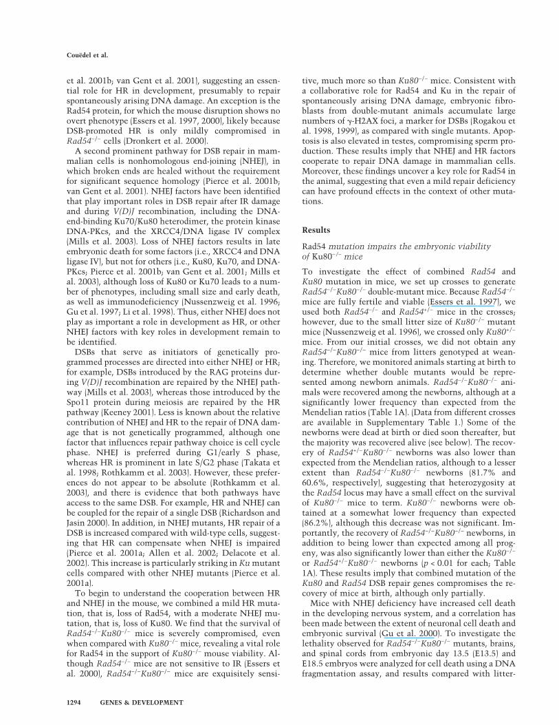
The difference in survival of Rad54−/−Ku80−/− mice bythe time of weaning relative to Ku80−/− mice led us toexamine their postnatal survival in more detail. Fosteredanimals were monitored daily so that dead animals couldbe genotyped, including dead newborns (Fig. 1). Perinatalmortality was observed for all three Ku80−/− genotypes(Fig. 1A). Approximately 10% of the Ku80−/− animals and20% of both the Rad54+/−Ku80−/− and Rad54−/−Ku80−/−
animals that were born were either stillborn or died dur-ing the first day after birth (day 0). Despite fostering,50% of the Ku80−/− and Rad54+/−Ku80−/− mutants thatwere born were dead within 3 d after birth, at whichpoint the animals’ viability stabilized until weaning at 3wk of age, such that at weaning, 44% and 43%, respec-tively, of animals that were obtained at birth were stillalive. However, Rad54−/−Ku80−/− mice died more pre-cipitously in the first week or so after birth. The cause ofdeath is unknown, but it was observed that someRad54−/−Ku80−/− mice that died perinatally had diffi-culty breathing and had not fully inflated their lungs(data not shown). Double-mutant mice continued to dieuntil weaning, although at a slower rate than in the daysimmediately after birth. As a result, only 21% of thedouble-mutant mice were still alive at weaning.
After weaning, we continued to observe the fosteredmice for survival (Fig. 1B). In contrast to Rad54−/− mice,all three Ku80−/− genotypes experienced a decline in thenumber of surviving animals in the first few weeks afterweaning, implying that separation from the foster motherimpaired survival. The decline in the number of surviv-ing animals was again more precipitous for the double-mutant mice. Thus, only 33% of the Rad54−/−Ku80−/−
mice that were alive at weaning were still alive at 8 wkof age, whereas for either the Ku80−/− or Rad54+/−Ku80−/−
mice >75% of the mice were still alive at this age. Afterthis point, the death rate decreased, such that by 7 mo ofage, only a fraction of the mice that were weaned werestill alive (∼30% for Ku80−/− and Rad54+/−Ku80−/−; 7%for Rad54−/−Ku80−/−). Considering overall survival sincebirth, 14% and 12% of the Ku80−/− and Rad54+/−Ku80−/−
mice, respectively, were still alive at 7 mo of age, in contrastto 1.5% of the Rad54−/−Ku80−/− mice.
As Ku80−/− mice are smaller than their wild-type coun-terparts, the increased mortality of Rad54−/−Ku80−/−
mice could be related to increased frailness due to poor
weight gain. To examine this, male and femalemice were weighed once a week over a 6-mo period,starting at 10 d of age (Fig. 2A,B). Like Ku80−/−
mice, Rad54+/−Ku80−/− and Rad54−/−Ku80−/− mice weresignificantly smaller than control mice. Rad54 muta-tion, however, had little effect on the size of Ku80−/−
mice, as the double mutants weighed, on average, only10% less than the single mutants. After an initial growthspurt during the first 45 d or so of life, the weight gain ofall three Ku80 genotypes became shallower and then lev-eled off at ∼15 g. This contrasts with Rad54−/− and con-trol mice, which had a steeper initial growth spurt andthen continued to gain weight over the course of theexperiment.
To gain further insight into the underlying pathologyleading to the death of the mutant animals, several mice
Figure 1. Impaired postnatal survival of Rad54−/−Ku80−/−
mice. The total number of mice for each genotype (n) is indi-cated. (A) Survival of mice during fostering. Curves begin atbirth. Total numbers at birth include animals that were still-born. Percentage of mice still alive at weaning (i.e., 21 d of age)is indicated at right. (B) Survival of mice after weaning fromfoster mothers. Curves begin with mice that were alive at wean-ing (i.e., 21 d of age). As a reference, a survival curve of Rad54−/−
mice that were nursed by their birth mother is depicted. Per-centage of weaned mice still alive at 3 or 7 mo of age is indi-cated; in parentheses, the percentage of mice still alive at 3 or 7mo relative to the total number of mice recovered at birth is alsoindicated.
Couëdel et al.
1296 GENES & DEVELOPMENT

from each genotype were necropsied. Gross observationindicated lymphoid deficiency in Ku80−/− mice of eachRad54 genotype (data not shown), as would be expectedby Ku deficiency. Mice that died from ∼10 d to 3 mo ofage generally showed no other significant lesion (Table2). Mice older than 3 mo, however, were often found tohave infections, presumably as a result of impaired im-mune system function, and in addition, lymphomas, al-though not other tumors. Neoplastic lymphocytes werefound in the thymus, with extension to pulmonary in-terstitium, epicardium of the heart, and other tissues ina number of mice. A somewhat lower percentage of lym-phomas in mice older than 3 mo of age was foundin Rad54−/−Ku80−/− mice compared with Ku80−/− orRad54+/−Ku80−/− mice (Table 2), although the differencewas not significant (p = 0.6 for each). However, consid-ering the lymphoma frequency in the entire cohort ofmice since birth, Rad54−/−Ku80−/− were six- to sevenfoldless likely to die from lymphoma than were Ku80−/− andRad54+/−Ku80−/− mice (Table 2; p < 0.01, see Supplemen-tary Table 2). This presumably highlights the reduced
fitness of the Rad54−/−Ku80−/− mice, which leads to pre-mature death from other causes, rather than an inabilityto undergo neoplastic transformation. Consistent withthis, the mean age of death from lymphoma for all threeKu80 genotypes was similar, about 5 mo of age (Table 2).
Extreme radiation sensitivity of Rad54−/−Ku80−/− mice
Because both Rad54 and Ku80 have roles in DSB repair,the compromised survival of Rad54−/−Ku80−/− mice maybe related to or the result of impaired DNA repair. Todetermine whether disruption of both genes affects thesensitivity of mice exposed to exogenous DNA damagingagents, we tested whether Rad54 mutation heightensthe previously described radiation sensitivity of Ku80−/−
mice (Nussenzweig et al. 1997). Rad54−/− Ku80−/− micewere exposed to ionizing radiation (IR) and compared fortheir survival to single mutant and control mice. Two- tofour-month-old animals were used in this analysis, asthere is little mortality during this time for any of the geno-types (Fig. 1B). Consistent with previous observations,all Rad54−/− mice survived 100 or 200 cGy of radiation(Essers et al. 2000), as did wild-type or Rad54+/−Ku80+/−
mice (Fig. 3). All Ku80−/− and Rad54+/−Ku80−/− mice alsosurvived 100 cGy (Fig. 3A). In contrast, Rad54−/−Ku80−/−
mice started to die 10 d after exposure to this dose and allwere dead within 17 d of irradiation (Fig. 3A). At thehigher dose of 200 cGy, death of Rad54−/−Ku80−/− miceoccurred earlier, starting 2 d after irradiation, and allwere dead within 8 d (Fig. 3B). At this dose, Ku80−/− andRad54+/−Ku80−/− mice presented an intermediate sensi-tivity, compared with the severely affected double mu-tants and the unaffected Rad54−/− and control mice(Fig. 3B). Therefore, Rad54−/−Ku80−/− mice are extreme-ly radiosensitive, more so than Ku80−/− mice, implyingthat both genes contribute to DNA repair in the adultanimal.
Accumulation of �-H2AX foci in Rad54−/−Ku80−/−
embryonic fibroblasts
Given the hypersensitivity of Rad54−/−Ku80−/− mice toIR, we surmised that the cells of the double-mutant ani-mals could be compromised in their inability to repairspontaneously arising DNA damage. We determinedwhether spontaneous damage could be detected inRad54−/−Ku80−/− cells in culture, using mouse embry-onic fibroblasts (MEFs) obtained from E13.5 embryos.Consistent with previous results (Nussenzweig et al.1996), proliferation of Ku80−/− cells was severely im-paired, as was BrdU incorporation (Supplementary Fig.2). Rad54+/−Ku80−/− and Rad54−/−Ku80−/− cells exhibiteda similar growth defect with minimal expansion over the10-d culture period. These cells showed no sign of el-evated apoptosis, however, as there was no increase inTUNEL-positive nuclei for Ku80−/− or Rad54−/−Ku80−/−
cells compared with control cells (data not shown; also,see below), suggesting that the proliferation defect was
Figure 2. Growth retardation of Ku80−/− mice, regardless ofRad54 genotype. Males (A) and females (B) were weighed once aweek starting 10 d after birth. Average weights of control mice(i.e., wild-type mice or mice heterozygote for either Rad54or Ku80 mutation, or both) and Rad54−/−, Ku80−/−,Rad54+/−Ku80−/−, or Rad54−/−Ku80−/− mice are plotted againstdays after birth. The total number of mice for each genotype (n)is indicated. Because some mice died or were sacrificed duringthe observation period for other analyses, the number of miceremaining at the end of the study is reduced and is indicated inparentheses. Gray areas indicate weight variations based onstandard deviations.
Rad54 and Ku80 support genomic integrity
GENES & DEVELOPMENT 1297

due to cell cycle arrest and/or senescence rather than celldeath.
One of the first cellular responses to DSBs is phos-phorylation of histone H2AX at sites of damage, which isvisible as foci of �-H2AX (i.e., phosphorylated H2AX;Rogakou et al. 1998, 1999). MEFs were subjected to in-direct immunofluorescence to detect �-H2AX foci andsubsequently counterstained with DAPI to visualize nu-clei. The majority of wild-type cells had no detectablefoci; similarly, Rad54−/− or Rad54−/−Ku80+/− cells withspontaneous foci were also infrequent (Fig. 4A; data notshown). In contrast, many Ku80−/− cells and nearly allRad54−/−Ku80−/− cells were positive for �-H2AX foci(Fig. 4A).
These results were quantified by counting the numberof cells with �-H2AX foci, and, in those cells, the num-ber of foci. Spontaneously arising foci were detectable in30% of Rad54−/− cells (Fig. 4B), and most of these posi-tive cells had only a small number of foci (i.e., 1–5 foci;Fig. 4C). Wild-type cells showed a very similar number ofcells with foci (27%) and a similar distribution, withpositive cells exhibiting only a few foci (Mills et al.2004). More than twice as many Ku80−/− cells were
found to have foci (i.e., 69%; Fig. 4B). Most of theKu80−/− cells with foci had a small number of foci, al-though some cells had a moderate number (i.e., 6–10foci) or even a large number of foci (>10 foci; Fig. 4C).Strikingly, however, >90% of the Rad54−/−Ku80−/− cellshad foci (Fig. 4B), and of these, the majority had a largenumber of foci (Fig. 4C). The increased number of cellswith �-H2AX foci was not the result of apoptosis, be-cause TUNEL-positive cells were rare for all genotypes(data not shown), as were cleaved caspase-3-positive cells(Fig. 4D). These results imply an elevated level of DNAdamage when Ku80 is disrupted, but a dramatically el-evated level of DNA damage when both Ku80 and Rad54are disrupted.
Apoptosis in the testis of Rad54−/−Ku80−/− mice
The high level of spontaneous �-H2AX foci inRad54−/−Ku80−/− MEFs suggested that tissues composedof highly proliferative cells in adult animals would havecellular defects due to the accumulation of unrepairedDNA damage. Testis represents a tissue with a high pro-liferative capacity, in particular, the spermatogonia, the
Table 2. Pathological assessment of mutant mice
Genotype Age (months) Viabilitya
Pathology (number of mice) Lymphoma
Complete necropsytotal
No significantlesionb Infection
Lymphoma(mean age)c >3 mod Overalle
Ku80−/− �3 68% 9 8 1 0 81% 20%>3 32% 13 1 5 8 (24)
Rad54+/−Ku80−/− �3 69% 4 4 0 0 62% 19%>3 31% 8 0 3 5 (22)
Rad54−/−Ku80−/− �3 93% 10 9 1 0 44% 3%>3 7% 9 1 7 4 (23.5)
aPercent viability at the indicated age is obtained from survival curves (Fig. 1).bLymphoid deficiency is not included in this tabulation. Excluding one mouse, lymphoid deficiency was observed in all animals inwhich it could be evaluated. In some cases, autolysis precluded definitive evaluation of lymphoid deficiency, as did lymphoma,because infiltration by neoplastic lymphocytes may have obliterated the normal architecture of the organs.cMean age of death from lymphoma is indicated in weeks in parentheses.dFrom necropsied mice older than 3 mo.eFrom necropsied mice relative to the entire cohort since birth. See Supplementary Table 2 for calculation.
Figure 3. Ionizing radiation sensitivity ofRad54−/−Ku80−/− mice. (A) Survival after 100 cGy�-irradiation. Each curve represents six mice. Con-trols are two mice each Rad54−/−, Rad54+/−Ku80+/−,and wild-type. (B) Survival after 200 cGy �-irradia-tion. Each curve represents eight mice, except thecontrols, which are the same as in A.
Couëdel et al.
1298 GENES & DEVELOPMENT

premeiotic germ cells. Both Rad54−/− and Ku80−/− malesare fertile (Nussenzweig et al. 1996; Essers et al. 1997),and Ku80−/− mice have been reported to have normallevels of apoptosis in the testis (Espejel et al. 2002).Therefore, we tested whether cellular defects could bedetected in the testis of double-mutant animals, usinghistological analysis of 2–4-month-old animals.
As expected, testes from Ku80−/− and Rad54−/−Ku80+/−
animals showed normal testicular morphology and cel-lularity on gross observation, and the epididymidesshowed abundant sperm, similar to Rad54+/−Ku80+/−
control mice (Fig. 5A, panels i–v; data not shown). A fewseminiferous tubules had apoptotic cells, as detected byTUNEL staining, which were generally located near thebase of the tubule (arrowhead, Fig. 5A, panel iii). Usuallythere were just one or a few apoptotic cells per tubule, ifany at all (Fig. 5B).
In contrast, results from Rad54−/−Ku80−/− mice werevariable. Although the testes from one animal appearednormal (Fig. 5B, Rad54−/−Ku80−/− #1), testes from twoother double-mutant animals were quite aberrant. In oneanimal, there was a pronounced lack of cellularity insome tubules (Fig. 5A, panel vi, arrowhead). Other tu-bules in this animal were hypocellular, and these had anumber of apoptotic cells both at the base of the tubuleand more luminal (Fig. 5A, panels vi,vii;Rad54−/−Ku80−/− #2), so that the overall number of tu-bules with apoptotic cells was increased (Fig. 5B). Notsurprisingly, the sperm concentration in the epididymis
was substantially reduced (Fig. 5A, panel viii). In anotheranimal, there was only a single testis. The testis in thisanimal was more cellularized, but large numbers of ap-optotic cells were apparent in most tubules (Fig. 5A,panel ix,x; Rad54−/−Ku80−/− #3; Fig. 5B). The apoptoticcells were also located at the base of the tubule and moreluminal, and closer examination suggested that apopto-tic cells included spermatogonia, spermatocytes, andspermatids. Again, the sperm concentration in the epi-didymis was substantially reduced (Fig. 5A, panel xi).
A summary of the apoptotic cell counts for threeanimals for each genotype shows that ∼10% of seminif-erous tubules from the Rad54−/−Ku80+/−, Ku80−/−, andRad54+/−Ku80−/− animals have two or more apoptoticcells, similar to Rad54+/−Ku80+/− mice (Fig. 5C). For theRad54−/−Ku80−/− mice, the range of apoptotic cells pertubule is more variable, but can reach 90% (Fig. 5C). Themean number of tubules with two or more apoptoticcells in the double mutant is greater than fourfold higherthan the other genotypes, although the standard devia-tion is quite large, given the variability of phenotypesseen between the individual mice.
Discussion
In this study, we investigated the collaboration of HRand NHEJ components on survival of the mouse and onthe DNA damage response. For this, we combined a mildHR mutation, Rad54, with the moderately affected
Figure 4. Accumulation of �-H2AX foci in Rad54−/−Ku80−/− cells. (A) Immunofluorescence detection of �-H2AX foci in passage-2MEFs of indicated genotypes. (B) Fraction of cells with �-H2AX foci (light-green bars) or no foci (white bars) is shown for each cell lineof indicated genotype. Rad54−/−Ku80+/x refers to combined results from Rad54−/−Ku80+/+ and Rad54−/−Ku80+/− MEFs. Counts representaverages from two independent experiments. (C) Fraction of cells exhibiting 0 (white bars), 1–5 (gray bars), 6–10 (light-green bars),or >10 (dark-green bars) �-H2AX foci per nucleus. Counts represent averages from two independent experiments. (D) Immunu-fluorescence detection of cleaved caspase-3 (�Caspase-3) in UV-irradiated wild-type or p53−/− MEFs and in untreated wild-type,Rad54−/−Ku80+/−, Ku80−/x, or Rad54−/−Ku80−/− MEFs. Fraction of �Caspase-3-positive cells are shown and represent averages from twoindependent experiments.
Rad54 and Ku80 support genomic integrity
GENES & DEVELOPMENT 1299

NHEJ mutant Ku80. We find that Rad54−/−Ku80−/− miceare significantly compromised for their survival, suchthat by 31 wk of age, ∼10-fold fewer double-mutant miceare alive than are Ku80 single-mutant mice. Loss of vi-ability occurs at several stages, that is, during embryo-genesis, immediately after birth, and after weaning. Thiscontrasts with combined Rad54 and DNA-PKcs muta-tion, as Rad54−/−scid mice are not significantly compro-mised for viability (Essers et al. 2000; J. Essers and R.Kanaar, unpubl.). Rad54−/−Ku80−/− mice are only mar-ginally smaller than Ku80 mutant mice and die mostlyfrom unspecified causes. Lymphoma frequency is re-duced for Rad54−/−Ku80−/− mice compared with Ku80−/−
mice, presumably as a result of the earlier death of thedouble-mutant animals.
Our results imply that DNA damage repair is syner-gistically affected when both HR and NHEJ are impaired.Consistent with this, Rad54−/−Ku80−/− mice are exquis-
itely sensitive to low doses of IR. Moreover, the DNAdamage response is activated in double mutants in theabsence of exogenous DNA damage: Spontaneous�-H2AX foci are significantly elevated in embryonic fi-broblasts and apoptosis is increased in testes in two ofthe three examined double-mutant animals. In the testis,neither Rad54−/− mice nor Ku80−/− mice had elevatedapoptosis compared with wild-type mice, whereas in theRad54−/−Ku80−/− mouse with the most extreme pheno-type, significant numbers of apoptotic cells were foundin nearly every seminiferous tubule. That testicular ap-optosis is not increased in all double-mutant animals,suggests incomplete penetrance or that a genetic modi-fier may be present (see Karanjawala et al. 2003). How-ever, apoptosis was not markedly increased in the devel-oping brain of Rad54−/−Ku80−/− embryos. The high levelof apoptosis in Ku80−/− animals may mask a contribu-tion of Rad54, or alternatively, Rad54 may not have a
Figure 5. Increased cell death in Rad54−/−Ku80−/− testes. (A) Testis and epididymis sections stained with TUNEL/PAS and DAPI,respectively. In Rad54+/−Ku80+/− (panel i), Ku80−/− (panels ii,iii), and Rad54−/−Ku80+/− (panel v) mice, TUNEL-positive testicular cellsare rare, but when present, are usually located at the base of the seminiferous tubule (e.g., arrowhead in panel iii). Similar results wereobtained for one Rad54−/−Ku80−/− mouse (mouse #1; data not shown). In two other Rad54−/−Ku80−/− mice, mice #2 (panels vi,vii) and#3 (panels ix,x), TUNEL-positive cells are abundant. In mouse #2, tubules with reduced cellularity and germ-cell depletion (arrowheadin panel vi) are evident. In mouse #3, cellularity of tubules is not as visibly reduced, but TUNEL-positive cells are even more abundant,including round spermatids (arrowhead in panel x). Mature sperm in the epididymis is abundant in all controls, including Ku80−/− mice(panel iv) and Rad54−/−Ku80−/− mouse #1 (data not shown), whereas it is significantly reduced for both Rad54−/−Ku80−/− mice #2 (panelviii) and #3 (panel xi). Magnification: panels i,ii,v,vi,ix, 100×; panels iii,vii,x, 400×; panels iv,viii,xi, 200×. (B) Percentages of tubulesexhibiting 0 or 1 (white bars), 2–4 (gray bars), 5–10 (light-green bars), or >10 (dark-green bars) TUNEL-positive cells. Counts representaverages from three mice, except for Rad54−/−Ku80−/− mice, which are individually depicted. (C) Percentages of tubules exhibiting twoor more TUNEL-positive cells. Counts represent averages from three mice.
Couëdel et al.
1300 GENES & DEVELOPMENT

significant role in the developing brain, unlike other HRfactors (Deans et al. 2000). Our results, therefore, dem-onstrate that different tissues respond differentially toloss of the Rad54 and Ku80 DNA repair factors.
Many of the phenotypes we observed forRad54−/−Ku80−/− mice are p53 dependent (C. Couëdeland M. Jasin, unpubl.). p53 heterozygosity substantiallyrestores the viability of Rad54−/−Ku80−/− mice, decreasesthe neuronal apoptosis observed in embryonic brains,and marginally increases the weight of double-mutantanimals. Complete loss of p53 may also abrogate theaccumulation of �-H2AX foci in the Rad54−/−Ku80−/−
MEFs, as is seen for Rad54−/−Lig4−/− MEFs (Mills et al.2004). Loss of p53, however, renders Rad54−/−Ku80−/−
mice susceptible to the same type of aggressive B-celllymphomas containing chromosomal translocations andgene amplifications that are seen in Ku80−/−p53−/− mice(Difilippantonio et al. 2000, 2002). Taken together, p53deficiency may therefore allow the use of alternate repairpathways as cells traverse the cell cycle unchecked.
Because HR and NHEJ are two genetically distinct re-pair pathways, they have been thought to act indepen-dently in a mutually exclusive fashion. Rad54 is part ofthe Rad52 epistasis group of genes that were originallyidentified as being involved in HR repair of IR damage inyeast (Symington 2002). Similar to yeast, Rad54 mutantmouse embryonic stem (ES) cells are IR sensitive and aredeficient for HR, as measured by repair of a DSB, genetargeting, and damage-induced sister-chromatid ex-change (Essers et al. 1997; Dronkert et al. 2000). Thedecrease in HR repair of a DSB is rather small comparedwith other HR mutants, which likely accounts for thelack of an embryonic phenotype for Rad54−/− mice. Incontrast to ES cells, Rad54 mutant mice are not sensi-tive to IR, even at such high doses as 7.5 Gy (Essers et al.2000). As shown here, however, Rad54 mutation aug-ments the IR sensitivity of Ku80 mutant mice, as it doeswith DNA-PKcs mutant mice (Essers et al. 2000), imply-ing that Rad54 has a role in IR repair in adult animalswhen NHEJ is impaired. A synergistic increase in IR sen-sitivity was previously reported for Rad54−/−Ku70−/−
Drosophila larvae and chicken cells, although in both ofthese systems, Rad54 mutation itself caused IR sensitiv-ity (Takata et al. 1998; Kooistra et al. 1999).
Whereas Ku mutant cells have severely reduced NHEJin the context of V(D)J recombination (van Gent et al.2001; Mills et al. 2003), the repair of an endonuclease-generated DSB by HR has been shown to be increased inKu mutant cells (Fukushima et al. 2001; Pierce et al.2001a). This suggests that HR and NHEJ can be compet-ing pathways for the repair of a DSB in some circum-stances. Possibly, when Ku binds to DNA ends, it inhib-its end-processing (Pellicioli et al. 2001) or another stepof HR, such that in its absence, HR can compensate forNHEJ in DSB repair. This is further supported by thedecreased IR sensitivity of Ku-deficient chicken cells atthe cell cycle stage when HR is active; that is, late S/G2(Takata et al. 1998).
The combined disruption of HR and NHEJ pathwayswould lead, therefore, to the accumulation of a substan-
tial number of unrepaired DSBs. Consistent with this,we find that �-H2AX foci were present in nearly allRad54−/−Ku80−/− cells, with a large portion of these cellshaving several foci. We cannot rule out, however, thatsome other lesion is being sensed instead of, or in addi-tion to DSBs, as �-H2AX foci may form in response toother signals (see Mahadevaiah et al. 2001). The increasein �-H2AX foci in the double-mutant cells is much morethan additive; although Ku80−/− cells have a clear in-crease in foci compared with wild-type cells, Rad54−/−
cells do not, such that the combined loss of Rad54and Ku leads to a synergistic increase in this DNA dam-age response. This parallels results obtained withRad54−/−Lig4−/− cells, in which �-H2AX foci accumulateto a high level, further strengthening a cooperative rolefor HR and NHEJ in DNA repair (Mills et al. 2004). Aswould be expected for cells with severely compromisedDNA repair, HR and NHEJ double mutants accumulatea high level of chromosome abnormalities. In particular,Rad54−/−Lig4−/− cells accumulate a high number of chro-matid breaks (Mills et al. 2004), as do Rad54−/−Ku70−/−
chicken cells (Takata et al. 1998).It is interesting to speculate on the lesion(s) that arise
at high frequency as a result of combined Rad54 andKu80 deficiency, especially those that may lead to DNAbreaks. HR is considered to be of primary importanceduring S and G2 (Fig. 6, blue arrows; Takata et al. 1998;Rothkamm et al. 2003). Consistent with this, the pri-mary template for HR repair, that is, sister chromatids(Johnson and Jasin 2000), is available during these cellcycle phases. Although NHEJ is considered to play a keyrole in DSB repair during G1 (Takata et al. 1998), it isoperational throughout the cell cycle (Fig. 6, green ar-rows; Rothkamm et al. 2003) and can even be coupled toHR for the repair of a single DSB (Fig. 6, red arrow;Richardson and Jasin 2000). HR is thought to be of criti-cal importance for repair of lesions resulting from repli-cation blocks (Saintigny et al. 2001; Rothkamm et al.2003), such that replication can be restarted (Cromie etal. 2001). DSBs arising in the context of replication areexpected to primarily be one-ended (Fig. 6, arrow 1),which would not be faithfully repaired by NHEJ. One
Figure 6. Relative contribution of NHEJ and HR in repair ofDSBs. See text for details.
Rad54 and Ku80 support genomic integrity
GENES & DEVELOPMENT 1301

possibility is that if HR is impaired, such that replicationcannot be restarted from a one-ended DSB, replicationfrom the adjacent fork proceeds to the stalled fork (Fig. 6,arrow 2), and then itself is stalled, forming a second end(Fig. 6, arrow 3), which can be joined to the first end byNHEJ. Alternatively, more conventional two-endedDSBs that can be acted upon by HR or NHEJ may arisefrom processes other than DNA replication, for example,oxidative metabolism or processing of aberrant DNAstructures. Notably, both HR and NHEJ can participatein resolving abnormal DNA structures that arise at pal-indromic sequences in mice (Zhou et al. 2001; Cunning-ham et al. 2003). Identifying the origin of DSBs and otherlesions that arise in cells will clearly be important fordetermining how genomic integrity can be compro-mised. Understanding the cooperative roles of HR andNHEJ components in the repair of such lesions adds an-other layer of complexity to understanding how genomicintegrity is maintained.
Materials and methods
Generation and screening of mice
Ku80 and Rad54 mutant mice have been previously generated(Nussenzweig et al. 1996; Essers et al. 1997). (See Supplemen-tary Table 1 for crosses and genotyping details.) Genotyping ofthe mice obtained from maternal nursing was done at weaning(i.e., 21 d of age). For those animals that were fostered, Ku80−/−
mice of any Rad54 genotype were identified at birth by theirsmall size, placed with foster mothers, and monitored daily. Atweaning, food was placed on the bottom of the cage for easyaccess by the mutant mice. To ascertain the genotypes of new-born animals, pups were counted at birth and any animals thatwere found dead on the day of birth up until 10 d of age werecollected for genotyping. All animals remaining alive at 10 dwere genotyped at that time.
Statistical analysis
For each genotype, percent recovery was determined by dividingthe observed number of mice by the expected number of mice,calculated according to the Mendelian frequencies. (See Supple-mentary Table 1 for the breakdown of mice derived from eachcross.) Significance of comparisons was assessed by simulation.As part of this process, null distributions were simulated in ahierarchical manner. First, the numbers of mice correspondingto a particular type of cross were assigned on the basis of thetrinomial distribution with parameters following the observeddata in Supplementary Table 1. Second, the numbers of micewithin crosses were apportioned assuming Hardy-Weinbergequilibrium. The observed counts were compared with the re-sulting distributions of expected counts to test for compromisedviability. In addition, from the same simulations, the counts ofobserved to expected were compared between the Ku80−/− orRad54+/−Ku80−/− and Rad54−/−Ku80−/− mice. The test statisticused was the ratio of observed over expected between groups(Table 1). The simulations were repeated 10,000 times. Lym-phoma rates among viable mice were compared using Fisher’sexact test. All p-values are two-sided.
Histopathology
Complete postmortem evaluations were performed on fosteredmice that were at least 10 d old. Tissues were fixed in 10%
neutral-buffered formalin overnight, processed by routine meth-ods, and embedded in paraffin wax. Sections (5 µm) were stainedwith hematoxylin and eosin, and major organs (heart, lungs,kidneys, liver, spleen, thymus, and brain) were evaluated withan Olympus BX45 light microscope (New York/New Jersey Sci-entific, Inc.).
Mouse embryonic fibroblasts
MEFs were obtained from E13.5 embryos after removal of theorgan block tissue, the head (used for histological analysis), andthe tail along with the back legs (used for genotyping). Cellswere grown in DME-HG medium supplemented with 10% fetalcalf serum (FCS), 1% L-Glutamine, and antibiotics in a 5%CO2-humidified, 37°C incubator. For detection of �-H2AX foci,passage-2 MEFs were grown on sterilized, gelatinized coverslips.Cells were washed with PBS and fixed for 10 min in 3% form-aldehyde/2% glucose, followed by a 10-min permeabilizationwith 0.1% Triton X-100 in PBS prior to overnight incubationwith polyclonal rabbit �-�-H2AX antibody (Bassing et al. 2002)in 2% FCS/PBS at 4°C. Cells were then washed with threechanges of PBS and incubated for 1 h at room temperature withFITC-conjugated goat anti-rabbit secondary antibody (VectorLaboratories). Nuclear DNA was stained with DAPI and cover-slips were mounted with Vectashield mounting medium. Im-ages were captured on a Nikon microscope using black andwhite CCD camera, and analyzed using Applied Spectral Imag-ing software.
Histological analysis
Testes and epididymides from 24 month-old mice were fixed in4% paraformaldehyde or Bouin’s solution, respectively, paraffinembedded, and serially sectioned (8 µm). Epididymis sectionswere stained with DAPI to detect nuclei, and coverslips weremounted with Vectashield mounting medium (MolecularProbe). A terminal deoxynucleotidyl transferase (TdT)-mediateddUTP biotin nick end-labeling (TUNEL) assay was performedon testes sections as follows: after dewaxing, rehydration, andproteinase K treatment for 15 min, sections were fixed in 4%paraformaldehyde for 10 min, and endogenous peroxidase wasquenched in 0.1% H202 for 15 min. TdT (Roche)-mediateddUTP-biotin (Roche) labeling was performed at 37°C for 2 h ina humid chamber and amplified with A&B reagents (VectorLaboratories) in a room temperature, humid chamber for 30–60min. In some cases, an additional amplification step was per-formed using the TSA Biotin System (Perkin Elmer) for 15 minaccording to the supplier’s instructions, followed by an addi-tional 30-min amplification step with A&B reagents. TritonX-100-treated sections were then stained with diaminobenzi-dine (DAB) and counterstained with periodic acid-Schiff (PAS)prior to mounting in permount.
Irradiation
Two- to four-month-old animals were simultaneously exposedto ionizing radiation from a 137Cs source and observed daily for28 d. Two groups of six or eight Ku80−/−, Rad54+/−Ku80−/−, andRad54−/−Ku80−/− mice (females and males) were exposed to ei-ther 100 or 200 cGy, respectively. The effects of �-irradiation onRad54−/−, Rad54+/−Ku80+/− double heterozygotes, and wild-typelittermates were identical, and those three genotypes were usedas controls. Two mice (one female and one male) of each geno-type were used for both experiments.
Couëdel et al.
1302 GENES & DEVELOPMENT

Acknowledgments
We thank Dr. Katia Manova and the staff of the Molecular Cy-tology Core Facility at MSKCC for help and advice in histology.We also thank Dr. Krista La Perle from the Genetically Engi-neered Mouse Phenotyping Core at MSKCC for performing thenecropsies and helpful discussion. This work was supported inpart by the Association pour la Recherche contre le Cancer(C.C.), the Lalor Foundation (M.B.), an American-Italian CancerFoundation Fellowship (M.B.), and NIH grants CA56909(G.C.L), CA78497 (G.C.L), GM54668 (M.J.), and HD40916(M.J.).
The publication costs of this article were defrayed in part bypayment of page charges. This article must therefore be herebymarked “advertisement” in accordance with 18 USC section1734 solely to indicate this fact.
References
Allen, C., Kurimasa, A., Brenneman, M.A., Chen, D.J., andNickoloff, J.A. 2002. DNA-dependent protein kinase sup-presses double-strand break-induced and spontaneous ho-mologous recombination. Proc. Natl. Acad. Sci. 99: 3758–3763.
Bassing, C.H., Chua, K.F., Sekiguchi, J., Suh, H., Whitlow, S.R.,Fleming, J.C., Monroe, B.C., Ciccone, D.N., Yan, C., Vlasa-kova, K., et al. 2002. Increased ionizing radiation sensitivityand genomic instability in the absence of histone H2AX.Proc. Natl. Acad. Sci. 99: 8173–8178.
Cromie, G.A., Connelly, J.C., and Leach, D.R. 2001. Recombi-nation at double-strand breaks and DNA ends: Conservedmechanisms from phage to humans. Mol. Cell 8: 1163–1174.
Cunningham, L.A., Cote, A.G., Cam-Ozdemir, C., and Lewis,S.M. 2003. Rapid, stabilizing palindrome rearrangements insomatic cells by the center-break mechanism. Mol. Cell.Biol. 23: 8740–8750.
Deans, B., Griffin, C.S., Maconochie, M., and Thacker, J. 2000.Xrcc2 is required for genetic stability, embryonic neurogen-esis and viability in mice. EMBO J. 19: 6675–6685.
Delacote, F., Han, M., Stamato, T.D., Jasin, M., and Lopez, B.S.2002. An xrcc4 defect or Wortmannin stimulates homolo-gous recombination specifically induced by double-strandbreaks in mammalian cells. Nucleic Acids Res. 30: 3454–3463.
Difilippantonio, M.J., Zhu, J., Chen, H.T., Meffre, E., Nussenz-weig, M.C., Max, E.E., Ried, T., and Nussenzweig, A. 2000.DNA repair protein Ku80 suppresses chromosomal aberra-tions and malignant transformation. Nature 404: 510–514.
Difilippantonio, M.J., Petersen, S., Chen, H.T., Johnson, R., Ja-sin, M., Kanaar, R., Ried, T., and Nussenzweig, A. 2002.Evidence for replicative repair of DNA double-strand breaksleading to oncogenic translocation and gene amplification. J.Exp. Med. 196: 469–480.
Dronkert, M.L., Beverloo, H.B., Johnson, R.D., Hoeijmakers,J.H., Jasin, M., and Kanaar, R. 2000. Mouse RAD54 affectsDNA double-strand break repair and sister chromatid ex-change. Mol. Cell. Biol. 20: 3147–3156.
Espejel, S., Franco, S., Rodriguez-Perales, S., Bouffler, S.D.,Cigudosa, J.C., and Blasco, M.A. 2002. Mammalian Ku86mediates chromosomal fusions and apoptosis caused bycritically short telomeres. EMBO J. 21: 2207–2219.
Essers, J., Hendriks, R.W., Swagemakers, S.M., Troelstra, C., deWit, J., Bootsma, D., Hoeijmakers, J.H., and Kanaar, R. 1997.Disruption of mouse RAD54 reduces ionizing radiation re-sistance and homologous recombination. Cell 89: 195–204.
Essers, J., van Steeg, H., de Wit, J., Swagemakers, S.M., Vermeij,M., Hoeijmakers, J.H., and Kanaar, R. 2000. Homologous andnon-homologous recombination differentially affect DNAdamage repair in mice. EMBO J. 19: 1703–1710.
Fukushima, T., Takata, M., Morrison, C., Araki, R., Fujimori,A., Abe, M., Tatsumi, K., Jasin, M., Dhar, P.K., Sonoda, E., etal. 2001. Genetic analysis of the DNA-dependent proteinkinase reveals an inhibitory role of Ku in late S-G2 phaseDNA double-strand break repair. J. Biol. Chem. 276: 44413–44418.
Gu, Y., Seidl, K.J., Rathbun, G.A., Zhu, C., Manis, J.P., van derStoep, N., Davidson, L., Cheng, H.L., Sekiguchi, J.M., Frank,K., et al. 1997. Growth retardation and leaky SCID pheno-type of Ku70-deficient mice. Immunity 7: 653–665.
Gu, Y., Sekiguchi, J., Gao, Y., Dikkes, P., Frank, K., Ferguson,D., Hasty, P., Chun, J., and Alt, F.W. 2000. Defective embry-onic neurogenesis in Ku-deficient but not DNA-dependentprotein kinase catalytic subunit-deficient mice. Proc. Natl.Acad. Sci. 97: 2668–2673.
Jasin, M. 2002. Homologous repair of DNA damage and tumori-genesis: The BRCA connection. Oncogene 21: 8981–8993.
Johnson, R.D. and Jasin, M. 2000. Sister chromatid gene con-version is a prominent double-strand break repair pathway inmammalian cells. EMBO J. 19: 3398–3407.
Karanjawala, Z.E., Hsieh, C.L., and Lieber, M.R. 2003. Overex-pression of Cu/Zn superoxide dismutase is lethal for micelacking double-strand break repair. DNA Repair 2: 285–294.
Keeney, S. 2001. Mechanism and control of meiotic recombina-tion initiation. Curr. Top. Dev. Biol. 52: 1–53.
Kooistra, R., Pastink, A., Zonneveld, J.B., Lohman, P.H., andEeken, J.C. 1999. The Drosophila melanogaster DmRAD54gene plays a crucial role in double-strand break repair afterP-element excision and acts synergistically with Ku70 in therepair of X-ray damage. Mol. Cell. Biol. 19: 6269–6275.
Li, G.C., Ouyang, H., Li, X., Nagasawa, H., Little, J.B., Chen,D.J., Ling, C.C., Fuks, Z., and Cordon-Cardo, C. 1998. Ku70:A candidate tumor suppressor gene for murine T cell lym-phoma. Mol. Cell 2: 1–8.
Liang, F., Han, M., Romanienko, P.J., and Jasin, M. 1998. Ho-mology-directed repair is a major double-strand break repairpathway in mammalian cells. Proc. Natl. Acad. Sci.95: 5172–5177.
Mahadevaiah, S.K., Turner, J.M., Baudat, F., Rogakou, E.P., deBoer, P., Blanco-Rodriguez, J., Jasin, M., Keeney, S., Bonner,W.M., and Burgoyne, P.S. 2001. Recombinational DNAdouble-strand breaks in mice precede synapsis. Nat. Genet.27: 271–276.
Mills, K.D., Ferguson, D.O., and Alt, F.W. 2003. The role ofDNA breaks in genomic instability and tumorigenesis. Im-munol. Rev. 194: 77–95.
Mills, K.D., Ferguson, D.O., Essers, J., Eckersdorff, M., Kanaar,R., and Alt, F.W. 2004. Rad54 and DNA Ligase IV cooperateto maintain mammalian chromatid stability. Genes & Dev.(this issue).
Nussenzweig, A., Chen, C., da Costa Soares, V., Sanchez, M.,Sokol, K., Nussenzweig, M.C., and Li, G.C. 1996. Require-ment for Ku80 in growth and immunoglobulin V(D)J recom-bination. Nature 382: 551–555.
Nussenzweig, A., Sokol, K., Burgman, P., Li, L., and Li, G.C.1997. Hypersensitivity of Ku80-deficient cell lines and miceto DNA damage: The effects of ionizing radiation on growth,survival, and development. Proc. Natl. Acad. Sci. 94: 13588–13593.
Paques, F. and Haber, J.E. 1999. Multiple pathways of recombi-nation induced by double-strand breaks in Saccharomycescerevisiae. Microbiol. Mol. Biol. Rev. 63: 349–404.
Rad54 and Ku80 support genomic integrity
GENES & DEVELOPMENT 1303

Pellicioli, A., Lee, S.E., Lucca, C., Foiani, M., and Haber, J.E.2001. Regulation of Saccharomyces Rad53 checkpoint ki-nase during adaptation from DNA damage-induced G2/Marrest. Mol. Cell 7: 293–300.
Pierce, A.J., Hu, P., Han, M., Ellis, N., and Jasin, M. 2001a. KuDNA end-binding protein modulates homologous repair ofdouble-strand breaks in mammalian cells. Genes & Dev.15: 3237–3242.
Pierce, A.J., Stark, J.M., Araujo, F.D., Moynahan, M.E., Berwick,M., and Jasin, M. 2001b. Double-strand breaks and tumori-genesis. Trends Cell. Biol. 11: S52–S59.
Richardson, C. and Jasin, M. 2000. Coupled homologous andnonhomologous repair of a double-strand break preserves ge-nomic integrity in mammalian cells. Mol. Cell. Biol.20: 9068–9075.
Rogakou, E.P., Pilch, D.R., Orr, A.H., Ivanova, V.S., and Bonner,W.M. 1998. DNA double-stranded breaks induce histoneH2AX phosphorylation on serine 139. J. Biol. Chem.273: 5858–5868.
Rogakou, E.P., Boon, C., Redon, C., and Bonner, W.M. 1999.Megabase chromatin domains involved in DNA double-strand breaks in vivo. J. Cell Biol. 146: 905–916.
Rothkamm, K., Kruger, I., Thompson, L.H., and Lobrich, M.2003. Pathways of DNA double-strand break repair duringthe mammalian cell cycle. Mol. Cell. Biol. 23: 5706–5715.
Rouet, P., Smih, F., and Jasin, M. 1994. Introduction of double-strand breaks into the genome of mouse cells by expressionof a rare-cutting endonuclease. Mol. Cell. Biol. 14:8096–8106.
Saintigny, Y., Delacote, F., Vares, G., Petitot, F., Lambert, S.,Averbeck, D., and Lopez, B.S. 2001. Characterization of ho-mologous recombination induced by replication inhibitionin mammalian cells. EMBO J. 20: 3861–3870.
Symington, L.S. 2002. Role of RAD52 epistasis group genes inhomologous recombination and double-strand break repair.Microbiol. Mol. Biol. Rev. 66: 630–670.
Takata, M., Sasaki, M.S., Sonoda, E., Morrison, C., Hashimoto,M., Utsumi, H., Yamaguchi-Iwai, Y., Shinohara, A., and Tak-eda, S. 1998. Homologous recombination and non-homolo-gous end-joining pathways of DNA double-strand break re-pair have overlapping roles in the maintenance of chromo-somal integrity in vertebrate cells. EMBO J. 17: 5497–5508.
van Gent, D.C., Hoeijmakers, J.H., and Kanaar, R. 2001. Chro-mosomal stability and the DNA double-stranded break con-nection. Nat. Rev. Genet. 2: 196–206.
West, S.C. 2003. Molecular views of recombination proteinsand their control. Nat. Rev. Mol. Cell. Biol. 4: 435–445.
Zhou, Z.H., Akgun, E., and Jasin, M. 2001. Repeat expansion byhomologous recombination in the mouse germ line at pal-indromic sequences. Proc. Natl. Acad. Sci. 98: 8326–8333.
Couëdel et al.
1304 GENES & DEVELOPMENT