close neighbours and privileged partners
46
1 Muscle satellite cells and endothelial cells: close neighbours and privileged partners. Christo Christov, *†‡ Fabrice Chrétien,* †§ Rana Abou Khalil, † Guillaume Bassez, † Grégoire Vallet, † François-Jérôme Authier, † Yann Bassaglia, † Vasily Shinin, § Shahragim Tajbakhsh, § Bénédicte Chazaud, † Romain K. Gherardi. †‡ * Equal contribution † INSERM, Unité 841, IMRB, "Cell interactions in the nervous and muscular system" Team, Créteil, F-94000, France; Université Paris XII-Val de Marne, Créteil, France; Service d'Histologie, Département de Pathologie, Hôpital Henri Mondor, AP-HP, Créteil, France. ‡ "PICTURES" cell and tissue imaging unit of Institut Mondor de Médecine Moléculaire, IFR 10, Créteil, France. § "Stem cells and Development", Department of Developmental Biology, Pasteur Institute, Paris, France Financial support: this work was supported by the Association Française contre les Myopathies (AFM) Corresponding author: RK Gherardi, INSERM E0011, Faculté de Médecine, 8 rue du Général Sarrail, F-94000 Créteil, France. Phone: 33.1.49813662; Fax: 33.1.49813642; e-mail: [email protected] Number of words: 7954 Key words: satellite cells, endothelial cells, stem cell niche. HAL author manuscript inserm-00128985, version 1 HAL author manuscript Mol Biol Cell 07/02/2007; epub ahead of print
-
Upload
independent -
Category
Documents
-
view
4 -
download
0
Transcript of close neighbours and privileged partners

1
Muscle satellite cells and endothelial cells:
close neighbours and privileged partners.
Christo Christov,*†‡ Fabrice Chrétien,*†§ Rana Abou Khalil,† Guillaume
Bassez,† Grégoire Vallet,† François-Jérôme Authier,† Yann Bassaglia,† Vasily
Shinin,§ Shahragim Tajbakhsh,§ Bénédicte Chazaud,† Romain K. Gherardi.†‡
* Equal contribution
† INSERM, Unité 841, IMRB, "Cell interactions in the nervous and muscular system"
Team, Créteil, F-94000, France; Université Paris XII-Val de Marne, Créteil, France;
Service d'Histologie, Département de Pathologie, Hôpital Henri Mondor, AP-HP, Créteil,
France.
‡ "PICTURES" cell and tissue imaging unit of Institut Mondor de Médecine
Moléculaire, IFR 10, Créteil, France.
§ "Stem cells and Development", Department of Developmental Biology, Pasteur
Institute, Paris, France
Financial support: this work was supported by the Association Française contre les
Myopathies (AFM)
Corresponding author: RK Gherardi, INSERM E0011, Faculté de Médecine, 8 rue du
Général Sarrail, F-94000 Créteil, France. Phone: 33.1.49813662; Fax: 33.1.49813642;
e-mail: [email protected]
Number of words: 7954
Key words: satellite cells, endothelial cells, stem cell niche.
HA
L author manuscript inserm
-00128985, version 1
HAL author manuscriptMol Biol Cell 07/02/2007; epub ahead of print
2
Genetically engineered mice (Myf5nLacZ/+, Myf5GFP-P/+) allowing direct
muscle satellite cell (SC) visualization indicate that, in addition to being
located beneath myofiber basal laminae, SCs are strikingly close to capillaries.
After GFP+ bone marrow transplantation, blood-borne cells occupying SC
niches previously depleted by irradiation, were similarly detected near vessels,
thereby corroborating the anatomic stability of juxtavascular SC niches. BrdU
pulse-chase experiments also localise quiescent and less quiescent SCs near
vessels. SCs, and to a lesser extent myonuclei, were non-randomly associated
with capillaries in humans. Significantly, they were correlated with
capillarisation of myofibers, regardless to their type, in normal muscle. They
also varied in paradigmatic physiologic and pathologic situations associated
with variations of capillary density, including amyopathic dermatomyositis, a
unique condition in which muscle capillary loss occurs without myofiber
damage, and in athletes, in whom capillaries increase in number. Endothelial
cell (EC) cultures specifically enhanced SC growth, through IGF-1, HGF, bFGF,
PDGF-BB and VEGF, and, accordingly, cycling SCs remained mainly
juxtavascular. Conversely, differentiating myogenic cells were both
proangiogenic in vitro and spatiotemporally associated with neoangiogenesis
in muscular dystrophy. Thus, SCs are largely juxtavascular and reciprocally
interact with ECs during differentiation to support angio-myogenesis.
Muscle tissue repair is a complex biological process that crucially involves activation
of stem cells. Skeletal muscle contains two different stem cell types: (i) myogenic stem
cells, so-called satellite cells (SCs), that reside beneath the basal lamina of muscle
fibers (Mauro, 1961) and express both NCAM/CD56 and early myogenic cell markers
such as M-cadherin, Pax7 and Myf5 or Myf5nlacZ (Bischoff and Franzini-Armstrong, 2004;
HA
L author manuscript inserm
-00128985, version 1
3
Hawke and Garry, 2001; Beauchamp et al., 2000; Seale et al., 2000); (ii) interstitial
multipotent stem cells, that are extralaminal, exhibit fibroblastic morphology and do not
express myogenic markers (Asakura et al., 2001; Tamaki et al., 2002). SCs are
primarily quiescent in skeletal muscle, can self-renew (Collins et al., 2005) and, upon
activation, proliferate and further differentiate to become fusion-competent myoblasts
and ensure muscle regeneration (Bischoff and Franzini-Armstrong, 2004; Hawke and
Garry, 2001). Interstitial "muscle-derived" stem cells give rise to several lineages after
transplantation, and, in this setting, contribute to synchronized reconstitution of blood
vessels (pericytes, smooth muscle cells, and endothelial cells), peripheral nerve
(Schwann cells), and muscle cells (myofibers, and SCs) (Tamaki et al., 2005). However,
participation of multipotent interstitial stem cells in physiologic muscle repair appears to
be limited. Instead, it is widely accepted that sublaminal SCs represent the pre-eminent
muscle stem cell type used for muscle growth, repair and regeneration (Dhawan &
Rando, 2006).
Understanding how stem cell niches are organized in vivo and what interactions
their progeny develop with neighbouring cell types is a critical issue in stem cell biology
(Suda et al., 2005). The sublaminar location of SCs led to their identification, but little is
known about the anatomical organization of the surroundings and the constituents of
the niche. Moreover, signals that are likely conferred to myogenic cells by either direct
contacts with the adjacent myofiber or soluble factors released by neighbouring non-
muscle cells, constitute one of the major unexplored areas of SC biology (Dhawan &
Rando, 2006). It seems plausible that cell-cell contacts between myofibers and SCs play
a role in SC maintenance in a quiescence state (Dhawan & Rando, 2006). Such
quiescent stem cells anchored in their niche exhibit a low requirement for growth factors
(Suda et al., 2005).
Besides the quiescent stem cell niche, a maturation compartment may exist,
involving progenitor cells and supportive stromal cells (Suda et al., 2005). We
previously showed that activated SCs crucially interact with macrophages recruited at
the site of muscle regeneration, and receive mitogenic signals from macrophages,
HA
L author manuscript inserm
-00128985, version 1

4
mediated by the sequential release of different soluble factors (Chazaud, 2003b).
Myoblasts, and to a higher extent myotubes, also receive cell-contact-mediated pro-
survival signals from macrophages (Sonnet et al., 2006).
In the present study, we focussed on the microvascular bed as another partner of
SCs. Endothelial cells and myogenic cells may derive from common progenitors at
development stages (Kardon, 2002) and vascular endothelial progenitor cells are
probably essential for muscle organogenesis (Solursh 1987, Le Grand 2004). Moreover,
different types of adult stem/progenitor cells crucially interact with the microvascular
bed and the term of vascular niche was coined to define this compartment in the bone-
marrow (Suda, et al., 2005) and the hippocampus (Palmer et al., 2000). In bone
marrow, the vascular niche is anatomically distinct from the endosteal niche of
quiescent stem cells and is required for proliferation and terminal differentiation of
hematopoïetic progenitor cells (Suda et al., 2005). In the hippocampus, neural stem
cells reside and self renew near fine capillaries, receive EC instructive cues, and form
neurovascular units involved in an intimate combination of neurogenesis and
angiogenesis (Palmer et al., 2000, Shen et al., 2004). In both cases, stem/progenitor
cells were shown to secrete and respond to angiogenic factors (Tordjman et al., 2001;
Maurer et al., 2003).
The close association of SCs to microvessels is intriguing (Schmalbruch and
Hellhammer, 1977; Chazaud et al., 2003b), but it has been ignored or not
acknowledged (Bischoff and Franzini-Armstrong, 2004; Carpenter and Karpati, 2001).
In the present study, we documented this association using genetically engineered mice
allowing SC visualization, and two methods conventionally used to mark stem cell
niches in vivo (Lie and Xie, 2005), including bone marrow transplantation and label
retention studies. Furthermore, we examined human muscle at steady state and in
paradigmatic situations of microvascular loss or increase, to assess the interdependence
of SCs and capillaries. Finally, coculture experiments, angiogenesis tests, protein and
mRNA array-based detection of growth factors, and functional tests with blocking
antibodies allowed us to establish that endothelial cells specifically enhance satellite cell
HA
L author manuscript inserm
-00128985, version 1

5
growth, and that, in turn, differentiating myogenic cells are proangiogenic.
These findings suggest that quiescent satellite cells, tightly associated with the
myofiber in their sublaminal niche, are pre-positioned near capillaries and can,
therefore, easily interplay with endothelial cells upon activation to set up coordinated
angio-myogenesis in a functional manner.
RESULTS
Genetically engineered mice reveal juxtavascular location of most SCs
Neither electron microscopy nor teased fiber preparations are appropriate for the
study of the spatial relationships between SCs and capillaries. Because
immunocytochemical detection of mouse SCs is suboptimal, we used genetically
engineered mice to visualize SCs in tibialis anterior (TA) muscle cryosections. We first
used the heterozygous Myf5nlacZ/+ mouse, which has a reporter gene encoding nuclear-
localizing β-Galactosidase (β-Gal) targeted to the Myf5 locus such that expression of
endogenous Myf5 is reported by β-Gal activity, thus allowing detection of SC nuclei
(Tajbakhsh et al., 1996; Beauchamp et al., 2000). SCs appeared strikingly associated
with capillaries when β-gal activity revelation was combined with either
immunohistochemitry for basal lamina-specific collagen IV (Fig. 1A) or histoenzymatic
reaction for microvascular alkaline phosphatase activity (Fig. 1B). Then we used the
Myf5GFP-P/+ mouse which has a reporter gene encoding cytoplasmic green fluorescent
protein (GFP) targeted to the Myf5 locus (Kassar-Duchossoy et al., 2004). For unknown
reasons, this mouse allows detection of a smaller proportion of SC than the Myf5nlacZ/+
mouse, but offers the advantage of visualizing SC cytoplasmic processes. Cryosections
immunostained for laminin 1 showed that the mean distance separating GFP+ SCs (n=
100) from capillaries was 2.6± 3.3 µm : 82% of SCs were at <5 µm from endothelial
cells (ECs), 12% at 5-10 µm, and 7% at >10 µm (Fig. 1C).
It has been suggested that SCs form an heterogeneous population (Schultz and
Lipton 1982; Molnar et al., 1996; Rouger et al., 2004), raising the possibility that SC
HA
L author manuscript inserm
-00128985, version 1
6
subpopulations might have different spatial relationships with capillaries, according, for
example, to ontogenic or cycling rate specificities. We addressed this point by using two
commonly used stem cell niche spotting methods (Li and Xie, 2005) and detection of
quiescent stem cells retaining the BrdU label.
Juxtavascular niches can incorporate bone marrow-derived cells
We first tracked bone marrow-derived cells in mice in which stem cell niches are
first depleted through irradiation (Labarge and Blau, 2002). Although sublaminal SCs
are derived mainly from somites (Gros et al., 2005), a small numbers of SCs may be
generated from bone marrow grafts (Labarge and Blau, 2002; Dreyfus et al., 2004).
These ectopic SCs, express canonic SC markers (Dreyfus et al., 2004; Chretien et al,
2005) but exhibit very limited myogenic potential once re-isolated from muscle
(Sherwood et al., 2004). Thus, they might represent foreign stem cells squatting in SC
niches. If BMT experiments are of unknown significance in terms of SC lineage, they
uniquely allow demonstration that, in the absence of the resident SC, the niche may be
occupied by another cell. This information is important since adult stem cell niches are
currently conceived as specific anatomic stem cell anchoring sites, which are
strategically placed in the tissue and stable enough to welcome a novel stem cell (Li and
Xie, 2005); this view is opposed to that of a labile microenvironment, freely generated
at the stem cell request anywhere in the tissue, strictly dependent upon signals from a
given stem cell, and, thus disappearing with it.
Five 4 week-old, 9.0 Gy-irradiated, wild C57BL/6 mice were transplanted with 3.107
BM-derived cells from Tg:CAG-GFP transgenic mice in which the GFP transgene is
expressed under the control of a ubiquitous promoter (Okabe et al., 1997) and serves
as an unambiguous marker for donor-derived cells in host tissues (Labarge and Blau,
2002; Dreyfus et al., 2004). Six months post transplantation, TA muscle transections
immunostained for basal lamina markers (laminin or collagen IV) showed a total
number of 94 GFP+ cells, 38 of which were sublaminal (Fig. 1D). Co-immunostaining
showed nuclear Pax7 expression in a proportion of GFP positive cells (n=7), as
HA
L author manuscript inserm
-00128985, version 1

7
previously reported (Dreyfus et al., 2004; Chretien et al, 2005). Vascular proximity of
these cells became obvious when collagen IV immunostaining was done as a
supplementary reaction and merged with initial images of Pax7+ and GFP+ cells (Fig. 1
E-G).
The mean distance separating subliminal GFP+ cells from capillaries was 2.2±3.8 µm
: 89% were at <5 µm from ECs, 8% at 5-10 µm, 3% at >10 µm. This result is in
keeping with that obtained for SCs from Myf5GFP-P/+ mice, suggesting that juxta-vascular
SC niches are stable anchoring sites housing stem cells regardless of their origin.
Juxtavascular niches contain slowly cycling BrdU-retaining SCs
The thymidine analogue BrdU can label newly synthesized DNA in cycling cells. It is
assumed that, after a long period of chase, slowly cycling cells retain a concentration of
label sufficiently high to allow their detection (Fuchs et al., 2004). To readily
discriminate SC nuclei from both myonuclei and interstitial cell nuclei that can also
retain BrdU, we used Myf5nlacZ/+ mice. BrdU was administered intraperitoneally to 5 mice,
twice daily from post-natal day 3 to 8, an age at which about 90% of TA muscle SCs
incorporate BrdU (Shinin et al., 2006), presumably because they actively divide to
increase muscle mass. Those cells that did not divide subsequently were detected in TA
muscle as label-retaining cells after a chase period of 6 weeks. In pilot studies, due to
HCl pre-treatment required for BrdU immunohistochemistry, it proved difficult to
simultaneously produce high quality signals for SCs, capillaries and BrdU. Thus we
combined chromogenic and fluorescence revelation approaches on the same
preparations as shown in Fig. 1H-I. Among 100 consecutive β-gal+ SCs, 34 were BrdU-
positive SCs. The mean distance separating SCs from capillaries was 2.5±3.3 µm for
BrdU-retaining SCs and 2.9±3.3µm µm for BrdU-negative SCs : 80 vs 74% of SCs were
at <5 µm from capillaries, 10 vs 19% at 5-10 µm, 10 vs 7% at >10 µm (NS). After one
dose of notexin in a control animal subjected to the same BrdU exposure regime, the
percentage of BrdU-positive SCs dropped to 8% in the injected TA muscle, suggesting
that the signal diluted during cell division of the progeny of previously quiescent SCs.
HA
L author manuscript inserm
-00128985, version 1

8
Thus, juxtavascular niches house quiescent SCs but also label non-retaining cells.
SCs are juxtavascular but physically separated from ECs in mammal species
SC proximity to capillaries was similarly detected in other mammal species,
including rats (Fig. 2 A,B), dogs and humans (Fig. 2 C-E). Electron microscopy routinely
showed SCs separated from ECs by the respective basal laminae. Two distinct patterns
were found: close cell body apposition or SCs projecting a cytoplasmic process toward
EC from a more distal site (Fig. 2 A,B). In human deltoid muscle, confocal microscopy of
30 µm-thick sections immunostained for SC markers (NCAM or MYF5) and either
vonWillebrand (vW) factor, an EC marker, or collagen IV, allowed 3D reconstructions of
134 SCs. Even those SCs most closely associated with capillaries remained separated
from ECs by a patent interstitial space in at least one plane of observation. Under this
angle, SC-to-capillary distance was: <2µm (53%), 2-5µm (15%), 5-10µm (10%), 10-
20µm (5%), >20µm (17%). A similar proximity was found at all ages from 24 to 73
years (n=17, no correlation with age) and in all tested muscle groups (data not shown).
SCs are more frequently associated with capillaries than myonuclei
We then determined whether the proximity of capillaries to any structure situated at
the periphery of myofibers could be due to a chance event. We used large
reconstructions of 4 normal human deltoid muscle cross-sections to explore the overall
distribution of 337 SCs, 2972 myonuclei, and 2796 capillaries, detected by
immunostaining with nuclear counterstaining. We first determined whether spatial
distribution deviates from randomness, a pattern corresponding to the Poisson
distribution, towards either clustering, i.e. attraction of points, or regularity, a pattern
characterized by a minimal inviolable distance between points. We used Ripley's K
function (Ripley, 1988), a spatial point pattern analysis method that relies on systematic
computation of the position of neighbours of each object present in the field by
successive targeting. This method uniquely allows the formal demonstration of non-
randomness of a spatial distribution. It demonstrated both regularity due to
interposition of myofibers between points, and clustering of both SCs and myonuclei
HA
L author manuscript inserm
-00128985, version 1

9
with capillaries unrelated to their peripheral position (see supplementary data). Highly
different numbers of myonuclei and SCs precluded comparison of their respective
proximity to capillaries by Kr analysis. Therefore, we used the quadrat test, with grid
squares calibrated (21 µm diagonally) to enclose the largest clusters (18 µm) detected
by Kr analysis. Crosstabulation tables and Fisher's exact test allowed 2 by 2
comparisons of respective frequencies of colocalization with capillaries within the same
square of SCs, myonuclei, and virtual sarcolemmal points randomly distributed at the
periphery of fibers. The proportion of colocalization with capillaries was much higher for
SCs than myonuclei (88±6% vs 54±3%, p<0.0001) and for myonuclei than virtual
sarcolemmal points (54±3% vs 35±3%, p<0.003)."
SC numbers linearly correlate with capillarisation independently from the
myofiber type
In another morphometric approach, we determined the number of capillaries
immediately adjacent to each myofiber in 3 normal human deltoid muscle samples
(2332 myofibers). Histogram of myofiber capillarisation frequency was close to Gaussian
(Fig.2F). In each case, a majority of SCs (51, 66, 67%) was associated with myofibers
ringed by 5 or more capillaries, SC detection rate increased linearly with myofiber
capillarisation (χ2 test for trend, p<0.0001; Fig. 2F).
Fast-myosin immunostaining was used to discriminate fast (type II) from slow (type
I) muscle fibers. In each case, type II fibers had less surrounding capillaries than type I
fibers (Fig. 2G), the average capillarisation being 3.6±0.7 and 4.9±0.8 per fiber,
respectively (p<0.0001). Type II fibers also showed a lower number of SCs, largely
proportionate to their capillarisation (Fig. 2G).
Due to mosaicism of human muscle, a number of capillaries are shared by fibers of
different types. Capillaries contacting type I, type II, or both type I and II fibers were
similarly associated with SCs (range 2.4-5.2% vs 2.4-5.8% vs 2.8-6.0%, χ2 test: NS in
each case). Accordingly, capillaries contacting both type I and II fibers accounted for
67-78% of capillaries surrounding type II fibers, and were associated with 63-77% of
HA
L author manuscript inserm
-00128985, version 1

10
SCs in this fiber type. A similar correlation was found in type I myofibers (42-73% and
53-74%) pointing out capillarisation as a plausible factor governing SC frequency in
myofibers regardless of their type.
Muscle capillary loss is associated with proportionate SC decrease
As muscle blood vessels can proliferate or regress under the control of a variety of
physiological and pathological stimuli (Duscha et al., 1999; Emslie-Smith and Engel,
1990; Jensen et al., 2004), we explored how SCs react to the reduction or increase of
the microvascular bed. We deliberately chose situations in which microvascular changes
occur without interfering pathologic myofiber alterations.
We used amyopathic dermatomyositis (aDM) as a paradigm of pure capillary loss,
typically observed in the absence of both myofiber damage and inflammation (Emslie-
Smith and Engel, 1990; Cosnes et al., 1995). As compared to matched controls, 3 aDM
patients selected from a previous study (Cosnes et al., 1995) had myofiber
capillarisation decreased by 45%, as previously reported (Emslie-Smith and Engel,
1990). Importantly a roughly proportionate decrease (53%) in the mean SC number per
myofiber (Fig. 2H) was also observed. Undepleted SCs remained strongly colocalised
with capillaries (quadrat test: 91±2% in aDM vs 88±6% in controls), indicating joint
loss of SCs and capillaries in the same areas (Fig. 2I).
Capillary increase is associated with both SC increase and perivascular
accumulation in trained muscle
As workload is known to increase muscle capillary density (Bischoff and Franzini-
Armstrong, 2004; Carpenter and Karpati, 2001; Jensen et al., 2004), we analysed
deltoid muscle samples from 3 heavy-training athletes. Athletes showed increased mean
numbers of both capillaries (33%) and SCs (56%) per myofiber (Fig. 2I). SCs remained
close to capillaries (quadrat test: 87±5%) where they often formed perivascular
accumulations, appearing as pairs or triplets of SCs facing each other in adjacent
myofibers (Fig. 2J). Similar SC accumulations around vessels are routinely detected in
pathologic muscle biopsies showing repair or regeneration (data not shown). This
HA
L author manuscript inserm
-00128985, version 1

11
prompted us to examine possible EC effects on myogenic cell proliferation, in vitro.
EC monolayers specifically increase myogenic cell growth through soluble
factors
We further investigated EC interactions with myogenic cells, using cocultures under
conditions avoiding cell:cell contacts between cell types. In vitro, SCs are released from
quiescence and undergo exponential cell growth mimicking an injury model rather than
the stable state of quiescent sublaminal SCs. In the same way, the standard EC
monolayer culture is characterized by both inherent dedifferentiation of ECs and lack of
contact with mural cells, i.e. pericytes and smooth muscle cells, that tightly control EC
maturation (Korff et al 2001). Therefore, EC monolayers mimick a situation of immature
microvessels (Korff et al 2001). These characteristics imply good relevance of
EC:myogenic cell cocultures to dynamic states at work during muscle tissue formation
and regeneration.
Human myogenic cells were plated in the lower chamber of culture wells while the
upper transwell compartment, separated by a porous filter, was seeded with human
umbilical vascular ECs (HUVEC), human microvascular ECs (HMEC), human smooth
muscle cells (SMC), human fibroblast cell line (MRC-5) or, as a control, other human
myogenic precursor cells (mpc) derived from different primary cultures. Mpc growth was
strongly promoted in the presence of EC monolayers (Fig. 3A). As compared to controls,
both HUVECs and HMECs increased mpc growth, at day 3 (158%, p<0.02; 227%,
p<0.008), day 5 (245%, p<0.004; 203%, p<0.003), and day 7 (259%, p<0.0001;
212%,p=0.0005), respectively. In contrast, no increase of mpc growth was observed
with SMC or MRC-5 fibroblasts. These findings demonstrate that EC monolayers
specifically release soluble factors which promote myogenic cell growth.
EC-derived IGF-1, HGF, bFGF, PDGF-BB and VEGF promote SC growth
To detect the main EC-derived molecules increasing SC growth, we used both mRNA
profiling by DNA macroarray to screen mpc expression of growth factor receptors and
HA
L author manuscript inserm
-00128985, version 1

12
protein array to assess EC secretion of soluble effectors (see methods). Of the 397
genes represented on our DNA macroarray membrane, 84 were receptors for soluble
factors. Among them, receptors of 5 factors known to mediate mpc proliferation and
survival signals were constitutively expressed by human mpc (Fig. 3B). The protein
array allowed detection of the corresponding ligands, including IGF-1, HGF, bFGF,
PDGF-BB and VEGF, in HUVEC conditioned medium (Fig. 3B).
Functional involvement of the detected molecules was assessed using specific
blocking antibodies (Fig. 3C). Mpc were incubated for 3 days with antibodies deposited
at saturating concentrations in the lower chamber of the co-culture insert. Whereas no
changes in cell growth were detected when mpc were grown alone, significant effects of
blocking antibodies were observed in co-cultures. HUVEC-sustained mpc growth
decreased by 41 to 62.5% after IGF-1, HGF, bFGF, PDGF-BB and VEGF inhibition (all
p<0.01). Global inhibition obtained by pooling all 5 blocking antibodies resulted in a
90% inhibition of mpc growth (p<0.01). Incubation with immunoglobulins or non
relevant antibodies (MCP-1 and dysferlin) did not affect growth of mpc cocultured with
ECs. These results strongly implicate the 5 tested effectors in EC paracrine stimulation
of mpc growth.
Unlike other effectors, VEGF has not been definitely demonstrated to directly
stimulate muscle cell growth, although previous in vivo observations gave support for
this view (Arsic et al. 2004). Thus, we incubated mpc cultures with 25 ng/mL
recombinant VEGF and measured cell density every 2 days. Mpc growth increased at all
evaluated time points (paired Student t test : p<0.05), the increase reaching 58% at
day 4 (Fig. 3D), thus confirming SC responsiveness to VEGF.
Differentiating myogenic cells are proangiogenic in vitro
Resident cells of many tissues can signal to ECs and influence the angiogenic
process (Cleaver and Melton, 2003). We examined if muscle cells do so, using primary
cultures. Angiogenesis was assessed by measuring capillary-like structure formation of
HUVECs plated on MatrigelTM (Jones et al., 1999). A 24h incubation of HUVECs with day
HA
L author manuscript inserm
-00128985, version 1

13
14 human mpc-conditioned medium increased the proportion of connected ECs by
169% (42.4±13.2 in basal medium vs 71.5±8.7 in conditioned medium, p<0.04) and
the number of junctions between vascular tubes by 156% (10.5±1.2 per field vs
16.4±1.4, p<0.03). A proangiogenic effect of myogenic cells was observed from the
proliferation stage (day 7), increased with differentiation, as assessed by expression of
the late myogenic factor Myogenin, and culminated at the time of myoblast fusion into
myotubes, i.e. at day 14 in our assay (day 7 vs day 14, p<0.02) (Fig. 4I).
This effect is in agreement with our previous observation that human mpc produce
VEGF, the key angiogenic factor, during their differentiation (Chazaud et al., 2003b).
Therefore, we assayed for VEGF expression, in vivo (Fig. 4II). Normal human muscle
showed no VEGF expression by SCs, but faint diffuse microvascular immunostaining
(Fig. 4A), as previously illustrated (Rissanen et al, 2002). aDM muscle was almost
completely negative (Fig. 4B). In contrast, athlete muscles showed VEGF
immunostaining in a small proportion of SCs with elongated processes suggestive of
some degree of activation (Fig. 4C). Consistently, VEGF was detected in NCAM+
myogenic cells in routine muscle biopsies showing necrotic/regenerating fibers (Fig. 4D-
F).
Myogenesis and angiogenesis are spatio-temporally correlated in Duchenne
muscular dystrophy (DMD)
To further investigate possible relationships between myofiber repair and
angiogenesis we analyzed muscle biopsies of 3 patients with DMD, a severe myopathy
characterised by repeated cycles of intense myofiber degeneration/regeneration
occuring in small clusters (Carpenter and Karpati, 2001).
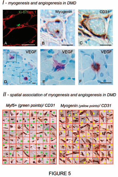
We first immunolabelled the Ki-67 nuclear antigen which is associated with all cell
proliferation phases but is absent in resting cells (G0). A maximum of 8% of
NCAM/CD56+ cells were Ki-67+ in DMD muscles. Double staining for Ki-67 and Laminin 1
showed that 82% of Ki-67+ sublaminal SCs were colocalised with capillaries (Fig. 5A).
Ki-67 was occasionally detected in SC pairs. Thus, SCs does not need to leave their
HA
L author manuscript inserm
-00128985, version 1

14
juxtavascular location to undergo active proliferation, and they can benefit from EC
supportive cues in this position.
Moreover, myogenic cell differentiation assessed by Myogenin expression was
associated with neoangiogenesis (Fig. 5BC). DMD muscle cross-sections contained a
total number of 175 undifferentiated MYF5+ SCs, and 211 Myogenin+ cells undergoing
myogenic differentiation. The quadrat test showed that 97.4% Myogenin+ nuclei (vs
88.5% MYF5+ nuclei) colocalized with CD31+ ECs. Moreover, the area occupied by ECs
was about two-fold higher in grid squares enclosing Myogenin+ cells than in those
enclosing MYF5+cells (48±28µm2 vs 19±18µm2, 59±33µm2 vs 32±18µm2, 44±25µm2 vs
23±18µm2, all p<0.0001) (Fig. 5II). In addition, most capillaries associated with
Myogenin+ cells were sectioned longitudinally, indicating a tortuous and highly
interconnected capillary network (Fig. 5II) typical of neoangiogenesis in skeletal muscle
(Hansen-Smith et al., 1996). These data reveal strong spatio-temporal correlation
between myogenic cell differentiation and neoangiogenesis.
The multifocal pattern of angio-myogenesis was consistent with the distribution of
VEGF immunostaining. In DMD, discrete foci of intense VEGF positivity were separated
by nearly negative areas (Fig. 5D-F). It was difficult to unambiguously identify cell
sources of VEGF in these foci, but the observed pattern was recently predicted by a
computational model in which VEGF focally produced by muscle cells remains
sequestered near sources of VEGF secretion bound to both VEGF receptors and heparan
sulfate proteoglycans present at high concentrations in the muscle microenvironment
(Mac Gabhann et al, 2006). Centronucleation, the hallmark of in vivo muscle
regeneration, was detected next to positive areas, and orientation of interstitial cells at
this level was suggestive of angiogenic sprouts growing at nearly 90 degrees from
parent vessels against the steep VEGF gradient (Fig. 5D-F).
DISCUSSION
In the present report we show that SC niches are juxtavascular, that they can
incorporate ectopic stem cells without loosing this anatomical characteristic, that most
HA
L author manuscript inserm
-00128985, version 1
15
SCs remain in close proximity to capillaries regardless of their state of quiescence,
proliferation and differentiation, and that individual myogenic cell differentiation is
spatio-temporally associated with new vessel formation. In light of our in vitro
experiments, these previously unappreciated morphologic characteristics likely favour
bi-directional signalling between ECs and differentiating SCs at work during muscle
regeneration.
Factors that govern SC frequency are not known (Bischoff and Franzini-Armstrong,
2004). Although the frequency of SCs appears to be much higher in slow compared to
fast muscles (Zammit and Beauchamp, 2001), there is little difference at birth, and the
metabolic properties per se may not be crucially involved (Bischoff and Franzini-
Armstrong, 2004). Capillaries are more numerous in slow muscles than in fast ones
(Hudlicka et al., 2004). This may represent a key factor if one considers the linear
increase of SCs with capillaries we observed at the individual myofiber level, regardless
of the myofiber type. Notably, to avoid influence of the different workload of slow and
fast individual muscles, a single type I: type II balanced muscle was chosen to compare
fiber types. Conversely, in aDM, where focal capillary loss is insidious enough to cause
no myofiber damage (Emslie-Smith and Engel, 1990), SC loss is proportionate to
capillary loss and selectively affects capillary-depleted myofibers. Of note, this is not the
case in full-blown dermatomyositis, where strong SC proliferation may be observed,
despite marked capillary loss, in reaction to conspicuous ischemic myofiber damage and
inflammation (Carpenter and Karpati, 2001). SCs are more resistant to acute ischemia
than myonuclei (Schultz et al., 1988; Snow, 1977), suggesting that disappearance of
SCs in areas of capillary dropout in aDM may reflect, at least in part, loss of supportive
paracrine EC signals. Investigating factors involved in SC maintenance and self-renewal
would likely require complex assays integrating EC and myofiber-to-SC signalling.
Detection of perivascular SC accumulations in trained muscle led us to examine the role
of ECs in more dynamic states associated with SC proliferation. We used indirect
EC:mpc coculture as an in vitro counterpart of muscle repair. EC monolayers that
mimick immature capillaries supported mpc growth through release of soluble factors.
HA
L author manuscript inserm
-00128985, version 1

16
Notably, mpc growth was not supported by fibroblasts and smooth muscle cells, two
other cell types that may be found in the endomysium close to SCs. A direct role for
paracrine signaling between ECs and surrounding target cells has been described during
embryonic development and cell differentiation of several organs (Cleaver and Melton,
2003) but data in muscle are scant. Endothelial progenitors have been isolated from
fetal tissues and adult blood (Le Grand et al., 2004; Shmelkov et al., 2005) and can be
instructed to co-differentiate into functional angiomyogenic colonies (Shmelkov et al.,
2005) and restore dystrophin in dystrophin-deficient mice and dogs (Le Grand et al.,
2004;Torrente et al., 2004, Sampaolesi et al., 2006). As shown here-in, circulating stem
cells can incorporate juxtavascular SC niches of irradiated mice (Labarge and Blau,
2002; Dreyfus et al., 2004) but the physiologic relevance of this phenomenon remains
far from clear (Sherwood et al, 2004). Nevertheless, vascular precursors enter the quail
limb buds around the time of muscle precursors (Nimmagadda et al., 2004), and
vessels develop before the appearance of SCs in the mouse limb (Le Grand et al.,
2004). In wing buds of avian embryos, migrating myogenic cells may be found in close
proximity to a prepatterned vascular network (Solursh et al., 1987). In line with these
observations, spatio-temporal correlations between the ingrowth of blood vessels into a
formerly ischemic area and detection of cells with myogenic capacities in that area has
been repeatedly reported (Kardon et al., 2002; Phillips et al., 1987). Although the
lineage relationships between ECs and muscle progenitors is not firmly agreed upon in
the literature, our present studies do strongly support a functional relationship.
Post-injury muscle regeneration is associated with an increase in both capillarisation
and cross-sectional area of individual regenerating myofibers (Luque et al., 1995). In
the same way, most data on compensatory muscle hypertrophy due to overload reveal
an almost linear relationship between capillary-to-fiber ratio and fiber area (Snow,
1977). Our anatomic and functional results indicate what cellular events may underpin
these structural changes. Human myogenic cells undergoing differentiation secrete
VEGF (Chazaud et al., 2003b). Conversely, we and others have shown that human ECs
can produce a series of mitogens for myogenic cells (Hawke and Garry, 2001), and our
HA
L author manuscript inserm
-00128985, version 1

17
co-culture studies indicated that bFGF, IGF-1, HGF, VEGF, and PDGF-BB mediate the
bulk of paracrine EC sustaining effect on mpc growth. Recently, attention has been paid
to the role of VEGF and its receptors in muscle biology (Williams and Annex, 2004).
Under normal in vivo conditions, mature ECs do not produce VEGF but express VEGF
receptors R1 (Flt-1) and R2 (KDR/Flk-1) which may bind low-levels of VEGF uniformly
released by normal muscle tissue (Mac Gabhann et al, 2006). Exercise-induced VEGF
upregulation in muscle cells operate physiological angiogenesis (Egginton et al, 2001;
Ameln et al, 2005). Myoblasts manipulated to produce VEGF specifically promote HUVEC
proliferation (Kim et al, 2005) and induce pronounced localized angiogenesis in their
immediate vicinity after i.m. transplantation (Springer et al, 2003). A steep gradient of
VEGF concentration emanating from the implantation site into neighboring host muscle
tissue has been repeatedly predicted, but was not previously documented by
immunolocalization (Springer et al, 2003; Mac Gabhann et al, 2006). It was visualized
herein in DMD, possibly because VEGF release occurs at a particularly high rate in
dystrophin deficient tissue (Nico et al, 2002).
VEGF receptor expression is not restricted to ECs and VEGF effects extend to a
variety of non-endothelial cell types, including myogenic cells (Rissanen et al, 2002,
Germani et al. 2003, Motoike et al, 2003, Arsic et al. 2004). We showed that VEGF
stimulates in vitro myogenic cell growth. In addition, VEGF stimulates their migration
(Germani et al. 2003), protects them from apoptosis (Germani et al. 2003, Arsic et al.
2004), up-regulate myoglobin expression (van Weel et al., 2004), and promotes
formation of centronucleated myofibers (Arsic et al. 2004).
On these grounds, evidence that, upon activation, SCs accumulate and proliferate
close to capillaries, receive efficient support from ECs for their growth, are
proangiogenic, and colocalize with new vessel formation during their myogenic
differentiation, strongly suggests that angiogenesis and myogenesis reciprocally signal.
It is likely that angiogenesis and myogenesis share VEGF as a co-regulatory factor
(Williams and Annex, 2004) probably in combination with other factors known to
stimulate both ECs and SCs, such as bFGF or IGF1 (Bischoff and Franzini-Armstrong,
HA
L author manuscript inserm
-00128985, version 1

18
2004; Hawke and Garry, 2001). We assume that, following workload or more
pronounced injury, angiogenesis is tightly coordinated with SC proliferation,
differentiation, and fusion to underlying myofibers. Newly SC-derived myonuclei may
remain close to capillaries, as suggested by the frequent colocalization of myonuclei
with capillaries. Occasional observation of SCs immediately adjacent to a myonucleus
(Fig. 2J) is even reminiscent of the model proposed by Moss and Leblond (1971) in
which assymetrical SC division generates a new SC and a daughter cell fated to become
a myonucleus. Newly incorporated myonuclei likely produce muscle-specific proteins
causing increase of the myofiber cross-section size. Harmonious increase of myofiber
capillarisation and caliber ensues, allowing larger myofibers to benefit from an
appropriately enhanced blood supply.
Such a myo-vascular unit may share similarities with the vascular neural stem cell
niche that supports intimate combination of neurogenesis and angiogenesis in the brain
(Palmer et al., 2000; Shen et al., 2004).
The knowledge that muscle SCs evolve into a juxtavascular niche may be critical for
several reasons. It provides a novel clue to investigate poorly understood muscle
pathologic processes combining capillary bed and myofiber size variations (Emslie-Smith
and Engel, 1990; Duscha et al., 1999; Hudlicka et al., 2004; Jensen et al., 2004).
Moreover, it could pave the way for improved myoblast transfer therapy of heart,
sphincter and skeletal muscle diseases. Dramatic non-mechanic transplanted cell death
is a major limitation of this therapeutic approach (Chazaud et al., 2003a). The present
results suggest that early cell death results from extrinsic growth factor deprivation of
mpc massively implanted in an inappropriate microanatomic environment (Chazaud et
al., 2003b). Recreating an appropriate endothelial cell or molecular support to the
transplanted stem cells thus represents a promising way to improve striated muscle cell
therapies.
MATERIAL AND METHODS
Transgenic mouse strains.
HA
L author manuscript inserm
-00128985, version 1

19
Mice models allowing visualization of satellite cells
Constructions of both Myf5nlacZ and Myf5GFP-P knock-in mice have been described
(Tajbakhsh et al., 1996; Kassar-Duchossoy et al., 2004). Both mice have a ScaI-BstYI
deletion in exon 1 (122 amino-acids) of Myf5 gene, thus lacking the basic-HLH domain
required for Myf5 transcription factor activity, and a reporter gene targeted to this exon.
Myf5GFP-P/GFP-P are reduced in size, whereas both heterozygote mice, Myf5nlacZ/+ and
Myf5GFP-P/+, are phenotypically normal and were used for the experiments.
Mice used for bone-marrow transplantation
C57BL/6 mice were transplanted with BM-derived cells from Tg:CAG-GFP transgenic
mice (C57BL/6 TgN[actEGFP]Osb YO1) in which the GFP transgene is expressed under
the control of a non-tissue specific promoter, chicken beta-actin with cytomegalovirus
enhancer, as a cytoplasmic protein (Okabe et al., 1996).
Animal experiments.
Mouse handling
All mice were housed in our level 2 biosafety animal facility, and received food and
water ad libitum. Prior to manipulations, animals were anaesthetized using
intraperitoneal injection of chloral hydrate. This study was conducted in accordance with
the EC guidelines for animal care [Journal Officiel des Communautés Européennes, L358,
december 18, 1986].
Bone marrow transplantation
Briefly, donor BM cells were obtained by flushing femurs of Tg:CAG-GFP mice with
DMEM medium (Invitrogen, Paisley, UK), and washed twice in cold PBS. Retro-orbital
injection of 3 x10⁷ BM cells in 0.1 ml mouse serum and PBS (1:1), was done in 9.0 Gy-
irradiated, 4 week-old B6 mice (60Co γ rays within 1 day before BM transplantation) as
previously reported (Dreyfus et al., 2004). After transplantation, mice received 10
mg/kg/day ciprofloxacin for 10 days to prevent infection during the aplastic phase. Blood
chimerism >95% was controled by flow cytometry 4 weeks post-transplantation and TA
muscles were examined 6 months post-transplantation.
HA
L author manuscript inserm
-00128985, version 1
20
BrdU pulse-chase experiments
Myf5nlacZ/+ pupps received 50 mg/kg BrdU diluted in 20 µL PBS by I.P. route twice
daily from post-natal day 3 to 8. After a chase period of 6 weeks, mice were sacrificed
and TA muscles were examined to evaluate BrdU retention by β-Gal+ satellite cell nuclei.
As a control, one Myf5nlacZ/+ mouse from the series received notexin injection (25µg/ml in
10 µl PBS) in TA muscle 6 weeks post-exposure and had the injected TA examined 2
weeks later to assess dilution of BrdU in the progeny of the previously quiescent satellite
cells.
Human muscle samples.
Normal muscle samples were obtained in 20 human adults (aged from 24 to 73 years,
from various muscle groups, including 12 deltoid, 4 vastus lateralis, 2 gastrocnemius and
2 peroneus brevis muscles), in 3 adult C57/B6 mice (tibialis anterior muscle), in 2 adult
retriever dogs (sartorius muscle) and, for electron microscopy, in 10 adult Wistar rats
(extensor digitorum longus muscle). Muscle samples were also collected from deltoid
muscle of 3 heavy-training athletes who had no significant myopathologic alterations
(males aged from 32 to 39 years practicing intense daily push-up training), of 3 patients
with amyopathic dermatomyositis collected from a previous study (Cosnes et al., 1995)
(2 females, 1 male aged from 32 to 39 years), and of 3 boys with Duchenne muscular
dystrophy (males aged from 2 to 14 years). Muscle samples were frozen and kept at -
80°C before processing.
Immunohistochemistry.
In mouse tissues, vessels were immunostained using anti-laminin 1 (1:50, polyclonal,
DakoCytomation, Glostrup, Denmark) or anti-collagen IV (1:50, polyclonal, Chemicon,
Temecula, CA) antibodies revealed by Cy3 conjugated secondary antibodies (Jackson
Immunoresearch Laboratories, West Grove, PA). Due to HCl pre-treatment required for
BrdU immunohistochemistry, we combined chromogenic and fluorescence revelation
approaches on the same preparation. First, endogenous alkaline phosphatase activity of
microvascular cells (Pearce et al., 2000) was detected using fast red (forming both
HA
L author manuscript inserm
-00128985, version 1

21
chromogenic and fluorescent red precipitate) followed by chromogenic
immunohistochemistry against β-Gal (using a polyclonal domestic antibody, from Pasteur
Institute, 1:2000e revealed by a peroxidase-conjugated secondary antibody) forming a
brown precipitate. Afterwards, sections were treated with 2N HCl for 30 min at 37°C and
BrdU was detected using a sheep polyclonal antibody (1:200e, Abcam Inc, Cambridge,
MA) revealed by FITC-conjugated secondary antibody (Jackson Immunoresearch lab).
In human tissues, several antibodies were used alone or in combination to localize
and identify SCs and their progeny, ECs and VEGF. They included antibodies against
laminin-1 (1:500, ref L9393, Sigma Aldrich, St-Louis, MO) for basal laminae, MYF5 (1:50,
ref sc302, Santa Cruz, Santa Cruz Biotechnology, CA) for undifferentiated SCs,
CD56/NCAM (1:100, ref PN6602705, Beckman Coulter, Fullerton, CA) for both quiescent
and differentiating myogenic cells, myogenin (1:200, ref M3559, DakoCytomation) for
differentiating myogenic cells, CD31 (1:50, ref M0823, DakoCytomation) and vWF
(1:500, ref A0082, DakoCytomation) for ECs, Ki-67 antigen (1:50, ref M7240,
DakoCytomation) for cycling cells and VEGF (1:100, AF293NA, R&D Systems,
Minneapolis, MN, for immunofluorescence; or 1:50, sc-152, Santa Cruz Biotechnology,
with SuperPicTure TM polymer detection kit, Zymed, and AEC as a chromogen for light
microscopy).
Dogs samples were labelled using the same antibodies to CD56/NCAM and vWF.
Electron and confocal microscopy. Transmission electron microscopy was
performed after conventional processing of muscle samples using a Philips-410
microscope. Confocal microscopy (Zeiss LSM 410 and a x63 PlanApo NA 1.4 oil objective,
Carl Zeiss, Oberkochen, Germany) on 10-30 µm-thick sections and deconvolution of
stacks of optical sections obtained along the Z-axis were used to obtain high resolution
images and three-dimensional volume reconstructions. Steps along the optical axis were
set at 0.2-0.3 µm and pixel size in X,Y at 0.11-0.14 µm thus respecting Nyqvist’s criteria
optimal for subsequent deconvolution. Z-stacks acquired separately for each channel
(green or red) were subjected to iterative optical deconvolution by the AxioVision 4.2
HA
L author manuscript inserm
-00128985, version 1
22
software package defining appropriate parameters of a theoretical Point Spread Function
and settings to correct for optical aberration. Deconvolution (15–25 iterations) was
performed on a personal computer equipped with a Pentium 4 processor (3.0 GHz, 2 Go
RAM). In rare cases, better results were obtained by the Nearest Neighbour algorithm of
AxioVision 4.2. 3D reconstructions combining both channels were then obtained in the
transparency option of the 4D module of AxioVision 4.2. Measurement of the minimal
distance between SC and the neighbouring capillary were carried out on the projection
(rotation) that excluded spurious colocalization of channels that could potentially result
from the considerable Z-thickness of the reconstruction (10-20 µm).
Morphometric data collection. Muscle areas showing motor endplates were
excluded from analysis. Immunostained sections were digitized with a Hamamatsu CDD
device (Hamamatsu Photonics KK, Hamamatsu, Japan) and subjected to morphometric
analysis using different modules of the KS4003.0 (Carl Zeiss) and Simple PCI image
analysis software (C-Imaging Compix Inc, Cranberry Township, PA). Each object (SC,
Cap, myonuclei or VSP) was defined as a point with X and Y coordinates in a study area
including at least 1000 myofibers per specimen. Because of either difficulty to
discriminate between closely associated structures on double-immunostainings, or limited
use of antibodies of the same species for double-immunostainings, some quantitative
evaluations were conducted using single immunostainings on alternate muscle cross-
sections. For example, alternate sections were immunostained for MYF5, CD31 and
myogenin, myogenic cell positions being manually reported in the graphic plane of image
reconstruction of the alternate immunostaining of capillaries.
Univariate spatial point pattern analysis. To explore if point distributions deviate
from complete spatial randomness (CSR) We calculated spatial point pattern statistics
with the ADE4 software package, using Ripley’s K-function for univariate point patterns,
and both the K12 function and a quadrat test for bivariate patterns (Ripley, 1988).
Basically, the K function assesses for each point the cumulative number of neighbouring
points within increments of a predefined distance t as compared with the expected
number of neighbours under the null hypothesis of CSR. These increments define "n"
HA
L author manuscript inserm
-00128985, version 1
23
concentric rings so that the outer radius of the external ring r = nt. According to
recommendations of the ADE4 manual, t and n values were determined for each set of
data, taking into account both size of the study area R and the observed minimal
distances between points. The definition of Ripley’s K is K(r) = N(r)/λ, where N(r) is the
number of neighbours within distance r and λ is the intensity of the pattern. Under CSR,
K(r) = πr2, in case of clustering, K(r) > πr2, and in case of regularity, K(r) < πr2. By
convention, K(r) is substituted by L(r) [L(r) = √K(r)/π - t ], this transformation offering
the advantage of L(r) = 0 under CSR, L(r) >0 for clustered, and L(r) < 0 for regular
patterns. Edge correction was carried out as proposed by Ripley (Ripley, 1988).
Deviation from CSR was tested by plotting L(r) values against the envelope of
significance at p<0.0001 for the null hypothesis of CSR. This envelope was built using the
Monte Carlo method that consists in the realization of 9999 CSR patterns of the same
intensity as the observed pattern. Graphically, values above the upper limit of the
envelope indicate clustering whereas values below its lower limit indicate regularity.
Bivariate point pattern analysis. The K12 function, and its L12 transformation, for
bivariate patterns is identical to Ripleys' K with the exception that points on which the
function is centered and neighbour points are of two different types, i.e. correspond to
different objects. Graphic expression of results was similar to that used for Ripleys'K. The
distribution of real objects, such as SC and myonuclei, relative to capillaries was
compared to that of randomly distributed virtual sarcolemmal objects, one object per
myofiber being randomly inserted following a clock dial scheme.
Quadrat test. A quadrat test grid was superimposed in the graphic plane of each
image. The grid square size (21 µm diagonally, 225 µm2) was chosen to enclose the
largest clusters of points detected by bivariate analysis, as deduced from the graphic
expression of the K function. Each square was examined for the presence of SC,
myonuclei, VSP and Cap and colocalization was estimated by a Fisher's exact test
comparing the relative number of squares containing a capillary and either SC,
myonuclei, or VSP. In some tests, the capillary area (µm2) in each square was measured
after color segmentation (DAB, brown) of CD31 labelling of vessels using KS400 3.0.
HA
L author manuscript inserm
-00128985, version 1

24
Myofiber capillarisation evaluation. Muscle fiber capillarisation in normal, aDM,
and athlete muscles was assessed by the number of capillaries bordering each individual
fiber, as previously described (Emslie-Smith and Engel, 1990). Cap numbers and
frequency distribution in aDM and control patients of our study were closely similar to
those previously reported (Emslie-Smith and Engel, 1990).
Cell cultures. Unless indicated, culture media components were from Gibco (Paisley,
Scotland) and culture plastics from TPP AG (Trasadingen, Switzerland). Human mpc were
cultured from muscle samples as previously described (Chazaud et al., 2003b). Mpc were
grown in HAM-F12 medium containing 15 % FCS. Only cultures presenting over 95 %
NCAM/CD56+ (1/20, 123C3, Sanbio/Monosan, Uden, Netherlands) cells were used. Mpc
differentiation was assessed by myogenin immunoblotting. MRC-5 human fibroblastic cell
line was obtained from ATCC (LGC-Promochem, Molsheim, France) and cultured in MEM
containing 10% FCS and 1% NEAA. HUVEC and HMEC were obtained from Promocell
GmbH (Heidelberg, Germany) and cultured in EC growth medium (Promocell). SMC were
obtained from human pulmonary arteries kindly provided by Dr S. Eddahibi (INSERM
U651, Créteil, France) and cultured in RPMI containing 10% FCS and 1% NEAA.
In vitro angiogenesis assay. HUVEC were seeded (30000 cells/well) in 24-well
tissue culture plates coated with growth factor-reduced Matrigel (0.3 ml/well, Becton
Dickinson) that was allowed to solidify at 37°C for 30 min. Cells were incubated in
serum-free medium or in 24h mpc-conditioned medium for 24 h at 37°C. The cells were
photographied using an inverted photomicroscope (DMIRB Leica, Leica Microsystems AG,
Wetzlar Germany) with a CCD camera (CoolSnap, Photometrics, Roper Scientific Inc.
Tucson, AZ). Tube formation was assessed in 5 randomly selected x20 powerfields, using
the number of branching points in the network per field and the proportion of connected
cells (number of connected cells among the total number of cells in the same field)
(Jones et al., 1999).
Indirect cocultures. MRC-5 fibroblasts were seeded at 15000 cells/cm2, SMC and
mpc at 20000 cells/cm2 in Falcon inserts (0.4 µm diameter pores) (Falcon, BD
HA
L author manuscript inserm
-00128985, version 1

25
Biosciences, Franklin Lakes, NJ); EC were seeded at 30000 cells/cm2 and 3 days after
confluence, EC monolayer integrity was assessed in 2 wells by absence of Trypan blue
(0.2% in 0.1% BSA) translocation from upper to lower chamber after 3 h incubation. Mpc
were seeded into 24 well plates at 3000 cells/cm2 and allowed to adhere in their culture
medium for 6 h. Then, inserts were placed over the mpc-containing well and the medium
of both chamber was changed for MCDB-131 medium containing 2% FCS and 1.5 µg/ml
ECGS. The medium was changed twice weekly. At each time point, mpc density was
determined by counting cells after trypsinization, and the number of cell present in the
upper chamber was also evaluated. In these conditions, the number of EC remains
constant at each time point as they form impermeable monolayers. In contrast, the
number of SMC, MRC-5 and control mpc increased during time of experiment to reach
approximatively 60000, 60000 and 40000 cells/cm2, respectively. To avoid bias due to
the different numbers of cells in the upper chamber at each time point, calculation of the
effect on mpc growth was normalized to 30000 cells/cm2, thus allowing comparison of
individual cell biological activity, as previously reported (Chazaud et al., 2003b). All these
in vitro experiments were performed using at least 3 different cultures. In some
experiments, blocking antibodies were added in the well at saturating concentrations
(calculated from IC50): anti-IGF-1 (6 µg/ml, AF291NA, R&D), anti-PDGF-BB (5 µg/ml,
AF220NA, R&D), anti-bFGF (6 µg/ml, AF233NA, R&D), anti-HGF (5 µg/ml, AF294NA,
R&D), anti-VEGF (6 µg/ml, AF293NA, R&D). Controls included addition of whole mice
IgGs (6 µg/ml, Vector Laboratories, Burlingame, CA), antibodies against muscle
membrane protein dysferlin (3 µg/ml, Novocastra laboratories, Newcastle, UK), and
antibodies against MCP-1 (5 µg/ml, P500-P34, Abcys, Paris, France), a chemokine that is
secreted by EC (Cleaver and Melton, 2003) and the receptor of which is expressed by
human mpc (DNA array, data not shown).
Mpc grown alone were subjected to similar blocking antibody experiments and to
recombinant human VEGF (RandD systems) added at 25 ng/ml.
DNA Array. Total RNA was prepared from mpc at day 14 of culture using the RNeasy
mini kit (Qiagen, Hilden, Germany). All further steps were performed according to the
HA
L author manuscript inserm
-00128985, version 1

26
manufacturer's instructions in Human cytokine array kit (R&D Systems). Analysis was
performed using Image Quant software (Amersham, Buckinghamshire, UK), that allows
background noise subtraction, correction for the variation of density for housekeeping
genes (all genes showed the same intensity variation between the 2 membranes), and
finally, densitometric analysis of signals above internal negative controls. Results were
expressed in arbitrary units.
Protein Array. 24h-HUVEC- conditioned medium was subjected to protein array
according to the manufacturer's instructions in RayBio® Human Cytokine Antibody Array
(RayBiotech Inc., Norcross,GA). Densitometric analysis was performed as described
above for DNA array.
Statistical analyses. Fischer's exact test was used to compare colocalizations in the
quadrat test. χ2 test was used to compare frequency of SCs associated with variously
capillarized myofibers, and with various Cap types. Student's t test was used in in vitro
experiments. A p value <0.05 was considered significant.
HA
L author manuscript inserm
-00128985, version 1
27
FIGURE LEGENDS
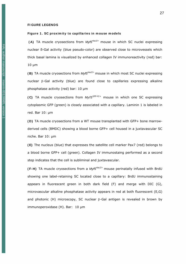
Figure 1. SC proximity to capillaries in mouse models
(A) TA muscle cryosections from Myf5nlacZ/+ mouse in which SC nuclei expressing
nuclear ß-Gal activity (blue pseudo-color) are observed close to microvessels which
thick basal lamina is visualized by enhanced collagen IV immunoreactivity (red) bar:
10 µm
(B) TA muscle cryosections from Myf5nlacZ/+ mouse in which most SC nuclei expressing
nuclear β-Gal activity (blue) are found close to capillaries expressing alkaline
phosphatase activity (red) bar: 10 µm
(C) TA muscle cryosections from Myf5GFP-P/+ mouse in which one SC expressing
cytoplasmic GFP (green) is closely associated with a capillary. Laminin 1 is labeled in
red. Bar 10: µm
(D) TA muscle cryosections from a WT mouse transplanted with GFP+ bone marrow-
derived cells (BMDC) showing a blood borne GFP+ cell housed in a juxtavascular SC
niche. Bar 10: µm
(E) The nucleus (blue) that expresses the satellite cell marker Pax7 (red) belongs to
a blood borne GFP+ cell (green). Collagen IV immunostaing performed as a second
step indicates that the cell is subliminal and juxtavascular.
(F-H) TA muscle cryosections from a Myf5nlacZ/+ mouse perinatally infused with BrdU
showing one label-retaining SC located close to a capillary: BrdU immunostaining
appears in fluorescent green in both dark field (F) and merge with DIC (G),
microvascular alkaline phosphatase activity appears in red at both fluorescent (E,G)
and photonic (H) microscopy, SC nuclear β-Gal antigen is revealed in brown by
immunoperoxidase (H). Bar: 10 µm
HA
L author manuscript inserm
-00128985, version 1

28
Figure 2. Topological relations between SCs and Capillaries
(A-B) Transmission electron microscopy of rat SCs. Both apposition of SC body to
EC (A) and apposition of a cytoplasmic process elongated from a distant SC toward EC
(B) are observed. For clarity, SC plasma membrane was underlined by red dots. Please
note the absence of direct SC-to-EC contact. Bars: 2 µm.
(C-E) Chromogenic or fluorescent immunostainings of human deltoid muscle
showing extreme proximity of SCs and capillaries. In C, SCs (black arrowheads) are
immunostained for NCAM and capillaries (red arrowheads) are identified without
immunostaining. In D, a SC nucleus is immunostained for Myf 5 (black arrowhead) and
capillaries (red arrowheads) are immunostained for CD31. In E, NCAM of SC plasma
membrane is green and the EC marker vWF is red. Bar : 10 µm.
(F-G) Correlations between SCs and capillaries in normal muscle. Left graph (F):
Histogramm of myofiber capillarisation frequency (top) is close to Gaussian while SC
frequency (bottom) increases linearly with myofiber capillarisation (data pooled from 3
normal deltoid muscles). Right graph (G): numbers of both capillaries (●) and SCs (+)
are different and largely proportionate in type I and type II myofibers (individual results
from 3 normal deltoid muscles identified by different colors).
(H) Correlations between SCs and capillaries in aDM muscle. A proportionate loss of
muscle capillaries (●) and SCs (+) was found in 3 patients with aDM (in red, grey and
white, respectively) as compared to 3 age, sex, and muscle-matched normal controls (in
black).
(I-J) Heavy-trained muscle. (I): as compared to their controls (in black), 3
athletes (in red, grey and white, respectively) show an increase of both muscle
capillaries (●) and SCs (+). (J): SC clusters are formed by accumulations of SCs
(NCAM+, brown) belonging to different myofibers, often adjacent to a myonucleus
(haematoxylin nuclear counterstaining in blue), and surrounding the same capillary (red
dots). Bar: 10 µm.
HA
L author manuscript inserm
-00128985, version 1
29
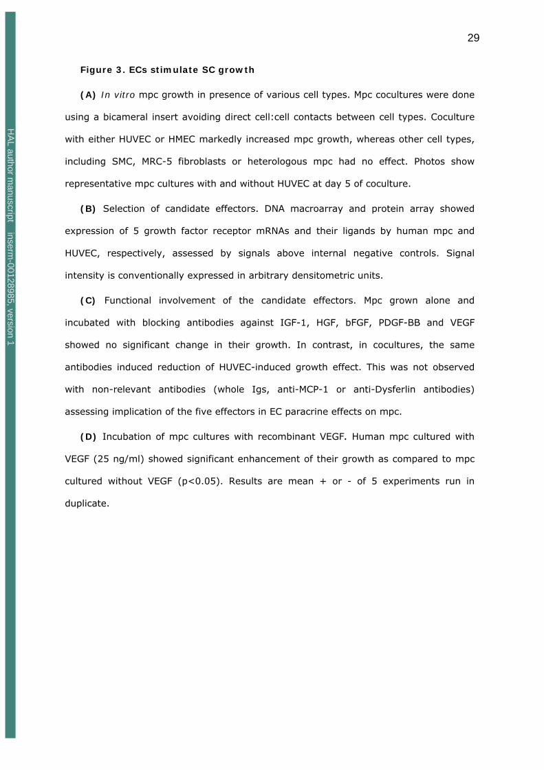
Figure 3. ECs stimulate SC growth
(A) In vitro mpc growth in presence of various cell types. Mpc cocultures were done
using a bicameral insert avoiding direct cell:cell contacts between cell types. Coculture
with either HUVEC or HMEC markedly increased mpc growth, whereas other cell types,
including SMC, MRC-5 fibroblasts or heterologous mpc had no effect. Photos show
representative mpc cultures with and without HUVEC at day 5 of coculture.
(B) Selection of candidate effectors. DNA macroarray and protein array showed
expression of 5 growth factor receptor mRNAs and their ligands by human mpc and
HUVEC, respectively, assessed by signals above internal negative controls. Signal
intensity is conventionally expressed in arbitrary densitometric units.
(C) Functional involvement of the candidate effectors. Mpc grown alone and
incubated with blocking antibodies against IGF-1, HGF, bFGF, PDGF-BB and VEGF
showed no significant change in their growth. In contrast, in cocultures, the same
antibodies induced reduction of HUVEC-induced growth effect. This was not observed
with non-relevant antibodies (whole Igs, anti-MCP-1 or anti-Dysferlin antibodies)
assessing implication of the five effectors in EC paracrine effects on mpc.
(D) Incubation of mpc cultures with recombinant VEGF. Human mpc cultured with
VEGF (25 ng/ml) showed significant enhancement of their growth as compared to mpc
cultured without VEGF (p<0.05). Results are mean + or - of 5 experiments run in
duplicate.
HA
L author manuscript inserm
-00128985, version 1
30
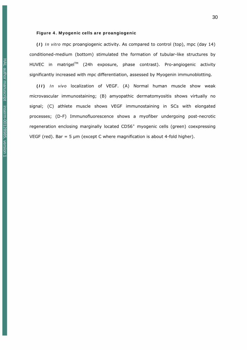
Figure 4. Myogenic cells are proangiogenic
(I) In vitro mpc proangiogenic activity. As compared to control (top), mpc (day 14)
conditioned-medium (bottom) stimulated the formation of tubular-like structures by
HUVEC in matrigelTM (24h exposure, phase contrast). Pro-angiogenic activity
significantly increased with mpc differentiation, assessed by Myogenin immunoblotting.
(II) In vivo localization of VEGF. (A) Normal human muscle show weak
microvascular immunostaining; (B) amyopathic dermatomyositis shows virtually no
signal; (C) athlete muscle shows VEGF immunostaining in SCs with elongated
processes; (D-F) Immunofluorescence shows a myofiber undergoing post-necrotic
regeneration enclosing marginally located CD56+ myogenic cells (green) coexpressing
VEGF (red). Bar = 5 µm (except C where magnification is about 4-fold higher).
HA
L author manuscript inserm
-00128985, version 1

31
Figure 5. Combined myogenesis and angiogenesis in DMD
(I) Myogenesis and angiogenesis in DMD. (A) Double immunostaining for nuclear
Ki-67 antigen (green) and laminin 1 (red) shows a sublaminal SC undergoing cell cycling
close to a capillary; (B, C) alternate sections immunostained for Myogenin and CD31,
showing differentiating myogenic cells directly adjacent to transversely oriented
neovessels; (D-F) VEGF immunostaining showing two discrete foci of intense positivity
visualizing VEGF, sequestered next to myofibers with a central nucleus, directing
transverse angiogenic sprouting.
(II) Spatial association of myogenesis and angiogenesis in DMD. Selected squares
enclosing both CD31+ ECs (brown) and either MYF5+ (green points) or myogenin+
(yellow points) cells from quadrat test applied to a DMD muscle. Myogenin+ cells
undergoing late myogenic differentiation were associated with larger EC cytoplasmic
areas than undifferentiated MYF5+ positive SCs (these illustrations were built from
alternate sections immunostained for MYF5, CD31 and Myogenin, myogenic cell
positions being manually reported in the graphic plane of image reconstruction of CD31
immunostaining).
Supplementary Figure. Non-random proximity of SCs and capillaries in
human skeletal muscle
Graphic expression of univariate (red curves) and bivariate (blue curves) point
pattern analyses obtained by L(r) transformation of Ripleys K function in human deltoid
muscle (n=4, age range 20-33 years). Values above the upper limit of the envelope (in
between black curves) of complete spatial randomness built using the Monte Carlo
method indicate clustering whereas values below its lower limit indicate regularity:
- univariate analysis of all objects taken together, including myonuclei, SCs, and
capillaries (left graph, red curve) shows both point clustering (0 to 18 µm) and
regularity (25 to 50 µm). Univariate analyses of myonuclei and capillaries studied alone
(right graphs, red curves) show no clustering whereas regularity imposed by the
HA
L author manuscript inserm
-00128985, version 1

32
myofiber cytoplasmic area is still observed.
- bivariate analysis of both SCs vs capillaries and myonuclei vs capillaries (left
graphs, blue curves) indicate clustering of both SCs and myonuclei with capillaries, a
finding not observed with virtual points introduced at random along sarcolemma (VSPs
vs capillaries, right graph, blue curve). All graphs were obtained from cross-sections
double-immunostained for NCAM/CD56 and CD31. (Mn means myonuclei, caps means
capillaries, SCs mean satellite cells, VSPs mean virtual sarcolemmal objects).
HA
L author manuscript inserm
-00128985, version 1

33
REFERENCES
Ameln, H., Gustafsson, T., Sundberg, C.J., Okamoto, K., Jansson, E., Poellinger,
L. and Makino, Y. (2005). Physiological activation of hypoxia inducible factor-1 in
human skeletal muscle. FASEB J. 19, 1009-1011
Asakura, A., Komaki, M. and Rudnicki, M. (2001). Muscle satellite cells are
multipotential stem cells that exhibit myogenic, osteogenic, and adipogenic
differentiation. Differentiation 68, 245-53.
Arsic, N., Zacchigna, S., Zentilin, L., Ramirez-Correa, G., Pattarini, L., Salvi, A.,
Sinagra, G. and Giacca, M. (2004). Vascular endothelial growth factor stimulates
skeletal muscle regeneration in vivo. Mol. Ther. 10, 844-854.
Beauchamp, J.R., Heslop, L., Yu, D.S., Tajbakhsh, S., Kelly, R.G., Wernig, A.,
Buckingham, M.E., Partridge, T.A. and Zammit, P.S. (2000). Expression of CD34 and
myf5 defines the majority of quiescent adult skeletal muscle satellite cells. J. Cell Biol.
151, 1221-1234.
Bischoff, R. and Franzini-Armstrong, C. (2004). Satellite and stem cells in muscle
regeneration. In Myology. Engel, A.G. and Franzini-Armstrong, C. editors. McGraw Hill,
New-York. 66-86.
Carpenter, S. and Karpati, G. (2001). Pathology of skeletal muscle. Oxford University
Press, New York.
Chazaud, B., Hittinger, L., Sonnet, C., Champagne, S., Le Corvoisier, P.,
Benhaiem-Sigaux, N., Unterseeh, T., Su, J., Merlet, P., Rahmouni, A., Garot, J.,
Gherardi, R. and Teiger, E. (2003a). Endoventricular porcine autologous myoblast
transplantation can be successfully achieved with minor mechanical cell damage.
Cardiovasc. Res. 58, 444-450.
Chazaud, B., Sonnet, C., Lafuste, P., Bassez, G., Rimaniol, A.C., Poron, F.,
Authier, F.J., Dreyfus, P.A. and Gherardi, R.K. (2003b). Satellite cells attract
monocytes and use macrophages as a support to escape apoptosis and enhance muscle
growth. J. Cell Biol. 163, 1133-1143.
Chretien, F., Dreyfus, P.A., Christov, C., Caramelle, P., Lagrange, J.L., Chazaud, B.
and Gherardi, R.K. (2005). In vivo fusion of circulating fluorescent cells with dystrophin-
deficient myofibers results in extensive sarcoplasmic fluorescence expression but limited
dystrophin sarcolemmal expression. Am. J. Pathol. 166, 1741-1748
Cleaver, O. and Melton, D.A. (2003). Endothelial signaling during development. Nat.
Med. 9, 661-668.
Collins, C.A., Olsen, I., Zammit, P.S., Heslop, L., Petrie, A., Partridge, T.A. and
Morgan, J.E. (2005). Stem cell function, self-renewal, and behavioral heterogeneity of
cells from the adult muscle satellite cell niche. Cell 122, 289-301.
HA
L author manuscript inserm
-00128985, version 1

34
Cosnes, A., Amaudric, F., Gherardi, R., Verroust, J., Wechsler, J., Revuz, J. and
Roujeau, J.C. (1995). Dermatomyositis without muscle weakness. Long-term follow-up
of 12 patients without systemic corticosteroids. Arch. Dermatol. 131, 1381-1385.
De Angelis, L., Berghella, L., Coletta, M., Lattanzi, L., Zanchi, M., Cusella-De
Angelis, M.G., Ponzetto, C. and Cossu, G. (1999). Skeletal myogenic progenitors
originating from embryonic dorsal aorta coexpress endothelial and myogenic markers and
contribute to postnatal muscle growth and regeneration. J. Cell Biol. 147, 869-878.
Dhawan, J., Rando, T.A. (2005). Stem cells in postnatal myogenesis: molecular
mechanisms of satellite cell quiescence, activation and replenishment. Trends Cell Biol.
15, 666-73.
Dreyfus, P.A., Chretien, F., Chazaud, B., Kirova, Y., Caramelle, P., Garcia, L.,
Butler-Browne, G. and Gherardi, R.K. (2004). Adult bone marrow-derived stem cells
in muscle connective tissue and satellite cell niches. Am. J. Pathol. 164, 773-779.
Duscha, B.D., Kraus, W.E., Keteyian, S.J., Sullivan, M.J., Green, H.J., Schachat,
F.H., Pippen, A.M., Brawner, C.A., Blank, J.M. and Annex, B.H. (1999). Capillary
density of skeletal muscle: a contributing mechanism for exercise intolerance in class II-
III chronic heart failure independent of other peripheral alterations. J. Am. Coll. Cardiol.
33, 1956-1963.
Egginton, S., Zhou, A.L., Brown, M.D. and Hudlicka, O. (2001) Unorthodox
angiogenesis in skeletal muscle. Cardiovasc. Res. 49, 634–646.
Emslie-Smith, A.M. and Engel, A.G. (1990). Microvascular changes in early and
advanced dermatomyositis: a quantitative study. Ann. Neurol. 27, 343-356.
Fuchs, E., Tumbar, T. and Guasch, G. (2004). Socializing with the neighbors: stem
cells and their niche. Cell 116, 769-78.
Germani, A., Di Carlo, A., Mangoni, A., Straino, S., Giacinti, C., Turrini, P.,
Biglioli, P. and Capogrossi, M.C. (2003). Vascular endothelial growth factor modulates
skeletal myoblast function. Am. J. Pathol. 163, 1417–1428.
Gros, J., Manceau, M., Thome, V. and Marcelle, C. (2005). A common somitic origin
for embryonic muscle progenitors and satellite cells. Nature 435, 954-8.
Hansen-Smith, F.M., Hudlicka, O. and Egginton, S. (1996). In vivo angiogenesis in
adult rat skeletal muscle: early changes in capillary network architecture and
ultrastructure. Cell Tissue Res. 286, 123-136.
Hawke, T.J. and Garry, D.J. (2001). Myogenic satellite cells: physiology to molecular
biology. J. Appl. Physiol. 91, 534-551.
Hudlicka, O., Brown, M.D. and Egginton, S. (2004). Microcirculation in muscle. In
Myology. A.G.Engel and C.Franzini-Armstrong, editors. McGraw Hill, New-York. 511-533.
HA
L author manuscript inserm
-00128985, version 1

35
Jensen, L., Bangsbo, J. and Hellsten, Y. (2004). Effect of high intensity training on
capillarisation and presence of angiogenic factors in human skeletal muscle. J. Physiol.
557, 571-582.
Jones, M.K., Wang, H., Peskar, B.M., Levin, E., Itani, R.M., Sarfeh, I.J. and
Tarnawski, A.S. (1999). Inhibition of angiogenesis by nonsteroidal anti-inflammatory
drugs: insight into mechanisms and implications for cancer growth and ulcer healing.
Nat. Med. 5, 1418-1423.
Kardon, G., Campbell, J.K. and Tabin, C.J. (2002). Local extrinsic signals determine
muscle and endothelial cell fate and patterning in the vertebrate limb. Dev. Cell 3, 533-
545.
Kassar-Duchossoy, L., Gayraud-Morel, B., Gomes, D., Rocancourt, D.,
Buckingham, M., Shinin, V. and Tajbakhsh, S. (2004). Mrf 4 determines skeletal
muscle identity in Myf5: MyoD double-mutant mice. Nature 431, 466-470.
Kim, K.I., Cho, H.J., Hahn, J.Y., Kim, T.Y., Park, K.W., Koo, B.K., Shin, C.S., Kim,
C.H., Oh, B.H., Lee, M.M., Park, Y.B. and Kim, H.S. (2006). ß-catenin overexpression
augments angiogenesis and skeletal muscle regeneration through dual mechanism of
vascular endothelial growth factor–mediated endothelial cell proliferation and progenitor
cell mobilization. Arterioscler. Thromb. Vasc. Biol. 26,91-98
Korff, T., Kimmina, S., Martiny-Baron, G., Augustin, H.G. (2001). Blood vessel
maturation in a 3-dimensional spheroidal coculture model: direct contact with smooth
muscle cells regulates endothelial cell quiescence and abrogates VEGF responsiveness.
FASEB J. 15, 447-457
LaBarge, M.A. and Blau, H.M. (2002). Biological progression from adult bone marrow
to mononucleate muscle stem cell to multinucleate muscle fiber in response to injury.
Cell 111, 589-601.
Le Grand, F., Auda-Boucher, G., Levitsky, D., Rouaud, T., Fontaine-Perus, J. and
Gardahaut, M.F. (2004). Endothelial cells within embryonic skeletal muscles: a potential
source of myogenic progenitors. Exp. Cell Res. 301, 232-241.
Li, L. and Xie, T. (2005). Stem cell niche : structure and function. Annu. Rev. Cell Dev.
Biol. 21, 605-631.
Luque, E., Pena, J., Martin, P., Jimena, I. and Vaamonde, R. 1995. Capillary supply
during development of individual regenerating muscle fibers. Anat. Histol. Embryol. 24,
87-89.
Mac Gabhann, F., Ji, J.W. and Popel, A.S. (2006). Computational model of vascular
endothelial growth factor spatial distribution in muscle and pro-angiogenic cell therapy.
PLoS Comput. Biol. 2:e127. Epub 2006 Aug 3
HA
L author manuscript inserm
-00128985, version 1

36
Maurer, M.H., Tripps, W.K., Feldmann, R.E. Jr. and Kuschinsky, W. (2003).
Expression of vascular endothelial growth factor and its receptors in rat neural stem cells.
Neurosci. Lett. 344, 165-8.
Mauro, A. (1961). Satellite cell of skeletal muscle fibers. J. Biophys. Biochem. Cytol. 9,
493-495.
Motoike, T., Markham, D.W., Rossant, J., Sato, T.N. (2003). Evidence for novel fate
of Flk1+ progenitor: contribution to muscle lineage. Genesis 35, 153-159.
Molnar, G., Ho, M.L. and Schroedl, N.A. (1996). Evidence for multiple satellite cell
populations and a non-myogenic cell type that is regulated differently in regenerating
and growing skeletal muscle. Tissue Cell 28, 547-56.
Moss, F.P. and Leblond C.P. (1971). Satellite cells as the source of nuclei in muscles of
growing rats. Anat. Rec. 170, 421-435
Nico, B., Corsi, P., Vacca, A., Roncali, L. and Ribatti, D. (2002). Vascular endothelial
growth factor and vascular endothelial growth factor receptor-2 expression in mdx mouse
brain. Brain Res. 953, 12–16.
Nimmagadda, S., Loganathan, P.G., Wilting, J., Christ, B. and Huang, R. (2004).
Expression pattern of VEGFR-2 (Quek1) during quail development. Anat. Embryol. 208,
219-24.
Okabe, M., Ikawa, M., Kominami, K., Nakanishi, T. and Nishimune, Y. (1997).
'Green mice' as a source of ubiquitous green cells. FEBS Lett. 407, 313-319.
Palmer, T.D., Willhoite, A.R. and Gage, F.H. (2000). Vascular niche for adult
hippocampal neurogenesis. J. Comp. Neurol. 425, 479-494.
Pearce, S.S., Hudlick, O. and Brown, M.D. (2000). Effect of indomethacin on capillary
growth and microvasculature in chronically stimulated rat skeletal muscles. J. Physiol.
526, 435-43.
Phillips, G.D., Lu, D.Y., Mitashov, V.I. and Carlson, B.M. (1987). Survival of
myogenic cells in freely grafted rat rectus femoris and extensor digitorum longus
muscles. Am. J. Anat. 180, 365-372.
Ripley, B.D. (1988). Statistical inference for spatial processes. Cambridge University
Press, Cambridge.
Rouger, K., Brault, M., Daval, N., Leroux, I., Guigand, L., Lesoeur, J., Fernandez,
B. and Cherel, Y. (2004). Muscle satellite cell heterogeneity: in vitro and in vivo
evidences for populations that fuse differently. Cell Tissue Res. 317, 319-326.
Sampaolesi, M., Blot, S., D'Antona, G., Granger, N., Tonlorenzi, R., Innocenzi, A.,
Mognol, P., Thibaud, J.L., Galvez, B.G., Barthelemy, I., Perani, L., Mantero, S.,
Guttinger, M., Pansarasa, O., Rinaldi, C., Cusella De Angelis, M.G., Torrente, Y.,
Bordignon, C., Bottinelli, R. and Cossu, G. (2006). Mesoangioblast stem cells
ameliorate muscle function in dystrophic dogs. Nature 444, 574-579.
HA
L author manuscript inserm
-00128985, version 1

37
Schmalbruch, H. and Hellhammer, U. (1977). The number of nuclei in adult rat
muscles with special reference to satellite cells. Anat. Rec. 189, 169-175.
Schultz, E. and Lipton, B.H. (1982). Skeletal muscle satellite cells: changes in
proliferation potential as a function of age. Mech. Ageing Dev. 20, 377-383.
Schultz, E., Albright, D.J., Jaryszak, D.L. and David, T.L. (1988). Survival of satellite
cells in whole muscle transplants. Anat. Rec. 222, 12-17.
Seale, P., Sabourin, L.A., Girgis-Gabardo, A., Mansouri, A., Gruss, P. and
Rudnicki, M.A. (2000). Pax7 is required for the specification of myogenic satellite cells.
Cell 102, 777-786.
Shen, Q., Goderie, S.K., Jin, L., Karanth, N., Sun, Y., Abramova, N., Vincent, P.,
Pumiglia, K. and Temple, S. (2004). Endothelial cells stimulate self-renewal and
expand neurogenesis of neural stem cells. Science 304, 1338-1340.
Sherwood, R.I., Christensen, J.L., Conboy, I.M., Conboy, M.J., Rando, T.A.,
Weissman, I.L. and Wagers, A.J. (2004). Isolation of adult mouse myogenic
progenitors: functional heterogeneity of cells within and engrafting skeletal muscle. Cell
19, 543-554.
Rissanen, T.T., Vajanto, I., Hiltunen, M.O., Rutanen, J., Kettunen, M.I., Niemi,
M., Leppanen, P., Turunen, M.P., Markkanen, J.E., Arve, K., Alhava, K.,
Kauppinen, R.A. and Yla-Herttuala, S. (2002). Expression of vascular endothelial
growth factor and vascular endothelial growth factor receptor-2 (KDR/Flk-1) in ischemic
skeletal muscle and its regeneration. Am. J. Pathol. 19, 1393-1403.
Shinin, V., Gayraud-Morel, B., Gomes, D. and Tajbakhsh, S. (2006). Asymetric
division and cosegregation of template DNA strands in adult muscle satellite cells. Nat.
Cell Biol. Published online 25 June 2006 DOI: 10.1038/ncb1425
Shmelkov, S.V., Meeus, S., Moussazadeh, N., Kermani, P., Rashbaum, W.K.,
Rabbany, S.Y., Hanson, M.A., Lane, W.J., St Clair, R., Walsh, K.A., Dias, S.,
Jacobson, J.T., Hempstead, B.L., Edelberg, J.M. and Rafii, S. (2005). Cytokine
preconditioning promotes codifferentiation of human fetal liver CD133+ stem cells into
angiomyogenic tissue. Circulation 111, 1175-1183.
Snow, M.H. (1977). Myogenic cell formation in regenerating rat skeletal muscle injured
by mincing. II. An autoradiographic study. Anat. Rec. 188, 201-217.
Solursh, M., Drake, C. and Meier, S. (1987). The migration of myogenic cells from the
somites at the wing level in avian embryos. Dev. Biol. 121, 389-396.
Sonnet, C., Lafuste, P., Arnold, L., Brigitte, M., Poron, F., Authier, F.J., Chretien,
F., Gherardi, R.K. and Chazaud, B. (2006). Human macrophages rescue myoblasts
and myotubes from apoptosis through a set of adhesion molecular systems. J. Cell Sci.
119, 2497-507.
HA
L author manuscript inserm
-00128985, version 1
38
Springer, M.L., Ozawa, C.R., Banfi, A., Kraft, P.E., Ip, T.K.,Brazelton, T.R. and
Blau H.M. (2003). Localized arteriole formation directly adjacent to the site of VEGF-
induced angiogenesis in muscle. Mol. Ther. 7, 441-449.
Suda, T., Arai, F. and Hirao, A. (2005). Hematopoietic stem cells and their niche.
Trends Immunol. 26, 426-33
Tajbakhsh, S., Bober, E., Babinet, C., Pournin, S., Arnold, H. and Buckingham, M.
(1996). Gene targeting the myf-5 locus with nlacZ reveals expression of this myogenic
factor in mature skeletal muscle fibres as well as early embryonic muscle. Dev. Dynam.
206, 291-300.
Tamaki, T., Akatsuka, A., Ando, K., Nakamura, Y., Matsuzawa, H., Hotta, T., Roy,
R.R. and Edgerton, V.R. (2002). Identification of myogenic-endothelial progenitor cells
in the interstitial spaces of skeletal muscle. J. Cell Biol. 157, 571-7.
Tamaki, T., Uchiyama, Y., Okada, Y., Ishikawa, T., Sato, M., Akatsuka, A. and
Asahara, T. (2005). Functional recovery of damaged skeletal muscle through
synchronized vasculogenesis, myogenesis, and neurogenesis by muscle-derived stem
cells. Circulation 112, 2857-66.
Tordjman, R., Delaire, S., Plouet, J., Ting, S., Gaulard, P., Fichelson, S., Romeo,
P.H. and Lemarchandel, V. (2001). Erythroblasts are a source of angiogenic factors.
Blood 97, 1968-74.
Torrente, Y., Belicchi, M., Sampaolesi, M., Pisati, F., Meregalli, M., D'Antona, G.,
Tonlorenzi, R., Porretti, L., Gavina, M., Mamchaoui, K., Pellegrino, M.A., Furling,
D., Mouly, V., Butler-Browne, G.S., Bottinelli, R., Cossu, G. and Bresolin, N.
(2004). Human circulating AC133(+) stem cells restore dystrophin expression and
ameliorate function in dystrophic skeletal muscle. J. Clin. Invest. 114, 182-195.
van Weel, V., Deckers, M.M.L., Grimbergen, J.M., van Leuven, K.J.M., Lardenoye,
J.H.P., Schlingemann, R.O., van Nieuw Amerongen, G.P., van Bockel, J.H., van
Hinsbergh, V.W.M. and Quax, P.H.A. (2004). Vascular endothelial growth factor
overexpression in ischemic skeletal muscle enhances myoglobin expression in vivo. Circ.
Res. 95, 58–66.
Williams, R. S. and Annex, B.H. (2004). Plasticity of myocytes and capillaries: a
possible coordinating role for VEGF.; Circ. Res. 95; 7-8
Zammit, P. and Beauchamp, J. (2001). The skeletal muscle satellite cell: stem cell or
son of stem cell? Differentiation 68, 193-204.
HA
L author manuscript inserm
-00128985, version 1

39
SUPPLEMENTARY DATA
SCs, myonuclei, and capillaries are not randomly distributed in human
muscle
We first explored spatial distribution of SCs, myonuclei, and capillaries in muscle
cross cryosections immunostained for SC markers, NCAM/CD56 or MYF5, and EC
markers, CD31 or Von Willebrand factor (vWF), with nuclear counterstaining (Fig 1).
Whether their distribution deviated from complete spatial randomness (CSR) towards
either clustering or regularity, was assessed by Ripley's K function (Kr). This spatial
point pattern analysis method relies on the systematic computation of the neighbours of
each point in the field (Ripley, 1988). Kr is conventionally used in geostatistics and
material science, and was preferred to usual histologic morphometric methods since it
uniquely allows the formal demonstration of spatial non-randomness. Normal human
deltoid muscle samples (n=4, age range 20-33 years) contained a total number of 2972
myonuclei, 337 SCs, and 2796 capillaries. In each case, overall distribution of
myonuclei, SCs and capillaries (univariate Kr) showed a particular pattern characterized
by point clustering within a distance of 0 to 18 µm from targeted points, followed by
regularity at a distance of 25 to 50 µm, and by randomness at longer distances (Fig 1a).
Regularity was imposed by the myofiber cytoplasmic area interposed between
peripherally situated subsarcolemmal myonuclei, sublaminal SCs, and interstitial
capillaries. Consistently, regularity was similarly observed when myonuclei or capillaries
were separately analyzed (Fig 1a), and when virtual sarcolemmal points (VSPs, n=567)
were introduced at random into images. In contrast, clustering was not found when
each type of point was separately analyzed. Thus, in normal muscle, concentrations of
homologous points could not account for clustering detected when all points were
analyzed together (Fig. 1a).
Both SCs and myonuclei show clustering with capillaries
To explain clustering in the overall pattern, we examined distributions of points
corresponding to different structures by bivariate point pattern analysis. Bivariate Kr
HA
L author manuscript inserm
-00128985, version 1

40
showed marked clustering between capillaries and both SCs and myonuclei (Fig 1a).
capillaries involved in these clusters were within a distance of 0 to 20 µm from SC or
myonuclei (Fig 1a). A similar analysis carried out with VSPs in place of SCs or myonuclei
showed that peripheral position of SCs and myonuclei does not account for their
pronounced clustering with capillaries (Fig 1a). Highly different numbers of myonuclei
and SCs precluded comparison between respective frequency of myonuclei and SC
clustering with capillaries by Kr analysis.
HA
L author manuscript inserm
-00128985, version 1