Differential Entrainment of Neuroelectric Delta Oscillations in Developmental Dyslexia
Upload
independentCategory
view
0download
0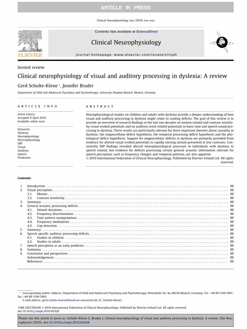
Clinical Neurophysiology xxx (2010) xxx–xxx
ARTICLE IN PRESS
Contents lists available at ScienceDirect
Clinical Neurophysiology
journal homepage: www.elsevier .com/locate /c l inph
Invited review
Clinical neurophysiology of visual and auditory processing in dyslexia: A review
Gerd Schulte-Körne *, Jennifer BruderDepartment of Child and Adolescent Psychiatry and Psychotherapy, University Hospital Munich, Munich, Germany
a r t i c l e i n f o a b s t r a c t
Article history:Accepted 9 April 2010Available online xxxx
Keywords:DyslexiaNeurophysiologyElectrophysiologyERPVisualAuditorySpeechPrediction
1388-2457/$36.00 � 2010 International Federation odoi:10.1016/j.clinph.2010.04.028
* Corresponding author. Address: Department of Chfax: +49 89 5160 5902.
E-mail address: [email protected]
Please cite this article in press as: Schulte-Körnrophysiol (2010), doi:10.1016/j.clinph.2010.04.0
Neurophysiological studies on children and adults with dyslexia provide a deeper understanding of howvisual and auditory processing in dyslexia might relate to reading deficits. The goal of this review is toprovide an overview of research findings in the last two decades on motion related and contrast sensitiv-ity visual evoked potentials and on auditory event related potentials to basic tone and speech sound pro-cessing in dyslexia. These results are particularly relevant for three important theories about causality indyslexia: the magnocellular deficit hypothesis, the temporal processing deficit hypothesis and the pho-nological deficit hypothesis. Support for magnocellular deficits in dyslexia are primarily provided fromevidence for altered visual evoked potentials to rapidly moving stimuli presented at low contrasts. Con-sistently ERP findings revealed altered neurophysiological processes in individuals with dyslexia tospeech stimuli, but evidence for deficits processing certain general acoustic information relevant forspeech perception, such as frequency changes and temporal patterns, are also apparent.� 2010 International Federation of Clinical Neurophysiology. Published by Elsevier Ireland Ltd. All rights
reserved.
Contents
1. Introduction . . . . . . . . . . . . . . . . . . . . . . . . . . . . . . . . . . . . . . . . . . . . . . . . . . . . . . . . . . . . . . . . . . . . . . . . . . . . . . . . . . . . . . . . . . . . . . . . . . . . . . . . . . 002. Visual perception . . . . . . . . . . . . . . . . . . . . . . . . . . . . . . . . . . . . . . . . . . . . . . . . . . . . . . . . . . . . . . . . . . . . . . . . . . . . . . . . . . . . . . . . . . . . . . . . . . . . . . 00
2.1. Motion . . . . . . . . . . . . . . . . . . . . . . . . . . . . . . . . . . . . . . . . . . . . . . . . . . . . . . . . . . . . . . . . . . . . . . . . . . . . . . . . . . . . . . . . . . . . . . . . . . . . . . . . . 002.2. Contrast sensitivity . . . . . . . . . . . . . . . . . . . . . . . . . . . . . . . . . . . . . . . . . . . . . . . . . . . . . . . . . . . . . . . . . . . . . . . . . . . . . . . . . . . . . . . . . . . . . . . 00
3. Summary . . . . . . . . . . . . . . . . . . . . . . . . . . . . . . . . . . . . . . . . . . . . . . . . . . . . . . . . . . . . . . . . . . . . . . . . . . . . . . . . . . . . . . . . . . . . . . . . . . . . . . . . . . . . 004. General acoustic processing deficits . . . . . . . . . . . . . . . . . . . . . . . . . . . . . . . . . . . . . . . . . . . . . . . . . . . . . . . . . . . . . . . . . . . . . . . . . . . . . . . . . . . . . . . 00
4.1. Stimuli durations. . . . . . . . . . . . . . . . . . . . . . . . . . . . . . . . . . . . . . . . . . . . . . . . . . . . . . . . . . . . . . . . . . . . . . . . . . . . . . . . . . . . . . . . . . . . . . . . . 004.2. Frequency discrimination . . . . . . . . . . . . . . . . . . . . . . . . . . . . . . . . . . . . . . . . . . . . . . . . . . . . . . . . . . . . . . . . . . . . . . . . . . . . . . . . . . . . . . . . . . 004.3. Tone pattern manipulations . . . . . . . . . . . . . . . . . . . . . . . . . . . . . . . . . . . . . . . . . . . . . . . . . . . . . . . . . . . . . . . . . . . . . . . . . . . . . . . . . . . . . . . . 004.4. Frequency modulation . . . . . . . . . . . . . . . . . . . . . . . . . . . . . . . . . . . . . . . . . . . . . . . . . . . . . . . . . . . . . . . . . . . . . . . . . . . . . . . . . . . . . . . . . . . . 004.5. Gap detection . . . . . . . . . . . . . . . . . . . . . . . . . . . . . . . . . . . . . . . . . . . . . . . . . . . . . . . . . . . . . . . . . . . . . . . . . . . . . . . . . . . . . . . . . . . . . . . . . . . 00
5. Summary . . . . . . . . . . . . . . . . . . . . . . . . . . . . . . . . . . . . . . . . . . . . . . . . . . . . . . . . . . . . . . . . . . . . . . . . . . . . . . . . . . . . . . . . . . . . . . . . . . . . . . . . . . . . 006. Speech specific auditory processing deficits. . . . . . . . . . . . . . . . . . . . . . . . . . . . . . . . . . . . . . . . . . . . . . . . . . . . . . . . . . . . . . . . . . . . . . . . . . . . . . . . . 00
6.1. Studies in children . . . . . . . . . . . . . . . . . . . . . . . . . . . . . . . . . . . . . . . . . . . . . . . . . . . . . . . . . . . . . . . . . . . . . . . . . . . . . . . . . . . . . . . . . . . . . . . 006.2. Studies in adults . . . . . . . . . . . . . . . . . . . . . . . . . . . . . . . . . . . . . . . . . . . . . . . . . . . . . . . . . . . . . . . . . . . . . . . . . . . . . . . . . . . . . . . . . . . . . . . . . 00
7. Speech perception as an early predictor . . . . . . . . . . . . . . . . . . . . . . . . . . . . . . . . . . . . . . . . . . . . . . . . . . . . . . . . . . . . . . . . . . . . . . . . . . . . . . . . . . . . 008. Summary . . . . . . . . . . . . . . . . . . . . . . . . . . . . . . . . . . . . . . . . . . . . . . . . . . . . . . . . . . . . . . . . . . . . . . . . . . . . . . . . . . . . . . . . . . . . . . . . . . . . . . . . . . . . 009. Conclusion and perspectives . . . . . . . . . . . . . . . . . . . . . . . . . . . . . . . . . . . . . . . . . . . . . . . . . . . . . . . . . . . . . . . . . . . . . . . . . . . . . . . . . . . . . . . . . . . . . 00
Acknowledgement. . . . . . . . . . . . . . . . . . . . . . . . . . . . . . . . . . . . . . . . . . . . . . . . . . . . . . . . . . . . . . . . . . . . . . . . . . . . . . . . . . . . . . . . . . . . . . . . . . . . . 00References . . . . . . . . . . . . . . . . . . . . . . . . . . . . . . . . . . . . . . . . . . . . . . . . . . . . . . . . . . . . . . . . . . . . . . . . . . . . . . . . . . . . . . . . . . . . . . . . . . . . . . . . . . . 00
f Clinical Neurophysiology. Published by Elsevier Ireland Ltd. All rights reserved.
ild and Adolescent Psychiatry and Psychotherapy, Pettenkofer Str. 8a, 80336 Munich, Germany. Tel.: +49 89 5160 5901;
enchen.de (G. Schulte-Körne).
e G, Bruder J. Clinical neurophysiology of visual and auditory processing in dyslexia: A review. Clin Neu-28

2 G. Schulte-Körne, J. Bruder / Clinical Neurophysiology xxx (2010) xxx–xxx
ARTICLE IN PRESS
1. Introduction
Dyslexia is a specific developmental disorder in learning toread, which is not the direct result of impairments in general intel-ligence, gross neurological deficits, uncorrected visual or auditoryproblems, emotional disturbances or inadequate schooling. (Inter-national Classification of Diseases, ICD-10, 2009; Dilling et al.,1991; DSM IV-TR American Psychiatric Association, 2000). Dys-lexia accompanies the individual throughout their lifespan andinterferes with academic achievement or activities of daily livingthat require reading skills (Shaywitz et al., 1999). It occurs in allknown languages (Lindgren et al., 1985; McBride-Chang et al.,2008) and is one of the most common developmental disordersaffecting around 5% of school-aged children (Shaywitz et al.,1990; Katusic et al., 2001). Socioeconomic status and family factorsare known to influence the development of reading abilities, butare not causally related to dyslexia (Stevenson and Fredman,1990; Vellutino et al., 2004).
Since the first description of dyslexic cases a familial aggregationwas observed (Morgan, 1896). Family and twin studies clearly pointto a genetic basis of this complex disorder and first candidate geneshave been found (Smith et al., 1983; Taipale et al., 2003; for reviewssee Paracchini et al., 2007; Schumacher et al., 2007). A key functionof these genes is their involvement in neuronal migration and axongrowth. Imaging studies have clearly demonstrated an altered corti-cal network in dyslexic subjects that comprises left and right supe-rior temporal cortices, left inferior temporal-occipital cortices andboth left and right inferior frontal and posterior temporo-parietalcortices (for review see Schlaggar and McCandliss, 2007).
A number of electrophysiological studies have provided evi-dence for basic perceptual deficits in dyslexia. Abnormal event-related potentials (ERPs; amplitude, latency, topography) forauditory processing of non-speech and speech sounds were foundin dyslexic children and adults. Analogously, altered visual evokedpotentials (VEPs) were reported in dyslexic subjects when non-lin-guistic stimuli were presented.
We conducted a PubMed search spanning two decades of re-search using dyslexia and reading disorder as keywords in combina-tion with event related potentials, ERPs, VEPs, motion onset, contrastsensitivity and mismatch negativity and found 74 papers reportingon electrophysiological correlates of dyslexia (studies with uncleargroup selection criteria or reporting magnetoencephalography(MEG) data were excluded). Forty articles primarily concerned vi-sual processing of non-linguistic material (e.g. graphical materialthat varied on spatial frequency, contrast and temporal frequency)and linguistic material (e.g. letters, words, lexical, syntactic andsemantic aspects of word comprehension). Auditory perception ofnon-linguistic material (e.g. sinus tones), speech sounds (e.g. speechcontrasts like /da/ vs. /ga/) and phonological processing (e.g. rhymejudgements) were reported in 34 articles. For both auditory and vi-sual perception approximately half of the articles investigated ERPcorrelates elicited by non-speech and non-linguistic stimuli. Thegoal of this review is to summarize and integrate investigations con-ducted on the neurophysiological correlates of basic perceptual vi-sual ERPs to motion and contrast sensitivity, as well as auditoryERPs to tones and speech sounds, in dyslexia over the last 20 years.Because the definition of reading disorders can be quite broad, we at-tempted to only include those studies which explicitly recruitedtheir participants according to below average reading (and in somecases spelling). In exceptional cases (e.g. Kraus et al., 1996) we in-cluded studies with less strict definitions of dyslexia, as we consid-ered them to be very important for the review. In these cases, thediscrepancy has been pointed out.
Visual and auditory perception deficits in dyslexia have been re-ported since the beginning of dyslexia research (Hinshelwood,
Please cite this article in press as: Schulte-Körne G, Bruder J. Clinical neurophyrophysiol (2010), doi:10.1016/j.clinph.2010.04.028
1895; Morgan, 1896; Borel-Maisonny, 1951). Since then, it hasbeen repeatedly shown that phonological processing is one of themost relevant factors for learning to read and spell and is impairedin dyslexic children, adults and in compensated adults (for reviewssee Rack, 1994; Snowling, 2000; Ramus et al., 2003).
Based on observations of aphasic children in the 1970’s (Tallaland Piercy, 1973a) a temporal processing theory was formulatedin order to explain perceptual deficits that could account for thephonological processing deficits observed in dyslexia (Tallal,1980a, 2004). Since then numerous studies were conducted to ex-plore the basic auditory deficits in dyslexia by investigating theability to discriminate non-speech stimuli (Farmer and Klein,1995; McArthur and Bishop, 2001). The temporal processing the-ory was extended to the perception of non-linguistic visual stimuli(Stein, 2001). For both areas, neurophysiological studies have mademajor contributions to the understanding of the neurobiologicalcorrelates of dyslexia.
For this review we chose to focus on these two research lines,visual and auditory processing of non-linguistic and sub-lexicalstimuli, in dyslexia. The first reason for this selection is that morethan 36 empirical papers have been published on these topics. Sec-ondly, several common remediation programs, at least in Europe,are based on the assumption of basic visual or auditory perceptiondeficits in dyslexia. These are often time consuming interventions,for both therapists and clients. If the empirical basis for such inter-ventions is low, the use of these interventions in therapeutic set-tings should be critically discussed. Thirdly, there continues to bean urgent need to improve the understanding of the aetiology ofdyslexia despite more than 100 years of research. Although a pho-nological deficit is often found in dyslexic individuals (between30% and 60% depending on the study), its aetiology remains, forthe most part poorly understood.
We begin with a summary of the ERP studies on visual process-ing of non-linguistic stimuli followed by a discussion on the liter-ature covering the basic auditory processing of non-linguisticstimuli. The auditory processing review culminates with speech(sub-lexical) perception and includes studies on early predictors.We have decided to integrate speech perception, which focuseson examining discrimination abilities between consonant–vowel(CV) stimuli and cortical auditory evoked potentials to CV stimuli,because there is accumulating evidence that speech perception isone of the best predictors of reading disability (Guttorm et al.,2005; Lyytinen et al., 2005a, b). Furthermore, the first patho-phys-iological pathway model, from gene function to speech perceptionin dyslexia, has recently been described (Roeske et al., 2009). Thisfinding renders ERP correlates of speech promising candidates forunderstanding the aetiology of dyslexia.
2. Visual perception
Dyslexia was first postulated to be a disorder of the visual sys-tem (Hinshelwood, 1895; Kussmaul, 1877). Since then, numerousempirical studies have described visual deficits for movementand contrast perception in dyslexic individuals (for reviews seeLaycock and Crewther, 2008; Stein, 2001). Some reports point todeficits only within sub-groups of dyslexia (Borsting et al., 1996;Heim et al. 2008; Reid et al., 2007). For example, Borsting et al.(1996) demonstrated that contrast sensitivity to low spatial fre-quencies was reduced only in a group of more severely impaireddyslexic individuals, who suffered from both whole word recalland auditory deficits; however, ERP studies have not yet systemat-ically examined visual potentials in these sub-groups.
Visual sensory impairments in dyslexia have been explainedby deficient functioning within the fast processing transient
siology of visual and auditory processing in dyslexia: A review. Clin Neu-

G. Schulte-Körne, J. Bruder / Clinical Neurophysiology xxx (2010) xxx–xxx 3
ARTICLE IN PRESS
pathway of the visual system, known as the visual magnocellularpathway (Stein, 2001; Ramus, 2004; Sperling et al., 2006;Laycock and Crewther, 2008). The magnocellular pathway ischaracterized by large cells widely distributed across the retinaand projects, via the ventral lateral geniculate nucleus, to thevisual cortex and thereafter largely to the posterior parietal cor-tex. Magnocells specialize in motion and positional relationships,and preferentially mediate fast temporal resolution, low contrast,and low spatial frequencies (Merigan and Maunsell, 1993). Ana-tomically, magnocellular deficits might be attributed to corticalanomalies in the visual system (Galaburda et al., 1985, 1994),where neurons in the lateral geniculate nucleus were found tobe smaller and less structured (Livingstone et al., 1991).Although promising, these findings should be regarded withsome degree of caution as they result from the investigation ofbrains of poorly defined dyslexics and have not yet been repli-cated in a more accurately defined groups. Furthermore, possibledevelopmental delays, acquired and genetic illnesses may haveinfluenced brain anatomy in these cases. Nonetheless, these find-ings are important for the validity of the magnocellular theory(Stein, 2001).
Both investigations of psychophysical and VEP responses haveprovided some evidence for deficits in the magnocellular systemin dyslexia by reporting on perceptual measurements of coherentmotion (Cornelissen et al., 1998), contrast sensitivity (Borstinget al., 1996), rapid motion (Demb et al., 1998), visible persistence(Winters et al., 1989; Schulte-Körne et al., 2004a) and spatial fre-quency (Livingstone et al., 1991). The merit of these results cou-pled with the questionable impact of the anatomical data hasbeen a matter of critical debate (Gross-Glenn et al., 1995; Skot-tun, 2000; Stuart et al., 2001; Amitay et al., 2002; Skoyles andSkottun, 2004; Skottun and Skoyles, 2007, 2008). For example,Skottun and Skoyles (2008) have questioned whether coherentmotion can be used as an appropriate test for understandingand interpreting magnocellular pathway function. Furthermore,the authors have argued that based on similar perceptual deficitsfound in other patient groups (e.g. schizophrenia and autism) themagnocellular deficit cannot be causally linked to dyslexia. De-spite the apparent controversery surrounding the possibility ofmagnocellular deficits in dyslexia, current knowledge pertainingto visual system function does suggest a number of functionallyimportant roles for magnocellular deficits in dyslexia (Laycockand Crewther, 2008). In a recent review, Laycock and Crewther(2008) explore how magnocellular deficits might impact reading.The authors describe how the magnocellular pathway contrib-utes to the rapid integration of visual information when reading,via spatial, temporal and attentional processes, including thecontrol, direction and organization of saccadic eye-movements.
A second visual processing subsystem known as the parvocellu-lar pathway is thought to be largely intact in dyslexia (however, forexceptions see Farrag et al., 2002; Skottun, 2000). The parvocellularpathway originates in small cells concentrated within the fovea,projects to the visual cortex via the dorsal lateral geniculate nu-cleus, and culminates in the temporal cortex. It is sensitive to med-ium and high spatial frequencies, has a moderate temporalresolution (Merigan and Maunsell, 1993) and is important for ob-ject discrimination based on colour, form, and texture.
2.1. Motion
Neurophysiological studies investigating the response charac-teristics of the magnocellular system have made significant contri-butions on its importance for dyslexia. Based on the high temporalresolution of the VEP, different ERP components were found to cor-relate to different visual processes, i.e. contrast sensitivity and mo-tion perception (Kuba et al., 2007).
Please cite this article in press as: Schulte-Körne G, Bruder J. Clinical neurophyrophysiol (2010), doi:10.1016/j.clinph.2010.04.028
Niedeggen and Wist (1999) differentiated two ERP componentsof motion onset in a coherent motion paradigm. At occipital sitesvisual motion onset evoked a positive component between the la-tency of 100 and 130 ms (P100), and a positivity at about 230 ms(P200) was recorded at central-parietal leads. Hirai et al. (2003)identified a negativity evoked by coherent motion onset occurringbetween 200 and 280 ms bilaterally over occipito-temporal elec-trodes. This negativity was nearly independent of luminance con-trast and its amplitude varied with stimulus velocity. Theseneurophysiological correlates of motion perception were also re-corded in 8 month old infants suggesting that even at a very youngage neurons of the visual systems are sensitive to motion percep-tion (Hirai and Hiraki, 2005).
These VEPs correspond to different functional properties of mo-tion processing neurons and cortical areas which are essential forthe analysis and perception of motion. Motion onset is primarilyrelated to local motion detectors which are located in the primarycortex of visual area V1. Coherent motion perception, i.e. the globalintegration of local motion information, is mainly related to corti-cal activity outside the primary visual cortex in the extrastriate vi-sual area V5, and the cortical regions bordering the temporal,parietal, and occipital lobes, respectively (Rodman and Albright,1989; Nowak and Bullier, 1997; Nichols and Newsome, 2002).V5, also known as MT (middle temporal), is involved in perceivingmotion, integrating local motion signals into global percepts andguiding some eye movements (Born and Bradley, 2005).
Motion-related VEPs were investigated in dyslexia in accor-dance with the hypothesis that the magnocellular visual pathwayis impaired in these individuals (Table 1). The VEP studies investi-gating motion onset in dyslexia consistently found prolongedlatencies and smaller amplitudes of the typically evoked P100. Thisfinding suggests a reduced velocity in visual processing and con-tributes to the hypothesis of a selective weakness of the visualtransient system.
Interestingly, in the Kubová studies (Kubová et al., 1996; Kubaet al., 2001) a developmental decrease in the N160 latency in dys-lexic subjects was reported, and suggested a maturational deficit ofthe magnocellular system. In 10-year old children with dyslexiaN160 latencies were recorded at 236 ms, whereas at age 14 theN160 latency had reduced to 162 ms. In comparison, latencies re-corded from children without reading problems at about 10 yearsof age did not differ from those recorded at 14 years of age(167 ms at 10 years, 158 ms at 14 years). Because 40–60% of sub-jects with dyslexia were found to have a longer N160 latency formotion perception (Kuba et al., 2001), the motion onset VEP wasrecommended as part of the diagnostic procedure for dyslexia(Kuba et al., 2007). However, altered motion onset VEPs were alsofound in other disorders such as optic neuritis (prolongation of mo-tion onset VEPs in 28% of the patients) and multiple sclerosis (17%of the patients) (Kuba et al., 2007).
Thus, the studies reporting on motion VEPs suggest deficits indyslexia. Prolonged P100 latencies and attenuated amplitudes arealtered to rapid movements, but not to static visual perception.The longer N160 latencies in younger children with dyslexia suggestthat motion detection might be developmentally delayed, and atadolescence the response is comparable to age-matched controls.
2.2. Contrast sensitivity
A substantial amount of research examining the magnocellulardeficit hypothesis comes from psychophysical and neurophysio-logical studies on contrast sensitivity, a measure of the ability todetect contrasts (both in the spatial and in the temporal realm)at differing thresholds (Lovegrove et al., 1980, 1982; Cornelissenet al., 1998). Based on the assumption that dyslexic subjects arecharacterized by altered magnocellular function, the expectation
siology of visual and auditory processing in dyslexia: A review. Clin Neu-

Table 1Motion: VEP study summary.
Study Sample Motion onset stimuli Motion VEP/ electrodes VEP group differences
Kubová et al.(1996)
CG: N = 16DG: N = 20Mean age: 10
Motion onset: isolated checks;velocity 10 deg/s; random movementfor 200 msControl condition: stationary phase
N160 peak & latency at 3 occ.electrodes: left, right & Oz
Motion onset: *N160 longer latencyin 70% in DGControl condition: n.s.
Kuba et al. (2001) 3 age groups: CG:N = 7Mean age: 13DG_1: N = 10Mean age: 14DG_2: N = 25Mean age: 10
Motion onset: Low contrast (10%);200 ms motion; ISI 1 s1. isolated checks, linear motion;velocity 10 deg/s; 5 colourmodifications (equivalentwavelengths)2. grey concentric frames;expansionControl condition:Transient pattern reversal, highcontrast (96%); black & whitecheckerboard
N160 peak & latency at 3 electrodes:occ. left, right, & Oz
Motion onset: *N160 longer latenciesin DG_2 for condition 1 (20%) & 2(48%); independent from colour;DG_1 latency shortens from 10–14 years; in both CG & DG_1amplitude is lowest at 14 yearsControl condition: n.s.
Schulte-Körne et al.(2004b)
CG: N = 12DG: N = 10Mean age: 12
White dots in rectangular patch onblack background; 5 deg/sCoherent motion: coherent
movement (10%, 20% or 40%);fraction of dotsControl condition: random
movement; 1000 ms
1. P1 & P2 at O1 & O22. P500 at O1, O2, T5, T6, TP7, TP8, P3,
P4, CP3 & CP4
Coherent motion: *P500 reduced areain DGControl condition: n.s.
Schulte-Körne et al.(2004c)
CG: N = 8DG: N = 14Mean age: 12
Motion onset: Sine wave verticalgratings, 2 cpd visual angle,contrast = 0.8, luminance 12 cpd/m2;3 velocities: 2, 8, 16 deg/s; each600 msControl condition: stationary phase
P1 at O1, O2, Oz;P2 at O1, O2, Oz, P3, Pz, P4, C3, Cz, C4
Motion onset: *P1 and P2 loweramplitude & longer latency in DG; P2interaction group & velocity:difference between groups increasedwith greater velocitiesControl condition: n.s.
CG = control group; DG = dyslexic group; cpd = cycles per degree; GFP = global field power; deg = degrees; ISI = interstimulus interval; � = significant group differences found;n.s. = not significant; n.a. = not applicable; occ. = occipital.
4 G. Schulte-Körne, J. Bruder / Clinical Neurophysiology xxx (2010) xxx–xxx
ARTICLE IN PRESS
is that differences in the early VEP components in contrast sensi-tivity paradigms should be observed under low contrasts and fastpresentation rates. Here, non-linguistic low spatial frequency stim-uli in the form of sinusoidal gratings and checkerboard pattern-reversals were presented at various frequencies and contrasts.
One of the earliest electrophysiological studies on contrast sen-sitivity used simple checker-board like pattern-reversal stimuli.Livingstone et al. (1991) reported diminished transient VEP re-sponses (negativity between 20 and 50 msec) in adults with dys-lexia for low contrast stimuli presented at high reversal rates(Table 2) and these results were at least partially confirmed in chil-dren with dyslexia (May et al., 1991; Lehmkuhle et al., 1993;Romani et al., 2001).
Delayed steady state responses based on longer latencies andreduced amplitudes of an early positivity (P100) and an early neg-ativity (N100) at occipital cortical areas suggested an altered mag-nocellular function in dyslexia (Lehmkuhle et al., 1993; Livingstoneet al., 1991). However, several studies (Victor et al., 1993; Johanneset al., 1996; May et al., 1991; Romani et al., 2001; Farrag et al.,2002; Schulte-Körne et al., 1999a; Brecelj et al., 1997-1998) foundinconsistent results, reporting shorter VEP latencies in contrast tothe control group. The latency reduction reported by Romaniet al. (2001) could be explained by the amplitude reduction ob-served at this latency and was interpreted as a ‘‘paradoxical” la-tency reduction.
Victor et al. (1993) and Johannes et al. (1996) used a range ofcontrasts and temporal frequencies that were highly comparableto the stimulus characteristics from the Livingston et al. studyand measured transient and steady state VEP responses in children(Victor et al., 1993) and adults (Johannes et al., 1996). Both studiesfailed to provide supporting evidence for diminished VEP re-sponses. Variability in responses between the dyslexic and controlgroups could not account for failure to replicate the findings,although subjects with attention deficit-hyperactivity disorder(ADHD) did have significantly more variability (Victor et al.,
Please cite this article in press as: Schulte-Körne G, Bruder J. Clinical neurophyrophysiol (2010), doi:10.1016/j.clinph.2010.04.028
1993). Thus, one explanation for the lack of replication in thesetwo studies might be that Livingstone et al.’s (1991) findings re-sulted from a comorbid disorder of dyslexia and ADHD in at leasta sub-group of participants. This possibility highlights the impor-tance of careful participant selection criteria. Here, a reduction incontrast sensitivity might be partially be explained by reducedattention focusing in children, which would presumably renderthe perception of low contrast stimuli more difficult.
In comparison, Brecelj et al. (1997-1998) found group differ-ences (prolongation of the P100 latency) for high contrast stimuli(100%) in children with dyslexia compared to controls, whereasFarrag et al. (2002) reported shortened P100 latencies. Unfortu-nately, such high contrasts are not suited to investigate contrastsensitivity as processed by the magnocellular system. Thus, thefindings of these studies can be interpreted as evidence for a par-vocellular deficit in dyslexic subjects.
Finally, Schulte-Korne et al. (1991a) examined VEPs in Germanadults with spelling disorder using sine wave vertical gratings ofvarying contrasts (0.2, 0.4, 0.6, 0.8) and found no group differences.However, laterality differences were observed. VEP responses re-corded from controls were greater over right occipital leads,whereas the individuals with spelling disorder showed a bilateraldistribution. Other studies have used reading as their main criteriafor study inclusion and therefore the inclusion of subjects based onspelling disorder may be one reason for the non-replication of pre-vious results. This might have led to identification of subjects thatare different from the typical reading disabled subjects in the otherstudies.
3. Summary
We have outlined the ERP research exploring visual processingin dyslexia. In the foreground of the research were studies explor-ing the functional role of the magnocellular system.
siology of visual and auditory processing in dyslexia: A review. Clin Neu-

Table 2Contrast: VEP study summary.
Study Sample Stimuli Contrast sensitivity VEP/electrodes
Relts
Livingston et al. (1991) CG: N = 7Mean age: 25.8DG: N = 5Mean age: 27.4
Checkerboard, vertical .0.16 cpd;horizontal 0.12 cpdTransient pattern reversal VEP:
Contrast 0.2–0.02 at reversal rate0.5 HzSteady state VEP: various contrast
reversals at varying frequencies
Early negativity (C1), P1 at CZ &Oz
Transient VEP:*DG missing or late early negativecomponent at 50 ms at .02 contrast*P1 delayed in DG for low contrastcondition at .02 contrastn.s. 0.2 contrast
Steady state VEP:*amplitude reduction in DG for lowcontrast conditions (.01, .02) at 15 Hzn.s. high contrasts
May et al. (1991) CG/DG: N = 10Mean age: 12.4
Sinusoidal gratings: pattern onset-offset-paradigm with spatialfrequencies (0.5, 1, 2, 4, and 8 cpd) at1.6 Hz
N1, P1, N2, P2 at Oz *Reduced N1 and P1 amplitude of theoffset response at low spatial frequency(0.5 cpd) in DG*Latency reduction of the N2 and P2 in DGat low spatial frequency (1 cpd)
Lehmkuhle et al. (1993) CG: N = 13DG: N = 8Mean age: 10.6
Sinusoidal gratings Pattern-onsetVEP; spatial frequencies 0.5 & 4.5 cpdat 0.1 contrastTwo background conditions: Steady
State & Uniform Field-Flickerbackground
One occipital electrode Steady state:*P1/N2 lower amplitude and delayedlatency at 0.5 cpdUniform Field Flicker:n.s.
Victor et al., 1993 CG: N = 11Mean age: 12DG: N = 10Mean age: 13.5
Checkerboard, stimulus contrastbetween 2% and 20%, reversal rate 4,8 and 16 Hz
P1 at Cz and Oz n.s.
Johannes et al. (1996) CG/DG: N = 6Mean age: 21
Checkerboard, 7 � 5 checks,sides = 4� of visual angleTransient pattern reversal VEP:Contrasts: 0.01, 0.02, 0.15, 0.2, 0.5 atreversal rate of 1 HzSteady state VEP: Contrasts: 0.01,0.02, 0.15, 0.2, 0.5 at reversal rates of10, 20, 30 Hz
C1, P1 and N1 at 13 frontal,central, temporal, parietal andoccipital electrodes
n.s.
Brecelj et al. (1998) CG/DG: N = 12Mean age: 12
Checkerbaords (240 , 490 &1800) with 3varying contrasts (5%, 42%, 100%);stimulation rate 2 Hz
P50, N95, P1 at 3 occipitalelectrodes
*P1 DG longer latencies for 24́ at 100%contrastn.s. P50, N95
Schulte-Körne et al.(1999a)
CG: N = 19Mean age: 22.3DG: N = 15Mean age: 25.9
Sinusoidal gratings, spatialfrequencies: 2, 11.33 cpd; contrasts:0.2, 0.4, 0.6, 0.8
P1 and P2 at occipital electrodes n.s. according to differences in contrastsand spatial frequenciesBut *laterality group effect. CG was right
lateralized for conditions. Thislateralization difference increased withlow frequencies. DG did not showlateralization effects.
Romani et al. (2001) CD/DG: N = 9Age: 10–17
Checkerboards, 0.5 and 2 cpdTransient pattern reversal VEP:
1.05 HzSteady state VEP: 4 Hz
Transient VEP: N70, P1 at OzSteady State VEP: Fast Fourier
Transform of the amplitudes andphases of the harmonics at 8, 16
and 24 Hz
Transient VEP:*N70 lower amplitude and shorter latencyin DG for low spatial frequency (0.5 cpd)n.s. P1Steady state VEP:
*lower amplitudes in DGFarrag et al. (2002) CG: N = 41
DG: N = 52Mean age:
children, age notgiven
Checkerboards1: low (50%) & high (100%) contrast
at 3 Hz; size 162: high contrast (100%) at 1 & 8 Hz;
size 163: high contrast (100%) at 3 Hz;
spatial frequencies low (8 Hz, largesize) & high (64 Hz, small size)
One electrode at Oz; P1 1: high contrasts: *shorter latency in DG;low contrasts: n.s. between groups, but inDG N1-P1 amplitude reduced whencomparing low contrast to high contrast.Finding not observed in CG2. both conditions n.s.3. 8 Hz condtion: *shorter P1 latency in
DG; 64 Hz condition: tendency for P1prolongation in DG; *within DG latenciesbetween conditions significantly different.In CG no differences
CG = control group; DG = dyslexic group; cpd = cycles per degree; GFP = global field power; deg = degrees; ISI = interstimulus interval; � = significant group differences found;n.s. = not significant.
G. Schulte-Körne, J. Bruder / Clinical Neurophysiology xxx (2010) xxx–xxx 5
ARTICLE IN PRESS
The VEPs recorded over occipital cortical brain areas at a latencyof 100–200 ms were prolonged mainly when rapidly moving stim-uli were presented at low contrasts. This finding strongly suggestsan altered magnocellular system; although, this interpretation hasbeen controversially discussed (Skottun and Skoyles, 2006;Schulte-Körne et al., 2004d), the VEP findings on motion have con-sistently reported delayed and altered processing of neurons acti-vated by rapidly moving non-linguistic stimuli.
Please cite this article in press as: Schulte-Körne G, Bruder J. Clinical neurophyrophysiol (2010), doi:10.1016/j.clinph.2010.04.028
However, for contrast sensitivity the results were less consis-tent. This is unexpected if the magnocellular deficit theory is validbecause low contrast stimuli presented at high temporal frequen-cies should specifically activate neurons of the magnocellular sys-tem (Skottun, 2000).
Some aspects that might explain the contradictory findings aresubject age, differences in experimental design across studies andcomorbid disorders like ADHD.
siology of visual and auditory processing in dyslexia: A review. Clin Neu-

6 G. Schulte-Körne, J. Bruder / Clinical Neurophysiology xxx (2010) xxx–xxx
ARTICLE IN PRESS
Furthermore, a substantial variability across experimentalmethods exists, rendering between study comparisons difficult,as can be seen in Tables 1 and 2. For example, contrast levels rangefrom very low (0.01) to very high (1) as do spatial frequencies (low0.15 to high 8 cpd).
The age range between 10 and 46 years and the small samplesizes (e.g. below 10 dyslexic subjects in Livingston et al., 1991;Johannes et al., 1996; Romani et al., 2001) further give evidencefor methodological drawbacks of the VEP studies in dyslexia.
Another critical aspect of the reviewed studies is the possibilityof comorbid ADHD amongst dyslexic subjects that was not re-ported in the majority of studies. A possible objection against theinfluence of ADHD on the ERP findings might be the use of a parvo-cellular control condition in most studies reported. The influence ofcomorbidity with ADHD would also be expected to impact the par-vocellular pathway, which was not the case. None-the-less, atten-tion remains a relevant aspect for understanding and interpretingthese ERP findings. Both visual attention and visuospatial attentionare associated with the magnocellular system and are likely relatedto reading problems in dyslexia (Boden and Giaschi, 2007). Themagnocellular system culminates in the posterior parietal cortex,which is well known to be involved in a range of attentional oper-ations (Constantinidis, 2006; Nachev and Husain, 2006). In dyslexicsubjects impaired visual attention has been repeatedly found(Facoetti et al., 2000a,b; Heiervang and Hugdahl, 2003; Kinseyet al., 2004) Also, ERP research reports evidence for a spatialselective attention deficit in dyslexia (Wijers et al., 2005) and fortop-down impaired attention modulating processes in a coherentmotion perception task (Schulte-Körne et al., 2004b). These pro-cesses were suggested to mediate processing in the magnocellularstream (Vidyasagar, 2005).
How a visual attention deficit contributes to impaired wordreading has been intensively discussed by Boden and Giaschi(2007) and empirically evaluated in behavioural studies (e.g. Solanet al., 2007). In summary, attention is required to filter out lettersthat do not belong to the word (spatial attention) and to directattention to the locus of fixation. From behavioural studies thereis evidence that strengthens the role of visual attention in wordreading in dyslexic subjects (for review see Boden and Giaschi,2007). For ERP research in particular, more adequate study designsare necessary to investigate the influence of visual attention on ba-sic visual perception in dyslexia.
600
Freq
uenc
y / H
z
800
100
Deviant (30 or 50 ms)
Time
Fig. 1. Depiction of the continuous stimulus train used by Huttunen et al. (2007).Deviant stimuli were shortened durations of the 600 Hz stimulus.
4. General acoustic processing deficits
The analysis of general acoustic information, and speech signalsin particular, requires successful interpretation of both temporaland spectral sound features. Tallal first suggested that poor lan-guage skills in dyslexia might arise from a general deficit in pro-cessing rapidly occurring temporal information (Tallal, 1975,1980b; Tallal and Piercy, 1973b). She and her coworkers couldshow that individuals with dyslexia performed worse when dis-criminating between both rapid speech and non-speech stimuli.When the stimuli were presented at slower rates, perception im-proved. Farmer and Klein (1995) extended Tallal’s claim to all sen-sory modalities, for example suggesting temporal processingdisorders in visual perception as well as in auditory perception.ERP research investigating general auditory processing has exam-ined not only temporal, but also spectral aspects of acoustic pro-cessing in dyslexia. Most notably, investigations have beenconducted in these areas with respect to discrimination abilitiesfor pitch discrimination (e.g. Baldeweg et al., 1999; Kujala et al.,2003), stimulus duration (e.g. Baldeweg et al., 1999), frequencymodulation (e.g. Stoodley et al., 2006), gap detection (e.g. Kujalaet al., 2006; Moisescu-Yiflach and Pratt, 2005), and temporal order
Please cite this article in press as: Schulte-Körne G, Bruder J. Clinical neurophyrophysiol (2010), doi:10.1016/j.clinph.2010.04.028
judgements (Kujala et al., 2000; Schulte-Körne et al., 1999b). Themajority of investigations examine the mismatch negativity com-ponent (MMN; Näätänen et al., 1978; Bishop, 2007; Näätänan,2007; Garrido et al., 2009).
4.1. Stimuli durations
The relevance of duration features for speech perception variesfrom language to language. In English for example, vowel durationcan indicate a number of linguistic cues such as distinctions be-tween long and short vowels, vowel stress, and informationregarding within syllable voiced or voiceless consonants (Klatt,1976). In some languages, such as Swedish, Finnish and Japanesevowel duration differences carry semantic relevance (Lidestam,2009). German is for example, a language less reliant on voweldurations for detecting word meaning, and it could be shown thatGerman speakers are less sensitive to duration changes (Kirmseet al., 2008). Relevant durations for speech stimuli are highly var-iable, for example the difference between /ba/-/da/ CV stimuli ischaracterized by a 40 ms duration transition (Mody et al., 1997),whereas a longer duration of 178 ms distinguishes the vowel/æ/in the English word back vs. the /æ/ in bag (Ko, 2009).
In order to examine duration processing devoid of linguisticinformation, Baldeweg et al. (1999) used both passive (e.g. partic-ipants should ignore all stimuli) and active (e.g. participants shouldreact to deviant stimuli) oddball paradigms to compare discrimina-tion between 1000 Hz tones of differing durations in 10 adultsdiagnosed with dyslexia against 10 age-matched controls. The dys-lexic adults were characterized by poor reading skills (in compar-ison to controls), poor working memory and poor phonologicalskills, and dyslexia was defined by a large discrepancy betweentheir general abilities and written language abilities. Four deviantstimuli of varying durations (160, 120, 80, 40 ms) were presentedtogether with a standard duration stimulus of 200 ms. MMN anal-ysis revealed normal duration processing in dyslexia for both MMNpeak latency and amplitude irrespective of attention. Similarly,Kujala et al. (2006) reported no group differences in adults withdyslexia for standard durations of 50 and 100 ms to duration devi-ants of 33 and 65 ms, respectively.
The above studies describe results pertaining to adults.Huttunen et al. (2007) examined children (8.8–14.2 years) withand without a reading disorder in a continuous sound paradigmwhere tones of 600 and 800 Hz (both 100 ms in duration) continu-ally alternated (i.e. no gap). Deviant stimuli were decreased dura-tions in length (30 or 50 ms) for the 600 Hz stimulus (see Fig. 1).The authors postulated that continuous sound would be a moresensitive measure as it avoids possible MMN analysis confoundsthat may arise due to the N1 elicited by stimulus onset (Pihkoet al., 1995). MMN was elicited by both groups to both deviantdurations, and no group differences were reported. Slight laterali-zation differences were found suggesting more activation overthe left hemisphere in the reading disabled group. It is surprising
siology of visual and auditory processing in dyslexia: A review. Clin Neu-
G. Schulte-Körne, J. Bruder / Clinical Neurophysiology xxx (2010) xxx–xxx 7
ARTICLE IN PRESS
that neither amplitude nor latency deficits were reported in thereading disabled group, as duration plays an important role inthe speech deficits in Finnish speaking populations (e.g. Lyytinenet al., 1995). A potential drawback to the continuous sound para-digm is that it restricts analysis to a very short window (300 ms),potentially eliminating relevant late components.
In conclusion, the ERP data show that dyslexic individuals donot suffer from a deficit in processing stimuli of differing durations.The durations used in the above studies are relevant for speechperception, ranging from 30 to 200 ms. Furthermore, even in pop-ulations where duration information is important for speech per-ception, no deficits were reported.
4.2. Frequency discrimination
Paramount to acoustic and speech signals are the frequenciesthat comprise them. In speech, these frequencies are referred toas formants. Formants are meaningful components of sound thatprovide distinguishing information for vowels and consonants.Vowels are usually made up of four to six formants, where the firsttwo formants generally provide enough information to render thevowel distinct. Consonants change the vowel formant structure invarying ways. For example, bilabial sounds like ‘b’ in ball and ‘p’ inmap cause the formants to lower, whereas plosives modify the po-sition of formants of surrounding vowels (Ladefoged, 2001). It fol-lows that if dyslexia is characterized by deficits perceiving andextracting frequency information, then these deficits would mostlikely impact their ability to interpret rapid frequency changesimportant for speech perception.
Investigations of frequency (e.g. pitch: the psychological corre-late of frequency) discrimination suggest a deficit in dyslexia onlybetween stimuli that differ with less than 100 Hz, in a graded fash-ion. An overview of the studies described below is summarized inTable 3.
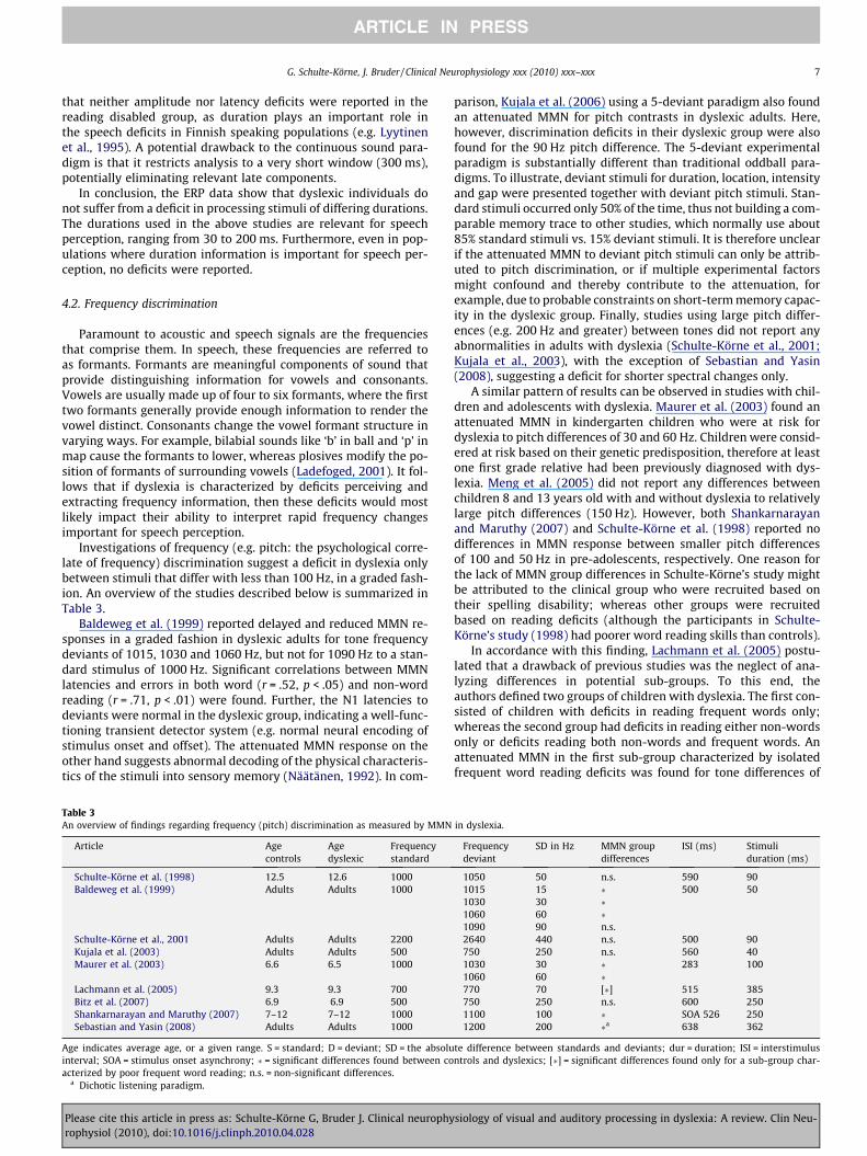
Baldeweg et al. (1999) reported delayed and reduced MMN re-sponses in a graded fashion in dyslexic adults for tone frequencydeviants of 1015, 1030 and 1060 Hz, but not for 1090 Hz to a stan-dard stimulus of 1000 Hz. Significant correlations between MMNlatencies and errors in both word (r = .52, p < .05) and non-wordreading (r = .71, p < .01) were found. Further, the N1 latencies todeviants were normal in the dyslexic group, indicating a well-func-tioning transient detector system (e.g. normal neural encoding ofstimulus onset and offset). The attenuated MMN response on theother hand suggests abnormal decoding of the physical characteris-tics of the stimuli into sensory memory (Näätänen, 1992). In com-
Table 3An overview of findings regarding frequency (pitch) discrimination as measured by MMN
Article Agecontrols
Agedyslexic
Frequencystandard
Schulte-Körne et al. (1998) 12.5 12.6 1000Baldeweg et al. (1999) Adults Adults 1000
Schulte-Körne et al., 2001 Adults Adults 2200Kujala et al. (2003) Adults Adults 500Maurer et al. (2003) 6.6 6.5 1000
Lachmann et al. (2005) 9.3 9.3 700Bitz et al. (2007) 6.9 6.9 500Shankarnarayan and Maruthy (2007) 7–12 7–12 1000Sebastian and Yasin (2008) Adults Adults 1000
Age indicates average age, or a given range. S = standard; D = deviant; SD = the absoluinterval; SOA = stimulus onset asynchrony; � = significant differences found between coacterized by poor frequent word reading; n.s. = non-significant differences.
a Dichotic listening paradigm.
Please cite this article in press as: Schulte-Körne G, Bruder J. Clinical neurophyrophysiol (2010), doi:10.1016/j.clinph.2010.04.028
parison, Kujala et al. (2006) using a 5-deviant paradigm also foundan attenuated MMN for pitch contrasts in dyslexic adults. Here,however, discrimination deficits in their dyslexic group were alsofound for the 90 Hz pitch difference. The 5-deviant experimentalparadigm is substantially different than traditional oddball para-digms. To illustrate, deviant stimuli for duration, location, intensityand gap were presented together with deviant pitch stimuli. Stan-dard stimuli occurred only 50% of the time, thus not building a com-parable memory trace to other studies, which normally use about85% standard stimuli vs. 15% deviant stimuli. It is therefore unclearif the attenuated MMN to deviant pitch stimuli can only be attrib-uted to pitch discrimination, or if multiple experimental factorsmight confound and thereby contribute to the attenuation, forexample, due to probable constraints on short-term memory capac-ity in the dyslexic group. Finally, studies using large pitch differ-ences (e.g. 200 Hz and greater) between tones did not report anyabnormalities in adults with dyslexia (Schulte-Körne et al., 2001;Kujala et al., 2003), with the exception of Sebastian and Yasin(2008), suggesting a deficit for shorter spectral changes only.
A similar pattern of results can be observed in studies with chil-dren and adolescents with dyslexia. Maurer et al. (2003) found anattenuated MMN in kindergarten children who were at risk fordyslexia to pitch differences of 30 and 60 Hz. Children were consid-ered at risk based on their genetic predisposition, therefore at leastone first grade relative had been previously diagnosed with dys-lexia. Meng et al. (2005) did not report any differences betweenchildren 8 and 13 years old with and without dyslexia to relativelylarge pitch differences (150 Hz). However, both Shankarnarayanand Maruthy (2007) and Schulte-Körne et al. (1998) reported nodifferences in MMN response between smaller pitch differencesof 100 and 50 Hz in pre-adolescents, respectively. One reason forthe lack of MMN group differences in Schulte-Körne’s study mightbe attributed to the clinical group who were recruited based ontheir spelling disability; whereas other groups were recruitedbased on reading deficits (although the participants in Schulte-Körne’s study (1998) had poorer word reading skills than controls).
In accordance with this finding, Lachmann et al. (2005) postu-lated that a drawback of previous studies was the neglect of ana-lyzing differences in potential sub-groups. To this end, theauthors defined two groups of children with dyslexia. The first con-sisted of children with deficits in reading frequent words only;whereas the second group had deficits in reading either non-wordsonly or deficits reading both non-words and frequent words. Anattenuated MMN in the first sub-group characterized by isolatedfrequent word reading deficits was found for tone differences of
in dyslexia.
Frequencydeviant
SD in Hz MMN groupdifferences
ISI (ms) Stimuliduration (ms)
1050 50 n.s. 590 901015 15 � 500 501030 30 �1060 60 �1090 90 n.s.2640 440 n.s. 500 90750 250 n.s. 560 401030 30 � 283 1001060 60 �770 70 [�] 515 385750 250 n.s. 600 2501100 100 � SOA 526 2501200 200 �a 638 362
te difference between standards and deviants; dur = duration; ISI = interstimulusntrols and dyslexics; [�] = significant differences found only for a sub-group char-
siology of visual and auditory processing in dyslexia: A review. Clin Neu-

Normal DiscriminationAbility
RW
1000
Hz
Stan
dard
Ton
e
900 950 1050 1100
Dyslexics
HzTone Frequency
MM
N A
mpl
itude
Representational Width for MMN Amplitude to Tone Frequencies
Fig. 2. Schematic representation (adapted from Näätänen and Alho, 1997) of theconcept of representational width (RW) for tone frequencies. RW shows the rangeof sound around the standard stimulus that will not be detected as a deviantstimulus and will not elicit an MMN. To illustrate, the RW might be widened indyslexia (dashed line), resulting in poorer discrimination of tones near the standard,but normal discrimination of tones further away from the standard. According tothis figure, a standard stimulus of 1000 Hz would elicit MMN in dyslexics withdeviants of 900 and 1100 Hz, but not for deviants of 950 or 1050 Hz. For non-dyslexics MMN would be elicited in both cases.
8 G. Schulte-Körne, J. Bruder / Clinical Neurophysiology xxx (2010) xxx–xxx
ARTICLE IN PRESS
70 Hz, whereas the group with non-word reading deficits or a com-bination of non-word and frequent word reading deficits had acomparable MMN to control children. It is unclear if this groupmight show diminished MMN were it homogenous for non-wordand frequent word reading disabilities. Furthermore, it is unclearwhy frequent word reading problems lead to pitch discriminationdeficits in tones and non-word reading problems do not.
In general, a number of reports point to the existence of sub-groups in dyslexia (e.g. Manis et al., 1996; Lovett et al., 2000; Wolfand Bowers, 1999, 2000) where distinct neurological deficits mightaccount for dyslexic sub-groups. In the case of the Lachmann et al.study, frequent word reading problems might be attributed to a vi-sual, or whole-word dyslexia sometimes referred to as dyseideticdyslexia. The group with combined deficits might be characterizedby auditory deficits, also described as dysphonetic dyslexia (Boder,1970, 1973). Therefore, understanding how these groups differ atthe behavioural and neurophysiological levels is relevant to under-standing the aetiology and the heterogeneity of dyslexia.
As reviewed here, a deficit in perceiving differences in pitch be-tween two sounds seems to characterize dyslexia. This deficitmight reflect a widened representational width (RW) in sound per-ception. RW is a concept proposed by Näätänen and Alho (1997) todescribe an individual’s discrimination accuracy, which is depen-dent on their particular ability to perceive differences in sound(Fig. 2). The narrower the width is, the better the discriminationability. The data reported here might reflect a widening of this con-struct in dyslexia, thus allowing them to accurately discriminate ingeneral, but to a less precise extent.
Tone-Pattern Condition Tone-Pair Condition
200 150 50
200 15050
150
50
* = ISI, ms50 Hz 30 ms=* = Deviant
condition
a
Fig. 3. (a) Depiction of stimuli used by Kujala et al. (2000) for a four tone pattern conditused with children. Both pattern deviations led to irregular MMN in dyslexics. Arrows indtone in an unexpected position and the absence of thetone in its expected position are
Please cite this article in press as: Schulte-Körne G, Bruder J. Clinical neurophyrophysiol (2010), doi:10.1016/j.clinph.2010.04.028
4.3. Tone pattern manipulations
Tone patterns reflect experiments with multiple sinus tonespresented in a rhythmic pattern with a combination of durationsand frequencies between tones. Deviations in the pattern thereforereflect a combination of stimulus duration and stimulus frequency.These more complicated acoustic signals provide a more validanalogy to speech than the presentation of tones in isolation,reflecting the necessity to perceive rapid sequences of incominginformation such as formant transitions and voice onset timing.
Kujala et al. (2000) used patterns composed of four or two 50 Hztones (30 ms duration) and varied the duration of intervals be-tween the onset of tones within the patterns in order to examinediscrimination abilities among adults with dyslexia. In the fourtone pattern the intervals between tones in the standard stimuluswere 200, 150 and 50 ms (see Fig. 3a). In the deviant condition the2nd (150 ms) and the 3rd (50 ms) interval durations wereswitched. In essence, this resulted in two deviant conditions: (1)the early onset of the 3rd tone; and (2) the absence of the 3rd tonein its expected position (see Fig. 2a).
Controls elicited two MMN responses corresponding to eachdeviant condition; however the individuals with dyslexia showedonly the second MMN response to the absence of the 3rd tone inits expected position. In controls this second response was lateral-ized to the right hemisphere, whereas the group with dyslexiashowed bilateral activity. In a control condition, MMN was similarbetween the two groups to a single interval deviant (Fig. 2b).
The presence of deficient MMN to an additional deviant condi-tion in adults listening to tone patterns was further substantiatedby Schulte-Körne et al. (1999) and Kujala et al. (2003). Increasedbackward masking effects were suggested as a potential contribu-tor to the discrimination deficit. Finally, in a study with Chinesechildren (8–13 years old) Meng et al. (2005) were able to demon-strate that children with dyslexia also perceive tone patterns irreg-ularly. The children exhibited attenuated MMN in a large timewindow from 150 to 500 ms (determined by averaging 20 msMMN time windows).
These results suggest that dyslexia is characterized by an inabil-ity to process short rhythmic patterns, and that this deficit is re-lated to the physical properties of the stimuli. Accurateperception of rhythmic patterns requires the ability to integratethese incoming sounds temporally. In healthy subjects, the integra-tion of incoming auditory features into neural representations insensory memory is thought to occur within a sliding temporal win-dow of about 150–200 ms referred to as the temporal window ofintegration (TWI; Näätänen, 1990). The length of this windowhas been established in a number of MMN studies (Schröger,1997; Tervaniemi et al., 1994; Wang et al., 2005; Winkler andNäätänen, 1992, 1994; Yabe et al., 1997, 1998. Within 150–200 ms incoming acoustic information is integrated into a singleauditory percept. Information occurring in the subsequent
Tone-Pattern Condition
150 350
* = ISI, ms2000 Hz 40 ms
=*
350 150
800 Hz 40 ms
=
b
ion and a tone pair condition with adult subjects. (b) Standard and deviant patternsicate the onset of the deviant patterns. In Tone-Pattern conditions both the onset of adeviations from the standard pattern.
siology of visual and auditory processing in dyslexia: A review. Clin Neu-

G. Schulte-Körne, J. Bruder / Clinical Neurophysiology xxx (2010) xxx–xxx 9
ARTICLE IN PRESS
150–200 ms is thought to be integrated into a second auditory per-cept, thus the TWI continuously slides forward in time. In supportof this, Winkler et al. (1998) could show that only one MMN waselicited if two deviations from a standard stimulus occurred within150 ms, in other words, the two events were treated as one deviantevent. If, on the other hand, the same two deviant events occurred250 ms apart two MMNs were elicited. Thus, it is plausible that theabsence of a MMN observed in the subjects with dyslexia in thestudies described above, might in fact reflect a wider TWI in dys-lexia. If this were the case, the deviations occurring in the tone pat-terns might have been integrated into a single temporal unitinstead of separate units as in the healthy controls, thus signifyingabnormal temporal integration into auditory short-term sensorymemory.
4.4. Frequency modulation
In order to investigate whether auditory perception is influ-enced by cognition in individuals with dyslexia, Stoodley et al.(2006) sampled high-functioning university students who hadbeen diagnosed with dyslexia in their childhood, and comparedthese subjects to non-impaired readers with comparable cognitivecapabilities. Despite their high achievement, reading skills contin-ued to differentiate the individuals with dyslexia from their peers.Assuming that university students with dyslexia must have devel-oped strategies to compensate for their difficulties in reading in or-der to achieve success in academia, the study was designed to tapauditory deficits that could not be influenced by top-down cogni-tive strategies.
Stoodley et al. (2006) employed a pure standard tone (1000 Hz)and deviant tones (1000 Hz frequency-modulated with 5, 20 or240 Hz) in 3 blocks (17% deviants) to test auditory perception ina passive oddball paradigm. MMN was attenuated in the 20 Hzcondition in both early (150–300 ms) and late (300–500 ms) win-dows in the group of students with dyslexia. The magnitude ofthe MMN correlated to measures of literacy: smaller MMN was re-lated to a larger discrepancy between cognitive and literacy abili-ties, suggesting a deficit in frequency modulation related toliteracy. The reason why a correlation with the 20 Hz frequencymodulation was found, but not for frequency modulation of 5 Hzand 50 Hz might be explained by the findings that the 20 Hz timeframe is important for distinguishing stop consonants (see Clarkand Yallop, 1995). Interestingly, in a separate test session, examin-ing psychophysical thresholds the group with dyslexia performedas well as controls in actively detecting the frequency modulatedchanges, possibly demonstrating top-down strategies for enhanc-ing performance. Therefore, MMN seems to be a more sensitiveindicator of perceptual deficits in dyslexia.
4.5. Gap detection
Gap detection is a temporal processing task which measures theminimum ISI required to perceive an interruption (gap) in a con-stant train of stimuli (Farmer and Klein, 1995). Moisescu-Yiflachand Pratt (2005) investigated the ERP correlates to standard stim-uli (85%; duration 280 ms) of white noise in comparison to deviantstimuli (15%; duration 280 ms) of white noise with gaps of 20 msin both active and passive paradigms. No group differences werefound at the behavioural level. However, both N1 and P3 latencieswere significantly prolonged in the dyslexic group, whereas N1magnitude was greater in comparison to controls. N1 effects wereobserved for both the active and the passive paradigms, whereasP3 was present in the active paradigm reflecting the allocation ofattentional resources (e.g. Picton, 1988). The N1 results furtherpoint to deficits in sensory processing, whereas P3 has been asso-ciated with a variety of deficits, including attention deficits. The in-
Please cite this article in press as: Schulte-Körne G, Bruder J. Clinical neurophyrophysiol (2010), doi:10.1016/j.clinph.2010.04.028
creased magnitude of the N1 in dyslexics has been reported inother studies (Georgiewa et al., 2002; Helenius et al., 2002) andmight be related to general allocation of pre-attentional resources.However, in high-functioning dyslexic participants it might alsoreflect compensatory mechanisms serving to increase arousal andreadiness; in turn the enhanced N1 in the dyslexic group might re-flect an early investment in stimulus feature analysis in order tocounteract perceptual impairments. In favour of this assumptionis the lack of group differences found for task performance. Thus,this study further strengthens the view that (at least) adults withdyslexia are characterized by auditory processing deficits whenprocessing non-speech stimuli, and highlights a further dimensionthat these deficits can manifest. The ERP data show that this per-ceptual impairment occurs within 100 ms from stimulus onset.
5. Summary
Deficits in general auditory processing in dyslexia are prevalentfor stimuli of increasing complexity and/or similarity. Detectingdifferences of frequencies between simple sinus tones revealMMN irregularities only when tones differed by about 100 Hz orless. This finding might be explained by a widened RW in dyslexiaand is relevant for speech perception. A deficit perceiving stimulusduration differences alone cannot explain speech perception defi-cits. Interestingly, more complex patterns of tones with variousdurations and frequencies consistently revealed diminished MMNin both children and adults. This finding is important for speechperception because tone patterns characterize the rapid and dy-namic transmission of natural speech sounds and might reflect awidened TWI. Furthermore, individuals with dyslexia may alsohave difficulties with frequency modulation and gap detection,although replications of these preliminary findings would be re-quired for any strong conclusions. Importantly, in some cases(Moisescu-Yiflach and Pratt, 2005; Stoodley et al., 2006) the resultsreveal how ERP studies more sensitively detect persistent deficitsassociated with dyslexia than psychophysical studies, where pre-sumably high-functioning adults with dyslexia have compensatedfor their difficulties in reading and writing in a number of ways anddeveloped cognitive strategies to enhance their performance inthese tasks.
6. Speech specific auditory processing deficits
A second area of investigation in dyslexia has focused on exam-ining perception specific to the acoustic processing of speech stim-uli. A speech specific deficit conjures with research suggesting thatthe core deficit in dyslexia is phonologically based (Shaywitz,1996; Ramus et al., 2003; Bishop and Snowling, 2004). Thishypothesis underlines deficits in phoneme awareness, or the expli-cit knowledge about the sound structure of speech. In some cases,it has been argued that auditory deficits in dyslexia are specific tospeech, and therefore cannot be attributed to general acoustic pro-cessing deficits (Mody et al., 1997; Schulte-Körne et al., 1998,2001; Bishop and Snowling, 2004; Bitz et al., 2007).
Speech perception involves the mapping of basic auditory infor-mation onto phonological units. The relationship or boundary be-tween acoustic sound features and phonemic processing is notwell defined. In spoken language, a single phonemic sound’s pro-duction is dependent on the surrounding phonemes, resulting inno fixed pattern for any phoneme (Lieberman and Blumstein,1988; Tunmer et al., 1984). Therefore, phonemic units alone areabstract, but meaningful acoustic representations of speech parts.Not surprisingly, an auditory speech-processing deficit was deter-mined in dyslexia in a number of ERP studies (see Table 4). Themajority of studies have focused on MMN, as an index of successful
siology of visual and auditory processing in dyslexia: A review. Clin Neu-

Table 4An overview of findings regarding consonant–vowel (CV) discrimination in both children and adults with dyslexia, including MMN and auditory evoked potential studies.
Article Agecontrols
Agedyslexic
CV S CV D ERP group differences ISI (ms) Stimulidur (ms)
Kraus et al. (1996) 6–15 6–15 /da/ /ga/ �(MMN) ? 100/ba/ /wa/ n.s.
Schulte-Körne et al. (1998) 12.6 12.5 /da/ /ba/ �(MMN) 590 110Cunningham et al. (2001) 10–13 10–13 /da/ in quiet n.a n.s. 550 40
/da/ in noise n.a �(P2,N2)(labelled P1́,N1́)/da/ in cue-enhancednoise
n.a n.s.
Schulte-Körne et al. (2001) Adults Adults /da/ /ga/ �(MMN) 500 110Maurer et al., 2003 6.6 Mean 6.5 Mean /ba/ /ta/ & /da/ �(MMR) 283 100Warrier et al. (2004) 8–13 8–13 /da/ in quiet n.a. n.s. 590 40
/da/ with backgroundnoise
n.a. [�] (N2)
Giraud et al. (2005) Adults Adults /ba/ n.a. Sub-group 1: [�] (extra components)Sub-group 2: [�] (missing N240)
1030 380
/pa/ n.a. Sub-group 1: [�] (extra components)Sub-group 2:[n.s.]
270
Lachmann et al. (2005) 8–11 8–11 /ba/ + /da/ + [�](MMN), �(N250) 515 385Meng et al. (2005) 8–13 8–13 /ba/ /ga/ �(MMN) 700 40
/dan/ /dai/ �(MMN)/ba1/ /ba2/ n.s.
Moisescu-Yiflach andPratt (2005)
Adults Adults /ta/ /pa/ �(N1,N2, P2,P3) 1900 ?/ba/ /pa/ �(N1,N2, P2,P3)
Cohen-Mimran (2006) 10–13 10–13 /pa/ /ba/ �(P3) 2000 230Bitz et al. (2007) 6–7 6–7 /ga/ /ka/ �(MMN) 600 250Shankarnarayan
and Maruthy (2007)7–12 7–12 /tRa/ /dZa/a & /sa/b [�] (MMN) SOA 526 250
/da/ /d8a/c & /das/d [�] (MMN) SOA 526 250 & 175Sebastian and Yasin (2008) Adults Adults /ta/ /ka/ n.s. 638 362
/ba/ /da/ n.s.
Age indicates average age, or a given range. S = standard; D = deviant; dur = duration; ISI = interstimulus interval; SOA = stimulus onset asynchrony; � = significant differencesfound between controls and dyslexics; [�] = significant differences found for a sub-group only; n.s. = non-significant differences; From the Kannada language in South India.Stimuli differ in.
a Voicing.b Manner of articulation.c Place of articulatio.d Vowel duration; n.a. = not applicable.
10 G. Schulte-Körne, J. Bruder / Clinical Neurophysiology xxx (2010) xxx–xxx
ARTICLE IN PRESS
discrimination between formant transitions (FT) (spectral changes;e.g. /da/ vs. /ga/), with some research examining voice onset timing(VOT) transitions (temporal changes; e.g. /ba/ vs. /pa/).
6.1. Studies in children
Kraus et al. (1996) first reported attenuated MMN to FT in agroup of learning disabled children and adolescents (aged 6–15).The children in the experimental group were defined by either adiagnosis of reading disorder, attention deficit disorder or both.Despite the heterogeneous make-up of the group, the children’sperformance on measures of reading was poorer than that of theage-matched controls. Participants were first tested on their abilityto discriminate between two CV syllables, /da/ vs. /ga/ (spectralchange) and /ba/ vs. /wa/ (temporal change). All learning disabledchildren were much poorer in both tasks, however some could dis-criminate /ba/-/wa/ contrasts relatively well. Therefore, in passiveoddball paradigms comparing both CV pairs, learning disabled chil-dren who could sufficiently discriminate /ba/-/wa/, but remainedpoor /da/-/ga/ perceivers, were compared to age-matched controls,who performed well in both discrimination measures. SignificantMMN (200–500 ms) to /da/-/ga/ was found for control children,but the amplitude of the MMN was diminished in the learning dis-abled group. Similar MMN was present for both groups to /ba/-/wa/ stimuli. MMN duration and area to /da/-/ga/ correlated moder-ately with /da/-/ga/ discrimination accuracy (r = �.40, r = �.42,respectively). Therefore, these findings suggest links betweenneurophysiological mechanisms as assessed by a MMN paradigm(i.e. independent of attention and response and actual discrimina-tion ability at the behavioural level; prior to conscious perception).
Please cite this article in press as: Schulte-Körne G, Bruder J. Clinical neurophyrophysiol (2010), doi:10.1016/j.clinph.2010.04.028
Furthermore, the discrimination deficits were highly specific,where deficits for contrasts differing in spectral content (/da/-/ga/), but not for contrasts differing in their temporal content(/ba/-/wa/), were found.
Deficits discriminating between CV changes with spectral infor-mation were reported in a number of other studies with children.Schulte-Körne et al. (1998) found an attenuated MMN (176–302 ms) in children with dyslexia (average 12.5 years) to /da/-/ba/ contrasts. In younger children (6–7 years) with a familial riskfor dyslexia, Maurer et al. (2003) reported earlier attenuation(109–140 ms) of a component similar to MMN, labeled MMR (mis-match response), to both /ba/-/ta/ and -/da/ contrasts. MMR is de-scribed as a component that emerges in place of MMN in veryyoung children when differences between stimuli are small andISIs are very short (in this experiment 283 ms). Short ISIs on theother hand do not allow for analysis of later components, whichare dependent on longer processing times. Therefore, with longerISIs these children may have also revealed deficits in later process-ing stages. MMN deficits were further reported in Chinese children(8–13 years) to da/-/ga/ stimuli in a very early, atypical time win-dow (0–100 ms) (Meng et al., 2005). Finally, Lachmann et al. (2005)examined the discrimination of /ba/-/da/ contrasts in children (8–11 years) with dyslexia and age-matched controls. The childrenwith reading problems were analyzed according to sub-groups.The first group was poor in fluent word reading only, whereasthe second was marked by impairments in either non-word read-ing only or both non-word and fluent word reading. Surprisingly,only those children marked by isolated frequent word reading def-icits had an attenuated MMN from 98–198 ms. A later time win-dow was not examined. This result is unexpected, as those
siology of visual and auditory processing in dyslexia: A review. Clin Neu-

G. Schulte-Körne, J. Bruder / Clinical Neurophysiology xxx (2010) xxx–xxx 11
ARTICLE IN PRESS
children who are marked by phonological impairments would beexpected to do most poorly on CV discrimination tasks. The read-ing of non-words requires good phoneme awareness in order tocorrectly apply grapheme–phoneme correspondence rules. There-fore, children who performed most poorly in the phonological taskmight be expected to discriminate the CV stimuli more poorly.These data suggest that grapheme–phoneme correspondence fail-ure, as indexed by non-word fluency, is not a result of early phone-mic discrimination processes. Interestingly, the analysis ofexogenous ERPs to deviant stimuli revealed an impaired N250 inboth sub-groups. N250 is thought to index general aspects of audi-tion and sound reception (Shafer et al., 2000; Ceponiene et al.,2001); therefore abnormal N250 is suggestive of a more generalimpairment to sound reception in dyslexia. In accordance withLachmann et al.’s results, Warrier et al. (2004) also reported abnor-mal latencies in the N250 range to /da/ stimuli presented withbackground noise in a subset of children with learning problems,but not in others. It is however unclear how many or if any ofthe children in this study had dyslexia. Reduced amplitudes to /da/ in noise within this time window have been reported else-where (Cunningham et al., 2001). Similar to these results, Shan-karnarayan and Maruthy (2007) reported individual results forboth spectral and temporal stimuli. The authors found that about2/3 of the children with dyslexia tested (n = 15) exhibited pro-longed or absent MMN latencies, whereas 1/3 revealed comparableMMN to controls. These children were not however further classi-fied into sub-groups based on behavioural measures.
Despite normal MMN to temporal information (/ba/-/wa/) inKraus et al.’s study, deviant MMN to VOT was found in two studieswith children. Bitz et al. (2007) examined /ga/-/ka/ contrasts in 6–7 year olds at risk for dyslexia, defined by poor phonologicaldecoding skills, compared to age-matched controls. The authors re-ported two significant late MMN windows in the control children(300–450 ms and 450–600 ms). Later MMN-like activity is also re-ferred to as late discriminative negativity (LDN; Cheour et al.,2001). LDN might be less sensitive to stimulus specific characteris-tics than MMN (Ceponiene et al., 2002) and is believed to reflectfurther processing of a deviant stimulus beyond sensory sound dis-crimination. LDN is, for example, enhanced to words in comparisonto non-words matched on all levels of acoustic complexity, thussuggesting some association to the cognitive meaning of thesestimuli (Korpilahti et al., 2001). However, the exact nature of thislate discriminative processing is not well understood. Bitz et al.’sfindings revealed attenuation in both MMN windows for the chil-dren with phonological deficits. Furthermore, similar to the find-ings from Kraus et al. (1996) moderate correlations were foundbetween MMN amplitude (at electrodes FC3/FC4) and phonologicaldecoding abilities (r = .42). Here, increasing MMN amplitude indi-cated enhanced behavioural performance, suggesting a potentiallink between pre-attentive discrimination at the neural level andphonological skills. Finally, in an active paradigm Cohen-Mimran(2006) found prolonged P3 latencies but normal P3 amplitudes to/pa/ vs. /ba/ stimuli in 10–13 year old Hebrew children diagnosedwith a reading disorder. This was coupled with reduced accuracyand longer reaction times. Correlations between later P3 latenciesat Cz and poorer phonological awareness skills (r = .-69, p < .001)were also reported.
6.2. Studies in adults
Schulte-Körne et al. (2001) reported a diminished LDN to /da/-/ga/ contrasts (490–620 ms) in adults with dyslexia who were re-cruited based on poor spelling achievement, and who were diag-nosed with dyslexia (poor reading and spelling) as children.MMN in an earlier time window (270–320 ms) was not signifi-cantly attenuated.
Please cite this article in press as: Schulte-Körne G, Bruder J. Clinical neurophyrophysiol (2010), doi:10.1016/j.clinph.2010.04.028
Reporting on auditory evoked responses Giraud et al. (2005)found evidence for two distinct response patterns to voiced (/ba/)and voiceless (/pa/) CV stimuli in adult participants with persistentreading deficits. In healthy subjects both stimuli elicited a P1/N2complex, and additionally /ba/ stimuli evoked an N240. N240 in-dexes the onset of voicing by /ba/, but is not present for voiceless/pa/ stimuli. Half of the participants with reading deficits elicitedan extra, earlier component at 50 ms (P50) to both stimuli andhad delayed off-responses. Despite these differences, these partic-ipants processed /ba/ stimuli in a similar manner as controls, as in-dexed by the presence of N240 to /ba/. P50 was absent in the otherhalf of the participants to both stimuli, who also lacked N240 to /ba/, thus ERP morphology was similar for both /ba/ and /pa/. Theseresults suggest diversity in acoustic processing deficits in dyslexia,despite similar reading abilities. The first pattern described here ischaracterized by additional acoustic processing and slowness inacoustic processing, yet a sustained ability to process voiced /ba/.The second pattern is characterized by an inability to perceiveand code important speech cues, such as those differentiatingvoiced and voiceless CVs.
Moisescu-Yiflach and Pratt (2005) examined both temporal andspectral acoustic qualities of speech stimuli using /ta/-/pa/ (spec-tral cue) and /ba/-/pa/ (temporal cue) in a group of high-function-ing adults with dyslexia. For both CV contrasts, active and passiveoddball paradigms were employed to rule out potential confoundsresulting from attention, response strategy or motivation on theneurophysiological response. Psychophysical results revealedequally good discrimination in the active condition across groups.However, at the neurophysiological level a number of differenceswere found. Regardless of whether CV contrasts involved spectralor temporal cues, passive listening revealed longer N1 latenciesin dyslexia and active participation resulted in prolonged N1, P2and P3 latencies. Furthermore, higher N1 amplitudes were foundwithin the dyslexic group in the active conditions.
The N1 is the most prominent exogenous component in re-sponse to acoustic input in adults and is thought to index basicencoding of acoustic information at the moment it enters primaryauditory cortex (e.g. Näätänen, 1990; Nagarajan et al., 1999). How-ever, the N1 response is complicated by a number of sub-processesthat contribute to its morphology (Key et al., 2005; Näätänen andPicton, 1987; Wood, 1995). Although it is not believed to be thesame component as the N250 in children (e.g. see Lachmannet al., 2005), these two components are thought to be correlatesof similar functions (Ceponiene et al., 2001). In this study (Moise-scu-Yiflach and Pratt, 2005), the N1 was found to be irregular insubjects with dyslexia for both spectral and temporal information,suggesting that the speech-processing deficit is general.
Finally, Sebastian and Yasin (2008) studied spectral and tempo-ral contrasts in university students diagnosed with dyslexia as chil-dren. As in Stoodley et al.’s (2006) study, these individuals wereconsidered to have compensated for their reading deficits in somemanner in order to have achieved an academic standing requiredfor university entry. Sebastian and Yasin (2008) reported no groupdifferences in MMN amplitudes and latencies. This is the onlystudy to report a lack of speech-processing deficit.
7. Speech perception as an early predictor
Recording ERPs elicited by passive listening to speech sounds atbirth seems to be a promising method for identifying early predic-tors of later reading disorders. Molfese (2000) applied a passive lis-tening experiment to 186 full-term babies within 36 h of birth.Synthetic speech stimuli /gi/, /bi/ and /di/ were presented duringsleep. Discriminate analysis at 8 years of age was conducted in or-der to classify the children according to speech ERPs at birth. The
siology of visual and auditory processing in dyslexia: A review. Clin Neu-

12 G. Schulte-Körne, J. Bruder / Clinical Neurophysiology xxx (2010) xxx–xxx
ARTICLE IN PRESS
children were either diagnosed with dyslexia (n = 17), were poorreaders (low IQ and reading ability; n = 7), or were normal readers(control children; n = 24). Eighty-one percent of a subset (n = 48) ofchildren could be correctly classified into their reading categoriesbased on three ERP components (N1, P2 and N2) at birth and read-ing and IQ scores. The ERPs at birth could successfully discriminate76.5% of children with dyslexia, 100% of poor readers and 79% ofcontrol children at 8 years of age (Molfese, 2000). Furthermore,correlations between the amplitude and latency of N1, measuredbetween ages 1 and 4, and word reading at age 8 were significant(Espy et al., 2004). In an additional analysis the ERP components,home environment (e.g. academic stimulation, stimulation of com-municative competence), socioeconomic status (e.g. parental edu-cation and occupation), preschool language score and short-termmemory were integrated into a regression analysis (Molfeseet al., 2001). Up to 70% of the variance of word reading was ex-plained by these factors. The ERP components, as well as preschoolspeech perception, made the strongest contribution (half or moreof the variance) to reading. It can therefore be concluded thatspeech perception influences reading abilities at a very early age.Interestingly, the neurophysiological correlates made a strongerprediction than the home environment and the SES.
In a second longitudinal study (Jyväskylä Longitudinal Study ofDyslexia, (Lyytinen et al., 2005a, b)) ERPs to speech stimuli wereregistered in one hundred babies at risk (defined as one first graderelative reporting dyslexia) and compared with ERPs from 100 ba-bies from control families. Both synthetic and natural spokenspeech stimuli were presented (Leppanen et al., 1999; Pihkoet al., 1999; Guttorm et al., 2005). Even at birth, distribution differ-ences were found for the standard and deviant response to the nat-ural speech sound /ka/. In the at-risk population an ERP componentbetween 540 and 630 ms at birth elicited by synthetic speech stim-uli had a larger and prolonged response in the right hemispheredue to a slower polarity shift from the major positive to the nega-tive deflection. In the control group the differences between stan-dard and deviant stimuli were largest in the left hemisphere,whereas in the at risk group the ERP difference was largest in theright hemisphere (Guttorm et al., 2005; Leppanen et al., 1999).The anomalous ERP activity in the right hemisphere in the at-riskchildren correlated with lower word and non-word reading accu-racy in the first grade of school (Lyytinen et al., 2005a, b) poorerlanguage skills at 2.5 years; verbal memory at 5 years (Guttormet al., 2005) and reduced phonological skills, slower lexical accessand less knowledge of letters at 6.5 years (Guttorm et al., 2009).These findings suggest that already at birth the speech processingin at-risk children differs from controls and that the neurophysio-logical correlate of this functional difference is located over theright hemisphere. It is not yet clear how well the ERPs will alsopredict an actual dyslexia diagnosis. However, the poorer language,memory and phonological skills found at ages 2.5, 5 and 6.5 in theat-risk children are often found in older children and adults whohave a diagnosis of dyslexia, thus it would not be surprising if a sig-nificant portion of the at-risk children will eventually developdyslexia.
8. Summary
Speech specific processing deficits to CV stimuli have beenshown consistently in both children and adults, for both spectraland temporal transitions, and in active and passive paradigms.Early MMN deficits suggest difficulties discriminating betweentwo stimuli, whereas late MMN abnormalities might be indicativeof faulty long term memory traces (Näätänen, 2001). Finally, audi-tory ERPs at birth can be predictive for later reading skills andshort-term verbal memory in children at risk for dyslexia.
Please cite this article in press as: Schulte-Körne G, Bruder J. Clinical neurophyrophysiol (2010), doi:10.1016/j.clinph.2010.04.028
A number of authors, including our own group, have argued foror against a speech specific vs. a general auditory processing deficitin dyslexia based on isolated studies (e.g. Bitz et al., 2007;Schulte-Körne et al., 1998, 2001). Unfortunately to date no studyof dyslexia has addressed this question using equally comparablespeech and non-speech stimuli (e.g. such as those of Tampaset al., 2005), and tone conditions were often sinus tone compari-sons with differences greater than 100 Hz. Given the above review,it is clear that certain non-speech deficits are apparent in dyslexia,and these are attributed to discrimination of small frequency dif-ferences and to detection of differences in complex tone patterns,both of which are relevant to speech discrimination.
9. Conclusion and perspectives
In this review we cover ERP research on basic auditory and vi-sual processing in dyslexia. Higher cognitive processes, such asphonological processing, word recognition and orthographic pro-cessing are also impaired in dyslexia, but were not addressed. A re-view on the ERP literature pertaining to these areas would also behelpful to conclude the current understanding of the electrophys-iology of dyslexia.
Throughout this review we have highlighted the neurophysio-logical literature pertaining to altered visual and auditory sensoryprocessing in dyslexia. The literature touches on infancy, child-hood, adolescence and adulthood, demonstrating the persistenceof these information processing deficits throughout the lifespan,yet does not present evidence for causality. For example, the iden-tification of the phase locked processes in auditory and visual pro-cessing points to sensory processes that might be important for theunderlying development of reading, but these findings can not bestrongly linked to reading and writing deficits.
Given the heterogeneity of the research reported thus far, it isimportant to consider the methodologies employed across studies.The following concerns are important to take into consideration forfuture research.
First, the careful selection of individuals with dyslexia forempirical investigation is of great importance. With the evidencefor sub-groups of dyslexia mounting and the high comorbidityrates reported, it is apparent that different selection procedurescertainly influence the ERP results. Thus, we emphasize the impor-tance of developing stringent inclusion criterion to be appliedacross labs. As mentioned, the comorbidity between dyslexia andADHD is very high, but dyslexia is also comorbid with other disor-ders including anxiety disorder (Caroll & Iles, 2006), ADHD(Stevenson et al., 2005), antisocial behavior (Maughan et al.,1996), and depression (Willcutt and Pennington, 2000). Most ofthe ERP studies reviewed did not screen for the existence of ADHDsymptoms in their sample of individuals with dyslexia. Assumingthat 25% of dyslexic cases are also characterized by comorbidADHD, it is reasonable to assume that in the ERP studies discussedmany children with dyslexia also would be characterized by ADHD.Due to the fact that ADHD strongly influences visual and auditoryERP responses (for review see Banaschewski and Brandeis, 2007);this unexposed comorbidity might be a simple explanation ofmany of the heterogeneous findings.
Secondly, the diagnostic criteria widely differ between studies.Whereas lower word reading is one of the most common criteriafor defining dyslexia, the usage of a discrepancy criterion (e.g.age, grade or IQ discrepancy) is quite different, additionally themagnitude of discrepancy (e.g. 1, 1, 1.5 or 2 standard deviations be-low the norm) varies greatly. These differences might lead to theidentification of different sub-groups from the large populationsuffering from dyslexia and to different grades of severity of affect-edness. Since it has been recently shown that genetic effects on
siology of visual and auditory processing in dyslexia: A review. Clin Neu-

G. Schulte-Körne, J. Bruder / Clinical Neurophysiology xxx (2010) xxx–xxx 13
ARTICLE IN PRESS
dyslexia is for some candidate genes higher in severely affectedindividuals only (e.g. Schumacher et al., 2006), this aspect of selec-tion bias could also influence results of neurophysiological studies.
Furthermore, due to unspecified criteria for dyslexia diagnosis,plus uncontrolled possible comorbidities of other developmentaldisorders like ADHD or specific language disorder (developmentaldysphasias), neurophysiological studies in this field should beinterpreted with caution rather than as established scientificknowledge. In addition, there is a need for clinical control groupsin future studies.
Turning toward the future we see two emerging and promisingresearch areas. First, the early identification of children who are atrisk for dyslexia is appealing. This perspective will lead to the pos-sibility of successfully preventing reading problems before chil-dren enter the school system and before behavioural problems inprimary and secondary school have a chance to develop. Currently,accurate tools to diagnosis dyslexia exist for older children only,generally first available in the 3rd or 4th grades, which is relativelylate. The consequence of diagnosing dyslexia so late, as well as thelate identification of reading problems, is that children have al-ready accumulated negative experiences in school, such as failureto perform well in reading exercises despite adequate instruction.This often leads to detrimental psychological and social problems.However, the only available ERP study predicting reading develop-ment at age 8, based on ERP measures at age 3, showed that only incombination with behavioural measures of language and measuresof home environment could ERPs significantly predict reading abil-ity (explaining 60–70% of reading ability variance) (Molfese et al.2001).
From a clinical perspective we believe there is only minimalevidence for both the application of non-linguistic visual or audi-tory stimuli measurements for diagnosing dyslexia, as well as forthe development and usage of basic perceptual interventions,such as temporal processing trainings. Although the ERP researchpresented generally demonstrates deficits in early perceptualcomponents, it is not possible to conclude whether these findingsare a result of altered top down regulation processes or bottom-up perceptual processes. Attention might be an importantcandidate for investigations on top-down control as increasingevidence shows its role in modulating both visual and auditorysensory processing.
From a research perspective, besides the methodological as-pects that were discussed above, we encourage the introductionof ERP research into molecular genetic studies. The heritability ofdifferent ERP components (P300, MMN) have been shown to behigh: approximately 60% of the variance of these componentscan be explained by genetic factors (Hall et al., 2006). Linkage stud-ies that aimed to identify susceptibility genes for an ERP trait, i.e.component, were able to find suggestive evidence for loci at chro-mosome 10 for a slow wave correlated with working memory(Hansell et al., 2006). In dyslexia, the speech MMN has proven tobe an excellent candidate trait marker. Roeske et al. (2009) identi-fied a relationship between MMN and a trans-regulation effect onSLC2A3, a gene on chromosome 12, which is a predominant facili-tative glucose transporter in neurons in children. The attenuatedMMN found for the subjects with dyslexia was related to the riskhaplotype, which is thought to lead to a reduced expression ofthe SLC2A3 gene. Thus, this reduction in glucose levels within thebrain during childhood might impact brain development andfunction. To our knowledge this is the first study that shows apatho-physiological pathway from gene expression to altered brainfunctions (i.e. reduced speech MMN) in dyslexia, and therefore wefind that this is a promising and powerful research strategy whichmight lead to stronger conclusions about causality, and which goesbeyond study designs of group comparisons based on small sam-ples of affected and non-affected subjects.
Please cite this article in press as: Schulte-Körne G, Bruder J. Clinical neurophyrophysiol (2010), doi:10.1016/j.clinph.2010.04.028
Acknowledgement
We would like to thank the reviewers for their thoughtful com-ments and helpful critique.
References
Amitay S, Ben-Yehudah G, Banai K, Ahissar M. Disabled readers suffer from visualand auditory impairments but not from a specific magnocellular deficit. Brain2002;125(10):2272–85.
Baldeweg T, Richardson A, Watkins S, Foale C, Gruzelier J. Impaired auditoryfrequency discrimination in dyslexia detected with mismatch evokedpotentials. Ann Neurol 1999;45(4):495–503.
Banaschewski T, Brandeis D. Annotation: what electrical brain activity tells us aboutbrain function that other techniques cannot tell us – a child psychiatricperspective. J Child Psychol Psychiatry 2007;48(5):415–35.
Bishop DV. Using mismatch negativity to study central auditory processing indevelopmental language and literacy impairments: where are we and whereshould we, be going? Psychol Bull 2007;133(4):651–72.
Bishop DV, Snowling MJ. Developmental dyslexia and specific languageimpairment: same or different? Psychol Bull 2004;130(6):858–86.
Bitz U, Gust K, Spitzer M, Kiefer M. Phonological deficit in school children isreflected in the mismatch negativity. Neuroreport 2007;18(9):911–5.
Boden C, Giaschi D. M-stream deficits and reading-related visual processes indevelopmental dyslexia. Psychol Bull 2007;133(2):346–66.
Boder E. Developmental dyslexia: a new diagnostic approach based on theidentification of three subtypes. J School Health 1970:289–90.
Boder E. Developmental dyslexia: a diagnostic approach based on three atypicalreading-spelling patterns. Dev Med Child Neurol 1973;15:663–87.
Borel-Maisonny S. Speech disorders in dyslexia and dysorthography. Enfance1951;4(5):400–44.
Born RT, Bradley DC. Structure and function of visual area MT. Annu Rev Neurosci2005;28 (157–189).
Borsting E, Ridder 3rd WH, Dudeck K, Kelley C, Matsui L, Motoyama J. The presenceof a magnocellular defect depends on the type of dyslexia. Vision Res1996;36(7):1047–53.
Brecelj J, Strucl M, Raic V. Simultaneous pattern electroretinogram and visualevoked potential recordings in dyslexic children. Doc Ophthalmol 1997–1998;94(4):355–64.
Ceponiene R, Shestakova A, Balan P, Alku P, Yiaguchi K, Näätänen R. Children’sauditory event-related potentials index sound complexity and ‘‘speechness”. IntJ Neurosci 2001;109(3–4):245–60.
Ceponiene R, Yaguchi K, Shestakova A, Alku P, Suominen K, Näätänen R. Soundcomplexity and ‘speechness’ effects on pre-attentive auditory discrimination inchildren. Int J Psychophysiol 2002;43:199–211.
Cheour M, Korpilahti P, Martynova O, Lang AH. Mismatch negativity and latediscriminative negativity in investigating speech perception and learning inchildren and infants. Audiol Neurootol 2001;6:2–11.
Clark J, Yallop C. An introduction to phonetics and phonology. 2nded. Oxford: Blackwell; 1995.
CohenMimran R. Temporal processing deficits in Hebrew speaking children withreading disabilities. J Speech Lang Hear Res 2006;49(1):127–37.
Constantinidis C. Posterior parietal mechanisms of visual attention. Rev Neurosci2006;17(4):415–27.
Cornelissen PL, Hansen PC, Gilchrist I, Cormack F, Essex J, Frankish C. Coherentmotion detection and letter position encoding. Vision Res1998;38(14):2181–91.
Cunningham J, Nicol T, Zecker SG, Bradlow A, Kraus N. Neurobiologic responses tospeech in noise in children with learning problems: deficits and strategies forimprovement. Clin Neurophysiol 2001;112(5):758–67.
Demb JB, Boynton GM, Best M, Heeger DJ. Psychophysical evidence for amagnocellular pathway deficit in dyslexia. Vision Res 1998;38(11):1555–9.
Diagnostic and Statistical Manual of Mental Disorders – DSM-IV-TR. 4th ed. TextRevision. Washington, DC: American Psychiatric Association; 2000.
Dilling H, Mombour W, Schmidt MH (Hrsg.) (1991). Internationale Klassifikationpsychischer Störungen. Bern: Verlag Hans Huber.
Epsy KA, Molfese DL, Molfese VJ, Modglin A. Development of auditory event-relatedpotentials in young children and relations to word-level reading abilities at age8 years. Ann Dyslexia 2004;54(1):9–38.
Facoetti A, Paganoni P, Lorusso ML. The spatial distribution of visual attention indevelopmental dyslexia. Exp Brain Res 2000a;132(4):531–8.
Facoetti A, Paganoni P, Turatto M, Marzola V, Mascetti GG. Visual-spatial attentionin developmental dyslexia. Cortex 2000b;36(1):109–23.
Farmer ME, Klein RM. The evidence for a temporal processing deficit linked todyslexia. Psychonomic Bull Rev 1995;2:460–93.
Farrag AF, Khedr EM, Abel-Naser W. Impaired parvocellular pathway in dyslexicchildren. Eur J Neurol 2002;9(4):359–63.
Galaburda AM, Menard MT, Rosen GD. Evidence for aberrant auditory anatomy indevelopmental dyslexia. Proc Nat Acad of Sci USA 1994;91:1810–3.
Galaburda AM, Sherman GF, Rosen GD, Aboitiz F, Geschwind N. Developmentaldyslexia: four consecutive patients with cortical anomalies. Ann Neurol1985;18(2):222–33.
Garrido MI, Kilner JM, Stephan KE, Friston KJ. The mismatch negativity: a review ofunderlying mechanisms. Clin Neurophysiol 2009;120(3):453–63.
siology of visual and auditory processing in dyslexia: A review. Clin Neu-

14 G. Schulte-Körne, J. Bruder / Clinical Neurophysiology xxx (2010) xxx–xxx
ARTICLE IN PRESS
Georgiewa P, Rzanny R, Gaser C, Gerhard UJ, Vieweg U, Freesmeyer D, Mentzel HJ,Kaiser WA, Blanz B. Phonological processing in dyslexic children: a studycombining functional imaging and event related potentials. Neurosci Lett2002;318(1):5–8.
Giraud K, Demonet JF, Habib M, Marquis P, Chauvel P, Liegeois-Chauvel C. Auditoryevoked potential patterns to voiced and voiceless speech sounds in adultdevelopmental dyslexics with persistent deficits. Cereb Cortex2005;15(10):1524–34.
Gross-Glenn K, Skottun BC, Glenn W, Kushch A, Lingua R, Dunbar M, Jallad B, LubsHA, Levin B, Rabin M, et al. Contrast sensitivity in dyslexia. Vis Neurosci1995;12:153–63.
Guttorm TK, Leppänen PH, Hämäläinen JA, Eklund KM, Lyytinen HJ. Newborn event-related potentials predict poorer pre-reading skills in children at risk fordyslexia. J Learn Disabil 2009 (epub). .
Guttorm TK, Leppänen PH, Poikkeus AM, Eklund KM, Lyytinen P, Lyytinen H. Brainevent-related potentials (ERPs) measured at birth predict later languagedevelopment in children with and without familial risk for dyslexia. Cortex2005;41(3):291–303.
Hall MH, Schulze K, Rijsdijk F, Picchioni M, Ettinger U, Bramon et al. . Heritabilityand reliability of P300, P50 and duration mismatch negativity. Behav Genet2006;36:845–857. .
Hansell NK, Medland SE, Ferreira MA, Geffen GM, Zhu G, Montgomery GW, DuffyDL, Wright MJ, Martin NG. Linkage analyses of event-related potential slowwave phenotypes recorded in a working memory task. Behav Genet2006;36(1):29–34.
Heim S, Tschierse J, Amunts K, Wilms M, Vossel S, Willmes K, Grabowska A, HuberW. Cognitive subtypes of dyslexia. Acta Neurobiol Exp 2008;68(1):73–82.
Heiervang E, Hugdahl K. Impaired visual attention in children with dyslexia. J LearnDisabil 2003;36(1):68–73.
Helenius P, Salmelin R, Richardson U, Leinonen S, Lyytinen H. Abnormal auditorycortical activation in dyslexia 100 msec after speech onset. J Cogn Neurosci2002;14(4):603–17.
Hinshelwood J. Word-blindness and visual memory. Lancet1895;146(3773):1564–70.
Hirai M, Fukushima H, Hiraki K. An event-related potentials study of biologicalmotion perception in humans. Neurosci Lett 2003;344(1):41–4.
Hirai M, Hiraki K. An event-related potentials study of biological motion perceptionin human infants. Cogn Brain Res 2005;22(2):301–4.
Huttunen T, Halonen A, Kaartinen J, Lyytinen H. Does mismatch negativity showdifferences in reading-disabled children compared to normal children andchildren with attention deficit? Dev Neuropsychol 2007;31(3):453–70.
International Classification of Diseases (ICD 10). WHO 2009. Available at: http://www.who.int/classifications/icd/en/. Accessed August 18, 2009.
Johannes S, Kussmaul CL, Münte TF, Mangun G. Developmental dyslexia: passivevisual stimulation provides no evidence for a magnocellular processing defect.Neuropsychologia 1996;34:1123–7.
Katusic SK, Colligan RC, Barbaresi WJ, Schaid DJ, Jacobsen SJ. Incidence of readingdisability in a population-based birth cohort, 1976–1982, Rochester, Minn.Mayo Clinic Proc 2001;76:1081–92.
Key APF, Dove GO, Maguier MJ. Linking brainwaves to the brain: An ERP primer. DevNeuropsychol 2005:183–215.
Kinsey K, Rose M, Hanson P, Richardson A, Stein J. Magnocellular mediated visual–spatial attention and reading ability. Neuroreport 2004;15:2215–8.
Kirmse U, Ylinen S, Tervaniemi M, Vainio M, Schröger E, Jacobsen T. Modulation ofthe mismatch negativity (MMN) to vowel duration changes in native speakersof Finnish and German as a result of language experience. Int J Psychophysiol2008;67:131–43.
Klatt D. Linguistic uses of segmental duration in English. J Acoust Soc Am1976;59(5):1208–21.
Ko ES. Development of perceptual sensitivity to extrinsic vowel durations in infantslearning American English. J Acoust Soc Am 2009;126(5):134–9.
Korpilahti P, Krause CM, Holopainen I, Lang AH. Early and late mismatch negativityelicited by words and speech-like stimuli in children. Brain Lang2001;76:332–9.
Kraus N, McGee T, Carell T, Zecker S, Nicol T, Koch D. Auditory neurophysiologicresponse and discrimination defcits in children with learning problems. Science1996;273:971–3.
Kuba M, Kubová Z, Kremlacek J, Langrova J. Motion-onset VEPs: characteristics,methods, and diagnostic use. Vision Res 2007;47(2):189–202.
Kuba M, Szanyi J, Gayer D, Kremlacek J, Kubová Z. Electrophysiological testing ofdyslexia. Acta Medica (Hradec Kralove) 2001;44(4):131–4.
Kubová Z, Kuba M, Peregrin J, Nonakova V. Visual evoked potential evidence formagnocellular system deficit in dyslexia. Phyiol Res 1996;45(1):87–9.
Kujala T, Belitz S, Tervaniemi M, Näätänen R. Auditory sensory memory disorder indyslexic adults as indexed by the mismatch negativity. Eur J Neurosci2003;17(6):1323–7.
Kujala T, Lovio R, Lepisto T, Laasonen M, Näätänen R. Evaluation of multi-attributeauditory discrimination in dyslexia with the mismatch negativity. ClinNeurophysiol 2006;117(4):885–93.
Kujala T, Myllyviita K, Tervaniemi M, Alho K, Kallio J, Näätänen R. Basic auditorydysfunction in dyslexia as demonstrated by brain activity measurements.Psychophysiology 2000;37(2):262–6.
Kussmaul A. Die Störungen der Sprache. 1877. Vogel, Leipzig.Lachmann T, Berti S, Kujala T, Schroger E. Diagnostic subgroups of developmental
dyslexia have different deficits in neural processing of tones and phonemes. IntJ Psychophysiol 2005;56(2):105–20.
Please cite this article in press as: Schulte-Körne G, Bruder J. Clinical neurophyrophysiol (2010), doi:10.1016/j.clinph.2010.04.028
Ladefoged Peter. Vowels and consonants: an introduction to the sounds of language,Maldern. MA: Blackwell; 2001.
Laycock R, Crewther SG. Towards an under standing of the role of the‘magnocellular advantage’ in fluent reading. Neurosci Biobehav Rev2008;32(8):1494–506.
Lehmkuhle S, Garzia R, Turner L, Hash T, Baro JA. A defective visual pathway inchildren with reading disability. N Engl J Med 1993;328:989–96.
Leppanen PH, Pihko E, Eklund KM. Cortical responses of infants with and without agenetic risk for dyslexia: II. Group effects. Neuroreport 1999;10(5):969–73.
Lidestam B. Visual discrimination of vowel duration. Scand J Psychol2009;50:427–35.
Lieberman P, Blumstein SE. Cambridge studies in speech science andcommunication. Speech physiology, speech perception, and acousticphonetics. New York: Cambridge University Press; 1988.
Lindgren SD, DeRenzi E, Richman LC. Cross-national comparisons of developmentaldyslexia in Italy and the United States. Child Dev 1985;56:1404–17.
Livingstone MS, Rosen GD, Drislane FW, Galaburda AM. Physiological andanatomical evidence for a magnocellular defect in developmental dyslexia.Proc Natl Acad Sci USA 1991;88(18):7943–7.
Lovegrove WJ, Bowling A, Badcock D, Blackwood M. Specific reading disability:differences in contrast sensitivity as a function of spatial frequency. Science1980;210(4468):439–40.
Lovegrove W, Martin F, Bowling A, Blackwood M, Badcock D, Paxton S. Contrastsensitivity functions and specific reading disability. Neuropsychologia1982;20(3):309–15.
Lovett MW, Steinbach KA, Frijters JC. Remediating the core deficits ofdevelopmental reading disability: a double-deficit perspective. J Learn Disabil2000;33:334–58.
Lyytinen P, Eklund K, Lyytinen H. Language development and literacy skills in late-talking toddlers with and without familial risk for dyslexia. Ann Dyslexia2005a;55(2):166–92.
Lyytinen H, Guttorm TK, Huttunen T, Hämäläinen J, Leppänen PHT, VesterinenM. Psychophysiology of developmental dyslexia: a review of findingsincluding studies of children at risk for dyslexia. J Neurolinguistics2005b;18(2):167–95.
Lyytinen H, Leinonen S, Nikula M, Aro M, Leiwo M. In search of core features ofdyslexia: Observations concerning dyslexia in the highly orthographicknowledge II: relationships to phonology, reading and writing Dordrecht, TheNetherlands: Kluver Academic Publishers; 1995. p. 177–204).
Manis FR, Seidenberg MS, Doi LM, Mcbride-Chang C, Petersen A. On the bases of twosubtypes of developmental dyslexia. Cognition 1996;58:157–95.
Maughan B, Pickles A, Hagell A, Rutter M, Yule W. Reading problems and antisocialbehaviour: developmental trends in comorbidity. J Child Psychol Psychiatry1996 May;37(4):405–18.
Maurer U, Bucher K, Brem S, Brandeis D. Altered responses to tone and phonememismatch in kindergartners at familial dyslexia risk. Neuroreport2003;14(17):2245–50.
May JG, Lovegrove WJ, Martin F, Nelson P. Pattern-elicited visual evoked potentialsin good and poor readers. Clin Vision Sci 1991;6:131–6.
McArthur GM, Bishop DV. Auditory perceptual processing in people with readingand oral language impairments: current issues and recommendations. Dyslexia2001;7(3):150–70.
McBride-Chang C, Lam F, Lam C, Doo S, Wong SW, Chow YY. Word recognition andcognitive profiles of Chinese pre-school children at risk for dyslexia throughlanguage delay or familial history of dyslexia. J Child Psychol Psychiatry2008;49(2):211–8.
Meng X, Sai X, Wang C, Wang J, Sha S, Zhou X. Auditory and speech processing andreading development in Chinese school children: behavioural and ERP evidence.Dyslexia 2005;11(4):292–310.
Merigan WH, Maunsell JH. How parallel are the primate visual pathways? Annu RevNeurosci 1993;16:369–402.
Mody M, Studdert-Kennedy M, Brady S. Speech perception deficits in poor readers:auditory processing or phonological coding? J Exp Child Psychol1997;64(2):199–231.
Moisescu-Yiflach T, Pratt H. Auditory event related potentials and source currentdensity estimation in phonologic/auditory dyslexics. Clin Neurophysiol2005;116(11):2632–47.
Molfese DL. Predicting dyslexia at 8 years of age using neonatal brain responses.Brain Lang 2000;72(3):238–45.
Molfese VJ, Molfese DL, Modgline AA. Newborn and Preschool Predictors of Second-Grade Reading Scores: An Evaluation of Categorical and Continuous Scores. JLearn Disabil 2001;34(6):545–54.
Morgan WP. A case of congenital word-blindness (inability to learn to read). BritMed J 1896;2:1543–4.
Näätänen R. The role of attention in auditory information processing as revealed byERP and other brain measures of cognitive function. Behav Brain Sci1990;13:201–88.
Näätänen R. Attention and brain function, Erlbaum, Hillsdale, NJ, Berry Wijers,1992.
Näätänen, R. The perception of speech sounds by the human brain as reflected bythe mismatch negativity (MMN) and its magnetic equivalent (MMNm).Psychophysiology 2001;38(1):1–21.
Näätänen R, Alho K. Mismatch negativity–the measure for central soundrepresentation accuracy. Audiol Neurootol 1997;2(5):341–53.
Näätänen R, Gaillard AW, Mantysalo S. Early selective-attention effect on evokedpotential reinterpreted. Acta Psychol (Amst) 1978;42(4):313–29.
siology of visual and auditory processing in dyslexia: A review. Clin Neu-

G. Schulte-Körne, J. Bruder / Clinical Neurophysiology xxx (2010) xxx–xxx 15
ARTICLE IN PRESS
Näätänen R, Paavilainen P, Rinne T, Alho K. The mismatch negativity (MMN) in basicresearch of central auditory processing: a review. Clin Neurophysiol2007;118(12):2544–90.
Näätänen R, Picton T. The N1 Wave of the human electric and magnetic response tosound: a review and an analysis of the component structure. Psychophysiology1987;24:375–425.
Nachev P, Husain M. Disorders of visual attention and the posterior parietal cortex.Cortex 2006;42(5):766–73.
Nagarajan S, Mahncke H, Salz T, Tallal P, Roberts T, Merzenich MM. Cortical auditorysignal processing in poor readers. Proc Natl Acad Sci USA 1999;96(11):6483–8.
Nichols MJ, Newsome WT. Middle temporal visual area microstimulation influencesveridical judgments of motion direction. J Neurosci 2002;22(21):9530–40.
Niedeggen M, Wist ER. Characteristics of visual evoked potentials generated bymotion coherence onset. Brain Res Cogn Brain Res 1999;8(2):95–105.
Nowak LG, Bullier J. The timing of information transfer in the visual system. In:Rocklund K, Kaas J, Peters A, editors. Cerebral cortex, extrastriate cortex inprimates. 12, New York:Plenum; 1997. p. 205–241.
Paracchini S, Scerri T, Monaco AP. The Genetic Lexicon of Dyslexia. Annu RevGenomics Hum Genet 2007;8:57–79.
Picton T. Handbook of EEG and clinical neurophysiology. In: Picton T, editor. Humanevent related potentials, vol. 3. Amsterdam: Elsevier Science Publishers, 1988.
Pihko E, Leppanen PH, Eklund KM, Cheour M, Guttorm TK, Lyytinen H. Corticalresponses of infants with and without a genetic risk for dyslexia: I. Age effects.Neuroreport 1999;10(5):901–5.
Pihko E, Leppasaari T, Lyytinen H. Brain reacts to occasional changes in duration ofelements in a continuous sound. Neuroreport 1995;6(8):1215–8.
Rack P. Dyslexia: the phonological deficit hypothesis. In: Fawcett A, Nicolson R,editors. Dyslexia in children: multidisciplinary perspectives. London: HarvesterWheatsheaf; 1994.
Ramus F. Neurobiology of dyslexia: a reinterpretation of the data. Trends Neurosci2004;27(12):720–6.
Ramus F, Rosen S, Dakin SC, Day BL, Castellote JM, White S, Frith U. Theories ofdevelopmental dyslexia: insights from a multiple case study of dyslexic adults.Brain 2003;126(4):841–65.
Reid AA, Szczerbinski M, Iskierka-Kasperek E, Hansen P. Cognitive profiles of adultdevelopmental dyslexics: theoretical implications. Dyslexia 2007;13(1):1–24.
Rodman HR, Albright TD. Single-unit analysis of pattern-motion selective propertiesin the middle temporal visual area (MT). Exp Brain Res 1989;75:53–64.
Roeske D, Ludwig KU, Neuhoff N, Becker J, Bartling J, Bruder J, Brockschmidt FF,Warnke A, Remschmidt H, Hoffmann P, Müller-Myhsok B, Nöthen MM, Schulte-Körne G. First genome-wide association scan on neurophysiologicalendophenotypes points to trans-regulation effects on SLC2A3 in dyslexicchildren. Mol Psychiatry 2009 (epub).
Romani A, Conte S, Callieco R, Bergamaschi R, Versino M, Lanzi G, Zambrino CA, CosiV. Visual evoked potential abnormalities in dyslexic children. Funct Neurol2001;16(3):219–29.
Schlaggar BL, McCandliss BD. Development of neural systems of reading. Annu RevNeurosci 2007;30:465–503.
Schröger E. On the detection of auditory deviants: a pre-attentive activation model.Psychophysiol 1997;34:245–57.
Schulte-Körne G, Bartling J, Deimel W, Remschmidt H. Attenuated hemisphericlateralization in dyslexia: evidence of a visual processing deficit. Neuroreport:For Rapid Commun Neurosci Res 1999a;10(17):3697–701.
Schulte-Körne G, Bartling J, Deimel W, Remschmidt H. Spatial-frequency- andcontrast-dependent visible persistence and reading disorder: no evidence for abasic perceptual deficit. J Neural Transm 2004a;111(7):941–50.
Schulte-Körne G, Bartling J, Deimel W, Remschmidt H. Visual evoked potentialselicited by coherently moving dots in dyslexic children. Neurosci Lett2004b;357:207–10.
Schulte-Körne G, Bartling J, Deimel W, Remschmidt H. Motion-onset VEPs in dyslexia– evidence for visual perceptual deficit. Neuroreport 2004c;15(6):1075–8.
Schulte-Körne G, Deimel W, Bartling J, Remschmidt H. Auditory processing anddyslexia: evidence for a specific speech processing deficit. Neuroreport1998;9(2):337–40.
Schulte-Körne G, Deimel W, Bartling J, Remschmidt H. Pre-attentive processing ofauditory patterns in dyslexic human subjects. Neurosci Lett1999b;276(1):41–4.
Schulte-Körne G, Deimel W, Bartling J, Remschmidt H. Speech perception deficit indyslexic adults as measured by mismatch negativity (MMN). Int J Psychophysiol2001;40(1):77–87.
Schulte-Körne G, Remschmidt H, Scheuerpflug P, Warnke A. The role of themagnocellular pathway in dyslexia–reply to Skottun and Skoyles. ClinNeurophysiol 2004d;115(12):2836–8.
Schumacher J, Anthoni H, Dahdouh F, König IR, Hillmer AM, Kluck N, Manthey M,Plume E, Warnke A, Remschmidt H, Hülsmann J, Cichon S, Lindgren CM,Propping P, Zucchelli M, Ziegler A, Peyrard-Janvid M, Schulte-Körne G, NöthenMM, Kere J. Strong genetic evidence of DCDC2 as a susceptibility gene fordyslexia. Am J Hum Genet 2006;78(1):52–62.
Schumacher J, Hoffmann P, Schmäl C, Schulte-Körne G, Nöthen MM. Genetics ofdyslexia: the evolving landscape. J Med Genet 2007;44:289–97.
Sebastian C, Yasin I. Speech versus tone processing in compensated dyslexia:discrimination and lateralization with a dichotic mismatch negativity (MMN)paradigm. Int J Psychophysiol 2008;70(2):115–26.
Shafer VL, Schwartz RG, Morr ML, Kessler KL, Kurtzberg D. Deviantneurophysiological asymmetry in children with language impairment.Neuroreport 2000;11(17):3715–8.
Please cite this article in press as: Schulte-Körne G, Bruder J. Clinical neurophyrophysiol (2010), doi:10.1016/j.clinph.2010.04.028
Shankarnarayan VC, Maruthy S. Mismatch negativity in children with dyslexiaspeaking Indian languages. Behav Brain Funct 2007;3:36.
Shaywitz SE. Dyslexia. Sci Am 1996;275(5):98–104.Shaywitz SE, Fletcher JM, Holahan JM, Shneider AE, Marchione KE, Stuebing KK,
Francis DJ, Pugh KR, Shaywitz BA. Persistence of dyslexia: the ConnecticutLongitudinal Study at adolescence. Pediatrics 1999;104(6):1351–9.
Shaywitz SE, Shaywitz BA, Fletcher JM, Escobar MD. Prevalence of reading disabilityin boys and girls. JAMA 1990;264(8):998–1002.
Skottun BC. The magnocellular deficit theory of dyslexia: the evidence fromcontrast sensitivity. Vision Res 2000;40(1):111–27.
Skottun BC, Skoyles JR. Is coherent motion an appropriate test for magnocellularsensitivity? Brain Cogn 2006;61(2):172–80.
Skottun BC, Skoyles J. Dyslexia, direction selectivity and magnocellular sensitivity.Vision Res 2007;47(14):1974–5.
Skottun BC, Skoyles J. Coherent motion, magnocellular sensitivity and the causationof dyslexia. Int J Neurosci 2008;118:185–90.
Skoyles J, Skottun BC. On the prevalence of magnocellular deficits in the visualsystem of non-dyslexic individuals. Brain Lang 2004;88:79–82.
Smith SD, Kimberling WJ, Pennington BF, Lubs HA. Specific reading disability:identification of an inherited form through linkage analysis. Science1983;219(4590):1345–7.
Snowling MJ, editor. Dyslexia. 2nd ed. Malden, MA: Blackwell Publishers; 2000.Solan HA, Shelley-Tremblay JF, Hansen PC, Larson S. Is there a common linkage
among reading comprehension, visual attention, and magnocellularprocessing? J Learn Disabil 2007;40(3):270–8.
Sperling AJ, Lu ZL, Manis FR, Seidenberg MS. Motion-perception deficits and readingimpairment: it’s the noise, not the motion. Psychol Sci 2006;17(12):1047–53.
Stein J. The magnocellular theory of developmental dyslexia. Dyslexia2001;7:12–36.
Stevenson J, Fredman G. The social environmental correlates of reading ability. JChild Psychol Psychiatry 1990;31(5):681–98.
Stevenson J, Langley K, Pay H, Payton A, Worthington J, Ollier W, Thapar A. Attentiondeficit hyperactivity disorder with reading disabilities: preliminary geneticfindings on the involvement of the ADRA2A gene. J Child Psychol Psychiatry2005;46(10):1081–8.
Stoodley CJ, Hill PR, Stein JF, Bishop DV. Auditory event-related potentials differ indyslexics even when auditory psychophysical performance is normal. Brain Res2006;1121(1):190–9.
Stuart GW, McAnally KI, Castles A. Can contrast sensitivity functions in dyslexia beexplained by inattention rather than a magnocellular deficit? Vision Res2001;41:3205–11.
Taipale M, Kaminen N, Nopola-Hemmi J, Haltia T, Myllyluoma B, Lyytinen H, MullerK, Kaaranen M, Lindsberg PJ, Hannula-Jouppi K, Kere J. A candidate gene fordevelopmental dyslexia encodes a nuclear tetratricopeptide repeat domainprotein dynamically regulated in brain. Proc Natl Acad Sci USA2003;100(20):11553–8.
Tallal P. A different view of auditory processing factors in language disorders. JSpeech Hear Disord 1975;40(3):413–5.
Tallal P. Auditory temporal perception, phonics, and reading disabilities in children.Brain Lang 1980a;9:182–98.
Tallal P. Language disabilities in children: a perceptual or linguistic deficit. J PediatrPsychol 1980b;5(2):127–40.
Tallal P, Piercy M. Developmental aphasia: impaired rate of non-verbal processingas a function of sensory modality. Neuropsychologia 1973a;11:389–98.
Tallal P, Piercy M. Defects of non-verbal auditory perception in children withdevelopmental aphasia. Nature 1973b;241(5390):468–9.
Tallal P. Improving language and literacy is a matter of time. Nat Rev Neurosci2004;5:721–8.
Tampas JW, Harkrider AW, Hedrick MS. Neurophysiological indices of speechand nonspeech stimulus processing. J Speech Lang Hear Res 2005;48(5):1147–64.
Tervaniemi M, Saarinen J, Paavilainen P, Danilova N, Näätänen R. Temporalintegration of auditory information in sensory memory as reflected by themismatch negativity. Biol Psychol 1994;38:157–67.
Tunmer W, Pratt C, Herriman M. Metalinguistic awareness in children. Theory,research and implication. New York: Springer; 1984.
Vellutino FR, Fletcher JM, Snowling MJ, Scanlon DM. Specific reading disability(dyslexia): what have we learned in the past four decades? J Child PsycholPsychiatry 2004;45(1):2–40.
Victor JD, Conte MM, Burton L, Nass RD. Visual evoked potentials in dyslexics andnormals: Failure to find a difference in transient or steady-state responses. VisNeurosci 1993;10(5):939–46.
Vidyasagar TR. Attentional gating in primary visual cortex: a physiological basis fordyslexia. Perception 2005;34(8):903–11.
Wang W, Datta H, Sussman E. The development of the length of the temporalwindow of integration for rapidly presented auditory information as indexed byMMN. Clin Neurophysiol 2005;116:1695–706.
Warrier CM, Johnson KL, Hayes EA, Nicol T, Kraus N. Learning impaired childrenexhibit timing deficits and training-related improvements in auditory corticalresponses to speech in noise. Exp Brain Res 2004;157(4):431–41.
Wijers AA, Been PH, Romkes KS. Dyslexics show a deviant lateralization ofattentional control: a brain potential study. Neurosci Lett2005;10;374(2):87–91.
Willcutt EG, Pennington BF. Psychiatric comorbidity in children and adolescentswith reading disability. J Child Psychol Psychiatry 2000;41(8):1039–48.
siology of visual and auditory processing in dyslexia: A review. Clin Neu-
16 G. Schulte-Körne, J. Bruder / Clinical Neurophysiology xxx (2010) xxx–xxx
ARTICLE IN PRESS
Winkler I, Näätänen R. Event-related potentials in auditory backward recognitionmasking: a new way to study the neurophysiological basis of sensory memoryin humans. Neurosci Lett 1992;140:239–42.
Winkler I, Näätänen R. The effects of auditory backward masking oneventrelated brain potentials. Electroencephalogr Clin Neurophysiol1994;44:185–189.
Winkler I, Czigler I, Jaramillo M, Paavilainen P, Näätänen R. Temporal constraints ofauditory event synthesis: evidence from ERPs. Neuroreport 1998;9:495–499.
Winters RL, Patterson R, Shontz W. Visual persistence and adult dyslexia. J LearnDisabil 1989;22(10):641–5.
Wolf M, Bowers PG. The double deficit hypothesis for the developmental dyslexias. JEduc Psychol 1999;91:415–38.
Please cite this article in press as: Schulte-Körne G, Bruder J. Clinical neurophyrophysiol (2010), doi:10.1016/j.clinph.2010.04.028
Wolf M, Bowers PG. Naming speed processes and developmental readingdisabilities: an introduction to the special issue on the double-deficithypothesis. J Learn Disabil 2000;33:322–4.
Wood DL. The component structure of the N1 wave of the human auditory evokedpotential. In: Karmos G, Molnár M, Csépe V, Czigler I, Desmedt JE, editors.Perspectives of event-related potentials research, vol EEG Supplement 44.Elsevier; 1995. p 102–109.
Yabe H, Tervaniemi M, Reinikainen K, Näätänen R. Temporal window of integrationrevealed by MMN to sound omission. Neuroreport 1997;8:1971–4.
Yabe H, Tervaniemi M, Sinkkonen J, Huotilainen M, Ilmoniemi RJ, Näätänen R.Temporal window of integration of auditory information in the human brain.Psychophysiology 1998;35:615–9.
siology of visual and auditory processing in dyslexia: A review. Clin Neu-
Copyright © 2022 FDOKUMEN