CLH report - ECHA
184
CLH REPORT FOR BORDEAUX MIXTURE 1 CLH report Proposal for Harmonised Classification and Labelling Based on Regulation (EC) No 1272/2008 (CLP Regulation), Annex VI, Part 2 Substance Name: Bordeaux mixture; Reaction products of copper sulphate with calcium dihydroxide EC Number: not allocated CAS Number: 8011‐63‐0 Index Number: - Contact details for dossier submitter: ANSES (on behalf of the French MSCA) 253 avenue du General Leclerc F-94701 Maisons-Alfort Cedex +33 1 56 29 19 30 [email protected] Version number: 3 Date: 12/12/2013
-
Upload
khangminh22 -
Category
Documents
-
view
0 -
download
0
Transcript of CLH report - ECHA

CLH REPORT FOR BORDEAUX MIXTURE
1
CLH report
Proposal for Harmonised Classification and Labelling
Based on Regulation (EC) No 1272/2008 (CLP Regulation),
Annex VI, Part 2
Substance Name: Bordeaux mixture; Reaction products
of copper sulphate with calcium dihydroxide
EC Number: not allocated
CAS Number: 8011‐63‐0
Index Number: -
Contact details for dossier submitter: ANSES (on behalf of the French MSCA)
253 avenue du General Leclerc
F-94701 Maisons-Alfort Cedex
+33 1 56 29 19 30
Version number: 3 Date: 12/12/2013

CLH REPORT FOR BORDEAUX MIXTURE
2
CONTENTS
Part A.
1 PROPOSAL FOR HARMONISED CLASSIFICATION AND LABELLING ................................................. 5
1.1 SUBSTANCE ........................................................................................................................................................... 5 1.2 HARMONISED CLASSIFICATION AND LABELLING PROPOSAL .................................................................................. 5 1.3 PROPOSED HARMONISED CLASSIFICATION AND LABELLING BASED ON CLP REGULATION AND/OR DSD CRITERIA
7
2 BACKGROUND TO THE CLH PROPOSAL ..................................................................................................... 9
2.1 HISTORY OF THE PREVIOUS CLASSIFICATION AND LABELLING .............................................................................. 9 2.2 SHORT SUMMARY OF THE SCIENTIFIC JUSTIFICATION FOR THE CLH PROPOSAL .................................................... 9 2.3 CURRENT HARMONISED CLASSIFICATION AND LABELLING .................................................................................... 9 2.4 CURRENT SELF-CLASSIFICATION AND LABELLING ................................................................................................. 9
3 JUSTIFICATION THAT ACTION IS NEEDED AT COMMUNITY LEVEL .............................................. 10
SCIENTIFIC EVALUATION OF THE DATA ........................................................................................................... 11
1 IDENTITY OF THE SUBSTANCE .................................................................................................................... 11
1.1 NAME AND OTHER IDENTIFIERS OF THE SUBSTANCE ............................................................................................ 11 1.2 COMPOSITION OF THE SUBSTANCE ...................................................................................................................... 11
1.2.1 Composition of test material ..................................................................................................................... 12
2 MANUFACTURE AND USES ............................................................................................................................ 14
2.1 MANUFACTURE ................................................................................................................................................... 14 2.2 IDENTIFIED USES : ............................................................................................................................................... 14
3 CLASSIFICATION FOR PHYSICO-CHEMICAL PROPERTIES ................................................................ 15
3.1 EXPLOSIVE PROPERTIES ........................................................................................................................................ 15 3.2 FLAMMABILITY ..................................................................................................................................................... 15 3.3 OXIDISING POTENTIAL .......................................................................................................................................... 16
3.3.1 Comparison with criteria .......................................................................................................................... 16 3.3.2 Conclusions on classification and labelling ............................................................................................. 16
4 HUMAN HEALTH HAZARD ASSESSMENT .................................................................................................. 16
4.1 TOXICOKINETICS (ABSORPTION, METABOLISM, DISTRIBUTION AND ELIMINATION) ............................................. 16 4.1.1 Non-human information ............................................................................................................................ 16 4.1.2 Human information ................................................................................................................................... 18 4.1.3 Summary and discussion on toxicokinetics ............................................................................................... 19
4.2 ACUTE TOXICITY ................................................................................................................................................. 20 4.2.1 Non-human information ............................................................................................................................ 22
4.2.1.1 Acute toxicity: oral ................................................................................................................................................ 22 4.2.1.2 Acute toxicity: inhalation ....................................................................................................................................... 24 4.2.1.3 Acute toxicity: dermal ............................................................................................................................................ 27 4.2.1.4 Acute toxicity: other routes .................................................................................................................................... 28
4.2.2 Human information ................................................................................................................................... 28 4.2.3 Summary and discussion of acute toxicity ................................................................................................ 30 4.2.4 Comparison with criteria .......................................................................................................................... 31 4.2.5 Conclusions on classification and labelling ............................................................................................. 31
4.3 SPECIFIC TARGET ORGAN TOXICITY – SINGLE EXPOSURE (STOT SE).................................................................. 32 4.3.1 Summary and discussion of Specific target organ toxicity – single exposure ........................................... 32 4.3.2 Comparison with criteria .......................................................................................................................... 33 4.3.3 Conclusions on classification and labelling ............................................................................................. 33

CLH REPORT FOR BORDEAUX MIXTURE
3
4.4 IRRITATION ......................................................................................................................................................... 33 4.4.1 Skin irritation ............................................................................................................................................ 33
4.4.1.1 Non-human information ......................................................................................................................................... 33 4.4.1.2 Human information ................................................................................................................................................ 33 4.4.1.3 Summary and discussion of skin irritation ............................................................................................................. 34 4.4.1.4 Comparison with criteria ........................................................................................................................................ 34 4.4.1.5 Conclusions on classification and labelling ........................................................................................................... 34
4.4.2 Eye irritation ............................................................................................................................................. 35 4.4.2.1 Non-human information ......................................................................................................................................... 35 4.4.2.2 Human information ................................................................................................................................................ 37 4.4.2.3 Summary and discussion of eye irritation .............................................................................................................. 37 4.4.2.4 Comparison with criteria ........................................................................................................................................ 37 4.4.2.5 Conclusions on classification and labelling ........................................................................................................... 38
4.4.3 Respiratory tract irritation ....................................................................................................................... 38 4.5 CORROSIVITY ...................................................................................................................................................... 38 4.6 SENSITISATION .................................................................................................................................................... 39
4.6.1 Skin sensititsation ..................................................................................................................................... 39 4.6.1.1 Non-human information ......................................................................................................................................... 39 4.6.1.2 Human information ................................................................................................................................................ 40 4.6.1.3 Summary and discussion of skin sensitisation ....................................................................................................... 41 4.6.1.4 Comparison with criteria ........................................................................................................................................ 41 4.6.1.5 Conclusions on classification and labelling ........................................................................................................... 42
4.6.2 Respiratory sensitisation ........................................................................................................................... 42 4.7 REPEATED DOSE TOXICITY .................................................................................................................................. 42
4.7.1 Non-human information ............................................................................................................................ 45 4.7.1.1 Repeated dose toxicity: oral ................................................................................................................................... 45 4.7.1.2 Repeated dose toxicity: inhalation ......................................................................................................................... 55 4.7.1.3 Repeated dose toxicity: dermal .............................................................................................................................. 57 4.7.1.4 Repeated dose toxicity: other routes ...................................................................................................................... 58 4.7.1.5 Human information ................................................................................................................................................ 58 4.7.1.6 Other relevant information ..................................................................................................................................... 58 4.7.1.7 Summary and discussion of repeated dose toxicity ................................................................................................ 58 4.7.1.8 Summary and discussion of repeated dose toxicity findings relevant for classification according to DSD ........... 61 4.7.1.9 Comparison with criteria of repeated dose toxicity findings relevant for classification according to DSD ........... 61 4.7.1.10 Conclusions on classification and labelling of repeated dose toxicity findings relevant for classification
according to DSD .................................................................................................................................................................. 62 4.8 SPECIFIC TARGET ORGAN TOXICITY (CLP REGULATION) – REPEATED EXPOSURE (STOT RE) ... ERREUR ! SIGNET
NON DEFINI. 4.8.1 Summary and discussion of repeated dose toxicity findings relevant for classification as STOT RE
according to CLP Regulation .......................................................................................... Erreur ! Signet non défini. 4.8.2 Comparison with criteria of repeated dose toxicity findings relevant for classification as STOT RE
Erreur ! Signet non défini. 4.8.3 Conclusions on classification and labelling of repeated dose toxicity findings relevant for classification
as STOT RE ..................................................................................................................... Erreur ! Signet non défini. 4.9 GERM CELL MUTAGENICITY (MUTAGENICITY) .................................................................................................... 62
4.9.1 Non-human information ............................................................................................................................ 66 4.9.1.1 In vitro data ............................................................................................................................................................ 66 4.9.1.2 In vivo data ............................................................................................................................................................ 66
4.9.2 Human information ................................................................................................................................... 72 4.9.3 Other relevant information ....................................................................................................................... 73 4.9.4 Summary and discussion of mutagenicity ................................................................................................. 73 4.9.5 Comparison with criteria .......................................................................................................................... 74 4.9.6 Conclusions on classification and labelling ............................................................................................. 74
4.10 CARCINOGENICITY ......................................................................................................................................... 75 4.10.1 Non-human information ....................................................................................................................... 79
4.10.1.1 Carcinogenicity: oral ........................................................................................................................................ 79 4.10.1.2 Carcinogenicity: inhalation ............................................................................................................................... 91 4.10.1.3 Carcinogenicity: dermal .................................................................................................................................... 91
4.10.2 Human information .............................................................................................................................. 91 4.10.3 Other relevant information .................................................................................................................. 98 4.10.4 Summary and discussion of carcinogenicity ........................................................................................ 99 4.10.5 Comparison with criteria ................................................................................................................... 101 4.10.6 Conclusions on classification and labelling ....................................................................................... 102

CLH REPORT FOR BORDEAUX MIXTURE
4
4.11 TOXICITY FOR REPRODUCTION ..................................................................................................................... 103 4.11.1 Effects on fertility ............................................................................................................................... 107
4.11.1.1 Non-human information ................................................................................................................................. 107 4.11.1.2 Human information ......................................................................................................................................... 123
4.11.2 Developmental toxicity ....................................................................................................................... 125 4.11.2.1 Non-human information ................................................................................................................................. 125
4.11.3 Other relevant information ................................................................................................................ 142 4.11.4 Summary and discussion of reproductive toxicity .............................................................................. 143 4.11.5 Comparison with criteria ................................................................................................................... 144 4.11.6 Conclusions on classification and labelling ....................................................................................... 145
4.12 OTHER EFFECTS ............................................................................................................................................ 145 4.12.1 Non-human information ..................................................................................................................... 145
4.12.1.1 Neurotoxicity .................................................................................................................................................. 145 4.12.1.2 Immunotoxicity .............................................................................................................................................. 146 4.12.1.3 Specific investigations: other studies .............................................................................................................. 146 4.12.1.4 Human information ......................................................................................................................................... 146
4.12.2 Summary and discussion .................................................................................................................... 146 4.12.3 Comparison with criteria ................................................................................................................... 147 4.12.4 Conclusions on classification and labelling ....................................................................................... 147
5 ENVIRONMENTAL HAZARD ASSESSMENT ............................................................................................. 147
5.1 DEGRADATION .................................................................................................................................................. 148 5.4.1 Fish ......................................................................................................................................................... 167
5.4.1.1 Short-term toxicity to fish .................................................................................................................................... 167 5.4.1.2 Long-term toxicity to fish .................................................................................................................................... 168
5.4.2 Aquatic invertebrates .............................................................................................................................. 169 5.4.2.1 Short-term toxicity to aquatic invertebrates ......................................................................................................... 169 5.4.2.2 Long-term toxicity to aquatic invertebrates ......................................................................................................... 170
5.4.3 Algae and aquatic plants ........................................................................................................................ 171 5.4.4 Other aquatic organisms (including sediment) ....................................................................................... 172
5.5 COMPARISON WITH CRITERIA FOR ENVIRONMENTAL HAZARDS (SECTIONS 5.1 – 5.4) ........................................ 172 5.6 CONCLUSIONS ON CLASSIFICATION AND LABELLING FOR ENVIRONMENTAL HAZARDS (SECTIONS 5.1 – 5.4) ..... 173
6 OTHER INFORMATION .................................................................................................................................. 174
7 REFERENCES .................................................................................................................................................... 175
8 ANNEXES ............................................................................................................................................................ 182
CLH REPORT FOR BORDEAUX MIXTURE
5
Part A.
1 PROPOSAL FOR HARMONISED CLASSIFICATION AND LABELLING
1.1 Substance
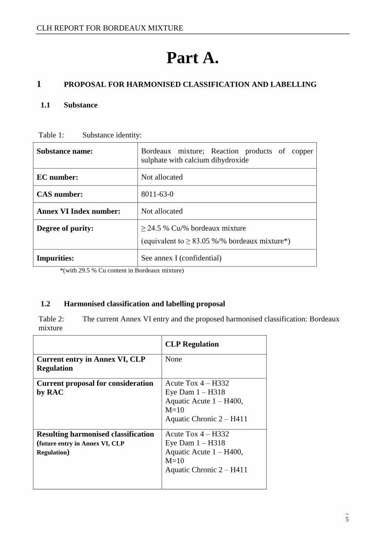
Table 1: Substance identity:
Substance name: Bordeaux mixture; Reaction products of copper
sulphate with calcium dihydroxide
EC number: Not allocated
CAS number: 8011-63-0
Annex VI Index number: Not allocated
Degree of purity: ≥ 24.5 % Cu/% bordeaux mixture
(equivalent to ≥ 83.05 %/% bordeaux mixture*)
Impurities: See annex I (confidential)
*(with 29.5 % Cu content in Bordeaux mixture)
1.2 Harmonised classification and labelling proposal
Table 2: The current Annex VI entry and the proposed harmonised classification: Bordeaux
mixture
CLP Regulation
Current entry in Annex VI, CLP
Regulation
None
Current proposal for consideration
by RAC
Acute Tox 4 – H332
Eye Dam 1 – H318
Aquatic Acute 1 – H400,
M=10
Aquatic Chronic 2 – H411
Resulting harmonised classification
(future entry in Annex VI, CLP
Regulation)
Acute Tox 4 – H332
Eye Dam 1 – H318
Aquatic Acute 1 – H400,
M=10
Aquatic Chronic 2 – H411

CLH REPORT FOR BORDEAUX MIXTURE
6
Copper and some copper compounds are under review as Biocides (BPD) and/or Plant Protection
Product (PPP) Directives and CLH dossier to set or revise their harmonised classification are
submitted in parallel for these compounds (see summary in annex II).

CLH REPORT FOR BORDEAUX MIXTURE
7
1.3 Proposed harmonised classification and labelling based on CLP Regulation
Table 3: Proposed classification according to the CLP Regulation
CLP
Annex I
ref
Hazard class Proposed
classification
Proposed SCLs
and/or M-
factors
Current
classification 1)
Reason for no
classification 2)
2.1. Explosives
None Conclusive but not
sufficient for classification
2.2. Flammable gases None Not relevant
2.3. Flammable aerosols None Not relevant
2.4. Oxidising gases None Not relevant
2.5. Gases under pressure None Not relevant
2.6. Flammable liquids None Not relevant
2.7. Flammable solids
None Conclusive but not
sufficient for classification
2.8. Self-reactive substances and
mixtures
None Conclusive but not
sufficient for classification
2.9. Pyrophoric liquids None Not relevant
2.10. Pyrophoric solids
None Conclusive but not
sufficient for classification
2.11. Self-heating substances and
mixtures
None Conclusive but not
sufficient for classification
2.12. Substances and mixtures
which in contact with water
emit flammable gases
None Conclusive but not
sufficient for classification
2.13. Oxidising liquids None Not relevant
2.14. Oxidising solids
None Conclusive but not
sufficient for classification
2.15. Organic peroxides
None Conclusive but not
sufficient for classification
2.16. Substance and mixtures
corrosive to metals
None Conclusive but not
sufficient for classification
3.1. Acute toxicity - oral
None Conclusive but not
sufficient for classification
Acute toxicity - dermal
None Conclusive but not
sufficient for classification
Acute toxicity - inhalation
Acute Tox 4 –
H332
None None
3.2. Skin corrosion / irritation
None Conclusive but not
sufficient for classification

CLH REPORT FOR BORDEAUX MIXTURE
8
3.3. Serious eye damage / eye
irritation
Eye Dam 1 –
H318
None None
3.4. Respiratory sensitisation None Data lacking
3.4. Skin sensitisation
None Conclusive but not
sufficient for classification
3.5. Germ cell mutagenicity
None Conclusive but not
sufficient for
classification
3.6. Carcinogenicity
None Conclusive but not
sufficient for
classification
3.7. Reproductive toxicity
None Conclusive but not
sufficient for classification
3.8. Specific target organ toxicity
–single exposure
None Conclusive but not
sufficient for classification
3.9. Specific target organ toxicity
– repeated exposure
None Conclusive but not
sufficient for classification
3.10. Aspiration hazard
None Conclusive but not
sufficient for classification
4.1.
Hazardous to the aquatic
environment
Aquatic Acute
1 – H400
Aquatic
Chronic 2 –
H411
M-factor = 10 None
5.1. Hazardous to the ozone layer
None Conclusive but not
sufficient for classification
1) Including specific concentration limits (SCLs) and M-factors
2) Data lacking, inconclusive, or conclusive but not sufficient for classification
Labelling: Signal word: Danger
Pictograms: GHS 05, GHS 07 , GHS 09
Hazard statements: H332, H318, H400, H411
Precautionary statements: not harmonised
Proposed notes assigned to an entry: none

CLH REPORT FOR BORDEAUX MIXTURE
9
2 BACKGROUND TO THE CLH PROPOSAL
2.1 History of the previous classification and labelling
Bordeaux mixture has no current harmonised classification.
No registration dossier is available for Bordeaux mixture (last consultation: 5/02/2013).
2.2 Short summary of the scientific justification for the CLH proposal
Bordeaux mixture has a moderate acute toxicity by inhalation and a classification as Acute Tox 4 –
H332 is proposed.
Besides, Bordeaux mixture induced irritancy in the rabbit eyes that persisted and justify a
classification Eye Dam 1 – H318.
Taking into account the recommendations of the Annex IV of the Guidance to Regulation (EC) No
1272/2008 Classification, Labelling and Packaging of substances and mixtures, a metal compound
is considered as readily soluble if the water solubility is greater or equal to the acute ERV of the
dissolved metal ion concentration. The water solubility of Bordeaux mixture is equal to 2.2 mg/L
and 1.1 mg/L at pH 6.8 and 9.8 respectively. Therefore, this compound is considered as ready
soluble metal compound.
For acute toxicity classification, the lowest ERV- Bordeaux mixture (0.10 mg/l) is below the trigger
value of 1 mg/L which leads to the aquatic environmental hazard acute category 1, H400. An M-
factor of 10 should also be applied.
For chronic toxicity classification, there is evidence of rapid removal from water column. The
lowest chronic ERV- Bordeaux mixture (0.025 mg/L) is between the trigger values of 0.01 and 0.1
mg/L which leads to the aquatic environmental hazard chronic category 2, H411.
2.3 Current harmonised classification and labelling
No current harmonised classification in Annex VI of CLP.
2.4 Current self-classification and labelling
The classification applied by the notifier in the scope of the Plant Protection Product Directive
(91/414/CE) is as follows:
CLP Regulation
Acute Tox 4 – H332

CLH REPORT FOR BORDEAUX MIXTURE
10
Eye Dam 1 – H318
Aquatic Acute 1 – H400
3 JUSTIFICATION THAT ACTION IS NEEDED AT COMMUNITY LEVEL
Bordeaux mixture is an active substance in the meaning of Directive 91/414/EEC (PPP). In
accordance with Article 36(2) of the CLP Regulation, Bordeaux mixture shall be subjected to a full
harmonised classification and labelling. Therefore, this proposal considers all human health and
environmental end points. In particular, harmonised classification is proposed for acute toxicity, eye
irritation and environment (with a M-factor), which justifies action at community level.

CLH REPORT FOR BORDEAUX MIXTURE
11
Part B.
SCIENTIFIC EVALUATION OF THE DATA
1 IDENTITY OF THE SUBSTANCE
1.1 Name and other identifiers of the substance
Table 5: Substance identity
EC number: Not allocated
EC name: Not allocated
CAS number (EC inventory): Not allocated
CAS number: 8011-63-0
CAS name: Bordeaux mixture
IUPAC name: None
CLP Annex VI Index number: -
Molecular formula: unspecified
Molecular weight range: unspecified
Structural formula: unspecified
1.2 Composition of the substance

CLH REPORT FOR BORDEAUX MIXTURE
12
Table 6 : Constituents (non-confidential information)
Constituent Concentration range Remarks
Bordeaux mixture; Reaction
products of copper sulphate
with calcium dihydroxide
Cas: 8011-63-0
≥24.5% w/w (Cu/Bordeaux mixture)
(equivalent to ≥ 83 % bordeaux
mixture*)
*Calculation using 29.5 % Cu
content in Bordeaux mixture
Current Annex VI entry: see Part A (section 2.3)
Impurities (non-confidential information)
Impurities are confidential. See confidential annex.
Additives (non-confidential information)
Confidential information, see confidential annex.
1.2.1 Composition of test material
Some information in the literature shows that nanomaterials containing copper compounds may
exist. However, the information available in the biocidal and plant protection products dossiers and
their production process do not seem to indicate that the substance exist under this shape for these
applications.
In this context, it was decided not to take into consideration the potential nanoform of copper
compounds in this report and the present CLH dossier is proposed for the bulk form of Bordeaux
mixture. A specific dossier and hazard evaluation may be necessary for nanoforms of this
substance.
The purity of the tested material is specified when available and/or relevant in the different parts of
the CLH report.
Physico-chemical properties

CLH REPORT FOR BORDEAUX MIXTURE
13
Table 9: Summary of physico - chemical properties
Property Value Reference Comment
(e.g.
measured
or
estimated)
State of the substance at
20°C and 101,3 kPa
Odourless, light green,, very fine, non-free
flowing powder containing soft aggregates O’Connor and
Mullee, 2003 observation
and
olfactive
test
Melting/freezing point No melting point before decomposition
(An endothermic event over the range 110°C
to 190°C is attributed to dehydration of the molecule)
O’Connor and Mullee, 2003
measured
Boiling point No boiling point before decomposition
(An endothermic event over the range 110°C
to 190°C is attributed to dehydration of the molecule)
O’Connor and
Mullee, 2003 measured
Relative density 3.12 at 19.5 °C using a 26.7 % purity
test item
O’Connor and
Mullee, 2003 measured
Vapour pressure Not applicable (from the structure and the
melting temperature result, it was anticipated
that no definitive value could be obtained due to the negligible volatility of the test material
-
Surface tension 68.9 mN/m at 21°C
O’Connor and
Mullee, 2003 measured
using a 26.7
% purity test
item
Water solubility Determined at 20.0 0.5°C
At pH 2.9 : > 124 g/L as Cu >33.1
At pH 6.8 : 2.20x10⁻³ g/L as Cu 5.87x10⁻⁴
At pH 9.8 : 1.1x10⁻³ g/L as Cu 2.94x10⁻⁴
O’Connor and Mullee, 2003
measured
using a 26.7
% purity test item
Flask method
Partition coefficient n-
octanol/water
not applicable due to the negligible solubility in
water and n-octanol (< 10 mg/L) statement
Flash point Not required (solid) -
Flammability Not flammable
Van Beijnen,
A.J.M. (2000) measured
using a 20%
purity test item
Explosive properties No explosive properties Van Beijnen,
A.J.M. (2000) measured
using a 20%
purity test item
Self-ignition temperature Not auto-flammable Van Beijnen,
A.J.M. (2000) measured
using a 20%
purity test item
Oxidising properties Not oxidizing Van Beijnen,
A.J.M. (2000) measured
using a 20%
purity test
item

CLH REPORT FOR BORDEAUX MIXTURE
14
Granulometry No data
Stability in organic
solvents and identity of
relevant degradation
products
No data
Dissociation constant Copper is a poorly soluble metallic element
that can only remain in solution in a totally
dissociated ionic state; a non reversible
process. Since its solubility is low and it does
not exist in solution in an associated state, it
does not therefore have a measurable
dissociation constant.
Statement
Viscosity Not required (solid) -
Henry’s law constant not applicable
Solubility in organic
solvent
Determined at 20.0 0.5°C
n-hexane < 9.8 mg/L
Toluene < 9.6 mg/L
dichloromethane < 8.8 mg/L
methanol < 9.0 mg/L
acetone < 8.8 mg/L ethyl acetate < 8.4 mg/L
O’Connor and
Mullee, 2003 Measured
2 MANUFACTURE AND USES
2.1 Manufacture
Not relevant
2.2 Identified uses :
Copper is fungistatic and bacteristatic in action and is used in the treatment and prevention of fungal and bacterial
diseases.

CLH REPORT FOR BORDEAUX MIXTURE
15
3 CLASSIFICATION FOR PHYSICO-CHEMICAL PROPERTIES
Table 10: Summary table for relevant physico-chemical studies
Physical and chemical properties
Methods-
Results Remarks Reference
Flammabilty (EEC A 10)
Not flammable measured using a
20% purity test item
(equivalent copper)
Van Beijnen, A.J.M.
(2000)
Auto-inflammability (EEC A 16)
Not auto-flammable measured using a
20% purity test item (equivalent copper)
Van Beijnen, A.J.M.
(2000)
Flash point
Not required as the active
substance is a solid
Explosive properties (EEC A 14)
No explosive properties measured using a
20% purity test item (equivalent copper)
Van Beijnen, A.J.M.
(2000)
Oxidising properties (EEC A 17)
No oxidizing properties measured using a
20% purity test item (equivalent copper)
Van Beijnen, A.J.M.
(2000)
3.1 Explosive properties
Bordeaux Mixture consists of a mixture of inorganic components (copper II sulphate and hydrated
calcium hydroxide). None of these components or grouping are associated with explosive hazards.
All are stable groupings in high oxidation states. The oxygen is bound up in very stable structural
groupings with strong oxygen bonds. An endothermic event over the range 110°C to 190°C is
attributed to dehydration of the molecule. Bordeaux Mixture is considered inert under the
conditions of oxidation.
3.2 Flammability
Bordeaux mixture is a mixture of inorganic salts and minerals in solid form (copper II sulphate &
hydrated calcium hydroxide). Inorganic salts are not combustible or flammable and in addition,
experience indicates the material is not flammable and not auto-flammable. As such this material is
not likely to undergo self heating under bulk storage conditions and is unlikely to auto-ignite. Self
heating or auto-ignition has not been observed with Bordeaux mixture following use for many
years.
The determination of flash point is not required because the active substance is a solid.
So we can conclude that Bordeaux mixture is not flammable.
.

CLH REPORT FOR BORDEAUX MIXTURE
16
3.3 Oxidising potential
Oxidising compounds are materials that can easily transfer oxygen to other compounds i.e. they
contain weakly bound oxygen, for example NO3 and peroxides. Bound oxygen must also become
available through a low energy degradation route with a low energy of activation. The oxygen in
Bordeaux Mixture is bound in a stable structural groupings with strong oxygen bonds (sulphates
and hydroxides). The decomposition temperature of Bordeaux Mixture is also high indicating a
high energy of activation. Bordeaux Mixture is therefore considered inert under the conditions of
oxidation.
Experience in using Bordeaux Mixture over many years also indicates that it is not associated with
oxidising hazards.
3.3.1 Comparison with criteria
none
3.3.2 Conclusions on classification and labelling
none
4 HUMAN HEALTH HAZARD ASSESSMENT
Considering that in mammalian the toxic form of any copper salt is the Cu2+
ion, a read across
between the different salts (copper sulphate, dicopper oxide, copper hydroxide, copper oxide,
copper carbonate, copper thiocyanate, copper powder, copper oxychloride and Bordeaux mixture)
will be used for assessment of repeated toxicity, mutagenicity, carcinogenicity and reprotoxicity of
copper compounds. Therefore, the report of these endpoints will be common in the different CLH
report of each compound. However, the acute toxicity and local toxicity as irritation and
sensitization will be specific for each substance.
4.1 Toxicokinetics (absorption, metabolism, distribution and elimination)
4.1.1 Non-human information
The following summary of toxicokinetics of the copper ion Cu2+
is derived from the pesticide and
biocide assessment reports made for the review of copper compounds under directive 91/414/EEC
and 98/8/EEC.
Absorption
Absorption in both rats and humans varies according to diet. For humans: on a copper-adequate
diet, absorption is 36 %, on a low copper diet 56 %, and on a high copper diet 12%. Similar figures
have been obtained for rats.
Distribution

CLH REPORT FOR BORDEAUX MIXTURE
17
After oral absorption, when entering interstitial fluid and blood plasma, absorbed copper initially
becomes bound to two proteins; albumin and transcuprein. Although the affinity of transcuprein for
copper is higher than that of albumin, copper ions are freely exchangeable between them. Most of
the copper bound to albumin and transcuprein is rapidly transported via portal blood to the liver
(main organ of regulation), although some also goes directly to other tissues, especially to the
kidney. The liver controls the distribution of copper to the rest of the body via the bloodstream,
bound to ceruloplasmin.
By other routes of exposure (mainly inhalation), absorbed copper does not pass first by the liver,
therefore, a wider distribution through the body is possible.
Metabolism
Metabolism does not occur. Copper is a monatomic ion and cannot be metabolised. It is however
used in every cell in the body, and every cell can regulate its copper content. Many enzymes and
other proteins containing copper have been described.
Interspecies differences
Albumin, one of the major copper transport proteins of the blood, contains histidine in position 3
which is essential for tight binding of copper. In dogs and pigs, this histidine is replaced by a
tyrosine, and consequently the albumin does not have the same affinity for copper. Dog and pig
albumins have several low-affinity sites for copper, but albumin is still an effective transport protein
in those species. Dogs show unusually high levels of copper in the liver, ten times the levels in other
species. While dog liver rapidly took up copper injected intravenously, dogs do not appear to be
able to excrete copper via the bile as readily as other species. It is possible that dogs express the
WND protein less than other species resulting in accumulation of copper in the liver. Based on
these differences in albumin structure and the liver of the dog, it was concluded that the dog is not a
good animal model for human risk assessment of copper and that is why no dog study is outlined in
this report.
Accumulation
Accumulation does not occur except in cases of genetic disease or chronic administration of
exceptionally high doses (60 mg/person/day), where copper accumulates in the liver.
Excretion
Excretion in most species is via the bile, in a trypsin-independent protein fragment such that entero-
hepatic circulation does not occur. A significant amount of copper is excreted bound to
metallothioneins contained in intestinal brush border cells sloughed off and lost in faeces. Minor
amounts are also excreted in urine and from skin and hair.
Excretion is rapid. An oral dose of 20 mg Cu/kg to rats was completely eliminated from the liver by
48 h. Blood plasma levels did not increase during this period.
Bioequivalence
In mammalian toxicity, it is considered that the toxic form of any copper salt is the Cu2+
ion.
This is shown through the comparison of bioavailability and hence toxicity of the most soluble
(copper sulphate) and relatively insoluble copper salts. In effect, the use of copper sulphate data
would represent a worst-case scenario for the determination of the systemic effect of relatively
insoluble copper compounds in mammalian toxicity. This has also been confirmed in a series of
bioavailability studies conducted by several authors who have compared the bioavailability of
copper sulphate to other copper salts including copper oxide, copper powder, copper thiocyanate
and copper carbonate. Moreover, in an other study copper was administered orally to bile-canulated

CLH REPORT FOR BORDEAUX MIXTURE
18
rats, as copper sulphate, copper hydroxide, copper oxychloride, Bordeaux mixture, tribasic copper
sulphate and copper (I) oxide. There were no differences in absorption, copper levels in plasma,
liver or bile, or in excretion rates between the five forms and copper sulphate. This study
demonstrates bioequivalence between the five forms and copper sulphate, such that repeated dose
toxicity studies on copper sulphate, or on only one of the five forms, may be considered
representative of the other forms for systemic effects.
In 2010, Rodriguez et al, assessed the relative/dissolution of copper ions from copper materials and
copper compounds in gastric mimetic fluid, simulated oral exposure.
The copper compounds tested, include: copper wires massive copper materials), copper powder
(130 µm median diameter), coated copper flakes (8.5 µm), cupric oxide and cuprous chloride.
Loading rates between 100 mg/L and 2 g/L were assessed. The results are expressed as % mass
recovered at the end of the bio-elution test and compred with the results obtained from soluble
copper sulphate.
The results are summarised in the table below.
Relative bio-solubility of copper and copper compounds, assessed from the recovery of copper after
a bio-elution tests in gastric fluids.
1. Material tested 2. Bio-elution recovery
3. (as% of Cu content)
4. Cu massive 5. 0.096-0.105
6. Cu powder 7. 1.1
8. Cu flake 9. 42-71
10. CuO 11. 68-84
12. CuCl 13. 67-94
14. CuSO4 15. 100
The results show a highest solubility of CuSO4 and CuCl.
In conclusion, this study demonstrated large variability in the gastric bio-accessibility of copper
bearing materials.
Therefore in order to reduce the number of animal testing, as CuSO4 release more ion Cu2+
than the
other copper compounds and it is considered that the toxic form is the Cu2+
ion, all long term
studies by oral routes could be conducted on CuSO4, as the worst case.
4.1.2 Human information
Literature review on ADME
Copper is a micronutrient. It is essential for life and is employed in all living cells. It is used in
many enzyme systems, particularly in energy transfer where the property of electron transfer is
exploited in photosynthesis and catabolism. It has been the subject of intense research.

CLH REPORT FOR BORDEAUX MIXTURE
19
Copper is present in almost all foods, with some foods (nuts, shellfish, chocolate) naturally
containing more than 20 ppm copper.
Most human diets naturally include between 1 and 2 mg/person/day of copper, with some
containing up to 4 mg/person/day. Copper levels in blood and tissues are generally stable. The body
is able to maintain a balance of dietary copper intake and excretion that allows normal physiological
processes to take place.
As with all micronutrients (minerals), copper is absorbed, used, stored and excreted. This applies at
the level of the individual cell, at the organ and at the level of the whole organism. The cell
membrane transport mechanisms for copper have been studied extensively, and the genetic codes
for the individual transporter proteins are very similar in many different organisms: bacteria, fungi
and fish, indicating that the process is ancient.
The copper transport mechanisms at the level of the organism form part of the system of
homeostasis, the process by which the levels of copper in the body (and ultimately the cell) are
regulated. Copper can be considered to show a flattened “U”-shaped dose-response curve.
The left side of the “U” curve represents deficiency, where intake is less than the requirement. This
can be lethal, especially in children, where copper is needed for growth. Copper deficiency is
associated with growth retardation, anaemia, skin lesions, impaired immunity, intestinal atrophy,
impaired cardiac function, reproductive disturbance, neurological defects and skeletal lesions.
Copper is essential for normal physiological function such as cellular respiration, free radical
defence, synthesis of melanin, connective tissue, iron metabolism, regulation of gene expression,
and normal function of the heart, brain and immune system.
The central near-horizontal part of the “U” curve represents homeostasis, where intake and
excretion are balanced, and copper levels are said to be normal.
The right-hand part of the “U” represents toxicity or excess copper disease.
The natural homeostatic regulation of copper means that an individual on a low copper diet will
retain more of an artificial dose of copper than an individual on a high copper diet.
4.1.3 Summary and discussion on toxicokinetics
Copper is widely distributed in biological tissues, where it occurs largely in the form of organic
complexes, many of which are metalloproteins and function as enzymes. Copper enzymes are
involved in a variety of metabolic reactions, such as the utilisation of oxygen during cell
respiration and energy utilisation. They are also involved in the synthesis of essential compounds,
such as the complex protein of connective tissues of the skeleton and blood vessels, and in a range
of neuroactive compounds concerned in nervous tissue function.
Copper is present in almost all foods, most human diets naturally include between 1 to 2
mg/person/day of copper, with some containing up to 4 mg/person/day. Copper levels in blood and
tissues are generally stable; the body is able to maintain a balance of dietary copper intake and
excretion that allows normal physiological processes to take place. Up to 93 % of the copper in the
blood is bound to the enzyme caeruloplasmin, with the majority of the rest bound to albumin and
amino acids; there is strong evidence that absorbed copper is never released free in the blood or in
the cells.
A bioequivalence study was performed to compare copper hydroxide, copper oxychloride,
Bordeaux mixture, tribasic copper sulphate and copper (I) oxide with copper sulphate pentahydrate
on bile cannulated rats. Absorption, distribution and excretion rates were similar between the six
variants of copper following oral ingestion of 20 mg Cu/kg bw; liver was the principal organ of
regulation of copper and main excretion was via the bile. Liver copper levels increased
significantly following dosing with Tmax at 12 hours; depuration was rapid, with levels returning to

CLH REPORT FOR BORDEAUX MIXTURE
20
control by 48 hours after dosing. Plasma concentrations in both control and dose rats remained
unchanged.
Oral absorption of copper varies according to the diet, for humans a copper-adequate diet results in
36 % absorption, while a low copper diet results in 56 % absorption and a high copper diet in 12 %
absorption. Similar figures were found in rat, 50 % oral absorption was considered for this specie.
Distribution was directly from the intestine to the liver, which controls the distribution of copper to
the rest of the body via the bloodstream, bound to ceruloplasmin. Metabolism does not occur.
Copper do not accumulate except in cases of genetic disease or chronic administration of high
doses, where copper accumulates in the liver. Excretion is rapid, via the bile, in a trypsin-
independent protein fragment such that entero-hepatic circulation does not occur. Significant
amounts of copper are excreted bound to metallothioneins contained in intestinal brush border cells
sloughed off and lost in faeces; minor amounts are also excreted in urine and from skin and hair.
4.2 Acute toxicity

CLH REPORT FOR BORDEAUX MIXTURE
21
Table 11: summary table of relevant acute toxicity studies performed with Bordeaux mixture
Method Results Remarks Reference
Oral
Rat
Sprague Dawley
5/sex/dose
1330; 2000 and3000 mg/kg
bw
Acute oral exposure
14 days post exposure
LD50: 2437 mg/kg/bw (♂+♀)
OECD 401
GLP
No deviation
Purity:26.35% (w/w copper)
Vehicle: purified water
Griffon, B.
(2000a)
Rat
Sprague Dawley
5/sex/dose
1600; 2000; 2400 and 2800
mg/kg bw
Acute oral exposure
14 days post exposure
LD50: 2302 mg/kg/bw (♂+♀) OECD 401
GLP
No deviation
Purity: not stated
Vehicle: carboxymethyl
cellulose 0.5%
Jackson , D.
(1994b)
Inhalation
Rat
Sprague Dawley
5/sex
Nominal concentration =
2.67; 5.42 and 8.65mg/L
(MMAD =1.75-2.2-2.15µm)
Whole body exposure
4 hour exposure
14 days post exposure
Acute inhalation
LC50 (4-hour): 1.97 mg/L (♂)
LC50 (4-hour): > 2.02 mg/L (♀)
OECD 403
GLP
No deviation
Purity: not stated
Merkel, D.J.
(2001)
Rat
Sprague Dawley
5/sex/dose
Nominal concentration =
1.10; 1.96 and 4.88 mg/L
(MMAD= 3.83; 3.27;
3.53µm)
Nose-only
4 hours of exposure
14 days post exposure
Acute inhalation
LC50 (4-hour): 3.98 mg/L (♂)
LC50 (4-hour): > 4.88 mg/L (♀)
OECD 403
GLP
No deviation
Purity: 26.5% (w/w copper)
Anderson, B.
T. and
Stewart, D.
(2002)
Dermal
Rat
Sprague Dawley
5/sex/dose
2000 mg/kg bw
LD50: > 2000 mg/kg/bw
(♂+♀)
OECD 402
GLP
No deviation
Purity: 26.35% (w/w copper)
Griffon, B.
(2000b)

CLH REPORT FOR BORDEAUX MIXTURE
22
(limit test)
24h exposure
14 days post-exposure
Acute dermal
Vehicle: purified water
Rat
Sprague Dawley
5/sex/dose
2000 mg/kg bw
(limit test)
24h exposure
14 days post-exposure
Acute dermal
LD50: > 2000 mg/kg/bw
(♂+♀)
OECD 402
GLP
No deviation
Purity: not stated
Vehicle: purified water
Jackson, D.
(1994d)
4.2.1 Non-human information
4.2.1.1 Acute toxicity: oral
Reference: Griffon, B. (2000a)
Guideline: OECD 401
GLP: Yes
Deviation No
Bordeaux mixture (batch number 44, containing 26.35 % w/w copper, content of Bordeaux mixture
not stated) was administered in purified water. Groups of five male and five female Sprague-
Dawley rats weighing 170 7 g (males) and 143 9 g (females) were used. The rats were housed
in groups of up to seven by sex, acclimatised, and fasted overnight prior to dosing. Food was
returned four hours after dosing. Dose levels (based on a range-finding study) of 1330, 2000 and
3000 mg/kg bw were administered by single oral administration by gavage in 10 mL/kg on Day 1.
Animals were maintained for up to 14 days post-dosing (1330 and 2000 mg/kg bw groups) or 21
days post-dosing (3000 mg/kg bw group). Animals were observed frequently on the day of dosing
and then at least once daily for the 14/21-day post-dosing period. Animals were weighed prior to
administration and after 7 days (on Day 8) and after 14 days (on Day 15), 21 days (on Day 22) or at
death. Decedents and animals surviving to 14/21 days were subject to gross necropsy.
Mortalities and clinical observations: There were no mortalities at 1330 mg/kg bw. At higher doses,
the frequency of mortality increased and three males and three females died following 3000 mg/kg
bw. The deaths occurred between Day 4 and Day 8. There was a variety of clinical signs recorded
including hypoactivity, dyspnoea, piloerection, stiff gait, sedation, soft faeces, lateral recumbence
and swollen abdomen. Most symptoms occurred from Day 3 and persisted up to Day 3, 13 and 17 in
animals dosed at 1330, 2000 and 3000 mg/kg bw, respectively. A summary of mortalities is
presented in Table below.
Body weight: Most surviving animals dosed at 2000 or 3000 mg/kg bw lost weight in the first
week after treatment but thereafter showed acceptable weight gain. In animals dosed at 1330 mg/kg
bw, body weight gain was lower than historical controls in males but comparable to historical
controls in females.
Necropsy: No gross findings were recorded in surviving animals. In animals which died during the
study, the digestive tract was distended and contained a greenish coloured liquid.

CLH REPORT FOR BORDEAUX MIXTURE
23
Table 12. Bordeaux mixture: mortalities following oral administration to rats
Dose
(mg/kg bw)
Males Females
Mortality Time of death Mortality Time of death
1330 0/5 - 0/5 -
2000 2/5 Day 6; Day 7 3/5 Day 4; Day 7;
Day 8
3000 3/5 Day 4; Day 6;
Day 7
3/5 Day 6; Day 7 (2)
Figures in parenthesis are the number which died on the day specified if more than one.
The acute oral LD50 (calculated by probit analysis) of Bordeaux Mixture to the rat was 2437 mg/kg
bw for the sexes combined (with 95% confidence limits of 1938 to 3861 mg/kg bw).
Reference: Jackson, D. (1994b)
Guideline: OECD 401
GLP: Yes
Deviation No
Bordeaux mixture (batch number 3/0371, content of copper and Bordeaux Mixture not stated) was
administered as a solution in 0.5% carboxymethyl cellulose. Groups of five male and five female
Sprague-Dawley rats weighing 128 to 249 g were used. The rats were housed in groups of up to
five by sex, acclimatised, and fasted overnight prior to dosing. Food was returned four hours after
dosing. Dose levels (based on a range-finding study) of 1600, 2000, 2400 and 2800 mg/kg bw were
administered by single oral administration by gavage in 10 mL/kg on Day 1. Animals were
observed frequently on the day of dosing and then once daily for the 14-day post-dosing period.
Animals were weighed prior to administration and after 7 days (on Day 8) and after 14 days (on
Day 15) or at death. Decedents and animals surviving to 14 days were subject to gross necropsy.
Mortalities and clinical observations: There were no mortalities at 1600 mg/kg bw. At higher doses,
the frequency of mortality increased and all males and four females died (or were killed in extremis)
following 2800 mg/kg bw. The deaths occurred between Day 2 and Day 13. There was a variety of
clinical signs recorded including piloerection, soiled coat, reduced activity, red nasal and ocular
discharge, ataxia, pale and hunched appearance, swollen abdomen, tail lesions, subdued behaviour,
diarrhoea, dark eyes, laboured breathing, prostration, hypothermia, alopecia and emaciation. Most
symptoms occurred between Day 2 and 8 though some effects persisted up to Day 15. A summary
of mortalities is presented in Table below.
Body weight: Surviving animals showed acceptable weight gain during the study.
Necropsy: No gross findings were recorded in surviving animals except for dark red foci on the
lungs in one male dosed at 1600 mg/kg bw. Findings in animals that died during the study included
reddening of the glandular mucosa of the stomach, liquid retention in the stomach and intestines,
green coloured kidneys, small seminal vesicles and light coloured foci on the caecum.
Table 13 Bordeaux mixture: mortalities following oral administration to rats
Dose
(mg/kg bw)
Males Females
Mortality Time of death Mortality Time of death
1600 0/5 - 0/5 -
2000 1/5 Day 2 1/5 Day 3
2400 4/5 Day 9 (2); Day
10; Day 13
2/5 Day 4; Day 7
2800 5/5 Day 3; Day 5; 4/5 Day 5 (2);

CLH REPORT FOR BORDEAUX MIXTURE
24
Day 6; Day 9 (2) Day 6 (2) Figures in parenthesis are the number which died on the day specified if more than one.
The acute oral LD50 (calculated by probit analysis) of Bordeaux Mixture to the rat was
2187 mg/kg bw for males (with 95 % confidence limits of 1843 to 2499 mg/kg bw), 2433 mg/kg bw
for females (with 95 % confidence limits of 2052 to 3344 mg/kg bw) and 2302 mg/kg bw for the
sexes combined (with 95 % confidence limits of 2098 to 2516 mg/kg bw).
4.2.1.2 Acute toxicity: inhalation
Reference: Merkel, D.J. (2001)
Guideline: OECD 401
GLP: Yes
Deviation No
Bordeaux Mixture (batch number 27/7/00, content of copper not stated, containing 47 % w/w
copper hydroxysulphates) was used for the study. Test atmospheres were generated with a
modified Wright dust generator. Male and female Sprague-Dawley rats were housed individually
and acclimatised prior to exposure. Groups of animals were exposed to an aerosol atmosphere of the
test substance at gravimetric concentrations of 1.05 (five males), 2.02 mg/L (five males and five
females) and 3.08 mg/L (five males, maximum practical concentration) for four hours using a whole
body exposure system. The gravimetric test concentrations were determined by dividing the total
weight of the test substance collected on a glass fibre filter positioned in the breathing zone of the
animals by the volume of air sampled. The nominal test concentration was calculated by dividing
the total weight of the test substance used by the total volume of air (average airflow multiplied by
total time) passing through the chamber during exposure. The MMAD and the particle size
distribution were calculated from two samples taken during exposure from the breathing zone of the
animals with an Andersen cascade impactor. Chamber environmental conditions were monitored
during the exposure. Animals were observed for mortality and reaction to treatment at least every
30 minutes during exposure (1.05 and 2.02 mg/L treatments) or for the first hour of exposure
(3.08 mg/L treatment) on Day 1 until accumulation of the test substance on the walls of the
exposure chamber prevented this. After exposure, animals were observed at least once daily for
14 days. Body weights were recorded prior to exposure and after 7 days (Day 8), and 14 days (Day
15) or at death. Gross pathological examinations were performed on decedents and animals
surviving for 14 days.
The exposure parameters are summarised in Table 14.
Table 14: Bordeaux Mixture: exposure parameters in acute inhalation toxicity study
Parameter Value
Nominal concentration (mg/L) 2.67 5.42 8.65
Gravimetric concentration (mg/L) 1.05 2.02 3.08
Flow rate (L/min.) 50.6 50.7 50.6
Particle size: MMAD (µm)
(standard geometric deviation)
1.75
(2.24)
2.2
(2.22)
2.25
(2.15)
Respirable particles < 9 µm (%) 98.5 96.4 97.4
Mortalities and clinical observations: There were no mortalities at 1.05 mg/L. At 2.02 mg/L, three
males died but there were no female mortalities. At 3.08 mg/L, all males died. All deaths occurred
on the day of exposure or within one day after exposure. Clinical symptoms during exposure
CLH REPORT FOR BORDEAUX MIXTURE
25
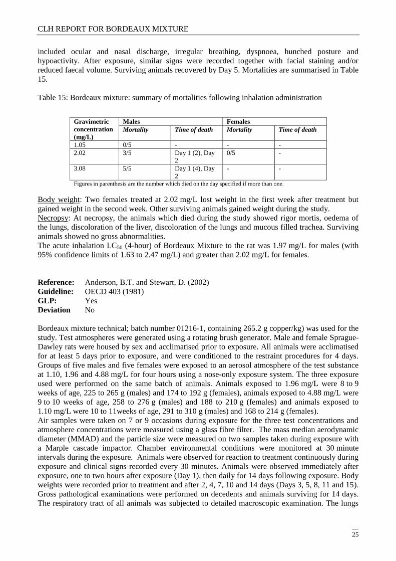
included ocular and nasal discharge, irregular breathing, dyspnoea, hunched posture and
hypoactivity. After exposure, similar signs were recorded together with facial staining and/or
reduced faecal volume. Surviving animals recovered by Day 5. Mortalities are summarised in Table
15.
Table 15: Bordeaux mixture: summary of mortalities following inhalation administration
Gravimetric
concentration
(mg/L)
Males Females
Mortality Time of death Mortality Time of death
1.05 0/5 - - -
2.02 3/5 Day 1 (2), Day
2
0/5 -
3.08 5/5 Day 1 (4), Day
2
- -
Figures in parenthesis are the number which died on the day specified if more than one.
Body weight: Two females treated at 2.02 mg/L lost weight in the first week after treatment but
gained weight in the second week. Other surviving animals gained weight during the study.
Necropsy: At necropsy, the animals which died during the study showed rigor mortis, oedema of
the lungs, discoloration of the liver, discoloration of the lungs and mucous filled trachea. Surviving
animals showed no gross abnormalities.
The acute inhalation LC50 (4-hour) of Bordeaux Mixture to the rat was 1.97 mg/L for males (with
95% confidence limits of 1.63 to 2.47 mg/L) and greater than 2.02 mg/L for females.
Reference: Anderson, B.T. and Stewart, D. (2002)
Guideline: OECD 403 (1981)
GLP: Yes
Deviation No
Bordeaux mixture technical; batch number 01216-1, containing 265.2 g copper/kg) was used for the
study. Test atmospheres were generated using a rotating brush generator. Male and female Sprague-
Dawley rats were housed by sex and acclimatised prior to exposure. All animals were acclimatised
for at least 5 days prior to exposure, and were conditioned to the restraint procedures for 4 days.
Groups of five males and five females were exposed to an aerosol atmosphere of the test substance
at 1.10, 1.96 and 4.88 mg/L for four hours using a nose-only exposure system. The three exposure
used were performed on the same batch of animals. Animals exposed to 1.96 mg/L were 8 to 9
weeks of age, 225 to 265 g (males) and 174 to 192 g (females), animals exposed to 4.88 mg/L were
9 to 10 weeks of age, 258 to 276 g (males) and 188 to 210 g (females) and animals exposed to
1.10 mg/L were 10 to 11weeks of age, 291 to 310 g (males) and 168 to 214 g (females).
Air samples were taken on 7 or 9 occasions during exposure for the three test concentrations and
atmosphere concentrations were measured using a glass fibre filter. The mass median aerodynamic
diameter (MMAD) and the particle size were measured on two samples taken during exposure with
a Marple cascade impactor. Chamber environmental conditions were monitored at 30 minute
intervals during the exposure. Animals were observed for reaction to treatment continuously during
exposure and clinical signs recorded every 30 minutes. Animals were observed immediately after
exposure, one to two hours after exposure (Day 1), then daily for 14 days following exposure. Body
weights were recorded prior to treatment and after 2, 4, 7, 10 and 14 days (Days 3, 5, 8, 11 and 15).
Gross pathological examinations were performed on decedents and animals surviving for 14 days.
The respiratory tract of all animals was subjected to detailed macroscopic examination. The lungs

CLH REPORT FOR BORDEAUX MIXTURE
26
of all animals were weighed and lung: body weight ratios calculated. The exposure parameters are
summarised in Table 16.
Table 16: Bordeaux Mixture technical: exposure parameters in acute inhalation toxicity study
Parameter Value
Nominal concentration (mg/L) 1.10 1.96 4.88
Measured concentration (mg/L) 4.3 10.9 25.0
Flow rate (L/min.) 20 20 20
Particle size: MMAD
(standard geometric deviation)
3.83 (2.042) 3.27 (2.044) 3.53 (2.178)
Respirable particles < 4.4 m (%) 53.1 46.8 47.4
Mortalities and clinical observations: There were no deaths in females. There were no deaths in
males at a concentration of 1.10 mg/L. At 1.96 mg/L, one male died on day 1 after dosing, and at
4.88 mg/L, two males died on day 1 and a third male died on day 2 after dosing. During exposure,
clinical signs included laboured and slow respiration. After exposure, surviving animals had test
compound on their heads and showed laboured respiration, pale, unkempt appearance, subdued
behaviour, pallor, intermittent body tremors, hunched posture, unsteady on feet and two animals
were recorded as having one eye closed in the post exposure period. Survivors appeared normal by
Day 1 (1.10 mg/L), Day 2 (1.96 mg/L) or Day 5 (4.88 mg/L). Mortalities are summarised in Table
17.
Table 17: Bordeaux mixture: summary of mortalities following inhalation exposure
Males Females
Dose (mg/L) Mortality Time of
death
Dose (mg/L) Mortality Time of
death
1.10 0/5 - 1.10 0/5 -
1.96 1/5 Day 1 1.96 0/5 -
4.88 3/5 Day 1 (2);
Day2 (1)
4.88 0/5 -
Figures in parenthesis are the numbers which died on the day specified if more than one.
Body weight: Surviving animals at 4.88 mg/L showed body weight loss after exposure, but had
recovered to Day 0 values between days 4 and 7 of the observation period. There was an initial
body weight loss in the animals treated at 1.10 and 1.96 mg/L. Normal body weight gain was
recorded in all animals by the end of the observation period.
Necropsy: The three males found dead at 4.88 mg/L were in an autolysed condition; two of these
animals had spongy and dark lungs, the third had spongy and mottled lungs. The lungs of the single
male killed on day 1 at 1.96 mg/L were noted as mottled and spongy, and one male in this group
showed red foci on all lung lobes at scheduled terminal kill. There were no other abnormalities.
Lung:body weight: There was no effect on the lung:body weight ratio in surviving animals. In
animals that died during the study, lung weights were increased, and lung: body weight ratios were
increased.
The acute inhalation LC50 (4-hour) of Bordeaux Mixture technical for combined sexes was in
excess of 4.88 mg/L. However, as there were no deaths in females, the LC50 must be calculated
using male data only. The LC50 (4-hour) for males was 3.98 mg/L (with 95% confidence limits of
1.24 to 12.15 mg/L).

CLH REPORT FOR BORDEAUX MIXTURE
27
4.2.1.3 Acute toxicity: dermal
Reference: Griffon, B. (2000b)
Guideline: OECD 402
GLP: Yes
Deviation: No
Bordeaux Mixture (batch number 44, containing 26.35 % w/w copper) was moistened with purified
water prior to application. Five male and five female Sprague-Dawley rats weighing 254 14 g
(males) and 221 8 g (females) were used. The rats were housed in groups of up to seven by sex,
and were acclimatised prior to dosing. A dose level of 2000 mg/kg bw was applied on a gauze pad
pre-moistened with 2 mL of water to an area of approximately 5 x 7 cm (males) or 5 x 6 cm
(females) of intact shorn skin (representing in both cases approximately 10 % of the body surface),
to each rat on Day 1. The gauze pad was held in contact with the skin with an adhesive semi-
occlusive dressing and a restraining bandage. After 24 hours, the dressing and any residual test
substance were removed. Animals were observed frequently for treatment-related clinical signs on
the treatment day and then at least daily for the 14-day post-dosing period. Skin reactions were
recorded daily from Day 2. Animals were weighed prior to treatment and after 7 days (Day 8) and
14 days (Day 15). Decedents and animals surviving to 14 days were subject to gross necropsy.
Mortalities and clinical observations: There were no mortalities and no clinical signs of toxicity or
skin reactions throughout the observation period.
Body weight: There was a reduction in body weight gain or slight body weight loss in three females
in the first week after treatment. All other animals showed expected weight gain during the study.
Necropsy: No gross findings were recorded.
The acute dermal LD50 of Bordeaux Mixture to the rat was greater than 2000 mg/kg bw for males
and females.
Reference: Jackson, D. (1994d)
Guideline: OECD 402
GLP: Yes.
Deviation No
Bordeaux Mixture (batch number 3/0371, content of copper and Bordeaux Mixture not stated) was
used. Five male and five female Sprague-Dawley rats weighing 186 to 319 g were housed in groups
of up to five by sex and acclimatised prior to dosing. A dose level of 2000 mg/kg bw was applied
on a surgical gauze patch (5 x 5 cm) moistened with distilled water to an area of intact shaven skin,
approximately 25 cm2 in size (approximately 10% of the total body surface area), of each rat on
Day 1. The patch was held in place with non-irritating occlusive tape wrapped around the trunk of
the animal. After 24 hours, the dressings were removed and the skin wiped with dampened tissue to
remove residual test substance. Animals were observed for treatment-related clinical signs
frequently on the day of administration and once daily for the 14-day post-dosing period. Animals
were weighed prior to treatment and after 7 days (Day 8) and 14 days (Day 15). Decedents and
animals surviving to 14 days were subject to gross necropsy.
Mortalities and clinical observations: There were no mortalities. The only clinical sign recorded was
a red nasal discharge in all females two and four hours after dosing.
Body weight: All animals showed acceptable weight gain during the study.
Necropsy: No gross findings were recorded at necropsy.
The acute dermal LD50 of Bordeaux Mixture to the rat was greater than 2000 mg/kg bw for males
and females.

CLH REPORT FOR BORDEAUX MIXTURE
28
4.2.1.4 Acute toxicity: other routes
No data available
4.2.2 Human information
Inhalation
Little information is available on acute effects in humans and inhalation of copper-containing
materials.
Published studies on acute effects in humans appear to have focussed on metal fume fever (MFF)1
and possible association with copper exposure. This subject has been reviewed extensively by
Borak et al (2000) with the aim of establishing whether there is an association between exposure to
copper and MFF. The review was based on seven reports, identified in a literature search as the only
reports that contained original descriptions of copper-exposed workers who developed symptoms
consistent with MFF. These seven reports are summarised below.
The earliest publication by Hansen (1911) provided a brief report of MFF-like symptoms in 10
males working in a research foundry where scrap copper was melted. The symptoms occurred as an
isolated incident. No qualitative or quantitative data concerning exposure were provided. The
isolated nature of this incident was considered by Borak et al to indicate an association with
exposure to contaminants other than copper.
Koelsch et al (1923) reported the occurrence of symptoms that included chest discomfort, shivering,
nausea and fever in 10 men performing hot rolling of copper bars in a rolling mill. The symptoms,
which had not previously been associated with the process, resolved in 24 hours. No qualitative or
quantitative exposure data were presented. As with the previous study, the isolated nature of this
incident suggested to Borak et al that contaminants other than copper were involved.
Friberg and Thrysin (1947) reported MFF-like syndrome in approximately 50 workers involved in
cleaning reactor ovens where pulverised copper was used as a catalyst. During the cleaning task,
heads and faces of the workers were reported to be covered in dust consisting mainly of cuprous
and cupric oxides. Initial symptoms included throat discomfort, burning eyes, nausea and headache,
followed by flu-like symptoms, nausea, vomiting, diarrhoea and chest discomfort. In many workers,
symptoms persisted for more than 72 hours. Quantitative exposure data was not provided. Dust
particles were reported to range from 1-15 µm diameter, with more than 70% >5 µm. Given that
MFF is typically associated with fine particles (< 1 µm diameter), Borak et al considered that the
study did not support association between copper and MFF. Further, the heavy exposure indicated
in this study is not generally associated with occurrence of MFF.
Schiotz (1949) reported the occurrence of initial symptoms such as metallic taste, throat dryness
and slight chest oppression, followed by shivering, sweating and fever among seven workers
involved in pulverising cuprous oxide during the production of marine paint. Symptoms subsided
1 Metal fume fever (MFF) is a transient illness which appears to develop 4-12 hours after occupational exposure to metal fume. MFF presents as an
influenza-like illness with cough and dyspnoea followed by fever, sweating and shivering. Other accompanying clinical signs and symptoms are
nausea, headache, weakness, a sweet metallic taste, and muscle and joint pain.

CLH REPORT FOR BORDEAUX MIXTURE
29
after 20-30 hours. Quantitative exposure data were not provided, although the described working
conditions indicated very high levels of exposure.
Gleason (1968) reported symptoms in workers exposed to dust generated during polishing of copper
plates with aluminium oxide abrasives. Symptoms were reportedly similar to “the onset of a
common cold with ……..chills or warmth, stuffiness of the head, etc”. Lower respiratory symptoms
were not reported, nor were other symptoms characteristic of MFF. Quantitative exposure data were
limited to a single breathing zone sample, indicating 0.12 mg/m3, although the study’s author
suggested exposure levels may have been “two or three times” higher. In this report, symptoms
persisted for several weeks until ventilation was introduced, a feature which is not usually
associated with MFF. In view of the absence of many symptoms characteristic of MFF and the
persistence of the reported symptoms, Borak et al considered that the condition was unlikely to be
MFF. Further, co-exposure to aluminium oxide was also likely, a metal also implicated in MFF
aetiology.
Hopper (1978) described the single case of a foundry worker who developed an isolated episode of
symptoms which included headache, cough, chest pain, chills and shortness of breath. Symptoms
occurred shortly after exposure to a molten alloy of copper, beryllium and aluminium, which was
poured into vessel containing alcohol and adhesive glue. Exposure data were not presented. Borak
et al noted the co-exposure to other metals which have been implicated in MFF aetiology and the
likely exposure to other potentially harmful substances. Consequently this case-report was not
considered as providing evidence of an association between copper and MFF.
Armstrong et al (1983) reported symptoms of MFF in a group of 26 workers after cutting brass
pipes (containing 90% copper, 10% nickel, and smaller amounts of zinc) with torches in a confined
space. Symptoms included fever, chills, headache, dyspnoea and nausea. Exposure data for the
different metals were not provided, although a description of the process indicated that high
exposure levels were likely. As with the previous two studies, Borak et al considered that co-
exposure to other metals implicated in MFF prevented identification of copper as the causative
agent.
None of the seven studies covered by the review provided adequate exposure data, qualitative or
quantitative, to enable identification of the causative agent(s) associated with the reported
symptoms. Further, as noted by Borak et al, there was a lack of any occupational pattern associated
with the MFF symptoms, as indicated by the range of industrial processes covered (foundry work,
rolling mill, paint production, metal polishing and pipe cutting). The conclusion of Borak et al was
that, based on the seven studies identified in the literature search, there is insufficient evidence to
conclude that exposure to copper dust or fume causes MFF. Based on data which are currently
available, this conclusion would appear to be justified.
Dermal
There are no published data on acute dermal effects of copper or copper compounds.
Oral
Self-poisoning
Self-poisoning with copper sulphate is rare in western countries but has been a common method of
suicide among low income groups in some areas of India. The most extensive study concerns 48
cases, including 7 fatalities (15%), admitted to one hospital in Delhi and 5 fatalities reported to
other Dehli hospitals (Chuttani et al, 1965). The most frequent symptoms observed in subjects were
nausea, epigastrial burning and vomiting. In addition, diarrhoea was reported in 14 patients (29%).

CLH REPORT FOR BORDEAUX MIXTURE
30
Biopsy examination of fatalities indicated deep erosions in gastric mucosa, haemorrhage in the
stomach and small intestine and oedema in the sub mucosa. Jaundice of variable severity occurred
in 11/48 cases (23%). In the more severe cases, palpable liver enlargement, significantly elevated
serum glutamic oxaloacetic transaminase (SGOT, 252.4 ±142 IU) and elevated bilirubin (112 ±8.9
mg/litre) were observed. Biopsy examination of liver tissue from fatalities showed centrilobular
necrosis and biliary stasis. Post-mortem examination also indicated swollen and congested kidneys
with glomerular swelling and necrosis of tubular cells. Anuria was reported in 13/48 patients (27%)
and oliguria in 5/48 (10%). Red discolouration of urine was observed, with haemoglobinuria
confirmed in some patients. These findings suggest haemolysis and are consistent with other
reports. Haematocrit and serum/plasma appearances were not reported. Serum or blood levels of
copper in the cases were elevated 2- or 3-fold compared to normal values. Estimated quantities of
copper ingested were based on patients’ accounts and therefore are unreliable. Consequently, this
study provides no reliable data which can be used for human risk assessment.
Subsequent case reports describe massive overdoses of copper sulphate (175 g) by a 22 year-old
Indian male (Mittal, 1972) and 250 g by a 42 year old US male (Jantsch et al, 1985). Both patients
survived following rapid chelation therapy with single or multiple injections of dimercaprol. The
amounts ingested were considerably greater than the highest estimated dose reported by Chuttani
and co-workers (1965). It therefore seems probable that survival of these patients was attributable to
immediate chelation therapy.
Accidental ingestion
The ingestion of a relatively small amount of copper sulphate (3 g), together with an equal amount
of zinc sulphate, by an 86 year-old female patient has also been reported (Hantson et al, 1996). The
patient was admitted to hospital vomiting blue/green material and she had diarrhoea. Gastric lavage,
dehydration and chelation therapy with dimercaprol were performed. The patient then suffered
hypotension, bronchial inflammation and ulceration and a decline in respiratory function. These
symptoms were interpreted as corrosive pneumonitis. The patient was placed on a mechanical
ventilator for three days and subsequently made a complete recovery. In this case, the symptoms
may have been exacerbated by the patient’s age and health status, but may also have been mitigated
to some extent by the co-ingestion of zinc sulphate which may have served to limit copper uptake
and the severity of the systemic effects.
Therapeutic treatment
Systemic effects, including renal damage and thrombocytopaenic purpura, were reported in a 17-
year old boy who was given 1% copper sulphate (2 mg/day) orally for treating vitiligo (Pande and
Gupta, 1969).
4.2.3 Summary and discussion of acute toxicity
Acute oral studies
Bordeaux Mixture was of low toxicity by the oral route (LD50 identified as 2302-2437 mg/kg). The
range of clinical signs following exposure was: diarrhoea, hypoactivity, dyspnoea, piloerection,
soiled coat, tail lesions, sedation, hypothermia, alopecia, emaciation, red nasal and ocular discharge,
ataxia, pale and hunched appearance, impaired respiration and swollen abdomen.
Surviving animals generally showed no gross findings at necropsy except for dark red foci on the
lungs in one male dosed at 1600 mg/kg bw Bordeaux mixture. Animals which died during the

CLH REPORT FOR BORDEAUX MIXTURE
31
studies showed discoloration or haemorrhage in the digestive tract, effects on liver and/or kidney
and the presence of substances similar to the test material in the stomach or intestines.
Copper has been used in suicide attempts. Most of these involved copper sulphate pentahydrate.
Intoxication is associated with emesis, superficial or deep ulcerations of the gastric and intestinal
mucosa. Liver histopathology revealed dilatation of central veins, varying degrees of liver cell
necrosis and bile thrombi. In kidneys there was congestion of glomeruli swelling or necrosis of
tubular cells and haemoglobin casts. These findings are similar to those seen in animal studies.
Elevated serum copper levels are only seen in moderate to severe cases of intoxication.
Unfortunately, the amount of copper taken in these suicide attempts is never quantified accurately.
Acute dermal studies
Bordeaux Mixture was of low toxicity by the dermal route (LD50 greater than 2000 mg/kg/bw).
Clinical signs were absent or very minor.
Acute inhalation studies
Inhalation studies conducted according to the required guidelines for classification, Bordeaux
Mixture was of moderate toxicity by inhalation (LC50 greater than 1 mg/L and less than 5 mg/L).
Clinical signs were similar to those recorded following oral administration. Necropsy of decedents
and some survivors showed lung abnormalities.
In humans, although copper has been implicated in the aetiology of metal fume fever, a review of
the available literature suggests there is insufficient evidence to support this claim.
4.2.4 Comparison with criteria
The oral LD50 can be identified as 2302-2437 mg/kg and lies above the range (200-2000 mg/kg) for
a classification under regulation (EC) 1272/2008.
The dermal LD50 lies above the classification cut-off of 2000 mg/kg under regulation (EC)
1272/2008 therefore no classification is proposed.
The inhalation LC50 lies within the range (1-5 mg/l) for classification as Acute Inhalation
Tox. 4 (H332: Harmful if inhaled) under regulation (EC) 1272/2008.
4.2.5 Conclusions on classification and labelling
Based on the results of the acute inhalation toxicity studies, Bordeaux mixture is classified as Acute
Inhalation Tox.4-H332 according to CLP.
No classification is proposed by oral and dermal route.

CLH REPORT FOR BORDEAUX MIXTURE
32
4.3 Specific target organ toxicity – single exposure (STOT SE)
4.3.1 Summary and discussion of Specific target organ toxicity – single exposure
The human has well recorded homeostatic mechanisms to control excess copper levels in the body
by a combination of decreased absorption and increased excretion. Human epidemiological data is
available however information is limited regarding doses consumed and exposure. Acute toxicity in
humans is infrequent and generally results from ingestion of contaminated foodstuffs/beverages, for
suicide purposes.
A paper by Chuttani (Chuttani et al, 1965) reviewed 53 cases of copper sulphate poisoning with
ingestion varying between 1 and 100g. Jaundice was recorded as a symptom with post mortem
examinations showing that the liver had signs of severe histological changes. A kidney biopsy
showed swelling and necrosis in two patients, and following an autopsy of patients who had died, a
congested kidney was observed. Emesis and irritation of the gastric mucosa was observed in all
patients.
A case was reported where a male ingested an estimated 175g of copper sulphate, renal damage was
observed (Mittal, 1972).
In acute animal studies with Bordeaux mixture the following clinical signs and necropsy findings
were observed.
An acute oral was conducted in rats with Bordeaux mixture (Griffon, B. 2000a). There were no
mortalities at 1330 mg/kg bw. At higher doses, the frequency of mortality increased and three males
and three females died following 3000 mg/kg bw. No gross findings were recorded in surviving
animals. In animals which died during the study, the digestive tract was distended and contained a
greenish coloured liquid. Another acute oral toxicity test was conducted on rats (Jackson, D.
1994b), there were no mortalities at 1600 mg/kg bw. At higher doses, the frequency of mortality
increased and all males and four females died (or were killed in extremis) following 2800 mg/kg
bw. No gross findings were recorded in surviving animals except for dark red foci on the lungs in
one male dosed at 1600 mg/kg bw. Findings in animals that died during the study included
reddening of the glandular mucosa of the stomach, liquid retention in the stomach and intestines,
green coloured kidneys, small seminal vesicles and light coloured foci on the caecum.
Acute dermal toxicity in rats (Griffon, B. 2000b; Jackson, D. 1994d) showed there were no
mortalities and no clinical signs of toxicity or skin reactions throughout the observation period. The
only clinical sign recorded was a red nasal discharge in all females two and four hours after dosing.
In an acute inhalation study in rats (Merkel, D.J. 2001), There were no mortalities at 1.05 mg/L. At
2.02 mg/L, three males died but there were no female mortalities. At 3.08 mg/L, all males died. At
necropsy, the animals which died during the study showed rigor mortis, oedema of the lungs,
discoloration of the liver, discoloration of the lungs and mucous filled trachea. Surviving animals
showed no gross abnormalities. Another acute inhalation study in rats (Anderson, B.T and Stewart,
D. 2002) showed no deaths in females. There were also no deaths in males at a concentration of
1.10 mg/L. At 1.96 mg/L, one male died, and at 4.88 mg/L, three males died. The three males found
dead at 4.88 mg/L were in an autolysed condition; two of these animals had spongy and dark lungs,
the third had spongy and mottled lungs. The lungs of the single male killed on day 1 at 1.96 mg/L
were noted as mottled and spongy, and one male in this group showed red foci on all lung lobes at
scheduled terminal kill. There were no other abnormalities.
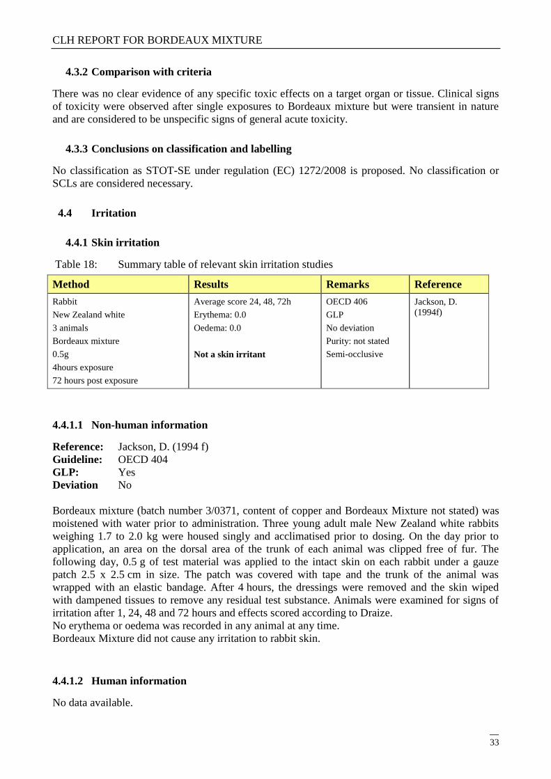
CLH REPORT FOR BORDEAUX MIXTURE
33
4.3.2 Comparison with criteria
There was no clear evidence of any specific toxic effects on a target organ or tissue. Clinical signs
of toxicity were observed after single exposures to Bordeaux mixture but were transient in nature
and are considered to be unspecific signs of general acute toxicity.
4.3.3 Conclusions on classification and labelling
No classification as STOT-SE under regulation (EC) 1272/2008 is proposed. No classification or
SCLs are considered necessary.
4.4 Irritation
4.4.1 Skin irritation
Table 18: Summary table of relevant skin irritation studies
Method Results Remarks Reference
Rabbit
New Zealand white
3 animals
Bordeaux mixture
0.5g
4hours exposure
72 hours post exposure
Average score 24, 48, 72h
Erythema: 0.0
Oedema: 0.0
Not a skin irritant
OECD 406
GLP
No deviation
Purity: not stated
Semi-occlusive
Jackson, D.
(1994f)
4.4.1.1 Non-human information
Reference: Jackson, D. (1994 f)
Guideline: OECD 404
GLP: Yes
Deviation No
Bordeaux mixture (batch number 3/0371, content of copper and Bordeaux Mixture not stated) was
moistened with water prior to administration. Three young adult male New Zealand white rabbits
weighing 1.7 to 2.0 kg were housed singly and acclimatised prior to dosing. On the day prior to
application, an area on the dorsal area of the trunk of each animal was clipped free of fur. The
following day, 0.5 g of test material was applied to the intact skin on each rabbit under a gauze
patch 2.5 x 2.5 cm in size. The patch was covered with tape and the trunk of the animal was
wrapped with an elastic bandage. After 4 hours, the dressings were removed and the skin wiped
with dampened tissues to remove any residual test substance. Animals were examined for signs of
irritation after 1, 24, 48 and 72 hours and effects scored according to Draize.
No erythema or oedema was recorded in any animal at any time.
Bordeaux Mixture did not cause any irritation to rabbit skin.
4.4.1.2 Human information
No data available.

CLH REPORT FOR BORDEAUX MIXTURE
34
4.4.1.3 Summary and discussion of skin irritation
One study was performed with 3 rabbits New Zealand. Bordeaux mixture was not irritating to rabbit
skin. No erythema or oedema was recorded at any observation time (24, 48 and 72 hours).
4.4.1.4 Comparison with criteria
1) Criteria in the CLP classification:
A substance shall be classified as irritant in category 2 if in at least 2 of 3 tested animals mean value
for erythema/eschar or for oedema is between 2.3 and 4.0 from gradings at 24, 48 and 72 hours after
patch removal or, if reactions are delayed, from grades on 3 consecutive days after the onset of skin
reactions. If inflammation persists to the end of the observation period normally 14 days in at least 2
animals, particularly taking into account alopecia (limited area), hyperkeratosis, hyperplasia, and
scaling, substance shall be also considered as irritant.
2) Comparison with criteria:
Here, mean scores 24 to 72 hours for erythema and oedema were 0.0.
4.4.1.5 Conclusions on classification and labelling
In this context, Bordeaux Mixture does not support classification for skin irritation under CLP
regulation criteria.

CLH REPORT FOR BORDEAUX MIXTURE
35
4.4.2 Eye irritation
Table 19: Summary table of relevant eye irritation studies
Method Results Remarks Reference
Rabbit
New Zealand white
3 animals
Bordeaux Mixture
0.1g (left eye)
Observations: 1, 24, 48 and 72 hrs
and daily up to 21 days
Scores 24, 48, 72 hours
Cornea: up to 4
Iris: up to 1
Conjunctival redness: up to 3
Conjunctival chemosis: up to 4
Severe eye irritant
Some effects persisted up to the
end of the study (21 days after
administration).
OECD 405
GLP
No deviation
Purity: 26.35% (w/w
copper)
Griffon, B.
(2000c)
Rabbit
New Zealand white
3 animals
Bordeaux Mixture
0.1g (left eye)
Observations: 1, 24, 48 and 72 hrs
and daily up to 21 days
Scores 24, 48, 72 hours
Cornea: up to 4
Iris: up to 1
Conjunctival redness: up to 2
Conjunctival chemosis: up to 3
Severe eye irritant
Cornea opacity persisted up to
the end of the study (21 days
after administration) in one
animal.
OECD 405
GLP
No deviation
Purity: not stated
Jackson, D.
(1994g)
4.4.2.1 Non-human information
Reference: Griffon, B. (2000c)
Guideline: OECD 405
GLP: Yes.
Deviation No
Bordeaux mixture (batch number 44, containing 26.35% w/w copper) used for the study. Three
male New Zealand white rabbits weighing 2.4 + 0.1 kg were housed individually and acclimatised
prior to dosing. 0.1 g of the test substance was administered into the conjunctival sac of the left eye
of each rabbit and the eyelids held together for one second before release. Animals were examined
for signs of eye irritation after 1, 24, 48 and 72 hours and daily up to 21 days after administration as
appropriate, and irritation scored according to Draize. Records of non relevant endpoints for
classification such as the area of the cornea affected and conjunctival discharge were also made in
the study but these are not presented in this summary, as they do not affect the outcome.
Bordeaux mixture caused corneal opacity (up to score 4), iris lesion (up to score 1), conjunctival
redness (up to score 3) and conjunctival chemosis (up to score 4) of the eyes in all animals at one or
more assessment times. Some effects persisted up to the end of the study (21 days after
administration). The results are summarised in Table below.
Table 20: Bordeaux mixture: summary of individual and mean eye irritation scores according to
Draize

CLH REPORT FOR BORDEAUX MIXTURE
36
Assessment
time
Scores according to Draize for animal number
Cornea opacity Iris lesion Conjunctival
redness
Conjunctival
chemosis
190 227 228 190 227 228 190 227 228 190 227 228
1 hour 0 0 0 1 1 1 2 2 2 3 2 2
24 hours 2 4 2 1 - b 1 2 3 3 3 4 3
48 hours 2 4 2 1 - b 1 2 3 3 3 4 3
72 hours 1 4 2 1 - b 1 3 3 3 3 4 2
4 days 1 4 2 1 - b 1 3 3 3 3 3 2
5 days 1 3 2 1 - b 1 2 2 3 3 2 2
6 days 1 3 2 1 - b 1 2 2 2 2 2 2
7 days 1 3 2 1 - b 1 2 2 2 2 2 2
8 days 1 3 3 1 - b -
b 2 2 2 2 2 2
14 days 0 - - 0 - - 1 - - 2 - -
21 days 0 - - 0 - - 1 - - 1 - -
Mean score a 1.7 4.0 2.0 1.0 - 1.0 2.3 3.0 3.0 3.0 4.0 2.7
Mean score a
for three
animals
2.6 1.0 2.8 3.2
a Mean scores after 24, 48 and 72 hours (shaded). b Assessment obscured (by corneal opacity).
The mean eye irritation scores recorded at 24, 48 and 72 hours in two or more animals were 2 or
greater but less than 3 (cornea opacity), 1 (iris lesion), greater than 2.5 (conjunctival redness) and
greater than 2.0 (conjunctival chemosis). Ocular lesions were severe as conjunctival effects
persisted until the end of the study.
Reference: Jackson, D.
Guideline: OECD 405
GLP: Yes
Deviation No
Bordeaux Mixture (batch number 3/0371, content of copper and Bordeaux Mixture not stated) was
used for the study. Three young adult male New Zealand white rabbits weighing 2.1 to 2.4 kg were
housed individually and acclimatised prior to dosing. 0.1 g of the test substance was administered
into the conjunctival sac of the right eye of each rabbit and the eyelids held together for one or two
seconds before release. Animals were examined for signs of eye irritation after 1, 24, 48 and 72
hours and up to 21 days after administration, and irritation scored according to Draize. Records of
non relevant endpoints for classification such as the area of the cornea affected and conjunctival
discharge were also made in the study but these are not presented in this summary, as they are not
relevant to the outcome.
Bordeaux Mixture caused corneal opacity (up to score 4), iris lesion (up to score 1), conjunctival
redness (up to score 2) and conjunctival chemosis (up to score 3) of the eyes in all animals at one or
more assessment times. Cornea opacity persisted up to the end of the study (21 days after
administration) in one animal. The results are summarised in Table below.
Table 21: Bordeaux Mixture: summary of individual and mean eye irritation scores according to
Draize
Assessment
time
Scores according to Draize for animal number
Cornea opacity Iris lesion Conjunctival
redness
Conjunctival
chemosis
401 402 403 401 402 403 401 402 403 401 402 403

CLH REPORT FOR BORDEAUX MIXTURE
37
1 hour 0 0 0 1 1 1 1 1 1 0 0 0
24 hours 1 2 4 1 1 - c 2 1 2 1 1 3
48 hours 1 2 4 1 1 1 1 1 1 1 1 1
72 hours 1 2 - b 1 1 -
b 1 1 -
b 0 1 -
b
4 days - 1 - - 1 - - 1 - - 0 -
5 days 1 1 - 0 0 - 0 0 - 0 0 -
6 days 1 1 - 0 0 - 0 0 - 0 0 -
7 days 1 1 - 0 0 - 0 0 - 0 0 -
8 days 1 - - 0 - - 0 - - 0 - -
10 days - 1 - - 0 - - 0 - - 0 -
11 days 1 - - 0 - - 0 - - 0 - -
14 days - 1 - - 0 - - 0 - - 0 -
15 days 0 - - 0 - - 0 - - 0 - -
17 days - 1 - - 0 - - 0 - - 0 -
21 days - 1 - - 0 - - 0 - - 0 -
Mean score a 1.0 2.0 4.0 1.0 1.0 1.0 1.3 1.0 1.5 0.7 1.0 2.0
Mean score a
for three
animals
2.1 1.0 1.3 1.3
a Mean scores after 24, 48 and 72 hours (shaded). b Animal sacrificed prior to 72 hour assessment. c Assessment obscured (by corneal opacity).
The mean eye irritation scores recorded at 24, 48 and 72 hours in two or more animals were 2 or
greater but less than 3 (cornea opacity), 1 (iris lesion), less than 2.5 (conjunctival redness) and less
than 2.0 (conjunctival chemosis). Ocular lesions were severe as cornea opacity persisted until the
end of the study.
4.4.2.2 Human information
No data available
4.4.2.3 Summary and discussion of eye irritation
In two studies performed according the guideline OECD 405, effects were observed on cornea, iris
and conjunctivae. Some effects persist until the end of the observation period at day 21.
4.4.2.4 Comparison with criteria
1) Criteria in the CLP classification :
A substance shall be classified as a substance which could induce reversible eye irritation, classified
in Category 2 (irritating to eyes), if when applied to the eye of an animal, a substance produces:
At least in 2 of 3 tested animals, a positive response of:
Corneal opacity 1 and/or
Iritis 1 and/or

CLH REPORT FOR BORDEAUX MIXTURE
38
Conjunctival redness 2 and/or
Conjunctival oedema 2
Calculated as the mean scores following grading at 24, 48, and 72 hours after instillation of the test
material, and which fully reverse within an observation period of 21 days.
2) Comparison with criteria:
In both studies performed with Bordeaux mixture on rabbits, the mean eye irritation scores recorded
at 24, 48 and 72 hours in two or more animals were 2 or greater but less than 3 (cornea opacity), 1
(iris lesion), greater than 2.5 (conjunctival redness) and greater than 2.0 (conjunctival chemosis).
Ocular lesions were severe as conjunctival effects or cornea opacity persisted until the end the
observation period (21 days). Based on these results, Bordeaux Mixture is considered to be
severely irritant to the rabbit eye and a classification is required
4.4.2.5 Conclusions on classification and labelling
Bordeaux mixture is considered to be severely irritant to the rabbit eye and a classification “Eye
damage, Cat. 1 – H318” is proposed, based on the CLP criteria.
4.4.3 Respiratory tract irritation
No data available
4.5 Corrosivity
No data available. However, no corrosivity properties are expected for Bordeaux mixture.

CLH REPORT FOR BORDEAUX MIXTURE
39
4.6 Sensitisation
4.6.1 Skin sensititsation
Table 22: Summary table of relevant skin sensitisation studies
Method Results Remarks Reference
Guinea pigs
Dunkin-hartley albino
Maximization test
N° of animals: 10 control , 20 test
Intradermal induction: 0.1% ±FCA
Topical induction : 30% in 0.9%
sodium chloride
Epidermal challenge : 30% in
0.9% sodium chloride
Bordeaux Mixture
No mortalities were observed.
- 4/20 test animals (20%) reveals positive
reactions at 48 hrs
- 0/10 control animals reveals positive
reactions at 24 and 48 hrs
Slight sensitising potential
OECD 406
GLP
No deviation
Purity: 26.35%
(w/w copper)
Griffon, B.
(2000d)
4.6.1.1 Non-human information
Reference: Griffon, B. (2000d)
Guideline: OECD 406.
GLP: Yes.
Deviation No
Bordeaux mixture (batch number 44 containing 26.35 % w/w copper) was used for the study. An
initial irritation screening test was performed to determine the highest non-irritant concentration for
the challenge phase of the study and an irritant concentration for the induction phase. A
concentration of 0.1% w/w in the vehicle (0.9% sodium chloride solution) produced moderate
irritation by injection and was selected for the intradermal induction phase. By the topical route,
30% w/w in the vehicle produced no irritation (the highest non-irritant concentration) and was
selected for the topical induction phase and challenge. Adult Dunkin-Hartley guinea pigs, weighing
374 + 17 g (males) and 360 + 21 g (females) at initiation were used for the main test. Intradermal
injections of test substance (0.1% w/w) in 0.9% sodium chloride, FCA at 50% v/v dilution with
0.9% sodium chloride and test substance (0.1% w/w) in FCA/0.9% sodium chloride (50:50 v/v)
were administered to the interscapular region of 20 animals (10 male, 10 female). Ten control
animals (five males, five females) received FCA at 50% v/v dilution with 0.9% sodium chloride,
0.9% sodium chloride and 0.9% sodium chloride at 50% w/w in FCA/0.9% sodium chloride
(50:50 v/v). After six days, the interscapular region of the animals was clipped and treated with
0.5 mL of sodium lauryl sulphate at a concentration of 10% w/w in vaseline to induce local
irritation. The next day, the topical induction phase was performed on the test animals: a filter paper
pad (8 cm2 in area) was fully loaded with a 30% concentration of the test substance and applied to
the shaved interscapular area previously injected and held in place for 48 hours with an occlusive
dressing. Control animals received a filter paper pad fully loaded with 0.9% sodium chloride for the
same period. 21 days after initiation, the filter paper pad of a ‘Finn Chamber’ was fully loaded with
the challenge dose of 30% w/w test substance in vehicle and applied to a naïve shaved site on test
and control animals and held in place for 24 hours with an occlusive dressing. The challenge patch
was removed and the animals were examined for erythema as indication of a sensitisation response

CLH REPORT FOR BORDEAUX MIXTURE
40
24 and 48 hours after removal of the dressings. Results were compared with earlier controls run at
the same laboratory with mercaptobenzothiazole.
No skin reaction was recorded in the control animals 24 hours or 48 hours after challenge or in the
test animals 24 hours after challenge. 48 hours after challenge, a discrete or moderate erythema
(score 1 or 2) was recorded in four of the 20 test animals. The incidence of response is summarised
in Table below. Positive reactions were recorded with mercaptobenzothiazole consistent with its
known sensitising potential.
Table 23 : Bordeaux Mixture: skin sensitisation challenge response
Group Topical induction treatment Incidence of significant
response
Total
responders
24 hours 48 hours
Control vehicle (sodium chloride) 0/10 0/10 0/10
Bordeaux Mixture (30% w/w) 0/10 0/10 0/10
Treated vehicle (sodium chloride) 0/20 0/20 0/20
Bordeaux Mixture (30% w/w) 0/20 4/20 4/20
Bordeaux Mixture induced skin sensitisation in 20% of animals in the maximisation test.
4.6.1.2 Human information
The few cases of skin sensitisation from exposure to copper or its compounds reported in the
literature are restricted to clinical case reports involving small numbers of patients, and in
evaluation of a case-series of patients from dermatology clinics.
Allergic dermatitis with positive patch tests
Barranco (1972) reviewed the literature and noted that only six cases of allergic contact dermatitis
to copper have been reported by then – 3 cases occurred as a result of contact with brass (copper
and zinc alloy). The other cases were due in each case to CuSO4, copper metal, and copper in
jewellery respectively. To evaluate the prevalence of skin sensitisation to a range of metals
encountered in the ceramics industry, Motolese and co-workers (1993) assessed 190 enamellers and
decorators by patch tests. While the patch tests showed several cases positive to other metals, there
was only a single case of a positive patch test to red copper oxide in the group.
Sterry and Schmoll (1985) described contact urticaria with a positive patch test in a patient exposed
to copper (II)-acetyl acetonate used in self-adhesive disinfection pads applied to the skin.
Cross-reactivity
Metal objects such as spectacle frames have caused dermatitis (Gaul, 1958), but the role of copper
in these cases is uncertain, as there is often concomitant exposure to other known sensitisers such as
nickel compounds. Cross-reactivity between copper and other metal sensitisers have been
documented. Hackel and co-workers (1991) described a patient with palladium sensitisation who
also reacted positively to patch tests using nickel sulphate and CuSO4 (1% petrolatum preparation).
Nordlind (1992) showed cross reactivity between CuSO4 and mercuric chloride in patients with oral
lesions associated with mercury amalgam restorations.
Skin reactions following use of copper IUD
Barkoff (1976) reported a case of a woman who developed urticaria a month after insertion of a
copper-based intra-uterine contraceptive device (IUD). Skin patch tests using 1% CuSO4 solution

CLH REPORT FOR BORDEAUX MIXTURE
41
were negative, but scratch tests using the same test material resulted in an erythematous flare
reaction.
Romaguera and Grimalt (1981) described four women who developed papulo-erythematous skin
lesions between 1 and 4 months after insertion of a copper-containing IUD. Patch tests were
positive for 2% CuSO4 in all four cases, although one of the patients also tested positive to nickel
sulphate. All four patients improved after removal of the IUDs and provision of topical treatment.
The first report of IUD-induced copper sensitisation was by Barranco (1972) who obtained a
positive patch test with 5% CuSO4 solution. Other subsequent similar reports include those by
Frenz and Teilum (1980) and Rongioletti et al (1985) who demonstrated a positive patch test
reaction to 1% CuSO4 in water in a housewife with a 2-month history of dermatitis, and a copper-
containing IUD inserted a few weeks before the onset of symptoms. Removal of the IUD resulted in
abatement of the symptoms. Pujol et al (1998) reported a case of a woman with a 2-year history of
recurrent non-pruritic skin eruption and abdominal pain. It was reported that the woman had had a
copper-containing IUD “placed 12 years earlier”. Whilst not clearly stated, this suggests that the
same IUD remained in place for the whole period and therefore represents misuse. Patch tests were
positive for CuSO4 (2%) and for nickel and cobalt salts. Symptoms resolved after removal of the
IUD. The authors suggest the copper-containing IUD as a cause for the dermatitis. However, it is
possible that the other substances to which the patient reacted with a positive patch test may be the
causative factor.
In an assessment of 37 female patients with side-effects following usage of a copper impregnated
IUD, Joupilla et al (1979) showed that skin tests to copper were negative despite a history of skin
rashes experienced by ten of the patients after insertion of the IUD. Allergy to copper was therefore
not thought responsible for the skin and other side effects.
Prevalence of allergic dermatitis from copper salts
To establish the prevalence of irritant and allergic contact dermatitis from pesticides, Lisi et al
(1987) patch tested 652 outpatients with pre-existing skin disorders. 564 subjects were tested with
1% CuSO4, of which 4 cases (<1%) demonstrated an allergic reaction, with none of the cases
deemed to have an irritant reaction to CuSO4. The inclusion of 2% CuSO4 in a routine patch test
series assessing 1190 eczema patients over a three-year period showed a positive reaction to CuSO4
in only 13 patients. Copper salts are not common as skin sensitiers (Karlberg, 1983).
These findings indicate the relative rarity of copper compounds in comparison to other metals as a
cause of allergic contact dermatitis.
4.6.1.3 Summary and discussion of skin sensitisation
One maximisation test has been performed with guinea-pigs on Bordeaux mixture. In this test,
Bordeaux Mixture induced skin sensitisation in 20% of animals.
4.6.1.4 Comparison with criteria
In accordance with the CLP regulation (2nd
ATP criteria have been considered), positive results
were observed in less than 30% of the test animals, as 20% positive response was observed in tested

CLH REPORT FOR BORDEAUX MIXTURE
42
and control animals (0/10 and 4/20 animals). Therefore, Bordeaux Mixture will not be classified in
category 1 “skin sensitiser”.
4.6.1.5 Conclusions on classification and labelling
Bordeaux mixture is not a skin sensitizer to guinea-pig in the maximisation test and therefore no
classification is warranted.
4.6.2 Respiratory sensitisation
No data available
4.7 Specific target organ toxicity (CLP Regulation) – repeated exposure (STOT RE)
A metabolism/bioequivalence study has been performed to demonstrate that the ion, as present in
the form of copper sulphate, is similarly or more bioavailable to the other forms of copper following
oral administration. Data from studies with the sulphate, and other forms that liberate the copper
ion, may be used in the assessment process.
Note: the terms copper sulphate, cupric sulphate, copper sulphate pentahydrate and cupric sulphate
pentahydrate have been used by various authors in studies quoted. These terms all refer to the same
substance, CuSO4.5H2O, properly known as cupric sulphate pentahydrate, but more typically called
copper sulphate.
Table 24: Summary table of relevant repeated dose toxicity studies
Method Results Remarks Reference
Oral
Rat
Fisher 344/N
5/sex/dose/species
Copper sulphate pentahydrate
Drinking water
15 days
0, 300, 1000, 3000, 10000 ppm
Correspond to 0, 10, 29, 45, 36
mg Cu/kg bw/d in males rats and
0, 10, 26, 31, 31 mg Cu/kg bw/d
in females rats
10000 ppm: all rats died or were killed
moribund. Clinical signs included
ruffled fur, emaciation, abnormal
posturing, hypoactivity, dyspnoea,
tremors and prostration.
3000 ppm: Significant ↓ mean bw
gains. ↓water consumption (poor
palatability of the solution).
300 and 1000 ppm: ↑ size and number
of protein droplets in epithelial cells of
the proximal convoluted tubules of the
kidney of males.
LOAEL of 300 ppm (equivalent to 10
mg Cu/kg bw/d)
No guideline
GLP
Deviation: 15d
instead of 28
days
Purity: 99-
100%
Hébert, C.D.,
Elwell, M.R.,
Travlos, G.S.,
Fitz, C.J. and
Bucher, J.R.
(1993)
Mice
B6C3F1
> 3000 ppm: mortality, significant ↓
mean bw gains. ↓water consumption
Microspcopic cellular depletion in
No guideline
GLP
Hébert, C.D.,
Elwell, M.R.,
Travlos, G.S.,

CLH REPORT FOR BORDEAUX MIXTURE
43
5/sex/dose/species
Copper sulphate pentahydrate
Drinking water
15 days
0, 300, 1000, 3000, 10000 ppm
Correspond to 10, 24, 58 and 133
mg copper/kg bw/d in males mice
and 15, 36, 6 and174 mg
copper/kg bw/d in females mice
several tissues.
NOAEL: 1000 ppm (equivalent to 24
or 36 mg Cu/kg bw/d for males and
females)
Deviation: 15d
instead of 28
days
Purity: 99-
100%
Fitz, C.J. and
Bucher, J.R.
(1993)
Rat
Fisher 344/N
5/sex/dose/species
Copper sulphate pentahydrate
Feeding studies
15 days
0, 1000, 2000, 4000, 8000, 16000
ppm
Correspond to 23, 44, 162, 196,
285 mg Cu/kg bw/d in males
mice and 23,46, 92, 198, 324 mg
Cu/kg bw/d in females rats
≥ 2000ppm: Chronic inflammation of
the liver.Hyperplasia and
hyperkeratosis of the squamous
mucosa on the limiting ridge separating
the forestomach from the glandular
stomach. Depletion of haematopoetic
cells in bone marrow occurred. A
minimal to mild decrease in erythroid
haematopoesis was seen in the
spleens.There was an increase in the
number and size of protein droplets in
the cytoplasm and lumen of the renal
cortical tubules in the male and female
rats similar to that seen in the drinking
water studies.
NOAEL of 1000 ppm (equivalent to 23
mg Cu/kg bw/d)
No guideline
GLP
Deviations: 15
days instead of
28 days.
Purity: 99-
100%
Hébert, C.D.,
Elwell, M.R.,
Travlos, G.S.,
Fitz, C.J. and
Bucher, J.R.
(1993)
Mice
B6C3F1
5/sex/dose/species
Copper sulphate pentahydrate
Feeding studies
15 days
0, 1000, 2000, 4000, 8000, 16000
ppm in diet
Correspond to 0, 43, 92, 197,
294, 717 mg Cu/kg bw/d in males
mice and 0, 53, 104, 216, 398,
781 mg Cu/kg bw/d in females
mice
16000ppm: ↓ significantly bw gains in
female. ↓ mean food consumption.
≥ 2000ppm: minimal hyperplasia and
hyperkeratosis of the squamous
mucosa on the limiting ridge separating
the forestomach from the glandular
stomachs.
NOAEL of 1000 ppm (43 and 53 mg
Cu/kg bw/d in male and female,
respectively)
No guideline
GLP
Deviations: 15
days instead of
28 days.
Purity: 99-
100%
Hébert, C.D.,
Elwell, M.R.,
Travlos, G.S.,
Fitz, C.J. and
Bucher, J.R.
(1993)
Rat
Fisher 344/N
10/ animals sex/dose
Copper sulphate pentahydrate
Feeding studies
90 days
0, 500, 1000, 2000, 4000, 8000
ppm in diet
Corresponds to 8, 16, 32, 66, 140
mg Cu/kg bw/d in male and 9,
17, 34, 68, 134 mg Cu/kg bw/d in
female rats
≥4000ppm: ↓bw gain; Haematological
changes. Hyperplasia and
hyperkeratosis in the forestomac
mucosa, probably as a result of irritant
effects of the compound.
≥ 2000ppm: histological changes in the
liver and kidney were recorded.
NOAEL of 1000 ppm in rat (16 or 17
mg Cu/kg bw/d for males and females)
No guideline
GLP
Purity: 99-
100%
Hébert, C.D.,
Elwell, M.R.,
Travlos, G.S.,
Fitz, C.J. and
Bucher, J.R.
(1993)
Mice
B6C3F1
≥4000ppm: ↓bw gain; Hyperplasia and
hyperkeratosis in the forestomac
No guideline
GLP
Hébert, C.D.,
Elwell, M.R.,

CLH REPORT FOR BORDEAUX MIXTURE
44
10/ animals sex/dose
Copper sulphate pentahydrate
Feeding studies
90 days
0, 1000, 2000, 4000, 8000 and
16000 ppm in diet
Corresponds to 44, 97.2, 187.3,
397.8, 814.7 mg Cu/kg bw/d in
male and 52.2, 125.7, 266.7, 536
and 1058 mg Cu/kg bw/d in
female mice
mucosa, probably as a result of irritant
effects of the compound.
NOAEL of 2000 ppm (97.2 mg Cu/kg
bw/din male and 125.7 mg cu/kg bw/d
in female) in mouse
Purity: 99-
100%
Travlos, G.S.,
Fitz, C.J. and
Bucher, J.R.
(1993)
Rats
4 males per group (9 groups)
Copper sulphate
Feeding study
1, 2, 3, 6, 9 or 15 weeks
2000 mg/kg diet
Correspond to 165 mg Cu/kg
bw/day
The treatment was associated with
reduced bodyweight gains and toxicity
to the liver and kidneys.
Toxicity (including hyperplasia and
cellular damage) was marked at 6
weeks of dietary administration, but by
15 weeks, animals had shown almost
total adaptation and recovery at the
cellular level.
NOAEL < 165 mg Cu/kg/d
No guideline
No GLP
Haywood, S
(1980a)
Rats
Wistar
4 males/group
Copper sulphate
Feeding study
Exposure during 2, 3, 4, 5, 6 or
15 weeks
0, 3000, 4000, 5000 or 6000 ppm
Correspond to 150, 200, 250 or
300 mg Cu/kg bw/day)
Toxicity after 6 weeks followed by
regeneration of the liver up to 5000
ppm.
6000 ppm resulted in unsustainable
liver damage and death by six weeks.
No guideline
No GLP
Haywood, S.
(1985)
Rats
4 males/group
Copper sulphate
Feeding study
Killed at intervals of 1, 2, 3, 6, 9
and 15 weeks
0 or 2000 ppm Cu
Correspond to 200 mg/kg bw/day
in the young rat, or
100 mg/kg bw/day in the older rat
Dietary copper, administered at high
levels to weanling rats was associated
with increased blood and plasma
copper concentrations after six weeks
(with an initial transient rise in plasma
concentration in the first week) to
reach a maximum at nine weeks.
Similarly, ceruloplasmin activity
increased significantly at six weeks.
Alanine aminotransferase activity rose
gradually from the first week to reach a
maximum at nine weeks. Alkaline
phosphatase activity and bilirubin
concentration showed no change. The
changes in enzyme activity and
ceruloplasmin levels coincide with
liver toxicity seen at higher levels in
subsequent studies, and may reflect
increased competence to manage high
levels of copper following the initial
insult.
No guideline
stated
No GLP
Haywood, S. and
Comerford, B.
(1980b)
Inhalation
Guinea pigs After 70 days of exposure animals No guideline Pimentel (1969)

CLH REPORT FOR BORDEAUX MIXTURE
45
6 male/group exposed daily for 5
minutes aerosols
Inhalation
0.4% aqueous solutions of either
copper oxychloride (containing
50% copper) or copper
oxychloride (containing 37.5%
copper) plus zineb (16%).
Animals killed after 60, 120, 200,
270 and 420 periods of exposure
showed copper inclusions within
swollen Kupffer cells and histiocytes
in the portal tracts and subcapsular
areas. In three animals killed after
270 days of exposure, a close
association was noted between the
lesion reported and perisinusoidal and
portal fibrosis.
The exposure of limited numbers of
animals to copper formulations
indicates that animals show similar
lesions to humans.
No GLP
Rat
Sprague Dawley
10/ animals sex/dose for low, med-
low and med-high dose and 20/
animals sex/dose for control and
high dose
Cuprous oxide
Dust aerosol
Whole-body inhalation exposure
as a 6-hour/day exposure
0, 0.2, 0.4, 0.8 and 2 mg/m3
for 1, 2, 3, or 4 weeks
13-week recovery period
Following a 13-week recovery period
at 2 mg/m3, there were no test
substance related effects on
hematology parameters, BALF
parameters, or lung, lymph node or
nasal histopathology. The effects on
lung weights were greatly reduced,
but still slightly detectable following
the recovery period. But there were
no microscopic findings or changes in
BALF parameters that correlated with
the higher lung weights at the
recovery necropsy.
OECD 412
GLP
Kirkpatrick, 2010
Dermal
Rabbit
5/sex/groups
3 weeks exposure
Copper hydroxide
1000 or 2000 mg/kg/day of the
formulation
Correspond to 500 or 1000 mg
Cu/kg bw/day)
2000 mg/kg: 3 deaths not related to
treatment.
Body weight loss. Increased incidence
of dermal necropsy findings. The skin
of treated animals was discoloured
blue by test material.
NOAEL = 1000 mg/kg bw/d
(=500mg Cu/kg bw/d)
OECD 410
No GLP
Purity not
stated
Painter O.E.
(1965)
4.7.1 Non-human information
4.7.1.1 Repeated dose toxicity: oral
Reference: Hébert, C.D, (1993)
Guideline: No
GLP: Yes
Several studies were realised:
- Studies on rats and mice by drinking exposure during 15 days,
- Studies on rats and mice by diet exposure during 15 days,
- Studies on rats and mice by diet exposure during 92 days.
Duration of treatment: 15 days
Deviations:

CLH REPORT FOR BORDEAUX MIXTURE
46
Study duration is less than recommended,
no haematology or clinical chemistry investigations,
adrenals and spleen are not weighted at necropsy.
These deficiencies do not, however, necessarily compromise the validity of the data generated.
Five males and five females Fischer 344/N rats and 5 males and 5 females B6C3F1 mice were
exposed to copper sulphate pentahydrate at concentrations of 0, 300, 1000, 3000, 10000 ppm in the
drinking water.
Five others males and females Fischer 344/N rats and 5 males and females B6C3F1 mice were
exposed to copper sulphate pentahydrate at concentrations of 0, 1000, 2000, 4000, 8000 and 16000
ppm in the diet.
During the studies, clinical observation and mortality were reported. At termination all animals
were given a full macroscopic examination and body and organ weights (liver, thymus, right
kidney, right testis, heart, lungs, brain) were determined. Histopathological examination was
performed on control animals (plant diet or untreated drinking water), any unscheduled kill animals,
all animals in the highest dose group with 60% survival rate and all animals in higher dose groups.
Target organs (liver, kidney, forestomach) were examined to a no-effect level in lower exposure
groups.
Drinking water studies results:
Table 25: Body weight, water and compound consumption in rats
Dose level (ppm)
0 300 1000 3000 10000
Male
Final body weight (g) 169 171 174 88** -
Water consumed (g/day) 17.9 17.3 14.7 4.8 1.0
Calculated compound
consumption
(mg/kg/day)
0 41 113 175 140
Female
Final body weight (g) 139 141 131 75** -
Water consumed (g/day) 16.3 15.3 11.3 3.2 0.9
Calculated compound
consumption
(mg/kg/day)
0 39 102 121 120
** P < 0.01.
Table 26: Body weight, water and compound consumption in mice
Dose level (ppm)
0 300 1000 3000 10000
Male
Final body weight (g) 27.2 27.7 26.5 21.1** -
Water consumed (g/day) 4.8 3.6 2.4 1.6 1.1
Calculated compound
consumption (mg/kg/day) 0 41 95 226 524
Female
Final body weight (g) 22.2 21.5 21.1 14.6** -

CLH REPORT FOR BORDEAUX MIXTURE
47
Water consumed (g/day) 5.6 4.1 2.8 1.3 1.1
Calculated compound
consumption (mg/kg/day) 0 58 140 245 683
** P < 0.01
Clinical signs of both rats and mice in the highest two groups included ruffled fur, emaciation,
abnormal posturing, hypoactivity, dyspnoea, tremors and prostration. Animals from the two highest
groups also showed a decreased water consumption, which was attributed to poor palatability of the
cupric sulphate solution.Final mean body weight gains for surviving animals of both species from
the 3,000 ppm groups were significantly reduced.
All rats and all mice in the 10,000 ppm groups and one female rat, one male mouse and three
female mice in the 3,000 ppm groups died or were killed moribund during the study.
Any changes in absolute organ and relative organ weights were attributed to the lower body weights
of animals receiving 3,000 ppm, rather than a direct toxic effect of treatment.
Microscopic lesions in rats were limited to an increase in the size and number of protein droplets in
epithelial cells of the proximal convoluted tubules of the kidney of males in the 300 and 1,000 ppm
groups. No kidney lesions were observed in female rats or in mice of either sex. The only
microscopic lesion in mice was cellular depletion, present in numerous tissues in mice from the two
highest dose groups and which was attributed to the marked decrease in water consumption and
body weight gain in these groups.
Concentrations of cupric sulphate above 3,000 ppm were lethal to rats and mice within two weeks.
Slight kidney changes were observed in male rats at 300 and 1,000 ppm but female rats and mice of
both sexes were not affected.
Feeding studies results:
Table 26: Body weight, food and compound consumption in rats
Dose level (ppm)
0 1000 2000 4000 8000 16000
Male
Final body weight (g) 184 186 183 178 151** 122**
Food consumed (g/day) 14.6 15.2 14.7 14.4 13.3 9.2
Calculated compound
consumption (mg/kg/day)
0 92 180 363 777 1275
Female
Final body weight (g) 138 139 138 136 128* 106*
Food consumed (g/day) 11.4 11.6 11.2 11.7 11.7 7.1
Calculated compound
consumption (mg/kg/day)
0 89 174 637 769 1121
* P < 0.05.
** P < 0.01
Table 27: Body weight, food and compound consumption in mice
Dose level (ppm)
0 1000 2000 4000 8000 16000

CLH REPORT FOR BORDEAUX MIXTURE
48
Male
Final body weight (g) 25.1 25.1 25.4 24.6 23.6 23.6
Food consumed (g/day) 4.4 4.1 4.5 4.7 3.3 4.0
Calculated compound
consumption
(mg/kg/day)
0 168 362 773 1154 2817
Female
Final body weight (g) 21.2 21.4 20.2 20.8 20.2 20.0*
Food consumed (g/day) 4.1 4.3 4.0 4.3 3.8 3.7
Calculated compound
consumption
(mg/kg/day)
0 210 408 849 1563 3068
* P < 0.05.
No animals died or were killed during the study. Final mean body weights gains of male and female
rats of 8,000 and 16,000 ppm groups and of female mice receiving 16,000 ppm were significantly
lower than the controls. These decreases were attributed to decreased feed consumption in animals,
considered to be due to the poor palatability of the feed mixture rather than to specific cupric
sulphate toxicity.
Changes in organ weights and organ to body weight ratios were sporadic and were considered to be
related to decreased body weights rather than to toxicity of the cupric sulphate.
Microscopic findings in rats at 2,000 ppm and above included hyperplasia and hyperkeratosis of the
squamous mucosa on the limiting ridge separating the forestomach from the glandular stomach. A
similar finding was observed in mice but the severity was minimal. This was considered due to the
irritant effects of cupric sulphate and the authors noted that there were no adverse effects on the
health of the animals. Additionally in rats, chronic active inflammation of the liver characterised as
minimal to mild mononuclear inflammatory cell infiltrate was observed in males at 8,000 ppm (4/5)
and 16,000 ppm (5/5) and in females at 16,000 ppm (3/5). Depletion of haematopoetic cells in bone
marrow occurred in male and female rats in the 8,000 and 16,000 ppm groups, consisting of a
decreased cellularity of bone marrow erythroid/myeloid elements and an increase in the prominence
of fat cells normally present in the bone shaft. In several high dose animals bone mass (cortex and
trabecular density) was reduced when compared to controls. This was considered a consequence of
reduced body weight gain rather directly related to treatment. A minimal to mild decrease in
erythroid haematopoesis was seen in the spleens of rats in the 16,000 ppm group. There was an
increase in the number and size of protein droplets in the cytoplasm and lumen of the renal cortical
tubules in the male and female rats of the three highest dose groups, similar to that seen in the
drinking water studies.
Microscopic findings were more severe in rats than in mice and at levels of 2,000 ppm and above
included hyperplasia and hyperkeratosis of the squamous mucosa of the limiting ridge of the
stomach. This finding was minimal in mice, and may have been associated with the sulphate ion,
rather than copper. Administration at 4,000 ppm and above was associated with inflammation of the
liver, changes in the kidney similar to the drinking water study and changes in bone marrow cells.
Duration of treatment: 92 days:
Deviations:
No ophthalmoscopy was performed,
adrenals were not weighted at necropsy.

CLH REPORT FOR BORDEAUX MIXTURE
49
Copper sulphate pentahydrate was administered in the diet to groups of 10 male and 10 female
Fischer 344/N rats at dietary levels of 0, 500, 1,000, 2,000, 4,000 and 8,000 ppm for 92 days. Also
groups of 10 male and 10 female B6C3F1 mice received treated diet at levels of 0, 1,000, 2,000,
4,000, 8,000 and 16,000 ppm for 92 days. Chemical analyses of the formulations showed that they
were within 10% of theoretical concentrations.
Clinical signs and mortality were reported but schedule for observations were not indicated.
Bodyweights and organ weights (liver, thymus, right kidney, right testis, heart, lungs and brain)
were determined at the termination of the study for all rats and mice.
Haematology and clinical chemistry evaluations (haematocrit, haemoglobin concentration, mean
cell volume, platelets, erythrocyte count, total and differential leukocyte count, reticuloocyte count,
blood urea nitrogen, creatinine, total protein, albumin, alanine aminotransferase, alkaline
phosphatase, sorbitol dehydrogenase, 5’-nucleotidase, bile salts) were performed on Days 5 and 21
on supplemental rats (10 animals/sex/per group) and on the main study rats on Day 92
(termination).
Urinalysis (clarity, colour, volume, specific gravity, creatinine, glucose, total protein, aspartate
aminotransferase (AST), N-acetyl-3-glucosaminidase (NAG)) was performed on Day 19 on
supplemental rats (10 animals/sex/per group) and on the main study rats on Day 92.
Gross necropsy was performed on all animals.
Histopathology was performed on decedents, all control animals, on animals from the highest dose
group with 60% survival rate and on any higher dose group animals. Target organs (liver, kidney
and forestomach) were examined to a no-effect level in the lower exposure groups.
No mortality was reported. There were no clinical signs observed that could be directly attributed to
treatment among rats and mice.
Food consumption was generally similar to the controls in all groups in both rats and mice except
for the highest dose group in the rat (8,000 ppm).
Final mean body weight gains were significantly reduced in male rats in the two highest dose
groups (4,000 and 8,000 ppm) and in female rats in the highest dose group (8,000 ppm).
Treated mice showed a dose-related reduction in body weight gain that occurred earlier than in the
rat and was more severe at the higher dose levels.
Haematology showed significant changes in rats of both sexes at all time points but generally
limited to the 2,000, 4,000 and 8,000 ppm dose groups. Initially (day 5), significant increases in
haematocrit (HCT), haemoglobin (HGB), platelet count and erythrocytes (RBC) were seen in the
8,000 ppm group which were consistent with polycythemia related to dehydration. Also on day 5,
significant decreases in reticulocyte count, mean cell volume (MCV), and mean cell haemoglobin
(MCH) were noted in high-dose animals. By Day 21, HCT and HGB levels were significantly
decreased for male rats in the 2 highest dose groups and female rats in the 3 highest dose groups
together with MCV and MCH and these persisted until the end of the study. Significant increases in
RBC and reticulocytes were noted in high dose males at the end of the study.
Clinical chemistry showed significant elevations of alanine aminotransferase (ALT) and sorbitol
dehydrogenase (SDH) activities throughout the study, indicating hepatic injury. Decrease in
alkaline phosphatise (AP) activity were noted on days 5 and 21 in both sexes in the two highest

CLH REPORT FOR BORDEAUX MIXTURE
50
dose groups, but AP activity had returned to control levels by day 92. Total protein and albumin
concentrations were significantly decreased and urea nitrogen increased in the two highest dose
groups at all time points. Variations occurred in other parameters with reversal of trends at differing
time points.
Significant changes in urinalysis parameters included an increase in aspartate aminotransferase
(AST) and N-acetyl-β-D-glucosaminidase (NAG) and 5’-nucleotidase (5’NT, males only) activities
in the two highest dose groups.
Generally, absolute organ weights of both species were reduced in the two highest dose groups
when compared with the controls and the relative organ weights were similar or increased with
decreasing body weight. It was considered that the changes could be attributed to the lower final
mean body weight in the higher dose groups.
Gross and histopathology observations showed (tables 28 and 29):
For forestomach:
Gross lesion in rats was characterized by an enlargement of the limiting ridge in all animals in the
4000 and 8000 ppm groups and in 7 females and 9 males in the 2000 ppm group.
In mice, the limiting ridge had focal white discoloration of the squamous mucosa where it forms a
junction with the glandular gastric mucosa.
Histopathological findings included dose-related minimal to moderate hyperplasia with
hyperkeratosis of the squamous mucosa of the forestomach from 2000 ppm, at the site of the
limiting ridge. Severe incidences of this lesion were often accompanied by an increase in the
number of inflammatory cells and/or oedema in the lamina propria of the limiting ridge. Rats were
more severely affected than mice at similar dose levels. The difference between the species may be
associated with the lower stomach pH (less acidic) in the rat. It may be anticipated that the
hydrochloric acid in the stomach may react with copper sulphate to produce copper chloride and
sulphuric acid. This acid may have caused the irritation, and the rat stomach, being adapted to a
less-acidic environment, showed more effects than the mouse.
For liver:
There was a dose-related increase in the incidence and severity of chronic inflammation in the livers
of rats, characterised by multiple foci of a mixture of mononuclear inflammatory cells. Staining of
the livers for the presence of copper showed a presence in the 4,000 and 8,000 ppm groups. At
8,000 ppm staining had a clear periportal to midzonal distribution and consisted of a few to
numerous red granules of 1 to 2 mm in the cytoplasm of hepatocytes. At 4,000 ppm staining was
periportal and there was a marked reduction in the number of cells stained and in the number of
granules per cell. Positive minimal staining of livers for copper was evident in the high dose male
and female mice and consisted of only a few positive-staining hepatocytes in the entire liver section.
For kidneys:
Changes in the kidneys included an increase in the size and number of cytoplasmic protein droplets
present in the epithelium of proximal convoluted tubules of rats at doses of 2,000 ppm and higher
and was less severe in females than in males. Many of the protein droplets in the male rats had large
irregular crystalline shapes, which were not present in the females. Minimal nuclear enlargement
(karyomegaly) in renal tubule cells was present in the high dose group. Degeneration of renal tubule
epithelium was present in three females from the 8,000 ppm group. Positive staining for copper was
seen in the kidneys at 4,000 ppm, and to a greater extent, in the 8,000 ppm groups and consisted of

CLH REPORT FOR BORDEAUX MIXTURE
51
red granules in the cytoplasm of the renal tubule epithelium and a diffuse red staining of the protein
droplets in the cytoplasm and the tubule lumen. There was no staining for copper in the kidneys of
any mice.
In conclusion, administration of copper sulphate pentahydrate to rats and mice for 92 days via the
diet produced hyperplasia and hyperkeratosis in the forestomach mucosa, although this may be
associated with the sulphate ion, rather than copper.
The NOAEL for this lesion was 1,000 ppm for rats and 2,000 ppm for mice.
In rats damage to the liver was produced with a NOAEL of 1000 ppm for males and 2000 ppm for
females.
In rats damage to the kidney was produced with a NOAEL of 1000 ppm for both sexes.
A NOAEL for mice could not be derived for liver and kidney toxicity as lesions were not seen in
these organs even at the highest concentration.
Sperm morphology and vaginal cytology were also realised. There were no changes in testis,
epididymis or cauda epididymis weight, or spermatid counts or sperm motility in males of either
species at any dose level. Similarly, there were no changes in oestrous cycle length or in the timings
in each phase of the cycle in females of either species.
Table 28: Rats - histopathological findings - incidence and severity
Incidence and mean severity ( ) at dose level (ppm)
0 500 1000 2000 4000 8000
Male
Forestomach, hyperplasia and
hyperkeratosis
0
-
-
10 (1.6)
10 (2.8)
10 (2.8)
Liver, inflammation 0 - 0 1 (1.0) 10 (1.0) 10 (1.9)
Kidney, droplets 0 - 0 3 (1.0) 10 (2.0) 10 (2.5)
Kidney, karyomegaly 0 - 0 0 0 10 (1.0)
Female
Forestomach, hyperplasia and
hyperkeratosis
0
-
-
7 (1.3)
10 (2.5)
10 (2.5)
Liver, inflammation 0 - 0 0 6 (1.2) 10 (1.9)
Kidney, droplets 0 - 1 (1.0) 9 (1.0) 10 (1.0) 10 (1.0)
Kidney, karyomegaly 0 - 0 0 0 10 (1.1)
Kidney, degeneration 0 - 0 0 0 3 (1.3)
Mean severity (in brackets) based on number of animals with lesions 1, minimal; 2, mild; 3, moderate; 4,
marked
Table 29: Mice -histopathological findings - incidence and severity

CLH REPORT FOR BORDEAUX MIXTURE
52
Incidence and mean severity ( ) at dose level (ppm)
0 1000 2000 4000 8000 16000
Male
Forestomach, hyperplasia and
hyperkeratosis 0 - 0 2 (1.0) 6 (1.0) 10 (1.6)
Female
Forestomach, hyperplasia and
hyperkeratosis 0 - 0 5 (1.0) 8 (1.0) 10 (1.7)
a mean severity (in brackets) based on number of animals with lesions 1, minimal; 2, mild; 3, moderate;
4, marked
Reference: Haywood, S. (1980a)
Guideline: No
GLP: No
Male weanling rats of uniform age and weight were allocated to nine groups of four animals.
Groups 1 to 6 were fed powdered laboratory diet (Spillers expanded) to which 2000 mg/kg diet as
CuSO4 (equivalent to 165 mg Cu/kg/d) had been added, for up to 15 weeks. Groups 7 to 9 received
unsupplemented diet and served as controls.
Rats on the copper supplemented diets from groups 1 to 6 were killed in weeks 1, 2, 3, 6, 9 and 15
respectively.
Two control animals were killed at the same time.
Animals were exsanguinated under ether anaesthesia and liver and kidneys were dissected free and
weighed. Slices of the liver and kidney were preserved for histological examination; other parts
were frozen (-70oC) and triplicate samples analysed for copper content following acid digestion
using atomic absorption spectrophotometry.
There were no deaths.
Animals receiving copper showed reduced body weight gain, and reduced liver weight. The liver to
body organ weight ratio was similar in all groups.
Macroscopic liver changes were recorded from week 6, when clearly defined peripheral areas of
necrosis were recorded in the right and median lobes. By week 9, pale areas were still visible but
not so clearly defined, and by week 15 the livers were apparently normal, except for fine scarring
on the lobular surface.
Histological changes were noted from week 2, with hypertrophy of the periportal parenchymal
cells. Copper was present in the outer zones of lobules in sections stained with rubeanic acid.
In week 3, inflammatory foci were present, restricted to the periportal zone. Lesions consisted of
aggregates of hypertrophied hyperchromatic parenchymal cells, some of which showed signs of
necrosis. There was marked deposition of copper in outer zones of the lobules, pericanalular in
distribution.
By week 6 there were marked changes in the livers of all animals, although there was considerable
individual variation. The changes were always more severe in the right and median lobes. Necrosis
was widespread, with marked cellular inflammatory reaction consisting of polymorpho-nuclear
neutrophil leukocytes and mononuclear cells. There was extensive copper in the cells, considered to
be lysosome-bound copper. There was also bile duct hyperplasia and some attempted regeneration
of still-viable cells.
By week 9 there was extensive regeneration of parenchymal tissue, and individual cells were
normal in size, with plentiful glycogen. Necrosis was limited to a cuff of cells in the periportal zone

CLH REPORT FOR BORDEAUX MIXTURE
53
and the cellular response had subsided but was still present. Copper had largely disappeared from
the rubeanic acid-stained sections.
By week 15, all livers showed advanced healing, although there was still architectural distortion of
the right and median lobes. Bile duct hyperplasia was still present, and necrotic remnants consisting
of eosinophilic (hyaline) bodies occasionally with nuclear material, were present in portal areas.
In the kidney, macroscopic changes were limited to greenish discolouration in some animals at
week 6.
Histological changes were noted from week 3, when small eosinophilic droplets were present in the
cytoplasm of the proximal convoluted tubules. Extrusion of the droplet-containing cells into the
lumen of the tubule was common. Copper was not detected in the rubeanic acid stained sections. By
week 6 there were marked changes in the proximal convoluted tubule, although there was
considerable individual variation in degree. The cytoplasmic droplets were larger, more numerous
and assumed the appearance of green globules. Rubeanic acid staining revealed copper in
particulate form, and in the droplets visible in the H & E stained sections. In some kidneys there
was extensive desquamation of the epithelial cells of the proximal convoluted tubule, with the
lumen frequently obliterated by debris. Regeneration was also evident among surviving cells, with
mitosis common. The remainder of the nephron was unaffected.
By week 9 the regeneration was mostly completed, with copper still present in rubeanic acid stained
sections.
At week 15, regeneration was complete, with little particulate copper in the rubeanic acid stained
sections.
Analysis of copper content in both liver and kidney matched the rubeanic acid staining; both rose to
maximum values in week 6, after which levels fell.
Dietary administration of copper as sulphate was associated with histological changes in the liver
and kidney, reaching a maximum after six weeks of treatment, followed by recovery to week 15.
Initially copper accumulated with little effect, but from 2-3 weeks, histological changes were
evident in both tissues. Accumulation eventually caused a crisis, associated with severe necrosis,
followed by regeneration and recovery.
Reference: Haywood, S. (1985)
Guideline: No
GLP: No
Male weanling Wistar rats were caged in fours and allocated to groups receiving 0, 3000, 4000,
5000 or 6000 ppm copper as copper sulphate for up to 15 weeks. All rats were fed a standard
laboratory diet (Labsure Animal Diet, RHM Agriculture South Ltd., with a copper content of
10 ppm). Animals were regularly inspected and weighed. In each dietary group, a cage of four rats
was killed after 2, 3, 4, 5, 6 and 15 weeks of dietary administration. At necropsy, the liver and
kidneys were removed for histological examination. Kidney and parts of the right median liver lobe
were preserved for histological examination (H & E or Gomoris reticulin stain for general
histopathology, and rubeanic acid and rhodanine for copper, and orcin for ‘copper-associated
protein); other samples were frozen (-70oC) and triplicate samples analysed for copper content
following acid digestion using atomic absorption spectrophotometry

CLH REPORT FOR BORDEAUX MIXTURE
54
Rats at 3000 ppm showed reduced weight gain, with ‘staring’ coats between weeks 4 and 5, but by
week 15, coats were described as sleek and the animals active, although they weighed less than
controls (202 g compared to 438 g for controls).
Rats at 4000 and 5000 ppm showed clinical deterioration between 3 and 4 weeks and subsequent
recovery.
Rats at 6000 ppm showed no weight gain. Two animals died in week 2, and by week 6 the
remaining animals showed weight loss and deteriorating condition and were sacrificed.
Control mean liver copper concentration was 17.8 μg/g dry weights.
At 3000 ppm, liver copper concentration rose rapidly to 4780 μg/g dry weight between 4 and 5
weeks, but fell significantly to 2412 μg/g dry weight at week 6. By week 15, copper content had
fallen further to the same level as at week 2 (approximately 1500 μg/g dry weight).
At 6000 ppm, maximum liver concentrations occurred at week 2 (approximately 3800 μg/g dry
weight), and fell only to 2000 μg/g dry weight by week 6, when the animals were terminated.
Renal copper concentration in controls was 34 μg/g dry weights.
Renal copper concentration at 3000 ppm rose more slowly than in the liver, with a maximum of
1188 μg/g dry weight between 4 and 5 weeks which was maintained to 15 weeks.
In the kidney, copper concentration at 6000 ppm continued to rise to week 4, when it equalled the
liver value (approximately 2500 μg/g dry weight).
Similar patterns occurred in the liver and kidney at 3000 and 4000 ppm, although the maximum
occurred earlier at week 3 (values not stated in paper).
Histological findings in the liver at up to 5000 ppm showed an earlier onset, but were essentially
similar to those seen in the earlier study, with hepatic hypertrophy and necrosis, followed by
regeneration and recovery.
At 6000 ppm necrotic changes were evident in the first week, increased in severity to weeks 2-3,
and resulted in chronic hepatitis at 6 weeks.
Renal histopathology at 3000 ppm was similar to that seen at 2000 ppm in the earlier study.
However, at 4000 and 5000 ppm, the findings showed earlier onset and correlated with the earlier
liver findings. Findings were more marked, with numerous copper-staining granules and droplets in
the cells of the proximal convoluted tubule. Extrusion of droplets and exfoliation of whole cells was
common in the distal or collecting tubules, with extensive degeneration in many proximal tubules,
with the occlusion of the lumen by copper-containing debris and its passage into the distal tubule.
By week 15, regeneration was complete. The author concluded that the kidney has the capacity to
excrete copper as well as the liver in cases of copper overload, and excrete high doses of copper via
the urine.
Dietary doses of 6000 ppm (approximately equivalent to 300 mg/kg bw/day) of copper produced
unsustainable liver damage by 6 weeks of administration. Doses of between 3000 and 5000 ppm
(approximately equivalent to 150 and 250 mg/kg bw/day) result in liver and kidney damage after
between 2 and 5 weeks, with subsequent full recovery by week 15. Regeneration of both organs
takes place at 5000 ppm and below, and the kidney appears to develop the capacity to excrete
copper when the liver is overloaded.
Reference: Haywood, S. and Comerford, B. (1980b)
Guideline: No
GLP: No

CLH REPORT FOR BORDEAUX MIXTURE
55
Copper sulphate was administered in the diet to six groups of four male weanling rats at a level of
2,000 mg copper/kg diet. Three similar groups of rats received unsupplemented diet and served as
controls. Animals from group 1 to 6 were killed at intervals of 1, 2, 3, 6, 9 and 15 weeks and 2
control animals were killed at each of these time.
During the necropsy of each animal, a blood sample was withdrawn from the vena cava.
The samples (5-10 mL) were taken into lithium heparin anti-coagulant; 1 mL was retained for
copper analysis and plasma obtained from the remainder. Copper content was determined in the
whole blood and plasma. Alanine aminotransferase (GPT), ceruloplasmin (plasma copper oxidase),
alkaline phosphatase and bilirubin were determined on plasma.
Copper content in blood and plasma: the copper concentration in plasma rose significantly after
Week 1 but fell to normal at Week 2. Both the plasma copper and the blood copper concentrations
increased significantly at Week 6 and thereafter although a slight fall occurred in Week15.
Plasma enzyme activities and bilirubin concentrations: GPT activity was significantly greater than
the control value at Week 1 and thereafter rose to a maximum activity around 6 to 9 weeks which
was maintained until Week 15. This early rise in activity coincided with the time of pathological
changes in the liver seen at higher dose levels but there was not the subsequent decline to parallel
the regeneration of the liver.
Alkaline phosphatase activity did not differ greatly from the control value throughout the trial.
Ceruloplasmin activity was similar to the control value for the first three weeks but was high at
Week 6 and thereafter.
Bilirubin concentration was similar to the control throughout the trial
Dietary copper, administered at high levels to weanling rats (2,000 ppm, equivalent to
200 mg/kg bw/day in the young rat, or 100 mg/kg bw/day in the older rat) was associated with
increased blood and plasma copper concentrations after six weeks (with an initial transient rise in
plasma concentration in the first week) to reach a maximum at nine weeks. Similarly, ceruloplasmin
activity increased significantly at six weeks. Alanine aminotransferase activity rose gradually from
the first week to reach a maximum at nine weeks. Alkaline phosphatase activity and bilirubin
concentration showed no change.
The changes in enzyme activity and ceruloplasmin levels coincide with liver toxicity seen at higher
levels in subsequent studies, and may reflect increased competence to manage high levels of copper
following the initial insult.
4.7.1.2 Repeated dose toxicity: inhalation
Reference: Pimentel, J.C. (1969)
Guidelines: Not standard
GLP: No
Four groups of six guinea pigs housed in poorly ventilated glass cages were treated, by inhalation,
as follows: one group was untreated and served as controls; one group was treated with a finely
pulverised Bordeaux mixture (solution of copper sulphate neutralised with hydrated lime). This was
done three times a day, such that the atmosphere of the cage was completely saturated with the
spray. The second group was similarly treated with a solution of wine tartar using a Flit spray gun
and the fourth group treated three times a day with sulphur dioxide fumes produced by burning
‘sulphur wicks’ such as are used for the disinfection of wine vats. The animals were treated daily

CLH REPORT FOR BORDEAUX MIXTURE
56
for at least 6 months. The guinea pigs were radiographed at the start of the study, at the second
month and at the end of the 6 months treatment. Radiographic changes were noted in the animals
treated with Bordeaux Mixture. Four of these animals were sacrificed at the end of treatment and
two were retained untreated for three months when further radiographs were taken before sacrifice.
Histopathological examination of the pulmonary lesions was performed, including staining for
copper.
The lungs of the animals exposed to sulphur dioxide showed scanty intra-alveolar cells containing
yellow/dark brown granules; staining indicated the presence of sulphur-containing amino acids.
The wine tartar spray treated animals showed occasional inter-alveolar cells with a brown/yellow
granular pigment that stained for copper. The Bordeaux Mixture-treated animals killed at the end of
treatment showed micronodular lesions, characterised by foci involving a variable number of
alveoli filled with plugs of desquamated macrophages with inclusions of a substance rich in copper.
Additionally, in one guinea pig small histocytic granulomas were seen in the septa with the
appearance of fibro-hyaline scars similar to those found in human cases. In the two animals killed
three months after exposure an apparently total regression of the lesions was noted on the
radiograph. Microscopic examination revealed fibrous bands, small groups of alveoli filled with
macrophages, hyaline deposits and small areas of condensation of the reticulin fibres of the septa in
regions not involved when using the routine stains.
Reference Kirkpatrick (2010)
Guideline: OECD 412
GLP: Yes
Deviations: None
Cuprous oxide was administered via whole-body inhalation exposure as a 6-hour/day exposure
duration to male and female Sprague Dawley Crl:CD(SD) rats for 1, 2, 3, or 4 weeks (5 days/week),
test substance-related effects observed at exposure levels of 0.2, 0.4, 0.8 and 2.0 mg/m3 (particle
size of 1.725 µm MMAD +- 1.73 µm GSD).
For the core study, 20 males and 20 females per concentration (control and high) and 10 males and
10 females per concentration (low, med-low and med-high) were used. For the satellite study which
evaluate whether a plateau was observed when a time course was conducted for effects following 5,
10 and 15 exposures, 10 males and 10 females per exposure (control and high) and time point (1,2
or 3 weeks) were used.
After 4 weeks of exposure, there was an exposure concentration-related increase in microscopic
findings in the lung, and increased lung, bronchial lymph node, and mediastinal lymph node
weights. Lung histopathology included alveolar histiocytosis, acute inflammation, and perivascular
mononuclear cell infiltrates. At 0.2 mg/m3, alveolar histiocytosis was minimal, progressing to
moderate severity at 0.8 and 2.0 mg/m3.
Higher blood neutrophil counts were observed following 4 weeks of exposure to cuprous oxide.
Inhalation exposure resulted in higher LDH, total protein, and total cell counts, and a higher
proportion of neutrophils in the bronchoalveolar lavage fluid of rats following 1, 2, and 3 weeks of
exposure (2.0 mg/m3 group on study days 5, 12, and 19) and following 4 weeks of exposure at the
end of exposure evaluation (0.2 mg/m3 or higher, except 0.4 mg/m
3 or higher for total cell count, at
study week 3 after a minimum of 20 exposures as the first week of exposure is study week 0). In the
nasal cavity after 4 weeks of exposure, findings considered test substance-related were minimal
olfactory epithelium degeneration in a small number of males from the 0.8 and 2.0 mg/m3 groups
and mild subacute inflammation in a small number of males from the 2.0 mg/m3 group.

CLH REPORT FOR BORDEAUX MIXTURE
57
Most test substance-related effects at 2.0 mg/m3 appeared to show a peak in the effect prior to
completion of 4 weeks of exposure and therefore, the results were consistent with a possible
plateau. Only lung weights and the incidence of lymphoid hyperplasia of the bronchial lymph node
in males appeared to continue to increase relative to control through 4 weeks of exposure.
At the lowest exposure level of 0.2 mg/m3, the inflammatory effects in the alveoli were minimal and
present in only 2 of 10 animals. There was no microscopic evidence for alveolar epithelial or
endothelial cell injury or the presence of edema at any exposure level.
Following a 13-week recovery period at 2 mg/m3, there were no test substance related effects on
hematology parameters, BALF parameters, or lung, lymph node or nasal histopathology. The
effects on lung weights were greatly reduced, but still slightly detectable following the recovery
period. But there were no microscopic findings or changes in BALF parameters that correlated with
the higher lung weights at the recovery necropsy.
4.7.1.3 Repeated dose toxicity: dermal
Reference: Paynter, O.E. (1965)
Guideline: OECD 410
GLP: No
Deviations: Yes
Animals were treated five days per week for three weeks, instead of continuously for 28
days, as recommended,
haematology and histological investigations were performed, but the number of parameters
investigated was smaller than the modern guideline,
the test was performed on a formulation, not on the technical material.
Copper hydroxide (wettable powder formulation KOCIDE101) was applied as a 53% w/v aqueous
suspension to the shaved backs of adult albino rabbits. Suspensions of test material were applied at
1000 and 2000 mg/kg bw/day of the formulation which represente 500 and 1000 mg/kg bw/day
copper as hydroxide. The control group consisted of 5 males and 5 females and the test groups of 10
males and 10 females. Animals were treated for five days per week for three weeks. Half of the
animals by sex in each group were subject to mild abrasion of the skin prior to dosing at the
beginning of each week. The dose site was covered with a light gauze bandage. Animals were
treated for 6 – 8 hours per day. At the end of each exposure, bandage and collar were removed and
the treated area washed lightly with water and wiped dry. All animals were sacrificed three or four
days after the last application and necropsied. Sections of liver, kidney and skin were preserved.
There were two deaths in the low dose group and three deaths in the high dose group. None of the
deaths in dose groups could be conclusively related to the test material; all deaths were considered
to be due to apparent gastroenteritis. There were no indications of irritation.
There was an overall mean bodyweight loss in the high dose group. There were no adverse effects
on food consumption.
There were no adverse effects of treatment on haematological or urinalysis parameters.
Necropsy and histopathology revealed degenerative changes in the skin. Five control animals
showed minimal findings such as focal leukocyte infiltration of the dermis or focal peri-follicular
thickening. There were no histological findings in the skin of low dose animals. Ten high dose

CLH REPORT FOR BORDEAUX MIXTURE
58
animals (five abraded and five intact skin) showed skin abnormalities at histopathology. Findings
included epidermal thickening, focal leukocyte infiltration of the dermis, keratin thickened or
distorted, atrophied hair follicles. There were single instances of dermal fibrosis, dermal oedema,
eschar formation and slight ulceration.
4.7.1.4 Repeated dose toxicity: other routes
No data available.
4.7.1.5 Human information
O’Donohue, J.W. (1993) reports a case of chronic self-administration. A 26-year-old Irishman took
30 mg Cu/day for two years (apparently without ill effect), then increased the dose to 60 mg Cu/day
in the third year and suffered liver failure.
Araya, M, (2001) reports that relatively low concentrations of free copper in water induce nausea in
humans. In an international trial, 179 individuals were given water containing copper sulphate at 0,
2, 4, 6 or 8 mg Cu/L in a 200 mL bolus of water (equivalent to a dose of 0, 0.4, 0.8, 1.2 and 1.6 mg
Cu). Subjects were monitored for nausea and other symptoms. The no-adverse-effect-level for
nausea was 4 mg Cu/L. However, this represents a taste effect of a soluble copper salt in water.
Copper sulphate is a gastric irritant, and the nausea is probably associated with irritation of the
stomach. Natural levels of copper in food include 6 mg/kg (= ppm) for shrimp and liver, 10 mg/kg
for mushrooms, and 27 mg/kg for dark (bitter) chocolate. Consumption of 200 g of shrimp or liver
in a meal, or 160 g of mushrooms, or 50 g of dark chocolate (which would each provide the same
amount of bound copper as was administered in drinking water in the drinking water nausea study)
would not be expected to induce nausea.
Araya et al. (2003) report another study, in which copper sulphate was administered by the same
protocol than the previous investigation but in bottled spring water rather than deionised water and
using an entirely female study population (n=269). Consistent with the previous study, nausea was
the earliest and most commonly reported gastrointestinal symptom, occurring mostly within 15 min
of copper ingestion with a no-adverse-effect-level of 4 mg Cu/L.
Olivares et al.(1998) report a study in which the effect of copper supplementation in the drinking
water at the level of 2 mg/L was investigated in formula-fed and breast-fed infants from 3 to 12
months old in Chile. This study failed to demonstrate any adverse effects in infants who had
consumed water with a copper content during the first 12 months of life. The only observed effect
in children with copper relative to control was an increase in ceruloplasmine at 9 months only.
Other human epidemiological data are available and summarised in section 4.10.
4.7.1.6 Other relevant information
No data available.
4.7.1.7 Summary and discussion of repeated dose toxicity
Oral route:
Several studieswere available for the assessment of the toxicity after repeated administration:
15-days drinking water studies in rat and mice
15-days feedings studies in rat and mice

CLH REPORT FOR BORDEAUX MIXTURE
59
15-weeks feedings studies in rat
90-days feedings studies in rat and mice
Human data
2 week drinking study:
All rats and all mice in the 10000 ppm groups and one female rat died, one male mouse and three
female mice in the 3000 ppm groups died or were killed during the study. Clinical signs of both rats
and mice in the highest two groups included emaciation, abnormal posturing, hypoactivity,
dyspnoea, tremors and prostration. Final mean body weight gains for surviving animals of both
species from the 3000 ppm groups were significantly reduced. Animals from the two highest groups
also showed a decreased water consumption, which was attributed to poor palatability of the cupric
sulphate solution. Microscopic lesions in rats were limited to an increase in the size and number of
protein droplets in epithelial cells of the proximal convoluted tubules of the kidney of males in the
300 and 1000 ppm groups. No kidney lesions were observed in female rats or in mice of either sex.
2 week feeding study: No animals died or were killed during the study. Final body weights of male
and female rats of 8,000 and 16,000 ppm groups and of female mice receiving 16,000 ppm were
significantly lower than the controls. Mean food consumption for rats in the 16,000 ppm group and
for mice in the 8,000 and 16,000 ppm groups was lower than controls. These decreases were
considered to be due to the poor palatability of the feed mixture rather than to specific cupric
sulphate toxicity. Microscopic findings in rats at 2,000 ppm and above included hyperplasia and
hyperkeratosis of the squamous mucosa on the limiting ridge separating the forestomach from the
glandular stomach. A similar finding was observed in mice but the severity was minimal. This was
considered due to the irritant effects of cupric sulphate and the authors noted that there were no
adverse effects on the health of the animals. Additionally in rats, chronic active inflammation of the
liver characterised as minimal to mild mononuclear inflammatory cell infiltrate was observed in
males at 8,000 ppm (4/5) and 16,000 ppm (5/5) and in females at 16,000 ppm (3/5). Depletion of
haematopoetic cells in bone marrow occurred in male and female rats in the 8,000 and 16,000 ppm
groups, consisting of a decreased cellularity of bone marrow erythroid/myeloid elements and an
increase in the prominence of fat cells normally present in the bone shaft. In several high dose
animals bone mass (cortex and trabecular density) was reduced when compared to controls. This
was considered a consequence of reduced body weight gain rather directly related to treatment. A
minimal to mild decrease in erythroid haematopoesis was seen in the spleens of rats in the 16,000
ppm group. There was an increase in the number and size of protein droplets in the cytoplasm and
lumen of the renal cortical tubules in the male and female rats of the three highest dose groups,
similar to that seen in the drinking water studies.
90-day feeding studies (Hebert, 1993):
Fischer rats (10 males and 10 females per group) were treated with copper sulphate (hydrated salt)
administered in the diet at doses of 0, 500, 1,000, 2,000, 4,000 and 8,000 ppm for 92 days. B6C3F1
mice (10 males and 10 females) were treated to concentration of 0, 1,000, 2,000, 4,000, 8,000 and
16,000 ppm for 92 days. All rats and mice, except one female rat in the 1,000 ppm group
(accidental death), survived to the end of the study. Final mean body weight gains were
significantly reduced in male rats in the two highest dose groups (4,000 and 8,000 ppm) and in
female rats in the highest dose group (8,000 ppm). Treated mice showed a dose‐related reduction in
body weight gain that occurred earlier than in the rat and was more severe at the higher dose levels.
Food consumption was generally similar to the controls in all groups in both rats and mice except
for the highest dose group in the rat (8,000 ppm).
Significant changes in haematology were noted in rats of both sexes at all time points but generally
limited to the 2,000, 4,000 and 8,000 ppm dose groups. The effects were more marked in males.

CLH REPORT FOR BORDEAUX MIXTURE
60
Significant increases in haematocrit (HCT), haemoglobin (HGB), platelet count and erythrocytes
(RBC) were seen in the 8,000 ppm group which were consistent with polycythemia related to
dehydration. By Day 21, HCT and HGB levels were significantly decreased together with MCV and
MCH and these persisted until the end of the study. Significant increases in RBC and reticulocytes
were noted in high dose males at the end of the study. There was a dose‐related increase in the
incidence and severity of chronic inflammation in the livers of rats, characterised by multiple foci of
a mixture of mononuclear inflammatory cells.
Staining of the livers for the presence of copper showed a presence in the 4,000 and 8,000 ppm
groups. At 8,000 ppm staining had a clear periportal to midzonal distribution and consisted of a few
to numerous red granules of 1 to 2 mm in the cytoplasm of hepatocytes. At 4,000 ppm staining was
periportal and there was a marked reduction in the number of cells stained and in the number of
granules per cell. Positive minimal staining of livers for copper was evident in the high dose male
and female mice and consisted of only a few positive‐staining hepatocytes in the entire liver section.
Changes in the kidneys included an increase in the size and number of cytoplasmic protein droplets
present in the epithelium of proximal convoluted tubules of rats at doses of 2,000 ppm and higher,
and was less severe in females than in males. Many of the protein droplets in the male rats had large
irregular crystalline shapes, which were not present in the females. Minimal nuclear enlargement
(karyomegaly) in renal tubule cells was present in the high dose group. Degeneration of renal tubule
epithelium was present in three females from the 8,000 ppm group. Positive staining for copper was
seen in the kidneys at 4,000 ppm, and to a greater extent, in the 8,000 ppm groups and consisted of
red granules in the cytoplasm of the renal tubule epithelium and a diffuse red staining of the protein
droplets in the cytoplasm and the tubule lumen. There was no staining for copper in the kidneys of
any mice. There was a reduction in iron‐positive granules in the cytoplasm of splenal macrophages
in the 8,000 ppm group, which was also evident but less prominent in the 2,000 and 4,000 ppm dose
groups.
Human epidemiological data
Data are available however information are limited regarding doses consumed and exposure. The
information is based on estimated quantities of copper ingested, which are reliant on patient
accounts and are therefore biased, or effects observed are of differing severity which are not
consistent with reported copper exposure concentrations. A case study is available detailing an
individual who consumed 30mg/day of copper as a dietary supplement (well above the Tolerable
upper intake level of 5 mg/day suggested by the Scientific Committee on Food) for 2 years (10
times the RDA) with no apparent ill effects. He then increased the copper intake to 60mg/day and
was finally admitted to hospital showing signs of malaise and jaundice. His symptoms included
cirrhosis of the liver and six weeks after admission to hospital he was given a liver transplant and
made a good postoperative recovery (O’Donohue et al, 1993).
Inhalation exposure:
Two studies are available and were performed in guinea-pigs and rats. Other data are available in
human in the chronic/cancerogenicity section (See 4.10.2).
The study of Pimentel (1969) was not performed according to standard guideline. In this study,
Guinea-pigs showed interstitial pulmonary lesions, possibly leading to respiratory insufficiency
(without a NOAEL).
In human, similar pulmonary lesion were seen after inhalation of Bordeaux mixture. However, in
theses epidemiological data analysis different confusing situation were identified (smoking, wood
dust, arsenic, etc…) and therefore no link could be established.

CLH REPORT FOR BORDEAUX MIXTURE
61
Furthermore, in the 4-weeks inhalation toxicity study, performed with current guideline in rat, there
was an exposure concentration-related increase in microscopic findings in the lung, and increased
lung, bronchial lymph node, and mediastinal lymph node weights. Lung histopathology included
alveolar histiocytosis, acute inflammation, and perivascular mononuclear cell infiltrates. However,
following a 13-week recovery period, the effects on lung weights were greatly reduced and the
microscopic findings were no more observed. As the effects were reverible, they are not considered
as severe or significant;
Dermal exposure:
Three-week dermal exposure in rabbit show slight dermal effects above 500 mg Cu/kg bw/d.
4.7.1.8 Summary and discussion of repeated dose toxicity findings relevant for classification
as STOT RE according to CLP Regulation
Oral route:
Target organs of copper upon oral administration were the liver (inflammation), kidneys
(histopathological changes) and hyperplasia and hyperkeratosis of the forstomach in rats,
haematological changes were also observed in this specie, while mice were less sensitive, showing
adverse effects only in the stomach. Thus, minimal to moderate effects were observed at > 433 mg
Cu4(OH)6SO4.3CaSO4./kg bw/day (32 mg Cu/kg bw/d). More severe effects were observed above 894 mg Cu4(OH)6SO4.3CaSO4/kg bw/day (66 mg Cu/kg bw/d).
Inhalation route:
The 4-weeks study in rat, performed with standard guideline is considered the most relevant.
In this study, no irreversible adverse effects were observed up to 2 mg/m3 Cu.
Dermal route:
No adverse effects were observed at or below 500 mg/kg bw Cu in the available study (3 weeks
exposure).
4.7.1.9 Comparison with criteria of repeated dose toxicity findings relevant for classification
as STOT RE
Evaluation of non human data
Oral route:
In the 90-day study in rat at the dose level of 433 mg/kg bw Cu4(OH)6SO4.3CaSO4, liver and
kidney change were observed but were not considered “significant/severe” effects at this dose level.
In the 90-day study in mice, no adverse effects were observed below 50 mg/kg bw/d.
Overall, in the available studies in mice and rat, no serious adverse effects were observed below the
harmful cut-off values for classification Cat 2 (10-100 mg/kg bw) in the CLP regulation Therefore,
no classification is warranted.
Inhalation route:
In the 4-weeks study by inhalation in rat, no serious adverse effects were observed at the maximum
tested concentration (2 mg/m3). Therefore, no classification is warranted according to the CLP
criteria.
Dermal route:

CLH REPORT FOR BORDEAUX MIXTURE
62
In the 3 weeks study performed in rabbit, no adverse effects were observed at or below 500 mg/kg
Cu. No classification is therefore necessary according to the CLP criteria.
Evaluation of human data
Human epidemiological data is available however information is limited regarding doses consumed
and exposure. The information is based on estimated quantities of copper ingested, which are reliant
on patient accounts and are therefore biased, or effects observed are of differing severity which are
not consistent with reported copper exposure concentrations.
A case study is available detailing an individual who consumed 30mg/day of copper as a dietary
supplement (well above the Tolerable upper intake level of 5 mg/day suggested by the Scientific
Committee on Food) for 2 years (10 times the RDA) with no apparent ill effects. He then increased
the copper intake to 60mg/day and was finally admitted to hospital showing signs of malaise and
jaundice. His symptoms included cirrhosis of the liver and six weeks after admission to hospital he
was given a liver transplant and made a good postoperative recovery (O’Donohue et al, 1993).
Other human epidemiological data are available and summarised in section 4.10.
Weight of evidence of all data, including human incidents, epidemiology, and studies conducted in
experimental animals, do not to support classification for specific target organ toxicity following
repeated exposure.
4.7.1.10 Conclusions on classification and labelling of repeated dose toxicity findings relevant
for classification as STOT RE
No classification is considered necessary for repeated exposure.
4.8 Germ cell mutagenicity (Mutagenicity)
Copper has been extensively investigated in a series of mutagenicity studies in various salts. The
majority of studies were from the literature, but there are three core guideline compliant GLP
studies.
Table 30: Summary table of relevant in vitro and in vivo mutagenicity studies
Method Results Remarks Reference
In vitro
Ames
S. typhimurium
TA 98, TA100, TA1535,
TA1537, TA102.
Copper sulphate pentahydrate
Five concentration: 50, 100, 200,
400 and 800 µg/plates
+/- metabolic activation system
Positive and negative controls
Pre-incubation: 1 hour with
metabolic activation
Cytotoxicity at 800 µg/plates
and at 200 and 400 µg/plates
with S9
+S9: negative
-S9: negative
OECD 471
GLP
Purity: 99-100.5%
Ballantyne
(1994)
Ames +S9: Not investigated No guideline Marzin, D.R., Phi,

CLH REPORT FOR BORDEAUX MIXTURE
63
S. typhimurium TA 102
Copper sulphate
10, 30, 100, 300, 100 and nM/plate
Positive and negative controls
Triplicate
-S9: negative No GLP
Lack of data
H.V.
(1985)
Ames
S. typhimurium TA 98, 100, 1535,
1537, 1538, and E. coli.WP2
Copper sulphate
Up to 5000 μg/plate.
Oxine copper
Up to 50 μg/plate.
Copper sulphate
Cytotoxicity not stated.
+S9: negative
-S9: negative
Oxine copper
Cytotoxicity above 5µg/plate
+S9: negative
-S9: negative
Guidelines followed
with lacks.
No GLP
Moriya, M., Ohta,
T., Watanabe, K.,
Miyazawa, T.,
Kato, K. Shirasu,
Y.
(1983)
In vitro UDS
Primary rat hepatocytes
Copper sulphate
Concentrations :
7.9, 15.7, 41.4, 78.5μM
(incubation 20h)
+/- hydroxyurea
Positive and negative controls
Triplicate
Lowest concentration non-
cytotoxic
Highest concentration
moderately cytotoxic
+S9: Not investigated
-S9: Positive
Significant stimulation
of 3H-thymidine incorporation
into the DNA, both in presence
and absence of hydroxyurea and
at all concentrations (dose-
dependent).
Guideline not stated
No GLP
Lacks of data
Denizeau, F.,
Marion, M.
(1989)
Ames
S. typhimurium TA 98, 100, 1535,
1537, 1538.
Technical Bordeaux
mixture
First replicate: 33, 100, 333, 1000,
3333, 10000
μg/plate
Second replicate: 312.5, 625,
1250, 2500, 5000, 10.000 μg/plate.
Third: 1000,2000,
3000, 4000, 5000, 6000μg/plate
(TA 98 and 100)
+S9: negative
-S9: negative
OECD 471
GLP
Deviation: lack of
strain TA 102 or E.
coli.WP2
Purity: not stated
Dillon, D. M.,
Riach, G. C.
(1994a)
Ames
S. typhimurium TA 98, 100, 1535,
1537, 1538
Copper
oxychloride.
First: 33, 100, 333, 1000, 3333,
10000μg/plate
Second: 312.5, 625, 1250, 2500,
5000, 10000 μg/plate
Third: 1000, 2000, 3000, 4000,
5000, 6000 μg/plate (TA 98 and
Toxic effects observed above
3333μg/plate
+S9: negative
-S9: negative
OECD 471
No GLP
Deviation: only 2
tested strain
Purity: 98.3%
Dillon, D. M.,
Riach, G. C.
(1994b)

CLH REPORT FOR BORDEAUX MIXTURE
64
100)
Ames
S. typhimurium TA 98, TA100
Copper Nordox Technical
0.1, 1.0, 10, 20 μg/plate.
+S9: negative
-S9: negative
Bossotto, A.,
Allegri, R.,
Chujman, A.,
Terceño, A.,
Mannocci, S.
(2000)
Ames
S. typhimurium TA 98,TA102,
TA1535, TA1537
Copper chloride
160ppm and 200ppm (no
more precision).
+S9: negative
-S9: negative
Guideline not stated
No GLP
Deviations: lack of
data
Purity: not stated
Wong P.K. (1988)
Rec-assay
Cold incubation assay in
recombination-repair deficient
strains of
Bacillus Subtilis
CuCl and CuCl2
+S9: Not investigated
-S9: negative
Non-guideline study
No GLP
Deviations: Lack of
information on
concentrations.
Purity: not stated
Kanematsu, N.,
Hara, M., Kada,
T.
(1980)
UDS and SCE
assays
CHO V79 cells
Copper (II) nitrate
+S9: Not investigated
-S9: Assay showed binding
to DNA and weak
positive SCE
Non-guideline study
Deviations: Lack of
information on
concentrations.
No positive control,
experimented not
duplicated.
Purity: not stated
Sideris, E.G.,
Charalambous,
A.T. Katsaros, N.
(1988)
In vivo
Mouse micronucleus
CD-1 mice
5/sex/groups
Copper sulphate
Oral gavage
First: LD50 745 mg/kg
Main study:
2 days at 447 mg/kg
(i.e. 113.76 mg Cu/kg).
Twice on consecutive days
Sacrifice 24 and 48h, after second
treatment.
Positive control
Negative
Decreased ratio of PCE to NCE
after 24h compared to vehicle
control indicated that copper
sulphate had been absorbed into
the bone marrow.
OECD 474
GLP
Purity: 99-100.5%
Riley, S.E.
(1994)
UDS Rat (hepatocytes)
Wistar rats.
6 males/group
Copper sulphate
Oral gavage
632.5, 2000
mg/kg (equivalent to
161 and 509 mg
copper/kg/day),
Negative
No production of a group mean
net grain counter greater than
1.0 in primary cultures of
hepatocytes treated with 3H
thymidine.
No more than 1.0 % of cells
found in repair at either dose.
No guideline
GLP
Purity: 99-100.5%
Ward, P.J.
(1994)

CLH REPORT FOR BORDEAUX MIXTURE
65
once
sampling times: 12-14h or 2-4h
post dosing
positive and negative controls
Swiss mice
Groups of 3 mice
Copper sulphate
Bone marrow
chromosome aberration
study, micronucleus
assay and sperm
abnormality assay
in the mouse
ip injection
Bone marrow
chromosome aberration
study
IP inj, doses 5, 10, 20 mg/kg,.
Mice killed after 6h (20 mg/kg),
24h (5, 10, 20mg/kg) and 48h
(20mg/kg).
Other group with IP inj.of 4
mg/kg/day during 5days. Mice
killed 24h after the last injection
Oral or SC: dose: 20mg/kg;
sacrifice 24 h later.
Micronucleus
5, 10, 20mg/kg/day; 2 inj. at 24h
interval. Mice killed 6h after the
2nd. inj.
Sperm abnormality assay
IP inj. Doses: 1, 2, 4 mg/kg/day 5
consecutive days.
Sacrifice 35 days after the first inj.
500 sperm examined for each
animal.
Bone marrow
chromosome aberration
Aberrations such as gaps more
frequent than, breaks, fragments,
exchange of rings.
Greatest effect with IP inj.
Micronucleus:
Significant dose-dependent
increase in the incidence of
micronuclei.
Sperm abnormality assay:
Significant dose-related increase
in the mean number of abnormal
sperm (head shapes, tail
attachments and double tailed
sperm).
Guideline not stated
No GLP
Lack of information
Bhunya, S.P.
Pati, P.C.
(1987)
Bone marrow
chromosome aberration
Swiss albino Mice
6 mice/groups
Copper sulphate
IP injection
Positive
The aberrations induced were
mainly of the chromatid type
and only in the higher dose
groups were chromosomal
breaks significantly enhanced.
There were positive trends with
increasing dose for the number
Guideline not stated
(meet OECD 475)
No GLP
Agarwal, K.,
Sharma, A.
Talukder, G.
(1990)

CLH REPORT FOR BORDEAUX MIXTURE
66
Doses: 1.1, 1.65, 2.0,
3.3, 6.6 mg/kg.
Sacrifice of 6 mice at 6, 12, 24h,
after treatment for each dose
Positive control:
Mitomycine C.
of chromosomal aberrations per
cell and the % damaged cells at
all hours of exposure.
Mouse micronucleus
Male CBA mice
5 animals/group
Copper sulphate
i.p. injection
Doses: 6.6, 13.2, 19.86 mg/kg.
Sacrifice of 6 mice at
24h (all doses) or 48h (6.6 mg/kg),
after treatment.
Positive controls:
Cyclophosphamide and Vincristine
sulphate.
Negative
Reduced PE/NE ratio indicated
that copper sulphate had been
absorbed into the bone marrow.
Deviation: Statistical analyses
not performed
Guideline not stated
GLP: not stated
Deviation: no
statistical analysis
Purity: not satated
Tinwell, H.
Ashby, J.
(1990)
4.8.1 Non-human information
4.8.1.1 In vitro data
The in vitro systems, particularly those involving isolated mammalian cells, may not be valid in the
risk assessment of copper. Copper absorbed by the body is always bound , and transfer from
blood/plasma to cells is regulated such that copper passed through the cell membrane is also bound
to metallothioneins within the cell, before being incorporated in various enzymes. The in vitro tests
bypass these strict control mechanisms and effectively present the cell with a totally artificial
situation of excess free copper ion. The free copper ion is highly reactive, and the presence of high
quantities of free ion in cell cultures will cause disruption of the cellular processes.
As in vitro data are not appropriate to assess genotoxicity of copper (Arce, 1998) and that several
data in vivo were available, in vitro data are only summarized in the table above.
4.8.1.2 In vivo data
Reference: Ward, P. J. (1994)
Guideline: No
GLP: No
Deviations: None

CLH REPORT FOR BORDEAUX MIXTURE
67
Doses of 623.5 and 2000 mg/kg bw of copper II sulphate pentahydrate were administered to male
Wistar rats by gavage at a dose volume of 10 mL/kg bw to groups of 6 rats. Doses were
administered on two occasions separated by 2 hours. Negative control animals received water only.
Positive control animals received an oral dose of 2-Acetamidofluorene, suspended in corn oil at 75
mg/kg (experiment 1) or dimethylnitrosamine, suspended in water at 10 mg/kg (experiment 2).After
12-14 hours (experiment 1) or 2-4 hours (experiment 2), the rats were killed and the livers perfused
with collagenase to provide a primary culture of hepatocytes. Cultures were made from 5 animals
per group and were treated with [3H] thymidine. Slides were treated with photographic emulsion to
prepare autoradiograms, and examined microscopically. The net grain count, the number of grains
present in the nucleus minus the number of grains in 3 equivalent areas of cytoplasm, was
determined.
Negative (vehicle) controls and positive controls confirmed the validity of the assay. Treatment
with 632.5 or 2000 mg/kg bw copper II sulphate (equivalent to 161 or 509 mg copper/kg bw) did
not produce a group mean net grain count greater than -1.0, nor were there any more than 1.0%
cells found in repair at either dose (table 31).
Copper II sulphate pentahydrate has no genotoxic activity in the in vivo rat liver UDS assay.
Table 31: Group mean net grain counts for experiment 1 and 2
12-14 hour sacrifice time
Dose (mg/kg) Net nuclear grain count (NG)
Net grain count of cells in
repair
Percent of cells in repair
(NG≥5)
Mean SD Mean SD Mean SD
0 water -1.3 0.6 0 - - -
632.5 -1.3 0.3 10.2 6.4 0.6 0.9
2000 -1.0 0.3 5.5 0.9 1 1
75 2-AAF 12.7 0.9 13.7 0.8 90.0 4.0
2-4 hour sacrifice time
Dose (mg/kg) Net nuclear grain count (NG)
Net grain count of cells in
repair
Percent of cells in repair
(NG≥5)
Mean SD Mean SD Mean SD
0 water -2.2 0.3 0 - - -
632.5 -2.2 0.2 0 - - -
2000 -3.2 0.5 0 - - -
10 DMN 17.2 2.8 17.3 2.7 99.6 0.9
Reference: Riley, S. E. (1994)

CLH REPORT FOR BORDEAUX MIXTURE
68
Guideline: OECD 474
GLP: Yes
Deviations: Yes
Only one dose tested in the main study.
Copper II sulphate pentahydrate was administered orally by gavage to groups of male and female
CD-1 mice. In the main study, mice were treated on two consecutive days at 447 mg/kg bw/day to
groups of 5 male and female mice, that were killed either 24 or 48 hours after the second dose.
Groups of mice were also dosed on two consecutive days with vehicle (distilled water) only and
killed either 24 or 48 hours after the second dose, and other groups of 5 male and 5 female mice
were dosed with the positive control cyclophosphamide dissolved in purified water at 80 mg/kg bw
and killed after 24 hours.
Erythrocytes of bone marrow were observed in all animals, in order to determine
polychromatic/normochromatic erythrocytes ratio and frequency of micronucleated PCE/1000 cells
determined.
Several animals in the main study died prior to scheduled sacrifice (5 out of 10 males and 3 out of
10 females), indicating that it would not have been possible to administer the test material at a
significantly higher dose. Mice treated with copper II sulphate pentahydrate showed decreased
ratios of polychromatic erythrocytes (PCE) to normochromatic erythrocytes (NCE) when sampled
after 24 hours, compared to concurrent vehicle controls, indicating that copper II sulphate
pentahydrate had been absorbed into the bone marrow. The PCE/NCE ratios seen in animals
sampled at 48 hours were similar to those of control animals. Mice treated with copper II sulphate
pentahydrate exhibited frequencies of micronucleated PCE which were similar to vehicle controls at
all sampling times. There were no instances of statistically significant increases in micronucleus
frequency for any group receiving the test chemical at either sampling time. The positive control
animals exhibited increased numbers of micronucleated polychromatic erythrocytes, such that the
frequency of micronuclei was significantly greater than in concurrent controls (table 32).
Table 32: Summary of group mean findings
Treatment group (mg/kg)
twice
Kill time
(hours) Sex
Mean ratio
PCE/NCE
Group mean frequency of
micronucleated PCE (per 1000)
Per sex Per treatment
group
Vehicle control
24 ♂ 1.07 0.4
0.35 ♀ 1.20 0.3
48 ♂ 1.44 0.38
0.33 ♀ 0.83 0.3
447
24 ♂ 0.70 0.6
0.5 ♀ 0.84 0.4
48 ♂ 1.12 0.5
0.45 ♀ 1.32 0.4
Positive control CPA, 80+ 24 ♂ 0.52 26.87
28.07 ♀ 0.48 29.27
Conclusion: Copper II sulphate pentahydrate did not induce micronuclei in the polychromatic
erythrocytes of the bone marrow of mice at 447 mg/kg bw/day (equivalent to
113.76 mg copper/kg bw/day), a dose at which limited mortality was observed.
Reference: Bhunya, S.P. and Pati, P.C. (1987)

CLH REPORT FOR BORDEAUX MIXTURE
69
Guideline: No
GLP: No
Deviations: Yes
Only three animals per group were used,
no positive control group,
in the micronucleus test animals were killed 6 h after the last injection.
Three parameters were analysed:
Bone marrow chromosome aberration assay,
micronucleus assay,
sperm abnormality assay.
Bone marrow chromosome aberration assay:
Swiss mice, with an average body weight of 25g, were administered hydrated copper sulphate, by a
single intraperitoneal injection, at dose levels of 5, 10 and 20 mg/kg and groups of three were killed
after 6 h (20 mg/kg), 24 h (5, 10 and 20 mg/kg) and 48 h (20 mg/kg). Another group of animals was
administered the test article at a dose level of 20 mg/kg divided into 5 equal parts, each part
administered daily by intraperitoneal injection (4 mg/kg/d during 5 days) and the animals were
killed 24 h after the last injection. A similar group of animals was administered double distilled
water and served as controls. Further groups of animals were given a single administration of the
test article orally or by subcutaneous injection at a dose level of 20 mg/kg and were killed after
24 h. Groups of animals were given double distilled water by similar methods to serve as controls.
Colchicine was used, shortly before sacrifice, as a spindle inhibitor. Bone marrow smears were
prepared and 100 metaphases per animal were scored for aberrations.
Micronucleus assay:
The test article was administered to groups of three Swiss mice by two intraperitoneal injections,
separated by 24 h, at dose levels of 5, 10 and 20 mg/kg. A similar group received double distilled
water and served as controls. The animals were killed 6 h after the second injection. Bone marrow
smears were prepared and 1,000 erythrocytes per animal scored for micronuclei.
Sperm abnormality assay:
The test article was administered to groups of three Swiss mice by intraperitoneal injection at dose
levels of 5, 10 and 20 mg/kg, each dose being split into five equal parts and each part being injected
daily at 24 h intervals. A similar group received double distilled water and served as controls. The
animals were killed 35 days after the first injection. Sperm were collected from the cauda
epididymides and slides prepared. Five hundred sperm from each animal were examined and sperm
abnormalities categorised. Statistical analyses were performed on each series of tests.
Bone marrow chromosome aberration assay:
Treatment induced aberrations such as gaps, breaks, fragments, double minutes, exchanges and
rings, with gaps being more frequent than breaks. Repeated exposure of fractionated doses induced
less aberration than that of the equivalent dose as a single dose. The greatest effect was produced
when copper sulphate was administered by intraperitoneal injection (table 33).
Table 33: Chromosomal aberrations (%)
Kill (h) Dose level (mg/kg)
0 5 10 20
Single intraperitoneal injection

CLH REPORT FOR BORDEAUX MIXTURE
70
6 - - - 4.00*
24 - 4.00* 4.66* 5.00*
48 0.70 - - 4.33*
Multiple intraperitoneal injection
120 - - - 4.00*
Oral
24 0.66 - - 4.00*
Subcutaneous injection
24 0.66 - - 4.66*
* Statistically significant using an equality of proportion test
In all cases, chromosomal aberrations were predominantly chromatid gaps and when gaps are
excludes, results were similar to negative controls.
Micronucleus assay:
There was a significant dose-dependent increase in the incidence of micronuclei (table 34).
However, a statistically significant increase in the frequency of nuclei in lysis compared to controls
was also reported for all doses investigated, indicating that all the doses of copper sulfate used in
this study were cytotoxic.
Table 34: Bone marrow counts (mean %)
Dose level (mg/kg)
0 5 10 20
Number cells examined 3000 3000 3000 3000
Poly and normochromatic erythrocytes with
micronuclei
0.15 0.98 1.41 1.76
Poly/normochromatic ratio 0.88 1.10 1.10 1.10
Immature white cells with micronuclei 0.06 0.40 0.88 1.23
Nuclei in lysis - 0.20 0.30 0.46
Total 0.21 1.58* 2.59* 3.45*
* Statistically significant using an equality of proportion test
Sperm abnormality assay:
There was a significant dose-related increase in the mean number of abnormal sperm. Varieties of
abnormal sperm were induced, including various head shapes, tail attachments, double headed and
double tailed sperm (table 35).
Table 35: Incidence of sperm abnormality
Dose level (mg/kg)
0 5 10 20
Number sperm examined 1500 1500 1500 1500
Number abnormal sperm 62 87 166 231
Mean % 2.06 5.80* 11.60* 15.40*
* Statistically significant using an equality of proportion test
Conclusion: Results indicated that copper sulphate solution administered by intra-peritoneal
injection (where free copper is injected directly to the abdominal cavity) caused mutagenic activity
in bone marrow cells and in sperm. However, this route of administration is inappropriate, as it
avoids the normal processes of copper absorption and distribution.

CLH REPORT FOR BORDEAUX MIXTURE
71
Chromosomal aberration study in vivo where copper is administered orally (the natural route,
whereby uptake is controlled by homeostatic mechanisms) is positive (at 20 mg/kg). However,
chromosomal aberrations were predominantly chromatid gaps and when gaps are excludes, results
were similar to negative controls. Moreover, only three animals are used whereas in the guideline
10 animals are recommended.
Dose, route and time influenced significantly the frequency of chromosomal aberration, incidence
of micronucleus and sperm abnormality. The study deviated from the guideline and the findings are
consequently considered to be unreliable.
Reference: Agarwal, K., Sharma, A. and Talukder, G. (1990)
Guideline: No. Generally meets requirements of OECD 475
GLP: No.
Deviations: Yes
Groups of six male mice were used for each dose level at each time point,
no cytotoxicity was observed and reported in this study,
only 50 metaphase plates from each 6 animals per dose were scored, whereas OECD
guideline 475 require that “At least 100 cells should be analysed for each animal.
The test article, copper sulphate pentahydrate in isotonic saline, was administered by intraperitoneal
injection to groups of Swiss albino male mice at dose levels of 1.1, 1.65, 2.0, 3.3 and 6.6 mg/kg.
Prior to sacrifice (1.5 h) the mice were injected with 4 mg/kg colchicine, a spindle inhibitor. Groups
of six mice were killed at 6, 12 and 24 h after treatment for each dose. A similar group of mice was
treated with 1.5 mg/kg mitomycin C (a positive control article) and then animals killed after 6 h.
Bone marrow smears were prepared by standard methods and 50 metaphases from each of the six
animals from each group were scored for aberrations, excluding gaps.
The aberrations induced were mainly of the chromatid type (isochromatid breaks and chromatid
gaps) and only in the higher dose groups were chromosomal breaks significantly enhanced. When
gaps were excluded, there were positive trends with increasing dose for the number of chromosomal
aberrations per cell and the % of cells with at least one chromosomal aberration at all time points
and doses investigated. Further analysis of the data demonstrated that both chromosome
aberrations/cell and % of cells with at least one chromosomal aberration (excluding gap) were
significantly higher at 6h compared to 12 and 24h at all doses investigated, indicating a relative
early onset of clastogenesis. The highest concentration of copper sulphate produced higher values in
the chromosomal aberrations per cell and % damaged cells at 6 and 12 h exposure than the positive
control, mitomycin C (Table 36).
Table 36 : Chromosomal aberrations
Exposure
(h)
Mitomycin C
1.5 mg/kg
Dose level (mg/kg)
0 1.1 1.65 2 3.3 6.6
Chromosome aberrations (excluding gaps)
6 0.077 0.017 0.053 0.060 0.073 0.067 0.100
12 0.017 0.023 0.040 0.037 0.050 0.087
24 0.010 0.037 0.047 0.047 0.040 0.050
% damaged cells with at least 1 aberration
6 7.667 1.670 5.330 6.000 7.330 6.670 10.00
12 1.670 2.330 4.000 3.670 5.000 8.670
24 1.000 3.670 4.670 4.670 4.000 5.000

CLH REPORT FOR BORDEAUX MIXTURE
72
Conclusion: Results show that copper sulphate is a moderate clastogenic agent in mice causing a
significant increase in aberrations at higher dose levels. Intraperitoneal injection bypasses the
natural mechanisms for binding copper, and is not an appropriate route to assess oral exposure.
Reference: Tinwell, H. and Ashby, J. (1990)
Guideline: No but very close
GLP: No
Deviations: Yes
Statistical analyses were not performed.
The study was performed as a direct response to the previous study and published in the same
journal. Hydrated copper sulphate dissolved in sterile deionised water was administered by a single
intraperitoneal injection to groups of six male CBA mice at dose levels of 6.6, 13.2 and
19.86 mg/kg. Other groups of six mice of the same age, sex and strain were given distilled water
and served as controls. Positive control articles, cyclophosphamide and vincristine sulphate
dissolved in sterile deionised water, were administered to groups of mice at dose levels of 65 mg/kg
(two mice) and 0.1 mg/kg (one mouse), respectively. A dose volume of 10 mL/kg was used. The
animals were killed 24 h (all doses) or 48 h (6.6 mg/kg dose only) after treatment. Bone marrow
smears were prepared and 2,000 polychromatic erythrocytes (PE) were assessed for micronucleated
PE. The ratio of PE to normocytes (NE) was determined from 1,000 erythrocytes. Statistical
analyses were not performed as the results were considered to be obvious.
No toxicity was reported at the lowest dose level (6.6 mg/kg) during the course of the experiment.
At the other two dose levels, the animals were reported as appearing subdued. In addition, a marked
depression in erythropoiesis (reduced PE/NE ratio) was observed at both 13.2 and 19.86 mg/kg,
indicating cytotoxicity. The dose of 19.86 mg/kg (60% of LD50) was estimated to be the maximum
tolerated dose. Copper sulphate failed to induce micronuclei in the bone marrow at any of doses or
time points investigated. These results conflict with those of the preceding study.
Table 37: Mean bone marrow counts
Dose level (mg/kg) Cyclophos-
phamide
(65 mg/kg)
Vincristine
sulphate
(0.1 mg/kg) 0
Test 1
0
Test 2 6.6 13.2 19.8
MPE/1000 PE at 24 h 2.6 1.5 3.3 2.1 2.0 65.25 10.5
at 48 h 2.5
PE/NE ratio at 24 h 0.9 0.9 1.0 0.5 0.45 0.7 0.7
at 48h 0.9
Conclusion: Copper sulphate did not induce micronuclei in the bone marrow of mice. As this
conflict with a preceding study and the age and sex of the animals were the same, there is the
possibility of a strain specific bone marrow response, although no precedent exists.
4.8.2 Human information
No data available.

CLH REPORT FOR BORDEAUX MIXTURE
73
4.8.3 Other relevant information
Literature review on copper genotoxicity:
Reference: Arce, G. T. (1998), Griffin, Unpublished report.
This report is a summary of evidence from the literature. The report emphasises the essential nature
of copper including its presence in the cell nucleus associated with stabilising genetic materials and
with DNA polymerases. Copper appears to be essential for the replication of DNA, and
transcription of RNA.
The report notes that in vitro systems, particularly those involving isolated mammalian cells, may
not be valid in the risk assessment of copper. Copper absorbed by the body is always bound, and
transfer from blood/plasma to cells is regulated such that copper passed through the cell membrane
is also bound to metallothioneins within the cell, before being incorporated in various enzymes. The
in vitro tests bypass these strict control mechanisms and effectively present the cell with a totally
artificial situation of excess free copper ion. The free copper ion is highly reactive, and the presence
of high quantities of free ion in cell cultures will cause disruption of the cellular processes. These
effects may be manifest as gene mutations, but their occurrence is not evidence for mutagenic
activity of copper, but shows that the proper concentration of copper is vital for the correct
functioning of all cells.
Copper has rarely been found to be mutagenic alone. In combination with certain chemicals or UV
light, it can cause mutation by allowing the production of hydroxyl radicals, where excess copper is
the catalyst producing oxidation through the Cu (II)/Cu(I) redox cycle. The report also notes that
copper, like iron, has been shown to be responsible for inducing mutations through the formation of
metal-generated free radicals, often in the presence of another chemical.
One such report cited the role of copper in DNA strand breaks when the chemical menadione is
added to Chinese hamster fibroblast cultures. No additional copper was added. There is enough
natural copper present in the cells: menadione induced the release of sufficient stored copper from
the cell to produce hydrogen peroxide through the redox reaction, which produced sufficient oxygen
free radicals to cause DNA damage. Similar studies have been performed with UV light,
hydroquinone and ascorbic acid.
4.8.4 Summary and discussion of mutagenicity
The potential mutagenicity of copper compounds has been investigated in a number of in vitro
assays in bacterial and mammalian cells, and in several in vivo assays.
Ames tests were negative. Two in vitro studies were positive. The in vitro UDS positive results is
considering not relevant as the in vivo UDS study was negative. The SCE weak positive result is
considered equivocal due to the lack of information in this study.
Two in vivo tests performed by the oral route (a micronucleus assay and a UDS test of Riley, S.E.,
1994 and Ward, P.J., 1994, respectively) presented no concern about their validity and were
negative. Only the chromosomal aberrations study of Bhunya (1987) presented positive results at 20
mg/kg. However, chromosomal aberrations were predominantly chromatid gaps and when gaps are
excludes, results were similar to negative controls. Moreover, only three animals are used whereas
in the guideline 10 animals are recommended. Consequently, the findings were not considered.
Results of these studies provide no evidence that copper compounds are mutagenic in vivo upon
oral administration.

CLH REPORT FOR BORDEAUX MIXTURE
74
Following non-oral exposure, two tests via ip (intra-peritoneal) (Bhunya, S.P., 1987 and Argawal,
K., 1990) showed positive results, although they had some short comings: no positive control had
been used for one, a low number of animals had been used and a low number of cells examined, and
both studies were no GLP. Moreover, this route of administration is inappropriate, as it avoids the
normal processes of copper absorption and distribution. Furthermore, these results are in conflict
with an additional well-conducted negative in vivo micronucleus study via intraperitoneal injection
(Tinwell, H., 1990).
To conclude, a number of studies have been performed, but several suffer of deficiencies.
Consideration of the weight of evidence from in vitro and in vivo tests, leads to the conclusion that
copper compounds are likely not mutagenic.
Overall, data indicates that copper compounds do not meet the criteria for classification as a
genotoxic.
4.8.5 Comparison with criteria
1) Criteria in the CLP classification:
A substance shall be classified in category 2 for germ cell mutagenicity endpoint if the substance
causes concern for humans owing to the possibility that they may induce heritable mutation in the
germ cells of humans. This classification is based on:
Positive evidence obtained from experiments in mammals and/or in some cases from in vitro
experiments, obtained from:
o Somatic cell mutagenicity tests in vivo, in mammals (mammalian bone marrow
chromosome aberration test, mouse spot test or mammalian erythrocyte
micronucleus test); or
o Other in vivo somatic cell genotoxicity test (UDS or SCE assay) which are
supported by positive results from in vitro mutagenicity assays (in vitro mammalian
chromosome aberration test, in vitro mammalian cell gene mutation test or bacterial
reverse mutation test).
2) Comparison with criteria:
For copper compounds, positive results were observed for bone marrow micronucleus assay
(Bhunya, 1987) and bone marrow chromosome aberration assays (Bhunya, 1987 and Agarwal,
1990) when the substance was administered by intra-peritoneal route. However, this route is
considered as inappropriate as it avoids the normal process of copper absorption and distribution.
And another bone marrow micronucleus assay (Tinwell, 1990), with less deficiency than the
Bhunya’s study, was available and gave negative result. Moreover, two in vivo reliable test (bone
marrow micronucleus assay (Riley, 1994), UDS in hepatocyte cells (Ward, 1994) performed by the
oral route (natural route, whereby uptake is controlled by homeostatic mechanisms) were negative.
4.8.6 Conclusions on classification and labelling
In this context, the available data do not support a classification for mutagenicity endpoints.
However, there was insufficient evidence to exclude a local genotoxic potential of copper as some
studies by I.P route were positive (but with a low reliability) and that UDS and SCE in vitro tests
without metabolic activation were also positive.

CLH REPORT FOR BORDEAUX MIXTURE
75
4.9 Carcinogenicity
Table 38: Summary table of relevant carcinogenicity studies
Method Results Remarks Reference
Rat
Sprague-Dawley
Daily in diet
Sodium copper chlorophyllin
20/sex/group exposed for 104
weeks 2/sex/group exposed for 10
weeks and 3/sex/group exposed for
52 weeks
0.1, 1 or 3% (=2.7, 27
or 80 mg Cu/kg bw/day)
3%
22% Survival vs 30% in control. Plasma copper level slightly
elevated (303 µg/100ml vs 180µg/100mL in control).
There were no indication of increased tumour incidence at 104
weeks
Guideline not stated
No GLP
Deviations:
Number of animals too small to concluded on a
carcinogenic potential.
Numbers of organs were not examined.
Harrisson,
J.W.E.,
Levin,
S.E.,
Trabin, B.
(1954)
Rat
Sprague-Dawley
Daily in diet
Copper sulphate
25/sex/group for 44 weeks
530, or 1600 ppm
(=27 or 80
mg Cu/kg bw/day)
1600 ppm
Marked reduction in bw in comparison to control. ↓food
efficiency. Moderate ↑ in blood urea nitrogen. ↑ liver, kidney,
stomach weight. Icteric pigmentation and abnormal cytoplasmic
staining properties of liver.
≥ 530 ppm
Marked accumulation of copper levels in liver and kidneys.
Guideline not stated
No GLP
Deviations:
No report but a published paper.
Number of animals too small to concluded on a
carcinogenic potential.
Numbers of organs were not examined.
The study duration is short: 44 weeks.
Harrisson,
J.W.E.,
Levin,
S.E.,
Trabin, B.
(1954)
Rat
Sprague-Dawley
Daily in diet
Copper gluconate
25/sex/group for 44 weeks
1600 ppm (=80 mg Cu/kg bw/day)
1600 ppm
90% of the animals died between the fourth and eight month.
Marked reduction in bw in comparison to control. ↑ in blood urea
nitrogen. Marked accumulation of copper levels in liver and
kidneys. ↑ liver, kidney, stomach weight. Icteric pigmentation and
abnormal cytoplasmic staining properties of liver.
Guideline not stated
No GLP
Deviations:
No report but a published paper.
Number of animals too small to concluded on a
carcinogenic potential.
Numbers of organs were not examined.
The study duration is short 44 weeks.
Harrisson,
J.W.E.,
Levin,
S.E.,
Trabin, B.
(1954)

CLH REPORT FOR BORDEAUX MIXTURE
76
Rats
4 male weanling /groups
For 1, 2, 3, 6, 9, and 15 weeks
In diet
2000 ppm copper
(equivalent to approximately 200
mg/kg bw/day)
No deaths. ↓body weight gain.↓ liver weight.
Copper content in both liver and kidney rose to maximum values
in week 6, after which levels fell. Dietary administration of copper
as sulphate at 2000 ppm was associated with histological changes
to the liver and kidney, reaching a maximum after six weeks of
treatment, followed by recovery to week 15. Initially copper
accumulated with little effect, but from 2-3 weeks, histological
changes were evident in both tissues. Accumulation eventually
caused a crisis, associated with severe necrosis, followed by
regeneration and recovery.
No guideline study
No GLP
Deviations: too short to be used for carcinogenicity
assessment.
Haywood
(1980)
Rats
Wistar
Male weanling
Copper sulphate
4/groups for 1, 2, 3, 4, 5, 6 and 15
weeks
In diet
0, 3000, 4000, 5000 or 6000 ppm
approximately equivalent to 150,
200, 250, 300 mg/kg bw/day
3000 ppm
↓body weight gain. Liver copper concentration rose rapidly
between 4 and 5 weeks but fell significantly at week 6. By week
15, copper content had fallen to the same level as at week 2.
Renal copper concentration rose more slowly than in the liver,
with a maximum between 4 and 5 weeks. This concentration
declined very slightly to week 15.
Liver and kidney damage between 2 and 5 weeks, subsequent full
recovery.
Renal histopathology at 3000 ppm was similar to that seen at 2000
ppm in the earlier study.
4000 and 5000 ppm
Clinical deterioration between 3 and 4 weeks and subsequent
recovery.
Liver and kidney damage between 2 and 5 weeks, with subsequent
full recovery.
The findings showed earlier onset and were correlated with the
earlier liver findings. Findings were more marked.
6000 ppm
No weight gain. Two animals died in week 2. At week 6
remaining animals showed weight loss and deteriorating condition
and were sacrificed.
Maximum liver concentrations at week 2 and fell only by week 6.
In the kidney, copper concentration rise until week 4, when it
equalled the liver value. Necrotic liver changes evident in the first
No guideline study
No GLP
Deviations: too short to be used for carcinogenicity
assessment.
Haywood,
S.,
(1985)

CLH REPORT FOR BORDEAUX MIXTURE
77
week, increased in severity to weeks 2-3, and resulted in chronic
hepatitis at 6 weeks.
Rat
Male weanling
Sequential kills 15, 20, 29 and 52
we Diet
Copper sulphate
3000ppm for 52 weeks (250 mg
Cu/kg bw/d )
3000 ppm for 15 weeks followed
by 6000 ppm for 3 weeks
Animals treated with copper at 3000 ppm for one year showed no
long-term evidence of liver toxicity: an adaptive response was
shown similar to the earlier shorter study, and at 52 weeks, copper
concentrations were lower than at 15 weeks. Animals previously
treated with copper at 3000 ppm for 15 weeks that were then
given 6000 ppm (double the dose) for three weeks did not show
altered liver copper concentrations, whereas previously untreated
rats of the same age and strain given 6000 ppm copper showed
moderate to severe hepatocellular necrosis.
No guideline study
No GLP
Deviations: Yes
This study can not be considered as a key study, as
it only focus on growth rate and liver copper
content. The longest of the 3 experiments(52
weeks) does not allow the assessment of the
carcinogenic potential of copper.
Haywood,
S.,
Loughran,
M.
(1985)
Carcinogen co-administration
Rats
5/sex/groups
Exposed for 16 or 19 months
In diet or in finely ground maize
Liver carcinogen: p-
dimethylaminobenzene (DMAB)
at 0.9% w/w
Copper acetate and/or ferric citrate
were also added at 0.5% and 2.0%
respectively to some groups
Copper, when added to rat diets containing the known carcinogen
p-dimethylaminobenzene significantly reduced the incidence of
liver tumours, and delayed the onset of histological changes
leading to cirrhosis and hyperplasia.
No guideline study
No GLP
The design of the study did not permit assessment
of tumour incidence of copper administered alone.
However, if copper were to have any carcinogenic
action either alone, or as a co-carcinogen, this type
of study would certainly have shown an increased
incidence of tumours, and an earlier onset.
Howell,
J.S.
(1958)
Investigation of the effects of oral
CuSO4 on the incidence of 7,12-
dimethylbenz(α)anthracene
(DMBA) induced ovarian tumours,
tumours of the breast and
lymphomas in C57BL/6J mice
Mouse
C57BL/6J
Female 10-12 animals/group
Copper sulphate pentahydrate
46 weeks
Oral drinking water
The incidences of ovarian tumours after 46 weeks were 0/10, 0/12,
11/11 and 6/11 in the untreated controls, copper treated mice,
DMBA-treated mice and DMBA-copper-treated mice
respectively.
This suggests that copper sulphate may possibly inhibit DMBA-
induced tumour development. CuSO4 had no effect on the
incidence of DMBA-induced adenomas of the lung, lymphomas
and breast tumours.
No guideline study
No GLP
Purity not stated
Burki,
H.R. and
Okita, G.T.
(1969)

CLH REPORT FOR BORDEAUX MIXTURE
78
198g/L
Rat
Sprague Dawley
50-58 animals/male/group
9 months
Oral diet
Copper sulphate
The excess Cu diet contained 800
ppm Cu as CuSO4
Liver necrosis (3/32) and transitional nodules in the liver (1/32)
was observed at 40 mgCu/kg/bw/day whereas one kidney tumour
(1/42) was observed in the low Copper group (not thought
significant). Decreased body weight gain and increased mortality
were found in the high copper group. Exposure to known
carcinogens increased the incidence of liver necrosis and
transitional nodules and each induced a similar incidence of liver
tumours in rats fed excess copper or copper-deficient diets.
In the DMN group, 17/30 rats on the copper-deficient diet and
kidney tumours compared to 0/29 given excess copper. The
incidence of AAF-induced extrahepatic neoplasms was apparently
reduced by the excess copper diet. (5/30 vs 11/27).
No guideline study
No GLP
Purity not stated
Carlton,
W.W. and
Price, P.S.
(1973)

CLH REPORT FOR BORDEAUX MIXTURE
79
4.9.1 Non-human information
4.9.1.1 Carcinogenicity: oral
Reference: Harisson (1954)
Guideline: No
GLP: No (Prior to GLP).
Deviations:
This is not a report but a published paper in J. American Pharm ASS.,
number of animals too small, with several interim sacrifices, all being not due to bad
conditions,
due to the small number of rats per group it is impossible to make any conclusion on a
carcinogenic potential,
number of organs were not examined,
adrenals were not weighed at necropsy, clinical chemistry parameters not performed..
Potassium sodium copper chlorophyllin (104 weeks, but interim sacrifices, see below).
Twenty males and 20 females Sprague-Dawley rats were dosed with 0, 0.1, 1, and 3 % of potassium
sodium copper chlorophyllin. in the diet (equivalent of 53, 530 and 1600 ppm copper in the diet or
equivalent of 2.7, 27 and 80 mg Cu/kg b.w./day ). The animals were observed at least three times a
week for mortality and clinical observations. Body weights, food and water consumption were
measured weekly.
During the course of the study 5 males and 5 females from each group were paired for mating for a
period of one week.
The females were allowed to litter and rear pups to maturity. Numbers of pups born and the number
raised to maturity were counted.
Haematology and urinalysis were performed at regular intervals throughout the study.
Necropsies were performed after 10 weeks (2 animals per sex per group), 52 weeks (3 animals per
sex per group)and 104 weeks (up to 10 animals per sex per group) and organ weights (heart, lungs,
liver, spleen, kidneys, stomach, brain, uterus, ovaries, seminal vesicles testes) were determined.
Samples of liver, kidneys and spleen were examined for copper and iron content from animals
killed after 10, 52 and 104 weeks.
Histopathology was performed on all animals from the 52-week kill and at terminal sacrifice.
Plasma and faecal samples were taken after 62 days and analysed for copper and ‘chlorophyllin’
content.
Mortality: Control group 30 %, group 0,1 % in diet 18 %, group 3 % in diet 22 %. There is no
indication, in the published paper, for the 1 % in diet group.
Bodyweight: At 3% (80 mg/kg), there is a slight decrease in comparison to controls but there were
no significant differences in body weights and body weight gains in the chlorophyllin treated
animals compared with the controls over the 104 weeks of the study.
Food consumption and food efficiency were similar for all groups.

CLH REPORT FOR BORDEAUX MIXTURE
80
Mating: Not all females were pregnant, although the period allowed for mating was only 1 week.
Mean numbers of pups born were 7.2 for controls and 6.5 to 9 for the treated groups. The number of
pups raised to maturity was 5.2 for the controls and 4.5 to 6.2 for the treated groups. There were no
differences that could be attributed to treatment. The report does not state the duration of pre-mating
treatment.
Haematology and urine examinations: There were no differences in any of the parameters measured
including the oxygen carrying capacity of haemoglobin.
Plasma chlorophyllin and plasma copper:
Table 39: Plasma chlorophyllin and copper
Diet Chlorophyllin Copper
µg/mL µg/100 mL
Control None 189
2.7 mg/kg b.w None 174
27 mg/kg b.w 58 196
80 mg/kg b.w 116 303
Plasma copper levels were slightly elevated in the high dose group
Tissue stored copper: The high dose animals had a slightly higher liver copper concentration (not
significant) after two years treatment compared with the controls. Kidney and spleen copper
contents of the chlorophyllin treated animals were similar to the controls.
Necropsy, organ weights and histopathology: Organ weight analysis and necropsy findings at the
interim and final kills were not adversely affected by treatment.
Findings at terminal kill included ventricular oedema, areas of pulmonary consolidation, occasional
liver tumour and occasional cystic areas, retention cysts and minor congestion of the kidneys,
pituitary tumours, hyperplasia of the lymphoid tissue of the small intestine and occasional atrophy
of the reproductive organs. The study authors reported that these findings were distributed among
control and test groups and were consistent with the age and strain of animals. No detail on the
incidence of these tumours was available.
There were no significant differences in organ weight ratios of the chlorophyllin treated animals.
At 1600 ppm, the kidneys, liver, stomach, small intestine and spleen of animals sacrificed after 52
weeks, showed only tinctorial changes with no cell injury. All sections of control and test animals
showed interstitial scarring, tubular atrophy, and dilatated tubules filled with hyaline material and
minor inflammatory changes in kidney, at termination. Apart from minor adrenal cortical changes
of a cystic and old hemorrhagic nature in the cortex of 2 high level animals and a small adenoma at
the same dose there were no adverse effects at histopathological examination of the chlorophyllin
treated animals.
There was no observation of increased tumour incidence in rats at 104 weeks.
Copper administered to rats as potassium sodium copper chlorophyllin showed moderate adverse
effects following prolonged (104 weeks) dietary administration at 1600 ppm (ca 80 mg Cu/kg
bw/day). NOAEL = 27 mg Cu/kg bw/day.
Copper sulphate (42 weeks)

CLH REPORT FOR BORDEAUX MIXTURE
81
Twenty-five males and 25 females Sprague-Dawley rats were fed diets containing copper sulphate,
equivalent in copper content to the copper in the 3 and 1% potassium sodium copper chlorophyllin
diets, i.e. 1,600 ppm and 530 ppm (equivalent of 80 and 27 mg Cu/kg b.w./day), respectively for up
to 44 weeks A third control group received the basal diet only. Similar data were collected
throughout this study as in the study with potassium sodium copper chlorophyllin.
An interim sacrifice was carried out at 33 weeks in which 4 animals from the control group and 4
animals from the group fed 1600 ppm Cu were sacrificed. The balance of the animals was
continued in the study, and all surviving animals of all groups were sacrificed at 40 – 44 weeks.
Mortality: A higher proportion of the high dose sulphate treated animals died compared to controls.
Bodyweight: The growth of animals on the high level of CuSO4 was adversely affected by
treatment. This was readily discernible at the 26th
week, when male control animals and animals
receiving 530 ppm Cu weighed at least 50% more than animals on the 1600 ppm Cu intake.
Animals of both sexes receiving 530 ppm copper as sulphate showed body weights that were
essentially similar to controls.
Food consumption and food efficiency:. Although the intake of food was less during the first twelve
weeks, the gain in weight per gram of food consumed was similar for all groups
Blood and urine examinations: Blood nonprotein nitrogen levels were high in the high dose (83
mg% with expected range = 60-70 mg%).
Tissue-stored copper: The liver copper levels were several times higher than the controls or the
chlorophyllin treated animals and were produced in relatively shorter time. In the high dose sulphate
treated animals showed higher levels in kidney and spleen than the chlorophyllin treated animals.
Necropsy, organ weights and histopathology: Treated animals (killed in Weeks 33 and 42) findings
in the high dose groups included bronzed kidneys exhibiting sharp demarcation between the cortex
and the medulla; bronzed or yellowish livers; hypertrophied ridges between the cardiac and peptic
portions of the stomach, occasional ulcers and some blood; bloody mucous in the intestinal tract.
Some slight differences in the organ weight ratios in the treated animals were probably related to
the lower body weights of the treated animals rather than a direct result of treatment. Stomach
weight ratios of the high dose female animals were increased compared with controls.
Other organs examined were heart, lungs, liver, spleen, kidneys, uterus, ovaries, seminal vesicles,
testes and brain. There were increase of liver, kidneys and stomach weights at 1600 ppm.

CLH REPORT FOR BORDEAUX MIXTURE
82
Histopathology was performed on the organs of animals in the 1600 ppm group (sacrificed at 30 to
35 weeks), and also on the liver, kidney and testes of animals in the 530 ppm group (sacrificed at 40
to 44 weeks). The following organs were normal in all animals: Spleen; adrenals; small intestine;
large intestine; stomach; sciatic nerve. The livers of animals in the 1600 ppm group showed well-
defined abnormalities of a toxic nature in both males and females; icteric pigmentation was
increased and cytoplasmic staining properties were abnormal. The kidneys of animals in the 1600
ppm group showed minor changes. Varying degrees of testicular degeneration were noted in both
treatment groups; the ovaries of the females were not noticeably affected to any degree. The
kidneys, liver and testes of all the control animals were found to be normal. No microscopic
evidence of neoplasms was reported.
Copper administered to rats as sulphate showed adverse effects following prolonged (but limited to
44 weeks) dietary administration at 1600 ppm and a far less extent at 530 ppm (equivalent to
approximately 27 mg Cu/kg bw/day). The NOAEL is ≤ 27 mg Cu/kg bw/day).
Copper gluconate (42 weeks)
Guideline: No
GLP: No
Deviation: Yes
Number of animals too small (25 males and 25 females per group). Only one or two
group(s) of treated animals,
the study duration is short 44 weeks. Due to the short duration it is impossible to make any
conclusion on a carcinogenic potential,
number of organs not convenient.
Twenty-five males and 25 females were fed diet containing copper gluconate with a copper
equivalent to 1,600 ppm or 80 mg Cu/kg b.w./day up to 44 weeks.
Mortality: Ninety percent died between the fourth and eight month
Bodyweight: There was a very marked reduction of bodyweight gains, from week 8 in males and
week 26 in females.
Food consumption and food efficiency: Slight variations were observed, although the intake of food
was less during the first twelve weeks, the gain in weight per gram of food consumed was similar
for all groups.
Blood and urine examinations: Blood nonprotein nitrogen levels were high in the high dose (109
mg% with expected range = 60-70 mg%).
Copper content of tissues: The liver copper levels were several times higher than the controls or the
chlorophyllin treated animals and were produced in relatively shorter time. The very high levels
seen in the gluconate treated animals correlated with the high death rate and the high blood non-
protein nitrogen in these animals. In the high dose gluconate treated animals showed higher levels
in kidney and spleen than the chlorophyllin treated animals.

CLH REPORT FOR BORDEAUX MIXTURE
83
Necropsy, organ weights and histopathology: Treated animals findings included bronzed kidneys
and livers, hypertrophied limiting ridges in the stomach with occasional ulcers and bloody mucous
in the intestinal tract. The stomachs of some animals were sometimes flabby and distended.
Some slight differences in the organ weight ratios in the gluconate treated animals were probably
related to the lower body weights of the treated animals rather than a direct result of treatment.
Stomach weight ratios of the high dose gluconate animals were increased compared with controls.
The uterus and ovary weight ratios were reduced in the gluconate treated females, and mean testis
weight was slightly reduced in the gluconate treated males at 42 weeks.
There were minor histopathological changes, but not consistent, in the kidney sections of the high
dose animals. Icteric pigmentation was increased in the liver with abnormal cytoplasmic staining
properties.
There were no observations of increased tumour incidence
Copper administered to rats as gluconate showed marked adverse effects following prolonged (but
limited to 44 weeks) dietary administration at 1600 ppm (equivalent to approximately 27 mg Cu/kg
bw/day).NOAEL is < 1600 ppm or < 80 mg Cu/kg bw/day.
Table 40: Copper content of tissues (mg Cu/100 g tissue)
Week Dose level (%) potassium sodium copper chlorophyllin
0 0.1 1 3
Liver – Males
10 0.41 0.47 0.58 0.56
52 0.78 1.46 0.81 1.06
104 1.82 1.47 1.85 2.18
Liver – Females
10 0.48 0.57 0.74 0.56
52 1.09 1.14 2.43 2.14
104 1.10 1.85 2.02 3.71
Kidney – Males
10 1.07 1.47 1.58 1.48
52 2.08 1.52 1.83 2.11
104 3.45 2.03 2.35 2.48
Kidney – Females
10 1.72 1.52 1.57 1.65
52 4.46 2.44 3.79 2.97
104 2.25 2.55 3.19 3.22
Spleen – Males
10 0.96 0.52 0.40 0.68
52 1.83 2.92 3.05 2.36
104 3.38 3.34 2.75 3.01
Spleen – Females
10 1.59 0.46 0.72 0.52
52 4.00 3.26 3.46 3.61
104 6.96 1.92 2.34 2.96
Dose level (ppm) copper sulphate and copper gluconate
0 530 sulphate 1600 sulphate 1600 gluconate
Liver – Males
Term 1.16 12.47 38.28 75.1
Liver – Females
Term 1.78 32.36 45.77 56.6
Kidney – Males
Term 2.48 3.49 15.83 59.57

CLH REPORT FOR BORDEAUX MIXTURE
84
Kidney – Females
Term 3.53 6.91 12.11 54.1
Spleen – Males
Term 3.34 5.63 13.91 12.39
Spleen – Females
Term 4.83 5.12 6.07 13.77
The two studies of Haywood 1980 and 1985 are summarized in the repeated toxicity part.
Reference: Haywood, S., and Loughran M. (1985)
Guideline: No
GLP: No
Male weanling Wistar rats were given 3000 ppm copper as copper sulphate via the diet for one year
(equivalent for 250 mg Cu/kg b.w/day). At 15, 20, 29 and 52 weeks, groups of three or four rats
were weighted, killed and the livers examined. In a second experiment, sixteen male weanling
Wistar rats were fed diet containing 3000 ppm copper as copper sulphate for 15 weeks. At 15
weeks, four rats were killed and the livers examined as before. The remaining animals were given a
diet containing 6000 ppm copper as copper sulphate (equivalent for 500 mg Cu/kg b.w/day) for a
further three weeks at which time they were also killed and the livers examined. A further 16 rats
were given control diet for 15 weeks, four were killed and the livers examined, and the remaining
rats were also given the diet containing 6000 ppm copper as copper sulphate. At 18 weeks the
animals were killed and the livers examined.
There were no deaths reported.
In the first experiment, at 52 weeks, the control group mean body weight was 513 g, and the group
mean body weight of rats receiving 3000 ppm was 433 g. Mean liver copper concentration in the
treated animals was 1303 μg/g at 15 weeks and fell to 440 μg/g at 52 weeks.
In the second experiment, the change of diet at 15 weeks to 6000 ppm did not affect the condition of
the ‘primed’ rats previously fed copper at 3000 ppm, but the unprimed group were lethargic with
ruffled coats. Liver copper content of the ‘primed’ group did not alter significantly (1395 μg/g
compared to 1342 μg/g at week 18) at the change of diet, but liver copper content of the unprimed
group rose from 18.0 μg/g at week 15 to 1835 μg/g at week 18 – higher than the primed animals.
Histologically, at 15 weeks, animals that had received 3000 ppm showed complete lobular recovery
with only some fine residual scarring and some hyalinised cells in the portal areas, in line with
recovery seen in earlier studies. There were no further changes in primed animals receiving
6000 ppm for the additional three weeks. In the animals with no previous copper supplementation,
there was moderate to severe hepatocellular necrosis with an associated inflammatory response after
3 weeks administration of diet containing 6000 ppm.
Animals treated with copper at 3000 ppm for one year showed no long-term evidence of liver
toxicity: an adaptive response was shown similar to the earlier shorter study, and at 52 weeks,
copper concentrations were lower than at 15 weeks. Animals previously treated with copper at
3000 ppm for 15 weeks that were then given 6000 ppm (double the dose) for three weeks did not

CLH REPORT FOR BORDEAUX MIXTURE
85
show altered liver copper concentrations, whereas previously untreated rats of the same age and
strain given 6000 ppm copper showed moderate to severe hepatocellular necrosis.
No information on tumors development was available in this study.
The following studies did not permit assessment of tumour incidence of copper administered alone
but showed a beneficial effect of copper when administered together with known carcinogens. In
this context, theses studies must be only be considered illustrative. However, if copper were to have
any carcinogenic action either alone, or as a co-carcinogen, this type of study would certainly have
shown an increased incidence of tumours, and an earlier onset. It did neither.
Reference: Howell, J.S. (1958)
Guideline: No
GLP: No
During experiment A, groups of 5 male and 5 female rats received the known carcinogen p-
dimethylaminoazobenzene in either standard laboratory diets or maize supplemented with ferric
acid and copper acetate for their whole lifespan. Liver biopsies were performed regularly.
Experiment B was performed to confirm the inhibitory effect of copper acetate. Groups of 5 male
and 5 female rats received dimethylaminobenzebe (DMAB) in maize with or without ferric acid at
2% or copper acetate at 0.5%. In addition, groups with alternating feeding were included to reduce
the likelihood of copper acetate interfering with DMAB absorption in the gut. The animals were
sacrificed when palpable liver tumours were observed. Spleen weights were determined and
histopathology of liver and spleen was conducted.
Copper, when added to rat diets containing the known carcinogen p-dimethylaminobenzene
significantly reduced the incidence of liver tumours, and delayed the onset of histological changes
leading to cirrhosis and hyperplasia.
It was concluded that copper has a beneficial effect in reducing the action of the carcinogen. The
study indicates that copper has no carcinogenic potential when administered in the diet.
Reference: Carlton, W.W. and Price, P.S., (1973)
Guideline: No
GLP: No
Deviations: Yes
A study was carried out to determine whether a high level of Cu would have an inhibitory effect on
the induction of neoplasia by acetylaminofluorene (AAF) or dimethylnitrosamine (DMN) and to
determine whether the incidence of neoplasia would be increased, or whether neoplasms would
appear earlier in rats fed a diet low in Cu.
Six experimental groups of Sprague-Dawley rats were included in this study. Three groups were fed
a basal diet containing 1 ppm Cu (“Cu-deficient diet”) (equivalent for 0.05 mg Cu/kg bw/d) and a
further 3 groups received the basal diet supplemented with CuSO4 to give a Cu concentration of 800
ppm (“excess-Cu diet”) (equivalent for 40 mg Cu/kg bw/d). Within each of these two dietary
regimens, one group received DMN in the drinking water and the other received AAF in the diet.

CLH REPORT FOR BORDEAUX MIXTURE
86
Groups without these carcinogens served as controls. The initial number of animals used in each
group was as follows: Cu-deficient control, 50 rats; Cu-deficient-DMN, 74 rats; Cu-deficient-AAF,
55 rats; excess-Cu-control, 58 rats; excess-Cu-DMN, 102 rats; excess-Cu-AAF, 65 rats. The
numbers in each group varied because preliminary studies showed that higher DMN concentrations
were toxic.
DMN was added to the drinking water for 6 months at a concentration of 50 ppm for 4 days out of
every 8. Similarly, AAF was added to the diets for 6 months at a concentration of 0.06% for 4 days
out of every 8.
After 90 days, 5 rats from each diet group were killed. Each 30 days thereafter, an additional 5
animals from each group were killed. Spleen, kidneys, lungs, heart, thyroid gland, adrenals,
duodenum and pancreas were taken from each animal and fixed in 10% formalin. The liver was
divided into 2 portions; one of which was retained for analysis of Cu content; the other was fixed in
formalin. Liver and enlarged neoplastic kidneys were weighed prior to fixation. Fixed tissues were
processed, sectioned and stained with H&E for histological examination.
Liver and kidney Cu concentrations were determined by atomic absorption spectrophotometry. The
analyses were run in triplicate and precautions were taken to prevent Cu-contamination of the
tissues.
Rats fed the Cu-deficient control diet consistently had the highest mean bodyweights. Mean weights
of other groups decreased in the following sequence: Cu-deficient-DMN; excess-Cu control and
excess-Cu DMN had similar mean weights; Cu-deficient AAF; excess-Cu-AAF. AAF was
considered to be markedly toxic.
After 3 months, mortality in the 6 groups was as follows:
Table 41: Mortality after 3 months and at termination
Mortality after 3 months Study termination
Cu-deficient control 2% 16% (minimum)
Cu-deficient-DMN 38% 57% (maximum)
Cu-deficient-AAF 15% -
Excess-Cu control 33% 45%
Excess-Cu -DMN 69% -
Excess-Cu-AAF 39% 54%

CLH REPORT FOR BORDEAUX MIXTURE
87
Macroscopic investigations showed that:
Livers from control rats fed both Cu-deficient and excess-Cu diets were grossly normal.
The incidence of hepatic neoplasms in DMN-treated rats was similar for the Cu-deficient and
excess-Cu groups. Livers of rats fed the Cu-deficient-DMN diet for 3 or 4 months varied in
appearance from those that were grossly normal to those with severe macroscopic changes. Some
were tan-coloured and slightly swollen. Features of livers from rats fed this diet for 5-8 months
included: swelling, colour variation, presence of clear cysts, haematocysts and/or neoplasms. Livers
from excess-Cu-DMN rats were either normal or slightly off-colour after 3 and 4 months. Few
further changes were observed after 5 and 6 months, except for prominent capsular vessels. Cysts,
swollen lobes and haematocysts occurred in livers of rats fed for 7 months. Livers from 4 rats killed
after 8 months were more severely affected; haematocysts were observed in 2 livers and a neoplasm
in one other.
Gross hepatic lesions were observed at monthly samplings in Cu-deficient-AAF rats. At 3 months,
these included discoloration, enlargement and presence of focal pale areas. After 4 months, a few
clear cysts were also present. Later, livers were pale, cystic and enlarged. Neoplasms of varying
size were found in all lobes. At 3 months, the surface of the liver of one rat fed the excess-Cu-AAF
diet was converted into a mass of nodules. This was also seen in one or more livers at the other
autopsy periods, and was more marked on the visceral surface. Clear cysts were also present
peripherally after 5 months. Increased hepatic size, cysts and small white foci appeared after 6
months. Neoplasms were larger after 7 months, and all livers at 8 months had clear cysts, neoplasms
and capsular nodularity.
The numbers of hepatic neoplasms in AAF-treated rats on the Cu-deficient and excess-Cu diets
were similar and it appeared that the concentration of Cu had no effect upon the incidence of
hepatic neoplasms. However, the latency period may have been slightly increased, as hepatocellular
carcinomas and metastases occurred 1 month later in the excess-Cu group.
Kidneys grossly enlarged with neoplasms were seen after 5 months in Cu-deficient-DMN
rats.
The kidneys of 4/5 rats had neoplasms of various sizes. After 6 months, neoplasms were present in
all 5 rats. Grossly apparent neoplasms were present in 3/5 rats examined after 7 months. Only one
renal neoplasm was obvious at autopsy in 5 rats killed after 8 months. 3/13 rats on this treatment
which died during the study had grossly apparent renal neoplasms.
Abnormalities observed at autopsy in Cu-deficient-DMN rats included pale, expanding
masses in the lungs of 2 rats.
Grossly detectable neoplasms were observed in the lungs of excess-Cu-DMN rats after 7 and 8
months.
Neoplasms at locations other than the liver were most numerous in Cu-deficient-AAF rats. After 5
months, 3 rats had grossly obvious neoplasms in one or more of the following locations: ventral
throat area, middle of side, groin area and base of ear. After 6 months, neoplasms were noted in the
lungs of 2 rats and in the spleen of another. At month 7, neoplasms were present in the ventral
thorax, spleen, abdomen, perianal region, base of ear, right rear leg and small intestine.
Fewer extrahepatic neoplasms were found in excess-Cu-AAF rats (17% compared with 40% in the
excess-Cu-AAF). Those that occurred werelocated at the base of the ear, along the lateral abdomen
and in the lungs. It was considered that the Cu supplement acted to reduce the number of
extrahepatic neoplasms.

CLH REPORT FOR BORDEAUX MIXTURE
88
No gross abnormalities were observed in the urinary bladder of animals in any group.
Histopathology showed that:
Commonly occurring non-neoplastic lesions in the livers of carcinogen-treated rats included
biliary-ductule cell hyperplasia, proliferation of biliary ducts and the presence of
haematocysts.
Transitional nodules were localized groups of hepatocytes showing only minimal deviation of
nuclear morphology and no compression of the surrounding parenchyma. Hepatomas were larger
foci of hepatocytes showing changes in nuclear morphology and causing compression of the
surrounding parenchyma. Hepatocellular carcinomas were large, highly cellular neoplasms showing
marked alterations in nuclear and cytoplasmic morphology, containing areas of necrosis and blood
cysts and invading blood and lymph vessels. In addition to hepatomas and hepatocellular
carcinomas, a fibrosarcoma and cholangiocarcinoma were observed in Cu-deficient-DMN rats.
Hepatomas, hepatocellular carcinomas, cholangiomas and one cholangiocarcinoma were observed
in livers of Cu-deficient-AAF rats. The Cu level of the diet appeared to have no effect on the
incidence rate of hepatic neoplasms.
Fibrosarcomas, adenomas and adenocarcinomas were seen in kidneys of Cu-deficient-DMN
rats.
One fibrosarcoma was found in a kidney from a rat fed the Cu-deficient control diet. No renal
neoplasms were observed either grossly or microscopically in the rats from other groups killed for
autopsy. One renal adenoma was observed in a rat that died after 7 months on the excess-Cu-DMN
treatment.
Neoplasms in locations other than liver and kidneys included those of the lung, spleen, skin
and -intestine.
The neoplasms observed included adnexal gland adenocarcinomas, keratoacanthomas, splenic
lymphoma, alveolar-cell adenomas and adenocarcinomas, adenocarcinoma arising from the
epithelium of the intestinal mucosa, squamous cell carcinomas of the skin and lungs, fibrosarcoma
of the dermis and a rhabdomyosarcoma. The incidences of these neoplasms were less in rats
receiving excess Cu and a carcinogen.
Table 42: Incidence of hepatic lesions and neoplasms in rats fed copper-deficient and excess-
copper diets with DMN or AAF treatment and killed at monthly intervals for autopsy.
Incidence (%)* of
Experimental
group
Total no.
of rats
killed
Liver
necrosis
Transitional
nodule Hepatomas
Hepatocellu
lar
Carcinomas
Metasta
se
Kidney
neoplasm
Other
neoplasm
Copper
deficient:
Control 42 0.0 0.0 0.0 0.0 0.0 2.4 0.0
+ DMN 30 30.8 76.7 23.3 10.0 0.0 56.7 30.0
+ AAF 27 22.2 100.0 92.6 40.7 3.7 0.0 40.0

CLH REPORT FOR BORDEAUX MIXTURE
89
Excess-copper
diet:
Control 32 9.4 3.1 0.0 0.0 0.0 0.0 0.0
+ DMN 29 55.2 82.8 27.6 13.8 0.0 0.0 24.1
+ AAF 30 30.0 100.0 90.0 30.0 10.0 0.0 16.7 DMN – 50 ppm dimethylnitrosamine in drinking-water for 4-day periods alternating with distilled water. AAF – 0.06% acetylaminofluorene in the diet for 4-day periods alternating with control diet.
* Percentage of rats affected
To conclude
Liver: livers from excess-Cu control rats confirmed the occurrence of liver necrosis and transitional
nodules in 3/32 and 1/32 animals, respectively. Neither of these lesions was found in the livers of
animals fed a Cu-deficient diet. Exposure to DMN and AAF increased the incidence of liver
necrosis and transitional nodules, and each induced a similar incidence of liver tumours in rats fed
both the Cu-deficient and excess-Cu diets. It was concluded that the Cu level of the diet had no
effect on the incidence of hepatic neoplasms.
Kidney: In the DMN group, 17/30 rats on the Cu-deficient diet had kidney tumours compared with
0/29 given excess Cu. There were no kidney tumours in the AAF-treated groups.
Other organs: The incidence of AAF-induced extra-hepatic tumours was apparently reduced by the
excess-Cu diet (5/30, compared with 11/27 in the Cu-deficient group).
Reference: Burki, H.R. and Okita, G.T. (1969)
Guideline: No
GLP: No
A study was carried out to investigate the effects of oral CuSO4 on the incidence of 7,12-
dimethylbenz(α)anthracene (DMBA)-induced ovarian tumours, tumours of the breast and
lymphomas in C57BL/6J mice and of tumours of the lung in strain A mice. The study was divided
into four separate experiments, designated A, B, C and D.
In all cases, CuSO4 was dissolved in drinking water at a concentration of 198 mg/l (equivalent to
approximately 50 mg Cu2+
/l or 10 mg Cu/kg b.w/day). CuSO4-treated animals had access to the
solution ad libitum over the entire experimental period.
Experiment A: CuSO4 was administered in the drinking water of 5 female mice (C57BL/6J) aged 4
– 6 months. Two weeks after commencement of copper treatment, the mice received an intravenous
(i.v.) injection of 0.75 mg dimethylbenz(α)anthracene (DMBA), a known carcinogen. A second
group of 5 mice received DMBA alone. Five untreated mice served as controls. The experiment was
terminated 74 weeks after DMBA treatment.
Experiment B: CuSO4 was administered in the drinking water of 11 female mice (C57BL/6J) aged
12 – 15 weeks. After commencement of copper treatment, the mice received an i.v. injection of 0.75
mg DMBA.
A second group of 11 mice received DMBA alone. Ten untreated mice and 12 mice receiving
CuSO4 served as controls. The experiment was terminated 44 weeks after DMBA treatment.
Experiment C: CuSO4 was administered in the drinking water of 9 female mice (strain A) aged 12 –
16 weeks. After commencement of the copper treatment, the mice received an i.v. injection of 0.75
mg DMBA and, 12 days later, an intraperitoneal (i.p.) injection of 0.5 mg DMBA. Ten other mice

CLH REPORT FOR BORDEAUX MIXTURE
90
received 0.75 mg DMBA i.v., and 0.5 mg DMBA i.p only. Nineteen untreated mice and 12 mice
receiving CuSO4 served as controls. The experiment was terminated 33 weeks after the first DMBA
treatment.
Experiment D: CuSO4 was administered in the drinking water of eighteen pseudopregnant
C57BL/6J female mice (i.e. virgins housed with vasectomised males), each of which also received 6
dermal applications of 0.5 ml of a 0.5% DMBA solution in olive oil at biweekly intervals. A
separate group of 19 pseudopregnant females received dermal applications of DMBA, but did not
receive CuSO4 in their drinking water. Eleven untreated mice and 17 pseudopregnant mice
receiving CuSO4 served as controls. The experiment was terminated 50 weeks after the first DMBA
treatment.
Animals in all experiments were observed daily. All mice found dead and those sacrificed were
subject to post-mortem evaluation. Sections of the liver, lung, kidney, spleen, thymus, ovaries and
all tumour-like structures were fixed in 10% formalin in phosphate buffer at pH 7.4. Specimens
were embedded in wax, sectioned for light microscopy and stained by haematoxylin and eosin.
Vaginal smears were also taken and stained with Wright’s stain.
Experiments A and B: The incidences of ovarian tumours in Experiment A after 76 weeks were 0/5,
4/5, and 0/5 in the untreated controls, DMBA-treated mice and DMBA plus Cu-treated mice,
respectively. The incidences of these tumours in Experiment B after 46 weeks were 0/10, 0/12,
11/11 and 6/11 in the untreated controls, copper-treated mice, DMBA-treated mice and
DMBA/copper treated mice respectively. The results of these two experiments suggested that
CuSO4 may inhibit DMBA-induced tumour development.
The incidences of lymphomas in Experiment A were 0/5, 1/5, and 5/5 in the untreated controls,
DMBA-treated mice and DMBA plus Cu treated mice respectively. Although these results implied
that incidence of lymphomas were greater in DMBA plus CuSO4-treated mice than in those
receiving DMBA only, this finding could not be repeated in Experiment B (incidences of
lymphoma 1/10, 2/12, 3/11 and 3/11 in the untreated controls, Cu-treated mice, DMBA-treated
mice and DMBA plus Cu-treated mice, respectively). It was therefore concluded that CuSO4 had no
effect on the induction of lymphomas by DMBA.
Experiment C: Tumour incidence in the 12 mice given CuSO4 alone (1 breast tumour, 2
lymphomas and no lung or ovarian tumours) was similar to that in the 19 untreated controls (2
lymphomas, no breast, lung or ovarian tumours). CuSO4 had no effect on the incidence of DMBA-
induced lung adenomas (incidence 4/9 in DMBA plus Cu-treated mice and 4/10 in mice treated
with DMBA only), although it appeared to prolong the survival of DMBA-treated mice (mean
survival 28 weeks compared with 19 weeks in mice treated with DMBA only), and to slightly
reduce the total number of tumours seen, as compared with mice given DMBA only.
Experiment D: No information was given on the tumour incidence in mice given CuSO4 alone.
However, mice given DMBA plus CuSO4 had a greater number of mammary tumours (9 tumours
amongst an original group of 18) than those given DMBA alone (5 tumours amongst an original
group of 19). This increase was attributed to the greater longevity of Cu-treated mice.
No toxic effects were observed in otherwise untreated mice fed CuSO4 at the concentration used in
these four experiments.
DMBA was injected or administered by skin paintings to C57BL/6J and to strain A female mice
kept on a diet supplemented with CuSO4. It was found that CuSO4 had no effect on the incidence

CLH REPORT FOR BORDEAUX MIXTURE
91
of DMBA-induced adenomas of the lung, lymphomas and breast tumours. CuSO4 did not prevent
the induction of pre-cancerous lesions in the ovary, but may have delayed the development of
granulosa cell tumours.
4.9.1.2 Carcinogenicity: inhalation
No data available.
4.9.1.3 Carcinogenicity: dermal
No data available.
4.9.2 Human information
In the VRA, a number of epidemiological studies have investigated the health hazards, including
cancers (most frequently lung cancer) and non-malignant diseases, associated with occupational
exposures in the copper mining, copper smelting and refining, and copper alloy industries. Most of
these studies are confounded by numerous factors including co-occurring exposures to known
carcinogenic compounds, such as arsenic; lack of consideration of individual exposures; failure to
consider smoking status; and the use of biomarkers of copper status, such as serum copper levels,
that are altered by the disease state. None of the available studies provide convincing evidence that
copper plays an aetiological role in the development of cancer in humans.
Copper mining
Two cohort mortality studies of the same population of over 7000 copper miners in Tongling, China
have addressed the risks associated with copper mining (Chen et al, 1993; 1995). In these studies,
lung cancer mortality was found to be significantly increased, for underground miners and for
drilling miners (both p<0.01). Cigarette smoking was a partial contributor to this excess mortality.
Other cancers (oesophagus, stomach and liver) were also studied, but did not demonstrate a
statistically significant increase. Although these were large, well-conducted epidemiological
studies, the Chinese miners and smelters are subject to different genetic and environmental
influences. Chinese workers are also likely to be subject to very different occupational exposures
than European copper miners, such as those in Sweden. Although limited information regarding
occupational exposures to dust and ionizing radiation was included in the two Chinese studies,
exposure specifically to copper compounds was not measured. The results should therefore be
extrapolated to European workers with caution. No well-designed epidemiological studies of
European copper miners were available for review.
Copper smelting
The majority of the epidemiological studies have reported on large populations of copper smelter
workers in the USA, at Anaconda in Washington State (Welch et al, 1982; Viren and Silvers, 1994),
Tacoma in Montana (Enterline el al, 1995) and the Gila basin region of Arizona (Marsh et al, 1997;
1998). Additionally, the cancer risk of environmentally exposed residents in Arizona has been
investigated by the latter authors. Other reports have described occupationally exposed populations
in China (Chen et al, 1995), Japan, Sweden, (Welch et al, 1982; Viren and Silvers, 1994) and at a
nickel copper smelter in Finland (Karjalainen et al, 1992).
Ten studies of copper smelters were identified which predominantly studied lung cancer mortality in
populations of smelter workers, in most cases, focussing on the association with arsenic exposure.

CLH REPORT FOR BORDEAUX MIXTURE
92
Potential involvement of copper in cancer mortality did not feature in any of these studies. Most of
these studies demonstrated a statistically significant increase in lung cancer mortality. Of these, four
out of five found a linear increase in the excess relative risk of respiratory cancer with increasing
exposure to airborne arsenic (Pinto et al, 1978; Welch et al, 1982; Viren and Silvers, 1994; Lubin et
al, 2000; Enterline et al, 1995). Four of these five studies were from populations in the USA, the
other study analysed two published cohort studies from the USA and Sweden. It is notable that none
of the available studies present exposure data for copper or other pollutants (apart from nickel
exposure at a nickel/copper smelter in the study by Karjainen et al, 1992).
Community-based studies have reported some positive evidence for the association between lung
cancer risk and reported copper smelter related employment, however there was little evidence of a
positive association between lung cancer mortality and residential exposure to smelter emissions
(Marsh et al, 1997; 1998).
Several studies have also examined mortality from other cancers. Results reported show little
concordance. Some studies demonstrated no statistically significant increased mortality for other
cancers (Pinto et al, 1978), while others demonstrated statistically significant increases in mortality
due to other causes (Chen et al, 1995; Welch et al, 1982; Lubin et al, 2000; Enterline et al, 1995).
There is little consistency between studies with respect to sites of excess non-respiratory cancers;
urinary tract cancer (Welch et al, 1982), cancer of the large intestine and bone cancer (Enterline et
al, 1995).
Copper refining
A single study has been published on mortality among 4,802 workers in nine US copper and zinc
refineries with the aim of determining whether any excess mortality was associated with specific
refining operations (Logue et al 1982). As 74% of the study population were employed in copper
refining only, causes of mortality were separately analysed for this group, involving 335 decedents.
[In the study report, this group is misleadingly referred to as the “cohort exposed only to copper”].
In this cohort, statistically significant increases were demonstrated for all cancers (63 observed;
61.01 expected; SMR 128) and for cancer of the digestive tract (20 observed; 15.71 expected; SMR
157). The significant excess mortality due to all cancers, including respiratory cancers, among this
cohort was largely attributable to one plant which unlike the other study plants had its refinery
adjacent to a smelter. A number of workers at this refinery had transferred from the smelter. It is
therefore possible that previous occupational exposure could have contributed to the excess cancer
mortality. This study provides no qualitative or quantitative exposure data, or data on smoking
history. Consequently, association between exposure and the excess cancer mortality reported
cannot be explored.
Copper alloys
A single cohort study of mortality in 347 copper cadmium alloy workers has been reported,
focussing on the relationship between cadmium oxide exposure and mortality from lung cancer and
non-malignant diseasea of the respiratory system (Sorahan et al 1995). This study showed a
statistically significant increased risk of mortality from chronic, non-malignant respiratory disease
in workers exposed to cadmium oxide fume, but found no increase in risk for lung cancer.
Serum copper levels and cancer
Several studies have investigated the possible association between serum copper concentrations and
cancer risk. However these investigations are complicated by the fact that alterations in serum
copper concentration may be related to the disease-state. Therefore epidemiological studies
investigating serum copper levels only following diagnosis of cancer provide little useful

CLH REPORT FOR BORDEAUX MIXTURE
93
information regarding the possible causal role of copper in cancer (Cavallo et al, 1991, Dabek et al,
1992, Prasad et al, 1992).
In the few prospective studies where copper serum levels were measured prior to diagnosis, there is
no convincing evidence that dietary intake or serum-copper levels play an aetiological role in
carcinogenisis. For example, Coates et al (1989) investigated serum copper levels in a cohort
diagnosed with a range of cancers up to 10 years prior to diagnosis with cancer. This study found
that there was only a positive correlation between copper levels and cancer risk in cases diagnosed
fewer than 2-4 years after blood draw. In cases diagnosed more than 2-4 years after collection of the
blood sample there was no statistically significant relationship. Cancer is a complex multistage
process generally regarded to take many years to develop clinical features. If elevated copper serum
levels were truly a risk factor for cancer, an association between copper serum levels and
subsequent disease would have been expected to be maintained in cases where blood samples where
taken many years prior to diagnosis. A single cohort study of over 5000 healthy women from
Guernsey, studied between 1968 and 1975 investigated the influence of hormonal and other factors
on breast cancer (Overvad et al, 1993). This study reported an association between raised serum
copper levels and the risk of developing breast cancer. However, the authors concluded that
elevated serum copper was probably disease mediated or an incidental association, rather than
causal.
In summary, although serum copper appears to be elevated in some cancer patients and may be a
potential marker of disease-state there is little or no convincing evidence that dietary copper plays
an aetiological role in human cancer.
Genetic diseases in human:
Two rare genetic diseases of copper in the human provide evidence that copper is not carcinogenic
following systemic absorption. These are Wilson’s disease (WD) and Menkes’ disease (MD). The
following data were extracted from the Draft Assessment Report of copper compounds.
Wilson’s disease is a defect in the ATPase for copper transport ATP7B (or WND), expressed
mainly in the liver, resulting in faulty copper transport, impaired incorporation of copper into
ceruloplasmin, impaired copper biliary excretion, and copper accumulation in the liver and brain.
Hepatic copper levels range from 200 to 800 µg/g dry weight (normal range 20 to 50 µg/g), and
patients present with hepatic cirrhosis and fatty infiltration of the liver. Urinary copper is much
higher than normal (as in rats given sufficiently high oral doses to cause liver toxicity). Treatment is
by chelation therapy using D-penicillamine, such that intestinal absorption is reduced, and chelated
copper complexes are excreted in the urine, and liver and body levels are kept below levels at which
liver disease occurs. Zinc therapy (orally as zinc sulphate) acts to induce excess metallothionein in
the intestinal cells. Metallothionein has a stronger affinity for copper than zinc. The copper remains
bound in the gut cells, which are then sloughed off, and the copper is lost. In the second or third
decade of the disease, neurological symptoms can occur. Copper accumulation in the brain causes
degeneration of the basal ganglia, resulting in defective movement, slurred speech, difficulty in
swallowing, facial and other muscular spasms, dystonia and poor motor control. Depression and
schizophrenia have been reported. Copper may also be deposited in the cornea (Kayser-Fleischer
rings).
Menkes disease is an X-linked copper deficiency disease that is usually fatal in early childhood.

CLH REPORT FOR BORDEAUX MIXTURE
94
The symptoms result from a defect in the MNK protein, producing an inability to export copper
from cells, particularly from the basal membrane of the small intestine, where copper is absorbed.
This leads to very high concentrations of copper in sloughed intestinal cells, but the failure to export
the “absorbed” copper to the bloodstream results in an effective copper deficiency for the rest of the
body. The disease shows progressive mental retardation, hypothermia, seizures, poor muscle tone,
feeding difficulties, jaundice, diarrhoea and a general failure to thrive. There are abnormalities of
connective tissue with deformities of the skull, long bones and ribs. The hair is abnormal with a
wiry texture and a spiral twist.
Both diseases result from genetic defects where the subject is unable to produce respectively the
copper ATPases ATP7B and ATP7A. These are members of the human cation-transporting P-type
ATPase family. The P-type ATPases are a large group of membrane proteins that utilise the energy
of ATP hydrolysis to transport various ions across cell membranes. During the catalytic cycle the
γ-phosphate of ATP is transferred to the invariant aspartic acid residue within the nucleotide-
binding site of ATPase with the formation of acylphosphate intermediate: this property
distinguishes the P-type ATPases from other cation-transporting pumps. Over 100 Ptype ATPases
have been described. The loci of the encoding genes have been identified for both WD and MD.
Both pump copper across cell membranes. The MD pump (ATP7A) is the pump that actually moves
copper through the basal membrane of the intestinal epithelial cells so that copper enters the hepatic
portal system where it binds to albumin, transcuprein and histidine to reach the liver. In the MD
subject, ATP7A is inactive, and copper from the diet accumulates in the intestinal epithelial cells,
bound to induce metallothionein. The presence of copper within the cell induces the production of
more metallothionein, and the copper-metallothionein complex accumulates during the life of the
cell. When the cells are sloughed off into the intestinal lumen, as is the normal course of events, the
cells and the copper within them are excreted in the faeces, and the copper is lost to the body.
Subjects with Menkes’ disease can still absorb small amounts of copper. Copper accumulates in
fibroblasts and in the kidney of Menkes’ disease subjects, but there is no evidence of increased
incidence of cancer in these tissues either. Menkes’ disease is effectively a disease of copper
deficiency. In terms of risk assessment of copper in the normal human, the accumulation of copper
in the intestinal epithelium on Menkes’ subjects can be considered as the equivalent of an excessive
oral dose of copper to the epithelial cells.
Carcinogens of the intestine may act by irritation or some other means to cause proliferation of the
intestinal epithelium that eventually results in hyperplasia and tumour formation. MD subjects do
not suffer from increased incidence of cancer of the intestine. This shows conclusively that excess
copper in the intestinal cells does not cause cancer or long-term toxicity in that tissue. Wilson’s
disease (WD) involves the other ATPase previously referred to, ATP7B. In normal humans, this
enzyme is primarily active in hepatocytes. It is involved in the trans-Golgi network (TGN). Copper
absorbed by the hepatocyte via the inbound membrane pump hCTR1 (human copper transporter
protein 1) and is bound to metallothionein within the cell. It may be bound by ATP7B to
ceruloplasmin (a protein that binds up to 6 copper ions tightly and transports them to various tissues
for use, including the brain. If there is excess copper in the hepatocyte, ATP7B is induced to traffic
to vesicular compartments (lysosomes) and directly to the apical membrane, where copper is
secreted from the cell bound to a trypsin-independent fragment of ceruloplasmin and excreted in the
bile. In WD, ATP7B is inactive and the absorbed copper accumulates in the hepatocytes bound to
metallothionein. The bile of WD subjects does not contain copper. In the hepatocyte, excess copper
may accumulate in mitochondria, in the cytoplasm and in lysosomes, bound to metallothionein.
Eventually the cell’s copper storage capacity is exceeded.
Mitochondrial damage occurs and eventually the hepatocyte dies, whence the cell contents are
released to the circulation, depositing copper in extrahepatic tissues. Wilson’s disease thus leads to
massive accumulation of copper in the liver. The disease usually manifests in late adolescence, and

CLH REPORT FOR BORDEAUX MIXTURE
95
is ultimately fatal if not treated, but death is from liver failure, not from cancer. Treatment involves
administration of penicillamine, which forms a copper complex capable of urinary excretion. There
is no evidence of increased incidence of liver cancer in WD subjects. This shows that even massive
accumulation of copper in the target organ, the liver, does not result in cancer in the human.
Accumulation of copper leads to cell death, but this is only in the presence of excessive copper
concentrations, brought about by a genetic condition resulting in the disruption of the natural
homeostatic mechanisms for copper.
It should be noted that Wilson’s disease is genetic, and the accumulation of copper and resulting
liver failure occur under the natural levels of copper in the diet, not as a result of exposure to
excessive levels of copper in the environment. However, the accumulation of copper in the liver
may be taken as a model for accumulation of excess copper in a toxicity study, and the conclusion
drawn that chronic high liver levels do not result in increased incidence of cancer.
Vineyard sprayer’s lung: an occupational disease
Reference: Pimentel, J.C. and Marques, F. (1969)
Guideline: No
GCP: No.
Case reports of two male rural workers, whose main occupations were spraying vineyards using
‘home-made’ Bordeaux Mixture (solution of copper sulphate neutralised with hydrated lime) and/or
cleaning the tartar from wine presses, admitted to the Thoracic Surgery Centre for investigation. In
both cases tuberculosis had been diagnosed some months previously and had been treated. In one
case there was improvement but not complete clearing and as his sputum was persistently negative
for tubercle bacilli surgical lung biopsy was proposed. Similarly in the other case after improvement
with treatment the symptoms reappeared on his return to work and lung biopsy was performed. The
paper notes that the Bordeaux Mixture used to be applied to vines up to 14 times a season. The
preparation of Bordeaux Mixture on the farm, from copper sulphate and lime is not relevant to the
purchase of factory-prepared materials, as the home-made preparation is imprecisely neutralised,
leading to excess of either copper sulphate or lime in the preparation. The home-made preparation
was also applied by relatively primitive methods, e.g. by hand using a rush broom, or manual
sprayers. Such practices should not be taken into account when assessing the application of modern
commercial formulations with modern machinery at the significantly lower application rates
(approx. 8 kg/Ha compared to >24 kg/Ha historically). The paper also describes an inhalation study
in guinea pigs.
In Case 1, lung lesions had a focal distribution and corresponded to three distinct patterns; a varying
number of alveoli filled with desquamated macrophages, granulomas in the alveoli septa and fibro-
hyaline nodules which seemed to be the scars of the granulomas. Copper was found in the granular
material contained in the intra-alveolar macrophages. Similar findings were present in Case 2. In a
separate experimental study using guinea pigs, similar findings were reproduced.
This investigation showed the need for protective measures for workers while spraying and that
lung biopsy was required for the correct identification of this type of condition. The fact that the
condition has not been reported in the recent literature indicates that the condition was primarily
associated with uncontrolled use of ‘home-made’ product without any protective measures, and that
modern application techniques for copper products are not associated with the condition. It does
highlight the need for respiratory protection.
Reference: Pimentel, J.C. and Menezes, A.P. (1975)

CLH REPORT FOR BORDEAUX MIXTURE
96
Guideline: No
GCP: No.
Three cases of death were examined, one was an alcoholic and all were rural workers involved with
spraying vineyards using Bordeaux Mixture, a copper sulphate solution neutralised with hydrated
lime (referred to in this summary as “home-made” Bordeaux mixture). All had characteristic
pulmonary lesions described previously for vineyard sprayers using ‘home-made’ Bordeaux
Mixture. Livers were examined histopathologically either at necropsy or from percutaneous biopsy
material. Various staining techniques for the sections were used, including histochemically for
copper. The sections were also viewed using ordinary and polarised light.
In all cases hepatic changes were found consisting of proliferation and diffuse swelling of Kupffer’s
cells and the formation of well defined histiocytic or sarcoid-type granulomas all with inclusions of
copper. These lesions were always found near the portal tracts. The identification of copper within
the lesions characterises the nature of these granulomas. The lesions were different from those
observed in conditions such as primary biliary cirrhosis in which copper deposits can be found in
hepatocytes; granulomas containing copper are never found. In the present condition, copper
deposits were never found in the hepatocytes.
The occupational exposure to ‘home-made’ Bordeaux Mixture, the characteristic pulmonary lesions
of vineyard sprayer’s lung and the presence of copper in the liver of these patients define this new
variety of hepatic granulomatosis.
Reference: Pimentel, J.C. and Menezes, A.P. (1977)
Guideline: No
GCP: No
The livers of 30 rural workers who sprayed vineyards with Bordeaux Mixture (solution of copper
sulphate with hydrated lime) for periods that varied from 3 to 45 years were studied. The paper
states that spraying was carried out from 15 to 100 days per year, and 600 litres of mixture were
sprayed each day by each worker. As has been observed previously, these practices from more than
25 years ago, using home-made Bordeaux mixture and primitive application techniques and
significantly higher application rates should not be used in a risk assessment of factory-produced
copper plant protection products, applied using modern engineering equipment and protective
clothing, at modern (lower) application rates.
The spleens of four of cases were also examined. All cases with other possible causes of liver
damage, such as hepatitis, alcoholism etc were excluded. Several stains were used for sections
including those for histochemical localisation of copper. Various light forms including
conventional, polarised, phase contrast and interference microscopy were used. Normal livers were
used as controls.
The pathological findings were varied and included diffuse and focal swelling and proliferation of
Kupffer cells, (diagnostic, and present in all cases), histiocytic and sarcoid-like granulomata (7
cases) fibrosis of variable degree in the perisinusoidal, portal and subcapsular areas (8 cases),
accompanied by atypical proliferation of the sinusoidal lining cells, one case of liver angiosarcoma,
micronodular cirrhosis (3 cases) and idiopathic portal hypertension (2 cases). Abundant deposits of
copper were revealed, by histochemical techniques, within pulmonary and hepatic lesions. These
cases were characterised by long-term exposure. The single case of angiosarcoma was in a man who
had sprayed vineyards with ‘copper sulphate’ from the age of 18 to 53 (35 years). The average
exposure in the cases of fibrosis was 29 years, and the two cases of cirrhosis followed exposure for
28 and 30 years.
The presence of abundant deposits of copper within the liver suggest a relationship between the
occupational exposure and liver disease. This is explored further in following summaries.

CLH REPORT FOR BORDEAUX MIXTURE
97
Reference: Villar, T.G. (1974)
Guideline: No
GCP: No
Description of 15 consecutive patients admitted to Lisbon University Hospital, and review of earlier
papers (cited above). Patients were 35 to 76 years of age, average 54 years. Patients had all been
exposed to Bordeaux Mixture. The periods of exposure were not stated for all subjects, but some
had been exposed for over 20 years. Most had used ‘manual pulverizers carried on their backs’,
although one subject had used a rush broom. Seven of the patients smoked, one had been exposed to
pigeon droppings and another to wood dust. Lung x-rays, biopsies, autopsies (where deceased) and
histopathology were performed.
The initial diagnosis was Vineyards Sprayer’s Lung (VSL) in three cases, pigeon fancier’s lung in
one case, tuberculosis in five cases, and pulmonary granulomatosis in two cases. In all cases, VSL
was subsequently noted. The paper noted that in some cases, the condition remained clinically
“silent” until a bronchiopulmonary bacterial or viral infection , or exposure to some other dust
triggered further progression of the disease. It is interesting that the authors made an association
between lung cancer and VSL, both in the Abstract (describing it as ‘remarkable’) and in several
places in the paper, ignoring the relationship between lung cancer and cigarette smoking. The paper
contained no information as to which of the patients had smoked, only that seven of the fifteen had
smoked.
Fifteen patients suffering from VSL were in some cases initially misdiagnosed, but all followed
chronic exposure to Bordeaux Mixture. The authors noted that three patients also showed lung
cancer, and that seven patients had smoked cigarettes, although the paper gave no information as to
the smoking habits of the patients with lung cancer, preferring to emphasise a “remarkable
incidence” of lung cancer in patients with VSL.
Reference: Villar, T.G. and Nogueira, T. (1980)
Guideline: No
GCP: No
The study cites a review of 20,000 autopsies of (presumably Portuguese) rural workers. Vineyard
Sprayer’s Lung (VSL) was identified in 832 cases (retrospectively), corresponding to 4% of all
autopsies and 20% of those with respiratory symptoms.
The paper also cites 33 patients admitted to Lisbon University Hospital. There is no information in
the paper to determine if some of these patients had been described previously in an earlier paper
(5.9.2/04). It is worthy of note that the description of the single female in this study matches closely
the single female in the previous study, and it is reasonable to assume that the fifteen cases in the
earlier paper have been included in this paper. Where possible, lung function tests were performed,
as were biopsies, autopsies, and histopathology.
The age range of the patients was 35 to 76 years, average 53 years. Twenty-four percent were stated
to be medium to heavy smokers (8 of the 33 cases), although number of non-smokers was not
stated. The single female in the study was stated to have sprayed vines from the ages of 10 to 14,
and to have suffered pneumonia at the age of 50, during which she developed diffuse progressive
fibrosis. She then presented with lung disease and was diagnosed with VSL. There were seven cases
of lung cancer. The paper is seriously compromised in that there are no data to correlate smoking,
which is known to be associated with lung cancer, and exposure to Bordeaux Mixture and VSL.
The author repeats an earlier conclusion that VSL is associated with high incidence of lung cancer,
but ignores any possible association with cigarette smoking.

CLH REPORT FOR BORDEAUX MIXTURE
98
Reference: Plamenac, P. Santic, Z., Nikulin, A. and Serdarevic, H. (1985)
Guideline: No
GCP: No
Study of workers in the former Yugoslavia (Listica, Herzegovina) using “home-made” Bordeaux
Mixture prepared by neutralising copper sulphate solution with lime. Unlike previous studies in
Portugal, the study also recorded the smoking habits of the workers examined. The author
performed some particularly stomach-churning sputum analyses in workers professionally exposed
to regular inhalation of Bordeaux Mixture, who at the time of investigation showed no sign of
pulmonary or any other disease. Sputum specimens were obtained from 52 exposed rural workers
and 51 unexposed rural workers, from the same region who did not work in vineyards and did not
come into contact with copper. These acted as controls. Sputum samples were obtained by morning
cough on three consecutive days. Only expectorated material containing pulmonary macrophages
was accepted as sputum. Sputa samples were fixed in 75% alcohol, embedded in paraffin and
sections stained with H & E. These were then tested for iron (Turnbull stain)and for copper with
rubeanic acid and benzidine.
Smokers produced sputa containing abnormal columnar cells in all cases. Macrophages containing
copper granules in the cytoplasm were found in 64% of workers engaged in vineyard spraying,
compared to none in the control group. Sputum specimens were evaluated for eosinophils,
respiratory spirals, respiratory cell atypia and squamous metaplasia. Abnormal findings were more
frequent in smokers than non-smokers. Atypical squamous metaplasia was observed in 29% of
smokers who were vineyard workers, but only in 5% of cases in the non-smoking vineyard
sprayers. There was enhanced expectoration of sputum in a high percentage of vineyard sprayers
and in smoking controls, indicating that exposure to copper and cigarette smoke affects the
respiratory epithelium.
Exposure to (home-made) Bordeaux Mixture in vineyard spraying affects the sputum. Smoking
appears to exacerbate the effects.
Reference: Menzes, A.P., and Pimentel, J.C. (1996)
Guideline: No
GCP: No
Abstract only. Summarises changes seen in liver of patients with Vineyard Sprayer’s Lung, and
notes that similar liver lesions have been recorded in the livers of workers exposed to other
pathogenic dusts (cement, cork, fur, mica and wood).
The foreign material could be identified within the lesions, using appropriate histological and
histochemical techniques. It would appear that inhaled particulates can be transported to the liver,
and can cause liver changes.
The authors conclude that the identification of foreign materials stored by the liver can be an
important diagnostic tool in inhalatory disease.
4.9.3 Other relevant information
Reference: Stoner, G.D., Shimkin, M.B., Troxell, M.C., Thompson, T.L. and Terry, L.S. (1975)
Guideline: No
GLP: No

CLH REPORT FOR BORDEAUX MIXTURE
99
Cupric acetate (one of several metallic compounds investigated) in 0.85% sodium chloride solution
was administered by intra-peritoneal injection to groups of 10 male and female Strain A/Strong
mice at dose levels of 36, 90 and 180 mg/kg body weight. The injections were given three times a
week for eight weeks (24 injections). Similar groups of mice were given 0.85% sodium chloride
solution (24 injections), a single injection of urethan (positive control at 20 mg/animal) or remained
untreated. The mice were weighed every 2 weeks during the injection period and at monthly
intervals thereafter. They were killed 30 weeks after the first injection and their lungs removed and
fixed in Tellyesniczky’s fluid. After 1 to 2 days milky-white nodules on the lungs were counted; a
few nodules were examined histopathologically to confirm the adenoma. Other selected organs
(liver, intestines, thymus, kidney, spleen, salivary and endocrine glands) were examined
histopathologically. Statistical analyses were performed.
Mean numbers of lung tumours in the vehicle and untreated control mice were similar indicating
that occurrence was not significantly affected by the injections (table below). In the positive control
the results demonstrated that the strain A was suitable for the induction of lung tumours. In mice
treated with cupric acetate there was no statistically significant response to the numbers of tumours
produced although the high dose produced a mean of 2.0. This result was based on only five
surviving animals.
Table 43: Measurement of lung tumours
Treatment Dose level
(mg/kg)
Number of
survivors
Animals with
lung tumours
(%)
Mean number lung
tumours/animal
0.85% NaCl solution NA 19/20 37 0.42
Urethan 20 mg NA 18/20 100 21.6
Untreated NA 19/20 31 0.28
Cupric acetate
180 5/20 60 2.00
90 18/20 50 0.56
36 15/20 27 0.40
The average numbers of tumours per lung increased in a dose-dependent manner but was not
stastically significant. There was no evidence for any other tumors in the limited number of organs
investigated. However, this study presented some deficiencies to assess carcinogenicity properties
as the term of exposure, the inadequate numbers of animals, inappropriate exposure route and
limited histopathological investigation.
4.9.4 Summary and discussion of carcinogenicity
Copper has been administered orally to rats in long term studies up to two years in duration. None
of the studies presented below meets exactly the requirements of the International Guidelines, but
they do show conclusively that copper has no carcinogenic activity.
Three types of studies have been performed:
- investigative toxicity studies demonstrating the long-term effects of very high dose levels
(Haywood S., 1980 and 1985; Haywood S. and al., 1985),
- co-administration with known carcinogens to demonstrate that copper is effective at
reducing the incidence and delaying the onset of tumours (Howell, J.S., 1958; Burki, H.R.
and al. 1969; Carlton W.W. and al. 1973) and
- a two-year dietary administration study (Harrisson J.W.E., 1954).

CLH REPORT FOR BORDEAUX MIXTURE
100
The investigative toxicity studies, which were up to 52 weeks in duration, showed that dietary dose
levels equivalent to 250 mg Cu/kg bw/day were associated with initial (week 6) liver damage
including hypertrophied hyperchromatic parenchymal cells, necrosis and marked inflammatory
reaction, and kidney damage to the proximal convoluted tubule. Both liver and kidney showed
complete recovery between 9 and 15 weeks of continued copper administration, through to
scheduled termination at 52 weeks. Subsequently, these animals were able to tolerate even higher
doses of copper, up to 300 mg/kg bw/day, even though this dose was lethal to naïve rats. There
were no indications of pre-cancerous changes, and no tumours, up to 52 weeks administration
(scheduled termination). The studies investigated high doses only; there was no attempt to derive
no-effect levels.
The co-administration study was designed to show effects of copper when administered with a
known liver carcinogen to two strains of rats for up to 19 months, and is one of several in the
literature. The study showed that co-administration of copper significantly reduced the incidence
and onset of liver tumours, which occurred at very high incidence in groups receiving the
carcinogen without additional copper, and at control incidence in some groups receiving the
carcinogen and additional copper. Thus copper has apparently a beneficial effect on liver cancer
induction by a known carcinogen. It can also be concluded that copper has no activity as a
cocarcinogen, or promoter (if copper had been a promoter, the liver tumours would have arisen
earlier in the rats exposed to the carcinogen plus copper).
The two-year dietary study compared the administration of copper as sulphate or as gluconate with
copper as potassium sodium copper chlorophyllin. The study showed that there was no increase in
incidence of any tumour type after two years dietary administration of potassium sodium copper
chlorophyllin at 3% dietary inclusion (approximately 80 mg Cu/kg bw/day).
But these studies suffered of real insufficiencies.
Copper is an essential nutrient, naturally present in almost all foodstuffs. Humans are exposed to
copper in the diet from weaning as an essential micronutrient. Most western diets contain between 1
and 2 mg Cu/person/day. As such the population is exposed to copper in the diet every day. The
various natural mechanisms for regulating copper in humans were described previously.
There are genetic abnormalities which lead to accumulation of copper in the liver, kidney and in the
brain (Wilson’s disease), and in the intestinal epithelium, kidney and fibroblasts (Menkes’ disease).
Both diseases can be fatal if not treated, but there is no evidence for increased incidence of cancer
in victims of either Wilson’s or Menkes’ disease, despite the chronic high tissue copper levels.
The condition known as Vineyard Sprayer’s Lung (VSL) has been reported in several papers,
mostly from Portugal, but also from the former Yugoslavia. The condition is characterised by lung
lesions with a focal distribution corresponding to three distinct patterns; a varying number of alveoli
filled with desquamated macrophages, granulomas in the alveoli septa and fibro-hyaline nodules
which appear to be the scars of the granulomas. Hepatic changes included proliferation and diffuse
swelling of Kupffer’s cells and the formation of well defined histiocytic or sarcoid-type granulomas
all with inclusions of copper. These lesions were always found near the portal tracts. The
identification of copper within the lesions characterised the nature of these granulomas. Copper
deposits were never found in hepatocytes. The papers describe the preparation on-site of Bordeaux
Mixture, as a copper sulphate solution neutralised with hydrated lime, and primitive application
techniques at higher rates than those used in modern agriculture, where Bordeaux Mixture is
formulated under controlled conditions in dedicated factories, and applied using modern machinery
by workers wearing appropriate protective equipment. Most of the published findings date from the
1970s and 1980s. Some of the papers were compromised because the authors did not adequately

CLH REPORT FOR BORDEAUX MIXTURE
101
describe the smoking habits of the subjects, only noting that certain subjects were heavy smokers.
The Yugoslav paper surveyed smoking and non-smoking rural workers, including those which did
and those which did not use home-made Bordeaux mixture, and found that there were indications of
adverse effects in users of Bordeaux Mixture that were exacerbated by smoking.
Bordeaux Mixture is a highly complex mineral mixture. If the reaction of the lime and copper
sulphate is not strictly controlled, the resulting mixture may not be sufficiently neutralised, and may
contain significant amounts of plaster and gypsum, in a form that if inhaled, may result in lung
disease. One paper also notes that similar liver lesions to those in VSL have been recorded in
workers exposed to other pathogenic dusts (cement, cork, fur, mica and wood), where the inhaled
dust has been transported, presumably by macrophages, to the liver.
In theses epidemiological data analysis different confusing situation were identified (smoking,
wood dust, arsenic, etc…). On the other hand, the IPCS publication (IPCS, 1998) on
epidemiological studies excluded a link between Lung cancer and copper compound inhalation
exposure.
Based on the limited information available in epidemiological studies, the link between Vineyard
Sprayers Lung and lung cancer cannot be established.
The weight of evidence in humans and rats is that copper is not carcinogenic.
4.9.5 Comparison with criteria
1) Criteria in the CLP classification :
A substance shall be classified in category 2 for carcinogenic endpoint if the substance is suspected
as human carcinogen. The placing of a substance in this category is done on the basis of evidence
obtained from human and/or animal studies, but which is not sufficiently convincing to place the
substance in category 1, based on strength of evidence together with additional consideration.
2) Comparison with criteria:
For copper compounds, no increase incidences of tumors were observed in the different animal
studies by oral route. Moreover, there are two genetic conditions in human (Wilson’s disease and
Menkes’ disease) that result in major alterations in copper absorption, distribution and excretion.
Wilson’s disease (where copper is absorbed in the intestine but cannot be pumped out of the liver to
bile) leads to accumulation of copper in the principal target organ, the liver, and also in the kidney,
brain and the cornea. People with Menkes’ disease (where copper is absorbed by intestinal cells but
cannot be pumped out of these cells to the hepatic portal system) can only absorb minimal amounts
of copper, and show chronic accumulation of copper in the intestinal epithelium and high levels in
kidney and in fibroblasts. Human subjects with these conditions may die of the condition itself (if
untreated), but they do not show any increased incidence of cancer. If abnormally high levels of
copper are present over long periods in an organ or tissue, yet there is no association between the
high copper levels and cancer in these organs or tissues, in chronic disease, then it is reasonable to
conclude that copper is not carcinogenic in these tissues.

CLH REPORT FOR BORDEAUX MIXTURE
102
4.9.6 Conclusions on classification and labelling
In this context, the available data do not support a classification for the carcinogenic endpoint.

CLH REPORT FOR BORDEAUX MIXTURE
103
4.10 Toxicity for reproduction
Table 44: Summary table of relevant reproductive toxicity studies
Method Results Remarks Reference
Fertility
2-generation study
Sprague-Dawley rats
30/sex/group
Oral, diet
Copper sulphate pentahydrate
0, 100, 500, 100 or 1500 ppm
equivalent to in actual doses (P1-F1):
0, 1.53-2.65, 7.7-13.3, 15.2-26.7, and
23.6-43.8 mg/kg body weight/day
Parental toxicity
No treatment related effect on mortality, clinical signs, bw gain, food consumption, food
efficiency in either sex in any generation.
At 1500 ppm: ↓ spleen weight in female. ↓ liver iron concentration in P1 females at 1500
ppm.
Fertility effects
No adverse effects on fertility, general reproductive performance or offspring viability and
growth.
Offspring effects
At 1500 ppm: ↓ spleen weight in F1and F2 male and female weanlings. ↑ Brain copper
concentration in F1 females and F1 and F2 male weanlings. ↓ plasma iron concentration in
F2 male and female weanlings. ↑ liver copper concentration in F1 female.
1000ppm: ↑ liver copper concentration of F1 males and F1 and F2 male and female
weanlings.
The majority of effects are reported in weanlings and in dams at the end of lactation - the
food intake and compound consumption data show that both of these “populations” were
consuming significantly higher amounts of diet than towards the end of the pre-mating
maturation periods, and that the spleen effects are not seen in males at termination, when
compound consumption is much lower. From this it may be concluded that the spleen
effects may be transient even at high doses, and that when the dietary intake i.e. dose level
is reduced, the spleen effect diminishes.
OECD 416
GLP
Mylchreest
, E.
(2005)
Fertility (cross mating)
Wistar rats
20 females/groups
Gavage
Copper gluconate
0, 3 or 30 mg/kg/day
No differences between treated and control groups in any of the parameters studied
(pregnancy rate, implantation, resorption, live foetuses, gross fetal anomalies, duration of
gestation, litter size, number of live young, gross anomalies, litter and mean pup weight
throught the weaning.
No GLP
De la
Iglesia F.
W. et al
(1973)
Fertility/ No adverse effects on mating performance and pregnancy rate. No GLP Lecyk, M.

CLH REPORT FOR BORDEAUX MIXTURE
104
teratology
Mouse
Copper sulphate
0, 500, 1000, 1500, 2000, 3000 or
4000 ppm
4000ppm correspond approximately
to 570 mg/kg bw/d
Deviations: No detail
given on the size of the
groups. The study did not
measure maternal bw
gains or maternal liver
histology or copper
content.
(1980)
Developmental toxicity
Teratology
NZW Rabbit
22 females/group
Gavage
Copper hydroxide
0, 6, 9 or 18 mg/kg/day
Maternal toxicity
3 deaths and 2 abortions (subsequently sacrified) at 18 mg/kg/day. Animal found dead
showed diarrhoea, red staining, weakness and irregular respiration.
Marked initial weight loss at and above 9 mg/kg bw/d. Mean weight gain was 31% and
72% at 9 and 18 mg/kg bw/d, respectively. Marked inappetance during the initial part of
the treatment period.
Developmental effects
↓ mean foetal bw at 18 mg/kg bw/d (9% lower than control). 3 treated foetus and 1 control
animal have malformations. These malformations were considered spontaneous and
unrelated to treatment.↑ Incidence of foetal skeletal findings at 9 and 18 mg/kg/day.
OECD 414
GLP
Purity: 61.14% w/w
Munley, S.
(2003a to
d)
Teratology
Rat
Gavage
Copper gluconate
0, 0.1, 3 or 30 mg/kg/day
No maternal or developmental effects
No GLP
Deviations: Partial
summary. Treatment
duration too short (day5-
15 of pregnancy). Size of
the groups not given.
De la
Iglesia F.
W. et al
(1972a)
Teratology
Swiss Mice
Gavage
Copper gluconate
0, 0.1, 3 or 30 mg/kg/day
No maternal effects. Litter parameters were not adversely affected by treatment.
No GLP
Deviations: The
treatment duration is too
short, the methodology
suffers of insufficiencies,
and there was no
information in the
summary on examination
for visceral and skeletal
defects. Size of the
groups not given
De la
Iglesia F.
W. et al
(1972b)

CLH REPORT FOR BORDEAUX MIXTURE
105
Teratology
Cu2+
as copper wire (Intra uterine
device)
Rat Wistar
Developing foetuses were exposed to
intrauterine copper from days 9 to 21
of pregnancy
There was no significant increase in the incidence of congenital malformations or growth
retardation in foetuses from uterine horns containing copper coils, when compared with
those from unoperated horns, sham-operated horns, or horns containing stainless-steel
coils.
But there were significant increases in fetal brain, fetal liver, placenta and uterine copper
levels in comparison with rats containing steel coils or no coils.
No guideline
No GLP
Purity = 99.9%
Investigation of the
effects of intrauterine
exposure to copper IUDs
and prenatal
development in the rat
Barlow,
S.M.,
Knight,
A.F. and
House, I.
(1981)
Teratology
Cu2+
as copper wire (Intra uterine
device)
Rat: Holtzman strain.
Hamster: Not stated.
Rabbit: New Zealand White.
Rat and Hamster: approximately
2.75 µg per day
Rabbit: approximately 5.50 µg per
day
Rat/hamster: From day 6 of
pregnancy until sacrifice of parent
Rabbit: From day 7 of pregnancy
until sacrifice of parent
No adverse effects (teratogenicity or growth and development) attributable to the exposure
of parent females to copper were seen in F1 or F2 animals.
No guideline
No GLP
Purity = 99.9%
Investigation of the
effects of intrauterine
exposure to copper IUDs
and prenatal
development
Chang,
C.C. And
Tatum,
H.J.
(1973)
Teratology
Wistar Rat
14 mated females/group
Copper acetate
Oral (drinking water)
0.185% w/v (approximately 65 mg
Cu/kg body weight per day).
Duration: 7 weeks immediately prior
to mating
Histology of maternal liver and kidney showed changed consistent with toxicity.
Foetal liver and kidney histologically normal, with some delays to ossification of skeleton.
No guideline (published
paper)
No GLP
Haddad,
D.S., Al-
Alousi,
L.A. and
Kantarjian,
A.H.
(1991)

CLH REPORT FOR BORDEAUX MIXTURE
106

CLH REPORT FOR BORDEAUX MIXTURE
107
4.10.1 Effects on fertility
4.10.1.1 Non-human information
Reference: Mylchreest, E. (2005)
Guideline: OECD 416
GLP: Yes
Deviations: Yes
Testicular histopathological examinations are not fully described
Copper sulphate pentahydrate was selected as a representative form of copper for
investigation of gonadal function, effects of conception, parturition and growth/development
of rats over two generations. One set of litters were produced in each generation. Five groups
of 30 male and 30 female Sprague-Dawley (Crl:CD (SD)IGS) rats were given copper sulphate
pentahydrate in diet (by direct admixture – no vehicle included in test substance/diet mixture)
at dose concentrations of 0, 100, 500, 1000 or 1500 ppm (equivalent to 0, 1.53-2.65, 7.7-13.3,
15.2-26.7, 23.6-43.8 mg/kg body weight/day). Animals in the P1 generation were dosed for at
least 70 days prior to mating, continuing through to sacrifice on test day 109-113 (males) or
day 21 postpartum (females). The F1 generation were given treated diet at same test substance
concentrations from day 21, for at least 70 days prior to mating and then continuing to
sacrifice on test day 119 (F1 males) or day 21 post-partum (F1 female dams) or the day of
weaning for F1 or F2 pups.
Fresh treated diet was prepared for each group at weekly intervals throughout the study. The
untreated diet, fed to controls, was a standard rodent breeder diet – certified Rodent LabDiet
5002. Diets were sampled for assessment of homogeneity and stability at room temperature
for 7 or 14 days, and under refrigerated and/or frozen storage for periods of 7, 14 or 21 days.
Drinking water and standard diet were sampled for analysis of copper concentration.
For the P1 generation, 165 male and 165 female rats were obtained at approximately 8 weeks
of age and in a weight range of 262-332g (males) or 166-231g (females). The rats were non-
siblings. The rats were housed individually in suspended stainless steel mesh cages except for
mating when males and females were housed as breeding pairs. After completion of
cohabitation phase the females were individually housed in polycarbonate pans (if no
evidence of copulation), or, if pregnant, returned to stainless steel mesh cages for gestation
and then transferred to polycarbonate pans from day 20 of gestation and through lactation.
Food and water were provided ad libitum through out the study.
For the P1 generation, the obtained rats were ranked by weight after a suitable acclimation
period and then allocated to study groups using a stratified randomisation procedure to ensure
group mean initial bodyweights were not statistically different. For the F1 litters, offspring
were randomly selected on day 21 postpartum, one rat/sex/litter where possible, for allocation
as parents for the F2 generation.
During the study cageside observations for assessment of clinical signs or evidence of
moribundity/death were completed at least once daily and a full clinical examination
(including handling and examination for abnormal appearance and/or behaviour) was
completed weekly during pre-mating, gestation and lactation phases. Bodyweights were
recorded at weekly intervals, pre-mating, and weekly thereafter for males and for females
without evidence of copulation, or that did not deliver a litter. For the F1 generation,
additional weights were recorded on achievement of developmental landmarks (vaginal
patency or preputial separation). During gestation and lactation the dams were weighed on

CLH REPORT FOR BORDEAUX MIXTURE
108
days 0, 7, 14 and day 21. Food consumption was recorded, and reported weekly during the 70
day pre-mating phase for both P1 and F1 generations (values for food efficiency and daily test
substance intake were derived from food consumption during this phase of the study). Food
consumption was also recorded for pregnant P1 and F1 dams on days 0, 7, 14 and 21 of
gestation and 0, 7 and 14 of lactation.
After approximately 10 weeks exposure to treated diet, the rats were pair-housed for breeding
(1:1 with non-sibling mate), remaining together for up to two weeks or until evidence of
copulation was observed. Vaginal lavage samples were analysed for oestrous cycling from all
females beginning 3 weeks prior to mating period up to end of cohabitation or time of mating.
Additional samples were collected at terminal sacrifice. Sperm parameters (number of motile
sperm and abnormal sperm per 200 cells per animal, sperm count per cauda epididymis and
per gram epididymis, spermatid count per testis and per gram testis) were evaluated from the
left testis for males of both parental generations at terminal sacrifice. The right testis was
preserved in Bouin’s fluid for traditional histopathology.
From Day 20 of gestation, after transferring dams to polycarbonate pans, the females were
examined twice daily for signs of delivery/offspring. During lactation, on days 0, 4, 7, 14 and
21, pups were handled and examined for abnormal behaviour and appearance. On day 0, live
and dead pups were counted and live pups were sexed and weighed. Litters were culled to
4/sex (where possible) on day 4 when pups were again weighed and counted. Pups were
weighed again on days 7, 14 and 21.
For the F1 generation, offspring (1 rat/sex/litter) from the F1 litters were selected.
Developmental landmarks (vaginal patency, preputial separation) were checked.
Terminal procedures for all P1 and F1 parents involved macroscopic examination and
examination of uteri for presence and number of implantation sites. Blood samples were
collected from ten animals of each group. Tissues (males: testis, epididymides, prostate,
seminal vesicles, coagulating glands; females: ovaries, uterus, vagina, cervix; both sexes:
brain, liver, gross abnormalities, kidneys, pancreas, femur, intestines, heart.) were collected
from each adult and preserved for possible histopathology.
Pups found dead during lactation and those surviving to termination were subject to gross
pathological examination and the carcass preserved. From the pups culled on lactation day 4,
six of each sex were selected per group and samples of brain and liver collected and stored
deep frozen.
For the F1 and F2 weanlings - all showing gross abnormalities or clinical signs were subject
to gross pathological examination; one pup/sex/litter was also subject to necropsy. Gross
lesions and tissue from potential target organs (brain and liver) were preserved and
microscopic examination of these tissues completed for F1 and F2 high dose and control pups.
Blood samples were collected from ten rats of each sex from F1 and F2 males and females.
Tissue samples (brain, liver, kidney, pancreas, femur, intestine and heart) were collected from
the same pups and stored, after freezing in liquid nitrogen, for possible chemical analysis or
microscopic evaluation.
Organ weights were collected for P1 and F1 adults males: testes, epididymides, right cauda
epididymis, seminal vesicles, prostate; females: ovaries, uterus; both sexes: liver, brain,
kidneys, spleen, adrenal, pituitary and thyroid. Final bodyweight data were used for
calculation of relevant organ/weight ratios. No organ weights were recorded for nursing pups
but liver, brain, spleen and thymus weights were recorded for one pup/sex/litter for F1 and F2
weanlings.
Tissues designated for histopathological examination included: reproductive organs, gross
abnormalities, liver and brain for P1 and F1 adults – only high dose and control groups
examined. In addition, reproductive organs were examined for all mated animals failing to
produce a litter.

CLH REPORT FOR BORDEAUX MIXTURE
109
No microscopic examinations were completed for nursing offspring.
Liver, brain and gross abnormalities were examined from one pup/sex/litter for F1 and F2
weanlings of control and high dose groups only.
Quantitative assessment of primordial and growing ovarian follicles was completed for ten
lactating F1 females from control and high dose groups only.
Analytical findings:
Stability evaluation indicated the test substance was stable in diet for the study duration. The
test substance stability analysis indicated the test material was stable for the duration of the
assay.
Homogeneity analyses indicated that the mixing procedures were adequate for the study.
Concentration assessment indicated that the nominal target dose levels had been achieved.
The mean copper content of control diet was 13.7 ppm. The mean copper concentration added
to test diet diets was in the range of 25 to 382 ppm (100 to 1500 ppm copper sulphate
pentahydrate). Copper concentration in drinking water, analysed on two occasions during the
study were 0.014 and 0.024 ppm.
Test substance achieved intake is tabulated in table 45, for the various phases of the study and
for each generation.
Table 45: Summary of achieved test substance intake (mg/kg bw/day)
Group/study phase: Dose level (nominal ppm concentration)
100 500 1000 1500
P1 males – pre-mating 1.53 7.7 15.2 23.6
P1 females – pre-mating 1.92 9.6 19.1 29.5
P1 females – gestation 1.67 8.6 17.0 26.2
P1 females – first two weeks of lactation 3.39 17.7 33.8 55.7
F1 males – pre-mating 2.25 11.5 23.5 36.1
F1 females – pre-mating 2.65 13.3 26.7 43.8
F1 females – gestation 1.69 8.5 17.1 26.5
F1 females – first two weeks of lactation 3.27 17.6 35.2 55.4
There were no clinical reactions to treatment throughout the study for the P1 male rats. The
P1 females showed no clinical reaction to treatment during pre-mating, gestation or lactation
at any of the four dose concentrations. Similarly there were no clinical signs of reaction to
treatment for the F1 males or F1 females at any dose level or at any stage of the study.
There were no effects, considered attributable to treatment with copper sulphate pentahydrate,
on either body weight or body weight gain in comparison with controls, for the males and
females of the P1 generation. Occasional statistically significant increases (males) or
decreases (females) were small in magnitude, of sporadic occurrence or showing no dose
relationship and were considered spurious findings.
Similarly, for the F1 generation adults, there were no treatment related effects on bodyweight
or weight gain in either sex at any of the dose concentrations.
While there were occasional statistically significant differences in food consumption and food
utilisation efficiency (tables 46 and 47) between treated and control groups in both sexes in
the P1 and F1 adult groups, these were either small in magnitude or showed no dose
relationship. In summary, there were no consistent effects on food consumption of food

CLH REPORT FOR BORDEAUX MIXTURE
110
conversion efficiency to indicate an effect of treatment for the males in either generation nor
for the females, either pre-mating or during gestation/lactation.
Table 46: Food consumption P1 adults
Week Males (ppm) Females (ppm)
0 100 500 1000 1500 0 100 500 1000 1500
Pre-
mating
(g/day)
0-7 25.3
[0.235]
26.5
[0.233]
25.4
[0.243]
25.5
[0.241]
27.1
[0.206]
*
18.6
[0.174]
20.0*
[0.143]
19.4
[0.154]
19.7
[0.147]
19.7
[0.154]
7-14 25.2
[0.190]
25.9
[0.190]
25.8
[0.205]
25.3
[0.206]
26.4
[0.169]
18.7
[0.109]
19.9
[0.103]
19.4
[0.097]
18.4
[0.089]
18.6
[0.075]
14-21 25.7
[0.180]
26.2
[0.161]
26.8
[0.166]
26.7
[0.165]
26.8
[0.161]
19.1
[0.064]
20.2
[0.079]
20.6*
[0.072]
19.8
[0.099]
19.5
[0.094]
21-28 27.0
[0.156]
26.6
[0.140]
27.1
[0.148]
27.0
[0.158]
26.6
[0.149]
20.4
[0.107]
21.0
[0.109]
20.6
[0.072]
19.8
[0.10]
20.0
[0.083]
28-35 27.4
[0.136]
26.9
[0.126]
28.0
[0.139]
27.4
[0.131]
27.7
[0.121]
19.9
[0.053]
20.6
[0.053]
20.6
[0.084]
20.2
[0.067]
20.7
[0.074]
35-42 27.6
[0.108]
27.1
[0.114]
27.5
[0.115]
26.9
[0.109]
26.7
[0.113]
19.6
[0.047]
20.3
[0.031]
19.2
[0.002]*
19.4
[0.041]
20.1
[0.027]
42-49 27.4
[0.106]
26.9
[0.104]
27.3
[0.111]
26.6
[0.102]
26.3
[0.115]
18.6
[0.051]
19.3
[0.062]
18.5
[0.079]
18.6
[0.044]
19.4
[0.064]
49-56 27.2
[0.074]
27.1
[0.075]
27.3
[0.067]
26.9
[0.069]
27.2
[0.062]
18.7
[0.037]
19.5
[0.063]
18.9
[0.045]
19.1
[0.043]
19.4
[0.018]
56-63 26.4
[0.074]
27.6
[0.087]
28.2*
[0.087]
27.4
[0.077]
27.3
[0.039]
18.6
[0.043]
18.9
[0.052]
18.9
[0.048]
19.5
[0.062]
19.8
[0.067]
63-70 26.3
[0.059]
27.0
[0.061]
28.0*
[0.062]
27.8*
[0.073]
27.6
[0.070]
18.8
[0.042]
19.6
[0.047]
19.5
[0.023]
20.2
[0.043]
20.3*
[0.026]
During
gestation
(g/day)
0-7 23.1
[0.218]
23.7
[0.214]
23.9
[0.212]
23.3
[0.215]
24.7
[0.209]
7-14 24.1
[0.170]
25.4
[0.160]
25.8
[0.170]
26.0
[0.175]
25.6
[0.171]
14-21 23.6
[0.428]
23.9
[0.400]
25.0
[0.409]
24.4
[0.413]
25.4
[0.428]
0-21 23.5
[0.272]
24.3
[0.257]
24.9
[0.264]
24.6
[0.265]
25.2*
[0.270]
During
lactation
(g/day)
0-7 35.9
[0.059]
38.3
[0.071]
40.2
[0.079]
37.8
[0.045]
42.7*
[0.059]
7-14 49.7
[-0.002]
53.5*
[0.008]
56.8*
[0.009]
53.1
[0.007]
58.7*
[0.006]
0-14 42.8
[0.025]
45.9
[0.035]
48.5*
[0.037]
45.5
[0.025]
50.7*
[0.028]
[ ] food conversion efficiency {grams weight gain/grams food consumed}
* Statistically significantly different from controls p < 0.05
Table 47: Food consumption F1 adults
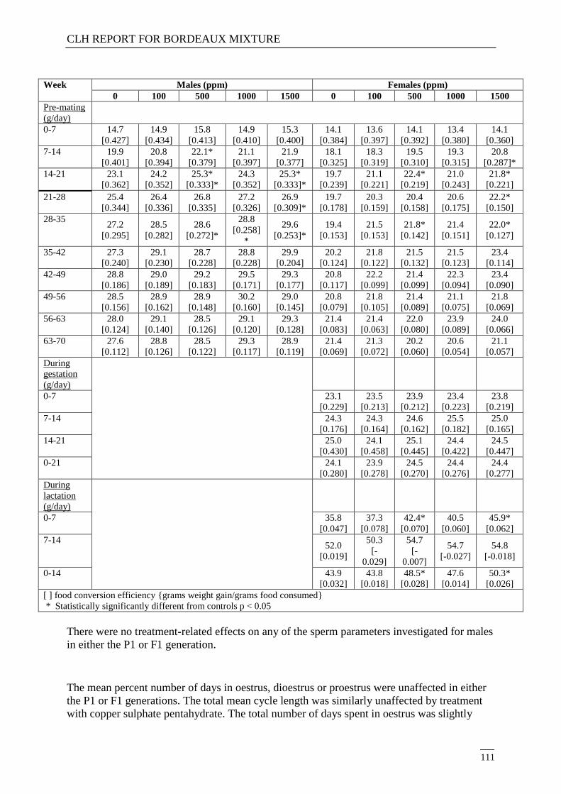
CLH REPORT FOR BORDEAUX MIXTURE
111
Week Males (ppm) Females (ppm)
0 100 500 1000 1500 0 100 500 1000 1500
Pre-mating
(g/day)
0-7 14.7
[0.427]
14.9
[0.434]
15.8
[0.413]
14.9
[0.410]
15.3
[0.400]
14.1
[0.384]
13.6
[0.397]
14.1
[0.392]
13.4
[0.380]
14.1
[0.360]
7-14 19.9
[0.401]
20.8
[0.394]
22.1*
[0.379]
21.1
[0.397]
21.9
[0.377]
18.1
[0.325]
18.3
[0.319]
19.5
[0.310]
19.3
[0.315]
20.8
[0.287]*
14-21 23.1
[0.362]
24.2
[0.352]
25.3*
[0.333]*
24.3
[0.352]
25.3*
[0.333]*
19.7
[0.239]
21.1
[0.221]
22.4*
[0.219]
21.0
[0.243]
21.8*
[0.221]
21-28 25.4
[0.344]
26.4
[0.336]
26.8
[0.335]
27.2
[0.326]
26.9
[0.309]*
19.7
[0.178]
20.3
[0.159]
20.4
[0.158]
20.6
[0.175]
22.2*
[0.150]
28-35 27.2
[0.295]
28.5
[0.282]
28.6
[0.272]*
28.8
[0.258]
*
29.6
[0.253]*
19.4
[0.153]
21.5
[0.153]
21.8*
[0.142]
21.4
[0.151]
22.0*
[0.127]
35-42 27.3
[0.240]
29.1
[0.230]
28.7
[0.228]
28.8
[0.228]
29.9
[0.204]
20.2
[0.124]
21.8
[0.122]
21.5
[0.132]
21.5
[0.123]
23.4
[0.114]
42-49 28.8
[0.186]
29.0
[0.189]
29.2
[0.183]
29.5
[0.171]
29.3
[0.177]
20.8
[0.117]
22.2
[0.099]
21.4
[0.099]
22.3
[0.094]
23.4
[0.090]
49-56 28.5
[0.156]
28.9
[0.162]
28.9
[0.148]
30.2
[0.160]
29.0
[0.145]
20.8
[0.079]
21.8
[0.105]
21.4
[0.089]
21.1
[0.075]
21.8
[0.069]
56-63 28.0
[0.124]
29.1
[0.140]
28.5
[0.126]
29.1
[0.120]
29.3
[0.128]
21.4
[0.083]
21.4
[0.063]
22.0
[0.080]
23.9
[0.089]
24.0
[0.066]
63-70 27.6
[0.112]
28.8
[0.126]
28.5
[0.122]
29.3
[0.117]
28.9
[0.119]
21.4
[0.069]
21.3
[0.072]
20.2
[0.060]
20.6
[0.054]
21.1
[0.057]
During
gestation
(g/day)
0-7 23.1
[0.229]
23.5
[0.213]
23.9
[0.212]
23.4
[0.223]
23.8
[0.219]
7-14 24.3
[0.176]
24.3
[0.164]
24.6
[0.162]
25.5
[0.182]
25.0
[0.165]
14-21 25.0
[0.430]
24.1
[0.458]
25.1
[0.445]
24.4
[0.422]
24.5
[0.447]
0-21 24.1
[0.280]
23.9
[0.278]
24.5
[0.270]
24.4
[0.276]
24.4
[0.277]
During
lactation
(g/day)
0-7 35.8
[0.047]
37.3
[0.078]
42.4*
[0.070]
40.5
[0.060]
45.9*
[0.062]
7-14 52.0
[0.019]
50.3
[-
0.029]
54.7
[-
0.007]
54.7
[-0.027]
54.8
[-0.018]
0-14 43.9
[0.032]
43.8
[0.018]
48.5*
[0.028]
47.6
[0.014]
50.3*
[0.026]
[ ] food conversion efficiency {grams weight gain/grams food consumed}
* Statistically significantly different from controls p < 0.05
There were no treatment-related effects on any of the sperm parameters investigated for males
in either the P1 or F1 generation.
The mean percent number of days in oestrus, dioestrus or proestrus were unaffected in either
the P1 or F1 generations. The total mean cycle length was similarly unaffected by treatment
with copper sulphate pentahydrate. The total number of days spent in oestrus was slightly

CLH REPORT FOR BORDEAUX MIXTURE
112
higher for the P1 females dosed at 1000 or 1500 ppm (47 and 40% respectively) in
comparison with controls (30%) but since there were no effects on mean oestrous cycle length
nor any adverse reproductive changes, this minor change was not considered to be
biologically significant.
At termination the distribution of oestrous cycle stages was similar for P1 and F1 females and
no treatment effect was postulated.
For the P1 and F1 generations there were no treatment-related effects on any of the
reproductive indices investigated at any of the four dose concentrations (tables 48 and 49).
These included precoital interval length, mating and fertility indices, gestation length, the
number of implantation sites and the implantation efficiency.
Table 48: P1 adult reproductive performance
Group 1 2 3 4 5
Treatment (ppm) Control 100 500 1000 1500
Males: n
Number mating
Mating index%
30
27
90.0
30
30
100
30
28
93.3
30
28
93.3
30
29
96.7
Females: n
Number pregnant
Fertility index (%)
30
25
92.6
30
29
96.7
30
27
96.4
30
25
89.3
30
27
93.1
Mean gestation length
(days) 22.2 22.4 22.3 22.3 22.4
Total resorption 0 0 0 0 0
Mean number of
implantation sites per
pregnant female
Number of pregnant females
14.5
25
14.1
29
13.9
27
14.0
25
13.8
27
Implantation efficiency (%) 93.3 92.4 91.6 93.2 91.7
Mean number of pups born
per litter 13.6 13.2 13.0 13.1 13.6
Table 49:. F1 adult reproductive performance
Group 1 2 3 4 5
Treatment (ppm) Control 100 500 1000 1500
Males: n
Number mating
Mating index%
30
29
96.7
30
30
100
30
30
100
29
29
100
30
30
100
Females: n
Number pregnant
Fertility index (%)
30
28
96.6
30
30
100
30
28
93.3
29
25
86.2
30
30
100
Mean gestation length
(days) 22.2 22.2 22.2 22.2 22.3
Total resorption 0 0 0 0 0
Mean number of
implantation sites per
pregnant female
Number of pregnant females
15.0
28
14.5
30
14.7
27
14.0
25
14.2
30
Implantation efficiency (%) 94.7 95.8 93.8 95.6 91.7
Mean number of pups born
per litter 14.2 13.9 13.8 13.3 13.2

CLH REPORT FOR BORDEAUX MIXTURE
113
Treatment with copper sulphate pentahydrate had no effect on the number of pups born, the
number of liveborn pups or the numbers of pups surviving to 4, 7, 14 or 21 days post-partum
(tables 50 and 51). In either generation, F1 or F2 offspring, there were any treatment-related
effects on the sex ratio within litters, or survival indices during lactation at any of the dose
concentrations tested.
Table 50: Litter data for F1 pups
Group 1 2 3 4 5
Treatment (ppm) Control 100 500 1000 1500
Number of pregnant females 25 29 27 25 26
Mean litter size – birth 13.6 13.2 13.0 13.1 13.6
Mean number live born 13.6 13.1 12.7 12.9 13.5
Mean number of pups pre-
culling on day 4 13.4 12.9 13.2 12.8 13.4
Mean number of pups per
litter post day 4 culling 7.8 7.8 7.9 7.9 8.0
Mean number of pups per
litter on day 7 7.8 7.8 7.9 7.9 8.0
Mean number of pups per
litter on day14 7.8 7.8 7.9 7.9 8.0
Mean number of pups per
litter on day 21 7.8 7.8 7.9 7.9 8.0
Sex ratio (% males) 52 48 53 49 50
Gestation index (% litters
with at least one live pup) 100 100 100 100 100
Mean percent born alive 99.5 98.9 95.2 98.9 99.5
Viability Day 0-4 (%) 98.6 98.8 99.5 98.9 99.2
Lactation index 99.5 100 99.5 100 100
Litter survival (% litters with
at least one pup alive at day
21)
100 100 100 100 100
Mean pup weight (g) –
Day 0
Day 4 pre-culling
Day 4 post-culling
Day 7
Day 14
Day 21
6.6
10.7
10.7
17.3
34.8
57.8
6.7
11.3
11.3
18.5
36.4
59.5
6.7
11.0
11.0
18.0
36.2
59.0
6.5
10.7
10.8
17.2
34.7
55.7
6.7
11.1
11.1
17.8
35.7
57.0
Table 51: Litter data for F2 pups
Group 1 2 3 4 5
Treatment (ppm) Control 100 500 1000 1500
Number of pregnant females 28 30 27 24 30
Mean litter size – birth 14.2 13.9 13.8 13.3 13.2
Mean number live born 14.1 13.7 13.7 13.2 13.1
Mean number of pups pre-
culling on day 4 13.9 13.6 13.3 13.0 13.0
Mean number of pups per
litter post day 4 culling 8.0 8.0 7.8 8.0 7.8

CLH REPORT FOR BORDEAUX MIXTURE
114
Mean number of pups per
litter on day 7 8.0 8.0 7.8 8.0 7.8
Mean number of pups per
litter on day14 7.9 8.0 7.8 8.0 7.8
Mean number of pups per
litter on day 21 7.9 7.9 7.8 8.0 7.7
Sex ratio (% males) 49 53 56 49 51
Gestation index (% litters
with at least one live pup) 100 100 100 100 100
Mean percent born alive 99.5 98.9 99.7 99.0 99.3
Viability Day 0-4 (%) 98.3 99.3 96.2 98.5 99.4
Lactation index 99.6 99.2 100 99.5 99.6
Litter survival (% litters with
at least one pup alive at day
21)
100 100 100 96.0 100
Mean pup weight (g) –
Day 0
Day 4 pre-culling
Day 4 post-culling
Day 7
Day 14
Day 21
6.3
10.2
10.2
16.8
34.2
56.0
6.4
10.9
10.9
17.7
35.3
58.1
6.5
10.7
10.6
17.6
36.5
58.4
6.4
11.0
10.9
17.7
35.1
57.3
6.6
10.8
10.9
17.5
35.4
56.7
Clinical signs were noted among the pups of the F1 or F2 generation but at low incidence and
showing no dose-relationship. The clinical observations were not considered to be treatment-
related or toxicologically significant.
An increase in mean pup weight in F1 litters dosed at 100 ppm (low dose) on lactation day 7
was not treatment-related since there were no other dose correlations. There were no
treatment-related effects on pup weight at any dose levels for the F1 or F2 offspring.
There were no treatment-related effects on preputial separation for F1 males at any dose level.
For the F1 females the mean age at vaginal opening was increased for the high dose group
(1500 ppm) in comparison with concurrent controls – 33.6 versus 32.1 days. However, the
historical control data for this parameter indicates a mean vaginal opening time of 32.3 days
and a minimum and maximum range of 31.3 to 33.9 days. Hence the difference was small
(1.5 days) and well within the range of historical control data. The apparent slight delay in
vaginal opening was not considered an effect of treatment with copper sulphate pentahydrate.
Pathology findings:
There were no significant differences between the high dose (1500 ppm) and control groups
in respect of total numbers for primordial and pre-antral follicles.
There were no test-substance related deaths during the course of the study. Of the 120 P1 and
120 F1 males, only one was sacrificed in extremis with a fractured nose (killed on day 14).
From the same number of P1 and F1 females, only three rats died during the study. One was
sacrificed in extremis on day 119 due to dystocia; one was found dead on day 17 – the cause
of death being pyelonephritis and one was sacrificed in extremis on day 119 but the cause of
morbidity was not established.

CLH REPORT FOR BORDEAUX MIXTURE
115
For the adult P1 rats there was a small decrease in mean absolute and relative spleen weight
(circa 9% reduction compare with controls) in the high dose group (1500 ppm). The effect
was statistically significant for females. While there were no significant differences among
the males, the trend for a slight reduction in spleen weight at higher doses was evident. None
of the other organs weighed for P1 animals showed any effect of treatment. Results are
summarised in table 52.
For the F1 weanlings of the high dose group (1500 ppm), small decreases in absolute (9%)
and relative (10-11 %) spleen weight were apparent in comparison with controls. None of the
other organs weighed for F1 weanlings showed any effect of treatment. Results are
summarised in table below.
There were no changes in organ weight among the F1 adults that were considered attributable
to treatment
For the F2 weanlings of the high dose group (1500 ppm), small decreases in absolute (10%
males, 15% females) and relative (10% males, 15% females) spleen weight were apparent in
comparison with controls. The high dose group effects were significantly lower than the
controls. None of the other organs weighed for F2 weanlings showed any effect of treatment.
Results are summarised in table below.
Table 52: Summary of spleen weights for males and females in P1, and for F1 and F2
weanlings
Males Females
Dose
concentration
(ppm)
0 100 500 1000 1500 0 100 500 1000 1500
P1 adults
Final body weight 595.4 600.1 603.9 599.5 586.8 328.8 332.2 335.8 333.3 331.9
Absolute spleen
weight [g] 0.866 0.887 0.892 0.881 0.841# 0.643 0.629 0.639 0.605 0.586#
Relative spleen
weight [g/100g
bw]
0.146 0.148 0.148 0.147 0.143 0.195 0.190 0.190 0.182 0.177*
F1 adults
Final body weight 593.5 619.2 600.0 598.5 584.7 326.0 328.2 335.0 332.9 329.2
Absolute spleen
weight [g] 0.897 0.887 0.867 0.900 0.841 0.624 0.641 0.632 0.642 0.612
Relative spleen
weight [g/100g
bw]
0.151 0.143 0.145 0.150 0.145 0.192 0.195 0.189 0.193 0.186
F1 weanlings
Final body weight 58.3 60.1 60.9 56.6 58.7 54.5 56.8 56.2 53.5 55.3
Absolute spleen
weight [g] 0.256 0.290 0.280 0.238 0.232# 0.245 0.283* 0.265 0.236 0.223#
Relative spleen
weight [g/100g
bw]
0.439 0.477 0.460 0.417 0.394 0.449 0.498* 0.470 0.429 0.401
F2 weanlings
Final body weight 56.9 59.3 59.2 59.8 57.3 54.6 56.8 56.8 55.3 54.7
Absolute spleen
weight [g] 0.253 0.269 0.254 0.252 0.227# 0.254 0.265 0.252 0.243 0.217*
Relative spleen
weight [g/100g
bw]
0.440 0.451 0.430 0.421 0.397* 0.462 0.465 0.444 0.440 0.396*

CLH REPORT FOR BORDEAUX MIXTURE
116
# considered to be a treatment-related effect (decreased weight).
* Statistically significantly different from controls p < 0.05
The small decrease in spleen weight for F1 and F2 weanlings was considered an effect of
treatment although weanling spleen weights are highly variable (e.g statistically significant
increase in absolute weight (+16 %) for the low dose F1 females). The effect could be
considered adverse, in the absence of any confirmatory microscopic examinations. However,
the effect may reflect a transient physiological change such as a marginal decrease in
sinusoidal dilatation. The pathologist’s review of data indicated the ranges for the high dose
spleen weights were similar to control ranges. Of 111 weanlings in the high dose group, only
4 had spleen weights that were lower than the control range. There were no treatment-related
effects on thymus weight to indicate a test substance related effect on the lymphoid system.
Extramedullary haematopoiesis in the livers of control and high dose weanlings was normal
suggesting the haematopoietic system was unaffected by treatment.
The other ogan weights showed no changes that were considered attributable to treatment
with copper sulphate pentahydrate.
There were no treatment-related changes apparent during necropsy of the P1 adult rats, F1
adults or F1 and F2 weanlings or F1 and F2 pups.
All macroscopic observations in the adult rats, P1 or F1, were within the range of normal
background lesions. Among the F1 and F2 weanlings the incidence of gross lesions was low
and observations were randomly distributed across control and treated groups. For the F1 and
F2 pups, the observations of non-expanded lungs or no milk spot in stomach were considered
non-specific lesions that are commonly observed among stillborn pups and were therefore not
considered to be an effect of treatment with copper sulphate pentahydrate.
All microscopic findings seen in the P1 adults, F1 adults or F1 and F2 weanlings were
considered to be incidental and common background lesions for the strain of rat used in the
study. There were no treatment-related histopathological changes in liver, brain or
reproductive organs.
Eighteen P1 and nine F1 pairs failed to produce litters. The cause of reproductive failure in 22
of these pairs was not determined. One F1 female had dystocia and in three of the P1 females
there was an absence of recent corpora lutea in the ovaries. None of the breeding failures were
considered attributable to test substance administration.
Tissue metal concentrations:
Specifically assessments of copper, iron, manganese and zinc concentrations were
investigated in liver and brain and plasma for each subset of animals. Results were as follows.
For the P1 males there were no test-substance related changes in copper, iron, manganese and
zinc concentrations at any dose level. Plasma samples were not obtained for these animals.
There was a decreased in liver iron concentration in the high dose group but this was not
considered a treatment effect due to high inter-individual variability and a lack of consistency
with the female response and an absence of any dose relationship.
The P1 females dosed at 1500 ppm had a treatment-related increase in liver copper
concentration and a decrease in liver iron concentration. Copper and iron levels in the brain
were unaffected and there were no changes in manganese or zinc concentrations in liver, brain
or plasma.
For the F1 adult males, liver copper concentration was increased in groups dosed at 1000 or
1500 ppm. Compared with the magnitude of similar changes seen in the P1 generation, the

CLH REPORT FOR BORDEAUX MIXTURE
117
effects in F1 males were small but in comparison with controls, some individuals showed a 2-
3 fold increase and the effect was considered attributable to treatment. Copper concentrations
in brain or plasma were unaffected by treatment at any dose level. There were no treatment-
related changes in iron, manganese or zinc concentrations in liver, brain or plasma at any dose
level.
For the F1 adult females, liver and brain copper concentrations were increased at 1500 ppm.
Copper concentration in plasma was not affected at any dose level. There were no treatment-
related changes in iron, manganese or zinc concentrations in liver, brain or plasma at any dose
level.
For the F1 and F2 weanlings, there was a treatment-related increase in liver copper
concentration for males and females dosed at 1000 and 1500 ppm in each generation. Brain
copper concentrations were slightly increased for the males (but not females) dosed at 1500
ppm in each generation. No plasma data were available for the F1 weanlings and there were
no changes in plasma copper concentration for the F2 weanlings. A treatment-related decrease
in plasma iron concentration was evident for the male and female F2 weanlings dosed at 1500
ppm. Changes in manganese and zinc concentrations in liver, brain or plasma were all
considered to be spurious since they showed no dose relationship, had high inter-individual
variability or the changes were small in magnitude.
In summary, (tables 53, 54, 55 and 56), the concentration of copper in the liver of F1 males
and F1 and F2 male and female weanlings dosed at 1000 and 1500 ppm was increased. The
concentration of copper in the liver of P1 and F1 females dosed at 1500 ppm was also
increased. Copper concentrations in the brain were increased for F1 females and F1 and F2
male weanlings dosed at 1500 ppm. The concentration of iron in the liver of P1 females dosed
at 1500 ppm was decreased and plasma iron concentration was decreased in F2 male and
female weanlings in the 1500 ppm dose group.
Table 53: Summary of tissue concentrations of copper, iron, manganese and zinc in brain,
liver or plasma for males and females P1
Males – P1 Females – P1
Dose
concentration
(ppm)
0 100 500 1000 1500 0 100 500 1000 1500
Copper (ppm)
Liver 6.44 4.47 5.20 5.60 5.98 4.76 5.30 5.46 5.67 8.73*
Plasma -- -- -- -- -- 1.43 1.36 1.35 1.48 1.38
Brain 3.27 3.46 431 4.98 3.26 3.17 3.41 3.58 2.93 3.38
Iron (ppm)
Liver 158 143 155 143 128* 150 151 138 150 107*
Plasma -- -- -- -- -- 2.96 2.90 3.24 3.17 3.32
Brain 22.4 24.4 26.2 22.8 24.9 20.4 21.0 20.8 19.9 18.9
Manganese
(ppm)
Liver 2.45 2.26 2.62 2.75 2.34 3.46 3.49 3.20 3.52 3.56
Plasma -- -- -- -- -- -- -- -- -- --
Brain 0.451 0.525 0.534 0.459 0.573 0.419 0.438 0.411 0.433 0.422
Zinc (ppm)
Liver 33.1 31.6 33.8 32.4 28.7 28.8 28.5 29.4 30.5 29.2
Plasma -- -- -- -- -- 1.94 1.90 1.93 1.86 1.72
Brain 17.5 16.6 16.8 16.3 16.6 14.7 14.1 14.7 15.4 13.4
* Statistically significantly different from controls p < 0.05

CLH REPORT FOR BORDEAUX MIXTURE
118
Table 54: Summary of tissue concentrations of copper, iron, manganese and zinc in brain,
liver or plasma for males and females F1 adults
Males – F1 adults Females – F1 adults
Dose
concentration
(ppm)
0 100 500 1000 1500 0 100 500 1000 1500
Copper (ppm)
Liver 4.56 4.87 6.16 7.36* 7.53* 5.70 5.16 5.36 5.35 15.3*
Plasma 1.24 1.38 1.28 1.51 1.44* 1.49 1.52 1.34 1.50 1.37
Brain 2.59 2.64 2.83 3.11* 2.80 2.89 2.93 3.00 3.23 3.49*
Iron (ppm)
Liver 121 133 124 143 110 149 149 163 116 133
Plasma 2.41 2.27 2.72 2.35 2.88 3.46 4.00 4.01 3.56 4.12
Brain 18.5 18.4 16.5 17.2 17.5 18.5 18.4 21.2 20.6 19.9
Manganese
(ppm)
Liver 1.93 2.00 2.20 2.14 1.82 3.46 3.18 3.28 3.06 3.64
Plasma -- -- -- -- -- -- -- -- -- --
Brain 0.355 0.350 0.343 0.376 0.319 0.368 0.394 0.405 0.412 0.438*
Zinc (ppm)
Liver 26.7 27.8 32.4* 28.3 25.8 31.9 30.7 32.6 27.9 32.2
Plasma 0.971 0.916 0.989 1.019 1.080 1.93 1.87 2.03 1.54* 1.80
Brain 13.3 13.4 14.1 14.0 12.1 14.6 15.1 15.0 15.1 15.4
* Statistically significantly different from controls p < 0.05
Table 55: Summary of tissue concentrations of copper, iron, manganese and zinc in brain,
liver or plasma for males and females F1 weanlings
Males – F1 weanlings Females – F1 weanlings
Dose
concentration
(ppm)
0 100 500 1000 1500 0 100 500 1000 1500
Copper (ppm)
Liver 14.7 24.2 25.2 50.0* 82.7* 21.5 22.8 23.5 53.1* 86.8*
Plasma -- -- -- -- -- -- -- -- -- --
Brain 2.26 2.27 2.28 2.44 2.59* 2.43 2.35 2.43 2.40 2.60
Iron (ppm)
Liver 33.9 33.0 32.0 37.4 36.1 33.6 36.1 38.3 39.5 37.4
Plasma -- -- -- -- -- -- -- -- -- --
Brain 11.1 15..3 13.1 11.5 11.0 16.6 12.4 13.7 11.3 12.8
Manganese
(ppm)
Liver 2.01 1.95 2.01 2.08 2.27 2.08 2.13 1.96 2.18 2.23
Plasma -- -- -- -- -- -- -- -- -- --
Brain 0.500 0.503 0.527 0.561 0.565 0.562 0.505 0.522 0.539 0.629
Zinc (ppm)
Liver 31.4 31.5 35.2 37.4* 36.8* 32.5 31.8 34.3 39.7* 37.3*
Plasma -- -- -- -- -- -- -- -- -- --
Brain 13.8 13.6 14.1 14.2 14.5 14.1 13.9 14.5 15.4 14.4
* Statistically significantly different from controls p < 0.05
Table 56: Summary of tissue concentrations of copper, iron, manganese and zinc in brain,
liver or plasma for males and females F2 weanlings

CLH REPORT FOR BORDEAUX MIXTURE
119
Males – F2 weanlings Females – F2 weanlings
Dose
concentration
(ppm)
0 100 500 1000 1500 0 100 500 1000 1500
Copper (ppm)
Liver 16.4 30.2 28.0 47.6* 64.3* 24.9 21.5 27.9 38.8* 53.5*
Plasma 0.526 0.533 0.582 0.543 0.554 0.581 0.550 0.587 0.573 0.543
Brain 2.55 3.12 2.35 2.49 3.24* 2.59 2.63 2.52 2.41 2.78
Iron (ppm)
Liver 33.8 37.0 37.1 36.8 29.8 41.8 38.1 39.1 42.2 35.2
Plasma 3.20 3.98 2.78 2.73 1.55 3.21 3.54 3.19 2.54 1.41*
Brain 11.1 11.4 11.8 11.0 10.0 11.6 11.1 12.5 11.4 10.7
Manganese
(ppm)
Liver 2.04 2.03 2.03 2.06 2.24 2.12 1.92 2.21 2.03 2.30
Plasma -- -- -- -- -- -- -- -- -- --
Brain 0.490 0.535 0.465 0.510 0.570* 0.479 0.555 0.524 0.521 0.570*
Zinc (ppm)
Liver 30.9 30.3 33.3 29.8 31.2 34.2 27.3* 31.6 33.2 31.7
Plasma 2.07 2.30 2.07 2.42 2.04 2.38 2.19 2.15 1.95 2.18
Brain 14.7 15.2 14.8 14.7 16.5* 15.6 15.1 15.1 15.0 14.5
* Statistically significantly different from controls p < 0.05
Overall summary of findings.
There were no effects considered to be related to copper sulphate treatment on the following
parameters at any concentration (100 to 1500 ppm):
Mortality and clinical signs of toxicity in P1 and F1 males and females
Body weights, weight gain, food consumption, food efficiency in P1 and F1 males
and females
Sperm and estrous cycle parameters in P1 and F1 males and females
Mating, precoital interval, fertility, gestation length, number of implantation sites, and
implantation efficiency in the P1 and F1 generations
Number of pups born, born alive, alive on day 4, 7, 14, or 21, sex ratio, and survival
indices during the lactation period in F1 and F2 litters
Body weights and clinical observations in F1 and F2 litters during lactation
Age at preputial separation in F1 males and vaginal opening in F1 females
Ovarian follicle counts in F1 females
Weight of testes, epididymides, right cauda epididymis, seminal vesicles, prostate,
ovaries, uterus, thyroid gland, brain, liver, adrenal glands, kidneys and pituitary in P1
and F1 males and females; Weight of liver, brain and thymus in F1 and F2 weanlings;
Weight of the spleen in P1 males and F1 males and females
Gross observations in P1 and F1 adults and F1 and F2 weanlings
Microscopic observations in the liver, brain and reproductive organs in P1 and F1
adults
Microscopic observations in the liver and brain in F1 and F2 weanlings.
Potentially adverse effects considered to be related to copper sulphate treatment were limited
to the 1500 ppm groups and were comprised of:
Decreased spleen weight in P1 adult females, and F1 and F2 male and female
weanlings.

CLH REPORT FOR BORDEAUX MIXTURE
120
Under the conditions of this study there were no treatment-related effects in either generation
(P1 and F1 adults or F1 and F2 offspring) on reproduction parameters or indications of
systemic toxicity at any of the dose concentrations used (doses of 100 to 1500 ppm). There
were no adverse effects of treatment at up to 1500 ppm on fertility, general reproductive
performance or offspring viability and growth at any dose level (dietary levels 0, 100, 500,
1000 and 1500 ppm CuSO4). Dietary intake varied with stage of maturation and effects
observed at the high dose may reflect changes in food intake and test substance consumption
for the different populations within the study. Actual dosed values were 1.53-2.65, 7.7-13.3,
15.2-26.7 and 23.6-43.8 mg/kg body weight/day, for the 100, 500, 1000 and 1500 ppm
groups, respectively. However since young rats consume more diet the mg/kg bw/day
exposure is greater at weaning and at the beginning of each maturation phase. Pregnant and
lactating females also consume more diet and are subject to a greater mg/kg bw/day exposure.
The concentration of copper was increased in the liver of F1 males and F1 and F2 male and
female weanlings at 1000 and 1500 ppm and in P1 and F1 females at 1500 ppm. Brain copper
concentration was increased in F1 females and F1 and F2 male weanlings at 1500 ppm. The
concentration of liver iron was decreased in P1 females at 1500 ppm. The concentration of
plasma iron was decreased in F2 male and female weanlings at 1500 ppm. There were
decreased spleen weight in P1 adult females, and F1 and F2 male and female weanlings.
The majority of effects are reported in weanlings and in dams at the end of lactation - the food
intake and compound consumption data show that both of these “populations” were
consuming significantly higher amounts of diet than towards the end of the pre-mating
maturation periods (the food intake of the weanlings is virtually the same as in the first week
of the F1 maturation period, when compound consumption of F1 males is 58 mg Cu/kg/day,
for example, and during lactation when the adult females are eating lots to feed their young),
but that the spleen effects are not seen in males at termination, when compound consumption
is much lower (22.9 mg Cu/kg/day in F1 males). From this it may be concluded that the
spleen effects may be transient even at high doses, and that when the dietary intake i.e. dose
level is reduced, the spleen effect diminishes. However, while the iron effects and the brain
copper effects at 1500 ppm are also probably temporary and related to high dietary intakes (in
that the male weanlings showed the finding, but when those weanlings grew older they did
not), there is insufficient evidence to support 1500 ppm as a NOAEL.
From these results, the no-observed-effect level (NOEL) for reproductive toxicity was 1500
ppm, the highest concentration tested. The systemic NOEL for P1 and F1 rats and F1 and F2
offspring during lactation was 1000 ppm, based on reduced spleen weight in P1 adult females,
and F1 and F2 male and female weanlings at 1500 ppm. The dietary concentration of 1000
ppm was equivalent to mean daily intakes of copper of 15.2 - 23.5 mg/kg body weight/day for
male rats during pre-mating and 17.0 - 26.7 mg/kg body weight/day for female rats during
pre-mating and gestation.
Reference: De la Iglesia F. W. (1973)
Guideline: No. Cross-mating fertility study
GLP: No
Three groups of 20 female Wistar rats were given copper gluconate orally by gavage at 0, 3 or
30 mg/kg/day for two weeks prior to mating through to either day 20 of pregnancy or day 21
post partum. Females were paired (1m:2f) with untreated males. In a parallel study, two
groups of 10 males received copper gluconate at 3 mg/kg/day for 60 days prior to pairing

CLH REPORT FOR BORDEAUX MIXTURE
121
(1m:2f) with either untreated females or females that had received copper gluconate at
3 mg/kg/day for 60 days prior to mating. A further group of 10 males and 20 females were
maintained untreated for 60 days and allowed to mate. Parameters investigated included
pregnancy rate (percentage of pregnancies), day 20 litter parameters including implantations,
resorptions, live foetuses, gross foetal anomalies; litter parameters included duration of
gestation, litter size, number of live young, gross anomalies, litter and mean pup weights
through to weaning.
There were no significant differences between treated and control groups in any of the
parameters studied.
Copper gluconate did not affect the fertility of either the male or female rat, following oral
administration. This is discussed further at the end of this section.
Reference: Lecyk, M. (1980)
Guideline: No
GLP: No
Deviations: Yes (from OECD 414)
Housing and feeding conditions of test animals,
information on the age and weight of test animals,
no detail given on the size of the groups,
in several dose groups, the number of pregnant animals was smaller than
recommended by the guideline (16 animals).
in the absence of information on the weight of test animals and the weight of treated
diet consumed, it was not possible to accurately determine the dose received on a
mg/kg bodyweight basis,
no information on maternal toxicity was presented in the report,
no post-mortem information was presented in the report for dams,
no information was presented on: the weight of gravid uteri; the number of corpora
lutea; degrees of resorption of dead foetuses,
the sex ratio of live foetuses was not reported,
no justification is provided for use of mouse whereas the preferred rodent species is rat
for this study,
males were fed the appropriate test diet prior to mating and no information on male
toxicity was then reported,
the study did not measure maternal bw gains or maternal liver histology or copper
content.
Copper sulphate was administered to groups of male and female mice, strains C57BL and
DBA, by admixing the aqueous solution with the diet at dose levels of 0, 500, 1,000, 1,500,
2,000, 3,000 and 4,000 ppm corresponding approximately to 0, 71, 142, 214, 285, 427 and
570 mg/kg bw/day . The feed was granulated and dried before administering to the animals.
The males and females were paired after one month of treatment and the day of mating
(appearance of a vaginal plug) was designated Day 0 of gestation. On Day 19 of gestation the
females were killed and foetuses (living and dead) were counted and weighed. One half of the
foetuses in each group was examined for visceral abnormalities (Wilson technique) and the
other half was cleared and stained with alizarin for skeletal examination.

CLH REPORT FOR BORDEAUX MIXTURE
122
Although the paper does not give details of group size and pregnancy rate, from the numbers
of pregnant females (particularly at 4000, 3000 ppm), pregnancy rate was not adversely
affected by dietary administration of copper at up to 4000 ppm for one month prior to mating
(table below).
In both strains of mice, there was no effect on the embryonic growth at the lower doses,
2,000 ppm and below. The authors claimed a slight stimulation indicated by lower % foetal
mortality and slightly higher weights of the foetuses than the controls at doses up to
2000 ppm. A treatment-related effect was noted at higher levels, at 3,000 and 4,000 ppm,
where decreased foetal weights and a higher mortality were recorded (table 57). It should be
noted that mean litter size was smaller than normal for the mouse in all groups. Various
development malformations were observed in both these groups in both strains, although there
was no consistent pattern of type. Abnormalities classed by the authors as malformations at
3000 ppm (3 foetuses in total) were last lumbar vertebra included in sacrum (one foetus) and
unilateral fused rib (two foetuses); at 4000 ppm, hernia of the thoracic wall, hydrocephalus
and fusion of thoracic ribs and vertebrae, (each one foetus, two foetuses with encephalocoel
and two foetuses with (last lumbar) hemivertebra as part of sacrum. However, as no
information was presented in this study regarded maternal toxicity, the possibility that the
effects on embryonic development were secondary to maternal toxicity cannot be excluded.
Table 57: Mouse embryonic development
Dose level (ppm)
0 500 1000 1500 2000 3000 4000
C57BL mice
Number of pregnant females 21 10 18 7 10 22 18
Number of live foetuses
(%)
65
(83.1)
46
(89.2)
81
(86.5)
31
(87.1)
42
(78.6)
55
(72.8)
35
(71.5)
Number of dead foetuses
(%)
11
(16.9)
5
(10.8)
11
(13.5)
4
(12.9)
9
(21.4)
15
(27.2)
10
(28.5)
Mean litter size 3.09 4.60 4.50 4.42 4.20 2.50 1.94
Mean foetal weight (g) 1.10 1.35 1.22 1.14 1.25 1.00 0.99
Abnormal foetuses
(%) - - - - -
1
(1.8)
3
(8.5)
DBA mice
Number of pregnant females 17 10 10 14 10 18 20
Number of live foetuses
(%)
76
(84.3)
54
(90.8)
51
(88.3)
58
(82.8)
41
(83.0)
56
(75.0)
45
(70.4)
Number of dead foetuses
(%)
12
(15.7)
5
(9.2)
6
(11.7)
10
(17.2)
7
(17.0)
14
(25.0)
16
(29.6)
Mean litter size 4.47 5.40 5.10 4.14 4.10 3.11 2.70
Mean foetal weight (g) 0.96 1.24 1.19 1.17 1.13 1.11 1.09
Abnormal foetuses
(%) - - - - -
2
(3.7)
4
(7.4)
Dietary administration of 3,000 and 4,000 ppm copper as sulphate (approximately equivalent
to dose levels of 430 and 570 mg/kg bw/day, using the US FDA conversion factor of 7 for
mice) for one month prior to pairing did not adversely affect mating performance or

CLH REPORT FOR BORDEAUX MIXTURE
123
pregnancy rate but caused an increase in foetal mortality, decreases in foetal weights and
slight increase in incidence of malformations.. It should be noted that the study did not
measure maternal bodyweight gains, or maternal liver histology or copper content. The NOEL
for fertility effects was greater than 4,000 ppm (approximately 570 mg/kg bw/day) and the
NOEL for foetal effects was 2,000 ppm (approximately 285 mg/kg bw/day).
4.10.1.2 Human information
Reference: Ralph, A. and McArdle, H. (2001)
Guideline: No
GCP: No
The publication is a review of data on copper metabolism and toxicity during pregnancy and
lactation, with emphasis on the human.
The review considers the following aspects:
Fertilisation: Copper metal is known to interrupt implantation and development of the
blastocyst when present in the uterus as an intra-uterine contraceptive device (IUD), but once
implantation has taken place, IUDs do not show adverse effects on maintenance of pregnancy.
Maternal serum copper levels and ceruloplasmin levels rise steadily throughout pregnancy,
and fall significantly at parturition. The concentration in the mother is higher than in the
foetus, which establishes a concentration gradient from the mother to the foetus. The rise in
plasma concentration may be due to either enhanced uptake from food or decreased biliary
excretion. It is induced by oestrogen. Various studies have shown that copper requirements of
pregnant humans are up to one third greater than non-pregnant human females. Copper and
ceruloplasmin are present in amniotic fluid, but uptake from amniotic fluid by the foetus is
small. The placenta has been shown to take copper from the maternal blood as both
ceruloplasmin and by lower-weight complexes (albumin, histidine), but that delivery by
ceruloplasmin is more efficient. Ceruloplasmin is not itself passed across the placenta, but
ceruloplasmin and histidine may deliver copper to the placental cells via specific cell surface
receptors. The placenta has a regulatory role on the transfer of copper from mother to baby, as
infant serum concentrations of copper do not correlate with those of the mother. This has been
demonstrated in both human and rat. Women with Wilson’s disease can give birth to healthy
babies if the condition is well managed (zinc sulphate therapy). Pregnant women with
untreated Wilson’s disease tend to have spontaneous abortions. In the Brewer study (2000), of
26 pregnancies in 19 women who were on zinc therapy throughout their pregnancy, 24 new-
borns were normal, one had a heart defect (corrected by surgery) and another showed
anencephaly. Anencephaly has also been associated with very low maternal copper serum
levels, and there have been two reported cases of anencephaly where an IUD was used
(Graham et al. 1980).
Foetal development: copper accumulates in the placental layers and is transferred to the foetus
by an active process driven by foetal needs; it is thought to be incorporated in the foetal liver
into foetally synthesised ceruloplasmin. Copper is present in the foetal circulation in
ceruloplasmin, albumin, α-fetoprotein, transcuprein and low molecular weight ligands. The
human foetus accumulates copper at a rate of 50 μg/kg/day during the latter half of pregnancy,
and 50% of it is stored as metallothionein in the liver. The ratio of copper in the liver of
newborn infants to adults is 15:4. There are no reports of adverse effects of acute toxicity of
copper in human pregnancy. Foetal copper accumulation occurs in the third trimester, and

CLH REPORT FOR BORDEAUX MIXTURE
124
premature and low-weight babies are at risk of copper deficiency. Studies indicate that the
capacity of pre-term infants to utilise copper from the diet is limited; most of the ingested
copper is present in the stool, indicating either ineffective absorption or limited ability to
retain and store copper.
Parturition. Serum maternal plasma level returns to normal in the human within two to five
weeks. The timing of the return to normal may be influenced by the duration of breast-
feeding.
Lactation. Ceruloplasmin occurs in the milk of humans and other mammals, concentrations
being higher in the early stages of lactation. Approximately 20-25% of copper in human milk
is present as ceruloplasmin. Breast-feeding supplies up to 60 μg/kg/day, and is approximately
24% bioavailable. Maternal copper blood levels are under hormonal control (e.g. oestrogen,
see above), but alterations in maternal copper intake through dietary supplementation, or
elevated blood levels through other factors, such as severe infections, and even Wilson’s
disease, do not alter copper content of breast milk. It is likely that there are homeostatic
mechanisms that regulate mammary gland uptake of copper and its secretion in milk, but
these have not been explained. In human breast milk, approximately 75% of the copper is in
the whey, bound to soluble albumin or low molecular weight ligands. Another 15-20% is in
lipids, bound to the outer fat globule membrane, and about 5% is in insoluble form, possibly
bound to casein. Differences in composition of other milks (cow, soy) affect the
bioavailability to the human baby. Absorption and retention rates from formula milks are very
low, although toxicity has been observed where infants have been given substantial amounts
of cow’s milk boiled in untinned copper vessels. Awareness of the disease in India [Indian
Childhood Cirrhosis] and Austria [Idiopathic Copper Toxicosis] has resulted in use of other
containers, and the incidence has fallen. Human milk, unsurprisingly, contains the most
bioavailable copper for the human baby. Healthy infants fed exclusively on cow’s milk for 6
months became copper deficient, but the condition reversed on weaning to solid foods.
Growth and development. Neonatal humans have high concentrations of copper in the liver
and low concentrations of serum copper and ceruloplasmin. Newborn humans also show high
concentrations of metallothionein that decrease after birth. Copper in the new-born’s liver
appears to provide much of the copper requirements of the infant while it is breast-fed, until
weaning at 4-6 months. However, milk must provide a significant contribution, as mice
showing ‘toxic milk mutation’ die if they are kept on mother’s milk, because the mother
cannot secrete the normal amounts of copper into the milk, and the pups die of copper
deficiency. Premature birth restricts the hepatic storage of copper (as the mother’s supply via
the placenta is no longer available), and milk formulae for premature infants contains
additional copper to compensate for this. Low copper levels at this time may have
neurological implications during the critical period of brain growth. Excess copper in drinking
water at concentrations of approximately 8 mg/L showed chronic toxicity in adults but not in
children under 6 years of age. As the infant grows, levels of ceruloplasmin increase. Studies
in rats show that copper absorption is high during the neonatal period, but decreases by
weaning, as more is retained in the intestinal mucosa. With increasing postnatal age, more is
transported to the liver and less is bound to the intestine. There is evidence in rats that during
lactation, intestinal copper absorption occurs by diffusion and solvent drag, and only after
weaning does a saturable (adult, see Section B.6.1) copper transport system become evident.
Children require higher levels of copper in the diet than adults, especially during periods of
rapid growth. Girls aged 6-10 were fed on diets of copper ranging from 1.1 to 3.8 mg/day. At
intakes under 2 mg/day, copper balance was negative. A positive copper balance was
achieved on a vegetarian diet with a copper intake of over 2.8 mg/day. It was suggested that
an intake of 1.3 mg/day was sufficient for equilibrium, but that 2.5 mg/day was necessary for

CLH REPORT FOR BORDEAUX MIXTURE
125
growth. Serum of normal children reaches a peak of 1.57 mg/L between 6 and 11 years and
falls to 1.1 mg/L in adults between 22 and 75 years.
Intake: the review found no evidence of copper toxicity from customary dietary intake, unless
the food had been accidentally contaminated with copper during preparation e.g. acid fruit
such as apples, were stewed in a copper vessel, or there was repeated ingestion of milk heated
in copper vessels. A study of three cities in the US state of Massachusetts showed no
incidence of ill-health in adults or children under 6 years of age, despite drinking water
concentrations of over 8 mg/L. Most dietary intakes are below the 10-12 mg/adult/day set by
international organisations.
There is no evidence for adverse effects of oral exposure through customary diets worldwide
(which includes countries where copper is used in agriculture) for any adverse effects of
copper on pregnancy, parturition, lactation or growth and development in the human. There is
evidence of toxicity particularly to neonates repeatedly exposed to milk heated in copper
vessels, or exposure to acid fruit stewed in copper vessels.
4.10.2 Developmental toxicity
4.10.2.1 Non-human information
Reference: Munley S. M. (2003a)
Guideline: No. Range-finding study designed to assess relative tolerance of five technical
copper substances in the rabbit.
GLP: Yes
Five technical materials, copper hydroxide (batch number 380-71-05, copper content 60.1%
w/w), copper oxychloride (batch number 27003B, copper content 57% w/w), Bordeaux
Mixture (batch number 1/170), copper content 26.38% w/w), tribasic copper sulphate (batch
number 471/2002, copper content 31.12% w/w), and copper (I) oxide, (batch number 280802,
copper content 87% w/w) were given orally by gavage as suspension in 0.5% aqueous methyl
cellulose in deionised water to non-pregnant female Hra:(NZW)SPF rabbits. The animals
were approximately 6 to 6.5 months old and weighed from 3382 g to 4116 g on the day after
arrival. Animals were quarantined for at least 5 days before dosing. Rabbits were housed
individually, and were given 150 g laboratory rabbit diet each day, and tap water ad libitum.
Food consumption and bodyweight were recorded daily, clinical observations were recorded
daily and post dosing. Dose formulations were prepared daily at a dose volume of 1 mL/kg
bw, based on that day’s bodyweight. Any animals found dead were necropsied. At
termination, all animals were given a gross external and visceral examination. Lesions were
retained in an appropriate fixative. In the first part of the study, groups of two rabbits were
dosed with each technical material for up to 14 days. Concentrations were calculated to give
30 mg as copper /kg bw/day. In view of the moderate toxicity seen at 30 mg Cu/kg bw/day,
doses of 50 mg Cu/kg bw/day were given to a further group of 2 rabbits per technical
substance, to assess tolerance to a higher dose. Mortalities occurred after the first dose and
surviving rabbits were given 40 mg Cu/kg bw/day for the remaining six days of
administration.
Animals at 30 mg Cu/kg bw/day showed bodyweight loss during the first half of the treatment
period, followed by recovery during the second week of treatment (Figure 6.7.3.1.1.). There
were no marked differences between the five technical substances. Food consumption
reflected bodyweight changes; during the first week of dosing animals showed marked
reductions in food consumption, and in the second week the animals generally resumed
eating.

CLH REPORT FOR BORDEAUX MIXTURE
126
Figure 1: Bodyweight change with five forms of copper at 30 mg/kg/day
I: copper hydroxide; II: copper (I) oxide; III: copper oxychloride; IV: tribasic copper sulphate;
V: Bordeaux mixture
There were no deaths among animals treated with copper hydroxide, Bordeaux mixture,
tribasic copper sulphate or copper oxide. One animal dosed with copper oxychloride was
found dead on day 2. There were no indications of any adverse effects of treatment, or of
dosing error, and this animal was replaced by a similar animal from the same batch. A second
animal dosed with oxychloride died on day 11. The animal showed no remarkable necropsy
findings other than fur staining. During the study, it was discovered that the two animals
dosed with tribasic copper sulphate were underdosed by approximately 40%, because of a
calculation error. These animals were also replaced by two similar animals, and the
food/bodyweight data from the underdosed animals was not used in the comparison of the
five substances. Three animals were inadvertently sacrificed prematurely in the second week
of treatment. Necropsy revealed various stomach findings, including ulceration, red or dark
discolouration, and haemorrhagic areas in one animal dosed with copper hydroxide, both
animals dosed with copper oxide, and three of the four animals dosed with tribasic copper
sulphate.
At 50 mg Cu/kg bw/day, one of the two animals died after the first dose in each group except
tribasic copper sulphate. The Study Director immediately reduced the dose concentration to
40 mg Cu/kg bw/day (i.e. from day 2) and there were no further deaths. All decedents showed
either stomach ulceration or dark discolouration and thickening of the non-glandular portion
of the stomach. Survivors at 40 mg Cu/kg bw/day showed weight loss (Figure 6.7.3.1.2) and
reduced food consumption. At termination, all survivors showed stomach ulcerations.
Figure 2: Bodyweight change with five forms of copper at 50/40 mg/kg/day
-450
-400
-350
-300
-250
-200
-150
-100
-50
0
50
1 3 5 8 15
Mean
Bo
dy W
eig
ht
Gain
(g
)
Days on Test
I
II
III
IV
V

CLH REPORT FOR BORDEAUX MIXTURE
127
I: copper hydroxide; II: copper (I) oxide; III: copper oxychloride; IV: tribasic copper sulphate;
V: Bordeaux Mixture
The general pattern and degree of inappetance and weight loss followed by recovery, and the
observation of stomach ulceration at necropsy was considered sufficient to show that there
were no major differences in the sensitivity of the rabbit to the five copper substances. Doses
greater than 30 mg Cu/kg bw/day were considered unsustainable for repeat dosing studies. As
there were no major differences between the five substances, further preliminary
investigations would be performed on only one substance, copper hydroxide.
Reference: Munley S. M. (2003 b)
Guideline No. Range-finding study designed to assess effect of treatment equal in
duration to a teratology study in the non-pregnant rabbit.
GLP: Yes
Technical copper hydroxide (batch number 380-71-05, copper content 60.1% w/w) was given
orally by gavage as suspension in 0.5% aqueous methyl cellulose in deionised water to groups
of five non-pregnant female Hra:(NZW)SPF rabbits for 23 consecutive days. Dose levels
were 0, 7.5, 15 or 30 mg Cu/kg bw/day. Analysis of dose formulations confirmed stability,
homogeneity and verified the accuracy of formulation. The animals were approximately 6 to
6.5 months old and weighed from 2936 g to 3748 g on the first day of dosing. Animals were
quarantined for at least 5 days before dosing. Rabbits were housed individually, and were
given 150 g laboratory rabbit diet each day, and tap water ad libitum. Food consumption and
bodyweight were recorded daily, clinical observations were recorded daily and post dosing.
Dose formulations were prepared daily at a dose volume of 1 mL/kg bw, based on that day’s
bodyweight. Any animals found dead were necropsied. At termination on day 24, all animals
were given a gross external and visceral examination. Lesions were retained in an appropriate
fixative.
-900
-800
-700
-600
-500
-400
-300
-200
-100
0
1 3 5 8 M
ean
Bo
dy W
eig
ht
Ch
an
ges (
g)
Days on Test
I
II
III
IV
V

CLH REPORT FOR BORDEAUX MIXTURE
128
There were two deaths at 30 mg Cu/kg bw/day, on day 2 and 3 respectively. The latter animal
showed lethargy, weakness and abnormal gait or mobility prior to death. Both decedents
showed haemorrhages and/or discolouration of the stomach lining. There were no deaths at 15
mg Cu/kg bw/day. Two animals at 7.5 mg Cu/kg/bw/day died on days 2 or 5 due to intubation
errors. Necropsy findings included punctured lung tissue.
Bodyweights and food consumption at 30 and 15 mg Cu/kg bw/day were lower than controls
from the start of treatment. There was a group mean bodyweight loss during the first week of
treatment, with recovery to initial mean values by day 19 in both groups (Figure 6.7.3.2.1).
Food consumption and bodyweight gains at 7.5 mg Cu/kg bw/day were not adversely affected
by treatment.
Figure 3: Bodyweight change of females
I: Control; II: 7.5 mg/kg/day; III: 15 mg/kg/day; IV: 30 mg/kg/day
Necropsy findings in animals surviving to termination were limited to haemorrhages and/or
discolouration of the stomach lining in one animal at 30 mg Cu/kg bw/day.
Treatment at 30 and 15 mg Cu/kg bw/day was associated with initial inappetance and
bodyweight loss, followed by recovery. There were two deaths at 30 mg Cu/kg bw/day.
Necropsy findings in decedents and one animal at 30 mg Cu/kg bw/day included to
haemorrhages and/or discolouration of the stomach lining. There were no adverse effects of
treatment at 7.5 mg Cu/kg bw/day.
Reference: Munley S. M. (2003c)
Guideline: No. Range-finding study in the pregnant rabbit.
GLP: Yes
Technical copper hydroxide (batch number 021121/1, copper content 61.14% w/w) was given
orally by gavage as suspension in 0.5% aqueous methyl cellulose in deionised water to groups
of five time-mated female Hra:(NZW)SPF rabbits during days 7 to 28 of pregnancy. Dose
levels were 0, 7.5, 15 or 30 mg Cu/kg bw/day. Analysis of dose formulations confirmed
stability, homogeneity and verified the accuracy of formulation. The animals were
approximately 6 to 6.5 months old and weighed from 2885 g to 4330 g on the day of mating,
which was defined as day 0 of pregnancy. Initial group size was five, but intubation errors
resulted in deaths in treated groups; the dead animals were replaced with similar time-mated
does from the same supplier. Group sizes were thus 5, 8, 9 and 8. Animals were quarantined
for at least 5 days before dosing. Rabbits were housed individually, and were given 150 g
laboratory rabbit diet each day, and tap water ad libitum. Food consumption and bodyweight
were recorded daily from day 4. Clinical observations were recorded daily and post dosing.
Dose formulations were prepared daily at a dose volume of 1 mL/kg bw, based on that day’s
bodyweight. Any dams dying prior to planned termination were necropsied and pregnancy
status was assessed. At termination on day 29, all surviving animals were given a gross
external and visceral examination. Lesions were retained in an appropriate fixative. The
gravid uterus was weighed, and corpora lutea were counted. Numbers of live and dead
foetuses, early and late resorptions were recorded. Live foetuses were euthanased, weighed
Mea
n b
od
y w
eigh
t ch
an
ges
(g)

CLH REPORT FOR BORDEAUX MIXTURE
129
and examined externally. Any dams dying prior to planned termination were necropsied and
pregnancy status was assessed.
There were two deaths at 30 mg Cu/kg/ bw/day. One dam was sacrificed in extremis on day 9.
Necropsy revealed stomach haemorrhages. Subsequent histopathology indicated a haemolytic
event that resulted in haemoglobin nephropathy and probable renal failure, consistent with
acute copper toxicity. The second female was found dead on day 26; necropsy revealed a
small liver and moderate autolysis.
Five other animals (two each at 7.5 and 15 mg Cu/kg/ bw/day, and one at
30 mg Cu/kg/ bw/day) were either accidentally killed or were found dead as a result of
intubation injuries. These deaths were not considered treatment-related. These animals were
replaced on study with similar time-mated does from the same supplier.
Clinical observations were limited to low incidence of diarrhoea that was considered not to be
related to treatment.
There were clear bodyweight losses and reduced food consumption at 15 and
30 mg Cu/kg/ bw/day (Figure 6.7.3.3.1). At 30 mg Cu/kg/ bw/day, overall bodyweight gain
during the treatment period was reduced by 88% relative to the control group. Food
consumption was also markedly reduced, being 44% lower than controls. At termination,
mean bodyweight was 9% lower than controls. Similar but less pronounced effects were noted
at 15 mg Cu/kg/ bw/day, where overall bodyweight gain and food consumption were 11% and
22% lower than controls, respectively.
Bodyweight gain and food consumption at 7.5 mg Cu/kg/ bw/day were not adversely affected
by treatment.
Figure 4: Bodyweight change of dams
I: Control; II: 7.5 mg/kg/day; III: 15 mg/kg/day; IV: 30 mg/kg/day
Mean foetal weight at 30 mg Cu/kg/ bw/day was reduced by 12% relative to the control
group.
Incidence of total resorptions was slightly increased (1.3 ±0.5 versus 0.3 ±0.5 in controls) and
there were four foetuses (2 from 2 litters) with onphalocoele. These foetuses tended to be very
low weight and immature (e.g. the two foetuses from one dam weighed only 28.95 g and
20.86 g respectively, compared to mean control foetal weight of 41.28 g). Omphalocoele is
-300
-200
-100
0
100
200
300
7 10 13 16 19 22 25 29
Bo
dy-w
eig
ht g
ain
s (
g)
Days of Gestation
I
II
III
IV

CLH REPORT FOR BORDEAUX MIXTURE
130
protrusion of the intestines at the umbilicus. During development, the intestines are contained
within the membranes of the peritoneum and amnion. As the foetus matures, the body wall
gradually encloses the abdominal cavity and the membrane-bound intestines effectively
withdraw into the body, until by late gestation, the body wall has reached the umbilicus.
Omphalocoele can occur in low-weight foetuses as a consequence of foetal immaturity
secondary to marked maternal weight loss.
Litter parameters at 15 mg Cu/kg/ bw/day were similar to controls, and there were no
malformed foetuses. There was one foetus at 7.5 mg Cu/kg/ bw/day with anasarca, domed
head and short tail.
Treatment at 30 mg Cu/kg/ bw/day was associated with death and necropsy findings
consistent with acute copper toxicity, marked maternal bodyweight loss and reduced food
consumption, reduced mean foetal weight and foetal defects consistent with immaturity.
Treatment at 15 mg Cu/kg bw/day was also associated with maternal bodyweight loss and
reduced food consumption, but litter parameters were not adversely affected by maternal
treatment. There were no adverse effects of treatment at 7.5 mg Cu/kg bw/day.
Reference: Munley S. M. (2003d)
Guideline: OECD 414
GLP: Yes
Deviations None
Technical copper hydroxide (batch number 021121/1, copper content 61.14% w/w) was given
orally by gavage as suspension in 0.5% aqueous methyl cellulose in deionised water to groups
of 22 time-mated female Hra:(NZW)SPF rabbits during days 7 to 28 of pregnancy. Dose
levels were 0, 6, 9 or 18 mg Cu/kg bw/day. Analysis of dose formulations confirmed stability,
homogeneity and verified the accuracy of formulation. The animals were approximately 5
months old and weighed from 2988 g to 4412 g on the day of mating, which was defined as
day 0 of pregnancy. Animals were quarantined for at least 5 days before dosing. Rabbits were
housed individually, and were given 150 g laboratory rabbit diet each day, and tap water ad
libitum. Food consumption and bodyweight were recorded daily from day 4, clinical
observations were recorded daily and post dosing. Dose formulations were prepared daily at a
dose volume of 1 mL/kg bw, based on that day’s bodyweight. Any dams dying prior to
planned termination were necropsied and pregnancy status was assessed. At termination on
day 29, all surviving animals were given a gross external and visceral examination. Lesions
were retained in an appropriate fixative. The gravid uterus was weighed, and corpora lutea
were counted. Numbers of live and dead foetuses, early and late resorptions were recorded.
Live foetuses were euthanased, weighed and examined for external and visceral alterations.
The eyelids of each foetus were removed to allow examination of the eyes. Foetal sex was
recorded during visceral examination. The skull was part-sectioned between the parietal and
frontal bones to allow inspection of the brain. After examination, foetuses were eviscerated,
fixed in alcohol and stained with Alizarin red S for skeletal examination.
There were three deaths and two females with abortion (subsequently sacrificed) at
18 mg/kg bw/day. The dead animals were found on days 9, 10 and 16, and the aborted
animals were killed on day 22 of pregnancy. One of the animals found dead showed
diarrhoea, red staining of under-cage board, weakness and irregular respiration prior to death.
The other two animals appeared normal prior to death, but all three showed necropsy findings
including stomach haemorrhage and/or ulceration, dark discolouration or mottling o flung
tissue, pale liver, gelatinous tan rectal discharge and brown liquid in the chest cavity. One of
the animals showing abortion had diarrhoea. Necropsy of the other aborted animal showed red
discoloured stomach lining. Abortion in mid to late pregnancy is observed in rabbits that show

CLH REPORT FOR BORDEAUX MIXTURE
131
marked inappetance and weight loss. One other female at 18 mg/kg bw/day was killed
following intubation injury on day 15 of pregnancy. Necropsy findings included stomach
haemorrhage and evidence of intubation injury to lung tissue.
There were no substance-related deaths among animals dosed at 9 or 6 mg/kg bw/day. One
female at 6 mg/kg bw/day aborted on day 27of pregnancy. This was not considered to be
related to treatment, as there were no abortions at 9 mg/kg bw/day, and the abortion occurred
later than those at 18 mg/kg bw/day. Single instances of abortion in late pregnancy are not
uncommon in groups of pregnant rabbits.In addition to clinical observations noted previously
for decedents, occasional animals in all groups showed alopecia. This was not considered
treatment-related. One control animal, and 6, 2 and 7 animals at 6, 9 and 18 mg/kg bw/day
showed one or more daily records of diarrhoea.
Group mean bodyweight data showed marked initial weight losses at 18 and 9 mg/kg bw/day
during the initial part of the treatment period, followed by part-recovery during middle and
late pregnancy (Figure 6.7.3.4.1.). At termination, mean weight gain of animals at
9 mg/kg bw/day was 31% lower than controls, and mean weight gain of animals at
18 mg/kg bw/day was 72% lower than controls. Group mean bodyweight gains at
6 mg/kg bw/day were marginally lower than controls.
Figure 5: Bodyweight change of dams
I: Control; II: 6 mg/kg/day; III: 9 mg/kg/day; IV: 18 mg/kg/day
Group mean food consumption was consistent with bodyweight data: animals at 9 and
18 mg/kg bw/day showed marked inappetance during the initial part of the treatment period.
At 18 mg/kg bw/day animals showed reduced food consumption throughout the remainder of
the study, but at 9 mg/kg bw/day, food consumption during the latter half of the study was
only slightly below controls. Total food consumption at 9 and 18 mg/kg bw/day was 17 and
30% lower than in the control group. Food intake at 6 mg/kg bw/day was marginally lower
than controls.
Pregnancy rate was high. The number of litters available for examination was lower at
18 mg/kg bw/day because of the deaths and animals with abortion. One female at
6 mg/kg bw/day showed total resorption, but as this was a single incidence, and there were no
similar findings at higher dose levels, this is considered unrelated to treatment (table below).
Table 58: Summary of adult performance
-300
-200
-100
0
100
200
300
7 10 13 16 19 22 25 29
Bo
dy-w
eig
ht g
ain
s (
g)
Days of Gestation
I
II
III IV

CLH REPORT FOR BORDEAUX MIXTURE
132
Number of females: Dose level (mg/kg bw/day)
0 6 9 18
Mated 22 22 22 21
Pregnant 21 21 21 21
Aborted (killed) 0 1 0 2
Found dead 0 0 0 3
Intubation error (killed) 0 0 0 1
Total resorptions 0 1 0 0
With live young 21 19 21 15
The number of foetuses, and numbers of early and late embryonic deaths were not adversely
affected by maternal treatment (table below). Mean foetal weight was slightly lower at
18 mg/kg bw/day (9% lower than controls). The difference from control was considered
treatment-related, but it was not statistically significant.
Table 59: Group mean litter data
Group Mean Litter
parameter
Dose level (mg/kg bw/day)
0 6 9 18
Corpora lutea 10.0 10.2 9.1 10.1
Number of implantations 8.8 9.0 7.8 9.0
Early embryonic death 0.8 0.7 0.2 0.4
Late embryonic death 0.1 0.3 0.0 0.2
Total embryonic death 1.0 1.0 0.2 0.6
Number of live young 7.9 8.0 7.6 8.4
Percent males in litter 48 58 50 48
Mean foetal weight (g) 42.95 41.71 43.93 38.91
Number with malformations 1 1 0 2
There was a total of four foetuses with malformations: one control foetus showed fused ribs,
one foetus at 6 mg/kg bw/day showed ectopic kidney, and two foetuses (from separate litters)
at 18 mg/kg bw/day showed hemivertebra (table below). These malformations were
considered spontaneous and unrelated to treatment.
Table 60: Incidence of foetal variations
Variation Dose level (mg/kg bw/day)
0 6 9 18
Number examined 165 152 159 126
Developmental
External 0 0 0 0
Visceral 0 0 0 0
Head 0 0 0 0
Extra rib (%) 105 (64) 102 (67) 127 (80) 110 (87)
Fused sternebrae 1 2 0 0
Retardation
Kidney small papilla 2 6 6 2
Ossification mandible 0 0 0 1
Ossification pelvis 0 1 1 2
Ossification skull 0 0 1 5
Ossification sternebrae 65 60 76 51
Ossification vertebrae 1 0 0 0
Total (%) with retardation 68 (41) 67 (44) 80 (50) 57 (45)
Total (%) with variations 125 (76) 124 (82) 143 (90) 118 (94)

CLH REPORT FOR BORDEAUX MIXTURE
133
Percentage values calculated from group totals, not from means of individual litter percentages
There was a slight increase in incidence of foetuses at 18 mg/kg bw/day with retarded
ossification of skull and pelvic bones. However, there was no correlation with foetal weight,
and the biological significance of such a slight increase is uncertain, as there was no increase
in the incidence of retarded sternebral ossification. Retarded sternebral ossification is a more
common indicator of foetal immaturity. Rib alterations occurred at a very high incidence
across all groups in this study; almost all litters were affected. The biological significance of
an increase in incidence of a very common finding is uncertain.
Administration of copper to pregnant rabbits at 18mg/kg bw/day was associated with marked
initial bodyweight loss, inappetance, abortion and death. Pups in litters from surviving dams
showed slightly lower mean foetal weight, and slightly increased incidence of a common
skeletal variant. Maternal treatment at 9 mg/kg bw/day was associated with initial bodyweight
loss and inappetance; pups also showed slightly increased incidence of a common skeletal
variant, but mean foetal weights were not adversely affected. Maternal administration of
copper hydroxide was not associated with increased incidence of foetal malformations, pre-
implantation losses, or foetal (embryonic) deaths. The maternal and foetal no observed effect
level was 6 mg/kg bw/day, based on maternal weight loss, inappetance, and an increased
incidence of a common skeletal variant in foetuses at 9 mg/kg bw/day.
Comments:
Copper hydroxyde appears to be more toxic in rabbits than in any other animal
species.
The study suffered of some events, including errors of intubation.
The nutrition of the rabbit depends on bacterial digestion of cellulose, where the
vegetation which forms the bulk of the diet is broken down by bacteria in the caecum
to form sugars. These are ejested as soft faeces, and immediately eaten, a process
known as refection or copography. Copper is known to have bacteriostatic/bactericidal
activity, and oral administration of copper will affect the activity of the caecal
bacteria, compromising the efficiency of the digestive process and effectively reducing
the calorific intake of the rabbits, resulting in nutritional impairment. Metabolism
studies show that copper is excreted in the bile, and copper from biliary excretion and
any unabsorbed copper are excreted in faeces. Copper is an element; it is stable.
Copper present in faeces will be taken in orally during coprophagy , so that the rabbit
will have an extra dose of a stable material such as copper from its own faeces.
It is therefore impossible to quantify the actual daily oral dose, because the total oral intake is
significantly more than the administered dose, and the study NOEL is too conservative. It can
be concluded that the rabbit NOEL is not appropriate for establishing human risk assessment
endpoints for copper.
Reference: De la Iglesia F. W. et al. (1972a)
Guideline: No
GLP: No
Deviations: Yes
Partial summary,
treatment duration too short (day5-15 of pregnancy),
size of the groups not given.
Three groups of pregnant female Wistar rats were given copper gluconate orally by gavage at
0, 0.1, 3 or 30 mg/kg/day from days 5 to 15 of pregnancy. Bodyweight and food intake were

CLH REPORT FOR BORDEAUX MIXTURE
134
recorded weekly. Day of sacrifice not stated in FAO summary, but presumed day 20. Litter
parameters (corpora lutea, implantation sites, implantation losses, resorptions, numbers of live
foetuses, foetal weight, crown-rump length) were recorded. Foetuses were examined for
visceral and skeletal defects.
Maternal body weights and food intake were similar in all groups. Litter parameters were not
adversely affected by treatment The incidence of skeletal and visceral abnormalities was not
affected by maternal treatment. The NOEL was 30 mg/kg/day.
Copper as gluconate was stated to be not embryotoxic or teratogenic when administered orally
to rats during the period of organogenesis.
Reference: De la Iglesia F. W. et al. (1972 b)
Guideline: No
GLP: No
Deviations: Yes
The treatment duration is too short, the methodology suffers of insufficiencies, and
there was no information in the summary on examination for visceral and skeletal
defects,
size of the groups not given.
Three groups of pregnant female Swiss mice were given copper gluconate orally by gavage at
0, 0.1, 3 or 30 mg/kg/day from days 6 to 14 of pregnancy. Bodyweight and food intake were
recorded weekly. Day of sacrifice not stated in FAO summary, but presumed day 20. Litter
parameters (corpora lutea, implantation sites, implantation losses, resorptions, numbers of live
foetuses, foetal weight, crown-rump length) were recorded. There was no information in the
summary on examination for visceral and skeletal defects.
Maternal body weights and food intake were similar in all groups. Litter parameters were not
adversely affected by treatment. The NOEL was 30 mg/kg/day.
Copper as gluconate was stated to be not embryotoxic or teratogenic when administered orally
to mice during the period of organogenesis.
Reference: Barlow, S.M., Knight, A.F. and House, I. (1981).
Guideline: No
GLP: No
Deviations: Yes
IUDs were implanted on Day 9, and not prior to implantation
Group sizes are smaller than recommended by the guideline
The number of dose levels is fewer than recommended
Levels of food consumption are not reported.
Foetal sex is not reported.
These deficiencies do not, however, compromise the validity of the data reported.
A study was carried out to investigate the potential for intrauterine copper IUDs to affect
prenatal development in the rat.
Female Wistar rats aged about 12 weeks and weighing 200-250 g were used in this study.
For 2 weeks before mating and throughout the experiment they were held at 21-24°C under
reversed lighting conditions (12 h red light, 12 h white light). Food and water were fed ad
libitum. At the beginning of the experimental period, female rats were housed in groups of 3

CLH REPORT FOR BORDEAUX MIXTURE
135
and a male was introduced into each cage in the morning. Males were removed in the
evening and vaginal smears taken. The day on which spermatozoa were found in the smear
was designated Day 1 of pregnancy. Rats were weighed daily from Days 1 to 21 of
pregnancy.
On Day 9 of pregnancy, rats were assigned randomly to treatment groups. Animals
receiving IUDs were anaesthetized and one uterine horn exposed through an incision in the
flank. A coil was inserted between each implantation site by making an incision in the
uterus with an intravenous cannula with cutting needle. The other horn was left unoperated
as a control. To control for the physical presence of devices in the uterus, some animals had
similar-sized coils of stainless-steel wire inserted into one horn, leaving the other
unoperated. To control for the stress of the operation and other factors such as loss of
uterine fluid, other animals were sham-operated, with no IUDs inserted. Animals in another
group were left unoperated. Rats were returned to the animal room until sacrifice on Day
21.
This study was reported in terms of three separate experiments. The details of Experiments 1
and 2 are shown in the following table:
Table 61: Details of experiment 1 and 2
Experiment Group
No. of
animals Uterine horn*
1
1 (COPPER IUD) 9 A OPERATED (9)
B unoperated (8)
2 (SHAM-OPERATED) 10 A operated (10)
B unoperated (9)
3 (No operation) 10 Unoperated (20)
2
4 (Copper IUD) 13 A operated (13)
B unoperated (13)
5 (Steel IUD) 14 A operated (14)
B unoperated (13)
6 (No operation) 7 Unoperated (14)
* Figures in parentheses indicate number of horns containing implantation sites.
Experiment 3 was carried out to determine whether copper released from IUDs penetrated
into foetuses. Pregnant rats were treated as follows: on Day 9 of pregnancy, copper IUDs
were inserted between each embryo in both uterine horns of 2 rats (Group 7). In another 2
rats, steel IUDs were inserted in both horns (Group 8). One rat was left as an unoperated
control. Test animals were killed on Day 22 of pregnancy, and samples of maternal liver and
uterus, all foetal brains, foetal livers and placentae were removed for copper analysis.

CLH REPORT FOR BORDEAUX MIXTURE
136
Rats were anaesthetised on Day 21 of pregnancy and a maternal blood sample taken for
copper analysis. After sacrifice, the uterus was exposed and opened up. In IUD-bearing
animals, copper or steel coils were removed, washed and weighed. The number and posi tion
of live and full-term dead foetuses (no signs of maceration), late resorptions (maceration,
death occurring at the foetal stage), and early resorptions (death occurring at the embryonic
stage) were noted. Numbers of corpora lutea in each ovary were also noted. Foetuses were
weighed and examined for gross external abnormalities. They were then either fixed in
Bouin's fluid for examination of soft tissues by the slicing technique of Wilson or in alcohol
and stained with Alizarin red S for skeletal examination.
Experiment 1 results: Gravimetric analysis of the IUDs before insertion on Day 9 and after
removal on Day 21 of pregnancy showed a mean ± s.e.m copper loss of 48 ± 3 µg (about 4
µg/coil/day). Maternal plasma copper levels (mean ± s.e.m.) on Day 21 of pregnancy were
203 ± 5 (n = 9), 208 ± 12 (n = 10) and 200 ± 5 (n = 10) µg/100 ml in Groups 1, 2 and 3
respectively. Differences between the groups were not significant. Two rats had unilateral
pregnancies, the remainder were bilateral. The only significant differences in comparisons of
the 5 sub-groups of uterine horns were between resorptions in Group lA and Group 2A or 2B
(P < 0.015) and between Group lA and Group 3 (P = 0.03). There were no significant
differences between the sub-groups in either overall incidence of abnormal foetuses or
specific abnormalities and anomalies.
Table 62: Outcome of pregnancy in rats carrying coiled copper IUDs from days 9 to 21
of pregnancy (experiment 1)
Experiment 2 results: Gravimetric analysis of the IUDs before insertion on Day 9 and after
removal on Day 21 of pregnancy showed a mean ± s.e.m copper loss/coil of 74 ± 4 µg, i.e.
about 6 µg/coil/day. No significant reduction in weight of the steel coils was found between
insertion and removal. Mean ± s.e.m. copper levels in maternal plasma on Day 21 of
pregnancy were 207 ± 6 (n = 13), 194 ± 9 (n = 12) and 208 ± 14 (n = 7) µg/100 ml in Groups
4, 5 and 6, respectively. The differences are not significant. There was a significant increase
in the incidence of resorptions in Groups 4A and 5A in comparison with Groups 4B and 5B
(P < 0.005). There was no significant difference between Groups 4A and 5A. There were no
significant differences in the overall incidence of foetal abnormalities. The only significant
difference in the incidence of specific soft tissue abnormality was an excess of
tracheobronchomegaly in Group 4A compared with Group 4B (P < 0.02). However, the
difference between Group 4A and Group 6 was not significant. The only significant

CLH REPORT FOR BORDEAUX MIXTURE
137
difference in the incidence of skeletal anomalies was a slight excess of extra 14th rib in
foetuses from Group 4B in comparison with Group 6 (P < 0.05).
Table 63: Outcome of pregnancy in rats carrying coiled copper IUDs from days 9 to 21
of pregnancy (experiment 2)
Experiment 3 results: Foetal brain and liver and placental copper levels were significantly
elevated in Group 7 animals, compared with those from Group 8 or the unoperated control.
Variance in foetal copper levels in Group 7 was low, suggesting relatively uniform exposure
of embryos and foetuses. Maternal liver levels of copper were not elevated in Group 7 (5.0
and 6.8 µg/g) compared with Group 8 (4.9 and 5.2 µg/g) or the unoperated control (4.5 µg/g).
Uterine copper levels were considerably elevated in Group 7 (33.1 and 21.3 µg/g) compared
with values in Group 8 (2.0 and 2.1 µg/g) and the control animal (1.8 µg/g).
Examination of the offspring for structural abnormalities confirmed that copper had no
significant teratogenic or growth-retarding effect in the rat. The incidence of major
malformations was low in all groups and the minor disturbances that were seen in all groups
are known to be common spontaneous malformations in the strain of rat used. The copper
ions released from intrauterine wire were insufficient to elevate maternal plasma copper
levels. Copper levels in the rat maternal liver were not elevated, but the copper released from
the IUDs did penetrate the foetus. Foetal brain copper levels were increased by 65% and
foetal liver levels by more than 100% in copper-exposed offspring compared with those from
mothers with steel IUDs or no IUDs. The lack of teratogenicity of copper released from IUDs
cannot therefore be attributed to lack of exposure of the conceptuses. Moreover, the embryos
were exposed to copper throughout organogenesis. The IUDs were inserted on the morning of
Day 9 of pregnancy, which corresponds to the primitive-streak stage marking the onset of
organogenesis, and is well before the time of neural tube closure on Days 10-11.
Intrauterine mortality rates of 19 and 24% in copper IUD horns were significantly higher than
in sham-operated (4%) or untreated controls (0 and 8%), but were no higher than in horns
carrying inert steel IUDs (25%). These results suggest that the deaths were probably due to
trauma from the insertion and the physical presence of devices in the uterus, rather than to any
specific effect of copper.
Reference: Chang, C.C. and Tatum, H.J. (1973).
Guideline: No
GLP: No

CLH REPORT FOR BORDEAUX MIXTURE
138
Deviations: Yes (from OECD 414)
The toxicity of copper to reproduction / teratogenicity was assessed only after
implantation of embryos in the Parent females. No copper was administered to males,
only a single ‘dose level’ was used. The dose received by parent females was
estimated, not measured,
F1 and F2 generations were not exposed to copper during their growth, mating and
reproduction,
test and control groups generally contain fewer animals than recommended,
effects on the oestrus cycle were not assessed,
sperm parameters were not assessed.
These deficiencies do not, however, necessarily compromise the validity of the data
generated.
A study was carried out to determine whether copper wire, placed within the uterus after
implantation and kept in situ throughout pregnancy, produced any teratogenic effects on the
embryo, or altered in any way the development and subsequent growth of the offspring of
rats, hamsters and rabbits. The potential for adverse effects on the fertility of treated animals
was also assessed.
Nulliparous female rats of the Holtzman strain, adult cycling female hamsters and adult albino
New Zealand female rabbits were used.
In rats and hamsters, positive matings were verified by the presence of sperm in vaginal
smears. The day of insemination was designated as Day 1 of pregnancy. In rabbits, visual
observation only was used to confirm copulation, and that day was designated as Day 0 of
pregnancy.
Copper wire (99.9% pure) was inserted into the endometrial cavities of both uterine horns of
rats and hamsters on Day 6 of pregnancy. It was estimated that the rate of dissolution of the
wire used in the cycling rat was approximately 2.75 µg per day.
In rabbits, the wire was inserted into the uterine horns on Day 7 of pregnancy. The amount of
copper released in 24 hrs from the wire was estimated to be approximately 5.50 µg on the
assumption that the rate of dissolution of the wire used in the rabbit is similar to that in the rat.
The wire was left in situ during pregnancy and lactation, and the gestation period was
recorded. The mothers were sacrificed at the time of weaning and the ovaries, uteri and
adrenals were fixed with Bouin's solution for histological examination. The number and sex
of the pups of rats and hamsters were recorded at birth and the offspring were observed for
gross abnormalities. The body weight of F1 generation rats was recorded at 5-day intervals
from the age of 5 days through 60 days. The offspring of rats and hamsters were weaned at
the age of 25 days and the number of surviving F1 generation was recorded. In the meantime,
the females were separated from the males and maintained in separate cages to raise F2 and F3
generations. In rabbits, laparotomy was done on Day 15 of pregnancy and the number of
implantation sites was recorded. The offspring were weaned when 30-35 days of age. Some of
the Fl generation rabbits were sacrificed at the age of either 3 or 6 months.
When the Fl generation rat and hamster females reached the age of 90 days and the males 120
days, each female was cohabited with one fertile male and each male with 2 virgin cycling
females for 10 days. The fertility of the Fl generation animals was evaluated by the following
regimens: a) the ratio of the animals mated over the animals used and b) the number of
implantation sites or the number of pups delivered. Some of the animals delivered by the Fl
generation were eliminated at the time of weaning and examined for gross malformations.

CLH REPORT FOR BORDEAUX MIXTURE
139
The remaining animals were used for fertility testing when they reached maturity. The fertility
of F2 generation animals was tested in a manner similar to that described for the Fl generation.
At autopsy, the body weight and the weights of the following organs were determined:
ovaries, uteri and adrenals in the females; testes, seminal vesicles, epididymus (in the rabbit
only), ventral prostate and adrenals in the males.
Rats: There was no difference in gestation periods between the mothers bearing the wire in
the uterus and controls. All copper wire treated and control mothers delivered normally.
However, a comparison of the average number of pups delivered from treated mothers to
those from untreated rats showed that the copper wire treated females delivered 6.5 ± 0.7
pups, a number significantly lower than that of the untreated controls (8.6 ± 0.6) at the 5%
confidence level. It is considered likely that the incidence of fewer pups in the treated group
was due to manipulation of the uterus and damage to the embryos at or near the site when the
copper wire was inserted.
No teratogenic effects were evident in offspring. No abnormalities were observed at birth, at
weaning or at the time of the fertility test. There was no effect of copper wire on survival rates
of the Fl generation animals at the time of weaning. Survival rates of the descendants of
treated and untreated mothers indicates that lactation was not interrupted by the wire. Fl
generation animals of both sexes grew normally, as evidenced by the increases in body
weight. There were no significant differences in fertility of offspring of copper treated and
untreated mothers of either sex in the F1 generation. There were no significant differences in
organ weights of offspring of copper wire treated and untreated mothers in either sex of the F1
generation.
There were no significant differences in fertility among F2 generation descendants of copper
wire treated and untreated animals. There were no significant differences in body weights or
organ weights in either sex of the F2 generation.
At autopsy, there were no gross anatomical deformities noted in Parent, F1 or F2 generations.
Histological examination of the ovaries, uteri and adrenals of Parent females, and of female
and male tissues of F1 and F2 generations did not show deviations from normal.
Hamsters: There was no difference in the average number of pups born between the group
bearing copper wire and the control group. The gestation period for treated animals was not
different from the controls. Lactation in treated mothers was considered to be normal, based
on the average body weights of pups and the percentage lost at weaning. No teratogenic
effects were observed in the F1 generation animals at birth and at weaning. Histological
examination of the ovaries, uteri and adrenals of mothers with copper wire showed no
deviation from normal.
There was no apparent effect on the fertility of offspring of treated and untreated mothers in
either sex of the F1 generation. There were no significant differences in organ weights of
offspring of copper wire treated and untreated mothers in either sex of the F1 generation.
There were no significant differences in fertility among F2 generation descendants of copper
wire treated and untreated animals. However, the average number of pups delivered in BB
females (descendants of control Parent) and AA males (descendants of copper treated Parent)
was significantly lower than that of normal animals (2.0±1.0 vs 7.8±0.9) for female parents,
3.1±0.6 vs 7.9±0.8 for mal parents). The cause for this difference in the F2 generation is not
known.There were no significant differences in body weights or organ weights in either sex of
the F2 generation, other than an unexplained increase in the adrenal weights of control males.
At autopsy, there were no gross anatomical deformities noted in Parent, F1 or F2 generations.
Histological examination of the ovaries, uteri and adrenals of Parent females, and of female

CLH REPORT FOR BORDEAUX MIXTURE
140
and male tissues of F1 and F2 generations did not show deviations from normal.
Rabbits: At the time of insertion of the copper wire (Day 7 of pregnancy), there was no
difference in the average number of implantation sites between the animals which were to be
exposed to copper wire and the controls. However, at laparotomy on Day 15 of pregnancy, the
number of implantation sites was significantly less than that observed on Day 7 of pregnancy.
The number of pups subsequently delivered from these animals was reduced as compared to
that in the control animals. This difference was thought to be due to manipulation of the
uterus at the time of insertion of the copper wire.
There were no gross anatomical deformities noted in Fl generation at birth, at weaning or at
autopsy. The fertility of F1 generation was normal.
The Parent females were autopsied after weaning. Histological examination of the ovaries,
uteri and adrenals showed no deviations from normal.
No teratogenic effects were observed in F1 generation animals and their growth rate was
normal. There were no significant differences in body weight and organ weights between the
Fl generation animals of either sex from copper treated and untreated mothers. Histological
examination of the female and male reproductive tissues of Fl generation animals showed no
deviations from normal.
Reference: Haddad et al.(1991)
Guideline: No
GLP: No.
Water loaded with copper acetate was administered to Wistar albino rats at increasing
stepwise concentration of the copper acetate to 0.185% over a period of seven weeks
(approximately 65 mg Cu/kg body weight per day). A group of control animals received
demineralised water. At the end of seven weeks 7 rats from each group were sacrificed to
serve as non-pregnant controls. The remaining rats were mated singly. The pregnant females
were divided into three groups. The first group with 7 controls and 14 experimental rats were
sacrificed at 11.5 days of gestation; the second group with 7 controls and 14 experimental rats
were sacrificed at 21.5 days of gestation and the third group with 7 controls and 14
experimental rats were allowed to litter. Blood samples were collected for the measurement
of serum aspartate aminotransferase (AST), alanine aminotransferase (ALT) and alkaline
phosphatase levels.
Histopathology was performed on liver and kidneys, including staining for copper and iron.
Samples of liver were subjected to atomic absorption spectrophotometry for copper levels.
Embryos from the dams killed after 11.5 days were examined for growth and development
and 21.5 day foetuses and newborn pups were counted, weighed and examined for external
malformations. Two foetuses and two newborn pups (from each litter) were processed and
examined for visceral malformations. Histopathological examination was performed on
sections of liver and kidney from one foetus and one newborn pup. The remaining foetuses
and newborn pups were processed for skeletal assessment. Statistical analyses were
performed.
General observations: There were no treatment related clinical signs throughout dosing and
maternal weight gains for the treated animals were similar to those in the controls. Pregnancy
rate was not adversely affected by maternal treatment.
Duration of gestation: There was no difference in the duration of gestation between the
controls and the copper loaded group.

CLH REPORT FOR BORDEAUX MIXTURE
141
Clinical chemistry: There were no differences in the serum AST, ALT and alkaline
phosphatase activities between the control and the copper loaded groups (Table below).
Table 64: Clinical chemistry parameters
Parameter Controls Copper loaded
AST (IU/L) 27.3 25.9
ALT (IU/L) 14.2 17.3
Alkaline phosphatase
(IU/L)
11.8 9.6
Histopathology: Liver and kidney sections from the control animals showed normal histology
with no copper deposits. Liver sections from the copper loaded rats showed copper
deposition in the hepatocytes and to a lesser extent in the Kupffer cells; copper was present as
clusters or granules in the cytoplasm. Analysis of copper content showed that copper levels
of treated rats was higher than controls (207.7 μg/g dry weight in treated compared to 23.4
μg/g dry weight in controls). Lesions included hypertrophy of the hepatocytes with cloudy
eosinophilic cytoplasm, areas of focal necrosis surrounded by inflammatory foci of
polymorph and lymphocyte infiltration, the presence of sinusoidal dilatation and the
appearance of cytoplasmic vacuolation. In the kidneys, copper deposition was present in the
proximal convoluted tubules. Lesions were confined to the proximal convoluted tubules,
characterised as cloudy swelling due to hydropic degeneration and obliteration of the lumen
with occasional desquamation of the epithelial cells. Liver and kidney sections stained for
iron showed no deposits. These findings are similar to those seen in papers by Haywood et.al
(Section B.6.5.2.3 –B.6.5.2.5), where liver damage and kidney changes were recorded at high
levels of dietary copper administration. The histological changes indicated that the levels of
copper loading were in excess of the maximum tolerated dose, although actual analysed liver
levels of copper were lower than where analysed by Haywood (Section B.6.5.2.4). Foetal and
newborn liver and kidney sections showed a normal histological pattern with no copper
deposits.
Foetal and newborn examinations: At 11.5 days gestation, overt embryonic development was
similar in most parameters analysed. However, there were minor changes in mean somite
number, mean crown-rump length and mean yolk sac diameter were slightly decreased when
compared with the controls (Table below). These changes indicated a slight delay in
development for time of gestation, although the small sample size, and the imprecise nature of
the parameters measured must be taken into account.
Table 65: Growth and development of 11.5 day old embryos
Parameter Controls Copper
loaded
Number of dams examined 14 6
Number of embryos examined 56 95
Number (%) of embryos showing:
Presence of heart beat 56 (100) 95 (100)
Presence of fused allantois 56 (100) 94 (99)
Normally closed anterior
neuropore
56 (100) 92 (96)

CLH REPORT FOR BORDEAUX MIXTURE
142
Normally closed posterior
neuropore
53 (94) 80 (84)
Presence of normal turning 54 (96) 87 (91)
Presence of forelimb buds 56 (100 95 (100)
Presence of normal optic vesicle 56 (100) 92 (96)
Presence of normal otic vessel 56 (100) 93 (97)
Mean somite number 23.48 22.03*
Mean crown-rump length in mm 2.98 2.71*
Mean yolk sac diameter in mm 4.56 3.98*
* P < 0.005
The number of offspring per litter and the mean foetal weights of the treated animals were
stated to be similar to controls. Similarly, external and visceral examination revealed no
differences. Skeletal examination showed reduction in the number of ossified centres in
almost all the ossification centres examined, which was significant generally in 21.5 day old
foetuses but significant only in cervical vertebrae, caudal vertebrae and hindlimb phalanges in
newborn pups (Table below). These ossification findings are generally considered transient,
in that they reflect the stage of the ossification process, and it is significant that the incidence
was much lower in the new-born pups than in the day 20.5 foetuses. It should be noted the
presence or absence of an ossification centre is not the same as the presence or absence of the
feature itself; absence means that the feature has not yet ossified i.e. it is still cartilage. The
differences may reflect maternal copper-calcium balance, leading to slightly reduced
availability of calcium to the foetus.
4.10.3 Other relevant information
Table 66: Summary of investigative studies (data from literature)
Method
Species
Exposure conditions and doses Observations and remarks
Pregnancy
Marois (1972)
No GLP
Rat and Rabbit
Sub cutaneous (rat) ;
i.v. (rabbit)
Copper acetate
10 or 15 mg/kg with or without
progesterone to rat,
8 mg/kg copper acetate only to
rabbit
Copper acetate alone terminated pregnancy in
3/6 rats; copper acetate + progesterone did
not.
Authors state that copper interrupted CNS
control of pregnancy in rat.
Post-implantation embryo in
vivo and in vitro
O’Shea (1979)
No GLP
Mouse
6 mated females/group
i.v.
in vitro phase in culture bath
Copper sulphate
4 mg Cu/kg bw i.v.
In vitro phase 0.332, 1.60 or
3.2 μg copper/mL culture bath
Injection on early day 7 of pregnancy killed
all embryos. Injection on day 8 showed a high
incidence of neural tube, cranial and heart
defects, injection on day 9 showed fewer
anomalies. Embryoculture showed similar
malformations
Fertility
Auerlich et al. (1982)
No GLP
Mink
12/sex/group
Diet
Copper sulphate
0, 25, 50, 100 or 200 ppm
(approximately 3, 6, 12 or 24 mg
Cu/kg body weight per day)
Duration: 9 months before mating
and 3 months after mating
Reduced offspring survival at 100 and
200 ppm.
Reduce kit weight was observed at and above
100 ppm.
Elevated copper levels in liver and plasma in
mink.
No information was provided on
developmental malformations.

CLH REPORT FOR BORDEAUX MIXTURE
143
Post implantation embryo
development
Ferm (1974)
No GLP
Golden Hamster
10 mated females/ groups.
i.v.
Copper sulphate
2.13, 4.25, 7.50 or
10.0 mg Cu/kg bw or copper citrate
complex 0.25-1.50, 1.80, 2.20, or
4.9 mg Cu/kg bw
High doses maternally lethal, near-lethal doses
increased resorption and embryos with neural
tube, cranial, tail, thoracic wall and heart
malformation. Sulphate much better tolerated
than citrate complex.
Post implantation embryo
development
Di Carlo(1979)
No GLP
Golden Hamster
12 -17 mated females/groups
i.p.
Copper citrate complex
2.7 mg Cu/kg bw
5% of embryos with heart defects
Antitesticular effect
Kamboj (1963)
No guideline applicable
Swiss albino rats 6 males/group
Swiss albino mice
3 males/group
Cupric sulphate
Rat single intratesticular injection
to the left testis of 0.02, 0.04 and
0.08 mM/kg
Rat and mice subcutaneous
injection 0.08 mM/kg
Killed after 2-7 days in rats
And after 30 days in mice
Note: the route of administration is
not appropriate for risk assessment
purposes.
Intratesticular injection in rats caused
dose-related degrees of degeneration of the
seminiferous epithelium and the interstitium.
Single subcutaneous injection was ineffective.
Continuous administration to mice caused
slight weight change but no histopathological
changes.
There were no changes following
subcutaneous administration.
In these studies single high doses of copper have been administered parentally, via
intravenous, sub cutaneous or intra peritoneal injection. These studies appear to have been
performed in order to study induction of foetal malformations, and the routes of
administration were chosen because it is not possible to engender similar malformations by
oral administration. The studies are not strictly relevant to the classification of copper
hydroxide but they are known to the regulatory authorities.
4.10.4 Summary and discussion of reproductive toxicity
Non human data
Fertility
Effects on fertility were investigates in a two generation study in rat. In this study, no
treatment related effects were observed on reproduction parameters or systemic toxicity.
In the four other fertility studies (non guideline, not GLP), there were no differences from
controls in any of the parameters studied.
Development
Developmental toxicity of copper has been investigated in a well-conducted GLP study in
rabbit. In this study, no malformation of concern was noted and the observed developmental
effects were considered to be secondary non specific consequence of the maternal toxicity and
not a direct effect on development.

CLH REPORT FOR BORDEAUX MIXTURE
144
Moreover the extensive data on the absorption and excretion of copper in the human, in
livestock and in laboratory animals, show that there is no potential for any cumulative effect
over several generations.
Human data
There is a comprehensive review of copper in pregnancy and childhood in the human. This
review identified risk to neonates fed cows milk boiled in untinned copper vessels. In most of
the reported studies, there were no indications of any adverse effects on pregnancy, birth or
growth that were associated with exposure to copper.
However, Graham et al. (1980) reported two cases of anencephaly in women where an intra-
uterine contraceptive device (IUD) was used. Anyway, copper released from these devices
significantly increases copper concentrations only in the intrauterine fluid in the first 12
months of utilization, but it do not increase serum copper or caeroplasmin concentrations
(Gosden et al., 1977). In addition, the mean daily release of copper by the IUD corresponds at
only 1% of the mean daily copper intake by the alimentation. Moreover, others more recent
studies reported that the IUD does not increase the risk of congenital abnormalities (Pasquale
1996; Weissmann-Brenner et al. 2007).
4.10.5 Comparison with criteria
Reprotoxic substances can be toxic to the development of the unborn child or can cause
impairment of fertility in male and female subjects.
Reprotoxic substances are divided into 2 groups;
- Effects on male or female fertility, including adverse effects on libido, sexual
behaviour, any aspect of spermatogenesis or oogenesis, or on hormonal activity or
physiological response.
- Developmental toxicity, including any effect interfering with normal development
before and after birth.
1) Criteria in the CLP classification :
Fertility and developmental toxicity Substances are classified in Category 2 for reproductive toxicity when there is some evidence
from humans or experimental animals, possibly supplemented with other information, of an
adverse effect on sexual function and fertility, or on development, and where the evidence is
not sufficiently convincing to place the substance in Category 1. If deficiencies in the study
make the quality of evidence less convincing, Category 2 could be the more appropriate
classification. Such effects shall have been observed in the absence of other toxic effects, or if
occurring together with other toxic effects the adverse effect on reproduction is considered not
to be a secondary non-specific consequence of the other toxic effects.
2) Comparison with criteria:
As in a rat two-generation study (guideline and GLP) and in four other fertility studies
(non guideline, not GLP) there were no differences from controls in any of the parameters
studied, no classification is proposed for copper concerning fertility and reproduction

CLH REPORT FOR BORDEAUX MIXTURE
145
As no malformation of concern was noted in a well-conducted GLP study in rabbit, no
classification is proposed for copper concerning developmental toxicity reproduction
4.10.6 Conclusions on classification and labelling
Based on all the available data and the weight of evidence on the impact of copper on
developmental toxicity, no classification is required for copper compounds concerning
reproductive and developmental effects.
4.11 Other effects
In the review of copper toxicity database of Stern et al. (2007), no new information was
available in human or animals.
4.11.1 Non-human information
4.11.1.1 Neurotoxicity
Table 67: Neurotoxicity study results
Method
Species
Exposure conditions and doses Observations and remarks
Malhotra (1982)
No guideline applicable
No GLP
12 male/groups treated for 21
days
Group 1: lead acetate 100mg/Pb/L
Group2: i.p cupric chloride at 2 mg
cu/kg
Group 3: lead acetate (100mg/L) in
dreaking water + cupric chloride (2
mg cu/kg) i.p.
Group 4: control: sodium acetate
100mg/L in drinking water
Copper treatment alone had no effect on the
levels of copper in the brain mitochondria, and
did not affect enzyme activities in
mitochondria.
The presence of copper reduced the levels of
lead and the adverse effects of lead.
Murthy et al., (1981)
No guideline applicable
No GLP
6 male rats
Dietary administration of 250
mg/kg Cu (as copper (II) sulphate
in pentahydrate) for 30 days,
equivalent to about 20 mg/kg
bw/day
No affect their locomotor activity, learning
ability or re-learning capacity and memory.
But analysis of biogenic amines in the brain
revealed an increase in the dopamine and
norepinephrine levels of animals receiving the
protein-adequate diet. Furthermore, the
administration of Cu was associated with
decreased levels of calcium and zinc in the
brains of rats fed both the low- and high-
protein diets. The Cu content of the brain was
elevated in all Cu-treated animals (+174 and
+172% for low- and high-protein diets,
respectively). The neurotoxicological
significance of these findings is unclear, given
that there were no associated effects on
behaviour.
Summary and discussion of neurotoxicity
The limited amount of evidence indicates that excess copper does not adversely affect
function of brain mitochondria. In the many toxicity studies on animals, there have been no

CLH REPORT FOR BORDEAUX MIXTURE
146
indications that copper is selectively neurotoxic. However, in humans with the genetic
condition Wilson’s disease, where the copper transporter WND protein is inactive, copper
progressively accumulates in the liver and in the brain, and subjects in the later stages of the
disease, which is fatal through liver failure if not treated, show signs of neurotoxicity. In
genetically normal humans, and in normal laboratory animals, the natural homeostatic
mechanisms that regulate copper prevent any accumulation in brain and neural tissues, such
that copper is never neurotoxic.
Acute, short term and long term studies where copper has been administered in the diet to
laboratory animals have not shown any neurotoxic signs, and histopathology of neural tissues
have not shown any adverse effects associated with copper administration.
4.11.1.2 Immunotoxicity
Table 68: Immunotoxicity study results
Method
Species
Exposure conditions and doses Observations and remarks
Immune response
Pocino, M (1991)
No standard guideline
C57BL/6 mice
Males and females
CuSO4
Drinking water
50, 100 and 200 ppm
3-10 weeks
100 or 200 ppm: depressed levels of all of the
four immunological parameters investigated,
including both cellular and humoral immune
responses. Other studies in humans have
shown that the nausea threshold for copper as
sulphate in drinking water is 6 – 8 μg/L.
4.11.1.3 Specific investigations: other studies
Table 69: Complementary studies Method
Species
Exposure conditions and doses Observations and remarks
Johansson, A (1983&1984)
No standard guideline
8 male rabbits
CuCl2
Inhalation
0.6mg/m3
No effects on alveolar type II cells, alveolar
macrophages, and no increased pulmonary
phospholipids
Acute toxicity of copper to
mink
Auerlich (1982)
No guideline applicable
dark mink
6animals groups
Copper sulphate intraperitoneal
injection
3.1, 6.2, 9.4, 12.5 and 25.0 mg/kg
Copper acetate
5, 10 and 20 mg/kg
LD50 of copper sulphate was 7.5 mg/kg, and
the LD50 of copper acetate was 5.0 mg/kg.
4.11.1.4 Human information
No data available
4.11.2 Summary and discussion
Neurotoxicity

CLH REPORT FOR BORDEAUX MIXTURE
147
The limited amount of evidence indicates that excess copper does not adversely affect
function of brain mitochondria. In many toxicity studies on animals, there have been no
indications that copper is selectively neurotoxic.
Immunotoxicity
In a drinking water study in mice, concentrations of copper sulphate as high as 200 ppm were
associated with inhibition of the immune response, although the authors indicated that the
effects were similar to zinc deficiency immune inhibition, as excess copper can cause zinc
deficiency through induction of metallothionein, which removes both metals. A NOAEL of
50 ppm was demonstrated.No data on food or water consumption or on bodyweights were
present in this paper, so it was impossible to assess either the dose administered or to quantify
a NOEL in terms of actual intake of copper
Other studies
A series of studies was performed on salts of copper, cadmium and cobalt, to determine if
rabbit alveolar type II cells and alveolar macrophages showed similar changes to those
induced in earlier studies with nickel. Rabbits were exposed 6 hours/day, daily for 4 to 6
weeks to 0.6mg Cu/m3 as CuCl2 but findings were generally similar to controls.
In an acute intra peritoneal study on mink, the LD50 of copper sulphate was 7.5 mg/kg and
the LD50 of copper acetate was 5.0 mg/kg.
4.11.3 Comparison with criteria
In many toxicity studies on animals, there have been no indications that copper is selectively
neurotoxic. No classification under Regulation (EC) 1272/2008 is proposed. No classification
or SCLs are considered necessary.
Excess copper is associated with inhibition of the immune response in mice. However, this
may be an indirect effect of copper-induced zinc deficiency rather than a direct effect of
copper. Thus, immune system is not a primary target organ of toxicity for copper.
4.11.4 Conclusions on classification and labelling
Copper compounds should not be classified. No SCL is considering necessary.
5 ENVIRONMENTAL HAZARD ASSESSMENT
The environmental fate properties assessment of Copper is based on the Draft Assessment
Report, Addenda to the Draft Assessment Report and the Conclusion on Pesticide Peer
Review of Copper compounds (2008) for the environmental behaviour and based on the EU
Voluntary Risk Assessment (Existing Substances Regulation) of Copper, Copper II sulphate
pentahydrate, Copper (I) oxide, Copper (II) oxide, Dicopper chloride trihydroxide (2008), and
the Assessment Report for Copper (II) Oxide for Biocidal Product PT08 (2010) for the
aquatic toxicity.

CLH REPORT FOR BORDEAUX MIXTURE
148
5.1 Degradation
In soil
Copper is an inorganic compound that cannot be degraded in soils. It is therefore not possible
and not relevant to define a route and a rate of degradation in soils as usually made for
organic compounds.
However, copper can be present under different forms, most of which are strongly bound to
inorganic and organic ligands contained within soil and sediments; while a marginal fraction
of copper is present as various species in the soil solution. The fate and behaviour of copper,
as its bio availability, strongly depend on the distribution of these different forms.
The distribution and equilibrium between the different forms of copper in soil depend on
many factors, such as soil pH, texture and organic matter content. If the mobile, active and
toxicologically significant substance is mainly the free copper ions Cu2+
present in the soil
solution, it is not possible to predict how much this form will represent from the total applied
amount of copper. The activity of the free copper ion will steadily increase with decreasing
pH for instance, while the contribution of complex species will decrease. The binding
affinities of Cu2+
with organic or inorganic matter are also dependent on the presence of
competing metal ions and inorganic anions.
In water
Metals are indeed natural elements and are therefore, by definition, not degradable. In water,
copper cannot be transformed into related metabolites or degradation products and
consequently hydrolysis and bio-degradation processes in water will have no action on copper
in this respect. Although unable to degrade, copper are subject to chemical transformation
processes with a wide array of materials so that the vast majority of copper in aquatic systems
is rapidly bound to mineral particles, precipitated as insoluble inorganic salts, or bound to
organic matter. In pure water very low levels of the free Cu2+
ion are present in solution, with
amounts governed by the propensity of the metal cation to hydrolysis in water, as shown in
the following equation:
Cu(H2O)62+
+ H2O CuOH(H2O)5+ + H3O
+
The reaction is pH dependent with a distribution constant equal to 6.8. Therefore, below pH
5.8 the predominant form will be Cu(H2O)62+
, whilst above pH 7.8, the predominant form will
be CuOH(H2O)5+. This latter form of copper is an inorganic complex for which a wide range
of other possible types could be formed in natural water, with either cupric or cuprous ions
and a range of inorganic ligands (e.g. OH–, HCO3
–/CO3
2–, H2PO4
–/HPO4
2–, Cl
–, SO4
2– and S
2–
) and organic ligands (e.g. humic and fulvic acids) associated with dissolved organic matter.
In natural water, the solubility of copper is regulated primarily by the formation of malachite
(Cu2(OH)2CO3) at pH < 7 and by tenorite (CuO) at pH > 7. The concentration of Cu2+
ions in
solution will be higher at low pH, however the exact concentration will depend considerably
on the type and concentration of ligands presenting the water.
Copper entering a water body is rapidly bound to material in the water phase resulting in very
low levels of free Cu2+
ion in solution. In a water-sediment system, total copper was re-
distributed from the surface water to the sediment, at a worst case dissipation rate of 30.5 days
(considered as a DT50 for the water column), calculated using first-order kinetics. The
majority of the applied copper in the sediment is bound to solid matter. Therefore, in a
complex environment, total or even dissolved copper levels are not appropriate to assess bio-

CLH REPORT FOR BORDEAUX MIXTURE
149
available copper exposure. Within the soluble water phase, complexation process reduces the
actual amount of copper, available for uptake by biological organism.
In the Guidance to Regulation (EC) No 1272/2008 Classification, Labelling and Packaging of
substances and mixtures (metal annex), it is stated that ‘Environmental transformation of one
species of a metal to another species of the same does not constitute degradation as applied to
organic compounds and may increase or decrease the availability and bioavailability of the
toxic species. However as a result of naturally occurring geochemical processes metal ions
can partition from the water column. Data on water column residence time, the processes
involved at the water – sediment interface (i.e. deposition and re‐mobilisation) are fairly
extensive, but have not been integrated into a meaningful database. Nevertheless, using the
principles and assumptions discussed above in Section IV.1, it may be possible to incorporate
this approach into classification. This approach will be accepted if a laboratory/mesocosm
study is available to validate the following principles
1) Soluble metal concentration are shown to have decreased by > 70% in 28 days
2) The absence of remobilization is verified
3) Demonstration that changes in the sediment redox will not result in release of the metal.
In the sediment compartment, copper binds to the sediment organic carbon (particulate and
dissolved) and to the anaerobic sulphides, resulting in the formation of CuS. CuS has a very
low stability constants/solubility limit (LogK=‐41 (Di Toro et al.,1990) – see section
adsorption/desorption) and therefore the ‘insoluble’ CuS keeps copper in the anaerobic
sediment layers, limiting the potential for remobilization of Cu‐ions into the water column.
Simpson et al (1998) and Sundelin and Erikson (2001).
In order to demonstrate removal from the water column to assess the “persistence” or lack of
degradation of metal ions, responsible for the toxicity of metals and metal compounds (> 70%
removal within 28 days), the copper Task force provided the following study (Rader (2013)).
EXECUTIVE SUMMARY
Introduction
In line with the GHS guidance, “rapid degradation” for metals requires one to demonstrate not
only rapid loss from the water column, but also limited remobilization potential from
sediment.
The two main objectives of this work are:
• Simulate copper removal from the water column of a generalized lake environment
using a transport and fate model, TICKET-UWM, to see if the rapid removal
benchmark of 70% removal of dissolved copper in 28 days is met and assess the extent
to which copper deposited to sediment is remobilized to the water column.
• Assess the predictive capabilities of the TICKET-UWM by testing it against data from
several laboratory and field datasets including a lake, a reservoir, a large enclosure in a
lake, and laboratory microcosms.
The Tableau Input Coupled Kinetics Equilibrium Transport Unit World Model for Metals in
Lakes (hereafter referred to simply as the TICKET-UWM) (Farley et al., 2007; Farley et al.,

CLH REPORT FOR BORDEAUX MIXTURE
150
2008; Farley et al., 2010) was developed to address the complexities of metal speciation and
its influence on the fate and effects of metals in the environment. Processes considered by the
model include complexation by aqueous inorganic and organic ligands such as dissolved
organic carbon (DOC), adsorption to particulate phases such as particulate organic carbon
(POC) and iron/manganese oxides, binding to biological receptors (biotic ligands), dissolution
kinetics of metals powders, and cycling of organic matter and sulfide production in lakes.
Generalized Lake Simulations
The model was used in time-variable mode to simulate copper removal following a single
short-term addition to the water column of a generalized lake based upon the EUSES model
lake (RIVM, 2004; ECHA, 2010). The initial copper concentrations used for the simulations
were set at the pH-specific chronic ERVs (Table 1), 0.1 mg/L, and 1 mg/L. The water
chemistries for three pH values (6, 7, and 8) were based upon directives in Annex IV of the
Guidance on the Application of the CLP Criteria (ECHA, 2011) and Annex 10 of the Globally
Harmonized System of Classification and Labelling of Chemicals (GHS) (United Nations,
2011) (Tables 2 and 3).
Table 1. Summary of Initial Copper Concentrations Used for TICKET-UWM
Simulations.
Table 2. TICKET Unit World Model Input Parameters for EUSES Model Lake

CLH REPORT FOR BORDEAUX MIXTURE
151
Table 3. Distribution Coefficients, Fraction Particulate Values, and Maximum Rapid
Removal Depths for Different pH Valuesa
These parameters were determined to be close to the 10-90th
percentile ranges observed in EU
surface waters (Table 4), probably with the exception of the concentration of chlorides (not a
critical parameter in the assessment).
Table 4. ARCHE overview of physico-chemical characteristics of EU surface waters

CLH REPORT FOR BORDEAUX MIXTURE
152
For the generalized lake removal calculations, copper adsorption onto suspended solids was
described by means of two different approaches: 1) using empirical distribution coefficient
values (log KD) from the copper risk assessment (Cu RA) document (Heijerick et al., 2005)
for the water column and sediment; and 2) using the speciation models within TICKET-UWM
to calculate “instantaneous” distribution coefficients based upon water chemistry and the
concentration of particulate sorbents (e.g., particulate organic carbon, POC). Based on the
description of the rapid removal definition in Annex IV, removal was evaluated by comparing
the concentration of dissolved copper at a particular time to the initial concentration (Figures
1 and 2).
Figure 1. a) Total and b) dissolved copper (Cu) removal from the water column using
EUSES model parameters and the linear partitioning method. The initial total copper
concentration in the water column, CTot(0), is 35 μg/L. The horizontal dashed lines
represents a) CTot(t)/CTot(0) = 0.3 (70% removal of total copper) and b)
CDiss(t)/CDiss(0) = 0.3 (70% removal of dissolved copper).

CLH REPORT FOR BORDEAUX MIXTURE
153
Figure 2. a) Total and b) dissolved copper removal from the water column under
different pH conditions. Copper speciation (including binding to POC) was calculated
using WHAM V in TICKET-UWM. The initial total copper concentration in the water
column, CTot(0), was set at the pH-specific acute ERV values (Table 2-1). The
horizontal dashed lines represents a) CTot(t)/CTot(0) = 0.3 (70% removal of total
copper) and b) CDiss(t)/CDiss(0) = 0.3 (70% removal of dissolved copper). Note for b)
the color of the dashed line corresponds to the pH of the simulation to which it applies
Using the empirical distribution coefficient, copper removal was rapid: 31% of the copper
initially added to the system was removed immediately via equilibrium partitioning onto
particles, and the remaining 39% left the water column within 3.3 days. In an alternate, more
conservative approach in which adsorbed copper was considered equally bioavailable to
dissolved copper, total copper was compared to the initial concentration and the rapid
removal benchmark was met 4.7 days after copper addition.
Using the speciation calculation approach, model-estimated distribution coefficients at the
three pH values were higher than the empirical value from the Cu RA document. As a result,
70% removal of dissolved copper occurred instantaneously via initial partitioning for most
test cases. The time required for 70% removal of total copper varied between 1.5 and 3.2
days. Therefore, for a generalized lake environment, copper removal from the water
column satisfies the definition for rapid removal of 70% dissolved copper removal in 28
days.
Various water column sensitivity analyses were conducted. These examined the effect of
different loadings (from the chronic ERVs up to 1 mg/L), increased DOC concentration (from
2 to 15 mg/L) (Table 5 and Figure 3) and lowered settling velocity (from 2.5 to 0.24 m/d).
The sensitivity analyses provided additional support that copper is rapidly removed from the
water column (70% within 28 days).
Table 5. Distribution Coefficients and Fraction Particulate Values for Different pH
Values at a DOC Concentration of 15 mg/La

CLH REPORT FOR BORDEAUX MIXTURE
154
Figure 3. a) Dissolved and b) total copper removal from the water column under
different pH conditions for a DOC concentration of 15 mg/L. Copper speciation
(including binding to POC) was calculated using WHAM V in TICKET-UWM. The
initial total copper concentration in the water column, CTot(0), was set at the pH-
specific acute ERV (Table 2-1). The horizontal dashed line represents C(t)/CTot(0) = 0.3
or 70% removal of copper.
To examine the potential for remobilization of copper from sediments, a series of 1-year
simulations were made. These focused on resuspension, diffusion, and burial to/from the
sediment layer and their net effect on copper concentrations in the water column. For the base
case scenario, the default EUSES parameters, pH 7 water chemistry and acute ERV loading of
35 μg/L were used. Sediment bulk and porewater chemistry was specified based on data from
Besser et al. (2010), Flemish waterways (Vangheluwe et al., 2000), and sediment monitoring
data from 1995 (Personal communication with M. Vangheluwe, 2010). Rates of resuspension,
diffusion, and burial were set to EUSES model lake values. Remobilization from the sediment
was evaluated by examining the water column copper concentration response with and
without feedback from the sediment. Simulations were made with an oxic sediment layer
(Figure 4) as well as with an anoxic sediment layer (Figure 5) (with varying concentrations of
AVS) and varying resuspension rates (up to 10 times the default EUSES model lake value).
Figure 4. Effect of resuspension rate on total and dissolved copper concentration at day
15 in oxic sediment

CLH REPORT FOR BORDEAUX MIXTURE
155
Figure 5. Flux time series plots in anoxic sediment.
For the oxic case, sulfide production and metal sulfide precipitation were not included. Metals
can sorb to POC, HFO, and HMO in the sediment and precipitate as carbonates and/or
hydroxides (Figure 6). For the anoxic case, metal binding to HFO and HMO was not
considered (Figure 7). Metals can sorb to POC and precipitate as sulfides, carbonates, and/or
hydroxides. A model run was also made with empirical distribution coefficients from the Cu
RA document.
Figure 6. Summary of day 20 sediment copper speciation for initial copper
concentrations of 10, 100, and 1,000 μg/L, in oxic sediment

CLH REPORT FOR BORDEAUX MIXTURE
156
Figure 7. Summary of day 20 sediment copper speciation in anoxic sediment
In the simulations without sediment feedback (i.e., no resuspension or diffusion), water
column total and dissolved copper concentrations decreased rapidly and within 20 days of
copper addition were more than 4 orders of magnitude below the concentration corresponding
to 70% removal (Figure 8). With feedback, the water column copper concentrations leveled
off within 50 days of addition as the resuspension and settling fluxes set up a pseudo steady-
state in the water column. For the remainder of the simulation time, copper was slowly
depleted out of the water column / active sediment layer domain via the effect of burial. In
simulations with AVS present, copper in the sediment was precipitated as insoluble copper
sulfide solid (CuS or Cu2S). In simulated sediments with AVS present in excess of copper,
essentially all sediment copper was present as copper sulfide precipitate. As a result of this
strong binding, the sediment log KD greatly exceeded the water column log KD and the net
diffusive flux of copper was directed into the sediment. For all cases considered, the pseudo
steady-state total and dissolved copper concentrations were lower than the concentration
corresponding to 70% removal. Research (Simpson et al., 1998; Sundelin and Eriksson, 2001)
suggests that the potential for copper release from sulfides and other sediment binding phases
is limited. This supports the idea that additional metal immobilization capacity afforded by
sulfides in sediment will be long-lived. This indicates that the potential for copper
remobilization from sediment is limited.

CLH REPORT FOR BORDEAUX MIXTURE
157
Figure 8. TICKET-UWM simulations with water column isolated from the sediment
(i.e., no feedback/remobilization) for initial total copper equal to 35 μg/L. The dashed
line is at a concentration corresponding to 30% remaining (70% removal) based on the
initial total concentration.
Various sediment sensitivity analyses were conducted (Table 6). These examined the effect of
different loadings (0.01, 0.1 and 1 mg/L), varying pH values in the water column (6-8) and
sediment (7-7.5), varying hardness (factor of 2 variation), and decreased sediment solids
concentration (500 to 150 g/L). The sensitivity analyses provided additional support that the
potential for copper remobilization from the sediment is limited.
Table 6. Copper Sediment Sensitivity Analysis Runs

CLH REPORT FOR BORDEAUX MIXTURE
158
TICKET-UWM Testing with Laboratory and Field Datasets
The ability of the TICKET-UWM to described copper removal in laboratory and field systems
was evaluated using data from
1. Two shallow lakes in the Limousin region of France: Lake Courtille and the Saint
Germain les Belles Reservoir.
2. A mesocosm study using large enclosures in Lake Baldegg (Lucerne, Switzerland).
3. A microcosm study conducted at the Fraunhofer Institute for Molecular Biology and
Applied Ecology (IME).
Lake Courtille and the Saint Germain les Belles Reservoir were dosed with copper sulfate
(CuSO4∙5H2O) to control the algae population and the copper concentrations in the water
column were monitored (Van Hullebusch et al., 2002a, 2002b, 2003a, 2003b, 2003c).
Observed dissolved and total copper removal from the two waterbodies was rapid. For
Lake Courtille, 70% removal of dissolved and total copper occurred 15 and 17 days
after copper addition, respectively (Figure 9). For the Saint Germain, 70% removal of
dissolved and total copper occurred 1.5 and 7 days after copper addition, respectively
(Figure 10).
Figure 9. Comparison of TICKET-UWM output (lines) and measured data (points) for
copper in the water column of Lake Courtille: EUSES scenario. Model results are from
a) the EUSES scenario (water column KD 104.48
L/kg; sediment KD = 104.39
L/kg, and
settling velocity = 2.5 m/d) and b) the EUSES scenario with the settling velocity reduced
to 0.70 m/d.

CLH REPORT FOR BORDEAUX MIXTURE
159
Figure 10. Comparison of TICKET-UWM output (lines) and measured data (points) for
copper in the water column of the Saint Germain les Belles Reservoir: EUSES scenario.
Model results are from a) the EUSES scenario (water column KD 104.48
L/kg; sediment
KD = 104.39
L/kg, and settling velocity = 2.5 m/d) and b) the EUSES scenario with the
settling velocity reduced to 1.02 m/d.
For the model testing, physical and chemical parameters serving as input for the TICKET-
UWM were specified based on measurements in Van Hullebusch et al. (2002a, b; 2003a, b,
c). TICKET-UWM input parameters not directly measured in the studies, such as settling
velocity and burial rate, were set to regional values from the EUSES model lake. Copper
partitioning to suspended solids was described using the two approaches discussed above as
well as using an observed log KD based upon data from the actual sites. While the settling
velocity was initially set at the EUSES model lake value of 2.5 m/d, it was adjusted as
necessary to optimize the model fit to the measured data.
Key findings from model testing with the Lake Courtille and the Saint Germain les Belles
Reservoir datasets include the following:
• The Cu RA log KD was more consistent with observed values than log KD values
resulting from TICKET-UWM speciation calculations. These tended to overestimate
the extent to which copper binds to particles;
• Predicted copper removal rates with the EUSES settling velocity value of 2.5 m/d
were notably higher than observed; and
• Reasonable model fits to the data were achieved with settling velocities between 0.68
and 1.02 m/d. These values are within the settling velocity ranges for organic particles
indicated by Burns and Rosa (1980) and O’Connor (1988).
The MELIMEX (MEtal LIMnological EXperiment) study was undertaken to study the effects
of increased metal loading (relative to natural levels) on lacustrine biota and investigate the
speciation, distribution and fate of added metals (Gächter, 1979). The experiment was
conducted in Lake Baldegg (Lucerne, Switzerland) using large enclosures called limno-
corrals (12 meters in diameter and 10 meters deep) to isolate portions of the lake water
column and sediment for study. Copper was added continuously to the limno-corrals and
periodically the water column was sampled at several depths. The samples were analyzed for
several water quality parameters including total and dissolved copper (Figure 11).

CLH REPORT FOR BORDEAUX MIXTURE
160
Figure 11. Comparison of TICKET-UWM output (lines) and measured data (points) for
a) total and b) dissolved copper in the water column of the MELIMEX limno-corral(s):
EUSES scenario (water column KD 104.48 L/kg; sediment KD = 104.39 L/kg, and settling
velocity = 2.5 m/d). Observed epilimnetic total copper values from limno-corral L2 are
indicated with points while the estimated concentration over the entire water column are
denoted with whiskers. Dissolved values are averaged over the entire water column of
L1 and L2.
This study involved continuous copper addition to enclosures and the associated response was
an increase in copper in the water column. The performance of the TICKET-UWM was
evaluated based upon its ability to reproduce the copper increase in the water column. To
address the rapid removal benchmark, additional TICKET-UWM simulations (referred to as
post-loading simulations) were made in the absence of copper loading. The initial copper
concentration for these simulations was the final model-predicted concentrations from the
continuous load runs.
For model testing, physical and chemical parameters serving as input for the TICKET-UWM
were specified based on measurements from the study itself. TICKET-UWM input parameters
not directly measured in the studies were set to regional values from the EUSES model lake.
Baccini et al (1979a) estimated a log KD value and settling velocity of 4.12 and 0.2 m/d,
respectively, for the limno-corrals.
Key findings from model testing with the MELIMEX study dataset include the following:
• Using the above log KD and settling velocity, a reasonable fit to the observed copper
data was achieved (Figure 12);
• As was the case for Lake Courtille and the Saint Germain les Belles Reservoir, the
experimental data were well-described using settling velocities markedly lower than
the EUSES default value of 2.5 m/d;
• TICKET-UWM speciation calculations tended to overestimate the log KD; and
• For many of the post-loading simulations, the rapid removal benchmark was not met
(Figure 13).

CLH REPORT FOR BORDEAUX MIXTURE
161
Figure 12. TICKET-UWM output (lines) and measured data (points) for a) total and b)
dissolved copper in the water column of the MELIMEX limno-corral(s): Observed KD
and settling velocity scenario. Model simulations use experimentally-estimated values
for the water column KD (104.12 L/kg) and settling velocity (0.2 m/d) are indicated with
solid lines. Model output with increased settling velocity of 0.28 m/d are shown with
dashed lines. Observed epilimnetic total copper values from limno-corral L2 are
indicated with points while the estimated concentration over the entire water column are
denoted with whiskers. Dissolved values are averaged over the entire water column of
L1 and L2.
Figure 13. a) Total and b) dissolved copper TICKET-UWM results from post-loading
simulations for the observed KD and settling velocity scenario. Model simulations use the
experimentally-estimated values for the water column KD (104.12 L/kg) and settling
velocity (0.2 m/d). The blue and red dashed lines refer to simulations with the settling
velocity increased to 0.28 m/d. The horizontal black dashed line denotes 70% copper
removal.
However, because of the low settling velocity, low distribution coefficient, and low suspended
solids concentration (relative to the EUSES value), this field test case is more representative
of a “worst-case” scenario for copper removal from the water column and therefore not
necessarily an appropriate field test case to compare to the rapid removal definition.
A microcosm study was undertaken at the Fraunhofer Institute for Molecular Biology and
Applied Ecology (IME) to study the effects of continuous copper exposure on aquatic
organisms (Schäfers, 2003). Microcosms were filled with water and sediment collected from a
manmade pond near Schmallenberg-Oberkirchen, Germany and dosed to achieve six different
nominal concentrations: 5, 10, 20, 40, 80, and 160 μg/L. Model testing was performed using

CLH REPORT FOR BORDEAUX MIXTURE
162
dissolved copper data sampled 1, 24, and 48 hours after copper addition and total copper data
sampled 24 hours after addition. Based on half-lives calculated from the measured
dissolved copper data, the time required for 70% copper removal (relative to the initial
nominal copper concentration), is between 2.4 and 7.6 days. This is consistent with the
definition for rapid removal.
For the model testing, physical and chemical parameters serving as input for the TICKET-
UWM were specified based on measurements from the study itself. TICKET-UWM input
parameters not directly measured in the studies were set to regional values from the EUSES
model lake. Initial copper was specified in the model by 1) setting the initial total copper
concentration to the nominal copper concentration for each microcosm, and 2) setting the
initial total copper to produce the initial dissolved concentration extrapolated from measured
values at 1, 24, and 48 hours after copper addition.
Key findings from model testing with the IME microcosm study dataset include the following
(Figure 14):
• By optimizing settling velocities in the simulations for the 80 and 160 μg/L
microcosms, general agreement between model-predicted and experimental dissolved
copper removal rates was observed in each of the examined partitioning scenarios and
initial copper specification approaches.
• For the 5, 10, 20, and 40 μg/L microcosms, optimization of the model fit to the data
when the initial total copper was set at the nominal values was complicated by
measured total copper concentrations above the nominal value.
• With initial dissolved copper, CD(0), specified in TICKET-UWM by extrapolation
from measured data, agreement between model-predicted and experimental dissolved
copper removal rates was observed for all microcosms once the settling velocity was
optimized.
• Optimized settling velocities for simulations using the log KD calculated from
experiment data and the log KD obtained from TICKET-UWM speciation calculations
were 0.67 and 0.89 m/d, respectively. These values are consistent with the range
observed in the Lake Courtille and Saint Germain les Belles (0.68 – 1.02 m/d) and the
range associated with POC (Burns and Rosa, 1980);
• Unlike model applications to Lake Courtille, Saint Germain les Belles Reservoir, and
the MELIMEX mesocosms, TICKET-UWM speciation calculations for the IME
microcosms tended to underestimate the log KD;
• Both model simulations (i.e., with Cd(0) specified) and measured data indicate rapid
removal of dissolved copper; and
• The relatively shallow depth of the microcosms (75 cm) favors rapid removal.

CLH REPORT FOR BORDEAUX MIXTURE
163
Figure 14. Comparison of TICKET-UWM output (lines) to measured data (points) for
total copper (blue squares and lines) and dissolved copper (red circles and lines) in the
water column of the IME microcosms for the EUSES scenario with optimized settling
velocities. The horizontal black dashed line denotes 70% dissolved copper removal
relative to the nominal value.
Conclusions
In this study, simulations with the TICKET-UWM were made for a generalized lake
environment (EUSES model lake) and for four different surface water systems including a
lake, a reservoir, a large enclosure in a lake, and laboratory microcosms. The aims of these
analyses were to assess the removal of copper relative to the rapid removal definition and to
test the ability of the TICKET-UWM to describe copper dynamics in the water column of lake
systems to confirm its use as a screening level copper risk assessment tool.
The conclusions from this work are as follows:
• For a generalized lake environment consisting of the EUSES model lake parameters,
copper removal from the water column satisfies the definition for rapid removal of
70% dissolved copper removal in 28 days;
• For all sediment remobilization scenarios tested the pseudo steady-state copper
concentrations resulting from sediment feedback are markedly lower than that

CLH REPORT FOR BORDEAUX MIXTURE
164
corresponding to 70% removal suggesting that the sediment copper remobilization
potential is limited;
• In simulations with AVS present, copper in the sediment was precipitated as an
insoluble copper sulfide solid (CuS or Cu2S). In simulated sediments with AVS
present in excess of copper, essentially all sediment copper was present as copper
sulfide precipitate. As a result of this strong binding, the sediment log KD greatly
exceeded the water column log KD and the net diffusive flux of copper was directed
into the sediment. Research (Simpson et al., 1998; Sundelin and Eriksson, 2001)
suggests that the potential for copper release from sulfides and other sediment binding
phases is limited. This supports the idea that additional metal immobilization capacity
afforded by sulfides in sediment will be long-lived.
• The above findings for the generalized lake environment support ”rapid removal of
copper-ions,” equivalent to “biodegradation of organic substances”
• For the whole-lake spike addition studies (Lake Courtille and Saint Germain les Belles
Reservoir), TICKET-UWM results, in concert with the measured data indicate rapid
removal of copper (i.e. greater than 70% in 28 days);
• For the relatively shallow IME microcosms, both model simulations (i.e., with the
initial dissolved copper specified) and measured data indicate rapid removal of
dissolved copper.
• Hypothetical TICKET-UWM simulations modeling the removal of copper in the
MELIMEX limno-corrals following termination of copper loading indicate relatively
slow copper removal. However, because of low settling velocity, low distribution
coefficient, and low suspended solids concentration (relative to the EUSES value), this
field test case is more representative of a “worst-case” scenario for copper removal
from the water column and therefore not necessarily an appropriate field test case to
compare to the rapid removal definition.
• In most cases, the EUSES settling velocity produced more rapid copper removal that
observed.
• The TICKET-UWM (with linear partitioning calculations) provided satisfactory
descriptions of copper dynamics in the water column of Lake Courtille, the Saint
Germain les Belles Reservoir, and the IME microcosm study with calibrated settling
velocities ranging from 0.67 to 1.02 m/d. This range is consistent with the range
associated with POC (Burns and Rosa, 1980). For systems lacking information on
settling rates, use of a settling velocity within this range may be more justified than the
EUSES settling velocity.
• With the exception of the IME microcosm study, TICKET-UWM speciation
calculations using WHAM V and surface complexation model overestimated the log
KD for copper binding to suspended solids. Additional work is necessary to 1) modify
the TICKET-UWM codebase to allow for flexibility in the humic/fulvic acid
composition of particulate organic carbon (POC), 2) determine a set of model input
parameter guidelines (e.g. HA/FA acid composition, fraction active, etc.) that allows
for more accurate calculations of the distribution coefficient in WHAM V.
• The study confirms that a relatively simplistic model (e.g. one water column cell and
one sediment layer) can be used to simulate copper fate in the water column of lakes
in a reasonably accurate manner. The critical issue is accurate parameterization of the

CLH REPORT FOR BORDEAUX MIXTURE
165
characteristics and processes associated with the water body, particularly metal
partitioning and particle settling velocity.
RMS opinion:
The principle of rapid removal is based on the hypothesis that metal speciation transformation
in sediment leads to less or non-toxic forms. Speciation of copper in sediment is indeed well
known, but there might however remain uncertainty on copper toxicity towards sediment-
dwelling organisms as no toxicity thresholds are set for classification, and no comparison can
be done with aquatic organisms for endpoints are expressed in different units. For
information, in the biocide RAR, the HC5 based on a data set of NOEC values for sediment-
dwelling organisms was 19 mg/kg sed.
Apart from that general comment, RMS considers the model TICKET-UWM as globally well
designed and tested. Indeed, several simulations were conducted in order to assess the
sensitivity of parameters, such as suspended solids, DOC, pH, KD... AVS in sediments could
be a matter for discussion, for an increase of AVS amount leads a lower toxicity to aquatic
organisms. However, a low amount of AVS was considered in the model (0.77 µmol/g, see
Table 2). It can therefore be considered that a worst case was assessed.
RMS is therefore of the opinion that copper fulfils the criteria of rapid removal, as more than
70% of copper is removed from the water column within 28 days.
Moreover, it is also demonstrated in the study that the potential for copper remobilization
from sediment is limited in oxic and anoxic conditions.
5.2 Environmental distribution
5.2.1 Adsorption/Desorption
The adsorption studies submitted were reviewed in the EU monograph and were not accepted,
as they are not believed to be relevant. Therefore, no information about the sorption properties
of copper could be used.
Column leaching studies were performed on four German soils with an applied dose
equivalent to 18.1 kg copper/ha. Soils were leached with 393 ml of de-ionised water over 48h
at 20°C. Levels of copper measured in leachates were not significantly different between
control and treated soils. Results showed that most of applied copper remained in the top 6 cm
of the soils.
In the EU Voluntary Risk assessment of Copper compounds, it was stated that adsorption of
copper to soil, sediment, colloids and suspended particles plays an important role for the
behaviour of copper in the environment. Inorganic particles such as clay minerals and iron,
manganese and aluminium oxides, as well as organic materials, constitute the principal
adsorbents for copper in water, sediment and soil (Landner and Lindeström 1999).
pH and organic matter are the most important abiotic factors affecting the adsorption of
copper. Copper adsorption increases with pH. Organic matter restricts heavy metal movement
and availability, even under very acidic conditions (Tyler and McBride 1982).
5.2.2 Volatilisation

CLH REPORT FOR BORDEAUX MIXTURE
166
Not relevant for copper.
5.2.3 Distribution
According to the EU Voluntary Risk assessment of Copper compounds, the most important
parameters determining the distribution of copper in the aquatic and soil compartments is
adsorption onto solid materials and therefore the copper partitioning coefficients. From the
literature overview, the following partitioning coefficients have been derived for Cu metal and
Cu compounds:
Partition coefficient in suspended matter
Kpsusp = 30,246 l/kg (log Kp (pm/w) = 4.48) (50th percentile)
Partition coefficient in sediment
Kpsed = 24,409 l/kg (log Kp(sed/w) = 4.39) (50th percentile)
Partition coefficient in soil
Kpsoil = 2 120 l/kg (log Kp (soil/w) = 3.33) (50th percentile)
5.3 Aquatic Bioaccumulation
5.3.1 Aquatic bioaccumulation
5.3.1.1 Bioaccumulation estimation
Based on its log Pow of 0.44, no concern over any potential for bioaccumulation could be
concluded for copper compounds. No study is therefore available to determine
bioconcentration factors in fish.
Because of homeostasis of metals in vertebrates, BCF values are not indicative of potential
bioaccumulation.
The copper Risk Assessment Report (2008) provided detailed information on (1) the
essentiality of copper; (2) the homeostatic control of copper; (3) the mechanisms of action of
copper-ions; (4) the comparison between copper toxicity from dietary versus waterborne
exposures.
These data demonstrate that:
- Copper is an essential nutrient for all living organisms
- Copper ions are homeostatically controlled in all organisms and the control efficiencies
increase with trophic chain. As a consequence,
o copper BCF/BAF values
decrease with increasing exposure concentrations (water and food)
vary depending on nutritional needs (seasonal, life stage, species dependent)
vary pending on “internal detoxification” mechanisms
o Copper BMFs values are < 1
- Water-borne exposure (not diet borne exposure) is the exposure route critical to copper
toxicity

CLH REPORT FOR BORDEAUX MIXTURE
167
5.3.1.2 Measured bioaccumulation data
None
5.3.2 Summary and discussion of aquatic bioaccumulation
Taking into account homeostasis phenomenon, neither bioaccumulation nor biomagnification
are expected for copper compounds.
5.4 Aquatic toxicity
For data provided from the pesticide monograph, all the aquatic toxicity studies of copper
compounds were performed on GLP and according to OECD guidelines. Then, the reliability
factor is 1.
As long as copper compounds dissociate in water, all acute tests were conducted with the salt
of concern, when the chronic studies were conducted with other salts but considered relevant.
All endpoints are expressed as copper.
This section is based on the information available under the EU Voluntary risk assessment of
Copper compounds.
A proposal for classification was carried out within the framework of the EU risk assessment
of Copper compounds. This work is detailed in “Appendix K1: classification: Acute and
chronic ecotoxicity data on soluble copper species” of the EU-VRAR (Existing Substances
Regulation) of Copper, Copper II sulphate pentahydrate, Copper (I) oxide, Copper (II) oxide,
Dicopper chloride trihydroxide (2008). However, this work has never been discussed in a
technical group competent for classification.
A large copper database was taking into account to determine the proposal of classification.
Data were selected using both reliability and relevance criterion.
When more than one acceptable test is available for the same species the geometric mean of
the toxicity values was used as representative toxicity value for that species. Considering the
crucial importance of pH of the test media on the copper solubility and ecotoxicity, for the
acute and chronic toxicity endpoints, 3 pH categories were distinguished within the acute
and chronic ecotoxicity database: pH 5.5-6.5, >6.5-7.5 and >7.5-8.5.
The lowest species mean-specific acute L(E)C50 and chronic NOEC was selected as final
hazard classification entry at the three pH levels. The endpoints are expressed as dissolved
copper.
5.4.1 Fish
5.4.1.1 Short-term toxicity to fish
The relevant endpoints for short-term toxicity to fish extracted from the pesticide monograph
are presented in the table below:
Test substance Species Test system Endpoint (mg Cu/L) Reference

CLH REPORT FOR BORDEAUX MIXTURE
168
Bordeaux mixture O. mykiss Semi-static > 21.39
> 0.125
total (mean)
dissolved (mean)
Mallett,
Hayward (2000)
O. mykiss Semi-static 0.086 dissolved (mean) Kelly (1997b)
According to the EU Voluntary risk assessment of Copper compounds, 249 individual data
points for fish were selected for 5 standard species (Oncorhynchus mykiss, Pimephales
promelas, Lepomis macrochirus, Brachydanio rerio and Cyprinus carpio). When evaluating
the high quality L(E)C50 values at the three pH classes, sufficient data for the 3 pH classes
were found for 3 fish species (O. mykiss, P. promelas, L. macrochirus).
As expected, an increased LC50 with increasing pH was noted for these fish species. The
lowest recorded geomean LC50 value (0.0081 mg Cu/L) was recorded for P. promelas tested
in ecotoxicity media with low pH (between 5.5 and 6.5).
The results are presented in the table here below:
5.4.1.2 Long-term toxicity to fish
The relevant endpoints for long-term toxicity to fish extracted from the pesticide monograph
are presented in the tables below:
Test organism L(E)C50 (mg/L)
pH: 5.5-6.5 pH: >6.5-7.5 pH: >7.5-8.5 All pHs
Oncorhynchus mykiss
n
Min
Max
Geometric mean
6
0.0042
0.0820
0.0290
19
0.0028
0.8900
0.0594
28
0.0095
0.5160
0.1030
33
0.0028
0.8900
0.0734
Brachydanio rerio
n
Min
Max
Geometric mean
/
/
/
/
3
0.0350
0.1200
0.0740
1
0.1490
0.1490
0.1490
4
0.0350
0.1490
0.0880
Cyprinus carpio
n
Min
Max
Geometric mean
/
/
/
/
/
/
/
/
2
0.8000
0.8100
0.8049
2
0.8000
0.8100
0.8049
Pimephales promelas
n
Min
Max
Geometric mean
2
0.0044
0.0150
0.0081
32
0.0059
1.4000
0.2140
170
0.0124
1.0600
0.2181
204
0.0044
1.4000
0.1793
Lepomis macrochirus
n
Min
Max
Geometric mean
1
0.7100
0.7100
0.7100
2
1.0000
1.1000
1.0488
3
4.2500
9.1505
5.5093
6
0.7100
9.1505
2.2524

CLH REPORT FOR BORDEAUX MIXTURE
169
Test substance Species Test system NOEC (mg Cu/L) Reference
Copper Hydroxide
WP
O. mykiss Flow-through
(ELS)
0.0155 total (=nominal) Schäfers (2000)
Tribasic Copper
Sulphate SC
O. mykiss Flow-through
21 days
0.97 total (=nominal) Wüthrich (1992c)
Toxicity to fish embryo
Test substance Species Test system NOEC (mg Cu/L) Reference
Bordeaux Mixture D. rerio
(embryo)
Static/48 hours 22.5 total (=nominal)
Schäfers (2002b)
According to the EU Voluntary risk assessment of Copper compounds, 29 individual data
points for fish were selected for 3 standard species (Oncorhynchus mykiss, Pimephales
promelas and Salvelinus fontanilis). No chronic toxicity values for fish were gathered at pH
5.5-6.5.
The results are presented in the table here below:
5.4.2 Aquatic invertebrates
5.4.2.1 Short-term toxicity to aquatic invertebrates
The relevant endpoints for short-term toxicity to aquatic invertebrates extracted from the
pesticide monograph are presented in the table below:
Test substance Species Test system Endpoint (mg Cu/L) Reference
Bordeaux Mixture D. magna Static 1.87 total( mean) Mallett, Hayward
(2001)
Test organism NOEC (mg/L)
pH: 5.5-6.5 pH: >6.5-7.5 pH: >7.5-8.5 All pHs
Oncorhynchus mykiss
n
Min
Max
Geometric
mean
/
/
/
/
4
0.0022
0.0450
0.0161
1
0.0160
0.0160
0.0160
5
0.0022
0.0450
0.0161
Pimephales promelas
n
Min
Max
Geometric
mean
/
/
/
/
5
0.0048
0.0106
0.0077
10
0.0145
0.3380
0.0419
15
0.0048
0.3380
0.0239
Salvelinus fontanilis
n
Min
Max
Geometric
mean
/
/
/
/
9
0.0070
0.0490
0.0161
/
/
/
/
9
0.0070
0.0490
0.0161

CLH REPORT FOR BORDEAUX MIXTURE
170
According to the EU Voluntary risk assessment of Copper compounds, 91 individual data
points for aquatic invertebrates were selected for 2 standard species (Ceriodaphnia dubia and
Daphnia magna). Sufficient data for the 3 pH classes were found for these 2 invertebrate
species.
The results are presented in the table here below:
Test organism L(E)C50 (mg/L)
pH: 5.5-6.5 pH: >6.5-7.5 pH: >7.5-8.5 All pHs
Daphnia magna
n
Min
Max
Geometric mean
7
0.0338
0.3600
0.0657
11
0.0070
0.7920
0.1056
52
0.0098
0.5290
0.0550
70
0.0070
0.7920
0.0620
Ceriodaphnia dubia
n
Min
Max
Geometric mean
4
0.0095
0.0560
0.0344
4
0.0280
0.0840
0.0473
13
0.0085
0.2000
0.0298
21
0.0085
0.2000
0.0344
5.4.2.2 Long-term toxicity to aquatic invertebrates
The relevant endpoints for long-term toxicity to aquatic invertebrates extracted from the
pesticide monograph are presented in the table below:
Test substance Species Test system NOEC (mg Cu/L) Reference
Copper oxychloride D. magna Semi-static 0.0076 total (mean) Bellmann
(1993)
D. magna Semi-static 0.059 total
(=nominal)
Noack (2001)
Tribasic Copper
Sulphate SC
D. magna Semi-static 0.057 total (mean) Wüthrich
(1992d)
According to the EU Voluntary risk assessment of Copper compounds, 19 individual data
points for aquatic invertebrates were selected for 2 standard species (Ceriodaphnia dubia and
Daphnia magna). Sufficient data for the 3 pH classes were found for these 2 invertebrate
species.
The results are presented in the table here below:
Test organism NOEC (mg/L)
pH: 5.5-6.5 pH: >6.5-7.5 pH: >7.5-8.5 All pHs
Ceriodaphnia dubia
n
Min
Max
Geometric
mean
1
0.0200
0.0200
0.0200
4
0.0040
0.0190
0.0074
5
0.0063
0.1220
0.0259
10
0.0040
0.1220
0.0151
Daphnia magna
n
Min
Max
2
0.0215
0.0280
1
0.1810
0.1810
6
0.0126
0.1060
9
0.0126
0.1810

CLH REPORT FOR BORDEAUX MIXTURE
171
Test organism NOEC (mg/L)
pH: 5.5-6.5 pH: >6.5-7.5 pH: >7.5-8.5 All pHs
Geometric
mean
0.0245 0.1810 0.0455 0.0463
5.4.3 Algae and aquatic plants
The relevant endpoints for toxicity to aquatic plants extracted from the pesticide monograph
are presented in the table below:
Test substance Species Test
system
Endpoint (mg Cu/L) Reference
Bordeaux mixture S. capricornutum Static (72 h) EbC50 = 0.011
ErC50 = 0.041
total (mean) Mallett
(2000)
According to the EU Voluntary risk assessment of Copper compounds, 17 individual acute
data points for algae were selected for 1 standard species (Raphidocelis subcapitata).
Sufficient data for the 3 pH classes were found for this species.
The results are presented in the table here below:
Test organism L(E)C50 (mg/L)
pH: 5.5-6.5 pH: >6.5-7.5 pH: >7.5-8.5 All pHs
Raphidocelis subcapitata
n
Min
Max
Geometric mean
2
0.1520
0.1940
0.1717
3
0.0320
0.1631
0.0760
12
0.0129
0.2453
0.0618
17
0.0129
0.2453
0.0723
According to the EU Voluntary risk assessment of Copper compounds, 28 individual chronic
data points for algae were selected for 2 standard species (Raphidocelis subcapitata and
Chlorella vulgaris). Sufficient data for the 3 pH classes were found for these 2 species.
The results are presented in the table here below:
Test organism NOEC (mg/L)
pH: 5.5-6.5 pH: >6.5-7.5 pH: >7.5-8.5 All pHs
Chlorella vulgaris
n
Min
Max
Geometric
mean
5
0.0875
0.3055
0.1867
7
0.0211
0.1097
0.0683
4
0.0225
0.1009
0.0557
16
0.0211
0.3055
0.0889
Raphidocelis subcapitata
n
Min
Max
Geometric
mean
1
0.0947
0.0947
0.0947
3
0.0529
0.0655
0.0598
8
0.0157
0.1640
0.0345
12
0.0157
0.1640
0.0431

CLH REPORT FOR BORDEAUX MIXTURE
172
5.4.4 Other aquatic organisms (including sediment)
Test substance Species Test system NOEC (nominal, mg
Cu/L)
Reference
Tribasic Copper Sulphate SC C. riparius Static NOEC = 0.50 Stäbler (2002b)
Copper hydroxide WP Indoor
microcosm
study
6 applications
at 10-d
interval
NOEC =0.012 total
(nom) = 0.00312
dissolved
Schäfers, C.
(2000a)
5.5 Comparison with criteria for environmental hazards (sections 5.1 – 5.4)
The table below presents the comparison criteria for available data issued from the Pesticide
monograph:
Organism Test substance Species Test
conditions
LC50/ EC50 (mg Cu/L)1 NOEC (mg Cu/L)
1
Reliability
Fish Bordeaux mixture O. mykiss Acute Semi-
static
> 21.39 total (mm)
> 0.125 dissolved (mm)
1
O. mykiss Acute Semi-
static
0.086 dissolved (mm) 1
Copper Hydroxide
WP
O. mykiss Flow-
through
(ELS)
0.0155 total (nom) 1
Tribasic Copper
Sulphate SC
O. mykiss Flow-
through
21 days
0.97 total (nom) 1
Bordeaux Mixture D. rerio
(embryo)
48 hours 22.5 total (nom)
1
Invertebrates Bordeaux Mixture D. magna 48 hours 1.87 total( mm) 1
Copper oxychloride D. magna Semi-static-
21d
0.0076 total (mm) 1
D. magna Semi-static -
21d
0.059 total (nom) 1
Tribasic Copper
Sulphate SC
D. magna Semi-static
– 21d
0.057 total (mm) 1
Algae Bordeaux mixture S.
capricornutum
Static (72 h) EbC50 = 0.011 total (mm)
ErC50 = 0.041 total (mm) 2
1
Sediment
dwelling
organisms
Tribasic Copper
Sulphate SC
C. riparius Static 0.50 1
Microcosm Copper hydroxide
WP
Indoor
microcosm
study
6
applications
at 10-d
interval
0.012 total (nom)
0.00312 dissolved
1
1: nom = nominal concentrations ; mm = mean measured concentrations
2: critical acute endpoint
The acute and chronic reference values for aquatic organisms issued from the EU Voluntary
risk assessment for Copper compounds are presented in the table below:
pH range Reference values
L(E)C50 (mg/l) NOEC (mg/l)
pH 5.5-6.5 0.0292 0.0200

CLH REPORT FOR BORDEAUX MIXTURE
173
pH range Reference values
L(E)C50 (mg/l) NOEC (mg/l)
pH >6.5-7.5
pH >7.5-8.5
0.0473
0.0298
0.0074
0.0160
According to the recommendation of the Guidance on the Application of the CLP criteria
dated on November 2012, it is important to ensure that the data point to be used as the
justification for the classification is expressed in the weight of the molecule of the metal
compound to be classified. So, the classification is based on the Acute ERV2compound and
chronic ERVcompound calculated as follow:
Acute ERVcompound = acute ERV of the metal compound = acute ERV of metal ion x
(molecular weight of metal compound/(atomic weight of the metal x number of metal ions))
Chronic ERVcompound = chronic ERV of the metal compound = chronic ERV of metal ion x
(molecular weight of metal compound//(atomic weight of the metal x number of metal ions))
The table below summarises the acute and chronic ERV-bordeaux mixture which should be
taken into account for classification of Bordeaux mixture compound.
Source pH range
Environmental Reference values (ERV) for Bordeaux
mixture
Acute ERV- Bordeaux
mixture (mg/l)
Chronic ERV- Bordeaux
mixture (mg/l)
DAR 0.14
RAR
pH 5.5-6.5
pH >6.5-7.5
pH >7.5-8.5
0.10
0.16
0.10
0.068
0.025
0.054
(Molecular weight of Bordeaux mixture = 860.5, atomic weight of copper ion = 63.546)
5.6 Conclusions on classification and labelling for environmental hazards (sections
5.1 – 5.4)
Conclusion of environmental classification according to Regulation EC 1272/2008
Taking into account the recommendations of the Annex IV of the Guidance to Regulation
(EC) No 1272/2008 Classification, Labelling and Packaging of substances and mixtures, a
metal compound is considered as readily soluble if the water solubility is greater or equal to
the acute ERV of the dissolved metal ion concentration. The water solubility of Bordeaux
mixture is equal to 2.2 mg/L and 1.1 mg/L at pH 6.8 and 9.8 respectively. Therefore, this
compound is considered as ready soluble metal compound.
2 ERV : ecotoxicity reference value

CLH REPORT FOR BORDEAUX MIXTURE
174
For acute toxicity classification, the lowest ERV- Bordeaux mixture (0.10 mg/l) is below the
trigger value of 1 mg/L which leads to the aquatic environmental hazard acute category 1,
H400. An M-factor of 10 should also be applied.
For chronic toxicity classification, there is evidence of rapid removal from water column. The
lowest chronic ERV- Bordeaux mixture (0.025 mg/L) is between the trigger values of 0.01
and 0.1 mg/L which leads to the aquatic environmental hazard chronic category 2, H411.
6 OTHER INFORMATION
None

CLH REPORT FOR BORDEAUX MIXTURE
175
7 REFERENCES
Authors Year title
Agarwal, K.,
Sharma, A.
Talukder, G.
1990
Clastogenic effects of copper sulphate on the bone marrow chromosomes of
mice in vivo. Centre of Advanced Study in Cell and Chromosome Research,
University of Calcutta. Mutation Research, 243:1-6.
Araya M., Chen B.,
Klevay L. M., Strain J.
J., Johnson L-A.,
Robson P., Shi W.,
Nielsen F., Zhu H.,
Olivares M., Pizarro F.,
and Haber, L. T.
2003
Confirmation of an acute no-observed-adverse-effect and low-observed-
adverse-effect level for copper in bottled drinking water in a multisite
international study. Regulatory Toxicology and Pharmacology 38 (2003) 389-
399.
ARCE, G. T. 1998 The Genetic Toxicology of Copper compounds. Griffin Report No.
AUERLICH, R.J., RINGER,
R.K., BLEAVINS, M.R.,
NAPOLITANO, A.
1982 Effects of supplemental dietary copper on growth, reproductive performance and kit
survival of standard dark mink and the acute toxicity of copper to mink. Dept of
Animal Science, Michigan State University. (Part Mink Farmer’s Research Foundation
and Heger Co). Journal of Animal Science, 55:337-343.
BALLANTYNE, M. 1994 Study to determine the ability of copper II sulphate pentahydrate to induce mutation in
five histidine-requiring strains of Salmonella typhimurium. Hazleton Europe, Report
No. 456/31.
BARKOFF J.R. 1976 Urticaria secondary to a copper intrauterine device. Int. J. Dermatol. 15:594-595.
BARLOW, S.M., KNIGHT,
A.F. AND HOUSE, I.
1981 Intrauterine exposure to copper IUDs and prenatal development in the rat. J. Rep. Fert. ,
62: 123 – 130.
BARRANCO V.P. 1972 Eczematous dermatitis caused by internal exposure to copper. Arch. Derm. 106:386-
387.
BHUNYA, S.P.
PATI, P.C.
1987 Genotoxicity of an inorganic pesticide, copper sulphate in mouse in vivo test system.
Laboratory of Genetic Toxicology, Utkal University, Vani Vihar, India. Cytologia,
52:801-808.
BIEN, E. 1992 Guinea Pig Maximisation Test of Skin Sensitization with URA-08740-F-O-WP. IBR.
Doc. No.: URA-97-08740-050
BORAK J., COHEN H.
AND HETHMON T.A.
2000 Copper exposure and metal fume fever: lack of evidence for a causal relationship. Am.
Ind. Hyg. Assoc. J. 61:832-836.
BOSSOTTO, A., ALLEGRI,
R., CHUJMAN, A.,
TERCEÑO, A.,
MANNOCCI, S.
2000 Mutagenicity tests: reverse mutation of Salmonella typhimurium Copper Nordox
technical. Microquim s.a. Report No. 21236
BURKI, H.R. AND
OKITA, G.T.
1969 Effect of oral copper sulfate on 7,12-dimethylbenz(α)anthracene carcinogenesis in
mice. Br. J. Cancer Sep; 23(3): 591-596.
CARLTON, W.W. AND
PRICE, P.S.
1973 Dietary Copper and the Induction of Neoplasms in the Rat by Acetylaminofluorene
and Dimethylnitrosamine. Fd Cosmet. Toxicol. 11: 827-840
Cavallo F., Gerber M.,
Marubini E., Richardson
S., Barbieri A., Costa A.,
DeCarli A. and Pujol H.
1991 Zinc and copper in breast cancer. Cancer 67, 738-745.

CLH REPORT FOR BORDEAUX MIXTURE
176
CHANG, C.C. AND
TATUM, H.J.
1973 Absence of teratogenicity of intrauterine copper wire in rats, hamsters and rabbits.
Contraception, 7(5): 413 – 434.
Chen R., Wei L. and
Chen R.L. 1995
Lung cancer mortality update and prevalence of smoking among copper miners
and smelters. Scand. J. Work Environ. Health 21, 513-516.
Chen R.L., Wei L. and
Huang H. 1993 Mortality from lung cancer among copper miners. Br. J. Ind. Med. 50, 505-09.
Coates R.J., Weiss N.S.,
Daling J.R., Rettmer
R.L. and Warnick G.R.
1989 Cancer risk in relation to serum copper levels. Cancer Res. 49, 4353-4356.
Dabek J.T., Hyvönen-
Dabek M., Härkönen M.
and Adlercreutz H.
1992 Evidence for increased non-ceruloplasmin copper in early-stage human breast
cancer serum. Nutr. Cancer 17, 195-201.
de la Iglesia F. W. et al 1972 Teratology and embryotoxicity study of W10219A (Copper gluconate) in rats
Research Report 250-0653 Warner-Lambert Research Institute, Sheridan, Ontario,
cited in Joint FAO/WHO Expert Committee on Food Additives: Copper.
Toxicological Evaluation of Certain Food Additives WHO Food Additives Series
17 (1982).
de la Iglesia F. W. et al
(summary cited does not
give all authors’ names)
1973 Fertility study of W10219A (Copper gluconate) in male and female albino Wistar
rats. Research Report 250-0061 Warner-Lambert Research Institute, Sheridan,
Ontario, cited in Joint FAO/WHO Expert Committee on Food Additives: Copper.
Toxicological Evaluation of Certain Food Additives WHO Food Additives Series
17 (1982).
Denizeau, F.
Marion, M.
1989 Genotoxic effects of heavy metals in rat hepatocytes. Dept of Chemistry,
University du Québec à Montréal. Cell Biology and Toxicology, 5 (1):15-25.
DiCarlo, Jr., F.J. 1979 Copper-induced heart malformations in hamsters. Experientia 35(6):827-828.
Dillon, D. M., Riach, G.
C.
1994a Technical Bordeaux Mixture testing for mutagenic activity with Salmonella
typhimurium TA1535, TA1537, TA1538, TA98 and TA 100. Inveresk Research
International, Report No. 10001.
Dillon, D. M., Riach, G.
C.
1994b Technical copper oxychloride testing for mutagenic activity with Salmonella
typhimurium TA1535, TA1537, TA1538, TA98 and TA 100. Inveresk Research
International, Report No. 9999.
EFSA Scientific Report 2008 187, 1-101, Conclusion on the peer review of copper compounds
Enterline P.E., Day R.
and Marsh G.M. 1995
Cancers related to exposure to arsenic at a copper smelter. Occup. Environ.
Med. 52, 28-32.
European Powder Metallurgy Association (2000). Personal communication.
European Commission 2007 DAR draft assessment report of copper. Volume 3, Annex B.6
European Copper
institute 2007 Voluntary Risk Assessment Report (VRA) copper
Ferm, V.H., Hanlon, D.P. 1974 Toxicity of copper salts in hamster embryonic development. Biology of Reproduction
11:97-101.
France April 2007 European Commission Draft Assessment Report Copper compounds
France April 2008 European Commission Addendum 1 to the Draft Assessment Report Copper
compounds
France June 2008 European Commission Addendum 2 to the Draft Assessment Report Copper
compounds

CLH REPORT FOR BORDEAUX MIXTURE
177
France May 2008 European Commission Addendum 3 to the Draft Assessment Report Copper
compounds
France February
2010
European Commission Final Draft Competent Authorities Report Copper (II) oxide
France February
2010
European Commission Final Draft Competent Authorities Report Copper (II)
hydroxide
France February
2010
European Commission Final Draft Competent Authorities Report Basic copper
carbonate
Frenz G. and Teilum D. 1980 Cutaneous eruptions and intrauterine contraceptive copper device. Acta Derm-ven
(Stockholm) 60:631-637
Gaul LE. 1958 Dermatitis from metal spectacles. Arch Dermatol 78:475-478
Hackel H., Miller K.,
Elsner P. and Burg G.
1991 Unusual combined sensitization to palladium and other metals. Contact Dermatitis
24:131-157.
Haddad, D.S.,
Al-Alousi, L.A.,
Kantarjian, A.H.
1991 The effect of copper loading on pregnant rats and their offspring. Functional and
Developmental Morphology, 1:17-22.
Hantson P., Lievens M.
and Mahieu P.
1996 Accidental ingestion of a zinc and copper ulphate preparation. Clin. Toxicol. 34:725-
730.
Harrisson, J.W.E.,
Levin, S.E.
Trabin, B.
1954 The safety and fate of potassium sodium copper chlorophyllin and other copper
compounds.
Lawall and Harrisson Research Laboratories, Philadelphia.
J Amer Pharm Ass, Vol. XL111(12):722-737.
Haywood, S. 1980 The effect of excess dietary copper on the liver and kidney of the male rat. J. Comp.
Path 90: 217-232.
Haywood, S. 1985 Copper toxicosis and tolerance in the rat, Changes in copper content of the liver and
kidney. J. Path 145: 149-158.
Haywood, S., Loughran,
M.
1985 Copper toxicosis and tolerance in the rat, II Tolerance – a liver protective adaptation.
Liver 5:267-275.
Haywood, S.,
Comerford, B.
1980 The effect of excess dietary copper on plasma enzyme activity and on the copper
content of the blood of the male rat. Dept of Veterinary Pathology, University of
Liverpool. J Comp Path, 90:233-238.
Hébert, C.D. 1993 National Toxicology Program Report Number 29 on Toxicity Studies of Cupric
Sulphate (CAS No 7758-99-8) Administered in Drinking Water and Feed to F344/N
Rats and B6C3F1 Mice. National Institute of Health Publication 93-3352.
Hébert, C.D.,
Elwell, M.R.,
Travlos, G.S.,
Fitz, C.J.,
Bucher, J.R
1993 Subchronic toxicity of cupric sulfate administered in drinking water and feed to rats and
mice. National Institute of Environmental Health Sciences, North Carolina and
Biotechnical Services Inc, Arkansas. Fund and App Tox, 21: 461-475.
Howell, J.S. 1958 The effect of copper acetate on p-dimethylaminobenzene carcinogenesis. Br. J. Cancer
12: 594-610.
IPCS 1998 Copper: Environmental Health Criteria 200. Geneva: WHO Publication.
Italy June 2008 European Commission Voluntary Risk Assessment of Copper, Copper II sulphate
pentahydrate, Copper (I) oxide, Copper (II) oxide, Dicopper chloride trihydroxyde
Italy June 2008 European Commission Voluntary Risk Assessment of Copper – Appendix K1:
Classification: Acute & chronic ecotoxicity data on soluble copper species

CLH REPORT FOR BORDEAUX MIXTURE
178
Italy June 2008 European Commission Voluntary Risk Assessment of Copper – Appendix K3: OECD
dissolution transformation test for cupric oxide
Johansson, A., Camner, P.,
Jarstrand C., Wierniks, A.
1983 Rabbit alveolar macrophages after inhalation of soluble cadmium, cobalt and copper: a
comparison with the effects of soluble nickle. Environmental Research, 31:340-354.
Johansson, A., Curstedt,
T., Robertson, B., Camner,
P.
1984 Lung morphology and phospholipids after experimental inhalation of soluble cadmium,
cobalt and copper. Environmental Research, 34:295-309.
Jouppila P., Niinimaki A.
and Mikkonen M.
1979 Copper allergy and copper IUD. Contraception 19:631-7.
Kamboj, V.P., Kar, A.B. 1963 Antitesticular effect of metallic and rare earth salts. Central Drug Research Institute.
Journal of Reproduction and Fertility, 7: 21-28.
Kanematsu, N., Hara, M.,
Kada, T.
1980 REC assay and mutagenicity studies on metal compounds. Mutation research, 77:109-
116.
Karjalainen S., Raimo
K. and Pukkala E. 1992
Cancer risk among workers at a copper/nickel smelter and nickel refinery in
Finland. Int. Arch. Occup. Environ. Health 63, 547-551.
Karlberg, A.T.; Boman,
A.; Wahlberg, J.E.
1983 Copper - a rare sensitizer. Contact Dermatitis 9; 134-139.
Kirkpatrick, D 2010 A four-week inhalation toxicity study of cuprous oxide in sprague dawley rats
with a time course evaluation and a 13-week recovery evaluation; WIL Research
Laboratories, LLC; WIL-708003; 19 August 2010
Lecyk, M. 1980 Toxicity of CuSO4 in mice embryonic development. Dept. of Comparative
Anatomy, Wrocław University. Zoologica Poloniae, 28:101-105.
Leuschner, J 2005 Examination of copper hydroxide in a skin sensitisation test in guinea pigs
according to Magnusson and Kligman (maximisation test). Report No. 19184/05.
LPT Laboratory of Pharmacology and Toxicology KG, Germany.
Lisi P., Caraffini S. and
Assalve D.
1987 Irritation and sensitisation potential of pesticides. Contact Dermatitis 17:212-218.
Logue J.N., Koontz
M.D. and Hattwick
M.A.W.
(1982). A historical prospective mortality study of workers in copper and zinc
refineries. J. Occup. Med. 5, 398-408.
Lubin J.H., Pottern
L.M., Stone B.J. and
Fraumenti J.F.
2000 Respiratory cancer in a cohort of copper smelter workers: results from more
than 50 years of follow-up. Am. J. Epi. 6, 554-65.
Malhotra, K.M., Shukla,
G.S., Chandra, S.V.
1982 Neurochemical changes in rats co-exposed to lead and copper. Industrial
Toxicology Research Centre, Lucknow-226, India. Archives of Toxicology,
49:331-336.
MAROIS, M., BUVET, M. 1972 Etude de l’action de l’ion cuivre sur la gestation de la ratte et de la lapine (Study on
the effect of copper ions on the gestation of the rat and the rabbit). C.R. Seances Soc.
Biol Fil. (Paris) 166:1237-1240.
Marsh G.M., Esmen
N.A., Gula M.J., Gause
C.K., Petersen N.J.,
Rodney S. and Prybylski
D.
1998 A case-control study of lung cancer mortality in four rural Arizona smelter
towns. Arch. Environ. Health 53, 15-27.
Marsh G.M., Stone
R.A., Esmen N.A., Gula
M.J., Gause C.K.,
1997 A case-control study of lung cancer mortality in six gila basin arizona smelter
towns. Environ. Res. 75, 56-72.

CLH REPORT FOR BORDEAUX MIXTURE
179
Petersen N.J., Meaney
F.J., Rodney S. and
Prybylski D.
MARSH,
2000 General chemistry data development for Kocide cupric hydroxide Griffin LLC,
Report No. P00-052.
GLP, Unpublished.
Marzin, D.R.
Phi, H.V. 1985
Study of the mutagenicity of metal derivatives with Salmonella typhimurium
TA102.
Institute Pasteur de Lille, Laboratoire de Toxicologie Génétique, Lille.
Mutation Research, 155 : 49-51.
Menzes, A.P., Pimentel,
J.C., 1996
Liver Pathology in pulmonary diseases of inhalatory origin. Am. Rev. Respir.
Dis. 113(4):106 .
Merkel, D.J. 2001 Acute inhalation toxicity study in rats - defined LC50,
Product Safety Labs, Report No. 9990.
Mittal, S.R. 1972 Oxyhaemoglobinuria following copper sulphate poisoning: a case report and
review of the literature. Forens.Sci. 1:245-248.
Moriya, M., Ohta, T.,
Watanabe, K., Miyazawa,
T., Kato, K. Shirasu, Y.
1983
Further mutagenicity studies on pesticides in bacterial reversion assay systems.
Institute of Environmental Toxicology, Tokyo 187. Mutation Research, 116:
185-216.
Motolese A., Truzzi M.,
Giannini A. and Seidenari
S.
1993 Contact dermatitis and contact sensitization among enamellers and decorators in
the ceramic industry. Contact Dermatitis 28:59-62.
Munley, S. 2003a Five copper substances: repeated dose toxicity and tolerability study in non-
pregnant rabbits. Du Pont Haskell Laboratory, Report No. 11638.
Munley, S. 2003b Copper: a 23-day tolerability study in non-pregnant rabbits. Du Pont Haskell
Laboratory, Report No. 11762.
Munley, S. 2003c Copper hydroxide: pilot developmental toxicity study in rabbits. Du Pont Haskell
Laboratory, Report No. 11861.
Munley, S. 2003d Copper hydroxide: developmental toxicity study in rabbits. Du Pont Haskell
Laboratory, Report No. 11862.
Murthy, R.C., Lal, S.,
Saxena, D.K., Shukla,
G.S., Mohd Ali, M and
Chandra, S.V.
1981 Effect of Manganese and Copper Interaction on Behaviour and Biogenic Amines
in Rats Fed a 10% Casein Diet. Chem. Biol. Interactions, 37: 299 – 308.
Mylchreest, E. 2005 Copper Sulfate Pentahydrate: Multigeneration Reproduction Study in Rats Du
Pont Haskell Laboratory, Report No. 14226.
Nordlind K. and Lidén S. 1992 Patch test reactions to metal salts in patients with oral mucosal lesions associated
with amalgam restorations. Contact Dermatitis 27:157-160.
O’Connor and Mullee 2003 Nufarm Bordeaux mixture: Determination of general physico-chemical
properties. SafePharm Laboratories Limited, Report No. 1510/003.
GLP, Unpublished
O’Donohue, J.W., Reid,
M.A., Varghese, A.,
Portmann, B., Williams,
R.
1993
Micronodular cirrhosis and acute liver failure due to chronic copper self-
intoxication.
European Journal of Gastroenterology & Hepatology 5:561 562.
O’Shea, K.S., Kaufman,
M.H. 1979
Influence of copper on the early post-implantation mouse embryo: an in vivo and
in vitro study. Wilhelm Roux’s Archives 186:297-308.
Olivares, M., Pizarro, F.,
Speisky, H., Lönnerdal,
B., and Uauy, R.
1998
Copper in infant nutrition: safety of World Health Organization provisional
guideline value for copper content of drinking water. J Pediatr Gastroenterol
Nutr, 26(3):251-257.
Overvad K., Wang D.Y.,
Olsen J., Allen D.S.,
Thorling E.B., Bulbrook
R.D. and Hayward J.L.
1993 Copper in human mammary carcinogenesis: A case-cohort study. Am. J. Epi.
137, 409-14.

CLH REPORT FOR BORDEAUX MIXTURE
180
Pande R.S. and Gupta
Y.N. 1969
Thrombocytopenic purpura following copper sulfate therapy. J. Ind. Med. Assoc.
52:227.
Paynter, O.E. 1965 Repeated dermal application – rabbits Kocide 101. Hazleton Laboratories Inc.,
Report No. 778-104.
Pimentel, J.C. Marques,
F. 1969
‘Vineyard sprayer’s lung’: a new occupational disease. I.A.N.T. (Dept of
Pathology and Thoracic Surgery of Sanatorio D. Carlos I) and Institute of
Pathology, University of Lisbon. Thorax, 24: 678-688.
Pimentel, J.C., Menezes,
A.P. 1975
Liver granulomas containing copper in vineyard sprayer’s lung. Dept of
Pathology of Sanatorio D. Carlos I and Institute of Pathology, University of
Lisbon. American Review of Respiratory Disease, III:189-195.
Pimentel, J.C., Menezes,
A.P. 1977
Liver disease in vineyard sprayers. Dept of Pathology of Sanatorio D. Carlos I
and Institute of Pathology, University of Lisbon. Gastroenterology, 72:275-283.
Pinto S.S, Henderson V.
and Enterline P.E. 1978
Mortality experience of arsenic-exposed workers. Arch. Environ. Health 33,
325-31.
Prasad M.P.R., Krishen T.P., Pasricha S., Krishnaswamy K. and Quereshi M.A.
(1992). Esophageal cancer and diet – a case-control study. Nutr. Cancer 18, 85-
93.
Plamenac, P., Santic, Z.,
Nikulin, A., Serdarevic,
H.
1985 Cytologic changes of the respiratory tract in vineyard spraying workers. Eur. J.
Respir. Dis. 67:50-55.
Pocino, M., Baute, L.,
Malavé, I. 1991
Influence of oral administration of excess copper on the immune response. Fund.
and App. Toxicol. 16:249-256.
Pujol R.M., Randazzo L.,
Miralles J. and Alomar A. 1998
Perimenstrual dermatitis secondary to a copper-containing intrauterine
contraceptive device. Contact Dermatitis 38:288.
Rader K.J. 2013
Assessment of time-variable solutions for copper in the unit world model for
metals – revised final report. Mutch Associates, LLC, USA. Project number
EUCI.002. January 31, 2013
Ralph, A., McArdle, H. 2001
Copper metabolism and copper requirements in the pregnant mother, her fetus,
and children. International Copper Association New York, N.Y.USA. (ISBN 0-
943642-12-12).
Riley, S. E. 1994 Copper II sulphate pentahydrate: induction of micronuclei in the bone marrow of
treated mice. Hazleton Europe Report No. 456/33.
Romaguera C. and
Grimalt F. 1981
Contact dermatitis from a copper containing intrauterine contraceptive device.
Contact Dermatitis 7:163-164.
Rodriguez et al 2010 Copper and copper compounds: Bio-elution in gastric mimetic fluids. Testing
laboratory: CIMM
Rongioletti F., Rivara G.
and Rebora A. 1985
Contact dermatitis to a copper containing intra-uterine device. Contact
Dermatitis 13:343.
Shanaman, J.E., Wazeter,
F.X., Goldenthal, E.I. 1972
One-year chronic oral toxicity study of copper gluconate, W/02/09A, in beagle
dogs.
Warner-Lambert Research Institute
Report No. 955-0353, cited in FAO/WHO JECFA 1982 Copper, Toxicological
evaluation of certain food additives WHO Food Additives Series 17:265 296
(WHO Technical Report Series 683)
Sideris, E.G.,
Charalambous, A.T.
Katsaros, N.
1988
Mutagenesis, Carcinogenesis and the metal elements – DNA interaction. Institute
of Biology and Chemistry, National Research Center of Natural Sciences,
Athens, 153 10. Nutrition, Growth and Cancer 13-25.
Sorahan T., Lister A.,
Gilthorpe M.S. and
Harrington J.M.
1995 Mortality of copper cadmium alloy workers with special reference to lung
cancer and non-malignant diseases of the respiratory system, 1946-92. Occup.
Environ. Med. 52, 804-12.
Stern, B.R. 2007
Copper and human Health: Biochemistry, Genetics, and strategies for Modeling
Dose-response Relationships. Journal of toxicology and Environmental Health,
part B, 10:157-222, 2007

CLH REPORT FOR BORDEAUX MIXTURE
181
Sterry W, Schmoll M. 1985 Contact urticaria and dermatitis from self-adhesive pads. Contact Dermatitis.
13(4):284-5.
Stoner, G.D., Shimkin,
M.B., Troxell, M.C.,
Thompson, T.L., Terry,
L.S.
1975
Test for carcinogenicity of metallic compounds by the pulmonary tumor
response in strain A mice. Department of Community Medicine, University of
California. Cancer Research, 36:1744-1747.
Tinwell, H.
Ashby, J. 1990
Inactivity of copper sulphate in a mouse bone marrow micronucleus assay. ICI
Central Toxicology Laboratory, Macclesfield. Mutation Research, 245:223-226.
Van Beijnen, A.J.M. 2000 Bouillie Bordelaise RSR, Hazard Assessment, Elf Atochem Agri B.V., Report
No. ATO DL 00-004.
GLP, Unpublished.
Villar, T.G. 1974 Vineyard Sprayer’s Lung. American review of Respiratory Disease,
110:545-555.
Villar, T.G. Nogueira, T. 1980 Radiology and Respiratory Function in Vineyard Sprayer’s Lung.
Bronchopneumologie 30(1): 61-67.
Viren J.R. and Silvers A. 1994 Unit risk estimates for airborne arsenic exposure: an updated view based on
recent data from two copper smelter cohorts. Reg Toxicol Pharmacol 20, 125-
138.
Ward, P. J. 1994 Copper II sulphate pentahydrate: measurement of unscheduled DNA synthesis in
rat liver using an in vivo/in vitro procedure. Hazleton Europe, Report No. 456/32.
Welch K., Higgins I., Oh
M. and Burshfiel C. 1982
Arsenic exposure, smoking and respiratory cancer in copper smelter workers.
Arch. Environ. Health 37, 325-335.
Wong, P.K. 1988 Mutagenicity of heavy metals. Dept of Biology, The Chinese University of Hong
Kong. Bull Environ Contam Toxicol, 40:597-603.

CLH REPORT FOR BORDEAUX MIXTURE
182
8 ANNEXES
ANNEX I: purity and impurity profile (confidential)
See separate file

CLH REPORT FOR BORDEAUX MIXTURE
183
ANNEX II: summary of copper compounds under review for classification
Substance CAS
number
Current harmonised
classification
Proposed CLH
(changes displayed in bold)
Regula
-tory
progra
m
Copper
sulphate
(CAS :
7758-98-7)
Copper
sulphate
pentahydrate 7758-99-8
Acute Tox. 4 * - H302
Eye Irrit. 2 – H319
Skin Irrit. 2 – H315
Aquatic Acute 1 – H400
Aquatic Chronic 1 – H410
Acute Tox. 4 - H302
Eye Dam. 1 – H318
Skin Irrit. 2 – H315 Aquatic Acute 1 – H400, M=10
Aquatic Chronic 2 – H411
BPD
Tribasic
copper
sulphate 12527-76-3
Acute Tox. 4 * - H302
Eye Irrit. 2 – H319
Skin Irrit. 2 – H315
Aquatic Acute 1 – H400
Aquatic Chronic 1 – H410
Acute Tox. 4 - H302
Eye Irrit. 2 – H319
Skin Irrit. 2 – H315 Aquatic Acute 1 – H400, M=10
Aquatic Chronic 2 – H411
PPP
Copper thiocyanate 1111-67-7
Salts of thiocyanic acid:
Acute Tox. 4 * - H332
Acute Tox. 4 * - H312
Acute Tox. 4 * - H302
EUH32
Aquatic Chronic 3 – H412
EUH 32
Aquatic Acute 1 – H400, M=10
Aquatic Chronic 2 – H411
BPD
Basic copper carbonate 12069-69-1 None
Acute Tox. 4 - H302
Acute Tox. 4 - H332
Eye Irrit 2 – H319
Aquatic Acute 1 – H400, M=10
Aquatic Chronic 2 – H411
BPD
Copper hydroxide 20427-59-2 None
Acute Tox. 4 - H302
Acute Tox. 2 - H330
Eye Dam. 1 – H318
Aquatic Acute 1 – H400, M=10
Aquatic Chronic 1 – H410,
M=1
BPD
PPP
Copper (I) oxide 1317-39-1
Acute Tox. 4 * - H302
Aquatic Acute 1 – H400
Aquatic Chronic 1 – H410
Acute Tox. 4 - H302
Acute Tox. 4 - H332
Eye Irrit. 2 – H319
Aquatic Acute 1 – H400, M=100
Aquatic Chronic 1 – H410, M=1
BPD
PPP
Copper (II) oxide 1317-38-0 None
Acute Tox. 2 - H330
Aquatic Acute 1 – H400, M=10
Aquatic Chronic 1 – H410,
M=1
BPD

CLH REPORT FOR BORDEAUX MIXTURE
184
Copper oxychloride 1332-40-7 or
1332-65-6 None
Acute Tox. 3 - H301
Acute Tox. 4 - H332
Aquatic Acute 1 – H400, M=10
Aquatic Chronic 2 – H411
PPP
Coated copper flake 7440-50-8 None
Acute Tox. 4 - H302
Acute Tox. 3 - H331
Aquatic Acute 1 – H400, M=10
Aquatic Chronic 1 – H410,
M=1
BPD
Bordeaux mixture 8011-63-0 None
Acute Tox. 4 - H332
Eye Dam. 1 – H318
Aquatic Acute 1 – H400, M=10
Aquatic Chronic 2 – H411
PPP