cjm-2016-0229.pdf - TSpace
24
Draft Estimating genetic structure and diversity of cyanobacterial communities in Atlantic forest phyllosphere Journal: Canadian Journal of Microbiology Manuscript ID cjm-2016-0229.R1 Manuscript Type: Article Date Submitted by the Author: 07-Jun-2016 Complete List of Authors: Rigonato, Janaina; CENA-USP, DVPROD Gonçalves, Natalia; University of Sao Paulo Andreote, Ana; University of São Paulo, Center for Nuclear Energy in Agriculture Lambais, Marcio; ESALQ Fiore, Marli; CENA-USP, Keyword: Tropical forest, Clone library, DGGE, qPCR https://mc06.manuscriptcentral.com/cjm-pubs Canadian Journal of Microbiology
-
Upload
khangminh22 -
Category
Documents
-
view
3 -
download
0
Transcript of cjm-2016-0229.pdf - TSpace
Draft
Estimating genetic structure and diversity of cyanobacterial
communities in Atlantic forest phyllosphere
Journal: Canadian Journal of Microbiology
Manuscript ID cjm-2016-0229.R1
Manuscript Type: Article
Date Submitted by the Author: 07-Jun-2016
Complete List of Authors: Rigonato, Janaina; CENA-USP, DVPROD Gonçalves, Natalia; University of Sao Paulo Andreote, Ana; University of São Paulo, Center for Nuclear Energy in Agriculture Lambais, Marcio; ESALQ Fiore, Marli; CENA-USP,
Keyword: Tropical forest, Clone library, DGGE, qPCR
https://mc06.manuscriptcentral.com/cjm-pubs
Canadian Journal of Microbiology

Draft
1
Estimating genetic structure and diversity of cyanobacterial communities in Atlantic forest
phyllosphere
[Running title: Cyanobacteria in Atlantic forest phyllosphere]
Janaina Rigonato1,*
, Natalia Gonçalves1*
, Ana Paula Dini Andreote1, Marcio Rodrigues
Lambais2, Marli Fátima Fiore
1#
1University of São Paulo, Center for Nuclear Energy in Agriculture, CENA/USP, Piracicaba-SP,
Brazil
2 University of São Paulo, Department of Soil Science, ESALQ/USP, Piracicaba-SP, Brazil
* Janaina Rigonato and Natalia Gonçalves share the first authorship
#Corresponding author: Marli Fátima Fiore, Center for Nuclear Energy in Agriculture,
University of São Paulo. Av. Centenário 303, Postal Code 96, CEP 13400-970, Piracicaba, SP,
Brazil, Tel.: +55 19 3429-4657, Fax: +55 19 3429-4610, e-mail: [email protected]
Page 1 of 23
https://mc06.manuscriptcentral.com/cjm-pubs
Canadian Journal of Microbiology
Draft
2
Abstract
Cyanobacterial communities on the phyllosphere of four plant species inhabiting the endangered
Brazilian Atlantic Forest biome was evaluated using cultivation-independent molecular
approaches. Total genomic DNA was extracted from cells detached from leaves surface of
Euterpe edulis, Guapira opposita, Garcinia gardneriana and Merostachys neesii sampled in two
Brazilian Atlantic Forest locations along an elevational gradient, i.e., lowland and montane
forest. The DNA fingerprinting method PCR-DGGE revealed that the cyanobacterial
phyllosphere community structures were mainly influenced by the plant species, with a low
effect of the geographical location of the plant. The 16S rRNA gene sequences obtained by clone
libraries showed a predominance of nitrogen-fixing cyanobacteria of the order Nostocales, even
though the majority of retrieved OTUs (~60% of the sequences) showed similarity only to
uncultured cyanobacteria phylotypes. The leaf surfaces of Guapira opposita had the highest
richness and diversity of cyanobacteria, whereas the M. neesii (bamboo) had the largest number
of copies of cyanobacterial 16S rRNA gene per cm2
of leaf. This study investigated cyanobacteria
diversity and its distribution pattern in Atlantic forest phyllosphere. The results indicated that
plant species is the main driver of cyanobacteria community assemblage in the phyllosphere, and
that these communities are comprised by a high diversity of cyanobacterial taxa to be discovered.
Key words: Tropical forest, Clone library, DGGE, qPCR.
Page 2 of 23
https://mc06.manuscriptcentral.com/cjm-pubs
Canadian Journal of Microbiology
Draft
3
Introduction
The Atlantic Forest bears high levels of biological diversity and endemism (Ribeiro et al., 2009,
Colombo and Joly, 2010, Scarano and Ceotto, 2015). Nonetheless, its original area (~150 million
ha) was reduced to approximately 11 to 16 % due to the agricultural expansion and urbanization
(Ribeiro et al. 2011). The largest remaining area of continuous forest is located mainly along the
coastal mountains of the São Paulo State (Ribeiro et al. 2011). Associated with forest, there is a
rich diversity of epiphytic and endophytic microorganisms with essential functions for
maintenance and survival of such ecosystems.
The leaf surface (phyllosphere) is a dynamic and stressful environment, subjected to variations
of temperature, humidity and incidence of UV radiation (Lindow and Brandl, 2003). Despite
these conditions, microenvironments provided by the leaves can harbor a diverse and
underexplored microbial community (Lambais et al. 2006, Baldoto and Olivares 2008,
Fürnkranz et al. 2008, Peñuelas and Terradas, 2014). Bacterial structure communities from
phyllosphere can be determined by plant species and/or its geographical position (Yadav et al.
2004, Lambais et al. 2006, Finkel et al. 2011, Rigonato et al. 2012, Lambais et al. 2014). In
addition, the bacterial distribution on the leaf surfaces can be associated with anatomical
characteristics and availability of nutrients (micro-environment) (Leveau and Lindow 2001,
Monier and Lindow 2003, Dulla et al. 2005). However, Cyanobacteria phylum may be less
dependent of the plant for their nutrition since these organisms are autotrophic and some strains
can also fix atmospheric nitrogen.
Studies in the Atlantic Forest have revealed high levels of cyanobacterial diversity. Floristics
surveys have shown huge morphological richness of cyanobacteria (Branco et al. 2009,
Sant´anna et al. 2013, Gama et al. 2014) and novel species and genera have been described
(Fiore te al. 2007, Sant´Anna et al. 2010, Hentschke et al. 2016) in this biome. Nevertheless, the
Page 3 of 23
https://mc06.manuscriptcentral.com/cjm-pubs
Canadian Journal of Microbiology

Draft
4
genetic diversity of the phylum Cyanobacteria inhabiting the Atlantic Forest is still poorly
understood.
Even though several studies have characterized the bacterial community in phyllosphere of
plants of tropical forests, reports on cyanobacteria community composition in the phyllosphere
are scarce (Fürnkranz et al. 2008, Rigonato et al. 2012, Alvarenga et al., 2016, Venkatachalam et
al., 2016). Herein, the community structure of cyanobacteria inhabiting phyllosphere of four
plant species from the Brazilian Atlantic Forest biome was accessed using cultivation-
independent molecular approaches.
Materials and Methods
Site description and sampling:
The plant leaves were collected in two locations along an elevational gradient within Atlantic
Forest in Serra do Mar State Park (São Paulo State): Lowland Forest Permanent Sample Plot E
(PSPE - 23º20’05” S 44º49’55”W) at the Picinguaba nucleus, municipality of Ubatuba, and
Montane Forest Permanent Sample Plot N (PSPN - 23º20’36’’S 45º04’22’’W) at Santa Virginia
nucleus, municipality of São Luis do Paraitinga. The elevational gradient includes a network of
14 1-ha permanent plots established in 2005-2006 to study forest diversity and dynamics, and
ecosystem functioning of the Brazilian Coastal Atlantic Forest (Joly et al. 2012). The lowland
forest PSPE is inserted in wavy and scarped relief with moderately steep slopes, with altitudes
ranging from 64 to 89 m and an average annual rainfall over 2,200 mm, and an average annual
temperature of 22 °C (Joly et al. 2012). Towards the top of the mountain, there is a gradual
cooling along the slope, but there is no reduction in rainfall. The montane forest PSPN is almost
daily covered by a dense fog, with altitudes ranging from 1010 to 1040 m and an average annual
temperature of 17 °C (Joly et al. 2012).
Page 4 of 23
https://mc06.manuscriptcentral.com/cjm-pubs
Canadian Journal of Microbiology
Draft
5
The plant species were selected based on its richness according to a previous study (Joly et al.
2012) in the two different locations. Four adult individuals of Garcinia gardneriana (Planch. &
Triana) Zappi, Guapira opposita (Vell.) Reitz, and Euterpe edulis Mart. in the lowland forest
PSPE and Merostachys neesii Rupr., G. opposita and E. edulis in the montane forest PSPN were
sampled randomly, in a total of 24 samples. Approximately 10 leaves of each individual were
collected using long reach pruner under canopy (two to eight meters of height). The samples
were maintained at 4°C until transported to the laboratory, where they were immediately
processed to dislodge microbial cells from the leaf surface.
DNA extraction:
Three intact leaves of 24 samples (four individuals of each plant species and site) were placed in
500 mL Erleymeyer containing 100 mL of sterilized NaCl solution (9% w/v) and glass beads (3
mm ϕ) and were subjected to agitation (230 rpm) at 20 ºC for 1 h to promote detachment of the
cells from leaf surfaces. The suspension containing detached cells was transferred to a 50 ml
sterile tube and concentrated by centrifugation at 10,000 x g for 20 min. The pellet formed was
used to extract total DNA using the kit MoBio Powersoil (Laboratories Inc, Carlsbad, CA, USA)
according to manufacturer’s instructions. The DNA integrity was confirmed by electrophoresis
in agaroses gel 1% (w/v) and this DNA was used for the subsequent analysis of PCR-DGGE,
qPCR and the clone library construction.
Nested PCR and DGGE:
The cyanobacterial 16S rRNA gene amplicons of the 24 samples were obtained by Nested PCR
using the primer sets 27F1/1494Rc (Neilan et al. 1997) and CYA359F (40 GC bases
clamp)/CYA781aR and CYA781bR (equimolar concentration, Nübel et al. 1997).
Page 5 of 23
https://mc06.manuscriptcentral.com/cjm-pubs
Canadian Journal of Microbiology
Draft
6
DGGE was performed in 6% polyacrylamide gels with a denaturing gradient 25-60% of urea and
formamide in a constant voltage of 100V at 60 ºC for 15 h. DGGE analysis, 300 ng of each
amplified sample was used. The gel was stained with silver nitrate (Blum et al. 1987) and
photographed using a digital camera. The electrophoretic profiles of DGGE were evaluated using
the image analysis system Bionumerics (Applied Maths, NV, Belgium). The presence/absence of
each band was identified per sample and a binary matrix was built.
Nonmetric multidimensional scaling was used to investigate the relationships between
cyanobacterial community composition and exploratory variables (plant species and sampled
sites). The similarity matrix was determined using the Bray–Curtis Index of Similarity (Primer6,
Clarke and Gorley 2006). The significance of these patterns was tested by analysis of similarity
(ANOSIM), generating a test statistic R, with a score of 1 indicating complete separation and 0
indicating no separation.
Clone libraries:
Clone libraries of cyanobacterial 16S rRNA gene fragments were obtained for each plant species
and sites, in a total of six libraries. Each clone library was constructed using an amplicon pool of
four individuals. The amplicons were obtained using the same nested PCR approach described
above for PCR-DGGE analysis, except for the absence of the GC clamp in the CYA359F primer.
The clone libraries were generated and the sequencing was performed according to described in
Rigonato et al. (2012).
Nucleotide sequences were trimmed for the removal of low-quality bases and vector sequences
(quality parameter > 20) using the RDP pipeline (http://rdp.cme.msu.edu/index.jsp). The same
pipeline was applied to remove chimeric sequences. The sequences are available in GenBank
under accession numbers KP769524-KP769531. The sequences were grouped into operational
taxonomic units (OTUs) based on an evolutionary distance cut-off of 0.05 using the Mothur
Page 6 of 23
https://mc06.manuscriptcentral.com/cjm-pubs
Canadian Journal of Microbiology

Draft
7
program (Schloss et al. 2009). OTUs obtained were compared with that available in GenBank by
BLAST search and the best hit sequences were selected to represent each clone. Richness (ACE
and Chao1) and diversity (Shannon) indices were calculated using Mothur and the clone library
estimated coverage (Good 1953) was determined using SPADE program.
The phylogenetic tree was constructed by selecting one representative clone for each OTU and
the phylogeny was built using the Kimura two-parameter substitution model distance and
Neighbor-Joining method (Saitou and Nei 1987) in the MEGA 5 program (Tamura et al. 2011).
Bootstrap analyses involving the construction of 1000 resampled trees were performed.
Real Time PCR (qPCR):
The cyanobacterial 16S rRNA gene copy number was determined by qPCR. The reactions were
conducted in triplicate in reaction: 12.5 µl of Platinum SYBR Green qPCR SuperMix-UDG
(Life Technologies – Invitrogen, Grand Island, NY, USA), 0.2 mM of each primer
CYA359/CYA781A and B (Nübel et al., 1997), 0.5 µl of ROX (Life Technologies – Invitrogen)
and 10 ng of DNA. The temperature cycling condition was the same used for Nested PCR. The
reaction specifity was confirmed by Melting curve analyses. A standard curve was constructed
from a serial dilution of target DNA (0.001 to 10 ng of DNA) and the samples CT data were
interpolated to determine the copy numbers of target DNA. The results were expressed as log of
copies of the 16S rRNA per cm2 leaf. The Student’s t-test was used to compare the samples
(P<0.05).
Results
PCR-DGGE:
The DGGE banding profiles showed several equally intense bands, suggesting the presence of a
large number of equally abundant ribotypes on all phyllosperes evaluated (data not shown).
Page 7 of 23
https://mc06.manuscriptcentral.com/cjm-pubs
Canadian Journal of Microbiology

Draft
8
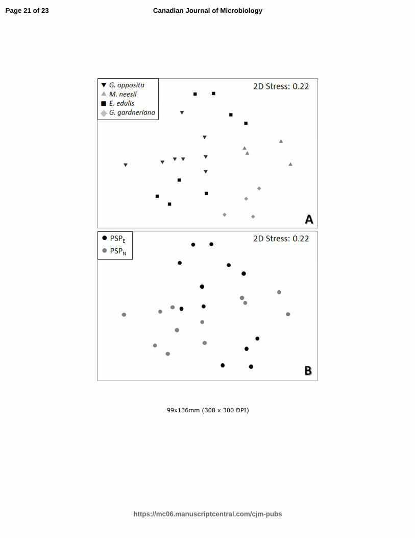
However, the NMDS (stress 0.22) and ANOSIM analysis (Fig. 1A, Table 1, respectively)
revealed that the cyanobacterial community structures on the phyllosphere are mainly driven by
the plant species. The similarity among cyanobacterial community structures from plants of
different locations, in most cases, suggests that the geographical position is a secondary factor
shaping the distribution of this group (Fig. 1B, ANOSIM R= 0.214, p= 0.050). An exception was
E. edulis (square in Fig. 1A), which harbored different assemblies of cyanobacteria according to
the geographical location.
Clone libraries:
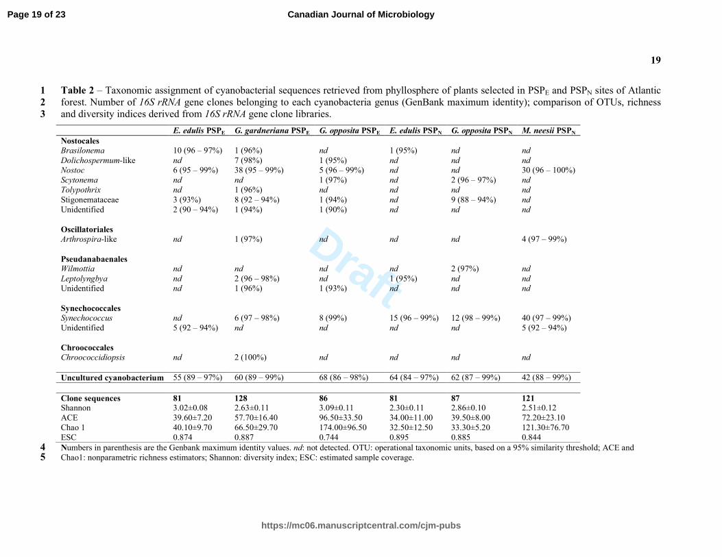
The cyanobacterial community compositions were evaluated based on 584 sequences. The clone
distribution according to plant species and sampling location, phylogenetic assignments, indices
of cyanobacterial diversity and richness and estimated sampling coverage are reported in Table
2. The estimated sampling coverage ranged from 74.4 to 89.5% (0.05 cutoff). The cyanobacterial
communities on the phyllosphere of the plants from PSPE showed higher values of richness,
especially G. opposita. However, in PSPN M. neesii, had the highest richness index.
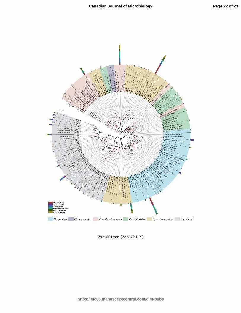
The sequences were clustered into 87 OTUs, considering a cutoff of 95% identity. BLASTn
analyses reveled high levels of similarity to several cyanobacterial sequences (Table 2), most of
them belonging to heterocytous-filamentous group (Dolichospermum, Brasilonema, Nostoc,
Tolypothrix and Scytonema). Furthermore, 60 OTUs upholding nearly 60% of the total retrieved
sequences, matched to uncultivated cyanobacteria sequences in the public database (Table 2). In
congruence, the phylogenetic tree (Fig. 2) demonstrated that the OUTs obtained herein, both the
previous taxonomically affiliated and some comprising uncultivated sequences, clustered with
sequences of known cyanobacteria. However, the majority of the uncultivated OTUs grouped as
a separated clade in the phylogram.
Page 8 of 23
https://mc06.manuscriptcentral.com/cjm-pubs
Canadian Journal of Microbiology

Draft
9
Real time PCR:
The amplification efficiency factor and R2 obtained were 87% and 0.99, respectively, allowing
us to confidently estimate the number of cyanobacterial 16S rRNA gene copies per cm2 of leaf of
each plant species.
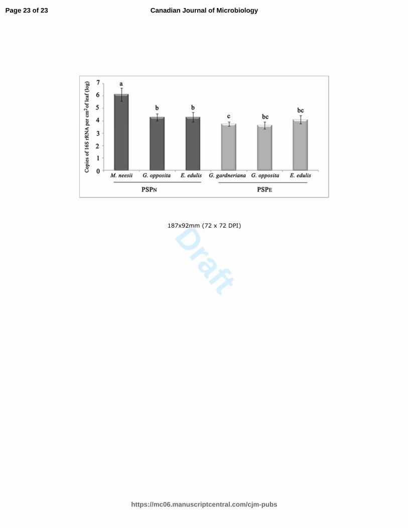
The number of copies of the 16S rRNA gene was significantly higher on the phyllosphere of M.
neesii (6.12 log of cyanobacterial 16S rRNA gene per cm2 of leaf), than on the phyllosphere of G.
opposita, G. gardneriana and E. edulis (Fig. 3). Guapira opposita and E. edulis had equal
abundances of cyanobacterial 16S rRNA gene copies in both sampled sites, and the three plant
species in PSPE did not differ statistically.
Discussion
Cyanobacteria diversity in the Atlantic Forest have been explored mainly using culture-
dependent approaches (Fiore et al. 2007, Branco et al. 2009, Sant´Anna et al. 2010, 2013,
Komárek et al. 2013, Gama et al. 2014, Hentschke et al. 2016). Also, none of the available
studies about this biome focused on the phyllosphere micro-habitat. However, there is no
information on the cyanobacteria community composition in the phyllosphere of plant species of
the Atlantic Forest. The molecular approaches applied in this study assessed the cyanobacteria
community inhabiting unexplored phyllospheres of four plant species from Atlantic Forest. This
investigation demonstrated that each plant species harbored a particular cyanobacteria
community, suggesting that intrinsic factors of each plant species may drive the assemblage of
these communities. Previous findings already indicated that different plant species select their
own total bacteria community in the Atlantic Forest canopies (Lambais et al. 2006, 2014). A
further study suggested that a coevolution may occur between trees and phyllosphere microbial
population (Lambais and Crowley 2014).
Page 9 of 23
https://mc06.manuscriptcentral.com/cjm-pubs
Canadian Journal of Microbiology

Draft
10
Our data also showed that the geographical distance between plants may also affect the structure
of cyanobacterial communities. Significant differences in the bacterial community structure in
phyllosphere of the same plant species separated by long-distances in the Atlantic forest have
been reported (Lambais and Crowley, 2014). Besides the plant genetic component driving the
bacterial community structure in the phyllosphere, environmental conditions associated with
particular geographical locations can also affect colonization (Finkel et al. 2011). In opposite, a
study conducted on mangrove phyllosphere showed that the geographical tree location was the
main factor driving cyanobacteria community structure (Rigonato et al. 2012). Likewise, several
studies indicate that the age of the leaves, the position in the canopy, anatomical structures, such
as trichomes and stomata, light incidence and microclimate can affect the microbial presence in
the phyllosphere (Mechaber et al. 1996, Monier and Lindow 2003, Yadav et al. 2004, Lambais et
al. 2006, Baldoto and Olivares 2008). Based on the available information, it is likely that the
phyllosphere colonization by cyanobacteria is modulated by multiple factors, such as plant
species, environmental conditions and geographical location at different scales.
The variation on abundance of cyanobacteria on leaves surfaces for each plant species was also
observed. The highest abundance of cyanobacteria was found in the surface leaves of bamboo
(M. neesii). This fact may be consequence of the life strategy of this plant species, that
differently of the others evaluated, is an r-strategist that colonizes gaps in vegetation, favoring
the cyanobacterial growth due to its photoautotrophic metabolism.
Most of the retrieved sequences were affiliated to Nostoc (cutoff 95%). This genus was also
reported as predominant diazotrophic colonizers of phyllospheres in a tropical forest in Costa
Rica (Fürnkranz et al. 2008). In general, the high abundance of OTUs affiliated to the
diazotrophic order Nostacales suggest that these organisms may contribute to an input of
nitrogen to the system. The Brazilian Atlantic Forest soils are highly leached and acidic with
very low nutrient availability (Leitão Filho et al. 1993). Despite these constraints, plant
Page 10 of 23
https://mc06.manuscriptcentral.com/cjm-pubs
Canadian Journal of Microbiology

Draft
11
productivity is remarkable in the forest, indicating that there are alternative sources of nutrients
to supply the plant demand. Cyanobacteria were already reported as a significant fraction of the
diazotrophic community responsible for N2 fixation in phyllosphere of tropical lowland
rainforest (Fürnkranz et al. 2008). Furthermore, N2 fixation in the phyllosphere was suggested as
the main mechanism for adding N in humid tropical ecosystems (Abril et al. 2005). There are
evidence that the N2 fixed in the phyllosphere can be incorporated by the plant (Bentley 1987,
Fritz-Sheridan and Portécop 1987, Freiberg 1998, Abril et al. 2005, Peñuelas et al., 2012),
suggesting an additional mechanism for N acquisition.
Many of the known cyanobacterial groups detected have occurrence already described in others
habitats of the Atlantic Forest, as Chroococcidiopsis (Büdel et al. 2002, Gama et al., 2014),
Synechococcus (Branco et al., 2009, Gama et al., 2014), Brasilonema (Fiore at al., 2007) and
Scytonema (Komárek et al., 2013). However, the sequence identity was lower than 97% in many
cases. This can be an indicative that a diversity of novel species or species which no genetic
information available are represented by these retrieved sequences. Several novel genera and
species have been described inhabiting Atlantic Forest different micro-habitats (Fiore et al. 2007,
Aguiar et al. 2008, Sant´Anna et al. 2010, Hentschke et al., 2016).
In this study, a great number of 16S rRNA sequences retrieved from the studied phyllospheres
matched uncultured cyanobacteria and the low identity of some OTUs to sequences available in
the GenBank database point out that a high number of taxa remain to be described in this
environment. Similar results were reported for the mangrove phyllosphere (Rigonato et al.,
2012), yet two novel cyanobacteria genera were described inhabiting this ecosystem (Alvarenga
et al. 2016).
The results obtained in this study provide the first genetic insight of cyanobacteria diversity in
the Brazilian Atlantic forest plant species phyllospheres and contribute to the understanding of
Page 11 of 23
https://mc06.manuscriptcentral.com/cjm-pubs
Canadian Journal of Microbiology

Draft
12
the hierarchy of ecological factors influencing the cyanobacterial colonization in this
environment.
Acknowledgements
This study was supported by grants from the State of São Paulo Research Foundation
(FAPESP/BIOTA 2008/50824-1). N. Gonçalves, J. Rigonato and A. P. D. Andreote were
supported by FAPESP graduate fellowship (Grants 2009/12196-4, 2007/08354-5, 2009/15402-1,
respectively). M. F. Fiore would also like to thank CNPq for a research fellowship
(306607/2012-3).
References
Abril, A.B., Torres, P.A., and Bucher, E.H. 2005. The importance of phyllosphere microbial
populations in nitrogen cycling in the Chaco semi-arid woodland. J. Trop. Ecol. 21: 103-107.
Aguiar, R., Fiore, M.F., Franco, M.W., M.C. Ventrella, A.S. Lorenzi, Vanetti, C.A., Alfenas,
A.C. 2008. A novel epiphytic cyanobacterial species from the genus Brasilonema causing
damage to Eucalyptus leaves. J. Phycol. 44:1322-1334.
Alvarenga, D.O., Rigonato, J., Branco, L.H.Z., Melo, I.S. and Fiore, M.F. 2016. Phyllonema
aviceniicola gen. et sp. nov. and Foliisarcina bertiogensis gen. et. sp. nov., novel epiphyllic
cyanobacteria associated with Avicennia schaueriana leaves. Int. J. Syst. Evol. Microbiol. 66:
689-700.
Baldoto, L.E.B. and Olivares, F.L. 2008. Phylloepiphytic interaction between bacteria and
different plant species in a tropical agricultural system. Can. J. Microbiol. 54: 918-931.
Bentley, B.L. 1987. Nitrogen fixation by epiphylls in a tropical rainforest. Ann. Mo. Bot. Gard.
74: 234-241.
Page 12 of 23
https://mc06.manuscriptcentral.com/cjm-pubs
Canadian Journal of Microbiology

Draft
13
Blum, H., Beier, H. and Gross, H. 1987. Improved silver staining of plant proteins, RNA and
DNA in polyacrilamyde gels. Electrophoresis 8: 93–99.
Branco, L.H.Z., Hoffmann, L., Teixeira, J.P., Ferreira, V., Morais-Filho, J.C. 2009. Aerophytic
cyanoprokaryotes from Atlantic rainforest region of São Paulo State, Brazil: Chroococcales and
Oscillatoriales. Cryptogamie Algol 30:135-152.
Büdel, B., Weber, H.M., Porembski, S., Barthlott, W. 2002. Cyanobacteria of inselbergs in the
Atlantic Rain-forest zone of Eastern Brazil. Phycologia 41:498-506.
Clarke, K.R. and Gorley, R.N. 2006. Primer 6.0: user manual/tutorial. Plymouth: Primer-E, pp.
190.
Colombo, A.F. and Joly, C.A. 2010. Brazilian Atlantic Forest lato sensu: the most ancient
Brazilian forest, and a biodiversity hotspot, is highly threatened by climate change. Braz. J. Biol.
70: 697-708.
Dulla, G., Marco, M., Quiñones, B. and Lindow, S.E. 2005. A Closer Look at Pseudomonas
syringae as a Leaf Colonist. ASM News 71: 469-475.
Finkel, O.M., Burch, A.Y., Lindow, S.E., Post, A.F. and Belkin, S. 2011. Geographical location
determines the population structure in phyllosphere microbial communities of a salt-excreting
desert tree. Appl. Environ. Microbiol. 77: 7647-7655.
Fiore, M.F., Sant’anna, C.L., Azevedo, M.T.P., Komárek, J., Kastovšký, J., Sulek, J., Lorenzi,
A.S. 2007. The cyanobacterial genus Brasilonema, gen. nov., a molecular and phenotypic
evaluation. J. Phycol. 43:789-798.
Freiberg, E. 1998. Microclimatic parameters influencing nitrogen fixation in the phyllosphere in
a Costa rican premontane rain forest. Oecologia 17: 9-18.
Fritz-Sheridan, R.P. and Portécop, J. 1987. Nitrogen fixation on the tropical volcano, La
Soufriere (Guadeloupe): a survey of nitrogen fixation by blue-green algal microepiphytes and
lichen endophytes. Biotropica. 19: 194-199.
Page 13 of 23
https://mc06.manuscriptcentral.com/cjm-pubs
Canadian Journal of Microbiology
Draft
14
Fürnkranz, M., Wanek, W., Richter, A., Abell, G. and Rasche, F. 2008. Nitrogen Fixation by
phyllosphere bacteria associated with higher plants and their colonizing epiphytes of a tropical
lowland rainforest of Costa Rica. ISME J. 2: 61-570.
Gama-Jr, W.A., Laughinghouse IV, H.D., Sant'Anna, C.L. 2014. How diverse are coccoid
cyanobacteria? A case study of terrestrial habitats from the Atlantic Rainforest (São Paulo,
Brazil). Phytotaxa 178:61-97.
Good, I.J. 1953. The population frequencies of species and the estimation of population
parameters. Biometrika. 40: 237-264.
Hentschke, G.S., Johansen, J.R., Pietrasiak, N., Fiore, M.F., Rigonato, J., Sant’anna, C.L.,
Komárek, J. 2016. Phylogenetic placement of Dapisostemon gen. nov. and Streptostemon, two
tropical heterocytous genera (Cyanobacteria). Phytotaxa 245(2): 129-143.
Joly, C.A., Assis, M.A., Bernacci, L.C., Tamashiro, J.Y., Campos, M.C.R., Gomes, J.A.M.A.,
Lacerda, M.S., Santos, F.A.M., Pedroni, F., Pereira, L.S., Padgurschi, M.C.G., Prata, E.M.B.,
Ramos, E., Torres, R.B., Rochelle, A., Martins F.R., Alves, L.F., Vieira, S.A., Martinelli, L.A.,
Camargo, P.B., Aidar M.P.M., Eisenlohr, P.V., Simões, E., Villani, J.P. and Belinello, R. 2012.
Floristic and phytosociology in permanent plots of the Atlantic Rainforest along an altitudinal
gradient in southeastern Brazil. Biota Neotrop. 12: 123-145.
Komárek, J., Sant’Anna, C.L., Bohunická, M., Mareš, J., Hentschke, G.S., Rigonato, J., Fiore,
M.F. 2013. Phenotype diversity and phylogeny of selected Scytonema-species
(Cyanoprokaryota) from SE Brazil. Fottea 13(2):173–200.
Lambais, M. R., Lucheta, A. R., and Crowley, D. E. (2014). Bacterial community assemblages
associated with the phyllosphere, dermosphere, and rhizosphere of tree species of the Atlantic
forest are host taxon dependent. Microb. Ecol. 68(3): 567-574.
Page 14 of 23
https://mc06.manuscriptcentral.com/cjm-pubs
Canadian Journal of Microbiology

Draft
15
Lambais, M.R. and Crowley, D.E. 2014. Bacterial Diversity in Tree Canopies of the Atlantic
Forest. Encyclopedia of Metagenomics. Springer Reference. Springer-Verlag. DOI
10.1007/SpringerReference_303983.
Lambais, M.R., Crowley, D.E., Cury, J.C., Bull, R.C. and Rodrigues, R.R. 2006. Bacterial
Diversity in Tree Canopies of the Atlantic Forest. Science. 312: 1917.
Leitão Filho, H.F., Pagano, S.N., Cesar, O., Timoni, J.L. and Rueda J.J. 1993. Ecologia da Mata
Atlântica em Cubatão. Paulista State University Ed/University of São Paulo, Campinas, Brazil,
184 pp.
Leveau, J.H. and Lindow, S.E. 2001. Appetite of an epiphyte: quantitative monitoring of
bacterial sugar consumption in the phyllosphere. Proc. Natl. Acad. Sci. USAPNAS. 98:3446-
3453.
Lindow, S.E. and Brandl, M.T. 2003. Microbiology of the phyllosphere. Appl. Environ.
Microbiol. 69:1875-1883.
Mechaber, W.L., Marshall, D.B., Mechaber, R.A., Jobe, R.T. and Chew, F.S. 1996. Mapping
leaf surface landscapes. Proc. Natl. Acad. Sci. USAPNAS. 93: 4600-4603.
Monier, J.M. and Lindow, S.E. 2003. Differential survival of solitary and aggregated bacterial
cells promotes aggregate formation on leaf surfaces. Proc. Natl. Acad. Sci. USAPNAS. 100(26):
15977-15982.
Neilan, B.A., Jacobs, D., Del dot, T., Blackall, L.L., Hawkins, P.R., Cox, P.T. and Goodman,
A.E. 1997. rRNA sequences and evolutionary relationships among toxic and nontoxic
cyanobacteria of the genus Microcystis. Int. J. Syst. Bacteriol. 47: 693-697.
Nübel, U., Garcia-Pichel, F. and Muyzer, G. 1997. PCR primers to amplify 16S rRNA genes
from cyanobacteria. Appl. Environ. Microb. 63: 3227-3332.
Peñuelas, J., and Terradas, J. 2014. The foliar microbiome. Trends Plant Sci. 19(5): 278-280.
Page 15 of 23
https://mc06.manuscriptcentral.com/cjm-pubs
Canadian Journal of Microbiology

Draft
16
Peñuelas, J., Rico, L., Ogaya, R., Jump, A. S. and Terradas, J. 2012. Summer season and long-
term drought increase the richness of bacteria and fungi in the foliar phyllosphere of Quercus
ilex in a mixed Mediterranean forest. Plant Biol. 14: 565–575. doi: 10.1111/j.1438-
8677.2011.00532.x
Ribeiro, M. C., Martensen, A. C., Metzger, J. P., Tabarelli, M., Scarano, F. and Fortin, M. J.
2011. The Brazilian Atlantic Forest: a shrinking biodiversity hotspot. In Biodiversity Hotspots:
Distribution and Protection of Conservation Priority Areas. Edited by F.E. Zachos, J.C. Habel,
Springer, Berlin pp. 405-434.
Ribeiro, M. C., Metzger, J. P., Martensen, A. C., Ponzoni, F. J., and Hirota, M. M. 2009. The
Brazilian Atlantic Forest: How much is left, and how is the remaining forest distributed?
Implications for conservation. Biological Biol. ConservationConserv., 142(6): 1141-1153.
Rigonato, J., Alvarenga, D.O., Andreote, F.D., Dias, A.C.F., Melo, I.S., Kent, A. and Fiore, M.
F. 2012. Cyanobacterial diversity in the phyllosphere of a mangrove forest. FEMS Microbiol.
Ecol. 80:312-322.
Rigonato, J., Kent, A. D., Alvarenga, D. O., Andreote, F. D., Beirigo, R. M., Vidal-Torrado, P.
and Fiore, M. F. 2013. Drivers of cyanobacterial diversity and community composition in
mangrove soils in south-east Brazil. Environ. Microbiol. 15: 1103-1114.
Saitou, N. and Nei, M. 1987. The neighbor-joining method: a new method for reconstructing
phylogenetic trees. Mol. Biolog. Evol. 4: 406-425.
Sant’Anna, C.L., Azevedo, T.M.P., Kaštovskský, J., Komárek, J. 2010. Two form–genera of
aerophytic heterocytous cyanobacteria from Brazilian rainy forest Mata Atlântica. Fottea 10:217-
228.
Sant’anna, C.L., Kaštovský, J., Hentschke, G.S., Komárek, J. 2013. Phenotypic studies on
terrestrial stigonematacean Cyanobacteria from the Atlantic Rainforest, São Paulo State, Brazil.
Phytotaxa 89:1-23.
Page 16 of 23
https://mc06.manuscriptcentral.com/cjm-pubs
Canadian Journal of Microbiology
Draft
17
Scarano, F. R., and Ceotto, P. 2015. Brazilian Atlantic forest: impact, vulnerability, and
adaptation to climate change. Biodivers. Conserv. 24(9): 2319-2331.
Schloss, P.D., Westcott, S.L., Ryabin, T., Hall, J.R., Hartmann, M., Hollister, E.B., Lesniewski,
R.A., Oakley, B.B., Parks, D.H., Robinson, C.J., Sahl, J.W., Stres, B., Thallinger, G.G., Van
Horn, D.J. and Weber, C.F. 2009. Introducing mothur: open-source, platform-independent,
community-supported software for describing and comparing microbial communities. Appl.
Environ. Microbiol. 75: 7537-7541.
Tamura, K. Peterson, D. Peterson, N. Stecher, G. Nei, M. and Kumar, S. 2011. MEGA5:
Molecular Evolutionary Genetics Analysis using Maximum Likelihood, Evolutionary Distance,
and Maximum Parsimony Methods. Mol. Biol. Evol. 28: 2731-2739.
Venkatachalam, S., Ranjan, K., Prasanna, R., Ramakrishnan, B., Thapa, S., and Kanchan, A.
2016. Diversity and functional traits of culturable microbiome members, including cyanobacteria
in the rice phyllosphere. Plant Biol. doi:10.1111/plb.12441.
Yadav, R.K.P., Halley, J.M., Karamanoli, K., Constatinidou, H.I. and Vokou, D. 2004. Bacterial
populations on the leaves of mediterranean plants: quantitative features and testing of
distribution models. Environ. Exp. Bot. 52:63-77.
Page 17 of 23
https://mc06.manuscriptcentral.com/cjm-pubs
Canadian Journal of Microbiology

Draft
18
Tables
Table 1 – Comparison of cyanobacterial communities from distinct plant species determined by
ANOSIM statistical test (R).
Groups Statistic R Level of significance
M. neesii x G. opposita 0.651 0.002
M. neesii x E. edulis 0.513 0.004
M. neesii x G. gardneriana 0.781 0.029
G. opposita x E. edulis 0.264 0.011
G. opposita x G. gardneriana 0.679 0.002
E. edulis x G. gardneriana 0.540 0.004
Page 18 of 23
https://mc06.manuscriptcentral.com/cjm-pubs
Canadian Journal of Microbiology
Draft
19
Table 2 – Taxonomic assignment of cyanobacterial sequences retrieved from phyllosphere of plants selected in PSPE and PSPN sites of Atlantic 1
forest. Number of 16S rRNA gene clones belonging to each cyanobacteria genus (GenBank maximum identity); comparison of OTUs, richness 2
and diversity indices derived from 16S rRNA gene clone libraries. 3
E. edulis PSPE G. gardneriana PSPE G. opposita PSPE E. edulis PSPN G. opposita PSPN M. neesii PSPN
Nostocales
Brasilonema 10 (96 – 97%) 1 (96%) nd 1 (95%) nd nd
Dolichospermum-like nd 7 (98%) 1 (95%) nd nd nd
Nostoc 6 (95 – 99%) 38 (95 – 99%) 5 (96 – 99%) nd nd 30 (96 – 100%)
Scytonema nd nd 1 (97%) nd 2 (96 – 97%) nd
Tolypothrix nd 1 (96%) nd nd nd nd
Stigonemataceae 3 (93%) 8 (92 – 94%) 1 (94%) nd 9 (88 – 94%) nd
Unidentified 2 (90 – 94%) 1 (94%) 1 (90%) nd nd nd
Oscillatoriales
Arthrospira-like nd 1 (97%) nd nd nd 4 (97 – 99%)
Pseudanabaenales
Wilmottia nd nd nd nd 2 (97%) nd
Leptolyngbya nd 2 (96 – 98%) nd 1 (95%) nd nd
Unidentified nd 1 (96%) 1 (93%) nd nd nd
Synechococcales
Synechococcus nd 6 (97 – 98%) 8 (99%) 15 (96 – 99%) 12 (98 – 99%) 40 (97 – 99%)
Unidentified 5 (92 – 94%) nd nd nd nd 5 (92 – 94%)
Chroococcales
Chroococcidiopsis nd 2 (100%) nd nd nd nd
Uncultured cyanobacterium 55 (89 – 97%) 60 (89 – 99%) 68 (86 – 98%) 64 (84 – 97%) 62 (87 – 99%) 42 (88 – 99%)
Clone sequences 81 128 86 81 87 121
Shannon 3.02±0.08 2.63±0.11 3.09±0.11 2.30±0.11 2.86±0.10 2.51±0.12
ACE 39.60±7.20 57.70±16.40 96.50±33.50 34.00±11.00 39.50±8.00 72.20±23.10
Chao 1 40.10±9.70 66.50±29.70 174.00±96.50 32.50±12.50 33.30±5.20 121.30±76.70
ESC 0.874 0.887 0.744 0.895 0.885 0.844
Numbers in parenthesis are the Genbank maximum identity values. nd: not detected. OTU: operational taxonomic units, based on a 95% similarity threshold; ACE and 4 Chao1: nonparametric richness estimators; Shannon: diversity index; ESC: estimated sample coverage. 5
Page 19 of 23
https://mc06.manuscriptcentral.com/cjm-pubs
Canadian Journal of Microbiology

Draft
20
Figure captions 6
Fig. 1 - Clustering analysis determined by NMDS based on DGGE fingerprint comparing the 7
cyanobacterial community structure A) comparison based on different plant species B) comparison 8
based on different locations. 9
10
Fig. 2 – Phylogenetic tree of 16S rRNA gene sequences of cyanobacterial clones obtained from the 11
phyllosphere of plants found in the Atlantic forest. Node values were determined by bootstrap analysis, 12
determined based on 1000 replications. Coloured figure in online version. 13
14
Fig. 3 – Absolute quantification, based on quantitative real time PCR of cyanobacterial 16S rRNA gene 15
in the phyllosphere of targeted plant species. Values are represented as number of targeted gene per 16
leaf area, columns indicate the average log values from biological replications, and bars indicate the 17
standard error.*statistical differences evaluated by student t-test. 18
Page 20 of 23
https://mc06.manuscriptcentral.com/cjm-pubs
Canadian Journal of Microbiology
Draft
99x136mm (300 x 300 DPI)
Page 21 of 23
https://mc06.manuscriptcentral.com/cjm-pubs
Canadian Journal of Microbiology
Draft
742x881mm (72 x 72 DPI)
Page 22 of 23
https://mc06.manuscriptcentral.com/cjm-pubs
Canadian Journal of Microbiology
Draft
187x92mm (72 x 72 DPI)
Page 23 of 23
https://mc06.manuscriptcentral.com/cjm-pubs
Canadian Journal of Microbiology