Chronic obstructive pulmonary disease is associated with osteoporosis and low levels of vitamin D
7
ORIGINAL ARTICLE Chronic obstructive pulmonary disease is associated with osteoporosis and low levels of vitamin D C. B. Franco & G. Paz-Filho & P. E. Gomes & V. B. Nascimento & C. A. M. Kulak & C. L. Boguszewski & V. Z. C. Borba Received: 19 November 2008 / Accepted: 21 January 2009 # International Osteoporosis Foundation and National Osteoporosis Foundation 2009 Abstract Summary We did a cross-sectional analysis of chronic pulmonary obstructive disease (COPD) patients without chronic use of systemic glucocorticoids (CUG). Osteoporosis was found in 51% and bone mineral density (BMD) was correlated with severity of disease. Low levels of vitamin D were found in 94%. All COPD patients may benefit from vitamin D supplementation and screening for low BMD. Introduction Patients with chronic pulmonary obstructive disease have low bone mineral density, caused by chronic use of systemic glucocorticoids and hypovitaminosis D. However, patients without CUG may also have low BMD. Methods We performed a cross-sectional analysis in 49 patients (21 men, 28 postmenopausal women), with COPD without CUG, from Brazil (25° 25' S). Several markers of bone metabolism were measured, plus BMD. Osteoporosis risk factors and history of fractures were investigated. Respiratory function was assessed by venous gasometry, spirometry, and oximetry. BMD results were compared to those of 40 healthy non-smokers controls. Results COPD patients had lower BMD at all sites (p < 0.01). Osteoporosis was observed in 51%. BMD indepen- dently correlated with stage of disease (lumbar spine, R = 0.38, p =0.01; total femur, R =0.36, p =0.01; femoral neck, R =0.40, p <0.01). Ninety-four percent had low levels of vitamin D (<30 ng/mL) and 67% had secondary hyper- parathyroidism. Vitamin D was correlated with oxygen saturation (R =0.36, p =0.01), with lower levels in those with saturation <88% (p =0.01). Conclusion Patients with COPD without CUG have in- creased risk for osteoporosis. Such patients have hypovita- minosis D, which is correlated with the severity of disease. Screening for low BMD and vitamin D supplementation may be warranted to all COPD patients. Keywords BMD . Bone . COPD . Glucocorticoids . Osteoporosis . Vitamin D Introduction Chronic pulmonary obstructive disease (COPD) is a complex disease that affects not only respiratory function, but is also accompanied by other comorbidities, such as decreased physical activity, right ventricular heart failure, and decreased quality of life. Osteoporosis is a common finding in patients with COPD, especially in those with advanced disease [1] with high risk of fractures [2]. The pathophysiology of osteopo- rosis in patients with COPD is not well established, and it has been suggested that excess tobacco consumption, malnutrition, vitamin D deficiency, hypogonadism, and inactivity may play a role [3]. In addition, chronic use of systemic and inhaled glucocorticoids also contributes to increase the risk of bone loss [4, 5] and their use in the treatment of COPD is controversial. Currently, systemic glucocorticoids are limited to the most severely affected patients with tendency to exacerbations. In patients with COPD, prevalence of osteoporosis can be as high as 60%, which increases with the progression of the pulmonary disease [5, 6]. At least one vertebral fracture can be found in 4% to 63% of the patients with COPD, Osteoporos Int DOI 10.1007/s00198-009-0890-5 C. B. Franco : G. Paz-Filho : P. E. Gomes : V. B. Nascimento : C. A. M. Kulak : C. L. Boguszewski : V. Z. C. Borba (*) Serviço de Endocrinologia e Metabologia do Hospital de Clínicas da Universidade Federal do Paraná (SEMPR), Rua. Agostinho Leão Junior, 285, Curitiba, Brazil, CEP: 80030-110 e-mail: [email protected]
Transcript of Chronic obstructive pulmonary disease is associated with osteoporosis and low levels of vitamin D

ORIGINAL ARTICLE
Chronic obstructive pulmonary disease is associated
with osteoporosis and low levels of vitamin D
C. B. Franco & G. Paz-Filho & P. E. Gomes &
V. B. Nascimento & C. A. M. Kulak &
C. L. Boguszewski & V. Z. C. Borba
Received: 19 November 2008 /Accepted: 21 January 2009# International Osteoporosis Foundation and National Osteoporosis Foundation 2009
Abstract
Summary We did a cross-sectional analysis of chronic
pulmonary obstructive disease (COPD) patients without
chronic use of systemic glucocorticoids (CUG). Osteoporosis
was found in 51% and bone mineral density (BMD) was
correlated with severity of disease. Low levels of vitamin D
were found in 94%. All COPD patients may benefit from
vitamin D supplementation and screening for low BMD.
Introduction Patients with chronic pulmonary obstructive
disease have low bone mineral density, caused by chronic
use of systemic glucocorticoids and hypovitaminosis D.
However, patients without CUG may also have low BMD.
Methods We performed a cross-sectional analysis in 49
patients (21 men, 28 postmenopausal women), with COPD
without CUG, from Brazil (25° 25' S). Several markers of
bone metabolism were measured, plus BMD. Osteoporosis
risk factors and history of fractures were investigated.
Respiratory function was assessed by venous gasometry,
spirometry, and oximetry. BMD results were compared to
those of 40 healthy non-smokers controls.
Results COPD patients had lower BMD at all sites (p<
0.01). Osteoporosis was observed in 51%. BMD indepen-
dently correlated with stage of disease (lumbar spine, R=
0.38, p=0.01; total femur, R=0.36, p=0.01; femoral neck,
R=0.40, p<0.01). Ninety-four percent had low levels of
vitamin D (<30 ng/mL) and 67% had secondary hyper-
parathyroidism. Vitamin D was correlated with oxygen
saturation (R=0.36, p=0.01), with lower levels in those
with saturation <88% (p=0.01).
Conclusion Patients with COPD without CUG have in-
creased risk for osteoporosis. Such patients have hypovita-
minosis D, which is correlated with the severity of disease.
Screening for low BMD and vitamin D supplementation
may be warranted to all COPD patients.
Keywords BMD . Bone . COPD . Glucocorticoids .
Osteoporosis . Vitamin D
Introduction
Chronic pulmonary obstructive disease (COPD) is a
complex disease that affects not only respiratory function,
but is also accompanied by other comorbidities, such as
decreased physical activity, right ventricular heart failure,
and decreased quality of life.
Osteoporosis is a common finding in patients with
COPD, especially in those with advanced disease [1] with
high risk of fractures [2]. The pathophysiology of osteopo-
rosis in patients with COPD is not well established, and it
has been suggested that excess tobacco consumption,
malnutrition, vitamin D deficiency, hypogonadism, and
inactivity may play a role [3]. In addition, chronic use of
systemic and inhaled glucocorticoids also contributes to
increase the risk of bone loss [4, 5] and their use in the
treatment of COPD is controversial. Currently, systemic
glucocorticoids are limited to the most severely affected
patients with tendency to exacerbations.
In patients with COPD, prevalence of osteoporosis can
be as high as 60%, which increases with the progression of
the pulmonary disease [5, 6]. At least one vertebral fracture
can be found in 4% to 63% of the patients with COPD,
Osteoporos Int
DOI 10.1007/s00198-009-0890-5
C. B. Franco :G. Paz-Filho : P. E. Gomes :V. B. Nascimento :
C. A. M. Kulak :C. L. Boguszewski :V. Z. C. Borba (*)
Serviço de Endocrinologia e Metabologia do Hospital de Clínicas
da Universidade Federal do Paraná (SEMPR),
Rua. Agostinho Leão Junior, 285,
Curitiba, Brazil, CEP: 80030-110
e-mail: [email protected]
which leads to impairment of respiratory function. It is
estimated that each vertebrae that is fractured determines a
decrease by 9% of the predicted forced vital capacity
(FVC). Even though the prevalence of vertebral fractures in
patients with COPD is high, there is discordance in the
literature about the need for BMD evaluation in these
patients. Many studies suggest that only high-risk patients
for osteoporosis should be evaluated, such as the ones
in use of glucocorticoids, postmenopausal or amenorrheic
women, hypogonadic and patients with past history of
fractures or body mass index (BMI) below 22 kg/m2 [1, 7,
9]. Others, however, suggest that all patients with advanced
forms of COPD should be screened for osteoporosis [3].
Data in the literature on BMD and fracture risk in
patients with COPD is scarce [1, 9]. The aim of this study
was to evaluate the prevalence of low bone mass in a group
of patients with COPD without chronic use of systemic
glucocorticoids. In addition, we investigated other factors
that could affect the bone mass, such as low levels of
vitamin D.
Materials and methods
Patients
The study protocol was approved by the HC-UFPR ethics
committee. Informed written consents were obtained from
all subjects.
We consecutively recruited 49 patients (21 males, 28
females, mean age 65.4±9.2 years) from the Pulmonary
Outpatient Clinic of the Hospital de Clínicas da Universi-
dade Federal do Paraná (HC-UFPR), in Curitiba, Brazil
(25° 25' S). Recruitment was undertaken between March
and September 2004. We included only men and post-
menopausal women who fulfilled the COPD diagnostic
criteria according to the Global Strategy for the Diagnosis,
Management, and Prevention of COPD (GOLD) [10].
Exclusion criteria were: (1) prolonged immobilization
within the past 6 months, (2) presence of comorbidities or
use of drugs that cause interference on bone metabolism,
and (3) use of oral or intravenous glucocorticoids for three
or more consecutive months, in an equivalent dose of ≥
5 mg per day of prednisone, or in a cumulative dose of
prednisolone >1,000 mg [11].
Methods
This is a cross-sectional study. Participants were evaluated
during a single visit that included a medical interview,
physical examination, laboratory testing, BMD measure-
ment, and spirometry. Medical history was obtained
through a questionnaire applied by a single investigator,
or from the patients' charts. Data obtained included age,
race, BMI, menstrual status, frequency of physical activity,
drinking, smoking and dietary habits, age at diagnosis of
COPD, duration of disease (years), history of fractures, and
use of medications (past and present). Patients who
engaged three or more hours of physical activity per week
were considered physically active. Patterns of alcohol
consumption were evaluated according to the National
Council on Alcohol Abuse and Alcoholism, and catego-
rized into past or present drinking, and mild, moderate, or
heavy drinking [12]. Present or past smoking history was
determined, as well as smoking intensity, expressed in
number of cigarette packs per day per year of smoking.
Patients that had stopped smoking less than 6 months prior
to the evaluation were considered to have a present
smoking history. In case of pipe smoking or use of hand-
rolled cigarettes, the following equivalency was considered:
one puff on a pipe was equal to one hand-rolled cigarette or
to two cigarettes [13]. Calcium intake was estimated by the
amount of dairy products ingested daily [14].
In order to quantify the use of oral or intravenous
glucocorticoids, we recorded the duration and number of
cycles, and converted the cumulative doses into their
respective equivalent doses of prednisolone. Use of inhalatory
glucocorticoids was categorized by potency as high
(>800 mcg/day of beclometasone, >400 mcg/day of
budesonide, >500 mcg/day of fluticasone, and >1,200 mcg/
day of triamcinolone or flunisolide), moderate (400–800 mcg/
day of beclometasone, 200–400 mcg/day of budesonide, 200–
500 mcg/day of fluticasone, 800–1,200 mcg/day of
triancinolone or flunisolide) or low dose (100–400 mcg/day
of beclometasone, 100–200 mcg/day of budesonide or
fluticasone, and 400–800 mcg/day of triamcinolone or
flunisolide) [15].
Fasting blood was drawn in spring of 2005. Urine was
collected during 24 h, starting the day before the blood
draw. We measured total serum calcium, phosphorus,
alkaline phosphatase, and albumin by standard kits, which
were analyzed in automated spectrophotometric equipment
(ADVIA1650, Bayer, Leverkusen, Germany). Twenty-
four-hour urine calcium was measured by the same
methods. Venous gasometric parameters were assessed by
standard gasometry (Blood gas analyzer pH348, Bayer,
Leverkusen, Germany). In addition, levels of intact para-
thyroid hormone (iPTH) and total testosterone (only in
men) were determined by chemiluminescence (DPC,
Immulite 2000, Los Angeles, CA, USA). Levels of 25-
hydroxi vitamin D (25OHD) were determined by radioim-
munoassay (I125 DiaSorin Stillwater, Minnesota, USA). A
level of 25OHD equal or greater than 30 ng/mL (75 nmol/L)
was considered to indicate sufficient vitamin D. Patients with
levels of 25OHD between 21 to 29 ng/mL (52 to 72 nmol/L)
were considered vitamin D-insufficient. Those with 25OHD
Osteoporos Int

levels <20 ng/mL (50 nmol/L) were considered vitamin D-
deficient [16].
Bone mineral density was measured at the lumbar
spine, femoral neck, and total femur by dual energy X-ray
absorptiometry (DXA) using a Hologic 1000 densitometer
(Hologic, Bedford, MA, USA). BMD was classified as
normal, low BMD, or osteoporosis according to the
International Society for Clinical Densitometry [17].
Results were compared to a non-smoker group of 40
healthy individuals matched by age, gender, ethnicity, and
BMI, without previous history of pulmonary diseases. All
COPD patients were submitted to a pulmonary function
test. Spirometry was undertaken with a Survey Plus spirom-
eter (Collins Medical, Louisville, KY, USA), using the
Knudson 76 protocol [18]. For this study, we considered
the results of the forced expiratory volume in 1 s (FEV1),
the forced vital capacity (FVC), and the FEV1/FVC ratio.
Patients with FEV1/FVC<0.7 are diagnosed with COPD
[10]. The results were compared to the parameters published
by the American Thoracic Society [19]. Arterial oxygen
saturation (O2Sat) was determined by a non-invasive
device (Oxypleth Oximeter, Novametrix Medical System,
Wallingford, USA).
Statistics
Data was analyzed using the software SPSS 13.0® for
Windows (SPSS, Chicago, IL, USA). Results were expressed
as mean±SD or median (range). Groups were compared by
the Student t-test or by non-parametric tests. One-way
analysis of variance (ANOVA) or the chi-square test was
used when appropriate. Pearson and Spearman coefficients
were used for correlation analysis. Two-sided tests were used
with p<0.05 being considered significant. Multiple linear
regression analysis was undertaken in order to assess the
relationship between BMD and other variables.
Results
Medical history and spirometry
Medical history and spirometric parameters are illustrated
in Table 1. Patients were divided into four groups,
according to the results of the spirometry [10]: stage I—
mild COPD (n=14, 28.6%; six males and eight females);
stage II—moderate COPD (n=21, 43.0%; eight males and
Table 1 Clinical characteristics, laboratory exams and pulmonary function tests of patients with chronic obstructive pulmonary disease
Variable Total (n=49) Men (n=21) Women (n=28)
Age (years) 65.4±9.2 64.9±11.7 65.8±6.8
BMI (kg/m2) 25.8±5.3 25.6±7.7 26.1±5.9
Current smoking [n (%)] 16 (32.6%) 6 (28.6%) 10 (37.7%)
Smoking (pack-years ) 54.5±33.5 65.9±36.6 46.4±29.1a
Time without smoking (months) 72 (7–396) 60 (10–396) 7 (10–300)
Total calcium (mg/dl) 8.5 (7.0–10.7) 8.6 (7.0–9.8) 8.5 (7.7–10.7)
PTH intact (pg/mL) 86.5±36.7 84.8±30.8 87.7±41.1
Albumin (g/dL) 4.7±0.6 4.6±0.6 4.7±0.7
Phosphorus (mg/dL) 3.4±0.5 3.0±0.5 3.5±0.4a
Urinary calcium (mg/24 h) 114.5 (18.5–309.5) 1113.5 (20.2–309.5) 93.8 (18.5–298.0)
Alkaline phosphatase (U/L) 183.9±54.8 171.3±48.6 193.4±58.1
25OH Vitamin D (ng/mL) 20.8±0.9 20.9±5.6 20.6±6.5
Testosterone (ng/ml) 649.2 (263–1,409) 649.2 (263–1,409) –
Oxygen saturation (%) 93 (81–98) 93 (82–98) 93 (82–97)
PCO2 (mmHg) 47.2±6.3 48.8±6.4 46.0±6.2
pH 7.38±0.04 7.36±0.04 7.39±0.04a
Bicarbonate (mmol/L) 27.7 (21.0–34.7) 27.6 (21.0–33.9) 27.9 (23.4–34.7)
FEV1 (L) 1.3 (0.3–3.8) 1.2 (0.3–2.57) 1.16 (0.7–3.8)
Values are expressed as mean±SD or median (minimum–maximum). Normal range: total calcium 9–10.8 mg/dl, PTH intact 12–65 pg/mL;
albumin 3.4–4.8 g/dl; phosphorus 2.5–4.8 mg/dl; urinary calcium 100–300 mg/24 h; alkaline phosphatase 64–300 U/L (women) and 80–300 U/L
(men); 25OH Vitamin D >30 ng/mL; total testosterone 262–1,593 ng/ml; oxygen saturation 95–99%; PCO2 38–50 mmHg, pH7.33–7.43;
bicarbonate 23–27 mmol/L.
BMI body mass index, PCO2 partial pressure of carbon dioxide, FEV1 forced expiratory volume in first seconda p<0.05 vs men
Osteoporos Int
13 females); and stage III—severe COPD (n=11, 22.4%;
six males and five females); and stage IV—very severe
COPD (n=3, 6%; one male and two females).
Twenty-five patients (51%) had past or present history of
use of inhalatory glucocorticoids (stage I—six patients, stage
II—eight patients, stage III—eight patients, stage IV—three
patients). Nine patients had used high doses of inhalatory
glucocorticoids for a mean of 1.4±1.2 years, 13 had used a
moderate dose for a median of 0.4 year (0.08 to 5 years), and
three had used a low dose for a median of 0.3 year (0.08 to
1.5 years). None of the patients had chronic use of systemic
glucocorticoids and 13 patients (26.5%) had used systemic
glucocorticoids only during an acute exacerbation, with a
lifetime cumulative dose of 398.3±265.4 mg of prednisolone.
Laboratory assays and BMD
The laboratory results are shown in Table 1. Secondary
hyperparathyroidism was observed in 33 patients (67%).
There was an inverse correlation between serum calcium
and iPTH (R=−0.35, p=0.01). Mean levels of 25OHD were
20.8±0.9 ng/mL. Only three patients (6.1%) had normal
levels of 25OHD (≥30 ng/mL), 59.2% (n=29) were vitamin
D-insufficient, and 34.7% (n=17) were vitamin D-deficient.
The mean O2Sat was 93%. We found a positive correlation
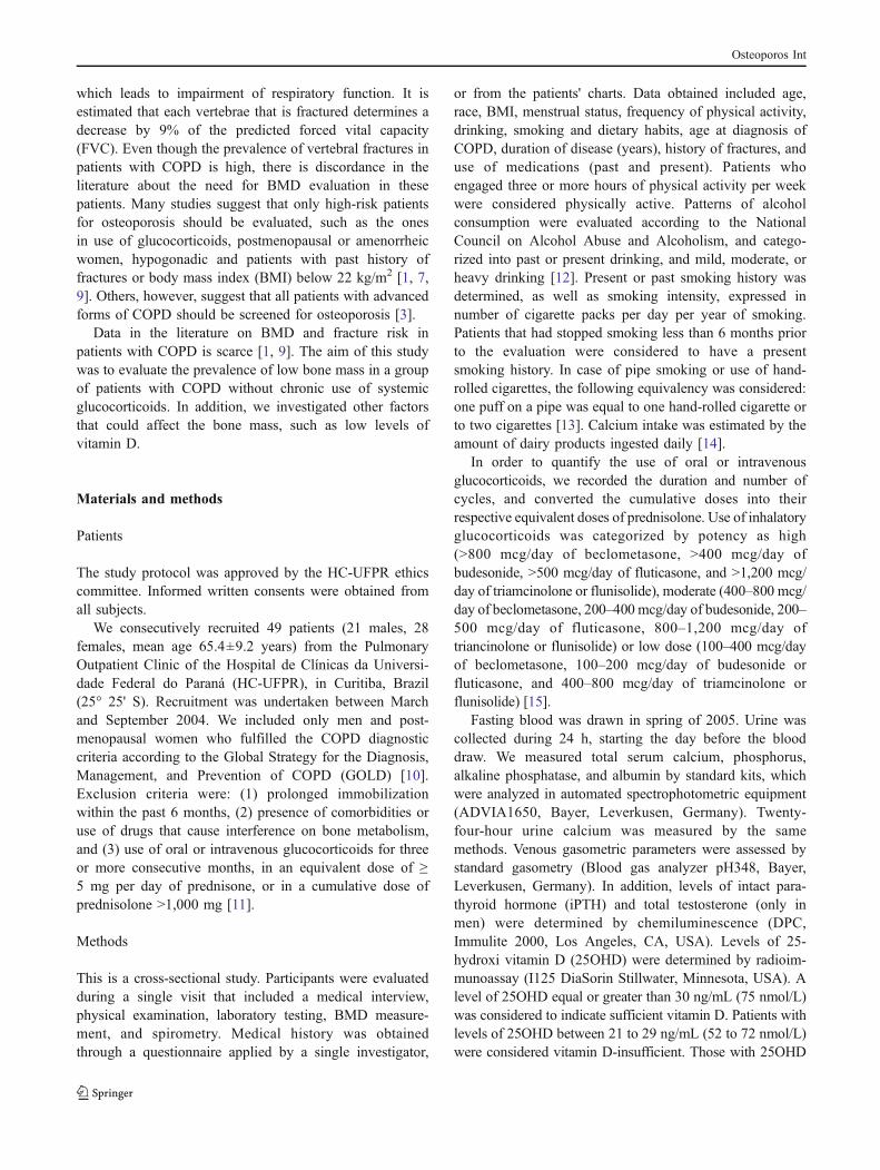
between O2Sat and levels of 25OHD (R=0.36, p=0.01).
Patients with O2Sat≥88% had higher mean levels of
25OHD, when compared with patients with O2Sat<88%
(22.1±5.1 ng/mL vs. 14.5±7.1 ng/mL, p=0.01), as seen in
Fig. 1. All patients had normal levels of phosphorus,
alkaline phosphatase, pH, and bicarbonate. All men had
normal levels of total testosterone.
Normal BMD was observed in six patients (12.2%; five
males and one female). Low BMD was seen in 43
participants (87.7%; 16 males and 27 females). Osteoporo-
sis was diagnosed in 25 patients (51.0%; five males and 20
females). To evaluate the effects of systemic and inhalatory
glucocorticoids on the bone, patients were analyzed
separately in three groups: patients who had never used
systemic glucocorticoids (all participants), patients who had
never used inhalatory glucocorticoids, and patients who had
past or present history of use of glucocorticoids. We
observed no differences regarding BMD at the lumbar
spine (p=0.987), femoral neck (p=0.865), and total femur
(p=0.848). Moreover, no differences were observed regard-
ing T-scores at the lumbar spine (p=0.974), femoral neck
(p=0.927), and total femur (p=0.872). The use of inhala-
tory glucocorticoids in high doses and/or for more than
2.5 years also determined no differences in BMD and in T-
scores, as shown by the comparison of the results of that
subgroup, with those of patients who had never used
inhalatory glucocorticoids (p=0.471, 0.339, and 0.625, for
lumbar, femoral neck, and total femur BMDs, respectively;
p=0.381, 0.313, and 0.179, for lumbar, femoral neck, and
total femur T-scores, respectively). The absolute bone
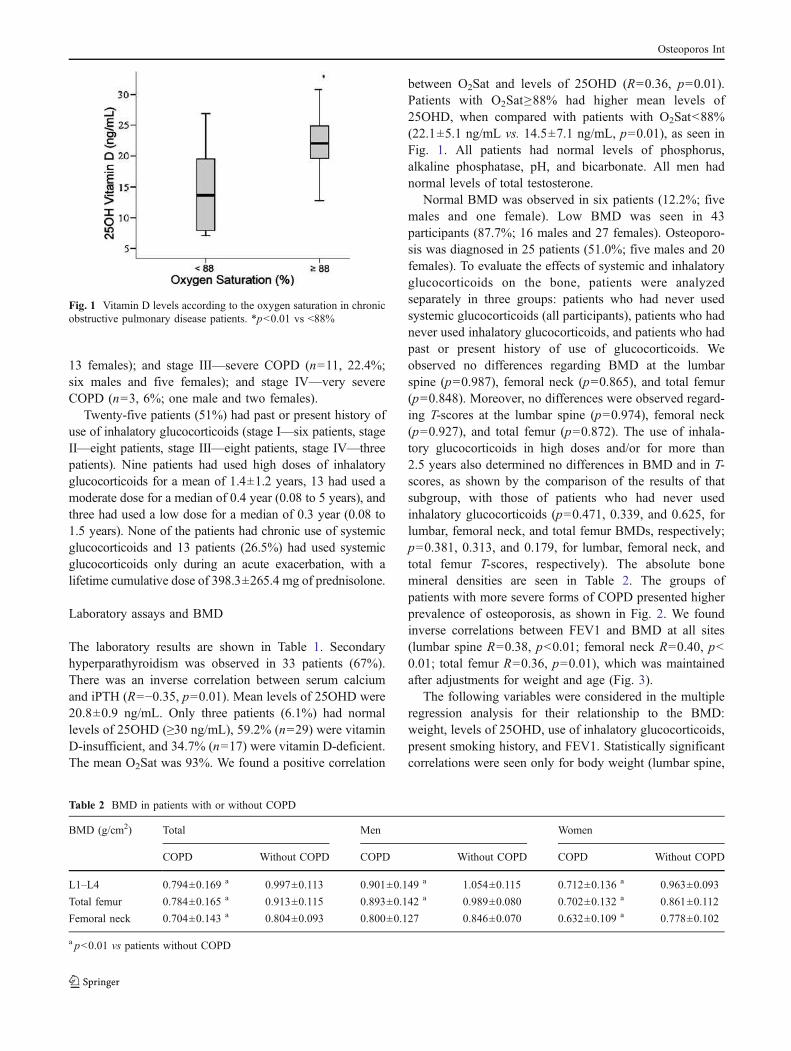
mineral densities are seen in Table 2. The groups of
patients with more severe forms of COPD presented higher
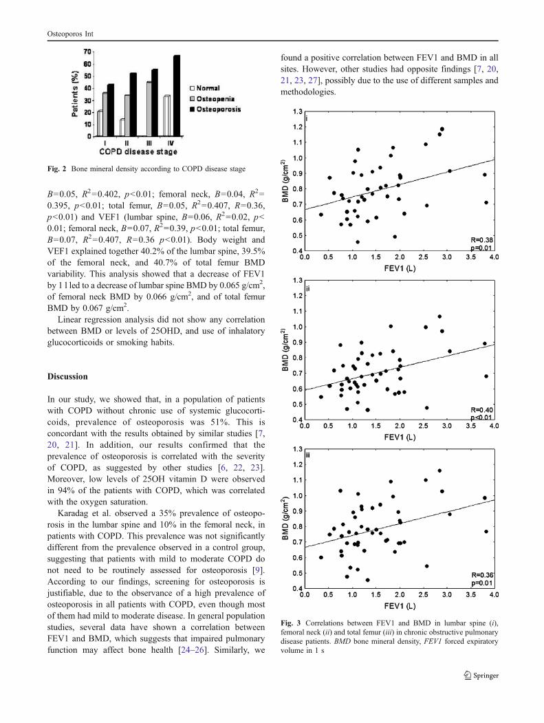
prevalence of osteoporosis, as shown in Fig. 2. We found
inverse correlations between FEV1 and BMD at all sites
(lumbar spine R=0.38, p<0.01; femoral neck R=0.40, p<
0.01; total femur R=0.36, p=0.01), which was maintained
after adjustments for weight and age (Fig. 3).
The following variables were considered in the multiple
regression analysis for their relationship to the BMD:
weight, levels of 25OHD, use of inhalatory glucocorticoids,
present smoking history, and FEV1. Statistically significant
correlations were seen only for body weight (lumbar spine,
Fig. 1 Vitamin D levels according to the oxygen saturation in chronic
obstructive pulmonary disease patients. *p<0.01 vs <88%
Table 2 BMD in patients with or without COPD
BMD (g/cm2) Total Men Women
COPD Without COPD COPD Without COPD COPD Without COPD
L1–L4 0.794±0.169 a 0.997±0.113 0.901±0.149 a 1.054±0.115 0.712±0.136 a 0.963±0.093
Total femur 0.784±0.165 a 0.913±0.115 0.893±0.142 a 0.989±0.080 0.702±0.132 a 0.861±0.112
Femoral neck 0.704±0.143 a 0.804±0.093 0.800±0.127 0.846±0.070 0.632±0.109 a 0.778±0.102
a p<0.01 vs patients without COPD
Osteoporos Int
B=0.05, R2=0.402, p<0.01; femoral neck, B=0.04, R2=
0.395, p<0.01; total femur, B=0.05, R2=0.407, R=0.36,
p<0.01) and VEF1 (lumbar spine, B=0.06, R2=0.02, p<
0.01; femoral neck, B=0.07, R2=0.39, p<0.01; total femur,
B=0.07, R2=0.407, R=0.36 p<0.01). Body weight and
VEF1 explained together 40.2% of the lumbar spine, 39.5%
of the femoral neck, and 40.7% of total femur BMD
variability. This analysis showed that a decrease of FEV1
by 1 l led to a decrease of lumbar spine BMD by 0.065 g/cm2,
of femoral neck BMD by 0.066 g/cm2, and of total femur
BMD by 0.067 g/cm2.
Linear regression analysis did not show any correlation
between BMD or levels of 25OHD, and use of inhalatory
glucocorticoids or smoking habits.
Discussion
In our study, we showed that, in a population of patients
with COPD without chronic use of systemic glucocorti-
coids, prevalence of osteoporosis was 51%. This is
concordant with the results obtained by similar studies [7,
20, 21]. In addition, our results confirmed that the
prevalence of osteoporosis is correlated with the severity
of COPD, as suggested by other studies [6, 22, 23].
Moreover, low levels of 25OH vitamin D were observed
in 94% of the patients with COPD, which was correlated
with the oxygen saturation.
Karadag et al. observed a 35% prevalence of osteopo-
rosis in the lumbar spine and 10% in the femoral neck, in
patients with COPD. This prevalence was not significantly
different from the prevalence observed in a control group,
suggesting that patients with mild to moderate COPD do
not need to be routinely assessed for osteoporosis [9].
According to our findings, screening for osteoporosis is
justifiable, due to the observance of a high prevalence of
osteoporosis in all patients with COPD, even though most
of them had mild to moderate disease. In general population
studies, several data have shown a correlation between
FEV1 and BMD, which suggests that impaired pulmonary
function may affect bone health [24–26]. Similarly, we
found a positive correlation between FEV1 and BMD in all
sites. However, other studies had opposite findings [7, 20,
21, 23, 27], possibly due to the use of different samples and
methodologies.
Fig. 3 Correlations between FEV1 and BMD in lumbar spine (i),
femoral neck (ii) and total femur (iii) in chronic obstructive pulmonary
disease patients. BMD bone mineral density, FEV1 forced expiratory
volume in 1 s
Fig. 2 Bone mineral density according to COPD disease stage
Osteoporos Int
Most of the studies that evaluated the effect of COPD on
BMD did not exclude patients who had used systemic
glucocorticoids for long periods. This is an important
confounding factor that hinders the role of COPD progres-
sion on BMD [7, 23–25, 29]. This confounding factor was
avoided in our study by the exclusion of patients who had
been exposed to a cumulative dose of systemic glucocorti-
coids equal or higher than 1,000 mg of prednisolone [11].
Nevertheless, 13 patients (26.5%) had received systemic
glucocorticoids for the treatment of acute exacerbations. In
these patients, the cumulative dose was still below
1,000 mg of prednisolone (398 mg) and their BMD was
not significantly different from the BMD of the patients
who had never used systemic glucocorticoids. The use of
inhalatory glucocorticoids may also have negative effects
on BMD, which could have been a confounding factor to
our study [28]. However, in our study, previous use of
inhalatory glucocorticoids was not associated with changes
in BMD or in T-scores, in concordance with other studies
[29–32]. In discordance with another study [33], we
showed no association between use of inhalatory glucocor-
ticoids and reductions in BMD, when 11 patients who had
used inhalatory glucocorticoids for more than 2.5 years or
in high doses were analyzed separately.
Our patients lived in Curitiba, Brazil (25° 25' S), and were
evaluated during spring. We observed high prevalence of
vitamin D insufficiency (59.2%) and deficiency (34.7%). The
prevalence of insufficiency was higher than the one observed
in a study conducted in postmenopausal women living in
Recife, Brazil (12º S), where 43% of them had vitamin D
insufficiency [34]. However, our findings were similar to the
prevalence observed in a group of elderly in São Paulo, Brazil
(23º 34’ S) [35]. In our study, the mean levels of vitamin D
were comparable to the levels found in similar studies that
evaluated patients with COPD, without chronic use of systemic
glucocorticoids [21, 27, 36]. The low levels of vitamin D led
to secondary hyperparathyroidism, which was seen in 67% of
our patients and could justify the low bone mass.
We found a correlation between O2Sat and levels of
vitamin D, in discordance with other studies [20, 36, 37].
Frequently, oxygen therapy is recommended to patients
with O2Sat<88%, which is a marker of severity of COPD
[38]. In our patients with O2Sat<88%, we observed lower
levels of vitamin D. This finding could be explained by the
fact that patients with severe forms of COPD are less active
and, therefore, less exposed to sunlight. However, we did
not observe differences regarding physical activity between
patients with O2Sat<88% or ≥88%. In a general population,
levels of vitamin D and FEV1 are independently associated
[39]. We believe that the levels of vitamin D can be
considered as a marker of the severity of COPD, similarly
to the correlation between hypovitaminosis D and increased
risk of several comorbidities and mortality [40, 41].
In concordance with other studies, we could not find any
correlations between smoking habits and BMD [9, 20–25,
27]. However, in general populations, this correlation may
exist [42–45]. This discordance may be attributed to the
small sample size, to the design of the study, and to the
method of evaluation of smoking habits.
Past history of traumatic fractures was present in 22.5%
of our patients. In this subgroup, BMD was lower. Studies
that evaluated fractures in patients with COPD have
controversial findings. For some of them, the risk of
fracture may not be increased in patients with COPD [9,
27]. Other study showed more severe fractures in patients
with COPD [8]. Our study had a small number of patients
with severe disease, which may explain the lack of
correlation between fractures and severity of disease.
Our study has some limitations. First, we did not
perform thoracic X-ray for morphometric vertebral evalua-
tions, which may have contributed to the underestimation
of the prevalence of fractures. Second, we did not include a
control group for the evaluation of fractures and levels of
vitamin D. The possible presence of vertebral fractures may
have contributed to the underestimation of FEV1, which
may have influenced the correlation between BMD and
FEV1.
In conclusion, our findings clearly show that patients
with COPD, without chronic use of systemic glucocorti-
coids, have increased risk for osteoporosis and low levels of
vitamin D, which is correlated with the severity of the
disease. Use of inhalatory glucocorticoids by part of our
sample was not associated with changes in BMD. We
suggest that patients with COPD should routinely have their
BMD evaluated, even in the absence of severe disease or
use of glucocorticoids. This approach allows the reduction
of risk of fractures, and allows adequate treatment of
osteoporosis. In addition, supplementation of vitamin D
may be needed in all patients with COPD, especially to
those with O2Sat less than 88%. Future studies need to
evaluate long-term risk of fractures and the outcomes after
routine BMD screening and vitamin D supplementation in
patients with COPD.
Acknowledgments We thank Dr. Lêda Maria Rabelo for her
assistance during the preparation of this manuscript.
Conflicts of interest None.
References
1. Biskobing DM (2002) COPD and osteoporosis. Chest 121(2):609–620
2. Iqbal F, Michaelson J, Thaler L et al (1999) Declining bone mass
in men with chronic pulmonary disease: contribution of gluco-
corticoid treatment, body mass index, and gonadal function. Chest
116(6):1616–1624
Osteoporos Int
3. Jorgensen NR, Schwarz P (2008) Osteoporosis in chronic
obstructive pulmonary disease patients. Curr Opin Pulm Med 14
(2):122–127
4. Kanis JA, Johansson H, Oden A et al (2004) A meta-analysis of
prior corticosteroid use and fracture risk. J Bone Miner Res 19
(6):893–899
5. McEvoy CE, Ensrud KE, Bender E et al (1998) Association
between corticosteroid use and vertebral fractures in older men
with chronic obstructive pulmonary disease. Am J Respir Crit
Care Med 157(3 Pt 1):704–709
6. Vrieze A, de Greef MH, Wijkstra PJ, Wempe JB (2007) Low bone
mineral density in COPD patients related to worse lung function,
low weight and decreased fat-free mass. Osteoporos Int 18
(9):1197–1202
7. Incalzi RA, Caradonna P, Ranieri P et al (2000) Correlates of
osteoporosis in chronic obstructive pulmonary disease. Respir
Med 94(11):1079–1084
8. Papaioannou A, Parkinson W, Ferko N et al (2003) Prevalence of
vertebral fractures among patients with chronic obstructive pulmo-
nary disease in Canada. Osteoporos Int 14(11):913–917
9. Karadag F, Cildag O, Yurekli Y, Gurgey O (2003) Should COPD
patients be routinely evaluated for bone mineral density? J Bone
Miner Metab 21(4):242–246
10. Fabbri LM, Hurd SS (2003) Global strategy for the diagnosis,
management and prevention of COPD: 2003 update. Eur Respir J
22(1):1–2
11. Dubois EF, Roder E, Dekhuijzen PN et al (2002) Dual energy X-
ray absorptiometry outcomes in male COPD patients after
treatment with different glucocorticoid regimens. Chest 121
(5):1456–1463
12. NCAAA. Task Force on Recommended Alcohol Questions—
National Council on Alcohol Abuse and Alcoholism Recommended
Sets of Alcohol Consumption Questions 2003 [cited 2008
September 15]; Available from:
13. Lolio C, Souza J, Santo A, Buchalla C (1993) Prevalence of
smoking in a city of southeastern Brazil. Rev Saúde Pública 27:4
14. Lloyd T, Rollings N, Eggli DF et al (1997) Dietary caffeine intake
and bone status of postmenopausal women. Am J Clin Nutr 65
(6):1826–1830
15. Expert Panel Report 3 (EPR-3) (2007) Guidelines for the
diagnosis and management of asthma—summary report 2007. J
Allergy Clin Immunol 120(5 Suppl):S94–S138
16. Holick MF (2007) Vitamin D deficiency. N Engl J Med 357
(3):266–281
17. Hans D, Downs RW Jr, Duboeuf F et al (2006) Skeletal sites for
osteoporosis diagnosis: the 2005 ISCD Official Positions. J Clin
Densitom 9(1):15–21
18. Knudson RJ, Slatin RC, Lebowitz MD, Burrows B (1976) The
maximal expiratory flow-volume curves normal standards variability
and effect of age. Am Rev Respir Dis 113:587–600
19. Standardization of Spirometry (1995) 1994 Update. American
Thoracic Society. Am J Respir Crit Care Med 152(3):1107–1136
20. Jorgensen NR, Schwarz P, Holme I et al (2007) The prevalence of
osteoporosis in patients with chronic obstructive pulmonary
disease: a cross sectional study. Respir Med 101(1):177–185
21. Katsura H, Kida K (2002) A comparison of bone mineral density
in elderly female patients with COPD and bronchial asthma. Chest
122(6):1949–1955
22. Forli L, Halse J, Haug E et al (2004) Vitamin D deficiency, bone
mineral density and weight in patients with advanced pulmonary
disease. J Intern Med 256(1):56–62
23. Kjensli A, Mowinckel P, Ryg MS, Falch JA (2007) Low bone
mineral density is related to severity of chronic obstructive
pulmonary disease. Bone 40(2):493–497
24. Lekamwasam S, Trivedi DP, Khaw KT (2002) An association
between respiratory function and bone mineral density in women
from the general community: a cross sectional study. Osteoporos
Int 13(9):710–715
25. Lekamwasam S, Trivedi DP, Khaw KT (2005) An association
between respiratory function and hip bone mineral density in older
men: a cross-sectional study. Osteoporos Int 16(2):204–207
26. Sin DD, Man JP, Man SF (2003) The risk of osteoporosis in
Caucasian men and women with obstructive airways disease. Am
J Med 114(1):10–14
27. Riancho JA, Gonzalez Macias J, Del Arco C et al (1987) Vertebral
compression fractures and mineral metabolism in chronic obstructive
lung disease. Thorax 42(12):962–966
28. Goldstein MF, Fallon JJ Jr, Harning R (1999) Chronic glucocor-
ticoid therapy-induced osteoporosis in patients with obstructive
lung disease. Chest 116(6):1733–1749
29. de Vries F, van Staa TP, Bracke MS et al (2005) Severity of
obstructive airway disease and risk of osteoporotic fracture. Eur
Respir J 25(5):879–884
30. Elmstahl S, Ekstrom H, Johnell O et al (2006) No association
between inhaled corticosteroids and whole body DXA in post-
menopausal women. Pharmacoepidemiol Drug Saf 15(7):527–535
31. Halpern MT, Schmier JK, Van Kerkhove MD et al (2004) Impact
of long-term inhaled corticosteroid therapy on bone mineral
density: results of a meta-analysis. Ann Allergy Asthma Immunol
92(2):201–207 quiz 7-8, 67
32. Jones AM, Munavvar M, Vail A et al (2002) Prospective, placebo-
controlled trial of 5 vs 10 days of oral prednisolone in acute adult
asthma. Respir Med 96(11):950–954
33. Richy F, Bousquet J, Ehrlich GE et al (2003) Inhaled cortico-
steroids effects on bone in asthmatic and COPD patients: a
quantitative systematic review. Osteoporos Int 14(3):179–190
34. Bandeira F, Griz L, Dreyer P et al (2006) Vitamin D deficiency: a
global perspective. Arq Bras Endocrinol Metabol 50(4):640–646
35. Saraiva GL, Cendoroglo MS, Ramos LR et al (2005) Influence of
ultraviolet radiation on the production of 25 hydroxyvitamin D in
the elderly population in the city of Sao Paulo (23 degrees 34'S),
Brazil. Osteoporos Int 16(12):1649–1654
36. Dimai HP, Domej W, Leb G, Lau KH (2001) Bone loss in patients
with untreated chronic obstructive pulmonary disease is mediated
by an increase in bone resorption associated with hypercapnia.
J Bone Miner Res 16(11):2132–2141
37. Donovan DS Jr, Papadopoulos A, Staron RB et al (1998) Bone mass
and vitamin D deficiency in adults with advanced cystic fibrosis lung
disease. Am J Respir Crit Care Med 157(6 Pt 1):1892–1899
38. Sutherland ER, Cherniack RM (2004) Management of chronic
obstructive pulmonary disease. N Engl J Med 350(26):2689–2697
39. Black PN, Scragg R (2005) Relationship between serum 25-
hydroxyvitamin d and pulmonary function in the third national
health and nutrition examination survey. Chest 128(6):3792–3798
40. Melamed ML, Michos ED, Post W, Astor B (2008) 25-
hydroxyvitamin D levels and the risk of mortality in the general
population. Arch Intern Med 168(15):1629–1637
41. Thomas MK, Lloyd-Jones DM, Thadhani RI et al (1998)
Hypovitaminosis D in medical inpatients. N Engl J Med 338
(12):777–783
42. Gerdhem P, Obrant KJ (2002) Effects of cigarette-smoking on
bone mass as assessed by dual-energy X-ray absorptiometry and
ultrasound. Osteoporos Int 13(12):932–936
43. Law MR, Hackshaw AK (1997) A meta-analysis of cigarette
smoking, bone mineral density and risk of hip fracture: recogni-
tion of a major effect. BMJ 315(7112):841–846
44. Szulc P, Garnero P, Claustrat B et al (2002) Increased bone
resorption in moderate smokers with low body weight: the Minos
study. J Clin Endocrinol Metab 87(2):666–674
45. Ward KD, Klesges RC (2001) A meta-analysis of the effects of
cigarette smoking on bone mineral density. Calcif Tissue Int 68
(5):259–270
Osteoporos Int