Choledochoduodenostomy in the Management of Common Duct Stones or Associated Pathology – An...
8
HPB Surgery, 1996, Vol.10, pp.27-33 Reprints available directly from the publisher Photocopying permitted by license only (C) 1996 OPA (Overseas Publishers Association) Amsterdam B.V. Published in The Netherlands by Harwood Academic Publishers GmbH Printed in Malaysia Choledochoduodenostomy in the Management of Common Duct Stones or Associated Pathology- An Obsolete Method? ANTONIO CASTRO MENDES DE ALMEIDA, NOEL MEDINA DOS SANTOS and FERNANDO JOSI ALDEIA University Hospital of Santa Maria- Medicina Operat6ria, Lisbon Medical School, Av.Egas Moniz, 1699 Lisboa Codex Portugal (Received 17 December 1994) Choledochoduodenostomy (CDD) has been reported as a more effective treatment of CBD stones than T- tube drainage but it is regarded as a last resort or obsolete therapeutic method due to fears of higher mobidity, cholangitis, "sump" syndrome and liver dysfunction. We aimed to assess the aforementioned issues analyzing prospectively our experience from 1976 through Dec.92. Methods: CDD was performed in 89 females and 36 males,, aged 60.2+8.7 years, 26 during repeat surgery. Duct stones were the indication in 94, Sphincter of oddi (SO) dysfunction in 23 and obstructive pancreatitis nodule in 8. Peroperative liver biopsies were obtained in 44 patients. The "follow-up" schedule (> 2.5 years in 110) included clinical interview and LFT’s on an yearly basis. Ultra sound (USG) was obtained every one or two years. ERC was done in 10 symptomatic patients and in 25 others for protocul purposes. Liver biopsies were taken four to nine years post surgery in 11 patients-five at relaparotomy for non-biliary causes and six percutaneously by fine needle. Ductal mucosa biopsy could safely be performed in one patient 10 years after surgery. The long-term results were classified as excellent, good, fair or poor. Poor meant the need for further invasive therapy (resurgery or EST). Results: There were two operative deaths (1.6%). The long-term results (123 survivors) were considered excellent in 89, good in 22,fair in 9 and poor in three. Three patients died from unrelated causes and eight others ceased the "follow-up" evaluation three to five years post surgery. All of them were considered as having excellent or good results. A widely patent anastomosis of approximately 20 mms without mucosal inflammatory changes was documented in every patient assessed via ERC. food "debris" was detected within the distal duct of four patients yet it was easily flushed through the stoma. Normal tissue patterns were observed in all long-term liver biopsies. Likewise the ductal mucosa biopsy failed to reveal any acute or chronic inflammatory changes. Conclusions." 1) CDD is ahighly effective short and long-term treatment of CBD lithiasis.2) It does not lead to bacterial or "chemical" cholangitis, to "sump" syndrome or to hepatic dysfunction, provided a wide anastomosis is accomplished.3) CDD should only be considered as obsolete after extensive, long-term, prospective, randomized assessments of laparoscopic or combined laparoendoscopic approaches have been shown to be as effective as or superior to CDD. KEY WORDS: Common duct lithiasis laparoscopic cholecystectomy bilio-digestive anastomoses cholangitis choledochoduodenostomy INTRODUCTION It is no longer disputed that laparoscopic cholecy- stectomy is the procedure of choice in a large propor- tion of patients suffering from symptomatic biliary Correspondence to: Dr. A. C. Mendes de Almeida Praa Principe Real, 23-3 Esq, 1200 Lisbon Portugal. lithiasis. However controversy still remains as to what should be considered the optimal treatment for pa- tients who in addition to gall-bladder (GB) calculi also harbour common bile duct (CBD) stones. Endoscopic sphincterotomy (EST) became widely accepted as the most appropriate treatment of retained/recurrent duct stones independent of the patient’s age and risk. It is also the most judicious treatment for CBD calculi in elderly or otherwise "unfit" patients with GB "in situ". 27
-
Upload
independent -
Category
Documents
-
view
0 -
download
0
Transcript of Choledochoduodenostomy in the Management of Common Duct Stones or Associated Pathology – An...

HPB Surgery, 1996, Vol.10, pp.27-33Reprints available directly from the publisherPhotocopying permitted by license only
(C) 1996 OPA (Overseas Publishers Association)Amsterdam B.V. Published in The Netherlands
by Harwood Academic Publishers GmbHPrinted in Malaysia
Choledochoduodenostomy in the Managementof Common Duct Stones or AssociatedPathology- An Obsolete Method?ANTONIO CASTRO MENDES DE ALMEIDA, NOEL MEDINA DOS
SANTOS and FERNANDO JOSI ALDEIAUniversity Hospital of Santa Maria- Medicina Operat6ria, Lisbon Medical School, Av.Egas Moniz,
1699 Lisboa Codex Portugal
(Received 17 December 1994)
Choledochoduodenostomy (CDD) has been reported as a more effective treatment ofCBD stones than T-tube drainage but it is regarded as a last resort or obsolete therapeutic method due to fears of highermobidity, cholangitis, "sump" syndrome and liver dysfunction. We aimed to assess the aforementionedissues analyzing prospectively our experience from 1976 through Dec.92.Methods: CDD was performed in 89 females and 36 males,, aged 60.2+8.7 years, 26 during repeat surgery.Duct stones were the indication in 94, Sphincter ofoddi (SO) dysfunction in 23 and obstructive pancreatitisnodule in 8. Peroperative liver biopsies were obtained in 44 patients. The "follow-up" schedule (> 2.5 yearsin 110) included clinical interview and LFT’s on an yearly basis. Ultra sound (USG) was obtained everyone or two years. ERC was done in 10 symptomatic patients and in 25 others for protocul purposes. Liverbiopsies were taken four to nine years post surgery in 11 patients-five at relaparotomy for non-biliarycauses and six percutaneously by fine needle. Ductal mucosa biopsy could safely be performed in onepatient 10 years after surgery. The long-term results were classified as excellent, good, fair or poor. Poormeant the need for further invasive therapy (resurgery or EST).Results: There were two operative deaths (1.6%). The long-term results (123 survivors) were consideredexcellent in 89, good in 22,fair in 9 andpoor in three. Three patients died from unrelated causes and eightothers ceased the "follow-up" evaluation three to five years post surgery. All of them were considered ashaving excellent or good results. A widely patent anastomosis of approximately 20 mms without mucosalinflammatory changes was documented in every patient assessed via ERC. food "debris" was detectedwithin the distal duct of four patients yet it was easily flushed through the stoma. Normal tissue patternswere observed in all long-term liver biopsies. Likewise the ductal mucosa biopsy failed to reveal any acuteor chronic inflammatory changes.Conclusions." 1) CDD is ahighly effective short and long-term treatment of CBD lithiasis.2) It does notlead to bacterial or "chemical" cholangitis, to "sump" syndrome or to hepatic dysfunction, provided a wideanastomosis is accomplished.3) CDD should only be considered as obsolete after extensive, long-term,prospective, randomized assessments of laparoscopic or combined laparoendoscopic approaches havebeen shown to be as effective as or superior to CDD.
KEY WORDS: Common duct lithiasis laparoscopic cholecystectomybilio-digestive anastomoses cholangitis choledochoduodenostomy
INTRODUCTION
It is no longer disputed that laparoscopic cholecy-stectomy is the procedure of choice in a large propor-tion of patients suffering from symptomatic biliary
Correspondence to: Dr. A. C. Mendes de Almeida Praa PrincipeReal, 23-3 Esq, 1200 Lisbon Portugal.
lithiasis. However controversy still remains as to whatshould be considered the optimal treatment for pa-tients who in addition to gall-bladder (GB) calculi alsoharbour common bile duct (CBD) stones. Endoscopicsphincterotomy (EST) became widely accepted as themost appropriate treatment ofretained/recurrent ductstones independent of the patient’s age and risk. It isalso the most judicious treatment for CBD calculi inelderly or otherwise "unfit" patients with GB "in situ".
27

28 A.C.M. DE ALMEDIA et al.
Thus, endoscopic removal of duct stones followedby laparoscopic cholecystectomy is becoming the an-ticipated scenario of the future because a higher inci-dence ofduct stones occurs in people over 65. Should theduct calculi be detected laparoscopically, the re-verse approach seems to be preferable. Nevertheless ithas been shown that EST doesn’t offer significantadvantages with respect to morbidity, mortality andsuccess as compared to conventional CBDE1, andthat "fit" patients should be treated by surgery alonewithout properative EST 2. There are two other ran-dom and prospective studies that concluded that ESTfollowed by conventional or laparoscopic surgeryis not superior to surgery alone. Moreover debatecontinues to question whether sphincter restenosisand/or stone reformation are a long-term risks follow-ing EST 5. Thus, although EST followed by surgerymay be regarded as an appealing approach 6, openCBDE is still preferred by a large number of sur-geons, specifically when deciding the most appropri-ate therapy for a young, "fit", patient for whom thelong-term effectiveness of the elected procedure is amajor factor.Among the various surgical modalities at our dis-
posal, choledochoduodenoustomy (CDD) has beenshown to be more effective when compared to tempo-rary T-tube decompression in avoiding long-term mor-bidity secondary to retained /recurrent stones7-1.However, many surgeons consider CDD as a last re-sort and even as obsolete due to fears of increasedoperative morbidity and mortality risks, long-termbacterial or "chemical" cholangitis and the possibleoccurrence of the "sump" syndrome. There are alsoconcerns of eventual hepatic dysfunction and/orparenchymatous changes resulting from repeatedbouts of cholangitis, from long-standing duodeno-choledochal reflux of enteric secretions, or from theloss of sphincter of oddi activity.
Continuing a previously reported study 11 we havecontinued to investigate whether or not these fearsand/or alegations are justified and to define the shortand long-term efficacy of CDD.
PATIENTS, METHODS
This study reports the analysis of data retrospectivelyretrieved from January 1973 through December 1975and prospectively documented from 1976-Dec. 92 ac-cording to a protocol. It relates to a consecutive seriesof 125 CDD which were part of the management ofsymptomatic biliary lithiasis. Table summarizes the
Table 1 Summary of data relating to 125 CDD (Jan.73-Dec.92)
Data Nr. %
Female Patients 89 71.2Age (X+3xsd) in yrs 63.1+3x9.7
Male Patients 36 28.8Age (X+3xsd) in yrs 58.7+3x 13.2
Nr. of Patients<50 yrs 28 22.4Nr. of Patients>70 yrs 32 25.6Primary Surgery 99 79.2Resurgery 26 20.8Duct width 10-15 mms 39 31.2Duct width> 15 mms 86 68.8Operative Morbidity 12 9.6Operative Mortality 2 1.6Choledocholithiasis 94 75.0Obstructive Pancreatitis Nodule 8 6.0SO dysfunction 23 18.0
data related to this sub-group of patients. Wgureoutlines our approaches to GB and CBD lithiasisduring primary and secondary surgery. All of theaforementioned patients were evaluated and operatedupon by the same surgical team. The senior author waseither the acting surgeon or the first assistant attend-ing the Chief Resident. Thus a uniform set of criteriawas ensured.
Other than a "standard" evaluation, the preopera-tive "work-up included oral cholecystography (OCG)or ultrasonography (USG), serum proteins and bio-chemistry profiles, and liver function tests (LFT). Theclinical suspicion of actual or past presence of intra-ductal calculi and/or of sphincter of Oddi dysfunction(SO) would prompt us to obtain an intravenouscholangiography (IVC) and/or endoscopic retrogradecholangiography (ERC). We used IVC to confirm orrule out the presence of a dilated CBD (>10-12 mms)as suggested on USG. Most importantly we used it toconfirm the possible existence of a sluggish biliarydrainage into the gut 11, even though this test is knownto be unpredictable in diagnosing duct stones. Thecriteria for the diagnosis of SO dysfunction and/or anincreased likelihood of choledocholithiasis were asfollows: 1) complaints of upper abdominal pain aftermeals, 2) intermittent jaundice (bilirubin >3 mgrms), 3)biochemichal cholestasis, 4) presence, up to 120 min-utes, ofcontrast material within a dilated duct on IVC,5) indications of a dilated duct on USG. Two or moreof these criteria complementing our clinical assess-ment would prompt us to perform an ERC and/or athorough intra-operative search for the actual pres-ence of intra-ductal calculi. This evaluation wouldinclude a wide kocherization, a careful extra-ductaland pancreatic head palpation, a direct measure-ment of the duct diameter with a ruler above the

CHOLEDOCHODUODENOSTOMY- IS IT OBSOLETE? 29
57,5 w/out CBDE (3+)
709 CHTS, Stones 97
134 with CBDE (19%) SO dysf. 29Pancreat.Nod. 8
T TubeSPT o
CDJ
35 REOPER. CDE g22Stones 19 [ T Tube
SO dysl’. 9 1 SPT 3
Strictures, 5 D CDJ s (1’)Pancreat.Nod. 2 2 (1+)
Figure 1 Schematic outline of the surgical approaches to symptomatic biliary lithiasis during primary and secondary surgery(Jan.73-Dec.92)CHT-Cholecystectomy; CDE-Common Duct ExplorationSPT-Sphincteroplasty CDJ-Choledochojejunostomy+-Operative Death; *-Referred from elsewhere
40
30
20
n=9
tO n=5
n=19
=29
Black Pigment
Mixed
Brown Pigment
Figure 2 Percentage of various types of duct calculi according to the morphological criteria of Madden [12], prospectively classiffied(Jan.79-Dec.84)
cystic-choledochal junction and, in selected instances,an operative cholangiogram (OPC). Operative USGor choledochoscopy were no used.When deemed necessary a longitudinal choledocho-
tomy, following guidelines previously described ll,would confirm or rule out the actual presence ofstonesand/or inflammatory changes such as thickened ductwalls, fibrin strands, hyperemic mucosa or biliary"mud". As reported before 7,10, and herein illustratedin Figure we broadened the indications for CDD inthe management of dilated (> 10-12 mms) stone-con-taining ducts. The choledocho-duodenal anastomosisand corresponding technical requisites were alreadyreported 1. The fundamental aim was the constructionof a wide (>20 mms), side-to-side, well blood-suppliedanastomosis. Stents were never used. The detection offibrotic scars or inflammatory changes secondary toduodenal peptic ulcer disease or cholecystocholedo-cho-duodenal fistula or any other anatomical distor-tion contraindicated performing a CDD on various
occasions. Thus, some other form of biliary drainagewas considered (Figure 1).
Peroperative liver biopsies were obtained uponwritten consent in 44 patients. From January 1979through December 1984 we prospectively classified, asper Madden’s criteria 12, the intra-ductally detectedstones. As documented in Figure 2 these were classi-fied as 1) pure cholesterol stones, 2) brown pigment,calcium bilirubinate, stasis stones, 3) black pigmentcalculi and 4) mixed type of stones. The prospective"follow-up" schedule, covering more than four yearsin 91 patients and over 12 years in 38 (Fig. 3), includedclinical interview by independent observer and LFT’s(same as preoperatively) every four to six months forone year and yearly thereafter. USG was obtainedevery one or two years. ERC was deemed necessary in10 symptomatic patients and, for protocol study pur-poses, was performed in 25 other consenting patientsthree to six years post surgery. Our aim was to look forretained/recurrent stones, mucosal inflammatory

30 A.C.M. DE ALMEDIA et al.
z:IS choledochoduodenostomies
112 Known tO b 3 ’Lateat 18 Lostto’lO:llow.upAIIve>12 months 2/,...4% / 13-5 Yrs Postop
90%
II111191 > 4 Yrs .."38> 12 Yr$ ................................ .’"
OP. Deaths1.6%
EXCELLENT 89 (72%)GOOD 22 (18%) resultslong term )..
FAIR 9 (08%) >"
P(X)R 3 (02%)
Figure 3 Length of"follow-up" and classification of long-term results of 123 CDD after a mean number of 9.3+3x4.2 years.
changes at the anastomotic level, the possible presenceof food "debris" within the distal "cul-de-sac" or anyother anatomical abnormality. Endoscopic biopsy ofthe duct mucosa could be safely taken in one patient10 years after surgery. Liver biopsies were obtainedfour to nine years postoperatively in 11 consentingpatients five at relaparotomy for non-biliary causes:carcinoma of the colon in two, gastric peptic ulcer inone, ventral incisional herina in one and distal eso-phageal peptic stenosis in one. Six had the biopsypercutaneously by fine needle.
Based on the data, long-term results were classified(Fig. 3). Excellent was defined as freedom from anysymptoms related to the biliary or upper G.I tract theoperation or a complication of the CBDE. Good wasdefined as occasional and minor GI upset, psychoso-matic complaints or wound imperfections, with nor-mal LFT,s. Fair was defined as significant complaints,manageable by non-invasive treatment, such as thoseascribable to the blindsac syndrome or to duodenal-gastric reflux documented on endoscopy, with abnor-mal LFT’s in at least one occasion.Poor was defined asresidual or recurrent stones, cholangitis, jaundice and/or severely deranged LFT’s requiring further invasivetherapy- resurgery or EST.
Operative death or complications were consideredas those occurring within 30 days of surgery. Onlycomplications delaying the patient’s discharge wereaccounted for in Tables and 2. A wound infectionwas defined as one with purulent discharge irrespec-tive of negative bacteriology. Acute cholangitis wasdiagnosed when the corresponding clinical syndrome(jaundice, high spiking fever with rigors, sepsis, leuko-cytosis) was confirmed by deranged LFT’s and USG.The persistence of bile drainage through the operativewound or out of a drain site that lasted for more thanfive to six days or its radiographic documentationby contrast extravasation, was taken as evidence of
Table 2 Postoperative Complications in 123 Patients (two opera-tive deaths not included)
Complication Nr. %
*Wound Infection 5 4.1*Pneumonia 2 1.6*Congestive Heart Failure 2 1.6*Biliary Fistula 2 1.6*Sepsis 0.8*Total 12 9.8
biliary fistula. The causes of death were established atpostmortem examination.
SHORT TERM RESULTS
There were two operative deaths (1.6%). A woman of74 died on the seventh postop day of primary surgeryfrom massive upper GI bleeding. An intact stoma wasdocumented during autopsy. A woman of 62 died onthe third postop day after resurgery. Necrotizing pan-creatitis with anastomotic disruption was observed onpostmortem examination. This patient had had sev-eral episodes ofpancreatitis and jaundice after havingbeen submitted to cholecystectomy, choledocholitho-tomy and T-tube decompression at another hospitalthree years prior to being admitted to our Institutionwithjaundice. Ofthe remaining 123 patients, 12 (9.8%)developed significant morbidity (Table 2).
In 44 patients clinical and biochemical evidence ofjaundice and cholestasis was discernible on the day ofadmission. Acute, non-suppurative, cholangitis wasthe presenting clinical syndrome of 14 patients (11.2%).Although under antibiotic coverage, this syndrome com-pletely subsided only three to five days after CDD hadbeen performed in five of the aforementioned patients.
Twenty-nine of 62 choledochal stones (46.7%)were considered to be of pure brown pigment type.
CHOLEDOCHODUODENOSTOMY- IS IT OBSOLETE? 31
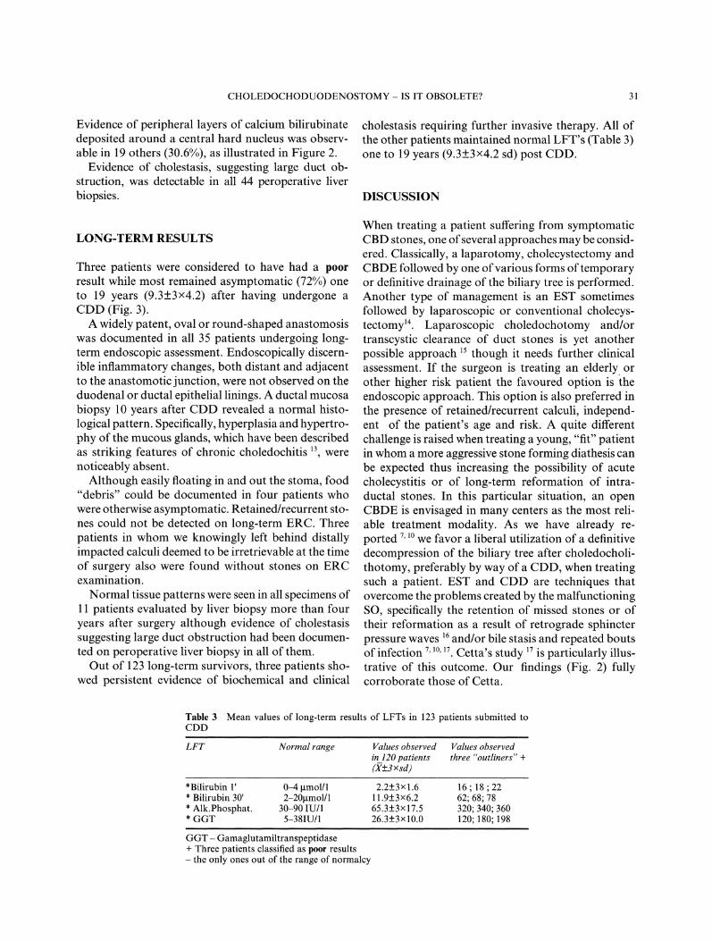
Evidence of peripheral layers of calcium bilirubinatedeposited around a central hard nucleus was observ-able in 19 others (30.6%), as illustrated in Figure 2.
Evidence of cholestasis, suggesting large duct ob-struction, was detectable in all 44 peroperative liverbiopsies.
cholestasis requiring further invasive therapy. All ofthe other patients maintained normal LFT’s (Table 3)one to 19 years (9.3+34.2 sd) post CDD.
DISCUSSION
LONG-TERM RESULTS
Three patients were considered to have had a poorresult while most remained asymptomatic (72%) oneto 19 years (9.3+34.2) after having undergone aCDD (Fig. 3).A widely patent, oval or round-shaped anastomosis
was documented in all 35 patients undergoing long-term endoscopic assessment. Endoscopically discern-ible inflammatory changes, both distant and adjacentto the anastomotic junction, were not observed on theduodenal or ductal epithelial linings. A ductal mucosabiopsy 10 years after CDD revealed a normal histo-logical pattern. Specifically, hyperplasia and hypertro-phy of the mucous glands, which have been describedas striking features of chronic choledochitis 13, werenoticeably absent.Although easily floating in and out the stoma, food
"debris" could be documented in four patients whowere otherwise asymptomatic. Retained/recurrent sto-nes could not be detected on long-term ERC. Threepatients in whom we knowingly left behind distallyimpacted calculi deemed to be irretrievable at the timeof surgery also were found without stones on ERCexamination.Normal tissue patterns were seen in all specimens of
11 patients evaluated by liver biopsy more than fouryears after surgery although evidence of cholestasissuggesting large duct obstruction had been documen-ted on peroperative liver biopsy in all of them.Out of 123 long-term survivors, three patients sho-
wed persistent evidence of biochemical and clinical
When treating a patient suffering from symptomaticCBD stones, one ofseveral approaches may be consid-ered. Classically, a laparotomy, cholecystectomy andCBDE followed by one ofvarious forms oftemporaryor definitive drainage of the biliary tree is performed.Another type of management is an EST sometimesfollowed by laparoscopic or conventional cholecys-tectomy14. Laparoscopic choledochotomy and/ortranscystic clearance of duct stones is yet anotherpossible approach 15 though it needs further clinicalassessment. If the surgeon is treating an elderly, orother higher risk patient the favoured option is theendoscopic approach. This option is also preferred inthe presence of retained/recurrent calculi, independ-ent of the patient’s age and risk. A quite differentchallenge is raised when treating a young, "fit" patientin whom a more aggressive stone forming diathesis canbe expected thus increasing the possibility of acutecholecystitis or of long-term reformation of intra-ductal stones. In this particular situation, an openCBDE is envisaged in many centers as the most reli-able treatment modality. As we have already re-ported v. 10 we favor a liberal utilization of a definitivedecompression of the biliary tree after choledocholi-thotomy, preferably by way of a CDD, when treatingsuch a patient. EST and CDD are techniques thatovercome the problems created by the malfunctioningSO, specifically the retention of missed stones or oftheir reformation as a result of retrograde sphincterpressure waves 16 and/or bile stasis and repeated boutsof infection 7, 10, 17. Cetta’s study 17 is particularly illus-trative of this outcome. Our findings (Fig. 2) fullycorroborate those of Cetta.
Table 3 Mean values of long-term results of LFTs in 123 patients submitted toCDD
LFT Normal range Values observed Values observedin 120 patients three "outliners" +(X+_-3 xsd)
*Bilirubin 1’ 0-4 gmol/1 2.2+3xl.6 16 18 22* Bilirubin 30’ 2-20gmol/1 11.9+3x6.2 62; 68; 78* Alk.Phosphat. 30-90 IU/1 65.3+3x 17.5 320; 340; 360* GGT 5-38IU/1 26.3+3x10.0 120; 180; 198
GGT Gamaglutamiltranspeptidase+ Three patients classified as poor resultsthe only ones out of the range of normalcy
32 A.C.M. DE ALMEDIA et al.
It is undisputable that EST is more comfortable andoffers a shorter hospitalization than a formal opera-tion. However the short-term effectiveness of retro-grade EST has recently been shown 18 to be less thanperfect when compared to that of the current study.Although all-comers were included in ours as well as inthe Manchester series, the immediate morbidity andmortality rates were not significantly different. How-ever, a mean number of 1.9 EST session were neededto achieve a less than optimal 81.6% rate of completeductal stones clearance. This data compares unfa-vorably to what we and others have accomplished withCDD 19, 20. The complication and mortality risks ofEST have been variously defined and are thereforenot easily interpreted. The mortality of this procedurehas been reported 18,21 as occurring at a rate around 2%when the same criteria utilized in surgical series isadopted. It’s worth emphasizing that in one of theseseries 21, which included all-comers, the mean age ofthepatients was within the same range as in the currentseries. Possibly ofgreater significance is the fact that thelong-term effects of EST remain as yet incompletelyclarified 5, the rate of restenosis and thus the increasedchances of stone reformation having been described asranging from 3 to 10% 22,23. It must be stressed that tworecent random and prospective studies 3, concludedthat the laparoendoscopic appraoch is not superiorto a standard cholecystectomy and CBDE, even interms of short-term efficacy. Another study 24
showed that the stone reformation is, actually, en-hanced after EST and suggested that a non-function-ing, partially restenosed, SO was responsible. Thedata in our study does not support the contentionthat bilio-duodenal bypass leads to hepatic dysfunctionresulting from repeated bouts of cholangitis, nordoes it support the claim that significant gastrointes-tinal complaints such as persistent diarrhoea withnutritional impairment occur. These complications,which have been described as the sump or blindsacsyndrome, presumably derive from stasis and reflu-xed duodenal contents in to the terminal commonduct, with bacterial overgrowth enhancing bile saltdeconjugation. This phenomenon leads not only todiarrhoea, as observed in the "blind-loop" syndromearising anywhere else in the gut, but it also facilitatesdeposition and reformation of calcium bilirubinatestones 17, 24. In the present series, though, only threepatients were considered as poor results consequentto repeated bouts of cholangitis. Recurrent stonescould not be documented in spite of an exhaustivesearch. A very wide anastomosis could possibly bethe explanation.
In one of the patients considered to have had a poorresult, severely disturbed LFT’s antedated our firstsurgical procedure. At the time, a left hepatic ductstenosis, presumably deriving from previous surgery,was missed. We eventually corrected this patient’scholangitic syndrome with a hilar Roux-Y hepatic-ojejunostomy followed by long-term good results. Apoorly draining CDD, sited too high in the commonhepatic duct was the reason for a poor result in oneother patient. Long-term good results also ensued af-ter a Roux-Y hepaticojejunostomy in this patient. Upto this time we are unable to define the exact cause forthe third poor result. Repeated ERC examinationsfailed to disclose any evidence of stoma stenosis, re-current stones or any other form of biliary obstruc-tion. The long-term results after EST were not entirelysatisfactory in this patient.The blind-sac syndrome can be avoided by a wide
anastomosis. This prevents stasis, avoids the build-upof excessive intra-ductal pressure and permits a freeflow of common duct contents including duodenalrefluxate as well as eventual retained/recurrent calculiback into the duodenum. Using the triangular tech-nique, as previously described 11, it is technically feasi-ble to construct a biliary-duodenal anastomosis ofsufficient diameter as to prevent these complicationseven on a duct 10 to 12 mms wide. The possible long-term effects on liver function allegedly resulting fromloss of the odditic sphincter activity remain to bedetermined. Certainly there was no evidence in ourstudy.
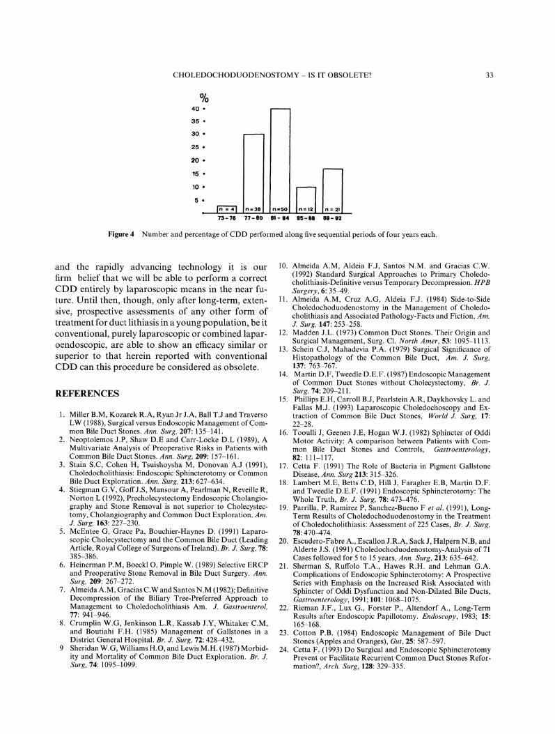
In conclusion our data, corroborating others 8,9,19,20,indicate that CDD is a very safe procedure with mor-bidity and mortality risks similar to those of ESTwhenever similar cohorts of patients are compared.The alleged late complications of liver dysfunctionand blind-sac syndrome are low enough in incidenceto warrant a broadening of indications for CDD inyoung patients. It’s beyond any dispute that ESTfollowed by laparoscopic or conventional cholecystec-tomy is the most judicious treatment for a great num-ber of patients with symptomatic duct lithiasis. It’sWorth emphasizing that most patients with this clini-cal syndrome belong to an age group where other fatalnon-biliary diseases are likely to occur before Sorestenosis and/or stone reformation do so. The declin-ing number ofCDD in the current series, as illustratedin figure 4, clearly directs our thoughts to this respect.However in a young, "fit", patient it is our contentionthat a carefully performed CDD is the most appropri-ate, treatment of most dilated stone-containing ducts.With the current emphasis on laparoscopic procedures
CHOLEDOCHODUODENOSTOMY- IS IT OBSOLETE? 33
%40
35
30
25
20
15
10
5.
n:38 n-50]" n:21
77-e0 81-84 85-88 89-92
Figure 4 Number and percentage of CDD performed along five sequential periods of four years each.
and the rapidly advancing technology it is ourfirm belief that we will be able to perform a correctCDD entirely by laparoscopic means in the near fu-ture. Until then, though, only after long-term, exten-sive, prospective assessments of any other form oftreatment for duct lithiasis in a young population, be itconventional, purely laparoscopic or combined lapar-oendoscopic, are able to show an efficacy similar orsuperior to that herein reported with conventionalCDD can this procedure be considered as obsolete.
REFERENCES
1. Miller B.M, Kozarek R.A, Ryan Jr J.A, Ball T.J and TraversoLW (1988), Surgical versus Endoscopic Management ofCom-mon Bile Duct Stones. Ann. Surg, 207: 135-141.
2. Neoptolemos J.P, Shaw D.E and Carr-Locke D.L (1989), AMultivariate Analysis of Preoperative Risks in Patients withCommon Bile Duct Stones. Ann. Surg, 209:157-161.
3. Stain S.C, Cohen H, Tsuishoysha M, Donovan A.J (1991),Choledocholithiasis: Endoscopic Sphincterotomy or CommonBile Duct Exploration. Ann. Surg, 213: 627-634.
4. Stiegman G.V, Goff J.S, Mansour A, Pearlman N, Reveille R,Norton L (1992), Precholecystectomy Endoscopic Cholangio-graphy and Stone Removal is not superior to Cholecystec-tomy, Cholangiography and Common Duct Exploration. Am.J. Surg, 163: 227-230.
5. McEntee G, Grace Pa, Bouchier-Haynes D. (1991) Laparo-scopic Cholecystectomy and the Common Bile Duct (LeadingArticle, Royal College of Surgeons of Ireland). Br. J. Surg, 78:385-386.
6. Heinerman P.M, Boeckl O, Pimple W. (1989) Selective ERCPand Preoperative Stone Removal in Bile Duct Surgery. Ann.Surg, 209: 267-272.
7. Almeida A.M, Gracias C.W and Santos N.M (1982); DefinitiveDecompression of the Biliary Tree-Preferred Approach toManagement to Choledocholithiasis Am. J. Gastroenterol,77: 941-946.
8. Crumplin W.G, Jenkinson L.R, Kassab J.Y, Whitaker C.M,and Boutiahi F.H. (1985) Management of Gallstones in aDistrict General Hospital. Br. J. Surg, 72: 428-432.
9 Sheridan W.G, Williams H.O, and Lewis M.H. (1987) Morbid-ity and Mortality of Common Bile Duct Exploration. Br. J.Surg, 74: 1095-1099.
10. Almeida A.M, Aldeia F.J, Santos N.M. and Gracias C.W.(1992) Standard Surgical Approaches to Primary Choledo-cholithiasis-Definitiv versus Temporary Decompression. HPBSurgery, 6: 35-49.
11. Almeida A.M, Cruz A.G, Aldeia F.J. (1984) Side-to-SideCholedochoduodenostomy in the Management of Choledo-cholithiasis and Associated Pathology-Facts and Fiction, Am.J. Surg, 147: 253-258.
12. Madden J.L. (1973) Common Duct Stones. Their Origin andSurgical Management, Surg. C1. North Amer, 53:1095-1113.
13. Schein C.J, Mahadevia P.A. (1979) Surgical Significance ofHistopathology of the Common Bile Duct, Am. J. Surg,137: 763-767.
14. Martin D.F, Tweedle D.E.F. (1987) Endoscopic Managementof Common Duct Stones without Cholecystectomy, Br. J.Surg, 74:209-211.
15. Phillips E.H, Carroll B.J, Pearlstein A.R, Daykhovsky L. andFallas M.J. (1993) Laparoscopic Choledochoscopy and Ex-traction of Common Bile Duct Stones, Worm J. Surg, 17:22-28.
16. Tooulli J, Geenen J.E, Hogan W.J. (1982) Sphincter of OddiMotor Activity: A comparison between Patients with Com-mon Bile Duct Stones and Controls, Gastroenterology,82: 111-117.
17. Cetta F. (1991) The Role of Bacteria in Pigment GallstoneDisease, Ann. Surg 213:315-326.
18. Lambert M.E, Betts C.D, Hill J, Faragher E.B, Martin D.F.and Tweedle D.E.F. (1991) Endoscopic Sphincterotomy: TheWhole Truth, Br. J. Surg, 78: 473-476.
19. Parrilla, P, Ramirez P, Sanchez-Bueno F et al. (1991), Long-Term Results of Choledochoduodenostomy in the Treatmentof Choledocholithiasis: Assessment of 225 Cases, Br. J. Surg,78: 470-474.
20. Escudero-Fabre A., Escallon J.R.A, Sack J, Halpern N.B, andAlderte J.S. (1991) Choledochoduodenostomy-Analysis of 71Cases followed for 5 to 15 years, Ann. Surg, 213: 635-642.
21. Sherman S, Ruffolo T.A., Hawes R.H. and Lehman G.A.Complications of Endoscopic Sphincterotomy: A ProspectiveSeries with Emphasis on the Increased Risk Associated withSphincter of Oddi Dysfunction and Non-Dilated Bile Ducts,Gastroenterology, 1991; 101: 1068-1075.
22. Rieman J.F., Lux G., Forster P., Altendorf A., Long-TermResults after Endoscopic Papillotomy. Endoscopy, 1983; 15:165-168.
23. Cotton P.B. (1984) Endoscopic Management of Bile DuctStones (Apples and Oranges), Gut, 25: 587-597.
24. Cetta F. (1993) Do Surgical and Endoscopic SphincterotomyPrevent or Facilitate Recurrent Common Duct Stones Refor-mation?, Arch. Surg, 128: 329-335.

Submit your manuscripts athttp://www.hindawi.com
Stem CellsInternational
Hindawi Publishing Corporationhttp://www.hindawi.com Volume 2014
Hindawi Publishing Corporationhttp://www.hindawi.com Volume 2014
MEDIATORSINFLAMMATION
of
Hindawi Publishing Corporationhttp://www.hindawi.com Volume 2014
Behavioural Neurology
EndocrinologyInternational Journal of
Hindawi Publishing Corporationhttp://www.hindawi.com Volume 2014
Hindawi Publishing Corporationhttp://www.hindawi.com Volume 2014
Disease Markers
Hindawi Publishing Corporationhttp://www.hindawi.com Volume 2014
BioMed Research International
OncologyJournal of
Hindawi Publishing Corporationhttp://www.hindawi.com Volume 2014
Hindawi Publishing Corporationhttp://www.hindawi.com Volume 2014
Oxidative Medicine and Cellular Longevity
Hindawi Publishing Corporationhttp://www.hindawi.com Volume 2014
PPAR Research
The Scientific World JournalHindawi Publishing Corporation http://www.hindawi.com Volume 2014
Immunology ResearchHindawi Publishing Corporationhttp://www.hindawi.com Volume 2014
Journal of
ObesityJournal of
Hindawi Publishing Corporationhttp://www.hindawi.com Volume 2014
Hindawi Publishing Corporationhttp://www.hindawi.com Volume 2014
Computational and Mathematical Methods in Medicine
OphthalmologyJournal of
Hindawi Publishing Corporationhttp://www.hindawi.com Volume 2014
Diabetes ResearchJournal of
Hindawi Publishing Corporationhttp://www.hindawi.com Volume 2014
Hindawi Publishing Corporationhttp://www.hindawi.com Volume 2014
Research and TreatmentAIDS
Hindawi Publishing Corporationhttp://www.hindawi.com Volume 2014
Gastroenterology Research and Practice
Hindawi Publishing Corporationhttp://www.hindawi.com Volume 2014
Parkinson’s Disease
Evidence-Based Complementary and Alternative Medicine
Volume 2014Hindawi Publishing Corporationhttp://www.hindawi.com