Chemical synthesis and binding activity of the trypanosomatid cap-4 structure
10
METHOD Chemical synthesis and binding activity of the trypanosomatid cap-4 structure MAGDALENA LEWDOROWICZ, 1 YAEL YOFFE, 2 JOANNA ZUBEREK, 1 JACEK JEMIELITY, 1 JANUSZ STEPINSKI, 1 RYSZARD KIERZEK, 3 RYSZARD STOLARSKI, 1 MICHAL SHAPIRA, 2 and EDWARD DARZYNKIEWICZ 1 1 Department of Biophysics, Institute of Experimental Physics, Warsaw University, 02-089, Warsaw, Poland 2 Department of Life Sciences, Ben Gurion University of the Negev, Beer Sheva 84105, Israel 3 Institute of Bioorganic Chemistry, Polish Academy of Sciences, 61-704 Poznan, Poland ABSTRACT Leishmania and other trypanosomatids are early eukaryotes that possess unusual molecular features, including polycistronic transcription and trans-splicing of pre-mRNAs. The spliced leader RNA (SL RNA) is joined to the 5 end of all mRNAs, thus donating a 5 cap that is characterized by complex modifications. In addition to the conserved m 7 GTP, linked via a 5-5- triphosphate bound to the first nucleoside of the mRNA, the trypanosomatid 5 cap includes 2-O methylations on the first four ribose moieties and unique base methylations on the first adenine and the fourth uracil, resulting in the cap-4 structure, m 7 Gpppm 3 6,6,2 Apm 2 Apm 2 Cpm 2 3,2 U, as reported elsewhere previously. A library of analogs that mimic the cap structure to different degrees has been synthesized. Their differential affinities to the cap binding proteins make them attractive compounds for studying the molecular basis of cap recognition, and in turn, they may have potential therapeutic significance. The inter- actions between cap analogs and eIF4E, a cap-binding protein that plays a key role in initiation of translation, can be moni- tored by measuring intrinsic fluorescence quenching of the tryptophan residues. In the present communication we describe the multistep synthesis of the trypanosomatid cap-4 structure. The 5 terminal mRNA tetranucleotide fragment (pm 3 6,6,2 Apm 2 Apm 2 Cpm 2 3,2 U) was synthesized by the phosphoramidite solid phase method. After deprotection and purifi- cation, the 5-phosphorylated tetranucleotide was chemically coupled with m 7 GDP to yield the cap-4 structure. Biological activity of this newly synthesized compound was confirmed in binding studies with eIF4E from Leishmania that we recently cloned (LeishIF4E-1), using the fluorescence time-synchronized titration method. Keywords: cap-4; chemical synthesis; Leishmania; eIF4E; fluorescence INTRODUCTION Leishmania and other trypanosomatids belong to the order Kinetoplastida and lead a digenetic life cycle, migrating be- tween invertebrate vectors and mammalian hosts. They re- side as promastigotes in the alimentary canal of sand fly females that transmit them to a mammalian host during their blood meal. Upon transmission, the parasites enter macrophages and cells of the immune system where they transform into amastigotes and become obligatory intracel- lular organisms. Leishmania parasites cause a wide spectrum of diseases, ranging from the self-healing skin lesions to the lethal visceral disease. Trypanosomatids are early eukaryotic protozoans that possess unique molecular features. Their RNA pol II genes are transcribed polycistronically, and the pre-mRNAs are further processed to monocistronic molecules by trans- splicing and polyadenylation. During trans-splicing, a con- served capped spliced leader RNA (SL RNA) is donated to the 5 end of all mRNAs. Cap biogenesis in trypanosoma- tids proceeds by posttranscriptional addition of the consen- sus m 7 GTP, and is followed by modifications on the first four transcribed nucleotides. These include the addition of 2-O methyl groups on all four ribose moieties and unique base methylations on the first adenosine and the fourth uridine. The resulting cap, m 7 Gpppm 3 6,6,2 Apm 2 Apm 2 - Cpm 2 3,2 U, is denoted cap-4 and is found on the 5 end of all mRNA molecules in all trypanosomatids (Bangs et al. 1992). The 5 cap structure in eukaryotes, as well as in trypano- somatids, participates in a variety of biological mechanisms, including mRNA transport between the nucleus and the cytoplasm, control of mRNA stability, and formation of the Reprint requests to: Edward Darzynkiewicz, Department of Biophysics, Institute of Experimental Physics, Warsaw University, 93 Zwirki and Wigury St., 02-089 Warsaw, Poland; e-mail: [email protected]; fax: 48-22-72 14 787. Article published online ahead of print. Article and publication date are at http://www.rnajournal.org/cgi/doi/10.1261/rna.7510504. RNA (2004), 10:1469–1478. Published by Cold Spring Harbor Laboratory Press. Copyright © 2004 RNA Society. 1469
Transcript of Chemical synthesis and binding activity of the trypanosomatid cap-4 structure

METHOD
Chemical synthesis and binding activity of thetrypanosomatid cap-4 structure
MAGDALENA LEWDOROWICZ,1 YAEL YOFFE,2 JOANNA ZUBEREK,1 JACEK JEMIELITY,1 JANUSZ STEPINSKI,1
RYSZARD KIERZEK,3 RYSZARD STOLARSKI,1 MICHAL SHAPIRA,2 and EDWARD DARZYNKIEWICZ1
1Department of Biophysics, Institute of Experimental Physics, Warsaw University, 02-089, Warsaw, Poland2Department of Life Sciences, Ben Gurion University of the Negev, Beer Sheva 84105, Israel3Institute of Bioorganic Chemistry, Polish Academy of Sciences, 61-704 Poznan, Poland
ABSTRACT
Leishmania and other trypanosomatids are early eukaryotes that possess unusual molecular features, including polycistronictranscription and trans-splicing of pre-mRNAs. The spliced leader RNA (SL RNA) is joined to the 5� end of all mRNAs, thusdonating a 5� cap that is characterized by complex modifications. In addition to the conserved m7GTP, linked via a 5�-5�-triphosphate bound to the first nucleoside of the mRNA, the trypanosomatid 5� cap includes 2�-O methylations on the first fourribose moieties and unique base methylations on the first adenine and the fourth uracil, resulting in the cap-4 structure,m7Gpppm3
6,6,2�Apm2�Apm2�Cpm23,2�U, as reported elsewhere previously. A library of analogs that mimic the cap structure to
different degrees has been synthesized. Their differential affinities to the cap binding proteins make them attractive compoundsfor studying the molecular basis of cap recognition, and in turn, they may have potential therapeutic significance. The inter-actions between cap analogs and eIF4E, a cap-binding protein that plays a key role in initiation of translation, can be moni-tored by measuring intrinsic fluorescence quenching of the tryptophan residues. In the present communication we describethe multistep synthesis of the trypanosomatid cap-4 structure. The 5� terminal mRNA tetranucleotide fragment(pm3
6,6,2�Apm2�Apm2�Cpm23,2�U) was synthesized by the phosphoramidite solid phase method. After deprotection and purifi-
cation, the 5�-phosphorylated tetranucleotide was chemically coupled with m7GDP to yield the cap-4 structure. Biologicalactivity of this newly synthesized compound was confirmed in binding studies with eIF4E from Leishmania that we recentlycloned (LeishIF4E-1), using the fluorescence time-synchronized titration method.
Keywords: cap-4; chemical synthesis; Leishmania; eIF4E; fluorescence
INTRODUCTION
Leishmania and other trypanosomatids belong to the orderKinetoplastida and lead a digenetic life cycle, migrating be-tween invertebrate vectors and mammalian hosts. They re-side as promastigotes in the alimentary canal of sand flyfemales that transmit them to a mammalian host duringtheir blood meal. Upon transmission, the parasites entermacrophages and cells of the immune system where theytransform into amastigotes and become obligatory intracel-lular organisms. Leishmania parasites cause a wide spectrumof diseases, ranging from the self-healing skin lesions to thelethal visceral disease.
Trypanosomatids are early eukaryotic protozoans thatpossess unique molecular features. Their RNA pol II genesare transcribed polycistronically, and the pre-mRNAs arefurther processed to monocistronic molecules by trans-splicing and polyadenylation. During trans-splicing, a con-served capped spliced leader RNA (SL RNA) is donated tothe 5� end of all mRNAs. Cap biogenesis in trypanosoma-tids proceeds by posttranscriptional addition of the consen-sus m7GTP, and is followed by modifications on the firstfour transcribed nucleotides. These include the addition of2�-O methyl groups on all four ribose moieties and uniquebase methylations on the first adenosine and the fourthuridine. The resulting cap, m7Gpppm3
6,6,2�Apm2�Apm2�-Cpm2
3,2�U, is denoted cap-4 and is found on the 5� end ofall mRNA molecules in all trypanosomatids (Bangs et al.1992).
The 5� cap structure in eukaryotes, as well as in trypano-somatids, participates in a variety of biological mechanisms,including mRNA transport between the nucleus and thecytoplasm, control of mRNA stability, and formation of the
Reprint requests to: Edward Darzynkiewicz, Department of Biophysics,Institute of Experimental Physics, Warsaw University, 93 Zwirki andWigury St., 02-089 Warsaw, Poland; e-mail: [email protected]; fax:48-22-72 14 787.
Article published online ahead of print. Article and publication date areat http://www.rnajournal.org/cgi/doi/10.1261/rna.7510504.
RNA (2004), 10:1469–1478. Published by Cold Spring Harbor Laboratory Press. Copyright © 2004 RNA Society. 1469

translation initiation complex (Varani 1997). The generaltranslation factor eIF4E binds the 5� cap, and is part of alarger complex, eIF4F, that recruits the small ribosomalsubunit (Gingras et al. 1999). Thus, the addition of syn-thetic m7GTP inhibits translation in an in vitro system veryefficiently (Cai et al. 1999). The interaction between m7GTPand purified eIF4E can be followed by a biophysical methodthat monitors fluorescence quenching of Trp residuesfound in the cap-binding proteins (Carberry et al. 1989;Blachut-Okrasinska et al. 2000; Niedzwiecka et al. 2002). Alibrary of cap analogs was synthesized and examined fortheir ability to bind eIF4E and to inhibit translation in vitro(Cai et al. 1999). Such compounds, once they penetrate thecells, could be used to inhibit translation in vivo. Syntheticanalogs were also used to discriminate between the bindingspecificities of the different eIF4E isoforms in the nematodeCaenorhabditis elegans (Miyoshi et al. 2002).
An eIF4E homolog from Leishmania was cloned and ex-pressed in bacteria. Although the cloned gene showed only43% similarity with the mouse eIF4E, the recombinant pro-tein, LeishIF4E-1, bound the eukaryote m7GTP very effi-ciently, allowing its purification to homogeneity overm7GTP-Sepharose (Y. Yoffe, J. Zuberek, M. Lewdorowicz,
Z. Zeira, Ch. Keasar, B. Shaanan, I. Orr-Dahan, M.Jankowska-Anyszka, E. Darzynkiewicz, and M. Shapira, inprep.). In an attempt to determine whether and how themodifications on the first four nucleotides of the Leish-mania cap-4 structure affect its binding to the eIF4E iso-forms, we prepared a synthetic cap-4 analog.
Chemical synthesis of the 5�-phosphorylated tetranucleo-tide mRNA fragment was achieved by the phosphoramiditesolid phase method. For the final step of the capping reac-tion we developed a new strategy, which included couplingof 7-methylguanosine 5�-diphosphate imidazolide (Sawai etal. 1999; Jemielity et al. 2003) with a 5�-phosphorylatedtetranucleotide, in the presence of zinc chloride as a catalyst.This coupling approach appeared to be more efficient thanthat one reported previously (Zuberek et al. 2002, 2003).
RESULTS AND DISCUSSION
Chemical synthesis
The 5�-phosphorylated tetramer (pm36,6,2�Apm2�Apm2�-
Cpm23,2�U, compound 13, where m denotes methyl groups
within nuclotide units), the key intermediate for the syn-thesis of 5�-terminal cap of Leishmania (m7Gpppm3
6,6,2�-Apm2�Apm2�Cpm2
3,2�U, Cap-4, compound 15) was pre-
FIGURE 1. Chemical synthesis of 5�-O-dimethoxytrityl-N6,N6,O2�-trimethyladenosine-3�-O-[(2-cyanoethyl)-(N,N-diisopropyl)]phosphor-amidite.
FIGURE 2. Chemical synthesis of the uridine derivative attached tothe resin.
Lewdorowicz et al.
1470 RNA, Vol. 10, No. 9

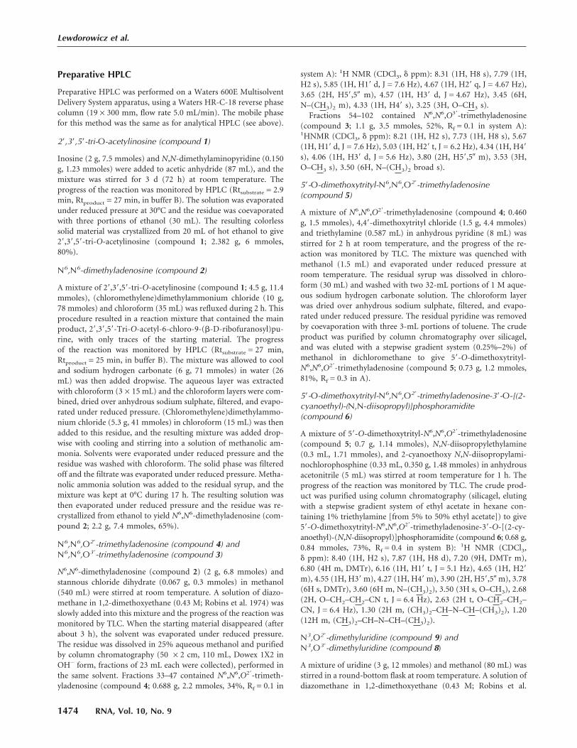
pared by the standard phosphoramidite solidphase method (Beaucage and Caruthers1981). However two of the four protectedmonomers, that is, N6,N6,O2�-trimethyladeno-sine and N3,O2�-dimethyluridine are not com-mercially available. Synthesis of N6,N6,O2�-tri-methyladenosine (compound 4) and its de-rivatization to the suitable building block(compound 6) for solid phase synthesis areshown in Figure 1. The synthetic route in-cluded (1) introduction of the N6,N6-dimeth-ylamino group into adenosine following pre-viously published protocols (Zemlicka andSorm 1965; Morris et al. 1976), (2) O�-meth-ylation (Robins et al. 1974), (3) 5�-O-dime-thoxytritylation (Sproat and Lamond 1991),and (4) activation with a phosphoramidite re-agent (Kierzek et al. 1986; Scaringe et al. 1990;Sproat and Lamond 1991). The other uniquestructure, N3,O2�-dimethyluridine (compound9) was prepared in a one-pot methylation ofuridine that included (1) the reaction of uridinewith diazomethane to give N3-uridine (com-pound 7; Szer and Shugar 1968), (2) subsequentmethylation of the O2� and O3� positions withthe same reagent, after addition of stannouschloride as a catalyst (Robins et al. 1974), and(3) HPLC separation of N3,O2�-dimethyluridineand N3,O3�-dimethylridine mixture. The protec-tion of compound 9 in the O5� position withdimethoxytrityl group gave 5�-O-dime-thoxytri-tyl-N3,O2�-dimethyluridine (compound 10),which was attached to the solid support usingstandard methodology (Kierzek et al. 1986;Sproat and Lamond 1991) based on a succiniclinker (Fig. 2). The resulting resin was used asthe starting material in the automated solidphase synthesis. After two subsequent cou-pling stages, which added O2�-methylcytidineand O2�-methyladenosine, the fourth nucleoside subunit(N6,N6,O2�-trimethyladenosine, compound 6) was attached.The last step of the solid phase synthesis involved phos-phorylation of the tetranucleotide 5� end with 2-cyano-ethyl-3-(4,4�-dimethoxytrityloxy)-2,2-di(ethoxycarbonyl)-propyl-1-N,N-diisopropyl phosphoramidite (Guzaev et al.1995). Conventional ammonolysis was used to cleave theRNA tetramer from the solid support, resulting with thesimultaneous removal of the protecting groups from thenucleic bases and intranucleotide phosphates. The resultingproduct, still selectively protected at the 5�-phosphate, wasseparated from impurities by preparative reverse phaseHPLC. Successive detritylation with 80% acetic acid and abrief treatment with methylamine generated an RNAtetramer with a free 5�-phosphate group, which was puri-fied by preparative HPLC. The final capping reaction was
performed by a method that was newly developed in ourlaboratory. It included coupling of the 5�-phosphorylatedtetramer with an imidazolide derivative of 7-methylguano-sine 5�-diphosphate [Im(m7GDP)], in the presence of an-hydrous zinc chloride as a catalyst, dissolved in DMF (Fig.3). Comparable yields of capped oligoribonucleotides werereported for a similar capping strategy that was performedin water and in the presence of manganese(II) ions (Sawaiat al. 1999); however, a larger excess of Im(m7GDP) wasused in that case. We also found the new coupling methodto be superior in terms of yield and reproducibility over thepreviously reported method (Zuberek et al. 2002, 2003),which involved activation of a 5�-phosphorylated oligori-bonucleotide with imidazole and its coupling with m7GDP.
To perform more profound structure–function studieswe also synthesized the 5�-terminal dinucleoside triphos-
FIGURE 3. Chemical synthesis of cap-4.
Chemical synthesis and activity of cap-4
www.rnajournal.org 1471

phate m7Gpppm36,6,2�A, denoted cap-1 (compound 17; Fig.
4). The synthesis was carried out in solution, using a strat-egy that was similar to that developed previously (Stepinskiet al. 2001; Jemielity et al. 2003). The synthetic route in-cluded preparation of N6,N6,O2�-trimethyladenosine (com-pound 4; Fig. 1) from inosine, its 5�-phosphorylation by theYoshikawa procedure (Yoshikawa 1967), and a final cou-pling performed with Im(m7GDP) (Fig. 4).
The structure of the newly synthesized cap-4 was con-firmed by NMR spectroscopy and mass spectrometry (ESIMS) as well as careful NMR analysis of all monomer units.The key intermediates of the synthesis were isolated andanalyzed by NMR and MS (for details, see Materials andMethods).
LeishIF4E-1 binds m7GTP and cap-4
Purification of LeishIF4E-1 was based on its ability to bindm7GTP. The recombinant protein was purified by m7GTP-Sepharose affinity column chromatography, using a single-step procedure. Pellets of bacteria that were induced toexpress LeishIF4E-1 were loaded onto a column of m7GTP-Sepharose. Only washes containing free m7GTP and notGTP released LeishIF4E-1 from the column matrix (Fig. 5).For the fluorescence studies, elution was performed withhigh salt, rather than with the free ligand, resulting in asimilar degree of purification.
To validate that the synthetically prepared cap-4 com-pound was biologically active, its ability to interact with thecap-binding protein from Leishmania was examined. Stud-ies on the tertiary structure of eIF4E from different organ-isms illustrate the structural basis for cap recognition, andindicate a requirement for several conserved Trp residues(Marcotrigiano et al. 1997; Matsuo et al. 1997; Tomoo et al.2003). Binding of the cap structure and its analogs results in
quenching of the Trp fluorescence, and this can be used asa tool to calculate the Kas values (Carberry et al. 1989;Niedzwiecka et al. 2002). Because seven out of eight Trpresidues are conserved between LeishIF4E-1 and eIF4Efrom mouse and yeast (Y. Yoffe, J. Zuberek, M. Lewdoro-wicz, Z. Zeira, Ch. Keasar, B. Shaanan, I. Orr-Dahan,M. Jankowska-Anyszka, E. Darzynkiewicz, and M. Shapira,in prep.), fluorescence assays were used to evaluate thebinding affinity of LeishIF4E-1 with different cap structures.
Kas values for complexes of LeishIF4E-1 with the syn-thetic cap analogs were determined by the fast and accuratetime-synchronized fluorescence titration method (TST;Niedzwiecka et al. 2002; Fig. 6; Table 1). This method takesinto account the emission of the free cap analog and theconcentration of the active protein, as free parameters of thenumerical fitting. The Kas values determined for cap-4 andm7GTP were comparable, with a slight preference for cap-4(Kas = 0.253 ± 0.003 µM−1 for cap-4, and Kas = 0.156 ±0.005 µM−1 for m7GTP). These results emphasize that thesynthetic cap-4 compound was efficiently recognized by thecorresponding LeishIF4E-1. Examination of Kas values mea-sured for analogs that represent intermediate compounds incap-4 biosynthesis revealed that the binding affinity de-creased by three- to fourfold, as compared to cap-4(Kas = 0.0724 ± 0.002 µM−1 for m7GpppA and Kas =0.0654 ± 0.0041 µM−1 for m7Gpppm3
6,6,2�A; Table 1). A re-duced Kas for dinucleotide analogs was previously reportedfor other eIF4E proteins as well (Niedzwiecka et al. 2002).Multiple eIF4E homologs that exist in lower eukaryotessuch as C. elegans show differential binding characteristicsto m7GTP and the hypermethylated m3
2,2,7GTP (TMG).Some show exclusive binding with m7GTP (IFE-3) whereas
FIGURE 4. Chemical synthesis of cap-1.
FIGURE 5. Chromatography and purification of LeishIF4E-1 onm7GTP-sepharose. Crude extracts from noninduced BL21 bacterialcells (crude) and the soluble fraction (Sup) following induction byIPTG were applied onto a column of m7GTP-Sepharose. The flow-through (FT) was collected and the column was washed (Wash) firstwith equilibration buffer and then with equilibration buffer containingGTP (100 µM). The protein was eluted by a wash containing m7GTP(200 µM).
Lewdorowicz et al.
1472 RNA, Vol. 10, No. 9

others bind both structures (IFE-5; Miyoshi et al. 2002).LeishIF4E-1 does not bind TMG (Y. Yoffe, J. Zuberek,M. Lewdorowicz, Z. Zeira, Ch. Keasar, B. Shaanan, I. Orr-Dahan, M. Jankowska-Anyszka, E. Darzynkiewicz, and M.Shapira, in prep.), and binds both m7GTP and cap-4 withalmost similar affinities, with a slight preference for theparasite cap-4. Thus it appears that m7GTP may play a keyrole in the interaction between LeishIF4E-1 and cap-4, andthe remaining modified nucleotides possibly contribute tothe additional specificity of binding.
Synthetically prepared cap analogs provide an importantresearch tool for establishing the structural basis of caprecognition by the corresponding binding proteins. To date,the role of the unique and complex cap found in trypano-somatids is only partially understood. Cap formation on theSL RNA proceeds in a 5�–3� direction, as indicated by ex-periments performed with permeable trypanosomes (Ulluand Tschudi 1995). It appears that at least part of the modi-fications occur cotranscriptionally, as shown in studies us-ing permeable trypanosomes (Mair et al. 2000) and thatsequences within the SL RNA coding gene are required forobtaining the complete cap-4 structure (Ullu and Tschudi1995; Zeiner et al. 2003). However, it was recently shownthat the cytoplasmic stage is necessary for SL RNA biogen-esis (Zeiner et al. 2003). It was further suggested that U4modification of the SL RNA most probably serves as a signalfor transport of snRNPs between the nucleus and the cyto-plasm (Mandelboim et al. 2003). The availability of a syn-thetic and functional cap-4, as well as its intermediate com-
pounds, is therefore of great importance. Such materialsshould also contribute to the identification of enzymes thatparticipate in cap-4 biogenesis and in turn, could serve as abasis for the development of new therapeutic molecules,with antiparasitic activity.
The ability to chemically synthesize an analog of cap-4, astructure that is found on the 5� end of mRNAs in alltrypanosomatids, is of great importance. Trypanosomatidsare known for their unusual molecular mechanisms, and ithas been well established that differential gene expression inthese organisms is achieved by posttranscriptional mecha-nisms such as processing and translation of mRNAs (Zilkaet al. 2001). The 5� cap has a key role in directing theseprocesses in all eukaryotes including in trypanosomatids,but the latter possess a unique cap-4 structure that is foundonly in this group of organisms. Thus the availability of thissynthetic structure as well as its intermediates is essential foridentifying the components that participate in the biosyn-thetic pathway of cap-4. Once identified, such molecules, inprinciple, could be used as novel drug targets against Leish-mania as well as other trypanosomatid parasites.
MATERIALS AND METHODS
Monitoring of the chemical synthesis
TLC analysis was performed on TLC plates coated with silicagel 60F-254 (Merck), developed by chloroform-methanol-triethylamine8.9:1:0.1 v/v/v (A), hexane-acetone-triethylamine 45:45:10 v/v/v(B), chloroform-methanol 8:2 v/v (C), dichloromethane-methanol95:5 v/v (D).
Analytical HPLC was performed on a Spectra-Physics SP8800apparatus, using a Supelcosil LC-18-T reverse phase column(4.6 × 250 mm, flow rate 1.3 mL/min) with various gradients ofbuffer A (0.05 M ammonium acetate at pH 5.9) and buffer B(methanol and buffer A, 1:1 v/v).
1H NMR and 13C NMR spectra were run on a Varian UNITY-plus spectrometer at 400 and 100.56 MHz, respectively, at ambienttemperatures and at concentrations of ca. 2 mg/mL (1H NMR),and ca. 20 mg/mL (13C NMR) using tetramethylsilane (TMS) asthe internal standard in CDCl3 and sodium 3-trimethylsilyl-[2,2,3,3-D4]-propinonate (TSP) in D2O.
Mass spectra were recorded on a Micromass QToF 1 MS spec-trometer using electrospray ionization.
FIGURE 6. Fluorescence titration measurements on binding ofLeishIF4E-1 to cap analogs. LeishIF4E-1 proteins, at a concentrationof 0.3 µM, were titrated by the cap analogs: cap-4 (open squares),m7GTP (open circles), m7GpppA (black squares), m7Gpppm3
6,6,2�A(open triangles), and GTP (black triangles), at 20°C in 50 mM HEPES/NaOH, 100 mM NaCl, 1 mM EDTA, 1 mM DTT. Protein fluores-cence, presented as relative values, was excited at 295 nm and observedat 345 nm. The observed increasing fluorescence intensity at a higherconcentration of ligand originates from the emission of the free capanalogs in solution.
TABLE 1. The equilibrium association constants (Kas) for com-plexes of LeishIEF4-1 with cap-4 and other cap analogs at 20°Cdetermined by fluorescence measurements
Cap analogs Kas (µM)−1
m7GTP 0.156 ± 0.005m7GpppA 0.0724 ± 0.0020m7Gpppm3
6,6,2� A 0.0654 ± 0.0041cap-4 0.253 ± 0.003GTP <0.005
Chemical synthesis and activity of cap-4
www.rnajournal.org 1473
Preparative HPLC
Preparative HPLC was performed on a Waters 600E MultisolventDelivery System apparatus, using a Waters HR-C-18 reverse phasecolumn (19 × 300 mm, flow rate 5.0 mL/min). The mobile phasefor this method was the same as for analytical HPLC (see above).
2�,3�,5�-tri-O-acetylinosine (compound 1)
Inosine (2 g, 7.5 mmoles) and N,N-dimethylaminopyridine (0.150g, 1.23 mmoles) were added to acetic anhydride (87 mL), and themixture was stirred for 3 d (72 h) at room temperature. Theprogress of the reaction was monitored by HPLC (Rtsubstrate = 2.9min, Rtproduct = 27 min, in buffer B). The solution was evaporatedunder reduced pressure at 30°C and the residue was coevaporatedwith three portions of ethanol (30 mL). The resulting colorlesssolid material was crystallized from 20 mL of hot ethanol to give2�,3�,5�-tri-O-acetylinosine (compound 1; 2.382 g, 6 mmoles,80%).
N6,N6-dimethyladenosine (compound 2)
A mixture of 2�,3�,5�-tri-O-acetylinosine (compound 1; 4.5 g, 11.4mmoles), (chloromethylene)dimethylammonium chloride (10 g,78 mmoles) and chloroform (35 mL) was refluxed during 2 h. Thisprocedure resulted in a reaction mixture that contained the mainproduct, 2�,3�,5�-Tri-O-acetyl-6-chloro-9-(�-D-ribofuranosyl)pu-rine, with only traces of the starting material. The progressof the reaction was monitored by HPLC (Rtsubstrate = 27 min,Rtproduct = 25 min, in buffer B). The mixture was allowed to cooland sodium hydrogen carbonate (6 g, 71 mmoles) in water (26mL) was then added dropwise. The aqueous layer was extractedwith chloroform (3 × 15 mL) and the chloroform layers were com-bined, dried over anhydrous sodium sulphate, filtered, and evapo-rated under reduced pressure. (Chloromethylene)dimethylammo-nium chloride (5.3 g, 41 mmoles) in chloroform (15 mL) was thenadded to this residue, and the resulting mixture was added drop-wise with cooling and stirring into a solution of methanolic am-monia. Solvents were evaporated under reduced pressure and theresidue was washed with chloroform. The solid phase was filteredoff and the filtrate was evaporated under reduced pressure. Metha-nolic ammonia solution was added to the residual syrup, and themixture was kept at 0°C during 17 h. The resulting solution wasthen evaporated under reduced pressure and the residue was re-crystallized from ethanol to yield N6,N6-dimethyladenosine (com-pound 2; 2.2 g, 7.4 mmoles, 65%).
N6,N6,O2�-trimethyladenosine (compound 4) andN6,N6,O3�-trimethyladenosine (compound 3)
N6,N6-dimethyladenosine (compound 2) (2 g, 6.8 mmoles) andstannous chloride dihydrate (0.067 g, 0.3 mmoles) in methanol(540 mL) were stirred at room temperature. A solution of diazo-methane in 1,2-dimethoxyethane (0.43 M; Robins et al. 1974) wasslowly added into this mixture and the progress of the reaction wasmonitored by TLC. When the starting material disappeared (afterabout 3 h), the solvent was evaporated under reduced pressure.The residue was dissolved in 25% aqueous methanol and purifiedby column chromatography (50 × 2 cm, 110 mL, Dowex 1X2 inOH− form, fractions of 23 mL each were collected), performed inthe same solvent. Fractions 33–47 contained N6,N6,O2�-trimeth-yladenosine (compound 4; 0.688 g, 2.2 mmoles, 34%, Rf = 0.1 in
system A): 1H NMR (CDCl3, � ppm): 8.31 (1H, H8 s), 7.79 (1H,H2 s), 5.85 (1H, H1� d, J = 7.6 Hz), 4.67 (1H, H2� q, J = 4.67 Hz),3.65 (2H, H5�,5� m), 4.57 (1H, H3� d, J = 4.67 Hz), 3.45 (6H,N–(CH3)2 m), 4.33 (1H, H4� s), 3.25 (3H, O–CH3 s).
Fractions 54–102 contained N6,N6,O3�-trimethyladenosine(compound 3; 1.1 g, 3.5 mmoles, 52%, Rf = 0.1 in system A):1HNMR (CDCl3, � ppm): 8.21 (1H, H2 s), 7.73 (1H, H8 s), 5.67(1H, H1� d, J = 7.6 Hz), 5.03 (1H, H2� t, J = 6.2 Hz), 4.34 (1H, H4�s), 4.06 (1H, H3� d, J = 5.6 Hz), 3.80 (2H, H5�,5� m), 3.53 (3H,O–CH3 s), 3.50 (6H, N–(CH3)2 broad s).
5�-O-dimethoxytrityl-N6,N6,O2�-trimethyladenosine(compound 5)
A mixture of N6,N6,O2�-trimethyladenosine (compound 4; 0.460g, 1.5 mmoles), 4,4�-dimethoxytrityl chloride (1.5 g, 4.4 mmoles)and triethylamine (0.587 mL) in anhydrous pyridine (8 mL) wasstirred for 2 h at room temperature, and the progress of the re-action was monitored by TLC. The mixture was quenched withmethanol (1.5 mL) and evaporated under reduced pressure atroom temperature. The residual syrup was dissolved in chloro-form (30 mL) and washed with two 32-mL portions of 1 M aque-ous sodium hydrogen carbonate solution. The chloroform layerwas dried over anhydrous sodium sulphate, filtered, and evapo-rated under reduced pressure. The residual pyridine was removedby coevaporation with three 3-mL portions of toluene. The crudeproduct was purified by column chromatography over silicagel,and was eluted with a stepwise gradient system (0.25%–2%) ofmethanol in dichloromethane to give 5�-O-dimethoxytrityl-N6,N6,O2�-trimethyladenosine (compound 5; 0.73 g, 1.2 mmoles,81%, Rf = 0.3 in A).
5�-O-dimethoxytrityl-N6,N6,O2�-trimethyladenosine-3�-O-[(2-cyanoethyl)-(N,N-diisopropyl)]phosphoramidite(compound 6)
A mixture of 5�-O-dimethoxytrityl-N6,N6,O2�-trimethyladenosine(compound 5; 0.7 g, 1.14 mmoles), N,N-diisopropylethylamine(0.3 mL, 1.71 mmoles), and 2-cyanoethoxy N,N-diisopropylami-nochlorophosphine (0.33 mL, 0.350 g, 1.48 mmoles) in anhydrousacetonitrile (5 mL) was stirred at room temperature for 1 h. Theprogress of the reaction was monitored by TLC. The crude prod-uct was purified using column chromatography (silicagel, elutingwith a stepwise gradient system of ethyl acetate in hexane con-taining 1% triethylamine [from 5% to 50% ethyl acetate]) to give5�-O-dimethoxytrityl-N6,N6,O2�-trimethyladenosine-3�-O-[(2-cy-anoethyl)-(N,N-diisopropyl)]phosphoramidite (compound 6; 0.68 g,0.84 mmoles, 73%, Rf = 0.4 in system B): 1H NMR (CDCl3,� ppm): 8.40 (1H, H2 s), 7.87 (1H, H8 d), 7.20 (9H, DMTr m),6.80 (4H m, DMTr), 6.16 (1H, H1� t, J = 5.1 Hz), 4.65 (1H, H2�m), 4.55 (1H, H3� m), 4.27 (1H, H4� m), 3.90 (2H, H5�,5� m), 3.78(6H s, DMTr), 3.60 (6H m, N–(CH3)2), 3.50 (3H s, O–CH3), 2.68(2H, O–CH2–CH2–CN t, J = 6.4 Hz), 2.63 (2H t, O–CH2–CH2–CN, J = 6.4 Hz), 1.30 (2H m, (CH3)2–CH–N–CH–(CH3)2), 1.20(12H m, (CH3)2–CH–N–CH–(CH3)2).
N3,O2�-dimethyluridine (compound 9) andN3,O3�-dimethyluridine (compound 8)
A mixture of uridine (3 g, 12 mmoles) and methanol (80 mL) wasstirred in a round-bottom flask at room temperature. A solution ofdiazomethane in 1,2-dimethoxyethane (0.43 M; Robins et al.
Lewdorowicz et al.
1474 RNA, Vol. 10, No. 9

1974) was then slowly added into this mixture and the progress ofthe reaction was monitored by TLC. After 1 h all the startingmaterial disappeared and N3-methyluridine (compound 7)(Rf = 0.2 in system C) dominated the reaction mixture. Stannouschloride dihydrate (0.061 g, 0.27 mmoles) and the second portionof an ethereal solution of diazomethane were then slowly addedand the reaction was completed after 1 h. Two regioisomers:N3,O2�-dimethyluridine (compound 9) and N3,O3�-dimethyluri-dine (compound 8) were separated by preparative HPLC (Rt8 = 35min, Rt9 = 38 min, using a linear 0%–100% gradient of buffer Bin buffer A within 40 min) to yield 47% of N3,O2�-dimethyluri-dine (compound 9) and 50% of N3,O3�-dimethyluridine (com-pound 8).
1H NMR for N3,O2�-dimethyluridine (compound 9): (D2O, �ppm): 7.90 (1H, H6 d, J = 8.02 Hz), 6.00 (1H, H1� d, J = 3.67 Hz),5.98 (1H, H5 d, J = 8.02 Hz), 4.30 (1H, H3� t, J = 6.01 Hz), 4.11(1H, H4� m), 4.00 (1H, H2� t, J = 3.68 Hz), 3.85 (2H, H5�,5� m),3.54 (3H, N–CH3), 3.30 (3H s, O–CH3).
5�-O-dimethoxytrityl-N3,O2�-dimethyluridine (compound 10)
A mixture of N3,O2�-dimethyluridine (compound 9; 0.5 g, 1.8mmoles), 4,4�-dimethoxytrityl chloride (1.2 g, 3.5 mmoles) andtriethylamine (0.6 mL) in anhydrous pyridine (7 mL) was stirredfor 2 h at room temperature, and the progress of the reaction wasmonitored by TLC. The reaction was quenched with methanol(1.5 mL) and evaporated under reduced pressure at room tem-perature. The residual syrup was dissolved in chloroform (33 mL)and washed with two 33-mL portions of 1 M aqueous sodiumhydrogen carbonate solution. The chloroform layer was dried overanhydrous sodium sulfate, filtered, and evaporated under reducedpressure. The residual pyridine was removed by coevaporationwith 3 × 3 mL of toluene. The crude product was purified bycolumn chromatography (silicagel, eluted by a stepwise gradient ofmethanol in dichloromethane [ranging from 0.25% to 2% metha-nol]), to give 5�-O-dimethoxytrityl-N3,O2�-dimethyluridine (com-pound 10) (0.84 g, 1.5 mmoles, 83%, Rf = 0.7 in system A): 1HNMR (CDCl3, � ppm): 8.01 (1H, H6 d, J = 8.00 Hz), 7.40 (9H m,DMTr), 6.82 (4H m, DMTr), 5.97 (1H, H1� s), 5.35 (1H, H5 d,J = 8.00 Hz), 4.46 (1H, H3� q), 3.99 (1H, H2� t, J = 7.89 Hz), 3.79(6H s, –O–CH3, DMTr), 3.77 (1H, H4� s), 3.67 (3H s, N–CH3),3.55 (2H, H5�,5� m), 3.32 (3H s, O–CH3) 13C NMR (CDCl3, �ppm): 162.81 (1C, C(4)=O), 158.67, 144.32, 135.31, 130.15,129.09, 113.25, 87.00 (19C, DMTr), 150.89 (1C, C2=O), 137.62(1C, C6), 101.42 (1C, C5), 87.60, 83.97, 83.06, 68.25, 61.05, 58.63(6C, C1�, C2�, C3�, C4�, C5�, O2�–CH3), 55.20 (2C, O–CH3,DMTr), 27.45 (1C, N–CH3).
5�-O-dimethoxytrityl-N3,O2�-dimethyluridine-3�-O-pentachlorophenylsuccinate (compound 11)
A mixture of 5�-O-dimethoxytrityl-N3,O2�-dimethyluridine (com-pound 10; 0.534 g, 0.92 mmoles), succinic anhydride (0.101 g, 1.01mmoles), and N,N-dimethylaminopyridine (0.134 g, 1.1 mmoles)in dichloromethane (5 mL) was stirred for 2 h at room tempera-ture. The progress of the reaction was monitored by TLC (Rf = 0.3in system D). N,N-dicyclohexylocarbodiimide (0.246 g, 1.2mmoles) and pentachlorophenol (0.270 g, 1.1 mmoles) were thenadded. The mixture was allowed to stand overnight and the pre-cipitate was filtered off. The filtrate was extracted with a saturatedaqueous solution of sodium dihydrogen phosphate and purified by
column chromatography (silicagel). Elution was performed with astepwise gradient (1%–2%) of methanol in dichloromethane,to give 5�-O-dimethoxytrityl-N3,O2�-dimethyluridine-3�-O-penta-chlorophenylsuccinate (compound 11; 0.798 g, 0.86 mmoles, 93%,Rf = 0.8 in system D): 1H NMR (CDCl3, � ppm): 7.87 (1H, H6 d,J = 7.99 Hz), 7.40 (9H m, DMTr), 7.30 (9H m, DMTr), 6.82 (4Hm, DMTr), 6.00 (1H, H1� d, J = 3.20 Hz), 5.42 (1H, H5 d, J = 7.99Hz), 4.26 (1H, H3� q), 4.10 (1H, H2� m, J = 3.10 Hz), 3.78 (6H s,–O–CH3, DMTr), 3.78 (1H, H4� s), 3.52 (2H, H5�,5� m), 3.50 (3H,N–CH3 s, N3-methyluracyl ring), 3.32 (3H s, O2�–CH3), 2.90 (4Hm, –C(O)–CH2–CH2–C(O)–).
5�-O-dimethoxytrityl-N3,O2�-dimethyluridine-3�-O-succinamide attached to the resin (compound 12)
A mixture of 5�-O-dimethoxytrityl-N3,O2�-dimethyluridine-3�-O-pentachlorophenylsuccinate (compound 11; 0.150 g, 0.16mmoles), triethylamine (0.4 mL), and Fractosil 500 (1 g; Merck) indimethylformamide (4 mL) was shaken overnight at room tem-perature. The resin was then washed successively with four por-tions of dichloromethane and methanol, filtered, and dried. FreeNH2-groups on the resin were acetylated with acetic anhydride (1mL), N,N-dimethylaminopyridine (0.010 g, 0.082 mmoles) in an-hydrous pyridine (5 mL). The mixture was shaken for 1 h and thenthe resin was filtered off, washed successively with four portions ofdichloromethane, and methanol. Afterward the resin was dried.
Solid phase synthesis
The RNA tetramer was synthesized on an Applied Biosystems 392synthesizer using a 1 µmole scale protocol with 10 min couplingfor each step. The starting material (compound 12) was placed ina standard reaction vessel and, successively, building blocks werecoupled for elongation of the ribonucleic chain. N-Benzoyl-5�-dimethoxytrityl-2�-O-methyl-cytidine-3�-[(2-cyanoethyl)-(N,N-diisopropyl)]-phosphoramidite and 5�-dimethoxytrityl-2�-O-methyl-N-phenoxyacetyladenosine-3�-[(2-cyanoethyl)-(N,N-diisopropyl)]-phosphoramidite were commercial products (Glen Research). 5�-Dimethoxytrityl-N6,N6,O2�-trimethyladenosine-3�-[(2-cyanoethyl)-(N,N-diisopropyl)]-phosphoramidite was synthetically prepared(compound 6). The 5�-phosphorylating reagent 2-cyanoethyl-3-(4,4�-dimethoxytrityloxy)-2,2-di(ethoxycarbonyl)propyl-1-N,N-diisopropyl phosphoramidite, which was employed in the last stepof the automated synthesis, was purchased from Glen Research.
Unprotected RNA fragment (pm36,6,2�Apm2�Apm2�Cpm2
3,2�U)(compound 13)
A 5�-blocked RNA fragment (obtained from the solid phase syn-thesis) was detritylated for 2 h with 80% acetic acid. After 2 hacetic acid was evaporated under reduced pressure and then me-thylamine (6 mL) was added. After 15 min, the methylamine wasremoved using P4O10 in a vacuum desiccator and the product waspurified by preparative HPLC (Rtproduct = 16.9 min, linear0–100% gradient of buffer B in buffer A over 50 min) to give 0.021g (0.0143 mmoles) of pm3
6,6,2�Apm2�Apm2�Cpm23,2�U (com-
pound 13) as an ammonium salt (m/z 1384.6 by ESI-MS).
P2-imidazolide 7-methylguanosine 5�-diphosphate,Im(m7GDP) (compound 14)
A mixture of triethylammonium salt of 7-methylguanosine 5�-diphosphate (0.1 g, 0.131 mmoles), imidazole (0.178 g, 2.63
Chemical synthesis and activity of cap-4
www.rnajournal.org 1475

mmoles), 2,2�-dithiodipyridine (0.230 g, 1.048 mmoles), triethyl-amine (18 µL), and triphenylphosphine (0.275 g, 1.05 mmoles) indimethylformamide (1.5 mL) was stirred overnight at room tem-perature. A solution of sodium perchlorate (0.09 g, 0.74 mmoles)in anhydrous acetone (4 mL) was then added. The pure productthat appeared as a sodium salt of P2-imidazolide 7-methylguano-sine 5�-diphosphate (compound 14) was filtered off, washed withfour portions of acetone, and dried over P4O10 (0.063 g, 0.11mmoles, 84%).
Cap-4 (m7Gpppm36,6,2�Apm2�Apm2�C pm2
3,2�U) (compound 15)
A mixture of the ammonium salt of pm36,6,2�Apm2�Apm2�-
Cpm23,2�U (compound 13; 10 mg, 6.8 µmoles), the sodium salt of
P2-imidazolide 7-methylguanosine 5�-diphosphate Im(m7GDP)(compound 14; 18 mg, 34.3 µmoles) and ZnCl2 (46 mg, 340µmoles) in dimethylformamide (0.08 mL) was stirred for 16 h atroom temperature. The progress of the reaction was monitored byHPLC (Rtsubstrate = 17.8 min, Rtproduct = 16.5 min, using a linear0%–100% gradient of buffer B in buffer A over 20 min). Themixture was then diluted with water (3.5 mL). Preparative HPLCpurification (linear 0%–100% gradient of buffer B in buffer A over50 min) gave m7Gpppm3
6,6,2�Apm2�A pm2�Cpm23,2�U (compound
15) as ammonium salt (5.14 mg, 2.64 µmoles, 40%): 1H NMR(D2O, � ppm): 8.23 (1H, H8 s, adenine), 8.05 (1H, H8 s, N6,N6-dimethyladenine), 8.04 (1H, H2 s, adenine), 7.67 (1H, H2 s,N6,N6-dimethyladenine), 7.82 (1H, H6 d, cytidine) 7.50 (1H, H6,s, N3-methyluracyl), 5.98 (2H, broad s, H1� N6,N6,O2�-trimeth-yladenosine, H1� O2�-methyladenosine), 5.90 (1H, H1� d, O2�-methylcytidine), 5.79 (2H, t, H5 cytidine, H5 N3-methyluracyl),5.62 (1H, H1� s, 7-methylguanine), 5.39 (1H, H1� d, N3,O2�-dimethyluridine), 4.90–3.80 (25H, H2�, H3�, H4�, H5�,5� m), 3.96(3H, s N7CH3), 3.71, 3.58, 3.51, 3.24 (15H m, N6,N6-dimethyl-adenosine O2�–CH3, adenosine O2�–CH3, cytosine O2�–CH3,N3,O2�-dimethyluridine O2�–CH3, and N3–CH3); 4.8 and 3.3(6H, broad s N–(CH3)2 N6,N6-dimethyladenine; m/z 1825.5 byESI-MS).
N6,N6,O2�-trimethyladenosine 5�-monophosphate(compound 16)
A mixture of N6,N6,O2�-trimethyladenosine (compound 4; 0.058g, 0.188 mmoles) and trimethyl phosphate (1.55 mL) was stirredin an ice bath for 3.5 h. Into this mixture phosphorus oxychloride(0.044 mL) was added and the progress of the reaction was moni-tored by analytical HPLC (Rtsubstrate = 24.3 min, Rtproduct = 17.3min, linear 0%–100% gradient of buffer B in buffer A, over 20min). The mixture was diluted with water (50 mL), neutralized bythe addition of 1.4 M solution of triethylammonium bicarbonate(TEAB), and purified by column chromatography (DEAE-Sepha-dex, A-25, HCO3
− form) using a linear gradient of 0–0.7 M TEABto obtain N6,N6,O2�-trimethyladenosine 5�-monophosphate as tri-ethylammonium salt (compound 16; 0.067 g, 0.113 mmoles,60%).
Cap-1 (m7Gpppm36,6,2�A) (compound 17)
A mixture of triethylammonium salt of N6,N6,O2�-trimethyladeno-sine 5�-monophosphate (compound 16; 67 mg, 113 µmoles),
a sodium salt of P2-imidazolide 7-methylguanosine 5�-diphos-phate Im(m7GDP) (compound 14; 63 mg, 109 µmoles), and ZnCl2(160 mg, 1200 µmoles) in dimethylformamide (1.2 mL) wasstirred for 2 h at room temperature. The progress of the reac-tion was monitored by HPLC (Rtsubstrate = 17.8 min, Rtproduct
= 14.2 min, linear 0%–100% gradient of buffer B in buffer A over20 min). The mixture was then diluted with water (60 mL), andEDTA (704 mg, 1.9 µmoles) was added. Then the solution wasneutralized with NaHCO3. The crude product was purified bycolumn chromatography (DEAE-Sephadex, using a linear gradientof 0–1.2 M TEAB) to give (compound 17) (m7Gpppm3
6,6,2�A,cap-1) as a triethylammonium salt (15 mg, 13 µmoles, 12%): 1HNMR (D2O, � ppm): 8.32 (1H, H8 s, N6,N6,O2�-adenine), 8.09(1H, H2 s, N6,N6-adenine), 6.05 (1H, H1� s, N6,N6,O2�-trimeth-yladenosine), 5.74 (1H, H1� s, 7-methylguanosine), 4.63 (1H, H2�t, N6,N6,O2�-trimethyladenosine), 4.34 (10H broad m, H2�7-methylguanosine, H3�, H4�, H5�, H5� 7-methylguanosine andN6,N6,O2�-trimethyladenosine), 3.97 (3H s, N7–CH3) 3.42 (9Hbroad s, N6,N6,O2�-trimethyladenosine O–CH3 and N6–CH3).
Cloning and expression of the Leishmania eIF4E-1 inbacteria for protein purification
The eIF4E-1 isoform from Leishmania major was amplified byPCR, using genomic DNA as template. The forward primer cor-responded to positions 1–21 GGAATTCCATATGTCAGCCCCGTCTTCAGTT-3� and the reverse primer corresponded to posi-tions 625–645 5�-CGCGGATCCCTAAGACGCCTCGCCGTGCTT-3�. All primers contained anchor sequences that incorpor-ated the NdeI and BamHI restriction sites at the amino (fwdprimer) and carboxy (rev primer) termini, respectively (the re-striction sites are underlined). The amplified fragments were di-gested with NdeI and BamHI and further cloned into the corre-sponding site of the pHis-parallel vector (Sheffield et al. 1999),resulting in plasmid pLeishIF4E-1. This cloning strategy elimi-nated the His-tag and generated a recombinant protein that ini-tiated from its own first methionine. The nucleotide sequences ofthe amplified fragments were compared to the genomic sequenceobtained from the L. major genome project.
The recombinant pLeishIF4E-1 was introduced into Escherichiacoli BL21 cells and expression was induced at 20°C in culturesgrown to OD660 0.5 for 3 h, by the addition of 0.5 mM isopropyl-1-thio-�-D-galactopyranoside (IPTG). Cells were washed once insonication buffer (20 mM HEPES at pH 7.6, 1 mM DTT and 2mM EDTA, 5% glycerol), harvested, and disrupted by sonicationduring 2 min on ice, using pulses of 20 sec interspaced by intervalsof 20 sec. The cell extract was clarified by centrifugation during 30min at 20,000g and the supernatant was loaded on m7GTP-Sepha-rose, equilibrated in the sonication buffer. The protein was elutedin the presence of 150 µM of the free cap analog m7GTP (Sigma).Alternatively, for fluorescence assays the protein was eluted byhigh salt (200 mM NaCl).
Fluorescence measurements and data analysis
Freshly prepared protein samples were used for the fluorescenceassays, after removal of the high salt by filter centrifugation over 4mL Biomax 5K NMWL columns (Millipore). Protein concentra-
Lewdorowicz et al.
1476 RNA, Vol. 10, No. 9

tions were determined by spectrophotometry, assuming �280 =47,500 M−1cm−1, which were calculated from the amino acid com-position (Gill and von Hippel 1989). Fluorescence measurementswere performed on a LS-50B Spectrofluorometer (Perkin ElmerCo.), in a quartz semi-micro cuvette (Hellma) with optical lengths4 mm and 10 mm for absorption and emission, respectively. Fluo-rescence time-synchronized titrations (TST; Niedzwiecka et al.2002) were performed in 50 mM HEPES/NaOH (pH 7.2), 100mM NaCl, 1 mM EDTA, and 1 mM DTT at 20°C by adding 1-µLaliquots of the cap analog solution to 1.4 mL of 0.1 or 0.3 µMprotein solution of LeishIF4E-1. Each titration consisted of 40–65data points. Fluorescence intensities (excited at 280 or 295 nm andobserved at 337 or 345 nm) were corrected for sample dilution(<5%) and for the inner filter effect, using empirical calibrationcurve (Lakowicz 1999). The theoretical curve for the fluorescenceintensity (F) as a function of [L] was fitted to the experimentaldata points according to the following equation:
F = F(0) − [cx] · (�� + �lig−free) + [L] · �lig−free,
where the equilibrium concentration of the cap-LeishIF4Ecomplex [cx] is given by
�cx� =�L� + �Pact�
2+
1 − ��Kas��L� − �Pact�� + 1�2 + 4Kas � �Pact�
2Kas
.
The parameters to be extracted from the fit were as follows: Kas,the association constant; [Pact], the concentration of the activeprotein; �� = �P−act−free − �cx, the difference between the fluo-rescence efficiencies of the apo-protein and the complex; �lig-free,the fluorescence efficiency of the free cap analog in the solution;F(0), the initial fluorescence intensity (Niedzwiecka et al. 2002).The final Kas was calculated as a weighted average of two to fiveindependent titrations; numerical least-squares nonlinear regres-sion analysis was performed using ORIGIN 6.0 (Microcal SoftwareInc.).
ACKNOWLEDGMENTS
We are indebted to Dr. Nahum Sonenberg from McGill Univer-sity, Canada, for his generous gift of the eIF4E(28–217) plasmid.This research was supported by the State Committee for ScientificResearch (Grants PBZ-KBN 059/T09/10, PBZ-KBN 059/T09/14,and KBN 3 P04A 021 25 to E.D., R.K., and R.S., accordingly), andgrants from the German-Israel Foundation (GIF, 728-23.2/2002)and Israel Ministry of Health 5440 (to M.S.).
The publication costs of this article were defrayed in part bypayment of page charges. This article must therefore be herebymarked “advertisement” in accordance with 18 USC section 1734solely to indicate this fact.
Received April 5, 2004; accepted May 28, 2004.
REFERENCES
Bangs, J.D., Crain, P.F., Hashizume, T., McClosky, J.A., andBoothroyd, J.C. 1992. Mass spectrometry of mRNA cap 4 fromTrypanosomatids reveals two novel nucleosides. J. Biol. Chem.267: 9805–9815.
Beaucage, S.L. and Caruthers, M.H. 1981. Deoxynucleotide phos-phoramidites—A new class of key iintermediates for deoxypoly-nucleotide synthesis. Tetrahedron Lett. 22: 1859–1862.
Blachut-Okrasinska, E., Bojarska, E., Niedzwiecka, A., Chlebicka, L.,
Darzynkiewicz, E., Stolarski, R., Stepinski, J., and Antosiewicz, J.M.2000. Stopped-flow and Brownian dynamics studies of electrostaticeffects in the kinetics of binding of 7-methyl-GpppG to the proteineIF4E, Eur. Biophys. J. 29: 487–498.
Cai, A., Jankowska-Anyszka, M., Centers, A., Chlebicka, L., Stepinski,J., Stolarski, R., Darzynkiewicz, E., and Rhoads, R.E. 1999. Quan-titative assessment of mRNA cap analogues as inhibitors of in vitrotranslation. Biochemistry 38: 8538–8547.
Carberry, S.E., Rhoads, R.E., and Goss, D.J. 1989. A spectroscopicstudy of the binding of m7GTP and m7GpppG to human proteinsynthesis initiation factor 4E. Biochemistry 28: 8078–8083.
Gill, S.C. and von Hippel, P.H. 1989. Calculation of protein extinctioncoefficients from amino acid sequence data. Anal. Biochem.182: 319–326.
Gingras, A.C., Raught, B., and Sonenberg, N. 1999. eIF4 initiationfactors: Effectors of mRNA recruitment to ribosomes and regula-tors of translation. Annu. Rev. Biochem. 68: 913–963.
Guzaev, A., Salo, H., Azhayev, A., and Lonnberg, H. 1995. A newapproach for chemical phosphorylation of oligonucleotides at the5�-terminus. Tetrahedron 51: 9375–9384.
Jemielity, J., Fowler, T., Zuberek, J., Stepinski, J., Lewdorowicz, M.,Niedzwiecka, A., Stolarski, R., Darzynkiewicz, E., and Rhoads, R.E.2003. Novel “anti-reverse” cap analogs with superior translationalproperties. RNA 9: 1108–1122.
Kierzek, R., Caruthers, M.H., Longfellow, C.E., Swinton, D., Turner,D.H., and Freier, S.M. 1986. Polymer-supported RNA synthesisand its application to test the nearest-neighbor model for duplexstability. Biochemistry 25: 7840–7846.
Lakowicz, J.R. 1999. Instrumentation for fluorescence spectroscopy. InPrinciples of fluorescence spectroscopy, 2nd ed. pp. 25–61. KluwerAcademic/Plenum, New York.
Mair, G., Ullu, E., and Tschudi, C. 2000. Cotranscriptional Cap 4formation on the Trypanosoma brucei Spliced Leader RNA. J. Biol.Chem. 275: 28994–28999.
Mandelboim, M., Barth, S., Biton, M., Liang, X.H., and Michaeli, S.2003. Silencing of Sm proteins in Trypanosoma brucei by RNAinterference captured a novel cytoplasmic intermediate in splicedleader RNA biogenesis. J. Biol. Chem. 278: 51469–51478.
Marcotrigiano, J., Gingras, A.C., Sonenberg, N., Burley, S.K. 1997.Cocrystal structure of the messenger RNA 5� cap-binding protein(eIF4E) bound to 7-methyl-GDP. Cell 89: 951–961.
Matsuo H., Li, H., McGuire, A.M., Fletcher, C.M., Gingras, A.C.,Sonenberg, N., Wagner, G. Structure of translation factor eIF4Ebound to m7GDP and interaction with 4E-binding protein. Nat.Struct. Biol. 4: 717–724.
Miyoshi, H., Dwyer, D.S., Keiper, B.D., Jankowska-Anyszka, M., Dar-zynkiewicz, E., and Rhoads, R.E. 2002. Discrimination betweenmono- and trimethylated cap structures by two isoforms of Cae-norhabditis elegans eIF4E. EMBO J. 21: 4680–4690.
Morris, M.J., MacCoss, M., Naik, S.R., and Ramani, G. 1976. Nucleicacid related compounds. 21. Direct fluorination of uracil and citi-zen bases and nucleosides using trifluoromethyl hypofluorite.Mechanism, stereochemistry, and synthetic applications. J. Am.Chem. Soc. 98: 7381–7390.
Niedzwiecka, A., Marcotrigiano, J., Stepinski, J., Jankowska-Anyszka,M., Wyslouch-Cieszynska, A., Dadlez, M., Gingras, A.-C., Mak, P.,Darzynkiewicz, E., Sonenberg, N., et al. 2002. Biophysical studiesof eIF4E cap-binding protein: Recognition of mRNA 5� cap struc-ture and synthetic fragments of eIF4G and 4E-BP proteins. J. Mol.Biol. 319: 615–635.
Robins, M.J., Naik, S.R., and Lee, A.S.K. 1974. Nucleic acid relatedcompounds. 12. The facile and high-yield stannous chloride cata-lyzed monomethylation of the cis-glycol system of nucleosides bydiazomethan. J. Org. Chem. 39: 1891–1899.
Sawai, H., Wakai, H., and Nakamura-Ozaki, A. 1999. Synthesis andreaction of nucleoside 5�-diphosphate imidazolide. A nonenzy-matic capping agent for 5�-monophosphorylated oligoribonucleo-tides in aqueous solution. J. Org. Chem. 64: 5836–5840.
Scaringe, S.A., Francklyn, C., and Usman, N. 1990. Chemical synthesis
Chemical synthesis and activity of cap-4
www.rnajournal.org 1477

of biologically active oligoribonucleotides using �-cyanoethyl pro-tected ribonucleoside phosphoramidite. Nucleic Acid Res. 18:5433–5441.
Sheffield, P., Garrard, S., and Derewenda, Z. 1999. Overcoming ex-pression and purification problems of RhoGDI using a family of“parallel expression vectors.” Protein Expr. Purif. 15: 34–39.
Sproat, B.S. and Lamond, A.I. 1991. 2�-Methyloligoribonucleotides:Synthesis and applications. In Oligonucleotides and Analogues (ed.F. Eckstein), pp. 49–86. Oxford University Press, New York.
Stepinski, J., Waddell, C., Stolarski, R., Darzynkiewicz, E., and Rhoads,R.E. 2001. Synthesis and properties of mRNAs containing thenovel “anti-reverse” cap analogues 7-methyl(3�-O-methyl)GpppGand 7-methyl (3�-deoxy)GpppG. RNA 7: 1486–1495.
Szer, W. and Shugar, D. 1968. 2�,3�-O-Isopropylidene-3-methyl-uridine. In Synthetic procedures in nucleic acid chemistry, Vol. 1(eds. W.W. Zorbach and R.S. Tipson), pp. 433–435. Wiley, NewYork.
Tomoo, K., Shen, X., Okabe, K., Nozoe, Y., Fukuhara, S., Morino, S.,Sasaki, M., Taniguchi, T., Miyagawa, H., Kitamura, K., et al. 2003.Structural features of human initiation factor 4E, studied by X-raycrystal analyses and molecular dynamics simulations. J. Mol. Biol.328: 365–383.
Ullu, E. and Tschudi, C. 1995. Accurate modification of the trypano-some spliced leader cap structure in a homologous cell-free system.J. Biol. Chem. 270: 20365–20369.
Varani, G. 1997. A cap for all occasions. Structure 5: 855–858.
Yoshikawa, M., Kato, T., and Takenishi, T. 1967. A novel method forphosphorylation of nucleosides to 5�-nucleotides. Tetrahedron Lett.50: 5065.
Zeiner, G.M., Sturm, N.R., and Campbell, D.A. 2003. Exportin 1 me-diates nuclear export of the Kinetoplastid Spliced Leader RNA.Eukaryot. Cell 2: 222–230.
Zemlicka, J. and Sorm, F. 1965. The reaction of dimethylchloro-methyleneammoniumchloride with 2�,3�,5�-tri-O-acetylinosine. Anew synthesis of 6-chloro-9-(�-D-ribofuranosil)purine. CollectionCzech. Chem. Commun. 30: 1880–1889.
Zilka, A., Garlapati, S., Dahan, E., Yavelski, V., and Shapira, M. 2001.Developmental regulation of HSP83 in Leishmania: 3� processingand mRNA stability control transcript abundance and translationis directed by a determinant in the 3� untranslated region. J. Biol.Chem. 276: 47922–47929.
Zuberek, J., Stepinski, J., Niedzwiecka, A., Stolarski, R., Salo, H.,Lonnberg, H., and Darzynkiewicz, E. 2002. Synthesis of tetraribo-nucleotide cap analogue m7GpppAm2�pUm2�pAm2� and its interac-tion with eukaryotic initiation factor eIF4E. Collection SymposiumSeries. 5: 399–403.
Zuberek, J., Wyslouch-Cieszynska, A., Niedzwiecka, A., Dadlez, M.,Stepinski, J., Augustyniak, W., Gingras, A.-C., Zhang, Z., Burley,S.K., Sonenberg, N., et al. 2003. Phosphorylation of eIF4E attenu-ates its interaction with mRNA 5� cap analogs by electrostaticrepulsion: Intein-mediated protein ligation strategy to obtainphosphorylated protein. RNA 9: 52–61.
Lewdorowicz et al.
1478 RNA, Vol. 10, No. 9