Chemical Constituents and Anticancer Effects of the Essential Oil from Leaves of Xylopia laevigata
8
Introduction ! The Xylopia species (family Annonaceae) are aro- matic plants that have both nutritional and me- dicinal uses. Several studies have reported cyto- toxic activity for plants belonging to this genus, such as Xylopia aethiopica (Dunal) A. Rich. [1], Xy- lopia aromatica (Lam.) Mart. in Mart. [2], Xylopia langsdorffiana St.‑Hil. & Tul. [3], and Xylopia seri- cea A. St.‑Hil. [4]. Xylopia laevigata (Mart.) R. E. Fries is a medicinal plant found in the tropical American forest. In Brazil, it is popularly known as “meiú” and “pin- daíba” and can be found in the States of Piauí, Pa- raiba, Sergipe, Rio de Janeiro, and São Paulo [5]. Although the principles of the popular use of this plant are almost unknown, by interview of the lo- cal population, we found that a decoction of its leaves and flowers is used in the folk medicine of the Brazilian Northeast for several purposes, in- cluding painful disorders, heart disease, and in- flammatory conditions. In addition, some Xylopia species have been used in the folk medicine for treating tumors. Nonetheless, almost no scientific research was made of this plant. A phytochemical and biological study of the stem from X. laevigata was performed by Silva et al. [6]. The ent-kaurane diterpenoids, ent-kaur-16-en- 19-oic acid, 4-epi-kaurenic acid, ent-16β-hy- droxy-17-acetoxy-kauran-19-al, ent-3β-hydroxy- kaur-16-en-19-oic acid, and ent-16β,17-dihy- droxy-kauran-19-oic acid, as well as spathulenol and a mixture of β-sitosterol, stigmasterol, and campesterol, were isolated from the hexane ex- tract from X. laevigata stem. Potent larvicidal ac- tivity against Aedes aegypti larvae and antifungal activity against Candida glabrata and Candida dubliniensis were found for ent-3β-hydroxy- kaur-16-en-19-oic acid. However, chemical anal- ysis or investigation of the biological properties Abstract ! Xylopia laevigata, popularly known as “meiú” and “pindaíba”, is a medicinal plant used in the folk medicine of the Brazilian Northeast for several purposes. The chemical constituents of the essen- tial oil from leaves of X. laevigata, collected from wild plants growing at three different sites of the remaining Atlantic forest in Sergipe State (Brazil- ian Northeast), were analyzed by GC/FID and GC/ MS. The effect of the essential oil samples was as- sessed on tumor cells in culture, as well on tumor growth in vivo. All samples of the essential oil were dominated by sesquiterpene constituents. A total of 44 compounds were identified and quantified. Although some small differences were observed in the chemical composition, the pres- ence of γ-muurolene (0.60–17.99 %), δ-cadinene (1.15–13.45 %), germacrene B (3.22–7.31 %), α-co- paene (3.33–5.98 %), germacrene D (9.09– 60.44 %), bicyclogermacrene (7.00–14.63 %), and (E)-caryophyllene (5.43–7.98%) were verified as major constituents in all samples of the essential oil. In the in vitro cytotoxic study, the essential oil displayed cytotoxicity to all tumor cell lines tested, with the different samples displaying a similar profile; however, they were not hemolytic or genotoxic. In the in vivo antitumor study, tu- mor growth inhibition rates were 37.3–42.5 %. The treatment with the essential oil did not sig- nificantly affect body weight, macroscopy of the organs, or blood leukocyte counts. In conclusion, the essential oil from the leaves of X. laevigata is chemically characterized by the presence of γ- muurolene, δ-cadinene, germacrene B, α-co- paene, germacrene D, bicyclogermacrene, and (E)-caryophyllene as major constituents and pos- sesses significant in vitro and in vivo anticancer potential. Chemical Constituents and Anticancer Effects of the Essential Oil from Leaves of Xylopia laevigata Authors Jullyana de S. S. Quintans 1 , Bruno M. Soares 2 , Rosana P. C. Ferraz 1 , Allan C. A. Oliveira 1 , Thanany B. da Silva 3 , Leociley R. A. Menezes 3 , Marília F. C. Sampaio 3 , Ana Paula do N. Prata 4 , Manoel O. Moraes 2 , Claudia Pessoa 2 , Angelo R. Antoniolli 1 , Emmanoel V. Costa 3 , Daniel P. Bezerra 1 Affiliations The affiliations are listed at the end of the article Key words l " Xylopia laevigata l " Annonaceae l " essential oil l " chemical composition l " antitumor received August 30, 2012 revised October 15, 2012 accepted Dec. 2, 2012 Bibliography DOI http://dx.doi.org/ 10.1055/s-0032-1328091 Published online January 10, 2013 Planta Med 2013; 79: 123–130 © Georg Thieme Verlag KG Stuttgart · New York · ISSN 0032‑0943 Correspondence Prof. Dr. Daniel P. Bezerra Department of Physiology Federal University of Sergipe Av. Marechal Rondon, Jardim Rosa Elze 49100–000, São Cristóvão Sergipe Brazil Phone: + 55 79 21 05 66 44 [email protected] 123 Quintans JSS et al. Chemical Constituents and … Planta Med 2013; 79: 123–130 Original Papers Downloaded by: Dot. Lib Information. Copyrighted material.
-
Upload
independent -
Category
Documents
-
view
3 -
download
0
Transcript of Chemical Constituents and Anticancer Effects of the Essential Oil from Leaves of Xylopia laevigata
Abstract!
Xylopia laevigata, popularly known as “meiú” and“pindaíba”, is a medicinal plant used in the folkmedicine of the Brazilian Northeast for severalpurposes. The chemical constituents of the essen-tial oil from leaves of X. laevigata, collected fromwild plants growing at three different sites of theremaining Atlantic forest in Sergipe State (Brazil-ian Northeast), were analyzed by GC/FID and GC/MS. The effect of the essential oil samples was as-sessed on tumor cells in culture, as well on tumorgrowth in vivo. All samples of the essential oilwere dominated by sesquiterpene constituents.A total of 44 compounds were identified andquantified. Although some small differences wereobserved in the chemical composition, the pres-ence of γ-muurolene (0.60–17.99%), δ-cadinene(1.15–13.45%), germacrene B (3.22–7.31%), α-co-paene (3.33–5.98%), germacrene D (9.09–
60.44%), bicyclogermacrene (7.00–14.63%), and(E)-caryophyllene (5.43–7.98%) were verified asmajor constituents in all samples of the essentialoil. In the in vitro cytotoxic study, the essentialoil displayed cytotoxicity to all tumor cell linestested, with the different samples displaying asimilar profile; however, they were not hemolyticor genotoxic. In the in vivo antitumor study, tu-mor growth inhibition rates were 37.3–42.5%.The treatment with the essential oil did not sig-nificantly affect body weight, macroscopy of theorgans, or blood leukocyte counts. In conclusion,the essential oil from the leaves of X. laevigata ischemically characterized by the presence of γ-muurolene, δ-cadinene, germacrene B, α-co-paene, germacrene D, bicyclogermacrene, and(E)-caryophyllene as major constituents and pos-sesses significant in vitro and in vivo anticancerpotential.
Chemical Constituents and Anticancer Effects of theEssential Oil from Leaves of Xylopia laevigata
Authors Jullyana de S. S. Quintans1, Bruno M. Soares2, Rosana P. C. Ferraz1, Allan C. A. Oliveira1, Thanany B. da Silva3,Leociley R. A. Menezes3, Marília F. C. Sampaio3, Ana Paula do N. Prata4, Manoel O. Moraes2, Claudia Pessoa2,Angelo R. Antoniolli1, Emmanoel V. Costa3, Daniel P. Bezerra1
Affiliations The affiliations are listed at the end of the article
Key wordsl" Xylopia laevigatal" Annonaceael" essential oill" chemical compositionl" antitumor
received August 30, 2012revised October 15, 2012accepted Dec. 2, 2012
BibliographyDOI http://dx.doi.org/10.1055/s-0032-1328091Published online January 10,2013Planta Med 2013; 79: 123–130© Georg Thieme Verlag KGStuttgart · New York ·ISSN 0032‑0943
CorrespondenceProf. Dr. Daniel P. BezerraDepartment of PhysiologyFederal University of SergipeAv. Marechal Rondon, JardimRosa Elze49100–000, São CristóvãoSergipeBrazilPhone: + [email protected]
123Original Papers
Dow
nloa
ded
by: D
ot. L
ib In
form
atio
n. C
opyr
ight
ed m
ater
ial.
Introduction!
The Xylopia species (family Annonaceae) are aro-matic plants that have both nutritional and me-dicinal uses. Several studies have reported cyto-toxic activity for plants belonging to this genus,such as Xylopia aethiopica (Dunal) A. Rich. [1], Xy-lopia aromatica (Lam.) Mart. in Mart. [2], Xylopialangsdorffiana St.‑Hil. & Tul. [3], and Xylopia seri-cea A.St.‑Hil. [4].Xylopia laevigata (Mart.) R.E. Fries is a medicinalplant found in the tropical American forest. InBrazil, it is popularly known as “meiú” and “pin-daíba” and can be found in the States of Piauí, Pa-raiba, Sergipe, Rio de Janeiro, and São Paulo [5].Although the principles of the popular use of thisplant are almost unknown, by interview of the lo-cal population, we found that a decoction of itsleaves and flowers is used in the folk medicine ofthe Brazilian Northeast for several purposes, in-
Quintans JSS et al.
cluding painful disorders, heart disease, and in-flammatory conditions. In addition, some Xylopiaspecies have been used in the folk medicine fortreating tumors. Nonetheless, almost no scientificresearch was made of this plant.A phytochemical and biological study of the stemfrom X. laevigatawas performed by Silva et al. [6].The ent-kaurane diterpenoids, ent-kaur-16-en-19-oic acid, 4-epi-kaurenic acid, ent-16β-hy-droxy-17-acetoxy-kauran-19-al, ent-3β-hydroxy-kaur-16-en-19-oic acid, and ent-16β,17-dihy-droxy-kauran-19-oic acid, as well as spathulenoland a mixture of β-sitosterol, stigmasterol, andcampesterol, were isolated from the hexane ex-tract from X. laevigata stem. Potent larvicidal ac-tivity against Aedes aegypti larvae and antifungalactivity against Candida glabrata and Candidadubliniensis were found for ent-3β-hydroxy-kaur-16-en-19-oic acid. However, chemical anal-ysis or investigation of the biological properties
Chemical Constituents and… Planta Med 2013; 79: 123–130

124 Original Papers
of the essential oil from leaves of X. laevigata has not been previ-ously performed.The chemical composition of the essential oil from leaves of X.laevigata, collected from wild plants growing at three differentsites of the remaining Atlantic forest in Sergipe State (BrazilianNortheast), was analyzed by GC/FID and GC/MS. In addition, theeffect of the essential oil samples was assessed on tumor cells inculture as well as on tumor growth in vivo.
Dow
nloa
ded
by: D
ot. L
ib In
form
atio
n. C
opyr
ight
ed m
ater
ial.
Material and Methods!
Reagents5-Fluorouracil (5-FU, purity > 99%), doxorubicin (purity > 98%),3-(4,5-dimethyl-2-thiazolyl)-2,5-diphenyl-2H-tetrazolium bro-mide (MTT), ficoll-hypaque, phytohemagglutinin, and resazurinwere purchased from Sigma Chemical Co. Triton X-100 (purity> 98%) was purchased from Vetec. RPMI 1640 medium, fetal bo-vine serum, penicillin, and streptomycin were purchased fromCultilab. Carbon dioxide (CO2) was purchased from White Mar-tins. All other reagents were of analytical grade.
CellsThe cytotoxicity of the essential oil was tested against HL-60(promyelocytic leukemia), OVACAR-8 (ovarian carcinoma), SF-295 (glioblastoma), and HCT-116 (colon carcinoma) human can-cer cell lines, all obtained from the National Cancer Institute, Be-thesda, MD, USA. Cells were grown in RPMI-1640 medium sup-plemented with 10% fetal bovine serum, 2mM glutamine,100 µg/mL streptomycin, and 100U/mL penicillin, and incubatedat 37°C with a 5% CO2 atmosphere.Sarcoma 180 tumor cells, which had beenmaintained in the peri-toneal cavity of Swiss mice, were obtained from the Laboratory ofExperimental Oncology at the Federal University of Ceará.In order to get healthy human peripheral blood mononuclearcells (PBMC), heparinized blood (from healthy, nonsmoking do-nors who had not taken any drug at least 15 days prior to sam-pling) was collected, and the PBMC were isolated by a standardmethod of density-gradient centrifugation over ficoll-hypaque.The PBMC were washed and resuspended. Cells were grownunder the same conditions as above plus the addition of phytohe-magglutinin (4%). The Human Research Ethics Committee ap-proved this experimental protocol.
AnimalsA total of 40 Swiss mice (males, 25–30 g), obtained from the cen-tral animal house of the Federal University of Sergipe, Brazil,were used. Animals were housed in cages with free access to foodand water. All animals were kept under a 12:12-h light-darkcycle (lights on at 6:00 a.m.). Animals were treated according tothe ethical principles for animal experimentation of SBCAL (theBrazilian association of laboratory animal science), Brazil. TheAnimal Studies Committee from the Federal University of Sergipeapproved the experimental protocol (number 08/2012).
Plant materialX. laevigata leaves were collected from wild plants growing atthree different sites of the remaining Atlantic forest in SergipeState (Brazilian Northeast). Sample A (voucher number 15440)was collected in April 2010 at Itabaiana Mountain National Parkin the Municipality of Itabaiana (coordinates: S 10°44′ 53′′ W 37°20′ 21′′). Sample B (voucher number 15443) was collected in
Quintans JSS et al. Chemical Constituents and… Planta Med 2013; 79: 123–130
March 2010 at “Mata do Crasto” in the Municipality of Santa Lu-zia do Itanhy (coordinates: S 11°22′ 54′′W 37°25′ 15′′). And sam-ple C (voucher number 17359) was collected in July 2010 nearthe campus of the Federal University of Sergipe in the Municipal-ity of São Cristóvão (coordinates: S 10°55′ 08′′ W 37°06′ 13′′). Allspecimens were obtained from flowering plants. The species wasidentified by Dr. Ana Paula do Nascimento Prata, a plant taxono-mist from the Department of Biology at the Federal University ofSergipe. The voucher botanic specimens were deposited at theHerbarium of the Federal University of Sergipe.
Essential oil isolation and chemical analysisOil isolation: The X. laevigata leaf specimen samples (200 g each)were dried separately in a stove with circulating air at 40°C for24 h and submitted to hydrodistillation for 3 h using a Cle-venger-type apparatus (Amitel). The hydrodistillation was per-formed in triplicate. The essential oil was dried over anhydroussodium sulphate and stored in a freezer until analysis. The essen-tial oil yields (v/w), calculated from dry plant weight, were1.18 ± 0.13% for sample A, 1.58 ± 0.12% for sample B, and1.40 ± 0.14% for sample C.GC/FID analysis: GC analyses were carried out using a ShimadzuGC-17A fitted with a flame ionization detector (FID) and an elec-tronic integrator. Separation of the compounds was achieved em-ploying a ZB-5MS fused capillary column (30m× 0.25mm× 0.25 µm film thickness) coated with 5%-phenyl-arylene-95%-methylpolysiloxane. Helium was the carrier gas at 1.2mL/minflow rate. The column temperature program was: 50°C/2min,then a temperature increase of 4°C/min to 200°C, followed byanother temperature increase of 15°C/min to 300°C, finishingwith 300°C/15min. The injector and detector temperatures were250°C and 280°C, respectively [7]. Samples (0.5 µL in CH2Cl2)were injected with a 1:100 split ratio. Retention indices weregenerated with a standard solution of n-alkanes (C9-C18). Peakareas and retention times were measured by an electronic inte-grator. The relative amounts of individual compounds were com-puted fromGC peak areas without FID response factor correction.GC/MS analysis: GC/MS analyses were performed on a ShimadzuQP5050AGC/MS system equippedwith an AOC-20i auto-injector.A J&W Scientific DB-5MS (coated with 5%-phenyl-95%-methyl-polysiloxane) fused capillary column (30m× 0.25mm ×0.25 µmfilm thickness) was used as the stationary phase. MS were takenat 70 eV with a scan interval of 0.5 s and fragments from 40–500Da. All other conditions were similar to the GC analysis [7].
Identification of constituentsThe essential oil components were identified by comparing theretention times of the GC peaks with standard compounds ranunder identical conditions, by comparison of retention indices[8] and MS [9] with those in the literature, and by comparison ofMSwith those stored in the NIST and Wiley databases.
In vitro cytotoxic evaluation of the essential oilDetermination of the effect of the essential oil on cultured tumorand normal cells: Tumor cell growth was quantified by the abilityof living cells to reduce the yellow dye MTT to a purple formazanproduct, as described by Mossman [10]. For all experiments,100 µL of a solution of cells (0.7 × 10⁵ cells/mL for adherent cellsor 0.3 × 10⁶ cells/mL) were seeded in 96-well plates. After 24 h,the essential oil (0.39–50 µg/mL), dissolved in dimethyl sulfoxide(DMSO), was added to eachwell (using the HTS – highthroughputscreening – Biomek 3000; Beckman Coulter, Inc.) and incubated
125Original Papers
Dow
nloa
ded
by: D
ot. L
ib In
form
atio
n. C
opyr
ight
ed m
ater
ial.
for 72 h. For this, a stock solution (10mg/mL) was prepared usingDMSO, and then this stock solution was diluted using medium.Doxorubicin was used as the positive control. Negative controlwas treated with the vehicle used for diluting the test substance(0.5% DMSO). At the end of incubation, the plates were centri-fuged, and the medium was replaced by fresh medium (150 µL)containing 0.5mg/mLMTT. Three hours later, the formazan prod-uct was dissolved in 150 µL of DMSO, and the absorbance wasmeasured using a multiplate reader (DTX 880 Multimode Detec-tor; Beckman Coulter, Inc.). The drug effect was quantified as thepercentage of control absorbance of reduced dye at 595 nm.PBMC cell growth was determined by the Alamar blue assay, asdescribed by Ahmed et al. [11]. For all experiments, 100 µL of asolution of cells (0.3 × 10⁶ cells/mL) were seeded in 96-well plates.After 24 h, the essential oil (0.39–50 µg/mL), dissolved in DMSO,was added to each well (using HTS, high-throughput screening,Biomek 3000, Beckman Coulter, Inc.) and incubated for 72 h.Doxorubicin was used as the positive control. Negative controlwas treated with the vehicle used for diluting the test substance(0.5% DMSO). Twenty-four hours before the end of incubation,10 µL of stock solution (0.312mg/mL) of Alamar blue (Resazurin)was added to each well. The absorbance was measured using amultiplate reader (DTX 880 Multimode Detector; BeckmanCoulter®), and the drug effect was quantified as the percentageof control absorbance at 570 nm and 595 nm.Determination of the effect of the essential oil on mouse erythro-cytes: The hemolytic activity was performed using mouse eryth-rocyte, as described by Jimenez et al. [12]. The test was per-formed in 96-well plates using a 2% mouse erythrocyte suspen-sion in 0.85% NaCl containing 10mMCaCl2. The essential oil sam-ples were tested at concentrations ranging from 31.25 to 500 µg/mL. After incubation at room temperature for 30min and centri-fugation, the supernatant was removed, and the hemoglobin re-leased was measured spectrophotometrically as the absorbanceat 540 nm.Determination of the genotoxic effect of the essential oil by the al-kaline comet assay: The alkaline (pH > 13) version of the cometassay (Single Cell Gel Electrophoresis) was performed as de-scribed by Singh et al. [13] with minor modifications [14]. Slideswere prepared in duplicate, and 100 cells were screened per sam-ple (50 cells from each duplicate slide) using a fluorescence mi-croscope (Zeiss) equipped with a 515–560 nm excitation filter, a590 nm barrier filter, and a 40X objective. Cells were scored visu-ally into five classes according to tail length: (1) class 0: undam-aged, without a tail; (2) class 1: with a tail shorter than the diam-eter of the head (nucleus); (3) class 2: with a tail length 1–2× thediameter of the head; (4) class 3: with a tail longer than 2× thediameter of the head; (5) class 4: comets with no heads. A valueof damage index (DI) was assigned to each comet according to itsclass, using the formula: DI = (0 × n0) + (1 × n1) + (2 × n2) + (3 ×n3) + (4 × n4), where n = number of cells in each class analyzed.The damage index ranged from 0 (completely undamaged: 100cells × 0) to 400 (with maximum damage: 100 cells × 4). DI isbased on migration length and on the amount of DNA in the tail,and it is considered a sensitive DNA measure [15].
In vivo antitumor evaluation of the essential oilDetermination of the effect of the essential oil on the growth of sol-id tumor in vivo: The in vivo antitumor effect was evaluated usingsarcoma 180 ascites tumor cells following protocols previouslydescribed [16–18]. Ten-day-old sarcoma 180 ascites tumor cells(2 × 10⁶ cells per 500 µL) were implanted subcutaneously into
the left hind groin of mice. The essential oil was dissolved in 5%DMSO and given to mice intraperitoneally once a day for 7 con-secutive days. At the beginning of the experiment, the mice weredivided into four groups, as follows: Group 1: animals treated byi.p. injection of vehicle 5% DMSO (n = 12); Group 2: animalstreated by i.p. injection of 5-FU (25mg/kg/day) (n = 10); Group3: animals treated by i.p. injection of the essential oil (50mg/kg/day) (n = 8); Group 4: animals treated by i.p. injection of the es-sential oil (100mg/kg/day) (n = 10). The treatments were startedone day after tumor injection. The dosages were determinedbased on previous articles [18,19]. On day 8, the animals weresacrificed by cervical dislocation, and the tumors were excisedand weighed. The drug effects were expressed as the percent in-hibition of control.Systemic toxicological evaluation: Body weight loss, organ weightalteration, and hematological analyses were determined at theend of the above experiment, as previous described [17,18]. Pe-ripheral blood samples of the mice were collected from the retro-orbital plexus under light ether anesthesia, and the animals weresacrificed by cervical dislocation. After sacrifice, the liver, kidney,and spleens were removed and weighed. In hematological analy-sis, total leukocyte counts were determined by standard manualprocedures using light microscopy.
Statistical analysisData were presented as mean ± SEM (or SD) or IC50 values, andtheir 95% confidence intervals (CI 95%) obtained by nonlinear re-gression. The differences between experimental groups werecompared by ANOVA (analysis of variance) followed by the Stu-dent-Newman-Keuls (p < 0.05). All statistical analyses were per-formed using the GraphPad program (Intuitive Software for Sci-ence).
Results!
Initially, chemical compositions of three essential oil sampleswere analyzed by GC/FID and GC/MS. Hydrodistillation of the X.laevigata leaf specimens yielded essential oil superior to 1% in re-lation to the dry weight of the plant material. The best yield wasobserved for sample B with 1.58 ± 0.12%. All samples of the es-sential oil were dominated by sesquiterpene constituents with95.35% in sample A, 98.60% in sample B, and 91.18% in sampleC. l" Fig. 1 presents a representative chromatogram from GC‑MSanalysis of the essential oil samples. As shown inl" Table 1, it waspossible to identify 44 compounds: 37 in sample A, 19 in sampleB, and 28 in sample C. The major compounds identified in the es-sential oil of sample A were γ-muurolene (17.99%), δ-cadinene(13.45%), germacrene D (9.09%), bicyclogermacrene (7.00%), α-copaene (5.98%), (E)-caryophyllene (5.43%), γ-cadinene (4.52%),aromadendrene (3.99%), γ-amorphene (3.92%), and germacreneB (3.22%), while germacrene D (60.44%), bicyclogermacrene(11.65%), (E)-caryophyllene (6.37%), germacrene B (3.67%), γ-ca-dinene (3.38%), and α-copaene (3.33%) were the main constitu-ents in the essential oil of sample B. In sample C, the followingpredominated: germacrene D (43.62%), bicyclogermacrene(14.63%), (E)-caryophyllene (7.98%), germacrene B (7.31%), δ-el-emene (4.50%), and α-copaene (3.56%). Although some small dif-ferences were observed in the chemical composition of the es-sential oil samples from X. laevigata specimens, the presence ofγ-muurolene (0.60–17.99%), δ-cadinene (1.15–13.45%), germa-crene B (3.22–7.31%), α-copaene (3.33–5.98%), germacrene D
Quintans JSS et al. Chemical Constituents and… Planta Med 2013; 79: 123–130
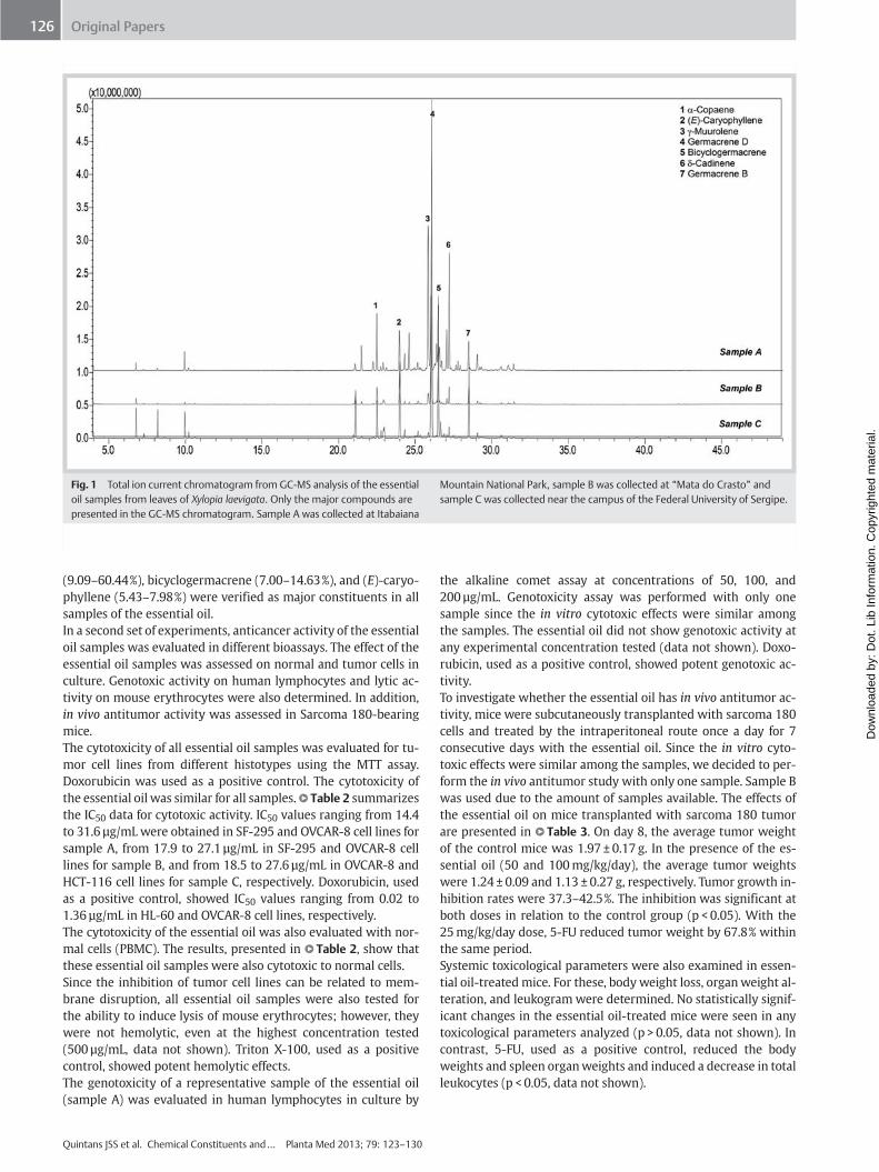
Fig. 1 Total ion current chromatogram from GC‑MS analysis of the essentialoil samples from leaves of Xylopia laevigata. Only the major compounds arepresented in the GC‑MS chromatogram. Sample A was collected at Itabaiana
Mountain National Park, sample B was collected at “Mata do Crasto” andsample C was collected near the campus of the Federal University of Sergipe.
126 Original Papers
Dow
nloa
ded
by: D
ot. L
ib In
form
atio
n. C
opyr
ight
ed m
ater
ial.
(9.09–60.44%), bicyclogermacrene (7.00–14.63%), and (E)-caryo-phyllene (5.43–7.98%) were verified as major constituents in allsamples of the essential oil.In a second set of experiments, anticancer activity of the essentialoil samples was evaluated in different bioassays. The effect of theessential oil samples was assessed on normal and tumor cells inculture. Genotoxic activity on human lymphocytes and lytic ac-tivity on mouse erythrocytes were also determined. In addition,in vivo antitumor activity was assessed in Sarcoma 180-bearingmice.The cytotoxicity of all essential oil samples was evaluated for tu-mor cell lines from different histotypes using the MTT assay.Doxorubicin was used as a positive control. The cytotoxicity ofthe essential oil was similar for all samples.l" Table 2 summarizesthe IC50 data for cytotoxic activity. IC50 values ranging from 14.4to 31.6 µg/mL were obtained in SF-295 and OVCAR-8 cell lines forsample A, from 17.9 to 27.1 µg/mL in SF-295 and OVCAR-8 celllines for sample B, and from 18.5 to 27.6 µg/mL in OVCAR-8 andHCT-116 cell lines for sample C, respectively. Doxorubicin, usedas a positive control, showed IC50 values ranging from 0.02 to1.36 µg/mL in HL-60 and OVCAR-8 cell lines, respectively.The cytotoxicity of the essential oil was also evaluated with nor-mal cells (PBMC). The results, presented in l" Table 2, show thatthese essential oil samples were also cytotoxic to normal cells.Since the inhibition of tumor cell lines can be related to mem-brane disruption, all essential oil samples were also tested forthe ability to induce lysis of mouse erythrocytes; however, theywere not hemolytic, even at the highest concentration tested(500 µg/mL, data not shown). Triton X-100, used as a positivecontrol, showed potent hemolytic effects.The genotoxicity of a representative sample of the essential oil(sample A) was evaluated in human lymphocytes in culture by
Quintans JSS et al. Chemical Constituents and… Planta Med 2013; 79: 123–130
the alkaline comet assay at concentrations of 50, 100, and200 µg/mL. Genotoxicity assay was performed with only onesample since the in vitro cytotoxic effects were similar amongthe samples. The essential oil did not show genotoxic activity atany experimental concentration tested (data not shown). Doxo-rubicin, used as a positive control, showed potent genotoxic ac-tivity.To investigate whether the essential oil has in vivo antitumor ac-tivity, mice were subcutaneously transplanted with sarcoma 180cells and treated by the intraperitoneal route once a day for 7consecutive days with the essential oil. Since the in vitro cyto-toxic effects were similar among the samples, we decided to per-form the in vivo antitumor study with only one sample. Sample Bwas used due to the amount of samples available. The effects ofthe essential oil on mice transplanted with sarcoma 180 tumorare presented in l" Table 3. On day 8, the average tumor weightof the control mice was 1.97 ± 0.17 g. In the presence of the es-sential oil (50 and 100mg/kg/day), the average tumor weightswere 1.24 ± 0.09 and 1.13 ± 0.27 g, respectively. Tumor growth in-hibition rates were 37.3–42.5%. The inhibition was significant atboth doses in relation to the control group (p < 0.05). With the25mg/kg/day dose, 5-FU reduced tumor weight by 67.8% withinthe same period.Systemic toxicological parameters were also examined in essen-tial oil-treated mice. For these, body weight loss, organweight al-teration, and leukogram were determined. No statistically signif-icant changes in the essential oil-treated mice were seen in anytoxicological parameters analyzed (p > 0.05, data not shown). Incontrast, 5-FU, used as a positive control, reduced the bodyweights and spleen organweights and induced a decrease in totalleukocytes (p < 0.05, data not shown).
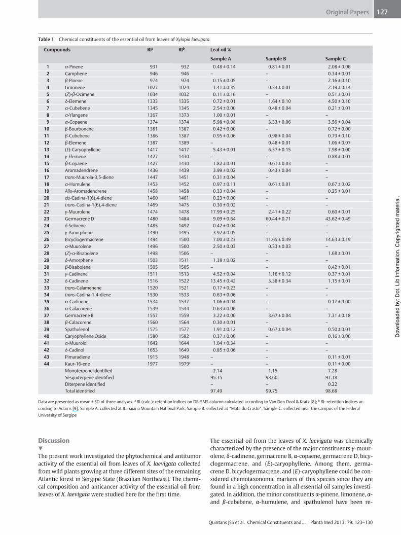
Table 1 Chemical constituents of the essential oil from leaves of Xylopia laevigata.
Compounds RIa RIb Leaf oil %
Sample A Sample B Sample C
1 α-Pinene 931 932 0.48 ± 0.14 0.81 ± 0.01 2.08 ± 0.06
2 Camphene 946 946 – – 0.34 ± 0.01
3 β-Pinene 974 974 0.15 ± 0.05 – 2.16 ± 0.10
4 Limonene 1027 1024 1.41 ± 0.35 0.34 ± 0.01 2.19 ± 0.14
5 (Z)-β-Ocimene 1034 1032 0.11 ± 0.16 – 0.51 ± 0.01
6 δ-Elemene 1333 1335 0.72 ± 0.01 1.64 ± 0.10 4.50 ± 0.10
7 α-Cubebene 1345 1345 2.54 ± 0.00 0.48 ± 0.04 0.21 ± 0.01
8 α-Ylangene 1367 1373 1.00 ± 0.01 – –
9 α-Copaene 1374 1374 5.98 ± 0.08 3.33 ± 0.06 3.56 ± 0.04
10 β-Bourbonene 1381 1387 0.42 ± 0.00 – 0.72 ± 0.00
11 β-Cubebene 1386 1387 0.95 ± 0.06 0.98 ± 0.04 0.79 ± 0.10
12 β-Elemene 1387 1389 – 0.48 ± 0.01 1.06 ± 0.07
13 (E)-Caryophyllene 1417 1417 5.43 ± 0.01 6.37 ± 0.15 7.98 ± 0.00
14 γ-Elemene 1427 1430 – – 0.88 ± 0.01
15 β-Copaene 1427 1430 1.82 ± 0.01 0.61 ± 0.03 –
16 Aromadendrene 1436 1439 3.99 ± 0.02 0.43 ± 0.04 –
17 trans-Muurola-3,5-diene 1447 1451 0.31 ± 0.04 – –
18 α-Humulene 1453 1452 0.97 ± 0.11 0.61 ± 0.01 0.67 ± 0.02
19 Allo-Aromadendrene 1458 1458 0.33 ± 0.04 – 0.25 ± 0.01
20 cis-Cadina-1(6),4-diene 1460 1461 0.23 ± 0.00 – –
21 trans-Cadina-1(6),4-diene 1469 1475 0.30 ± 0.02 – –
22 γ-Muurolene 1474 1478 17.99 ± 0.25 2.41 ± 0.22 0.60 ± 0.01
23 Germacrene D 1480 1484 9.09 ± 0.64 60.44 ± 0.71 43.62 ± 0.49
24 δ-Selinene 1485 1492 0.42 ± 0.04 – –
25 γ-Amorphene 1490 1495 3.92 ± 0.05 – –
26 Bicyclogermacrene 1494 1500 7.00 ± 0.23 11.65 ± 0.49 14.63 ± 0.19
27 α-Muurolene 1496 1500 2.50 ± 0.03 0.33 ± 0.03 –
28 (Z)-α-Bisabolene 1498 1506 – – 1.68 ± 0.01
29 δ-Amorphene 1503 1511 1.38 ± 0.02 – –
30 β-Bisabolene 1505 1505 – – 0.42 ± 0.01
31 γ-Cadinene 1511 1513 4.52 ± 0.04 1.16 ± 0.12 0.37 ± 0.01
32 δ-Cadinene 1516 1522 13.45 ± 0.42 3.38 ± 0.34 1.15 ± 0.01
33 trans-Calamenene 1520 1521 0.17 ± 0.23 – –
34 trans-Cadina-1,4-diene 1530 1533 0.63 ± 0.06 – –
35 α-Cadinene 1534 1537 1.06 ± 0.04 – 0.17 ± 0.00
36 α-Calacorene 1539 1544 0.63 ± 0.06 – –
37 Germacrene B 1557 1559 3.22 ± 0.00 3.67 ± 0.04 7.31 ± 0.18
38 β-Calacorene 1560 1564 0.30 ± 0.01 – –
39 Spathulenol 1575 1577 1.91 ± 0.12 0.67 ± 0.04 0.50 ± 0.01
40 Caryophyllene Oxide 1580 1582 0.37 ± 0.00 – 0.16 ± 0.00
41 α-Muurolol 1642 1644 1.04 ± 0.34 – –
42 δ-Cadinol 1653 1649 0.85 ± 0.06 – –
43 Pimaradiene 1915 1948 – – 0.11 ± 0.01
44 Kaur-16-ene 1977 1979c – – 0.11 ± 0.00
Monoterpene identifiedSesquiterpene identifiedDiterpene identifiedTotal identified
2.1495.35–97.49
1.1598.60–99.75
7.2891.180.22
98.68
Data are presented as mean ± SD of three analyses. a RI (calc.): retention indices on DB-5MS column calculated according to Van Den Dool & Kratz [8]; b RI: retention indices ac-
cording to Adams [9]; Sample A: collected at Itabaiana Mountain National Park; Sample B: collected at “Mata do Crasto”; Sample C: collected near the campus of the Federal
University of Sergipe
127Original Papers
Dow
nloa
ded
by: D
ot. L
ib In
form
atio
n. C
opyr
ight
ed m
ater
ial.
Discussion!
The present work investigated the phytochemical and antitumoractivity of the essential oil from leaves of X. laevigata collectedfromwild plants growing at three different sites of the remainingAtlantic forest in Sergipe State (Brazilian Northeast). The chemi-cal composition and anticancer activity of the essential oil fromleaves of X. laevigatawere studied here for the first time.
The essential oil from the leaves of X. laevigata was chemicallycharacterized by the presence of the major constituents γ-muur-olene, δ-cadinene, germacrene B, α-copaene, germacrene D, bicy-clogermacrene, and (E)-caryophyllene. Among them, germa-crene D, bicyclogermacrene, and (E)-caryophyllene could be con-sidered chemotaxonomic markers of this species since they arefound in a high concentration in all essential oil samples investi-gated. In addition, the minor constituents α-pinene, limonene, α-and β-cubebene, α-humulene, and spathulenol have been re-
Quintans JSS et al. Chemical Constituents and… Planta Med 2013; 79: 123–130

Table 2 In vitro cytotoxic activity of the essential oil from leaves of Xylopia laevigata.
Cell lines Histotype IC50 (µg/mL)
Sample A Sample B Sample C Doxorubicin
OVCAR-8 Ovarian carcinoma 31.626.6–37.6
27.123.1–31.8
18.515.3–23.0
1.360.98–1.89
SF-295 Glioblastoma 14.410.2–20.3
17.913.5–23.8
20.518.8–22.5
0.160.13–0.23
HCT-116 Colon carcinoma 22.211.7–42.1
25.511.7–55.6
27.619.3–39.6
Nd
HL-60 Promyelocytic leukemia 17.511.7–22.4
25.010.4–36.2
20.616.3–25.9
0.020.01–0.02
PBMC Peripheral lymphoblast 63.739.1–103.7
35.321.2–59.0
56.938.6–83.8
0.970.52–1.80
Data are presented as IC50 values, and their 95% confidence interval obtained by nonlinear regression from two independent experiments performed in duplicate. Doxorubicin was
used as a positive control. Sample A was collected at Itabaiana Mountain National Park, sample B was collected at “Mata do Crasto”, and sample C was collected near the campus of
the Federal University of Sergipe. Nd = not determined
Table 3 In vivo antitumor activity of the essential oil from leaves of Xylopialaevigata.
Drug Dose
(mg/kg/day)
Tumor
(g)
Inhibition
(%)
5% DMSO – 1.97 ± 0.14
5-FU 25 0.63 ± 0.16* 67.8
Essential oil 50 1.24 ± 0.09* 37.3
100 1.13 ± 0.27* 42.5
Mice were injected with sarcoma 180 (2.0 × 10⁶ cells/animal, s. c.). Starting one day
after tumor implantation, the animals were treated for seven consecutive days by in-
traperitoneal route. Data are presented as mean ± SEM for 8–12 animals. Negative
control was treated with the vehicle used for diluting the test substance (5% DMSO).
5-Fluorouracil (5-FU) was used as a positive control. A representative sample of the
essential oil (sample B) was used. Sample B was collected at “Mata do Crasto”.
* p < 0.05 compared with the 5% DMSO group by ANOVA followed by Student-New-
man-Keuls
128 Original Papers
Dow
nloa
ded
by: D
ot. L
ib In
form
atio
n. C
opyr
ight
ed m
ater
ial.
ported in essential oils of several other species belonging to thegenus Xylopia, some of them, such as α-pinene, limonene, andspathulenol, have been found in high concentrations [20–22].The presence of the major compounds along with the minor con-stituents identified α-pinene, limonene, and spathulenol, indi-cating that this species is a typical member of the Annonaceaefamily. Although some of these volatile compounds are alsopresent in essential oils of other families, their occurrence in An-nonaceae in high concentrations, mainly in the case of bicyclo-germacrene, (E)-caryophyllene, and germacrene, is very commonand has an important chemotaxonomic relevance [7,20–22].Although the essential oil samples have shown some differencein chemical composition, as the concentrations of germacrene Dand γ-muurolene, they presented similar in vitro cytotoxic effect.Probably, the associations of the major and minor constituentsare responsible for the cytotoxic activity of this essential oil. Thedifferences in chemical composition can be related to soil and cli-mate conditions, water stress, collection place, nutrition, andother abiotic factors.All samples of the essential oil displayed cytotoxicity to all tumorcell lines tested, with IC50 values ranging from 14.4 to 31.6 µg/mL.According to the preclinical anticancer drug-screening programused in this study, only those extracts/oils presenting IC50 valuesbelow 30 µg/mL in tumor cell line assays are considered promis-
Quintans JSS et al. Chemical Constituents and… Planta Med 2013; 79: 123–130
ing for anticancer drug development [23]. Therefore, the essentialoil obtained from X. laevigata presented promising results. In ad-dition, it inhibited tumor growth in mice in a dose-dependentmanner. On the other hand, the essential oil was also cytotoxicto normal cells, presenting low selectivity, but it was not hemo-lytic or genotoxic.The erythrocyte membrane is a dynamic structure that can dic-tate significant changes in its interaction with drugs [24]. The ab-sence of lytic effects suggests that the cytotoxicity of the essentialoil is not related to membrane disruption; probably a more spe-cific cellular pathway is responsible for its action. On the otherhand, genotoxic effects could be biologically relevant as an alter-native strategy for killing tumor cells. The lack of a genotoxic ef-fect suggests that the cytotoxicity of the essential oil is not relatedto DNA damage. Moreover, this effect needs to be extensivelyevaluated to assess the safety of novel drugs.For the Xylopia genus, as cited above, some studies have reportedthat plants belonging to this genus present cytotoxic activity. Theethanolic extract of X. aethiopica fruits was able to induce DNAdamage, cell cycle arrest in the G1 phase, and apoptotic cell death.These effects seem to be related to the diterpene ent-15-oxokaur-16-en-19-oic acid [1]. Themethanol extract of X. aethiopica seedswas cytotoxic to some tumor cell lines [25]. Interestingly, the es-sential oil of X. aethiopica fruits also showed cytotoxic activity toepidermoid carcinoma cells (Hep-2) [26]. Extracts of the bark,wood, and stem of X. aromatica [2,27] as well as numerous ace-togenins isolated from this species [28,29] showed cytotoxicityto several tumor cell lines. In X. langsdorffiana, the isolated com-pound xylodiol was able to inhibit cell proliferation probably byinvolvement of the induction of cell differentiation and apoptosisinduction [3,30]. Pita et al. [31] showed that trachylobane-360, aditerpene isolated from X. langsdorffiana, displays antitumor ef-fects in vitro and in vivo,without substantial systemic toxicity. Inaddition, the diterpenoid kaurenoic acid, isolated from X. sericea,showed cytotoxic, genotoxic, and mutagenic effects in many ex-perimental protocols because it induced DNA double-strandbreaks and/or inhibition of topoisomerase I [4]. In this presentwork, the essential oil from leaves of X. laevigata showed signifi-cant in vitro and in vivo anticancer potential.Some constituents of the essential oil from leaves of X. laevigatahave been studied for their cytotoxic activity. (E)-caryophylleneand α-pinene were previously tested by our group and showedIC50 values > 25 µg/mL [32]. In another study, (E)-caryophyllene
129Original Papers
Info
rmat
ion.
Cop
yrig
hted
mat
eria
l.
showed cytotoxic activity with an IC50 value of ~ 20 µg/mL to re-nal cell adenocarcinoma (ACHN) and amelanotic melanoma (C32)[33]. Amiel et al. [34] reported that (E)-caryophyllene has cyto-toxic activity to tumor cell lines but not to normal cells. More-over, (E)-caryophyllene caused a potent induction of apoptosis,accompanied by DNA ladder and caspase-3 catalytic activity intumor cell lines [34]. α-Humulene showed cytotoxic activity withan IC50 value of 11.2 µg/mL to hormone-dependent prostate car-cinoma cells (LNCaP) [35]. Areche et al. [36] showed that spathu-lenol was cytotoxic to gastric adenocarcinoma cells (AGS). Limo-nene inhibits the growth of human gastric cancer cells through amechanism of inducing their apoptosis [37]. In addition, limo-nene showed anti-metastatic and anti-angiogenic effects in ani-mal models [38]. Others constituents such as γ-muurolene, δ-ca-dinene, and α-copaene have not been previously tested againsttumor cell lines.The systemic toxicological aspects were also subjected to investi-gation in the present study. Essential oil-treated animals did notshow any significant change in body weight, macroscopy of theorgans (kidney, liver, and spleen), or blood leukocyte counts. Un-like the essential oil, 5-FU, used as a positive control, reduced thebody weights and spleen organ weights and induced severe leu-kopenia, which represents an important immunosuppressionside effect [39].In conclusion, the essential oil from leaves of X. laevigata is chem-ically characterized by the presence of γ-muurolene, δ-cadinene,germacrene B, α-copaene, germacrene D, bicyclogermacrene, and(E)-caryophyllene as major constituents and possesses signifi-cant in vitro and in vivo anticancer potential. Therefore, furtherinvestigations to identify the molecule(s) responsible for its ac-tivity as well as elucidate the mechanism(s) of the antitumor ef-fect exhibited are necessary.
Dow
nloa
ded
by: D
ot. L
ib
Acknowledgements!
This work was financially supported by Capes (Coordenação deAperfeiçoamento de Pessoal de Nível Superior), CNPq (ConselhoNacional de Desenvolvimento Cientifico e Tecnológico), and FA-PITEC/SE (Fundação de Amparo à Pesquisa e à InovaçãoTecnológ-ica do Estado de Sergipe). The authors thank Richard Berger forediting the English manuscript.
Conflict of Interest!
There is no conflict of interest among the authors.
Affiliations1 Department of Physiology, Federal University of Sergipe, São Cristóvão,Sergipe, Brazil
2 Department of Physiology and Pharmacology, Federal University of Ceará,School of Medicine, Fortaleza, Ceará, Brazil
3 Department of Chemistry, Federal University of Sergipe, São Cristóvão,Sergipe, Brazil
4 Department of Biology, Federal University of Sergipe, São Cristóvão, Sergipe,Brazil
References1 Choumessi AT, Danel M, Chassaing S, Truchet I, Penlap VB, Pieme AC,Asonganyi T, Ducommun B, Valette A. Characterization of the antiprolif-erative activity of Xylopia aethiopica. Cell Div 2012; 7: 1–8
2 Taylor P, Arsenak M, Abad MJ, Fernández A, Milano B, Gonto R, Ruiz MC,Fraile S, Taylor S, Estrada O, Michelangeli F. Screening of Venezuelanmedicinal plant extracts for cytostatic and cytotoxic activity against tu-
mor cell lines. Phytother Res advance online publication 2012; DOI:10.1002/ptr.4752
3 Castello-Branco MV, Anazetti MC, Silva MS, Tavares JF, Diniz MF, Frungil-lo L, Haun M, Melo PS. Diterpenes from Xylopia langsdorffiana inhibitcell growth and induce differentiation in human leukemia cells. Z Na-turforsch C 2009; 64: 650–656
4 Cavalcanti BC, Ferreira JR, Moura DJ, Rosa RM, Furtado GV, Burbano RR,Silveira ER, Lima MA, Camara CA, Saffi J, Henriques JA, Rao VS, Costa-Lo-tufo LV, Moraes MO, Pessoa C. Structure-mutagenicity relationship ofkaurenoic acid from Xylopia sericeae (Annonaceae). Mutat Res 2010;701: 153–163
5 Pontes AF, Barbosa MRV, Maas PJM. Flora Paraibana: Annonaceae Juss.Acta Bot Braz 2004; 18: 281–293
6 Silva DM, Costa EV, Nogueira PCL, Moraes VRS, Cavalcanti SCH, SalvadorMJ, Ribeiro LHG, Gadelha FR, Barison A, Ferreira AG. Ent-kaurane diter-penoids and other constituents from the stem of Xylopia laevigata (An-nonaceae). Quim Nova 2012; 35: 1570–1576
7 Costa EV, Dutra LM, De Jesus HCR, Nogueira PCL, Moraes VRS, SalvadorMJ, Cavalcanti SCH, Dos Santos RLC, Prata APN. Chemical compositionand antioxidant, antimicrobial, and larvicidal activities of the essentialoils of Annona salzmannii and A. pickelii (Annonaceae). Nat Prod Com-mun 2011; 6: 907–912
8 Van Den Dool H, Kratz PD. A generalization of the retention index sys-tem including linear temperature programmed gas-liquid partitionchromatography. J Chromatogr 1963; 11: 463–471
9 Adams RP. Identification of essential oil components by gas chromatog-raphy/mass spectrometry. Illinois: Allured Publishing Corporation;2007
10 Mossman T. Rapid colorimetric assay for cellular growth and survival:application to proliferation and cytotoxicity assays. J Immunol Meth-ods 1983; 65: 55–63
11 Ahmed SA, Gogal RM,Walsh JE. A new rapid and simple non-radioactiveassay tomonitor and determine the proliferation of lymphocytes an al-ternative to [3H] thymidine incorporation assay. J Immunol Methods1994; 170: 211–224
12 Jimenez PC, Fortier SC, Lotufo TMC, Pessoa C, Moraes MEA, Moraes MO,Costa-Lotufo LV. Biological activity in extracts of ascidians (Tunicata,Ascidiacea) from the northeastern Brazilian coast. J Exp Mar Biol Ecol2003; 287: 93–101
13 Singh NP, Mccoy MT, Tice RR, Schneider ELA. Single technique for quan-titation of low levels of DNA damage in individual cells. Exp Cell Res1988; 175: 184–191
14 Hartmann A, Speit G. The contribution of cytotoxicity to DNA effects inthe single cell gel test (comet assay). Toxicol Lett 1997; 90: 183–188
15 Speit G, Hartmann A. The comet assay (single-cell gel test). A sensitivegenotoxicity test for the detection of DNA damage and repair. MethodsMol Biol 1999; 113: 203–212
16 Bezerra DP, Castro FO, Alves APNN, Pessoa C, Moraes MO, Silveira ER, Li-ma MAS, Elmiro FJM, Costa-Lotufo LV. In vivo growth-inhibition of sar-coma 180 by piplartine and piperine, two alkaloid amides from Piper.Braz J Med Biol Res 2006; 39: 801–807
17 Bezerra DP, Castro FO, Alves APNN, Pessoa C, Moraes MO, Silveira ER, Li-ma MAS, Elmiro FJM, Alencar NMN, Mesquita RO, Lima MW, Lotufo LVC.In vitro and in vivo antitumor effect of 5-FU combined with piplartineand piperine. J Appl Toxicol 2008; 28: 156–163
18 Britto ACS, Oliveira ACA, Henriques RM, Cardoso GMB, Bomfim DS, Car-valho AA, Moraes MO, Pessoa C, Pinheiro MLB, Costa EV, Bezerra DP. Invitro and in vivo antitumor effects of the essential oil from the leavesof Guatteria friesiana. Planta Med 2012; 78: 409–414
19 Bezerra DP, Marinho Filho JD, Alves AP, Pessoa C, de Moraes MO, PessoaOD, Torres MC, Silveira ER, Viana FA, Costa-Lotufo LV. Antitumor activityof the essential oil from the leaves of Croton regelianus and its compo-nent ascaridole. Chem Biodivers 2009; 6: 1224–1231
20 Lago JHG, De Ávila Jr. P, Moreno PRH, Limberger RP, Apel MA, HenriquesAT. Analysis, comparison and variation on the chemical compositionfrom the leaf volatile oil of Xylopia aromatica (Annonaceae). BiochemSyst Ecol 2003; 31: 669–672
21 Maia JGS, Andrade EHA, Da Silva ACM, Oliveira J, Carreira LMM, AraújoJS. Leaf volatile oils from four Brazilian Xylopia species. Flavour Fragr J2005; 20: 474–477
22 Tavares JF, Silva MVB, Queiroga KF, Martins RM, Silva TMS, Camara CA,Agra MF, Barbosa-Filho JM, Da Silva MS. Composition and molluscicidalproperties of essential oils from leaves of Xylopia langsdorffiana A.St. Hil. Et Tul. (Annonaceae). J Essent Oil Res 2007; 19: 282–284
Quintans JSS et al. Chemical Constituents and… Planta Med 2013; 79: 123–130
130 Original Papers
d m
ater
ial.
23 Suffness M, Pezzuto JM. Assays related to cancer drug discovery. In: Hos-tettmann K, editor. Methods in plant biochemistry: assays for bioactiv-ity. London: Academic Press; 1990: 71–133
24 Aki H, Yamamoto M. Drug binding to human erythrocytes in the pro-cess of ionic drug-induced hemolysis. Flow microcalorimetric ap-proaches. Biochem Pharmacol 1991; 41: 133–138
25 Kuete V, Krusche B, YounsM, Voukeng I, FankamAG, Tankeo S, Lacmata S,Efferth T. Cytotoxicity of some Cameroonian spices and selectedmedic-inal plant extracts. J Ethnopharmacol 2011; 134: 803–812
26 Asekun OT, Adeniyi BA. Antimicrobial and cytotoxic activities of thefruit essential oil of Xylopia aethiopica from Nigeria. Fitoterapia 2004;75: 368–370
27 Suffredini IB, Paciencia ML, Varella AD, Younes RN. In vitro cytotoxic ac-tivity of Brazilian plant extracts against human lung, colon and CNSsolid cancers and leukemia. Fitoterapia 2007; 78: 223–226
28 Colman-Saizarbitoria T, Gu ZM, Zhao GX, Zeng L, Kozlowski JF, McLaugh-lin JL. Venezenin: a new bioactive Annonaceous acetogenin from thebark of Xylopia aromatica. J Nat Prod 1995; 58: 532–539
29 Colman-Saizarbitoria T, Zambrano J, Ferrigni NR, Gu ZM, Ng JH, SmithDL, McLaughlin JL. Bioactive annonaceous acetogenins from the barkof Xylopia aromatica. J Nat Prod 1994; 57: 486–493
30 Castello-Branco MVS, Tavares JF, Silva MS, Filho JMB, Anazetti MC, Frun-gillo L, Haun M, Diniz MFFM, Melo PS. Xylodiol from Xylopia langsdorfi-ana induces apoptosis in HL60 cells. Braz J Pharmacogn 2011; 21:1035–1042
31 Pita JC, Xavier AL, de Sousa TK, Mangueira VM, Tavares JF, de OliveiraJúnior RJ, Veras RC, Pessoa Hde L, da Silva MS, Morelli S, Ávila Vde M, daSilva TG, Diniz Mde F, Castello-Branco MV. In vitro and in vivo antitumoreffect of trachylobane-360, a diterpene from Xylopia langsdorffiana.Molecules 2012; 17: 9573–9589
Quintans JSS et al. Chemical Constituents and… Planta Med 2013; 79: 123–130
hte
32 Ribeiro SS, Jesus AM, Anjos CS, Silva TB, Santos ADC, Jesus JR, Andrade MS,SampaioTS, GomesWF, Alves PB, Carvalho AA, Pessoa C, Moraes MO, Pin-heiro MLB, Prata APN, Blank AF, Silva-Mann R, Moraes VRS, Costa EV, No-gueira PCL, Bezerra DP. Evaluation of the cytotoxic activity of some Bra-zilian medicinal plants. Planta Med 2012; 78: 1601–1606
33 Tundis R, Loizzo MR, Bonesi M, Menichini F, Dodaro D, Passalacqua NG,Statti G, Menichini F. In vitro cytotoxic effects of Senecio stabianus La-caita (Asteraceae) on human cancer cell lines. Nat Prod Res 2009; 23:1707–1718
34 Amiel E, Ofir R, Dudai N, Soloway E, Rabinsky T, Rachmilevitch S. β-Cary-ophyllene, a compound isolated from the biblical balm of gilead (Com-miphora gileadensis), is a selective apoptosis inducer for tumor celllines. Evid Based Complement Alternat Med 2012; 2012: 872394
35 Loizzo MR, Tundis R, Statti GA, Menichini F. Jacaranone: A cytotoxic con-stituent from Senecio ambiguus subsp. ambiguus (Biv.) DC. against re-nal adenocarcinoma ACHN and prostate carcinoma LNCaP cells. ArchPharm Res 2007; 30: 701–707
36 Areche C, Schmeda-Hirschmann G, Theoduloz C, Rodríguez JA. Gastro-protective effect and cytotoxicity of abietane diterpenes from the Chil-ean Lamiaceae Sphacele chamaedryoides (Balbis) Briq. J Pharm Pharma-col 2009; 61: 1689–1697
37 Lu XG, Feng BA, Zhan LB, Yu ZH.D-limonene induces apoptosis of gastriccancer cells. Zhonghua Zhong Liu Za Zhi 2003; 25: 325–327
38 Lu XG, Zhan LB, Feng BA, Qu MY, Yu LH, Xie JH. Inhibition of growth andmetastasis of human gastric cancer implanted in nude mice by d-limo-nene. World J Gastroenterol 2004; 10: 2140–2144
39 Takiguchi N, Saito N, Nunomura M, Kouda K, Oda K, Furuyama N, Naka-jima N. Use of 5-FU plus hyperbaric oxygen for treating malignant tu-mors: evaluation of antitumor effect andmeasurement of 5-FU in indi-vidual organs. Cancer Chemother Pharmacol 2001; 47: 11–14
Dow
nloa
ded
by: D
ot. L
ib In
form
atio
n. C
opyr
ig