Anticancer activity of Aristolochia ringens Vahl. (Aristolochiaceae)
7
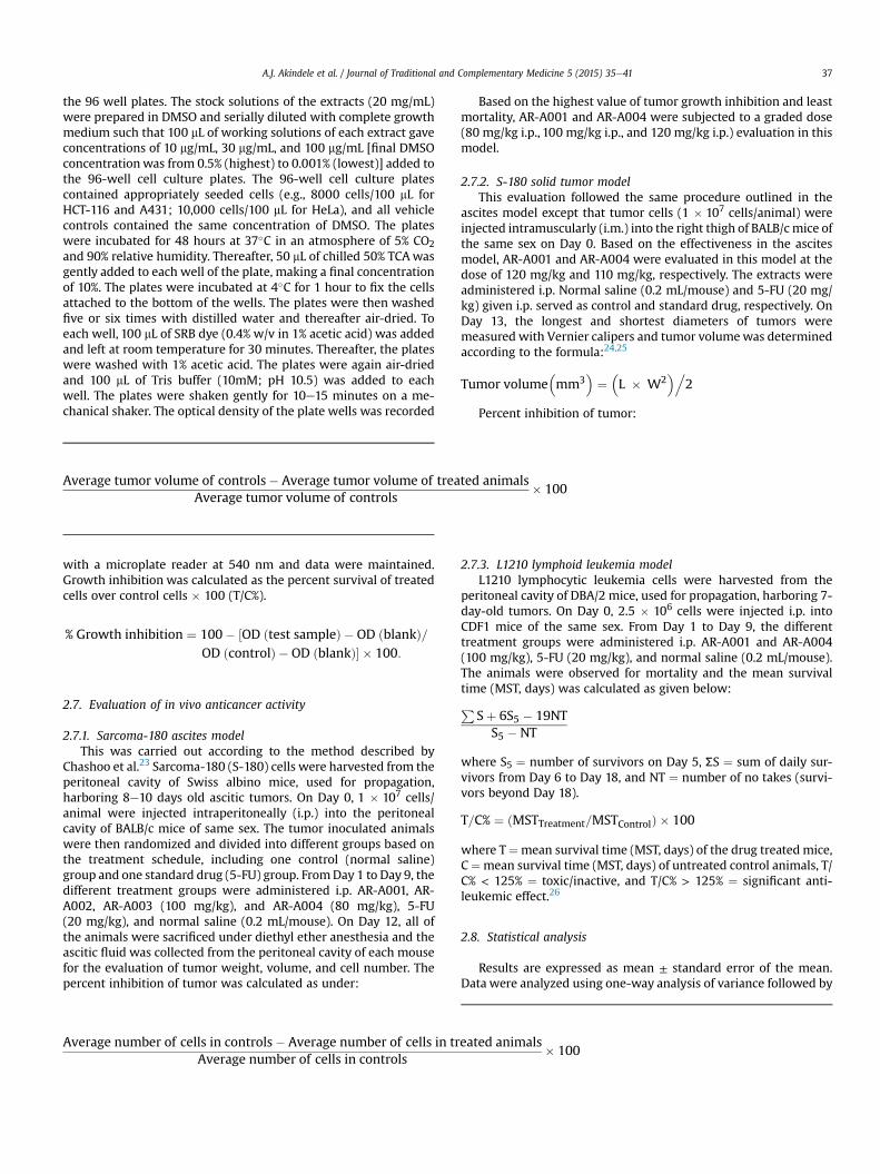
Original article Anticancer activity of Aristolochia ringens Vahl. (Aristolochiaceae) Abidemi James Akindele a, b, * , Zahoor Wani a , Girish Mahajan a , Sadhana Sharma a , Flora Ruth Aigbe b , Naresh Satti c , Olufunmilayo Olaide Adeyemi b , Dilip Manikrao Mondhe a, ** a Cancer Pharmacology Division, Indian Institute of Integrative Medicine, Council of Scientific and Industrial Research, Jammu, Jammu and Kashmir, India b Department of Pharmacology, Therapeutics and Toxicology, Faculty of Basic Medical Sciences, College of Medicine, University of Lagos, Lagos, Nigeria c Natural Products Chemistry Division, Indian Institute of Integrative Medicine, Council of Scientific and Industrial Research, Jammu, Jammu and Kashmir, India article info Article history: Received 28 December 2013 Received in revised form 21 May 2014 Accepted 22 May 2014 Available online 18 December 2014 Keywords: Anticancer activity Aristolochia ringens Cytotoxicity Lymphoid leukemia Solid tumor abstract Cancer is a leading cause of death worldwide and sustained focus is on the discovery and development of newer and better tolerated anticancer drugs especially from plants. The sulforhodamine B (SRB) in vitro cytotoxicity assay, sarcoma-180 (S-180) ascites and solid tumor, and L1210 lymphoid leukemia in vivo models were used to investigate the anticancer activity of root extracts of Aristolochia ringens Vahl. (Aristolochiaceae; 馬兜鈴 mǎ d ou líng). AR-A001 (IC 50 values of 20 mg/mL, 22 mg/mL, 3 mg/mL, and 24 mg/mL for A549, HCT-116, PC3, and THP-1 cell lines, respectively), and AR-A004 (IC 50 values of 26 mg/mL, 19.5 mg/mL, 12 mg/mL, 28 mg/mL, 30 mg/mL, and 22 mg/mL for A549, HCT-116, PC3, A431, HeLa, and THP-1, respectively), were observed to be significantly active in vitro. Potency was highest with AR-A001 and AR-A004 for PC3 with IC 50 values of 3 mg/mL and 12 mg/mL, respectively. AR-A001 and AR-A004 produced significant (p < 0.05e0.001) dose-dependent inhibition of tumor growth in the S-180 ascites model with peak effects produced at the highest dose of 120 mg/kg. Inhibition values were 79.51% and 89.98% for AR-A001 and AR-A004, respectively. In the S-180 solid tumor model, the inhibition of tumor growth was 29.45% and 50.50% for AR-A001 (120 mg/kg) and AR-A004 (110 mg/kg), respectively, compared to 50.18% for 5-fluorouracil (5-FU; 20 mg/kg). AR-A001 and AR-A004 were also significantly active in the leukemia model with 211.11% and 155.56% increase in mean survival time (MST) compared to a value of 211.11% for 5-FU. In conclusion, the ethanolic (AR-A001) and dichlor- omethane:methanol (AR-A004) root extracts of AR possess significant anticancer activities in vitro and in vivo. Copyright © 2015, Center for Food and Biomolecules, National Taiwan University. Production and hosting by Elsevier Taiwan LLC. All rights reserved. 1. Introduction Cancer is a deadly disease and about one in four people will get it in some form during their lifetime; at the present time, about one in five of all deaths are due to cancer. 1 Normal diploid human cells multiply for a finite number of generations and then enter a state of replicative senescence, but cancer cells can proliferate indefinitely. 2 About 12.7 million cancer cases and 7.6 million cancer deaths are estimated to have occurred in 2008; of these, 56% of the cases and 64% of the deaths occurred in the economically developing world. 3 Sur- gery is useful in removing visible tumors, but may leave smaller nests of cancer cells in the patient which continue to proliferate, while radiation therapy is relatively imprecise as it can kill both cancer cells and normal cells, and thus has toxic side effects which may them- selves be lethal to the patients. 2 Chemotherapy with antiproliferative agents, including alkylating agents, antimetabolites, antibiotics, and hormones, apart from being complementary to surgical intervention and radiotherapy, is essential in cases of metastasis. According to Sikora et al, 4 although 92 approved anticancer drugs are available for the treatment of > 200 different tumor en- tities, effective therapies for most of these tumors are lacking. Furthermore, out of the 92 registered drugs, 17 are considered by oncologists to be more broadly applicable and 12 additional agents are perceived as having certain advantages in some clinical set- tings. 5 Limitations in the application of chemotherapeutic agents include toxicity, manifestation of deleterious side-effects, and a * Corresponding author. Department of Pharmacology, Therapeutics and Toxi- cology, Faculty of Basic Medical Sciences, College of Medicine, University of Lagos, Idi-Araba Campus, Lagos P.M.B. 12003, Nigeria. ** Corresponding author. Cancer Pharmacology Division, Indian Institute of Inte- grative Medicine, Council of Scientific and Industrial Research, Jammu-Tawi 180001, Jammu and Kashmir, India. E-mail addresses: [email protected] (A.J. Akindele), [email protected] (D.M. Mondhe). Peer review under responsibility of The Center for Food and Biomolecules, National Taiwan University. HOSTED BY Contents lists available at ScienceDirect Journal of Traditional and Complementary Medicine journal homepage: http://www.elsevier.com/locate/jtcme http://dx.doi.org/10.1016/j.jtcme.2014.05.001 2225-4110/Copyright © 2015, Center for Food and Biomolecules, National Taiwan University. Production and hosting by Elsevier Taiwan LLC. All rights reserved. Journal of Traditional and Complementary Medicine 5 (2015) 35e41
-
Upload
independent -
Category
Documents
-
view
0 -
download
0
Transcript of Anticancer activity of Aristolochia ringens Vahl. (Aristolochiaceae)
ble at ScienceDirect
Journal of Traditional and Complementary Medicine 5 (2015) 35e41
Contents lists availa
HOSTED BYJournal of Traditional and Complementary Medicine
journal homepage: http: / /www.elsevier .com/locate/ j tcme
Original article
Anticancer activity of Aristolochia ringens Vahl. (Aristolochiaceae)
Abidemi James Akindele a, b, *, Zahoor Wani a, Girish Mahajan a, Sadhana Sharma a,Flora Ruth Aigbe b, Naresh Satti c, Olufunmilayo Olaide Adeyemi b,Dilip Manikrao Mondhe a, **
a Cancer Pharmacology Division, Indian Institute of Integrative Medicine, Council of Scientific and Industrial Research, Jammu, Jammu and Kashmir, Indiab Department of Pharmacology, Therapeutics and Toxicology, Faculty of Basic Medical Sciences, College of Medicine, University of Lagos, Lagos, Nigeriac Natural Products ChemistryDivision, Indian Institute of IntegrativeMedicine, Council of Scientific and Industrial Research, Jammu, JammuandKashmir, India
a r t i c l e i n f o
Article history:Received 28 December 2013Received in revised form21 May 2014Accepted 22 May 2014Available online 18 December 2014
Keywords:Anticancer activityAristolochia ringensCytotoxicityLymphoid leukemiaSolid tumor
* Corresponding author. Department of Pharmacocology, Faculty of Basic Medical Sciences, College of MIdi-Araba Campus, Lagos P.M.B. 12003, Nigeria.** Corresponding author. Cancer Pharmacology Divigrative Medicine, Council of Scientific and Industrial RJammu and Kashmir, India.
E-mail addresses: [email protected] (A.J. Aki(D.M. Mondhe).
Peer review under responsibility of The CenterNational Taiwan University.
http://dx.doi.org/10.1016/j.jtcme.2014.05.0012225-4110/Copyright © 2015, Center for Food and Bio
a b s t r a c t
Cancer is a leading cause of death worldwide and sustained focus is on the discovery and development ofnewer and better tolerated anticancer drugs especially from plants. The sulforhodamine B (SRB) in vitrocytotoxicity assay, sarcoma-180 (S-180) ascites and solid tumor, and L1210 lymphoid leukemia in vivomodelswere used to investigate the anticancer activity of root extracts of Aristolochia ringens Vahl. (Aristolochiaceae;馬兜鈴mǎ d�ou líng). AR-A001 (IC50 values of 20 mg/mL, 22 mg/mL, 3 mg/mL, and 24 mg/mL for A549, HCT-116,PC3, andTHP-1 cell lines, respectively), andAR-A004 (IC50 values of 26mg/mL,19.5 mg/mL,12 mg/mL, 28 mg/mL,30 mg/mL, and 22 mg/mL for A549, HCT-116, PC3, A431, HeLa, and THP-1, respectively), were observed to besignificantlyactive invitro. PotencywashighestwithAR-A001andAR-A004 forPC3with IC50values of 3mg/mLand 12 mg/mL, respectively. AR-A001 and AR-A004 produced significant (p < 0.05e0.001) dose-dependentinhibition of tumor growth in the S-180 ascites model with peak effects produced at the highest dose of120mg/kg. Inhibitionvalueswere79.51%and89.98% forAR-A001andAR-A004, respectively. In theS-180solidtumor model, the inhibition of tumor growth was 29.45% and 50.50% for AR-A001 (120mg/kg) and AR-A004(110 mg/kg), respectively, compared to 50.18% for 5-fluorouracil (5-FU; 20 mg/kg). AR-A001 and AR-A004were also significantly active in the leukemia model with 211.11% and 155.56% increase in mean survivaltime (MST) compared to a value of 211.11% for 5-FU. In conclusion, the ethanolic (AR-A001) and dichlor-omethane:methanol (AR-A004) root extracts of AR possess significant anticancer activities in vitro and in vivo.Copyright © 2015, Center for Food and Biomolecules, National Taiwan University. Production and hosting
by Elsevier Taiwan LLC. All rights reserved.
1. Introduction
Cancer is a deadly disease and about one in four people will get itin some form during their lifetime; at the present time, about one infive of all deaths are due to cancer.1 Normal diploid human cellsmultiply for a finite number of generations and then enter a state ofreplicative senescence, but cancer cells can proliferate indefinitely.2
About 12.7 million cancer cases and 7.6 million cancer deaths are
logy, Therapeutics and Toxi-edicine, University of Lagos,
sion, Indian Institute of Inte-esearch, Jammu-Tawi 180001,
ndele), [email protected]
for Food and Biomolecules,
molecules, National Taiwan Unive
estimated tohaveoccurred in2008; of these,56%of the cases and64%of the deaths occurred in the economically developing world.3 Sur-gery is useful in removing visible tumors, butmay leave smaller nestsof cancer cells in the patient which continue to proliferate, whileradiation therapy is relatively imprecise as it can kill both cancer cellsand normal cells, and thus has toxic side effects which may them-selves be lethal to the patients.2 Chemotherapywith antiproliferativeagents, including alkylating agents, antimetabolites, antibiotics, andhormones, apart from being complementary to surgical interventionand radiotherapy, is essential in cases of metastasis.
According to Sikora et al,4 although 92 approved anticancerdrugs are available for the treatment of > 200 different tumor en-tities, effective therapies for most of these tumors are lacking.Furthermore, out of the 92 registered drugs, 17 are considered byoncologists to be more broadly applicable and 12 additional agentsare perceived as having certain advantages in some clinical set-tings.5 Limitations in the application of chemotherapeutic agentsinclude toxicity, manifestation of deleterious side-effects, and a
rsity. Production and hosting by Elsevier Taiwan LLC. All rights reserved.

A.J. Akindele et al. / Journal of Traditional and Complementary Medicine 5 (2015) 35e4136
narrow margin of error. These days, renewed and concerted effortsare geared towards the discovery and development of newer andbetter tolerated anticancer drugs, especially from natural products,mainly plants. New targets for anticancer agent development arerapidly emerging in the post-genome era, and improvements inprotein structure determination, combinatorial chemistry, andhigh-throughput small-molecule screens may accelerate the gen-eration of new agents to be studied in the clinic.6
Aristolochia ringens Vahl. (Aristolochiaceae (AR); 馬兜鈴 mǎ d�oulíng) is a glabrous bushy climber native of tropical America, intro-duced to most West African countries as a garden ornamental, andhas become naturalized in roadside bush in Sierra Leone, Ghana,Nigeria,7 and DR Congo.8 The plant is commonly called “Dutchman'spipe”and “Snakework”but localnames inNigeria include “Ako-igun”(Yoruba, Southwest Nigeria) and “Dumandutsee” (Hausa, NorthernNigeria). Preparations of the leaves, roots, andwhole plant have beenreported to be used traditionally in Nigeria for the treatment ofdiverse ailments including guinea worm, skin diseases, typhoid,sores, as an antidote to snake poison, an emmenagogue, and ananthelmintic remedy.9 In South America, the plant is used for thetreatmentof snakebites, fever, ulcers, and colic,10while the root of theplant is used in Senegal as an antidote for snakebites.11 Sonibare andGbile12 stated that the root of the plant is used in Southwest Nigeriafor the treatmentof asthma,while Soladoye et al13 reported its use forthe treatment of hemorrhoids. The decoction/infusion of the root ofthe plant is also used as an antidiabetic.14 Antiinflammatory15 andantitrypanosomal16 activities of the plant have also been reported.
Based on the fact that some species of the genus Aristolochiahave been reported to possess anticancer activity,17e19 this studywas designed to investigate the anticancer activity of root extractsof AR using in vitro and in vivo methods.
2. Materials and methods
2.1. Plant material
AR roots were obtained from a local herbal market in Mushin,Lagos State, Nigeria. The plant material was identified andauthenticated by Mr. T.K. Odewo of the Department of Botany andMicrobiology, University of Lagos, Lagos, Nigeria, where a herbari-um specimen was deposited with voucher number LUH 4061.
2.2. Extraction
Fresh roots of AR were cut into pieces and air-dried until a con-stant weight was attained. The dried materials were milled anddivided into four portions of 100 g each. Three portions wereseparately macerated in about 1500mL of alcohol (95% ethanol: AR-A001), hydro-alcohol (ethanol and water, 1:1; AR-A002), anddistilled water (AR-A003). Maceration was sustained for 3 hourswith mechanical stirring (Heidolph RZR 2051 Control; HeidolphInstruments GmbH & Co. KG, Schwabach, Germany) at 400 � g.Filtration was thereafter carried out using Whatman filter paper(150 mm). Residues were remacerated (� 2) to achieve exhaustiveextraction. The combined filtrate of the alcoholic (AR-A001) andhydroalcoholic (AR-A002) extracts were concentrated using a Hei-dolph Rotavapor (LABORATA 4000) at 120 � g and 40�C. The com-bined filtrate of the aqueous extract was lyophilized (AR-A003). Thefourth portion of 100 g of the powdered plant material was put into2000mL separating funnels with the bottom lined with cottonwooland 1 L of dichloromethane:methanol (DCM:MeOH; 70:30) was putinto the separating funnel. Twenty-four hours later, the solventmixture was drained and the extraction liquid was filtered usingWhatman filter paper (150mm). On the 2nd day and 3rd day, 600 mLof the DCM:MeOH solvent mixture was added to the separating
funnel, drained 24 hours after, and filtered. The combinedDCM:MeOH extract was concentrated with a Rotavapor withoutvacuum (AR-A004). All dried extracts were weighed and stored in adesiccator. The yields of the extracts were 4.41% (AR-A001), 16.00%(AR-A002), 11.88% (AR-A003), and 5.93% (AR-A004).
2.3. Chemicals
The chemicals used in this study include RPMI-1640, minimumessential medium, fetal calf serum, trypsin, trypan blue, ethanol,penicillin, streptomycin, gentamycin, dimethyl sulfoxide (DMSO),sulforhodamine B (SRB), mitomycin-C, paclitaxel, 5-fluorouracil (5-FU; Sigma Chemical Co., St Louis, MO, USA), phosphate buffer saline(Merck, Darmstadt, Germany); trichloroacetic acid (TCA), distilledwater, sodium hydroxide, Tris-EDTA buffer, Tris buffer (Hi-Media,Mumbai, India), acetic acid, sodium bicarbonate, hydrochloric acid(Rankem, New Delhi, India), isopropanol (Sisco, Mumbai, India),and Tris-acetate-EDTA buffer. All other chemicals used in this studywere purchased locally and were of analytical grade.
2.4. Cell lines and cell cultures
The human cancer cell lines used in this study, including A549(lung), HCT-116 (colon), PC3 (prostate), A431 (skin), HeLa (cervix),and THP-1 (leukemia) were obtained from the National Center forCell Science, Pune, India, or the National Cancer Institute, Frederick,MD, USA. The cells were grown and maintained in appropriatemedium, pH 7.4, supplemented with 10% fetal calf serum, glutamine(2mM), penicillin (100 units/mL), and streptomycin (100 mg/mL).The cell cultures were grown in a carbon dioxide incubator (Her-aeus, GmbH, Germany) at 37�C with 90% humidity and 5% CO2.20,21
2.5. Animals
The animals used in this study, including inbred BALB/c and DBA/2, outbred Swiss albino, and F1 hybrid CDF1 mice, were obtainedfrom the animal facility of the Indian Institute of Integrative Medi-cine, Jammu, India and maintained at 22 ± 2�C with 20e25 com-plete air changes with 100% fresh air. Relative humidity wasmaintained at 50e60%. Animals were fed with commercially avail-able pelleted feed (M/s Ashirwad Industries, Chandigarh, India) andwere provided autoclaved water ad libitum. Animals were housed intransparent polycarbonate filter top cages in animal isolator cabins.The animals selected for the experiments were in the weight rangeof 18e23 g (about 2months of age) andwere observed to be healthyand free from any disease. All of the animals selected for individualexperiments were of the same sex and strain. The protocols adoptedin this study were approved by the Institutional Animal EthicsCommittee, Indian Institute of Integrative Medicine, Jammu, India.
2.6. In vitro cytotoxicity against human cancer cell lines
The in vitro cytotoxicity of the extracts of ARwas determined bya semiautomated assay using SRB.20e22 The human cancer cell lineswere grown in tissue culture flasks at 37�C in an atmosphere of 5%CO2 and 90% relative humidity in complete growth medium. Flaskswith subconfluent stage of growth were selected and cells wereharvested by treatment with trypsin-EDTA. The numbers of cells/mL of suspension were counted using hemocytometer. The celldensity was adjusted to 10,000 cells/100 mL, or as appropriate foreach cell line, in the cell suspension. A volume of 100 mL of cellsuspension was added to each well of 96 well plates with the helpof handy-step. The plates were incubated at 37�C in an atmosphereof 5% CO2 and 90% relative humidity for 24 hours. Thereafter, 100 mLof aworking solution of each test material was added to thewells of
A.J. Akindele et al. / Journal of Traditional and Complementary Medicine 5 (2015) 35e41 37
the 96 well plates. The stock solutions of the extracts (20 mg/mL)were prepared in DMSO and serially diluted with complete growthmedium such that 100 mL of working solutions of each extract gaveconcentrations of 10 mg/mL, 30 mg/mL, and 100 mg/mL [final DMSOconcentrationwas from 0.5% (highest) to 0.001% (lowest)] added tothe 96-well cell culture plates. The 96-well cell culture platescontained appropriately seeded cells (e.g., 8000 cells/100 mL forHCT-116 and A431; 10,000 cells/100 mL for HeLa), and all vehiclecontrols contained the same concentration of DMSO. The plateswere incubated for 48 hours at 37�C in an atmosphere of 5% CO2and 90% relative humidity. Thereafter, 50 mL of chilled 50% TCAwasgently added to each well of the plate, making a final concentrationof 10%. The plates were incubated at 4�C for 1 hour to fix the cellsattached to the bottom of the wells. The plates were then washedfive or six times with distilled water and thereafter air-dried. Toeach well, 100 mL of SRB dye (0.4% w/v in 1% acetic acid) was addedand left at room temperature for 30 minutes. Thereafter, the plateswere washed with 1% acetic acid. The plates were again air-driedand 100 mL of Tris buffer (10mM; pH 10.5) was added to eachwell. The plates were shaken gently for 10e15 minutes on a me-chanical shaker. The optical density of the plate wells was recorded
Average tumor volume of controls� Average tumor volume of treated animalsAverage tumor volume of controls
� 100
with a microplate reader at 540 nm and data were maintained.Growth inhibition was calculated as the percent survival of treatedcells over control cells � 100 (T/C%).
% Growth inhibition ¼ 100� ½OD ðtest sampleÞ � OD ðblankÞ=OD ðcontrolÞ � OD ðblankÞ� � 100:
2.7. Evaluation of in vivo anticancer activity
2.7.1. Sarcoma-180 ascites modelThis was carried out according to the method described by
Chashoo et al.23 Sarcoma-180 (S-180) cells were harvested from theperitoneal cavity of Swiss albino mice, used for propagation,harboring 8e10 days old ascitic tumors. On Day 0, 1 � 107 cells/animal were injected intraperitoneally (i.p.) into the peritonealcavity of BALB/c mice of same sex. The tumor inoculated animalswere then randomized and divided into different groups based onthe treatment schedule, including one control (normal saline)group and one standard drug (5-FU) group. FromDay 1 to Day 9, thedifferent treatment groups were administered i.p. AR-A001, AR-A002, AR-A003 (100 mg/kg), and AR-A004 (80 mg/kg), 5-FU(20 mg/kg), and normal saline (0.2 mL/mouse). On Day 12, all ofthe animals were sacrificed under diethyl ether anesthesia and theascitic fluid was collected from the peritoneal cavity of each mousefor the evaluation of tumor weight, volume, and cell number. Thepercent inhibition of tumor was calculated as under:
Average number of cells in controls� Average number of cells in trAverage number of cells in controls
Based on the highest value of tumor growth inhibition and leastmortality, AR-A001 and AR-A004 were subjected to a graded dose(80 mg/kg i.p., 100 mg/kg i.p., and 120 mg/kg i.p.) evaluation in thismodel.
2.7.2. S-180 solid tumor modelThis evaluation followed the same procedure outlined in the
ascites model except that tumor cells (1 � 107 cells/animal) wereinjected intramuscularly (i.m.) into the right thigh of BALB/c mice ofthe same sex on Day 0. Based on the effectiveness in the ascitesmodel, AR-A001 and AR-A004 were evaluated in this model at thedose of 120 mg/kg and 110 mg/kg, respectively. The extracts wereadministered i.p. Normal saline (0.2 mL/mouse) and 5-FU (20 mg/kg) given i.p. served as control and standard drug, respectively. OnDay 13, the longest and shortest diameters of tumors weremeasured with Vernier calipers and tumor volumewas determinedaccording to the formula:24,25
Tumor volume�mm3
�¼
�L � W2
�.2
Percent inhibition of tumor:
2.7.3. L1210 lymphoid leukemia modelL1210 lymphocytic leukemia cells were harvested from the
peritoneal cavity of DBA/2 mice, used for propagation, harboring 7-day-old tumors. On Day 0, 2.5 � 106 cells were injected i.p. intoCDF1 mice of the same sex. From Day 1 to Day 9, the differenttreatment groups were administered i.p. AR-A001 and AR-A004(100 mg/kg), 5-FU (20 mg/kg), and normal saline (0.2 mL/mouse).The animals were observed for mortality and the mean survivaltime (MST, days) was calculated as given below:P
Sþ 6S5 � 19NTS5 �NT
where S5 ¼ number of survivors on Day 5, ƩS ¼ sum of daily sur-vivors from Day 6 to Day 18, and NT ¼ number of no takes (survi-vors beyond Day 18).
T=C% ¼ ðMSTTreatment=MSTControlÞ � 100
where T¼mean survival time (MST, days) of the drug treated mice,C¼mean survival time (MST, days) of untreated control animals, T/C% < 125% ¼ toxic/inactive, and T/C% > 125% ¼ significant anti-leukemic effect.26
2.8. Statistical analysis
Results are expressed as mean ± standard error of the mean.Data were analyzed using one-way analysis of variance followed by
eated animals� 100

Fig. 3. In vitro cytotoxic activity of AR-A003 against various human cancer cells lines inthe sulforhodamine B assay. Estimated IC50 values are NA, NA, >100 mg/ml, >100 mg/ml,>100 mg/ml, and 93 mg/ml for A549, HCT-116, PC3, A431, HeLa, and THP-1, respectively.Samples were assayed in quadruplicate. Values are mean ± standard deviation.NA ¼ not active.
Fig. 1. In vitro cytotoxic activity of AR-A001 against various human cancer cells lines inthe sulforhodamine B assay. Estimated IC50 values are 20 mg/ml, 22 mg/ml, 3 mg/ml,>100 mg/ml, >100 mg/ml, and 24 mg/ml for A549, HCT-116, PC3, A431, HeLa, and THP-1,respectively. Samples were assayed in quadruplicate. Values are mean ± standarddeviation.
A.J. Akindele et al. / Journal of Traditional and Complementary Medicine 5 (2015) 35e4138
Dunnett's multiple comparison test using GraphPad Prism 5(GraphPad Software Inc., CA, USA). Values were considered signif-icant at p < 0.05.
3. Results
3.1. In vitro cytotoxic activity
Figs. 1e4 show the effects of AR-A001, AR-A002, AR-A003, andAR-A004, respectively, against the human cancer cell lines used inthis study. AR-A001 produced significant cytotoxic activity againstA549, HCT-116, PC3, and THP-1 human cancer cell lines with IC50values of 20 mg/mL, 22 mg/mL, 3 mg/mL, and 24 mg/mL, respectively.AR-A002 and AR-A003 did not elicit significant activity against thehuman cancer cell lines used in this study, with IC50 valuesgenerally > 100 mg/mL. AR-A004 showed significant and consistentcytotoxic activity against all the human cancer cell lines in thisstudy, with IC50 values of 26 mg/mL,19.5 mg/mL,12 mg/mL, 28 mg/mL,30 mg/mL, and 22 mg/mL for A549, HCT-116, PC3, A431, HeLa, andTHP-1, respectively.
3.2. In vivo anticancer activity of AR extracts against S-180 ascitesin BALB/c mice
Initial screening results for the effects of AR extracts on the S-180ascites model in mice are shown in Table 1. AR-A001 (100 mg/kg)and AR-A004 (80 mg/kg) elicited 73.56% and 72.16% inhibition intumor growthwith nomortality recorded. These effectswere highlysignificant (p < 0.001) in respect of the number of tumor cells pre-sent in the peritoneal cavity of treated mice as compared to controlmice, but significantly lower (p < 0.05, 0.01) as compared to 5-FU
Fig. 2. In vitro cytotoxic activity of AR-A002 against various human cancer cells lines inthe sulforhodamine B assay. Estimated IC50 values are >100 mg/ml, >100 mg/ml,>100 mg/ml, 67 mg/ml, 66 mg/ml, and >100 mg/ml for A549, HCT-116, PC3, A431, HeLa,and THP-1, respectively. Samples were assayed in quadruplicate. Values aremean ± standard deviation.
(97.84% inhibition). AR-A002 and AR-A003 (100 mg/kg) elicited80.66% and 77.24% tumor growth inhibition with mortalities of57.14% and 71.43%, respectively. As shown in Table 2, the effects ofAR-A001 and AR-A004 were dose-dependent with peak tumor cellgrowth inhibitory effects elicited at a dose of 120mg/kg (79.51% and89.98% inhibition, respectively). However, mortality was not recor-ded at this dose for AR-A001,while it was 60% in respect of AR-A004.
3.3. In vivo anticancer activity of AR-A001 and AR-A004 extractsagainst S-180 solid tumor in BALB/c mice
The effects of active extracts against S-180 solid tumor are showninTable 3. AR-A001 (120mg/kg) andAR-A004 (110mg/kg) produceda significant reduction (p < 0.05) in the body weight of mice andsignificant inhibition in tumor growth on Day 9 and Day 13 ascompared to control animals. The inhibition in solid tumor growthproduced by AR-A001 was reduced from 31.81% on Day 9 to 29.45%on Day 13. The effect of AR-A001 on tumor volume on Day 9 wassignificantly lower (p < 0.05) than that produced by 5-FU (63.39%)while the effect of the extract on Day 13 was comparable and notsignificantly different (p > 0.05) from that elicited by 5-FU (50.18%).Tumor growth inhibition produced by AR-A004 reduced from55.74% on Day 9 to 50.50% on Day 13, with the later effect beingcomparable andnot significantlydifferent (P>0.05) relative to 5-FU.
3.4. Effects of AR-A001 and AR-A004 extracts against L1210lymphoid leukemia in CDF1 mice
The effects of AR-A001 and AR-A004 (100 mg/kg) on L1210lymphoid leukemiamodel are shown in Table 4. AR-A001 increasedthe MST from 9 days in control to 19 days, corresponding to a
Fig. 4. In vitro cytotoxic activity of AR-A004 against various human cancer cells lines inthe sulforhodamine B assay. Estimated IC50 values are 26 mg/ml, 19.5 mg/ml, 12 mg/ml,28 mg/ml, 30 mg/ml and 22 mg/ml for A549, HCT-116, PC3, A431, HeLa, and THP-1,respectively. Samples were assayed in quadruplicate. Values are mean ± standarddeviation.

Table 1Effects of Aristolochia ringens (AR) extracts against sarcoma-180 (ascites) in BALB/c mice.
Treatments Dose (mg/kg) Body weight (g) Day 12 Tumor growthinhibition (%)
Mortality(%)
Day 1 Day 5 Day 9 Body weight (g) Weight oftumor (g)
Volume of asciticfluid (mL)
Number of tumorcells (� 107)
Normal saline (0.2 mL/mouse) 22.91 ± 0.74 24.82 ± 0.86 28.64 ± 1.19 29.36 ± 1.44 9.73 ± 1.00 9.88 ± 0.96 277.86 ± 37.20 0 05-FU 20 21.14 ± 0.55* 20.86 ± 0.63*** 17.71 ± 0.84*** 16.60 ± 0.75*** 0.50 ± 0.21*** 0.52 ± 0.22*** 6.00 ± 2.45*** 97.84 0AR-A001 100 20.43 ± 0.48** 20.86 ± 0.55*** 21.43 ± 0.95***,## 21.43 ± 1.00***,## 5.86 ± 1.37*,## 6.01 ± 1.35*,## 73.47 ± 15.71***,## 73.56 0AR-A002 100 20.57 ± 0.37** 18.43 ± 0.20***,# 16.83 ± 0.48*** 16.67 ± 1.33*** 1.85 ± 0.98*** 2.00 ± 1.04*** 53.75 ± 27.17*** 80.66 57.14AR-A003 100 20.71 ± 0.42* 17.71 ± 0.42***,### 16.50 ± 0.22*** 15.00 ± 3.00* 2.71 ± 2.71 2.75 ± 2.75 63.25 ± 63.25 77.24 71.43AR-A004 80 21.00 ± 0.62* 20.14 ± 0.55c 18.57 ± 0.65*** 17.29 ± 1.32*** 2.56 ± 0.71***,# 2.73 ± 0.74***,# 77.34 ± 26.44***,# 72.16 0
Data are presented as mean ± standard error of the mean (n ¼ 7, n ¼ 11 for control).*p < 0.05, **p < 0.01, ***p < 0.001 versus normal saline.#p < 0.05, ##p < 0.01, ###p < 0.001 versus 5-FU (one-way analysis of variance followed by Dunnett's multiple comparison test).5-FU ¼ 5-fluorouracil.
Table 2Effects of AR-A001 (80 mg/kg, 120 mg/kg) and AR-A004 extracts (100 mg/kg, 120 mg/kg) against sarcoma-180 (ascites) in BALB/c mice.
Treatments Dose (mg/kg) Body weight (g) Day 12 Tumor growthinhibition (%)
Mortality(%)
Body weight (g) Weight oftumor (g)
Volume of asciticfluid (mL)
Number of tumorcells (� 107)
Day 1 Day 5 Day 9
Normal saline (0.2 mL/mouse) 21.40 ± 0.75 23.80 ± 0.86 27.20 ± 1.24 27.60 ± 1.12 9.81 ± 1.01 9.96 ± 0.77 474.25 ± 45.26 0 05-FU 20 21.20 ± 0.58 22.60 ± 0.75 21.40 ± 1.08** 20.60 ± 1.44** 0.50 ± 0.22*** 0.48 ± 0.22*** 10.00 ± 4.18*** 97.89 0AR-A001 80 20.80 ± 0.66 21.80 ± 1.07 22.60 ± 1.69* 23.00 ± 1.70* 4.91 ± 1.06**,## 5.10 ± 1.07**,## 138.55 ± 30.42***,## 70.79 0
120 20.80 ± 0.66 21.20 ± 0.80* 21.20 ± 1.07** 20.80 ± 1.93* 3.71 ± 1.08**,# 3.70 ± 1.12**,# 97.18 ± 31.07***,# 79.51 0AR-A004 100 20.80 ± 0.66 20.20 ± 1.07* 18.60 ± 1.03***,# 18.60 ± 1.12*** 2.35 ± 0.92*** 2.52 ± 0.99*** 55.14 ± 22.88*** 88.37 0
120 20.80 ± 0.66 20.80 ± 0.97* 19.60 ± 1.50** 22.50 ± 2.50 2.36 ± 2.36 2.50 ± 2.50 47.50 ± 47.50** 89.98 60
Data are presented as mean ± standard error of the mean (n ¼ 5).*p < 0.05, **p < 0.01, ***p < 0.001 versus normal saline.#p < 0.05, ##p < 0.01 versus 5-FU (one-way analysis of variance followed by Dunnett’s multiple comparison test).5-FU ¼ 5-fluorouracil.
A.J.A
kindeleet
al./Journal
ofTraditional
andCom
plementary
Medicine
5(2015)
35e41
39

Table 3Effects of AR-A001 and AR-A004 extract against sarcoma-180 (solid) tumor in BALB/c mice.
Treatments Dose (mg/kg) Body weight (g) Day 9 Day 13
Day 1 Day 5 Day 9 Day 13 Tumor volume (mm3) Inhibition(%)
Tumor volume(mm3)
Inhibition(%)
Normalsaline
(0.2 mL/mouse) 21.80 ± 0.58 23.00 ± 0.55 23.80 ± 0.58 24.00 ± 0.32 1848.50 ± 108.31 0.00 2283.50 ± 136.58 0.00
5-FU 20 21.80 ± 0.58 20.60 ± 1.17 18.80 ± 2.08* 19.80 ± 2.15 676.80 ± 188.15*** 63.39 1137.60 ± 247.45** 50.18AR-A001 120 20.80 ± 0.86 21.20 ± 1.02 20.60 ± 1.21* 20.80 ± 1.02* 1260.40 ± 237.90*,# 31.81 1611.00 ± 290.72* 29.45AR-A004 110 20.80 ± 0.86 20.20 ± 1.50 19.80 ± 1.39* 19.60 ± 1.21** 818.10 ± 124.32*** 55.74 1130.40 ± 137.88*** 50.50
Values are mean ± standard error of the mean (n ¼ 5).*p < 0.05, **p < 0.01, ***p < 0.001 versus normal saline.#p < 0.05 versus 5-FU (one-way analysis of variance followed by Dunnett’s multiple comparison test).5-FU ¼ 5-fluorouracil.
A.J. Akindele et al. / Journal of Traditional and Complementary Medicine 5 (2015) 35e4140
211.11% increase in MST. This value was the same as for 5-FU. Inrespect of AR-A004, MST was 14 days compared to 9 days forcontrol, corresponding to 155.56% increase in MST.
4. Discussion
Cancer is the leading cause of death in economically developedcountries and the second leading cause of death in developingcountries.27 The burden of cancer is increasing in economicallydeveloping countries as a result of population aging and growth,and adoption of cancer-associated lifestyle choices includingsmoking, physical inactivity and “westernized” diets.3 Schwarts-mann et al6 stated that the total number of new cases of cancer willrise from 10 million in year 2000 by approximately 25% in eachdecade, reaching 24 million new cases/year in the year 2050. Thetotal number of deaths will rise from 6 million in the year 2000 to10 million in 2020, and to > 16 million in the year 2050.28,29
Furthermore, it was reported that in the year 2050, there will be17 million new cases of cancer in less developed countries, whileonly 7million new cases of cancer will occur in themore developedcountries, with the assertion that public health authorities shouldexpect cancer to become a major challenge not only in developedcountries but especially in developing countries as well.29e31
The major challenges associated with currently available anti-cancer agents include selectivity, toxicity, resistance, and develop-ment of a secondary malignancy. These drawbacks have motivatedthe search for newer, more efficacious, and better tolerated anti-tumor drugs, with natural products, especially plants, offering aninexhaustible reservoir for new drug discovery and development.
In this study, the DCM:MeOH extract of the root of AR (AR-A004)was significantly active against PC3, HCT-116, THP-1, A549, A431,and HeLa human cancer cell lines with an IC50 range of 12e30 mg/mL in the SRB cytotoxicity assay. Based on the recommendation ofthe National Cancer Institute (NCI, USA), 30 mg/mL is the upper IC50limit considered promising for purification of a crude extract.32 Theethanol extract (AR-A001) was also found to be significantly active
Table 4Effects of AR-A001 and AR-A004 extracts against L1210 lymphoid leukemia in CDF1mice.
Treatments Dose (mg/kg) Mean survivaltime (days)
% Increase inmean survivaltime
Inference
Normal saline (0.2 mL/mouse) 9 e e
5-FU 20 19 211.11 Significantactivity
AR-A001 100 19 211.11 Significantactivity
AR-A004 100 14 155.56 Significantactivity
5-FU ¼ 5-fluorouracil.T/C% < 125% ¼ toxic/inactive; T/C% > 125% ¼ significant antileukemic effect.
against PC3, A549, HCT-116, and THP-1 cell lines, with an IC50 rangeof 3e24 mg/mL. AR-A004 but not AR-A001 was found to be active inthe S-180 solid tumor model (50.50% tumor growth inhibition at adose of 110 mg/kg relative to a value of 50.18% for 5-FU at 20 mg/kg). The initial activity against S-180 ascites was statistically sig-nificant (72.16%, 88.37%, and 89.98% tumor growth inhibition atdoses of 80 mg/kg, 100 mg/kg, and 120 mg/kg as compared to97.89% for 5-FU at 20 mg/kg). The dose of AR-A004 used in the solidtumor model was 110 mg/kg, because although the highest tumorgrowth inhibitory activity was observed at a dose of 120 mg/kg,mortality was 60% at this dose. AR-A001 also produced dose-dependent antitumor activity in the S-180 ascites model, with tu-mor growth inhibition values of 73.56%, 70.79%, and 79.51% atdoses of 80 mg/kg, 100 mg/kg, and 120 mg/kg, respectively. How-ever, the tumor growth inhibition value was just 29.45% in the S-180 solid tumor model for AR-A001. Both AR-A001 and AR-A004(100 mg/kg) were also significantly active in the L1210 lymphoidleukemia model (T/C 211.11 and 155.56%, respectively, relative to211.11% for 5-FU), with AR-A001 showing greater activity, compa-rable to 5-FU, in this model. This is in view of the interpretation byTodorova et al26 that T/C% value < 125% indicates toxicity/inactivitywhile a value > 125% depicts a significant antileukemic effect. Theresults obtained in this study established for the first time thein vitro and in vivo anticancer activity of AR with the DCM:MeOHand ethanolic root extracts showing significant activity in the solidtumor and leukemia models used in this study.
In this study, AR-A002 and AR-A003 did not elicit significantactivity against the human cancer cell lines used, with IC50 valuesgenerally > 100 mg/mL. However, these extracts elicited significanttumor growth inhibition in the S-180 ascites model, althoughmortality was significant. It should be noted that inactivity in vitrodoes not automatically translate to inactivity in vivo, because insome cases, inactive phytochemical principles may need metabolicactivation which is not possible under the in vitro environment.
Other studies have reported the anticancer activity of the DCMand MeOH,17 chloroform,33,34 and aqueous19 extracts of otherspecies of the Aristolochia family. The anticancer activity of variousspecies of Aristolochia has been reported but not of AR. Voloudakis-Baltatzis et al35 evaluated the anticancer activity of the Greek herbsubstances IBV-BK (Aristolochia, Iska) on human primary culturesderived from malignant and nonmalignant breast tissues in vitroand in vivo in rats with Walker carcinoma. Based on findings in thestudy, the authors concluded that IBK-BK, with Aristolochia as oneof the constituents, possesses potent anticancer activity in vitro andin vivo. Mongelli et al17 investigated the cytotoxic and DNA inter-action of extracts of Argentinean medicinal plants including Aris-tolochia triangularis. Aristolochia triangularis, along with otherplants, was established to contain cytotoxic compounds against KBcells based on inhibition of growth of crown gall tumors. The assayemployed showed good correlation with the 3PS (in vivo murine

A.J. Akindele et al. / Journal of Traditional and Complementary Medicine 5 (2015) 35e41 41
leukemia) antitumor assay.36 Masud Rana and Khanam33 investi-gated the antitumor effect of the whole plant extract of Aristolochiaindica against Ehrlich ascites carcinoma in mice. The crude chlo-roform extract resulted in significant tumor cell growth inhibitionand increased the MST. The root extract of Aristolochia indica hasbeen reported to inhibit the growth of human CA-755 and HeLacells and to be effective against adenocarcinoma.37 Chaouki et al34
investigated the antiproliferative effect of four different polarityextracts of Aristolochia baetica on the human breast cancer cell lineMCF-7 and reported the chloroform extract to be the most active.Benarba et al19 investigated the cytotoxic and apoptogenic activ-ities of the aqueous extract of Aristolochia longa in Burkitt's lym-phoma BL41 cells by flow cytometry. Based on the significantefficacy demonstrated in the study, the aqueous extract of Aristo-lochia longa was reported to be a promising source of novel treat-ment of Burkitt's and other lymphomas. The establishment ofanticancer activity of AR in this study is in agreement with thereport of antiproliferative effects for other species of the genus.
5. Conclusion
In conclusion, the findings in this study suggest that the etha-nolic (AR-A001) and DCM:MeOH (AR-A004) root extracts of ARpossess significant anticancer activities in vitro and in vivo. AR-A001was significantly active in the S-180 ascites and L1210 lymphoidleukemia in vivo models. AR-A004 was additionally significantlyactive in the S-180 solid tumor model. Further extensive studies arerequired to afford the identification, isolation, and characterizationof specific phytomolecules responsible for bioactivities observed inthis study and their inherent mechanism(s) of action.
Conflicts of interest
We declare that there are no conflicts of interest, financial orotherwise, pertaining to this study.
Acknowledgments
The authors are profoundly grateful to the Federation of IndianChambers of Commerce and Industry (FICCI) and the Department ofScience and Technology (DST), Ministry of Science and Technology,Government of India, for the CV Raman Postdoctoral Fellowshipaward (June 5, 2010eDecember 1, 2012) given to Dr. Abidemi JamesAkindele. Dr. RamA. Vishwakarma, Director IIIM (Indian Institute ofIntegrative Medicine, Jammu, India), is also very much appreciatedfor accepting to host the first author for the postdoctoral research.The immense support and guidance of Dr. A.K. Saxena, Chief Sci-entist and Head, Cancer Pharmacology Division, IIIM, is alsoappreciated.
References
1. Chhajed M, Sowle A, Baraskar R, Parihar N. Buserelin as a wonder drug forprostate cancer: a comprehensive study. Int J Pharm Res Sci. 2012;1:150e172.
2. Rao MRP, Adagale UR, Shetty A, Namjoshi P, Gaitonde P, Jain P. Cancer Immu-notherapy; 2007. Accessed on 20th July, 2013, http://www.pharmainfo.net/reviews/cancer-immunotherapy.
3. Jemal A, Bray F, Center MM, Ferlay J, Ward E, Forman D. Global cancer statistics.CA Cancer J Clin. 2011;61:69e90.
4. Sikora K, Advani S, Koroltchouk V, et al. Essential drugs for cancer therapy: aWorld Health Organization consultation. Ann Oncol. 1999;10:385e390.
5. Aherne W, Garret M, McDonald T, Workman P. Mechanism-based high-throughput screening for novel anticancer drug discovery. In: Baguley BC,Kerr DJ, eds. Anticancer Drug Development. London, UK; San Diego, CA: Aca-demic Press; 2002:249e267.
6. Schwartsmann G, Ratain MJ, Cragg GM, Wong JE, Saijo N, Parkinson DR, et al.Anticancer drug discovery and development throughout the World. J ClinOncol. 2002;20:47se59s.
7. Burkill HM. The Useful Plants of West Tropical Africa. Vol 1. Kew: Royal BotanicGardens; 1985.
8. De Groot H, Wanke S, Neinhuis C. Revision of the genus Aristolochia (Aristo-lochiaceae) in Africa, Madagascar and adjacent islands. Bot J Linn Soc. 2006;151:219e238.
9. Anonymous. Nigerian medicinal plants database. 2013. www.medicinalplantsinnigeria.com/Plantsdatabase.pdf. Accessed on 3rd January.
10. Van Wyk B, Wink M.Medicinal Plants of the World: An Illustrated Scientific Guideto the Important Medicinal Plants and Their Uses. Portland, OR, USA: TimberPress; 2004.
11. Neuwinger HD. African Traditional Medicine. A Dictionary of Plant Use andApplication. Stuttgart, Germany: Medpharm Scientific Publishers; 2000.
12. Sonibare MA, Gbile ZO. Ethnobotanical survey of anti-asthmatic plants in SouthWestern Nigeria. Afr J Tradit Complement Altern Med. 2008;5:340e345.
13. Soladoye MO, Adetayo MO, Chukwuma EC, Adetunji AN. Ethnobotanical surveyof plants used in the treatment of haemorrhoids in South-Western Nigeria.J Adv Dev Res. 2011;2:100e111.
14. Olabanji SO, Omobuwajo OR, Ceccato D, Adebajo AC, Buoso MC, Moschini G.Accelerator-based analytical technique in the study of some anti-diabeticmedicinal plants of Nigeria. Nucl Instrum Methods Phys Res B. 2008;266:2387e2390.
15. Aigbe FR, Adeyemi OO. The aqueous root extract of Aristolochia ringens (Vahl.)prevents chemically induced inflammation. Planta Med. 2011;77:PF28. http://dx.doi.org/10.1055/s-0031-1282416.
16. Osho IB, Lajide L. Prescreening evaluation of some plant extracts used in ethno-veterinary practices as antitrypanosomal agents. J Med Plants Res. 2012;6:2056e2060.
17. Mongelli E, Pampuro S, Coussio J, Salomon H, Ciccia G. Cytotoxic and DNAinteraction activities of extracts from medicinal plants used in Argentina.J Ethnopharmacol. 2000;71:145e151.
18. Ruffa MJ, Ferraro G, Wagner ML, Calcagno ML, Campos RH, Cavallaro L. Cyto-toxic effect of Argentine medicinal plant extracts on human hepatocellularcarcinoma cell line. J Ethnopharmacol. 2002;79:335e339.
19. Benarba B, Ambroise G, Aoues A, Meddah B, Vazquez A. Aristolochia longaaqueous extract triggers the mitochondrial pathway of apoptosis in BL41Burkitt's lymphoma cells. Int J Green Pharm. 2012;6:45e49.
20. Samanta S, Pain A, Dutta S, Saxena AK, Shanmugavel M. Antitumor activity ofnitronaphthal-NU, a novelmixed function agent. J Exp TherOncol. 2005;5:15e22.
21. Sharma PR, Mondhe DM, Muthiah S, Pal HC, Shahi AK, Saxena AK, et al. Anti-cancer activity of an essential oil from Cymbopogon flexuosus. Chem BiolInteract. 2009;179:160e168.
22. Storeng PS, Scudiero RD, Monks A, Mc Mahon J, Vistica D. New colorimetriccytotoxicity assay for anti-cancer drug screening. J Natl Cancer Inst. 1990;82:1107e1112.
23. Chashoo G, Singh SK, Sharma PR, Mondhe DM, Hamid A, Saxena A, et al.A propionyloxy derivative of 11-keto-boswellic acid induces apoptosis in HL-60 cells mediated through topoisomerase I & II inhibition. Chem Biol Interact.2011;189:60e71.
24. Sourbier C, Scroggins BT, Ratnayake R, Prince TL, Lee S, Lee MJ, et al. Englerin Astimulates PKCq to inhibit insulin signaling and to simultaneously activateHSF1: pharmacologically induced synthetic lethality. Cancer Cell. 2013;23:228e237.
25. Ren XR, Wei J, Lei G, Wang J, Lu J, Xia W, et al. Polyclonal HER2-specific anti-bodies induced by vaccination mediate receptor internalization and degrada-tion in tumor cells. Breast Cancer Res. 2012;14:R89.
26. Todorova N, Ilarionova M, Todorov K, Todorov D. Antileukemic activity ofepirubicin conjugated with biopolymer dextran against lymphoid leukemial1210 as tumor model. Biotechnol & Biotechnol Eq. 2004;18:128e130.
27. World Health Organization. The Global Burden of Disease: 2004 Update. Geneva:World Health Organization; 2008.
28. Murray CJ, Lopez A. The Global Burden of Disease. Cambridge, MA, USA: HarvardUniversity Press; 1996.
29. United Nations Development Programme (UNDP). Human Development Report2000. Oxford, United Kingdom: Oxford University Press; 2000.
30. Ferlay J, Bray F, Pisani P, Parkin DM. GLOBOCAN 2000: Cancer Incidence, Mor-tality and Prevalence Worldwide, Version 1.0 (IARC, CancerBase No 5). Lyon,France: IARC Press; 2001.
31. Schwartsmann G. Breast cancer in South America: challenges to improve earlydetection and medical management of a public health problem. J Clin Oncol.2011;19:118se124s.
32. Mothana RA, Lindequist U, Gruenert R, Bednarski PJ. Studies of the in vitroanticancer, antimicrobial and antioxidant potentials of selected Yemeni me-dicinal plants from the island Soqotra. BMC Complement Altern Med. 2009;9:7.
33. Masud Rana AYKM, Khanam JA. Aristolochia indica whole plant extract as anantineoplastic agent. J Med Sci. 2002;2:202e205.
34. Chaouki W, Leger DY, Eljastimi J, Beneytout J-L, Hmamouchi M. Anti-proliferative effect of extracts from Aristolochia baetica and Origanum com-pactum on human breast cancer cell line MCF-7. Pharm Biol. 2010;48:269e274.
35. Voloudakis-Baltatzis IE, Karydas I, Pateras C. Experimental applications of herbsof Greek flora with anticancer properties in breast cancer in vitro and in ratsin vivo. Anticancer Res. 1992;12:1883.
36. Bryant FO, Cutler HG, Parker SR. Effect of fungal natural products in an Agro-bacterium tumefaciens potato disc assay. J Nat Prod. 1994;57:640e643.
37. Klein S. Substances which inhibit the growth of tumors. J Am Chem Soc.1962;57:2350.