Aerobic biomineralization of Mg-rich carbonates: Implications for natural environments
Upload
khangminh22Category
view
4download
0
Characterization of two forms of sepioliteand related Mg-rich clay minerals from
Yenidogan (Sivrihisar, Turkey)
M. YENIYOL*
University of Istanbul, Department of Geology, 34850 Avcilar, Istanbul, Turkey
(Received 12 March 2013; revised 13 February 2014; Editor: George Christidis)
ABSTRACT: An Early Pliocene sedimentary succession in the Yenidogan area, Sivrihisar, Turkey,
consists of sepiolite, stevensite, kerolite, dolomite and magnesite. The geology, mineralogy and
geochemistry of the succession was examined by extensive field work along several trenches and a
representative measured section, followed by X-ray diffraction (XRD), Fourier transform infrared
spectroscopy (FTIR), scanning electron microscopy (SEM), thermal, and chemical analyses.
Structurally, two distinct forms of sepiolites were distinguished by XRD: (a) well crystallized
sepiolite with a 110 reflection at 12.07�12.3 A, and (b) poorly crystallized sepiolite in which the 110
reflection occurs at 12.7�13.0 A (denoted as sepiolite-13A). Differences in crystal chemistry,
thermal and morphological properties of these forms, the vibrational spectra and XRD
characterization of the related phyllosilicates were also documented.
Stevensite, kerolite and sepiolite were formed by direct precipitation from alkaline lake water rich
in Mg and Si. Sepiolite-13A was probably formed by transformation from precursor smectite via
dissolution-precipitation, more likely during early diagenesis. Environmental conditions such as ion
concentration, salinity and variations in pH may have controlled the formation of the phyllosilicates.
KEYWORDS: sepiolite, stevensite, kerolite, magnesite, dolomite, Yenidogan, Turkey.
Nodular sepiolite (Meerschaum) is a well known
clay that has been exploited in Eskis� ehir province
(Turkey) for centuries. Since 1967, several layered
sepiolite occurrences have been discovered and
documented in Neogene lacustrine sediments near
Eskis� ehir (Akıncı, 1967; Bilgin, 1972; Oncel &
Denizci, 1982; Yeniyol, 1992, 1993b, 2012;
Gencoglu et al., 1993; Ece & Coban, 1994),
Konya (Karakaya et al., 2004), Denizli (Akbulut
& Kadir, 2003) and Ankara (Karakaya et al., 2011).
Of these, four layered sepiolite occurrences are
currently mined around Eskis� ehir, and low grade
sepiolite clays are exploited for the production of
cat litter.
The sepiolite occurrence found SW of Eskis� ehir(Fig. 1) is the largest deposit of commercial value
discovered in Turkey. So far the geology, miner-
alogy and genesis of this deposit have been
described in a general sense (Yeniyol, 1992).
Recently, detailed studies which were made by
the present author led to the distinction of a poorly
crystallized sepiolite for the first time, besides the
widely known sepiolite. In addition to these two
forms of sepiolite, stevensite and kerolite are Mg-
rich phyllosilicate minerals found in association in a
sequence with dolomite. The objective of this study
is to describe the geology, mineralogy, geochem-
istry of the distinct sepiolite forms and related clay
minerals present in this deposit.* E-mail: [email protected]: 10.1180/claymin.2014.049.1.08
ClayMinerals, (2014) 49, 91–108
# 2014 The Mineralogical Society
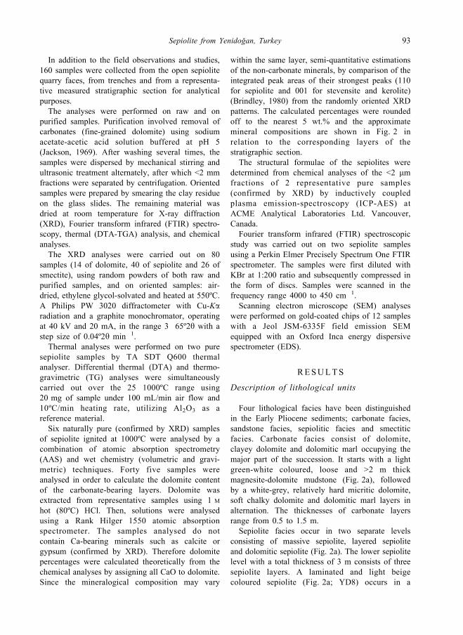
GEOLOGICAL SETT ING
The basement lithology of the area is represented
by rock units that range from Palaeozoic to
Cretaceous and Upper Cretaceous. Palaeozoic to
Cretaceous marbles, recrystallized limestones and
schists crop out to the north and notheast of the
area together with Upper Cretaceous granitoids
(MTA, 2002). In the southwest of the study area,
recrystallized limestones (occasionally dolomitic)
are present (Yeniyol, 1982). They contain schist
layers of Cretaceous age, and ophiolitic rocks
including serpentinites and gabbroic blocks that
were emplaced as large overthrust slabs over the
older lithology during the Upper Cretaceous.
The region has been affected by extensional
tectonic movements between the Lutetian and
Neogene (Erentoz, 1975), producing northwest-
southeast trending regional normal faults (Yeniyol,
1982) and depressions that were bordered by these
tectonic lines. Miocene-Pliocene lacustrine sedi-
ments, extending over a large area at the vicinity of
Eskis� ehir, have been deposited in these depressions
and overlie older rocks unconformably. These
sediments are represented mainly by conglomerates
at the base and margins of the basin. The upper part
of the Miocene consists of claystone, marl,
dolomite, tuff and gypsum beds (Yeniyol, 1992).
During the Late Miocene�Early Pliocene,
volcanism provided materials of variable composi-
tion including dacites, trachytes, trachyandesites,
rhyodacites and tuffs (Kulaksız, 1981).
The upper part of Miocene-Pliocene sequence is
represented by Early Pliocene lacustrine sediments
where chemical precipitation has predominated
(Yeniyol, 1992). Due to the presence of two distinct
sepiolite forms and related Mg-rich minerals, it was
considered important to study these sediments in
depth.
FIG. 1. Location and geological map of the study area (adapted from 1/500,000 scale geological map of Turkey;
MTA, 2002).
92 M. Yeniyol
In addition to the field observations and studies,
160 samples were collected from the open sepiolite
quarry faces, from trenches and from a representa-
tive measured stratigraphic section for analytical
purposes.
The analyses were performed on raw and on
purified samples. Purification involved removal of
carbonates (fine-grained dolomite) using sodium
acetate-acetic acid solution buffered at pH 5
(Jackson, 1969). After washing several times, the
samples were dispersed by mechanical stirring and
ultrasonic treatment alternately, after which <2 mm
fractions were separated by centrifugation. Oriented
samples were prepared by smearing the clay residue
on the glass slides. The remaining material was
dried at room temperature for X-ray diffraction
(XRD), Fourier transform infrared (FTIR) spectro-
scopy, thermal (DTA-TGA) analysis, and chemical
analyses.
The XRD analyses were carried out on 80
samples (14 of dolomite, 40 of sepiolite and 26 of
smectite), using random powders of both raw and
purified samples, and on oriented samples: air-
dried, ethylene glycol-solvated and heated at 550ºC.
A Philips PW 3020 diffractometer with Cu-Karadiation and a graphite monochromator, operating
at 40 kV and 20 mA, in the range 3�65º2y with a
step size of 0.04º2y min�1.
Thermal analyses were performed on two pure
sepiolite samples by TA SDT Q600 thermal
analyser. Differential thermal (DTA) and thermo-
gravimetric (TG) analyses were simultaneously
carried out over the 25�1000ºC range using
20 mg of sample under 100 mL/min air flow and
10ºC/min heating rate, utilizing Al2O3 as a
reference material.
Six naturally pure (confirmed by XRD) samples
of sepiolite ignited at 1000ºC were analysed by a
combination of atomic absorption spectrometry
(AAS) and wet chemistry (volumetric and gravi-
metric) techniques. Forty five samples were
analysed in order to calculate the dolomite content
of the carbonate-bearing layers. Dolomite was
extracted from representative samples using 1 M
hot (80ºC) HCl. Then, solutions were analysed
using a Rank Hilger 1550 atomic absorption
spectrometer. The samples analysed do not
contain Ca-bearing minerals such as calcite or
gypsum (confirmed by XRD). Therefore dolomite
percentages were calculated theoretically from the
chemical analyses by assigning all CaO to dolomite.
Since the mineralogical composition may vary
within the same layer, semi-quantitative estimations
of the non-carbonate minerals, by comparison of the
integrated peak areas of their strongest peaks (110
for sepiolite and 001 for stevensite and kerolite)
(Brindley, 1980) from the randomly oriented XRD
patterns. The calculated percentages were rounded
off to the nearest 5 wt.% and the approximate
mineral compositions are shown in Fig. 2 in
relation to the corresponding layers of the
stratigraphic section.
The structural formulae of the sepiolites were
determined from chemical analyses of the <2 mmfractions of 2 representative pure samples
(confirmed by XRD) by inductively coupled
plasma emission-spectroscopy (ICP-AES) at
ACME Analytical Laboratories Ltd. Vancouver,
Canada.
Fourier transform infrared (FTIR) spectroscopic
study was carried out on two sepiolite samples
using a Perkin Elmer Precisely Spectrum One FTIR
spectrometer. The samples were first diluted with
KBr at 1:200 ratio and subsequently compressed in
the form of discs. Samples were scanned in the
frequency range 4000 to 450 cm�1.
Scanning electron microscope (SEM) analyses
were performed on gold-coated chips of 12 samples
with a Jeol JSM-6335F field emission SEM
equipped with an Oxford Inca energy dispersive
spectrometer (EDS).
RESULTS
Description of lithological units
Four lithological facies have been distinguished
in the Early Pliocene sediments; carbonate facies,
sandstone facies, sepiolitic facies and smectitic
facies. Carbonate facies consist of dolomite,
clayey dolomite and dolomitic marl occupying the
major part of the succession. It starts with a light
green-white coloured, loose and >2 m thick
magnesite-dolomite mudstone (Fig. 2a), followed
by a white-grey, relatively hard micritic dolomite,
soft chalky dolomite and dolomitic marl layers in
alternation. The thicknesses of carbonate layers
range from 0.5 to 1.5 m.
Sepiolite facies occur in two separate levels
consisting of massive sepiolite, layered sepiolite
and dolomitic sepiolite (Fig. 2a). The lower sepiolite
level with a total thickness of 3 m consists of three
sepiolite layers. A laminated and light beige
coloured sepiolite (Fig. 2a; YD8) occurs in a
Sepiolite from Yenidogan, Turkey 93
50�60 cm thick layer, between two sandstone beds,
which are composed of rounded and sandy-sized
dolomite intraclasts embedded in the sepiolite matrix
or vice versa. The thickness of this level decreases
laterally, and its composition grades to dolomitic
sepiolite. The upper sepiolite level occurs after
massive clayey dolomite layers with 7 m total
thickness. This level comprises almost pure sepiolite
lenses, either massive or layered, alternating
dolomitic sepiolite and dolomite layers. Nearly
pure sepiolite occurs in lenses as thin as 0.5 m.
The positions of the lenses change both vertically
and horizontally. When they are superimposed the
total thickness of sepiolite reaches up to 3.5 m.
Both massive and layered sepiolites are wax-like
when wet. When dry, sepiolite is light-weight with a
light beige colour, turning to dark brown or even
black when organic material is present. In places,
sepiolite is laminated or thin layered. Rounded
intraclasts of sepiolite up to 2 cm across, representing
the reworked material may be occasionally found
embedded in sepiolite clay matrix. In some places,
sepiolite shows a massive structure where well
developed layering is lacking. Also, thin tuff
(0.5�10 cm), opal bands and nodules, and diatomac-
eous earth accompany the sepiolite in places.
Dolomitic sepiolite and sepiolitic marl layers
0.4�2.6 m thick alternate with sepiolite (Fig. 2a).
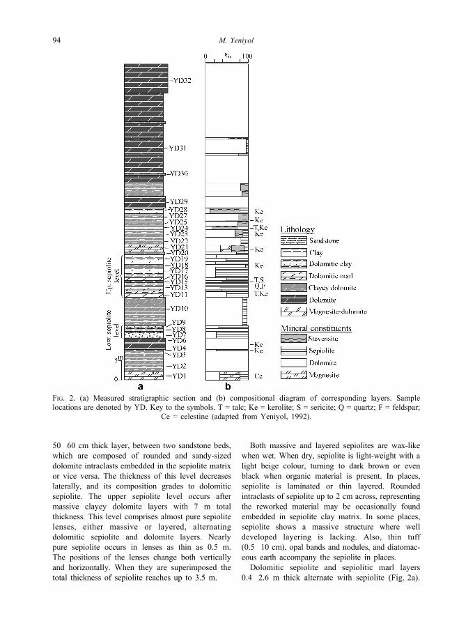
FIG. 2. (a) Measured stratigraphic section and (b) compositional diagram of corresponding layers. Sample
locations are denoted by YD. Key to the symbols. T = talc; Ke = kerolite; S = sericite; Q = quartz; F = feldspar;
Ce = celestine (adapted from Yeniyol, 1992).
94 M. Yeniyol
They are massive in appearance and vary in colour
from white to light beige according to the sepiolite
content. These layers often show a detrital texture
and contain dolomite and sepiolite intraclasts up to
1 cm in size. In addition, casts of roots, burrowed
organisms, fenestrae structures and fissures which
are filled with carbonate are often recognized in
sepiolite-bearing materials. The upper sepiolite
level extends throughout the study area, comprising
a total thickness of ~10 m.
After the upper sepiolite level, smectitic facies
occurs in dolomitic clay layers, in which smectite is
the predominant clay mineral. These layers alternate
clayey dolomite layers constituting the middle part
of the sequence (Fig. 2a; from YD20 to YD28). The
dolomitic clay layers have colours varying from
white to light green and even light beige. They are
massive showing scarce fine rounded or angular
intraclasts, rare fenestrae structures, desiccation
fissures and traces of bioturbation and are
0.8�1 m thick.
Clay mineralogy
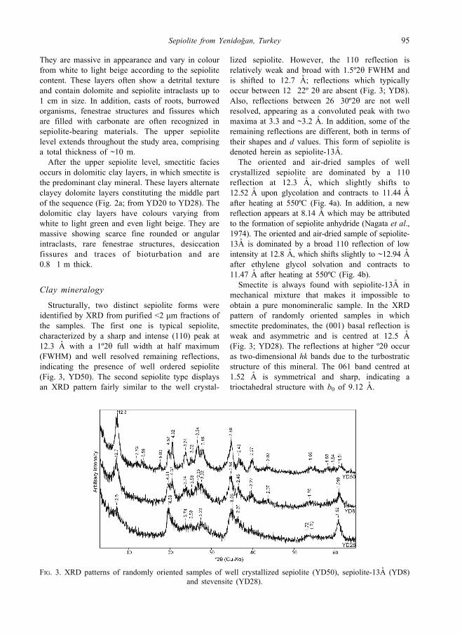
Structurally, two distinct sepiolite forms were
identified by XRD from purified <2 mm fractions of
the samples. The first one is typical sepiolite,
characterized by a sharp and intense (110) peak at
12.3 A with a 1º2y full width at half maximum
(FWHM) and well resolved remaining reflections,
indicating the presence of well ordered sepiolite
(Fig. 3, YD50). The second sepiolite type displays
an XRD pattern fairly similar to the well crystal-
lized sepiolite. However, the 110 reflection is
relatively weak and broad with 1.5º2y FWHM and
is shifted to 12.7 A; reflections which typically
occur between 12�22º 2y are absent (Fig. 3; YD8).
Also, reflections between 26�30º2y are not well
resolved, appearing as a convoluted peak with two
maxima at 3.3 and ~3.2 A. In addition, some of the
remaining reflections are different, both in terms of
their shapes and d values. This form of sepiolite is
denoted herein as sepiolite-13A.
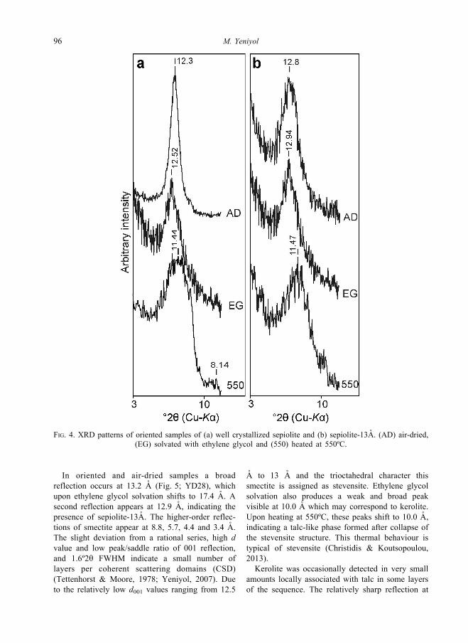
The oriented and air-dried samples of well
crystallized sepiolite are dominated by a 110
reflection at 12.3 A, which slightly shifts to
12.52 A upon glycolation and contracts to 11.44 A
after heating at 550ºC (Fig. 4a). In addition, a new
reflection appears at 8.14 A which may be attributed
to the formation of sepiolite anhydride (Nagata et al.,
1974). The oriented and air-dried sample of sepiolite-
13A is dominated by a broad 110 reflection of low
intensity at 12.8 A, which shifts slightly to ~12.94 A
after ethylene glycol solvation and contracts to
11.47 A after heating at 550ºC (Fig. 4b).
Smectite is always found with sepiolite-13A in
mechanical mixture that makes it impossible to
obtain a pure monomineralic sample. In the XRD
pattern of randomly oriented samples in which
smectite predominates, the (001) basal reflection is
weak and asymmetric and is centred at 12.5 A
(Fig. 3; YD28). The reflections at higher º2y occur
as two-dimensional hk bands due to the turbostratic
structure of this mineral. The 061 band centred at
1.52 A is symmetrical and sharp, indicating a
trioctahedral structure with b0 of 9.12 A.
FIG. 3. XRD patterns of randomly oriented samples of well crystallized sepiolite (YD50), sepiolite-13A (YD8)
and stevensite (YD28).
Sepiolite from Yenidogan, Turkey 95
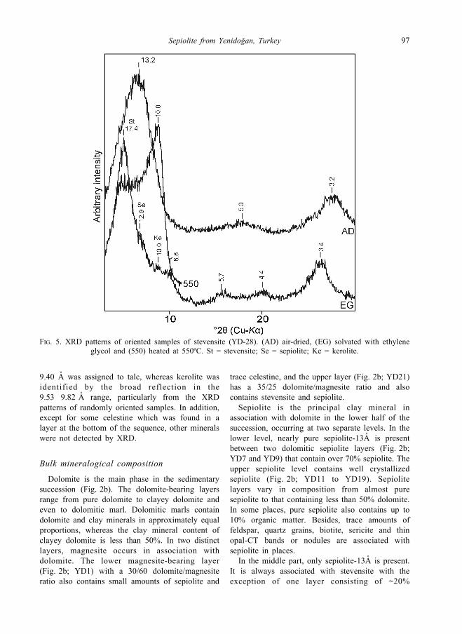
In oriented and air-dried samples a broad
reflection occurs at 13.2 A (Fig. 5; YD28), which
upon ethylene glycol solvation shifts to 17.4 A. A
second reflection appears at 12.9 A, indicating the
presence of sepiolite-13A. The higher-order reflec-
tions of smectite appear at 8.8, 5.7, 4.4 and 3.4 A.
The slight deviation from a rational series, high d
value and low peak/saddle ratio of 001 reflection,
and 1.6º2y FWHM indicate a small number of
layers per coherent scattering domains (CSD)
(Tettenhorst & Moore, 1978; Yeniyol, 2007). Due
to the relatively low d001 values ranging from 12.5
A to 13 A and the trioctahedral character this
smectite is assigned as stevensite. Ethylene glycol
solvation also produces a weak and broad peak
visible at 10.0 A which may correspond to kerolite.
Upon heating at 550ºC, these peaks shift to 10.0 A,
indicating a talc-like phase formed after collapse of
the stevensite structure. This thermal behaviour is
typical of stevensite (Christidis & Koutsopoulou,
2013).
Kerolite was occasionally detected in very small
amounts locally associated with talc in some layers
of the sequence. The relatively sharp reflection at
FIG. 4. XRD patterns of oriented samples of (a) well crystallized sepiolite and (b) sepiolite-13A. (AD) air-dried,
(EG) solvated with ethylene glycol and (550) heated at 550ºC.
96 M. Yeniyol
9.40 A was assigned to talc, whereas kerolite was
identified by the broad reflection in the
9.53�9.82 A range, particularly from the XRD
patterns of randomly oriented samples. In addition,
except for some celestine which was found in a
layer at the bottom of the sequence, other minerals
were not detected by XRD.
Bulk mineralogical composition
Dolomite is the main phase in the sedimentary
succession (Fig. 2b). The dolomite-bearing layers
range from pure dolomite to clayey dolomite and
even to dolomitic marl. Dolomitic marls contain
dolomite and clay minerals in approximately equal
proportions, whereas the clay mineral content of
clayey dolomite is less than 50%. In two distinct
layers, magnesite occurs in association with
dolomite. The lower magnesite-bearing layer
(Fig. 2b; YD1) with a 30/60 dolomite/magnesite
ratio also contains small amounts of sepiolite and
trace celestine, and the upper layer (Fig. 2b; YD21)
has a 35/25 dolomite/magnesite ratio and also
contains stevensite and sepiolite.
Sepiolite is the principal clay mineral in
association with dolomite in the lower half of the
succession, occurring at two separate levels. In the
lower level, nearly pure sepiolite-13A is present
between two dolomitic sepiolite layers (Fig. 2b;
YD7 and YD9) that contain over 70% sepiolite. The
upper sepiolite level contains well crystallized
sepiolite (Fig. 2b; YD11 to YD19). Sepiolite
layers vary in composition from almost pure
sepiolite to that containing less than 50% dolomite.
In some places, pure sepiolite also contains up to
10% organic matter. Besides, trace amounts of
feldspar, quartz grains, biotite, sericite and thin
opal-CT bands or nodules are associated with
sepiolite in places.
In the middle part, only sepiolite-13A is present.
It is always associated with stevensite with the
exception of one layer consisting of ~20%
FIG. 5. XRD patterns of oriented samples of stevensite (YD-28). (AD) air-dried, (EG) solvated with ethylene
glycol and (550) heated at 550ºC. St = stevensite; Se = sepiolite; Ke = kerolite.
Sepiolite from Yenidogan, Turkey 97
sepiolite-13A and 80% dolomite (Fig. 2b; YD22).
In the remaining layers the smectite content varies
from 15% to 70% (Fig. 2b; YD24 to YD28), whilst
the sepiolite-13A content ranges from 15% to 30%.
Kerolite and talc are also present in traces (less than
10%) in the middle part and in some layers of the
lower half of the succession.
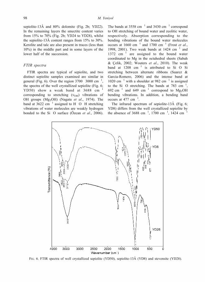
FTIR spectra
FTIR spectra are typical of sepiolite, and two
distinct sepiolite samples examined are similar in
general (Fig. 6). Over the region 3700�3000 cm�1,
the spectra of the well crystallized sepiolite (Fig. 6;
YD50) show a weak band at 3688 cm�1
corresponding to stretching (nOH) vibrations of
OH groups (Mg3OH) (Nagata et al., 1974). The
band at 3622 cm�1 assigned to H�O�H stretching
vibrations of water molecules are weakly hydrogen
bonded to the Si�O surface (Ozcan et al., 2006).
The bands at 3558 cm�1 and 3430 cm�1 correspond
to OH stretching of bound water and zeolitic water,
respectively. Absorption corresponding to the
bending vibrations of the bound water molecules
occurs at 1660 cm�1 and 1700 cm�1 (Frost et al.,
1998, 2001). Two weak bands at 1424 cm�1 and
1372 cm�1 are assigned to the bound water
coordinated to Mg in the octahedral sheets (Sabah
& Celik, 2002; Wouters et al., 2010). The weak
band at 1208 cm�1 is attributed to Si�O�Sistretching between alternate ribbons (Suarez &
Garcia-Romero, 2006) and the intense band at
1020 cm�1 with a shoulder at 982 cm�1 is assigned
to the Si�O stretching. The bands at 783 cm�1,
692 cm�1 and 649 cm�1 correspond to Mg3OH
bending vibrations. In addition, a bending band
occurs at 477 cm�1.
The infrared spectrum of sepiolite-13A (Fig. 6;
YD8) differs from the well crystallized sepiolite by
the absence of 3688 cm�1, 1700 cm�1, 1424 cm�1
FIG. 6. FTIR spectra of well crystallized sepiolite (YD50), sepiolite-13A (YD8) and stevensite (YD28).
98 M. Yeniyol

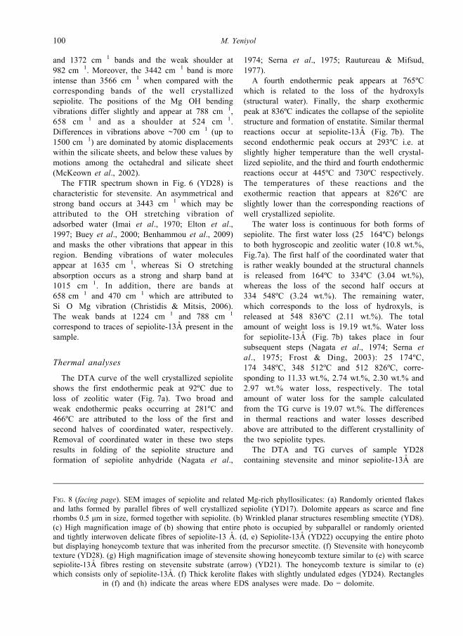
FIG. 7. DTA and TG analyses of (a) well crystallized sepiolite (YD50), (b) sepiolite-13A (YD8) and (c) stevensite
with sepiolite-13A (YD28).
Sepiolite from Yenidogan, Turkey 99
and 1372 cm�1 bands and the weak shoulder at
982 cm�1. Moreover, the 3442 cm�1 band is more
intense than 3566 cm�1 when compared with the
corresponding bands of the well crystallized
sepiolite. The positions of the Mg�OH bending
vibrations differ slightly and appear at 788 cm�1,
658 cm�1 and as a shoulder at 524 cm�1.
Differences in vibrations above ~700 cm�1 (up to
1500 cm�1) are dominated by atomic displacements
within the silicate sheets, and below these values by
motions among the octahedral and silicate sheet
(McKeown et al., 2002).
The FTIR spectrum shown in Fig. 6 (YD28) is
characteristic for stevensite. An asymmetrical and
strong band occurs at 3443 cm�1 which may be
attributed to the OH stretching vibration of
adsorbed water (Imai et al., 1970; Elton et al.,
1997; Buey et al., 2000; Benhammou et al., 2009)
and masks the other vibrations that appear in this
region. Bending vibrations of water molecules
appear at 1635 cm�1, whereas Si�O stretching
absorption occurs as a strong and sharp band at
1015 cm�1. In addition, there are bands at
658 cm�1 and 470 cm�1 which are attributed to
Si�O�Mg vibration (Christidis & Mitsis, 2006).
The weak bands at 1224 cm�1 and 788 cm�1
correspond to traces of sepiolite-13A present in the
sample.
Thermal analyses
The DTA curve of the well crystallized sepiolite
shows the first endothermic peak at 92ºC due to
loss of zeolitic water (Fig. 7a). Two broad and
weak endothermic peaks occurring at 281ºC and
466ºC are attributed to the loss of the first and
second halves of coordinated water, respectively.
Removal of coordinated water in these two steps
results in folding of the sepiolite structure and
formation of sepiolite anhydride (Nagata et al.,
1974; Serna et al., 1975; Rautureau & Mifsud,
1977).
A fourth endothermic peak appears at 765ºC
which is related to the loss of the hydroxyls
(structural water). Finally, the sharp exothermic
peak at 836ºC indicates the collapse of the sepiolite
structure and formation of enstatite. Similar thermal
reactions occur at sepiolite-13A (Fig. 7b). The
second endothermic peak occurs at 293ºC i.e. at
slightly higher temperature than the well crystal-
lized sepiolite, and the third and fourth endothermic
reactions occur at 445ºC and 730ºC respectively.
The temperatures of these reactions and the
exothermic reaction that appears at 826ºC are
slightly lower than the corresponding reactions of
well crystallized sepiolite.
The water loss is continuous for both forms of
sepiolite. The first water loss (25�164ºC) belongs
to both hygroscopic and zeolitic water (10.8 wt.%,
Fig.7a). The first half of the coordinated water that
is rather weakly bounded at the structural channels
is released from 164ºC to 334ºC (3.04 wt.%),
whereas the loss of the second half occurs at
334�548ºC (3.24 wt.%). The remaining water,
which corresponds to the loss of hydroxyls, is
released at 548�836ºC (2.11 wt.%). The total
amount of weight loss is 19.19 wt.%. Water loss
for sepiolite-13A (Fig. 7b) takes place in four
subsequent steps (Nagata et al., 1974; Serna et
al., 1975; Frost & Ding, 2003): 25�174ºC,174�348ºC, 348�512ºC and 512�826ºC, corre-
sponding to 11.33 wt.%, 2.74 wt.%, 2.30 wt.% and
2.97 wt.% water loss, respectively. The total
amount of water loss for the sample calculated
from the TG curve is 19.07 wt.%. The differences
in thermal reactions and water losses described
above are attributed to the different crystallinity of
the two sepiolite types.
The DTA and TG curves of sample YD28
containing stevensite and minor sepiolite-13A are
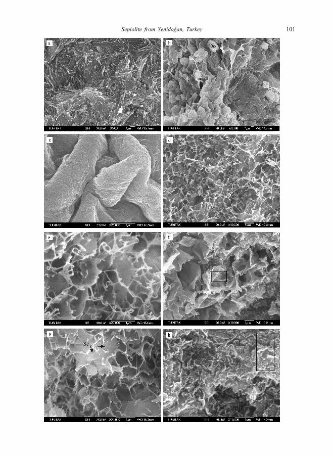
FIG. 8 (facing page). SEM images of sepiolite and related Mg-rich phyllosilicates: (a) Randomly oriented flakes
and laths formed by parallel fibres of well crystallized sepiolite (YD17). Dolomite appears as scarce and fine
rhombs 0.5 mm in size, formed together with sepiolite. (b) Wrinkled planar structures resembling smectite (YD8).
(c) High magnification image of (b) showing that entire photo is occupied by subparallel or randomly oriented
and tightly interwoven delicate fibres of sepiolite-13 A. (d, e) Sepiolite-13A (YD22) occupying the entire photo
but displaying honeycomb texture that was inherited from the precursor smectite. (f) Stevensite with honeycomb
texture (YD28). (g) High magnification image of stevensite showing honeycomb texture similar to (e) with scarce
sepiolite-13A fibres resting on stevensite substrate (arrow) (YD21). The honeycomb texture is similar to (e)
which consists only of sepiolite-13A. (f) Thick kerolite flakes with slightly undulated edges (YD24). Rectangles
in (f) and (h) indicate the areas where EDS analyses were made. Do = dolomite.
100 M. Yeniyol
Sepiolite from Yenidogan, Turkey 101
shown in Fig. 7c. These curves are rather similar to
the well crystallized sepiolite and sepiolite-13A.
The first endothermic peak at 88ºC corresponds to
loss of the majority of interlayer water of stevensite
and zeolitic water of sepiolite-13A. The main
difference is the presence of an endothermic peak
at 825ºC which is attributed to the loss of the
remaining hydroxyl water of stevensite (Imai et al.,
1970), whereas the exothermic peak at 808ºC
indicates the collapse of the sepiolite structure and
formation of enstatite. The water loss for this
sample corresponds to different types of water in
the sepiolite and the zeolitic, interlayer waters and
hydroxyls of the stevensite. The total amount of
water loss is 19 wt.%.
SEM analyses
In the SEM the well crystallized sepiolite appears
in bundles of parallel and straight fibres in the form
of planar structures and laths up to 5 mm long,
occasionally associated with fine dolomite rhombs
(Fig. 8a; YD17), suggesting that it was formed by
precipitation from the solution with the dolomite.
Sepiolite-13A appears in the form of wrinkled
flakes resembling smectite (Fig. 8b; YD8). Under
higher magnification, it is clear that the flakes
consist of sub-parallel or randomly oriented
interwoven delicate sepiolite fibres ~1 mm long
(Fig . 8c) . In another layer where only
sepiolite-13 A coexists with dolomite (Figs. 8d
and e; YD22), sepiolite displays a honeycomb
texture resembling smectite. However, the entire
material consists of randomly oriented short
sepiolite fibres. In both samples, fibres of a late
generation up to 3 mm long also occur in the
interstitial spaces projecting from the tips of the
flakes. They are curled, and in some cases are
interlocked with each other, displaying a lacework
texture (Fig. 8d and e).
Stevensite occurs in the form of large or fine
crinkled planar structures or uneven flakes, forming
a typical honeycomb texture (Fig. 8f and g).
Although stevensite is the dominant constituent, it
is always associates with sepiolite-13A. As a rule,
sepiolite fibres of varying amounts rest on the
stevensite substrate (Fig. 8g; YD21).
Under the SEM, accessory kerolite is always seen
to be present in association with stevensite.
Morphologically it is rather different from steven-
site by appearing in the form of small, even thick
flakes with slightly undulating edges (Fig. 8h;
YD24) whereas stevensite appears in the form of
thin and larger undulated flakes (Fig. 8f; YD28).
Qualitative EDS analyses provided MgO and SiO2
values approximating to the composition of talc and
thus suggesting the presence of kerolite.
Chemical analyses
The major element oxide analyses of the samples
are given in Table 1. Analyses using wet chemical
methods were performed on some well crystallized
sepiolite samples that were pure in the raw state
(SS8 and SY16/8). In these samples, SiO2 varied
from 53.30 wt.% to 54.04%. Al2O3 is present in
small amounts being less than 2.85%. Also, all
samples showed low Fe2O3 and TiO2 contents.
To compare the crystal chemistry of well
crystallized sepiolite and sepiolite-13A, ICP
analyses were performed on essentially monomi-
neralic samples (YD8 and YD50) (Table 1).
Sepiolite-13A (YD8) has Al2O3, Fe2O3, and TiO2
contents rather lower and MgO content higher than
the well crystallized sepiolite (YD50). The struc-
tural formulae calculated per half unit cell (32
oxygen a toms) a re : (Ca0 . 0 5Na0 . 0 8K0 . 0 7 )
(Mg7.17Al0.32Fe0.11Ti0.02&0.38)(Si11.96Al0.04)
O30(OH)4 for sepiolite-13A and (Ca0.05Na0.09K0.10)
(Mg5.90Al0.96Fe0.36Ti0.04&0.74)(Si11.81Al0.19)
O30(OH)4 for well crystallized sepiolite. The
principal cation in the octahedral sheet is Mg.
Small amounts of Al, Fe and Ti substitute for Mg in
the octahedral sheet of sepiolite-13A. The numbers
of structural vacancies (shown by squares), which
are common in sepiolite structures, are lower in
sepiolite-13A than in well crystallized sepiolite. The
latter has a lower Mg content than the ideal
composition, which is due to higher substitution
by Al, Fe and also Ti; this increases the number of
octahedral vacancies (Garcia-Romero & Suarez,
2010). Depending on its high Al content, it can
be termed an Al-rich sepiolite.
Stevensite is always found in association with
sepiolite-13A and minor kerolite. Therefore, it was
not possible to obtain pure phases for chemical
analyses. However, it was considered convenient to
evaluate the chemical analyses obtained by EDS on
isolated particles as an approximation to the crystal
chemistry of these minerals (Fig. 9f and h). Kerolite
(Table 1; YD21) has a lower SiO2 and higher MgO
content than stevensite (Table 1; YD28). The CaO
content originates from the presence of dolomite
that was probably masked by kerolite particles.
102 M. Yeniyol
After assigning the the CaO content to dolomite,
the theoretical formula of Mg3Si4O10(OH)2 can be
calculated for kerolite. The structural formula is
similar to talc without vacancies.
Besides MgO and SiO2 stevensite also contains
Al2O3 and Fe2O3 in very small amounts. The
structural formula of stevensite calculated on the
basis of O10(OH)2 is: (Ca0.56)(Mg2.47Al0.07Fe0.03&0.43)Si4.16O10(OH)2. After making correc-
tions for the excess SiO2 (0.16 atoms), which were
assigned to amorphous silica, the following
s t r u c t u r a l f o r m u l a w a s o b t a i n e d :
Ca0.06(Mg2.82Al0.08Fe0.04&0.06)Si4O10(OH)2. In
both formulae, Mg is the principal octahedral
cation, with small amounts of Al and Fe
substituting for Mg in the octahedral sheet.
Although this structural formula is indicative, with
high Mg and low Al and Fe contents, the absence
of tetrahedral substitution and the octahedral
vacancies suggest the presence of stevensite, in
accordance with the XRD and FTIR data.
D I SCUSS ION
Two distinct forms of sepiolite were distinguished
in this study: a well crystallized and a poorly
crystallized sepiolite, the latter being referred to as
sepiolite-13A. The well crystallized sepiolite shows
a sharp and strongest peak at ~12.3 A (110), and
well resolved remaining reflections similar to
references in the literature with respect to their
shapes, d values and intensities. The overall XRD
pattern of sepiolite-13A, although rather similar to
the widely-known sepiolite, has some characteristics
typical of poor crystal order.
The first description of poorly-ordered sepiolite
was made by Brindley (1959) for a sample which
was obtained from Eskis� ehir (most probably
sepiolite of the meerschaum type). It was a very
poorly crystallized sepiolite with fairly broad and
poorly resolved reflections and with the 110
reflection appearing at 12.3 A. Sepiolites with
fairly similar XRD patterns with higher d values of
the 110 reflection were reported in later studies
(Yeniyol & Oztunalı, 1985; Yeniyol, 1986, 1993a;
Ece & Coban, 1994; Yalcın & Bozkaya, 1995;
Chahi et al., 1997; Ece, 1998). These works
describe poorly-ordered sepiolite having broad 110
reflections with high d values, showing a convo-
luted single reflection in the 26�30º2y region and
remaining reflections which are not well resolved,
rather similar to the work of Brindley (1959).
Sepiolite-13A also shows some characteristics
similar to these poorly-ordered sepiolites. The 110
reflection is wide (1.5º2y), appearing at ~13 A with
an intensity that is considerably weaker than the
corresponding reflection of the well crystallized
sepiolite. The high d value of this reflection
resembles loughlinite (Na-rich variety of sepiolite),
although the present sepiolite is Na-deficient and
has a different XRD pattern from loughlinite
(Yeniyol, 1997; Kadir et al., 2002). Three
reflections which typically occur in the 12�22º2yregion were not observed. Moreover, two relatively
strong reflections occurring at 4.52 and 4.31 A
either show nearly equal intensity or the second one
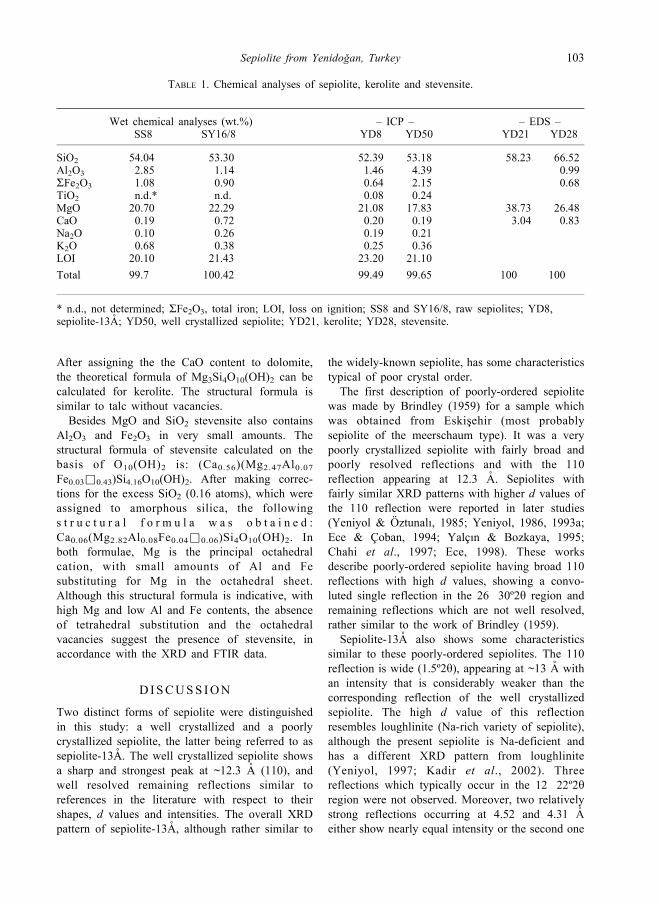
TABLE 1. Chemical analyses of sepiolite, kerolite and stevensite.
Wet chemical analyses (wt.%) – ICP – – EDS –SS8 SY16/8 YD8 YD50 YD21 YD28
SiO2 54.04 53.30 52.39 53.18 58.23 66.52Al2O3 2.85 1.14 1.46 4.39 � 0.99SFe2O3 1.08 0.90 0.64 2.15 � 0.68TiO2 n.d.* n.d. 0.08 0.24 � �MgO 20.70 22.29 21.08 17.83 38.73 26.48CaO 0.19 0.72 0.20 0.19 3.04 0.83Na2O 0.10 0.26 0.19 0.21 � �K2O 0.68 0.38 0.25 0.36 � �LOI 20.10 21.43 23.20 21.10 � �Total 99.7 100.42 99.49 99.65 100 100
* n.d., not determined; SFe2O3, total iron; LOI, loss on ignition; SS8 and SY16/8, raw sepiolites; YD8,sepiolite-13A; YD50, well crystallized sepiolite; YD21, kerolite; YD28, stevensite.
Sepiolite from Yenidogan, Turkey 103
is less intense than the first one, in contrast to the
well crystallized sepiolite where the second
reflection is more intense than the first.
Furthermore, the convoluted reflection at
26�30º2y and the remaining reflections are not
well resolved. These data demonstrate the poorly-
crystallized nature of sepiolite-13A, with a struc-
tural organization poorer than the typical sepiolite.
Similar XRD data relevant to sepiolite-13A were
obtained from the analyses performed on numerous
monomineralic samples that were collected from
different layers containing sepiolite of this form.
Mineral impurities other than sepiolite (e.g.
stevensite and/or kerolite, which frequently coexist
with sepiolite) were not detected by XRD.
FTIR spectra of both types of sepiolite are
identical in general and fairly similar to those
reported elsewhere (Sabah & Celik, 2002; Wouters
et al., 2010; and Tunc et al., 2012). However, some
weak bands are absent, and the positions and
intensities of some bands are slightly different in
sepiolite-13A, indicating its poor crystallinity.
Neither the overall spectrum nor the position or
shape of the individual bands are identical to the
FTIR spectra, indicating the presence of stevensite
and kerolite (e.g. Imai et al., 1970; Davidson, 1989;
Elton et al., 1997; Christidis & Mitsis, 2006; Dekov
et al., 2008; Benhammou et al., 2009).
The thermal curves of well crystallized sepiolite
are rather similar to those shown in previous work
on thermal reactions and water loss steps (Nagata et
al., 1974; Serna et al., 1975; Perez-Rodriguez &
Galan, 1994; Singer et al. 1998, among others). The
DTA and TG curves of sepiolite-13A are almost
identical to well crystallized sepiolite. However
some differences in thermal properties arise from its
poor crystallinity and thereby the presence of some
weaker structural bonds. The second half of
coordinated water and structural hydroxyls are
rather weakly bonded to the structure. Therefore,
the last two thermal reactions involving the losses
of these types of water appear fairly continuous and
at lower temperatures. Also, the exothermic
reaction at 826ºC, corresponding to collapse of the
sepiolite structure and subsequent formation of
enstatite, occurs at slightly lower temperature than
the well crystallized sepiolite, due to the poor
crystallinity.
Well crystallized sepiolite contains variable
proportions of Al2O3. However, some samples are
richer in Al2O3 and poorer in MgO than the ideal
sepiolite. A high Al content is probably associated
with devitrification of the volcanic glass which was
then transferred from solution to the sepiolite
structure. Sepiolite-13A (sample YD8) has low
Al2O3, Fe2O3, TiO2 and higher MgO contents than
the Mg-rich typical sepiolite.
Stevensi te almost a lways occurs with
sepiolite-13A and kerolite. SEM observations and
crystal chemical data indicate a high Mg content,
insignificant Al and Fe contents, absence of
tetrahedral substitution and the presence of
structural vacancies in the octahedral sheets.
Kerolite was distinguished by the fairly broad
001 reflection appearing at 9.53�9.82 A in the
XRD patterns. Thick and rather flat flakes with
morphology distinct from that of stevensite under
the SEM were assigned to kerolite. In addition, the
theoretical formula which was calculated from EDS
analysis from the selected area under the SEM is in
accordance with kerolite.
The Miocene-Pliocene sediments of the study
area were deposited in an alkaline lake environ-
ment. Stratigraphic and lithological features indicate
that semi-arid or arid climatic conditions with short-
lasting wet seasons had prevailed (Yeniyol, 1992).
During the Early Pliocene, an alkaline shallow lake
environment rich in Mg, Ca and Si was developed
in which chemical precipitation dominated. All
mineral phases present are authigenic except for
traces of feldspar, quartz, biotite and sericite that
are present in some layers.
The sepiolitic and smectitic facies of the Early
Pliocene are interpreted as palustrine to mudflat
deposits formed in the alkaline lake margin where
high Mg and Si were available by feeding of the
surface waters (Pozo & Casas, 1999; Galan & Pozo,
2011). Massive and layered sepiolites precipitated
at short-lived ponds at the edges of main water
bodies and the marshlands (palustrine), at which
short-lived plants had grown. Sepiolite was
deposited in dry periods following the formation
of dolomite (Galan & Ferrero, 1982). Well crystal-
lized sepiolite was formed by direct precipitation
from lake water or by crystallization from pore
solutions during diagenesis under low Al, high Si
and Mg activities, and a pH ranging between 8.0
and 9.5 (Wollast et al., 1968; Khoury et al., 1982;
Galan & Castillo, 1984; Abtahi, 1985). Opal-CT
bands and nodules found in association with
sepiolite indicate a high activity of SiO2 that
favours the formation of sepiolite (Birsoy, 2002).
In those layers consisting only of sepiolite-13A
(samples YD8 and YD22), randomly oriented or
104 M. Yeniyol
parallel delicate fibres occupy the entire mass.
However, the wrinkled planar or honeycomb texture
which is characteristic for smectite was inherited
and was remarkably preserved. This relationship
may suggest an epitaxial growth on a smectite
substrate, but SEM and XRD data reveal that the
material consists entirely of sepiolite without a
planar structure which might indicate the presence
of stevensite or kerolite precursor minerals. Based
on these relationships, it is suggested that this form
of sepiolite was developed by in situ transformation
via a dissolution-precipitation mechanism from the
precursor stevensite during diagenesis, as suggested
previously (Eberl et al., 1982; Khoury et al., 1992;
Chahi et al., 1997 and Pozo & Casas, 1999). The
relative SiO2 enrichment during the dry periods
resulted in low pH and low salinity that should
decrease the stability of stevensite and probably
favoured the formation of sepiolite-13A (Khoury et
al., 1982; Galan & Pozo, 2011). This type of
sepiolite is present associated with stevensite,
indicating that the dry episodes did not last
enough to complete this transformation (Chahi et
al., 1997).
It is noteworthy that all poorly ordered sepiolites
mentioned in the literature were formed by
transformation of pre-existing minerals such as
magnesite (Yeniyol & Oztunalı, 1985; Yeniyol,
1986, 1993a,b; Ece, 1998) or stevensite (Chahi et
al., 1997). It is thus suggested that the poorly
ordered sepiolite-13A may be due to the transfor-
mation from stevensite, probably involving struc-
tural reorganization, in contrast to the mechanism
described by Guven & Carney (1979) for the
sepiolite to stevensite transformation.
Stevensite-bearing layers were probably depos-
ited in a mudflat environment under relatively wet
climatic conditions by direct chemical precipitation
from alkaline lake water due to the high pH and
salinity, and depending on high Mg and Si activities
(Siffert, 1962; Birsoy, 2002). Kerolite was also
formed in mudflat and palustrine environments as
was suggested by Pozo & Casas (1999) by direct
precipitation from the solution at pH<8 (Stoessell &
Hay, 1978) under conditions of lower salinity and
higher Mg activity than that required for stevensite
(Jones, 1986).
CONCLUS IONS
A poorly crystallized sepiolite form was discovered
in a sepiolite deposit which also contains the widely
known well crystallized sepiolite. This form was
denoted herein as sepiolite-13A, having structural,
crystallochemical, thermal and textural properties
similar to typical sepiolite, although differences in
these properties are in accordance with the poorly
crystallized nature of this type of sepiolite.
The two distinct sepiolite forms occur in separate
levels as natural almost pure materials or associated
with stevensite, kerolite and dolomite. Sepiolites
and related Mg-rich phyllosilicates were formed in
mudflat and palustrine environments of a shallow
alkaline lake margin depending on the changes in
salinity and pH, and on the activities of Al, Si and
Mg. The well crystallized sepiolite was formed by
direct precipitation from the lake water rich in Si
and Mg ions during dry periods and from pore
waters during diagenesis. Sepiolite-13A was prob-
ably formed at the expense of precursor stevensite
via a dissolution-precipitation mechanism during
diagenesis.
Stevensite is always associated with sepiolite-13A
and traces of kerolite and was formed by direct
precipitation during relatively wet periods. The
formation of kerolite was favoured by rather
similar conditions and by a decrease in pH.
ACKNOWLEDGMENTS
The present work was supported by the Research Fund
of Istanbul University Project No. UDP-6650/
26032010. I would like to thank A. Kas� goz for FTIR
analysis (Istanbul University, Laboratories of
Advanced Analyses) and I. Yusufoglu (Istanbul
University, Department of Metallurgical and
Materials Engineering) for providing the thermal
analysis. I am also grateful to the editor for his
constructive support and for making corrections to the
manuscript, to E. Garcıa-Romero and to two anon-
ymous referees for valuable comments they made when
reviewing this manuscript.
REFERENCES
Abtahi A. (1985) Synthesis of sepiolite at room
temperature from SiO2 and MgCl2 solution. Clays
and Clay Minerals, 20, 521�523.Akbulut A. & Kadir S. (2003) The geology and origin of
sepiolite and palygorskite and stevensite in Neogene
lacustrine sediments of the Serinhisar-Acıpayam
basin, Denizli, SW Turkey. Clays and Clay
Minerals, 51, 279�292.Akıncı O. (1967) Eskis� ehir I24�c1 paftasının jeolojisi
ve tabakalı luletas� ı zuhurları. Mineral Research and
Sepiolite from Yenidogan, Turkey 105
Exploration Bulletin of Turkey, 67, 82�97, Ankara.Benhammou A., Tanouti B., Nibov L., Yacoubi A. &
Bonnet, J. (2009) Mineralogical and physicochem-
ical investigation of Mg-smectite from Jbel
Ghassoul, Morocco. Clays and Clay Minerals, 57,
264�270.Bilgin T. (1972) Eskis� ehir ili kil imkanlarının genel
ekonomik prospeksiyon raporu. MTA Report, No.
4708, Ankara.
Birsoy R. (2002) Formation of sepiolite-palygorskite
and related minerals from solution. Clays and Clay
Minerals. 50, 736�745.Brindley G.W. (1959) X-ray and electron diffraction
data for sepiolite. American Mineralogist, 44,
495�500.Brindley G.W. (1980) Quantitative X-ray mineral
analysis of clays. Pp. 414�438 in: Crystal
Structures of Clay Minerals and their X-ray
Identification (G.W. Brindley & G. Brown, editors).
Mineralogical Society Monograph No. 5.
Mineralogical Society, London.
Buey C.S., Barrios M.S., Romero E.G. & Montoya M.D.
(2000) Mg-rich smectite ‘‘precursor’’ phase in the
Tagus basin, Spain. Clays and Clay Minerals, 48,
366�373.Chahi A., Fritz B., Duplay J., Weber F. & Lucas J.
(1997) Textural transition and genetic relationship
between precursor stevensite and sepiolite in lacus-
trine sediments (Jbel Rhassoul Morocco). Clays and
Clay Minerals, 45, 378�389.Christidis G.E. & Koutsopoulou E. (2013) A simple
approach to the identification of trioctahedral
smectites by X-ray diffraction. Clay Minerals, 48,
687�696.Christidis G.E. & Mitsis I. (2006) A new Ni-rich
stevensite from the ophiolite complex of Othrys,
Central Greece. Clays and Clay Minerals, 54,
653�666.Davidson P.J. (1989) Stevensite, a smectite group
mineral from Corstorphine Hill, Edinburgh.
Scottish Journal of Geology, 25, 63�67.Dekov V.M., Cuadros J., Shanks W.C. & Koski R.A.
(2008) Deposition of talc�kerolite-smecti-
te�smectite at seafloor hydrothermal vent fields:
Evidence from mineralogical, geochemical and
oxygen isotope studies. Chemical Geology, 247,
171�194.Eberl D.D., Jones B.F. & Khoury H.N. (1982) Mixed
layer kerolite�stevensite from the Amargosa Desert,
Nevada. Clays and Clay Minerals, 30, 321�326.Ece O.I. (1998) Diagenetic transformation of magnesite
pebbles and cobles to sepiolite (meerschaum) in the
Miocene Eskis� ehir lacustrine basin, Turkey. Clays
and Clay Minerals, 46, 436�445.Ece O.I. & Coban F. (1994) Geology, occurrence and
genesis of Eskis� ehir sepiolites, Turkey. Clays and
Clay Minerals, 42, 81�92.
Elton N.J., Hooper J.J. & Holyer A.D. (1997) An
occurrence of stevensite and kerolite in the Devonian
Cruosa gabbro at Dean Quarry, the Lizard, Cornwall,
England. Clay Minerals, 32, 241�252.Erentoz C. (1975) Explanatory Text of 1:500.000 scale
Geologic Map of Turkey. General Directorate of
Mineral Research and Exploration, Ankara.
Frost R.L. & Ding Z. (2003) Controlled rate thermal
analysis and differential scanning calorimetry of
sepiolites and palygorskites. Thermochimica Acta,
397, 119�128.Frost R.L., Cash G.A. & Kloprogge T.H. (1998) Rocky
Mountain leather, sepiolite and attapulgite � an
infrared emission spectroscopic study. Vibrational
Spectroscopy, 16, 173�184.Frost R.L., Locos O.B., Ruan H. & Kloprogge T.H.
(2001) Near-infrared and mid-infrared spectroscopic
study of sepiolites and palygorskites. Vibrational
Spectroscopy, 27, 1�13.Galan E. & Castillo A. (1984) Sepiolite-palygorskite in
Spanish Tertiary basins; genetical patterns in con-
tinental environments. Pp 87�124 in: Palygorskite-
Sepiolite Occurrences, Genesis and Uses (A. Singer
& E. Galan , edi tors ) . Deve lopments in
Sedimentology, 37, Elsevier, Amsterdam.
Galan E. & Ferrero A. (1982) Palygorskite-sepiolite
clays in Lebrija, southern Spain. Clays and Clay
Minerals, 30, 191�199.Galan E. & Pozo M. (2011) Palygorskite and sepiolite
deposits in continental environments. Description,
genetic patterns and sedimentary settings. Pp
125�173 in: Developments in Palygorskite-
Sepiolite Research, a New Outlook on these
Nanomaterials (E. Galan & A. Singer, editors).
Developments in Clay Science, 3, Elsevier,
Amsterdam.
Garcia-Romero E. & Suarez M. (2010) On the chemical
composition of sepiolite and palygorskite. Clays and
Clay Minerals, 58, 1�20.Gencoglu H., Irkec T. & Cokyaman S. (1993) Geology
and mineralogy of Eskis� ehir Sivrihisar area.
Pp. 25�127 in: Utilization of Sepiolite and Mg-
bearing Clays in Turkey. ITIT Project No. 90�1�5.MTA Report, No. 102778, Ankara.
Guven N. & Carney L.L. (1979) The hydrothermal
transformation of sepiolite to stevensite and the
effect of added chlorides and hydroxides. Clays and
Clay Minerals, 27, 253�260.Imai N., Otsuka R., Nakamura T. & Tsunashima A.
(1970) Stevensite from the Akatani mine, Niigata
Prefecture, northeastern Japan. Clay Science, 4,
11�29.Jackson M.L. (1969) Soil Chemical Analysis �
Advanced Course, 895 pp. 2nd edition. Published
by the author, Madison,Wisconsin.
Jones B.F. (1986) Clay mineral diagenesis in lacustrine
sediments. U.S. Geological Survey Bulletin, 1578,
106 M. Yeniyol
291�300.Kadir S., Bas� H. & Karakas� Z. (2002) Origin of sepiolite
and loughlinite in a Neogene sedimentary lacustrine
environment, Mihalıccık-Eskis� ehir, Turkey. The
Canadian Mineralogist, 40, 1091�1102.Karakaya M.N., Karakaya N. & Temel A. (2011)
Mineralogical and geochemical characteristics and
genesis of the sepiolite deposits at Polatlı basin
(Ankara, Turkey). Clays and Clay Minerals, 59,
286�314.Karakaya N., Karakaya M.N., Temel A., Kupeli S� . &
Tunoglu C. (2004) Mineralogical and chemical
characterization of sepiolite occurrences at
Karapınar (Konya Basin, Turkey). Clays and Clay
Minerals, 52, 495�509.Khoury H.N., Eberl D.D. & Jones F.B. (1982) Origin of
magnesium clays from the Amargosa desert, Nevada.
Clays and Clay Minerals, 30, 327�336.Kulaksız S. (1981) Sivrihisar kuzey-batı yoresinin
jeolojisi. Hacettepe Universitesi Yerbilimleri, 8,
103�124.McKeown D.A., Post J.E. & Etz E.S. (2002) Vibrational
analysis of palygorskite and sepiolite. Clays and
Clay Minerals, 50, 667�680.MTA (2002) 1:500,000 Scale Geological Map Series of
Turkey, Ankara sheet. General Directorate of
Mineral Research and Exploration, Ankara.
Nagata H., Shimoda S. & Sudo T. (1974) On dehydra-
tion of bound water of sepiolite. Clays and Clay
Minerals, 22, 285�293.Oncel, Z. & Denizci, F. (1982) Eskis� eh ir bolgesi luletas� ı
ve magnezit etudleri raporu. MTA Report, No. 1781,
Ankara.
Ozcan A., Oncu E.M. & Ozcan A.S. (2006) Adsorption
of Acid Blue 193 from aqueous solutions onto
DEDMA-sepiolite. Journal of Hazardous Materials,
B129, 244�252.Perez-Rodriguez J.L. & Galan E. (1994) Determination
of impurity in sepiolite by thermal analysis. Journal
of Thermal Analysis. 42, 131�141.Pozo M. & Casas J. (1999) Origin of kerolite and
associated Mg clays in palustrine-lacustrine environ-
ments. The Esquivias deposit (Neogene Madrid
Basin, Spain). Clay Minerals, 34, 395�418.Rautureau M. & Mifsud A. (1977) Etude par microscope
electronique des differents etats d’hydration de la
sepiolite. Clay Minerals, 12, 309�318.Sabah E. & Celik M.S. (2002) Interaction of pyridine
derivatives with sepiolite. Journal of Colloid and
Interface Science, 251, 33�38.Serna C., Ahlrichs J.L. & Serratosa J.M. (1975) Folding
in sepiolite crystals. Clays and Clay Minerals, 23,
452�457.Siffert B. (1962) Quelques reactions de la silice en
solution; La formation des argiles: Memoires du
Service de la Carte Geologique d’Alsace-Lorraine,
21, 100 pp.
Singer A., Stahr K. & Zarei M. (1998) Characteristics
and origin of sepiolite (Meerschaum) from Central
Somalia. Clay Minerals, 33, 349�362.Stoessell R.K. & Hay R.L. (1978) The geochemical
origin of sepiolite and kerolite at Amboseli, Kenya.
Contributions to Mineralogy and Petrology, 65,
255�267.Suarez M. & Garcia-Romero E. (2006) FTIR spectro-
scopic study of palygorskite: Influence of the
composition of the octahedral sheet. Applied Clay
Science, 31, 154�163.Tettenhorst R. & Moore G.E. (1978) Stevensite oolites
from the Green River Formation, Central Utah.
Journal of Sedimentary Petrology. 48, 587�594.Tunc S., Duman O. & Kancı B. (2012) Rheological
measurements of Na-bentonite and sepiolite particles
in the presence of tetradecyltrimethylammonium
bromide, sodium tetradecyl sulfonate and Brij 30
su r f a c t a n t s . Co l l o i d s and Su r f a c e s A :
Physicochemical and Engineering Aspects, 398,
37�47.Wollast R., Mackenzie F.T. & Bricker D.P. (1968)
Experimental precipitation and genesis of sepiolite at
earth surface conditions. American Mineralogist, 53,
1645�1661.Wouters M., Rentrop C. & Willemsen P. (2010) Surface
structuring and coating performance Novel biocide-
free nanocomposite coatings with anti-fouling and
fouling-release properties. Progress in Organic
Coatings, 68, 4�11.Yalcın H. & Bozkaya O. (1995) Sepiolite and paly-
gorskite from the Hekimhan region (Turkey). Clays
& Clay Minerals, 43, 705�717.Yeniyol M. (1982) Yunak (Konya) magnezitlerinin
olus� um sorunları, degerlendirilmeleri ve yore
kayaclarının petrojenezi. Istanbul Yerbilimleri, 3,
21�51.Yeniyol M. (1986) Vein-like sepiolite occurrence as a
replacement of magnesite in Konya, Turkey. Clays
and Clay Minerals, 34, 353�356.Yeniyol M. (1992) Geology, mineralogy and genesis of
Yenidogan (Sivrihisar) sepiolite deposit. Mineral
Research and Exploration Bulletin of Turkey, 114,
71�84.Yeniyol M. (1993a) Meerschaum sepiolite and paly-
gorskite occurrence in Central Anatolia, Turkey. Pp
378�382 in: Clays: Controling the Environment;
Proceedings of 10th International Clay Conference
Adelaide.
Yeniyol M. (1993b) Sivrihisar’da (Eskis� ehir) sediman-
ter-diyajenetik olus� umlu yeni bir luletas� ı turu.
Mineral Research and Exploration Bulletin of
Turkey, 115, 81�90.Yeniyol M. (1997) The mineralogy and economic
importance of a loughlinite deposit at Eskis� ehir,Turkey. Pp 83�88 in: Clays: For Our Future;
Proceedings of the 11th International Clay
Sepiolite from Yenidogan, Turkey 107

Conference, Ottawa.
Yeniyol M. (2007) Characterization of a Mg-rich and
low-charged saponite from the Neogene lacustrine
basin of Eskis� ehir, Turkey. Clay Minerals, 42,
541�548.Yeniyol M. (2012) Geology and mineralogy of a
sepiolite-palygorskite occurrence from SW
Eskis� ehir (Turkey). Clay Minerals, 47, 93�104.Yeniyol M. & Oztunalı O. (1985) Yunak sepiolitinin
mineralojisi ve olus� umu. Pp 171�186 in:
Proceedings of 2nd National Clay Symposium of
Turkey, Ankara.
108 M. Yeniyol
Copyright © 2022 FDOKUMEN