Biotic and Abiotic Imprints on Mg-Rich Stromatolites
25
Biotic and Abiotic Imprints on Mg-Rich Stromatolites: Lessons from Lake Salda, SW Turkey Nurgul Balci a , Yagmur Gunes a ,J er^ ome Kaiser b , Sena Akcer On c , Kadir Eris a , Bradley Garczynski d , and Briony H. N. Horgan d a Faculty of Mines, Department of Geological Engineering, Istanbul Technical University–Ayazaga Campus, Istanbul, Turkey; b Leibniz-Institute for Baltic Sea Research (IOW), Rostock, Germany; c Faculty of Engineering, Department of Geological Engineering, Mu gla Sıtkı Kocman University, Mu gla, Turkey; d Department of Earth, Atmospheric, and Planetary Sciences, Purdue University, West Lafayette, IN, USA ABSTRACT Modern hydrated Mg rich stromatolites are actively growing along the shallow shorelines of Lake Salda (SW Turkey). An integrated approach involving isotopic, mineralogical, microscopic, and organic/geochemical techniques along with culture-independent molecular methods were applied to various lake samples to assess the role of microbial processes on stromatolite formation. This study further explores the biosignature preservation potential of fossil stromatolites by comparing with textures, lipid profiles and isotopic composition of the modern stromatolites. Similar lipid pro- file and d 13 C isotope values in active and fossil stromatolites argue that CO 2 cycling delicately bal- anced between photosynthetic and heterotrophic (aerobic) activity as in the active ones may have regulated stromatolite formation in the lake. A decrease in the exopolymeric substances (EPS) pro- file of the mat and concurrent hydromagnesite precipitation imply a critical role for EPS in the for- mation of stromatolite. Consistently, a discrete, discontinuous lamination and clotted micropeloidal textures with cyanobacterial remnants in the fossil stromatolites likely refer to partial degradation of EPS, creating local nucleation sites and allowing precipitation of hydrated Mg min- erals and provide a link to the active microbial mat in the modern stromatolites. Our results for the first time provide strong evidence for close coupling of cyanobacterial photosynthesis and aer- obic heterotrophic respiration on hydromagnesite textures involved in the stromatolite formation of Lake Salda. The creation of photosynthesis induced high-pH conditions combined with a change in the amount and properties of the EPS and the repetition of these processes over time seems to be a possible pathway for stromatolite growth in the lake. Understanding these micro- bial symbioses and their mineralized records may provide new insights on the formation mechan- ism of Mg-rich carbonates not only for terrestrial geological records but also for planetary bodies like Mars, where hydrated Mg-carbonate deposits have been identified in possible paleolake deposits at Jezero crater, the landing site of the NASA Mars 2020 rover. ARTICLE HISTORY Received 24 July 2019 Accepted 28 December 2019 KEYWORDS Early Earth; hydromagnesite; Jezero Crater; lipid biomarkers; Lake Salda; Mars; stromatolites Introduction Microbial carbonates formed as a result of atmosphere–bio- sphere interactions exist from Archean to present day (Awramik and Grey 2005; Chagas et al. 2016; Frantz et al. 2015; Grotzinger and Knoll 1999; Hofmann et al. 1999; Schopf 2006). One of the best examples of such structures are stromatolites, which are defined as organosedimantary structures, representing the oldest fossils on Earth (Allwood et al. 2006; Grotzinger and Knoll 1999). These unusual car- bonates first appeared in the Archean 3.5 billion years ago and flourished in the Proterozoic time (about 2500–500 mil- lion years ago) (Schopf 2006). The reason why a nosedive in their morphological and textural diversity occurred in the Phanerozoic era is still unsolved. Despite extensive research comprising, sedimentological, geochemical and micropaleon- tologic features of ancient stromatolites, the controversy about the origin of these carbonate-bearing rocks still exists (Awramik and Grey 2005; Grotzinger and Knoll 1999). Various experimental studies on a purely chemical basis and mathematical models proposed an abiotic model for stro- matolite development and further emphasize difficulties in interpreting the origin of ancient stromatolites (Allwood et al. 2006; Batchelor et al. 2005; Corsetti and Storrie- Lombardi 2003; Grotzinger and Rothman 1996; Jogi and Runegar 2005). Based on comparison with modern stroma- tolites, various microbial processes such as cyanobacterial oxygenic photosynthesis (Altermann et al. 2006; Riding 2006), anoxygenic photosynthesis (Bosak et al. 2007) and sulfur metabolism (Wacey et al. 2011) have been suggested for the formation of ancient stromatolites. Additionally, the interpretation of structures observed in thick Archean sedi- mentary sequences as microbially deposited stromatolites CONTACT Nurgul Balci [email protected] Faculty of Mines, Department of Geological Engineering, Istanbul Technical University–Ayazaga Campus, Istanbul 34469, Turkey. Supplemental data for this article is available online at https://doi.org/10.1080/01490451.2019.1710784. ß 2020 Informa UK Limited, trading as Taylor & Francis Group GEOMICROBIOLOGY JOURNAL 2020, VOL. 37, NO. 5, 401–425 https://doi.org/10.1080/01490451.2019.1710784 C\ Taylor & Francis Taylor&FrancisGroup 11) Check lor updates
-
Upload
khangminh22 -
Category
Documents
-
view
0 -
download
0
Transcript of Biotic and Abiotic Imprints on Mg-Rich Stromatolites

Biotic and Abiotic Imprints on Mg-Rich Stromatolites: Lessons from Lake Salda,SW Turkey
Nurgul Balcia, Yagmur Gunesa, J�erome Kaiserb, Sena Akcer Onc, Kadir Erisa, Bradley Garczynskid, andBriony H. N. Horgand
aFaculty of Mines, Department of Geological Engineering, Istanbul Technical University–Ayazaga Campus, Istanbul, Turkey; bLeibniz-Institutefor Baltic Sea Research (IOW), Rostock, Germany; cFaculty of Engineering, Department of Geological Engineering, Mu�gla Sıtkı KocmanUniversity, Mu�gla, Turkey; dDepartment of Earth, Atmospheric, and Planetary Sciences, Purdue University, West Lafayette, IN, USA
ABSTRACTModern hydrated Mg rich stromatolites are actively growing along the shallow shorelines of LakeSalda (SW Turkey). An integrated approach involving isotopic, mineralogical, microscopic, andorganic/geochemical techniques along with culture-independent molecular methods were appliedto various lake samples to assess the role of microbial processes on stromatolite formation. Thisstudy further explores the biosignature preservation potential of fossil stromatolites by comparingwith textures, lipid profiles and isotopic composition of the modern stromatolites. Similar lipid pro-file and d13C isotope values in active and fossil stromatolites argue that CO2 cycling delicately bal-anced between photosynthetic and heterotrophic (aerobic) activity as in the active ones may haveregulated stromatolite formation in the lake. A decrease in the exopolymeric substances (EPS) pro-file of the mat and concurrent hydromagnesite precipitation imply a critical role for EPS in the for-mation of stromatolite. Consistently, a discrete, discontinuous lamination and clottedmicropeloidal textures with cyanobacterial remnants in the fossil stromatolites likely refer to partialdegradation of EPS, creating local nucleation sites and allowing precipitation of hydrated Mg min-erals and provide a link to the active microbial mat in the modern stromatolites. Our results forthe first time provide strong evidence for close coupling of cyanobacterial photosynthesis and aer-obic heterotrophic respiration on hydromagnesite textures involved in the stromatolite formationof Lake Salda. The creation of photosynthesis induced high-pH conditions combined with achange in the amount and properties of the EPS and the repetition of these processes over timeseems to be a possible pathway for stromatolite growth in the lake. Understanding these micro-bial symbioses and their mineralized records may provide new insights on the formation mechan-ism of Mg-rich carbonates not only for terrestrial geological records but also for planetary bodieslike Mars, where hydrated Mg-carbonate deposits have been identified in possible paleolakedeposits at Jezero crater, the landing site of the NASA Mars 2020 rover.
ARTICLE HISTORYReceived 24 July 2019Accepted 28 December 2019
KEYWORDSEarly Earth;hydromagnesite; JezeroCrater; lipid biomarkers;Lake Salda; Mars;stromatolites
Introduction
Microbial carbonates formed as a result of atmosphere–bio-sphere interactions exist from Archean to present day(Awramik and Grey 2005; Chagas et al. 2016; Frantz et al.2015; Grotzinger and Knoll 1999; Hofmann et al. 1999;Schopf 2006). One of the best examples of such structuresare stromatolites, which are defined as organosedimantarystructures, representing the oldest fossils on Earth (Allwoodet al. 2006; Grotzinger and Knoll 1999). These unusual car-bonates first appeared in the Archean 3.5 billion years agoand flourished in the Proterozoic time (about 2500–500 mil-lion years ago) (Schopf 2006). The reason why a nosedive intheir morphological and textural diversity occurred in thePhanerozoic era is still unsolved. Despite extensive researchcomprising, sedimentological, geochemical and micropaleon-tologic features of ancient stromatolites, the controversy
about the origin of these carbonate-bearing rocks still exists(Awramik and Grey 2005; Grotzinger and Knoll 1999).Various experimental studies on a purely chemical basis andmathematical models proposed an abiotic model for stro-matolite development and further emphasize difficulties ininterpreting the origin of ancient stromatolites (Allwoodet al. 2006; Batchelor et al. 2005; Corsetti and Storrie-Lombardi 2003; Grotzinger and Rothman 1996; Jogi andRunegar 2005). Based on comparison with modern stroma-tolites, various microbial processes such as cyanobacterialoxygenic photosynthesis (Altermann et al. 2006; Riding2006), anoxygenic photosynthesis (Bosak et al. 2007) andsulfur metabolism (Wacey et al. 2011) have been suggestedfor the formation of ancient stromatolites. Additionally, theinterpretation of structures observed in thick Archean sedi-mentary sequences as microbially deposited stromatolites
CONTACT Nurgul Balci [email protected] Faculty of Mines, Department of Geological Engineering, Istanbul Technical University–Ayazaga Campus, Istanbul34469, Turkey.
Supplemental data for this article is available online at https://doi.org/10.1080/01490451.2019.1710784.
� 2020 Informa UK Limited, trading as Taylor & Francis Group
GEOMICROBIOLOGY JOURNAL2020, VOL. 37, NO. 5, 401–425https://doi.org/10.1080/01490451.2019.1710784
C\ Taylor & Francis ~ Taylor&FrancisGroup
11) Check lor updates

along with putative cyanobacterial microfossils reported inthe Warrawoona stromatolitic formation further implies thatmicrobial communities, particularly Cyanobacteria, mayhave contributed to the formation of stromatolites (Allwoodet al. 2006; Knoll 2003; Schopf and Packer 1987; Schopfet al. 2007). Despite this extensive research and particularlythe lack of microfossils in Precambrian rocks, there is stillan on-going debate on how to recognize biotic and abioticprocesses involved in the development of stromatolites(Frantz et al. 2015). This long-standing question holds verysignificant implications in terms of recognizing biologicalfingerprints in the rock records. Therefore, modern stroma-tolites provide an excellent natural laboratory to understandand elucidate biotic and abiotic processes controlling theformation of stromatolites and in turn to interpret theancient analogous.
Today, modern stromatolites form in distinct environ-mental settings such as open marine, hot springs and hyper-saline/alkaline lakes (Chagas et al. 2016 and referencestherein). Bahamas and Shark Bay stromatolites are well-studied examples of marine stromatolites (Goh et al. 2009;Reid et al. 2000; Visscher et al. 1998) and are considered asgood analogs to ancient stromatolites due to their macrofa-brics (Reid et al. 2003). Recent studies also revealed thatsome Archean stromatolites are in fact formed in lakes suchas those from the Tumbiana formation (2.7 Ga) (Awramikand Buchheim 2009; Schopf 2006). In contrast to marinestromatolites, lacustrine stromatolites form in a wide rangeof water chemistries (e.g., fresh, saline) and thus recorddiverse geochemical conditions in their mineralogy andstructure. These features of lacustrine stromatolites alongwith the discovery of ancient stromatolites with lacustrineorigin make them important for exobiology studies (Souzaet al. 2012). Therefore, the recent microbial mat develop-ment, the key metabolic functions and formation mecha-nisms of various modern lacustrine microbialites are widelyinvestigated (G�erard et al. 2013, 2018; Kazmierczak et al.2011; Power et al. 2007; Saghai et al. 2015). Additionally, acouple of studies suggest that water chemistry may in factinfluence morphology, microstructure and geochemistry ofthe microbialites (Awramik and Buchheim 2015; Frantzet al. 2015). Despite these valuable contributions, our know-ledge of how physiochemical/geochemical characteristics ofwater and bio-geochemical parameters can affect and/orregulate formation of stromatolites in different lacustrineenvironments and most importantly, to what extent bioticand/or abiotic processes can be deduced from the livingstromatolites, located in geologically different settings, isstill missing.
Hydrated Mg-rich stromatolites in Lake Salda were firstdocumented by Braithwaite and Zedef (1996) and their for-mation has been solely attributed to cyanobacterial and algalactivity (Kaiser et al. 2016; Kazanci et al. 2004; Mavromatiset al. 2015; Russell et al. 1999; Shirokova et al. 2011, 2013).Further studies on the lake have been limited to geochemicaland limnological characterization of the lake (Kazanci et al.2004). Despite the considerable progress, our knowledgeabout microbial diversity of modern Lake Salda
stromatolites, their functional roles in the lacustrine system,and the mineralized records in the fossil stromatolites out-cropped around the lake is still limited. Recent mineralogicaland geochemical discoveries on Mars have revealed theexistence of hydrated Mg-bearing carbonates in Jezero cra-ter, and the fact that NASA has chosen this location as thelanding site for its upcoming Mars 2020 rover mission hasfueled interests into such environments (Goudge et al. 2015;Salvatore et al. 2018). The Mg-carbonate deposits detected atJezero may be a result of diagenetically altered hydromagne-site precipitated along the paleolake shoreline (Horgan et al.2019). Elucidating physicochemical processes responsible forhydromagnesite formation in modern terrestrial environ-ments may provide valuable information for understandingthe habitability of early Mars. There are only a few modernlakes on Earth that actively precipitate hydromagnesite andall of these are located within mafic or ultramafic rockssimilar to those observed in the Jezero watershed(Garczynski et al. 2019). Of these lakes, Lake Salda is theonly deep lake environment and the only one that containsfluvial deltaic input, making the lake an excellent analog fordeltaic deposition and carbonate precipitation in aJezero paleolake.
In this context, our objective is to assess biotic and abi-otic fingerprints preserved in Lake Salda’s modern and fossilstromatolites to understand processes controlling the forma-tion of stromatolites and to interpret fossil ones preservedin the geological records. For this purpose, an integratedapproach combining molecular microbiology, mineralogical,organic and inorganic geochemistry, and petrography wasused. Our results may help to delineate major microbialprocesses responsible for the formation of Mg-rich stroma-tolites and provide new perspectives on the recognition ofthe biosignature potential of such environments preserved inthe geological records.
Geological setting and lake features
Lake Salda, located in the SW lake district of Turkey at analtitude of 1140m above sea level, has a surface area of ca.45 km2 with an average depth of 80m and a maximumdepth of 200m. The alkaline (0.015–0.041mol/L) and mag-nesium-rich (51–408.4mg/l) lake is in a closed drainagebasin and is perennial. The lake has no visible surface watertributaries except ephemeral small streams and thus thewater level is mainly controlled by precipitation and evapor-ation. A few groundwater seeping is present on the SW andNE part of the lake (Figure 1). An annual-water level fluctu-ation of ca. 50 cm due to complex interplay of extensiveevaporation, irrigational use of the groundwater and karsticaquifers around the lake has been reported by Kazanci et al.(2004). Ultramafic rocks, mainly harzburgite metamor-phosed to lizardite in places, are the dominant rocks under-lying the main part of lake basin and watershed. Duniteoutcrops are locally observed around the lake. DolomitizedCretaceous limestone underlies the eastern portion of thelake and watershed. The meteoric waters cycling throughthe ultramafic rocks and ultramafic-derived alluvial fan
402 N. BALCI ET AL.

deltas around the SE, SW and NW of the lake cause Mgenrichment and high alkalinity in the lake water (Kazanciet al. 2004).
The modern stromatolites with varying size and surfacemorphology currently form on both coarse, fine size pebblesand muds in the littoral zone of the lake, particularly alongthe western (site S1,) and eastern coasts (site S2) (Figure 1,Figure S1). These pebbles (Figure S1(a–d)) and muds(Figure S1(d–f)) are covered by a thick easily dispersedgreen olive, yellowish pale orange gelatinous materials.Braithwaite and Zedef (1996) reported occurrences of cauli-flower stromatolite mounts on the northern part of the lakeas well but we do not observe these mounts, most likelyeroded by wind and waves. Stromatolite mounts, showingmini columnar structures, with a few centimeters to metersin diameter in shallow and deeper water (ca. 1m) are pre-sent on the eastern shorelines of the lake (site S2) (Figure 1,Figure S1(g,h)). A few small stromatolite islands with cauli-flower-shaped stromatolite domes, ca. 10 and 20m in diam-eter, are located in SW part of the lake named as KocalarBurnu (Figure S1(i), Figure 1). These stromatolites rise ca.8m above the lake floor and reach ca 2m of above the lakesurface. On the shoreline of Kocaadalar Burnu, stromatoliteswith a diameter of 20–30 cm also grow on muddy embay-ments (Figure S1(e,f)). However, extensive human activitycaused such unique structures to vanish in these places(Balci et al. 2018).
Materials and methods
Field sampling and analysis
Sampling was conducted on 20 August 2015, and 15 July2016 and 2017, in different littoral zones of the lake, where
the living and fossil stromatolites are present (Figure 1,Figures S1 and S2). A number of different microbial matsamples (n¼ 4) were collected from the two living stromato-lite sites (S1 and S2) (Figure S1, Figure 1). The first site (S1)has a dome and cauliflower-shaped stromatolites (FigureS1(a,b)) and the second site (S2) has a dome and almosttabular shape stromatolites (Figure S1(e,f)). A thick biofilmwas often observed on the muddy surface around the lake(Figure S1(d)). At site S1, the stromatolitic mat samplesSLM1 and SLM2 were collected from the upper layer of thestromatolites (Figure S1(a–c), Figure 1(a,b)), while SLM3and SLM4 mats were collected from water depths of 50 cmand 100 cm from stromatolites at site S2, respectively(Figure S1(g,h), Figure 1(e)). A thick biofilm sample wascollected from a lizardite pebble at a water depth of ca.50 cm (OM-1) (Figure 1(f)). All these mat samples were sub-jected to microbial diversity, optical and scanning electronmicroscopy (SEM) observations and organic and geochem-ical analysis. For microbiological analysis, a small portion ofthe mat sample was collected from each location using asterile spatula and forceps, placed into sterile 50ml falconcentrifuge tubes containing a nucleic acid preservation solu-tion (DNA/RNA Shield TM) and stored in a field typecooler and transferred to the laboratory within 8 h. Usingthese same aseptic techniques, one sediment (SS1) and onewater sample (SLW1) were collected from the close vicinityof the mat sampling site (site S2) for microbiological ana-lysis (Figure 1). For scanning electron microscopy – energydispersive spectroscopy (SEM-EDS-Philips XL30 ESEM-FEG/EDAX system, Philips, Amsterdam, the Netherlands)analyses the mat samples that were freeze-dried and coatedby Au/Pd to obtain morphological information at AdvancedTechnologies Research Center, Bosphorus University.Additionally, the SLM2 mat sample, representing the
Figure 1. Lake Salda sampling sites. Two modern stromatolite sampling sites indicated on the map as S1 and S2 and fossil stromatolites sites (FS1 and FS2) andsub/fossil site (SF1). Lake water (SLW) and groundwaters (SGW) are indicated as a star, and lake sediment (SS) as a circle.
GEOMICROBIOLOGY JOURNAL 403
C C C
"' <D
::;
C C C
"' "' ::;
730000
I
l * SLW7
~, * ,fl SLW6
b ssı ..
740000
SGWI
Lake SALDA
t=~=-----~--;73~5fuo,ooıoo LILEGEND
730000 ;- ,, Springs ::-::-:- Streams
- ~ -- Main roads Lake ----'-"""'-- Contours
740000
D Settlement
745000
_ ...., ""--- ........ ... ,_-' - \ I /
_\._-~ı~r ' ( - )
\., 1
( - ı
FS1, FS3
* Sampling locations
• / i
} )
C C g <D
::;
C C C
:ıı ::;

thickest mat (ca. 2.0 cm), was carefully dissected into layersfor further detailed microscopic and exopolymeric substan-ces (EPS) analysis (Figure 2(c,d)). The respective mat sam-ples were separated based on the color differences under adissecting microscope by using a sterile scalpel bladeand forceps.
In addition to microbiological analysis, lake surface water(SLW, n¼ 9) and surface sediment (SS, n¼ 4) samples werecollected for geochemical analysis (Figure 1). Sediment
samples were collected from the first upper 10 cm of thelakebed surface. Groundwater samples (SGW, n¼ 2) aroundthe lake were collected from wells. For water chemistry ana-lysis, 200ml water was collected and filtered in situ with asingle-use syringe filter (Whatmann, 0.2 mm), and 100mlwere saved for anion, cation and alkalinity analyses. Thewater samples for alkalinity analysis were filled up com-pletely into a Nalgene bottle and tightly closed and pre-vented from the sunlight. The rest (100ml) was stored by
Figure 2. Pictures of mat samples. (a) A smooth green olive mat covering stromatolite domes on Site S1 (Figure S1(a,b)). (b) A closer view of red square in a. Notethe color differences. (c) Mat sample from Site S2 (Figure S1(c)). (d) A closer view of red square in (c). Note the similar color development to (b). (e) Microbial matsample (SLM3) from underwater grown stromatolite with mini columnar structure on Site S2 (Figure S1(g,h)). (f) A large serpentinite grain covered by a thick biofilm(OM-1).
404 N. BALCI ET AL.

adding 1ml of concentrated ultra-pure HNO3 acid (Merck)for metal analysis. Major cation analysis (Mg, Ca, Na, K, Si)was carried out by a Perkin Elmer Optima inductivelycoupled plasma – optical emission spectrometer and anionswere analyzed with a Dionex ion chromatography atIstanbul Technical University. Physicochemical properties ofwater samples (pH, EC, temperature) were measured in situby WTW 333 probe. WTW 333 pH and EC probe were cali-brated against to buffer standard solutions at pH 4.01, 7.00,and 9.00 (analytical error: ±0.03 pH units) and at 0.01mol/LKCI (±0.1mS/cm), respectively.
The activity of dissolved species and the degree of satur-ation with respect to main carbonate minerals were deter-mined using the PHREEQC V. 2 hydrogeochemicalmodeling software (Parkhurst and Appelo 1999). The resultsof PHREEQC program were presented as saturation index(SI) for each predicted mineral phase where SI¼ log(IAP/Ksp). IAP is the ion activity product of dissolved mineralconstituents in a solubility product (Ksp) for a mineral. SI> 0 indicates oversaturation with respect to the mineralwhereas SI < 0 implies undersaturation. The hydrochemicalparameters and ion concentration used for all calculationsare presented in Table S1.
Mineralogical and geochemical analysis of stromatolitesand sediments
For geochemical and mineralogical analyses, fossil (FS,n¼ 2), subfossil (SF2, n¼ 1) and mat (SLM1 and SLM2)samples along with lake surface sediments (SLS, n¼ 4) wereused. Fossil stromatolites of FS3 and FS1 were collectedfrom the remnants of stromatolitic domes embedded inhydromagnesite terrace (Figure 1, Figure S2(a–c)), SF2 wascollected from sub/fossil stromatolites (Figure S2(i)). A smallportion of sub and fossil stromatolites was preserved tomake a petrographic thin section for optical microscopyobservation and the rest was preserved for radiocarbon dat-ing. Mineralogical composition of the mat, sub/fossil stro-matolites and surface sediment samples were determined byX-ray diffractometer(XRD) at Istanbul Technical University(ITU). Each sample (ca. 1 g) was ground in an agate mortarto a powdery size and then thoroughly rinsed with de-ion-ized water to remove salts and organic particles and allowedto dry at room temperature. The sample was then collectedon a silicon sample holder. Measurements were performedwith a Bruker diffractometer using CuKa radiation and datawere collected between 4 and 90� with a total counting timehalf an hour. The DIFFRAC.EVA software was used forbackground subtraction, peak identification and matchingwith XRD patterns of reference compounds (US Institute ofStandards and Technology, UnitedStates). Quantitative ana-lysis was performed using the fundamental-parameterRietveld refinement programs (BGMN/AutoQuan)(Bergmann et al. 1998; Hillier 2000).
Major and minor elemental chemistry of the samples sub-ject to mineralogical analysis was determined by InductivelyCoupled Plasma Mass Spectrometer (ICP-MS; PerkinElmer). Approximately 0.5 g of each sample split from XRD
analysis was acid digested in a 10ml volume solution of 5%ultra-pure HNO3 acid (Merck) and subsequently filteredthrough a 0.45mm cellulose acetate single-use syringe filterand used for the ICP-MS analysis
The total organic carbon (TOC) content in the mat, sub/fossil stromatolites and sediments was analyzed with aShimadzu TOC/TIC analyzer at ITU-EMCOL GeochemistryLaboratory. A complete description of methods used forthese analyses can be found in Ca�gatay et al. (2015). Theprecision of the TOC analysis with this method is betterthan 2% at a 95% confidence level.
C And O isotope analysis of stromatolites
The stable carbon isotope composition of all stromatolitesamples were obtained from their bulk carbon content(Fossil stromatolites, n¼ 6; living stromatolites, n¼ 8). Forliving stromatolites, carbonate patches embedded in the matsamples were carefully collected under a dissecting micro-scope for isotope analysis. Powdered samples were analyzedwith a Thermo Delta V Plus mass spectrometer interfacedto an elemental analyzer (EA) at Isotech Laboratories Inc.,USA. The carbon and oxygen isotope data are reported inthe standard d-notation relative to the Vienna PeedeeBelemnite standard (VPDB). The analytical precision ford13C and d18O is better than ±0.1&. Additionally, lake(n¼ 3) and groundwater (n¼ 3) samples were subjected tod18O analysis and are reported in the standard d-notationrelative to the Standard Mean Ocean Water (SMOW).
Radiocarbon dating
Radiocarbon dating of sub/fossil stromatolite (FS1, FS3,SF2,) samples were carried out at the Center for PhysicalSciences and Technology in Vilnius, Lithuania. For this pur-pose, bulk stromatolites samples (n¼ 3) were graphitizedusing a carbonate handling system connected to an auto-mated graphitization equipment AGE-3 (IonPlus AG,Switzerland). The 14C/12C ratio of the graphitized rock sam-ples was measured using the 250 kV single stage acceleratormass spectrometer (SSAMS, NEC, USA). The graphitizedphthalic acid (Merck KGaA, Germany) was used to estimatethe measurement background. It was determined to be2.45� 10�3 fM. The IAEA-C2 standard (pMC value of41.14 ± 0.03) was used as reference material. For the isotopicfractionation correction, the ratio of 13C/12C was used. Theobtained radiocarbon ages were calibrated using IntCal13(Reimer et al. 2013) with a calculated reservoir age of660 ± 42 years for Lake Salda (Akcer-€On et al. 2016).
Next generation sequencing (NGS)
The methods used for genomic DNA extraction and Real-time qPCR are provided as supplementary materials (S1)16S rRNA V3–V5 regions of genomic DNA isolates weresequenced via NGS. Adapter for Illumina-Miseq and indexsequences required each sample to be marked forbacteria and archaea, and integrated to Bact339-F
GEOMICROBIOLOGY JOURNAL 405

(50-CTCCTACGGGAGGCAGCAG-30)/Bact815-R (50-CTCCTACGGGAGGCAGCAG-30) and Arch349-F (50-GYGCASCAGKCGMGAAW-30)/Arch806-R (50-GGACTACVSGGGTATCTAAT-3’0 primer sets, respectively. After DNA librarieswere built, filtered, and quality controlled via fluorescent-based measurements, sequencing was performed with anIllumina-Miseq instrument. Sequencing data for an averageof 10,000 fragments per library created for each sample wasobtained. Reading obtained from sequencing were purifiedfrom adapter sequences integrated into each fragment.Using index sequences, samples belonging to each readingwere detected and readings were grouped in samples. Aftercropping adapter and index sequences, readings were sub-jected to necessary filtering steps with respect to sequencelength and quality. Reads containing ambiguous charactersand less than 150 nucleotides were removed. The resultingsequences were further denoised by using a pre-clusteringmethod allowing one mismatch (Schloss et al. 2011) andchimeric sequences were removed using chimera uchime(Edgar et al. 2011; Haas et al. 2011). Therefore, high-qualityreadings were obtained. Each taxonomic level was analyzedusing operational taxonomic unit (OTU) classification,BLASTn and GreenGenes databases (DeSantis et al. 2006),and readings per taxonomic levels were detected. OTU clas-sification was performed investigating the clustering ofsequences with 97% similarity (Sun et al. 2012). Finally,ratios of microorganisms in each sample were obtained foreach taxonomic level and the microbial communitywas revealed.
Determination of the EPS profile of the mat
The mat sample (SLM2) was used for EPS extraction byclosely following the methods published in Decho et al.(2003) and Klock et al. (2007). The mat sample was dis-sected into four layers (0.5 cm mm�1) and the layers weresubsequently mixed with ethylene diaminetetraacetic acid(EDTA, 100mM) by stirring. The suspension was then cen-trifuged (15,000 g; 5min) to separate EPS from cells anddetritus. The resulting unfiltered samples were subsequentlyused to determine the number of EPS. Two different assayswere used to estimate the quantity and depth profile of EPSwithin the mat. The phenol-sulfuric acid assay and theAlcian Blue assay were applied using the methods publishedin DuBois et al. (1956), Passow and Alldredge (1995), andBober et al. (2005), respectively. The phenol-sulfuric acidmethod allows determining the amount of reducing sugarsconstituting EPS upon hydrolysis, whereas Alcian Blueassays for EPS based on the presence of anionic functionalgroups (Braissant et al. 2007).
Lipid analysis of stromatolites
The method for lipid extraction and separation has beenpreviously described in Kaiser et al. (2016). Briefly, lipidswere extracted of ca. 1 g of dried and homogenized materialusing an accelerated solvent extraction device (ThermoScientificTM DionexTM ASETM 350) and a mixture of
DCM:MeOH (9:1, v:v) as a solvent. After evaporation todryness, the total lipid extracts were split into four fractionsby column chromatography using silica gel as solid phaseand hexane:DCM (apolar fraction), and DCM:MeOH (polarfraction) as solvents. Internal standards (squalane, 5a-andro-stan-3b-ol, C46 GTGT) added after the extraction was usedfor quantification.
The apolar and derivatized polar fractions were analyzedby both gas chromatography mass spectrometry and gaschromatography flame ionization detection as described inKaiser et al. (2016). Before derivatization, the polar fractionscontaining isoprenoid and branched glycerol dialkyl glyceroltetraethers (iGDGTs and bGDGTs, respectively) were ana-lyzed by high-performance liquid chromatography atmos-pheric pressure chemical ionization mass spectrometry(HPLC APCI-MS) as described in Kaiser et al. (2016) exceptfor a small modification. Following Hopmans et al. (2016),the separation of the individual GDGTs was achieved ontwo UHPLC silica columns (BEH HILIC, 2.1mm �150mm, 1.7mm; WatersTM) in series, fitted with a pre-col-umn of the same material (WatersTM) and maintained at30 �C. Using a flow rate of 0.2ml/min, the gradient of themobile phase was first held isocratic for 25min with 18%solvent B (n-hexane:isopropanol, 9:1, v:v) and 82% solventA (n-hexane), followed by a linear gradient to 35% solventB in 25min, followed by a linear gradient to 100% solvent Bin 30min. The column was further equilibrated with 18%solvent B for 20min before the next run. Note that hydroxy-lated iGDGTs were not analyzed in the present study.
Results
Hydrochemistry
The physicochemical and chemical parameters of the lakeand groundwater were measured during three different fieldcampaigns (2015, 2016, 2017) and the average values ofthese 3 years’ measurements for each parameter are pre-sented in Table S1. Due to the visual observations ofincreasing mat development on gravels in the shorelines oflake and summer temperature ranges favoring photosyn-thetic activity a special interest has been put on summersampling campaigns for lake water chemistry. The averagesurface water temperature was around 27.5 �C and the high-est temperature was measured in August as 35.4 �C. pHranged from 8.6 ± 0.04 to 9.4 ± 0.03 and dissolved oxygenconcentration from 5.1 to 8.3mg/l with lowest values in thesmall lagoon water separated from the actual lake water onthe southern shoreline of the lake (in the vicinity of SLW2)were measured (Figure 1). In contrast to groundwater (salin-ity, 3.4 g/l) the salinity of lake water ranges from 1.0 to1.60 g/l, the highest salinity values belong to SLW1 (1.7 g/l)and SLW2 (1.6 g/l) samples. The dissolved major constitu-ents of most lake water samples display the followingsequence: Mg>CI>Na>K > Ca and SO4 > NO3 >NH4
þ revealing characteristics of alkaline water chemistry.However, SWL1 and SWL2 water samples display differentwater chemistry characterized by high concentrations of Cl,Ca, Na, NO3, particularly SO4 and more resembling
406 N. BALCI ET AL.

groundwater characteristics (Table S1). Both water sampleswere collected nearby the settlements and may carryanthropogenic influences (e.g., CI) as well (Figure 1).Consistent with the lake water chemistry, the Mg content ofgroundwater is high but groundwater is more enriched inSO4, Cl, Ca, Si, and NO3 (Table S1). A significant differencein Mg/Ca ratio of groundwater (SGW1: 3.68 and SGW2:0.67) versus lake water exits indicating further enrichmentof Mg in the lake water as well as different groundwaterrecharged zone such as the dolomitized Cretaceous lime-stone underlying the eastern part of the lake. The high Mgcontent of the lake along with elevated Mg and Ca contentof groundwater imply weathering of ultramafic rocks as amain source of Mg and Ca for water. Compared to ground-water, even higher magnesium content of the lake mayresult from further leaching of Mg by meteoric water fromserpentinite, lizardite and gabbro pebbles in alluvial fan del-tas covering catchment areas around the lake, particularlythe SW parts of the lake nearby Kocaadalar Burnu (Figure1). The lack of significant carbonate minerals, high Mg andFe and low Ca contents of the surface lake sediments furtherindicate weathering of ultramafic rocks as the main reasonfor high Mg content in the lake water (Table S2).
Lake water alkalinity ranges from 0.024 to 0.040mol/l.The main form of dissolved inorganic nitrogen in LakeSalda is nitrate ranging from 0 to 95.3mg/l. As for ammo-nia, the highest nitrate concentration was measured in thelagoons where a higher microbial activity was observed(Figure S1(d), field observation of biofilm and gas bubbling).Nitrate concentration of the groundwater was strikingly highin all samples (95.3–32mg/l) indicating the agriculturalinfluence on groundwater in the region. The dissolvedorganic carbon (DOC) concentration, which ranges between5.2 and 32.7mg/l, is significantly higher in the water sam-ples where the active growth of stromatolite and mat sam-ples was observed (Figure S1). SI calculation for the lakeand groundwater showed that the lake water, with positivesaturation indices, was saturated with respect to the maincarbonate minerals dolomite (SI > 3.48), huntite (SI >2.31), magnesite (SI > 2.48), calcite (SI > 0.56) and aragon-ite (SI > 1.26). Unlike nesquehonite (SI < �0.75) lake waterwas also saturated in hydromagnesite (SI > 2.3). On thecontrary, the saturation indexes for groundwater werealways negative (Figure 3).
Mineral, major, and trace element compositions ofstromatolites and sediments
The stromatolites of Lake Salda contain 40–49.1% MgO,1.91–3.11% CaO, 0.18–5.0% Fe2O3, 1.04–2.31% SiO2, and noother elements exceed 1% (Table S2). The general sequenceof major ions content in the stromatolites wasMg>Ca>Na>K > Si> Fe>Al. The living stromatoliticmats (SLM1 and SLM2) contain slightly less (up to ca. 7%)MgO than their fossil counterparts. Mineralogical compos-ition of stromatolites is consistent with the chemical analy-ses, indicating hydromagnesite with a small amount ofaragonite as the main carbonate mineral form. The Rietveld
analysis of the active and fossil stromatolites revealed thattheir hydromagnesite content ranges from ca. 85 to 92%with ca. 3–5% aragonite (Figure 4). Chrysotile was also iden-tified in all samples ranging from 3 to 10%. In the mat sam-ples, hydromagnesite was dominated, particularly where thehighest cyanobacteria sequences were identified (SLM1 and2, Figure 5). Chrysotile was also higher in this mat sample(6.1%) (Figure 4). In contrast, aragonite content of SLM2mat sample, where the greater number of Firmicutes, a-proteobacteria and c- proteobacteria were determined, wasthe highest. Rhodochrosite, pyrolusite and chrysotile areinterpreted as likely trapped detrital grains rather thanauthigenic occurrences (Figure 4). However, occurrences ofauthigenic Mg silicates in stromatolites have been commonlyobserved in similar environments (Chagas et al. 2016;G�erard et al. 2018) More interestingly, some Mg silicatesassociated with manganese are also reported in modern
-15
-10
-5
0
5
10
15
SI v
alue
s
Anhydrite Aragonite Calcite HuntiteHydromagnesite Magnesite Nesquehonite Dolomite
SLW6 SLW7 SLW8 SLW10 SGW1 SGW2
Figure 3. Saturation index values of different minerals calculated for ground-water and lake surface water.
Figure 4. Mineralogical composition of surface sediment (SS1), mat (SLM) fossil(FS) and sub/fossil (SF2) stromatolites (Arg: aragonite; Ctl: chrysotile; Hmg:hydromagnesite; Prs: pyrolusite; Qtz: quartz; Rds: rhodochrosite).
GEOMICROBIOLOGY JOURNAL 407
• 1,1
100
80
,..._
l 60
= ... -= 8 40
!: ., = ~ 20
o
ıı,ı Hydroıııagnesite
• Rlıodochrosite
o l'.I
ı:ı
El
'-'!Aragonite
ıı: Pyrolusite
o il!
• Chrisotile-2Mc l

stromatolites (Zeyen et al. 2015). Presence of high amountof chrysotile particularly in SLM1 mat sample and identi-fied Si peak in SLM1 mat sample along with its high Sicontent (2.23%) (Figure 6(b), Table S2)) clearly suggestthe presence of Mg silicates in the mats although its min-eralogy (e.g., chrysotile) is questionable. Therefore, valid-ation of the presence of chrysotile as Mg silicate in themats deserves more detailed research. Consistently, thesediments collected from the littoral zone of the lake werealso dominated by hydromagnesite with chrysotile and arelatively lower amount of aragonite, pyrolusite, andrhodochrosite. Mineralogical composition of the stromato-lites are consistent with the lake water chemistry showinghigh Mg and relatively less Ca content. Si content of themats (SLM1, SLM2), subfossil (SF2), and fossil (FS1, FS3)stromatolites range from 1.04 to 2.3% (Tables S1 and S2).The mat sample SLM1 with the highest Si content alsoshow high Fe and Mn content. TOC content of the matsamples is significantly higher than the rest of the sam-ples. SLM2 sample even shows a higher value (32.9%)which is consistent with its EPS values (Figure 7). Majorand trace elements are more concentrated in stromatolitescompared to their ambient waters. Selenium, Ni, Zn, andB content of the mat samples particularly exhibited ele-vated concentrations (Table S2). The trace element contentof stromatolites and sediments are comparable with eachother indicating that erosion of the stromatolites is likelya major local source of sediments in the lake.
Radiocarbon age of stromatolites
Fossil FS1, FS3, and SF2 samples yielded corrected age of750 ± 120, 625 ± 100 years BP and 385 ± 100, respectively,considering a radiocarbon reservoir age of 660 ± 42 years(Akcer-€On et al. 2016). The reservoir/hard water effect forLake Salda has been calculated according to analysis on twolong (�5m) cores covering the last 4000 years BP (Akcer-€On et al. 2016). Wood and ostracod shell samples obtainedfrom 64 cm depth on profile dated as �1700 year cal. BP.On the other core Minoan tephra layer and C-14 datedostracod shells at around 300 cm dated as �3560 years BP.All this point out that ca. 3500–1700 years BP, reservoir agewas 660 ± 42 years and did not change through thistime interval.
These results are in agreement with the stratigraphicorder, that is, FS3 and FS1 were collected from the remnantsof stromatolitic domes preserved within the hydromagnesiteterrace deposits whereas SF2 was collected from the surfaceof a subfossil stromatolite (Figure S1(g), Figure S2(c)).
Microbial community composition of Lake Saldastromatolites
The analysis of OTU (n¼ 430 for bacteria and n¼ 560 forarchaea) obtained from the mats (n¼ 5), sediment (n¼ 1),and water sample (n¼ 1) revealed that the sequences of bac-teria represented around 97% of the total sequences
Figure 5. Distribution of the relative abundance (%) of bacterial and archaeal 16S rRNA genes from each sample assigned to different phyla (a) and genera (b).
408 N. BALCI ET AL.@
(a) 100
90
80 ';;' ~ 70 "' <J C 60 " ] 50 = .::, <: 40 "' ~
30 .ı .; ı:ıı: 20
10
(b)
~ 100 '°;; 90 ~ 80 ; 70
'"O 60
l so < ,o -~ 30 ~ 20 ~ 10 c,: o
o
Archaea
SLMl SLM2 SLM3 SLM4 OM-1 SLSl SLW
Archaea
- - -- - -- - -
• Emyarclıaeoıa
• O·ımnrclıneora
• n,mımarclıaeoıa
orlıers
• LMI • SLM2 • LM3 LM4 • OM-1 • S 1 • LWI
~,oo t 90 ~ 80 ; 70 "g 60 :: 50 ~ 40
-~ !~ ~ 10 c,: o
Bacteria 100
90 ~ 80 ~ ~
"' 70 <J = " 60
"O = = 50 .::, <: 40 "' ~ 30 " .; ı:ıı: 20
10
o SLMl SLM2 SLM3 SLM4 OM-1 SLSl SLW
Bactcria
• SLM I • SLM2 • SLM3 SLM4 • OM-1 ■ ssı • SLWI
• ;'-proteobactena • a-proreobactena • p-proıeobacıena • Cwoıobacıena ■ Fm,mcıres ■ Actmobacıena • Bacteroıdetes ■ Acıdolxr:tena • Clılorofle.,.ı Tenerıcmes Yernıcomıcrobıa
• Frısolxrıena • Others

compared to those of Archaea (3%) in all samples(Figure 5). A total of 10 phyla of bacteria and three phyla ofArchaea were identified in the community (Figure 5(a)). Inthe bacteria domain, ninety-eight percent of sequences
clustered in 5 phyla: Proteobacteria (55.2%) and Firmicutes(30.1%), followed by Cyanobacteria (7.3%), Actinobacteria(1%) and Bacteroidetes (1%), the other phyla were each rep-resented by less than 1% of the sequences and belonged to
Figure 6. (a) Hydromagnesite spheres showing grape like morphology. (b) Formation of rosette type hydromagnesites among the microbial cells and EPS (arrow).(c) A larger view of symbolled sphere in (a). (d) Bacterial remnant (arrow) with nanosize hydromagnesite (black arrow). (e) Aggregated microbial cells on hydromag-nesite flake. (f) A larger view of square in (e), note aragonite nanoglobules around the cells. (g) EDX pattern of the symbol in (c). (h) EDX pattern of the symbolin (f).
GEOMICROBIOLOGY JOURNAL 409
,~
,., 1A
o .. ı . ı
"""' """' u 1A
u '·'
_µaı._....,""".._...,,......ı.ı.~-~-~-~--..... -~-~-l,u .1-..ııı,.__..., .. .,..,. ... _,..._..,....,,-ı,,ıı.._~-~---......l
~ = - ~ = - ~ ~ ~ -Entıft' k•V D.50 1.1111 1.50 2..IO 2.50 l.llO 1-'G ,UD ,UO jJIII i.50 6
E■eıty bV

Chloroflexi, Acidobacteria, Verrucomicrobia, Tenericutes,and Fusobacteria.
All Archaea sequences were assigned to the phylum ofEuryarchaeota (98%), Thaumarchaeota (1.4%) andCrenarchaeota (<1%). Euryarchaeota phylum was largelydominated in all samples c-proteobacteria (47.6%) was themost dominant class of the Proteobacteria phylum followedby a-proteobacteria (7.5%), and b-proteobacteria (1%). Inthe Firmicutes phylum, Bacilli (23.7%) was the dominantclass. Actinobacteria was the only class identified in the phy-lum Actinobacteria. The dominant bacteria genera were rep-resented by sequences of nitrogen-fixing c-proteobacteriaPseudomonas (representing around 25.1% of the totalsequences of bacteria), followed by the generaChryseomicrobium (16.8%), Exiguobacterium (10.7%),Methylobacterium (7.2%), Alishewanella (8.4%),Rheinheimera (6.6%) and Rhizobium (1.4%) (Figure 5(b)).
The distribution of sequences belonging to the differentphylum appeared to vary as a function of depth.Cyanobacteria were the third most abundant phylum, in allsamples except SLM1 mat sample. Pustular mat samples,SLM1 and SLM2, obtained from the topmost part of cauli-flower shape stromatolites comprises the highest percentageof cyanobacteria sequences as 92 and 10%, respectively(Figure 1(a,b), Figure 5(a)). Unicellular Synechococcus sp.was the dominant genera in cyanobacteria along with fewerHalospirulina genera detected in these mat samples.Morphologically, the cyanobacteria Lyngba sp., Schizothrixsp., and Gloeocapsa sp. were dominated in the uppermostpart of the mat sample (SLM1) (Braithwaite and Zedef 1996;Shirokova et al. 2013). Notably, filamentous cyanobacteriastayed dominant only in the superficial mat sample (SLM1),particularly in the green part (Figure 1(b)). SLM2 mat sam-ple was also dominated by Firmicutes (around 42% of thebacteria sequences) and Proteobacteria (up to 40%) mostlypresented by c-proteobacteria (25% of bacterial sequences).Among all the mat samples, Proteobacteria and Firmicutesphylum were largely dominated in SLM3 (up to 67.2 and25.6%, respectively) and SLM4 (49.1 and 42.4%) mat sam-ples obtained from underwater grown stromatolites with
mini columnar structures. c-proteobacteria were largelydominated in SLM3 (around 45% of the bacterial sequences)more abundant than Cyanobacteria (only 4%) andFirmicutes (up to 25%). In contrast, Firmicutes dominatedthe SLM4 sample (up to 42%). Proteobacteria, dominantlypresented by c-proteobacteria (35% of the bacterial sequen-ces) and Firmicutes (up to 39%) were abundantly deter-mined in OM-1 sample recovered from subaqueous lizarditegravel (Figure 1(e,f)) and lake sediment (SS1). Consistentwith the sediment sample, Firmicutes phylum were domi-nated in lake water. As for bacteria, archaeal communitiesshowed differences among the mat samples. The dominantgenera of Archaea, Methanosphaera, Halorhabdus,Natronomonas, and Halobacteriaceae represented, respect-ively, 65.1, 9.6, 7.2, and 5.7% of the total sequences ofArchaea. Surface mat samples (SLM1 and SLM2) werelargely represented by Natronomonas (35 and 20%, respect-ively), Methanosphaera (45 and 20%, respectively) andHalorhabdus (up to 2 and 40%, respectively). Underwatergrown stromatolite mat samples (SLM3 and SLM4) weredominated by Methanosphaera (up to 60 and 22%, respect-ively), Halobacteriaceae (5 and 30%, respectively), andHalorubrum (up to 3 and 6%, respectively).
Lipid composition of stromatolites
The total contents of the main hydrocarbons, steroids andn-alkanols, iGDGTs, and bGDGTs in the samples are het-erogeneous and range between 0.5–4.5 mg/g, 2.8–13.6 mg/g,0.7–47.3mg/g, and 2–130mg/g dry weight, respectively. Themain hydrocarbon distribution in the samples is dominatedby the n-C27:0, n-C29:0, and n-C31:0 alkanes with mean rela-tive concentrations of 6–12% (Figure 7). n-C19:0, n-C20:0,and n-C21:0 alkanes have mean relative concentrationsbetween 4–8%. A C25:2 highly branched isoprenoid (HBI) ispresent with a mean relative concentration of 7%. The rela-tive amounts of the other hydrocarbons present in the sam-ples are <5%. n-C18 and n-C22 alkanols are the maincompounds of the fraction containing steroids and n-alka-nols (Figure 8). Cholestanol, stigmasterol, dinosterol, and
0 5000 10000 15000 20000
0
-5
-10
-15
-20
EPSμg/g (dw)
Dep
th (m
m)
Alcian Blue Assay
0 5000 10000 15000
0
-5
-10
-15
-20
EPSμg/g (dw)
Dep
th (m
m)
Phenol -H2SO4 assay(a) (b)
Figure 7. Changes in the amount of EPS with depth in the microbial mat (SLM2). (a) The phenol–sulfuric acid assay. (b) The Alcian Blue assay.
410 N. BALCI ET AL.
1
1
1
l
J
l::J

dinostanol have relative concentrations between 6–10%. Theanalyzed iGDGTs are present in all samples (Figure 9(a)).The distribution of tetramethylated (Ia, Ib, Ic), pentamethy-lated (IIa, IIa’, IIb, IIb’, IIc, IIc’), and hexamethylated (IIIa,IIIa’, IIIb, IIIb’, IIIc, IIIc’) bGDGTs is relatively homogenous(Figure 9(b)). bGDGT-Ia, -IIa, -IIb, and -IIIa’ have the high-est relative concentrations (11–22%). bGDGT-IIc’ and -IIIc’were not detected in the samples. The relative concentra-tions of iGDGT-1, iGDGT-2, iGDGT-3 and cren’ remainbetween 0–10%. The percentage of iGDGT-0 is higher (ca.85%) in samples SLM1 and FS1, while the percentage ofcren is higher (ca. 40%) in samples SLM2, FS2 and FS3(Figure 10).
Microbial mat structure
Lake Salda microbial mats occur on the surfaces of shallowcauliflower and dome shape stromatolites (Figure S1(a–c)),on muds (Figure S1(d)), pebbles and muddy embayment(Figure S1(e–f)). The surface of submerged stromatolites, par-ticularly those at 1m depth, are botryoidal and show a fewcentimeter mini columnar structure covered by orange colordominated gelatinous coatings (Figure S1(g–f)). Microbialmats of cauliflower-shaped stromatolites (SLM1 and SLM2),at the water-air interface, typically have a 1–2 cm thicknessrepresenting both poorly- and/or non-lithified characteristicsof gelatinous olive green-orange colored pustular smooth mat(Figure S1(a–c)). An endolithic intermixed green and orangecolored mat layer is present under this smooth surface
(Figure S1(b,c)). White carbonated patches were often identi-fied beneath the surface layer (Figure 11(a)). Microscopicobservation of the dissected parts of the pustular smooth mat(SLM2) revealed various structures (Figure 11). Consistentwith molecular analysis, optical observation of the uppermostpart of greenish smooth mat surface (SLM2-1; 0–5mm)revealed filamentous and coccus cyanobacteria consisting ofSchizothrix sp., and Synechococcus sp., and Gloeocapsa sp.with a sparse population of Lyngbya (Figure 11(a,d)). In gen-eral, a cyanobacterial meshwork containing living intertwinedfilaments concentrated at this uppermost part of the mat. Afew diatoms are present within this network (Figure 11(e)).In a deeper horizon of the mat (>10mm) orange-coloredmat is present (SLM2-3, SLM2-4), microbial cells with differ-ent shapes (e.g., rod and bacillus) closely associated with thecyanobacterial filaments are identified (Figure 11(e,f)). Whiteand brownish precipitates often occur around the- cells(Figure 11(f)). The number of EPS measured throughout themat profile (SLM2) by the phenol-sulfuric acid assay (PSAA)versus the Alcian Blue assay (ABM) showed some differences(Figure 7). These two assays (PSAA and ABM) provide anestimate of the number of EPS as sugar monomers and theamount of cationic binding sites in the EPS. The most abun-dant of EPS polymers is associated with the upper 5mm ofthe mat and presented by PSAA assay. In this top upper layer(0–5mm), the number of EPS by PSAA showed a relativelyconstant distribution following an almost 10-fold decrease(15–20mm) (Figure 7(a)). A peak at 5–10mm (1.5� 104 mg/gEPS) was measured by the ABM assay, a measure of sugaracidity, and EPS decreased by half in the deeper layers(15–20mm) of the mat which coincided with presence of rodand bacillus shape bacteria (Figures 11(f) and 7(b)).
Macroscopic and textural features of fossil stromatolites
Fossil stromatolites (FS1, FS3) are particularly present alongthe E (FS1, FS3) and SW part of the lake where thick hydro-magnesite terraces are deposited (Figure 1, Figure S2(a,b)).
0%
10%
20%
30%
40%
50%
60%
70%
80%
90%
100%
SLM1 SLM2 FS1 SF2 FS3
C17:07Me-C17:06Me-C17:0C18:0phytaneC19:1C19:0C20:1C20:0C25:2 HBIC21:1C21:0C22:1C22:0C23:1C23:0C24:1C24:0C25:1C25:0C26:1C26:0C27:0C28:0C29:0C30:0C31:0C32:0C33:0diploptene
Figure 8. Relative concentrations (%) of the major hydrocarbons detected inliving (SLM1 and SLM2) and fossil (FS1, and FS3) and sub/fossil (SF2) stromato-lites sampled at locations S1 and S2 in Lake Salda (see Figure S2).
0%
10%
20%
30%
40%
50%
60%
70%
80%
90%
100%
SLM1 SLM2 FS1 SF2 FS3
C18:0
C20:0
C22:0
cholesterol
cholestanol
stigmasterol
β-sitosterol
stigmastanol
dinosterol
dinostanol
Figure 9. Relative concentrations (%) of the major steroids and n-alkanolsdetected in living (SLM1 and SLM2) and fossil (FS1 and FS3) and sub/fossil (SF2)stromatolites sampled at locations S1 and S2 in Lake Salda (see Figure S1).
GEOMICROBIOLOGY JOURNAL 411
• • • • • • • • • • • •
• • • • • • • • • • • • • • • • • • • • • • • •

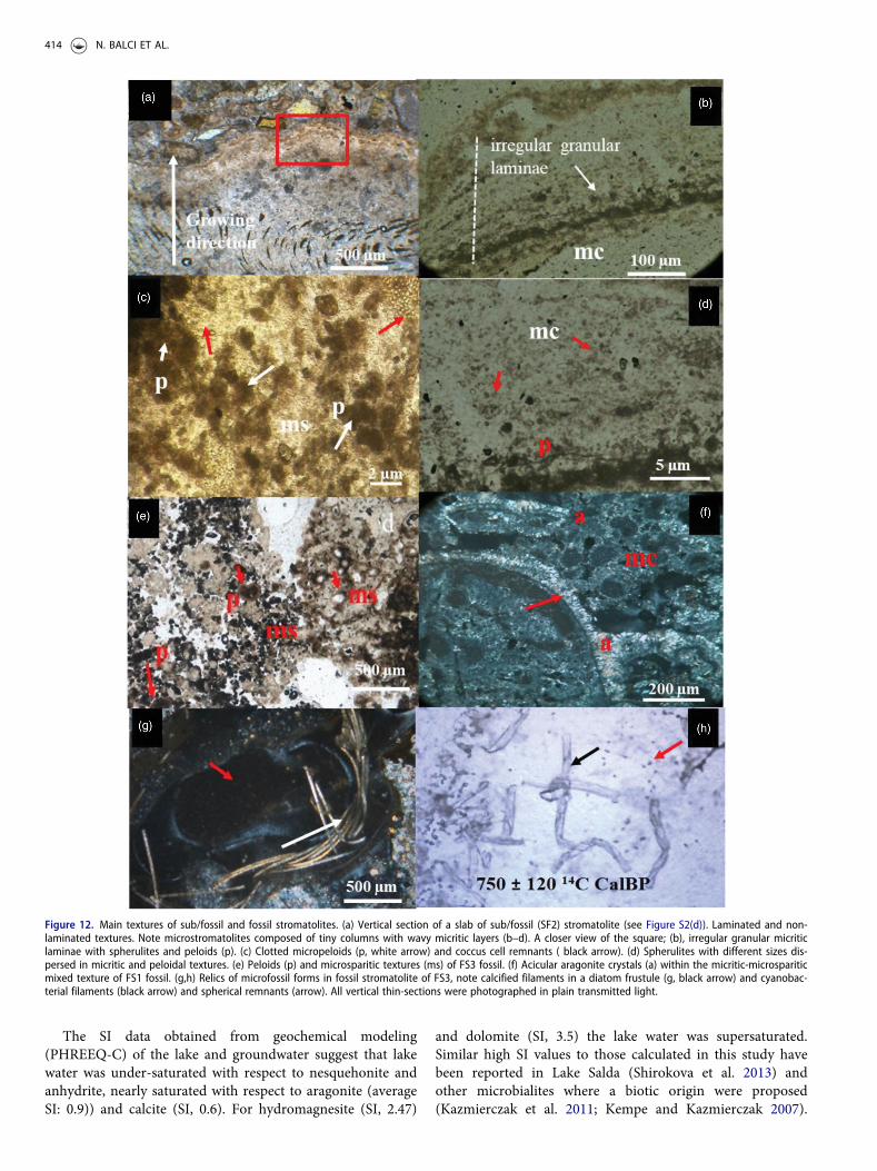
Some of the fossil stromatolites are intercalated with terracedeposits (Figure S2(a)) and remnants of dome structures arepreserved in the deposits (Figure S2(b)). The upper part ofthese terrace deposits shows blocky textures and cavities inthe cement (Figure S2(a), small box). Sub/fossil stromatolitesoccur in the form of cauliflower-like, nearly snow-white,large irregular mounds, domes (Figure S2(c)). These subfos-sil formations are particularly widespread along the SW partof the lake (Figure 1, Figure S2(c,d)). Their surfaces arepartly botryoidal and contain numerous rough knobs andgranules in various sizes (Figure S2(c,f,g)). The alternatingfine and coarse layers with millimeter-scale are easilyobserved. A cross-section of sub/fossil stromatolite slabshows mini radial columnar structures with internal layers(Figure S2(d)). Sub/fossil stromatolites with a columnarmorphology represent a rectangular (1), convex (2) andparabolic (3) lamination as internal structure (Figure S2(e,f)). Petrographic thin sections prepared from a slab of sub/fossil stromatolite revealed peloidal, microsparitic, andmicritic fabrics (Figure 12). On top of the growing directionof the slab, a course lamination with detrital grains on thetop was observed (Figure 12(a)). This course laminationwith detrital grains likely refer trapping and binding
processes near the surface. The detrital grains 100–500mmin diameter are composed of small fragments of mafic rocks.A concentrically laminated coliform texture is common onweak lamination composed of micritic and micro sporadiclayers (Figure 12(b)). A closer view of the irregular granularlaminae reveals peloids and irregularly dispersed spheres(Figure 12(c,d)). The micropeloidal structure and clusters ofremnant coccoids are closely associated with peloids inmicritic textures(Figure 12(c)). Various size of spheres iscommon in this layer (Figure 12(d)). In fossil stromatolites,peloidal textures are aggregated by dark micrite surroundedby microspar (Figure 12(e)). In some thin sections, radiallyfibrous and acicular crystals identified as aragonite inmicritic and/or microsparitic fabrics are observed (Figure12(f)). Examination with petrographic microscopy of fossilstromatolites dated to 750 ± 120 and 650 ± 100 calendar yearsBP revealed well-preserved remnants of cyanobacterial fila-ments and diatom frustules in the microfabrics (Figure12(g,h)). Furthermore, remnants closely resembling rodshape bacteria were occasionally present around the cyano-bacterial filaments (Figure 12(h)).
Stable C and O isotopes of stromatolites
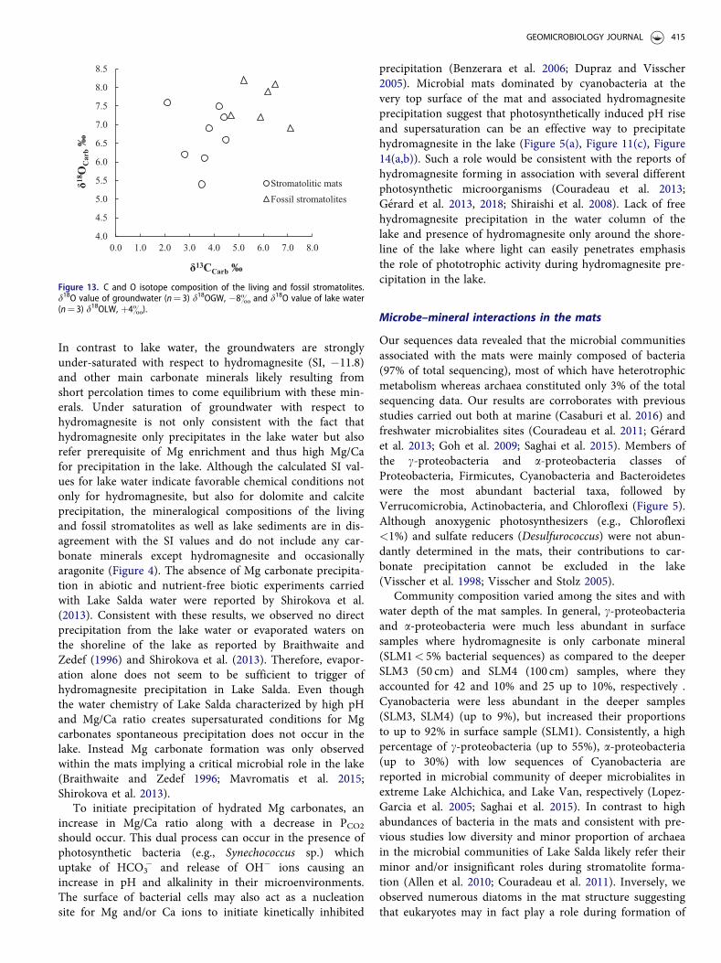
The d13C carb and d18Ocarb values of stromatolites are pre-sented in Figure 13. The d18Ocarb values of living and sub/fossil stromatolites are positive, ranging from þ5.4 toþ8.2&. These d18Ocarb values correspond to d18OV-smow val-ues ca. þ37 to þ40& (Coplen et al. 1983). Consistent withd18Ocarbvalues, the d13Ccarb values of all stromatolites arealso highly positive ranging from þ2.10 to þ7.10&. Thed13C carb values of living stromatolites are ca. þ2 to þ4&depleted in 13C relative to their fossil counterparts, whosed13C values fall within a range of þ4.7 to þ6.9&. Thed18Ocarb values of living stromatolites clustered in a narrowrange with an average value of ca. þ7& being comparablewith the fossil ones (þ7.6&). No significant correlationexists between d18Ocarb and d 13Ccarb values. d18OGW valuesof groundwater have a value of ca. �8& (n¼ 3), whereaslake water d18OLW values approximate þ4& (n¼ 4).
Discussion
Influence of physicochemical parameters onhydromagnesite precipitation
The concentration of magnesium ions is extremely highin a small number of closed lakes, and Lake Salda is oneof these. Mg compounds are more soluble than their cal-cium counterparts and therefore, it is rare for significantamounts of magnesium to precipitate in lakes with normalMg concentration. Nevertheless, our results demonstratedthat hydromagnesite with minor amount (up to 5%) ofaragonite precipitates within the mats of the living stro-matolites in Lake Salda indicating complex processes regu-lating precipitation reactions. Mg/Ca ratio of the reactivefluids has been considered as the main factor controllingcarbonates mineralogy and formation of hydrated Mg
0%
10%
20%
30%
40%
50%
60%
70%
80%
90%
100%
SLM1 SLM2 FS1 SF2 FS3
iGDGT-0
iGDGT-1
iGDGT-2
iGDGT-3
cren
cren'
0%
10%
20%
30%
40%
50%
60%
70%
80%
90%
100%
SLM1 SLM2 FS1 SF2 FS3
IaIbIcIIaIIa'IIbIIb'IIcIIc'IIIaIIIa'IIIbIIIb'IIIc
(a)
(b)
Figure 10. Relative concentrations (%) of iGDGTs (a) and bGDGTs (b) detectedin living (SLM1 and SLM2) and fossil (FS1 and FS3) and sub/fossil (SF2) stroma-tolites sampled at locations S1 and S2 in Lake Salda.
412 N. BALCI ET AL.

carbonates is usually attributed to high Mg/Ca ratio (>16)under low PCO2 values in lacustrine settings (H€anchenet al. 2008). These unique geochemical environments areusually consistent with alkaline lakes experiencing highevaporation conditions raising an abiotic origin forhydrated Mg carbonates such as hydromagnesite.However, a number of recent studies carried out in differ-ent geological settings (e.g., lakes) revealed otherwise andclearly showed microbial influences on precipitation of
hydrated Mg carbonate minerals (G�erard et al. 2013, 2018;Kazmierczak et al. 2011; Power et al. 2007; Saghai et al.2015; Sanz-Montero et al. 2019; Shirokova et al. 2013).Compared to local groundwater, elevated concentration ofMg along with high Mg/Ca ratio measured in the lakewater emphasis favorable conditions for chemical precipi-tation of particularly hydrated Mg carbonates (Table S1,Figure 3). The calculated SI values are in fact consistentwith this determination (Figure 3).
Figure 11. (a) Photomicrographs of the mat sample (SLM 2) and its dissected parts (b). (c, d) A view of intense cyanobacterial filaments of upper 0–5mm of themat (SLM2-1) representing meshwork structure. Note the filaments of Schizothrix sp. and Lyngba sp. (d) A view of Gloeocapsa sp. in the mat structure (SLM2-2). (e)Mat structure (SLM2-3), (1–4): diatoms; 1: Navicula sp.; 2: Pinnularia sp., 3: Amphora sp.; 4: Cyclotella sp. 5: filamentous cyanobacteria; 6: Cyanobacterial coccus; 7:rod shaped bacteria. (f) Bacterial cells (rod shapes, black arrow) around carbonate precipitation (the arrow) in the mat sample (SLM2-4). All mat samples were pho-tographed in transmitted plain light except (c) and (d) photographed in fluorescent microscope (10517).
GEOMICROBIOLOGY JOURNAL 413
a
The SI data obtained from geochemical modeling(PHREEQ-C) of the lake and groundwater suggest that lakewater was under-saturated with respect to nesquehonite andanhydrite, nearly saturated with respect to aragonite (averageSI: 0.9)) and calcite (SI, 0.6). For hydromagnesite (SI, 2.47)
and dolomite (SI, 3.5) the lake water was supersaturated.Similar high SI values to those calculated in this study havebeen reported in Lake Salda (Shirokova et al. 2013) andother microbialites where a biotic origin were proposed(Kazmierczak et al. 2011; Kempe and Kazmierczak 2007).
Figure 12. Main textures of sub/fossil and fossil stromatolites. (a) Vertical section of a slab of sub/fossil (SF2) stromatolite (see Figure S2(d)). Laminated and non-laminated textures. Note microstromatolites composed of tiny columns with wavy micritic layers (b–d). A closer view of the square; (b), irregular granular micriticlaminae with spherulites and peloids (p). (c) Clotted micropeloids (p, white arrow) and coccus cell remnants ( black arrow). (d) Spherulites with different sizes dis-persed in micritic and peloidal textures. (e) Peloids (p) and microsparitic textures (ms) of FS3 fossil. (f) Acicular aragonite crystals (a) within the micritic-microspariticmixed texture of FS1 fossil. (g,h) Relics of microfossil forms in fossil stromatolite of FS3, note calcified filaments in a diatom frustule (g, black arrow) and cyanobac-terial filaments (black arrow) and spherical remnants (arrow). All vertical thin-sections were photographed in plain transmitted light.
414 N. BALCI ET AL.
750 ± 120 14C CalBP
In contrast to lake water, the groundwaters are stronglyunder-saturated with respect to hydromagnesite (SI, �11.8)and other main carbonate minerals likely resulting fromshort percolation times to come equilibrium with these min-erals. Under saturation of groundwater with respect tohydromagnesite is not only consistent with the fact thathydromagnesite only precipitates in the lake water but alsorefer prerequisite of Mg enrichment and thus high Mg/Cafor precipitation in the lake. Although the calculated SI val-ues for lake water indicate favorable chemical conditions notonly for hydromagnesite, but also for dolomite and calciteprecipitation, the mineralogical compositions of the livingand fossil stromatolites as well as lake sediments are in dis-agreement with the SI values and do not include any car-bonate minerals except hydromagnesite and occasionallyaragonite (Figure 4). The absence of Mg carbonate precipita-tion in abiotic and nutrient-free biotic experiments carriedwith Lake Salda water were reported by Shirokova et al.(2013). Consistent with these results, we observed no directprecipitation from the lake water or evaporated waters onthe shoreline of the lake as reported by Braithwaite andZedef (1996) and Shirokova et al. (2013). Therefore, evapor-ation alone does not seem to be sufficient to trigger ofhydromagnesite precipitation in Lake Salda. Even thoughthe water chemistry of Lake Salda characterized by high pHand Mg/Ca ratio creates supersaturated conditions for Mgcarbonates spontaneous precipitation does not occur in thelake. Instead Mg carbonate formation was only observedwithin the mats implying a critical microbial role in the lake(Braithwaite and Zedef 1996; Mavromatis et al. 2015;Shirokova et al. 2013).
To initiate precipitation of hydrated Mg carbonates, anincrease in Mg/Ca ratio along with a decrease in PCO2should occur. This dual process can occur in the presence ofphotosynthetic bacteria (e.g., Synechococcus sp.) whichuptake of HCO3
� and release of OH� ions causing anincrease in pH and alkalinity in their microenvironments.The surface of bacterial cells may also act as a nucleationsite for Mg and/or Ca ions to initiate kinetically inhibited
precipitation (Benzerara et al. 2006; Dupraz and Visscher2005). Microbial mats dominated by cyanobacteria at thevery top surface of the mat and associated hydromagnesiteprecipitation suggest that photosynthetically induced pH riseand supersaturation can be an effective way to precipitatehydromagnesite in the lake (Figure 5(a), Figure 11(c), Figure14(a,b)). Such a role would be consistent with the reports ofhydromagnesite forming in association with several differentphotosynthetic microorganisms (Couradeau et al. 2013;G�erard et al. 2013, 2018; Shiraishi et al. 2008). Lack of freehydromagnesite precipitation in the water column of thelake and presence of hydromagnesite only around the shore-line of the lake where light can easily penetrates emphasisthe role of phototrophic activity during hydromagnesite pre-cipitation in the lake.
Microbe–mineral interactions in the mats
Our sequences data revealed that the microbial communitiesassociated with the mats were mainly composed of bacteria(97% of total sequencing), most of which have heterotrophicmetabolism whereas archaea constituted only 3% of the totalsequencing data. Our results are corroborates with previousstudies carried out both at marine (Casaburi et al. 2016) andfreshwater microbialites sites (Couradeau et al. 2011; G�erardet al. 2013; Goh et al. 2009; Saghai et al. 2015). Members ofthe c-proteobacteria and a-proteobacteria classes ofProteobacteria, Firmicutes, Cyanobacteria and Bacteroideteswere the most abundant bacterial taxa, followed byVerrucomicrobia, Actinobacteria, and Chloroflexi (Figure 5).Although anoxygenic photosynthesizers (e.g., Chloroflexi<1%) and sulfate reducers (Desulfurococcus) were not abun-dantly determined in the mats, their contributions to car-bonate precipitation cannot be excluded in the lake(Visscher et al. 1998; Visscher and Stolz 2005).
Community composition varied among the sites and withwater depth of the mat samples. In general, c-proteobacteriaand a-proteobacteria were much less abundant in surfacesamples where hydromagnesite is only carbonate mineral(SLM1< 5% bacterial sequences) as compared to the deeperSLM3 (50 cm) and SLM4 (100 cm) samples, where theyaccounted for 42 and 10% and 25 up to 10%, respectively .Cyanobacteria were less abundant in the deeper samples(SLM3, SLM4) (up to 9%), but increased their proportionsto up to 92% in surface sample (SLM1). Consistently, a highpercentage of c-proteobacteria (up to 55%), a-proteobacteria(up to 30%) with low sequences of Cyanobacteria arereported in microbial community of deeper microbialites inextreme Lake Alchichica, and Lake Van, respectively (Lopez-Garcia et al. 2005; Saghai et al. 2015). In contrast to highabundances of bacteria in the mats and consistent with pre-vious studies low diversity and minor proportion of archaeain the microbial communities of Lake Salda likely refer theirminor and/or insignificant roles during stromatolite forma-tion (Allen et al. 2010; Couradeau et al. 2011). Inversely, weobserved numerous diatoms in the mat structure suggestingthat eukaryotes may in fact play a role during formation of
4.0
4.5
5.0
5.5
6.0
6.5
7.0
7.5
8.0
8.5
0.0 1.0 2.0 3.0 4.0 5.0 6.0 7.0 8.0
Stromatolitic mats
Fossil stromatolites
δ13CCarb ‰
δ18O
Car
b‰
Figure 13. C and O isotope composition of the living and fossil stromatolites.d18O value of groundwater (n¼ 3) d18OGW, �8& and d18O value of lake water(n¼ 3) d18OLW, þ4&).
GEOMICROBIOLOGY JOURNAL 415@
t::,. t::,.t::,.
o o atı t::,.
o t::,. o
o o
o o e,.

stromatolites (Figure 11(e)). In future studies, we will assesstheir diversity, functions and exact roles in the lake.
Microbial community composition of the mat samplesimplies that precipitation of carbonates, mainly hydromag-nesite, in the lake may likely be a net result of metabolicactivities of phototrophs and heterotrophs. However, thesedeterminations may carry some uncertainties due to a
limited number of mat samples, mostly representing cauli-flower and dome shaped stromatolites in the lake.Nevertheless, prevailing occurrences of the cauliflower anddome shaped stromatolites in the lake give confidence that acorrelation between microbial diversity of the mats and theirmetabolic functions may in fact provide valuable insightsinto stromatolite formation. As indicated by the sequence
Figure 14. (a) Views of cyanobacterial filaments and plate like hydromagnesite crystal in the mat (SLM2). (b) Widespread occurrences of hydromagnesite crystalson the outer surface of cyanobacterial filaments and EPS (EDX from the cross). (c) Formation of first stage hydromagnesite spheres via aggregation of poorly packetplate like crystals on EPS. (d) Hydromagnesite spheres with tightly packet flakes. (e) A larger view of the square in (d), showing honey comb structure. (f)Hydromagnesite flakes covered by the cells glued by organic matrix.
416 N. BALCI ET AL.
A,;c. V Spoı ~lagn Deı WlJ 15.0 kV 4.0 10000x BSE 69
'
- . .• . •• ·•·•ı- , .. .... ,~• --·. --
··,;,:..·• ·· .. ·•···~· e ',\, .
-- 'I', '-, ' ' -~ .
llJIII I AK Si 1 ?!UlkV x:ıo .ooo 100rırı7" WI} 14 ')111111

data, oxygenic cyanobacteria (Synechococcus sp.Halospirulina, Gloeocapse sp.), particularly filamentous(Schizothrix sp., and Lyngbya sp) are dominant microbialcells at top 5mm of the mats (Figure 11(c,d)). SEM imagesof this zone (0–5mm, SLM2-1) revealed that the precipita-tion is closely associated with the cyanobacteria filamentsembedded by EPS (Figure 11(c,f), Figure 14(a,b)). Presenceof Mg, Ca and Si on the EPS matrix demonstrated thatchemical properties and their functional groups of the EPSprovide effective nucleation sites for Mg and Ca ions as pre-viously observed in various microbialites sites (Couradeauet al. 2011; Kazmierczak et al. 2011; Lopez-Garcia et al.2005; Saghai et al. 2015) (Figure 14(a), EDS spectra).Consistently, a large quantity of acidic functional groups inthe EPS (ABM assay) at upper surface implies a high bind-ing capacity of the EPS, particularly calcium, likely leavingMg more available for precipitation (Figure 7(b)) (Pace et al.2018). Mineralogical composition of the mat comprisedmainly of hydromagnesite further consistent with this(Figure 4). Besides providing nucleation sites, cyanobacteria(e.g., Schizothrix sp.) cause pH increase and alkalinity intheir microenvironments via their extracellular carbonicanhydrase by dissociating bicarbonate ions into OH andCO2 and. Later, consumption of CO2 by photosyntheticmicroorganisms trigger precipitation of excess Mg in thereactive fluid (Braissant et al. 2009; Kawaguchi and Decho2002; Reid et al. 2000). In addition to oxygenic photosynthe-sizers, anoxygenic photosynthetic bacteria (e.g., Chloroflexi)and heterotrophs such as Firmicutes (e.g., Exiguobacterium),c-proteobacteria (Pseudomonas and Rheinheimera species)identified in the mats may likely contribute to precipitationby increasing alkalinity.
At depths (>10mm) aerobic heterotrophic bacteria maycause pH increase by respiring N rich organic compounds, byproduct of oxygenic photosynthesis and oxidative deamin-ation (NH4/NH3 system) provides pH conditions similar tothose found at the surface of the microbial mat during photo-synthesis (Hatayama and Saito 2019; Yang et al. 2017). Thecapacity of heterotrophs to precipitate carbonates (e.g., ara-gonite, hydromagnesite and dolomite) has been documentedby experimental (Balci and Demirel 2016; Rivadeneyra et al.1999, 2004; Sanchez-Roman et al. 2011) and field studies(G�erard et al. 2013, 2018; Lopez-Garcia et al. 2005; Sanz-Montero et al. 2019).
Consistent with the other natural environments, abun-dant and diverse heterotrophic bacteria (e.g., Firmicutes)including many Proteobacteria and Firmicutes, identified inthe mats, particularly deeper part of the lake highlightsimportant role of heterotrophic bacteria, at least alkaliphilicmembers of Firmicutes, in formation of microbialites asstated earlier by Sanz-Montero et al. (2019).
Influence of microbial metabolisms on carbonatemicrofabrics
Hydromagnesite represents up to ca. 92% of the bulk min-eralogy of the mat samples (SLM1and SLM2) along withminor amount of aragonite (up to 5%) (Figure 4).
Previously, biologically induced hydromagnesite and aragon-ite formations are reported in different lacustrine microbia-lites (Couradeau et al. 2011, 2012; G�erard et al. 2013, 2018;Kazmierczak et al. 2011; Saghai et al. 2015). Morphologicalobservation of the dissected mat sample (SLM2) along withthe depth profile of EPS revealed varies types of hydromag-nesite microfabrics closely coupled with the degradation ofEPS (Figures 6 and 14).
Early incipient hydromagnesite precipitation is particu-larly widespread and coincided with cyanobacterial rich topmat layer (SLM2-1 section, 0-5mm) (Figure 2(b,d), Figure11(b), Figure 14(a,b)). Compared to 5–10mm of the matEPS polymers of this layer have relatively less acidic func-tional groups, providing limited nucleation sites for Mg andCa ions, as indicated by Alcien Blue content (ABA) of EPS(1.3� 104 mg/g EPS). Hydromagnesite produced in this toplayer consists of mostly plate like crystals growing on EPS(Figure 11(a,b); Figure 14(a–b) and are considered as aproduct of high photosynthesis with decreasing degreeof inhibition.
At depth 5–10mm where intermixed green and orangemat layer is located (SLM2-2, Figure 11(b)) the amount ofacidic functional groups in the EPS increased and scarcehydromagnesite spherulites with 10 and 20 mm in size wereformed via aggregation of plate like hydromagnesite crystals(Figure 14(b,c)). These spheres are likely a product of highphotosynthesis effect simultaneous to a high inhibition bypristine exopolymers as indicated by their enriched d13C val-ues and EPS profile (Figure 13) (Arp et al. 2012). At depth10–15mm a sharp decrease in the amount of EPS refers asuccessive heterotrophic activity leading to formation ofhydromagnesite spheres with honeycomb structures (Figure14(d,e)). Apparently, acidity of the EPS affected the morph-ology of hydromagnesite (Braissant et al. 2003) and partlydegraded EPS likely provided nucleation sites for Mg to pro-mote hydromagnesite precipitation. Similarly, formation ofhydromagnesite with honeycomb structure was reported inculture experiments with Synechococcus sp. isolated fromLake Salda by Shirokova et al. (2013) and in Lake Eras, anextreme alkaline lake in Spain, by Sanz-Montero et al.(2019). A dumbbell shape mineral, likely dolomite, andsome precipitates associated with microbial cells on the sur-face of these spheres are occasionally observed(Figure 14(d)).
Unlike EPS rich zones grape-like hydromagnesite spheru-lites, measuring between 2–3 mm, deposit among the plate-like crystals in the EPS-poor layers (SLM2-3, SLM2-4;15–20mm) (Figure 6(a–b)). This type microfabric observedonly at EPS minimum zone coincided with a high numberof heterotrophic bacteria (e.g., Firmicutes, Figure 5).Aggregations of hydromagnesite nanoparticles and bacterialsheaths occasionally covered by nanoglobules are often asso-ciated with these small spheres (Figure 6(c–d)). Clusters ofplate like hydromagnesite crystals on bacterial cellsembedded by EPS produced rosette type morphology(Figure 6(b)). Spherical carbonate nanoglobules tightly asso-ciated with cell surfaces of heterotrophic bacteria(e.g., Firmicutes) were identified in the deeper part of the
GEOMICROBIOLOGY JOURNAL 417
mat (Figure 6(e), SLM2-4), where the lowest amount of EPSwas measured (Figure 7). EDS pattern of these globules andXRD pattern the mat sample (SLM2) revealed Ca and ara-gonite, respectively (Figure 4, Figure 6(h)). Occurrences ofthese cell bound aragonite nanoglobules and smaller sizehydromagnesite spherulites covered by nanoparticles at theEPS minimum zone are attributed to a heterotrophicdecomposition of EPS matrix and thus decreasing degree ofinhibition (Braissant et al. 2009; Dupraz et al. 2004; Duprazand Visscher 2005).
Formation of nanoglobular carbonates, particularly ara-gonite, have been reported in microbialites (Couradeau et al.2011; G�erard et al. 2013; Lopez-Garcia et al. 2005;Sanz-Montero et al. 2019). Consistently, various spheruliticcarbonates have been shown to precipitate under the influ-ence of organic matter during aerobic heterotrophic activity(Benzerara et al. 2006; Gomez et al. 2018; Krause et al. 2012;Rivadeneyra et al. 1999). Sanz-Montero et al. 2019 attributedthe formation of carbonate nanoglobules to declining meta-bolic activity of heterotrophs due to organic substrate short-age. The same formation mechanism may apply for our caseand hydromagnesite and aragonite nanoglobules identifiedin the EPS-poor zone were likely produced by heterotrophicbacteria as a response to decreasing organic matter.
The lipid composition of living and fossil stromatolites
The lipid distribution reflects the major mat-building micro-organisms, as well as the incorporation into the mat systemof lipids derived from non-building aquatic and terrestrialorganisms. The presence of n-C17:0 alkane, 6Me-C17:0, 7Me-C17:0, n-C19:1 alkene and diploptene is indicative of cyano-bacterial activity as these compounds are known markers forcyanobacteria (Bauersachs et al. 2017; Coates et al. 2014;Jahnke et al. 2004). They have been found in hypersalinecyanobacterial mats (Jahnke et al. 2004; Scherf andRullk€otter 2009), in freshwater microbial mat constructed bycalcifying cyanobacteria (Schizothrix sp.; Thiel et al. 1997),as well as in stromatolites (Allen et al. 2010; Pag�es et al.2014). Diploptene may also originate from methylotrophs(Summons et al. 1994) and sulfate-reducing bacteria(Blumenberg et al. 2006). Both C25:2 HBI and stigmasterolare very likely produced by diatoms such as Navicula sp.and Amphora sp., respectively (Kaiser et al. 2016). Phytaneis also common in microbial mats (Allen et al. 2010; Jahnkeet al. 2008; Slowakiewicz et al. 2016). It may be derived dur-ing diagenesis from the phytyl side chain of chlorophyll a orbacteriochlorophyll (Volkman and Maxwell 1986), but alsofrom the phytanyl side chains of ether-linked polar lipidsproduced by methanogenic (Rowland and Robson 1990;Volkman 1986) and halophilic Archaea (Allen et al. 2010).
iGDGTs, including crenarchaeol (cren) and its isomer(cren’), are produced by Archaea (Schouten et al. 2013) andhave been detected previously in microbial mats (Schoutenet al. 2007; Slowakiewicz et al. 2016). While cren is consid-ered as specific marker for Thaumarchaeota, iGDGT-0 ismost abundant in methanogen Archaea (Schouten et al.2013). High relative amounts of iGDGT-0 in sample LS1
(Figure 6) likely reflect the high abundance of the methano-gen Archaea Methanosphaera (SLM1; Figure 5).Comparatively, the lower percentage of iGDGT-0 and therelatively higher percentage of cren in sample LS2 mayreflect the lower relative amount of methanogen Archaea(SLM2). bGDGTs are very likely produced by Acidobacteria(Sinninghe Damst�e et al. 2011; Weijers et al. 2006) and werefound in hot spring sediments and cyanobacterial mats(Hedlund et al. 2013; Slowakiewicz et al. 2016). bGDGTsmay be in situ produced in hot springs by Acidobacteriaindigenous to stromatolites, particularly for bGDGT-Ia, Ib,and Ic (Hedlund et al. 2013). Furthermore, bGDGTs mightalso be produced by bacteria of the phyla Bacteroidetes,Dictyoglomi, and the candidate phylum ‘Atribacteria’(Zhang et al. 2013). However, an allochthonous terrestrialsource cannot be ruled out since bGDGTs are known to beabundant in soils (Weijers et al. 2006).
Concerning lipids of non-building organisms, n-C27:0, n-C29:0, and n-C31:0 alkanes land plant wax lipids were alsofound in other microbial mats and stromatolites, and theirpresence reflects a significant incorporation of terrestrially-derived organic material into the mat system (Allen et al.2010; Thiel et al. 1997). The main sterols and stanols coun-terparts (produced by bacterial hydrogenation of sterols)were also found as most abundant steroids in other micro-bial mats and stromatolites (Allen et al. 2010; Scherf andRullk€otter 2009; Slowakiewicz et al. 2016; Thiel et al. 1997),and are very likely produced by the dinoflagellatePeridinium cinctum in Lake Salda (Kaiser et al. 2016).Although n-C18:0 alkanol has been found in cyanobacterialmats and stromatolites (Slowakiewicz et al. 2016), relativeamounts of n-C18:0, n-C20:0, and n-C22:0 alkanols as high asin Lake Salda were not observed in other stromatolites sofar. Microalgae may represent a direct source for these alka-nols (Rezenka et al. 1986).
The distribution of the main stable lipid biomarkersfound in Lake Salda living stromatolites seems to reflect partof the bacterial, archaeal and eukaryote communities. Boththe microbial and lipid composition suggest two differentstromatolite provinces in Lake Salda, one located in theSouthwest (S1 site, including SLM1 and SF2 samples) andone located in the east (S2 site, including SLM2, and FS3,FS1 samples). Based on the available dataset, we suggesthere that the differences between these two main provincesmay related to the geographical and sampling depth whereSLM1 and SF1 samples represents upper most part of theliving and subfossil stromatolites, respectively (Figures S1and S2). Importantly, living stromatolites are apparentlyexcellent analogs as the lipids produced by the microbialand eukaryote communities are very similar to thoseobserved from the fossil stromatolites.
Formation mechanisms of stromatolites: insights fromthe new findings
Formation of Lake Salda stromatolites is solely attributed tocyanobacterial activity by different authors (Braithwaite andZedef 1994, 1996; Russell et al. 1999; Shirokova et al. 2011,2013). However, microbial community structure and
418 N. BALCI ET AL.

microscopic observations of the stromatolitic mats suggestthat hydromagnesite precipitation is largely controlled by aconsortium of cyanobacteria and aerobic heterotrophic bac-teria (Figures 5 and 6, Figures 7 and 11, Figure 14). Ourresults for the first time provide strong evidence for a closeassociation of aerobic heterotrophic bacteria with precipita-tion of carbonates in Lake Salda stromatolitic mats.
A number of studies have clearly demonstrated the abilityof aerobic heterotrophic bacteria to use C and N rich pro-teinaceous substances produced by cyanobacteria, and thatthese organisms thus play a significant role in C cycling byregenerating CO2 required for photosynthesis (Behrens et al.2008; Ionescu et al. 2015; Pett-Ridge and Weber 2012).Carbonate precipitation in such a consortium would occur ifphotosynthesis (P) > respiration (R), and if R>P dissol-ution would take place (Pinckney and Reid 1997).Consequently, hydromagnesite precipitation is likely theresult of the net metabolic activities of aerobic heterotrophsand oxygenic photosynthesis regulating CO2 cycling and pHwithin the microbial mat. In the lake, activity of this micro-bial consortium was printed on carbonate mineralogy andtextures taking place on the decaying organic matrices (EPS)and bacterial cells in the mats (Figures 6 and 14).
Our results demonstrated that bacterial cells and abun-dance of EPS and their chemical functional groups (e.g.,acidic groups) play a critical role in the lake to overcomekinetically inhibited Mg precipitation as suggested by SI val-ues of the lake water.
Based on depth profile of EPS, microbial communitystructure and carbonate textures of the mat the following
processes likely responsible for carbonate, mainly hydromag-nesite, precipitation in the mat: (1) EPS binds Mg and Cavia their functional groups (e.g., acidic functional groups)and kinetically inhibits nucleation and thus precipitationand (2) depth profile of EPS demonstrate a degradationrelated to increasing aerobic heterotrophic activity (Figure15). In 10mm of the mat, cyanobacteria and possibly dia-toms colonized the surface of the mat and create carbonate-saturated conditions by consuming CO2 and thus promotealkalinity and pH increase. However, owing to a large quan-tity of acidic functional groups in the EPS this layer pos-sesses a high binding Mg and Ca capacity and therefore animmediate precipitation may not occur. Lack of continueslamination and presence of patchy carbonates, hydromagne-site, in the mat zone likely occur as a result of local degrad-ation of EPS providing nucleation sites and due to highMg/Ca ration in the overlying water. Taking into accountthat the lake water has low Ca concentration exopolymersbinding Ca will even limit or prevent Ca carbonate precipi-tation at 0–10mm of the mat (Dupraz et al. 2009) (TableS1). Fluctuation in water level, UV may also contribute toEPS degradation at the upper zone (0–5mm). It is highlylikely that a balance between degradation and continues pro-duction of EPS controls precipitation of hydromagnesite byproviding nucleation sites to overcome kinetic inhibition atthis zone.
As demonstrated by EPS profile increasing degradation ofEPS by heterotrophic bacteria occurs in lower part of themat (15–20mm), releasing Ca and Mg previously bound toEPS and greatly decrease kinetic inhibition for nucleation
Figure 15. A conceptual model describing Mg and Ca precipitation in the microbial mat.
GEOMICROBIOLOGY JOURNAL 419
EPSµg/g (dw)
O 5000 10000 15000 20000
--EPS (PSAA) ----EPS (ABA)
High photosynthesis, low EPS degradatioıı (e.g UV) Limited ııucleatioıı sites: (Initiatioıı of plate tike Hmg cıystal )
High plıotosynthesis at low nucleation sites: (iııitiation ofHmg sphenılites)
Degradation of EPS matrix, lıiglı nucleation sites: (Growth of Hıng spheıulites)
Stroııg EPS degradatioıı and lıigh ııucleation sites: Growtlı aııd accmnulation of sınall Hıııg spherulites Ca released by EPS degredation and removal as CaCO3
.---Filanıeııtous cyanobacteria
Coccus cyanobacteria
+ Plate like hydroınagnesite (Hıng) crystals
O Hydroınagııesite sphenılites
- Diatonıs _. Anoxygeııic cyanobacteria
::ı, Aerobic lıeterotrophic bacteria
~ Grape like hydronıagnesite splıenılites
.. aııoglobular aı·agonite
,e Trappiııg aııd biııdiııg particles

(Braissant et al. 2009; Ionescu et al. 2015). This degradationprocess may create microenvironments with a relatively highCa content and finally trigger Ca carbonate mineral precipi-tation. Grape like hydromagnesite spheres covered by nano-particles and aragonite nanoglobules closely associated withthe cell surfaces likely occur at this stage (Figure 6(a,d,f)).All these processes indicate that partial degradation of EPSis likely a rate-limiting step for hydromagnesite precipitationby creating local nucleation sites and thus allowing precipi-tation. Discontinues lamination with micropeloidal texturesidentified in the sub/fossil stromatolites can be attributed tolocal mineralization of EPS following its degradation(Dupraz et al. 2009) (Figure 12(c,e)).
Several metabolic pathways associated with aerobic het-erotrophic bacteria have been reported to contribute to car-bonate formation via production of ammonia throughoutdegradation of N rich organic compounds (e.g., peptone, N-compounds!NH3þH2O!NH4
þ þOH�), thereby increas-ing alkalinity, which favors precipitation (Balci et al. 2016;Balci and Demirel 2016; Gonz�alez-L�opez et al. 2005;Rivadeneyra et al. 1999, 2004; S�anchez-Rom�an et al. 2011,2019). Presence of abundant aerobic heterotrophs (e.g.,Firmicutes) in the mats suggest that the same microbialmetabolisms likely occur and promote saturation conditionsrequired for the precipitation of carbonates in the mat layers(Cabestrero and Sanz-Montero 2018). Consistently, the for-mation of Mg carbonates (e.g., hydromagnesite and dolomitenanoglobules) on decayed EPS closely associated with heter-otrophs are reported in alkaline lakes (Alc�antara-Hern�andezet al. 2017). Similar to Lake Salda, Mg carbonate precipita-tion (e.g., hydromagnesite, dolomite nanoglobules) regulatedby heterotrophic degradation of organic matrix (e.g., EPS) inan evaporative and ephemeral alkaline Lake Eras has beenrecently documented by Sanz-Montero et al. (2019). Thegeneral microbial community, carbonate mineralogy andtextures of Lake Salda mats are comparable to other micro-bialites independently of physicochemical conditions such aswater salinity, content, temperature or pH (G�erard et al.2013, 2018; Salvatore et al. 2018; Sanz-Montero et al. 2019).These similarities within the microbial populations and car-bonate mineralogy suggest that metabolic activities andenvironmental interactions may be more critical than taxo-nomic diversity and that a diverse range of metabolisms,rather than cyanobacteria, may have been likely involved instromatolite formations preserved in the geological records.
The lack of co-variance among d13Ccarb and d18Ocarb val-ues and the enrichment in d13Ccarb values of the bulk car-bonate (92–95% hydromagnesite) of the living stromatolitesindicate that 13C preferentially enriched in hydromagnesite(Figure 13). High carbonate d13C values measured in differ-ent microbialites (e.g., Pavillion, average þ2.31&; Great SaltLakes,) are generally attributed to photosynthetic uptake of12CO2 leaving isotopically heavy carbon for precipitation(Chagas et al. 2016, and references therein) whereas carbo-nates with negative d13C values are considered to reflect het-erotrophic activity producing carbon source with lowd13CDIC values such as sulfate reduction (Neuweiler et al.1997) and oxidative biodegradation. Our results
demonstrated that the precipitation in the stromatolitic matswas associated with coupled processes of photosynthetic andaerobic heterotrophic activity. Although it was not possibleto quantitatively determine contribution of each metabolicpathway to d13CDIC values of lake water, high d13C values ofthe hydromagnesite refer to photosynthetic activity as a pri-mary producer for carbonate mineral saturation in the lake.In contrast to hydromagnesite occurrences close associationof aragonites with bacteria surface demonstrated that ara-gonite is likely a product of heterotrophic activity occurringin the mat. However, low percentages of aragonite (3–6%)in the bulk carbonate hinder d13C values of aragonite pro-nounced on the bulk carbonate due to its low mass (Balciet al. 2007).
Hydromagnesite precipitation in the lake likely occurs asthe lake water is subject to evaporation as indicated by thehigh d18OLWvalues (þ4&) of lake water relative to d18OGW
values (�8&) of groundwater. The high d18Ocarb valuesidentified in this study are in good agreement to the d18Orange reported for Lake Salda (Braithwaite and Zedef 1996)and hydromagnesite rich carbonates closely associated withmicrobial mats in lacustrine lakes (Power et al. 2007; Sanz-Montero et al. 2019).
A good correlation exists between d13C values of theliving and fossil stromatolites suggest that the fossil stroma-tolites may have been mineralized in similar water chemistryunder the influence of similar microbial interactions tothose currently occurring in the lake, at least for thelast 750 years.
Biosignature preservation potential of Lake Saldastromatolites
Hydromagnesite formation has been reported in a range ofgeologically different settings (G�erard et al. 2013;Kazmierczak et al. 2011; Power et al. 2009; Sanz-Monteroet al. 2013, 2019). However, the persistence of hydromagne-site microbialites and their long-term stability is question-able (Russell et al. 1999). For Lake Salda stromatolites,Russel et al. (1999) argued that hydromagnesite stromatolitesdegenerate soon after their formation to porous, poorly lithi-fied rocks, and their subsequent diagenetic alteration tomagnesite might lower biosignature preservation potential ofthe stromatolites. However, these suggestions are largelybased on the macro-scale morphological features of the stro-matolites. In contrast, we find that the mineralogical com-position of the fossil stromatolites shows the persistence ofhydromagnesite at least for the last 750 years. Consistentwith our results, preservation of microfabrics and morpholo-gies of Miocene aged magnesitic microbialites and presenceof 2.8 ka BP aged microbially induced hydromagnesite inLake Alchichica (Puebla, Mexico) gives further evidence ofbiosignature integrity of Mg-rich microbialites (Kazmierczaket al. 2011; Sanz-Montero and Rodr�ıguez-Aranda 2012).
Petrographic observations of the mats clearly revealedhydromagnesite precipitation in cyanobacterial sheaths andEPS with various textures including spherical ones formedby clusters of plate-like crystals showing the honeycomb-like
420 N. BALCI ET AL.
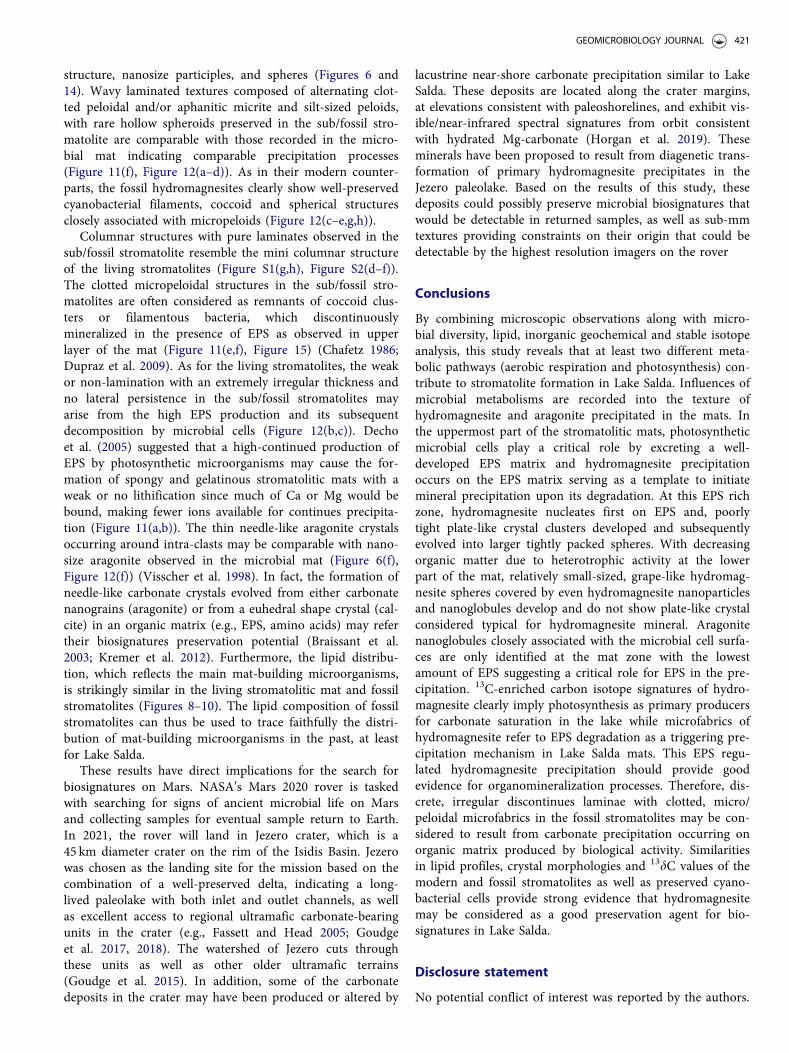
structure, nanosize participles, and spheres (Figures 6 and14). Wavy laminated textures composed of alternating clot-ted peloidal and/or aphanitic micrite and silt-sized peloids,with rare hollow spheroids preserved in the sub/fossil stro-matolite are comparable with those recorded in the micro-bial mat indicating comparable precipitation processes(Figure 11(f), Figure 12(a–d)). As in their modern counter-parts, the fossil hydromagnesites clearly show well-preservedcyanobacterial filaments, coccoid and spherical structuresclosely associated with micropeloids (Figure 12(c–e,g,h)).
Columnar structures with pure laminates observed in thesub/fossil stromatolite resemble the mini columnar structureof the living stromatolites (Figure S1(g,h), Figure S2(d–f)).The clotted micropeloidal structures in the sub/fossil stro-matolites are often considered as remnants of coccoid clus-ters or filamentous bacteria, which discontinuouslymineralized in the presence of EPS as observed in upperlayer of the mat (Figure 11(e,f), Figure 15) (Chafetz 1986;Dupraz et al. 2009). As for the living stromatolites, the weakor non-lamination with an extremely irregular thickness andno lateral persistence in the sub/fossil stromatolites mayarise from the high EPS production and its subsequentdecomposition by microbial cells (Figure 12(b,c)). Dechoet al. (2005) suggested that a high-continued production ofEPS by photosynthetic microorganisms may cause the for-mation of spongy and gelatinous stromatolitic mats with aweak or no lithification since much of Ca or Mg would bebound, making fewer ions available for continues precipita-tion (Figure 11(a,b)). The thin needle-like aragonite crystalsoccurring around intra-clasts may be comparable with nano-size aragonite observed in the microbial mat (Figure 6(f),Figure 12(f)) (Visscher et al. 1998). In fact, the formation ofneedle-like carbonate crystals evolved from either carbonatenanograins (aragonite) or from a euhedral shape crystal (cal-cite) in an organic matrix (e.g., EPS, amino acids) may refertheir biosignatures preservation potential (Braissant et al.2003; Kremer et al. 2012). Furthermore, the lipid distribu-tion, which reflects the main mat-building microorganisms,is strikingly similar in the living stromatolitic mat and fossilstromatolites (Figures 8–10). The lipid composition of fossilstromatolites can thus be used to trace faithfully the distri-bution of mat-building microorganisms in the past, at leastfor Lake Salda.
These results have direct implications for the search forbiosignatures on Mars. NASA’s Mars 2020 rover is taskedwith searching for signs of ancient microbial life on Marsand collecting samples for eventual sample return to Earth.In 2021, the rover will land in Jezero crater, which is a45 km diameter crater on the rim of the Isidis Basin. Jezerowas chosen as the landing site for the mission based on thecombination of a well-preserved delta, indicating a long-lived paleolake with both inlet and outlet channels, as wellas excellent access to regional ultramafic carbonate-bearingunits in the crater (e.g., Fassett and Head 2005; Goudgeet al. 2017, 2018). The watershed of Jezero cuts throughthese units as well as other older ultramafic terrains(Goudge et al. 2015). In addition, some of the carbonatedeposits in the crater may have been produced or altered by
lacustrine near-shore carbonate precipitation similar to LakeSalda. These deposits are located along the crater margins,at elevations consistent with paleoshorelines, and exhibit vis-ible/near-infrared spectral signatures from orbit consistentwith hydrated Mg-carbonate (Horgan et al. 2019). Theseminerals have been proposed to result from diagenetic trans-formation of primary hydromagnesite precipitates in theJezero paleolake. Based on the results of this study, thesedeposits could possibly preserve microbial biosignatures thatwould be detectable in returned samples, as well as sub-mmtextures providing constraints on their origin that could bedetectable by the highest resolution imagers on the rover
Conclusions
By combining microscopic observations along with micro-bial diversity, lipid, inorganic geochemical and stable isotopeanalysis, this study reveals that at least two different meta-bolic pathways (aerobic respiration and photosynthesis) con-tribute to stromatolite formation in Lake Salda. Influences ofmicrobial metabolisms are recorded into the texture ofhydromagnesite and aragonite precipitated in the mats. Inthe uppermost part of the stromatolitic mats, photosyntheticmicrobial cells play a critical role by excreting a well-developed EPS matrix and hydromagnesite precipitationoccurs on the EPS matrix serving as a template to initiatemineral precipitation upon its degradation. At this EPS richzone, hydromagnesite nucleates first on EPS and, poorlytight plate-like crystal clusters developed and subsequentlyevolved into larger tightly packed spheres. With decreasingorganic matter due to heterotrophic activity at the lowerpart of the mat, relatively small-sized, grape-like hydromag-nesite spheres covered by even hydromagnesite nanoparticlesand nanoglobules develop and do not show plate-like crystalconsidered typical for hydromagnesite mineral. Aragonitenanoglobules closely associated with the microbial cell surfa-ces are only identified at the mat zone with the lowestamount of EPS suggesting a critical role for EPS in the pre-cipitation. 13C-enriched carbon isotope signatures of hydro-magnesite clearly imply photosynthesis as primary producersfor carbonate saturation in the lake while microfabrics ofhydromagnesite refer to EPS degradation as a triggering pre-cipitation mechanism in Lake Salda mats. This EPS regu-lated hydromagnesite precipitation should provide goodevidence for organomineralization processes. Therefore, dis-crete, irregular discontinues laminae with clotted, micro/peloidal microfabrics in the fossil stromatolites may be con-sidered to result from carbonate precipitation occurring onorganic matrix produced by biological activity. Similaritiesin lipid profiles, crystal morphologies and 13dC values of themodern and fossil stromatolites as well as preserved cyano-bacterial cells provide strong evidence that hydromagnesitemay be considered as a good preservation agent for bio-signatures in Lake Salda.
Disclosure statement
No potential conflict of interest was reported by the authors.
GEOMICROBIOLOGY JOURNAL 421

Funding
This work was supported by Istanbul Technical University ResearchDivision (ITU-BAP) grant to N. Balci [40585] and the Scientific andTechnological Research Council of Turkey (TUBITAK) grant to N.Balci [113Y464].
References
Akcer-€On S, Bora €On Z, Ca�gatay MN, ERiS KK, Sakınc M, Wulf S,Helvacı C, Acar D, G€undo�gan _I, Akkiraz S, et al. 2016. High reso-lution Middle-Late Holocene climatic records from Lake Salda (W.Anatolia) Sediments. Proceedings of MedCLIVAR 2016 Conference,Athens, Greece.
Alcántara-Hernández RJ., Valdespino PM, Centeno CM, Alcocer J,Merino-Ibarra M, Falcón LI. 2017. Genetic diversity associated to Ncycle pathways in microbialites from Lake Alchichica, Mexico.Aquat Microb Ecol 78(2):121–133.
Allen MA, Neilan BA, Burns BP, Jahnke LL, Summons RE. 2010. Lipidbiomarkers in Hamelin Pool microbial mats and stromatolites. OrgGeochem 41(11):1207–1218.
Allwood AC, Walter MR, Kamber BS, Marshall CP, Burch IW. 2006.Stromatolite reef from the Early Archaean era of Australia. Nature441(7094):714–718.
Altermann W, Kazmierczak J, Oren A, Wright D. 2006. Microbial cal-cification and its impact on the sedimentary rock record during 3.5billion years of Earth history. Geobiology 4(3):147–166.
Arp G, Helms G, Karlinska K, Schumann G, Reimer A, Reitner J,Trichet J. 2012. Photosynthesis versus exopolymer degradation inthe formation of microbialites on the atoll of Kiritimati, Republic ofKiribati, Central Pacific. Geomicrobiol J 29(1):29–65.
Awramik SM, Buchheim HP. 2009. A giant, Late Archean lake system:the Meentheena Member (Tumbiana Formation; Fortescue Group),Western Australia. Precambrian Res 174(3–4):215–240.
Awramik SM, Buchheim HP. 2015. Giant stromatolites of the EoceneGreen River Formation (Colorado, USA). Geology 43(8):691–694.
Awramik SM, Grey K. 2005. Stromatolites: biogenicity, biosignatures,and bioconfusion. Paper presented at: Proceedings of SPIE, SanDiego, CA.
Balci N, Demirel C. 2016. Formation of carbonate nanoglobules by amixed natural culture under hypersaline conditions. Minerals 6(4):122.
Balci N, Demirel C, Akcer-On S, G€ultekin AH, Kurt MA. 2018.Evaluating abiotic and microbial factors on carbonate precipitationin Lake Acig€ol, a hypersaline lake in Southwestern Turkey.Quaternary Int 486:116–128.
Balci N, Menekse M, Karag€uler N, S€onmez MS, Meister P. 2016.Reproducing authigenic carbonate precipitation in the hypersalineLake Acıg€ol (Turkey) with microbial cultures. Geomicrobiol J 33(9):758–773.
Balci N, Shanks WC, Mayer B, Mandernack KW. 2007. Oxygen andsulfur isotope systematics of sulfate produced by bacterial and abi-otic oxidation of pyrite. Geochim Cosmochim Acta 71(15):3796–3811.
Batchelor M, Burne R, Henry B, Slatyer T. 2005. Statistical physics andstromatolite growth: new perspectives on an ancient dilemma.J Phys A 35:6–11.
Bauersachs T, Talbot HM, Sidgwick F, Sivonen K, Schwark L. 2017.Lipid biomarker signatures as tracers for harmful cyanobacterialblooms in the Baltic Sea. PLoS One 12(10):e0186360.
Behrens S, Losekann T, Pett-Ridge J, Weber PK, Ng W-O, StevensonBS, Hutcheon ID, Relman DA, Spormann AM. 2008. Linking micro-bial phylogeny to metabolic activity at the single-cell level by usingenhanced element labeling-catalyzed reporter deposition fluores-cence in situ hybridization (EL-FISH) and NanoSIMS. Appl EnvironMicrob 74(10):3143–3150.
Benzerara K, Menguy N, L�oPez-Garcia P, Yoon T-H, Kazmierczak J,Tyliszczak T, Guyot F, Brown EJ. Jr. 2006. Nanoscale detection of
organic signatures in carbonate microbialites. Proc Natl Acad Sci,USA 103(25):9440–9445.
Bergmann J, Friedel P, Kleeberg R. 1998. BGMN a new fundamentalparameter based Rietveld program for laboratory X-ray sources, itsuse in quantitative analysis and structure investigations.Commission of Powder Diffraction. Int Union Crystallogr 20:58.
Blumenberg M, Nauhaus K, Oppermann BI, Pape T, Krüger M, TalbotHM, Michaelis W. 2006. Biosynthesis of hopanoids by sulfate-reduc-ing bacteria (genus Desulfovibrio). Environ Microbiol 8:1220–1227.
Bober C, Mojica K, Cooney M. 2005. Quantification of single-speciesmarine biofilm with alcian blue. J Young Investigat 12:1–4.
Bosak T, Greene S, Newman DK. 2007. A likely role for anoxygenicphotosynthetic microbes in the formationof ancient stromatolites.Geobiology 5(2):119–126.
Braissant O, Cailleau G, Dupraz C, Verrecchia EP. 2003. Bacteriallyinduced mineralization of calcium carbonate in terrestrial environ-ments: the role of exopolysaccharides and amino acids. J SedimentRes 73:485–490.
Braissant O, Decho AW, Dupraz C, Glunk C, Przekop KM, VisscherPT. 2007. Exopolymeric substances of sulfate-reducing bacteria:interactions with calcium at alkaline pH and implication for forma-tion of carbonate minerals. Geobiology 5(4):401–411.
Braissant O, Decho AW, Przekop KM, Gallagher KL, Glunk C, DuprazC, Visscher PT. 2009. Characteristics and turn over of exopolymericsubstances in a hypersaline microbial mat. FEMS Microbiol 67(2):293–307.
Braithwaite CJR, Zedef V. 1996. Hydromagnesite stromatolites andsediments in an alkaline lake, Salda Golu, Turkey. J SedimentaryRes 66:91–1002.
Cabestrero O, Sanz-Montero ME. 2018. Brine evolution in two inlandevaporative environments: influence of microbial mats in mineralprecipitation. J Paleolimnol 59(2):139–157.
Ca�gatay MN, Wulf S, Sancar €U, €Ozmaral A, Vidal L, Henry P, AppeltO, Gasperini L. 2015. The tephra record from the Sea of Marmarafor the last ca. 70 ka and its palaeoceanographic implications. MarGeol 361:96–110.
Casaburi G, Duscher AA, Reid RP, Foster JS. 2016. Characterization ofthe stromatolite microbiome from Little Darby Island, The Bahamasusing predictive and whole shotgun metagenomic analysis. EnvironMicrobiol 18:1452–1469.
Chafetz HS. 1986. Marine peloids: a product of bacterially induced pre-cipitation of calcite. J Sed Petrol 56:812–817.
Chagas AP, Webb GE, Burne RV, Southam G. 2016. Modern lacustrinemicrobialites: towards a synthesis of aqueous and carbonate geo-chemistry and mineralogy. Earth-Sci Rev 162:338–363.
Coates RC, Podell S, Korobeynikov A, Lapidus A, Pevzner P, ShermanDH, Allen EE, Gerwick L, Gerwick WH. 2014. Characterization ofcyanobacterial hydrocarbon composition and distribution of biosyn-thetic pathways. PLoS One 9(1):e85140.
Coplen TB, Kendall C, Hopple J. 1983. Comparison of stable isotopereference. Nature 302(5905):236–238.
Corsetti FA, Storrie-Lombardi MC. 2003. Lossless compression of stro-matolite images: a biogenicity index? Astrobiology 3(4):649–655.
Couradeau E, Benzerara K, Gérard E, Estève I., Moreira D, Tavera R,López-García P. 2013. Cyanobacterial calcification in modern micro-bialites at the submicrometer scale. Biogeosciences 10:5255–5256.
Couradeau E, Benzerara K, Moreira D, Gérard E, Kazmierczak J,Tavera R, López-García P. 2011. Prokaryotic and eukaryotic com-munity structure in field and cultivated microbialites from the alka-line lake of Alchichica, Mexico. PloS ONE 6:e28767.
Decho AW, Kawaguchi T, Allison MA, Louchard EM, Reid RP,Stephens FC, Voss KJ, Wheatcroft RA, Taylor BB. 2003. Sedimentproperties influencing upwelling spectral reflectance signatures: the‘biofilm gel effect’. Limnol Oceanogr 48(1):431–443.
Decho AW, Visscher PT, Reid RP. 2005. Production and cycling ofnatural microbial exopolymers (EPS) within a marine stromatolithe.Palaeogeogr Palaeoclimatol Palaeoecol 219(1–2):71–86.
DeSantis TZ, Hugenholtz P, Larsen N, Rojas M, Brodie EL, Keller K,Huber T, Dalevi D, Hu P, Andersen GL. 2006. Greengenes, a
422 N. BALCI ET AL.
chimera-checked 16S rRNA gene database and workbench compat-ible with ARB. Appl Env Microbiol 72(7):5069–5072.
DuBois M, Gilles KA, Hamilton JK, Rebers PA, Smith F. 1956.Colorimetric method for determination of sugars and related sub-stances. Anal Chem 28(3):350–356.
Dupraz C, Visscher PT, Baumgartner LK, Reid RP. 2004.Microbe–mineral interactions: early carbonate precipitation in ahypersaline lake (Eleuthera Island, Bahamas). Sedimentology 51:745–765.
Dupraz C, Reid RP, Braissant O, Decho AW, Norman RS, VisscherPT. 2009. Processes of carbonate precipitation in modern microbialmats. Earth-Sci Rev 96(3):141–162.
Dupraz C, Visscher PT. 2005. Microbial lithification in marine stroma-tolites and hypersaline mats. Trends Microbiol 13(9):429–438.
Edgar RC, Haas BJ, Clemente JC, Quince C, Knight R. 2011. UCHIMEimproves sensitivity and speed of chimera detection. Bioinformatics27(16):2194–2200.
Fassett CI, Head JW. 2005. Fluvial sedimentary deposits on Mars:ancient deltas in a crater lake in the Nili Fossae region. J GeophysRes 32(14):L14201.
Frantz CM, Petryshyn VA, Corsetti FA. 2015. Grain trapping and bind-ing by filamentous cyanobacterial and algal mats: implications forstromatolite micro-fabrics through time. Geobiology 13(5):409–423.
Garczynski B, Horgan B, Kah LC, Balci N, Gunes Y. 2019. Searchingfor potential biosignatures in Jezero Crater with Mars 2020. A Vnirspectral investigation of terrestrial lacustrine carbonate analogs.Paper presented at Lunar and Planetary Science Conference, Texas.
G�erard E, De Goeyse S, Hugoni M, Agogu�e H, Richard L, Milesi V,Guyot F, Lecourt L, Borensztajn S, Joseph M-B, et al. 2018. Key roleof alphaproteobacteria and cyanobacteria in the formation of stro-matolites of Lake Dziani Dzaha (Mayotte, Western Indian Ocean).Front Microbiol 9:1–20
G�erard E, M�enez B, Couradeau E, Moreira D, Benzerara K, Tavera R,L�opez-Garc�ıa P. 2013. Specific carbonate-microbe interactions in themodern microbialites of Lake Alchichica (Mexico). Isme J 7(10):1997–2009.
Goh F, Allen MA, Leuko S, Kawaguchi T, Decho AW, Burns BP,Neilan BA. 2009. Determining the specific microbial populationsand their spatial distribution within the stromatolite ecosystem ofShark Bay. Isme J 3(4):383–396.
Gomez FJ, Mlewski C, Boidi FJ, Farias M, Gerard E. 2018. Calciumcarbonate precipitation in diatom-rich microbial mats: the lagunanegra hypersaline lake, Catamarca, Argentina. J Sedimentary Res88(6):727–742.
Gonz�alez-L�opez J, Rodelas B, Pozo C, Salmer�on-L�opez V, Mart�ınez-Toledo MV, Salmer�on V. 2005. Liberation of amino acids by hetero-trophic nitrogen fixing bacteria. Amino Acids 28(4):363–367.
Goudge TA, Aureli KL, Head JW, Fassett CI, Mustard JF. 2015.Classification and analysis of candidate impact crater-hosted closed-basin lakes on Mars. Icarus 260:346–336.
Goudge TA, Mohrig D, Cardenas BT, Hughes CM, Fassett CI. 2018.Stratigraphy and paleohydrology of delta channel deposits, Jezerocrater, Mars. Icarus 301:58–75.
Goudge TA, Milliken RE, Head JW, Mustard JF, Fassett CI. 2017.Sedimentological evidence for a deltaic origin of the western fandeposit in Jezero crater, Mars and implications for future explor-ation. Earth Planet Sci Lett 458:357–365.
Grotzinger JP, Knoll AH. 1999. Stromatolites in Precambrian carbo-nates: evolutionary mileposts or environmental dipsticks? Annu RevEarth Planet Sci 27(1):313–358.
Grotzinger JP, Rothman DH. 1996. An abiotic model for stromatolitemorphogenesis. Nature. 383(6599):423–425.
Haas BJ, Gevers D, Earl AM, Feldgarden M, Ward DV, Giannoukos G,Ciulla D, Tabbaa D, Highlander SK, Sodergren E, et al. 2011.Chimeric 16S rRNA sequence formation and detection in Sangerand 454-pyrosequenced PCR amplicons. Genome Res 21(3):494–504.
H€anchen M, Prigiobbe V, Baciocchi R, Mazzotti M. 2008. Precipitationin the Mg-carbonate system: effects of temperature and CO2 pres-sure. Chem Eng Sci 63(4):1012–1028.
Hatayama K, Saito K. 2019. Calcite formation induced by Ensiferadhaerens, Microbacterium testaceum, Paeniglutamicibacter kergue-lensis, Pseudomonas protegens and Rheinheimera texasensis. AntonieVan Leeuwenhoek 112:711–721.
Hedlund BP, Paraiso JJ, Williams AJ, Huang Q, Wei Y, Dijkstra P,Hungate BA, Dong H, Zhang CL. 2013. Wide distribution ofautochthonous branched glycerol dialkyl glycerol tetraethers(bGDGTs) in U.S. Great Basin hot springs. Front Microbiol 4:222.
Hillier S. 2000. Accurate quantitative analysis of clay and other miner-als in sandstones by XRD: comparison of a Rietveld and a referenceintensity ratio (RIR) method and the importance of sample prepar-ation. Clay Miner 35(1):291–302.
Hofmann HJ, Grey K, Hickman AH, Thorpe RI. 1999. Origin of 3.45Ga coniform stromatolites in Warrawoona Group, WesternAustralia. Geologic Soc Am Bull 111(8):1256–1262.
Hopmans EC, Schouten S, Sinninghe Damst�e JS. 2016. The effect ofimproved chromatography on GDGT-based palaeoproxies. OrgGeochem 93:1–6.
Horgan B, Anderson R, Dromart G, Amador E, Rice M. 2019. Themineral diversity of Jezero crater: evidence for possible lacustrinecarbonates. Icarus: 339 113526.
Ionescu D, Spitzer S, Reimer A, Schneider D, Daniel R, Reitner J, deBeer D, Arp G. 2015. Calcium dynamics in microbialite-formingexopolymer-rich mats on the atoll of Kiritimati, Republic of Kiribati,Central Pacific. Geobiology 13(2):170–180.
Jahnke LL, Embaye T, Hope J, Turk KE, van Zuilen M, Des Marais DJ,Farmer JD, Summons RE. 2004. Lipid biomarker and carbon iso-topic signatures for stromatolite-forming, microbial mat commun-ities and Phormidium cultures from Yellowstone National Park.Geobiology 2(1):31–47.
Jahnke LL, Orphan VJ, Embaye T, Turk KA, Kubo MD, Summons RE,Des Marais DJ. 2008. Lipid biomarker and phylogenetic analyses toreveal archaeal biodiversity and distribution in hypersaline microbialmat and underlying sediment. Geobiology 6(4):394–410.
Jogi PM, Runegar B. 2005. Quantitative methods for evaluating the bio-genecity of fossil stromatolites. Astrobiology 5:293.
Kaiser J, Belt ST, Tomczak M, Brown TA, Wasmund N, Arz HW.2016. C25 highly branched isoprenoid alkenes in the Baltic Sea pro-duced by the marine planktonic diatom Pseudosolenia calcar-avis.Org Geochem 93:51–58.
Kaiser J, €On B, Arz HW, Akcer-€On S. 2016. Sedimentary lipid bio-markers in the magnesium rich and highly alkaline Lake Salda.J Limnol 75:581–596.
Kawaguchi T, Decho AW. 2002. A laboratory investigation of cyano-bacterial extracellular polymeric secretions (EPS) in influencingCaCO3 polymorphism. J Cryst Growth 240(1–2):230–235.
Kazanci N, Girgin S, D€ugel M. 2004. On the limnology of Salda Lake,a large and deep soda lake in southwestern Turkey: future manage-ment proposals, aquatic conservation. Aquatic Conserv: Mar FreshwEcosyst 14(2):151–162.
Kazmierczak J, Kempe S, Kremer B, Lopez-Garcia P, Moreira D,Tavera R. 2011. Hydrochemistry and microbialites of the alkalinecrater lake Alchichica, Mexico. Facies 57:543–570.
Kempe S, Kazmierczak J. 2007. Hydrochemical key to the genesis ofcalcareous nonlaminated and laminated cyanobacterial microbialites.In: Seckbach J., editors. Algae and Cyanobacteria in ExtremeEnvironments. Cellular Origin, Life in Extreme Habitats andAstrobiology, Dordrecht, Springer, 11:242–264.
Klock J-H, Wieland A, Seifert R, Michaelis W. 2007. Extracellular poly-meric substances (EPS) from cyanobacterial mats: characterizationand isolation method optimization. Mar Biol 152(5):1077–1085.
Knoll A. 2003. Biomineralization and Evolutionary History. RevMineralogy Geochem 54(1):329–356.
Krause S, Liebetrau V, Gorb S, S�anchez-Rom�an M, McKenzie JA,Treude T. 2012. Microbial nucleation of Mg-rich dolomite in exopo-lymeric substances under anoxic modern sea water salinity: newinsight into an old enigma. Geology 40(7):587–590.
Kremer B, Kazmierczak J, Łukomska-Kowalczyk M, Kempe S. 2012.Calcification and silicification: fossilization potential of
GEOMICROBIOLOGY JOURNAL 423

cyanobacteria from stromatolites of Niuafoou's Caldera Lakes(Tonga) and implications for the early fossil record. Astrobiology12(6):535–548.
López-García P, Kaźmierczak J, Benzerara K, Kempe S, Guyot F,Moreira D. 2005. Bacterial diversity and carbonate precipitation inthe giant microbialites from the highly alkaline Lake Van, Turkey.Extremophiles 9:263–274.
Mavromatis V, Bundeleva IA, Shirokova LS, Millo C, Pokrovsky OS,B�en�ezeth P, Ader M, Oelkers EH. 2015. The continuous re-equili-bration of carbon isotope compositions of hydrous Mg carbonatesin the presence of cyanobacteria. Chem Geol 404:41–51.
Neuweiler F, Reitner J, Monty C, Feldmann M, Awramik S, BourquePA, Cirilli S, Gautret P, Marcoux J, Plaziat JC, et al. 1997.Biosedimentology of microbial buildups IGCP project no. 380Proceedings of 2nd Meeting, Göttingen/Germany 1996. Facies 36(1):195–284.
Pace A, Bourillot R, Bouton A, Vennin E, Braissant O, Dupraz C,Duteil T, Bundeleva I, Patrier P, Galaup S, et al. 2018. Formation ofstromatolite lamina at the interface of oxygenic–anoxygenic photo-synthesis. Geobiology 16(4):378–398.
Pagès A., Grice K, Vacher M, Welsh DT, Teasdale PR, Bennett WW,Greenwood P. 2014. Microbial communities and processes in amodern stromatolite. Environ Microbiol 16:2458–2474.
Parkhurst D, Appelo CAJ. 1999. User’s Guide to PhreeqC (Version2)—A Computer Program for Speciation, Batch-Reaction, One-Dimensional Transport, and Inverse Geochemical Calculations.Denver, CO: U.S. Department of the Interior.
Passow U, Alldredge AL. 1995. A dye-binding assay for the spectro-photometric measurement of transparent exopolymer particles(TEP). Limnol Oceanogr 40(7):1326–1335.
Pett-Ridge J, Weber PK. 2012. NanoSIP: NanoSIMS applications formicrobial biology. Meth Mol Biol 881:375–408.
Pinckney JL, Reid RP. 1997. Productivity and community compositionof stromatolitic microbial mats in the Exuma Cays. Bahamas Facies36:204–207.
Power IM, Wilson SA, Thom JM, Dipple GM, Gabites JE, Southam G.2009. The hydromagnesite playas of Atlin, British Columbia,Canada: a biogeochemical model for CO2 sequestration. Chem Geol260(3–4):286–300.
Power IM, Wilson SA, Thom JM, Dipple GM, Southam G. 2007.Biologically induced mineralization of dypingite by cyanobacteriafrom an alkaline wetland near Atlin, British Columbia, Canada.Geochem Trans 8:13.
Reid RP, James NP, Macintyre IG, Dupraz CP, Burne RV. 2003. SharkBay stromatolites: microfabrics and reinterpretation of origins.Facies 49:299–324.
Reid RP, Visscher PT, Decho AW, Stolz JF, Bebout BM, Dupraz C,Macintyre IG, Paerl HW, Pinckney JL, Prufert-Bebout L, et al. 2000.The role of microbes in accretion, lamination and early lithificationof modern marine stromatolites. Nature 406(6799):989–992.
Reimer PJ, Bard E, Bayliss A, Beck JW, Blackwell PG, Ramsey CB,Buck CE, Cheng H, Edwards RL, Friedrich M, et al. 2013. Intcal13and marine13 radiocarbon age calibration curves 0–50,000 years calbp. Radiocarbon 55(4):1869–1887.
Riding R. 2006. Cyanobacterial calcification, carbon dioxide concentrat-ing mechanisms, and Proterozoic–Cambrian changes in atmosphericcomposition. Geobiology 4(4):299–316.
Rivadeneyra MA, Delgado G, Soriano M, Ramos-Cormenzana A,Delgado R. 1999. Biomineralization of carbonates by Marinococcusalbus and Marinococcus halophilus isolated from Salar de Atacame(Chile). Curr Microbiol 39(1):53–57.
Rivadeneyra MA, P�arraga J, Delgado R, Ramos-Cormenzana A,Delgado G. 2004. Biomineralization of carbonates by Halobacillustrueperi in solid and liquid media with different salinities. FEMSMicrobiol Ecol 48(1):39–46.
Rowland SJ, Robson JN. 1990. The widespread occurrence of highlybranched a cyclic C20, C25 and C30 hydrocarbons in recent sedi-ments and biota - A review. Mar Environ Res 30(3):191–216.
Russell MJ, Ingham JK, Zedef V, Maktav D, Sunar F, Hall AJ, FallickAE. 1999. Search for signs of ancient life on Mars: expectations
from hydromagnesite microbialites, Salda Lake, Turkey. J Geol Soc156(5):869–888.
Saghai A, Zivanovic Y, Zeyen N, Moreira D, Benzerara K, DeschampsP. 2015. Metagenome-based diversity analyses suggest a significantcontribution of non-cyanobacterial lineages to carbonate precipita-tion in modern microbialites. Front Microbiol 6:797.
Salvatore MR, Goudge TA, Bramble MS, Edwards CS, Bandfield JL,Amador ES, Mustard JF, Christensen PR. 2018. Bulk mineralogy ofthe NE Syrtis and Jezero crater regions of Mars derived throughthermal infrared spectral analyses. Icarus 301:76–96.
Sánchez-Román M, Romanek CS, Fernandez-Remolar DC, Sanchez-Navas A, McKenzie JA, Pibernat RA, Vasconcelos C. 2011. Aerobicbiomineralization of Mg-Rich carbonates: implications for naturalenvironments. Chem Geol 281:143–150.
Sanz-Montero ME, Cabestrero �O, Rodr�ıguez-Aranda J. 2013.Hydromagnesite precipitation in microbial mats from a highly alka-line lake. Cent Spain Mineral Mag 77:26–28.
Sanz-Montero ME, Cabestrero O, S�anchez-Rom�an M. 2019. MicrobialMg-rich carbonates in an extreme alkaline lake (Las Eras, CentralSpain). Front Microbiol 10:148.
Sanz-Montero ME, Rodr�ıguez-Aranda JP. 2012. Magnesite formationby microbial activity: evidence from a Miocene hypersaline lake.Sediment Geol 263:6–15.
Schloss PD, Gevers D, Westcott SL. 2011. Reducing the effects of PCRamplification and sequencing artifacts on 16S rRNA-based studies.PLoS One 6(12):e27310.
Schopf JW. 2006. Fossil evidence of Archaean life. Phil Trans R Soc BB361:869–885.
Schopf JW, Walter M, Ruiji R. 2007. Earliest evidence of life on Earth.Precambrian Res 158(3–4):139–140.
Schopf JW, Packer BM. 1987. Early Archean (3.3-billion to 3.5-billion-year-old) microfossils from Warrawoona Group, Australia. Science237(4810):70–73.
Schouten S, Hopmans EC, Sinninghe Damst�e JS. 2013. The organicgeochemistry of glycerol dialkyl glycerol tetraether lipids: a review.Org Geochem 54:19–61.
Schouten S, van der Meer MTJ, Hopmans EC, Rijpstra WIC,Reysenbach AL, Ward DM, Sinninghe Damst�e JS. 2007. Archaealand bacterial glycerol dialkyl glycerol tetraether lipids in hot springsof Yellowstone National Park. Appl Env Microbiol 73(19):6181–6191.
Scherf AK, Rullk€otter J. 2009. Biogeochemistry of high salinity micro-bial mats – Part 1: lipid composition of microbial mats across inter-tidal flats of Abu Dhabi, United Arab Emirates. Org Geochem 40:1018–1028.
Shiraishi F, Bissett A, D. de Beer, Reimer A, Arp G. 2008.Photosynthesis, respiration and exopolymer calcium-binding in bio-film calcification (Westerhöfer and Deinschwanger Creek,Germany). Geomicrobiol J 25:83–94.
Shirokova LS, Mavromatis V, Bundeleva IA, Pokrovsky OS, B�en�ezethP, G�erard E, Pearce CR, Oelkers EH. 2013. Using Mg isotopes totrace cyanobacterially mediated magnesium carbonate precipitationin alkaline lakes. Aquat Geochem 19(1):1–24.
Shirokova LS, Mavromatis V, Bundeleva IA, Pokrovsky OS, B�en�ezethP, Pearce C, Gerard E, Balor S, Oelkers EH. 2011. Can Mg isotopesbe used to trace cyanobacteria-mediated magnesium carbonate pre-cipitation in alkaline lakes? Biogeosci Disc 8(4):6473–6517.
Sinninghe Damst�e JS, Rijpstra WIC, Hopmans EC, Weijers JWH,Foesel BU, Overmann J, Dedysh SN. 2011. 13,16-Dimethyl octacosa-nedioic acid (iso-diabolic acid), a common membrane-spanninglipid of acidobacteria subdivisions 1 and 3. Appl Environ Microbiol77(12):4147–4154.
Souza V, Siefert JL, Escalante AE, Elser JJ, Eguiarte LE. 2012. The cua-tro ci�enegas basin in Coahuila, Mexico: an astrobiological precam-brian park. Astrobiology 12(7):641–647.
Summons RE, Jahnke LL, Roksandic Z. 1994. Carbon isotopic fraction-ation in lipids from methanotrophic bacteria: relevance for inter-pretation of the geochemical record of biomarkers. GeochimCosmochim Acta 58:2853–2863.
424 N. BALCI ET AL.

Sun Y, Cai Y, Huse SM, Knight R, Farmerie WG, Wang X, Mai V.2012. A large-scale benchmark study of existing algorithms fortaxonomy- independent microbial community analysis. BriefBioinformatics 13(1):107–121.
Thiel V, Merz-Preiß M, Reitner J, Michaelis W. 1997. Biomarker stud-ies on microbial carbonates: extractable lipids of a calcifying cyano-bacterial mat (Everglades, USA). Facies 36(1):163–172.
Visscher PT, Reid RP, Bebout BM, Hoeft SE, Macintyre IG, ThompsonJA. 1998. Formation of lithified micritic laminae in modern marinestromatolites (Bahamas): the role of sulfur cycling. Am Mineral83(11–12):1482–1493.
Visscher PT, Stolz JF. 2005. Microbial mats as bioreactors: populations,processes, and products. Palaeogeogr Palaeoclimatol Palaeoecol219(1–2):87–100.
Volkman JK. 1986. Biological marker compounds as indicators ofthe depositional environments of petroleum source rocks. In:Fleet, AJ, Kelts, K, Talbot, MR, editors. Petroleum Source Rocks.London, UK: Geological Society London Special Publication,p103–122.
Volkman JK, Barrett SM, Blackburn SI, Mansour MP, Sikes EL, GelinF. 1998. Microalgal biomarkers: a review of recent research develop-ments. Org Geochem 29(5–7):1163–1179.
Volkman JK, Maxwell JR. 1986. Acyclic isoprenoids as biologicalmarkers. In: Johns, RB, editor. Biological Markers in theSedimentary Record. Oxford, UK: Elsevier, p1–42.
Wacey D, Kilburn MR, Saunders M, Cliff J, Brasier MD. 2011.Microfossils of sulfur metabolizing cells in �3.4 billion year oldrocks of Western Australia. Nature Geosci 4(10):698–670.
Weijers JWH, Schouten S, Hopmans EC, Geenevasen JAJ, David ORP,Coleman JM, Pancost RD, Sinninghe Damst�e JS. 2006. Membranelipids of mesophilic anaerobic bacteria thriving in peats have typicalarchaeal traits. Environ Microbiol 8(4):648–657.
Yang B, Xu-Jing G, Yun-Zhen L, Tao H. 2017. Experimental and visualresearch on the microbial induced carbonate precipitation byPseudomonas aeruginosa. AMB Express 7:57.
Zeyen N, Benzerara K, Li J, Groleau A, Balan E, Robert JL, Esteve I,Tavera R, Moreira D. Lopez-Garcıa P. 2015. Formation of low-Thydrated silicates in modern microbialites from Mexico and implica-tions for microbial fossilization. Frontiers Earth Sci 3.
Zhang CL, Wang J, Dodsworth J, Williams AJ, Zhu C, Hinrichs KU,Zheng F, Hedlund BP. 2013. In situ production of branched glyceroldialkyl glycerol tetraethers in a great basin hot spring (USA). FrontMicrobiol 4:181.
GEOMICROBIOLOGY JOURNAL 425