Characterization of the virulence of Mycobacterium avium complex (MAC) isolates in mice
7
Clin Exp Immunol 1994; 98:210-216 Characterization of the virulence of Mycobacterium avium complex (MAC) isolates in mice J. PEDROSA*, M. FLORIDO*, Z. M. KUNZE§, A. G. CASTRO*, F. PORTAELSt, J. McFADDEN§, M. T. SILVA* & R. APPELBERG*t *CentrodeCitologiaExperimentalandfAbelSalazarBiomedicalSciencesInstitute, University of Porto, Portugal, $Institute of Tropical Medicine "Prince Leopold", Antwerp, Belgium, and §Molecular Microbiology Group, School of Biological Sciences, University of Surrey, Guildford, UK (Accepted for publication 21 July 1994) SUMMARY The virulence of different isolates of MAC was studied in naturally susceptible BALB/c mice. In preliminary experiments, MAC bacteria forming smooth transparent colonies on solid media (SmT variants) were found to be virulent for BALB/c mice, causing progressive infection; smooth opaque (SmOp) were generally avirulent, being slowly eliminated from the infected organs; and rough (Rg) variants were either avirulent or as virulent as SmT variants. We chose to compare the virulence of different isolates of MAC of different origins, studying only the SmT morphotype. Strains of MAC isolated from naturally infected animals were those that most consistently caused progressive infections. AIDS patients-derived isolates were of intermediate virulence or devoid of virulence in mice. The environmental strains were eliminated from mice or did not proliferate. Strains of MAC isolated from individuals who were not infected by HIV varied in virulence from completely avirulent to highly virulent. There was no close correlation between virulence and restriction fragment length polymorphism (RFLP) type, although all highly virulent strains were of the A/I type. There was also no correlation between virulence analysed in vivo and the ability to grow in cultured macrophages. Keywords virulence macrophages AIDS mycobacteria INTRODUCTION The incidence of MAC infections has been rising in the last 10 years. Most of this can be accounted for by the extreme susceptibility of AIDS patients to infection by these bacteria [1]. In parallel, there has also been a rise in the numbers of detected MAC infections in individuals who are not infected with HIV [2]. Whether this is a real increase in incidence of these infections in unpredisposed individuals, or a bigger awareness of these infections with a consequent better detec- tion by the bacteriology laboratories is not yet clear. MAC bacteria are ubiquitous in nature, and have been isolated from the environment [3-5], from the stools of normal humans [6] and from infected wild animals [7]. A systematic analysis of the virulence of MAC isolates according to their origin has not been done. It is known that there is variability in their in vivo and in vitro virulence. Some of this variability can be accounted for by the different morphotypic variants of the bacterial strains. Three colonial variants of MAC have been described: smooth-transparent (SmT), smooth-opaque (SmOp), and Correspondence: Rui Appelberg, Centro de Citologia Experimen- tal, Rua do Campo Alegre 823, 4100 Porto, Portugal. rough (Rg). Some SmT variants studied are highly virulent for chickens and mice [8-12], SmOp variants are most often not virulent [8-12], and Rg variants have been reported to be either non-virulent [8-10] or highly virulent [10,11]. These studies have not, however, characterized the colonial morphology of the bacteria isolated from the infected animals, but only the morphology of the bacteria in the inocula used to infect the animals. It was thus not apparent if the morphology of the variants was maintained in vivo. Virulence may, in addition, be independent of the morpho- type studied. It is likely that different isolates thriving in distinct habitats may present distinct virulences to a mammalian host. It is also likely that the virulence of a MAC strain needed to infect a given host may vary according to its immune status; a less virulent strain may be able to infect an immunocom- promised host, whereas a higher virulence may be necessary for a certain isolate to persist or proliferate in a host with intact mechanisms of defence. In order to evaluate the unclear relationships between host and MAC virulence we compared here the virulence of the different isolates of MAC in mice naturally susceptible to infection by this pathogen. We also compared the use of in vitro infections of cultured macrophages to evaluate virulence with the results obtained in our in vivo 210
Transcript of Characterization of the virulence of Mycobacterium avium complex (MAC) isolates in mice

Clin Exp Immunol 1994; 98:210-216
Characterization of the virulence of Mycobacterium avium complex (MAC)isolates in mice
J. PEDROSA*, M. FLORIDO*, Z. M. KUNZE§, A. G. CASTRO*, F. PORTAELSt, J. McFADDEN§,M. T. SILVA* & R. APPELBERG*t *CentrodeCitologiaExperimentalandfAbelSalazarBiomedicalSciencesInstitute,
University of Porto, Portugal, $Institute of Tropical Medicine "Prince Leopold", Antwerp, Belgium, and§Molecular Microbiology Group, School of Biological Sciences, University of Surrey, Guildford, UK
(Acceptedfor publication 21 July 1994)
SUMMARY
The virulence of different isolates of MAC was studied in naturally susceptible BALB/c mice. Inpreliminary experiments, MAC bacteria forming smooth transparent colonies on solid media(SmT variants) were found to be virulent for BALB/c mice, causing progressive infection; smoothopaque (SmOp) were generally avirulent, being slowly eliminated from the infected organs; andrough (Rg) variants were either avirulent or as virulent as SmT variants. We chose to compare thevirulence of different isolates of MAC of different origins, studying only the SmT morphotype.Strains of MAC isolated from naturally infected animals were those that most consistently causedprogressive infections. AIDS patients-derived isolates were of intermediate virulence or devoid ofvirulence in mice. The environmental strains were eliminated from mice or did not proliferate.Strains of MAC isolated from individuals who were not infected by HIV varied in virulence fromcompletely avirulent to highly virulent. There was no close correlation between virulence andrestriction fragment length polymorphism (RFLP) type, although all highly virulent strains were
of the A/I type. There was also no correlation between virulence analysed in vivo and the ability togrow in cultured macrophages.
Keywords virulence macrophages AIDS mycobacteria
INTRODUCTION
The incidence ofMAC infections has been rising in the last 10years. Most of this can be accounted for by the extremesusceptibility of AIDS patients to infection by these bacteria[1]. In parallel, there has also been a rise in the numbers ofdetected MAC infections in individuals who are not infectedwith HIV [2]. Whether this is a real increase in incidence ofthese infections in unpredisposed individuals, or a biggerawareness of these infections with a consequent better detec-tion by the bacteriology laboratories is not yet clear. MACbacteria are ubiquitous in nature, and have been isolated fromthe environment [3-5], from the stools of normal humans [6]and from infected wild animals [7]. A systematic analysis of thevirulence of MAC isolates according to their origin has notbeen done. It is known that there is variability in their in vivoand in vitro virulence. Some of this variability can be accountedfor by the different morphotypic variants of the bacterialstrains. Three colonial variants of MAC have been described:smooth-transparent (SmT), smooth-opaque (SmOp), and
Correspondence: Rui Appelberg, Centro de Citologia Experimen-tal, Rua do Campo Alegre 823, 4100 Porto, Portugal.
rough (Rg). Some SmT variants studied are highly virulentfor chickens and mice [8-12], SmOp variants are most often notvirulent [8-12], and Rg variants have been reported to be eithernon-virulent [8-10] or highly virulent [10,11]. These studieshave not, however, characterized the colonial morphology ofthe bacteria isolated from the infected animals, but only themorphology of the bacteria in the inocula used to infectthe animals. It was thus not apparent if the morphology of thevariants was maintained in vivo.
Virulence may, in addition, be independent of the morpho-type studied. It is likely that different isolates thriving in distincthabitats may present distinct virulences to a mammalian host.It is also likely that the virulence of a MAC strain needed toinfect a given host may vary according to its immune status; aless virulent strain may be able to infect an immunocom-promised host, whereas a higher virulence may be necessaryfor a certain isolate to persist or proliferate in a host with intactmechanisms of defence. In order to evaluate the unclearrelationships between host and MAC virulence we comparedhere the virulence of the different isolates of MAC in micenaturally susceptible to infection by this pathogen. We alsocompared the use of in vitro infections of cultured macrophagesto evaluate virulence with the results obtained in our in vivo
210
211Virulence of Mycobacterium avium
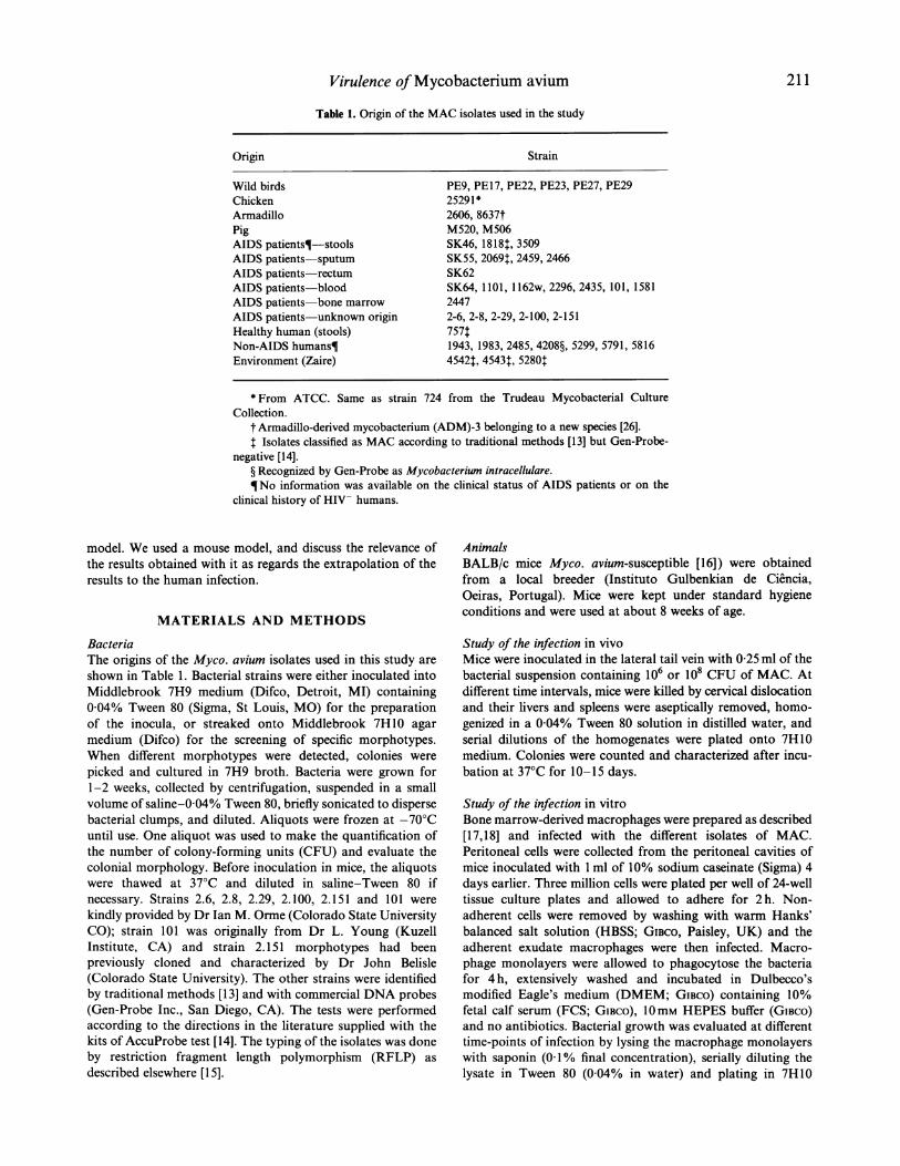
Table 1. Origin of the MAC isolates used in the study
Strain
Wild birdsChickenArmadilloPigAIDS patients¶ stoolsAIDS patients sputumAIDS patients rectumAIDS patients bloodAIDS patients bone marrow
AIDS patients- unknown originHealthy human (stools)Non-AIDS humanslEnvironment (Zaire)
PE9, PEI7, PE22, PE23, PE27, PE2925291 *2606, 8637tM520, M506SK46, 1818t, 3509SK55, 2069t, 2459, 2466SK62SK64, 1101, 1162w, 2296, 2435, 101, 158124472-6, 2-8, 2-29, 2-100, 2-151757t1943, 1983, 2485, 4208§, 5299, 5791, 5816
45421, 4543, 5280t
* From ATCC. Same as strain 724 from the Trudeau Mycobacterial CultureCollection.
t Armadillo-derived mycobacterium (ADM)-3 belonging to a new species [26].$ Isolates classified as MAC according to traditional methods [13] but Gen-Probe-
negative [14].§ Recognized by Gen-Probe as Mycobacteriwn intracellulare.¶No information was available on the clinical status of AIDS patients or on the
clinical history of HIV- humans.
model. We used a mouse model, and discuss the relevance ofthe results obtained with it as regards the extrapolation of theresults to the human infection.
MATERIALS AND METHODS
BacteriaThe origins of the Myco. avium isolates used in this study are
shown in Table 1. Bacterial strains were either inoculated intoMiddlebrook 7H9 medium (Difco, Detroit, MI) containing0 04% Tween 80 (Sigma, St Louis, MO) for the preparationof the inocula, or streaked onto Middlebrook 7H10 agar
medium (Difco) for the screening of specific morphotypes.When different morphotypes were detected, colonies were
picked and cultured in 7H9 broth. Bacteria were grown for1-2 weeks, collected by centrifugation, suspended in a smallvolume of saline-0-04% Tween 80, briefly sonicated to dispersebacterial clumps, and diluted. Aliquots were frozen at -700Cuntil use. One aliquot was used to make the quantification ofthe number of colony-forming units (CFU) and evaluate thecolonial morphology. Before inoculation in mice, the aliquotswere thawed at 37°C and diluted in saline-Tween 80 ifnecessary. Strains 2.6, 2.8, 2.29, 2.100, 2.151 and 101 were
kindly provided by Dr Ian M. Orme (Colorado State UniversityCO); strain 101 was originally from Dr L. Young (KuzellInstitute, CA) and strain 2.151 morphotypes had beenpreviously cloned and characterized by Dr John Belisle(Colorado State University). The other strains were identifiedby traditional methods [13] and with commercial DNA probes(Gen-Probe Inc., San Diego, CA). The tests were performedaccording to the directions in the literature supplied with thekits of AccuProbe test [14]. The typing of the isolates was doneby restriction fragment length polymorphism (RFLP) as
described elsewhere [15].
AnimalsBALB/c mice Myco. avium-susceptible [16]) were obtainedfrom a local breeder (Instituto Gulbenkian de Ciencia,Oeiras, Portugal). Mice were kept under standard hygieneconditions and were used at about 8 weeks of age.
Study of the infection in vivoMice were inoculated in the lateral tail vein with 0 25 ml of thebacterial suspension containing 106 or 108 CFU of MAC. Atdifferent time intervals, mice were killed by cervical dislocationand their livers and spleens were aseptically removed, homo-genized in a 0 04% Tween 80 solution in distilled water, andserial dilutions of the homogenates were plated onto 7H10medium. Colonies were counted and characterized after incu-bation at 37°C for 10-15 days.
Study of the infection in vitroBone marrow-derived macrophages were prepared as described[17,18] and infected with the different isolates of MAC.Peritoneal cells were collected from the peritoneal cavities ofmice inoculated with 1 ml of 10% sodium caseinate (Sigma) 4days earlier. Three million cells were plated per well of 24-welltissue culture plates and allowed to adhere for 2 h. Non-adherent cells were removed by washing with warm Hanks'balanced salt solution (HBSS; GIBCO, Paisley, UK) and theadherent exudate macrophages were then infected. Macro-phage monolayers were allowed to phagocytose the bacteriafor 4 h, extensively washed and incubated in Dulbecco'smodified Eagle's medium (DMEM; GIBCO) containing 10%fetal calf serum (FCS; GIBCO), 10mm HEPES buffer (GIBCO)and no antibiotics. Bacterial growth was evaluated at differenttime-points of infection by lysing the macrophage monolayerswith saponin (0-1% final concentration), serially diluting thelysate in Tween 80 (004% in water) and plating in 7H10
Origin

J. Pedrosa et al.
medium. Data were represented as growth indices which were
calculated by subtracting the mean log number ofCFU at day 0of infection from that found on day 11.
RESULTS
The origin of the different isolates is described in Table 1. Theinitial cultures arriving in our laboratory were composed ofeither pure morphotypes or combinations of two or threedistinct morphotypes. There was no predominant pattern,
nor any association of a given pattern with a particular originof isolation. The characteristic morphologies are shown in Fig. 1.SmT colonies were translucent with a smooth, bright surface;SmOp colonies were dome-shaped and opaque; Rg colonies
Fig. 1. Colony morphology of the variants used in the study. Mid-
dlebrook 7H10 agar plates seeded with the bacteria were incubated for
2 weeks and photographed with under illumination. (a) Smooth-
transport (SmT) colonies. (b) Smooth-opaque (SmOp) colonies. (c)One rough (Rg) (left) and one SmT (right) colonies.
had wrinkled surface and borders, and were opaque. Somecolonies would acquire pigmentation with time, but no extensivecharacterization of pigment production was done since virulencewas not associated with pigment production in the first strainsscreened.
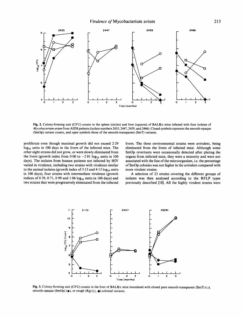
Establishment of the experimental protocolTo test and compare the virulence of different isolates, aparticular morphotype has to be used, otherwise differentproportions of distinct colonial variants in the inocula mayaccount for the differences in two different isolates. To assesswhich of the three morphotypes had consistently the highervirulence and could thus be used to compare different bacterialstrains, we made preliminary experiments either injectingbacterial inocula prepared directly from the sample receivedor, alternatively, selected a few isolates from which differentmorphotypes had been isolated, and tested them separately.The results are shown in Figs 2 and 3. Inocula prepared fromthe stock culture contained different proportions of SmT,SmOp and Rg morphotypes. In Fig. 2 we show the prolifera-tion of four strains of Myco. avium whose inocula had not beenselected for the presence of isolated morphotypes. Of these fourstrains, only one (strain 2435) contained a mixture of colonialvariants that could be easily identified in the inocula. Strain2447 was composed of SmT variant, and strains 2459 and 2466of SmOp variants. When SmOp variants were inoculated(strains 2459 and 2466) we could detect the emergence of theSmT forms after I month of infection. All four of the SmTvariants were able to persist or proliferate in the infectedorgans. Only the SmOp variant of strain 2435 was able toproliferate, whereas the SmOp variants of strains 2459 and2466 were slowly eliminated. In subsequent studies we havenever found any other strains which showed proliferation of theSmOp variant (not shown). It was not possible to ascertainwhether the emergence of the SmT variants of strains 2459 and2466 was due to their presence in the inocula in such lowfrequencies that their detection was not possible or, alterna-tively, whether they were revertants from the SmOp variants. Ina second set of experiments we studied the proliferation ofpurified morphotypes of three different strains of Myco. avium(Fig. 3). All SmT morphotypes were able to proliferate eventhough they showed differences in their virulence. The twoSmOp variants were eliminated, as was one of the Rg variantsfrom strain 25291. However, both another Rg morphotype ofthis latter strain and the Rg variant isolated from strain 2.151were able to proliferate in vivo. We thus decided to select SmTmorphotypes from the different strains for further studiesaimed at comparing virulence between isolates.
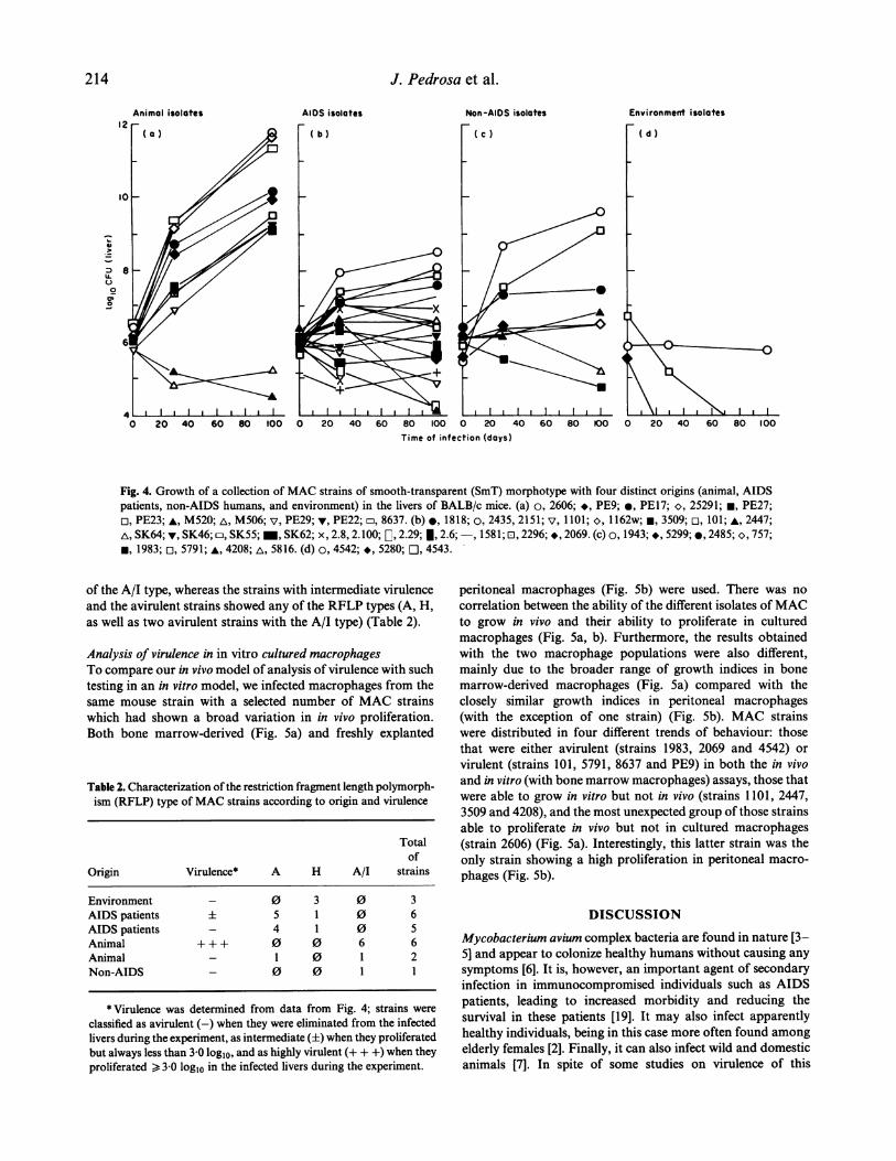
Virulence of MAC according to the origin of the strainWe studied the in vivo proliferation of 41 strains of MACderived from infected animals, AIDS patients, non-AIDS-infected human individuals, and from the environment. Theresults are shown in Fig. 4. The isolates originating frominfected animals (wild birds, chickens, pigs and armadillo)were highly virulent for mice, with two exceptions out of the11 strains studied (these latter two were both isolated frompigs). These highly virulent isolates proliferated from 3.18 to5.77 logl0 units in 100 days in the livers of the infected mice.Isolates from AIDS patients exhibited lower virulence inBALB/c mice. Some of the strains (11/19) were able to
212
Virulence ofMycobacterium avium
2435 2447
0 2 3 0 2 3 0Time (months)
2459 2466
2 3 0 1 2 3
Fig. 2. Colony-forming unit (CFU) counts in the spleen (circles) and liver (squares) of BALB/c mice infected with four isolates ofMycobacterium avium from AIDS patients (isolate numbers 2435, 2447, 2459, and 2466). Closed symbols represent the smooth-opaque(SmOp) variant counts, and open symbols those of the smooth-transparent (SmT) variants.
proliferate even though maximal growth did not exceed 2-29logl0 units in 100 days in the livers of the infected mice. Theother eight strains did not grow, or were slowly eliminated fromthe livers (growth index from 0-00 to -2 81 logl0 units in 100days). The isolates from human patients not infected by HIVvaried in virulence, including two strains with virulence similarto the animal isolates (growth index of 3 15 and 4 13 logl0 unitsin 100 days), four strains with intermediate virulence (growthindices of 0 39, 0-71, 0 99 and 1-06 log10 units in 100 days) andtwo strains that were progressively eliminated from the infected
10
U._O
:0
o 7Cal2
livers. The three environmental strains were avirulent, beingeliminated from the livers of infected mice. Although some
SmOp revertants were occasionally detected after plating theorgans from infected mice, they were a minority and were notassociated with the fate of the microorganism, i.e. the percentageofSmOp colonies was not higher in the avirulent compared withmore virulent strains.
A selection of 23 strains covering the different groups ofisolates was then analysed according to the RFLP typespreviously described [18]. All the highly virulent strains were
_ 2447
I I I I 'T .I I I I I I II
0 2 3 0 1 2 3Time (months)
Fig. 3. Colony-forming unit (CFU) counts in the liver of BALB/c mice inoculated with cloned pure smooth-transparent (SmT) (o),smooth-opaque (SmOp) (.), or rough (Rg) (cl, u) colonial variants.
9
8
m
U.
U-
0
0
6
213
J. Pedrosa et al.
Non-AIDS isolates
C(c)Environment isolates
F (d)
40 60 80 10 0 20 40 60 80 100
Fig. 4. Growth of a collection of MAC strains of smooth-transparent (SmT) morphotype with four distinct origins (animal, AIDSpatients, non-AIDS humans, and environment) in the livers of BALB/c mice. (a) o, 2606; *, PE9; e, PE17; 25291; PE27;n, PE23; A, M520; A, M506; v, PE29; v, PE22; D, 8637. (b) e, 1818; o, 2435, 2151; v, 1101; *, 1162w; m, 3509; n, 101; A, 2447;A, SK64; v, SK46; D, SK55; -, SK62; x, 2.8, 2.100; [, 2.29; 1, 2.6;-, 1581; E, 2296; *, 2069. (c) o, 1943; ., 5299; 0, 2485; o, 757;*, 1983; n, 5791; *, 4208; A, 5816. (d) o, 4542; *, 5280; O, 4543.
of the A/I type, whereas the strains with intermediate virulenceand the avirulent strains showed any of the RFLP types (A, H,as well as two avirulent strains with the A/I type) (Table 2).
Analysis of virulence in in vitro cultured macrophagesTo compare our in vivo model of analysis ofvirulence with suchtesting in an in vitro model, we infected macrophages from thesame mouse strain with a selected number of MAC strainswhich had shown a broad variation in in vivo proliferation.Both bone marrow-derived (Fig. 5a) and freshly explanted
Table 2. Characterization of the restriction fragment length polymorph-ism (RFLP) type of MAC strains according to origin and virulence
Totalof
Origin Virulence* A H A/I strains
Environment - 0 3 0 3AIDS patients 5 1 0 6AIDS patients - 4 1 0 5Animal +++ 0 0 6 6Animal - 1 0 1 2Non-AIDS - 0 0 1 1
* Virulence was determined from data from Fig. 4; strains wereclassified as avirulent (-) when they were eliminated from the infectedlivers during the experiment, as intermediate (+) when they proliferatedbut always less than 3-0 log1o, and as highly virulent (+ + +) when theyproliferated > 3 0 log1o in the infected livers during the experiment.
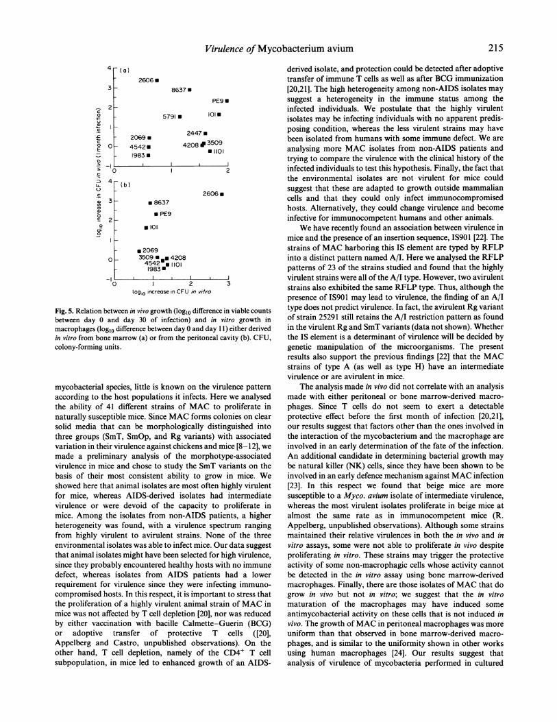
peritoneal macrophages (Fig. 5b) were used. There was no
correlation between the ability of the different isolates of MACto grow in vivo and their ability to proliferate in culturedmacrophages (Fig. 5a, b). Furthermore, the results obtainedwith the two macrophage populations were also different,mainly due to the broader range of growth indices in bonemarrow-derived macrophages (Fig. Sa) compared with theclosely similar growth indices in peritoneal macrophages(with the exception of one strain) (Fig. Sb). MAC strainswere distributed in four different trends of behaviour: thosethat were either avirulent (strains 1983, 2069 and 4542) or
virulent (strains 101, 5791, 8637 and PE9) in both the in vivoand in vitro (with bone marrow macrophages) assays, those thatwere able to grow in vitro but not in vivo (strains 1101, 2447,3509 and 4208), and the most unexpected group of those strainsable to proliferate in vivo but not in cultured macrophages(strain 2606) (Fig. 5a). Interestingly, this latter strain was theonly strain showing a high proliferation in peritoneal macro-
phages (Fig. 5b).
DISCUSSION
Mycobacterium avium complex bacteria are found in nature [3-5] and appear to colonize healthy humans without causing anysymptoms [6]. It is, however, an important agent of secondaryinfection in immunocompromised individuals such as AIDSpatients, leading to increased morbidity and reducing thesurvival in these patients [19]. It may also infect apparentlyhealthy individuals, being in this case more often found amongelderly females [2]. Finally, it can also infect wild and domesticanimals [7]. In spite of some studies on virulence of this
214
Animal isolates AIDS isolates
F ( b)12
DU-
C-0
80 100 0 20 4
Time of infection (days)
Virulence of Mycobacterium avium
4
3
2
02EI-
0
0
(a)
26060
86378PE9UM
206945421983
5791* 101 n
24472
420840 091101
-I0 2
4 (b)2606 .
3 - * 8637
* PE92
_ 101
- 20690 _ 3509 U*u*42084542 m 1101
1983
-I
0 2 3Logo increase in CFU in vitro
Fig. 5. Relation between in vivo growth (log1o difference in viable countsbetween day 0 and day 30 of infection) and in vitro growth inmacrophages (log1o difference between day 0 and day 11) either derivedin vitro from bone marrow (a) or from the peritoneal cavity (b). CFU,colony-forming units.
mycobacterial species, little is known on the virulence pattern
according to the host populations it infects. Here we analysedthe ability of 41 different strains of MAC to proliferate innaturally susceptible mice. Since MAC forms colonies on clearsolid media that can be morphologically distinguished intothree groups (SmT, SmOp, and Rg variants) with associatedvariation in their virulence against chickens and mice [8-12], wemade a preliminary analysis of the morphotype-associatedvirulence in mice and chose to study the SmT variants on thebasis of their most consistent ability to grow in mice. Weshowed here that animal isolates are most often highly virulentfor mice, whereas AIDS-derived isolates had intermediatevirulence or were devoid of the capacity to proliferate inmice. Among the isolates from non-AIDS patients, a higherheterogeneity was found, with a virulence spectrum rangingfrom highly virulent to avirulent strains. None of the threeenvironmental isolates was able to infect mice. Our data suggest
that animal isolates might have been selected for high virulence,since they probably encountered healthy hosts with no immune
defect, whereas isolates from AIDS patients had a lowerrequirement for virulence since they were infecting immuno-
compromised hosts. In this respect, it is important to stress thatthe proliferation of a highly virulent animal strain of MAC in
mice was not affected by T cell depletion [20], nor was reduced
by either vaccination with bacille Calmette-Guerin (BCG)
or adoptive transfer of protective T cells ([20],
Appelberg and Castro, unpublished observations). On the
other hand, T cell depletion, namely of the CD4' T cell
subpopulation, in mice led to enhanced growth of an AIDS-
derived isolate, and protection could be detected after adoptivetransfer of immune T cells as well as after BCG immunization[20,21]. The high heterogeneity among non-AIDS isolates maysuggest a heterogeneity in the immune status among theinfected individuals. We postulate that the highly virulentisolates may be infecting individuals with no apparent predis-posing condition, whereas the less virulent strains may havebeen isolated from humans with some immune defect. We are
analysing more MAC isolates from non-AIDS patients andtrying to compare the virulence with the clinical history of theinfected individuals to test this hypothesis. Finally, the fact thatthe environmental isolates are not virulent for mice couldsuggest that these are adapted to growth outside mammaliancells and that they could only infect immunocompromisedhosts. Alternatively, they could change virulence and becomeinfective for immunocompetent humans and other animals.
We have recently found an association between virulence inmice and the presence of an insertion sequence, IS901 [22]. Thestrains of MAC harboring this IS element are typed by RFLPinto a distinct pattern named A/I. Here we analysed the RFLPpatterns of 23 of the strains studied and found that the highlyvirulent strains were all of the A/I type. However, two avirulentstrains also exhibited the same RFLP type. Thus, although thepresence ofIS901 may lead to virulence, the finding of an A/Itype does not predict virulence. In fact, the avirulent Rg variantof strain 25291 still retains the A/I restriction pattern as foundin the virulent Rg and SmT variants (data not shown). Whetherthe IS element is a determinant of virulence will be decided bygenetic manipulation of the microorganisms. The presentresults also support the previous findings [22] that the MACstrains of type A (as well as type H) have an intermediatevirulence or are avirulent in mice.
The analysis made in vivo did not correlate with an analysismade with either peritoneal or bone marrow-derived macro-
phages. Since T cells do not seem to exert a detectableprotective effect before the first month of infection [20,21],our results suggest that factors other than the ones involved inthe interaction of the mycobacterium and the macrophage are
involved in an early determination of the fate of the infection.An additional candidate in determining bacterial growth may
be natural killer (NK) cells, since they have been shown to beinvolved in an early defence mechanism against MAC infection[23]. In this respect we found that beige mice are more
susceptible to a Myco. avium isolate of intermediate virulence,whereas the most virulent isolates proliferate in beige mice at
almost the same rate as in immunocompetent mice (R.Appelberg, unpublished observations). Although some strainsmaintained their relative virulences in both the in vivo and invitro assays, some were not able to proliferate in vivo despiteproliferating in vitro. These strains may trigger the protectiveactivity of some non-macrophagic cells whose activity cannot
be detected in the in vitro assay using bone marrow-derivedmacrophages. Finally, there are those isolates of MAC that dogrow in vivo but not in vitro; we suggest that the in vitro
maturation of the macrophages may have induced some
antimycobacterial activity on these cells that is not induced in
vivo. The growth ofMAC in peritoneal macrophages was more
uniform than that observed in bone marrow-derived macro-
phages, and is similar to the uniformity shown in other worksusing human macrophages [24]. Our results suggest that
analysis of virulence of mycobacteria performed in cultured
215

216 J. Pedrosa et al.
macrophages, namely in human monocytes [24,25], may give anerroneous estimate of mycobacterial virulence.
In conclusion, the study of the interactions between differ-ent MAC isolates and mice may reveal important aspects ofMAC physiology underlying virulence characteristics whichmay give us insights on future novel therapeutical approaches.
ACKNOWLEDGMENTS
This work was supported by grant STRDA/C/SAU/346/92 from theJunta Nacional de Investigagdo Cientifica e Tecnol6gica, and by a grantfrom the STD Programme from the European Communities. Theauthors are indebted to Dr Ian Orme for supplying some of thebacterial strains.
REFERENCES1 Horsburgh CR. Mycobacterium avium complex infection in theacquired immunodeficiency syndrome. New Engl J Med 1991;324:1332-8.
2 Prince DS, Peterson DD, Steiner RM et al. Infection with Myco-bacterium avium complex in patients without predisposing condi-tions. New Engl J Med 1989; 321:863-8.
3 Falkinham JO, Parker BC, Gruft H. Epidemiology of infection bynontuberculous mycobacteria. I. Geographic distribution in theeastern United States. Am Rev Resp Dis 1980; 121:931-7.
4 Wendt SL, George KI, Parker BC, Gruft H, Falkinham JO.Epidemiology of infection by nontuberculous mycobacteria. III.Isolation of potentially pathogenic mycobacteria from aerosols. AmRev Resp Dis 1980; 122:259-63.
5 Brooks RW, Parker BC, Gruft H, Falkinham JO. Epidemiology ofinfection by nontuberculous mycobacteria. V. Numbers in easternUnited States soils and correlation with soil characteristics. Am RevResp Dis 1984; 130:630-3.
6 Portaels F, Larsson L, Smeets P. Isolation of mycobacteria fromhealthy persons' stools. Int J Leprosy 1988; 56:468-71.
7 Lepper AWD, Corner LA. Naturally occurring mycobacteriosis ofanimals. In: Ratledge C, Stanford J, eds. The biology of themycobacteria, Vol. 2. Immunological and environmental aspects.London: Academic Press, 1983: 417-521.
8 Pattyn SR, Hermans-Boveroulle MT. Dissociation in M. avium.Pneumonology 1970; 142:119-25.
9 Pattyn SR. A study of group 111 non chromogenic mycobacteria.Correlation of chicken virulence with other in vitro charactersamong 20 strains. Zeitsch fur Tuberk 1967; 127:41-46.
10 Moehring JM, Solotorovsky MR. Relationship of colonial mor-phology to virulence for chickens of Mycobacterium avium and thenonphotochromogens. Am Rev Resp Dis 1965; 92:704-13.
11 Schaefer WB, Davis CL, Cohn ML. Pathogenicity of transparent,opaque, and rough variants of Mycobacterium avium in chickensand mice. Am Rev Resp Dis 1970; 102:499-506.
12 Anz W, Meissner G. Comparative virulence tests in chickens withtransparent and opaque colonies of strains of avian mycobacteria ofdifferent serotypes. Zbl Bakt Hyg A 1972; 221:334-42.
13 LUvy-Frebault V, Portaels F. Proposed minimal standards for thegenus Mycobacterium and for description of new slowly growingMycobacterium species. Int J Syst Bacteriol 1992; 42:315-23.
14 Gen-Probe Inc., AccuProbeTM, Mycobacterium avium. Mycobacter-ium intracellulare culture identification test. In: Manual for in vitrodiagnostic use. San Diego: Gen-Probe Inc.
15 McFadden JJ, Kunze ZM, Portaels F, Labrousse V, Rastogi N.Epidemiological and genetic markers, virulence factors and intra-cellular growth of Mycobacterium avium in AIDS. Res Microbiol1992; 143:423-30.
16 Appelberg R, Sarmento AM. The role of macrophage activationand of B c g-encoded macrophage function(s) in the control ofMycobacterium avium infection in mice. Clin Exp Immunol 1990;80:324-31.
17 Appelberg R, Orme IM, Sousa MIP, Silva MT. In vitro effects ofinterleukin-4 on interferon--y-induced macrophage activation.Immunology 1992; 76:553-9.
18 Appelberg R, Orme IM. Effector mechanisms involved in cytokine-mediated bacteriostasis of Mycobacterium avium infections inmurine macrophages. Immunology 1993; 80:352-9.
19 Horsburgh CR, Havlik JA, Ellis DA, Kennedy E, Fann SA, DuboisRE, Thompson SE. Survival of patients with acquired immunedeficiency syndrome and disseminated Mycobacterium avium com-plex infection with and without antimycobacterial chemotherapy.Am Rev Resp Dis 1991; 144:557-62.
20 Appelberg R, Pedrosa J. Induction and expression of protective Tcells during Mycobacterium avium infections in mice. Clin ExpImmunol 1992; 87:379-85.
21 Appelberg R, Castro AG, Pedrosa J, Silva RA, Orme IM, Min6prioP. The role of gamma interferon and tumor necrosis factor-alphaduring the T cell independent and dependent phases of Mycobac-terium avium infection. Infect Immun 1994; 62:3962-71.
22 Kunze ZM, Wall S, Appelberg R, Silva MT, Portaels F, McFaddenJJ. IS901, a new member of a widespread class of atypical insertionsequences, is associated with pathogenicity in Mycobacteriumavium. Mol Microbiol 1991; 5:2265-72.
23 Harshan KV, Gangadharam PRJ. In vivo depletion of natural killercell activity leads to enhanced multiplication of Mycobacteriumavium complex in mice. Infect Immun 1991; 59:2818-21.
24 Toba H, Crawford JT, Ellner JJ. Pathogenicity of Mycobacteriumavium for human monocytes: absence of macrophage-activatingfactor activity ofgamma interferon. Infect Immun 1989; 57:239-44.
25 Crowle AJ, Tsang AY, Vatter AE, May MH. Comparison of 15laboratory and patient-derived strains of Mycobacterium avium forability to infect and multiply in cultured human macrophages. JClin Microbiol 1986; 24:812-21.
26 Portaels F, Asselineau C, Baess I et al. A cooperative study ofmycobacteria isolated from armadillos infected with Mycobacteriumleprae. J Gen Microbiol 1986; 132:2693-707.