Characterization of the A 2AR–D 2R interface: Focus on the role of the C-terminal tail and the...
15
Characterization of the A 2A R–D 2 R interface: Focus on the role of the C-terminal tail and the transmembrane helices Dasiel O. Borroto-Escuela a , Wilber Romero-Fernandez a , Alexander O. Tarakanov b , Maricel Gómez-Soler c , Fidel Corrales d , Daniel Marcellino a , Manuel Narvaez e , Malgorzata Frankowska a , Marc Flajolet f , Nathaniel Heintz f , Luigi F. Agnati g , Francisco Ciruela c , Kjell Fuxe a,⇑ a Department of Neuroscience, Karolinska Institutet, Stockholm, Sweden b Russian Academy of Sciences, St. Petersburg Institute for Informatics and Automation, Saint Petersburg, Russia c Unitat de Farmacologia, Departament Patologia i Terapèutica Experimental, IDIBELL-Universitat de Barcelona, Spain d Centro de Neurociencias, La Habana, Cuba e Department of Physiology, School of Medicine, University of Málaga, Spain f Laboratory of Molecular and Cellular Neuroscience, The Rockefeller University, NY, USA g IRCCS Lido Venice, Italy article info Article history: Received 22 October 2010 Available online 30 October 2010 Keywords: Dopamine D 2 receptor Adenosine A 2A receptor Heteromerization G protein coupled receptors Transmembrane segments Protein–protein interaction abstract A single serine point mutation (S374A) in the adenosine A 2A receptor (A 2A R) C-terminal tail reduces A 2A R–D 2 R heteromerization and prevents its allosteric modulation of the dopamine D 2 receptor (D 2 R). By means of site directed mutagenesis of the A 2A R and synthetic transmembrane (TM) a-helix peptides of the D 2 R we further explored the role of electrostatic interactions and TM helix interactions of the A 2A R–D 2 R heteromer interface. We found evidence that the TM domains IV and V of the D 2 R play a major role in the A 2A R–D 2 R heteromer interface since the incubation with peptides corresponding to these domains significantly reduced the ability of A 2A R and D 2 R to heteromerize. In addition, the incubation with TM-IV or TM-V blocked the allosteric modulation normally found in A 2A R–D 2 R heteromers. The mutation of two negatively charged aspartates in the A 2A R C-terminal tail (D401A/D402A) in combination with the S374A mutation drastically reduced the physical A 2A R–D 2 R interaction and lost the ability of antagonistic allosteric modulation over the A 2A R–D 2 R interface, suggesting further evidence for the existence of an electrostatic interaction between the C-terminal tail of A 2A R and the intracellular loop 3 (IL3) of D 2 R. On the other hand, molecular dynamic model and bioinformatic analysis propose that spe- cific AAR, AQE, and VLS protriplets as an important motive in the A 2A R–D 2L R heteromer interface together with D 2L R TM segments IV/V interacting with A 2A R TM-IV/V or TM-I/VII. Ó 2010 Elsevier Inc. All rights reserved. 1. Introduction Biochemical and behavioral observations have indicated the existence of antagonistic A 2A R–D 2 R interactions in the plasma membrane that represents an allosteric mechanism in which stri- atal A 2A R–D 2 R heteromers produce an integration of adenosine and dopamine signals [1,2]. Previous evidence, using biochemical and biophysical techniques suggested that regions involved in the A 2A R–D 2 R interface are the N-terminal part of the IL3 of the D 2 R (short [D 2S R] and long [D 2L R] isoforms) and the C-terminal do- main of the A 2A R [3,4]. Specifically, the positively charged arginine rich motif in the N-terminal part of the IL3 of D 2L R may interact with two different negatively charged motifs from the C-terminal tail of A 2A R, the two adjacent aspartic residues (401D–402D) and the phosphorylated serine residue (S374), through the formation of electrostatic bonds with covalent-like strength [5]. In view of these results, these specific amino acid residues were further studied. We recently demonstrated that a single serine point mutation (S374A) in the adenosine A 2A R C-terminal tail re- duces A 2A R–D 2 R heteromerization and blocks the A 2A R allosteric modulation of the dopamine D 2 R [6] in which these results have, in part, been confirmed by others [7]. In the present study, site directed mutagenesis of the negatively charged double aspartates (D401A–D402A) in the A 2A R C-terminal tail were tested, with or without the serine mutation (S374A), to further explore the role of these negatively charged amino acids 0006-291X/$ - see front matter Ó 2010 Elsevier Inc. All rights reserved. doi:10.1016/j.bbrc.2010.10.122 ⇑ Corresponding author. Address: Department of Neuroscience, Karolinska Insti- tutet, Retzius väg 8, 17177 Stockholm, Sweden. Fax: +46 8 315721. E-mail addresses: [email protected] (D.O. Borroto-Escuela), Wilber. [email protected] (W. Romero-Fernandez), [email protected] (A.O. Tarakanov), [email protected] (M. Gómez-Soler), fi[email protected] (F. Corrales), Daniel. [email protected] (D. Marcellino), [email protected] (M. Narvaez), Malgorzata. [email protected] (M. Frankowska), fl[email protected] (M. Flajolet), luigiag- [email protected] (L.F. Agnati), [email protected] (F. Ciruela), [email protected] (K. Fuxe). Biochemical and Biophysical Research Communications 402 (2010) 801–807 Contents lists available at ScienceDirect Biochemical and Biophysical Research Communications journal homepage: www.elsevier.com/locate/ybbrc
-
Upload
independent -
Category
Documents
-
view
2 -
download
0
Transcript of Characterization of the A 2AR–D 2R interface: Focus on the role of the C-terminal tail and the...

Biochemical and Biophysical Research Communications 402 (2010) 801–807
Contents lists available at ScienceDirect
Biochemical and Biophysical Research Communications
journal homepage: www.elsevier .com/locate /ybbrc
Characterization of the A2AR–D2R interface: Focus on the role of the C-terminaltail and the transmembrane helices
Dasiel O. Borroto-Escuela a, Wilber Romero-Fernandez a, Alexander O. Tarakanov b, Maricel Gómez-Soler c,Fidel Corrales d, Daniel Marcellino a, Manuel Narvaez e, Malgorzata Frankowska a, Marc Flajolet f,Nathaniel Heintz f, Luigi F. Agnati g, Francisco Ciruela c, Kjell Fuxe a,⇑a Department of Neuroscience, Karolinska Institutet, Stockholm, Swedenb Russian Academy of Sciences, St. Petersburg Institute for Informatics and Automation, Saint Petersburg, Russiac Unitat de Farmacologia, Departament Patologia i Terapèutica Experimental, IDIBELL-Universitat de Barcelona, Spaind Centro de Neurociencias, La Habana, Cubae Department of Physiology, School of Medicine, University of Málaga, Spainf Laboratory of Molecular and Cellular Neuroscience, The Rockefeller University, NY, USAg IRCCS Lido Venice, Italy
a r t i c l e i n f o
Article history:Received 22 October 2010Available online 30 October 2010
Keywords:Dopamine D2 receptorAdenosine A2A receptorHeteromerizationG protein coupled receptorsTransmembrane segmentsProtein–protein interaction
0006-291X/$ - see front matter � 2010 Elsevier Inc. Adoi:10.1016/j.bbrc.2010.10.122
⇑ Corresponding author. Address: Department of Ntutet, Retzius väg 8, 17177 Stockholm, Sweden. Fax:
E-mail addresses: [email protected] ([email protected] (W. Romero-Fernandez), [email protected] (M. Gómez-Soler), [email protected]@ki.se (D. Marcellino), [email protected]@ki.se (M. Frankowska), flajolm@[email protected] (L.F. Agnati), [email protected] (F. Ciruela), K
a b s t r a c t
A single serine point mutation (S374A) in the adenosine A2A receptor (A2AR) C-terminal tail reducesA2AR–D2R heteromerization and prevents its allosteric modulation of the dopamine D2 receptor (D2R).By means of site directed mutagenesis of the A2AR and synthetic transmembrane (TM) a-helix peptidesof the D2R we further explored the role of electrostatic interactions and TM helix interactions of theA2AR–D2R heteromer interface. We found evidence that the TM domains IV and V of the D2R play a majorrole in the A2AR–D2R heteromer interface since the incubation with peptides corresponding to thesedomains significantly reduced the ability of A2AR and D2R to heteromerize. In addition, the incubationwith TM-IV or TM-V blocked the allosteric modulation normally found in A2AR–D2R heteromers. Themutation of two negatively charged aspartates in the A2AR C-terminal tail (D401A/D402A) in combinationwith the S374A mutation drastically reduced the physical A2AR–D2R interaction and lost the ability ofantagonistic allosteric modulation over the A2AR–D2R interface, suggesting further evidence for theexistence of an electrostatic interaction between the C-terminal tail of A2AR and the intracellular loop3 (IL3) of D2R. On the other hand, molecular dynamic model and bioinformatic analysis propose that spe-cific AAR, AQE, and VLS protriplets as an important motive in the A2AR–D2LR heteromer interface togetherwith D2LR TM segments IV/V interacting with A2AR TM-IV/V or TM-I/VII.
� 2010 Elsevier Inc. All rights reserved.
1. Introduction
Biochemical and behavioral observations have indicated theexistence of antagonistic A2AR–D2R interactions in the plasmamembrane that represents an allosteric mechanism in which stri-atal A2AR–D2R heteromers produce an integration of adenosineand dopamine signals [1,2]. Previous evidence, using biochemicaland biophysical techniques suggested that regions involved inthe A2AR–D2R interface are the N-terminal part of the IL3 of the
ll rights reserved.
euroscience, Karolinska Insti-+46 8 315721..O. Borroto-Escuela), [email protected] (A.O. Tarakanov),du.cu (F. Corrales), Daniel.
s (M. Narvaez), Malgorzata.ler.edu (M. Flajolet), [email protected] (K. Fuxe).
D2R (short [D2SR] and long [D2LR] isoforms) and the C-terminal do-main of the A2AR [3,4]. Specifically, the positively charged argininerich motif in the N-terminal part of the IL3 of D2LR may interactwith two different negatively charged motifs from the C-terminaltail of A2AR, the two adjacent aspartic residues (401D–402D) andthe phosphorylated serine residue (S374), through the formationof electrostatic bonds with covalent-like strength [5].
In view of these results, these specific amino acid residues werefurther studied. We recently demonstrated that a single serinepoint mutation (S374A) in the adenosine A2AR C-terminal tail re-duces A2AR–D2R heteromerization and blocks the A2AR allostericmodulation of the dopamine D2R [6] in which these results have,in part, been confirmed by others [7].
In the present study, site directed mutagenesis of the negativelycharged double aspartates (D401A–D402A) in the A2AR C-terminaltail were tested, with or without the serine mutation (S374A), tofurther explore the role of these negatively charged amino acids

802 D.O. Borroto-Escuela et al. / Biochemical and Biophysical Research Communications 402 (2010) 801–807
in the A2AR–D2R interface. Furthermore, the TM helix interactionsof the A2AR–D2R interface have been analyzed using TM syntheticpeptides by means of BRET1 saturation and competition assays.According to the experimental results, and using a novel bioinfor-matics approach to predict the receptor–receptor interface, weperformed a molecular dynamic simulation and a rigid-body pro-tein–protein docking of the A2AR–D2R heteromers constructedaround the most important transmembrane interfaces.
2. Materials and methods
2.1. Plasmid constructs
The cDNA encoding human A2AR cloned in pEYFP-N1 (Clontech,Germany) [8] was used as a template to mutate aspartic acid residue401 and 402 to alanine (D401A/D402A) and also to perform thetriple mutant S374A/D401A/D402A by means of the QuickChange™site-directed mutagenesis kit (Stratagene, The Netherlands) follow-ing the manufacturer’s protocol. The other plasmids used have beendescribed previously [6].
2.2. Cell culture, transfection and radioligand binding assay
HEK293T cells (American Type Culture Collection, USA) weregrown in Dulbecco’s modified Eagle’s medium supplementedwith 2 mM L-glutamine, 100 U/ml penicillin/streptomycin, and 10%(v/v) fetal bovine serum (FBS) at 37 �C and in an atmosphere of 5%CO2. For transfection, cells were plated in 6-well plates at a con-centration of 1 � 106 cells/well or in 75 cm2 flasks and culturedovernight prior transfection. Cells were transiently transfectedusing linear polyethyleneimine (Polysciences Inc., USA). Com-petition radioligand experiments were performed as describedpreviously [6].
2.3. Transmembrane peptide treatment
A series of peptides, representing each of the predicted TM seg-ments for the human D2LR (UniProt identifier number: P14416-1)were purchased from VitoTag-Biosciences (Barcelona, Spain) withP90% purity determined by HPLC. TM-I peptide consisted of resi-dues 38–60; TM-II peptide of residues 72–97; TM-III peptide ofresidues 109–130; TM-IV peptide of residues 152–174; TM-V pep-tide of residues 187–210; TM-VI peptide of residues 374–397; andTM-VII peptide of residues 406–429. Immediately before use, thepeptides were solubilized in dimethyl sulfoxide (DMSO) and di-luted in Krebs–Ringers-HEPES (KRH) medium (25 mM HEPES, pH7.4, 104 mM NaCl, 5 mM KCl, 2 mM CaCl2, 1 mM KH2PO4, 1.2 mMMgSO4) to yield a final concentration of 1% DMSO. Cells were incu-bated with the above mentioned peptides at 4 �C for 2 h prior toperforming BRET1 analysis or gene reporter assays [9].
2.4. BRET1 saturation assay
Forty-eight hours after transfection, HEK293T cells transientlytransfected with a constant (1 lg) or increasing amounts(0.5–8 lg) of plasmids encoding for D2LRRluc and A2ARYFP or A2AR–S374AYFP (A2AR–SYFP), A2AR–D401A–D402AYFP (A2AR–DDYFP), A2AR–S374A–D401A–D402AYFP (A2AR–SDDYFP), were rapidly washedtwice in PBS, detached, and resuspended in the same buffer. Cellsuspensions (20 lg proteins) were distributed in duplicate intothe 96-well microplates; black plate with transparent bottom(Corning 3651, Corning, Stockholm, Sweden) for fluorescence mea-surement or white plates with white bottom (Corning 3600) forBRET determination. For BRET1 ratio measurement, coelenterazineh substrate (Molecular Probes, Eugene, OR, USA) was added at a
final concentration of 5 lM, and readings were performed 1 minafter using the POLARstar Optima plate-reader as previouslydescribed [4]. The specificities of A2AR–D2LR interactions wereassessed by comparison with co-expression of D2LRRluc and A1RYFP.For determining the effects of the transmembrane peptides,transfected HEK293T cells were incubated with 0.4 mM of theTM segment peptides at 4 �C for 2 h prior to performing BRET1
analysis [9].
2.5. BRET1 competition assay
Forty-eight hours after transfection, HEK293T cells transientlytransfected with constant amounts (1 lg) of plasmids encodingfor D2LRRluc and A2ARYFP and increasing amounts (0.5–8 lg) of plas-mids encoding for triple mutant A2AR–SDDYFP, wild-type D2LR orA2AR and the mock pcDNA3.1+; respectively. The energy transferwas determined as described for the BRET1 saturation assay. Datawere fitted by adapting the dimer, trimer and tetramer model ofenergy transfer quenching proposed by Veatch and Stryer [10].
2.6. Luciferase reporter gene assay
A dual luciferase reporter assay was used to indirectly detectcAMP levels in transiently transfected HEK293T cell lines treatedwith different compounds in a range of concentrations (typically25 nM to 1 lM). The luciferase activity of cell extracts was deter-mined as previously described [6].
2.7. Bioinformatic analysis of the receptor–receptor interface
Based on a bioinformatic approach, a set of amino acid triplethomologies have been deduced that may be responsible for recep-tor–receptor interactions among receptor heterodimers [11]. It hasbeen indicated how such triplets of amino acid residues and their‘teams’ may be utilized to construct a ‘‘kind of code’’ that deter-mines (and/or predicts) which receptors should or should not formheterodimers. In this study the A2AR–D2LR heterodimer has beenanalyzed for the existence of a basic set of common triplets in eachparticipating receptor that may be responsible for the receptor–receptor interaction.
2.8. Statistical analysis
The number of samples (n) in each experimental condition isindicated in figure legends. When two experimental conditionswere compared, statistical analysis was performed using an un-paired t test. Otherwise, statistical analysis was performed byone-way analysis of variance (ANOVA) followed by Tukey’s Multi-ple Comparison post-test. The P value 0.05 and lower was consid-ered significant.
3. Results
3.1. Role of the A2AR C-terminal tail in the A2AR–D2R heteromerinterface
First, in order to map the A2AR C-terminal tail region responsiblefor the A2AR–D2R electrostatic interactions we performed com-bined mutagenesis and BRET experiments. Recently, we have dem-onstrated that the mutation of the serine 374 to alanine within thisC-terminal tail largely impairs the ability of the A2AR to interactwith the D2R (see [6] and Supplementary Fig. 1). Here we showthat the simultaneous substitution to alanine of two negativelycharged aspartates (D401A/D402A) within the same receptordomain (see Supplementary Fig. 1) also reduced the A2AR–D2R

D.O. Borroto-Escuela et al. / Biochemical and Biophysical Research Communications 402 (2010) 801–807 803
interaction as measured by BRET1 assays (Fig. 1A). Interestingly,the largest impairment in the ability of A2AR–D2R to interact wasachieved when the S374A/D401A/D402A triple A2AR mutant wasassessed (Fig. 1A and Supplementary Fig. 2A). In addition, compe-tition BRET1 assays showed that either A2AR or D2LR was able tofully displace the A2AR–D2R interaction in a concentration depen-dent manner (Fig. 1B). Interestingly, under these experimentalconditions the triple A2AR mutant produced only a 50% reductionat maximal dosages (Fig. 1B), thus corroborating its reduced abilityto interact with the D2R.
Next, examining the A2AR and D2LR agonist-mediated signalingchanges as measured by different luciferase reporter assays in het-erologous HEK293T cells co-expressing both receptors, we deter-mined that the forskolin stimulated luciferase activity producedby a direct activation of adenylate cyclase (AC) is counteractedby 50% with the D2R-like agonist quinpirole (25 nM). This phenom-enon is blocked by the A2AR agonist CGS21680 (50 nM) and theD2LR like antagonist raclopride (1 lM) (Fig. 2A). CGS21680 alonedoes not affect the forskolin induced increase of CRE-luciferaseactivity. At the contrary, the A2AR antagonist ZM241385 blocked
Fig. 1. BRET1 studies of A2AR and D2LR heteromerization in HEK293T cells. (A) BRET1 sathetero-oligomers. Plotted on the X-axis is the fluorescence value obtained from the YFcoelenterazine incubation. Mean ± SEM; n = 10, 8 replicates. ***: (d) and (b) are significan(a) (P < 0.05). (B) BRET1 competition experiment for the D2LR-5-A2AR heteromers. Studiestagged receptors in presence of increasing concentration of wild-type receptors. Plottedn = 4 in triplicate. ***: Significantly different compared to pcDNA3.1 in the range of 2–8 lof 5–8 lg cDNA) (P < 0.05).
the increase of luciferase activity observed after forskolin andCGS21680 treatment, demonstrating the dependency on A2ARactivity. A single point mutation (S374A) and a triple mutation(S374A; D401A–D402A) in the C-terminal tail of the A2AR abol-ished the ability of CGS21680 but not of the D2R-like antagonistraclopride to counteract the quinpirole induced inhibition of lucif-erase activity. In contrast, a double aspartate mutation (D401A–D402A) in the C-terminal tail of A2AR, did not affect the capacityof CGS21680 to counteract the quinpirole-induced inhibition offorskolin-induced luciferase activity. For all three mutated A2ARs,ZM241385 remained capable of counteracting the forskolin-in-duced luciferase activity.
As seen in Fig. 2B, CGS21680 could activate the three mutatedA2ARs to a similar degree as found after CGS21680-inducedactivation of the wild-type A2ARs based on the increase of CRE-luciferase activity. Interestingly, the quinpirole-induced inhibitionof CGS21680-induced CRE-luciferase activity was significantly en-hanced when using the serine point mutant and the triple mutant(S374A; D401A–D402A) in the C-terminal tail of the A2AR. A similarobservation seemed to be true also for the double aspartate
uration curves for the D2LR–A2AR hetero-oligomers compared to D2LR–A2AR mutantP, normalized with the luminescence value of D2LRRluc expression 10 min after h-tly different compared to [a] (P < 0.001), *: (c) is significantly different compared towere carried out using a fixed ratio (1:1) of expression levels of the D2LRRluc/A2ARYFP
on the X-axis is the concentration of cDNA transfected per competitor. Mean ± SEM;g cDNA) (P < 0.001), *: Significantly different compared to A2AR-SDDYFP in the range

Fig. 2. (A) D2LR and A2AR responses in a forskolin-induced CRE-luciferase reporter gene assay. HEK293T cells were transiently co-transfected with 1 lg firefly luciferase-encoding experimental plasmid (pGL4-CRE-luc2p), 1 lg of both (D2LRRluc and A2ARYFP) or (D2LRRluc and A2AR–DDYFP) or (D2LRRluc and A2AR–S374AYFP) or (D2LRRluc and A2AR-SDDYFP) expression vectors and 50 ng Renilla luciferase-encoding internal control plasmid (phRG-B). Thirty-six hours after transfection, cells were treated for 4 h with agonistor antagonist (in presence of 1 lM forskolin). Light emission is expressed as a percentage of the control forskolin-induced value. Mean ± SEM; n = 6 in triplicate. ***:Significantly different compared to control of each group, respectively (P < 0.001), &&&: Significantly different compared to quinp 25 nM of each group, respectively(P < 0.001), +++ and ++: Significantly different compared to quinp 25 nM of each group, respectively (P < 0.001 and P < 0.01). (B) CRE-luciferase reporter gene assay in responseto A2AR activation. HEK293T cells were co-transfected and treated as described above. The data represent the mean ± SEM; n = 4 in triplicate. **: Significantly differentcompared to control of each group, respectively (P < 0.01), #: Significantly different compared to CGS 50 nM of A2ARYFP–D2LR group (P < 0.05), ++: Significantly differentcompared to CGS 50 nM of each group, respectively (P < 0.01). Fk, forskolin (1 lM); CGS, CGS21680 (50 nM); ZM, ZM241385 (1 lM); quinp, quinpirole (25 nM) and racl,raclopride (1 lM).
804 D.O. Borroto-Escuela et al. / Biochemical and Biophysical Research Communications 402 (2010) 801–807
mutation (Fig. 2B). This is likely due to the absence of the allostericA2AR–D2LR interaction mediating a reduction of D2LR Gi/o coupling[12,13].
In the analysis of the antagonistic A2AR–D2LR interaction at thelevel D2LR recognition in D2LR containing heteromers with mutatedA2ARs, CGS21680 failed to reduce the affinity of the high affinityD2LR agonist binding site for all three A2AR mutants used, includingthe mutant with the double aspartate mutation (see Supplemen-tary Fig. 3).
3.2. Mapping the A2AR–D2R heteromer interface: role of the D2R TMhelices
In order to fully characterize the A2AR–D2R heteromer interfacewe analyzed the role the TM D2R domains in the heteromer func-tionality. Thus, by using D2R synthetic peptides that correspond tothese TM domains we have provided evidence for the identificationof the specific receptor regions responsible for the heteromer for-mation. As seen in Fig. 3A and Supplementary Fig. 2B, incubation
of the D2LR TM segments IV and V highly significantly and almostfully blocked the BRET1 signal from the D2LRRluc–A2ARYFP hetero-mers seen in the absence of D2LR TM fragments. A significantlyreduced BRET1 signal was also obtained when D2LR TM-VI wasincubated, but not if D2LR TM-I to TM-III and D2LR TM-VII wereincubated, as no reduction of the BRET1 signal was observed. Asseen in Fig. 3B, and in line with the results in Fig. 3A, incubationof D2LR TM-IV and TM-V produced a marked concentration depen-dent disappearance of the BRET1 signal in the range from 10 nM to1 lM. The competition curves with D2LR TM-VI and especially D2LRTM-III were substantially shifted to the right indicating a reducedability for them to compete with the D2LR for it’s binding to theA2AR.
As seen in Fig. 3C, incubation of TM-IV and TM-V fully blockedthe ability of CGS21680 to counteract the quinpirole-induced inhi-bition of forskolin-stimulated CRE-luciferase activity. TM-VI failedto have such an action and the A2AR agonist fully counteracted thequinpirole-induced inhibition of forskolin-stimulated CRE-lucifer-ase activity (Fig. 3C).

Fig. 3. Effects of D2R TM synthetic peptides on A2AR–D2LR BRET signal. (A) BRET1 saturation curves for the D2LR–A2AR heteromers and the effects of competing with variousTM peptides. Co-transfected cells were incubated with 0.4 mM of peptides and studied by BRET1. Plotted on the X-axis is the fluorescence value obtained from the YFP,normalized with the luminescence value of D2LRRluc expression 10 min after h-coelenterazine incubation. Mean ± SEM; n = 4, 6 replicates. ***: [TM-IV and TM-V] significantlydifferent compared to control (P < 0.001), *: [TM-VI] significantly different compared to control (P < 0.05). (B) The effect of different peptides on BRET1 signals of the D2LR–A2AR heteromers. Values represent percentages of maximal saturable BRET1 responses. Mean ± SEM; n = 4, 6 replicates. (C) D2LR and A2AR responses in a forskolin-inducedCRE-luciferase reporter gene assay in presence of different TM synthetic peptides. HEK293T cells transiently co-transfected as described in Materials and Methods and 36 hafter transfection, were incubated with peptides corresponding to TM segments of the D2R (0.4 mM) and treated 4 h with agonist or antagonist (in presence of 1 lMforskolin). Light emission is expressed as a percentage of the control forskolin-induced value. Mean ± SEM; n = 4 in triplicate. ***: Significantly different compared to control ofeach group, respectively (P < 0.001), && and &: Significantly different compared to quinp 25 nM of each group, respectively (P < 0.01 and 0.05), +++: Significantly differentcompared to quinp 25 nM of each group, respectively (P < 0.001). Fk, forskolin (1 lM); CGS, CGS21680 (50 nM); ZM, ZM241385 (1 lM); quinp, quinpirole (25 nM) and racl,raclopride (1 lM).
D.O. Borroto-Escuela et al. / Biochemical and Biophysical Research Communications 402 (2010) 801–807 805

806 D.O. Borroto-Escuela et al. / Biochemical and Biophysical Research Communications 402 (2010) 801–807
3.3. The triplets AAR, AQE, VLS and VYI may be crucial for A2AR–D2Rheterodimer formation
Based on a bioinformatic approach, Tarakanov and Fuxe [11]have deduced a set of triplet homologies that might be responsiblefor receptor–receptor interactions. This set consists of twonon-intersecting subsets: ‘pro-triplets’ and ‘contra-triplets’. Anypro-triplet appears as a homology in at least one heterodimerbut does not appear as a homology in any non-heterodimer. Thereverse is also true; mainly any contra-triplet appears in at leastone non-heterodimer but does not appear in any heterodimer.For example (Fig. 4), the triplet of amino acid residues AAR appearsas a homology in seven receptor heterodimers: A2A-D2, A2A-mGlu5,D2-mGlu5, mGluR5-GABAB1, D2-CCK2, 5-HT 1B-5HT 1D, and MHC1-
Fig. 4. In silico analysis and sequence alignment of A2AR and D2R receptor homologiescontaining the triplet homologies AAR, AQE, VLS, and VYI in the receptor interface.Amino acid codes of receptors have been obtained from (NCBI: http://www.ncbi.nlm.nih.gov/, 2009) where all of them have been considered human (Homo sapiens)for the case of uniformity. (A) The triplet homology VYI (grey-shaded) in theN-terminal of A2AR and in the fifth transmembrane a-helix (TM-V) of D2R. (B) Thetriplet homology VLS (grey-shaded) in the TM-IV of both A2AR and D2R. (C) The triplethomology AAR (grey-shaded) in the third intracellular loop (IL3, between TM-V andTM-VI) of both A2AR and D2R. Note that the triplet AQE (grey-shaded) is close to AAR inthe IL3 of D2R. (D) The triplet homology AQE (grey-shaded) in the IL3 of D2R and in theC-tail of A2AR. Note that the triplet LLS (grey-shaded) is close to AQE in the C-tail ofA2AR. Also note that the triplet AQE is adjacent to S374 which seems to play a key rolein the A2AR–D2R receptor interface.
CD8. As seen in Fig. 4, the triplet homology AAR exists in the IC3of the two protomers and the triplet homology AQE in the A2ARC-terminal tail and D2LR IC3 supporting the important role of theseintracellular domains in the heteromer interface. Instead the triplethomology VLS is located in the TM-IV of the two protomers and theVYI in the N-terminal of A2AR and TM-V of D2LR indicating a role ofTM-IV and TM-V in the A2AR–D2LR receptor interfaces. Taking to-gether with the BRET1 experimental results, these in silico predic-tions suggest the existence of a basic set of common triplets inthe two participating receptors in the A2AR–D2LR heteromers thatmay participate in the receptor–receptor interaction interfaces atthe intracellular and transmembrane levels with a potential rolealso of the N-terminal of A2AR (Fig. 4).
3.4. Mainly two TM domains are involved in the D2LR–A2AR heteromerinterface
As seen in Supplementary Fig. 4, the 3D-model of D2LR and A2ARwas constructed using the homology modeling method. Specialcare was taken during the alignment process to minimize theerrors when single template was used. Then, to further optimizethe structures and present them in a more natural way, a 20 nsmolecular dynamics simulation was performed with each receptorembedded in an explicit membrane-water environment to refineand explore the conformational space. Based on the structures ex-tracted from the molecular dynamics and taking into considerationthe experimental evidence previously described, the likely inter-face of D2LR–A2AR heterodimer was determined through the anal-ysis of protein–protein docking, cluster, shape complementaryand interaction energy. The rigid body protein–protein dockingswere performed using the Fast Fourier Transform-based algorithmprogram ZDOCK and the Fourier correlation algorithm ClusPro.Then the interaction energy was calculated as the major criteriato rank the poses (Supplementary Fig. 4 and Table). As a result,the A2AR (TM-IV/V)–D2R (TM-IV/V) and A2AR (TM-I/VII)–D2R (TM-IV/V) interaction pattern probably is the most favorable modelfrom the energy point of view; its interaction energy was the low-est among the different contact patterns analyzed. The next mostfavorable model based on energy level was A2AR (TM-VI/VII)–D2R(TM-IV/V), with an average interaction energy that was about407 kJ/mol lager than the one for the A2AR (TM-IV/V)–D2R (TM-IV/V) contact dimer. Collectively, these bioinformatics analysis onthe A2AR–D2R interaction are in agreement with our experimentalresults (see above) and suggest that the A2AR C-terminal tail andthe D2R TM-IV and TM-V are essential domains in the A2AR–D2Rinterface that mediate the allosteric A2AR–D2R interaction.
4. Discussion
The findings of this paper provide further evidence that D2R TMdomains IV and V play a major role in the A2AR–D2R interface sincethe incubation of co-transfected cells with peptides correspondingto these TM domains significantly reduced the ability of A2AR andD2R to heteromerize. In addition, the incubation with either TM-IV or TM-V blocked the allosteric communication between A2ARand D2R in which A2AR agonist-induced counteraction of quinpi-role-induced inhibition of CRE activity could no longer be ob-served. Overall, our results further support the previouslydemonstrated electrostatic interactions between the C-terminaltail of A2AR and the IL3 of D2R and demonstrate the role of theTM-IV and TM-V segments of D2R in mediating the antagonisticA2AR–D2R interaction (i.e., inhibition of the D2R coupling to theGi/o) [3,6,7,12,14].
Results from molecular dynamic modeling also imply TM-IV/Vof D2LR interacting with TM-IV/V of A2AR and TM-IV/V of D2LR

D.O. Borroto-Escuela et al. / Biochemical and Biophysical Research Communications 402 (2010) 801–807 807
interacting with TM-I/VII of A2AR. The models obtained also indi-cate the position of the pro-triplet homologies found via bioinfor-matic analysis (Supplementary Fig. 1) and are compatible with thetheory [11] that the pro-triplets AAR, AQE, and VLS through guide-clasp interactions play an important role in the A2AR–D2LR hetero-mer interface.
In addition, the continued analysis of the A2AR C-terminal tail-D2R IL3 interaction with site directed mutagenesis has given addi-tional evidence for a key role of S374 in the C-terminal tail of A2ARin the A2AR–D2R interface in both terms of the formation of A2AR–D2R heteromers and in terms of its allosteric communication. Themutation of the two negatively charged aspartic acid residues inthe A2AR C-terminal tail (D401A/D402A) alone partially reducedthe physical A2AR–D2R interaction and the allosteric communica-tion in this heteromer at both the levels of agonist recognitionand Gi/o coupling. These mutated aspartates in combination withthe S374A mutation further reduced the physical interactionbetween A2AR–D2R compared to the S374A mutation alone andthe mutated aspartates alone (Fig. 1A), suggesting the existenceof an orchestrated electrostatic interaction between the C-terminaltail of A2AR and the IL3 of D2R. The triple A2AR mutant, similar tothe single S374A mutant, fully lost its ability of antagonistic allo-steric communication over the A2AR–D2R interface. This was dem-onstrated both at the level of D2R agonist-induced inhibition offorskolin-mediated AC activation and at the level of D2R agonistrecognition through radioligand binding assays.
Acknowledgments
This work was supported by grants from the Swedish ResearchCouncil (04X-715), Torsten and Ragnar Söderberg Foundation,Hjärnfonden and Marianne and Marcus Wallenberg Foundationto KF and SAF2008-01462 and Consolider-Ingenio CSD2008-00005 from Ministerio de Ciencia e Innovación to F.C. A.O.T. hasnot received any support for this work. This work was supportedby the D.O.D. (W81XWH-08-1-0111) to Paul Greengard.
Appendix A. Supplementary data
Supplementary data associated with this article can be found, inthe online version, at doi:10.1016/j.bbrc.2010.10.122.
References
[1] K. Fuxe, D. Marcellino, A. Rivera, Z. Diaz-Cabiale, M. Filip, B. Gago, D.C. Roberts,U. Langel, S. Genedani, L. Ferraro, A. de la Calle, J. Narvaez, S. Tanganelli, A.Woods, L.F. Agnati, Receptor–receptor interactions within receptor mosaics.Impact on neuropsychopharmacology, Brain Res. Rev. 58 (2008) 415–452.
[2] L.F. Agnati, D. Guidolin, G. Leo, C. Carone, S. Genedani, K. Fuxe, Receptor–receptor interactions: a novel concept in brain integration, Prog. Neurobiol. 90(2010) 157–175.
[3] F. Ciruela, J. Burgueno, V. Casado, M. Canals, D. Marcellino, S.R. Goldberg, M.Bader, K. Fuxe, L.F. Agnati, C. Lluis, R. Franco, S. Ferre, A.S. Woods, Combiningmass spectrometry and pull-down techniques for the study of receptorheteromerization. Direct epitope–epitope electrostatic interactions betweenadenosine A2A and dopamine D2 receptors, Anal. Chem. 76 (2004) 5354–5363.
[4] M. Canals, D. Marcellino, F. Fanelli, F. Ciruela, P. de Benedetti, S.R. Goldberg, K.Neve, K. Fuxe, L.F. Agnati, A.S. Woods, S. Ferre, C. Lluis, M. Bouvier, R. Franco,Adenosine A2A-dopamine D2 receptor–receptor heteromerization: qualitativeand quantitative assessment by fluorescence and bioluminescence energytransfer, J. Biol. Chem. 278 (2003) 46741–46749.
[5] S.N. Jackson, H.Y. Wang, A. Yergey, A.S. Woods, Phosphate stabilization ofintermolecular interactions, J. Proteome Res. 5 (2006) 122–126.
[6] D.O. Borroto-Escuela, D. Marcellino, M. Narvaez, M. Flajolet, N. Heintz, L.Agnati, F. Ciruela, K. Fuxe, A serine point mutation in the adenosine A2AR C-terminal tail reduces receptor heteromerization and allosteric modulation ofthe dopamine D2R, Biochem. Biophys. Res. Commun. 394 (2010) 222–227.
[7] G. Navarro, S. Ferre, A. Cordomi, E. Moreno, J. Mallol, V. Casado, A. Cortes, H.Hoffmann, J. Ortiz, E.I. Canela, C. Lluis, L. Pardo, R. Franco, A.S. Woods,Interactions between intracellular domains as key determinants of thequaternary structure and function of receptor heteromers, J. Biol. Chem. 285(2010) 27346–27359.
[8] N. Cabello, J. Gandia, D.C. Bertarelli, M. Watanabe, C. Lluis, R. Franco, S. Ferre, R.Lujan, F. Ciruela, Metabotropic glutamate type 5, dopamine D2 and adenosineA2a receptors form higher-order oligomers in living cells, J. Neurochem. 109(2009) 1497–1507.
[9] K.G. Harikumar, D.I. Pinon, L.J. Miller, Transmembrane segment IV contributesa functionally important interface for oligomerization of the Class II G protein-coupled secretin receptor, J. Biol. Chem. 282 (2007) 30363–30372.
[10] W. Veatch, L. Stryer, The dimeric nature of the gramicidin A transmembranechannel: conductance and fluorescence energy transfer studies of hybridchannels, J. Mol. Biol. 113 (1977) 89–102.
[11] A.O. Tarakanov, K.G. Fuxe, Triplet puzzle: homologies of receptor heteromers,J. Mol. Neurosci. 41 (2010) 294–303.
[12] K. Fuxe, S. Ferre, M. Canals, M. Torvinen, A. Terasmaa, D. Marcellino, S.R.Goldberg, W. Staines, K.X. Jacobsen, C. Lluis, A.S. Woods, L.F. Agnati, R. Franco,Adenosine A2A and dopamine D2 heteromeric receptor complexes and theirfunction, J. Mol. Neurosci. 26 (2005) 209–220.
[13] K. Fuxe, D. Marcellino, S. Genedani, L. Agnati, Adenosine A(2A) receptors,dopamine D(2) receptors and their interactions in Parkinson’s disease, Mov.Disord. 22 (2007) 1990–2017.
[14] A.S. Woods, F. Ciruela, K. Fuxe, L.F. Agnati, C. Lluis, R. Franco, S. Ferre, Role ofelectrostatic interaction in receptor–receptor heteromerization, J. Mol.Neurosci. 26 (2005) 125–132.

SUPPLEMENTARY DATA
Figure 1. A two-dimensional topology of the human D2LR sequence (on the top) and the
human A2AR sequence (on the bottom) are represented (extracellular space at the top and
the intracellular space at the bottom). Filled circles (in yellow) represent the triplet of
amino acid residues determined by bioinformatic analysis to be implicated on the A2AR-
D2LR heteromer interface; filled circles (in magenta) represent the amino acid residues
experimentally determined to be implicated on the A2AR-D2LR heteromer interface;
highlighted helix represents the D2LR TM experimentally determined to be implicated in
the A2AR-D2LR heteromer interface.
Figure 2. (A) Detection of A2AR-D2LR heterodimerization by BRET1. HEK293T cells
were cotransfected with the indicated plasmids. Transfection with D2LRRluc alone or
D2LRRluc-A1RYFP served as negative controls, while transfection with EYFP-Rluc fusion
plasmid was used as a positive control for BRET1. After the addition of h-coelenterazine,
the BRET1 signal was measured. Cells expressing the negative controls yielded a
background BRET ratio of ∼20%, while the cells expressing the positive control or
coexpressing D2LRRluc and A2ARYFP yielded substantially higher BRET1 ratios, indicative
of energy transfer and protein-protein interaction. The data represent the mean ± S.E.M.; n
= 6 in triplicate. ***: Significantly different compared to D2LRRluc-A2ARYFP (P<0.001),
+++: Significantly different compared to D2LRRluc (P<0.001), by One-way analysis of
variance (ANOVA). (B) Effects of D2R TM synthetic peptides on the A2AR-D2LR BRET
signal. BRET1 ratios obtained from series of control experiments performed in HEK293T
cells after incubating with peptides corresponding to TM segments of the D2R are
presented. Co-transfected cells were incubated with 0.4 mM of peptides and studied by
BRET1. The data represent the mean ± S.E.M.; n = 5 in triplicate. ***: Significantly
different compared to control (P<0.001), *: Significantly different compared to control
(P<0.05), by One-way analysis of variance (ANOVA).
Figure 3. A2AR agonist-mediated modulation of D2LR agonist binding. Competition
experiments were performed using the D2-like receptor antagonist [3H]-Raclopride (2 nM)
versus increasing concentrations of dopamine in transiently co-transfected HEK293T cell
membranes. (A) D2LR agonist competition experiments with dopamine were performed in
the absence (●) or the presence ( ) of the A2AR agonist CGS21680 (200 nM) (B)
Competition experiments with dopamine in membrane preparations from transiently co-
transfected cells with A2AR-DDYFP were performed in the absence (●) or the presence ( )
of the A2AR agonist CGS21680 (200 nM). (C) Competition experiments with dopamine in
membrane preparations from transiently co-transfected cells with A2AR-SDDYFP were
performed in the absence (●) or the presence ( ) of the A2AR agonist CGS21680 (200
nM). Data are the means ± S.E.M. from one experiments performed in triplicate.
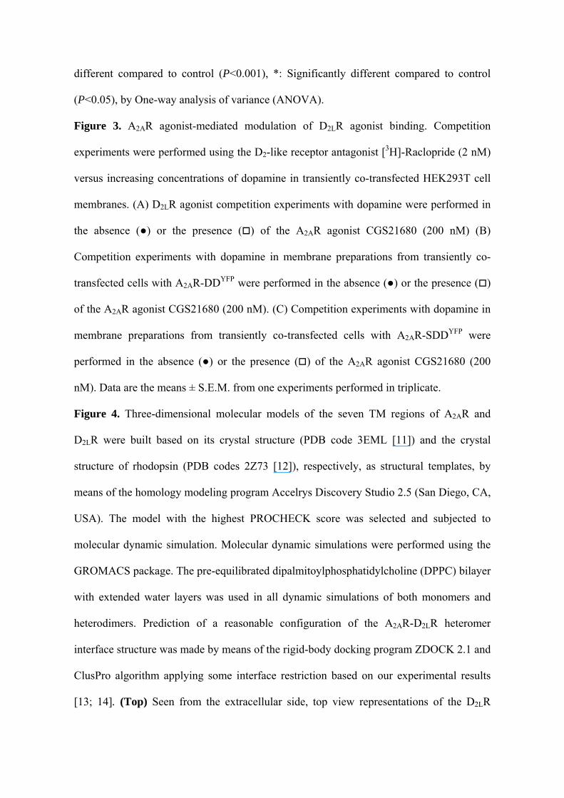
Figure 4. Three-dimensional molecular models of the seven TM regions of A2AR and
D2LR were built based on its crystal structure (PDB code 3EML [11]) and the crystal
structure of rhodopsin (PDB codes 2Z73 [12]), respectively, as structural templates, by
means of the homology modeling program Accelrys Discovery Studio 2.5 (San Diego, CA,
USA). The model with the highest PROCHECK score was selected and subjected to
molecular dynamic simulation. Molecular dynamic simulations were performed using the
GROMACS package. The pre-equilibrated dipalmitoylphosphatidylcholine (DPPC) bilayer
with extended water layers was used in all dynamic simulations of both monomers and
heterodimers. Prediction of a reasonable configuration of the A2AR-D2LR heteromer
interface structure was made by means of the rigid-body docking program ZDOCK 2.1 and
ClusPro algorithm applying some interface restriction based on our experimental results
[13; 14]. (Top) Seen from the extracellular side, top view representations of the D2LR

(TM-IV/V)-A2AR (TM-I/VII) and D2LR (TM-IV/V)-A2AR (TM-IV/V) model of A2AR-
D2LR heterodimer. Termini domains, loops and helix 8 are neglected. (Bottom) Average
contact energy of the three most favorable A2AR-D2LR heterodimer models. The table
shows the electrostatic, the van der Waals and the total energy (kJ/mol).

SUPPLEMENTARY DATAFig. 1 Borroto-Escuela et al. 2010
D2R
A2AR

SUPPLEMENTARY DATAFig. 2 Borroto-Escuela et al. 2010
A
B

SUPPLEMENTARY DATAFig. 3 Borroto-Escuela et al. 2010
[3H]-Raclopride displacement by DA in HEK 293 Cells
-12 -10 -8 -6 -4 -2
0
25
50
75
100
125controlControl + CGS
log [DA] (M)
% o
f con
trol
[3H]-Raclopride displacement by DA in HEK 293 Cells
-12 -10 -8 -6 -4 -2
0
25
50
75
100
125Double mutantDouble mutant + CGS
log [DA](M)
% o
f con
trol

SUPPLEMENTARY DATAFig. 3 (continuation) Borroto-Escuela et al. 2010
[3H]-Raclopride displacement by DA in HEK 293 Cells
-12 -10 -8 -6 -4 -2
0
25
50
75
100
125Triple mutantTriple mutant + CGS
log [DA](M)
% o
f con
trol

SUPPLEMENTARY DATAFig. 4 Borroto-Escuela et al. 2010
-1210-890-320(VI/VII)-(IV/V)
-1617-1132-485(IV/V)-(IV/V)
-1750-1250-500(I/VII)-(IV/V)
TotalVan der WaalsEnergy(kJ/mol)
ElectrostaticEnergy(kJ/mol)
InterfaceA2AR/D2R