Characterization of Age and Gender as Predisposing Facto
22
Citation: Kamel, A.S.; Mohamed, A.F.; Rabie, M.A.; Elsherbiny, M.E.; Ahmed, K.A.; Khattab, M.M.; Abdelkader, N.F. Experimental Evidence for Diiodohydroxyquinoline -Induced Neurotoxicity: Characterization of Age and Gender as Predisposing Factors. Pharmaceuticals 2022, 15, 251. https://doi.org/10.3390/ph15020251 Academic Editor: Nuno A. Silva Received: 14 January 2022 Accepted: 16 February 2022 Published: 19 February 2022 Publisher’s Note: MDPI stays neutral with regard to jurisdictional claims in published maps and institutional affil- iations. Copyright: © 2022 by the authors. Licensee MDPI, Basel, Switzerland. This article is an open access article distributed under the terms and conditions of the Creative Commons Attribution (CC BY) license (https:// creativecommons.org/licenses/by/ 4.0/). pharmaceuticals Article Experimental Evidence for Diiodohydroxyquinoline-Induced Neurotoxicity: Characterization of Age and Gender as Predisposing Factors Ahmed S. Kamel 1 , Ahmed F. Mohamed 1 , Mostafa A. Rabie 1 , Marwa E. Elsherbiny 2 , Kawkab A. Ahmed 3 , Mahmoud M. Khattab 1 and Noha F. Abdelkader 1, * 1 Department of Pharmacology and Toxicology, Faculty of Pharmacy, Cairo University, Giza 11562, Egypt; [email protected] (A.S.K.); [email protected] (A.F.M.); [email protected] (M.A.R.); [email protected] (M.M.K.) 2 Department of Pharmacology and Toxicology, Faculty of Pharmacy, Ahram Canadian University, 6th of October City 12566, Egypt; [email protected] 3 Department of Pathology, Faculty of Veterinary Medicine, Cairo University, Giza 12211, Egypt; [email protected] * Correspondence: [email protected] Abstract: Though quinoline anti-infective agents-associated neurotoxicity has been reported in the early 1970s, it only recently received regulatory recognition. In 2019, the European Medicines Agency enforced strict use for quinoline antibiotics. Thus, the current study evaluates the relation between subacute exposure to diiodohydroxyquinoline (DHQ), a commonly misused amebicide, with the development of motor and sensory abnormalities, highlighting age and gender as possible predisposing factors. Eighty rats were randomly assigned to eight groups according to their gender, age, and drug exposure; namely, four control groups received saline (adult male, adult female, young male, and young female), and the other four groups received DHQ. Young and adult rats received DHQ in doses of 176.7 and 247.4 mg/kg/day, respectively. After 4 weeks, rats were tested for sensory abnormality using analgesiometer, hot plate, and hind paw cold allodynia tests, and for motor function using open field and rotarod tests. Herein, the complex behavioral data were analyzed by principal component analysis to reduce the high number of variables to a lower number of representative factors that extracted components related to sensory, motor, and anxiety-like behavior. Behavioral outcomes were reflected in a histopathological examination of the cerebral cortex, striatum, spinal cord, and sciatic nerve, which revealed degenerative changes as well demyelination. Noteworthy, young female rats were more susceptible to DHQ’s toxicity than their counterparts. Taken together, these findings confirm previous safety concerns regarding quinoline-associated neurotoxicity and provide an impetus to review risk/benefit balance for their use. Keywords: diiodohydroxyquinoline; neurotoxicity; old/young/male/female rats; behavioral impairment; histopathological abnormalities; principal component analysis 1. Introduction 8-hydroxyquinoline (8-HQ) is one of the heterocyclic quinolone pharmacophores that possess a wide variety of applications. It has been used in cosmetics as a preservative and a chemical mediator in dye synthesis [1]. Moreover, it possesses antibacterial, antifungal, and antiprotozoal activities [2]. Despite these beneficial effects, signs of toxicity have been reported with its usage. First, liver and spleen hemosiderosis as well as signs of nephro- toxicity have been observed in male rats receiving 8-HQ in their diet [3]. Second, signs of neurotoxicity were reported in several species using halogenated 8-HQ. Noteworthy, 8-HQ produced axonal sheath depletion in sciatic nerves in rats [4]. Clioquinol (iodochlorhydrox- yquin) and iodoquinol (diiodohydroxyquinoline; DHQ) are the most known halogenated Pharmaceuticals 2022, 15, 251. https://doi.org/10.3390/ph15020251 https://www.mdpi.com/journal/pharmaceuticals
-
Upload
khangminh22 -
Category
Documents
-
view
0 -
download
0
Transcript of Characterization of Age and Gender as Predisposing Facto

�����������������
Citation: Kamel, A.S.; Mohamed,
A.F.; Rabie, M.A.; Elsherbiny, M.E.;
Ahmed, K.A.; Khattab, M.M.;
Abdelkader, N.F. Experimental
Evidence for Diiodohydroxyquinoline
-Induced Neurotoxicity:
Characterization of Age and Gender
as Predisposing Factors.
Pharmaceuticals 2022, 15, 251.
https://doi.org/10.3390/ph15020251
Academic Editor: Nuno A. Silva
Received: 14 January 2022
Accepted: 16 February 2022
Published: 19 February 2022
Publisher’s Note: MDPI stays neutral
with regard to jurisdictional claims in
published maps and institutional affil-
iations.
Copyright: © 2022 by the authors.
Licensee MDPI, Basel, Switzerland.
This article is an open access article
distributed under the terms and
conditions of the Creative Commons
Attribution (CC BY) license (https://
creativecommons.org/licenses/by/
4.0/).
pharmaceuticals
Article
Experimental Evidence for Diiodohydroxyquinoline-InducedNeurotoxicity: Characterization of Age and Gender asPredisposing FactorsAhmed S. Kamel 1 , Ahmed F. Mohamed 1, Mostafa A. Rabie 1 , Marwa E. Elsherbiny 2 , Kawkab A. Ahmed 3 ,Mahmoud M. Khattab 1 and Noha F. Abdelkader 1,*
1 Department of Pharmacology and Toxicology, Faculty of Pharmacy, Cairo University, Giza 11562, Egypt;[email protected] (A.S.K.); [email protected] (A.F.M.);[email protected] (M.A.R.); [email protected] (M.M.K.)
2 Department of Pharmacology and Toxicology, Faculty of Pharmacy, Ahram Canadian University,6th of October City 12566, Egypt; [email protected]
3 Department of Pathology, Faculty of Veterinary Medicine, Cairo University, Giza 12211, Egypt;[email protected]
* Correspondence: [email protected]
Abstract: Though quinoline anti-infective agents-associated neurotoxicity has been reported inthe early 1970s, it only recently received regulatory recognition. In 2019, the European MedicinesAgency enforced strict use for quinoline antibiotics. Thus, the current study evaluates the relationbetween subacute exposure to diiodohydroxyquinoline (DHQ), a commonly misused amebicide,with the development of motor and sensory abnormalities, highlighting age and gender as possiblepredisposing factors. Eighty rats were randomly assigned to eight groups according to their gender,age, and drug exposure; namely, four control groups received saline (adult male, adult female,young male, and young female), and the other four groups received DHQ. Young and adult ratsreceived DHQ in doses of 176.7 and 247.4 mg/kg/day, respectively. After 4 weeks, rats weretested for sensory abnormality using analgesiometer, hot plate, and hind paw cold allodynia tests,and for motor function using open field and rotarod tests. Herein, the complex behavioral datawere analyzed by principal component analysis to reduce the high number of variables to a lowernumber of representative factors that extracted components related to sensory, motor, and anxiety-likebehavior. Behavioral outcomes were reflected in a histopathological examination of the cerebral cortex,striatum, spinal cord, and sciatic nerve, which revealed degenerative changes as well demyelination.Noteworthy, young female rats were more susceptible to DHQ’s toxicity than their counterparts.Taken together, these findings confirm previous safety concerns regarding quinoline-associatedneurotoxicity and provide an impetus to review risk/benefit balance for their use.
Keywords: diiodohydroxyquinoline; neurotoxicity; old/young/male/female rats; behavioralimpairment; histopathological abnormalities; principal component analysis
1. Introduction
8-hydroxyquinoline (8-HQ) is one of the heterocyclic quinolone pharmacophores thatpossess a wide variety of applications. It has been used in cosmetics as a preservative anda chemical mediator in dye synthesis [1]. Moreover, it possesses antibacterial, antifungal,and antiprotozoal activities [2]. Despite these beneficial effects, signs of toxicity have beenreported with its usage. First, liver and spleen hemosiderosis as well as signs of nephro-toxicity have been observed in male rats receiving 8-HQ in their diet [3]. Second, signs ofneurotoxicity were reported in several species using halogenated 8-HQ. Noteworthy, 8-HQproduced axonal sheath depletion in sciatic nerves in rats [4]. Clioquinol (iodochlorhydrox-yquin) and iodoquinol (diiodohydroxyquinoline; DHQ) are the most known halogenated
Pharmaceuticals 2022, 15, 251. https://doi.org/10.3390/ph15020251 https://www.mdpi.com/journal/pharmaceuticals

Pharmaceuticals 2022, 15, 251 2 of 22
derivatives for 8-HQ nucleus. Clioquinol was used as intestinal amebicide to treat indiges-tion and diarrhea but it was withdrawn from the market due to the incidence of subacutemyelo-optic neuropathy (SMON) [5]; a syndrome characterized by sensory and motordysfunction of the lower limb, which has been reported worldwide since early 1970s [6,7].In addition, a previous study reported that oral clioquinol produced lumbar and sciaticneuronal degeneration to some extent and severe deleterious neuronal degeneration whenadministered intravenously [4]. Moreover, clioquinol demonstrated cognitive memoryimpairment in young rats in addition to disturbance in long-term potentiation [8]. However,oral DHQ is still present in the local market and is used as luminal amebicide alone orwith metronidazole for amebiasis [9], although it is approved worldwide only as a localantifungal. Moreover, a previous case report in children revealed signs of neurotoxic-ity associated with DHQ, including seizures and encephalopathy [10]; consequently, theAmerican Academy of Pediatrics banned their usage in children [11]. In addition, anotherstudy demonstrated potential mutagenic effects of DHQ in Swiss albino mice [12]. Thoughquinolone anti-infective agents-related neurotoxicity was reported at least five decadesago, it only recently received regulatory recognition. In 2013, the FDA published a safetyalert on mefloquine, a quinoline antimalarial, and added a black box warning to its labeldenoting neurologic adverse effects [13]. In 2019, the European Medicines Agency en-forced suspension and/or restriction of use for quinolone and fluoroquinolone antibiotics(EMA/175398/2019) [14]. Consequently, the question was raised again regarding DHQneurotoxic effect versus its beneficial role. Three factors were considered in this study,including age, sex, and treatment. Age was selected, as signs of toxicity are variable amongyoung and adult rats, due to different metabolic and excretory pathways of the drugs.Gender was also chosen, as the hormone level, estrogen, in females, affects the metabolismof drugs; hence the toxicity is gender-dependent. Thus, the present study was designedto characterize DHQ neurotoxic effects in both young and adult Wistar rats of both sexes.A battery of behavioral experiments for motor, sensory, and anxiety was used. Further-more, the involvement of central and peripheral nervous systems was investigated usinghistopathological examination of brain, spinal cord, and sciatic nerve tissues. Results werethen discussed considering the extensive neurotoxicity of various anti-infective quinolinesreported many decades ago, but insufficiently recognized and regulated.
2. Results2.1. Motor Impairments Instigated by Diiodohydroxyquinoline Administration in Young/AdultMale/Female Rats
Sub-acute administration of DHQ produced neuropathic changes in rats that weremanifested as both motor and sensory abnormalities. Figure 1A–C,G shows that DHQresulted in a significant decrease in OFTD (84.9%, 68.2%, 73.2%, and 79.2%) as well asRRFOL (98.8%, 83.8%, 84.1%, and 94.1%) for treated young males, young females, adultmales, and adult females, respectively, as compared to their control counterparts. Thechange in RRFOL was both age- and gender-dependent (Treatment × Age × Genderinteraction, p < 0.0001). It is noteworthy that neither OFTMS nor OFTTI were significantlyaffected by DHQ treatment. This was paralleled by a reduction in OFTCT as a result ofDHQ administration (40.9%, 29.3%, 18.8%, and 87.4%), reflecting a possible anxiogeniceffect of DHQ. No statistical significance, however, was witnessed in OFTCD or OFTTTdue to DHQ administration (Figure 1D–F).
2.2. Sensory Impairments Instigated by Diiodohydroxyquinoline Administration in Young/AdultMale/Female Rats
Sensory anomalies resulting from DHQ administration are depicted in Figure 2. Asshown in Figure 2A,B, rats that were administered DHQ had markedly lowered reac-tion latencies in the hind paw cold allodynia (52.6%, 41.7%, 51.4%, and 50.1%) as wellas in the hot plate test (56.9%, 33.3%, 50.7%, and 47.5%) in young males, young females,adult males, and adult females. These changes were more evident in young female rats(Treatment × Age × Gender, p = 0.015 “hot plate”). Figure 2C illustrates the mechanical

Pharmaceuticals 2022, 15, 251 3 of 22
hyperalgesia associated with DHQ administration that was evidenced by a significant re-duction in mechanical threshold in the Randall–Selitto test (43.8%, 23.4%, 41.5%, and 72.8%)in young males, young females, adult males, and adult females, respectively. Consistentwith findings from the hind paw cold allodynia test, mechanical hyperalgesia was moreevident in young females (Treatment × Age × Gender, p < 0.0001).
Pharmaceuticals 2022, 15, 251 3 of 21
Figure 1. Motor impairments instigated by diiodohydroxyquinoline administration in young/adult male/female rats. Panels represent: (A) distance traveled (OFTD), (B) mean speed (OFTMS), (C) time immobile (OFTTI), (D) central time (OFTCT), (E) central distance (OFTCT), and (F) thigmotaxis time (OFTTT) in the open field test as well as (G) fall off latency (RRFOL) in the rotarod test. Each bar with a vertical line represents the mean ± S.D. of 10 rats per group. a vs. control young male, b vs. control young female, c vs. control adult male, d vs. control adult female, e vs. DHQ young male, f vs. DHQ young female, and g vs. DHQ adult male using three-way ANOVA followed by Tukey’s post hoc test; p < 0.05. DHQ; diiodohydroxyquinoline.
2.2. Sensory Impairments Instigated by Diiodohydroxyquinoline Administration in Young/Adult Male/Female Rats
Sensory anomalies resulting from DHQ administration are depicted in Figure 2. As shown in Figure 2A,B, rats that were administered DHQ had markedly lowered reaction latencies in the hind paw cold allodynia (52.6%, 41.7%, 51.4%, and 50.1%) as well as in the hot plate test (56.9%, 33.3%, 50.7%, and 47.5%) in young males, young females, adult males, and adult females. These changes were more evident in young female rats (Treat-ment x Age x Gender, p = 0.015 “hot plate”). Figure 2C illustrates the mechanical hyperal-gesia associated with DHQ administration that was evidenced by a significant reduction in mechanical threshold in the Randall–Selitto test (43.8%, 23.4%, 41.5%, and 72.8%) in young males, young females, adult males, and adult females, respectively. Consistent with findings from the hind paw cold allodynia test, mechanical hyperalgesia was more evident in young females (Treatment × Age × Gender, p < 0.0001).
Figure 1. Motor impairments instigated by diiodohydroxyquinoline administration in young/adultmale/female rats. Panels represent: (A) distance traveled (OFTD), (B) mean speed (OFTMS), (C) timeimmobile (OFTTI), (D) central time (OFTCT), (E) central distance (OFTCT), and (F) thigmotaxis time(OFTTT) in the open field test as well as (G) fall off latency (RRFOL) in the rotarod test. Each bar witha vertical line represents the mean ± S.D. of 10 rats per group. a vs. control young male, b vs. controlyoung female, c vs. control adult male, d vs. control adult female, e vs. DHQ young male, f vs. DHQyoung female, and g vs. DHQ adult male using three-way ANOVA followed by Tukey’s post hoc test;p < 0.05. DHQ; diiodohydroxyquinoline.
2.3. Linear Regression and Principal Component Analysis
The correlations among variable behavioral traits associated with DHQ administrationwere illustrated using two-tailed Pearson’s correlation matrix (Table 1). OFTD correlatedpositively with all sensory measures, namely CAPWL, HPRL, and RSMT. Similar correla-tions with sensory measures were observed with RRFOL and OFTCT. OFTTT correlatednegatively with most sensory measures as well as RRFOL. Other measures within the testswere necessarily correlated. Principal component analysis (PCA) was then performed,which produced three factors with eigenvalues greater than 1. These three factors accountfor 78.5% of the total variation in the correlation matrix; varimax rotation was performed onthem. A graphical representation of the first three components and the Euclidean distancesof variables is provided in Figure 3A. Component patterns are provided in Table 2. PCIexplained 29.1% of variation (after rotation) and was highly positively loaded (factor load-ing> 0.5) by CAPWL, HPRL, and RSMT, hence this component was considered to reflectsensory function. PCII explained 27.4% of the variation and was positively loaded by OFTD,OFTMS, and RRFOL, whereas OFTTI was highly negatively loaded on this component,

Pharmaceuticals 2022, 15, 251 4 of 22
suggesting that PCII is related to the rats’ motor ability. For PCIII, which explained 22% oftotal variation, positively loading behavioral traits were OFTCT and OFTCD, while OFTTTwas negatively loaded; thus, PCIII represents anxiety-related behavior. Finally, PC scoresfor individual rats in each group were compared using MANOVA (Figure 3B). An analysisof PCI revealed that DHQ significantly affected sensory function in rats (p < 0.0001) andthat this effect was age- and gender-dependent (Treatment × Age × Gender interaction,p = 0.0002). Interestingly, MANOVA analysis of PCII scores demonstrated no significanteffect of DHQ on the overall motor ability of rats, although the treatment markedly affectedsome individual variables that loaded highly on this component. Similarly, the analysisof PCIII scores demonstrated no significant difference in anxiety-like behaviors due toDHQ treatment.
Pharmaceuticals 2022, 15, 251 4 of 21
Figure 2. Sensory impairments instigated by diiodohydroxyquinoline administration in young/adult male/female rats. Panels represent: (A) paw withdrawal latency (CAPWL) in the cold allodynia test, (B) reaction latency (HPRL) in the hot plate test, and (C) mechanical threshold (RSMT) in Randall–Selitto test. Each bar with a vertical line represents the mean ± S.D. of 10 rats per group. a vs. control young male, b vs. control young female, c vs. control adult male, d vs. control adult female, e vs. DHQ young male, and f vs. DHQ young female using three-way ANOVA fol-lowed by Tukey’s post hoc test; p < 0.05. DHQ; diiodohydroxyquinoline.
Figure 2. Sensory impairments instigated by diiodohydroxyquinoline administration in young/adultmale/female rats. Panels represent: (A) paw withdrawal latency (CAPWL) in the cold allodynia test,(B) reaction latency (HPRL) in the hot plate test, and (C) mechanical threshold (RSMT) in Randall–Selitto test. Each bar with a vertical line represents the mean ± S.D. of 10 rats per group. a vs. controlyoung male, b vs. control young female, c vs. control adult male, d vs. control adult female, e vs.DHQ young male, and f vs. DHQ young female using three-way ANOVA followed by Tukey’s posthoc test; p < 0.05. DHQ; diiodohydroxyquinoline.

Pharmaceuticals 2022, 15, 251 5 of 22
Table 1. Pearson’s correlation matrix (correlation coefficient “r” and two-tailed p-value) betweenmeasured behavioral variables.
OFTD OFTMS OFTTI OFTCT OFTCD OFTTT RRFOL CAPWL HPRL
OFTMS Rp
0.5927<0.0001
OFTTI rp
−0.5052<0.0001
−0.43640.0001
OFTCT rp
0.03740.7550
0.01110.9263
−0.12150.3092
OFTCD rp
0.34390.0030
0.14320.2300
−0.33760.0037
0.5176<0.0001
OFTTT rp
−0.14780.2151
−0.06620.5803
0.08590.4731
−0.7676<0.0001
−0.4508<0.0001
RRFOL rp
0.8741<0.0001
0.4706<0.0001
−0.5371<0.0001
0.14800.2147
0.43400.0001
−0.24580.0374
CAPWL rp
0.38650.0007
0.19170.1067
−0.31960.0062
0.27100.0213
0.26330.0254
-0.31380.0072
0.5724<0.0001
HPRL rp
0.35470.0022
0.14850.2132
−0.21790.0659
0.25370.0315
0.21980.0635
−0.27660.0186
0.5722<0.0001
0.8728<0.0001
RSMT rp
0.27520.0193
0.11310.3441
−0.20830.0791
0.26740.0231
0.23270.0491
−0.22550.0567
0.5419<0.0001
0.8355<0.0001
0.8662<0.0001
Correlations with p-value <0.05 are highlighted in red. OFT; open field test, OFTD; distance traveled, OFTMS;mean speed, OFTTI; time immobile, OFTCT; central time, OFTCT; central distance, OFTTT; thigmotaxis time,RRFOL; rotarod test fall off latency, CAPWL; cold allodynia paw withdrawal latency, HPRL; hot plate test reactionlatency, RSMT; Randall-Selitto mechanical threshold.
Pharmaceuticals 2022, 15, 251 6 of 21
Table 2. Rotated (Varimax-Kaiser normalization) matrix of extracted components with eigenvalue > 1.
Variables Component
Contribution 1 (29.1%)
2 (27.4%)
3 (22.0%)
OFTD 0.232 0.878 0.059 0.828 OFTMS −0.013 0.790 −0.021 0.625 OFTTI −0.107 −0.728 −0.128 0.558 OFTCT 0.150 −0.053 0.913 0.859 OFTCD 0.079 0.361 0.700 0.626 OFTTT −0.163 −0.021 −0.874 0.791 RRFOL 0.484 −0.771 0.155 0.853 CAPWL 0.901 0.217 0.173 0.888 HPRL 0.938 0.154 0.131 0.920 RSMT 0.932 0.105 0.127 0.896
Percent values give the portion of explained variance for each factor. Significant factor loadings (>0.7, <−0.7) are given in bold. OFT; open field test, OFTD; distance traveled, OFTMS; mean speed, OFTTI; time immobile, OFTCT; central time, OFTCT; central distance, OFTTT; thigmotaxis time, RRFOL; rotarod test fall off latency, CAPWL; cold allodynia paw withdrawal latency, HPRL; hot plate test reaction latency, RSMT; Randall-Selitto mechanical threshold.
Figure 3. Diagram of the first three components of principal component analysis in rotated space (A) and principal component scores of rats in each group derived from PCA of behavioral variables (B). Data points represent the mean score of each group. Scores for each component were compared using
Figure 3. Diagram of the first three components of principal component analysis in rotated space (A)and principal component scores of rats in each group derived from PCA of behavioral variables (B).Data points represent the mean score of each group. Scores for each component were compared usingMANOVA; p < 0.05. DHQ; diiodohydroxyquinoline, OFT; open field test, OFTD; distance traveled,OFTMS; mean speed, OFTTI; time immobile, OFTCT; central time, OFTCT; central distance, OFTTT;thigmotaxis time, RRFOL; rotarod test fall off latency, CAPWL; cold allodynia paw withdrawallatency, HPRL; hot plate test reaction latency, RSMT; Randall–Selitto mechanical threshold.

Pharmaceuticals 2022, 15, 251 6 of 22
Table 2. Rotated (Varimax-Kaiser normalization) matrix of extracted components with eigenvalue > 1.
Variables
Component
Contribution1(29.1%)
2(27.4%)
3(22.0%)
OFTD 0.232 0.878 0.059 0.828OFTMS −0.013 0.790 −0.021 0.625OFTTI −0.107 −0.728 −0.128 0.558OFTCT 0.150 −0.053 0.913 0.859OFTCD 0.079 0.361 0.700 0.626OFTTT −0.163 −0.021 −0.874 0.791RRFOL 0.484 −0.771 0.155 0.853CAPWL 0.901 0.217 0.173 0.888HPRL 0.938 0.154 0.131 0.920RSMT 0.932 0.105 0.127 0.896
Percent values give the portion of explained variance for each factor. Significant factor loadings (>0.7, <−0.7)are given in bold. OFT; open field test, OFTD; distance traveled, OFTMS; mean speed, OFTTI; time immobile,OFTCT; central time, OFTCT; central distance, OFTTT; thigmotaxis time, RRFOL; rotarod test fall off latency,CAPWL; cold allodynia paw withdrawal latency, HPRL; hot plate test reaction latency, RSMT; Randall-Selittomechanical threshold.
2.4. Histopathological Alterations Instigated by Diiodohydroxyquinoline Administration inCerebral Cortices of Young/Adult Male/Female Rats
Microscopically, cerebral cortices of control animals (young males, young females,adult males, and adult females) revealed normal histological structure (Figure 4a–d). Onthe contrary, treated young males showed neuropathic alterations as shrunken necroticneurons with pyknotic nuclei, focal gliosis (Figure 4e), and neuronophagia of degeneratedneurons. Additionally, examined cortices of treated young female rats revealed mas-sive necrosis of neurons, demyelination of nerve fibers (Figure 4f) meningeal congestion,and focal gliosis. Moreover, cerebral cortices of treated adult male rats showed vascularcongestion of cerebral blood vessels, necrosis, and pyknosis of neurons associated withneuronophagia (Figure 4g). Marked neuropathic alterations were observed in corticesof treated adult females, described as massive neuronal degeneration with formation ofneurofibrillary tangles (Figure 4h), neuronophagia, focal gliosis, and demyelination ofnerve fibers. The statistical analysis of histopathological scores for different indices ofcortical injury (Figure 4i–l) demonstrated marked degeneration, neurofibrillary tangles,focal gliosis as well as nerve fiber demyelination instigated by DHQ treatment. Most ofthese effects were largely gender dependent.
2.5. Histopathological Alterations Instigated by Diiodohydroxyquinoline Administration inStriatum of Young/Adult Male/Female Rats
Striatum of control animals (young males, young females, adult males, and adultfemales) revealed normal histological picture (Figure 5a–d). On the other hand, all treatedgroups demonstrated more or less similar histopathological alterations, which varied inseverity. The changes were more severe in female groups than male ones. The neuropathicchanges were confined as shrunken, atrophied, and pyknotic neurons associated withsevere demyelination and spongiosis (Figure 5e–g). Diffuse gliosis was also noticed insections from treated adult female rats (Figure 5h). Lesion scores demonstrate significantpyknosis, spongiosis, gliosis, and demyelination induced by DHQ, with most of theseeffects being both age and gender dependent (Figure 5i–l).

Pharmaceuticals 2022, 15, 251 7 of 22
Pharmaceuticals 2022, 15, 251 7 of 21
MANOVA; p < 0.05. DHQ; diiodohydroxyquinoline, OFT; open field test, OFTD; distance traveled, OFTMS; mean speed, OFTTI; time immobile, OFTCT; central time, OFTCT; central distance, OFTTT; thigmotaxis time, RRFOL; rotarod test fall off latency, CAPWL; cold allodynia paw withdrawal la-tency, HPRL; hot plate test reaction latency, RSMT; Randall–Selitto mechanical threshold.
2.4. Histopathological Alterations Instigated by Diiodohydroxyquinoline Administration in Cerebral Cortices of Young/Adult Male/Female Rats
Microscopically, cerebral cortices of control animals (young males, young females, adult males, and adult females) revealed normal histological structure (Figure 4a–d). On the contrary, treated young males showed neuropathic alterations as shrunken necrotic neurons with pyknotic nuclei, focal gliosis (Figure 4e), and neuronophagia of degenerated neurons. Additionally, examined cortices of treated young female rats revealed massive necrosis of neurons, demyelination of nerve fibers (Figure 4f) meningeal congestion, and focal gliosis. Moreover, cerebral cortices of treated adult male rats showed vascular con-gestion of cerebral blood vessels, necrosis, and pyknosis of neurons associated with neu-ronophagia (Figure 4g). Marked neuropathic alterations were observed in cortices of treated adult females, described as massive neuronal degeneration with formation of neu-rofibrillary tangles (Figure 4h), neuronophagia, focal gliosis, and demyelination of nerve fibers. The statistical analysis of histopathological scores for different indices of cortical injury (Figure 4i–l) demonstrated marked degeneration, neurofibrillary tangles, focal glio-sis as well as nerve fiber demyelination instigated by DHQ treatment. Most of these effects were largely gender dependent.
Figure 4. Photomicrograph of hematoxylin and eosin-stained cerebral cortices tissue sections of: (a–d) control rats, (a) young male, (b) young female, (c) adult male, and (d) adult female, showing normal histological picture. (e) DHQ young male showing shrunken necrotic neurons with pyknotic nuclei (black arrow) and focal gliosis (red arrow). (f) DHQ young female showing necrosis of neu-rons (black arrow) and demyelination of nerve fibers (red arrow). (g) DHQ adult male showing
Figure 4. Photomicrograph of hematoxylin and eosin-stained cerebral cortices tissue sections of:(a–d) control rats, (a) young male, (b) young female, (c) adult male, and (d) adult female, showingnormal histological picture. (e) DHQ young male showing shrunken necrotic neurons with pyknoticnuclei (black arrow) and focal gliosis (red arrow). (f) DHQ young female showing necrosis of neurons(black arrow) and demyelination of nerve fibers (red arrow). (g) DHQ adult male showing congestionof cerebral blood vessels (black arrow) and necrosis of neurons with neuronophagia (red arrow).(h) DHQ adult female showing massive neuronal degeneration (black arrow) with formation ofneurofibrillary tangles (red arrow) (scale bar 25 um). Graphical representation of histopathologicalscores for (i) degeneration and necrosis of neurons, (j) neuro fibrillary tangles, (k) focal gliosis, and(l) demyelination of nerve fibers. Each bar with a vertical line represents the mean ± S.D. of 5 rats pergroup. a vs. control young male, b vs. control young female, c vs. control adult male, d vs. controladult female, e vs. treated young male, f vs. treated young female, and g vs. treated adult male usingthree-way ANOVA followed by Tukey’s post hoc test; p < 0.05. DHQ; diiodohydroxyquinoline.
2.6. Histopathological Alterations Instigated by Diiodohydroxyquinoline Administration in SpinalCords of Young/Adult Male/Female Rats
Histological H&E-stained sections from spinal cords of all control groups revealed nor-mal histological structure (Figure 6a–d). Meanwhile, the treated male rats (young and adult)showed Wallerian degeneration, demyelination in the white matter, and diffuse microglio-sis in gray matter (Figure 6e,g). Examined sections from female groups revealed moresevere lesions than male groups. Spinal cords of treated young females showed markedWallerian degeneration, necrosis, and karyorrhectic nuclear changes with microgliosis andcentral chromatolysis of motor neurons (Figure 6f). Moreover, the treated adult femalesshowed necrosis of neurons with diffuse gliosis and demyelination (Figure 6h). Figure 6i–lillustrates marked Wallerian degeneration, necrosis, white matter demyelination, and graymatter microgliosis induced by DHQ.

Pharmaceuticals 2022, 15, 251 8 of 22
Pharmaceuticals 2022, 15, 251 8 of 21
congestion of cerebral blood vessels (black arrow) and necrosis of neurons with neuronophagia (red arrow). (h) DHQ adult female showing massive neuronal degeneration (black arrow) with for-mation of neurofibrillary tangles (red arrow) (scale bar 25 um). Graphical representation of histo-pathological scores for (i) degeneration and necrosis of neurons, (j) neuro fibrillary tangles, (k) focal gliosis, and (l) demyelination of nerve fibers. Each bar with a vertical line represents the mean ± S.D. of 5 rats per group. a vs. control young male, b vs. control young female, c vs. control adult male, d vs. control adult female, e vs. treated young male, f vs. treated young female, and g vs. treated adult male using three-way ANOVA followed by Tukey’s post hoc test; p < 0.05. DHQ; diiodohy-droxyquinoline.
2.5. Histopathological Alterations Instigated by Diiodohydroxyquinoline Administration in Striatum of Young/Adult Male/Female Rats
Striatum of control animals (young males, young females, adult males, and adult fe-males) revealed normal histological picture (Figure 5a–d). On the other hand, all treated groups demonstrated more or less similar histopathological alterations, which varied in severity. The changes were more severe in female groups than male ones. The neuropathic changes were confined as shrunken, atrophied, and pyknotic neurons associated with se-vere demyelination and spongiosis (Figure 5e–g). Diffuse gliosis was also noticed in sec-tions from treated adult female rats (Figure 5h). Lesion scores demonstrate significant pyknosis, spongiosis, gliosis, and demyelination induced by DHQ, with most of these ef-fects being both age and gender dependent (Figure 5i–l).
Figure 5. Photomicrograph of hematoxylin and eosin-stained striatum tissue sections of: (a–d) con-trol rats (a) young male, (b) young female, (c) adult male, and (d) adult female, showing the normal histological picture. (e) DHQ young male showing shrunken, atrophied and pyknotic neurons (black arrow) and demyelination (red arrow). (f) DHQ young female showing necrosis of neurons (black arrow) and severe demyelination with spongiosis of striatum (red arrow). (g) DHQ adult male showing necrosis of neurons (black arrow) and severe demyelination (red arrow). (h) DHQ adult female showing diffuse gliosis (black arrow) (scale bar 50 um). Graphical representation of
Figure 5. Photomicrograph of hematoxylin and eosin-stained striatum tissue sections of: (a–d) controlrats (a) young male, (b) young female, (c) adult male, and (d) adult female, showing the normalhistological picture. (e) DHQ young male showing shrunken, atrophied and pyknotic neurons(black arrow) and demyelination (red arrow). (f) DHQ young female showing necrosis of neurons(black arrow) and severe demyelination with spongiosis of striatum (red arrow). (g) DHQ adultmale showing necrosis of neurons (black arrow) and severe demyelination (red arrow). (h) DHQadult female showing diffuse gliosis (black arrow) (scale bar 50 um). Graphical representation ofhistopathological scores for (i) shrunken and pyknotic neurons, (j) demyelination, (k) spongiosis, and(l) diffuse gliosis. Each bar with a vertical line represents the mean ± S.D. of 5 rats per group. a vs.control young male, b vs. control young female, c vs. control adult male, d vs. control adult female,e vs. treated young male, f vs. treated young female, and g vs. treated adult male using three-wayANOVA followed by Tukey’s post hoc test; p < 0.05. DHQ; diiodohydroxyquinoline.
2.7. Histopathological Alterations Instigated by Diiodohydroxyquinoline Administration in SciaticNerves of Young/Adult Male/Female Rats
Histological H&E-stained sections from sciatic nerves of all control groups showed nor-mal histological architecture of nerve fascicles containing nerve fibers and surrounded byperineurium connective tissue (Figure 7a–d). On the other hand, all treated groups showedmore or less similar histopathological alterations described as demyelination of nerve fibers,marked Wallerian demyelination of nerve fibers, and dark degenerated myelinated nervefibers (Figure 7e–h). Figure 7i,j illustrates the lesion scores for demyelination and neuronaldegeneration in sciatic nerve in different experimental groups. DHQ treatment induced amarked deterioration of both indices.

Pharmaceuticals 2022, 15, 251 9 of 22
Pharmaceuticals 2022, 15, 251 9 of 21
histopathological scores for (i) shrunken and pyknotic neurons, (j) demyelination, (k) spongiosis, and (l) diffuse gliosis. Each bar with a vertical line represents the mean ± S.D. of 5 rats per group. a vs. control young male, b vs. control young female, c vs. control adult male, d vs. control adult female, e vs. treated young male, f vs. treated young female, and g vs. treated adult male using three-way ANOVA followed by Tukey’s post hoc test; p < 0.05. DHQ; diiodohydroxyquinoline.
2.6. Histopathological Alterations Instigated by Diiodohydroxyquinoline Administration in Spinal Cords of Young/Adult Male/Female Rats
Histological H&E-stained sections from spinal cords of all control groups revealed normal histological structure (Figure 6a–d). Meanwhile, the treated male rats (young and adult) showed Wallerian degeneration, demyelination in the white matter, and diffuse microgliosis in gray matter (Figure 6e,g). Examined sections from female groups revealed more severe lesions than male groups. Spinal cords of treated young females showed marked Wallerian degeneration, necrosis, and karyorrhectic nuclear changes with micro-gliosis and central chromatolysis of motor neurons (Figure 6f). Moreover, the treated adult females showed necrosis of neurons with diffuse gliosis and demyelination (Figure 6h). Figure 6i–l illustrates marked Wallerian degeneration, necrosis, white matter demye-lination, and gray matter microgliosis induced by DHQ.
Figure 6. Histological hematoxylin and eosin-stained sections from spinal cords of (a–d) control rats, (a) young male, (b) young female, (c) adult male, and (d) adult female, showing the normal histo-logical structure. (e) DHQ young male showing Wallerian degeneration (black arrow), demye-lination (red arrow), and diffuse microgliosis in the gray matter. (f) DHQ young female showing marked Wallerian degeneration (black arrows), necrosis, and karyorrhectic nuclear changes with microgliosis (red arrow) and central chromatolysis of motor neurons (arrowhead). (g) DHQ adult male showing Wallerian degeneration (black arrow) and severe demyelination (red arrow) in the white matter. (h) DHQ adult female showing necrosis of neurons with diffuse gliosis (black arrow) and demyelination (red arrow) (scale bar 50 um). Graphical representation of histopathological
Figure 6. Histological hematoxylin and eosin-stained sections from spinal cords of (a–d) controlrats, (a) young male, (b) young female, (c) adult male, and (d) adult female, showing the normalhistological structure. (e) DHQ young male showing Wallerian degeneration (black arrow), demyeli-nation (red arrow), and diffuse microgliosis in the gray matter. (f) DHQ young female showingmarked Wallerian degeneration (black arrows), necrosis, and karyorrhectic nuclear changes withmicrogliosis (red arrow) and central chromatolysis of motor neurons (arrowhead). (g) DHQ adultmale showing Wallerian degeneration (black arrow) and severe demyelination (red arrow) in thewhite matter. (h) DHQ adult female showing necrosis of neurons with diffuse gliosis (black arrow)and demyelination (red arrow) (scale bar 50 um). Graphical representation of histopathological scoresfor (i) Wallerian degeneration, (j) necrosis of neurons, (k) demyelination in the white matter, and(l) microgliosis in the gray matter. Each bar with a vertical line represents the mean ± S.D. of 5 ratsper group. a vs. control young male, b vs. control young female, c vs. control adult male, d vs. controladult female, e vs. treated young male, and f vs. treated young female using three-way ANOVAfollowed by Tukey’s post hoc test; p < 0.05. DHQ; diiodohydroxyquinoline.
2.8. Myelin Alterations Instigated by Diiodohydroxyquinoline Administration in Cortex, Striatum,Spinal Cords, and Sciatic Nerves of Young/Adult Male/Female Rats
Normal dark blue myelinated axons were observed in histological LFB-stainedsections from cortex, striatum, spinal cords, and sciatic nerves of all control groups(Figures 8a–d, 9a–d, 10a–d and 11a–d). In contrast, demyelination with faint blue-stainedaxons was demonstrated in the examined sections (striata, spinal cords, and sciatic nerves)from all treated groups (Figures 8e–h, 9e–h, 10e–h and 11e–h).

Pharmaceuticals 2022, 15, 251 10 of 22
Pharmaceuticals 2022, 15, 251 10 of 21
scores for (i) Wallerian degeneration, (j) necrosis of neurons, (k) demyelination in the white matter, and (l) microgliosis in the gray matter. Each bar with a vertical line represents the mean ± S.D. of 5 rats per group. a vs. control young male, b vs. control young female, c vs. control adult male, d vs. control adult female, e vs. treated young male, and f vs. treated young female using three-way ANOVA followed by Tukey’s post hoc test; p < 0.05. DHQ; diiodohydroxyquinoline.
2.7. Histopathological Alterations Instigated by Diiodohydroxyquinoline Administration in Sciatic Nerves of Young/Adult Male/Female Rats
Histological H&E-stained sections from sciatic nerves of all control groups showed normal histological architecture of nerve fascicles containing nerve fibers and surrounded by perineurium connective tissue (Figure 7a–d). On the other hand, all treated groups showed more or less similar histopathological alterations described as demyelination of nerve fibers, marked Wallerian demyelination of nerve fibers, and dark degenerated my-elinated nerve fibers (Figure 7e–h). Figure 7i–l illustrates the lesion scores for demye-lination and neuronal degeneration in sciatic nerve in different experimental groups. DHQ treatment induced a marked deterioration of both indices.
Figure 7. Histological hematoxylin and eosin-stained sections from sciatic nerves of (a–d) control rats, (a) young male, (b) young female, (c) adult male, and (d) adult female, showing the normal histological architecture of nerve fascicles containing nerve fibers and surrounded by perineurium connective tissue. (e) DHQ young male showing demyelination of nerve fibers (black arrow). (f) DHQ young female showing marked Wallerian demyelination of nerve fibers (black arrows) and dark degenerated myelinated nerve fibers. (g) DHQ adult male showing demyelination of nerve fibers (black arrow). (h) DHQ adult female showing demyelination of nerve fibers (black arrow) (scale bar 50 um). Graphical representation of histopathological scores for (i) demyelination of nerve fibers and (j) degeneration of myelinated nerve fibers. Each bar with a vertical line represents the mean ± S.D. of 5 rats per group. a vs. control young male, b vs. control young female, c vs. control adult male, d vs. control adult female, e vs. treated young male, and f vs. treated young female using three-way ANOVA followed by Tukey’s post hoc test; p < 0.05. DHQ; diiodohydroxyquinoline.
Figure 7. Histological hematoxylin and eosin-stained sections from sciatic nerves of (a–d) controlrats, (a) young male, (b) young female, (c) adult male, and (d) adult female, showing the normalhistological architecture of nerve fascicles containing nerve fibers and surrounded by perineuriumconnective tissue. (e) DHQ young male showing demyelination of nerve fibers (black arrow). (f) DHQyoung female showing marked Wallerian demyelination of nerve fibers (black arrows) and darkdegenerated myelinated nerve fibers. (g) DHQ adult male showing demyelination of nerve fibers(black arrow). (h) DHQ adult female showing demyelination of nerve fibers (black arrow) (scale bar50 um). Graphical representation of histopathological scores for (i) demyelination of nerve fibers and(j) degeneration of myelinated nerve fibers. Each bar with a vertical line represents the mean ± S.D.of 5 rats per group. a vs. control young male, b vs. control young female, c vs. control adult male, dvs. control adult female, e vs. treated young male, and f vs. treated young female using three-wayANOVA followed by Tukey’s post hoc test; p < 0.05. DHQ; diiodohydroxyquinoline.
Pharmaceuticals 2022, 15, 251 11 of 22
Pharmaceuticals 2022, 15, 251 11 of 21
2.8. Myelin Alterations Instigated by Diiodohydroxyquinoline Administration in Cortex, Striatum, Spinal Cords, and Sciatic Nerves of Young/Adult Male/Female Rats
Normal dark blue myelinated axons were observed in histological LFB-stained sections from cortex, striatum, spinal cords, and sciatic nerves of all control groups (Figures 8–11a–d). In contrast, demyelination with faint blue-stained axons was demonstrated in the examined sections (striata, spinal cords, and sciatic nerves) from all treated groups (Figures 8–11e–h).
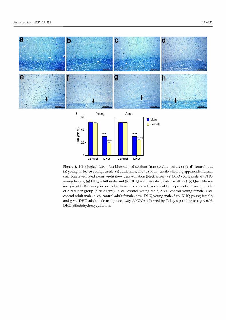
Figure 8. Histological Luxol fast blue-stained sections from cerebral cortex of (a–d) control rats, (a) young male, (b) young female, (c) adult male, and (d) adult female, showing apparently normal dark blue myelinated axons. (e–h) show demyelination (black arrow), (e) DHQ young male, (f) DHQ young female, (g) DHQ adult male, and (h) DHQ adult female. (Scale bar 50 um). (i) Quanti-tative analysis of LFB staining in cortical sections. Each bar with a vertical line represents the mean ± S.D. of 5 rats per group (5 fields/rat). a vs. control young male, b vs. control young female, c vs. control adult male, d vs. control adult female, e vs. DHQ young male, f vs. DHQ young female, and g vs. DHQ adult male using three-way ANOVA followed by Tukey’s post hoc test; p < 0.05. DHQ; diiodohydroxyquinoline.
Figure 8. Histological Luxol fast blue-stained sections from cerebral cortex of (a–d) control rats,(a) young male, (b) young female, (c) adult male, and (d) adult female, showing apparently normaldark blue myelinated axons. (e–h) show demyelination (black arrow), (e) DHQ young male, (f) DHQyoung female, (g) DHQ adult male, and (h) DHQ adult female. (Scale bar 50 um). (i) Quantitativeanalysis of LFB staining in cortical sections. Each bar with a vertical line represents the mean ± S.D.of 5 rats per group (5 fields/rat). a vs. control young male, b vs. control young female, c vs.control adult male, d vs. control adult female, e vs. DHQ young male, f vs. DHQ young female,and g vs. DHQ adult male using three-way ANOVA followed by Tukey’s post hoc test; p < 0.05.DHQ; diiodohydroxyquinoline.
Pharmaceuticals 2022, 15, 251 12 of 22Pharmaceuticals 2022, 15, 251 12 of 21
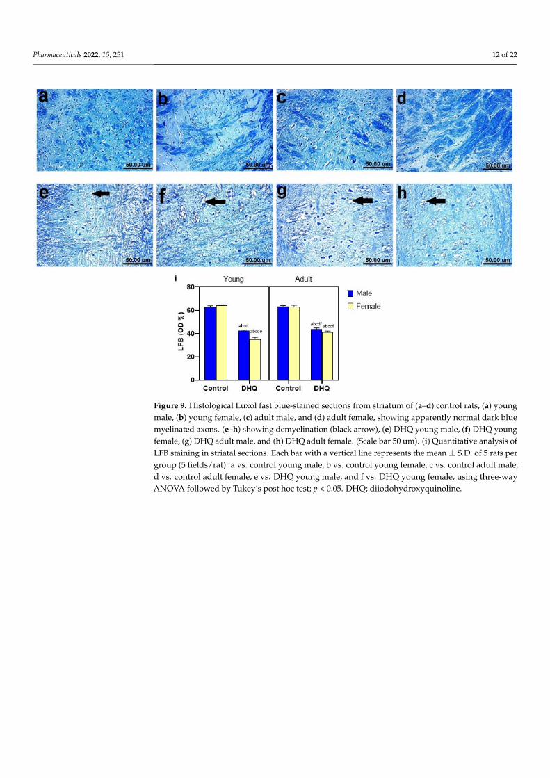
Figure 9. Histological Luxol fast blue-stained sections from striatum of (a–d) control rats, (a) young male, (b) young female, (c) adult male, and (d) adult female, showing apparently normal dark blue myelinated axons. (e–h) showing demyelination (black arrow), (e) DHQ young male, (f) DHQ young female, (g) DHQ adult male, and (h) DHQ adult female. (Scale bar 50 um). (i) Quantitative analysis of LFB staining in striatal sections. Each bar with a vertical line represents the mean ± S.D. of 5 rats per group (5 fields/rat). a vs. control young male, b vs. control young female, c vs. control adult male, d vs. control adult female, e vs. DHQ young male, and f vs. DHQ young female, using three-way ANOVA followed by Tukey’s post hoc test; p < 0.05. DHQ; diiodohydroxyquinoline.
Figure 9. Histological Luxol fast blue-stained sections from striatum of (a–d) control rats, (a) youngmale, (b) young female, (c) adult male, and (d) adult female, showing apparently normal dark bluemyelinated axons. (e–h) showing demyelination (black arrow), (e) DHQ young male, (f) DHQ youngfemale, (g) DHQ adult male, and (h) DHQ adult female. (Scale bar 50 um). (i) Quantitative analysis ofLFB staining in striatal sections. Each bar with a vertical line represents the mean ± S.D. of 5 rats pergroup (5 fields/rat). a vs. control young male, b vs. control young female, c vs. control adult male,d vs. control adult female, e vs. DHQ young male, and f vs. DHQ young female, using three-wayANOVA followed by Tukey’s post hoc test; p < 0.05. DHQ; diiodohydroxyquinoline.

Pharmaceuticals 2022, 15, 251 13 of 22
Pharmaceuticals 2022, 15, 251 12 of 21
Figure 9. Histological Luxol fast blue-stained sections from striatum of (a–d) control rats, (a) young male, (b) young female, (c) adult male, and (d) adult female, showing apparently normal dark blue myelinated axons. (e–h) showing demyelination (black arrow), (e) DHQ young male, (f) DHQ young female, (g) DHQ adult male, and (h) DHQ adult female. (Scale bar 50 um). (i) Quantitative analysis of LFB staining in striatal sections. Each bar with a vertical line represents the mean ± S.D. of 5 rats per group (5 fields/rat). a vs. control young male, b vs. control young female, c vs. control adult male, d vs. control adult female, e vs. DHQ young male, and f vs. DHQ young female, using three-way ANOVA followed by Tukey’s post hoc test; p < 0.05. DHQ; diiodohydroxyquinoline.
Pharmaceuticals 2022, 15, 251 13 of 21
Figure 10. Histological Luxol fast blue-stained sections from spinal cords of (a–d) control rats, (a) young male, (b) young female, (c) adult male, and (d) adult female, showing apparently normal dark blue myelinated axons. (e–h) showing demyelination in white and gray matter (black arrow), (e) DHQ young male, (f) DHQ young female, (g) DHQ adult male, and (h) DHQ adult female. (Scale bar 50 um). (i) Quantitative analysis of LFB staining in sections from spinal cords. Each bar with a vertical line represents the mean ± S.D. of 5 rats per group (5 fields/rat). a vs. control young male, b vs. control young female, c vs. control adult male, d vs. control adult female, e vs. DHQ young male, f vs. DHQ young female, and g vs. DHQ adult male using three-way ANOVA followed by Tukey’s post hoc test; p < 0.05. DHQ; diiodohydroxyquinoline.
Figure 11. Histological Luxol fast blue-stained sections from sciatic nerves of (a–d) control rats, (a) young male, (b) young female, (c) adult male, and (d) adult female, showing apparently normal dark blue myelinated axons. (e–h) showing demyelination of nerve fibers (black arrow), (e) DHQ young male, (f) DHQ young female, (g) DHQ adult male, and (h) DHQ adult female. (Scale bar 50 um). (i) Quantitative analysis of LFB staining in sections from sciatic nerves. Each bar with a vertical line represents the mean ± S.D. of 5 rats per group (5 fields/rat). a vs. control young male, b vs. control young female, c vs. control adult male, d vs. control adult female, e vs. DHQ young male, f vs. DHQ young female, and g vs. DHQ adult male using three-way ANOVA followed by Tukey’s post hoc test; p < 0.05. DHQ; diiodohydroxyquinoline.
Figure 10. Histological Luxol fast blue-stained sections from spinal cords of (a–d) control rats,(a) young male, (b) young female, (c) adult male, and (d) adult female, showing apparently normaldark blue myelinated axons. (e–h) showing demyelination in white and gray matter (black arrow),(e) DHQ young male, (f) DHQ young female, (g) DHQ adult male, and (h) DHQ adult female. (Scalebar 50 um). (i) Quantitative analysis of LFB staining in sections from spinal cords. Each bar with avertical line represents the mean ± S.D. of 5 rats per group (5 fields/rat). a vs. control young male, bvs. control young female, c vs. control adult male, d vs. control adult female, e vs. DHQ young male,f vs. DHQ young female, and g vs. DHQ adult male using three-way ANOVA followed by Tukey’spost hoc test; p < 0.05. DHQ; diiodohydroxyquinoline.

Pharmaceuticals 2022, 15, 251 14 of 22
Pharmaceuticals 2022, 15, 251 13 of 21
Figure 10. Histological Luxol fast blue-stained sections from spinal cords of (a–d) control rats, (a) young male, (b) young female, (c) adult male, and (d) adult female, showing apparently normal dark blue myelinated axons. (e–h) showing demyelination in white and gray matter (black arrow), (e) DHQ young male, (f) DHQ young female, (g) DHQ adult male, and (h) DHQ adult female. (Scale bar 50 um). (i) Quantitative analysis of LFB staining in sections from spinal cords. Each bar with a vertical line represents the mean ± S.D. of 5 rats per group (5 fields/rat). a vs. control young male, b vs. control young female, c vs. control adult male, d vs. control adult female, e vs. DHQ young male, f vs. DHQ young female, and g vs. DHQ adult male using three-way ANOVA followed by Tukey’s post hoc test; p < 0.05. DHQ; diiodohydroxyquinoline.
Figure 11. Histological Luxol fast blue-stained sections from sciatic nerves of (a–d) control rats, (a) young male, (b) young female, (c) adult male, and (d) adult female, showing apparently normal dark blue myelinated axons. (e–h) showing demyelination of nerve fibers (black arrow), (e) DHQ young male, (f) DHQ young female, (g) DHQ adult male, and (h) DHQ adult female. (Scale bar 50 um). (i) Quantitative analysis of LFB staining in sections from sciatic nerves. Each bar with a vertical line represents the mean ± S.D. of 5 rats per group (5 fields/rat). a vs. control young male, b vs. control young female, c vs. control adult male, d vs. control adult female, e vs. DHQ young male, f vs. DHQ young female, and g vs. DHQ adult male using three-way ANOVA followed by Tukey’s post hoc test; p < 0.05. DHQ; diiodohydroxyquinoline.
Figure 11. Histological Luxol fast blue-stained sections from sciatic nerves of (a–d) control rats,(a) young male, (b) young female, (c) adult male, and (d) adult female, showing apparently normaldark blue myelinated axons. (e–h) showing demyelination of nerve fibers (black arrow), (e) DHQyoung male, (f) DHQ young female, (g) DHQ adult male, and (h) DHQ adult female. (Scale bar50 um). (i) Quantitative analysis of LFB staining in sections from sciatic nerves. Each bar with avertical line represents the mean ± S.D. of 5 rats per group (5 fields/rat). a vs. control young male, bvs. control young female, c vs. control adult male, d vs. control adult female, e vs. DHQ young male,f vs. DHQ young female, and g vs. DHQ adult male using three-way ANOVA followed by Tukey’spost hoc test; p < 0.05. DHQ; diiodohydroxyquinoline.
3. Discussion
The previously reported tendency of clioquinol (one representative of 8-HQ) to affectsensorimotor function prompted us to assess the effect of DHQ on motor performance andpain-like behavior in rats. Pain-related behaviors were evaluated using different stimuli,such as hind paw cold allodynia, hot plate thermal algesia, and mechanical hyperalgesia.These tests can assess and quantify pain-like behaviors as well as detect any variability inpain-threshold. Surprisingly, DHQ-treated rats demonstrated allodynia and hyperalgesiaresponses with lowered pain threshold in the three aforementioned tests compared tocontrol groups; this was especially prominent in the young female group. This wasobserved in clioquinol-treated mice and was rationalized by the ability of clioquinol tochelate zinc and copper ions, as this chelation enhances activation of transient receptorpotential ankyrin 1 (TRPA1) in sensory neurons [15]. TRPA1 has been proposed as asensory transduction molecule for both cold and mechanical stimuli and any increase inTRPA1 is associated with hyperalgesia. The severity of histopathological alterations wasmore pronounced in female groups, especially those of young age. The female gendermay be a predisposing factor to the neurotoxicity of DHQ, as reported in the presentstudy by a lower threshold of tactile and pain sensation as well as in previous clinical

Pharmaceuticals 2022, 15, 251 15 of 22
cases subjected to clioquinol [16,17]. Specific scores of histopathological lesions for cortex,striatum, spinal cord, and sciatic nerve in diiodohydroxyquinoline-treated animals arepresented in supplementary Table S1.
There was a coincidence between heat, cold, and mechanical withdrawal responses, asindicated by Pearson’s correlation matrix, which attracts attention to the involvement ofthe sensory element that is common to these three stimuli in DHQ-induced neurotoxicity.In addition, while conducting this study, a gross inspection of the hind paw revealed noevidence of erythema or tissue damage after mechanical and thermal testing. This turnedthe sight to histopathological examinations. The present study demonstrates demyelinationin various investigated areas/organs, as confirmed by LFB-stain, and this shed light onthe demyelinating side effects of DHQ. Demyelination disrupts rates of nerve impulsetransmission and creates abnormal sensory phenomena, such as allodynia, hyperalgesia,and spontaneous pain. Abnormal sensory phenomena are associated with human periph-eral demyelinating neuropathies, such as Charcot–Marie–Tooth disease and Guillain-Barrésyndrome. The damage to sensory nerves as a result of peripheral demyelinating diseasehas been linked to pain and heightened sensitivity to touch [18,19]. This was also observedwith clioquinol, which causes symmetrical demyelination in lateral and posterior funiculiof the spinal cord in humans [20]. It was demonstrated that clioquinol lowers vitamin B12in the brain of mice and this rationalized the involvement of hydroxyquinoline derivatives,including DHQ in demyelination and neuropathy [21]. Ultimately, this is strong evidencethat sensory fiber dysfunction is a significant feature of DHQ intoxication.
The PCA is a data analytical, rather than statistical, procedure. The present studyutilized the privilege of using PCA for classification of a highly complex bundle of variablesof different behavioral tests, which can be reduced to only countable components thatmirror the behavioral features assessed by these variables. Factor analysis has been usedpreviously to explore the relation between behavioral tests [22] as well to detect sex- andage-specific behavior [23,24] and eventually to demonstrate the most prominent effectof the drug within several altered behavioral variables [25]. The Kaiser rule allowedus to choose components with eigenvalues of at least one concomitantly with the screeplot (data not shown) that resulted in the projection of three components that represent78.5% of the total variation. Varimax rotation maximized variance to increase the squaredcorrelation of items related to one factor and on the other hand decrease the correlationon other factors. In the current study, PCA allowed us to categorize variables of sensoryand motor tests into three components. The analysis produced PCI, which explained29.1% of variation and was mainly composed of sensory-related variables. Analysis ofPCI revealed that DHQ significantly affected sensory function in rats; this effect wasage- and gender-dependent, where DHQ-treated rats exhibited lower PCI scores thanuntreated rats, especially female young rats. This finding was documented by the AmericanAcademy of Pediatrics Committee on Drugs and cautioned against iodoquinol use inchildren and infants due to neurotoxicity [11] and it matches findings from clinical reportsof hydroxyquinoline products [17]. Obviously, it was found that sensory disturbances wereprofound in clioquinol-deteriorated patients in Japan, reaching 97% of mainly femininecases [16], which is in line with the present findings of PCA.
Regarding motor performance, rotarod and open field tests were utilized to analyzecoordination and balance. Motor disturbance is most often determined by the rotarodtest. Noteworthy, performance in the rotarod task depends mainly on functionality of thenigrostriatal dopamine system [26]. The findings of rotarod demonstrated a clear deficit inDHQ-treated rats, as they have shorter fall off latency compared to normal controls. This isin agreement with histopathological studies, which demonstrated neuropathic changes inthe nigrostriatal pathway confined as shrunken, atrophied, and pyknotic neurons associatedwith severe demyelination and spongiosis. The severity of histopathological alterationswas more pronounced in female groups’, especially the young age. This was consistentwith results from a previous study, where a halogenated quinolinol, clioquinol, causedmarked sensory and motor disturbance in the lower extremities in Japan [27,28]. Taken

Pharmaceuticals 2022, 15, 251 16 of 22
together, the present study utilized the rotarod accelerating protocol, as it is considereda more discriminative test that correlates motor deficits against lesion size [29] and thiswas elucidated herein where the most deteriorated gender, the female, showed significantlesions in the striatum and sciatic nerve concomitantly with increased falling rates ofrotarod compared with the counterparts.
The lack of direct evidence of specific motor disturbance, together with DHQ’s demon-strated degenerative effects in the animal model in the striatum and sciatic nerve, suggeststhat such impaired behavior, analogous to that previously observed with clioquinol, mayset a more plausible pathophysiological illustration for these symptoms. Herein, PCII ex-plained 27.4% of variation and was related to the rats’ motor performance. This componentof motor function is unrelated to sensory function, as PCII is orthogonal to sensory-relatedPCI. Sensory-related variables contributed to a much lesser extent to PCII. The variables,OFTD, OFTMS, OFFTTI and RROFL-loaded PCII, with the most affected by DHQ admin-istration, OFTD and RROFL. However, RROFL demonstrated positive correlation withwithdrawal threshold in the three sensory tasks. To our knowledge, alteration in the sensoryfunction is accompanied, accordingly, with alteration in proprioception, indicating motordeficits [30]. Muscle proprioceptors encode information and transmit it to networks inthe spinal cord to coordinate and adapt muscles according to posture and gait. Thus, themotor abnormality in the current study may be in part secondary to sensory dysfunction.It seems that the outcome of PCA is in line with the previous rationale that sensory-relatedvariables loaded profoundly PCI and captured 29.1% of total variance followed by mo-tor variables that loaded PCII. However, this study applies the profound difference ofPCA in respect to linear regression analysis. However, there were correlations betweenthe performance in different tasks, but generally, PCA bundles within-test variables anddiscriminates between-test variables more effectively than regression analysis [22]. There-fore, the motor disturbance may be a direct effect of DHQ on spinal cord and striatum aspresently witnessed by histopathological aberrations rather than as secondary to sensorydeficits. PCA demonstrated this speculation that to a less extent the motor function is notthe major deteriorated domain by DHQ, such as the sensory function.
The open field test assesses locomotor and exploratory activity, which can be correlatedwith locomotive function and neuromuscular disease [31–34]. In this regard, DHQ signifi-cantly affected locomotion activity in the open field arena, as evidenced by the reduced totaldistance covered by rats, which confirms the impairment of motor function, as previouslyrecorded in the rotarod. This is in line with a comparative study that documented anabnormality in locomotion in patients treated by clioquinol that lasted for 32 years, wherecomplete loss of locomotion was reached [16]. Biochemically, metal chelation with zincmakes hydroxyquinoline derivatives more lipophilic and consequently more bioavailable,affecting the central nervous system [5]. This mechanism could explain the neurotoxiceffect of DHQ, a clioquinol analogue, as well as the alteration of muscle coordinationand motor performance. Indeed, there is a positive forward relationship between motordysfunction and CNS demyelination: as the extent of CNS demyelination increases, theseverity of motor dysfunction increases [35]. This is a fact that was verified by resultsof the current study, where young female rats demonstrated severe motor impairment.This is in line with previous studies [6,36], which demonstrated that clioquinol inducedsymmetrical demyelination of lateral posterior funiculi of the spinal cord, optic nerve, andperipheral nerves.
Patients who suffered SMON, especially women, had significantly worse baselinescores characterized by enhanced anxiety and disturbed peace of mind [28]. During PCAanalysis, the third component PCIII explained 22% of the total variation, and representedanxiety-related behavior. This is observed in clioquinol-treated patients suffering fromirritability and neurosis by 27.8% and 13.6%, respectively [16], which is not as prevalentas sensory and motor abnormality. No previous studies elucidated the anxiogenic effect,but it may be related to N-methyl-D-aspartate receptor activation [28]. In the currentexperiment, DHQ reduced the OF central time that positively loaded on PCIII, which

Pharmaceuticals 2022, 15, 251 17 of 22
determined the anxiogenic effect of DHQ and influenced rats to leave the OF center arenain anxiety-like behavior.
The current study did not utilize different periods and pediatric age <P21 to studylong-term effects of DHQ, where the consequences and the underlying physiopathologicalmechanisms need further investigation. As well, per the findings of the present study, an-other approach that should be investigated is the effect of DHQ on the microbiota–gut–brainaxis and its effect on neuropsychiatric disorders, as demonstrated by Generoso et al. [37]and presented in a comparative study with another relevant antibiotic. The present studyassessed the damage in dopaminergic pathway viz the nigrostriatal pathway, in addition tothe anxiogenic tendency of DHQ; therefore, measuring the neurotransmitters in differentbrain areas could evolve the novel link between DHQ and neuropsychiatric disorders.
4. Materials and Methods4.1. Animals
The study was conducted using young and adult Wistar rats of both sexes (3 weeksold, weighing 90–100 g and 4 months old, weighing 250–300 g, respectively). Animalswere acquired from the Nile Company for Pharmaceuticals & Chemical Industries, Cairo,Egypt. They were allowed to acclimatize to laboratory conditions in the animal house of theFaculty of Pharmacy, Cairo University for 2 weeks before the experiment. They were keptunder standard laboratory conditions for room temperature (24–26 ◦C), relative humidity(60 ± 10%), and light/dark cycle (12/12 h). Animals were allowed free access to standardchow diet and water. All procedures performed on animals were in accordance with theGuide for Care and Use of Laboratory Animals (NIH Publication No. 85-23, revised 2011)and was approved by the Ethical Committee of the Faculty of Pharmacy, Cairo University(Permit Number: PT 2732).
4.2. Drugs and Chemicals
Diiodohydroxyquinoline was acquired from CID-Chemical Industries Development(Cairo, Egypt). DHQ was suspended in 1% tween 80 saline solution. All other chemicalswere of high purity and analytical grade.
4.3. Experimental Design
Eighty rats were randomly distributed using a computer-generated randomizationtable according to age and sex in eight groups (10 rats each). Animals in the first 4 groupsreceived saline daily by oral gavage for 4 weeks and served as control. The control youngmale and female rats were allocated to group I (CYM) and group II (CYF), respectively.Control adult male and female rats were allocated to group III (CAM) and group IV(CAF), respectively, whereas, rats in the remaining 4 groups received DHQ daily by oralgavage for 4 weeks. Treated young male and female rats were allocated to group V (TYM)and group VI (TYF), respectively, and they received DHQ at a dose of 247.4 mg/kg/day.Treated adult male and female rats were allocated to group VII (TAM) and group VIII(TAF), respectively, and they received DHQ at a dose of 176.7 mg/kg/day. The dosesof DHQ were converted from the maximum recommended daily human pediatric/adultdoses [38,39] to rat doses using the conversion guide provided by Nair and Jacob [40].Behavioral analysis was conducted on the last 2 days of the experiment in the followingsequence: open field, Randall–Selitto, and hind paw cold allodynia tests on day 27, inaddition to rotarod and hot plate tests on day 28. All behavioral testing was performedin a sound-isolated laboratory during the light phase, with a 2-h rest period between thetests [41]. Twenty-four hours after the completion of behavioral examination, rats weresacrificed by decapitation under light anesthesia. Brains, spinal cords, and sciatic nerveswere quickly harvested and rinsed with ice-cold saline. They were fixed in 10% bufferedformol saline for histopathological examination.

Pharmaceuticals 2022, 15, 251 18 of 22
4.4. Behavioral Assessment4.4.1. Open Field Test
Open field test (OFT) is widely used to test exploratory behavior and general activityof rodents. The open field consisted of a square wooden box (80 × 80 × 40 cm) withred walls and white smooth polished floor divided by black lines into 16 equal squares(20 × 20 cm). Each rat was placed gently in the central area of the open field and allowedto freely explore the area for 3 min. The floor and walls were cleaned after testing each ratto eliminate possible bias due to odors left by previous rats. A video camera was fixed onthe top of the box to record the movement and behavior of rats, which were analyzed usingANY-Maze video tracking software (Stoelting Co., Wood Dale, Illinois, USA). Total distancetraveled (OFTD), mean speed (OFTMS), time immobile (OFTTI), central time (OFTCT),central distance (OFTTD), and thigmotaxis time (OFTTT) were recorded [42,43].
4.4.2. Rotarod Test
The rotarod is a widely used to assess motor coordination and balance of rodents.Briefly, rats were trained for 2 consecutive days before the experimental procedures at aconstant speed of 4 rpm on an automated 5-lane rotarod apparatus (Model 47750, UgoBasile, Gemonio, VA, Italy) with 5-min cut-off time for 3 trials each day. The animals thatstayed on the rod for 5 min were selected for experimentation. On the test day, rats wereplaced on the rod at a speed accelerated from 4 to 40 rpm with a 5-min cut-off time for3 trials. The average rotarod fall off latency (RRFOL) was recorded for each rat [44].
4.4.3. Randall–Selitto Test
Randall–Selitto test is used to quantify neuropathic pain response in rodents. The me-chanical nociceptive threshold (RSMT) was assessed using a paw pressure analgesiometer(Model 7200, Ugo Basile, Italy), which applies a steadily increasing force to a rat’s left hindpaw. Animals were held with soft cotton cloth in order to immobilize them for measuringthe threshold. A withdrawal of the left hind paw or vocalization was considered as theendpoint. A cut-off pressure of 350 g was maintained to avoid tissue injury [45].
4.4.4. Hot Plate Test
Hot plate test is used to assess heat thermal sensitivity of rats. Each rat was gentlyplaced on the hot plate (Model 7280, Ugo Basile, Italy), which was set at 55 ± 1 ◦C and thetime until either hind paw licking or hopping to avoid heat pain was recorded as hot platereaction latency (HPRL), with a cut-off time of 20 s [46].
4.4.5. Hind Paw Cold Allodynia Test
Cold allodynia test is used to assess cold thermal sensitivity of rats. The hind pawsof each rat were immersed gently in a beaker containing ice-cold water maintained at4 ± 1 ◦C. Then, the paw withdrawal latency (CAPWL) for each rat was determined. Onlyone hind paw was evaluated during each immersion at a time, with a cut-off time of 20 s.For each animal, two readings were taken for each hind paw at a 5-min interval, andCAPWL was calculated as the mean of both hind paw’s readings. The extended CAPWLwas considered as antiallodynic effect, while the shorter CAPWL was interpreted as moresevere allodynia [47].
4.5. Histological Assessment
Specimens from brain, spinal cord, and sciatic nerve were quickly harvested from 5 ratsper each group and fixed in 10% buffered formol saline, embedded in paraffin, and then cutinto 4 µm sections. Sections were deparaffinized, hydrated, and stained with Hematoxylinand Eosin (H&E) and Luxol fast blue (LFB) for examination under the light microscope(Olympus BX50, Olympus LS, Shinjuku-ku, Tokyo, Japan). The recorded histopathologicallesions in H&E-stained sections from all rats were scored via assessing the percentageof lesions frequency from 5 randomly selected microscopic fields per animal using the

Pharmaceuticals 2022, 15, 251 19 of 22
following score system; 0 = absence of the lesions in all rats of the group, 1 = 1–10%,2 = 11–25%, 3 = 26–50%, 4 = 51–75%, and 5 = over 75% [48]. In addition, the optical densityof the LFB-stained sections was analyzed using image analysis software (Image J, version1.46a, NIH, Bethesda, MD, USA).
4.6. Statistical Analysis
Data are expressed as mean ± S.D. Comparisons between means were performedusing three-way analysis of variance (MANOVA) test, followed by Tukey’s multiple com-parisons test using GraphPad Prism® software package, version 8 (GraphPad SoftwareInc., San Diego, CA, USA). Correlations between behavioral measures were detected usingPearson’s correlation matrix (two-tailed). The level of significance was fixed at p < 0.05 forall statistical tests. To reduce the number of dimensions, a Factor analysis (principal com-ponent (PC) with Varimax rotation and Kaiser normalization, maximally 25 iterations toconvergence) based on the correlation matrix was performed for all variables. The Varimaxrotation was chosen because it is an orthogonal rotation method so that different factors donot intercorrelate; thus, each factor represents an independent behavioral pattern. Varimaxrotation reduces the number of variables with high multiple factor loadings. Extractedcomponents with eigenvalues higher than one were considered for further interpretation.Factor loadings >0.7 or <−0.7 were considered significant. Factor and correlation analyseswere performed using SPSS Version 20.
5. Conclusions
Experimentally, sensory and motor functions, as well as extensive structural abnor-malities in central and peripheral nervous tissues after the sub-acute administration ofDHQ, were demonstrated; this finding calls for further investigation of the long-term useof this class of drugs, considering their benefit against alternative antibiotics. The studyalso highlights the role of gender and age in exacerbating quinoline-induced neurotoxicity.More preclinical studies are needed to study the demyelinating hazards of quinoline deriva-tives. In addition, it would be essential to study the effects of other factors, such as raceand the microbiota–gut–brain interaction, which could affect the toxicity of the sub-acuteadministration of quinoline.
Supplementary Materials: The following are available online at https://www.mdpi.com/article/10.3390/ph15020251/s1, Table S1: Supplementary Table S1 Histopathological lesion scoring for cortex,striatum, spinal cord, and sciatic nerve in diiodohydroxyquinoline-treated animals.
Author Contributions: Conceptualization, investigation, and writing—original draft preparation,A.S.K., A.F.M. and M.A.R.; writing—review and editing, M.E.E.; investigation, writing—reviewand editing, K.A.A.; conceptualization, writing—review and editing, M.M.K.; conceptualization,writing—original draft preparation, writing—review and editing, N.F.A. All authors have read andagreed to the published version of the manuscript.
Funding: This research did not receive any specific grant from funding agencies in the public,commercial, or not-for-profit sectors.
Institutional Review Board Statement: The animal study protocol was approved by the EthicalCommittee of the Faculty of Pharmacy, Cairo University (Permit Number: PT 2732, July 2020).
Informed Consent Statement: Not applicable.
Data Availability Statement: Data is contained within the article and supplementary material.
Conflicts of Interest: The authors declare that they have no conflict of interest.

Pharmaceuticals 2022, 15, 251 20 of 22
Abbreviations
OFT Open field testOFTD Open field test distanceOFTMS Open field test mean speedOFTTI Open field test time immobileOFTCT Open field test central timeOFTCD Open field test central distanceOFTTT Open field test thigmotaxis timeRRFOL Rotarod fall off latencyCAPWL Cold allodynia paw withdrawal latencyHPRL Hotplate reaction latencyRSMT Randall–Salyetto mechanical thresholdCYM Control young maleCYF Control young femaleCAM Control adult maleCAF Control adult femaleTYM Treated young maleTYF Treated young femaleTAM Treated adult maleTAF Treated adult femaleDHQ Diiodohydroxyquinoline8-HQ 8-hydroxyquinolineSMON Subacute myelo-optic neuropathyPC Principal componentTRPA1 Transient receptor potential ankyrin 1H&E Hematoxylin and EosinLFB Luxol fast blueMANOVA Three-way analysis of variance testPCA Principal component analysis
References1. Schnitzer, R.J. IARC monographs on the evaluation of the carcinogenic risk of chemicals to man. Environ. Res. 1978, 17, 151.
[CrossRef]2. Song, Y.; Xu, H.; Chen, W.; Zhan, P.; Liu, X. 8-Hydroxyquinoline: A privileged structure with a broad-ranging pharmacological
potential. Med. Chem. Comm. 2015, 6, 61–74. [CrossRef]3. Yamamoto, R.S.; Williams, G.M.; Frankel, H.H.; Weisburger, J.H. 8-Hydroxyquinoline: Chronic toxicity and inhibitory effect on
the carcinogenicity of N-2-fluorenylacetamide. Toxicol. Appl. Pharmacol. 1971, 19, 687–698. [CrossRef]4. Maronpot, R.R.; Boorman, G.A. Interpretation of rodent hepatocellular proliferative alterations and hepatocellular tumors in
chemical safety assessment. Toxicol. Pathol. 1982, 10, 71–78. [CrossRef]5. Bareggi, S.R.; Cornelli, U. Clioquinol: Review of its mechanisms of action and clinical uses in neurodegenerative Disorders. CNS
Neurosci. Ther. 2012, 18, 41–46. [CrossRef]6. Tsubaki, T.; Honma, Y.; Hoshi, M. Neurological syndrome associated with clioquinol. Lancet 1971, 297, 696–697. [CrossRef]7. Nakae, K.; Yamamoto, S.I.; Shigematsu, I.; Kono, R. Relation between Subacute Myelo-Optic Neuropathy (S.M.O.N.) and
clioquinol: Nationwide survey. Lancet 1973, 301, 171–173. [CrossRef]8. Takeda, A.; Takada, S.; Ando, M.; Itagaki, K.; Tamano, H.; Suzuki, M.; Iwaki, H.; Oku, N. Impairment of recognition memory and
hippocampal long-term potentiation after acute exposure to clioquinol. Neuroscience 2010, 171, 443–450. [CrossRef]9. Mansour-Ghanaei, F.; Dehbashi, N.; Yazdanparast, K.; Shafaghi, A. Efficacy of saccharomyces boulardii with antibiotics in acute
amoebiasis. World J. Gastroenterol. 2003, 9, 1832–1833. [CrossRef]10. Fisher, A.K.; Walter, F.G.; Szabo, S. Iodoquinol associated seizures and radiopacity. Clin. Toxicol. 1993, 31, 113–120. [CrossRef]11. American Academy of Pediatrics Committee on Drugs. Clioquinol (iodochlorhydroxyquin, vioform) and iodoquinol (diiodohy-
droxyquin): Blindness and Neuropathy. Pediatrics 1990, 86, 797–798. Available online: https://pubmed.ncbi.nlm.nih.gov/2146587/ (accessed on 13 December 2021).
12. Ghaskadbi, S.; Vaidya, V.G. In vivo antimutagenic effect of ascorbic acid against mutagenicity of the common antiamebic drugdiiodohydroxyquinoline. Mutat. Res. Toxicol. 1989, 222, 219–222. [CrossRef]
13. Nevin, R.L. Idiosyncratic quinoline central nervous system toxicity: Historical insights into the chronic neurological sequelae ofmefloquine. Int. J. Parasitol. Drugs Drug Resist. 2014, 4, 118–125. [CrossRef] [PubMed]

Pharmaceuticals 2022, 15, 251 21 of 22
14. European Medicines Agency. Disabling and Potentially Permanent Side Effects Lead to Suspension or Restrictions of Quinolone andFluoroquinolone Antibiotics; EMA/175398/2019; European Medicines Agency: Amsterdam, Netherlands, 2019; Available on-line: https://www.ema.europa.eu/en/documents/referral/quinolone-fluoroquinolone-article-31-referral-disabling-potentially-permanent-side-effects-lead_en.pdf (accessed on 13 December 2021).
15. Andersson, D.A.; Gentry, C.; Moss, S.; Bevan, S. Clioquinol and pyrithione activate TRPA1 by increasing intracellular Zn2+. Proc.Natl. Acad. Sci. USA 2009, 106, 8374–8379. [CrossRef]
16. Konagaya, M.; Matsumoto, A.; Takase, S.; Mizutani, T.; Sobue, G.; Konishi, T.; Hayabara, T.; Iwashita, H.; Ujihira, T.; Miyata, K.;et al. Clinical analysis of longstanding subacute myelo-optico-neuropathy: Sequelae of clioquinol at 32 years after its ban. J.Neurol. Sci. 2004, 218, 85–90. [CrossRef]
17. Baumgartner, G.; Gawel, M.J.; Kaeser, H.E.; Pallis, C.A.; Rose, F.C.; Schaumburg, H.H.; Thomas, P.K.; Wadia, N.H. Neurotoxicityof halogenated hydroxyquinolines: Clinical analysis of cases reported outside Japan. J. Neurol. Neurosurg. Psychiatry 1979, 42,1073–1083. [CrossRef]
18. Pentland, B.; Donald, S.M. Pain in the Guillain-Barré syndrome: A clinical review. Pain 1994, 59, 159–164. [CrossRef]19. Wallace, V.C.J.; Cottrell, D.F.; Brophy, P.J.; Fleetwood-Walker, S.M. Focal Lysolecithin-Induced Demyelination of Peripheral
Afferents Results in Neuropathic Pain Behavior That Is Attenuated by Cannabinoids. J. Neurosci. 2003, 23, 3221–3233. [CrossRef]20. Konagaya, M. SMON: Toxicity of clioquinol and the status quo. Brain Nerve 2015, 67, 49–62. [CrossRef]21. Yassin, M.S.; Ekblom, J.; Xilinas, M.; Gottfries, C.G.; Oreland, L. Changes in uptake of vitamin B12 and trace metals in brains of
mice treated with clioquinol. J. Neurol. Sci. 2000, 173, 40–44. [CrossRef]22. Feyissa, D.D.; Aher, Y.D.; Engidawork, E.; Höger, H.; Lubec, G.; Korz, V. Individual differences in male rats in a behavioral test
battery: A multivariate statistical approach. Front. Behav. Neurosci. 2017, 11, 26. [CrossRef] [PubMed]23. Frick, K.M.; Baxter, M.G.; Markowska, A.L.; Olton, D.S.; Price, D.L. Age-related spatial reference and working memory deficits
assessed in the water maze. Neurobiol. Aging 1995, 16, 149–160. [CrossRef]24. Carreira, M.B.; Cossio, R.; Britton, G.B. Individual and sex differences in high and low responder phenotypes. Behav. Processes
2017, 136, 20–27. [CrossRef]25. Catuara-Solarz, S.; Espinosa-Carrasco, J.; Erb, I.; Langohr, K.; Gonzalez, J.R.; Notredame, C.; Dierssen, M. Combined Treatment
with Environmental Enrichment and (−)-Epigallocatechin-3-Gallate Ameliorates Learning Deficits and Hippocampal Alterationsin a Mouse Model of Down Syndrome. eNeuro 2016, 3, ENEURO.0103-16.2016. [CrossRef] [PubMed]
26. Lundblad, M.; Vaudano, E.; Cenci, M.A. Cellular and behavioural effects of the adenosine A2a receptor antagonist KW-6002 in arat model of L-DOPA-induced dyskinesia. J. Neurochem. 2003, 84, 1398–1410. [CrossRef] [PubMed]
27. Cahoon, L. The curious case of clioquinol. Nat. Med. 2009, 15, 356–359. [CrossRef]28. Ohta, Y.; Hishikawa, N.; Sato, K.; Takemoto, M.; Yamashita, T.; Doutare, S.; Abe, K. Improving anxiety in subacute myelo-optico-
neuropathy (SMON) after an automated telephone call service. Intern. Med. 2019, 58, 1081–1085. [CrossRef]29. Monville, C.; Torres, E.M.; Dunnett, S.B. Comparison of incremental and accelerating protocols of the rotarod test for the
assessment of motor deficits in the 6-OHDA model. J. Neurosci. Methods. 2006, 158, 219–223. [CrossRef]30. Bhattacherjee, A.; Winter, M.K.; Eggimann, L.S.; Mu, Y.; Gunewardena, S.; Liao, Z.; Christianson, J.A.; Smith, P.G. Motor,
somatosensory, viscerosensory and metabolic impairments in a heterozygous female rat model of rett syndrome. Int. J. Mol. Sci.2018, 19, 97. [CrossRef]
31. Walsh, R.N.; Cummins, R.A. The open-field test: A critical review. Psychol. Bull. 1976, 83, 482–504. [CrossRef]32. Belzung, C.; Griebel, G. Measuring normal and pathological anxiety-like behaviour in mice: A review. Behav. Brain Res. 2001, 125,
141–149. [CrossRef]33. Nagaraju, K.; Raben, N.; Loeffler, L.; Parker, T.; Rochon, P.J.; Lee, E.; Danning, C.; Wada, R.; Thompson, C.; Bahtiyar, G.; et al.
Conditional up-regulation of MHC class I in skeletal muscle leads to self-sustaining autoimmune myositis and myositis-specificautoantibodies. Proc. Natl. Acad. Sci. USA 2000, 97, 9209–9214. [CrossRef] [PubMed]
34. Raben, N.; Nagaraju, K.; Lee, E.; Plotz, P. Modulation of disease severity in mice with targeted disruption of the acid α-glucosidasegene. Neuromuscul. Disord. 2000, 10, 283–291. [CrossRef]
35. Franco-Pons, N.; Torrente, M.; Colomina, M.T.; Vilella, E. Behavioral deficits in the cuprizone-induced murine model ofdemyelination/remyelination. Toxicol. Lett. 2007, 169, 205–213. [CrossRef] [PubMed]
36. Tateishi, J. Subacute myelo-optico-neuropathy: Clioquinol intoxication in humans and animals. Neuropathology 2000, 20, 20–24.[CrossRef] [PubMed]
37. Generoso, J.S.; Giridharan, V.V.; Lee, J.; Macedo, D.; Barichello, T. The role of the microbiota-gut-brain axis in neuropsychiatricdisorders. Braz. J. Psychiatry 2021, 43, 293–305. [CrossRef] [PubMed]
38. Mandell, G.L.; Douglas, R.; Bennett, J. Principles and Practice of Infectious Diseases, 3rd ed.; Churchill Livingstone: New York, NY,USA, 1990.
39. Yodoxin (Glenwood). PDR Physicians’ Desk Reference, 43rd ed.; Medical Economics Company: Oradell, NJ, USA, 1989.40. Nair, A.; Jacob, S. A simple practice guide for dose conversion between animals and human. J. Basic. Clin. Pharm. 2016, 7, 27.
[CrossRef]41. Ibrahim, W.W.; Ismail, H.M.; Khattab, M.M.; Abdelkader, N.F. Cognitive enhancing effect of diapocynin in D-galactose-
ovariectomy-induced Alzheimer’s-like disease in rats: Role of ERK, GSK-3β, and JNK signaling. Toxicol. Appl. Pharmacol.2020, 398, 115028. [CrossRef]

Pharmaceuticals 2022, 15, 251 22 of 22
42. Abdelkader, N.F.; Saad, M.A.; Abdelsalam, R.M. Neuroprotective effect of nebivolol against cisplatin-associated depressive-likebehavior in rats. J. Neurochem. 2017, 141, 449–460. [CrossRef]
43. Sayed, N.H.; Fathy, N.; Kortam, M.A.; Rabie, M.A.; Mohamed, A.F.; Kamel, A.S. Vildagliptin attenuates Huntington’s diseasethrough activation of GLP-1 receptor/PI3K/Akt/BDNF pathway in 3-nitropropionic acid rat model. Neurotherapeutics 2020, 17,252–268. [CrossRef]
44. Wang, I.-C.; Chung, C.-Y.; Liao, F.; Chen, C.-C.; Lee, C.-H. Peripheral sensory neuron injury contributes to neuropathic pain inexperimental autoimmune encephalomyelitis. Sci. Rep. 2017, 7, 42304. [CrossRef] [PubMed]
45. Aswar, M.; Kute, P.; Mahajan, S.; Mahajan, U.; Nerurkar, G.; Aswar, U. Protective effect of hesperetin in rat model of partialsciatic nerve ligation induced painful neuropathic pain: An evidence of anti-inflammatory and anti-oxidative activity. Pharmacol.Biochem. Behav. 2014, 124, 101–107. [CrossRef] [PubMed]
46. Almohaimeed, H.; Amin, H.; Aziz, G.; Saleh, H. Arabic gum aracia improves diabetic peripheral neuropathy in rats: Ultrastruc-tural histopathalogical study. J. Interdiscip. Histopathol. 2019, 7, 1–9. [CrossRef]
47. Moustafa, P.E.; Abdelkader, N.F.; El Awdan, S.A.; El-Shabrawy, O.A.; Zaki, H.F. Extracellular matrix remodeling and modulationof inflammation and oxidative stress by sulforaphane in experimental diabetic peripheral neuropathy. Inflammation 2018, 41,1460–1476. [CrossRef]
48. Metwally, M.M.M.; Ebraheim, L.L.M.; Galal, A.A.A. Potential therapeutic role of melatonin on STZ-induced diabetic centralneuropathy: A biochemical, histopathological, immunohistochemical and ultrastructural study. Acta Histochem. 2018, 120,828–836. [CrossRef] [PubMed]