Characterization of a whole smoke in vitro exposure system (Burghart Mimic Smoker-01).
10
Inhalation Toxicology, 21:234–243, 2009 Copyright c Informa UK Ltd. ISSN: 0895-8378 print / 1091-7691 online DOI: 10.1080/08958370802482515 Characterization of a Whole Smoke In Vitro Exposure System (Burghart Mimic Smoker-01) Mariano J. Scian, ∗ Michael J. Oldham, David B. Kane, Jeffery S. Edmiston, and Willie J. McKinney Altria Client Services, Research Development & Engineering, 601 East Jackson Street, Richmond, VA 23219, USA ∗ RemX Specialty Staffing, c/o 2Altria Client Services, Research, Development & Engineering, 601 East Jackson Street, Richmond, VA 23219 In vitro systems are frequently used to study mechanisms of mainstream cigarette smoke (MS)- induced lung injury. Traditional methods of exposure involve the capture of MS particulate phase with filter pads or bubbling MS through phosphate buffered saline (PBS) or cell culture medium. Although useful for in vitro experiments, these exposure methods may fail to capture potential interactions between the gas and particulate phases. To better understand the effect of MS on the human airway, in vitro whole smoke exposure systems that utilize freshly generated whole smoke are needed. Here we report the characterization of a new in vitro whole smoke exposure system (Burghart Mimic Smoker-01 (MSB-01)). This system uses a smoke distribution manifold to simultaneously deliver MS to each well of a 96-well plate. Intraday and interday variations for particulate matter deposition were less than 5% and 13% respectively. Cytotox- icity measurements using lung epithelial BEAS-2B cells indicate variations in calculated EC 50 (half maximal effective concentration) values of 13% intraday and 20% interday. Smoke partic- ulate losses and changes in particle size distribution were also analyzed. The data indicate that 45–50% of the MS generated at the smoking ports is lost within the system prior to delivery into the exposure chamber; however, no changes in particle size distribution were detected through- out the system. Overall, the MSB-01 reproducibly delivered mainstream cigarette smoke in a dose dependent manner across the multiwell plate. The MSB-01 is a high throughput system capable of exposing cells to both the MS particulate and gas/vapor phases simultaneously. INTRODUCTION Mainstream cigarette smoke is an aerosol consisting of a gas and particulate phase containing over 4,000 individual chemi- cal constituents (Church & Pryor, 1985; Hoffmann & Wynder, 1986). Investigators have utilized in vitro studies to understand the mechanisms of MS-induced cellular injury. Traditional in vitro methods of cell exposure include isolation of the particu- late phase with a Cambridge filter pad, referred to as total par- ticulate matter (TPM) or cigarette smoke condensate (CSC), or cigarette smoke bubbled through phosphate buffered saline (sbPBS) or cell culture medium (Bombick et al., 1998a; Gebel & Muller, 2001; Kode et al., 2006; Muller & Gebel, 1994; Roemer et al., 2002). Although useful for in vitro studies, both methods of col- lecting smoke phases have limitations. Capture of the particu- late on a filter pad neglects the gas/vapor phase components of Received 9 June 2008; accepted 16 September 2008. Address correspondence to Jeffery S. Edmiston, Altria Client Ser- vices, Research Development & Engineering, 601 East Jackson Street, Richmond, VA 23219, USA. E-mail: [email protected] the mixture. Similarly, bubbling of cigarette mainstream smoke (MS) through PBS or medium fails to capture a significant amount of the particulate phase (Roemer et al., 2002). In the human airway, interactions between the two phases of MS may result in exacerbated or ameliorated cellular responses. There- fore, to understand the mechanistic basis of MS-related injury in the human airway, the development of an effective whole smoke in vitro exposure method is essential. In recent years, several in vitro whole smoke exposure sys- tems have been described in the literature. In general, these systems were designed to minimize the loss of smoke com- ponents typically observed with traditional trapping techniques and to mimic the events occurring in the smoker’s respiratory tract (Aufderheide et al., 2003a; Bombick et al., 1998b; Phillips et al., 2005; Ritter et al., 2004; Wolz et al., 2002). The most no- table of these whole smoke exposure systems is the CULTEX R exposure technology, which utilizes a limited number of cell culture inserts and a proprietary designed exposure chamber (Aufderheide et al., 2003a, 2003b). The porous nature of the cell culture insert allows the simultaneous exposure of the cells to the gas/vapor and particulate phases from above while being 234 Inhalation Toxicology Downloaded from informahealthcare.com by University of Virginia on 01/18/12 For personal use only.
Transcript of Characterization of a whole smoke in vitro exposure system (Burghart Mimic Smoker-01).

Inhalation Toxicology, 21:234–243, 2009Copyright c© Informa UK Ltd.ISSN: 0895-8378 print / 1091-7691 onlineDOI: 10.1080/08958370802482515
Characterization of a Whole Smoke In Vitro ExposureSystem (Burghart Mimic Smoker-01)
Mariano J. Scian,∗ Michael J. Oldham, David B. Kane, Jeffery S. Edmiston,and Willie J. McKinneyAltria Client Services, Research Development & Engineering, 601 East Jackson Street, Richmond, VA23219, USA ∗RemX Specialty Staffing, c/o 2Altria Client Services, Research, Development &Engineering, 601 East Jackson Street, Richmond, VA 23219
In vitro systems are frequently used to study mechanisms of mainstream cigarette smoke (MS)-induced lung injury. Traditional methods of exposure involve the capture of MS particulatephase with filter pads or bubbling MS through phosphate buffered saline (PBS) or cell culturemedium. Although useful for in vitro experiments, these exposure methods may fail to capturepotential interactions between the gas and particulate phases. To better understand the effect ofMS on the human airway, in vitro whole smoke exposure systems that utilize freshly generatedwhole smoke are needed. Here we report the characterization of a new in vitro whole smokeexposure system (Burghart Mimic Smoker-01 (MSB-01)). This system uses a smoke distributionmanifold to simultaneously deliver MS to each well of a 96-well plate. Intraday and interdayvariations for particulate matter deposition were less than 5% and 13% respectively. Cytotox-icity measurements using lung epithelial BEAS-2B cells indicate variations in calculated EC50
(half maximal effective concentration) values of 13% intraday and 20% interday. Smoke partic-ulate losses and changes in particle size distribution were also analyzed. The data indicate that45–50% of the MS generated at the smoking ports is lost within the system prior to delivery intothe exposure chamber; however, no changes in particle size distribution were detected through-out the system. Overall, the MSB-01 reproducibly delivered mainstream cigarette smoke in adose dependent manner across the multiwell plate. The MSB-01 is a high throughput systemcapable of exposing cells to both the MS particulate and gas/vapor phases simultaneously.
INTRODUCTIONMainstream cigarette smoke is an aerosol consisting of a gas
and particulate phase containing over 4,000 individual chemi-cal constituents (Church & Pryor, 1985; Hoffmann & Wynder,1986). Investigators have utilized in vitro studies to understandthe mechanisms of MS-induced cellular injury. Traditional invitro methods of cell exposure include isolation of the particu-late phase with a Cambridge filter pad, referred to as total par-ticulate matter (TPM) or cigarette smoke condensate (CSC),or cigarette smoke bubbled through phosphate buffered saline(sbPBS) or cell culture medium (Bombick et al., 1998a; Gebel &Muller, 2001; Kode et al., 2006; Muller & Gebel, 1994; Roemeret al., 2002).
Although useful for in vitro studies, both methods of col-lecting smoke phases have limitations. Capture of the particu-late on a filter pad neglects the gas/vapor phase components of
Received 9 June 2008; accepted 16 September 2008.Address correspondence to Jeffery S. Edmiston, Altria Client Ser-
vices, Research Development & Engineering, 601 East Jackson Street,Richmond, VA 23219, USA. E-mail: [email protected]
the mixture. Similarly, bubbling of cigarette mainstream smoke(MS) through PBS or medium fails to capture a significantamount of the particulate phase (Roemer et al., 2002). In thehuman airway, interactions between the two phases of MS mayresult in exacerbated or ameliorated cellular responses. There-fore, to understand the mechanistic basis of MS-related injury inthe human airway, the development of an effective whole smokein vitro exposure method is essential.
In recent years, several in vitro whole smoke exposure sys-tems have been described in the literature. In general, thesesystems were designed to minimize the loss of smoke com-ponents typically observed with traditional trapping techniquesand to mimic the events occurring in the smoker’s respiratorytract (Aufderheide et al., 2003a; Bombick et al., 1998b; Phillipset al., 2005; Ritter et al., 2004; Wolz et al., 2002). The most no-table of these whole smoke exposure systems is the CULTEX
©R
exposure technology, which utilizes a limited number of cellculture inserts and a proprietary designed exposure chamber(Aufderheide et al., 2003a, 2003b). The porous nature of thecell culture insert allows the simultaneous exposure of the cellsto the gas/vapor and particulate phases from above while being
234
Inha
latio
n T
oxic
olog
y D
ownl
oade
d fr
om in
form
ahea
lthca
re.c
om b
y U
nive
rsity
of
Vir
gini
a on
01/
18/1
2Fo
r pe
rson
al u
se o
nly.
CHARACTERIZATION OF THE BURGHART MIMIC SMOKER-01 235
maintained with culture medium from below (Aufderheide et al.,2003a, 2003b; Ritter et al., 2004). Although this system has al-ready been used to assess various biological activities such as in-duction of heme-oxygenase 1 (HO-1), cytotoxicity of sidestreamand mainstream cigarette smoke, changes in intracellular glu-tathione (GSH) levels, and mutagenicity of mainstream cigarettesmoke, it requires significant setup and instrumentation (Aufder-heide & Gressmann, 2007; Fukano et al., 2006b; Ritter et al.,2004; Wolz et al., 2002). Using a simpler system, Bombick et al.(1998b) introduced MS directly into a culture flask placed on arocking platform. The rocking allowed the cells to be exposed toMS then covered with culture medium. However, it is possiblethat some components of the cigarette smoke dissolved into theculture medium, thereby affecting the cellular response. Morerecently, using an exposure chamber where cells growing on cul-ture inserts are exposed to MS, Maunders et al. (2007) showedthat whole smoke could induce changes in gene expression inprimary human bronchial epithelial cells.
This study reports the characterization of a new wholesmoke exposure system (Burghart Mimic Smoker-01 (MSB-
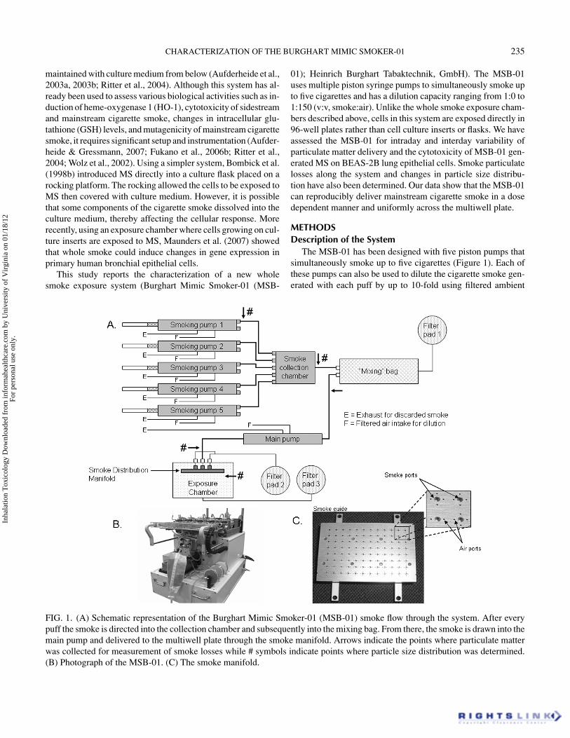
FIG. 1. (A) Schematic representation of the Burghart Mimic Smoker-01 (MSB-01) smoke flow through the system. After everypuff the smoke is directed into the collection chamber and subsequently into the mixing bag. From there, the smoke is drawn into themain pump and delivered to the multiwell plate through the smoke manifold. Arrows indicate the points where particulate matterwas collected for measurement of smoke losses while # symbols indicate points where particle size distribution was determined.(B) Photograph of the MSB-01. (C) The smoke manifold.
01); Heinrich Burghart Tabaktechnik, GmbH). The MSB-01uses multiple piston syringe pumps to simultaneously smoke upto five cigarettes and has a dilution capacity ranging from 1:0 to1:150 (v:v, smoke:air). Unlike the whole smoke exposure cham-bers described above, cells in this system are exposed directly in96-well plates rather than cell culture inserts or flasks. We haveassessed the MSB-01 for intraday and interday variability ofparticulate matter delivery and the cytotoxicity of MSB-01 gen-erated MS on BEAS-2B lung epithelial cells. Smoke particulatelosses along the system and changes in particle size distribu-tion have also been determined. Our data show that the MSB-01can reproducibly deliver mainstream cigarette smoke in a dosedependent manner and uniformly across the multiwell plate.
METHODSDescription of the System
The MSB-01 has been designed with five piston pumps thatsimultaneously smoke up to five cigarettes (Figure 1). Each ofthese pumps can also be used to dilute the cigarette smoke gen-erated with each puff by up to 10-fold using filtered ambient
Inha
latio
n T
oxic
olog
y D
ownl
oade
d fr
om in
form
ahea
lthca
re.c
om b
y U
nive
rsity
of
Vir
gini
a on
01/
18/1
2Fo
r pe
rson
al u
se o
nly.

236 M. J. SCIAN ET AL.
air. The smoke generated with each puff is then delivered intoa collapsible “mixing bag”; by simultaneously smoking multi-ple cigarettes and combining the generated mainstream smokeinto a single chamber, variation in the endpoints resulting fromcigarette to cigarette variation can be minimized. From this bag,the smoke is subsequently drawn into a sixth pump for deliveryto the multiwell plate. Further dilution of the smoke can be donewith this pump for a maximum possible dilution of 1:150. Asmoke distribution manifold disperses the MS simultaneouslyinto each individual well. The manifold provides each well witha separate smoke delivery and two exhaust ducts. A total of 35ml of smoke (roughly 0.365 ml per well) is delivered to the entireplate with each puff.
Once the wells/cells have been exposed, the cigarette smokeis removed from the plate and the exposure chamber by flushingboth with filtered ambient air. The presence of a vacuum duringthe flushing cycles aids in removal of the smoke from the cham-ber. The smoking conditions, i.e. puff volume, puff time, deliverytime, dilution time, exposure time, etc., are all controlled inter-actively through a software program and can be easily adjusted.The entire process, from puff to delivery of the cigarette smoketo the plate, takes approximately 6 seconds. The MSB-01 hasalso been fitted with a retractable stainless steel cover plate de-signed to fit between the smoke manifold and the cell cultureplate, consequently preventing the delivered smoke from reach-ing unintended wells. The cover plate can be set to sequentiallycover the rows of the multiwell plate after each puff, thus gener-ating a puff dependent gradient over the plate. The settings forthis cover plate are also managed through the software controlprogram provided with the instrument.
Extensive use of the MSB-01 required regular cleaning. Allcleaning was done with a 70% ethanol solution after smokingroughly 100 cigarettes. Cleaning included replacing the system’sexhaust filter pads, the collapsible “mixing bag”, and the tubingas well as wiping any surfaces exposed to cigarette smoke, in-cluding the smoke manifold. After cleaning, two dry runs (nocigarettes) and three runs using four cigarettes each were per-formed in order to condition the new tubing and bag. A qual-ity control (QC) plate was run after every cleaning and smokeparticulate deposition measured using the optical density (OD)method described below, and the results compared to previousdata. Additional conditioning runs were performed if necessary.
MSB-01 Smoking ConditionsAll cigarettes used in this study were University of Kentucky
2R4F reference research cigarettes. Cigarettes were smoked inbasic conformity with International Organization for Standard-ization standards of 35 ml puff volume, 2s puff duration, and60s puff interval (International Organization for Standardiza-tion, 1999, 2000). All cigarettes were pre-conditioned at a tem-perature (T ) of 22 ± 1◦C and 60 ± 2% relative humidity (RH)for 48 h prior to smoking. For all MSB-01 experiments, fourcigarettes from four different packs were smoked simultane-ously to reduce cigarette to cigarette variability, unless other-
wise stated. The lighting puff was rejected for a total of eightpuffs delivered to the 96-well experimental plate. The coverplate was set to cover one row after each puff for the concen-tration gradient experiments and deactivated during the wholeplate exposure experiments. The smoke manifold was adjustedto a height of 7 mm above the 96-well plate and the chambervacuum to 500 L/hr as recommended by the manufacturer. TheMSB-01 machine smoking conditions and settings were as fol-lows: active pumps, four; puff volume, 35 ml, duration of puff,1860 ms; gap time, 140 ms; delivery time, 600 ms; smokingpause (time between puffs), 60s; exposure time, 4s; vacuum de-lay, 1.9s; vacuum duration, 10s; flush volume, 80 ml; flush speed,2s; number of flushes, four. Three different dilutions, 1:0, 1:1,and 1:3 (v:v, smoke:air), were studied, with the 1:1 and 1:3 dilu-tions performed within the smoking pump by the MSB-01. Thedilution time was set to 500 ms. For each puff only a 35-ml puffvolume was delivered by the smoking pump into the collapsible“mixing bag”. For the 1:1 and 1:3 dilutions the excess smokewas exhausted out of the smoking pump prior to the next puff.Each experiment was performed in triplicate.
Multiwell Plate Particulate DepositionPlate particulate deposition was measured by reading the op-
tical density of smoke particulate dissolved in 100 µl of DMSO(dimethyl sulfoxide) at 370 nm in each well. After measuring thebackground DMSO reading in each well, all but 5 µl of the sol-vent were removed and the plate was exposed to cigarette smokein the MSB-01. At completion of the smoking run, the 95 µl ofDMSO were added back into each well and the plate gentlyshaken for 2 minutes to allow the particulate to dissolve into theDMSO. To ensure linearity of the measurement and estimatethe smoke particulate delivery, a standard curve was generated.CSC was captured on a Cambridge filter pad, weighed, and ex-tracted using DMSO. The extracted CSC was then diluted to thedesired concentrations of 1, 5, 10, 15, 20, 25, and 30 µg/ml andabsorbance measured at 370 nm. Standards were subsequentlyadded to every plate used to measure the smoke particulate de-position. Smoke delivery was evaluated using two strategies: (1)delivery across a puff dependent concentration gradient and (2)delivery across the entire plate. Three different smoke dilutionswere tested (1:0, 1:1, and 1:3).
Smoke Loss MeasurementsSmoke losses were determined by capturing particulate mat-
ter on a Cambridge filter pad at different points throughout thesystem (Figure 1, arrows) and measuring the mass equivalentspectrophotometrically (Dendo et al., 1998; Phalen et al., 1976).Four University of Kentucky 2R4F research cigarettes from fourdifferent packs were smoked simultaneously for each measure-ment. Experiments were performed in triplicate using two dif-ferent dilutions (1:0 and 1:3). The captured particulate matterwas extracted from the filter pad with methanol and absorbancemeasured at 350 nm. The total particulate captured at the smok-ing port was set to 100% and the amount of particulate collected
Inha
latio
n T
oxic
olog
y D
ownl
oade
d fr
om in
form
ahea
lthca
re.c
om b
y U
nive
rsity
of
Vir
gini
a on
01/
18/1
2Fo
r pe
rson
al u
se o
nly.

CHARACTERIZATION OF THE BURGHART MIMIC SMOKER-01 237
at each point of measurement was estimated as a percentage ofthat amount.
Particle Size MeasurementsParticle size distribution measurements of the cigarette smoke
were made at four locations (Figure 1, # symbols). Measure-ments were taken using a Mercer-style cascade impactor (In-ToxProducts, Albuquerque, NM). The mass median aerodynamic di-ameter (MMAD) and geometric standard deviation (GSD) weredetermined by the Probit method (Hinds, 1999). The smoke wassampled through a Y-connector in order to maintain a constantflow of 2 L/min through the impactor. Prior to each run the im-pactor flow was calibrated with a Gilabrator (Sensidyne, FL).Deposited particulate matter was extracted from the stainlesssteel substrates with methanol, and spectrophotometric mea-surements taken at 350 nm. A minimum of three particle sizedistribution measurements were performed at each location. Theroom temperature and relative humidity were monitored duringeach sampling period (T 21 ± 1◦C, RH 50 ± 3%).
Cell Culture, Smoke Exposure, and ViabilityMeasurements
BEAS-2B cells, a nonmalignant human bronchial epithelialcell line, were selected for our study (Adiseshaiah et al., 2007;Reddel et al., 1988, 1995). Cells were obtained from AmericanType Cell Culture (ATCC) and maintained as recommended bythe supplier. The growth medium was supplemented with 10,000IU/ml penicillin G, 10 µg/ml streptomycin, and 25 µg/ml am-photericin B (Hyclone). For treatment in the MSB-01, BEAS-2Bcells were plated at a density of 5.1 × 10⁴ cells/cm2 into roundbottom 96-well plates 24 h prior to exposure. The followingday, prior to exposure, the cells were washed twice with PBSto remove all traces of medium. For complete fluid removal the96-well plates were centrifuged upside down for 10 secondsagainst a sterile filter paper placed under the lid of the plate;4 µl of PBS were then added to the wells and the plates cen-trifuged once more for 10 seconds in the upright position to forma fluid meniscus over the cells. The plates were then exposedusing a puff concentration gradient at a 1:1 dilution. After ex-posure, 196 µl of complete medium was added to each well andthe cells were allowed to recuperate under normal growing con-ditions (37◦C, 5% CO2) for 24 h. Cell viability was measuredusing the neutral red uptake method 24 h post-exposure. Cellswere washed once with 200 µl of PBS, after which 100 µl of0.06% neutral red containing growth medium was added to eachwell and the cells incubated for 3 h under normal growing condi-tions. The cells were washed twice with 200 µl of PBS (Fisher),and 100 µl of elution solution (45% water, 50% ethanol, and 1%acetic acid) added to each well. The plates were then agitatedfor 10 minutes and photometric measurements taken at 540 nm.Percent viability was calculated as a ratio to the untreated cells (0puffs). Background absorbance readings were taken from wellscontaining 100 µl of reagent (but no cells) and their average
subtracted from the experimental readings prior to calculatingviability changes.
Statistical AnalysisThe intraday and interday variability were calculated accord-
ing to ISO 5725-2 (International Organization for Standardiza-tion, 1995). Percent variation was calculated as a function of theplate average and standard deviation. Group data from three in-dependent experiments performed on three (deposition studies)or five (cytotoxicity studies) different days were expressed asmeans ± SE for each dose. Statistical analysis was performedusing an analysis of variance (ANOVA) test followed by a Dun-nett’s post-hoc test. Probability values of ≤0.05 were consideredsignificant. The cytotoxicity data were modeled using a sigmoidfunction with an offset (y = a/{1.0 + e[–(X–b)/c] + d}). Thissigmoid function was selected because it was found to havethe best fit for the experimentally derived data. A curve was fitto the data from each experimental plate and used to calculateEC50pu f f values for each one. EC50pu f f was defined as the num-ber of puffs at the tested dilution necessary to induce a decreasein measured viability of 50%. The EC50pu f f values were thenused to calculate intraday and interday variability.
RESULTS
Multiwell Plate Particulate DepositionA standard curve was first generated using cigarette smoke
condensate (CSC) of known concentrations in order to estimatethe amount of MS particulate accumulated in the individual wellsof the multiwell plates (Figure 2). The data obtained show thatsmoke particulate amounts of these magnitudes can be easilyquantitated using this method and that this quantitation has alinear function. The MSB-01 was then tested using two types ofsmoke delivery strategies: (1) a puff concentration gradient and(2) a whole plate exposure (Figure 3). Three different dilutionswere tested using these exposure strategies (1:0, 1:1, and 1:3).All data presented in the figures correspond to the plate’s internalwells located within the black box in Figure 3. A puff dependentincrease in particulate deposition was observed in the concen-tration gradient experiments, while in the whole plate exposureexperiments no statistically significant differences were detectedbetween individual rows and the plate average (Figure 4). Fig-ure 4 shows the results obtained using undiluted smoke. Sim-ilar results were obtained when using 1:1 and 1:3 smoke di-lutions although, as expected, the OD readings obtained werelower (data not shown). All experimental OD readings obtainedwere within the range of the standard curve. Intraday and in-terday variations were calculated as the average percent varia-tion found across all exposed rows; a summary of the resultscan be found in Table 1. All calculated variability values werebelow 13%.
Inha
latio
n T
oxic
olog
y D
ownl
oade
d fr
om in
form
ahea
lthca
re.c
om b
y U
nive
rsity
of
Vir
gini
a on
01/
18/1
2Fo
r pe
rson
al u
se o
nly.

238 M. J. SCIAN ET AL.
FIG. 2. Cigarette smoke condensate (CSC) standards were generated and their optical density (OD) measured as described in the“Methods” section. The data show that quantities of cigarette smoke particulate of this magnitude can be measured using thismethod and that the quantitation is linear. TPM, total particulate matter.
Smoke Dilution and Number of CigarettesThe ability of the MSB-01 to dilute smoke was also eval-
uated. The interday averages from the three puff concentrationgradients were used for this purpose (Figure 5A). The data showthat the total amount of particulate deposited in the wells was alinear function of the number of puffs received regardless of thedilution used. The optical density obtained for each puff can alsobe graphed against the dilution factor (Figure 5B). Regardlessof the number of puffs delivered, dilution of the smoke waslinear with calculated correlation coefficients of 0.99 or greaterfor all puffs (Table 2). Using the standard curve measurementsas reference, we calculated the particulate deposition for eightpuffs (one cigarette) at 1:0, 1:1, and 1:3 smoke dilutions to beapproximately 2.4 µg, 1.4 µg, and 0.7 µg respectively (Table2). We also evaluated whether any differences would arise fromthe use of different numbers of cigarettes during any given run(i.e. 1 vs. 2 vs. 3). Optical density measurements were takenfrom smoking runs using two, three, or four cigarettes smokedsimultaneously. Less than 5% variation was observed betweenruns, indicating that similar particulate delivery can be achievedwhen smoking different numbers of cigarettes (Figure 5C).
FIG. 3. Gradient (left) and whole plate (right) smoke exposure strategies. The concentration gradient is achieved through the useof a sliding stainless steel plate that sequentially covers one row of wells after every puff of smoke is delivered to the plate.
Smoke Losses and Particle Size DistributionSmoke losses were estimated by collecting particulate matter
from different points throughout the system (Figure 1, arrows).Spectrophotometric data indicate that approximately 45–50% ofthe smoke generated at the smoking ports was lost within thesystem prior to delivery into the exposure chamber regardless ofthe dilution used (Table 3). On average only 2–7% of the smokeparticulate was lost within the smoke manifold. However, thetwo major areas of loss were between the smoking ports andthe mixing bag (12–14% loss) and between the mixing bag andthe manifold (18–23% loss). The smoke particle size distributiondid not change throughout the system with either dilution; how-ever, there was a significant decrease in the MMAD of smokediluted 1:3 in the system (Table 3). It is likely that this changewas due to evaporation of some of the semi-volatile smoke con-stituents (Seeman et al., 2004).
Neutral Red Uptake Assay and BEAS-2B CytotoxicityThe MSB-01 was specifically designed for the exposure of
cells to freshly generated MS. Therefore we have also evaluated
Inha
latio
n T
oxic
olog
y D
ownl
oade
d fr
om in
form
ahea
lthca
re.c
om b
y U
nive
rsity
of
Vir
gini
a on
01/
18/1
2Fo
r pe
rson
al u
se o
nly.

CHARACTERIZATION OF THE BURGHART MIMIC SMOKER-01 239
FIG. 4. Gradient and whole plate intraday deposition data points were obtained by averaging three independent experimentsperformed on the same day. Intraday averages were used to calculate the interday average. Data are presented as mean ± SD. Adose dependent increase in deposition occurs with increasing puff number. No statistically significant changes were observed inthe whole plate exposure. Single and double asterisks indicate p values ≤0.05 and ≤0.01 respectively.
the performance of the system using a human lung epithelial cellline. BEAS-2B cells were first exposed to mainstream cigarettesmoke using the three dilution settings tested above to determinethe most suitable exposure condition. Viability was measuredusing the neutral red assay and calculated as a function of un-treated cells (0 puffs of smoke) for each dilution. Based on thesepreliminary results a smoke dilution of 1:1 was chosen for sub-sequent experiments (data not shown). The cells were also testedfor their ability to survive outside of the cell culture incubator.BEAS-2B cells were exposed to filtered air in the MSB-01 (nocigarettes) for up to four consecutive run cycles, each taking a
TABLE 1Summary of calculated percent intraday and interday variation in the concentration gradient and whole plate deposition
experiments. The general mean, intraday, and interday average values are expressed as optical density (OD) units at 370 nm.
Gradient Whole plate
1:0 dilution 1:1 dilution 1:3 dilution 1:0 dilution 1:1 dilution 1:3 dilution
General mean 0.046 0.026 0.012 0.109 0.061 0.028Intraday average SD 0.002 0.002 0.001 0.004 0.004 0.003Intraday variation (%) 5.023 7.496 10.74 3.777 6.082 9.426Interday average SD 0.004 0.003 0.001 0.008 0.006 0.004Interday variation (%) 9.402 12.64 11.22 7.039 9.521 12.45
total of 8 minutes. BEAS-2B cells retained 90% or greater vi-ability for up to 32 minutes (four consecutive runs) or 32 filterair puffs (data not shown). To test the system for reproducibil-ity, BEAS-2B cells were exposed to MS using a concentrationgradient of 0–8 puffs. Statistically significant changes in cell via-bility were detected in all puff doses (Figure 6). Using a sigmoidfunction, the data were then used to calculate the EC50 value foreach experimental plate. The calculated EC50 values were thenused to determine the percent intraday and interday variation(Table 4). The intraday variation was calculated at 13.1% whileinterday variation was 20.3%.
Inha
latio
n T
oxic
olog
y D
ownl
oade
d fr
om in
form
ahea
lthca
re.c
om b
y U
nive
rsity
of
Vir
gini
a on
01/
18/1
2Fo
r pe
rson
al u
se o
nly.

240 M. J. SCIAN ET AL.
FIG. 5. (A) Number of puffs vs. puff particulate optical density at different dilutions. A dose dependent increase in depositionoccurs with increasing puff number at every dilution tested. (B) Smoke dilution vs. particulate optical density. The data show thatdilution of the smoke is a linear function regardless of the number of puffs used. (C) Particulate delivery is independent of thenumber of cigarettes used. Optical density measurements for particulate delivery from two, three, or four cigarettes using a 1:0dilution were taken. Less than 5% variation was detected between runs. All data are presented as mean ± SD. Single and doubleasterisks indicate p values ≤0.05 and ≤0.01 respectively.
DISCUSSIONWe have characterized a new exposure system (MSB-01)
designed to expose cells to both the gas/vapor and particulatephases of freshly generated MS. Although similar whole smokeexposure systems have been reported in the literature in recentyears, to our knowledge the MSB-01 is the first system reportedto use 96-well plates. This important distinction allows cells tobe grown directly in culture plates rather than in cell cultureinserts (Aufderheide et al., 2003a; Phillips et al., 2005). Addi-tionally, the MSB-01 has the added advantage that cells can be
exposed to multiple doses of cigarette smoke during a single runwithout multiple experimental setups.
The MSB-01 utilizes five automated piston pumps to simulta-neously smoke up to five cigarettes. Coupling of these smokingpumps with the smoke distribution manifold facilitates the rapiddelivery of the generated MS to the cells (i.e. approximately 6seconds). This relatively fast delivery time not only allows theexposure of cells to both phases of cigarette smoke but also al-lows the exposure of the cells to short lived smoke componentsthat may otherwise be lost with longer delivery times.
Inha
latio
n T
oxic
olog
y D
ownl
oade
d fr
om in
form
ahea
lthca
re.c
om b
y U
nive
rsity
of
Vir
gini
a on
01/
18/1
2Fo
r pe
rson
al u
se o
nly.

CHARACTERIZATION OF THE BURGHART MIMIC SMOKER-01 241
TABLE 2Correlation coefficient for smoke dilution vs. puff particulate optical density data and estimated particle deposition for each puff
at each dilution. The slope values are expressed as OD units at 370 nm/level of dilution.
1 puff 2 puffs 3 puffs 4 puffs 5 puffs 6 puffs 7 puffs 8 puffs
Slope 0.009 0.021 0.032 0.043 0.056 0.069 0.083 0.094Correlation coefficient 0.990 0.999 0.998 0.996 0.993 0.995 0.995 0.994Estimated particulate deposited per well (µg)
1:0 dilution 0.23 0.51 0.79 1.08 1.41 1.74 2.09 2.391:1 dilution 0.14 0.27 0.43 0.61 0.83 1.01 1.20 1.391:3 dilution 0.06 0.13 0.20 0.28 0.38 0.47 0.56 0.64
Our plate particulate deposition studies suggest that thesmoke manifold and the stainless steel cover plate performedas intended. A puff dependent deposition was measured in allthree dilutions tested, while no statistically significant changeswere observed during whole plate deposition experiments. Intra-day and interday variabilities were calculated at less than 11%and 13% respectively for these experiments, showing that theMSB-01 can consistently deliver MS into the exposure cham-ber. Additionally, dilution of the smoke was linear regardless ofthe number of puffs delivered, indicating that the MSB-01 canefficiently dilute the freshly generated cigarette smoke beforedelivery to the exposure chamber.
Cytotoxicity measurements also demonstrate that the MSB-01 can consistently deliver mainstream cigarette smoke. How-ever, it remains to be determined how well other MSB-01-induced biological responses such as protein and gene expres-sion of airway epithelial cells compare with different exposuremethods (sbPBS, CSC). Divergent cellular responses would beexpected with the whole smoke systems when compared with asingle MS phase exposure, since the particulate and gas phasesof smoke have been shown to have confounding interactions.For example, while the particulate phase of smoke can havecytotoxic and proinflammatory effects on cells, carbon monox-ide (CO), a gas component of cigarette smoke, has also been
TABLE 3Summary of smoke losses and particle size distribution throughout the Burghart Mimic Smoker-01 (MSB-01).
Smoke remaining (%)aParticle size distribution (µm)
1:0 dilution 1:3 dilution1:0 1:3
dilution dilution MMAD GSD MMAD GSD
Smoking port 100 ± 0 100 ± 0 0.41 1.6 0.31 1.6Immediately prior to mixing bag 86 ± 3 87 ± 4 0.43 1.7 0.31 1.6Immediately after mixing bag 80 ± 2 72 ± 2 ND ND ND NDImmediately prior to manifold 57 ± 5 53 ± 2 0.44 1.8 0.24 1.7Immediately after manifold 55 ± 3 52 ± 2 0.45 2.0 0.25 2.1
Note. MMAD, mass median aerodynamic diameter; GSD, geometric standard deviation, ND, not determined.aMean ± SD.
reported to have anti-inflammatory and anti-apoptotic effects(Ryter & Choi, 2006; Slebos et al., 2003; Smith et al., 2006).
It has already been shown that exposure of cells in vitro us-ing CSC can result in numerous transcriptional changes in lungepithelial cells that are consistent with in vivo MS exposuremodels (De Flora et al., 2005; Fields et al., 2005; Fukano et al.,2006a; Nagaraj et al., 2006; Stevenson et al., 2007). However,very little work has been done using in vitro whole smoke expo-sure systems (Fukano et al., 2006b; Lu et al., 2007; Maunderset al., 2007). More importantly, it is unknown whether exposureof cells to smoke components using any of these in vitro meth-ods accurately represents the cellular responses in a smoker’srespiratory tract.
Optical density measurements show that the majority of thesmoke losses measured occurred within the connecting tubingand pump valves. Particulate deposition within the system maynot only occur through impaction, diffusion, and sedimentation,but may also occur due to electrostatic or chemical interactionsbetween cigarette smoke components and the tubing itself. Ifsome of the measured losses are indeed due to chemical or elec-trostatic interactions between the smoke and the tubing material,it may be possible to decrease the total amount of loss by using anonreactive, conductive tubing material. Interestingly, the undi-luted smoke delivered into the exposure chamber has a similar
Inha
latio
n T
oxic
olog
y D
ownl
oade
d fr
om in
form
ahea
lthca
re.c
om b
y U
nive
rsity
of
Vir
gini
a on
01/
18/1
2Fo
r pe
rson
al u
se o
nly.

242 M. J. SCIAN ET AL.
FIG. 6. BEAS-2B cells were plated 24 h prior to the assay. Cells were exposed using concentration gradient exposure of a 1:1smoke:air dilution and viability measured following a 24-h post-exposure recovery period. Experiments were performed in triplicateon five different days. A dose dependent decrease in viability was observed with increasing number of puffs. Intraday and interdayaverages are presented as mean ± SD. Single and double asterisks indicate p values ≤0.05 and ≤0.01 respectively. NS, notsignificant.
particle size distribution as that of the smoke generated at thesmoking pump. This is important because it suggests that thechemical composition and properties of the smoke that the cellsare exposed to may be relatively similar to that of the smokegenerated with each puff. However, a change in the particle sizedistribution was observed when comparing undiluted versus di-luted smoke. This is not entirely surprising, since increasing thevolume of air should decrease coagulation of the particles andallow for more evaporation from individual particles, therebydecreasing their overall size. The MSB-01 has a total dilutioncapacity ranging from 1:0 to 1:150. Considering the data ob-tained here, one should keep in mind that with larger dilutions,the smoke losses and particle size distributions may change morethan those measured.
Given that a loss in the total amount of smoke generatedand changes in the particle size distribution were detected, the
TABLE 4Summary of percent intraday and interday variation using
calculated EC50puff values. The EC50puff value is defined as thenumber of puffs generated necessary to induce a 50% decrease
in the measured cell viability.
EC50puff
6/15/07a 6/20/07 6/27/07 7/13/07 7/18/07
Replicate 1 2.99 2.88 2.29 2.44 2.88Replicate 2 2.68 3.45 2.59 1.64 2.33Replicate 3 3.69 3.35 2.78 1.93 2.86EC50puff general 2.72
meanIntraday Interday
Average SD 0.3559 0.5502Variation (%) 13.09 20.23
aDate of experiment.
question remains as to whether any of these factors can affect thechemical composition of the MS delivered to the cells. A detailedchemical analysis of the gas and particulate phases comparingthe smoke captured at different locations of the exposure systemis needed in order to fully address such a question. Neverthe-less, the MSB-01 can consistently deliver mainstream cigarettesmoke into the exposure chamber. This is clearly evident fromour plate particulate deposition and cytotoxicity results.
As researchers strive to mimic in vivo exposure using in vitrosystems, new and improved methods of exposure will be de-veloped. Currently there are several systems available for thispurpose. The results obtained with the MSB-01 are comparableto results previously described using other systems of similardesign and purpose. Unfortunately a direct comparison betweenthem is not entirely possible since different cell lines, exper-imental setups, and test cigarettes were used by each group.The results presented here clearly demonstrate that the MSB-01can reproducibly deliver mainstream cigarette smoke both in adose/puff dependent manner and uniformly across the 96-wellplate as was intended. Therefore this system may be a valuableresearch tool and ideal for the development of high throughputbiological assays based on this plate format.
REFERENCESAdiseshaiah, P., Lindner, D. J., Kalvakolanu, D. V., and Reddy, S. P.
2007. FRA-1 proto-oncogene induces lung epithelial cell invasionand anchorage-independent growth in vitro, but is insufficient topromote tumor growth in vivo. Cancer Res. 67:6204–6211.
Aufderheide, M., and Gressmann, H. 2007. A modified Ames assayreveals the mutagenicity of native cigarette mainstream smoke andits gas vapour phase. Exp. Toxicol. Pathol. 58:383–392.
Aufderheide, M., Knebel, J. W., and Ritter, D. 2003a. An improved invitro model for testing the pulmonary toxicity of complex mixturessuch as cigarette smoke. Exp. Toxicol. Pathol. 55:51–57.
Aufderheide, M., Knebel, J. W., and Ritter, D. 2003b. Novel approachesfor studying pulmonary toxicity in vitro. Toxicol. Lett. 140–141:205–211.
Inha
latio
n T
oxic
olog
y D
ownl
oade
d fr
om in
form
ahea
lthca
re.c
om b
y U
nive
rsity
of
Vir
gini
a on
01/
18/1
2Fo
r pe
rson
al u
se o
nly.

CHARACTERIZATION OF THE BURGHART MIMIC SMOKER-01 243
Bombick, B. R., Murli, H., Avalos, J. T., Bombick, D. W., Morgan, W.T., Putnam, K. P., and Doolittle, D. J. 1998a. Chemical and biologicalstudies of a new cigarette that primarily heats tobacco. Part 2. In vitrotoxicology of mainstream smoke condensate. Food Chem. Toxicol.36:183–190.
Bombick, D. W., Ayres, P. H., Putnam, K., Bombick, B. R., andDoolittle, D. J. 1998b. Chemical and biological studies of a newcigarette that primarily heats tobacco. Part 3. In vitro toxicity ofwhole smoke. Food Chem. Toxicol. 36:191–197.
Church, D., and Pryor, W. 1985. Free-radical chemistry of cigarettesmoke and its toxicological implications. Environ. Health Perspect.64:111–126.
De Flora, S., Izzotti, A., D’Agostini, F., Bennicelli, C., You, M., Lubet,R. A., and Balansky, R. M. 2005. Induction and modulation of lungtumors: genomic and transcriptional alterations in cigarette smoke-exposed mice. Exp. Lung Res. 31:19–35.
Dendo, R. I., Phalen, R. F., Mannix, R. C., and Oldham, M. J. 1998.Effects of breathing parameters on sidestream cigarette smoke depo-sition in a hollow tracheobronchial model. Am. Ind. Hyg. Assoc. J.59:381–387.
Fields, W. R., Leonard, R. M., Odom, P. S., Nordskog, B. K., Ogden,M. W., and Doolittle, D. J. 2005. Gene expression in normal hu-man bronchial epithelial (NHBE) cells following in vitro exposureto cigarette smoke condensate. Toxicol. Sci. 86:84–91.
Fukano, Y., Oishi, M., Chibana, F., Numazawa, S., and Yoshida, T.2006a. Analysis of the expression of heme oxygenase-1 gene in hu-man alveolar epithelial cells exposed to cigarette smoke condensate.J. Toxicol. Sci. 31:99–109.
Fukano, Y., Yoshimura, H., and Yoshida, T. 2006b. Heme oxygenase-1gene expression in human alveolar epithelial cells (A549) followingexposure to whole cigarette smoke on a direct in vitro exposuresystem. Exp. Toxicol. Pathol. 57:411–418.
Gebel, S., and Muller, T. 2001. The activity of NF-kappaB in Swiss 3T3cells exposed to aqueous extracts of cigarette smoke is dependent onthioredoxin. Toxicol. Sci. 59:75–81.
Hinds, WC. 1999. Aerosol technology: Properties, behavior, and mea-surement of airborne particles, 2nd ed. New York: John Wiley andSons.
Hoffmann, D., and Wynder, E. 1986. Chemical constituents and bioac-tivity of tobacco smoke. IARC Sci. Publ. (74):145–165.
International Organization for Standardization. 1995. ISO 5725-2. Ac-curacy (trueness and precision) of measurement methods and re-sults. Basic methods for the determination of repeatability and repro-ducibility of a standard measurement method. Geneva: InternationalOrganization for Standardization.
International Organization for Standardization. 1999. ISO 3402. To-bacco and tobacco products—Atmosphere for conditioning and test-ing. Geneva: International Organization for Standardization.
International Organization for Standardization. 2000. ISO 3308. Rou-tine analytical cigarette-smoking machine—Definitions and stan-dard conditions. Geneva: International Organization for Standard-ization.
Kode, A., Yang, S. R., and Rahman, I. 2006. Differential effects ofcigarette smoke on oxidative stress and proinflammatory cytokinerelease in primary human airway epithelial cells and in a variety oftransformed alveolar epithelial cells. Respir. Res. 7:132.
Lu, B., Kerepesi, L., Wisse, L., Hitchman, K., and Meng, Q. R. 2007.Cytotoxicity and gene expression profiles in cell cultures exposed towhole smoke from three types of cigarettes. Toxicol. Sci. 98:469–478.
Maunders, H., Patwardhan, S., Phillips, J., Clack, A., and Richter, A.2007. Human bronchial epithelial cell transcriptome: gene expres-sion changes following acute exposure to whole cigarette smoke invitro. Am. J. Physiol. Lung Cell Mol. Physiol. 292:L1248–L1256.
Muller, T., and Gebel, S. 1994. Heme oxygenase expression in Swiss3T3 cells following exposure to aqueous cigarette smoke fractions.Carcinogenesis 15:67–72.
Nagaraj, N. S., Beckers, S., Mensah, J. K., Waigel, S., Vigneswaran,N., and Zacharias, W. 2006. Cigarette smoke condensate induces cy-tochromes P450 and aldo-keto reductases in oral cancer cells. Toxi-col. Lett. 165:182–194.
Phalen, R. F., Cannon, W. C., and Esparza, D. 1976. Comparison ofimpaction, centrifugal separation and electron microscopy for sizingcigarette smoke. In Fine particles, ed. B. Liu, pp. 731–737. SanDiego, CA: Academic Press.
Phillips, J., Kluss, B., Richter, A., and Massey, E. 2005. Exposure ofbronchial epithelial cells to whole cigarette smoke: assessment ofcellular responses. Altern. Lab. Anim. 33:239–248.
Reddel, R. R., De Silva, R., Duncan, E. L., Rogan, E. M., Whitaker, N.J., Zahra, D. G., Ke, Y., McMenamin, M. G., Gerwin, B. I., and Harris,C. C. 1995. SV40-induced immortalization and ras-transformationof human bronchial epithelial cells. Int. J. Cancer 61:199–205.
Reddel, R. R., Ke, Y., Gerwin, B. I., McMenamin, M. G., Lechner, J.F., Su, R. T., Brash, D. E., Park, J. B., Rhim, J. S., and Harris, C. C.1988. Transformation of human bronchial epithelial cells by infectionwith SV40 or adenovirus-12 SV40 hybrid virus, or transfection viastrontium phosphate coprecipitation with a plasmid containing SV40early region genes. Cancer Res. 48:1904–1909.
Ritter, D., Knebel, J., and Aufderheide, M. 2004. Comparative assess-ment of toxicities of mainstream smoke from commercial cigarettes.Inhal. Toxicol. 16:691–700.
Roemer, E., Tewes, F. J., Meisgen, T. J., Veltel, D. J., and Carmines, E.L. 2002. Evaluation of the potential effects of ingredients added tocigarettes. Part 3: in vitro genotoxicity and cytotoxicity. Food Chem.Toxicol. 40:105–111.
Ryter, S. W., and Choi, A. M. 2006. Therapeutic applications of carbonmonoxide in lung disease. Curr. Opin. Pharmacol. 6:257–262.
Seeman, J. I., Lipowicz, P. J., Piade, J. J., Poget, L., Sanders, E. B.,Snyder, J. P., and Trowbridge, C. G. 2004. On the deposition ofvolatiles and semivolatiles from cigarette smoke aerosols: relativerates of transfer of nicotine and ammonia from particles to the gasphase. Chem. Res. Toxicol. 17:1020–1037.
Slebos, D. J., Ryter, S. W., and Choi, A. M. 2003. Heme oxygenase-1and carbon monoxide in pulmonary medicine. Respir. Res. 4:7.
Smith, C. J., Perfetti, T. A., and King, J. A. 2006. Perspectives onpulmonary inflammation and lung cancer risk in cigarette smokers.Inhal. Toxicol. 18:667–677.
Stevenson, C. S., Docx, C., Webster, R., Battram, C., Hynx, D.,Giddings, J., Cooper, P. R., Chakravarty, P., Rahman, I., Marwick, J.A., Kirkham, P. A., Charman, C., Richardson, D. L., Nirmala, N. R.,Whittaker, P., and Butler, K. 2007. Comprehensive gene expressionprofiling of rat lung reveals distinct acute and chronic responses tocigarette smoke inhalation. Am. J. Physiol. Lung Cell Mol. Physiol.293(5):L1183–L1193.
Wolz, L., Krause, G., Scherer, G., Aufderheide, M., and Mohr, U.2002. In vitro genotoxicity assay of sidestream smoke using a hu-man bronchial epithelial cell line. Food Chem. Toxicol. 40:845–850.
Inha
latio
n T
oxic
olog
y D
ownl
oade
d fr
om in
form
ahea
lthca
re.c
om b
y U
nive
rsity
of
Vir
gini
a on
01/
18/1
2Fo
r pe
rson
al u
se o
nly.