Characterization and ex vivo Expansion of Human Placenta-Derived Natural Killer Cells for Cancer...
13
ORIGINAL RESEARCH ARTICLE published: 01 May 2013 doi: 10.3389/fimmu.2013.00101 Characterization and ex vivo expansion of human placenta-derived natural killer cells for cancer immunotherapy Lin Kang 1 ,Vanessa Voskinarian-Berse 1 , Eric Law 1 ,Tiffany Reddin 1 , Mohit Bhatia 1 , Alexandra Hariri 2 , Yuhong Ning 3 , David Dong 4 ,Timothy Maguire 4 , MartinYarmush 4 ,Wolfgang Hofgartner 1 , Stewart Abbot 1 , Xiaokui Zhang 1 * and Robert Hariri 1 1 Celgene Cellular Therapeutics, Warren, NJ, USA 2 Princeton University, Princeton, NJ, USA 3 Celgene Signal Research, San Diego, CA, USA 4 Department of Biomedical Engineering, Rutgers University, Piscataway, NJ, USA Edited by: Eric Vivier, Centre d’Immunologie de Marseille-Luminy, France Reviewed by: Akira Shibuya, University of Tsukuba, Japan HughT. Reyburn, Spanish National Research Council, Spain *Correspondence: Xiaokui Zhang, Celgene Cellular Therapeutics, 33Technology Drive, Warren, NJ 07059, USA. e-mail: [email protected] Recent clinical studies suggest that adoptive transfer of donor-derived natural killer (NK) cells may improve clinical outcome in hematological malignancies and some solid tumors by direct anti-tumor effects as well as by reduction of graft versus host disease (GVHD). NK cells have also been shown to enhance transplant engraftment during allogeneic hematopoietic stem cell transplantation (HSCT) for hematological malignancies.The limited ex vivo expansion potential of NK cells from peripheral blood (PB) or umbilical cord blood (UCB) has however restricted their therapeutic potential. Here we define methods to effi- ciently generate NK cells from donor-matched, full-term human placenta perfusate (termed Human Placenta-Derived Stem Cell, HPDSC) and UCB. Following isolation from cryopre- served donor-matched HPDSC and UCB units, CD56+CD3- placenta-derived NK cells, termed pNK cells, were expanded in culture for up to 3 weeks to yield an average of 1.2 bil- lion cells per donor that were >80% CD56+CD3-, comparable to doses previously utilized in clinical applications. Ex vivo-expanded pNK cells exhibited a marked increase in anti- tumor cytolytic activity coinciding with the significantly increased expression of NKG2D, NKp46, and NKp44 (p < 0.001, p < 0.001, and p < 0.05, respectively). Strong cytolytic activ- ity was observed against a wide range of tumor cell lines in vitro. pNK cells display a distinct microRNA (miRNA) expression profile, immunophenotype, and greater anti-tumor capacity in vitro compared to PB NK cells used in recent clinical trials. With further development, pNK may represent a novel and effective cellular immunotherapy for patients with high clinical needs and few other therapeutic options. Keywords: placental-derived natural killer cells, ex vivo expansion, anti-tumor cytolytic activity, miRNA, cellular immunotherapy INTRODUCTION The critical role of natural killer (NK) cells in the defense against cancer and virus infection has been increasingly appreciated since they were first discovered in mice more than 30 years ago (Her- berman et al., 1975a,b). Clinical studies exploring the biological activities of NK cells in the treatment of malignant disease and during allogeneic hematopoietic stem cell transplantation (HSCT) have provided promising results. Transplant studies have suggested alloreactive NK cells could mediate potent anti-leukemia effects without causing graft versus host disease (GVHD). In human leukocyte antigen (HLA)-mismatched, haploidentical allogeneic stem cell transplants (SCT), NK alloreactivity was associated with a higher rate of survival, a lower rate of relapse, and treatment related mortality post transplantation (Ruggeri et al., 1999, 2002; Velardi et al., 2002). Several clinical studies have convincingly demon- strated that adoptive transfer of NK cells isolated from peripheral blood (PB) of haploidentical donors can be successfully used for immunotherapy in acute myeloid leukemia (AML) patients (Miller et al., 2005; Rubnitz et al., 2010; Curti et al., 2011). However, a number of technical challenges have hampered the widespread application of NK cells in immunotherapy; these include a lim- ited ability to generate large numbers of effector cells, difficulty in maintaining high tumoricidal activity during ex vivo expansion and in vivo therapy, and a limited understanding of NK-specific tumor targeting profiles. Therefore, there is a need to overcome these challenges and enable a NK cell-based anti-tumor strategy in the clinic. To date, the most utilized source for NK cells in adoptive immunotherapy is PB (Sutlu and Alici, 2009), with clinically effec- tive doses reported in the range of 1 × 10 6 –9.3 × 10 6 PB NK cells/kg (Passweg et al., 2004; Miller et al., 2005; McKenna et al., 2007; Shi et al., 2008; Meyer-Monard et al., 2009; Rubnitz et al., 2010; Yoon et al., 2010; Curti et al., 2011). Embryonic stem cells (Woll et al., 2009) and umbilical cord blood (UCB) (Spanholtz et al., 2010) have also been used as sources of CD34+ cells that were differentiated into functional NK cells. Previous studies have www.frontiersin.org May 2013 |Volume 4 | Article 101 | 1
Transcript of Characterization and ex vivo Expansion of Human Placenta-Derived Natural Killer Cells for Cancer...

ORIGINAL RESEARCH ARTICLEpublished: 01 May 2013
doi: 10.3389/fimmu.2013.00101
Characterization and ex vivo expansion of humanplacenta-derived natural killer cells for cancerimmunotherapyLin Kang1,Vanessa Voskinarian-Berse1, Eric Law 1,Tiffany Reddin1, Mohit Bhatia1, Alexandra Hariri 2,Yuhong Ning3, David Dong4,Timothy Maguire4, MartinYarmush4,Wolfgang Hofgartner 1, Stewart Abbot 1,Xiaokui Zhang1* and Robert Hariri 1
1 Celgene Cellular Therapeutics, Warren, NJ, USA2 Princeton University, Princeton, NJ, USA3 Celgene Signal Research, San Diego, CA, USA4 Department of Biomedical Engineering, Rutgers University, Piscataway, NJ, USA
Edited by:Eric Vivier, Centre d’Immunologie deMarseille-Luminy, France
Reviewed by:Akira Shibuya, University of Tsukuba,JapanHugh T. Reyburn, Spanish NationalResearch Council, Spain
*Correspondence:Xiaokui Zhang, Celgene CellularTherapeutics, 33 Technology Drive,Warren, NJ 07059, USA.e-mail: [email protected]
Recent clinical studies suggest that adoptive transfer of donor-derived natural killer (NK)cells may improve clinical outcome in hematological malignancies and some solid tumorsby direct anti-tumor effects as well as by reduction of graft versus host disease (GVHD).NK cells have also been shown to enhance transplant engraftment during allogeneichematopoietic stem cell transplantation (HSCT) for hematological malignancies.The limitedex vivo expansion potential of NK cells from peripheral blood (PB) or umbilical cord blood(UCB) has however restricted their therapeutic potential. Here we define methods to effi-ciently generate NK cells from donor-matched, full-term human placenta perfusate (termedHuman Placenta-Derived Stem Cell, HPDSC) and UCB. Following isolation from cryopre-served donor-matched HPDSC and UCB units, CD56+CD3− placenta-derived NK cells,termed pNK cells, were expanded in culture for up to 3 weeks to yield an average of 1.2 bil-lion cells per donor that were >80% CD56+CD3−, comparable to doses previously utilizedin clinical applications. Ex vivo-expanded pNK cells exhibited a marked increase in anti-tumor cytolytic activity coinciding with the significantly increased expression of NKG2D,NKp46, and NKp44 (p < 0.001, p < 0.001, and p < 0.05, respectively). Strong cytolytic activ-ity was observed against a wide range of tumor cell lines in vitro. pNK cells display a distinctmicroRNA (miRNA) expression profile, immunophenotype, and greater anti-tumor capacityin vitro compared to PB NK cells used in recent clinical trials. With further development,pNK may represent a novel and effective cellular immunotherapy for patients with highclinical needs and few other therapeutic options.
Keywords: placental-derived natural killer cells, ex vivo expansion, anti-tumor cytolytic activity, miRNA, cellularimmunotherapy
INTRODUCTIONThe critical role of natural killer (NK) cells in the defense againstcancer and virus infection has been increasingly appreciated sincethey were first discovered in mice more than 30 years ago (Her-berman et al., 1975a,b). Clinical studies exploring the biologicalactivities of NK cells in the treatment of malignant disease andduring allogeneic hematopoietic stem cell transplantation (HSCT)have provided promising results. Transplant studies have suggestedalloreactive NK cells could mediate potent anti-leukemia effectswithout causing graft versus host disease (GVHD). In humanleukocyte antigen (HLA)-mismatched, haploidentical allogeneicstem cell transplants (SCT), NK alloreactivity was associated with ahigher rate of survival, a lower rate of relapse,and treatment relatedmortality post transplantation (Ruggeri et al., 1999, 2002; Velardiet al., 2002). Several clinical studies have convincingly demon-strated that adoptive transfer of NK cells isolated from peripheralblood (PB) of haploidentical donors can be successfully usedfor immunotherapy in acute myeloid leukemia (AML) patients
(Miller et al., 2005; Rubnitz et al., 2010; Curti et al., 2011). However,a number of technical challenges have hampered the widespreadapplication of NK cells in immunotherapy; these include a lim-ited ability to generate large numbers of effector cells, difficultyin maintaining high tumoricidal activity during ex vivo expansionand in vivo therapy, and a limited understanding of NK-specifictumor targeting profiles. Therefore, there is a need to overcomethese challenges and enable a NK cell-based anti-tumor strategyin the clinic.
To date, the most utilized source for NK cells in adoptiveimmunotherapy is PB (Sutlu and Alici, 2009), with clinically effec-tive doses reported in the range of 1× 10⁶–9.3× 106 PB NKcells/kg (Passweg et al., 2004; Miller et al., 2005; McKenna et al.,2007; Shi et al., 2008; Meyer-Monard et al., 2009; Rubnitz et al.,2010; Yoon et al., 2010; Curti et al., 2011). Embryonic stem cells(Woll et al., 2009) and umbilical cord blood (UCB) (Spanholtzet al., 2010) have also been used as sources of CD34+ cells thatwere differentiated into functional NK cells. Previous studies have
www.frontiersin.org May 2013 | Volume 4 | Article 101 | 1

Kang et al. Human placenta-derived NK cells
highlighted the potential to selectively isolate and expand NK cellsfrom UCB for adoptive cell transfer treatment of tumors (Xinget al., 2010). Over the last decade the phenotype and function ofdecidual NK (dNK) cells in placenta development have been stud-ied extensively (Koopman et al., 2003; Hiby et al., 2004; Kopcowet al., 2005, 2010; Apps et al., 2011; Male et al., 2011). However,little information is available on the role of NK cells from placentafor cellular immunotherapy.
Recently, human placenta has been demonstrated as a noveland valuable source of multipotential stem/progenitor cells ofmesenchymal and hematopoietic origin for multiple therapeuticapplications (Parolini et al., 2008; Prather et al., 2008). CelgeneCellular Therapeutics (CCT, a division of Celgene Corporation)is developing human placenta-derived stem cells (HPDSC) as anadjunct to UCB cells for allogeneic use in first-degree or second-degree blood relatives for augmentation of the stem cell graft inhematopoietic reconstitution. We have established a standardizedprocedure to perfuse donated full-term placentas with normalsaline to recover HPDSC. HPDSC were subsequently processedto remove red blood cells, non-viable cells and tissue debris fol-lowed by cryopreservation. HPDSC were neither expanded norcultured during processing. The process typically yields 100–500million total nucleated cells (TNC), approximately 1–5% of whichare CD34+ hematopoietic stem cells (HSCs). We hypothesize thatHPDSC combined with the donor-matched UCB could representan effective new source of NK cells that holds potential for furtherimmunotherapeutic development.
Unlike their antigen-specific lymphoid counterparts, such asT cells and B cells, NK cells, characterized as CD56+CD3−,recognize and subsequently kill virus-infected and transformedcells without prior immunization. NK cells operate via the bal-ance of signals from inhibitory receptors, such as the killer cellimmunoglobulin-like receptors (KIRs), and the C-type lectinfamily receptor: CD94/NKG2, with activating receptors, such asNKG2D, NKp46, NKp44, NKp30, and CD226 (Smyth et al., 2002;Huntington et al., 2007). Two major subtypes of CD56+ NK cellscan be distinguished according to the co-expression of the cell sur-face marker CD16 (Jacobs et al., 2001). It has been demonstratedthat CD56+CD16− NK cells have very few cytolytic granules,low or no expression of KIRs, high expression of KLR familymembers and are capable of producing cytokines and chemokinesupon activation. CD56+CD16+NK cells have abundant cytolyticgranules and high expression of KIRs. PB contains more than90% CD56+CD16+ NK cells, while more than 90% of NKcells in lymph nodes do not express CD16 (Cooper et al., 2001;Fehniger et al., 2003). Results from developmental NK cells stud-ies suggest that the CD56+CD16+ NK cells are derived fromthe CD56+CD16− NK cells (Lanier et al., 1986; Ferlazzo et al.,2004; Freud et al., 2005). In addition to cell surface markers,different miRNA expression profiles have been associated withNK cell development, maturation, and function (Bezman et al.,2010). To date, no study has investigated the miRNA profile start-ing from donor-matched HPDSC and UCB (hereafter referred toas “Combo unit”) followed by differentiation into functional NKcells.
In this study, we report that placenta is a rich source of placenta-derived NK (pNK) cells that can be readily isolated from Combo
units, followed by ex vivo expansion. We evaluated the prolifer-ation, immunophenotype, miRNA expression, and activation ofthese expanded cells, as well as their cytolytic activities in vitro.Our results demonstrate that pNK cells can be generated in clini-cally relevant quantities and may be developed as a highly cytotoxiccellular product that can be used to treat a wide range of cancers.
MATERIALS AND METHODSPROCESSING OF HPDSC AND UCBPostpartum placentas and umbilical cords were procured underfull-informed consent of donors with donor eligibility documen-tation, and were qualified using a series of tests, including serology,bacteriology, and HLA typing. HPDSC isolation and recovery wasachieved by cannulation of the umbilical vessels (two arteries andone vein) under sterile conditions with polyethylene catheters con-nected to a flow-controlled fluid circuit allowing perfusion ofthe placenta. A total of 750 ml of perfusion solution (0.9% NaClinjection solution USP Grade) (VWR) was collected from each pla-centa. UCB was obtained by cannulation of the umbilical vein andcollected into a bag containing citrate-phosphate-dextrose (Fen-wal). Both UCB and perfusate were then processed by red bloodcell depletion using Hetastarch, followed by volume reduction.The resulting cell populations were cryopreserved in a solutioncontaining 5% human albumin and 10% DMSO with a controlledrate freezer prior to final storage in the gas phase of a liquidnitrogen tank.
ISOLATION OF pNK CELLS FROM HPDSC AND UCBThe donor-matched cryopreserved HPDSC and UCB were ini-tially thawed, combined, and washed with RPMI 1640 (withoutphenol red) (Gibco) containing 5% v/v fetal bovine serum (FBS;Hyclone Laboratories). In some NK cell expansion studies, periph-eral blood mononuclear cells (PBMCs) obtained from buffy coat(Blood Center, NJ, USA) were prepared as an alternate source ofNK cells. After washing, cell pellets were resuspended at 5× 10⁷
cells/ml in RoboSep buffer (StemCell Technologies). DNase I(0.1 mg/ml solution) (StemCell Technologies) was added to thecell suspension to a final concentration of 100 µl/ml, mixed gentlyby pipetting and incubated for 15 min at room temperature (RT)prior to isolation of NK cells using the EasySep®NK Cell Enrich-ment Kit (StemCell Technologies). Human NK Cell EnrichmentCocktail (containing monoclonal antibodies to human cell surfaceantigens CD3, CD4, CD14, CD19, CD20, CD36, CD66b, CD123,HLA-DR, and glycophorin A) was added to the cell suspensionat a final concentration of 50 µl/ml and incubated for 10 minat RT. After incubation with EasySep®Magnetic Microparticles(final concentration of 100 µl/ml) for 5 min at RT, enrichmentof NK cells was performed according to the protocol providedby the manufacturer (StemCell Technologies). A CD56+CD3−population was thus collected and ready for further analysis orcultivation. The following equation was used for the calculationof the recovery of pNK cells: (purified NK cell count× purifiedCD56+CD3−%)/(TNC count× starting CD56+CD3−%).
EX VIVO EXPANSION OF pNK CELLSEnriched placental CD56+CD3− NK cells were cultured in StartMedium based on a modification of previously described proto-cols (Yssel et al., 1984). All components were from Sigma-Aldrich
Frontiers in Immunology | NK Cell Biology May 2013 | Volume 4 | Article 101 | 2

Kang et al. Human placenta-derived NK cells
unless otherwise specified. Briefly, Start Medium was composedof Iscove’s Modified Dulbecco’s Media (IMDM) (ATCC) supple-mented with 10% FBS (Hyclone), 35 mg/ml transferrin, 5 µg/mlinsulin, 20 µM ethanolamine, 1 µg/ml oleic acid, 1 µg/ml linoleicacid, 0.2 µg/ml palmitic acid, 2.5 µg/ml bovine serum albumin(BSA),and 0.1 µg/ml Phytohemagglutinin (PHA-P). NK cells wereresuspended at approximately 2.5× 10⁵/ml in Start Medium plusPenicillin-Streptomycin (Invitrogen) and 200 IU/ml IL-2 (R&DSystems). Mitomycin C-treated PBMC and K562 (ATCC) cellswere added together to Start Medium as feeder cells at a finalconcentration of 1× 10⁶/ml each. To initiate NK cell expansion,the feeder cells and NK cell suspension was transferred into a gaspermeable culture bag (American Fluoroseal) and was culturedin an incubator at 37˚C in 5% CO2. After culturing for 5–7 days,expanded cell populations were fed with Maintenance Mediumfor up to 21 days. Maintenance Medium was composed of IMDMsupplemented with 10% FBS, 2% Human AB serum (Gemini),Penicillin-Streptomycin, and 200 IU/ml IL-2. Total cell numberand cell viability were assessed using EasyCount (Immunicon) andEasyCount ViaSure Kit (Immunicon). Fold expansion was calcu-lated using the absolute number of CD56+CD3−NK cells on Day21/absolute number of NK cells on Day 0.
BRDU/7-AAD CELL CYCLE ANALYSISExpanded cells at different time points as indicated were labeledwith 5-bromo-2′-deoxyuridine (BrdU) (BD Bioscience) and cul-tured at 37˚C in 5% CO2 for 24 h. The cells were harvested, fixed,and stained with anti-BrdU and 7-aminoactinomycin-D (7-AAD)following the protocol provided by the manufacturer. The cell cycledata was collected via FACSCalibur (BD Biosciences), and analysiswas accomplished with FlowJo (Tree Star, Inc.).
IMMUNOPHENOTYPIC CHARACTERIZATIONThe phenotype of mononuclear cells (MNCs) or enriched NKcells from Combo unit, or expanded cells from Day 7, 14,21 cultures, was analyzed by multi-color flow cytometry. Cellswere stained with fluorochrome-conjugated monoclonal antibod-ies against human blood surface antigens: CD56-PerCP/-PE/-PE-Cy7, CD3-FITC/-APC-Cy7, CD16-FITC/-PerCP, CD158b-PE (KIR2DL2/2DL3), CD158e1-PE (KIR3DL1), NKG2D-APC,NKp46-APC, NKp44-PE, NKp30-PE, CD226-PE, 2B4-PE (all pur-chased from BD Biosciences Pharmingen), and CD94-PE (R&DSystems). All analyses were performed using FACSCanto I (BDBiosciences) and FlowJo analysis software.
PKH26/TO-PRO-3 CYTOTOXICITY ASSAYNatural killer cell in vitro cytotoxicity was examined using NK cellsas effector cells and various tumor cell lines as target cells. Tar-get cells were labeled with PKH26 (Sigma-Aldrich) (Lee-MacAryet al., 2001; Ferlazzo et al., 2004), placed in 96-well U-bottomtissue culture plates and incubated with effector cells at variouseffector to target (E:T) ratios in 200 µl RPMI 1640 supplementedwith 10% FBS. After 4 h incubation at 37˚C in 5% CO2, cellswere harvested and TO-PRO-3 (Invitrogen) was added to cul-tures at 1 µM final concentration followed by FACS analysis usingBD FACSCanto I. Cytotoxicity was expressed as the percentageof dead cells (PKH26+TO-PRO-3+) within the total PKH26+target tumor cells.
LACTATE DEHYDROGENASE RELEASE ASSAYAlternatively, NK cell in vitro cytotoxicity was examined by lactatedehydrogenase (LDH) release assay using CYTOTOX 96®colori-metric cytotoxicity assay kit (Promega). In this assay, effectorcells and target cells were placed in 96-well U-bottom tissue cul-ture plates and incubated at various E:T ratios in 100 µl RPMI1640 without phenol red (Invitrogen) supplemented with 2%human AB serum and incubated for 4 h at 37˚C in 5% CO2.After incubation, 50 µl supernatant was transferred to the enzy-matic assay plate for detection of LDH activity as instructed bythe manufacturer. Cytotoxicity was calculated using the followingequation: % Cytotoxicity= (experimental release – effector spon-taneous release – target spontaneous release)/(target maximumrelease – target spontaneous release)× 100.
miRNA PREPARATION AND QUANTITATIVE PCRMicroRNA was isolated from 0.5 to 1.5× 10⁶ cells using a MIR-VANA™miRNA Isolation Kit (Ambion) following the protocolprovided by manufacturer. The concentration and purity of therecovered small RNA was determined by measuring its absorbanceat 260 and 280 nm. Purified RNA samples were subjected tocDNA synthesis using TAQMAN®Reverse Transcription Reagents(Applied Biosystems) followed by real-time PCR analysis by the7900HT Fast Real-Time PCR System. Human miRNA Arrays(Applied Biosystems) were used for gene expression profiling andmiRNA profiling. For each miRNA, the mean ∆Ct from real-timePCR was calculated as ∆Ctmean=mean(Ctsample) – mean(Ctendo),where Ctsample is the Ct value of a miRNA and Ctendo is the Ctvalue of the endogenous control. miRNAs were unique to pNKor PB NK if they met the following criteria: (i) ∆Ctmean < 0, (ii)|∆Ctmean|≥ 2×Ctendo_SD, where |∆Ctmean| is the absolute valueof ∆Ctmean and Ctendo_SD is the standard deviation of the Ctendo,and (iii) the miRNAs that satisfied the previous two criteria wereexclusive to the cell type. The rationale for using the aforemen-tioned criteria was to confirm that a miRNA is abundant in atleast one donor sample, in comparison to the endogenous con-trol. Donor samples without detectable levels of a miRNA werenumerically ignored when averaging the Ct values. A negative∆Ctmean along with two standard deviations from the controlgene ensures that the particular gene is relatively abundant. Addi-tionally, all of the standard deviations of the reference genes wereless than 0.25, which confirms the quality of the control. Sig-nificantly expressed miRNAs between pNK and PB NK, as wellthose between Day 0 pNK and Day 21 pNK were determinedby a two-sample t -test (p value of 0.01 significance level) on the∆Ct values. Expression fold changes were calculated according toR= 2(∆Ct1_mean−∆Ct2_mean) where R is the fold change (Livak andSchmittgen, 2001); ∆Ct1_mean is the average ∆Ct of pNK on Day0, and ∆Ct2_mean is the average ∆Ct of PB NK or expanded pNK.
miRNA TARGET PREDICTION AND PATHWAY ANALYSISA search for miRNAs that were unique to either PB NK and pNK,as well as those that were highly expressed in expanded pNK, wasconducted within seven miRNA target gene prediction databases(Diana-microT, miRDB, miRTar, microRNA.org, MicroCosmTar-gets, picTar, and TargetScan) and three experimentally validatedtarget gene databases (TarBase, miRecords, and miRTarBase) by
www.frontiersin.org May 2013 | Volume 4 | Article 101 | 3

Kang et al. Human placenta-derived NK cells
using medium to high stringency search criteria. Genes that werepredicted by five or more databases were considered as high confi-dence targets. Such targeted genes were then examined in pathwayanalysis (Ingenuity Systems) in order to determine the associatedsignaling pathways and cellular functions. Pathways were scoredand ranked based on their p-values.
RESULTSPHENOTYPIC PROFILE OF HPDSC NK CELLSHuman placenta-derived stem cells was harvested from three pla-centas separately and analyzed for cell surface markers by flowcytometry and compared to the donor-matched UCB. The compo-sition of NK cells identified by CD56+CD3− expression was notsubstantially different between HPDSC (0.70± 0.24%) and thedonor-matched UCB (0.63± 0.36%) (n= 3). The CD56+CD3−NK cells were then examined in greater detail using fluorescence-conjugated monoclonal antibodies against specific NK recep-tors. The two-sample t -test was used to determine if populationmeans were equal in HPDSC and UCB. As shown in Figure 1A,NK cells from three pairs of donor-matched HPDSC and UCBunits exhibited phenotypic similarities, with no significant differ-ences in expression of sub-populations such as CD56+CD16−,CD56+CD16+, NKG2D, CD94, KIR3DL1, and KIR2DL2/L3.After NK expansion for 21 days separately, HPDSC NK, and UCBNK cells showed comparable cytotoxicity against K562 cells at var-ious E:T ratios, indicating functional similarity between HPDSCand UCB NK cells after expansion (Figure 1B).
Based on these similar phenotypic and functional characteris-tics and in order to increase the starting cell number, HPDSC anddonor-matched UCB units were combined into one Combo unit,to be used as the starting material for further cell expansion.
ISOLATION AND CHARACTERIZATION OF pNK FROM CRYOPRESERVEDCOMBO UNITSTo qualify pNK as a reliable feed stock for this study and for futureclinical production, a series of NK cell isolation experiments wereperformed to evaluate the consistency of recovery of pNK cellsfrom the cryopreserved Combo units. Among 30 isolation pro-cedures performed, an average number of 1.5× 10⁷ pNK cellswere recovered, enriching the abundance of CD56+ CD3− cellsapproximately 25-fold (71% compared to 3% in the starting mate-rial) (Table 1; Figures 2B,C). Our results indicated nearly 90%recovery of pNK cells from the cryopreserved Combo units.
To compare pNK cells to PB NK cells, pNK cells from 16Combo units and NK cells from 13 units of buffy coat obtainedfrom PB were subjected to an extensive immunophenotypic char-acterization. The expression of cell surface markers, includingKIRs (KIR3DL1, KIR2DL2/3), CD94, NKG2D, natural cytotoxicityreceptors NCRs (NKp46, NKp44, and NKp30), 2B4, and CD226was evaluated. Significant differences were observed in 7 out of11 sub-populations, including CD56+CD16−, CD56+CD16+,KIR2DL2/3+, NKp46+, NKp30+, 2B4+, and CD94+ (Figure 2A;Table 2). Notably, most (79%) of the PB NK cells displayed themature CD56+CD16+ phenotype, while a majority of the pNKpopulation (>60%) were immature CD56+CD16− cells.
In attempts to compare gene expression profiles in pNK cellsto PB NK cells, we discovered that individual donor variations
FIGURE 1 | Comparison of NK cells from donor-matched HPDSC andUCB units. (A) Immunophenotypic characterization of NK cells fromdonor-matched HPDSC and UCB units, the two-sample t -test was used todetermine if population means are equal in HPDSC and UCB. (B)Cytotoxicity of expanded NK cells from donor-matched HPDSC and UCBunits against K562 cells.
Table 1 | NK cell isolation from 30 cryopreserved Combo units.
Starting
TNC count
Purified
NK cell count
Purity
(% CD56+CD3)
Starting Purified
Range (min, max) 4.90E+07,
9.76E+08
3.97E+05,
5.90E+07
0.56,
13.9
23.1,
89.1
Median 3.80E+08 1.02E+07 2.40 79.80
Average 3.95E+08 1.46E+07 2.88 71.18
SD 2.46E+08 1.36E+07 2.63 20.86
SD, standard deviation.
in gene expression profiles were too large to evaluate differencesbetween the placenta and PB NK sources. As an alternative strat-egy, differences were further explored by miRNA analysis usingTaqMan Array Human MicroRNA Cards to compare expres-sion of 365 miRNAs in pNK and PB NK cells. These analy-ses identified four miRNAs unique to pNK cells (has-miR-337,
Frontiers in Immunology | NK Cell Biology May 2013 | Volume 4 | Article 101 | 4

Kang et al. Human placenta-derived NK cells
FIGURE 2 | Phenotypic characterization of pNK cells in comparison withPB NK cells. (A) Flow cytometric identification of NK cells from Combo unitand PB. CD56+CD3− gated NK cells expressed a repertoire of receptorsimportant for regulating NK-cell activity, including CD16, KIR3DL1, NKG2D,
KIR2DL2/L3, NKp46, CD94, CD226, NKp44, NKp30, and 2B4. (B) Percentageof CD56+CD3− pNK cells from Combo unit prior to NK cell isolation. (C)About 90% CD56+CD3− NK cells from Combo unit was achieved after NKcell isolation.
Table 2 | Comparison of sub-populations within pNK and PB NK cell
phenotype.
NK sub-populations Combo (n = 16 U) PB (n = 13 U) p Value
Mean (%) SD Mean% SD
CD16− 60.94 16.58 21.38 14.00 ***
CD16+ 39.05 16.58 78.63 14.01 ***
KIR3DL1+ 12.31 8.11 7.07 8.28 NS
KIR2DL2/L3+ 21.89 8.65 9.46 11.31 **
NKG2D+ 42.11 17.79 29.88 22.64 NS
NKp46+ 6.98 4.33 18.86 13.97 *
CD226+ 15.97 6.66 26.75 23.31 NS
NKp44+ 9.48 5.27 4.89 6.40 NS
NKp30+ 39.08 19.06 18.99 20.86 **
2B4+ 11.07 5.90 4.46 6.45 *
CD94+ 71.31 13.94 26.17 30.49 ***
NS, not significant; *p < 0.05; **p < 0.01; ***p < 0.001.
has-miR-422a, has-miR-549, and has-miR-618) and eight miRNAsuniquely expressed by PB NK cells (has-let-7b, has-miR-146b, has-miR-19b, has-miR-24, has-miR-347, has-miR-381, has-miR-517c,and has-miR-631). The pNK-unique miRNAs have not been wellcharacterized except hsa-miR-337, which has been associated with
chondrogenesis as a regulator of TGFBR2 expression (Zhong et al.,2012). Additionally, 20 miRNAs were expressed at a significantlyhigher level, and 29 miRNAs were expressed at a significantly lowerlevel in pNK cells compared to PB NK cells (Tables 3 and 4). Targetgene prediction analysis returned 14 highly expressed miRNAs inpNK and 24 highly expressed miRNA in PB NK with more thanone target gene (Table 5). Thus PB NK cells and pNK cells displaydistinct miRNA expression patterns.
EX VIVO EXPANSION OF pNK CELLSStarting from an average of 10 million pNK cells after isolationprocedures, we attempted a series of optimizations of the pNKcell expansion process based on a previously described protocolfor expansion of cytotoxic and helper T cells (Yssel et al., 1984).First, to optimize the feeder cell concentration, K562 cells andallogeneic PBMCs were tested at ratios of, 1:10, 1:5, and 1:1 (K562:PBMC), with the concentration of PBMC fixed at 1× 10⁶/ml. Asshown in Table 6, the ratio of 1:1 (1× 10⁶/ml K562:1× 10⁶/mlPBMC) resulted in the greatest NK cell expansion of 98-fold(n= 14), compared to 32-fold (n= 3) for the 1:5 ratio and 53-fold(n= 9) for the 1:10 ratio. Therefore the 1:1 ratio was used for fur-ther pNK cultivation. Next, we tested whether replenishing withfresh feeder cells could further enhance NK expansion during thecultivation process. To determine the optimal time window forreplenishing with fresh feeder cells, cell growth kinetics of pNKcells at Day 7, 14, 21, and 28 were evaluated with BrdU/7-AAD
www.frontiersin.org May 2013 | Volume 4 | Article 101 | 5

Kang et al. Human placenta-derived NK cells
Table 3 | Highly expressed miRNAs in pNK cells.
miRNA Fold increase in pNK p Value
hsa-miR-211 5.26 6.73E−03
hsa-miR-520c 5.58 7.70E−03
hsa-miR-125b 7.46 9.26E−04
hsa-miR-100 11.19 4.29E−04
hsa-miR-326 14.50 5.05E−05
hsa-miR-519c 18.74 6.32E−03
hsa-miR-515-5p 20.99 6.88E−03
hsa-miR-450 21.31 3.00E−03
hsa-miR-198 27.97 3.41E−04
hsa-miR-522 33.63 2.56E−03
hsa-miR-518e 39.78 7.17E−03
hsa-miR-497 54.47 8.88E−03
hsa-miR-566 75.98 1.32E−04
hsa-miR-519d 96.65 3.47E−04
hsa-miR-627 98.54 3.42E−04
hsa-miR-524 106.36 7.97E−04
hsa-miR-520g 291.10 3.24E−04
hsa-miR-302c 396.53 3.55E−04
hsa-miR-512-3p 640.56 3.16E−05
hsa-miR-520h 1793.82 9.60E−05
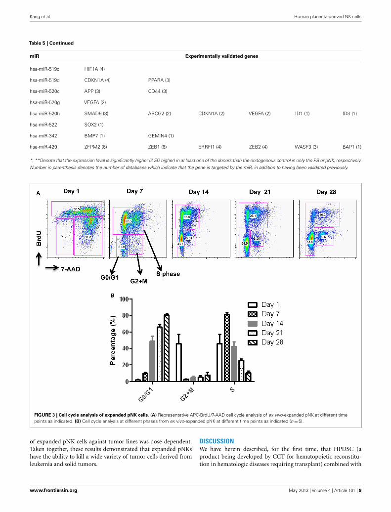
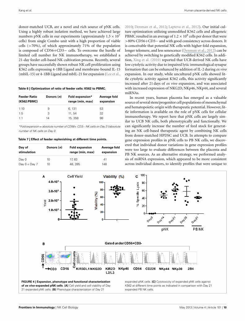
double-staining followed by flow cytometry. As seen in Figure 3,at Day 7, the majority of cultured NK cells were in S-phase, indi-cating that the cells were proliferating. The percentage of activelyproliferating/dividing cells decreased substantially during sub-sequent culture, suggesting Day 7 was the optimal window forre-stimulation. As shown in the Table 7, addition of fresh K562and PBMC feeder cells at Day 7 resulted in a threefold increase inexpansion of NK cells. This optimized 21-day NK culture methodwas repeated in 20 expansion experiments, yielding an averageof 1.2× 10⁹ CD56+CD3− NK cells with around 80% viability(Figure 4A).
CHARACTERIZATION OF EX VIVO-EXPANDED pNK CELLSImmunophenotypic and miRNA changes were characterized inexpanded pNK cells from 12 Combo units in comparison to unex-panded cells. First, Day 21 pNK cells showed a significant increasein the expression of activating receptors such as NKG2D, NKp46,NKp44, and NKp30, and a significant decrease in the expressionof bidirectional receptor 2B4. The expression of inhibitory KIRs,including KIR3DL1 and KIR2DL2/L3 was similar for expandedand unexpanded cells (Figure 4B; Table 8).
We have also investigated the immunophenotype of PB NKexpanded using the optimized isolation and expansion proce-dure established for pNK in nine donors. Comparison studies ofexpanded PB NK cells to expanded pNK cells have revealed thatthe most profound difference was the lower expression of NKp44(Table 9). Less cytotoxic activity was also observed with expandedPB NK cells at Day 21 (42± 7%) (Figure 4C).
Moreover, Expression of 23 miRNAs was increased while31 other miRNAs were downregulated after ex vivo expansion(Table 10). Interestingly, one of the miRNAs found to be upregu-lated was has-miR-155, which when overexpressed has been shown
Table 4 | Highly expressed miRNAs in PB NK cells.
miRNA Fold increase in PB NK p Value
hsa-miR-331 1.43 5.33E−03
hsa-miR-186 1.90 4.54E−03
hsa-miR-17-5p 2.38 2.34E−03
hsa-miR-26a 2.66 2.36E−03
hsa-miR-133b 2.69 8.19E−04
hsa-miR-181b 2.77 4.42E−03
hsa-miR-222 2.83 5.76E−03
hsa-miR-197 3.00 5.48E−05
hsa-miR-146b 3.05 2.92E−03
hsa-miR-342 3.06 3.23E−04
hsa-miR-181d 3.08 3.41E−03
hsa-miR-155 3.12 8.24E−04
hsa-miR-484 3.18 1.23E−03
hsa-let-7g 3.18 2.08E−03
hsa-miR-200c 3.66 1.91E−03
hsa-miR-181c 3.83 2.72E−04
hsa-miR-191 4.06 3.16E−04
hsa-miR-596 4.14 7.06E−03
hsa-miR-142-5p 4.63 4.84E−04
hsa-miR-95 4.86 2.99E−03
hsa-let-7a 5.04 3.91E−04
hsa-miR-21 5.10 2.87E−04
hsa-miR-152 5.46 1.76E−03
hsa-miR-642 5.56 4.70E−04
hsa-miR-24 5.91 2.54E−05
hsa-miR-10a 14.56 5.71E−03
hsa-miR-429 31.74 5.70E−03
hsa-let-7b 108.34 4.66E−05
hsa-miR-199b 2819.55 3.05E−03
to increase NK cell function via enhanced induction of IFN-γ(Trotta et al., 2012).
The cytolytic activity of expanded pNK was evaluated in aFACS-based PKH26/TO-PRO-3 cytotoxicity assay against K562cells. As shown in Figure 4C, while unexpanded pNK cells showedminimal cytolytic activity, there was a significant enhancement ofcytotoxicity against K562 cells by pNK cells at Day 21 versus Day 14at an E:T ratio of 10:1 (63± 15% versus 45± 4%, p < 0.001). Theincrease in cytolytic activity after 21-day expansion was associatedwith the increased expression in activating receptors (Table 8) andmiRNAs (Table 10). Extended cultivation to 28 days did not resultin further increases in activity.
IN VITRO ANTI-TUMOR CYTOLYTIC ACTIVITY OF EXPANDED pNK CELLSTo further evaluate expanded pNK cell activity against a range oftumor types, 11 additional tumor cells lines were co-cultured withDay 21 expanded pNK cells, and NK cell cytolytic activity was mea-sured at E:T ratios of 10:1, 5:1, 2:1, and 1:1 in a 4-h LDH releaseassay. At an E:T ratio of 10:1, expanded pNK cells exhibited greaterthan 50% cytotoxicity against multiple tumor cell lines, includ-ing U937 (89.2± 9.8%), WERI-RB-1 (73.3± 11.8%), RPMI8226(61.3%± 1.3%), HCT-116 (61± 5.1%), U266 (57.4± 4.7%), aswell as and K562 cells (88.6± 5.6%) (Figure 5). Cytolytic activity
Frontiers in Immunology | NK Cell Biology May 2013 | Volume 4 | Article 101 | 6

Kang et al. Human placenta-derived NK cells
Table 5 | Differentially regulated miRs and their validated target genes in pNK, compared to PB NK.
miR Experimentally validated genes
HIGHLY EXPRESSED MIRNAS IN PB NK
hsa-let-7a TRIM71 (7) HMGA1 (4) LIN28A (3) ACP1 (2) E2F2 (2) SMOX (1)
HMGA2 (6) THBS1 (4) CASP3 (3) RTCD1 (2) ITGB3 (2) MYC (1)
UHRF2 (5) NRAS (3) PRDM1 (2) CCND2 (2) LIN28 (1) TUSC2 (1)
MED28 (4) EIF2C4 (3) DICER1 (2) SLC20A1 (2) NF2 (1) BCL2 (1)
ZFP36L1 (1) NKIRAS2 (1) EGR3 (1) IL6 (1) NEFM (1)
hsa-let-7b* HMGA2 (6) CDC25A (3) GRPEL2 (2) NXT2 (2) CDIPT (1) SLC25A13 (1)
IGF2BP1 (5) AURKB (3) MARS2 (2) EIF2C3 (2) CDKAL1 (1) SLC25A1 (1)
TMEM2 (5) DHX57 (3) MRM1 (2) CCNA2 (2) CSNK1D (1) UHRF1 (1)
LIN28B (5) FNDC3A (3) POM121 (2) CCNF (2) DOCK5 (1) C20ORF72 (1)
CCNJ (5) RDH10 (3) PXDN (2) EDEM3 (2) FADS2 (1) SCAMP3 (1)
CDC34 (5) SLC25A24 (3) SCYL1 (2) TRABD (2) FAM96A (1) C2ORF18 (1)
IGF2BP2 (5) SNAP23 (3) SLC25A32 (2) PLAGL2 (2) GPR56 (1) CIAO1 (1)
DMD (5) LIN28A (3) SPRYD4 (2) FARP1 (2) IPO4 (1) BIRC6 (1)
E2F6 (5) PRDM1 (3) TAF9B (2) C7ORF58 (2) KIAA0409 (1) AURKA (1)
HMGA1 (4) NRAS (3) TTC9C (2) LIN28 (1) NEDD4 (1) ALG3 (1)
PGRMC1 (4) RRM2 (3) DLC1 (2) BCL7A (1) OPRS1 (1) ARID3A (1)
THBS1 (4) CCND1 (2) CCND2 (2) ACTG1 (1) RHOB (1) CCBL2 (1)
PDE12 (4) ATP6V1F (2) DICER1 (2) AARSD1 (1) RHOG (1) RRP1B (1)
E2F5 (4) GEMIN7 (2) GTF2I (2) ANAPC1 (1) SLC1A4 (1) TAB2 (1)
hsa-let-7g HMGA2 (6) EIF4G2 (5) IGF2BP1 (5) COL1A2 (3) BCL2L1 (1)
hsa-miR-10a NCOR2 (5) MAP3K7 (4) HOXA1 (1) USF2 (1) BTRC (1)
hsa-miR-133b BCL2L2 (3) MCL1 (2)
hsa-miR-146b* TRAF6 (7) IRAK1 (5) MMP16 (2) CARD10 (1)
hsa-miR-152 DNMT1 (5) HLA-G (2)
hsa-miR-155 SOCS1 (5) DET1 (4) SPI1 (3) SMAD1 (2) TM6SF1 (1) RAB34 (1)
TSHZ3 (5) IKBKE (3) MEIS1 (2) TRAM1 (2) FOXO3 (1) RAB6A (1)
HIVEP2 (5) PHF17 (3) SMAD2 (2) TRIP13 (2) ARL5B (1) SYPL1 (1)
TAB2 (4) BACH1 (3) CYR61 (2) FGF7 (2) ATG3 (1) VAMP3 (1)
JARID2 (4) RCN2 (3) TP53INP1 (2) KRAS (2) ATP6V1C1 (1) WDFY1 (1)
CEBPB (4) RCOR1 (3) ANKFY1 (2) HIF1A (2) BET1 (1) ETS1 (1)
ARID2 (4) ZNF652 (3) CHAF1A (2) C5ORF41 (2) CBFB (1) INPP5D (1)
DHX40 (4) MYB (3) CLDN1 (2) IKBIP (2) DNAJB1 (1) PAPOLA (1)
PICALM (4) TLE4 (3) MYO10 (2) TWF1 (2) DSG2 (1) SERTAD2 (1)
TRIM32 (4) CSF1R (3) NARS (2) CUX1 (2) FMNL2 (1) ERMP1 (1)
ZIC3 (4) FAR1 (3) PHC2 (2) SLA (2) PKN2 (1) C3ORF58 (1)
KBTBD2 (4) ZNF236 (3) SDCBP (2) AGTR1 (1) PRAF2 (1) HNRNPA3P1 (1)
MSH2 (1) PELI1 (1)
hsa-miR-17-5p E2F1 (1)
hsa-miR-181b GRIA2 (4) GATA6 (4) TIMP3 (4) MAP3K10 (3) NLK (2) PLAG1 (2)
CYLD (2) VSNL1 (1) KAT2B (1) BCL2 (1)
hsa-miR-181c GATA6 (4) KRAS (3) NLK (2) NOTCH4 (2) NOTCH2 (1)
hsa-miR-181d GATA6 (3) NLK (2) BCL2 (1)
hsa-miR-186 AKAP12 (1)
hsa-miR-191 TMC7 (2) SOX4 (1)
(Continued)
www.frontiersin.org May 2013 | Volume 4 | Article 101 | 7

Kang et al. Human placenta-derived NK cells
Table 5 | Continued
miR Experimentally validated genes
hsa-miR-197 FBXW7 (4) DPH1 (2) UMPS (2) CLIC1 (1) HNF4A (1) FOXO3 (1)
CHIC2 (4) ALMS1 (2) CPNE6 (2) WDR6 (1) PEX13 (1) IL1R1 (1)
ACVR1 (4) CES1 (2) RBM4 (2) NEK4 (1) C1ORF38 (1) CPSF1 (1)
RAB28 (3) ZNF302 (2) CYLD (2) PIPOX (1) MED16 (1) SNX1 (1)
HNRNPD (2) RAD51 (2) RFX1 (2) IGF2AS (1) LRP4 (1) KLF10 (1)
GOLGB1 (2) RXRB (2) IER3 (1) DCBLD2 (1) TSPYL1 (1) AGR2 (1)
TUSC2 (1) EHD2 (1)
hsa-miR-199b LAMC2 (1) HES1 (1)
hsa-miR-200c ZFPM2 (6) ZEB1 (5) ERRFI1 (5) ZEB2 (5) FN1 (5) UBE2I (3)
BAP1 (2) PTPN13 (1) BMI1 (1) JAG1 (1) TUBB3 (1)
hsa-miR-21 TGFBI (5) GLCCI1 (3) RASGRP1 (3) SGK3 (2) ANKRD46 (2) SLC16A10 (1)
NFIB (4) SOX5 (3) MSH2 (3) RP2 (2) ACTA2 (1) TIMP3 (1)
RECK (4) JAG1 (3) PCBP1 (3) SERPINB5 (2) BTG2 (1) TGFBR2 (1)
PDCD4 (3) BMPR2 (3) TOPORS (3) SPRY2 (2) SESN1 (1) NCAPG (1)
FAM3C (3) TIAM1 (3) APAF1 (2) RHOB (2) SOCS5 (1) IL1B (1)
RTN4 (1) PTX3 (1) CDK2AP1 (1)
hsa-miR-222 CDKN1B (5) FOS (5) KIT (4) CDKN1C (3) PPP2R2A (2) MMP1 (1)
SOD2 (1) BBC3 (1) PTEN (1) ICAM1 (1) ESR1 (1)
hsa-miR-24* CDKN1B (4) TRIB3 (4) MAPK14 (2) FURIN (2) BRCA1 (1) NOTCH1 (1)
ACVR1B (4) DND1 (3) NFAT5 (2) MLEC (2) KIAA0152 (1) CDKN2A (1)
KHSRP (1) HNF4A (1) TGFB1 (1)
hsa-miR-26a SMAD1 (5) HMGA1 (5) CDK8 (4) HMGA2 (3) CCND2 (2) CCNE2 (2)
STRADB (5) GSK3B (5) MTDH (4) CPEB4 (3) CTGF (2) LIF (2)
PTEN (5) EZH2 (4) MAP3K2 (4) SERBP1 (2) CDC6 (2) CPEB2 (1)
CPEB3 (1) SMAD4 (1)
hsa-miR-331 ERBB2 (2) CDCA5 (2) KIF23 (1)
hsa-miR-342 BMP7 (1) GEMIN4 (1)
hsa-miR-429 ZFPM2 (6) ZEB1 (6) ERRFI1 (4) ZEB2 (4) WASF3 (3) BAP1 (1)
HIGHLY EXPRESSED MIRNAS IN PNK
hsa-miR-100 MTOR (3) PLK1 (1) FGFR3 (1) IGF1R (1) ATM (1)
hsa-miR-125b LACTB (6) BMF (3) SAMD10 (3) MKNK2 (2) BBC3 (2) DICER1 (1)
BAK1 (5) BMPR1B (3) EIF4EBP1 (3) CBFB (2) QSOX2 (2) JUB (1)
ARID3B (5) ENTPD4 (3) KLF13 (3) LIN28A (2) LIN28 (1) DDX19B (1)
IRF4 (5) TOR2A (3) ULK3 (3) IGF2 (2) C10ORF104 (1) PABPC1 (1)
PRDM1 (4) KCNS3 (3) SLC7A6 (3) NKIRAS2 (2) B3GALT4 (1) AKT1 (1)
SLC35A4 (4) LIN28B (3) SLC7A1 (3) SEL1L (2) UBE2I (1) TP53 (1)
CGN (4) GRIN2A (3) ERBB3 (2) ATXN1 (2) RBM8A (1) CASC3 (1)
PPAT (4) STAT3 (3) CDKN2A (2) RAF1 (2) IGFBP3 (1) E2F3 (1)
CBX7 (3) LIF (3) ABTB1 (2) CYP24A1 (2) MAN1A1 (1) RNF144A (1)
SGPL1 (3) SMARCD2 (3) ARID3A (2) ABCC4 (2) SMO (1) LYPLA2 (1)
PLEKHA8 (1) TP53INP1 (1) VDR (1)
hsa-miR-211 KCNMA1 (3)
hsa-miR-302c ESR1 (2)
hsa-miR-326 PKM2 (2) SMO (1) GLI1 (1) NOTCH2 (1)
hsa-miR-422a** CYP8B1 (2)
(Continued)
Frontiers in Immunology | NK Cell Biology May 2013 | Volume 4 | Article 101 | 8
Kang et al. Human placenta-derived NK cells
Table 5 | Continued
miR Experimentally validated genes
hsa-miR-519c HIF1A (4)
hsa-miR-519d CDKN1A (4) PPARA (3)
hsa-miR-520c APP (3) CD44 (3)
hsa-miR-520g VEGFA (2)
hsa-miR-520h SMAD6 (3) ABCG2 (2) CDKN1A (2) VEGFA (2) ID1 (1) ID3 (1)
hsa-miR-522 SOX2 (1)
hsa-miR-342 BMP7 (1) GEMIN4 (1)
hsa-miR-429 ZFPM2 (6) ZEB1 (6) ERRFI1 (4) ZEB2 (4) WASF3 (3) BAP1 (1)
*, **Denote that the expression level is significantly higher (2 SD higher) in at least one of the donors than the endogenous control in only the PB or pNK, respectively.
Number in parenthesis denotes the number of databases which indicate that the gene is targeted by the miR, in addition to having been validated previously.
FIGURE 3 | Cell cycle analysis of expanded pNK cells. (A) Representative APC-BrdU/7-AAD cell cycle analysis of ex vivo-expanded pNK at different timepoints as indicated. (B) Cell cycle analysis at different phases from ex vivo-expanded pNK at different time points as indicated (n=5).
of expanded pNK cells against tumor lines was dose-dependent.Taken together, these results demonstrated that expanded pNKshave the ability to kill a wide variety of tumor cells derived fromleukemia and solid tumors.
DISCUSSIONWe have herein described, for the first time, that HPDSC (aproduct being developed by CCT for hematopoietic reconstitu-tion in hematologic diseases requiring transplant) combined with
www.frontiersin.org May 2013 | Volume 4 | Article 101 | 9
Kang et al. Human placenta-derived NK cells
donor-matched UCB, are a novel and rich source of pNK cells.Using a highly robust isolation method, we have achieved largenumbers pNK cells in our experiments (approximately 1.5× 10⁷
cells) from single Combo unit with a high proportion of viablecells (>70%), of which approximately 71% of the populationis composed of CD56+CD3− cells. To overcome the hurdle oflimited cell number for NK immunotherapy, we established a21-day feeder cell-based NK cultivation process. Recently, severalgroups have successfully shown robust NK cell proliferation usingK562 cells expressing 4-1BB Ligand and membrane-bound IL-15(mbIL-15) or 4-1BB Ligand and mbIL-21 for expansion (Lee et al.,
Table 6 | Optimization of ratio of feeder cells: K562 to PBMC.
Feeder Ratio
(K562:PBMC)
Donors (n) Fold expansion*
range (min, max)
Average fold
expansion
1:10 9 6, 131 53
1:5 3 11, 54 32
1:1 14 15, 358 98
*Fold expansion= absolute number of CD56+CD3−NK cells on Day 21/absolute
number of NK cells on Day 0.
Table 7 | Effect of feeder replenishing at different time points.
Day of
stimulation
Donors (n) Fold expansion
range (min, max)
Average fold
expansion
Day 0 10 17, 83 41
Day 0+Day 7 10 48, 395 148
2010; Denman et al., 2012; Lapteva et al., 2012). Our initial cul-ture optimization utilizing unmodified K562 cells and allogeneicPBMC resulted in an average of 1.2× 10⁹ cells per donor that were>80% CD56+CD3− and with good consistency across donors. Itis conceivable that potential NK cells with higher fold expansion,longer telomere, and less senescence (Denman et al., 2012) can beachieved by switching to genetically modified K562 cells. In addi-tion, Xing et al. (2010) reported that UCB derived NK cells havelow cytolytic activity due to impaired lytic immunological synapseformation that can be enhanced by addition of IL-2 during ex vivoexpansion. In our study, while uncultured pNK cells showed lit-tle cytolytic activity against K562 cells, this activity significantlyincreased after 21 days of ex vivo expansion, and was associatedwith increased expression of NKG2D, NKp46, NKp44, and severalmiRNAs.
In recent years, human placenta has emerged as a valuablesource of several stem/progenitor cell populations of mesenchymaland hematopoietic origin with therapeutic potential. However, lit-tle information is available on the role of pNK cells for cellularimmunotherapy. We report here that pNK cells are largely sim-ilar to UCB NK cells, both phenotypically and functionally. Wecan significantly increase the number of feed stock for generat-ing an NK cell-based therapeutic agent by combining NK cellsfrom donor-matched HPDSC and UCB. In attempts to comparegene expression profiles in pNK cells to PB NK cells, we discov-ered that individual donor variations in gene expression profileswere too large to evaluate differences between the placenta andPB NK sources. As an alternative strategy, we performed analy-sis of miRNA expression, which appeared to be more consistentacross individual donors, to identify profiles that were unique to
FIGURE 4 | Expansion, phenotype and functional characterizationof ex vivo-expanded pNK cells. (A) Cell yield and cell viability of Day21 expanded pNK cells. (B) Phenotype characterization of Day 21
expanded pNK cells. (C) Cytotoxicity of expanded pNK cells againstK562 at different time points as indicated in comparison with Day 21expanded PB NK cells.
Frontiers in Immunology | NK Cell Biology May 2013 | Volume 4 | Article 101 | 10

Kang et al. Human placenta-derived NK cells
Table 8 | Sub-population comparison of Day 21 expanded pNK versus
unexpanded pNK cells.
NK Sub-
populations
Day 21 pNK (n = 12 U) Day 0 pNK (n = 16 U) p Value
Mean (%) SD Mean (%) SD
CD56+CD16− 35.98 18.18 60.94 16.58 **
CD56+CD16+ 63.80 18.32 39.05 16.58 **
KIR3DL1+ 17.92 13.44 12.31 8.11 NS
KIR2DL2/L3+ 21.10 10.48 21.89 8.65 NS
NKG2D+ 89.28 12.88 42.11 17.79 ***
NKp46+ 88.74 5.34 6.98 4.33 ***
CD226+ 18.79 12.14 15.97 6.66 NS
NKp44+ 64.13 16.65 9.48 5.27 **
NKp30+ 84.53 12.40 39.08 19.06 ***
2B4+ 0.89 0.99 11.07 5.90 ***
CD94+ 74.82 12.45 71.31 13.94 NS
*p < 0.05; **p < 0.01; ***p < 0.001.
Table 9 | Sub-populations comparison in Day 21 expanded pNK versus
Day 21 expanded PB NK.
NK sub-populations Combo (12 U) PB (9 U) p Value
Mean (%) SD Mean (%) SD
CD16− 35.98 18.18 28.39 7.78 NS
CD16+ 63.80 18.32 71.50 7.78 NS
KIR3DL1+ 17.92 13.44 8.83 6.44 NS
KIR2DL2/L3+ 21.10 10.48 37.40 17.82 *
NKG2D+ 89.28 12.88 73.27 20.06 NS
NKp46+ 88.74 5.34 64.97 17.26 **
CD226+ 18.79 12.14 4.80 1.72 NS
NKp44+ 64.13 16.65 16.00 3.82 **
NKp30+ 84.53 12.40 55.77 1.96 *
2B4+ 0.89 0.99 2.16 1.76 NS
CD94+ 74.82 12.45 71.76 20.35 NS
*p < 0.05; **p < 0.01; ***p < 0.001.
pNK or PB NK cells. We identified four unique and 20 highlyexpressed miRNAs in pNK cells, and eight unique and 29 highlyexpressed miRNAs in PB NK cells. Target gene prediction analy-sis returned 14 highly expressed miRNAs in pNK and 24 highlyexpressed miRNA in PB NK with more than one target gene. InPB NK, highly expressed miRNAs included has-let-7a, has-let-7g, has-mir-133b, has-mir-181b, and has-mir-181d that target theanti-apoptotic genes Bcl-2 and Bcl-2L, which may indicate thatPB NK cells are closer to reaching the limit of cell expansion thanpNK cells. Additionally, miRNA hsa-mir-146b is highly expressedin PB NK cells, and its target TRAF6 has been reported to down-regulate NF-kB activity, suppress cell proliferation and enhancechemosensitivity (Paik et al., 2011). TRAF6 plays a critical role ininnate and adaptive immunity (Chiffoleau et al., 2003) in conjunc-tion with genes such as MAPK14, IL6, and FOS, which are targetedby has-mir-24, has-mir-7a, and has-mir-222 respectively, all found
Table 10 | Differentially regulated miRNAs during pNK ex vivo
expansion.
miRNA Fold change in Day 21 versus Day 0 p Value
hsa-miR-520g −2820.57 6.33E−05
hsa-miR-520h −2803.02 4.35E−04
hsa-miR-518a −2300.60 5.72E−05
hsa-miR-517b −1669.82 8.11E−05
hsa-miR-451 −1626.65 2.40E−03
hsa-miR-518c −609.88 2.93E−04
hsa-miR-127 −557.55 3.40E−04
hsa-miR-517a −288.89 8.27E−05
hsa-miR-382 −273.34 1.77E−04
hsa-miR-519d −245.09 7.13E−04
hsa-miR-486 −149.95 6.29E−03
hsa-miR-518b −112.10 9.70E−05
hsa-miR-522 −85.30 2.58E−03
hsa-miR-376a −72.14 2.95E−03
hsa-miR-198 −70.93 1.13E−03
hsa-miR-126 −51.46 1.97E−04
hsa-miR-487b −47.68 3.32E−03
hsa-miR-519c −47.52 4.88E−03
hsa-miR-518e −34.89 5.06E−03
hsa-miR-433 −18.07 5.68E−04
hsa-miR-125b −16.66 4.66E−04
hsa-miR-214 −16.38 5.74E−03
hsa-miR-130a −12.98 4.13E−03
hsa-miR-518d −10.75 8.73E−03
hsa-miR-99a −7.91 2.03E−03
hsa-miR-515-3p −4.93 2.50E−03
hsa-miR-95 −4.73 9.30E−04
hsa-miR-30a-3p −3.06 1.20E−03
hsa-miR-30d −2.59 4.32E−03
hsa-miR-26a −1.75 7.81E−03
hsa-miR-191 −1.32 2.12E−03
hsa-miR-331 1.53 6.74E−03
hsa-miR-181c 1.71 1.24E−03
hsa-miR-142-3p 2.24 5.55E−03
hsa-miR-155 2.48 3.47E−03
hsa-miR-24 2.66 1.03E−03
hsa-miR-23a 3.07 8.50E−03
hsa-miR-142-5p 3.21 3.34E−04
hsa-let-7d 3.22 3.29E−03
hsa-miR-195 3.77 2.92E−04
hsa-miR-141 3.80 5.20E−03
hsa-miR-98 3.83 2.96E−03
hsa-miR-222 3.86 7.71E−03
hsa-miR-545 5.28 5.60E−03
hsa-miR-642 7.17 2.82E−04
hsa-miR-21 13.46 1.94E−04
hsa-miR-210 13.87 6.66E−03
hsa-miR-221 21.73 3.04E−03
hsa-miR-34c 45.37 5.26E−04
hsa-miR-135b 49.26 1.17E−03
hsa-miR-34a 66.38 8.08E−03
hsa-miR-10a 72.27 1.27E−03
hsa-miR-380-3p 921.50 1.18E−03
hsa-miR-520a 10892.33 2.38E−05
www.frontiersin.org May 2013 | Volume 4 | Article 101 | 11

Kang et al. Human placenta-derived NK cells
% S
pe
cif
ic L
ys
is
CCRF-C
EM
KG
-1
KG-1
a
K56
2
KU81
2
U93
7
WER
I-RB-1
RPM
I822
6
U26
6
HCT-1
16
HCC22
18
HT-2
9
0
20
40
60
80
100 10:1
5:1
2:1
1:1
FIGURE 5 | Cytotoxicity of ex vivo-expanded Day 21 pNK cells againsta wide range of tumor cell lines. Cytotoxicity of ex vivo-expanded pNKcells (n=6) against a wide range of tumor cell lines at E:T ratio of 10:1, 5:1,2:1, and 1:1 as indicated.
in our study to be highly expressed in PB NK. Furthermore, weidentified a has-mir-181 group (has-mir-181b, has-mir-181c, has-mir-181d) that was threefold higher in PB NK. These miRNAstarget nemo-like kinase, a regulator of Notch signaling, whichplays an important role in the development of NK cells from
CD34+HSCs and IFN-γ production in primary CD56+NK cells(Cichocki et al., 2011). Lastly, within the group of highly expressedmiRNAs in pNK, we have identified miRNA: mRNA target pairscomprised of pluripotency markers in stem cells and cell cycleregulators, such as SOX2, BMPR1, SMO, AKT1, ATM, RAF1, andMTOR, most of which are not present in the targeted gene list(both experimental and validated) of miRNAs in PB NK. Fur-thermore, comparison studies have revealed the higher expressionof NKp44 and greater cytotoxic activity against K562 at Day 21(42± 7%) from expanded pNK compared to that of expanded PBNK cells.
In conclusion, we have characterized pNK cells from donor-matched HPDSC and UCB. pNK cells showed a distinct phenotypeand miRNA profile from PB NK cells. We have demonstratedthat pNK cells can be readily obtained from Combo units. Thesecells can be expanded, characterized, and activated to yield clini-cally relevant quantities of a highly cytotoxic cellular product withpotential as a treatment for a wide range of hematological cancers.Taken together, the results presented here provide an impor-tant advance in the development of NK cell-based therapeuticproducts.
ACKNOWLEDGMENTSThe authors thank Dr. Vivian Albert, Dr. Ellen Baum, Dr. JamesEdinger, and Dr. George Matcham for reviewing the manuscriptand providing critical feedback.
REFERENCESApps, R., Sharkey, A., Gardner, L., Male,
V., Kennedy, P., Masters, L., et al.(2011). Ex vivo functional responsesto HLA-G differ between bloodand decidual NK cells. Mol. Hum.Reprod. 17, 577–586.
Bezman, N. A., Cedars, E., Steiner,D. F., Blelloch, R., Hesslein, D.G., and Lanier, L. L. (2010). Dis-tinct requirements of microRNAsin NK cell activation, survival,and function. J. Immunol. 185,3835–3846.
Chiffoleau, E., Kobayashi, T., Walsh, M.C., King, C. G.,Walsh, P. T., Hancock,W. W., et al. (2003). TNF receptor-associated factor 6 deficiency duringhemopoiesis induces Th2-polarizedinflammatory disease. J. Immunol.171, 5751–5759.
Cichocki, F., Felices, M., McCullar, V.,Presnell, S. R., Al-Attar, A., Lutz,C. T., et al. (2011). Cutting edge:microRNA-181 promotes humanNK cell development by regulatingNotch signaling. J. Immunol. 187,6171–6175.
Cooper, M. A., Fehniger, T. A.,and Caligiuri, M. A. (2001). Thebiology of human natural killer-cell subsets. Trends Immunol. 22,633–640.
Curti, A., Ruggeri, L., D’Addio, A., Bon-tadini,A., Dan, E., Motta, M. R., et al.
(2011). Successful transfer of allore-active haploidentical KIR ligand-mismatched natural killer cells afterinfusion in elderly high risk acutemyeloid leukemia patients. Blood118, 3273–3279.
Denman, C. J., Senyukov, V. V.,Somanchi, S. S., Phatarpekar, P.V., Kopp, L. M., Johnson, J. L.,et al. (2012). Membrane-boundIL-21 promotes sustained ex vivoproliferation of human naturalkiller cells. PLoS ONE 7:e30264.doi:10.1371/journal.pone.0030264
Fehniger, T. A., Cooper, M. A., Nuovo, G.J., Cella, M., Facchetti, F., Colonna,M., et al. (2003). CD56bright nat-ural killer cells are present inhuman lymph nodes and are acti-vated by T cell-derived IL-2: apotential new link between adap-tive and innate immunity. Blood 101,3052–3057.
Ferlazzo, G., Thomas, D., Lin, S. L.,Goodman, K., Morandi, B., Muller,W. A., et al. (2004). The abundantNK cells in human secondary lym-phoid tissues require activation toexpress killer cell Ig-like receptorsand become cytolytic. J. Immunol.172, 1455–1462.
Freud, A. G., Becknell, B., Roy-chowdhury, S., Mao, H. C., Fer-ketich, A. K., Nuovo, G. J., etal. (2005). A human CD34(+)
subset resides in lymph nodesand differentiates into CD56brightnatural killer cells. Immunity 3,295–304.
Herberman, R. B., Nunn, M. E., andLavrin, D. H. (1975a). Natural cyto-toxic reactivity of mouse lymphoidcells against syngeneic acid allo-geneic tumors. I. Distribution ofreactivity and specificity. Int. J. Can-cer 16, 216–229.
Herberman, R. B., Nunn, M. E.,Holden, H. T., and Lavrin, D. H.(1975b). Natural cytotoxic reac-tivity of mouse lymphoid cellsagainst syngeneic and allogeneictumors. II. Characterization ofeffector cells. Int. J. Cancer 16,230–239.
Hiby, S. E., Walker, J. J., O’Shaughnessy,K. M., Redman, C. W., Carring-ton, M., Trowsdale, J., et al. (2004).Combinations of maternal KIR andfetal HLA-C genes influence therisk of preeclampsia and repro-ductive success. J. Exp. Med. 200,957–965.
Huntington, N. D., Vosshenrich, C. A.,and Di Santo, J. P. (2007). Devel-opmental pathways that generatenatural killer-cell diversity in miceand humans. Nat. Rev. Immunol. 7,703–714.
Jacobs, R., Hintzen, G., Kemper, A.,Beul, K., Kempf, S., Behrens, G.,
et al. (2001). CD56bright cells dif-fer in their KIR repertoire andcytotoxic features from CD56dimNK cells. Eur. J. Immunol. 31,3121–3127.
Koopman, L. A., Kopcow, H. D.,Rybalov, B., Boyson, J. E., Orange,J. S., Schatz, F., et al. (2003).Human decidual natural killer cellsare a unique NK cell subset withimmunomodulatory potential. J.Exp. Med. 198, 1201–1212.
Kopcow, H. D., Allan, D. S., Chen, X.,Rybalov, B., Andzelm, M. M., Ge,B., et al. (2005). Human decidualNK cells form immature activat-ing synapses and are not cytotoxic.Proc. Natl. Acad. Sci. U.S.A. 102,15563–15568.
Kopcow, H. D., Eriksson, M., Mselle,T. F., Damrauer, S. M., Wira,C. R., Sentman, C. L., et al.(2010). Human decidual NK cellsfrom gravid uteri and NK cellsfrom cycling endometrium are dis-tinct NK cell subsets. Placenta 31,334–338.
Lanier, L. L., Le, A. M., Civin, C. I.,Loken, M. R., and Phillips, J. H.(1986). The relationship of CD16(Leu-11) and Leu-19 (NKH-1) anti-gen expression on human periph-eral blood NK cells and cytotoxicT lymphocytes. J. Immunol. 136,4480–4486.
Frontiers in Immunology | NK Cell Biology May 2013 | Volume 4 | Article 101 | 12

Kang et al. Human placenta-derived NK cells
Lapteva, N., Durett, A. G., Sun, J.,Rollins, L. A., Huye, L. L., Fang, J., etal. (2012). Large-scale ex vivo expan-sion and characterization of naturalkiller cells for clinical applications.Cytotherapy 14, 1131–1143.
Lee, D. A., Verneris, M. R., and Cam-pana, D. (2010). Acquisition, prepa-ration, and functional assessmentof human NK cells for adoptiveimmunotherapy. Methods Mol. Biol.651, 61–77.
Lee-MacAry, A. E., Ross, E. L., Davies,D., Laylor, R., Honeychurch, J., Glen-nie, M. J., et al. (2001). Develop-ment of a novel flow cytometric cell-mediated cytotoxicity assay usingthe fluorophores PKH-26 and TO-PRO-3 iodide. J. Immunol. Methods252, 83–92.
Livak, K. J., and Schmittgen, T. D.(2001). Analysis of relative geneexpression data using real-timequantitative PCR and the 2(-DeltaDelta C(T)). Methods 4, 402–408.
Male, V., Sharkey, A., Masters, L.,Kennedy, P. R., Farrell, L. E., andMoffett, A. (2011). The effect ofpregnancy on the uterine NK cellKIR repertoire. Eur. J. Immunol. 41,3017–3027.
McKenna, D. H. Jr., Sumstad, D.,Bostrom, N., Kadidlo, D. M.,Fautsch, S., McNearney, S., et al.(2007). Good manufacturing prac-tices production of natural killercells for immunotherapy: a six-yearsingle-institution experience. Trans-fusion 47, 520–528.
Meyer-Monard, S., Passweg, J., Siegler,U., Kalberer, C., Koehl, U., Rovó, A.,et al. (2009). Clinical-grade purifi-cation of natural killer cells inhaploidentical hematopoietic stemcell transplantation. Transfusion 49,362–371.
Miller, J. S., Soignier, Y., Panoskaltsis-Mortari, A., McNearney, S. A., Yun,G. H., Fautsch, S. K., et al. (2005).Successful adoptive transfer andin vivo expansion of human hap-loidentical NK cells in patients withcancer. Blood 105, 3051–3057.
Paik, J. H., Jang, J. Y., Jeon, Y. K.,Kim, W. Y., Kim, T. M., Heo, D.
S., et al. (2011). MicroRNA-146adownregulates NFκB activity viatargeting TRAF6 and functions asa tumor suppressor having strongprognostic implications in NK/T celllymphoma. Clin. Cancer Res. 17,4761–4771.
Parolini, O., Alviano, F., Bagnara, G. P.,Bilic, G., Bühring, H. J., Evangelista,M., et al. (2008). Concise review:isolation and characterization ofcells from human term placenta:outcome of the first internationalWorkshop on Placenta Derived StemCells. Stem Cells 26, 300–311.
Passweg, J. R., Tichelli, A., Meyer-Monard, S., Heim, D., Stern, M.,Kühne, T., et al. (2004). Purifieddonor NK-lymphocyte infusion toconsolidate engraftment after hap-loidentical stem cell transplantation.Leukemia 18, 1835–1838.
Prather, W. R., Toren, A., and Meiron,M. (2008). Placental-derived andexpanded mesenchymal stromalcells (PLX-I) to enhance theengraftment of hematopoietic stemcells derived from umbilical cordblood. Expert Opin. Biol. Ther. 8,1241–1250.
Rubnitz, J. E., Inaba, H., Ribeiro, R. C.,Pounds, S., Rooney, B., Bell, T., etal. (2010). NKAML: a pilot study todetermine the safety and feasibilityof haploidentical natural killer celltransplantation in childhood acutemyeloid leukemia. J. Clin. Oncol. 28,955–959.
Ruggeri, L., Capanni, M., Casucci, M.,Volpi, I., Tosti, A., Perruccio, K., etal. (1999). Role of natural killer cellalloreactivity in HLA-mismatchedhematopoietic stem cell transplanta-tion. Blood 94, 333–339.
Ruggeri, L., Capanni, M., Urbani, E.,Perruccio, K., Shlomchik, W. D.,Tosti, A., et al. (2002). Effectivenessof donor natural killer cell alloreac-tivity in mismatched hematopoietictransplants. Science 295, 2097–2100.
Shi, J., Tricot, G., Szmania, S., Rosen,N., Garg, T. K., Malaviarachchi, P.A., et al. (2008). Infusion of haplo-identical killer immunoglobulin-like receptor ligand mismatched NK
cells for relapsed myeloma in the set-ting of autologous stem cell trans-plantation. Br. J. Haematol. 143,641–653.
Smyth, M. J., Hayakawa, Y., Takeda, K.,and Yagita, H. (2002). New aspects ofnatural-killer-cell surveillance andtherapy of cancer. Nat. Rev. Cancer2, 850–861.
Spanholtz, J., Tordoir, M., Eissens, D.,Preijers, F., van der Meer, A., Joosten,I., et al. (2010). High log-scaleexpansion of functional humannatural killer cells from umbilicalcord blood CD34-positive cells foradoptive cancer immunother-apy. PLoS ONE 5:e9221.doi:10.1371/journal.pone.0009221
Sutlu, T., and Alici, E. (2009). Nat-ural killer cell-based immunother-apy in cancer: current insights andfuture prospects. J. Intern. Med. 266,154–181.
Trotta, R., Chen, L., Ciarlariello, D.,Josyula, S., Mao, C., Costinean, S., etal. (2012). miR-155 regulates IFN-γ production in natural killer cells.Blood 119, 3478–3485.
Velardi, A., Ruggeri, L., Moretta, A., andMoretta, L. (2002). NK cells: a lessonfrom mismatched hematopoietictransplantation. Trends Immunol.23, 438–444.
Woll, P. S., Grzywacz, B., Tian, X., Mar-cus, R. K., Knorr, D. A., Verneris, M.R., et al. (2009). Human embryonicstem cells differentiate into a homo-geneous population of natural killercells with potent in vivo antitumoractivity. Blood 113, 6094–6101.
Xing, D., Ramsay, A. G., Gribben, J. G.,Decker, W. K., Burks, J. K., Mun-sell, M., et al. (2010). Cord bloodnatural killer cells exhibit impairedlytic immunological synapse forma-tion that is reversed with IL-2 exvivo expansion. J. Immunother. 33,684–696.
Yoon, S. R., Lee,Y. S.,Yang, S. H., Ahn, K.H., Lee, J. H., Lee, J. H., et al. (2010).Generation of donor natural killercells from CD34(+) progenitor cellsand subsequent infusion after HLA-mismatched allogeneic hematopoi-etic cell transplantation: a feasibility
study. Bone Marrow Transplant. 45,1038–1046.
Yssel, H., de Vries, J. E., Koken, M.,Van Blitterswijk, W., and Spits, H.(1984). Serum-free medium for thegeneration and propagation of func-tional human cytotoxic and helper Tcell clones. J. Immunol. Methods 72,219–227.
Zhong, N., Sun, J., Min, Z., Zhao, W.,Zhang, R., Wang, W., et al. (2012).MicroRNA-337 is associated withchondrogenesis through regulat-ing TGFBR2 expression. Osteoarthr.Cartil. 6, 593–602.
Conflict of Interest Statement: LinKang, Vanessa Voskinarian-Berse, EricLaw, Tiffany Reddin, Mohit Bhatia,Yuhong Ning, Wolfgang Hofgartner,Stewart Abbot, Xiaokui Zhang, andRobert Hariri hold Celgene employ-ment, equity ownership, and patents;David Dong, Timothy Maguire, andMartin Yarmush have received researchfunding from Celgene.
Received: 16 February 2013; accepted: 17April 2013; published online: 01 May2013.Citation: Kang L, Voskinarian-Berse V,Law E, Reddin T, Bhatia M, Hariri A,Ning Y, Dong D, Maguire T, YarmushM, Hofgartner W, Abbot S, Zhang X andHariri R (2013) Characterization andex vivo expansion of human placenta-derived natural killer cells for cancerimmunotherapy. Front. Immunol. 4:101.doi: 10.3389/fimmu.2013.00101This article was submitted to Frontiers inNK Cell Biology, a specialty of Frontiersin Immunology.Copyright © 2013 Kang , Voskinarian-Berse, Law, Reddin, Bhatia, Hariri,Ning , Dong , Maguire, Yarmush, Hof-gartner, Abbot , Zhang and Hariri. Thisis an open-access article distributed underthe terms of the Creative Commons Attri-bution License, which permits use, distri-bution and reproduction in other forums,provided the original authors and sourceare credited and subject to any copy-right notices concerning any third-partygraphics etc.
www.frontiersin.org May 2013 | Volume 4 | Article 101 | 13