CHARACTERISATION OF PROTEIN BIOFIBRE FROM CHICKEN FEATHERS IN NATURAL AND CARBONIZED FORMS
12
British Journal of Engineering and Technology ISSN: 2326 – 425X URL: http://www.bjet.baar.org.uk Vol. 1, No. 7, pp 1-12, MAY 2013 ২ CHARACTERISATION OF PROTEIN BIOFIBRE FROM CHICKEN FEATHERS IN NATURAL AND CARBONIZED FORMS Débora Damasceno Belarmino Programa de Pós Graduação em Engenharia Mecânica, Universidade Federal do Rio Grande do Norte- Natal - Brasil [email protected] Rasiah Ladchumananandasivam Programa de Pós Graduação em Engenharia Mecânica, Universidade Federal do Rio Grande do Norte – Natal - Brasil [email protected] Loilde Damasceno Belarmino Programa de Pós Graduação em Química, Universidade Federal do Rio Grande do Norte Natal - Brasil [email protected] ABSTRACT io-fibers may be obtained from different renewable sources. Those of vegetable origin have been most exploited; however, those of animal origin are entering the scientific scenario. The keratin is a protein, synthesized by many animals to form various structures of the body. Among them are the birds, which have in their feathers 90% of keratin (keratin). The structure and properties of the feathers make them unique fibers and preferable for several applications, among them, as reinforcement in composite materials and a source of carbon, with structures more resistant to high temperature and elevated mechanical stress. To validate its use, the chicken feather fibers in their natural (Keratin fiber - KF) and modified form were analyzed and compared by Fourier Transform Infrared Spectroscopy (FTIR), Scanning Electron Microscopy (SEM) and Energy-dispersive X-ray spectroscopy (EDS). First, they were treated superficially for the removal of impurities from its surface, with running water and a neutral detergent. Then part of the KF was semi-carbonized at 220 ºC for 24 h (Semi-Carbonized Feathers - SCF) and without atmospheric control. Another part was subjected to the carbonization process by pyrolysis at 220 ºC for 24h and 450 ºC, for 1h (Carbonized Feather - CF), to be compared among themselves and examine the changes occurred after carbonization. With the results of characterization analysis, it was observed that the KF had an internal structure, similar to a honeycomb and segments of knots and hooks on its external surface. Furthermore, present amide groups of which characterize their secondary structure (polypeptide chains) and in addition contain sulfur in its composition. The SCF has maintained the chemical composition of KF; however, when ground proved to be highly heterogeneous and brittle. The shiny black particles of CF presented in the form of homogeneous sheets, maintaining some hooks and chemical composition of KF. The quantity of all the elements has been reduced, mainly of sulfur in CF. Through this study; we reached the conclusion that to obtain carbonaceous material from chicken feathers; the most effective and desirable method is pyrolysis, and reduces the environmental problems created by this type of residue and avoid a burden for humanity. Key Words: biomaterials, biofibre, chicken feathers, keratin, pyrolysis, carbon B
Transcript of CHARACTERISATION OF PROTEIN BIOFIBRE FROM CHICKEN FEATHERS IN NATURAL AND CARBONIZED FORMS

British Journal of Engineering and Technology ISSN: 2326 – 425X URL: http://www.bjet.baar.org.uk
Vol. 1, No. 7, pp 1-12, MAY 2013
২
CHARACTERISATION OF PROTEIN BIOFIBRE FROM CHICKEN FEATHERS IN NATURAL AND CARBONIZED FORMS
Débora Damasceno Belarmino
Programa de Pós Graduação em Engenharia Mecânica, Universidade Federal do Rio Grande do Norte- Natal - Brasil
Rasiah Ladchumananandasivam
Programa de Pós Graduação em Engenharia Mecânica, Universidade Federal do Rio Grande do Norte – Natal - Brasil
Loilde Damasceno Belarmino
Programa de Pós Graduação em Química, Universidade Federal do Rio Grande do Norte Natal - Brasil [email protected]
ABSTRACT io-fibers may be obtained from different renewable sources. Those of vegetable origin have been most exploited; however, those of animal origin are entering the scientific scenario. The keratin is a protein,
synthesized by many animals to form various structures of the body. Among them are the birds, which have in their feathers 90% of keratin (keratin). The structure and properties of the feathers make them unique fibers and preferable for several applications, among them, as reinforcement in composite materials and a source of carbon, with structures more resistant to high temperature and elevated mechanical stress. To validate its use, the chicken feather fibers in their natural (Keratin fiber - KF) and modified form were analyzed and compared by Fourier Transform Infrared Spectroscopy (FTIR), Scanning Electron Microscopy (SEM) and Energy-dispersive X-ray spectroscopy (EDS). First, they were treated superficially for the removal of impurities from its surface, with running water and a neutral detergent. Then part of the KF was semi-carbonized at 220 ºC for 24 h (Semi-Carbonized Feathers - SCF) and without atmospheric control. Another part was subjected to the carbonization process by pyrolysis at 220 ºC for 24h and 450 ºC, for 1h (Carbonized Feather - CF), to be compared among themselves and examine the changes occurred after carbonization. With the results of characterization analysis, it was observed that the KF had an internal structure, similar to a honeycomb and segments of knots and hooks on its external surface. Furthermore, present amide groups of which characterize their secondary structure (polypeptide chains) and in addition contain sulfur in its composition. The SCF has maintained the chemical composition of KF; however, when ground proved to be highly heterogeneous and brittle. The shiny black particles of CF presented in the form of homogeneous sheets, maintaining some hooks and chemical composition of KF. The quantity of all the elements has been reduced, mainly of sulfur in CF. Through this study; we reached the conclusion that to obtain carbonaceous material from chicken feathers; the most effective and desirable method is pyrolysis, and reduces the environmental problems created by this type of residue and avoid a burden for humanity.
Key Words: biomaterials, biofibre, chicken feathers, keratin, pyrolysis, carbon
B

British Journal of Engineering and Technology ISSN: 2326 – 425X URL: http://www.bjet.baar.org.uk
Vol. 1, No. 7, pp 1-12, MAY 2013
৩
Introduction Several types of bio-fibres have recently appealed to a growing interest for engineers and scientists, for
their excellent properties, low-cost and, because they are ecofriendly. Plants have been the most exploited and among that of animal origin, keratin is present in human hair and wool, which have applications in textile and medical fields. Although the structure and properties of keratin bio-fibre from chicken feathers, have been reported by some researchers, but most of the published work based in their natural form. They were not investigated enough in the carbonized form. Furthermore, a few detailed studies on protein-based products for processing into structures in nano scale. The main properties of the chicken feathers will be detailed in the present article.
According to Francis Junior & Francis (2006), proteins are bio polymers formed essentially of amino acids linked together, in specific linear sequence, by peptide links. They present characteristic three-dimensional structures. The amino acids have as general structure, a central carbon (alpha carbon) connected to the functional groups: amine (NH2) and carboxyl (COOH); hydrogen atoms and a side chain (R) specific for each amino acid, Figure 1a, and are mainly connected by covalent peptide links. This connection occurs due to the condensation reaction between the carboxyl group of one amino acid to the amine group of another amino acid, Figure 1b. The sequence of amino acid constituents of the chain or the polypeptide chains of a protein is called the primary structure. The proteins can be classified according to the acquired conformational level. The sequence of the amino acids of a protein and the local conformation of some portion of a polypeptide, i.e. the three-dimensional arrangement of amino acids located closer within the primary structure is called as the secondary structure. The more common secondary structures are the alpha-helix and beta sheet. These structures appear simultaneously in the same protein, establishing parts of tertiary structure of a polypeptide helicoidal.
H2N C C
H
ROH
O
(a)
HN C NH C C C HN C NH C C + H2 O
H H O H O H H O H O
H OH H OH H H OH
C + H
Peptide Bond. (b)
Figure 1 - (a) General form of amino acids (b) Condensation reaction between two amino acid molecules of glycine, showing the formation of peptide link.
(adapted from: Francisco Junior & Francisco, 2006)
British Journal of Engineering and Technology ISSN: 2326 – 425X URL: http://www.bjet.baar.org.uk
Vol. 1, No. 7, pp 1-12, MAY 2013
৪
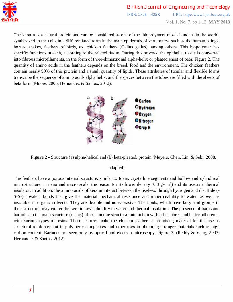
The keratin is a natural protein and can be considered as one of the biopolymers most abundant in the world, synthesized in the cells in a differentiated form in the main epidermis of vertebrates, such as the human beings, horses, snakes, feathers of birds, ex. chicken feathers (Gallus gallus), among others. This biopolymer has specific functions in each, according to the related tissue. During this process, the epithelial tissue is converted into fibrous microfilaments, in the form of three-dimensional alpha-helix or pleated sheet of beta, Figure 2. The quantity of amino acids in the feathers depends on the breed, food and the environment. The chicken feathers contain nearly 90% of this protein and a small quantity of lipids. These attributes of tubular and flexible forms transcribe the sequence of amino acids alpha helix, and the spaces between the tubes are filled with the sheets of beta form (Moore, 2005; Hernandez & Santos, 2012).
Figure 2 - Structure (a) alpha-helical and (b) beta-pleated, protein (Meyers, Chen, Lin, & Seki, 2008,
adapted)
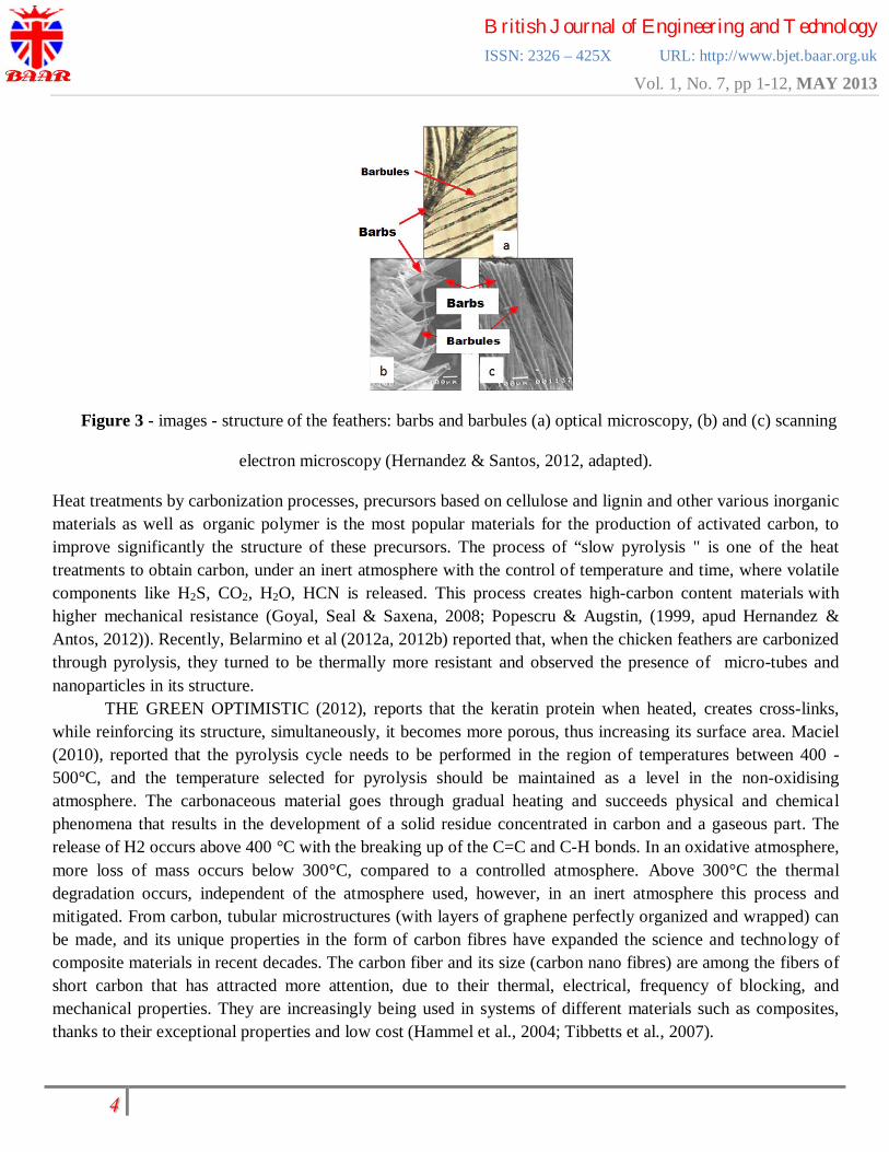
The feathers have a porous internal structure, similar to foam, crystalline segments and hollow and cylindrical microstructure, in nano and micro scale, the reason for its lower density (0.8 g/cm3) and its use as a thermal insulator. In addition, the amino acids of keratin interact between themselves, through hydrogen and disulfide (-S-S-) covalent bonds that give the material mechanical resistance and impermeability to water, as well as insoluble in organic solvents. They are flexible and non-abrasive. The lipids, which have fatty acid groups in their structure, may confer the keratin low solubility in water and thermal insulation. The presence of barbs and barbules in the main structure (rachis) offer a unique structural interaction with other fibres and better adherence with various types of resins. These features make the chicken feathers a promising material for the use as structural reinforcement in polymeric composites and other uses in obtaining stronger materials such as high carbon content. Barbules are seen only by optical and electron microscopy, Figure 3, (Reddy & Yang, 2007; Hernandez & Santos, 2012).
British Journal of Engineering and Technology ISSN: 2326 – 425X URL: http://www.bjet.baar.org.uk
Vol. 1, No. 7, pp 1-12, MAY 2013
৫
Figure 3 - images - structure of the feathers: barbs and barbules (a) optical microscopy, (b) and (c) scanning
electron microscopy (Hernandez & Santos, 2012, adapted).
Heat treatments by carbonization processes, precursors based on cellulose and lignin and other various inorganic materials as well as organic polymer is the most popular materials for the production of activated carbon, to improve significantly the structure of these precursors. The process of “slow pyrolysis " is one of the heat treatments to obtain carbon, under an inert atmosphere with the control of temperature and time, where volatile components like H2S, CO2, H2O, HCN is released. This process creates high-carbon content materials with higher mechanical resistance (Goyal, Seal & Saxena, 2008; Popescru & Augstin, (1999, apud Hernandez & Antos, 2012)). Recently, Belarmino et al (2012a, 2012b) reported that, when the chicken feathers are carbonized through pyrolysis, they turned to be thermally more resistant and observed the presence of micro-tubes and nanoparticles in its structure. THE GREEN OPTIMISTIC (2012), reports that the keratin protein when heated, creates cross-links, while reinforcing its structure, simultaneously, it becomes more porous, thus increasing its surface area. Maciel (2010), reported that the pyrolysis cycle needs to be performed in the region of temperatures between 400 - 500°C, and the temperature selected for pyrolysis should be maintained as a level in the non-oxidising atmosphere. The carbonaceous material goes through gradual heating and succeeds physical and chemical phenomena that results in the development of a solid residue concentrated in carbon and a gaseous part. The release of H2 occurs above 400 °C with the breaking up of the C=C and C-H bonds. In an oxidative atmosphere, more loss of mass occurs below 300°C, compared to a controlled atmosphere. Above 300°C the thermal degradation occurs, independent of the atmosphere used, however, in an inert atmosphere this process and mitigated. From carbon, tubular microstructures (with layers of graphene perfectly organized and wrapped) can be made, and its unique properties in the form of carbon fibres have expanded the science and technology of composite materials in recent decades. The carbon fiber and its size (carbon nano fibres) are among the fibers of short carbon that has attracted more attention, due to their thermal, electrical, frequency of blocking, and mechanical properties. They are increasingly being used in systems of different materials such as composites, thanks to their exceptional properties and low cost (Hammel et al., 2004; Tibbetts et al., 2007).

British Journal of Engineering and Technology ISSN: 2326 – 425X URL: http://www.bjet.baar.org.uk
Vol. 1, No. 7, pp 1-12, MAY 2013
৬
The current research work is related to the areas of high importance such as nanotechnology or environmental decontamination, opening interesting doors in multidisciplinary areas that could take advantage of the high performance that nature confers to the fibrils of the protein keratin. Thus, for a better understanding, the way the fibres from carbonized feathers may be used, and important avail themselves of further studies, in order to validate their characteristics, advantages and restrictions. Therefore, the materials of this proposed study were characterized by using the techniques of SEM/EDS and FTIR, to analyze the microstructures, chemical elements and functional groups present in the KF in natural form and confirm the structural changes in SCF and CF. Materials and Methods Materials The chicken feathers were obtained from a local poultry industry, in the city of Natal-RN, without any cost. Other samples were obtained from using these feathers, according to the methodology of Belarmino et al, 2012a, inspired by TGA analysis on the samples. Initially, the feathers (KF), Figure 4a, were washed in running water and neutral detergent, to remove the dirt on their surface, and dried at 100°C in an oven. The KF was semi-carbonized at 220°C without any atmospheric control, (SCF), Figure 4b. To obtain a higher level of carbonization and to increase the breaking of carbon-carbon bonds, the same were carbonized by pyrolysis (CF), Figure 4c, at a temperature of 220 ºC for 24 hours and with elevation of temperature to 450 ºC in a controlled atmosphere, respecting the rates of cooling and heating. The samples were crushed and stored for analysis and comparison.
Figure 4 - (a) KF, (b) SCFC and (c) CF.
Scanning Electron Microscopy (MEV) and Spectroscopy of Energy Dispersive X-ray (EDS) The micrographs of the samples were taken at a magnification of 250, 600, 800, and 1500 times for a better comparison between the morphologies. For the analysis of EDS, a detector installed in the vacuum chamber, of the equipment, measured the dispersive energy of the X-ray and have been identified, semi-quantitatively, the chemical elements present in the samples. The analyses were carried out using TM 3000 - Tabletop Microscope SEM - EDS.

British Journal of Engineering and Technology ISSN: 2326 – 425X URL: http://www.bjet.baar.org.uk
Vol. 1, No. 7, pp 1-12, MAY 2013
৭
Fourier Transform Infrared Spectroscopy (FTIR) The samples in their solid state were prepared by mixing a certain quantity of each sample with potassium bromide (KBr). This mixture was crushed and compacted at high pressures in order to ensure that it is translucent, forming a disc, through which light can pass through. Then the discs were subjected to radiation energy in the IR region between 400 to 4000 cm-1, using IRPestige-21 SHIMADZU. Results and Discussions Scanning Electron Microscopy (MEV) The KF has an external segmentation of knots and hooks, maintained even after crushing, Figure 5a. Internally, it has a micro-porous structure, better visualized with the whole KF, Figure 5b, as described in the literature. The SFC was brittle and consequently, oxidized, due to its method of obtention, not preserving the knots and hooks, Figure 5c. The CF presented as homogenous shiny black colored particles with irregular dimensions. Surface homogeneity, with preservation of axis and some hooks of KF, overlapping layers and formation of lamina, Figures 5d and 5e.
Figure 5 - electron micrographs of (the) external surface KF crushed, (b) internal structure of the KF whole, (c)
SCFC crushed, (d) and (e) CF.
British Journal of Engineering and Technology ISSN: 2326 – 425X URL: http://www.bjet.baar.org.uk
Vol. 1, No. 7, pp 1-12, MAY 2013
৮
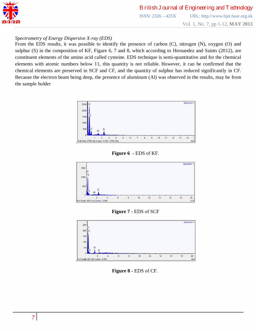
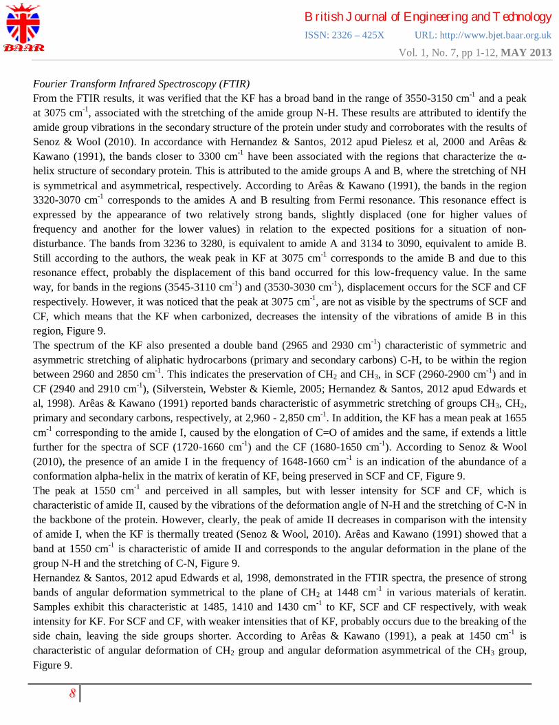
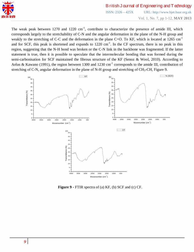
Spectrometry of Energy Dispersive X-ray (EDS) From the EDS results, it was possible to identify the presence of carbon (C), nitrogen (N), oxygen (O) and sulphur (S) in the composition of KF, Figure 6, 7 and 8, which according to Hernandez and Saints (2012), are constituent elements of the amino acid called cysteine. EDS technique is semi-quantitative and for the chemical elements with atomic numbers below 11, this quantity is not reliable. However, it can be confirmed that the chemical elements are preserved in SCF and CF, and the quantity of sulphur has reduced significantly in CF. Because the electron beam being deep, the presence of aluminum (Al) was observed in the results, may be from the sample holder
.
Figure 6 - EDS of KF.
Figure 7 - EDS of SCF
.
Figure 8 - EDS of CF.
British Journal of Engineering and Technology ISSN: 2326 – 425X URL: http://www.bjet.baar.org.uk
Vol. 1, No. 7, pp 1-12, MAY 2013
৯
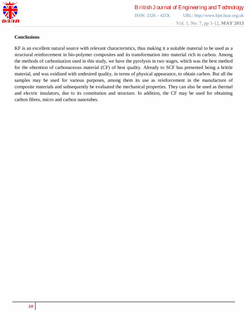
Fourier Transform Infrared Spectroscopy (FTIR) From the FTIR results, it was verified that the KF has a broad band in the range of 3550-3150 cm-1 and a peak at 3075 cm-1, associated with the stretching of the amide group N-H. These results are attributed to identify the amide group vibrations in the secondary structure of the protein under study and corroborates with the results of Senoz & Wool (2010). In accordance with Hernandez & Santos, 2012 apud Pielesz et al, 2000 and Arêas & Kawano (1991), the bands closer to 3300 cm-1 have been associated with the regions that characterize the α-helix structure of secondary protein. This is attributed to the amide groups A and B, where the stretching of NH is symmetrical and asymmetrical, respectively. According to Arêas & Kawano (1991), the bands in the region 3320-3070 cm-1 corresponds to the amides A and B resulting from Fermi resonance. This resonance effect is expressed by the appearance of two relatively strong bands, slightly displaced (one for higher values of frequency and another for the lower values) in relation to the expected positions for a situation of non-disturbance. The bands from 3236 to 3280, is equivalent to amide A and 3134 to 3090, equivalent to amide B. Still according to the authors, the weak peak in KF at 3075 cm-1 corresponds to the amide B and due to this resonance effect, probably the displacement of this band occurred for this low-frequency value. In the same way, for bands in the regions (3545-3110 cm-1) and (3530-3030 cm-1), displacement occurs for the SCF and CF respectively. However, it was noticed that the peak at 3075 cm-1, are not as visible by the spectrums of SCF and CF, which means that the KF when carbonized, decreases the intensity of the vibrations of amide B in this region, Figure 9. The spectrum of the KF also presented a double band (2965 and 2930 cm-1) characteristic of symmetric and asymmetric stretching of aliphatic hydrocarbons (primary and secondary carbons) C-H, to be within the region between 2960 and 2850 cm-1. This indicates the preservation of CH2 and CH3, in SCF (2960-2900 cm-1) and in CF (2940 and 2910 cm-1), (Silverstein, Webster & Kiemle, 2005; Hernandez & Santos, 2012 apud Edwards et al, 1998). Arêas & Kawano (1991) reported bands characteristic of asymmetric stretching of groups CH3, CH2, primary and secondary carbons, respectively, at 2,960 - 2,850 cm-1. In addition, the KF has a mean peak at 1655 cm-1 corresponding to the amide I, caused by the elongation of C=O of amides and the same, if extends a little further for the spectra of SCF (1720-1660 cm-1) and the CF (1680-1650 cm-1). According to Senoz & Wool (2010), the presence of an amide I in the frequency of 1648-1660 cm-1 is an indication of the abundance of a conformation alpha-helix in the matrix of keratin of KF, being preserved in SCF and CF, Figure 9. The peak at 1550 cm-1 and perceived in all samples, but with lesser intensity for SCF and CF, which is characteristic of amide II, caused by the vibrations of the deformation angle of N-H and the stretching of C-N in the backbone of the protein. However, clearly, the peak of amide II decreases in comparison with the intensity of amide I, when the KF is thermally treated (Senoz & Wool, 2010). Arêas and Kawano (1991) showed that a band at 1550 cm-1 is characteristic of amide II and corresponds to the angular deformation in the plane of the group N-H and the stretching of C-N, Figure 9. Hernandez & Santos, 2012 apud Edwards et al, 1998, demonstrated in the FTIR spectra, the presence of strong bands of angular deformation symmetrical to the plane of CH2 at 1448 cm-1 in various materials of keratin. Samples exhibit this characteristic at 1485, 1410 and 1430 cm-1 to KF, SCF and CF respectively, with weak intensity for KF. For SCF and CF, with weaker intensities that of KF, probably occurs due to the breaking of the side chain, leaving the side groups shorter. According to Arêas & Kawano (1991), a peak at 1450 cm-1 is characteristic of angular deformation of CH2 group and angular deformation asymmetrical of the CH3 group, Figure 9.
British Journal of Engineering and Technology ISSN: 2326 – 425X URL: http://www.bjet.baar.org.uk
Vol. 1, No. 7, pp 1-12, MAY 2013
ৰ
The weak peak between 1270 and 1220 cm-1, contribute to characterize the presence of amide III, which corresponds largely to the stretchability of C-N and the angular deformation in the plane of the N-H group and weakly to the stretching of C-C and the deformation in the plane C=O. To KF, which is located at 1265 cm-1 and for SCF, this peak is shortened and expands to 1220 cm-1. In the CF spectrum, there is no peak in this region, suggesting that the N-H bond was broken or the C-N link in the backbone was fragmented. If the latter statement is true, then it is possible to speculate that the intermolecular bonding that was formed during the semi-carbonisation for SCF maintained the fibrous structure of the KF (Senoz & Wool, 2010). According to Arêas & Kawano (1991), the region between 1300 and 1230 cm-1 corresponds to the amide III, contribution of stretching of C-N, angular deformation in the plane of N-H group and stretching of CH3-CH, Figure 9.
4000 3500 3000 2500 2000 1500 1000 5000
5
10
15
20
25
30
35
40
45
50
Tran
smitt
ance
(%)
Wavenumber (cm -1)
KF(a)
4000 3500 3000 2500 2000 1500 1000 5000
5
10
15
20
25
30
35
40
45
50(b)
Tran
smitt
ance
(%)
Wavenumber (cm -1)
% (SCF)
4000 3500 3000 2500 2000 1500 1000 5000
5
10
15
20
25
30
35
40
45
50
Tran
smitt
ance
(%)
Wavenumber (cm -1)
CF(c)
Figure 9 - FTIR spectra of (a) KF, (b) SCF and (c) CF.
British Journal of Engineering and Technology ISSN: 2326 – 425X URL: http://www.bjet.baar.org.uk
Vol. 1, No. 7, pp 1-12, MAY 2013
২১
Conclusions
KF is an excellent natural source with relevant characteristics, thus making it a suitable material to be used as a structural reinforcement in bio-polymer composites and its transformation into material rich in carbon. Among the methods of carbonisation used in this study, we have the pyrolysis in two stages, which was the best method for the obtention of carbonaceous material (CF) of best quality. Already to SCF has presented being a brittle material, and was oxidized with undesired quality, in terms of physical appearance, to obtain carbon. But all the samples may be used for various purposes, among them its use as reinforcement in the manufacture of composite materials and subsequently be evaluated the mechanical properties. They can also be used as thermal and electric insulators, due to its constitution and structure. In addition, the CF may be used for obtaining carbon fibres, micro and carbon nanotubes.

British Journal of Engineering and Technology ISSN: 2326 – 425X URL: http://www.bjet.baar.org.uk
Vol. 1, No. 7, pp 1-12, MAY 2013
২২
Reference:
1. Belarmino, D. D., Ladchumananandasivam, R., Belarmino, L. D., Andrade, S. M. B., Galvão, A. O.,
Ribeiro, L. M. (2012a). Estudo da Estabilidade Térmica de Fibra de Quertina – Keratin Fibre (KF) de
Penas de Frango para Obtenção de Carbono – Carbonised Featehrs (CF), Revista Holos, 3(3), 30-40.
Retrieved from http://www2.ifrn.edu.br/ojs/index.php/HOLOS/issue/view/48
2. Belarmino, D. D., Ladchumananandasivam, R., Belarmino, L. D., Pimentel, J. R. M., Rocha, B. G. R.,
Galvão, A. O., et al (2012b). Physical and Morphological Structure of Chicken Feathers (Keratin
Biofiber) in Natural, Chemically and Thermally Modified Forms, Materials Sciences and Applications.
3(12), 887-893. Retrieved from http://www.scirp.org/journal/PaperInformation.aspx?paperID=25534
3. Senoz, E., & Wool, R. P. (2010). Microporous carbon–nitrogen fibers from keratin fibers by pyrolysis.
Journal of Applied Polymer Science, 118(3), 1752-1765. Retrieved from
http://onlinelibrary.wiley.com/doi/10.1002/app.32397/full
4. Hernandez, A. L. M., Santos, C. V. (2012). Keratin Fibers from Chicken Feathers: Structure and
Advances in Polymer Composites. Nova Science Publishers, 149-211. Retrieved from
https://www.novapublishers.com/catalog/product_info.php?products_id=32840&osCsid=0cb997369f0ca
701f925dc2d1421b6f7
5. Silverstein, R. M., Webster, F. X., Kiemle, D. (2005). Spectrometric Identification of Organic
Compounds. John Wiley & Sons, 7, 512.
6. Arêas, E. P. G., Kawano, Y. (1991). Aplicações Técnicas Espectroscópicas Vibracionais ao Estudo
Conformacioanl de Proteínas. Química Nova. 14, 31-43.
7. Francisco Junior, W. E., Francisco, W. (2006). Proteína: Hidrólise, Precipitação e um Tema para o
Ensino de Química. Química Nova na Escola. 12-16. Retrieved from
http://qnesc.sbq.org.br/online/qnesc24/ccd1.pdf
8. Reddy, N., Yang, Y. (2007). Structure and Properties of Chicken Feather Barbs as Natural Protein
Fibers. Journal of Polymers and the Environment. 81-87. Retrieved from
http://digitalcommons.unl.edu/cgi/viewcontent.cgi?article=1025&context=textiles_facpub
9. Goyal, H. B., Seal, D, Saxena, R. C. (2008). Bio-fuels from thermochemical conversion of renewable
resources: A review, Renewable and Sustainable Energy Reviews. Elsevier. 12(2), 504-517. Retrieved
from http://www.sciencedirect.com/science/article/pii/S1364032106001171

British Journal of Engineering and Technology ISSN: 2326 – 425X URL: http://www.bjet.baar.org.uk
Vol. 1, No. 7, pp 1-12, MAY 2013
২৩
10. Moore, G. R. P., Martelli, S. M., Gandolfo, C. A., Pires, A. T. N., Laurindo, j. B. (2005). Queratina de
Penas de frango: extração, caracterização e obtenção de filmes. Ciênc. Tecnol. Aliment., 26(2), 421-427.
Retrieved from http://www.scielo.br/scielo.php?pid=S0101-
20612006000200027&script=sci_abstract&tlng=pt
11. Hammel, E., Tang. X., Trampert, M., Schmitt, T., Mauthner, K., Eder, A., et al. (2004). Carbon
Nanofibers for Composite Applications. Science direct. 42(5-6), 153-1158. Retrieved from
http://www.sciencedirect.com/science/article/pii/S0008622303006365.
12. Maciel, A. V. (2010). Estudo dos Processos de Redução Car- botérmica de Compostos de Zn, Cd e Sn
Assistidos pela Co-pirólise de Diferentes Biomassas para atenção de Materiais Nanoestruturados. Ph.D.
Thesis, Federal Uni- versity of Minas Gerais, Belo Horizonte. Retrieved from
http://www.bibliotecadigital.ufmg.br/dspace/bitstream/1843/SFSA-
CSNAF/1/tese_qu_mica_adriana_veloso_maciel.pdf
13. Meyers, M. A., Chen, P., Lin, A. Y., Seki, Y., Biological Materials: Structure and Mechanical
Properties. El- sevier. 53 (1), 1-206. Retrieved from
http://www.sciencedirect.com/science/article/pii/S0079642507000254
14. Tibbetts, G.G., Lake, M.L., Strong, K.L., Rice, B.P. (2007). A Review of the Fabrication and Properties
of Vapor-Grown Carbon Nanofiber/Polymer Composites. Composites Science and Technology. 67 (7-8),
1709-1718. Retrieved from http://www.sciencedirect.com/science/article/pii/S0266353806002338>
15. The Green Optimistic. (2009). Carboniizad chicken feathers hydrogen storage. The Green Optimistic.
Retrieved from http://www.greenoptimistic.com/2009/06/25/carbonized-chicken-feathers-hydrogen-
storage/
Acknowledgements The authors of this research are deeply grateful to NUPPRAR and Department of Materials Engineering
of UFRN, for letting us to carry out the analysis.