CHAPTER XV
31
CHAPTER XV EGG, SPERM, FERTILIZATION, AND CLEAVAGE As early as 6 to 10 weeks after setting, young C. virginiea of New England waters, then 6 to 8 mm. in height, develop primordial gonads of pro- fusely branching tubules (Coe, 1932a). At this stage the germinal epithelium is a layer of morpho- logically undifferentiated cells; some of them will transform into larger cells to become ovocytes, Le., the cells destined to develop into mature eggs. The smaller cells of the epithelium proliferate very rapidly and are recognizable as the male germ line, and eventually develop into spermatozoa. For several weeks the immature, or primary, gonad of an oyster remains nonfunctional and bisexual (ambisexual), for it contains both male and fe- male germ cells which will transform into mature spermatozoa or ova during the following summer. In some individuals the primary bisexual gonad is retained until the second year, a delay which Coe (l932a, 1938) attributes to poor nutrition. The more rapid multiplication of male germ cells suppresses the development of ovocytes and results in a predominance of males among the 1-year-old oysters and in the appearance of dif- ferent degrees ofintersexuality (predominance of the cells of one sex over the other). In the same brood which contains also distinctly ambisexual oysters there are, however, other young individuals in which the primary gonad develops directly into ovary or spermary. Local conditions on oyster beds apparently influence the tempo of changes. In the warmer waters at Beaufort, N.C., young oysters are more apt to develop directly into fe- males than in the northern cold waters of New FIGURE of a gonad of C. virginica at an early stage of differentiation. End of March, Woods Hole, Mass. The larger, clear cells are ovogonia, the smaller ones are undifferentiated cells of germinal epithelium. Bouin, hematoxylin-eosin. England. Coe (1938) found that the proportion of females to 100 males varied at the first breeding season between 37.1 and 48.8 at Beaufort; 5.6 and 24 at Milford, Conn.; and 3.3 and 12.5 at New Haven Harbor. The differences are not consistent with geographical latitude since the female to male ratio at West Sayville, Long Island, N.Y., was 31.2; at Delaware Bay 41.9; and at Apalachicola, Fla., 7.1. It is obvious that these variations can- not be attributed to temperature alone and are probably caused by a combined effect of environ- mental conditions. Toward the end of the second breeding season the primary gonad is transformed into a definite ovary or spermary (fig. 291). The gametogenesis, i.e., complete transformation of the primordial germ cells into mature ova (ovogenesis) or sperma- tozoa (spermatogenesis), is a very complex process. The differentiation is accompanied by rapid multi- plication of the new generations of cells which Microns o Page 325 326 327 328 335 338 341 342 343 344 344 351 Ovogenesis _ Spermatogenesis - . . _ Structure of the mature egg _ Cytoplasmic inclusions _ Structure of the mature spermatozoon _ Fertllization . . _ Acrosomal reaction . _ Fertlllzatlon of egg _ Aging of eggs and sperm _ Polarity ofegg _ Cleavage _ Bibliography _ 324 FISHERY BULLETIN: VOLUME 64, CHAPTER XV
-
Upload
khangminh22 -
Category
Documents
-
view
0 -
download
0
Transcript of CHAPTER XV

CHAPTER XV
EGG, SPERM, FERTILIZATION, AND CLEAVAGE
As early as 6 to 10 weeks after setting, youngC. virginiea of New England waters, then 6 to 8mm. in height, develop primordial gonads of profusely branching tubules (Coe, 1932a). At thisstage the germinal epithelium is a layer of morphologically undifferentiated cells; some of them willtransform into larger cells to become ovocytes,Le., the cells destined to develop into mature eggs.The smaller cells of the epithelium proliferate veryrapidly and are recognizable as the male germ line,and eventually develop into spermatozoa. Forseveral weeks the immature, or primary, gonad ofan oyster remains nonfunctional and bisexual(ambisexual), for it contains both male and female germ cells which will transform into maturespermatozoa or ova during the following summer.In some individuals the primary bisexual gonad isretained until the second year, a delay which Coe(l932a, 1938) attributes to poor nutrition.
The more rapid multiplication of male germcells suppresses the development of ovocytes andresults in a predominance of males among the1-year-old oysters and in the appearance of different degrees of intersexuality (predominance of thecells of one sex over the other). In the same broodwhich contains also distinctly ambisexual oystersthere are, however, other young individuals inwhich the primary gonad develops directly intoovary or spermary. Local conditions on oysterbeds apparently influence the tempo of changes.In the warmer waters at Beaufort, N.C., youngoysters are more apt to develop directly into females than in the northern cold waters of New
FIGURE 291.~Sectionof a gonad of C. virginica at an earlystage of differentiation. End of March, Woods Hole,Mass. The larger, clear cells are ovogonia, the smallerones are undifferentiated cells of germinal epithelium.Bouin, hematoxylin-eosin.
England. Coe (1938) found that the proportionof females to 100 males varied at the first breedingseason between 37.1 and 48.8 at Beaufort; 5.6 and24 at Milford, Conn.; and 3.3 and 12.5 at NewHaven Harbor. The differences are not consistentwith geographical latitude since the female to maleratio at West Sayville, Long Island, N.Y., was31.2; at Delaware Bay 41.9; and at Apalachicola,Fla., 7.1. It is obvious that these variations cannot be attributed to temperature alone and areprobably caused by a combined effect of environmental conditions.
Toward the end of the second breeding seasonthe primary gonad is transformed into a definiteovary or spermary (fig. 291). The gametogenesis,i.e., complete transformation of the primordialgerm cells into mature ova (ovogenesis) or spermatozoa (spermatogenesis), is a very complex process.The differentiation is accompanied by rapid multiplication of the new generations of cells which
Micronso
Page
325326327328335338
341342343344344351
Ovogenesis _Spermatogenesis - . . _Structure of the mature egg _Cytoplasmic inclusions _Structure of the mature spermatozoon _Fertllization . . _Acrosomal reaction . _Fertlllzatlon of egg _Aging of eggs and sperm _Polarity of egg _Cleavage ~ _Bibliography _
324 FISHERY BULLETIN: VOLUME 64, CHAPTER XV

1
FIGURE 293.-Portions of two follicles of a bisexual gonadof 4-month-old C. virginica. A-predominantly female,and B-predominantly male follicle; gc-genital canallined with ciliated cells; oc-large ovocyte; ocl-youngovocyte in spireme phase; spcI-primary spermatocytesin spireme phase; spcIJ-secondary spermatocytes;spt-spermatides. Photographically reproduced frOIDCoe, 1932a, fig. 6. Highly magnified.
FIGURE 292.-Follicle wall of the ovary of C. virginica.I-indifferent residual cell; oc-residual ovocyte; ocs-two young ovocytes in synaptic phase; og-group ofovogonia. Redrawn from Coe, 1932a, fig. 9. Highlymagnified.
FIGURE 294.-Two young ovocytes at spireme stage in amature ovary of C. virginica. Redrawn from Coe, 1932a,fig. 9. Highly magnified.
OVOGENESIS
extend inward and fill up the lumen of the follicles.Early at this stage the sex cells become dense andopaque, a condition which interferes with cytological study.
Gametogenesis of G. virginica and O. lurida hasbeen studied by Coe (1932a, 1932b, 1934, 1936,1938) and that of the Australian rock oyster,G. commercialis, by Cleland (1947). Only themain points of this process were disclosed by theseinvestigations.
In all animals the primordial germ cells becomedistinguishable as primary ovogonia in the femalesor spermatogonia in the males after a certain number of divisions. After a period of quiescence theybegin to divide again and give rise to secondaryovogonia or spermatogonia. After several generations the cells stop dividing and enter a growthperiod, which is more prolonged in the females thanin the males. The growth period is characterizedby a series of cytological changes, each differingfrom the preceding stage. The cells which will produce gametes are at this stage called auxocytes,from the Greek "auxesis" meaning growth, and arereferred to as ovocytes in the female and spermatocytes in the male.
Ovogenesis in oysters begins with the appearanceof enlarged cells in the germinal epithelium. Theseare the ovogonia, which in G. virginica and O.lurida are distinguished from other cells of thegerminal epithelium by their relatively large nucleiwith conspicuous nucleoli and loose chromatinnetwork (fig. 292, og). The ovogonia usually lienext to the follicle wall, and their distal sides donot protrude into the lumen. Differences betweenthe early ovogonia and indifferent residual cells (I)are not conspicuous. Examination of a series ofsections and study of the sequence of changes in theappearance and structure of the cells are necessaryto assure a positive identification.
After one or two divisions the ovogonia changein appearance as well as in size. This generation offemale sex cells called ovocytes can be recognizedby the presence of fibrillar mitochondrial bodies(sometimes called yolk nuclei), and by the spiremesof densely packed chromosomes (figs. 293 and 294).Their nucleoli become very conspicuous.
During the last stage of ovogenesis the ovocytebegins to grow rapidly, and the distal part, grosslyenlarged and rounded, protrudes into the lumenof a follicle. At the same time the connection
EGG, SPERM, FERTILIZATION, AND CLEAVAGE 325

with the basal membrane of the follicle wall isnarrowed to an elongated stem. The nucleusincreases greatly in bulk, and the developing eggassumes a pear-shaped form. An accumulationof dark granules (mitochondria) at the proximalend of the cells may indicate that food for thegrowing ovocyte is obtained through the wall ofthe follicle. The granules are not pronounced inthe ovocyte of O. virginica but are conspicuous insome other bivalves, particularly in Sphaerium(Woods, 1932). From the beginning of sexualdifferentiation to the final maturity of an ovum,the early ovocyte increases in volume more than3,000 times.
Ovogenesis in the Sydney rock oyster O. commerciaZis, described by Cleland (1947), is somewhat different ftom the ovogenesis of the Americanspecies. At the earliest stage before the start ofthe growth phase an ovocyte of the rock oysteris a small cell, 4 J.L to 5 J.L in diameter. Two-thirdsof the cell is occupied by a clump of chromosomessurrounded by a rim of clear cytoplasm. Clelandidentifies this stage as a definite auxocyte, i.e.,an ovocyte just before entry into the growthphase. At ths stage the cell has no nucleolus.
The definite auxocyte begins to grow and passesthrough three stages (called by Cleland AuxocyteI II and III) which differ in size and nuclear, ,structure. Auxocyte I has a diameter of about9 J.L, with a relatively large germinal vesicle (6 J.L)
and a nucleolus of about 1.7 J.L. The nucleus iscentrally placed in the homogeneous cytoplasmwith an excentric nucleolus which is not in contact with the nuclear membrane. Auxocyte IIhas diameter of 12.6 J.L, with the germinal vesicle(nucleus) about 7 J.L and nucleolus 2 J.L to 3 J.L.
The cell is usually spherical with a centrallylocated germinal vesicle and chromosomes spacedmore widely than in Auxocyte 1. At this stagea group of granules appears at one pole of thenucleus. Auxocyte III is a spherical cell 20 J.L indiameter, with the germinal vesicle measuring 11J.L and eccentrically located nucleolus of about4.2 J.L in diameter. The cell is separated from thewall and is free in the lumen of a follicle. Protein granules are abundant along the peripheryof the cell where they are found in a mature egg.The mature ovocyte of O. commerciaZis has adiameter of about 38 J.L. The nucleus is large,about 21 J.L across; the nucleolus is 4.6 J.L. Thechromosomes are paired and are usually placedperipherally in the germinal vesicle but not in
326
contact with the nuclear membrane. Crossingover is frequently seen at this stage but the chromosomes are not coiled.
SPERMATOGENESIS
Spermatogenesis in the oyster is known primarily from the studies by Coe (1931) on thedevelopment of the gonad of young O. Zurida.Comparison with the gonads of O. virginica showsthat there is close agreement in the general featuresof the process in both species. Progressive stagesof the formation of sperm begin with the undifferentiated gonia which line the inner wall of thegonad follicles. After a large number of descendants have been produced the spermatogonia canbe distinguished from the ovogoniaby their smallersize and position within the follicles. Ovogonialie in a single row along the wall; the primaryspermatogonia of C. virginica are found eithersingly or in groups between the ovogonia liningthe wall and in the lumen (fig. 295).
In the hermaphroditic gonad of O. Zurida asingle primary spermatogonium divides severaltimes to form a cluster of cells which becomeseparated from the follicle wall and occupy aposition toward the center of the lumen (fig. 296).
The number of divisions of spermatogoniapresumably depends on the amount of nourishmentavailable to the gonad. Coe estimates that in O.Zurida each primary spermatogonium divides sixto nine times to produce a cluster of 64 to 500cells. In spite of the close contact the adjacentcells of the clusters are separate but are heldtogether by a delicate noncellular secretion. Its
FIGURE 295.-Portion of bisexual gonad of young C.virginica. gc-genital canal; oc-ovocytes with spermatogonia filling the lumen; spc-spermatocytes; sptspermatids; spz-spermatozoa. Photographically reproduced from Ooe, 1934, fig. 5A. Highly magnified.
FISH AND WILDLIFE SERVICE

FIGURE 296.-Portion of an hermaphroditic gonad of O.lurida. go-genital canal; I-indifferent cells; ocovocytes; spc-primary spermatocytes; spc1-secondaryspermatocytes; spz-spermatozoa united into a spermball. Photographically reproduced from Coe, 1934, fig.5B. Highly magnified.
chemical nature has not yet been determined. Inpoorly preserved preparations the clusters sometimes have the appearance of syncytia with nucleiembedded in a common matrix. In both speciesall spermatogonia have a conspicuous nucleolusand loose chromatin reticulum. As the divisionsproceed the diameter of the spermatogonia diminishes from about 6p, to 3p, or less at the laststage leading to the formation of primary spermatocytes. In C. virginica these cells are globular,each with a large nucleus resolved into slenderthreads (spiremes). This leptonema stage isfrequently observed in the developing spermarybut the conjugation of chromosomes (synapsis)has not been described with any detail. However,in reference to the spermatogenesis in O. lurida,Coe (1931) states that the leptotene stage "isfollowed by the usual process of synapsis."
The appearance of secondary spermatocytes issimilar to that of the primary. In C. virginicathey can be distinguished by their radial orientation in the lumen and small size. Different phasesof spermatogenesis in C. virginica are shown in asemidiagrammatic drawing published by Coe(l932a) and reproduced in fig. 297. l\1eioticdivisions and the transformation of spermatidsinto mature spermatozoa have not been fullydescribed for C. virginica. In a mature spermarythe spermatozoa are always oriented with theirtails toward the center of the lumen. The photomicrograph shown in fig. 298 shows the gradualincrease in the number of male sex cells from the
EGG, SPERM, FERTILIZATION, AND CLEAVAGE
wall of the follicle toward the center. Successivestages of the spermatogenesis of O. lurida drawnby Coe are shown in fig. 299.
Secondary spermatocytes of O. lurida are heldtogether in spherical masses. Close contact bythe spermatids continues during their transformation into spermatozoa; in the sperm ball of a mature oyster the tails radiate from the center. Eachsperm ball is composed of from 200 to 2,000spermatozoa originating from a single spermatogonium (Coe, 1932b). During mitotic divisionsthe "prophase, metaphase, and telophase are all oftypical appearance, with a delicate spindle of theusual form" (Coe, 1931). Because of the crowdedcondition of the metaphase and anaphase plates,Coe was unable to determine the chromosomenumber which he states "is not very large." Intwo diagrammatic drawings of spermatocyte division Coe (1931, fig. 3, E and F) figures 10 chromosome pairs. This is the most common number ofchromosomes found by Cleland at the two- andfour-cell stage of cleavage in the fertilized egg ofC. commercialis (Cleland, 1947). The number ofchromosomes seen during the cleavage of C.virginica eggs is discussed later (p. 345).
STRUCTURE OF THE MATURE EGG
Eggs in the mature ovary of C. virginica arepear-shaped and compressed. Many of them areattached to the follicle wall by long, slenderpeduncles; others are free in the lumen ready tobe moved to the genital canals and discharged(fig. 300). The long axis of the eggs varies from55p, to 75p, depending on their shape; the widthat the broadest part measures from 35p, to 55p"and the diameter of the nucleus is from 25p, to 40p,.The oblong shape is retained for some time afterthe discharge of eggs into water but gradually theegg becomes globular and denser. Under thetransmitted light of a microscope the nucleusappears as a large, transparent area surroundedby densely packed granules (fig. 301). In aglobular egg the nucleus cannot be seen unless itis cleared in glycerol or other clarifying reagents(fig. 302).
Eggs of oysters living under marginal conditionsin water of salinity less than 10 0/00 frequentlybecome cytolyzed upon their removal from theovary; the nuclei appear larger than those ofnormal eggs. Only 1 or 2 percent of these eggsis fertilizable. The delicate primary or "vitelline"membrane surrounding the unfertilized egg is
327

/nd.-~·
,1---- sfC ;[1"""___"
,1---- 5 pc 2.
\~..llJ---- spt___ spz
FIGURE 297.-Mature spermary of C. virginica. ind-indifferent cells; spgl and spg2-primary and secondary spermatocytes; spt-sp«:lrmatids; spz-mature spermatozoa. Redrawn from Coe, 1932a, fig. 8.
secreted by the egg itself while it is still in theovary. Raven (1958) states that in some casesthe vitelline membranes of Ostrea, Mytilus,Dreissensia, and Dentalium are thrown off soonafter shedding. I have not seen this happen inthe eggs of C. virginica.
CYTOPLASMIC INCLUSIONS
Cytoplasmic components of an oyster egg arenot well known primarily because the ultrastructure has not been studied by electron microscopy.Certain types of minute granules can be seen,however, in examination of live eggs under high
328
magnification of phase contrast lenses; by applyingvital and metachromatic stains; by centrifugingwhole eggs or their homogenates in order to separate various components and study their stainingreactions. For descriptions of the techniquesused in modern cytology the reader is referred tothe textbooks on cytology and microscopic histochemistry (Gomori, 1952; DeRobertis, Nowinski,and Saez, 1960; and others). The yolk constitutesthe major part of the eggs of marine bivalves.Quantitative data on the amount of yolk in oystereggs are lacking, but for Cumingia tellinoides and1IJytilus californianus Costello (1939) found that
FISH AND WILDLIFE SERVICE

"
'.
.:o!,'-'----'----'1...---'--.1' ,
Microns 50, •\
,.I
FIGURE 298.-Photomicrograph of a crOSS section of one follicle of a fully mature spermary of C. virginica. Hematoxyuneosin.
yolk forms 35 and 31 percent respectively of thetotal egg volume. The estimates were obtainedafter a centrirugal force of 20,000 (Oumingia) and4,800 (Mytilus) times gravity had been appliedto the unfertilized eggs. In the cytoplasm of theeggs of the two species the relative volumes ofhyaline zone were 42 and 55 percent and of tbeoil 10 and 14 percent.
The yolk or molluscan eggs is made of two
EGG, SPERM, FERTILIZATION, AND CLEAVAGE
types of granules, one of proteid and the other offatty materials. In cytological literature thedistinction between the proteid yolk and fattyyolk is not always made clear. In descriptions ofthe cytoplasmic inclusions of an egg based onlight microscopy some authors apply the termexclusively to protein granules, while others, including Gatenby (1919), Gatenby and Woodger(1920), and Brambell (1924) in their studies of the
329

cB
o
~:\ii
.0 0 ,'-./
o
F G H
T
FIGURE 299.-Diagram of successive stages of spermatogenesis in O. lurida. A-two indifferent germ cells on the wallof the gonad; B-small group of spermatogonia,with reticular chromatin and conspicuous nucleoli; C-small groupof secondary spermatogonia; D-division of secondary spermatogonia to form spermatocytes; E-primary spermatocytes with slender chromosomes; F to K-division of primary spermatocytes; L to R-division of secondary spermatocyte; S to T-transformation of spermatid into the mature spermatozoon. Redrawn from Coe, 1931, fig. 2.
gametogenesis in the gastropods Helix, Limnaea,and Patella, identify the protein granules as"mitochondria" and restrict the term yolk to fattyinclusions.
The role of the mitochondria in the formation ofyolk has not been fully resolved. According toRai (1930), the fatty yolk in the eggs of C. cucullatais formed directly from the Golgi vesicles as it isin ascidians, Helix, and other invertebrates;mitochondria do not participate in the vitellogenesis, and albuminous yolk is absent in the egg ofthe Indian oyster (C. cucl1llata). This view is inagreement with the conclusion of Worley (1944),who found no protein yolk in the eggs of Mytilusand Ostrea. The question is not settled becauseapparently the cytologists have no clear agreementon the difference between the protein yolk andmitochondria.
A study of cytoplasmic inclusions was made byCleland (1947, 1951), who separated the granulesfound in egg cytoplasm of the oyster by differential centrifugation following homogenization.Mature eggs were suspended in a solution of0.2 M potassium cWoride and 0.02 N sodium citrate
330
buffered to pH 7.5. Homogenates were obtainedby blending the suspension in an electric blendersurrounded by an ice jacket. By centrifugingthe samples of homogenates at different speeds thefollowing types of granules were obtained: P granules or protein yolk; L granules or lipid yolk;M granules or mitochondria; and S or submicroscopic granules. The P granules obtained byCleland's technique are spherical and can bestained by Janus green B in the test tube. In theliving egg these granules are located along theperiphery and absorb Nile blue stain. Afterbeing centrifuged at 5,000 times gravity for 5minutes they form a thick centrifugal layer witha sharp upper boundary. Alpha or lipid yolkgranules are also spherical. They occupy thecentral part of the living egg. In the centrifugedegg they form a centripetal layer with a sharplower boundary. Alpha granules of phospholipidand neutral fat can be recovered from the supernatants of homogenate suspensions. M granulesor mitochondria in the live centrifuged egg forma thin, rather loose layer above the P granulesand stain both with Janus green B and Nile blue.
FISH AND WILDLIFE SERVICE

o Microns 40
,. 1
F1GURE 300.-Photornicrograph of eggs in the follicles of the ovary of C. virginica at the beginning of the spawning season.Ovocytes and small indifferent cells line the wall; mature eggs arc either free or connected to the wall with longpeduncles. Kahle, hematoxylin-eosin.
Cleland states that in the uncentrifuged matureegg they are unrecognizable. From homo~enat.es
the M granules can be separated by centrifugingfor 10 minutes at 10,000 times gravity. Cytoplasm also contains submicroscopic or S granules(according to Cleland's terminology), which areseparable by applying a centrifugal force of20,000 times gravity for 30 minutes. These Sgranules are probably homologous to mammalianmicrosomes, i.e., the submicroscopic ribonucleoprotein particles wbich are considered to be themajor sites of protein synthesis (a discussion ofthis problem is found in Shaver, 1957, andNovikoff, 1961b).
With the exception of pure lipid granules, thecytoplasmic components of the egg of C. commercialis show an increasing content of nucleicacid with decreasing size of granules, the groundcytoplasm containing the highest concentration
EGG, SPERM, FERTILIZATION, AND CLEAVAGE
of nucleic acid. Cleland's observations need tobe corroborated, using the eggs of different speciesof oysters.
The formation and composition of yolk in theeggs of animals other than bivalves have beenstudied by many investigators, frequently withdifferent and sometimes contradictory results.As Brachet (1944) stated nearly 20 years ago, theproblem cannot be resolved at present. Thisuncertainty about yolk and other granules stillpersists and probably will continue until theultrastructure of the marine egg is thoroughlyexplored by electron microscopy.
Examination with the light microscope of ripe,unfertilized, and unstained eggs of C. virginicadiscloses a multitude of tightly packed minutegranules in the cytoplasm which obscure the innerportion of the egg. The granules appear to beuniformly distributed around the nucleus (fig.
331
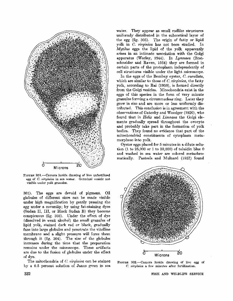
FIGURE 30l.-Camera lucida drawing of live unfertilizedegg of C. virginica in sea water. Germinal vesicle notvisible under yolk granules.
301). The eggs are devoid of pigment. Oilglobules of different sizes can be made visibleunder high magnification by gently pressing theegg under a coverslip; by using fat-staining dyes(Sudan II, III, or Black Sudan B) they becomeconspicuous (fig. 303). Under the effect of dye(dissolved in weak alcohol) the small granules oflipid yolk, stained dark red or black, graduallyfuse into large globules and penetrate the vitellinemembrane and a slight pressure will force themthrough it (fig. 304). The size of the globulesincreases during the time that the preparationremains under the microscope. These artifactsare due to the fusion of globules under the effectof dye.
The mitochondria of O. virginica can be stainedby a 0.5 percent solution of Janus green in sea
20Micronso
water. They appear as small rodlike structuresuniformly distributed in the subcortical layer ofthe egg (fig. 305). The origin of fatty or lipidyolk in O. virginica has not been studied. InMytilu8 eggs the lipid of the yolk apparentlyarises in an intimate association with the Golgiapparatus (Worley, 1944). In Lymnaea (Bretschneider and Raven, 1954) they are formed incertain parts of the protoplasm independently ofcell structures visible under the light microscope.
In the eggs of the Bombay oyster, O. c'UCullata,which are similar to those of O. virginica, the fattyyolk, according to Rai (1930), is formed directlyfrom the Golgi vesicles. Mitochondria exist in theeggs of this species in the form of very minutegranules forming a circumnuclear ring. Later theygrow in size and are more or less uniformly distributed. This conclusion is in agreement with theobservations of Gatenby and Woodger (1920), whofound that in Helix and Limnaea the Golgi elements gradually spread throughout the ovocyteand probably take part in the formation of yolkbodies. They found no evidence that part of themitochondrial constituents of cytoplasm metamorphose into yolk.
Oyster eggs placed for 5 minutes in a dilute solution (1 to 25,000 or 1 to 30,000) of toluidin blue 0and washed in sea water are colored metachromatically. Pasteels and Mulnard (1957) found
FIGURE 302.-Camera lucida drawing of live egg ofC. virginica a few minutes after fertilization.
Micronso
332 FISH AND WILDLIFE SERVICE

•••
•~. '. . . .
•
FIGURE 303.-0il globules in the unfertilized egg of C.virginica after staining in Sudan III. Whole mount.Drawing made from a photomicrograph.
FIGURE 304.-Unfertilized egg of C. virginica stained withBlack Sudan B; slight pressure on a coverslip forcesthe oil globules through the vitalline membrane. Drawnfrom life.
o Microns40
oMicrons
40
that the development of eggs of the Portugueseoyster, C. angulata, is not affected by toluidin blueused in such dilute solution for only a short time.The dye is fixed at the level of the small granules,which the cytologists designate as alpha granules,uniformly distributed in the cytoplasm betweenthe yolk vesicles. Later in the development of afertilized egg, new and larger granules, called betagranules, appear at the time of prophase. Theirhigher metachromasy is acquired at the expenseof the alpha granules. Subsequent studies (Mulnard, Auclair, and Marsland, 1959) have suggestedthat the beta granules are related to the Golgicomplexes of the eggs.
The alpha granules of the unfertilized eggs of C.angulata can be separated from the yolk vesicles bycentrifuging; they are displaced in the direction ofthe centrifugal force (Pasteels and Mulnard, 1957),while the beta granules at the pronucleus stage ofthe fertilized egg are moved in the centripetal
EGG, SPERM, FERTILIZATION, AND CLEAVAGE
1133-851 0-64--22
direction. The alpha and beta particles of Pasteelsand Mulnard probably correspond to the P and Lgranules of Cleland. Personal observations showthat in ripe but unfertilized eggs of New EnglandC. virginica stained with toluidin blue, elementscorresponding to the alpha particles of Pasteelsassume a lavender color while mitochondria andother smaller granules are bluish. The nucleolusis also of bluish color. After 10 minutes of centrifuging at 4,000 times gravity the yolk granules ofthe stained eggs concentrate at the lower (centrifugal) pole, while the alpha particles of lavenderhue and slightly bluish mitrochondria are at theopposite pole (fig. 306).
Metachromatic granules have been described inthe eggs of various bivalves. They were foundin Barnea candida (Pasteels and Mulnard, 1957);Mactra (Kostanecki, 1904, 1908; Mercenaria(Venus) mercenaria, Mytilus edulis, and Spisulasolidissima (Worley, 1944; Kelly, 1954, 1956;
333

-.••
••
-
•o Microns
•
-/
40
FIGURE 305.-Photomicrograph of an unfertilized egg of C. virginica. Janus green. Whole mount. Small rodlikeinclusions seen along the periphery are mitochondria stained green; larger yolk granules arc dark.
Allen, 1953; and Rebhun, 1960). The role ofthese almost submicroscopical bodies is not clear,but there is no doubt of their importance in tbephysiology and development of eggs. Recentpublications of Dalcq (1960), Brachet (1960), andRebhun (1960), and the reviews given by Novikoff(1961a, 1961b) should be consulted for ideas concerning the poss.ible role of these elements in themorphogenesis of mosa.ic eggs in which they areconcentrated in the posterior blastomeres.
334
Eggs of the surf clam S. solidissima contain aheparinlike blood anticoagulant which was alsoextracted from the tissues of this clam (Thomas,1954). Wbether substances with similar activityare present in oyster eggs is not known.
Tbe nuclms of a mature egg is surr.Qunded by anuclear membrane which can be clearly seen onsectioned and stained preparations of the ovary(fig. 300). A spherical, dense nucleolus is excentrically located; its diameter varies from 4 I' to 6 1'.
FISH AND WILDLIFE SERVICE

I I
OMicrons 30
FIGURE 307.-0vocyte of O. laperQusi stained with methylgreen and pyronin B after Navashin fixation. DarkgloDule is the karyosome, light shaded area is theplasmosome. From Kobayashi, 1959.
shown in solid black in fig. 307, and the plasmosome, lightly shaded, in close contact with eachother. The definite KP axis (Karyosome-Plasmosome) of the unfertilized egg changes after fertilization by the turning of the karyosome aroundthe plasmosome. The existence of this axis in theeggs of other species of oysters has not beendescribed.
STRUCTURE OF THE MATURESPERMATOZOON
The spermatozoon of bivalve mullusks appearsunder the microscope (Franzen, 1956; Lenhossek,1898; Retzius, 1904) to consist of an oval orround head with a pointed front, a middle pieceat the lower end of the head, and a long tail(flagellum) with a narrow "end piece" which islonger than the width of the head. The middlepiece consists of four, sometimes five, oval-shapedbodies clustered around the tail; and a minute"central granule" or centriole located in the center at the point of attachment of the tail. Thesharply outlined oval bodies are mitochondria;they are strongly osmiophilic and can be deeplystained with rosanilin. The head of the spermatozoon is formed by a compact nucleus cappedwith the apical body or acrosome with a pointedtip (perforatorium), which apparently assists thespermatozoa in penetrating the egg membrane atfertilization (Wilson, 1928). The features listedabove may be seen in properly fixed and stainedpreparations of the sperm of G. virginica and inlive spermatozoa examined with phase contrastoil immersion lenses. In live preparations thenucleus appears to be dark while the acrosomeand middle piece are light (fig. 308). The centerof the spermatozoon head is occupied by an axialbody, a relatively large, light-refracting structurewhich is separated from the acrosome.
The dimensions of normal, uncytolyzed spermatozoa have been measured by means of an eyepiecemicrometer of a light microscope. The head variesfrom 1.9 ~ to 3.6 ~ in length (median value 2.7 ~)
and between 1.0 ~ and 2 ~ in width. The tail isfrom 27 ~ to 39 ~ long (median value 36 ~). Thetails are usually slightly curved; specimens withstraight tails are rarely found.
Electron microscopy reveals much greater complexity in the structure of the spermatozoon(Galtsoff and Philpott, 1960). Study was madeof small sections of ripe spermary preserved incold 1 percent osmium tetroxide buffered to pH
40Microns
oFIGURE 306.-Unfertilized egg of C. virginica centrifuged
for 10 minutes at 4,000 times gravity after staining withtoluidin b:lue. Dark yolk granules are at the lower(centrifugal) pole while th"e mass of small inclusions consisting of lavender particles and bluish mitochondria areat the opposite end of the egg.
The chromosomes appear as fine threadlike structures near the nuclear membrane. Using methylgreen and pyronin B stains, Kobayashi (1959)found that the nucleolus in the eggs of G. gigas andO. laperousi consists of two parts, the karyosome,
EGG, SPERM, FERTILIZATION, AND CLEAVAGE 335

Microns
FIGURE 308.-Live spermatozoon of C. Virginica examinedunder light microscope with phase contrast oil immE'rsionlens. A-acrosome; ax. b.-axial body; G-centriole:e.p.-end piece of tail; h.-head; m.p.-middle piece;mt.-mitochondrial bodies; N-nucleus; t.-tail.
(Philpott, 1955), using a diamond knife. Sincethe preserved pieces consisted of a multitude ofspermatozoa arranged in a central mass with theirtails pointing outward, the individual spermatozoawere always cut at random in different plll,nes,regardless of how the embedded tissue may havebeen oriented on a microtome block. The resulting electron micrographs showed a number ofsperm heads cut at different planes and manytransverse sections of tails (fig. 309). The entirestructure of the head was diagrammatically reconstructed by bringing various elements togetherand placing them in their relative positions (fig.310). The oval-shaped head consists of slightlygranular, homogeneous material covered with anosmiophilic membrane made of two layers. Theapical portion of the nucleus is occupied by acaplike acrosome of highly osmiophilic substance.The acrosome is clearly separated from the nucleusby a sharply defined membrane. An egg-shapedbody in the central part of the nucleus extendsfrom the apex of the acrosome almost to the baseof the nucleus. This structure, named axial body(Galtsoff and Philpott, 1960), has a central stemof fibrous material which emerges from theflattened bottom and extends about two-thirds ofthe total length of the axial body. The indentedbase of the nucleus is near the base of the axialbody. The caved..·in space formed by this indentation consists of material of lesser electrondensity and extends under the nucleus to theupper surface of the centriole, which is surroundedby four mitochondrial bodies. Only two of themare shown in fig. 310.
The centriole of the sperm of O. virginica is ahollow, cylindrical structure with walls made ofnine bands; these can be seen in cross section (fig.311). The side view (fig. 310) shows that thecentriole is formed in several alternating andslightly constricted layers which connect with thefour mitochondrial bodies. These bodies havethe typical appearance of twisted lamallae enclosedin a membrane which encompasses the centrioleand continues over the tail.
The tail consists of a pair of axial filamentssurronnded by a ring of nine double filamentsspaced at equal intervals along the periphery(fig. 312). The filaments are interconnected bydelicate strands. The axial filaments begin nearthe basal plate (fig. 310) where the tail emerges.Radial trabeculae connect the ring filaments tothe outside wall of the tail and form nine separate
t.
h.
m.p.
e.p.
a.ax. b.---,..".".
n.--II....c.
m.b.
7.2-7.4 and embedded in a mixture of three partsbutyl and one part methyl methacrylate. Toincrease the contrast, some of the material wasplaced in 1 percent alcoholic phosphotungstic acidfor 5 to 6 hours before embedding. The embeddedmaterial was sectioned on Philpott's microtome
336 FISH AND WILDLIFE SERVICE

a Microns
FIGURE 309.-Electron micrograph of II section of ripe spermary of C. v1'rginica. Longitudinal and transverse sections ofsperm heads and tails can be seen in various parts of the micrograph. A-acrosome; ax. b.-axial body; c.t.-crosssection of tails; I.s.-Iongitudinal section of sperm head; l.t.-Iongitudinal section of part of a tail; m.b.-mitochondrial bodies; 1\-nucleus; t.s.-transverse section of head.
EGG, SPERM, FERTILIZATION, AND CLEAVAGE 337

t.
;'±-ri~~~IfM--a X. b.
·~~~~~I+-c.ax. b.
~/ll.1~~-m.b.
~~?;\\\- b·P!
eHt----- d.f.I~II---ax.f.
1.0 fL--~..1
chances of fertilization are increased. Becausespawning is usually initiated by the males, thewater into which the eggs are discharged alreadycontains active spermatozoa and fertilization takesplace within a few minutes following ovulation.It is obvious that the success of reproduction ofan oyster population in which spawning is mutuallystimulated by the discharge of sex cells is dependent on close proximity of the sexes and theirsimultaneous response to spawning stimuli.
Eggs and sperm secrete substances calledgamones which play an important role in fertilization. Secretion from an unfertilized egg has asignificant effect on spermatozoa. This effect canbe observed if a suspension of eggs is permitted tostand for 15 to 20 minutes and the supernatantfluid is decanted or filtered and added to the suspension of sperm. The resulting so-called "eggwater" (Lillie, 1919) causes the agglutination ofsperm. To observe the agglutination reactionwith the naked eye, a drop of egg water must beadded to a sperm suspension, which shortly formsirregular lumps (fig. 313). Under a high-powerlight microscope one sees that the heads of agglutinated spermatozoa stick together to form largeaggregates (fig. 314).
FIGURE 31O.-Reconstruction of the sperm head of C.virginica made from a large number of longitudinalsections. a.-acrosome; ax. b.-axial body; ax. f.axial filament of the tail; b.p.-basal plate of the tail;c.-centriole; c. ax. b.-core of the axial body; d.f.double filament on the periphery of the tail; m.b.mitochondrial body; n.-nucleus; t.-proximal part ofthe tail.
compartments filled with material of lesser electrondensity. The central radial strands are similarto the "spokes" described by Afzelius (1955) forthe sperm of the sea urchin Psammechinus miliaris.They are not present in the proximal portion ofthe tail where there are no axial filaments, butotherwise the ultrastructure of the sperm tail issimilar to that of cilia and flagella of variousanimals and plants.
FIGURE 31l.-Drawing based on electron micrographsof the cross section of the lower part of the middlepiece of the spermatozoon of C. virginica. Centriole,at the center, is surrounded by four mitochondrialbodies.
FERTILIZATION
The spawned eggs of O. virginica and O. gigasare heavier than water and quickly sink to thebottom. The time they remain in suspension maybe prolonged by horizontal currents and upwardmovements of the water, and consequently the
o Micron
338 FISH AND WILDLIFE SERVICE

Micron
FIGURE 312.-Transverse section of the tails of oyster sperm of C. virg1"nica slightly below the level of the middle piece.Electron micrograph.
EGG, SPERM, FERTILIZATION, AND CLEAVAGE 339

6Cent i meters
FIGURE 313.-Sperm suspension of C. virginica on a slidein sea water (left) and in sea water containing a smallquantity of egg water (right). Drawn from life.Natural size.
Agglutination also occurs in the sperm of C.gigas and in O. circumpicta Pilsbry. Terao (1927)experimented with the latter species using eggwater made by mixing 0.55 m!. of ripe eggs in 9 m!.of sea water and removini( the eggs by centrifuging and filtering after they had stood for 20minutes. The filtrate caused the isoagglutinationof sperm even in a dilution of 1 to 10 millions.Heteroagglutination by the egg water of O.
circumpicta has been observed in the suspensionsof sperm of the bivalve Area, sea urchin Toxocidaris tuberculatus, and starfish Luidia quinaria.
Lillie (1919) regarded the sperm agglutinatingfactor he discovered in Arbacia eggs as an essentialto fertilization, and to the active substance of eggwater he gave the name fertilizin. Tyler (1948)identified fertilizin with the jelly substance of theegg and on the basis of experimental data concluded that the presence of the jelly coat has afavorable effect on fertilization. By biochemicalanalysis of sea urchin eggs, Vasseur (1948a, 1948b)determined the composition of the jelly coat andfound that 80 percent of it consists of polysaccharide and 20 percent of amino acids. The substance was found to exert a heparinlike action in ablood-clotting system (Immel'S and Vasseur, 1949).After removing the jelly coat with acidified water,Hagstrom (1956a, 1956b, 1956c, 1956d) foundthat the rate of fertilization was higher than in thepresence of the coat. It is, therefore, apparentthat the jelly coat is not essential for fertilization.
o Microns
FiGURE 314.-Photomicrograph of sperm of C. virginica agglutinated by egg ,vater of the same species. Phase contrastoil immersion.
340 FISH AND WILDLIFE SERVICE

FIGURE 315.-Diagrammatic drawing of acrosomal reactionin the spermatozoon of C. virginica produced by eggwater. Only a part of the sperm tail is shown in thedrawing. Drawn from live preparation.
some membrane bursts, and the filament isdischarged. The reaction can be observed whena small drop of live sperm suspension is placed ona cover slip, a minute quantity of egg water isadded, and the cover slip then inverted on a slide.The preparation is examined with phase contrastoil immersion lens using anisol (Crown oil) ofrefractive index 1.515 instead of cedar oil.
The acrosome reaction of O. virginica is similarto that observed by Dan and Wada in three otherspecies of oysters. Under the effect of egg waterthe head becomes swollen and rounded and thefilament is ejected from the acrosome. The discharged filament is wider than the tail and isabout three to four times longer than the length of thehead (fig. 315). In my observations only a smallnumber of oyster spermatozoa suspended in eggwater discharged acrosomal filaments.
The exact role of the filament in the fertilizationof oyster eggs is still unknown. Investiga.tions
This view agrees with the conditions found inoyster eggs which have no jelly coat.
According to the modern view discussed in thereview of the problem by Runnstrom, Hagstrom,and Perlmann (1959), the jelly coat not only failsto improve fertilization but impedes it by actingas a sieve. Its action may be considered as anelimination process by which the number ofspermatozoa capable of attaching to the cytoplasmic surface is substantially reduced.
Fertilizin of sea urchin eggs has two distinctproperties: it agglutinates sperm suspension andactivates the motility of free, single spermatozoa.Both of these properties are present in the fertilizinof an oyster egg.
ACROSOMAL REACTION
Spermatozoa of various invertebrates have beenfound to carry a substance of protein character,probably a lysine, capable of dissolving thevitelline membrane of the egg. Such lysine ispresent in the sperm of the giant keyhole limpet,Megathura crenulata (Tyler, 1939), in the sperm ofMytilu8, where it is probably located in theacrosome (Berg, 1950; Wada, Collier, and Dan,1956), and in other marine animals (Tyler, 1948,1949).
Upon contact with the surface of an egg, thespermatozoon undergoes a so-called acrosomalreaction, which is described as the deteriorationof the surface of the acrosomal region of the headfollowed by a projection of a stalklike filament.The acrosomal reaction and the discharge of thefilament have been observed in starfish, holothurians, mollusks, and annelids. The reactionwas studied by Colwin, A. L., and L. H. Colwin(1955) and Colwin, L. H., and A. L. Colwin (1956)in the annelid Hydroide8 hexagonu8 and enteropneust Saccoglo88u8 kowalew8ki? . Using electronmicroscopy, the Colwins revealed many interestingdetails of the penetration of the spermatozooninto egg cytoplasm. In pelecypod mollusks thedischarge of the acrosomal filament was observedin Mytilu8 and in the three species of oysters, O.echinata, O. nippona, and O. giga8 (Wada, Collier,and Dan, 1956; Dan and Wada, 1955). Thereaction can be induced by egg water as well asby the contact of a spermatozoon with the eggsurface. The first sign of acrosomal reaction inoyster sperm is the flattening of the anteriorsurface of the spermatozoon. At the same timethe head becomes extremely adhesive, the acro-
oMicrons
5
EGG, SPERM, FERTILIZATION, AND CLEAVAGE 341
with eggs of other invertebrates suggest that theacrosome region of a spermatozoon is active duringthe first stages of fertilization and that it carriesa lysine which facilitates the attachment of thespermatozoon to tbe egg membrane and its penetration into the cytoplasm.
The old view that spermatozoa penetrate theegg by a mechanical action of screw-borer movements of the pointed end (the perforatorium) hasbeen abandoned. It is now generally acceptedthat the action of the sperm head is primarilychemical and that probably several enzymes arecarried by the acrosome. Readers interested inthe problem of fertilization are referred to comprehensive reviews of this subject by Runnstrom,Hagstrom, and Perlmann (1959), Colwin, A. I..,and L. H. Colwin (1961a, 1961b), and Colwin,L. H., and A. I.. Colwin (1961). o
Microns30
FERTILIZATION OF EGG
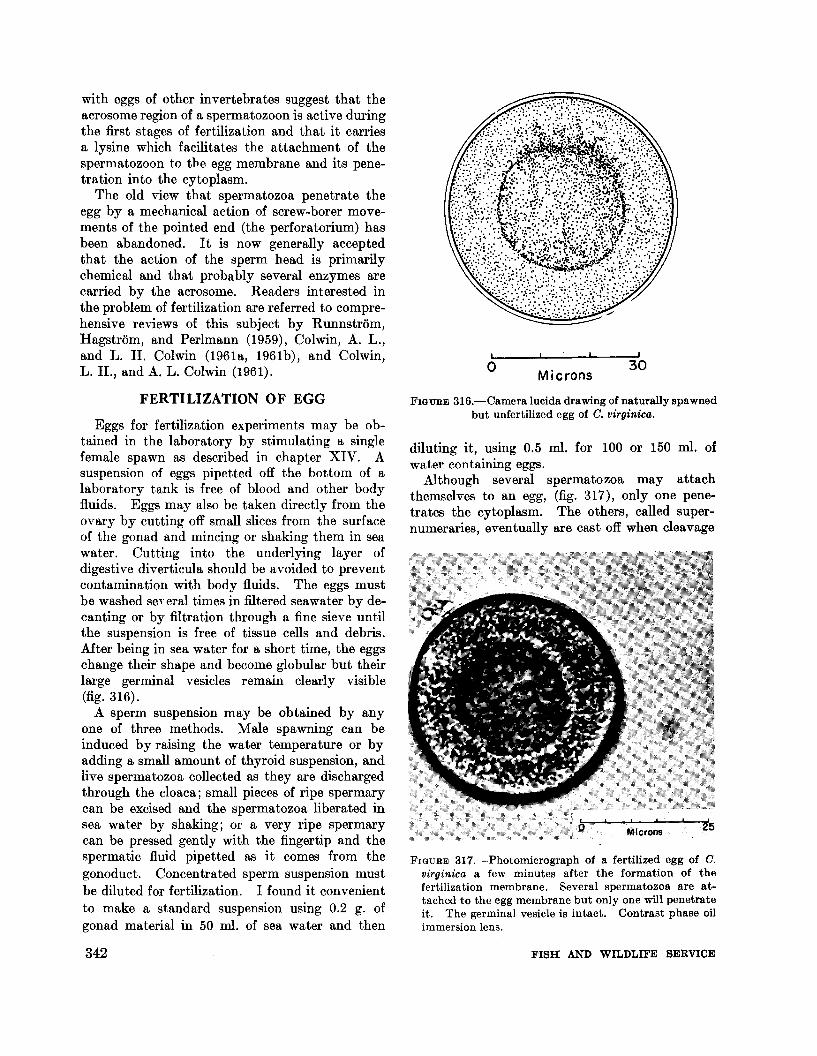
Eggs for fertilization experiments may be obtained in the laboratory by stimulating a singlefemale spawn as described in chapter XIV. Asuspension of eggs pipetted off the bottom of alaboratory tank is free of blood and other bodyfluids. Eggs may also be taken directly from theovary by cutting off small slices from the surfaceof the gonad and mincing or shaking them in seawater. Cutting into the underlying layer ofdigestive diverticula should be avoided to preventcontamination with body fluids. The eggs mustbe washed se, eral times in filtered seawater by decanting or by filtration through a fine sieve untilthe suspension is free of tissue cells and debris.After being in sea water for a short time, the eggschange their shape and become globular but theirlarge germinal vesicles remain clearly visible(fig. 316).
A sperm suspension may be obtained by anyone of three methods. Male spawning can beinduced by raising the water temperature or byadding a small amount of thyroid suspension, andlive spermatozoa collected as they are dischargedthrough the cloaca; small pieces of ripe spermarycan be excised and the spermatozoa liberated insea water by shaking; or a very ripe spermarycan be pressed gently with the fingertip and thespermatic fluid pipetted as it comes from thegonoduct. Concentrated sperm suspension mustbe diluted for fertilization. I found it convenientto make a standard suspension using 0.2 g. ofgonad material in 50 mI. of sea water and then
342
FIGURE 316.-Camera lucida drawing of naturally spawnedbut unfertilized egg of C. virginica.
diluting it, using 0.5 mI. for 100 or 150 mI. ofwater containing eggs.
Although several spermatozoa may attachthemselves to an egg, (fig. 317), only one penetrates the cytoplasm. The others, called supernumeraries, eventually are cast off when cleavage
FIGURE 317.-Photomicrograph of a fertilized egg of C.virginica a few minutes after the formation of thefertilization membrane. Several spermatozoa are attached to the egg membrane but only one will penetrateit. The germinal vesicle is intact. Contrast phase oilimmersion lens.
FISH AND WILDLIFE SERVICE

begins. If the sperm suspension is too concentrated, many spermatozoa enter one egg andcause polyspermy, a condition which may interfere with normal development of the egg.
The spermatozoon which succeeds in penetrating the egg's surface undergoes great changes.I ts acrosome region becomes swollen and disrupted and the tail loses its motility; the headgradually penetrates the egg membrane as thesperm moves deeper into the cytoplasm. At thesame time the fertilized egg contracts and assumesa globular shape if it was not round before; thecytoplasm becomes so dense that the germinalvesicle is no longer visible under the layer of yolkgranules. A few seconds after the sperm headtouches the egg's surface a thin, transparentfertilization membrane is elevated and under thelight microscope appears to be homogeneous.This membrane apparently is formed from thepre-existing vitelline membrane and is underlined by a layer of subcortical particles (fig. 318).The two layers are optically separated. It isgenerally accepted (Runnstrom, 1952) that inArbacia and many other species the fertilizationmembrane originates from the vitelline membranebecause it fails to form after the vitelline membrane has been removed with potassium chloride,trypsin, or urea. No experimental work of thistype has been done on oyster eggs.
AGING OF EGGS AND SPERM
The longevity of eggs of marine invertebrates,i.e., their ability to form fertilization membraneand undergo cleavage, was observed in the seaurchin (Arbacia) and in other common species(Harvey, 1956). Oyster eggs also undergo agingchanges and lose their ability to be fertilized.This has been demonstrated in a number of testsmade in the Bureau's shellfish laboratory at WoodsHole. Because of wide individual variability infertilization capacities only one female and onemale were used in each series of tests. Thefollowing technique was used: Suspension of eggswas made by shaking 0.5 g. of ripe ovary tissuein about 200 ml. of sea water: eggs released bythis action were permitted to settle on the bottomand the supernatant water was decanted; theremaining eggs were rinsed twice in sea water andtransferred to a beaker filled with 500 ml. of filteredsea water. The beaker was kept half submergedin running sea water to prevent heating to roomtemperature. Samples of eggs were taken forfertilization every hour during the first 4 hours,then at 2-hour intervals for the next 6 hours, andfinally one sample was taken each time after 12and 24 hours. Eggs were collected at randomfrom the bottom of the beaker and placed in afinger bowl in 100 ml. of filtered sea water. Tofertilize them 0.5 ml. of dilute stock suspensionof sperm was used; the water was gently stirred
o Microns 8
FIGURE 3l8.-Photomicrograph of a portion of fertilized egg of C. virginica shorly after the attachment of sperm. Fertilization membrane (Lm.) (outside layer) is underlined by the vitelline membrane (v.m.) with a dense row of subcortical particles (s.p.). Live preparation. Oil immersion phase contrast lens.
EGG, SPERM, FERTILIZATION, AND CLEAVAGE 343

to obtain uniform distribution of sperm, and thefinger bowl set aside for 5 hours, half submergedin running sea water. At the expiration of thisperiod a sample was taken for examination andcleaved and uncleaved eggs were counted. Ineach case 300 eggs of the sample were examined.All tests were made in water of 31 to 32% 0
salinity and 20.8 0 to 21.4° C. During the first 4hours of aging the percentages of cleaved eggsdeclined from 90 to 70. After 5 or 6 hours thepercentages dropped to 60. Then the fertilizability decreased to about 20 percent in 10 hours, andonly a few eggs cleaved normally after 12 and 24hours of aging.
It is common knowledge among embryologiststhat the fertilizing power of spermatozoa is notdecreased if sperm is kept at 10° to 12° C. in aconcentrated suspension in a tightly closed container. This is also true for oyster sperm. Itsfertilizing power is affected by dilution and increased temperature. At room temperature in adilute suspension, the spermatozoa lose theirfertilizing ability within 4 to 5 hours. However,in a concentrated suspension, protected fromevaporation, and stored in a refrigerator at about10° C. the sperm remains active and retains itsfull fertilizing power for 24 hours and possiblylonger.
The effect of cold storage on the fertilizabilityof eggs is not known. On several occasions ripefemales with intact shells were kept for 3 to 4days in a refrigerator (about 10° C.) and afterthat were successfully used in spawning experiments. The effect of cold storage on eggs of theexcised ovary has not been studied.
POLARITY OF EGG
The polarity of all molluscan eggs apparentlyis determined while they are still attached to thewall of the ovarian follicle. Presumably the sideon which the food reaches the growing ovocytebecomes the vegetative pole of the mature egg(Raven, 1958).
The metabolic gradient along the egg axis isindicated by a concentration of cytochrome oxidasewhich Kobayashi (1959) detected with M-Nadireaction; the activity of the enzyme was observedusing Graff's modification of this method. (Thereader not familiar with the reaction and its significance in cytochemical research is referred tothe publications of Danielli (1958), Deane, Barrnett, and Seligman (1960), and to a review by
344
Novikoff (1961a, p. 308).) In brief, the localization of oxidative enzymes and their presence inmitochondria can be determined by the stainingreactions. The rate of respiration of eggs of O.virginica increases with fertilization by a factor of1.4 (Ballentine, 1940) but in the eggs of theSydney rock oyster, O. commercialis, the rate ofrespiration increases only at the onset of the firstcleavage (Cleland, 1950).
CLEAVAGE
The spermatozoon may enter the oyster egganywhere. Its path inside the egg cytoplasmtoward the nucleus has not been described, andcytological details of the process leading to thefusion of the female and male pronuclei have notbeen studied. It is probable that the major features of these events are not different from thosefound in other mollusks. Maturation divisionsoccur in the oyster egg after the elevation of thefertilization membrane. The germinal vesicle(ovocyte nucleus) breaks down and moves towardthe egg's periphery. At temperatures of 22-0 to24 0 C. the first polar body is formed within 25 to50 minutes after the addition of sperm. The reduction of the number of chromosomes probablytakes place during the first meiotic division. Thisis to a certain extent corroborated by an examination of fertilized eggs of O. virginica stained intoto with Feulgen reagent or with acetic orcein.Unfortunately the results are not consistent enoughto draw a final conclusion and the question remainsunanswered, awaiting a complete cytological study.
The second polar body is formed shortly afterthe first, within 45 to 70 minutes after fertilization(at 22.5 0 to 24 0 C.). The two polar bodies remainattached to the surface of the egg (fig. 319) untilthe completion of cleavage and emergence of thetrochophore.
The first cleavage following the formation ofthe second polar body divides the egg meridionallyinto two unequal cells designated as AB and CD(fig. 320). The inequality of the blastomeres isdue to the occurrence of a polar lobe. Becausethe egg appears to consist of three cells this stagereceived the name trefoil.
The plane of the second division, also meridional,is at a right angle to the first. Both blastomeresdivide synchronously and separate into the fourquadrants. In fig. 321, drawn from a photographof a cleaving egg taken from the animal pole, theposition of the spindles indicates the plane of new
FISH AND WILDLIFE SERVICE

o Microns 10
FIGURE 320.-First cleavage division of the egg of C.virginica 70 minutes after fertilization. Blastomere AB(left) and CD (right). Polar body on top.
FIGURE 319.-Photomjcrograph of a live fertilized egg ofC. virginica after the formation of two polar bodies (topof egg). High-phase oil immersion lens.
20MicronsoFIGURE 322.-Section of an egg of C. virginica at the second
cleavage. Beginning of anaphase. Kahle, Heidenhainiron-hematoxylin.
FIGURE 321.-Beginning of second cleavage of the egg.Viewed from the animal pole. '''hole mount, Kahle,Feulgen stain.
first quartet of micromeres, small cells at theanimal pole, from the macromeres, or larger cellsat the vegetal pole (fig. 323).
At the fourth and fifth cleavages, resulting in16- and 32-cell stages, the micromeres overgrowthe macromeres. Only one of the macromeres isvisible in the figure 324 and two in 325, which showa side view of an oyster egg at these two stages ofdevelopment.
Cell lineage, or tracing the developmentalhistory of the cleavage blastomeres through to
40Microns6
furrows which will intersect the egg into four cells,A, B, C, and D. At this stage the mitotic figuresare fairly large and the chromosomes are in afavorable position for examination. In the bestsectioned prepa.rations of the cleaving egg, eightdaughter chromosomes were counted at the beginning of anaphase (fig. 322). It would be, however,premature to state that the diploid number ofchromosomes in G. 'lY£rginica is 8 because on otherpreparations 7, 9, and 10 were counted.
The third division of each quadrant cuts thecells in the equatorial plane and separates the
EGG, SPERM, FERTILIZATION, AND CLEAVAGE 345

oMicrons
20 o Microns 20
FIGURE 323.-Third division of fertilized egg of C. lJirginica; side view. Separation of micromeres(on top)from macromeres (bottom). Only one macromere isseen in the plane of view. Live egg.
FIGURE 325.-Fifth cleavage of egg of C. virginica andthe formation of the third quartet of micromeres (32cell stage). Side view. Drawn from photomicrographof live cell.
FIGURE 324.-Fourth cleavage of egg of C. lJirginica andthe formation of 8 micromeres (2d quartet). Side view.Drawn from photomicrograph of live cell.
their ultimate fates as parts of the larva or adult,was first described by Whitman (1878) for theegg of Glepsine, and followed by Wilson (1892) forthe egg of Nereis. The works of Lillie (1895)on the development of Unionidae, Conklin (1897,1908) on Grepidula and Fulyur, Meisenheimer
oMicrons
20
(1901a, 1901b) on Dreissensia and Gyclas, andWilson (1904a, 1904b) on Dentalium and Patellaconstitute major contributions to the embryologyof mollusks.
The nomenclature of the cleavage blastomeres,as developed by Wilson (1892), was progressivelymodified by Conklin (1897), Mead (1897), andChild (1900); the present system is based largelyon the work of Robert (1902) on the developmentof the Trochus egg. The system is a combinationof letters and numbers by which the blastomeresare identified.
The first four cells or macromeres are designatedas A, B, C, and D; in the majority of cases studiedD is the "largest of the four and is situated at theside which will develop into the posterior portionof the embryo. When the first four blastomeresdivide, their daughter cells are denominated 1a andlA, 1b and 1B, 1c and 1C, and 1d and 1D, thesmall letters in each case referring to the micromereand the capital letter to the macromere.
In successive divisions 1A divides into 2a and2A, 1B into 2b and 2B, and so on. When themicromeres divide, 1a is divided into 1a1 and 1a2,
the superscript 1 denoting the daughter cell whichis nearest to the animal pole and superscript 2 theone nearer to the vegetal pole. The nomenclatureis capable of indefinite expansion, but certain confusion arises when the two daughter cells resulting
346 FISH AND WILDLIFE SERVICE

FIGURE 326.-Formation of sterroblastula in C. virginicaegg; of the four macromeres only two are visible fromthe side. Micromeres begin to overgrow the vegetalpole. Drawn from photomicrograph of live egg.
FIGURE 327.-Advanced sterroblastula stage of C. virginica. One of the macromeres has divided into twodaughter cells of equal size. Drawn from photomicrograph of live egg.
20Microns
Microns
o
from the division of one cell lie at an equal distancefrom the pole. In this case the letters r for rightand I for left are used. The practice is, however,not generally followed.
Descriptions of various types of cleavage can befound in volume 1 of MacBride (1914). The equaland unequal cleavages in the spirally cleaving eggsof annelids and molluscs are discussed by Costello(1955) in chapter 2 of Willier, Weiss, and Hamburger (1955).
During division the micromeres of a quartet,viewed from the animal pole, become slightly displaced because the spindles of the dividing cells(not shown in fig. 324 or 325) occupy an obliqueposition with respect to the egg's axis. At thefollowing divisions the plane of separation ofdaughter cells is oriented approximately at a rightangle to the preceding divisions. The pattern ofsuch cleavage is called spiral. It gives rise to anirregular morula (sterroblastula according to Korschelt and Heider, 1895, from the Greek "sterros"meaning firm) found in annelids (Nereis), in somebivalves (Ostreidae, Teredo), and in gastropods(Orepidula, Fu}gur, Nassa) , and others. In allcases the sterroblastula arises from an unequalcleavage during which the micromeres overlie themacromeres, and at each division are slightly displaced to the right (dexiotropic cleavage) or to theleft (laeotropic cleavage). Sometimes, as in thecase of Dreissensia, the second dexiotropic cleavageis followed by a third dexiotropic cleavage afterwhich the normal alternating course is established(Meisenheimer, 1901a). In the case of oystereggs, as shown by Fujita (1929) for O. gigas, thecleavage is laeotropic.
The multicellular stages of a O. virginica egg arereached in the course of the sixth and ensuingcleavages (figs. 326 and 327) during which themicromeres divide much more rapidly than themacromeres, become progressively smaller andovergrow the vegetal pole. Approximately at thisstage the sterroblastula of an oyster is formed.
Gastrulation begins with epibolic extension ofthe micromeres. At 22° to 24° C. the stageshown in fig. 328 is reached within 4 to 6 hours.
The cell lineage of O.mrgtnica has not beenstudied; the stages of development of eggs of thespecies shown in figures 320 to 328 are similarto those previously described by Brooks (1898);Horst (I882) for O. edulis; Seno (1929) for O.denselamellosa; Hori (1933) for O. lurida; Yasugi(1938) for O. spwosa, and O. gigas. Yasugi
EGG, SPERM, FERTILIZATION, AND CLEAVAGE 347

FIGURE 328.-Early stage of gastrulation in the egg ofC. virginica. Seen at a section of an egg cut along itsaxis. Heidenhain, iron-hematoxylin.
found that equal cleavage can be induced artificially in eggs of the Japanese oyster by centrifuging for 2 minutes at 1,500 r.p.m. and at thecentrifuge radius of 14 cm.
FUJita (1929) gives a brief account of the celllineage of the eggs of G. gigas and states that themode of cleavage of this species is identical tothat of G. virginica. The main features describedby him are as follows. The first polar body inthe fertilized egg of G. gigas 11ppears 15 minutesafter insemination. At the two-cell stage (fig.329) the two blastomeres of unequal size, AB andCD, are separated along the meridional plane.Their position corresponds to the anterior (Ant.)and posterior (Pst.) ends of the embryo. Thesecond division, also meridional, separates thefour blastomeres A, B, C, and D (fig. 329b).
A and B represent the anterior, and C and Dthe posterior halves of the embryo, while BandC form its left and A and D its right halves(fig. 329b). The ensuing cleavage starts with theblastomere B and proceeds in laeotropic order toC, D, and A; the resulting daughter cells, themicromeres aI, b l, CI, and dl, retain the shape ofthe mother cells but are smaller. The macromereD and micromere dl are respectively the largest.The four daughter cells al through dl form the
o Microns 20
first quartet of micromeres located between themacromeres on the dorsal side of the embryo.
The 12-cell stage is initiated by the division ofthe macromere D; the ensuing larger cell d2(fig. 329c) is generally known as the first somatoblast X. (In the system of nomenclature usedby American and European embryologists (seep. 346) the ID cell gives rise to 2d and 2D and the2d is the X cell.).
The cleavage is continued laeotropically, andthe second generations of micromeres ~, b2,and C2 are smaller than the first macromeres.They lie on the outside of the macromeres. Thethird cleavage of macromeres A, B, and C continues in laeotropic order and results in themicromeres aa, ba, and Ca; they are larger thanother micromeres. After the third Cleavage themacromeres make no further contribution to theformation of micromeres and in the course ofdevelopment become the entoderm. The firstsomatoblast (X cell) gives rise to many organs ofectodermal origin. At the 18-cell stage of theembryo the position of cell X and its first divisionsmark the beginning of the transition from spiralto bilateral symmetry (fig. 329d).
The mesoderm begins to form at about the32-cell stage with the appearance of cell 4d,the second somatoblast, also designated as cellM. In bivalves the cell M remains at the surfacefor a long time, then divides into the two mesodermal teloblasts which sink into blastocoel(Raven, 1958, p. 117). The formation of mesoderm in G. gigas has not been followed in detail,but as a rule the mesoderm bands in bivalvesremain rather rudimentary (Raven, 1958). Fujitastates that the establishment of the three germinallayers in O. gigas is completed at the 30-cellstage (fig. 329 e and f).
The gastrula stage is reached in 4 to 6 hours.The cell lineage of O. gigas is generally comparableto that described for other bivalves (see: Raven,1958, p. 70), but for details the reader shouldconsult Fujita's (I929) original text and hisdrawings.
In about 4 to 6 hours after fertilization, an eggof G. virginica reaches the stage (fig. 330) whena few large cilia become visible at the vegetalpole, the oval-shaped body is covered with very
348 FISH AND WILDLIFE SERVICE

A
C~.I
Cz
X"1.1.1
"'.1./.1o
oMicrons
40
FIGURE 329.-Several stages of development of the egg of C. gigas. Redrawn from Fujita, 1929. a-Two-cell stage,Ant.-anterior, Pst.-posterior ends; b-Four-cell stage, formation of blastomeres A, B, C, and D; c-12-cell stage andthe formation of the first somatoblast (cell X), viewed from the animal pole; d-embryo viewed from vegetativepole after the formation of the mesomere M; e-cleavage of mesomere M and the first somatoblast X, posteriorview optical section; f-advanced stage of development showing the arrangement of the mesomeres M, M, and thesomatoblasts, X, X, Xi, Xi, posterior view optical section. Cleavage nomenclature as given by Fujita.
Microns
FIGURE 330.-Larva of C virginica ready to hatch.Drawn from photomicrograph of live larva.
fine ciliation, and two polar bodies still remainattached to the animal pole. The beating of thecilia is not coordinated at this stage, and themovements of the larva are irregulai' and spa.smodic. A few minutes later a girdle of powerfulcilia is formed, the polar bodies are lost, and thelarva begins to swim upward (fig. 331). In afinger bowl containing cleaving eggs, the newlyhatched larvae appear as white columns rising fromthe layer of fertilized eggs on the bottom of thecontainer (fig. 332). The larvae can be pipettedoff easily, transferred into larger containers andprovided with suitable food.
The time required to complete the developmentof an oyster egg varies, depending on conditionof eggs, temperature, salinity, oxygenation ofwater, and other environmental factors. Recordsof three sets of observations made in the Woods
EGG, SPERM, FERTILIZATION, AND CLEAVAGE
733-851 0-64--23
349

TABLE 37.-0bservations on the time required for artificiallyfertilized eggs of C. virginica to reach trochophore stage
All observations were made at Woods Hole in July at room temperaturesvar~g from 23° to 25° C. The time required to reach different stagesvarIed in different groups of eggs. The observations are arranged in twogroups: A and B, which differ primarily In the duration of time requiredto reach rotating blastula and trochophore stages.
STAGES
Hole laboratory at room temperatures varyingfrom 22..5° to 24.5° C. and salinity of water of32.2 %0 are given in table 37. To obtain records of rates of development at different temperatures, several hundred artificially fertilizedeggs were placed in each Syracuse dish filled withfresh sea water and covered to prevent evaporation.The debris was removed, and the water containedno unfertilized or cytolyzed eggs.
BAStage of development
FIGURE 33l.-Larva of C virginica at the timeemergence 6 to 6Y2 hours after fertilization.from a photomicrograph of a live larva.
o Microns 20
of itsDrawn
Fertilization memhrane 5 min _First polar body . 40 min _Second polar hody 1 hr. 10 min _First cleavage. 1 hr. 12 min _Second cleavage _Third cleavage 2 hr. 10 min _Morula stage _Rotating blastula 4 hr _Trochophore_ 5 hr _
10 to 25 min.25 to 52 min.40 to 65 min.45 min.52 to 120 min.55 to 195 min.135 min.6 hr. 30 min.8 to 9 hr.
6 Cen t imeters 5
FIGURE 332.-The emergence of larvae of C. virginica from fertilized eggs kept in a finger bowl. The free-swimminglarvae form columns, which tend to disperse at the surface. Drawn from life.
350 FISH AND WILDLIFE SERVICE

BIBLIOGRAPHY
AFZELIUS, BJORN A.1955. The fine structure of the sea urchin sperma
tozoa as revelaed by the electron microscope.Zeitschrift fUr Zellforschung und mikroskopischeAnatomie, Band 42, pp. 134-148.
ALLEN, ROBERT DAY.1953. Fertilization and artificial activation in the
egg of the surf-clam, Spisula solidissima. Biological Bulletin, vol. 105, No.2, pp. 213-239.
AMEMIYA, IKUSAKU.1926. Notes on experiments on the early develop
mental stages of the Portuguese, American andEnglish native oysters, with special reference to theeffect of varying salinity. Journal of the MarineBiological Association of the United Kingdom, vol.14, No.1, pp. 161-175.
BALLENTINE, ROBERT.1940. Analysis of the changes in respiratory activity
accompanying the fertilization of marine eggs.Journal of Cellular and Comparative Physiology,vol. 15, No.2, pp. 217-232.
BERG, WILLIAM E.1950. Lytic effects of sperm extracts on the eggs of
Mytilus edulis. Biological Bulletin, vol. 98, No.2,pp. 128-138.
BRACHET, JEAN.1944. Embryologie chimique. Masson et Cie, Paris,
509 pp.1960. The biochemistry of development. Pergamon
Press, New York, 320 pp.BRAMBELL, F. W. ROGERS.
1924. The nature and origin of yolk. Experimentalstudies of the oocytes of Helix aspersa and Patellavulgata. British Journal of Experimental Biology,vol. 1, No.4, pp. 501-517.
BRETSCHNEIDER, L. H., and CHR. P. RAVEN. '1954. Structural and topochemical changes in the
egg cells of Limnaea stagnalis L. during oogenesis.Archives Neerlandaises de Zoologie, tome 10,livraison 1 (1951), pp. 1-31.
BROOKS, W. K.1880. Development of the American oyster (Dstrea
virginica List.). Studies from the Biological Laboratory, Johns Hopkins University, Baltimore, Md.,No.4 (vol. 1), pp. 1-81.
1898. Embryonic development. In H. F. Moore,Oysters and methods of oyster-culture, pp. 270-272.U.S. Commission of Fish and Fisheries, Part 23,Report of the Commissioner for the year endingJune 30, 1897.
CHILD, CHARLES MANNING.1900. The early development of Arenicola and
Sternaspis. Archiv fUr Entwickelungsmechanikder Organismen, Band 9, pp. 587-723.
CLELAND, K. W.1947. Some observations on the cytology of oogenesis
in the Sydney Rock oyster (Dstrea commercialis 1.and R.). Proceedings of the Linnean Society ofNew South Wales, vol. 72, pp. 159-182.
EGG, SPERM, FERTILIZATION, AND CLEAVAGE
1950. Respiration and cell division in developingoyster eggs. Proceedings of the Linnean Societyof New South Wales, vol. 75, pp. 282-295.
1951. The enzymatic architecture of the unfertilizedoyster egg. Australian Journal of ExperimentalBiology and Medical Science, vol. 29, part 1,pp.35-45.
COE, WESLEY R.1931. Spermatogenesis in the California oyster
(Dstrea lurida). Biological Bulletin, vol. 61, No.3,pp. 309-315.
1932a. Sexual phases in the American oyster (Dstreavirginica). Biological Bulletin, vol. 63, No.3,pp. 419-441.
1932b. Development of the gonads and the sequenceof the sexual phases in the California oyster (Dstrealurida). Bulletin of the Scripps Institution ofOceanography of the University of California,Technical Series, vol. 3, No.6, pp. 119-144.
1934. Alternation of sexuality in oysters. AmericanNaturalist, vol. 68, No. 716, pp. 236-251.
1936. Environment and sex in the oviparous oysterDstrea virginica. Biological BUlletin, vol. 71, No.2,pp.353-359.
1938. Conditions influencing change of sex in mollusks of the genus Crepidula. Journal of Experimental Zoology, vol. 77, No.3, pp. 401-424.
COLWIN, ARTHUR L., and LAURA HUNTER COLWIN.1955. Sperm entry and the acrosome filament
(Holothuria atra and Asterias amurensis). Journalof Morphology, vol. 97, No.3, pp. 543-567.
1957. Morphology of fertilization: Acrosome filament formation and sperm entry. In Albert Tyler,R. C. von Borstel, and Charles B. Metz (editors),The beginnings of embryonic development, pp.135-168. American Association for the Advancement of Science, Publication No. 48, Washington,D.C.
1960. Egg membrane lytic activity of sperm extractand its significance in relation to sperm entry inHydroides hexagonus (Annelida). Journal of Biophysical and Biochemical Cytology, vol. 7, No.2,pp. 321-328.
1961a. Changes in the spermatozoon during fertilization in Hydroides hexagonus (Annelida). II.Incorporation with the egg. Journal of Biophysicaland Biochemical Cytology, vol. 10, No.2, pp. 255274.
1961b. Fine structure of the spermatozoon ofHydroides hexagonus (Annelida), with special reference to the acrosomal region. Journal of Biophysical and Biochemical Cytology, vol. 10, No.2,pp. 211-230.
COLWIN, LAURA HUNTER, and ARTHUR L. COLWIN.1956. The acrosome filament and sperm entry in
Thyone briareus (Holothuria) and Asterias. Biological Bulletin, vol. 110, No.3, pp. 243-257.
1960. Formation of sperm entry holes in the vitellinemembrane of Hydroides hexagonu8 (Annelida) andevidence of their lytic orgin. Journal of Biophysicaland Biochemical Cytology, vol. 7, No.2, pp. 315320.
351

COLWIN, LAURA HUNTER, and AUTHUR L. COLWIN.1961. Changes in the spermatozoon during fertiliza
tion in Hydroides hexagonus (Annelida). 1. Passage of the acrosomal region through the vitellinemembrane. Journal of Biophysical and Biochemical Cytology, vol. 10, No.2, pp. 231-254.
COKLIN, EDWIN GRANT.1897. The embroyology of Crepidula. A contribu
tion to the cell lineage and early development ofsome marine gasteropods. Journal of Morphology,vol. 13, No.1, pp. 1-226.
1908. The embryology of Fulgur: a study of theinfluence of yolk on development. Proceedings ofthe Academy of Natural Sciences of Philadelphia,vol. 59, 1907, pp. 320-359.
COSTELLO, DONALD PAUL.1939. The volumes occupied by the formed cyto~
plasmic components in marine eggs. PhysiologicalZoolo~y, vol. 12, No.1, pp. 13-21.
1955. Cleavage, blastulation and gastrulation. InBenjamin H. Willier, Paul A. Weiss, and ViktorHamburger (editors), Analysis of development,sec. 5, Embryogenesis: preparatory phases, ch. 2,pp. 213-229. W. B. Saunders Company, Philadelphia, Pa.
DALcQ, A. M.1960. Germinal organisation and induction phe
nomena. In W. W. Nowinski (editor), Fundamental aspects of normal and malignant growth,ch. 4, pp. 305-494. Elsevier, Amsterdam.
DAN, JEAN C., and SEIJI K. WADA.1955. Studies on the acrosome. IV. The acrosome
reaction in some bivalve spermatozoa. BiologicalBulletin, vol. 109, No.1, pp. 40-55.
DANIELLI, J. F.1958. General cytochemical methods. Vol. 1. Aca
demic Press, New York, 471 pp.DAVAINE, C.
1853. Recherches sur la g~n~ration des huttres.Comptes Rendus des S~ances et M~moires de laSoci~M de Biologie, tome 4, s~rie 1, ann~e 1852, pp.297-339.
DEANE, H. W., R. J. BARRNETT and A. M. SELIGMAN1960. Histochemical methods for the demonstration
of enzymatic activity. In Walther Graumann andKarlheinz Neumann (editors), Handbuch derHistochemie, Vol. 7, Enzymes, pt. 1, 202 pp.Gustav Fisher, Stuttgart.
DEROBERTIS, E. D. P., W. W. NOWINSKI, and FRANCISCOA. SAEZ.
1960. General cytology. 3d ed. W. B. SaundersCompany, Philadelphia, Pa., 555 pp.
DREW, GILMAN A.1900. Yoldia limatula. Memoirs from the Biolog
ical Laboratory of the Johns Hopkins University,IV, Selected morphological monographs, 3. TheJohns Hopkins Press, Baltimore, Md., 37 pp.
1906. The habits, anatomy, and embryology of thegiant scallop (Pecten tenuicostatus Mighels). TheUniversity of Maine Studies, Orono, Maine, No.6,71 pp.
352
FRANzEN, AKE.1956. Comparative morphological investigations into
the spermiogenesis among mollusca. ZoologiskaBidrag Fran Uppsala, Band 30, pp. 399-456.
FUJITA, TSUNENOBU.1929 On the early development of the common
Japanese oyster. Japanese Journal of Zoology,Transactions and Abstracts, vol. 2, No.3, pp.353-358.
GALTSOFF, PAUL S., and D. E. PHILPOTT.1960. Ultrastructure of the spermatozoon of the
oyster, Crassostrea v1:rginica. Journal of Ultrastructure Research, vol. 3, No.3, pp. 241-253.
GATENBY, J. BRONTE.1919. The cytoplasmic inclusions of the germ-cells.
Part V. The gametogenesis and early developmentof Limnaea stagnalis (L.), with special reference tothe Golgi apparatus and the mitochondria. Quarterly Journal of Microscopical Science, vol. 63,No. 252, pp. 445-491.
GATENBY, J. BRONTE, and J. H. WOODGER.1920. On the relationship between the formation of
yolk and the mitochondria and Golgi apparatusduring oogenesis. Journal of the Royal Microscopical Society for the year 1920, pp. 129-156.
GOMORI, GEORGE.1952. Microscopic histochemistry-principles and
practice. University of Chicago Press, Chicago,IlL, 273 pp.
HAGSTROM, B. E.1956a. Studies on the fertilization of jelly-free sea
urchin eggs. Experimental Cell Research, vol. 10,No.1, pp. 24-28.
1956b. The effect of removal of the jelly coat onfertilization in sea urchins. Experimental CellResearch, vol. 10, No.3, pp. 740-743.
1956c. The influence of the jelly coat in situ andin solution on cross fertilization in sea urchins.Experimental Cell Research, vol. 11, No.2, pp.306-316.
1956d. Further studies on cross fertilization in seaurchins. Experimental Cell Research, vol. 11,No.2, pp. 507-510.
HARVEY, ETHEL BROWNE.1956. The American Arbacia and other sea urchins.
Princeton University Press, Princeton, N.J., 298 pp.HATSCHEK, B.
1881. Ueber Entwicklungsgeschichte von Teredo.Arbeiten aus dem Zoologischen Institute derUniversitat Wien und der Zoologischen Station inTriest, tome 3, pp. 1-44.
HORI, Juzo.1933. On the development of the Olympia oyster,
Ostrea lurida Carpenter, transplanted from UnitedStates to Japan. Bulletin of the Japanese Societyof Scientific Fisheries, vol. 1, No.6, pp. 269-276.
HORST, R.1882. On the development of the European oyster
(Ostrea edulis L.). Quarterly Journal of Microscopical Science, vol. 22, pp. 341-346.
FISH AND WILDLIFE SERVICE

IMMERS, J., and E. VASSEUR.1949. Comparative studies on the coagulation proc
ess with heparin and sea-urchin fertilizin. Experientia, vol. 5, fascicule 3, pp. 124--125.
JACKSON, ROBERT TRACY.1888. The development of the oyster with remarks
on allied genera. Proceedings of the Boston Societyof Natural History, vol. 23, pp. 531-556.
1890. Phylogeny of the Pelecypoda. The Aviculidaeand their allies. Memoirs of the Boston Society ofNatural History, vol. 4, No.8, pp. 277-400.
KELLY, JOHN W.1954. Metachromasy in the eggs of fifteen lower
animals. Protoplasma, Band 43, pp. 329-346.1956. The metachromatic reaction. Protoplasma
tologia, Handbuch der Protoplasmaforschung,Band 2, D 2, 98 pp.
KOBAYASHI, HARUO.1959. Bipolarity of egg of oyster (Gryphaea gigas
(Thunberg». Cytologia, vol. 24, No.2, pp. 237243.
1960. Cytochemical studies on the second gradientaxis of fertilized oyster egg. Japanese Journal ofZoology, vol. 12, No.4, pp. 497-506.
KORSCHELT, E., and K. HEIDER.1895. Text-book of the embryology of invertebrates.
Part 1. Porifera, Cnidaria, Ctenophora, Vermes,Enteropneusta, Echinodermata. MacMillan andCompany, New York, 484 pp. Translated fromthe German by. Edward L. Mark and W. McM.Woodworth.
KOSTANECKI, K.1904. Cytologische Studien an kunstlich partheno
genetisch sich entwickelnden Eiern von Mactra.Archiv fUr mikroskopische Anatomie und Entwicklungsgeschichte, Band 64. pp. 1-98.
1908. Zur Morphologie der kunstlichen parthenogenetischen Entwicklung bei Mactra. Zugleichein Beitrag zur Kenntnis der veilpoligen mitose.Archiv fUr mikroskopische Anatomie und Entwicklungsgeschichte. Band 72, pp. 327-352.
LENHOSSEK, M. VON.1898. Untersuchungen uber Spermatogenese. Archiv
fur mikroskopische Anatomie und Entwicklungsgeschichte, Band 51, pp. 215-318.
LILLIE, FRANK R.1895. The embryology of the Unionidae. A study
in cell-lineage. Journal of Morphology, vol. 10,No.1, pp. 1-100.
1919. Problems of fertilization. University of Chicago Press, Chicago, Ill., 278 pp.
MACBRIDE, ERNEST WILLIAM.1914. Invertebrata. In Walter Heape (editor),
Text-book of embryology, vol. 1, 692 pp. Mac~
millan and Company, London.MEAD, A. D.
1897. The early development of marine annelids.Journal of Morphology, vol. 13, No.2, pp. 227-326.
MEISENHEIMER, JOHANNES.1897. Entwicklungsgeschichte von Ltmax maximus
L. Theil 1. Furchung und Keimblatterbildung.Zeitschrift fUr wissenschaftliche Zoologie, Band 69,pp.417-428.
EGG, SPERM, FERTILIZATION, AND CLEAVAGE
1901a. Entwicklungsgeschichte von Dreissensia polymorpha Pall. Zeitschrift fur wissenschaftlicheZoologie, Band 69, pp. 1-137.
1901b. Die Entwicklung von Herz, Perikard, Niereund Genitalzellen bei Cyclas im Verhiiltnis zu deniibrigen Mollusken. Zeitschrift fur wissenschaftliche Zoologic. Band 69, pp. 417-428.
MULNARD, JACQUES.1958. La metachromasie in vivo et son analyse
cytochimique dans I'oeuf de l'Annelide Chaetopteruspergamentaceus. Archives de Biologie, tome 69,pp. 645-685.
MULNARD, JACQUES. WALTER AUCLAIR. and DOUGLASMARSLAND.
1959. Metachromasia observed in the living eggs ofArbacia punctulata and its cytochemical analysis.Journal of Embryology and Experimental Morphology, vol. 7, part 2, pp. 223-240.
NOVIKOFF, ALEX B.1961a. Mitochondria (Chondriosomes). In Jean
Brachet and Alfred E. Mirsky (editors), The cell,vol. 2, ch. 5, pp. 299-421. Academic Press, NewYork.
1961b. Lysosomes and related particles. In JeanBrachet and Alfred E. Mirsky (editors). The cell.vol. 2, ch. 6, pp. 423-488. Academic Press, NewYork.
PASTEELS. J. J., and J. MULNARD.1957. La Metachromasie in vivo au bleu de toluidine
et son analyse cytochimique dans les oeufs de Barneacandida, Gryphaea angulata (Lamellibranches) et dePsammechinus miliaris. Archives de Biologie.tome 68, pp. 115-163.
PHILPOTT, DELBERT E.1955. A simple and economical microtome for ultra
thin sectioning. Experimental Medicine and Surgery, vol. 13, No.2, pp. 189-192.
RAI, HARDIT SINGH.1930. On the origin of yolk in the egg of Ostrea
cucullata. Journal of the Royal MicroscopicalSociety, vol. 50, series 3, pp. 210-217.
RAVEN, CHR. P.1958. Morphogenesis: the analysis of molluscan de
velopment. Pergamon Press, New York, 311 pp.REBHUN, LIONEL 1.
1960. Aster-associated particles in the cleavage ofmarine invertebrate eggs. Annals of the New YorkAcademy of Science. vol. 90, art. 2, pp. 357-380.
RETZIUS, GUSTAF.1904. Zur Kenntniss der Spermien der Everte~raten.
Biologische Untersuchungen, Neue Folge, 11, pp.1-32. Gustav Fischer, Jena.
ROBERT, A.1902. Recherches sur Ie developpement des troques.
Archives de Zoologie Experimentale et Generale,serie 3, tome 10, pp. 269-538.
RUNNSTROM, JOHN.1952. The cell surface in relation to fertilization.
Symposia of the Society for Experimental Biology,number 6, Structural aspects of cell physiology.Academic Press, New YorK, pp. 39-88.
353

RUNNSTROM, J., B. E. HAGSTROM, and P. PERLMANN.1959. Fertilization. In Jean Brachet and Alfred E.
Mirsky (editors), The cell, vol. 1, ch. 9, pp. 327397. Academic Press, New York.
SENO, HlDEMI.1929. A contribution to the knowledge of the de
velopment of Ostrea denselamellosa Lischke. Journal of the Imperial Fisheries Institute, vol. 24,No.5, pp. 129-135.
SENO, HlDEMI, Juzo HORI, and DAIJJRO KUSAKABE.1926. Effects of temperature and salinity on the de
velopment of the eggs of the common Japaneseoyster, Ostrea gigas Thunberg. Journal of theImperial Fisheries Institute, vol. 22, No.2, pp.41-47.
SHAVER, JOHN R.1957. Some observations on cytoplasmic particles
in early echinoderm development. In AlbertTyler, R. C. von Borstel, and Charles B. Metz(editors), The beginnings of embryonic development, pp. 263-290. American Association for theAdvancement of Science, Publication No. 48,Washington, D.C.
STAFFORD, J.1912. Supplementary observations on the develop
ment of the Canadian oyster. American Naturalist, vol. 46, No. 541, pp. 29-40.
1913. The Canadian oyster, its development. environment and culture. Committee on Fisheries,Game and Fur-bearing Animals, Commission ofConservation, Canada. The Mortimer Company.Ltd., Ottawa, Canada, 159 pp.
SWANSON. CARL P.1957. Cytology and cytogenetics. Prentice-Hall
Company. Englewood Cliffs. N.J., 596 pp.TERAO, ARATA.
1927. On the fertilizin of the oyster, Ostrea circumpicta. Science Reports of the T6hoku ImperialUniversity, series 4, Biology, vol. 2, No.2, pp.127-132.
THOMAS, LYELL J., JR.1951. A blood anti-coagulant from surf clams.
Biological Bulletin, vol. 101, No.2, pp. 230-231.1954. The localization of heparin-like blood anti
coagulant substances in the tissues of Spillula.~olidissima. Biological Bulletin, vol. 106, No.1,pp. 129-138.
TYLER, ALBERT.1939. Extraction of an egg membrane-lysin from
sperm of the giant keyhole limpet (Megathuracrenulata). Proceedings of the National Academyof Sciences of the United States, vol. 25, pp. 317323.
1948. Fertilization and immunity. PhysiologicalReviews, vol. 28, No.2, pp. 180-219.
1949. Properties of fertilizin and related substancesof eggs and sperm of marine animals. AmericanNaturalist, vol. 83, No. 11, pp. 195-219.
354
VASSEUR, ERIK.1948a. The sulphuric acid content of the egg coat
of the sea urchin, Strongylocentrotus droebachiensisMilll. Arkiv for Kemi, Mineralogi och Geologi,Band 25, B, No.6, pp. 1-2.
1948b. Chemical studies on the jelly coat of thesea-urchin egg. Acta Chemica Scandinavica, vol.2, pp. 900-913.
WADA, S. K., J. R. COLLIER, and J. C. DAN.1956. Studies on the acrosome. V. An egg
membrane lysin from the acrosomes of Mytilusedulis spermatozoa. Experimental Cell Research,vol. 10, No.1, pp. 168-180.
WHITMAN, CHARLES OTIS.1878. The embryology of Clepsine. Quarterly Jour
nal of Microscopical Science, vol. 18, new series,No. 71, Memoirs, pp. 215-314.
WILLIER, BENJAMIN, PAUL A. WEISS, and VIKTORHAMBURGER.
1955. Analysis of development. W. B. SaundersCompany, Philadelphia, Pa., 735 pp.
WILSON, EDMUND B.1892. The cell-lineage of Nereis. A contribution to
the cytogeny of the annelid body. Journal ofMorphology, vol. 6, No.3, pp. 361-480.
1904a. Experimental studies on germinal localization. 1. The germ-regions in the egg of Dentalium.Journal of Experimental Zoology, vol. 1, No.1,pp.I-72.
1904b. Experimental studies on germinal localization. II. Experiments on the cleavage-mosaic inPatella and Dentalium. Journal of ExperimentalZoology, vol. 1, No.2, pp. 197-268.
1928. The cell in development and heredity. 3d ed.Macmillan Company, New York, 1232 pp.
WOODS, FARRIS H.1932. Keimbahn determinants and continuity of
the germ cells in Sphaerium striatinum (Lam.).Journal of Morphology, vol. 53, No.2, pp. 345-365.
WORLEY, LEONARD G.1944. Studies of the vitally stained Golgi apparatus.
II. Yolk formation and pigment concentration inthe mussel Mytilus californianus Conrad. Journalof Morphology, vol. 75, No.1, pp. 77-101.
Y ASUGI, RYUICHI.1938. On the mode of cleavage of the eggs of the
oysters Ostrea spinosa and O. gigas under experimental conditions (a preliminary note). Annotationes Zoologicae Japonenses, vol. 17, pp. 295-300.
ZIEGLER, H. ERNST.1885. Die Entwicklung von Cyclas cornea Lam.
(Sphaerium corneaum L.). Zeitschrift fUr wissenschaftliche Zoologie, Band 41, pp. 525-569.
FISH AND WILDLIFE SERVICE