Chapter 1 Literature Review Terminalia spp in Africa with ...
90
16 Chapter 1 Literature Review Terminalia spp in Africa with special reference to its health status
-
Upload
khangminh22 -
Category
Documents
-
view
2 -
download
0
Transcript of Chapter 1 Literature Review Terminalia spp in Africa with ...
16
Chapter 1
Literature Review
Terminalia spp in Africa with special reference to its health
status

17
ABSTRACT The genus Terminalia is the second largest genus in the Combretaceae. The family is
distributed throughout the tropical and sub-tropical regions of the world and approximately
fifty species of Terminalia are naturally distributed throughout western, eastern and southern
Africa. Terminalia spp. range from small shrubs or trees to large deciduous forest trees. Some
species, such as T. ivorensis and T. superba develop as elements of the canopy or sub-canopy
layer in evergreen, semi-deciduous to deciduous, primary and secondary forests, whereas
species such as T. sericea, thrive well in open woodlands and mixed deciduous forests.
Terminalia spp. can be propagated naturally by seeds or through vegetative methods with
wildings, seedlings, stump plants or striplings. Terminalia spp. provide economical, medical,
spiritual and social benefits. Limited information on the pests and diseases affecting
Terminalia spp. exists. Many insect species are associated with Terminalia spp. but no
widespread pest problems have been recorded. Nevertheless, some locally common species
are potentially dangerous, mostly affecting the early stages of trees. Very few pathogens have
been reported from Terminalia spp. The majority of reports include limited detail, often
representing no more than a brief mention. Often the causal agents were identified based only
on morphology and were not classified to species level. Scanty information regarding the
pathogens associated with introduced and native Terminalia is a limitation that might be
detrimental for the survival and the successful exploitation of these trees.

18
1. INTRODUCTION Terminalia (Combretaceae, Myrtales) is a pantropical genus accommodating about 200
species (McGaw et al. 2001). About fifty of these are native to Africa and distributed
throughout the sub-saharan region (Lebrun and Stork 1991). Based on both their functional
uses and distribution in Africa, the most important are Terminalia ivorensis A. Chev. and T.
superba Engl. and Diels. in West and Central Africa and T. prunioides M.A. Lawson and T.
sericea Burch : DC in Southern Africa (Irvine 1961; Lamb and Ntima 1971; Coastes-Palgrave
1977; Groulez and Wood 1985; Schmidt et al. 2002; Lawes et al. 2004).
Terminalia trees are planted in several countries in the tropics as a source of high quality solid
timber for fine carpentry, joinery, building, flooring and plywood manufacture (Schmidt et al.
2002; Smith et al. 2004). Terminalia ivorensis and T. superba, especially, form an important
component of the forestry industries in many countries (Anonymous 1997). Terminalia spp.
are also commonly planted in mixed crop systems to establish a “taungya” agri-sylvicultural
system in which they provide shade and play a major role in increasing soil fertility (Nichols
et al. 2001; Norgrove and Hauser 2002a). Furthermore, members of the genus Terminalia are
among some of the plants most widely used for medicinal purposes in Africa (Masoko et al.
2005; Kamtchouing et al. 2006).
Despite the importance of Terminalia spp., very little research has been done regarding the
fungal diseases affecting these trees. Evidence of die-back, leaf spot and canker has been
reported from Terminalia spp. (Lamb and Ntima 1971; Ofosu Siedu and Cannon 1976;
Hodges and Fereira 1981). Gryzenhout et al. (2005), recently reported a serious disease
problem that emerged on non-native T. ivorensis in Ecuador, while in South Africa, two
Ceratocystis spp. have been reported from T. sericea (Roux et al. 2004; Kamgan et al. 2008).
The last or the 20th Century was marked by an increasing requirement for timber, fuel and
medicine from trees. This has resulted in unsustainable logging of native trees in Africa. To
supplement this requirement, plantations of non-native trees, including Eucalyptus spp., Pinus
spp., Acacia spp. and Cupressus spp. are been established in many parts of the tropics and the
southern hemisphere (Turnbull 1991; Wingfield et al. 2002; Anonymous 2007). In Africa, as
in most other countries, these non-native trees are established in close proximity to native
trees. This close association may in the long run, expose trees to new pests and diseases. One
19
might thus see the movement of native pests and pathogens onto introduced tree species. This
is of great concern since this could provide pathogens with an elevated opportunity to spread
to the country of origin of its new host, through reciprocal international trade of wood and
wood products, causing large-scale mortality of trees in their native ecosystems (Wingfield
2003; Slippers et al. 2005; Wingfield et al. 2008). On the other hand, the non-native tree
might be the source of non-native pathogens and pests, which may spread to the native trees
in its new country, resulting in disease epidemics. An increasing number of examples for
both case scenarios exist. For example, it has been shown that Chr. austroafricana Gryzenh.
& M. J. Wingf., the cause of canker and death of plantation grown Eucalyptus spp. in South
Africa, also occurs on native Myrtales in Africa (Heath et al. 2006; Nakabonge et al. 2006).
This pathogen is thought to have originated from Africa (Heath et al. 2006). On the other
hand, in California, native Monterey pine (Pinus radiata D. Don) are seriously affected by the
pitch canker pathogen, Fusarium circinatum Niremberg & O’Donnell, following its
introduction with Mexican pines (Gordon et al. 2001). In this respect, knowledge of
indigenous tree diseases would be useful to establish firm risk assessment programs.
In Africa, some species of Terminalia generally occur as elements of the canopy or subcanopy
layer in evergreen, semi-deciduous to deciduous primary and secondary forests. Other species
thrive in open woodlands and littoral areas. Within this natural habitat of Terminalia spp.,
several non-native tree species are frequently encountered. There is, therefore, a good chance
of introduced pathogens spreading onto native Terminalia trees, or innocuous fungi on
Terminalia spp., moving onto the non-native trees. The objective of this review is to present
a summary of knowledge pertaining to Terminalia spp. in Africa. A specific focus is given to
their origin and distribution, botanic description, ecology, propagation, management,
functional uses and international trade. Also, the limited knowledge regarding pests and
diseases on these trees is reviewed, providing a background for the contents of the dissertation
that follows this review and that focuses on fungi associated with native and introduced
Terminalia spp. on the African continent.

20
2. THE GENUS TERMINALIA
2.1. Origin and distribution
The family Combretaceae is comprised of 20 genera and about 475 species (Thiombiano et al.
2006). Of these about 200 belong to the genus Terminalia, making it the second largest genus
of the family after Combretum (McGaw et al. 2001). The family is distributed throughout the
tropical and sub-tropical regions of the world (Lamb and Ntima 1971). Approximately 54
species of Terminalia are naturally distributed throughout western, eastern and southern
Africa (Lebrun and Stork 1991; Smith et al. 2004). Terminalia ivorensis and T. superba are
the most important species found in West and Central Africa (Norgrove and Hauser 2002b),
but are also established in plantations within and outside their natural range, e.g. in South and
Central America, east Africa, Hawaii, Fiji and the Solomon Islands (Jones 1969). Within the
Malaysian region, further trials exist in Sabah, Kalimatan and the Philippines (Lamprecht
1989). On the other hand, T. prunioides, T. brachystemma Welw. ex Hiern, T. sericea, T.
gazensis Bak., T. mollis Laws. and T. sambesiaca Engl. & Diels. are the most common
species in eastern and southern Africa (Coates-Palgrave 1988; Masoko et al. 2005).
2.2. Botanical description and ecology
The genus Terminalia derives its Latin name (terminalis = end) from the position of the
leaves, which are crowded at the ends of the shoots (Lamb and Ntima 1971; Rogers and
Verotta 1996). The taxonomy of the genus has not been without problems, with the
assignment of sub-genera, especially presenting differing views (Stace 1965). However, the
different species have now been grouped into three sections according to characteristics of the
fruits. These sections include the Section Abbreviatae Excell with shorter fruits, Section
Psidioides Excell with longer fruits and the Section Platycarpae Engels & Diels emend.
Excell, with fruits that are broader in the centre (Carr 1988).
Terminalia spp. range from small and medium sized shrubs or trees to large deciduous forest
trees, ranging in height from 1.5 to 75 m tall (Lebrun and Stork 1991; Schmidt et al. 2002).
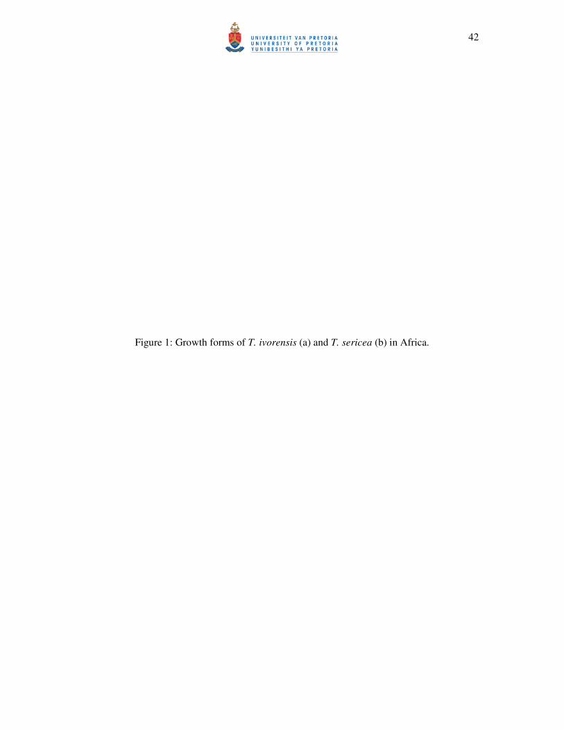
Some species, such as T. ivorensis, T. superba and T. trichipoda Diels have cylindrical boles
that are very straight and long (Figure 1a) with small to large, flat buttresses (six meters above
the soil surface) and are sometimes branchless for up to 30 m and 2-5 m in girth (Lemmens et

21
al. 1995). Mature trees are extensively flat topped, with a wide horizontal canopy of evenly
distributed foliage arising from the apex of the straight bole (Dupuy and Mille 1993). It is
these characteristics, and their relatively fast growth, that make T. ivorensis and T. superba
popular timber species. Other species, such as T. sericea and T. prunioides are common as
small shrubs (Figure 1b) to bushy trees that may be single or multi-stemmed with a girth of up
to 1.5 m (Coates-Palgrave 1977).
The bark of Terminalia trees is smooth and light grey to dark brown when young and on
branchlets. The inner bark and contact zone with the cambium is frequently yellow. In mature
trees, the bark surface cracks and flakes off in long thin strips or small patches, often
becoming blackish and developing deep longitudinal fissures as the trees grow (Keay 1989;
Lemmens et al. 1995).
The root systems of Terminalia trees are frequently fairly shallow. As the trees age, the tap
roots disappear. In the tall species such as T. ivorensis, buttresses from which descending
roots arise at some distance from the trunk, develop to support the trees (Keay 1989;
Lamprecht 1989).
The leaves are frequently simple and obovate, clustered spirally at the ends of the dwarfed
lateral branchlets, or crowded near the ends of the branches. Some species, like T. brassii
Exell., have prominent glands at the leaf bases (Lamb and Ntima 1971). In mature trees the
crown is usually flat or very slightly domed, giving Terminalia trees a distinctive shape.
Terminalia trees are bi-sexual or hermaphroditic with male and female flowers carried on the
same plants. These flowers are apetalous, small, and cream to pale, bright yellow or greenish-
white, in spicate inflorescences. The stalked male flowers tend to be grouped towards the
apex and the bisexual flowers towards the base of the inflorescences (Coates-Palgrave 1977).
Terminalia spp. have an effective system of self-incompatibility. Although male and female
flowers are in the same plant, self-pollination cannot produce viable zygotes (Newbegin et al.
1994). The flowers are pollinated by various insects (Coleoptera, Diptera, Hemiptera,
Hymenoptera and Lepidoptera) (Uzoechina 1978). The flowering-to-fruiting period may last
about 4 months, depending on the species and the locality where it is grown (Coates- Palgrave
1977; Keay 1989).

22
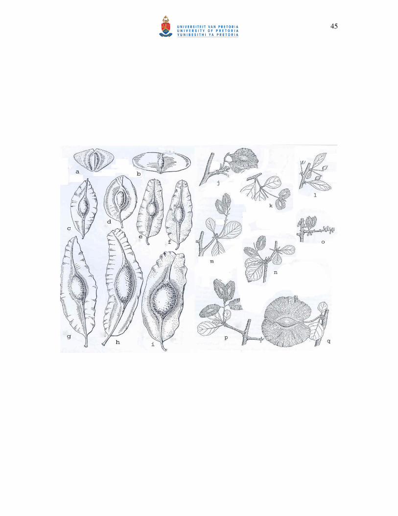
Terminalia fruits, in combination with leaf characteristics, are of great diagnostic value and
absolutely essential to distinguish between species (Coode 1969; Excell and Stace 1972). The
fruits are hard, flattened, two-winged and to some extent inconsistent in size, especially the
length and width of the wings. The general seed shape is consistent however, specific
characters such as fruit colour when ripe (yellow, red, purple green, brown or pink depending
on the species) and fruit morphology (elongate, broad, narrow, ovoid, oblong or elliptic)
(Figure 2) vary and are helpful in species differentiation (Dale and Greenway 1961; Coates
Palgrave 1977; Dale and Keay 1989). The fruits of some Terminalia trees, together with the
bark, are important sources of tannin (Lemmens and Wulijarni-Soetjipto 1991; Ellery and
Ellery 1997; Mabberley 1997). Terminalia fruits differ from those of closely related
Combretum spp. in having a sclerenchymatous endocarp (Lamb and Ntima 1971). Most of the
African Terminalia spp. produce fruits from January to September, with the exception of
species such as T. ivorensis and T. glaucescens Planch.: Benth. that produce fruits from July
onwards (Coates Palgrave 1977; Keay 1989).
The African species of Terminalia generally occur in various environments. Some species,
such as T. ivorensis and T. superba develop as elements of the canopy or sub-canopy layer in
evergreen, semi-deciduous to deciduous, primary and secondary forests (Keay 1989). Species
such as T. sericea on the other hand, thrive well in open woodlands to wooded savannahs and
mixed deciduous forests (Dale and Greenway 1961; Lebrun and Stork 1991; Carr 1994).
Terminalia trees can tolerate light to moderate shade, when young (Jones 1969). Thereafter,
they should receive full overhead light for optimal growth (Veenendaal et al. 1996). Few
individuals of this genus are able to grow at high altitude; most species perform well at
altitudes less than 2000 m a.s.l. The climates in which Terminalia trees grow varies from
areas with year round rain for species occurring in the forest areas (> 2000 mm per annum) to
seasonal with moderate rainfall (< 1200 mm per annum) for those occurring in savannah
zones (White 1983).
2.3. Propagation and management
Terminalia spp. can be propagated naturally by seeds or through vegetative methods with
wildings, seedlings, stump plants or striplings (Lemmens et al. 1995). However, obtaining
plants is difficult and with most Terminalia spp., propagation is not easy (Carr 1994).

23
2.3.1. Seed propagation
Freshly fallen seed should be collected from the ground, as seed still on the tree may not be
fully mature. Sometimes the seeds are collected from the trees by cutting off the branches,
because fallen seed are often parasitized by insects, leading to low viability. As far as
possible, seeds should be collected from healthy mother trees with a vigorous stem and crown
(Browse 1979; Hartman and Kester 1983). The number of fruits and seeds (per kilogram) vary
greatly between Terminalia spp. For example, T. ivorensis can produce 5500 - 7300 seeds per
kg (Lamb and Ntima 1971), T. superba can produce 8000 - 10000 seeds/kg (Groulez and
Wood 1985) and T. prunioides 8200 seeds/kg (Palmer and Pitman 1972). Seeds are extracted
from the fruits by placing them in a heap, spraying them with water and then covering them
with grass or leaves. After a day or so the fruit wing is stripped off and the seeds are extracted
manually (Carr 1994).
In general, seeds vary in the length of time that they remain viable. The viability of seed
varies between species. The viability of most species diminishes rapidly, with the exception
of T. superba, of which the seeds can be stored in sealed containers at 2-4 °C for one year,
adding to its suitability for commercial exploitation (Groulez and Wood 1985).
Seeds of Terminalia spp. must undergo a period of dormancy before germination occurs.
There are two types of dormancy in plants, namely physical and physiological dormancy
(Weber and Stoney 1986). Several methods of pre-treatment can be used to overcome the two
types of dormancy in seeds of Terminalia spp. As seeds of Terminalia spp. are covered by a
hard protective coat, the physical dormancy ends when the seed coat is opened through
different processes such as mechanical abrasion, nicking or soaking in water (Browse 1979;
Weber and Stoney 1986). Most often, for some species of Terminalia, seeds are pre-treated by
soaking in water for 12-48 hours, by manual scarification, or, in the case of T. ivorensis, by
alternate soaking and drying for one week (Lamb and Ntima 1971). For T. ivorensis the
germination rate is 10-50 %, but up to 93 % under experimental temperature fluctuations,
while for T. superba it is 60-80 % (Lemmens et al. 1995), 15-35 % for T. prunioides and 1-2
% for T. sericea (Carr 1994). However, as the viability in seeds is not assured, covering seed
or fruit in the seedbed is important for increasing the germination percentage. Light shade is
generally applied during germination, but it should be removed after one to two months. The
seedbed should be watered frequently to provide adequate moisture during germination
24
(Browse 1979). The sowing medium should be sand with low levels of "fines" or a light soil,
half mixed with sifted compost to promote good drainage and avoid water logging during
heavy rain periods (Carr 1994).
The physiological dormancy of Terminalia seed ends within two weeks after sowing and is
followed by epigeous germination which lasts two to five weeks. Pricking out should be done
early enough to avoid disturbing the rapidly developing taproot (Lemmens et al. 1995). For T.
superba, pricking out is recommended six weeks after sowing when two leaves have
developed (Groulez and Wood 1985), whereas for T. ivorensis, it should be as soon as the two
cotyledons unfold and the seedlings are 20-30 cm tall (Lamb and Ntima 1971).
2.3.2. Vegetative propagation
As an alternative to overcome seed viability problems, long growth periods and inconsistency
of seed germination (Carr 1994), vegetative propagation through cuttings and stump plants
can be used (Lemmens et al. 1995). However, vegetative propagation has in Africa been used
early for species such as T. ivorensis and T. superba in Africa (Fisher 1976).
The planting stock for vegetative propagation should be taken from young, vigorous shoots or
suckers from healthy, mature trees, during the dormant season, to encourage fast-growing
stems (Browse 1979). Short cuttings are taken from different parts of the stem (e.g. from the
rejuvenated stump plants and from branches). These stems (25-35 cm long) are cut just above
the proposed top bud and horizontally at the base, if possible dipping in a rooting hormone
before planting. Depending on the species, the cuttings can be placed either in pots filled with
water, or directly into a trench which is kept moist (Hartman and Kester 1983). After a period of time
shoots will produce roots, and they can then be transplanted to a permanent site. Stumps should have
a diameter of at least 1.3 cm. Cuttings of T. superba and T. ivorensis produce roots within two
weeks, with a rooting percentage of 11-100 % for T. superba according to the degree of
rejuvenation (Lemmens et al. 1995).
2.3.3. Tending of trees
Planting of Terminalia trees should be done early in the rainy season to ensure an adequate
water supply, and thus to avoid loss through drought (Lamb and Ntima 1971). Where
necessary, hand watering should be used to supplement insufficient rainfall. Weeding is

25
necessary during the first 3-4 years to give the seedlings adequate light and air circulation,
and to prevent competition for nutrients from weeds (Carr 1994).
Most Terminalia spp. have a good to extremely good self-pruning capacity (Lamb and Ntima
1971; Swaine and Hall 1983), adding to their popularity as plantation trees. The tall T.
superba tree is occasionally branchless up to 90 % of the total tree height (Lemmens et al.
1995). Pruning by hand is, therefore, not required in commercial plantations. However,
because of the wide spreading branches, the tree needs considerable space between stems
(Groulez and Wood 1985).
Terminalia spp. are planted in Africa on a wide variety of soil types ranging from alluvial,
sandy, salty and coral soils to heavy cracking clays (Coates-Palgrave 1988). The coppicing
ability is good for a number of Terminalia spp. planted in Africa and India. Terminalia
chebula Retz. and T. albida Scott-Elliot are known to withstand fire well, but T. superba and
T. ivorensis are very vulnerable in this respect. The average rotation age for T. superba and T.
ivorensis in Africa is 40 years, with trees reaching heights of 50-60 m and diameters of five
meters in this time (Keay 1989; Lemmens et al. 1995).
2.4. Functional uses of Terminalia trees
Terminalia spp. provide economical, medicinal, spiritual and social benefits. The wood of
Terminalia spp. is highly appreciated as constructional timber. It is currently used for light
construction, door and window frames, coffin boards, mouldings, beams, rafters, joists,
flooring, furniture, carts, tool handles, spindles, shuttles, picker sticks, walking sticks, bowls,
boat building, masts, mine props, foundation piles, veneer and plywood (Irvine 1961;
Lemmens et al. 1995; Schmidt et al. 2002; Smith et al. 2004). The fruits and bark of T.
sericea and T. catappa L. are important sources of tannin, as well as gum and resins for
glazing pottery (Irvine 1961; Lemmens and Wulijarni-Soetjipto 1991; Ellery and Ellery
1997). Dyes of various colours (black, red, orange, yellow, brown) are extracted from the
leaves, fruits, bark and roots of species such as T. mollis Lawson, T. ivorensis, T. laxiflora
Engl. & Diels., T. catappa L. and T. superba and used for decorating the walls of houses and
buildings with murals, for dyeing clothes, mattings, rattan, spoons and walking sticks (Dalziel
1937; Errington and Chisumpa 1987). The seed of some species is edible and considered one
of the best flavoured tropical nuts. Furthermore, consumable oil can be extracted from the

26
seed of T. catappa and used as a substitute for groundnut (Arachis hypogea L.), cotton seed
(Gossypium spp.) and silk cotton seed (Ceiba spp.) oils (Irvine 1961).
In Africa, forests are sometimes use for ritual and spiritual purposes. Certain trees can serve to
link the living with their ancestors, as this is often symbolized in the relationship between the
sky and the earth. In Southern Africa, Tswana people believe that good crops are ensured at
harvest and planting times by thrusting a stick of T. sericea into the floor of a shrine in
homage to ancestral spirits, and that cutting down an entire tree will result in hail-storms
(Coates-Palgrave 1977).
The importance of traditional medicines, derived from plants, is of great importance in most
parts of both Africa and Asia (Lawes et al. 2004; Steenkamp et al. 2004; Moshi and
Mbwambo 2005). Many Terminalia spp. have been identified as sources of medicines, for use
in pharmaceuticals and cosmetic production (Dalziel 1937; Irvine 1961). Extracts of the
flowers, fruits, bark, leaves, stems and roots from species such as T. glaucescens, T.
macroptera Gill.& Perr., T. laxiflora, T. superba, T. prunioides, T. brachystemma, T. sericea,
T. gazensis, T. catappa, T. mollis and T. sambesiaca are used in traditional medicine to treat
diseases such as malaria, eczema, candidosis, asthenia, gonorrhoea, diabetics, dermatitis,
scurfy affection, leprosy and tuberculosis (Batawila et al. 2005; Masoko et al. 2005; Fyhrquist
et al. 2006; Kamtchouing et al. 2006). The bark of T. macroptera is burned and used by
Sudanese women as a perfume (Irvine 1961).
Terminalia spp. have a number of agricultural uses. They are important sources of fodder for
animals. T. sericea is one of the palatable woody plants found in South African savannahs
(Owen-Smith and Cooper 1987; Lawes et al. 2004). Its leaves are browsed by domestic cattle,
goats and game during the hot, dry season (Ellery and Ellery 1997; Mabberley 1997; Katjiua
and Ward 2006).
Terminalia spp. are commonly established in the “taungya system” (Lamb and Ntima 1971),
where they are combined in the early stages with the growing of agricultural crops like banana
and cocoa (Nichols et al. 2001; Norgrove and Hauser 2002b). T. ivorensis and T. superba
have been successfully used in this respect throughout West Africa, particularly in Cameroon
(Diaw et al. 1999; Norgrove and Hauser 2002a, 2002b). In this part of the continent, cacao
farms are managed in harmony with several tree species by adjustment of forest cover to

27
provide shade for cacao trees. One of the advantages of the system is that it reduces the initial
costs of establishment by alleviating the cost of forest clearing and weeding in the first two or
three years of the agricultural crop. In addition, many of these shade trees provide additional
services and incomes through the provision of medicinal products and timber (Sonwa et al.
2000). The cultivation of the crops greatly stimulates the early growth of the trees, providing
fertilisation through the leaf litter (Singh et al. 2002; Goma-Tchimbakala and Bernhard-
Reversat 2006). This form of tree management through enhancement of carbon sequestration
confers environmental sustainability to the cocoa plantations and thus justifies its use as a
model of promotion within the framework of developing agroforestry systems with perennial
crops in Africa.
In West and Central Africa, native Terminalia spp. are of great economic importance as they
are among the most important export timbers. In 1995, Cameroon exported 62000 m3 of
Terminalia logs, together with 15000 m3 of sawn wood, 10000 m3 of veneer and an
unrecorded amount of plywood. This amount of exported Terminalia ranked it third in
national export for the country (Anonymous 1999). During the same period, the Democratic
Republic of Congo exported 3000 m3 of Terminalia logs, 1000 m3 of sawn wood and small
quantities of veneer, while the other Congo Republic exported 10000 m3 of logs (Anonymous
1997). Côte d'Ivoire exported 7000 m3 of logs and a small amount of veneer, while Ghana
exported 18000 m3 of Terminalia logs, 3000 m3 of sawn wood and 1000 m3 of veneer during
1995 (Anonymous 1997). The wood of African Terminalia spp. is used particularly in Egypt,
France, Belgium, Germany, Nederland, Switzerland, USA, Philippines, Brazil, Thailand, and
Japan (Anonymous 1997).
2.5. Pests and diseases
Relatively few studies of the pests and diseases affecting Terminalia spp. have been made.
There are many insects species associated with Terminalia spp. but, as yet, no widespread
pest problems have emerged. Nevertheless, some locally common species are potentially
dangerous, mostly affecting the early stages of the trees.

28
2.5.1. Insects
2.5.1.1. Fruit Borers
A 3-mm-long weevil (Nanophyes sp.), is considered the most serious pest of T. ivorensis and
T. superba in Nigeria and Ghana. The weevil deposits its eggs in the ripening seed on the tree
and can reduce germination by up to 40 % (Lamb and Ntima 1971). Attacked seed can be
recognised by the presence of a dark brown spot, consisting of excrement, on the seed surface.
2.5.1.2. Stem Borers
Stem damage, caused by a thyridid moth, Tridesmodes ramiculata Warren, and ambrosia
beetles (Dolipygus spp.) have been reported from higher altitude zones in Nigeria and Ghana
(Lamb and Ntima 1971). The larvae of the moth occur as shoot borers on Terminalia
seedlings in nurseries as well as in plantations (Lamb and Ntima 1971). The death of infested
shoots results in the production of multiple stems. This moth is considered to be of potential
importance (Groulez and Wood 1985). The ambrosia beetle prefers to attack newly felled
trees and those which are injured or sickly, leading to very serious economical damage as they
reduce the tree quality and consequently reduce the financial returns (Robert 1987).
Zeuzera coffeae Nietner is a branch boring caterpillar, primarily infesting branches of
Terminalia spp. in Africa and Asia (Lamb and Ntima 1971). However, it can also attack
saplings where it may be found in the main stems (Bigger 1998). It has a wide host range in
the South Pacific Region, including several important plantation species such as
Paraserianthes falcate Becker, Casuarina equisetifolia L., Eucalyptus deglupta Blume,
Swietenia macrophylla King, Tectona grandis L., Terminalia brassii Excell and T. ivorensis
(Bigger 1998). Elsewhere, in Panama, multiple xylem borer attacks by a Cossula sp. was
observed on T. ivorensis, affecting wood quality where the incidence is high (Kapp et al.
1997).
2.5.1.3. Defoliators
Many insects feed on the leaves of Terminalia spp. Zononocerus variegatus L., the
variegated grasshopper, is widely distributed throughout tropical Africa and feeds on the

29
leaves of young trees in nurseries and plantations (Akabi and Ashiru 1991, Messi et al. 2006).
A number of coleopterans (Trochalus sp., Maladera sp., Pseudotrochalus sp.) (Browne 1968)
and lepidopterans (Maurilia phaea Hamps., Negeta luminosa Wkr., Westermania cuprea
Hamps., Tortrix dinota Meyrick, Trabala lambaurni Beth-Baker) also feed on the leaves of
Terminalia spp. in nurseries and plantations (Browne 1968). In Côte d’Ivoire and Nigeria,
several species of caterpillars infest plantations of T. ivorensis and T. superba, but the most
serious defoliator is Epicerura pergrisea Hampson, which can cause severe damage to its
hosts (Akanbi and Ashiru 1991, Kanga and Fediere 1991). Sucking pests, such as Cryptoflata
spp., as well as Otionotus sp. and Tricoceps albescens Funkh. have been reported to occur
commonly in Ghana where their adults and colonies of nymphs affect the growth of T.
ivorensis shoots, mainly in nurseries (Browne 1968). In Papua New Guinea, Roeselia
lignifera Walker causes substantial harm to T. brassii plantations. The larva of this moth was
associated with defoliation of young entire plantations in that country (Lemmens et al. 1995),
while leaf cutting ants (Atta sp.) have damaged young plantations of T. ivorensis in Panama
(Kapp et al. 1997).
2.5.1.4. Termites
When Terminalia trees are split, many insects, notably termites, usually infest them (Lamb
and Ntima 1971). However, these insects occur only sporadically and are not a major threat.
2.5.2. Wildlife
In Eastern Nigeria, the foliage of T. ivorensis are prone to browsing by small antelopes
(Cephalopus maxwelli Hamilton-Smith, Maxwell’s duiker), which may give rise to injuries.
Other serious damage is caused by elephants (Lamb and Ntima 1971). These wounds, from
wildlife, could open trees for infection by pathogens, such as those residing in the genus
Ceratocystis, as recently shown in South Africa for T. sericea (Kamgan et al. 2008).
2.5.3. Diseases
Terminalia spp. are potential hosts for many fungal pathogens in Africa and elsewhere. It is,
however, evident that the incidence of pathogens on Terminalia spp. has not been studied
thoroughly. Very few pathogens have been reported from Terminalia spp. The majority of

30
the reports that have been made include limited detail, often representing no more than a brief
mention. Often the causal agents were identified based only on morphology, and not
classified to the species level. This is problematic for the establishment of quarantine
guidelines.
2.5.3.1. Root diseases
Howes (cit. Piening, 1962), reported Armillaria mellea (Vahl ex Fr.) Kummer, the honey
fungus, associated with a Terminalia sp. in Ghana. However, no particulars were given.
Certainly, modern taxonomic treatments of the genus Armillaria in Africa has shown that it
represents numerous species (Coetzee et al. 2000; Mwenje et al. 2006) suggest that the fungus
on Terminalia was identified only in the broad sense. More recently, it was observed that root
rot caused by species of Rosellinia and Phytophthora, leads to die-back of T. ivorensis in
Panama and Costa Rica (Kapp et al. 1997).
2.5.3.1. Stem diseases
In nurseries and forest plantations in Nigeria, Parker (1964) reported die-back, leaf spot and
canker due to a Sphaeronaema sp. on T. ivorensis. The main symptoms were a cessation of
growth, accompanied by die-back of the main shoots, but wilting and yellowing of the foliage
occurred occasionally. Otherwise, observations of black stem cankers, frequently associated
with reddening of leaves, causing mortality and stagnation in nursery plants were reported in
this country (Lamb and Ntima 1971). An Endothiella sp. has also been found on cankers on T.
ivorensis in Ghana (Ofosu Siedu and Cannon 1976), while pink disease, caused by
Erythricium salmonicolor Berk, causes stem canker on tropical almond (T. catappa) in India
(Thomson and Evans 2006). Recently, Gryzenhout et al. (2005) described of a new pathogen,
Rostraureum tropicale Gryzenh. & M. J. Wingf. in association with dying T. ivorensis in
Ecuador.
2.5.3.3. Leaf diseases
Some foliage diseases caused by unidentified species of Cercospora, Ramularia, Irenina and
Spaceloma have been reported from T. superba in Africa (Groulez and Wood 1985). In
Brazil, Korinomyces terminaliae Hodge & Ferreira causes leaf spots on seedlings and young
31
T. ivorensis plants (Hodges and Fereira 1981), and Auerswaldiella parvispora causes black
blotches on leaves (Farr 1989).
2.5.3.4. Stain diseases
When freshly felled, logs are liable to attack by fungi whose effect is mainly aesthetic, but
which is nevertheless serious in a wood renowned for its agreeable colour. In this respect,
blue stain of logs caused by Lasiodiplodia theobromae (Pat.) Griffon & Maubl was described
on T. superba by Fougerousse (1958). The fungus is able to completely spoil the lustrous
creamy-white colour of the log even if only the sheen of the wood is altered.
3. CONCLUSIONS Terminalia spp. are dominant trees in many African ecosystems and are planted extensively in
timber plantations and in intercropping systems. Furthermore, selected species such as T.
sericea that regenerate easily, are relatively fast growing and are favoured by local
communities and grown in protected stands as part of a process to rehabilitate degraded
woodlands. Terminalia spp. provides medicinal, spiritual and social benefits. However, it
appears from the present review that despite the fact that species of Terminalia are not
immune from disease infections, scanty information regarding the diseases associated with
introduced and native Terminalia trees are available. This limitation might be detrimental for
the survival and the successful exploitation of these trees, but also presents potential threats to
surrounding vegetation and crops.
Fungal pathogens and pests present a serious threat to the future of plants and trees on the
African continent. This is especially important because of the rate and ease of spread of
fungal pathogens between continents that is increasingly more common. Our native African
biodiversity is, therefore, under threat from numerous fungal pathogens, non-native to the
continent. These pathogens may enter the continent on a number of different hosts, especially
those related to our native trees.
The purpose of the studies presented in this dissertation will be to address the above
mentioned issues by studying the fungal flora and possible diseases of Terminalia spp. in
Africa. Surveys will be conducted in Southern and Western Africa in order to identify fungal

32
pathogens on these trees, as well as their means of infection and spread. Hopefully, results of
this study will serve as valuable tools in forestry management in Africa.

33
4. REFERENCES
Akanbi MO, Ashiru MO, 1991. Towards integrated pest management of forest defoliators:
The Nigerian situation. Forest Ecology and Management 39, 81-86.
Anonymous 1997. Annual review and assessment of the world tropical timber situation 1996.
ITTO, Yokohama.
Anonymous, 1999. Analysis of 1998 Statistics and Markets, vol. 10The ATIBIT Newsletter.
Anonymous, 2007. State of the world’s forests. FAO, Rome.
Batawila K, Kokou K, Koumaglo K, Gbe´assor M, de Foucault B, Bouchet Ph, Akpagana K,
2005. Antifungal activities of five Combretaceae used in Togolese traditional medicine.
Fitoterapia 76, 264-268.
Bigger M, 1988. The insect pests of forest plantation trees in the Solomon Islands. Solomon
Islands’ Forest Record No. 4. FAO, Rome.
Browne FG, 1968. Pests and diseases of forest plantation trees. Clarendon Press, Oxford.
Browse McP, 1979. Plant Propagation. The Royal Horticultural Society’s Encyclopedia of
Practical Gardening. Mitchell Beazley Publishers Ltd. London.
Carr JD, 1988. Combretaceae in Southern Africa. Publications of the Tree Society of
Southern Africa. Johannesburg.
Carr JD, 1994. The propagation and cultivation of indigenous trees and shrubs on the
highveld. Publications of the Sandton Nature conservation Society and the Tree Society of
South Africa. Sandton, Johannesburg.
Coates-Palgrave OH, 1977. Trees of Central Africa. National publications trust Rhodesia and
Nyasaland, Cape Town.

34
Coates-Palgrave K, 1988. Trees of southern Africa. C.S. Struik Publishers, Cape Town.
Coetzee MPA, Wingfield BD, Coutinho TA, Wingfield MJ, 2000. Identification of the causal
agent of Armillaria root rot of Pinus species in South Africa. Mycologia 92, 777-785.
Coode MJE, 1969. Manual of the forest trees of Papua New Guinea. Part 1: Combretaceae.
Division of Botany, Department of forestry, LAE, New Guinea.
Dale IR, Greenway PJ, 1961. Kenya trees and shrubs. Buchanan's Kenya Estates Ltd., in
association with Hatchards, London.
Dalziel JM, 1937. Useful Plants of West Tropical Africa. Crown Agents, London.
Diaw MC, Mekoulou AH, Dikongue E, 1999. Gestion communautaire des ressources
forestières: Evolution des concepts et mutations institutionnelles dans les zones de forêt
humide du Cameroun. Bulletin Arbres, Forêts et Communautés rurales 17, 13-23.
Dupuy B, Mille G, 1993. Timber plantations in the humid tropics of Africa. FAO Forestry
Paper. No. 98. Rome.
Ellery K, Ellery W, 1997. Plants of the Okavango Delta: a field guide. Tsaro Publisher,
Durban.
Errington L, Chisumpa SM, 1987. Natural Dyes of Zambia. Mission Press, Ndola.
Excel AW, Stace CA, 1972. Patterns of distribution in the Combretaceae, in: Valentine DH
(Eds), Taxonomy, Phytogeography and Evolution. London Academic Press. pp. 307-323.
Farr ML, 1989. Two new species of tropical fungi. Memoirs of the New York Botanical
Garden 49, 70-73.
Fisher JB, 1976. A quantitative study of Terminalia branching, in: Tomlinson PB,
Zimmermann M (Eds), Tropical trees as living systems. Cambridge University Press.
Cambridge. pp. 285-320.

35
Fougerousse M, 1958. Les alterations fongiques des bois frais en afrique tropicale et plus
particulièrement de l’ilomba et du limba. Revue bois et forêts des tropiques 60, 41-55.
Fyhrquist P, Mwasumbi L, Vuorela P, Vuorela H, Hiltunen R, Murphy C, Adlercreutz H,
2006. Preliminary antiproliferative effects of some species of Terminalia, Combretum and
Pteleopsis collected in Tanzania on some human cancer cell lines. Fitoterapia 77, 358-366.
Goma-Tchimbakala J, Bernhard-Reversat F, 2006. Comparison of litter dynamics in three
plantations of an indigenous timber-tree species (Terminalia superba) and a natural tropical
forest in Mayombe, Congo Forest. Ecology and Management 229, 304-313.
Gordon TR, Storer AJ, Wood DL, 2001. The pitch canker epidemic in California. Plant
Disease 85, 1128-1139.
Groulez J, Wood PJ, 1985. Terminalia superba, a Monograph. Commonwealth Forestry
Institute, Oxford.
Gryzenhout M, Myburg H, Wingfield BD, Montenegro F, Wingfield MJ, 2005. Rostraureum
tropicale gen. sp. nov. (Diaporthales) associated with dying Terminalia ivorensis in Ecuador.
Mycological Research 109, 1029-1044.
Hartmann HT, Kester DE, 1983. Plant Propagation: Principles and Practices, fourth ed.
Prentice-Hall, Englewood Cliffs, New Jersey.
Heath RN, Gryzenhout M, Roux J, Wingfield MJ, 2006. Discovery of the canker pathogen
Chrysoporthe austroafricana on native Syzygium spp. in South Africa. Plant Disease 90, 433-
438.
Hodges CS, Ferreira FA, 1981. Korunomyces, a new genus of fungi imperfecti from Brazil.
Mycologia 73, 334-342.
Irvine FR, 1961. Woody plants of Ghana with a special reference to their uses. Oxford
University Press, London.

36
Jones S, 1969. Forest tree improvement in Ghana. Commonwealth Forestry Review 48, 370-
376.
Kamgan NG, Jacobs K, De Beer ZW, Wingfield MJ, Roux J, 2008. Ceratocystis and
Ophiostoma species including three new taxa associated with wounds on native South African
trees species. Fungal Diversity 29, 37-59.
Kamtchouing P, Kahpui SM, Djomeni Dzeufiet PD, Tedong L, Asongalem EA, Dimo T,
2006. Anti-diabetic activity of methanol/methylene chloride stem bark extracts of Terminalia
superba and Canarium schweinfurthii on streptozotocin-induced diabetic rats. Journal of
Ethnopharmacology 104, 306-309.
Kanga L, Fediere G, 1991. Towards integrated control of Epicerura pergrisea (Lepidoptera:
Notodontidae), defoliator of Terminalia ivorensis and T. superba, in the Côte d’Ivoire. Forest
Ecology and Management 39, 73-79.
Kapp GB, Bee J, Lujan R, 1997. Species and site selection for timber production on farm
boundaries in the humid Atlantic lowlands of Costa Rica and Panama. Agroforestry Systems
35, 139-154.
Katjiua MLJ, Ward D, 2006. Cattle diet selection during the hot-dry season in a semi-arid
region of Namibia. African Journal of Range and Forage Science 23, 59-67.
Keay RWJ, 1989. Trees of Nigeria. Clarendon Press, Oxford.
Lamb AFA, Ntima OO, 1971. Terminalia ivorensis: Fast Growing Timber Trees of the
Lowland Tropics No. 5. Commonwealth Forestry Institute, Oxford.
Lamprecht H, 1989. Silviculture in the tropics: tropical forest ecosystems and their tree
species; possibilities and methods for their long-term utilization. Dt. Ges. für. Techn.
Zusammenarbeit (GTZ) GmbH, Eschborn.

37
Lawes MJ, Eerley HAC, Shackleton CM, Geach BGS, 2004. Indigenous forests and
woodlands in South Africa. University of Kwazulu-Natal Press. Scottsville, Pietermaritzburg.
Lebrun JP, Stork A, 1991. Énumération des plantes à fleurs d’Afrique tropicale, I-
Généralités et Annonaceae à Pandaceae. Conservatoire et Jardin botaniques de la ville,
Genève.
Lemmens RHMJ, Soerianegara I, Wong WC, 1995. Plant Resources of South-east Asia. No
5(2). Timber trees: minor commercial timbers. Backhuys Publishers, Leiden.
Lemmens RHMJ, Wulijarni-Soetjipto N, 1991. Plant resources of South-East Asia N°3. Dye
and tannin-producing plants. Pudoc, Wageningen.
Mabberley DJ, 1997. The plant-book: a portable dictionary of the vascular plants. 2nd ed.
Cambridge University Press, Edinburgh.
Masoko P, Picard J, Eloff JN, 2005. Antifungal activities of six South African Terminalia
species (Combretaceae). Journal of Ethnopharmacology 99, 301-308.
McGaw LJ, Rabe T, Sparg SG, Jäger AK, Eliff JN, van Staden J, 2001. An investigation on
the biological activity of Combretum spp. Journal of Ethnopharmacology 75, 45-50.
Messi J, Kekeunou S, Weise S, 2006. Abundance and life cycle of Zonocerus variegatus
(Orthoptera: Pyrgomorphidae) in the humid forest zone of southern Cameroon. Entomological
Science 9, 23-30.
Moshi MJ, Mbwambo ZH, 2005. Some pharmacological properties of extracts of Terminalia
sericea roots. Journal of Ethnopharmacology 97, 43-47.
Mwenje E, Wingfield BD, Coetzee MPA, Nemato H, Wingfield MJ, 2006. Armillaria species
on tea in Kenya identified using isozyme and DNA sequence comparisons. Plant Pathology
55, 343-350.

38
Nakabonge G, Gryzenhout M, Roux J, Wingfield BD, Wingfield MJ, 2006. Celoporthe
dispersa gen. et sp. nov. from native Myrtales in South Africa. Studies in Mycology 55, 255-
267.
Newbigin E, Anderson MA, Clarke AE, 1994. Gametophic self-incompatibility in Nicotiana
alata, in: Williams EG, Clarke AE, Knox RB (Eds), Genetic Control of Self-incompatibility
and Reproductive Development in Flowering Plants. Kluver Academic Publishers, Dordrecht,
Holland. pp. 5-18.
Nichols DL, Rosemeyer ME, Carpenter FL, Ketter J, 2001. Intercropping legume trees with
timber trees rapidly restores cover to eroded tropical pasture without fertilization. Forest
Ecology and Management 152, 195-209.
Norgrove L, Hauser S, 2002a. Yield of plantain grown under different tree densities and
‘slash and mulch’ versus ‘slash and burn’ management in an agrisilvicultural system in
southern Cameroon. Field Crops Research 78, 185-195.
Norgrove L, Hauser S, 2002b. Measured growth and tree biomass estimates of Terminalia
ivorensis in the 3 years after thinning to different stand densities in an agrisilvicultural system
in southern Cameroon. Forest Ecology and Management 166, 261-270.
Ofosu Siedu A, Cannon P, 1976. Terminalia ivorensis decline in Ghana. Pest Articles News
Summaries 22, 239-242.
Owen-Smith N, Cooper SM, 1987. Palatability of woody plants to browsing ungulates in a
South African savanna. Ecology 68, 319-331.
Palmer E, Pitman N, 1972. Trees of Southern Africa Vol. 2. A.A. BalKema, Cape Town.
Parker AK, 1964. Diseases of forest nurseries and plantations. Report to the Government of
Nigeria. N° 1883. FAO, Rome, Italy.
Piening LJ, 1962. A check list of fungi recorded from Ghana. Ministry of Agricultural
Bulletin N° 2, Accra.

39
Roberts H, 1987. DPI Entomology Bulletin: n° 45. Forest insect pests of Papua New guinea 1.
Under-bark borers of kamarere and Terminalia – Agrilus beetles. Harvest 12, 59-64.
Rogers CB, Verotta L, 1996. Chemistry and biological properties of the African
Combretaceae, in: Hostettman K, Chinyanganga F, Maillard M, Wolfender JL (Eds),
Chemistry, Biological and Pharmacological Properties of african Medicinal Plants.
University of Zimbabwe Publications, Harare. P. 136.
Roux J, Labuschagne L, Heath RN, Nkuekam GK, Wingfield MJ, 2004. The occurrence of
the wilt pathogen, Ceratocystis albofundus on native South African trees. Proceedings of the
American Phytopathological Society Meeting, July 31 - August 4, Anaheim, California.
Phytopathology 94, S89.
Schmidt E, Lötter M, McCleland W, 2002. Trees and shrubs of the Mpumalanga & Kruger
National Park. Jacana Publisher, Johannesburg.
Singh G, Singh B, Kuppusamy V, Bala N, 2002. Variations in foliage and soil nutrient
composition in Acacia tortilis plantation of different ages in North-Western Rajasthan. Indian
Forester 128, 514-521.
Smith N, Scott AM, Henderson A, Stevenson DWm, Scott VH, 2004. Flowering plants of the
tropics. Princeton University Press. Princeton, New Jersey.
Slippers B, Stenlid J, Wingfield MJ, 2005. Emerging pathogens: fungal host jumps following
anthropogenic introduction. TRENDS in Ecology and Evolution 20, 420-421.
Sonwa D, Weise SF, Tchatat M, Nkongmeneck BA, Adesina AA, Ndoye O, Gockowski J,
2000. The role of cocoa agroforests in rural and community forestry in Southern Cameroon.
Rural Development Forestry Network Paper 25, 1-10.
Stace CA, 1965. The significance of the leaf epidermis in the taxonomy of the Combretaceae:
a general review of tribal, generic and specific characters. Journal of the Linnean Society of
Botany 59, 229-253.
40
Steenkamp V, Mathivha E, Gouws MC, van Rensburg CE, 2004. Studies on antibacterial,
antioxidant and fibroblast growth stimulation of wound healing remedies from South Africa.
Journal of Ethnopharmacology 95, 353-357.
Swaine MH, Hall JB, 1983. Early succession on cleared forest land in Ghana. Journal of
Ecology 71, 601-627.
Thiombiano A, Schmidt M, Kreft H, Guinko S, 2006. Influence du gradient climatique sur la
distribution des espèces de Combretaceae au Burkina Faso (Afrique de l’ouest). Candollea
61, 189-213.
Thomson LAJ, Evans B, 2006. Terminalia catappa (tropical almond). Species Profiles for
Pacific Island Agroforstery. www.traditionaltree.org .
Turnbull JW, 1991. Future use of Eucalyptus: opportunities and problems in: Schönan APG
(Eds), Intensive forestry: the role of Eucalyptus. Proceeding of the IUFRO symposium.
Pretoria. pp. 2-27.
Uzoechina CV, 1978. A taxonomic study of two closely related species: Terminalia ivorensis
A. chev. and T. glaucescens Planch. ex Benth. in Nigeria. Annals of Botany 42, 1375-1381.
Veenendaal EM, Swaine MD, Lecha RT, Walsh MF, Abebrese JK, Owusa-Afryie K, 1996.
Responses of West African forest seedlings to irradiance and soil fertility. Functional Ecology
10, 501-511.
Weber FR, Stoney C, 1986. Reforestation in Arid Lands. Volunteers in Technical Assistance.
Arlington, Virginia.
White F, 1983. The Vegetation of Africa. A descriptive memoir to accompany the
UNESCO/AETFAT/UNSO vegetation map of Africa. UNESCO, New York.
Wingfield MJ, Coutinho TA, Roux J, Wingfield BD, 2002. The future of exotic plantation
forestry in the tropics and southern Hemisphere: Lessons from pitch canker. South African
Forestry Journal 195, 79-82.

41
Wingfield MJ. 2003. Daniel McAlpine Memorial Lecture. Increasing threat of diseases to
exotic plantation forests in the Southern Hemisphere: lessons from Cryphonectria canker.
Australasian Plant Pathology 23, 133-139.
Wingfield MJ, Slippers B, Hurley BP, Coutinho TA, Wingfield BD, Roux J, 2008. Eucalypt
pests and diseases: growing threats to plantation productivity. Southern Forests 70, 139-144.
42
Figure 1: Growth forms of T. ivorensis (a) and T. sericea (b) in Africa.

43

44
Figure 2: Fruits of some African Terminalia spp. (a) T. scutifera Planch. ex Lawson, (b) T.
superba, (c) T. laxiflora, (d) T. brownii, (e) T. glaucescens, (f) T. avicennioides, (g,h) T.
macroptera, (i) T. mollis, (j) T. kilimandscharica Engl., (k,m) T. brevipes Pampan, (l) T.
fatraea (Poir.) DC., (n) T. spinosa Engl., (o) T. parvula Engl. & Diels, (p) T. prunioides, (q)
T. orbicularis Engl. & Diels (Dale and Greenway 1961; Keay 1969).
45

46
Chapter 2
Botryosphaeriaceae associated with Terminalia catappa in
Cameroon, South Africa and Madagascar
This chapter has been accepted for publication as: Begoude BAD, Slippers B, Wingfield
MJ, Roux J, 2009. Botryosphaeriaceae associated with Terminalia catappa in Cameroon,
South Africa and Madagascar. Mycological Progress (in press). DOI: 10.1007/s11557-
009-0622-4. �

47
ABSTRACT
Species in the Botryosphaeriaceae represent some of the most important fungal pathogens of
woody plants. Although these fungi have been relatively well studied on economically
important crops, hardly anything is known regarding their taxonomy or ecology on native or
non-commercial tree species. The aim of this study was to compare the diversity and
distribution of the Botryosphaeriaceae on Terminalia catappa, a tropical tree of Asian origin
planted as an ornamental in Cameroon, Madagascar and South Africa. A total of 83 trees were
sampled, yielding 79 Botryosphaeriaceae isolates. Isolates were initially grouped based on
morphology of cultures and conidia. Representatives of the different morphological groups
were then further characterized using sequence data for the ITS, tef 1-�, rpb2, BOTF15 and �-
tub gene regions. Five species of the Botryosphaeriaceae were identified, including
Neofusicoccum parvum, N. batangarum sp. nov., Lasiodiplodia pseudotheobromae, L.
theobromae and L. mahajangana sp. nov. Lasiodiplodia pseudotheobromae and L.
theobromae, were the most commonly isolated species (62%), and were found at all the sites.
Neofusicoccum parvum and N. batangarum were found in South Africa and Cameroon
respectively, whereas L. mahajangana was found only in Madagascar. Greenhouse
inoculation trials performed on young T. catappa trees showed variation among isolates
tested, with L. pseudotheobromae being the most pathogenic. The Botryosphaeriaceae
infecting T. catappa appear to be dominated by generalist species that also occur on various
other hosts in tropical and sub-tropical climates.

48
1. INTRODUCTION
The Botryosphaeriaceae is a diverse group of fungi that accommodates numerous species
spread over many anamorph genera, the best known of which are Diplodia, Lasiodiplodia,
Neofusicoccum, Pseudofusicoccum, Dothiorella and Sphaeropsis (Crous et al. 2006).
Members of the Botryosphaeriaceae have a worldwide distribution and occur on a large
variety of plant hosts including monocotyledons, dicotyledons, gymnosperms and
angiosperms, on which they are found as saprophytes, parasites and endophytes (Slippers and
Wingfield 2007; von Arx 1987).
It has long been recognized that species of the Botryosphaeriaceae are important pathogens of
several plants (Von Arx 1987). Infected plants can exhibit a multiplicity of symptoms such as
die-back, canker, blight and rot on all above ground plant organs (Punithalingham 1980;
Slippers et al. 2007). A particularly dangerous feature of these fungi is that they can live as
endophytes in plant organs, in a latent phase, without producing clear symptoms, and diseases
only emerge following the onset of unfavourable conditions to the tree (Smith et al. 1996).
This implies that they can easily, and unobtrusively, be moved around the world with seeds,
cuttings and even fruit.
Extensive studies have been conducted on diseases of economically important species of fruit
(e.g. Lazzizera et al. 2008; Phillips 1998; Slippers et al. 2007; van Niekerk et al. 2004) and
timber trees (e.g. Mohali et al. 2007; Sanchez et al. 2003) caused by fungi in the
Botryosphaeriaceae. Much less is known about the Botryosphaeriaceae on plants with no
large-scale international commercial value (Denman et al. 2003; Pavlic et al. 2008), such as
Terminalia catappa, but which have social and environmental significance (Gure et al. 2005).
Without knowledge of the Botryosphaeriaceae on hosts with limited or no commercial value,
and hosts in their native environments, the impact and biology of the important pathogens in
this group will never be fully understood.
Terminalia catappa, frequently referred to as “tropical almond”, belongs to the Combretaceae
and originates from Southern India to coastal South-East Asia (Smith 1971). These trees are
widely cultivated in tropical and subtropical coastal areas and utilised by local communities
for a number of household uses. The multitude of non-wood products and services pertaining

49
to this tree species make it an important component, especially for coastal communities. The
tree is planted for shade and ornamental purposes in urban environments, the timber is
converted into decorative tools, furniture and many other applications, leaves and bark are
commonly used in traditional medicine and its fruits contain edible kernels from which high
energy oil is extracted and which can also be admixed into diesel fuel (Chen et al. 2000;
Hayward 1990; Kinoshita et al. 2007).
The diversity and spatial distribution of the Botryosphaeriaceae, associated with a specific
host, is important. Whether it accommodates similar or different fungal assemblages
depending on the environment, is useful in understanding the ecology and host-pathogen
relationships of these fungi. This knowledge in turn can be applied where recommendations
for disease management strategies are required. Several studies have compared assemblages
of fungal endophytes in different geographic regions (Fisher et al. 1994; Gallery et al. 2007;
Gilbert et al. 2007; Taylor et al. 1999). However, such studies dealing with a specific
endophytic group of fungi are limited. Similarly, very few studies have compared the
assemblages of Botryosphaeriaceae from a specific host at a regional level (Taylor et al. 2005;
Urbez-Torrez et al. 2006).
Among all the species of Terminalia present on the African continent, T. catappa is one of the
few species planted widely in West, Central, East and Southern Africa. As part of a larger
project in which we explore diseases of Terminalia spp. in Africa, the broad distribution of
this species over the continent made it an ideal candidate to characterise endophytic species of
the Botryosphaeriaceae under variable geographic and climatic conditions. The aims of this
study were, therefore, to investigate the diversity of the Botryosphaeriaceae occurring on
introduced T. catappa and to analyse the patterns of their distribution in three African
countries. Pathogenicity trials were also undertaken to assess the ecological significance of
the Botryosphaeriaceae collected from T. catappa.
50
2. MATERIALS AND METHODS
2.1. Isolates
Collections were made from T. catappa trees in Cameroon, Madagascar and South Africa. In
Cameroon, samples were collected along the beach front of Kribi, a seaside town within the
tropical forest and bordering the Atlantic Ocean (N2 58.064, E9 54.904, 7 m asl). The climate
in this area is characterized by high humidity, precipitation up to 4000 mm per annum and
relatively high temperatures, averaging 26 ˚C. In South Africa, sampling was done in
Richardsbay (S28 46.886 S, E32 03.816, 0 m asl), a harbour city on the Indian Ocean where
T. catappa trees are planted to provide shade in open spaces and in parking areas. Climatic
conditions in this area are typically subtropical to tropical. The average temperature in
summer is 28 °C and 22 °C in the winter. The humidity levels tend to be very high in summer
and the annual rainfall is ~1200 mm. In Madagascar, samples were collected from the towns
of Morondava (S20 17.923, E44 17.926, 3 m asl) and Mahajanga (S15 43.084, E46 19.073, 0
m asl), both located on the west coast of the country. In these areas, the climate is between
semi-arid and tropical humid with mean annual temperatures of 23.5 °C and average rainfall
between 400 and 1200 mm per annum.
Samples were collected from 83 T. catappa trees in all three countries in 2007. Forty trees
were randomly sampled in Kribi, 15 in Richardsbay, 20 and eight in Morondava and
Mahajanga, respectively. Except for the trees in Richardsbay, that were showing symptoms of
die-back at the time of collection, those at all the other sites were healthy. One branch (~0.5 -
1 cm diameter) per tree was cut and all the samples placed in paper bags and taken to the
laboratory where they were processed after one day.
From each branch, two segments (1 cm in length each) were cut and split vertically into four
halves. Samples were surface sterilized by dipping the wood pieces in 96 % ethanol for 1
min, followed by 1 min in undiluted 3.5 % sodium hypochlorite and 1 min in 70 % ethanol,
before rinsing in sterile distilled water and allowing them to dry under sterile conditions. The
four disinfected branch pieces from each tree were plated on 2 % malt extract agar (MEA) (2
% malt extract, 1.5 % agar; Biolab, Midrand, Johannesburg, S.A.) supplemented with 1 mg
ml-1 streptomycin (Sigma, St Louis, MO, USA) to suppress bacterial growth. The Petri dishes

51
were sealed with Parafilm and incubated at 20 °C under continuous near-Ultra Violet (UV)
light. One week later, filamentous fungi growing out from the plant tissues and resembling
the Botryosphaeriaceae were transferred to new Petri dishes containing fresh MEA.
All cultures are maintained in the Culture Collection (CMW) of the Forestry and Agricultural
Biotechnology Institute (FABI), University of Pretoria, Pretoria, South Africa.
Representatives of all species have also been deposited at the Centaalbureau voor
Schimmelcultures (CBS, Utrecht, Netherlands). Herbarium materials for previously
underscribed species have been deposited at the National Fungal Collection (PREM), Pretoria,
South Africa.
2.2. Morphology and cultural characteristics
Fungal isolates were grown on plates containing 1.5 % water agar (Biolab, S.A.) overlaid with
three double-sterilized pine needles and incubated at 25 °C under near UV-light for two to six
weeks to induce the formation of fruiting bodies (pycnydia and/or pseudothecia).
Morphological features of the resultant fruiting bodies were observed using a HRc Axiocam
and accompanying Axiovision 3.1 camera (Carl Zeiss Ltd., München, Germany). For
previously undescribed species, sections of fruiting bodies were made with a Leica CM1100
cryostat (Setpoint Technologies, Johannesburg, South Africa) and mounted on microscope
slides in 85 % lactic acid. For the undescribed species 50 measurements of all relevant
morphological characters were made for the isolate selected as the holotype and 30
measurements were made for the remaining isolates. These measurements are presented as the
extremes in brackets and the range calculated as the mean of the overall measurements plus or
minus the standard deviation.
The morphology of fungal colonies growing on 2 % MEA at 25 °C under near UV-light for
two weeks was described and colony colours (upper and reverse surfaces) of the isolates were
recorded using the colour notations of Rayner (1970). Growth rates of cultures on 2 % MEA
in the dark was determined at 5 °C intervals from 10 to 35 °C. For growth rates, evaluations
of five plates were used for each isolate at each temperature. Two measurements,
perpendicular to each other, were made after three days for each plate resulting in 10
measurements for each isolate at each temperature. The experiment was repeated once.

52
2.3. DNA extraction
Mycelium was scraped from 10-day-old cultures representing different morphological groups,
using a sterile scalpel and transferred to 1.5 µl Eppendorf tubes for freeze-drying. The freeze-
dried mycelium was mechanically ground to a fine powder by shaking for 2 min at 30.0 1s-1
frequency in a Retsch cell disrupter (Retsch Gmbh, Germany) using 2 mm-diameter metal
beads. Total genomic DNA was extracted using the method described by Möller et al. (1992).
The concentration of the resulting DNA was determined using a ND-1000 uv/Vis
Spectrometer (NanoDrop Technologies, Wilmington, DE USA) version 3.1.0.
2.4. PCR amplification
The oligonucleotide primers ITS1 (5’ TCCGTAGGTGAACCTGCGG 3’) and ITS4
(5’TCCTCCGCTTATTGA TATGC 3’) (White et al. 1990), EF1 (5’
TGCGGTGGTATCGACAAGCG T 3’) and EF2 (5’ AGCATGTTGTC GCCGTTGAAG 3’)
(Jacobs et al. 2004), BT2A (5’GGTAACCAAATCGGTGCTGCTTTC3’) and BT2B
(5’ACCCTCAGTGTAGTGACCCTTGGC 3’) (Glass and Donaldson 1995), RPB2bot6F
(5’GGTAGCGACGT CACTCCC3’) and RPB2bot7R (5’GGATGGATCTCGCAATGCG3’)
(Sakaladis 2004), BOT15 (5’ CTGACTT GTGACGCCGGCTC3’) and BOT16 (5’
CAACCTGCTCAGCAAGCGAC3’) (Slippers et al. 2004c) were respectively used to amplify
and sequence the internal transcribed spacer regions (ITS), including the complete 5.8S gene,
the translation elongation factor 1-� gene (tef 1-�), partial sequence of the �-tubulin gene (�-
tub), part of the second largest subunit of RNA polymerase II gene (rbp2) and an unknown
locus (BotF15) containing microsatellite repeats. A “hot start” polymerase chain reaction
(PCR) protocol was used on an Icycler thermal cycler (BIO-RAD, Hercules, CA, USA). The
25 µl PCR reaction mixtures for the ITS, BT and RPB2 regions contained 0.5 µl of each
primer (10 mM) (Integrated DNA Technology, Leuven, Belgium), 2.5 µl DNTPs (10 mM), 4
µl of a 10 mM MgCl2 (Roche Diagnostics GmbH, Mannheim, Germany), 2.5 µl of 10 mM
reaction buffer (25 mM) (Roche Diagnostics GmbH, Mannheim, Germany), 1 U of Taq
polymerase (Roche Diagnostics GmbH, Mannheim, Germany), between 60-100 ng/µl of
DNA and 13.5 µl of sterile distilled water (SABAX water, Adcock Ingram Ltd, Bryanston,
S.A.). The amplification conditions were as follows: an initial denaturation step at 96 °C for 1
min, followed by 35 cycles of 30 seconds at 94 °C, annealing for 1min at 54 °C, extension for

53
90 seconds at 72 °C and a final elongation step of 10 min at 72 °C. To amplify the tef 1-�
gene region, the 25 µl PCR reaction mixture contained 0.5 µl of each primer (10 mM), 2.5 µl
DNTPs (10 mM), 2.5 µl of 10 mM reaction buffer with MgCl2 (25 mM) (Roche Diagnostics
GmbH), 1 U of Taq polymerase, between 2-10 ng/µl of DNA and 17 µl of sterile SABAX
water. The amplification conditions used were similar to those of Al-Subhi et al. (2006) and
the conditions used to amplify the BotF15 locus were the same as those of Pavlic et al.
(2009a). The PCR amplification products were separated by electrophoresis on 2 % agarose
gels stained with ethidium bromide in a 1x TAE buffer and visualized under UV light.
2.5. DNA Sequencing
Amplified PCR fragments were cleaned using 6 % Sephadex G-50 columns with 50-150 µm
bead size (Sigma, Steinheim, Germany) following the manufacturer’s instructions. Thereafter,
25 amplification cycles were carried out for each sample on an Icycler thermal cycler to
generate sequences in both the forward and reverse directions using 10 µl mixtures. Each
mixture contained 1µl reaction buffer, 2 µl ready reaction buffer (Big dye), 1 µl primer (10
mM), 3 µl of the PCR product and 3 µl Sabax water. The following PCR conditions were
followed: One step at 96 °C for denaturation of the double stranded DNA (10 s), followed by
an annealing step at 50 °C (5 s) and primer extension at 60 °C (4 min). The BigDye
Terminator v 3.1 Cycle sequencing Kit (PE Applied Biosystems) was used for sequencing
reactions, following the manufacturer’s protocols, on an ABI PRISM 3130xl genetic analyzer
using Pop 7 polymer (Applied Biosystems, Foster City, California, USA).
2.6. DNA Sequence Analyses
Sequences of the Botryosphaeriaceae generated in this study were edited using MEGA
version 4 (Tamura et al. 2007). For the phylogenetic analyses, DNA sequences from this
study, together with those retrieved from published sequences in Genbank
(http://www.ncbi.nlm.gov) were aligned online using MAFFT (http://align.bmr.kyushu-
u.ac.jp/mafft/online/server/) version 6 (Katoh et al. 2005). The aligned sequences were
transferred to PAUP (Phylogenetic Analysis Using Parsimony) version 4.0b10 (Swofford
2001) where a final manual alignment was made. All the ambiguously aligned regions within
each data set were excluded from the analyses. Single gene phylogenetic analyses were run

54
for the datasets representing the different gene regions and three combinations of analyses
were also done: ITS and tef 1-� for all the isolates; ITS, tef 1-�, �-tub, rbp2 and BOTF15 for
Neofusicoccum and ITS, tef 1-� and �-tub for Lasiodiplodia isolates. In the analyses, gaps
were treated as fifth characters and all characters were unordered and of equal weight. The
phylogenetic analyses for all the datasets were performed using the maximum parsimony
(MP) option, with trees generated by heuristic searches with random stepwise addition in
1000 replicates, tree bisection and reconnection (TBR) as branch swapping algorithms, and
random taxon addition sequences for the construction of maximum parsimonious trees.
Branches of zero length were collapsed and all multiple, equally parsimonious trees were
saved. MAXTREES was set to auto-increase in all analyses. Guignardia philoprina (Berk. &
M.A. Curtis) Van der Aa was used as outgroup in analyses of ITS and tef 1-� data sets
whereas no outgroup was inserted in additional analyses for the Neofusicoccum and
Lasiodiplodia groups of isolates as the trees generated were unrooted. The support for
branches of the most parsimonious trees was assessed with a 1000 bootstrap replications
(Felsenstein 1985). Other measures used to assess the trees were tree length (TL), consistency
index (CI), rescaled consistency index (RC), and the retention index (RI) (Hillis and
Huelsenbeck 1992). A partition homogeneity test (Farris et al. 1995) was conducted in PAUP
to assess the possibility of combining the ITS and tef 1-� data sets in analyses of all the
isolates whereas Incongruence Length Difference (Farris et al. 1995) was used in combined
analyses for the groups of Neofusicoccum and Lasiodiplodia isolates.
Bayesian analyses using the Markov Chain Monte Carlo (MCMC) method were performed to
ascertain the topology of trees obtained with PAUP. Before launching the Bayesian analyses,
the best nucleotide substitution models for each dataset were separately determined with
MrModelTest version 2.2 (Nylander 2004) and included in each partition in MrBayes v3.1.2.
(Huilsenberg and Ronquist 2001). GTR+I+G and HKY+G were chosen as best-fitting models
for the ITS and tef 1-� data sets respectively for the general analyses. In the following
independent analyses, K80, HKY, GTR, and HKY + I, models were chosen for the ITS, tef 1-
�, rbp2, �-tub and BotF15 data sets respectively to analyse sequences of species in the
Neofusicoccum group. In the second analyses for species in the Lasiodiplodia group, the
following models were chosen: K80, HKY + I and HKY for the ITS, tef 1-� and �-tub data
sets respectively. The MCMC analyses, with four chains, started from random tree topology
and lasted one million generations. Trees were saved every 100th generation. The burn-in

55
number was graphically estimated from the likelihood scores and trees outside this point were
discarded in the analyses. The consensus trees were constructed in MEGA version 4 and
posterior probabilities were assigned to branches after 50 % majority rule.
2.7. Pathogenicity
Two-year-old nursery grown T. catappa plants with stems ranging from 50-100 cm in height
and 1- 1.5 cm in diameter, growing in peat moss soil in 20 L plastic bags were maintained in
the greenhouse at 22 °C and watered once a day for pathogenicity experiments. For
inoculations, 15 isolates of Botryosphaeriaceae representing all the species identified in the
study (Table 1) were grown on 2 % MEA for 10 days prior to inoculation. To inoculate trees,
wounds were made on the stems by removing the outer bark with a 7 mm diameter cork-
borer. A 7 mm-diameter plug of the test isolates was placed into each wound, with the
mycelium facing the cambium, and covered with a strip of Parafilm to prevent desiccation of
the wound and inoculum. Five trees, arranged in a completely randomized design, were used
for each isolate and the trial was repeated once. For the controls, sterile MEA plugs were used
instead of a fungal culture. After six weeks, the lengths of the bark and cambium lesions were
measured to obtain an indication of the pathogenicity of the isolates tested. Small pieces of
necrotic tissue from the edges of lesions were incubated on MEA to show that the inoculated
fungi were associated with the lesions. The trial was repeated once. As no significant
differences were noticed between the two repeats of the pathogenicity test, the data for all
isolates of a particular species were pooled in a single dataset for analyses. Variations in the
lengths of the lesions were assessed through a one-way analysis of variance (ANOVA) using
SAS (SAS systems, version 8.2; SAS Institute).
3. RESULTS
3.1. Isolates
In total, 79 isolates of Botryosphaeriaceae were obtained from 40 of the 83 T. catappa trees
sampled in Cameroon, Madagascar and South Africa. Of these, 19 originated from branches
on T. catappa in Kribi (Cameroon), 29 from Morondava, 8 from Mahajanga (Madagascar),
and 25 from Richardsbay (South Africa). Only one isolate per tree was used for further

56
morphological and molecular studies. The isolates obtained were grouped according to their
colony and conidial morphology, and representative isolates of each group were selected for
DNA sequence comparisons.
3.2. Morphologic characterization
All the isolates from T. catappa could group into two categories based on conidial
morphology (Table 2). The isolates in the first category (Group A) produced hyaline, elongate
and thin-walled, fusoid conidia. In the second category (Group B), isolates were characterized
by hyaline or dark, thick-walled, aseptate or one-septate, ovoid conidia sometimes exhibiting
longitudinal striations. Only anamorph structures were produced by the isolates collected from
T. catappa when incubated on pine needles.
Based on colony morphology, only one group could be distinguished for all isolates collected
in this study. All isolates on MEA grew fast, filling the Petri dishes within five days. The
aerial mycelium was originally white, turning dark greenish-grey or greyish after four to five
days at 25 °C under near UV-light (Table 2). Based on a combination of colony morphology
and morphology of conidia, it was possible to distinguish two groups of Botryosphaeriaceae
from T. catappa with confidence and these were used in DNA sequence comparisons.
3.3. DNA extraction and PCR amplification
A total of 40 isolates, each originating from a separate T. catappa tree, were selected for ITS
sequence comparisons to obtain a broad indication of their identities and to select isolates for
the data sets used in the final analyses. These comprised 12 from Group A and 28 from
Group B. Of these, 19 isolates were selected for tef 1-� sequence comparisons and were
considered in the final analyses. Sequences from the �-tub, rbp2 and BotF15 gene regions
were used to clarify the relationships between isolates that could not be clearly resolved with
ITS and tef 1-� sequences. DNA extraction and PCR was conducted successfully for all gene
regions selected. PCR fragments for the ITS were ~ 580 bp in size, while those for tef 1-�,
�-tub, rbp2 and BOTF15 were 710 bp, 440 bp, 615 bp and ~350 bp, respectively.

57
3.4. DNA sequence analyses
ITS analyses. The ITS dataset comprised 82 sequences of which 40 originated from T.
catappa and 42 sequences were retrieved from GenBank. Of the 543 characters present in the
ITS sequence data set, 24 % were parsimony informative. The MP analyses generated 11 trees
with identical topology (TL = 401, CI = 0.840, RI = 0.977, RC = 0.821). Isolates from T.
catappa grouped into five well separated clades, representing Neofusicoccum [Bootstrap
support (BS) = 74 % and Bayesian posterior probabilities (BPP) = 1] and Lasiodiplodia (BS =
51 %, BPP = 0.61), which also corresponded with the two groups defined based on isolate
morphology (Figure 1).
Within the Neofusicoccum clade, isolates from T. catappa were divided into two groups. The
first comprised only isolates from Cameroon, grouping in a single clade (with no Bootstrap
value) close to the recently described N. umdonicola Pavlic, Slippers, & M.J. Wingf. The
second clade accommodated isolates from T. catappa in South Africa, together with isolates
of N. parvum (Pennycook & Samuels) Crous, Slippers & A.J.L. Phillips., with no sequence
variation among them. Bayesian analyses supported the separation of the isolates in the
Neofusicoccum group as observed with MP analyses.
Isolates from T. catappa formed three clades within Lasiodiplodia based on ITS sequence
data. Isolates in the first two clades grouped with L. theobromae (Pat.) Griff. & Maubl. and L.
pseudotheobromae A.J.L. Phillips, A. Alves & Crous., and included isolates from all three
countries. Three isolates (CMW27801, CMW27818, CMW27820) from T. catappa in
Madagascar constituted a clade (BS = 63 % and BPP = 0.91), which did not group with any
known Lasiodiplodia sp., suggesting a possibly undescribed species, most closely related to L.
parva A.J.L. Phillips, A. Alves & Crous. The topology of the consensus tree generated with
Bayesian analyses was similar in overall topology to the one obtained with MP analyses.
ITS and tef 1-� analyses. The partition homogeneity test for the ITS and tef 1-� data sets
showed that they could be combined (P = 0.303) as no conflict was found between the gene
genealogies. The combined dataset consisted of 59 isolates and contained 804 characters of
which 38 % were parsimony informative. Gaps were treated as a fifth character. After
heuristic searches, eight most parsimonious trees were obtained (TL = 884; CI = 0.782, RI =

58
0.960, RC = 0.750, TreeBase No: SN4517). The topology of the tree generated from the
combined analyses with MP, as well as with the 50 % majority rule consensus tree (Figure 2),
was congruent with the trees obtained with the individual analyses of ITS and tef 1-�,
presenting the same clades. However, the Cameroonian isolates with hyaline, thin-walled
conidia formed a single sub-group (BS = 56 % and BPP = 0.94) close to N. umdonicola within
the larger clade containing species close to N. ribis, similar to that observed on the tree
obtained for the ITS analyses. The clade (BS = 100 % and BPP = 1) accommodating the
apparently undescribed Lasiodiplodia sp. from Madagascar (CMW27801, CMW27818,
CMW27820) was basal to L. plurivora as observed on the tree obtained using tef 1-� analyses.
Analyses of both ITS and tef 1-� separately, as well as combined, identified the same groups
amongst the isolates collected from T. catappa. These included N. parvum, L. theobromae, L.
pseudotheobromae and two previously unidentified groups. Some uncertainty was, however,
present regarding the Cameroonian isolates with hyaline, thin-walled conidia. Although these
isolates consistently grouped in a unique clade within the N. ribis/N. parvum complex, only
low statistical support was observed in all analyses. These results raised uncertainties
regarding their relationship with other closely related species and prompted analyses using
additional gene regions in an attempt to clarify their identity.
Additional analyses using five loci: To resolve uncertainties in the relationships among the
Cameroonian isolates (CMW28315, CMW28363, CMW28320, CMW28637) grouping close
to N. ribis, additional independent multilocus analyses were used for taxa included in the
genus. Twenty-one isolates taken from the N. ribis and N. parvum complex were included in
the ITS, tef 1-�, BOTF15, rbp2 and �-tub data sets (Table 5). For each data set, trees obtained
from both MP and Bayesian analyses showed identical topologies. Isolates from T. catappa in
Cameroon grouping in the N. ribis/ N. parvum complex formed a distinct clade in four of the
five individual partitions (Figure 3a, 3b, 3d, 3e). These isolates were more closely related to
N. ribis and N. umdonicola than to any other species of the complex. The clade
accommodating the Cameroonian Neofusicoccum isolates was characterized by Bootstrap and
Bayesian posterior probabilities values between 60 and 97 %. The ITS and BOTF15 gene
regions contained one unique fixed polymorphism each, while nucleotide sequences of tef 1-�
and �-tub showed a number of base variations (one deletion and two substitutions for tef 1-�
and one substitution for �-tub) from those representing N. ribis but no fixed polymorphism

59
was observed (Table 3). No differences were observed in rbp2 sequences between
Cameroonian isolates and those representing N. ribis (Table 3) (Figure 3c). However, analysis
of the data from the rbp2 locus remained informative and could not cancel out the lineage
sorting of the Cameroonian isolates. To provide a better resolution in the relationship of these
isolates, we combined the data from each partition into one phylogenetic analysis.
The Incongruence Length Difference calculated for all the data sets related to all the isolates
included in the N. ribis and N. parvum complex (I = 0) (Table 5) indicated that the gene
phylogenies were congruent. This was illustrated by strong statistical (BS = 86 %; BPP = 1)
support observed on the consensus tree obtained from both MP and Bayesian analyses of the
combined data set (Figure 3f). Analyses of the combined data sets confirmed the grouping of
the isolates from Cameroon into an undescribed and unique lineage (Figure 3f).
Additional analyses for the Lasiodiplodia clade, including nine isolates representing L.
theobromae, L. pseudotheobromae, L. parva and isolates of the apparently undescribed
species from Madagascar were conducted using part of the ITS, tef 1-� and �-tub gene regions
(Table 6). From the MP tree topologies, which were identical to those obtained in Bayesian
analyses, for each partition, as well as the combined analysis, it was clear that isolates from
Madagascar consistently formed a clade distinct from L. theobromae, L. pseudotheobromae
and L. parva (Figure 4a, 4b, 4c, 4d). There was considerable sequence variation across the
three gene regions among isolates representing the undescribed species and those of L.
theobromae, L. pseudotheobromae and L. parva (Table 4). The resolution in the relationship
of these isolates was improved by combining individual data sets in one phylogenetic
analysis. The Incongruence Length Difference calculated for all the data sets for this group of
isolates (I = 2) (Table 6) indicated a congruence in the phylogenies of all three genes. Strong
statistical (BS = 100 %; BPP = 1) support was observed for the consensus tree obtained for
both MP and Bayesian analyses (Figure 3d) of the combined data set. Analyses of the
combined data sets confirmed the monophyly of isolates (CMW27801, CMW27818,
CMW27820) from Madagascar in the Lasiodiplodia clade (Figure 4d).

60
3.5. Taxonomy
DNA sequence data for the ITS, tef 1-�, BOTF15, rbp2 and �-tub gene regions revealed the
presence of two previously undescribed species of Botryosphaeriaceae amongst the isolates
collected from T. catappa in this study. A study of the morphology of these isolates confirmed
that they are distinct from previously described species and they are consequently described
as new here:
Lasiodiplodia mahajangana Begoude, Jol. Roux, Slippers, sp. nov. MB514012
FIGURE 5.
Etymology: the name refers to the locality where this fungus was collected for the first time.
Conidia pycnidialia usque ad 300.0 µm lata, in foliis Pini in MEA in 14 diebus facta, solitaria
mycelio tecta, superficialia conica. Conidiophorae ad cellulas conidiogenas reductae. Cellulae
conidiogenae holoblasticae discretae hyalinae cylindricae. Conidia primo non septata,
hyalinae ellipsoideae vel ovoideae parietibus crassis < 2.5 µm, contentis granularibus, demum
semel septataet liberata colorata, matura verticaliter striata 17.5 x 11.5 µm.
Conidiomata: pycnidial (up to 300 µm wide), produced on pine needles on MEA within 14
days, solitary and covered by mycelium, superficial, conical, unilocular, with long necks (up
to 200 µm) and single ostioles at the tips, locule walls thick, consisting of two layers: an outer
dark brown textura angularis, lined with inner thin-walled, hyaline cells. Paraphyses: rare,
cylindrical, hyaline, aseptate 1-celled (27.5) 33.5-52.5 (66.0) x (2) 2.5-3.5 (5.0) µm, (average
50 paraphyses 43.0 x 3.0 µm), rounded at the tips, unbranched. Conidiophores: reduced to
conidiogenous cells. Conidiogenous cells: holoblastic, discrete, hyaline, cylindrical,
proliferating percurrently to form a periclinal thickening (10.0) 10.5-18.0 (26.0) x (3.0) 3.5-
5.5 (6.0) µm (average 50 conidiogenous cells 14.5 x 4.5 µm, l/w 3.2). Conidia: initially
aseptate, hyaline, ellipsoid to ovoid, thick-walled (< 2.5 µm), granular content, becoming one-
septate and pigmented after release, vertical striations observed at maturity, (13.5) 15.5-19.0
(21.5) x (10.0) 11.5-13.0 (14.0) µm (average 50 conidia 17.5 x 11.5 µm, l/w 1.4). Cultural
characteristics: white fluffy and abundant aerial mycelium, becoming pale olivaceous grey
(23’’’’’f) after 4 days, with the reverse sides of the colonies olivaceous grey (23’’’’’b).

61
Optimum temperature for growth 25-30 °C, covering a 90 mm diameter Petri dish after 3 days
on MEA in the dark, no growth observed at 10 °C.
Teleomorph: not observed
Host: Terminalia catappa.
Distribution: Madagascar, Mahajanga.
Specimen examined: Madagascar, Mahajanga, 15° 43’.084 N, 46° 19’.073 E, 0 m asl: isolated
from healthy branches of Terminalia catappa, Oct 2007, J. Roux, holotype (PREM 60288), a
dry culture on pine needles CMW27801 = CBS124925; ex-type culture CMW27820 =
CBS124927.
Additional specimens: Madagascar, Mahajanga, 15° 43’.084 N, 46° 19’.073 E, 0 m asl:
isolated from healthy branches of Terminalia catappa, Oct 2007, J. Roux, ex-paratype (PREM
60289) CMW27818 = CBS124926.
Neofusicoccum batangarum Begoude, Jol. Roux, Slippers, sp. nov. MB514013
FIGURE 6.
Etymology: Name refers to the Batanga people who live in the area where the type specimen
was collected.
Conidia pycnidialia in foliis Pini in 14 diebus facta, solitaria mycelio tecta, primo immersa,
matura ¾ per foliis emergentia, obpyriformia vel ampulliformia. Conidiophorae ad cellulas
conidiogenas reductae. Cellulae conidiogenae holoblasticae hyalinae cylindricae. Conidia non
septata, hyalinae fusoideae vel ovoideae parietibus tenuis, 15.5 x 5.5 µm.
Conidiomata: pycnidial produced on pine needles within 14 days, solitary and covered by
mycelium, initially embedded, ¾ erumpant through the pine needles at maturity, obpyriform
to ampulliform with a central and circular ostiole at the neck, unilocular, locule wall thick
consisting of two layers: an outer layer of dark brown textura angularis, lined with an inner
layer of thin-walled, hyaline cells. Conidiophores: reduced to conidiogenous cells.
Conidiogenous cells: holoblastic, hyaline, cylindrical, proliferating percurrently, sometimes
forming a periclinal thickening, smooth producing a single conidium, (11.0) 12.5-19.0 (27.0)
x (2.0) 2.5-3.0 (3.5) µm (average of 50 conidiogenous cells 15.5 x 2.5 µm, l/w 6). Conidia:
aseptate, hyaline, smooth, fusoid to ovoid, thin-walled, (12.0) 14.0 -17.5 (20.0) x (4.0) 4.5-6.0

62
(6.5) µm (average 50 conidia 15.5 x 5.5 µm, l/w 2.9). Cultural characteristics: colonies
forming concentric rings on MEA, mycelium white and immersed at the leading edge,
becoming smokey grey (21’’’’d) to grey olivaceous (21’’’’b) from the old ring after 5 days on
MEA. Optimum temperature for growth 25 °C, covering the 90 mm diameter Petri plate after
4 days on MEA in the dark, little growth observed at 10 and 35 °C.
Teleomorph: not observed
Host: Terminalia catappa.
Distribution: Cameroon, Kribi.
Specimen examined: Cameroon, Kribi, Beach , 2° 58’.064 N, 9° 54’.904 E, 7 m asl, isolated
from healthy branches of Terminalia catappa, Dec 2007, D. Begoude and J. Roux, ex-
paratype (PREM 60285), a dry culture on pine needles CMW28315 = CBS124922; ex-type
culture (PREM 60286) CMW28363 = CBS124924.
Additional specimens: Cameroon, Kribi, Beach, 2° 58’.064 N, 9° 54’.904 E, 7 m asl, isolated
from healthy branches of Terminalia catappa, Dec 2007, D. Begoude and J. Roux ex-paratype
(PREM 60284) CMW28320 = CBS124923; (PREM 60287) CMW28637.
3.6. Distribution of the Botryosphaeriaceae
In total, five species of Botryosphaeriaceae were isolated from T. catappa in South Africa,
Madagascar and Cameroon. Two cosmopolitan species, L. pseudotheobromae the most
commonly isolated species, which represented 42 % of the isolates collected and L.
theobromae, were collected from trees in all three countries. The other three species, N.
parvum, N. batangarum and L. mahajangana, were each isolated only in South Africa,
Cameroon and Madagascar respectively (Figure 8).
3.7. Pathogenicity
All inoculations with isolates of Botryosphaeriaceae collected in this study resulted in visible
lesions on the bark and cambium of T. catappa trees after six weeks. Analysis of variance
showed that there were significant differences in the pathogenicity among species (P <
0.0001). Overall L. pseudotheobromae, L. theobromae, N. parvum and N. batangarum
produced the longest lesions on both bark and cambium, whereas L. mahajangana produced
the smallest lesions (Figure 9,10). Considerable variation in levels of pathogencity was also

63
observed among isolates of the same species. There was a positive correlation (R2 = 75 %)
between lesions produced on the bark and those on the cambium. Re-isolations from lesions
on the inoculated trees resulted in the recovery of the inoculated fungi.
4. DISCUSSION
This study presents the first consideration of the possible fungal pathogens of T. catappa.
It is also the first study of the Botryosphaeriaceae on these popular ornamental trees. In
total, five species of the Botryosphaeriaceae were identified and two of these were new taxa
that were described and provided with the names N. batangarum and L. mahajangana.
Slippers et al. (2004a), in comparing the assemblage of Botryosphaeriaceae on native and
introduced Eucalyptus trees in Australia and South Africa, emphasized the importance of
individually identifying species affecting a specific host in every country or environment
where it occurs. This was because they found more pathogenic fungal species on
Eucalyptus spp. outside their native environment (South Africa), than in the area
(Australia) where these trees were native. Although the assemblage of the
Botryosphaeriaceae found in the current study varied from one country to another,
colonization patterns on T. catappa in the three areas showed similar trends. In each
country, three species of Botryosphaeriaceae were found, one of which was specific to
that country and two species occurring in all three countries. These patterns might be
explained by climatic differences as has been shown for the distribution of
Botryosphaeriaceae in California (Urbez-Torrez et al. 2006).
Phylogenetic relationships for the Botryosphaeriaceae from T. catappa and other known
members of this fungal family were determined using combined sequence data sets of the
ITS and tef 1-� gene regions. However, the resulting phylogenies did not clearly separate
all the species. This was especially true for isolates from Cameroon grouping in the N.
ribis / N. parvum complex. Within the Botryosphaeriaceae, species in the N. ribis / N.
parvum complex have been difficult to distinguish based on phylogenies of single gene
regions (Pavlic et al. 2007; Slippers et al. 2004b). A recent study by Pavlic et al. (2009a)
thus made use of the Genealogical Concordance Phylogenetic Species recognition (GCPSR)
approach (Taylor et al. 2000) to resolve species boundaries in the complex. These authors

64
were able to identify three cryptic species in the N. ribis / N. parvum complex. The same
approach was used in the present study, to confirm the unique nature of N. batangarum.
Isolates of N. batangarum were distinct from N. ribis based only on four fixed unique single
nucleotide polymorphisms (SNPs) out of 86 informative characters across four gene regions.
The gene genealogies across the five different loci were not different, as illustrated by the
similarity in the sums of the length of the gene trees for the observed and resampled data.
Under these conditions, a recent clonal mutation, most likely due to geographical and host
isolation (Geiser et al. 1998), provides the best explanation for these results.
Even though DNA sequence data provided the most important basis used to discriminate N.
batangarum from other species in the N. ribis / N. parvum complex, some morphologically
informative characters were also found. The most obvious of these were the fact that colonies
of N. batangarum formed concentric rings on MEA (Figure 7), a characteristic that was not
observed in any other species of the complex.
Neofusicoccum batangarum was found as an endophyte on healthy twigs of T. catappa in
Cameroon. Although no more information regarding its ecology is available, N. batangarum
was able to produce lesions on young T. catappa in pathogenicity trials. This suggests that N.
batangarum lives in a latent phase in plant organs and is able to convert to being a virulent
pathogen when environmental conditions become unfavorable for the tree host.
The second previously undescribed species, L. mahajangana, was found in samples from
Madagascar, a country where very few studies of microfungi have been conducted. L.
mahajangana is phylogenetically most closely related to L. theobromae and L. parva.
However, six and 14 SNPs amongst 60 informative characters across ITS, tef 1-� and �-tub
gene regions distinguish L. mahajangana from L. theobromae and L. parva respectively.
Moreover, L. mahajangana can also be distinguished from these fungi based on conidial size,
its paraphyses and growth characteristics. Conidia of L. mahajangana are smaller than those
of its closest relatives, L. theobromae and L. pseudotheobromae, but larger than those of L.
parva. The paraphyses in this species are aseptate while those of L. theobromae and L. parva
are septate. Moreover, L. mahajangana exhibited growth at temperatures as high as 35 °C.

65
Isolates of L. mahajangana were obtained from healthy plant material where they occurred as
endophytes. Besides this particular feature, there are no data relating to its ecology,
distribution and host range. Our consideration of its pathogenicity on T. catappa trees showed
that L. mahajangana was less pathogenic than the other Botryosphaeriaceae found on this
host. Lesions produced by L. mahajangana, although smaller than those produced by the other
species collected from T. catappa in this study, were also significantly different from the
control inoculations. The relatively small lesions produced by L. mahajagana, together with
the fact that is was isolated only from healthy material, provides an indication that it is not a
primary pathogen of these trees.
Lasiodiplodia theobromae is considered to be a pantropical pathogen that occurs on numerous
hosts worldwide (Punithalingam 1980). Thus, it was not surprising to isolate it from the
tropical T. catappa. The relatively common occurrence of L. theobromae in Cameroon,
compared to the other regions sampled in this study could also reflect a climatic influence.
Lasiodiplodia theobromae appears to occur most commonly in consistently warm areas
(Taylor et al. 2005; Urbez-Torrez et al. 2008) and the climatic conditions in the localities
where samples were collected in this study apparently support the findings.
Neofusicoccum parvum was the most common species collected form T. catappa in South
Africa and produced lesions on young trees of T. catappa in pathogenicity trials. N. parvum is
a well known pathogen of forest and fruit trees (Davidson and Tay 1983; Mohali et al.
2007; Slippers et al. 2004a; van Niekerk et al. 2004). In the current study, isolates of this
species were obtained from branches of T. catappa showing symptoms of die-back. This
might indicate that it is the pathogen responsible for branch die-back and death of T. catappa
in South Africa. In previous studies conducted in South Africa, N. parvum was common on
non-native Eucalyptus trees and on native Syzygium cordatum, where it has been shown to
be pathogenic to these hosts (Pavlic et al. 2007; Slippers et al. 2004a). The common
occurrence and wide host range of N. parvum in South Africa suggests that this fungus
might be native to this area.
Lasiodiplodia pseudotheobromae emerged from a recent separation of cryptic species
originally identified as L. theobromae (Alves et al. 2008). It is known from Africa,
Europe and Latin America, where it occurs on forest and fruit trees. However, no

66
information concerning its pathogenicity to these trees is available. L. pseudotheobromae
was the most abundant species isolated from T. catappa and it occurred in all the sampled
areas. The known host range of L. pseudotheobromae is very limited, with single isolates
obtained from Rosa sp. in the Netherlands, Gmelina arborea and Acacia mangium. in Costa
Rica, Coffea sp. in Democratic Republic of Congo and Citrus aurantium in Suriname (Alves
et al. 2008). Results of this study have substantially increased the geographic areas from
which the fungus is known and they suggest that L. pseudotheobromae, like L. theobromae,
has a worldwide distribution and a very wide host range.
The inoculation trials conducted in this study have shown that, L. pseudotheobromae was the
most pathogenic of all the species tested. Lasiodiplodia theobromae and N. parvum have
previously been shown to be pathogens of several hosts (Davidson and Tay 1983; Mohali et
al. 2007; Pavlic et al. 2007; Slippers et al. 2004a). It was, therefore, not surprising that
they caused lesions on T. catappa in this study. However, this study has provided the first
data for the pathogenicity of L. pseudotheobromae, which suggests that its importance has
been overlooked in the past, most likely because it was considered collectively with L.
theobromae. It will now be important to determine its host range and distribution in order to
understand the threat that it might pose as a pathogen, as well as to guide possible quarantine
and other control measures.
The origins of the species of Botryosphaeriaceae collected from T. catappa in this study
are unknown. However, its common occurrence on both introduced and native plants has
led to suggestions that N. parvum might be part of the indigenous fungal flora of South
Africa (Pavlic et al. 2007; 2008; 2009a). In contrast, L. theobromae, which has a wide
host range and has been reported on native and introduced hosts on many continents, may
have been introduced to Africa. Population genetic studies on this fungus will likely
provide answers to the questions related to its origin and movements. As limited
information is available regarding the recently described L. pseudotheobromae, the origin
of this species cannot be considered here. In this study, the close relationship between N.
batangarum and N. ribis suggests that N. batangarum, which was commonly isolated
form T. catappa in Cameroon, could be derived from a clonal mutation possibly arising
from geographical and host isolation of N. ribis, a fungus that has been reported with
certainty only from the United States of America on Ribes sp. (Slippers et al. 2004b).

67
More sampling, both in other areas and hosts is clearly needed to address the question of
its origin.

68
5. REFERENCES
Al-Subhi AM, Al-Adawi AO, Deadman ML, Van Wyk M, Wingfield MJ, 2006. Ceratocystis
omanensis, a new species from diseased mango trees in Oman. Mycological Research 110,
237-245.
Alves A, Crous PW, Correia A, Phillips AJL, 2008. Morphological and molecular data reveal
cryptic speciation in Lasiodiplodia theobromae. Fungal Diversity 28, 1-13.
Arx von JA, 1987. Plant Pathogenic Fungi. J. Cramer, Berlin.
Burgess TI, Barber PA, Mohali S, Pegg G, de Beer W, Wingfield MJ, 2006. Three new
Lasiodiplodia spp. from the tropics, recognized based on DNA sequence comparisons and
morphology. Mycologia 98, 423-435.
Chen P-S, Li J-H, Liu T-Y, Lin T-C, 2000. Folk medicine from Terminalia catappa and its
major tannin component, punicalagin, are effective against bleomycin-induced genotoxicity in
Chinese hamster ovary cells. Cancer Letters 152, 115-122.
Crous PW, Slippers B, Wingfield MJ, Rheeder J, Marasas WFO, Phillips AJL, Alves A,
Burgess T, Barber P, Groenewald JZ, 2006. Phylogenetic lineages in the Botryosphaeriaceae.
Studies in Mycology 55, 239-57.
Davidson EM, Tay CS, 1983. Twig, branch, and upper trunk cankers of Eucalyptus
marginata. Plant Disease 67, 1285-1287.
Denman S, Crous PW, Groenewald JZ, Slippers B, Wingfield BD, Wingfield MJ, 2003.
Circumscription of Botryosphaeria species associated with Proteaceae based on morphology
and DNA sequence data. Mycologia 95, 294-307.
Farris JS, Kallersjo M, Kluge AG, Bult C, 1995. Constructing a significance test for
incongruence. Systematic Biology 44, 570-72.

69
Felsenstein J, 1985. Confidence limits on phylogenies: an approach using the bootstrap.
Evolution 39, 783-791.
Fisher PJ, Petrini O, Petrini LE, Sutton BC, 1994. Fungal endophytes from the leaves and
twigs of Quercus ilex L. from England, Majorca and Switzerland. New Phytologist 127, 133-
137.
Gallery RE, Dalling JW, Arnold AE, 2007. Diversity, host affinity, and distribution of seed-
infecting fungi: a case study with cecropia. Ecology 88, 582-588.
Geiser DM, Pitt JI, Taylor JW, 1998. Cryptic speciation and recombination in the aflatoxin-
producing fungus Aspergillus flavus. Proceedings of the National Academy of Sciences 95,
388-393.
Gilbert GS, Reynolds DR, Bethancourt A, 2007. The patchiness of epifoliar fungi in tropical
forests: host range, host abundance, and environment. Ecology 88, 575-581.
Glass NL, Donaldson GC, 1995. Development of primer sets designed for use with the PCR
to amplify conserved genes from filamentous ascomycetes. Applied Environmental
Microbiology 61, 1323-1330.
Gure A, Slippers B, Stenlid J, 2005. Seed-borne Botryosphaeria spp. from native Prunus and
Podocarpus trees in Ethiopia, with a description of the anamorph Diplodia rosulata sp. nov.
Mycological Research 109, 1005-1014.
Hayward DF, 1990. The phenology and economic potential of Terminalia catappa L. in
South-Central Ghana. Vegetatio 90, 125-131.
Hillis DM, Huelsenbeck JP, 1992. Signal, noise, and reliability in molecular phylogenetic
analyses. Journal of Heredity 83, 189-195.
Huelsenbeck JP, Ronquist F, 2001. MrBayes: Bayesian inference of phylogenetic trees.
Bioinformatics 17, 754-755.

70
Jacobs K, Bergdahl DR, Wingfield MJ, Halik S, Seifert KA, Bright DE, Wingfield BD,
2004. Leptographium wingfieldii introduced into North America and found associated with
exotic Tomicus piniperda and native bark beetles. Mycological Research 108, 411-418.
Katoh K, Kuma K, Toh H, Miyata T, 2005. MAFFT version 5: improvement in accuracy of
multiple sequence alignment. Nucleic Acids Research 33, 511-518.
Kinoshita S, Inoue Y, Nakama S, Ichiba T, Aniya Y, 2007. Antioxidant and hepatoprotective
actions of medicinal herb, Terminalia catappa L. from Okinawa Island and its tannin
corilagin. Phytomedicine 14, 755-762.
Lazzizera C, Frisullo S, Alves A, Phillips AJL, 2008. Morphology, phylogeny and
pathogenicity of Botryosphaeria and Neofusicoccum species associated with drupe rot of
olives in southern Italy. Plant Patholology 57, 948-956.
Mohali S, Slippers B, Wingfield MJ, 2007. Identification of Botryosphaeriaceae from
Eucalyptus, Acacia and Pinus in Venezuela. Fungal Diversity 25,143-165.
Möller EM, Bahnweg G, Sandermann H, Geiger HH, 1992. A simple and efficient protocol
for isolation of high molecular weight DNA from filamentous fungi, fruit bodies, and infected
plant tissues. Nucleic Acids Research 20, 6115-6116.
Nylander JAA, 2004. MrModeltest 2.3. Program distributed by the author. Evolutionary
Biology Centre, Uppsala University, Sweden.
Pavlic D, Slippers B, Coutinho TA, Wingfield MJ, 2007. Botryosphaeriaceae occurring on
native Syzygium cordatum in South Africa and their potential threat to Eucalyptus. Plant
Patholology 56, 624-636.
Pavlic D, Wingfield MJ, Barber P, Slippers B, Hardy GEStJ, Burgess TI, 2008. Seven new
species of the Botrysophaeriaceae from baobab and other native trees in Western Australia.
Mycologia 100, 851-866.

71
Pavlic D, Slippers B, Coutinho TA, Wingfield MJ, 2009a. Multiple gene genealogies and
phenotypic data reveal cryptic species of the Botryosphaeriaceae: A case study on the
Neofusicoccum parvum /N. ribis complex. Molecular Phylogenetics and Evolution 51, 259-
268.
Pavlic D, Slippers B, Coutinho TA, Wingfield MJ, 2009b. Molecular and phenotypic
characterisation of three phylogenetic species discovered within the Neofusicoccum parvum /
N. ribis complex. Mycologia DOI: 10.3852/08-193 in press.
Phillips AJL, 1998. Botryosphaeria dothidea and other fungi associated with excoriose and
dieback of grapevines in Portugal. Journal of Phytopathology 146, 327-332.
Punithalingam E, 1980. Plant diseases attributed to Botryodiplodia theobromae Pat. J.
Cramer, Vaduz.
Rayner RW, 1970. A mycological colour chart. Commonwealth Mycological Institute and
British Mycological Society, Kew, Surrey, U. K.
Sakaladis M, 2004. Resolving the Botryosphaeria ribis- B. parva species complex; a
molecular and phenotypic investigation, Honors thesis. School of Biological Sciences and
Biotechnology, Murdoch University, Western Australia.
Sánchez ME, Venegas J, Romero MA, Phillips AJL, Trapero A, 2003. Botryosphaeria and
related taxa causing oak canker in southwestern Spain. Plant Disease 87, 1515-1521.
Slippers B, Fourie G, Crous PW, Coutinho TA, Wingfield BD, Carnegie AJ, Wingfield MJ,
2004a. Speciation and distribution of Botryosphaeria spp. on native and introduced
Eucalyptus trees in Australia and South Africa. Studies in Mycology 50, 343-358.
Slippers B, Crous PW, Denman S, Coutinho TA, Wingfield BD, Wingfield MJ, 2004b.
Combined multiple gene genealogies and phenotypic characters differenciate several species
previously identified as Botryosphaeria dothidea. Mycologia 96, 83-101.

72
Slippers B, Burgess T, Crous PW, Coutinho TA, Wingfield BD, Wingfield MJ, 2004c.
Development of SSR markers for Botryosphaeria spp. with Fusicoccum anamorphs.
Molecular Ecology Notes 4, 675-677.
Slippers B, Smit WA, Crous PW, Coutinho TA, Wingfield BD, Wingfield MJ, 2007.
Taxonomy, phylogeny and identification of Botryosphaeriaceae associated with pome and
stone fruit trees in South Africa and other regions of the world. Plant Pathology 56, 128-139.
Slippers B, Wingfield MJ, 2007. Botryosphaeriaceae as endophytes and latent pathogens of
woody plants: diversity, ecology and impact. Fungal Biology Reviews 21, 90-106.
Smith AC, 1971. Studies of Pacific Island Plants, XXIV. The genus Terminalia
(Combretaceae) in Fiji, Samoa, and Tonga. Brittonia 23, 394-412.
Smith H, Wingfield MJ, Crous PW, Coutinho TA, 1996. Sphaeropsis sapinea and
Botryosphaeria dothidea endophytic in Pinus spp. and Eucalyptus spp. in South Africa. South
African Journal of Botany 62, 86-88.
Swofford DL, 2001. PAUP. Phylogenetic analysis using parsimony (and other methods).
Version 4. Sinaur Associates, Sunderland, Massachusetts, USA.
Tamura K, Dudley J, Nei M, Kumar S, 2007. MEGA4: Molecular Evolutionary Genetics
Analysis (MEGA) software version 4.0. Molecular Biology and Evolution 24, 1596-1599.
Taylor JE, Hyde KD, Jones EBG, 1999. Endophytic fungi associated with the temperate palm,
Trachycarpus fortunei, within and outside its natural geographic range. New Phytologist 142,
335-346.
Taylor JW, Jacobson DJ, Kroken S, Kasuga T, Geiser DM, Hibbett DS, Fisher MC, 2000.
Phylogenetic species recognition and species concepts in fungi. Fungal Genetics and Biology
31, 21-32.
73
Taylor A, Hardy GES, Wood P, Burgess T, 2005. Identification and pathogenicity of
Botryosphaeria species associated with grapevine decline in Western Australia. Australasian
Plant Pathology 34, 187-95.
Úrbez-Torres JR, Leavitt GM, Voegel TM, Gubler WD, 2006. Identification and distribution
of Botryosphaeria spp. associated with grapevine cankers in California. Plant Disease 90,
1490-1503.
Urbez-Torrez JR, Laevitt GM, Guerrero JC, Guevara J, Gubler WD, 2008. Identification and
pathogenicity of Lasiodiplodia theobromae and Diplodia seriata, the causal agents of bot
canker disease of grapevines in Mexico. Plant Disease 92, 519-529.
van Niekerk JM, Crous PW, Groenewald JZ, Fourie PH, Halleen F, 2004. DNA phylogeny,
morphology and pathogenicity of Botryosphaeria species on grapevines. Mycologia 96, 781-
798.
White TJ, Bruns T, Lee S, Taylor J, 1990. Amplification and direct sequencing of fungal
ribosomal RNA genes for phylogenetics. In: Innis MA, Gelfand DH, Snisky JJ, White TJ
(Eds), PCR Protocols: a Guide to Methods and Applications. San Diego, CA, USA:
Academic Press, 315-22.

74
Table 1.Botryosphaeriaceae used for phylogenetic analyses in this study.
Species Culture
numbera
Originb Host Collectors Genbank Accession No.
ITS tef 1-� rbp2 �-tub BotF15
Botryosphaeria
dothidea
CMW7999
CMW8000
Switzerland
Switzerland
Ostrya sp.
Prunus sp.
B. Slippers
B. Slippers
AY236948
AY236949
AY236897
AY236898
Dichomera
eucalyptii
CMW15952
CMW15953
Australia
Australia
Eucalyptus
diversicolor
E. diversicolor
T. Burgess/K.LGoei
T. Burgess/K.LGoei
DQ093194
DQ093194
DQ093215
DQ093216
Diplodia
mutila
CBS112553
CBS230.30
Portugal
USA
Vitis vinifera
P. dactylifera
A.J.L. Phillips
L.L. Huilllier
AY259093
DQ458886
AY573219
DQ458869
D. seriata CMW7774
CMW7775
USA
USA
Ribes sp.
Ribes sp
B.Slippers/G.Hudler
B.Slippers/G.Hudler
EF445343
EF445344
EF445382
EF445383
Guignardia
philoprina
CMW7063 Netherlands T. baccata H.A. van der Aa AY236956 AY236905
Lasiodiplodia
crassispora
WAC12533
WAC12534
WAC12535
Venezuela
Australia
Australia
E. urophylla
Santalum album
S. album
S. Mohali
T.I. Burgess/B. Dell
T.I. Burgess/B. Dell
DQ103552
DQ103550
DQ103551
DQ103556
DQ103557
DQ103558
L. gonubiensis CBS115812
South Africa
Syzigium
cordatum
D. Pavlic
DQ458892
DQ458877

75
CBS116355 South Africa S. cordatum D. Pavlic AY639594 DQ103567
L. mahajangana CMW27801
CMW27818
CMW27820
Madagascar
Madagascar
Madagascar
Terminalia
catappa
T. catappa
T. catappa
J. Roux
J. Roux
J. Roux
FJ900595
FJ900596
FJ900597
FJ900641
FJ900642
FJ900643
FJ900630
FJ900631
FJ900632
L. margaritacea CMW26162
CMW26163
Australia
Australia
Adansonia
gibbosa
A. gibbosa
D. Pavlic
D. Pavlic
EU144050
EU144051
EU144065
EU144066
L. parva CBS356.59
CBS494.78
Sri Lanka
Colombia
Theobroma
cacao
Cassava-field
soil
A. Riggenbach
O. Rangel
EF622082
EF622084
EF622062
EF622064
EU673113
EU673114
L. plurivora STEU-5803
STEU-4583
South Africa
South Africa
Prunus salicina
V. vinifera
U.Damm
F.Halleen
EF445362
AY343482
EF445395
EF445396
L.
pseudotheobromae
CMW26721
CMW26716
CMW27802
CMW27817
CBS116459
CBS447.62
South Africa
South Africa
Madagascar
Madagascar
Costa Rica
Suriname
T. catappa
T. catappa
T. catappa
T. catappa
Gmelinea
arborea
Citrus
aurantium
D. Begoude/J. Roux
D. Begoude/J. Roux
J. Roux
J. Roux
J.Carranza/Velásquez
C. Smulders
FJ900598
FJ900599
FJ900600
FJ900601
EF622077
EF622081
FJ900644
FJ900645
FJ900646
FJ900647
EF622057
EF622060
EU673111
EU673112

76
L. rubropupurea WAC12535
WAC12536
Australia
Australia
E. grandis
E. grandis
T.I. Burgess/G.Pegg
T.I. Burgess/G.Pegg
DQ103553
DQ103554
DQ103571
DQ103572
L. theobromae CMW28317
CMW28319
CMW26715
CMW27810
CMW9074
CBS164.96
Cameroon
Cameroon
South Africa
Madagascar
Mexico
New Guinea
T. catappa
T. catappa
T. catappa
T. catappa
Pinus sp.
Fruit along
coral reef coast
D. Begoude/ J.Roux
D. Begoude/J. Roux
D. Begoude/J. Roux
D. Begoude/J. Roux
T. Burgess
Unknown
FJ900602
FJ900603
FJ900604
FJ900605
EF622074
AY640255
FJ900648
FJ900649
FJ900650
FJ900651
EF622054
AY640258
AY236930
EU673110
L. venezuelensis WAC12539
WAC12540
Venezuela
Venezuela
Acacia
mangium
A. mangium
S. Mohali
S. Mohali
DQ103547
DQ103548
DQ103568
DQ103569
Neofusicoccum
batangarum
CMW28315
CMW28363
CMW28320
CMW28637
Cameroon
Cameroon
Cameroon
Cameroon
T. catappa
T. catappa
T. catappa
T. catappa
D. Begoude/ J.Roux
D. Begoude/ J. Roux
D. Begoude/ J.Roux
D. Begoude/ J. Roux
FJ900606
FJ900607
FJ900608
FJ900609
FJ900652
FJ900653
FJ900654
FJ900655
FJ900614
FJ900615
FJ900616
FJ900617
FJ900633
FJ900634
FJ900635
FJ900636
FJ900622
FJ900623
FJ900624
FJ900625
N. cordaticola CMW13992
CMW14056
CMW14054
South Africa
South Africa
South Africa
S. cordatum
S. cordatum
S. cordatum
D. Pavlic
D. Pavlic
D. Pavlic
EU821898
EU821903
EU821906
EU821868
EU821873
EU821876
EU821928
EU821933
EU821936
EU821838
EU821843
EU821846
EU821802
EU821807
EU821810
N.
kwambonambiense
CMW14023
CMW14025
CMW14123
South Africa
South Africa
South Africa
S. cordatum
S. cordatum
S. cordatum
D. Pavlic
D. Pavlic
D. Pavlic
EU821900
EU821901
EU821924
EU821870
EU821871
EU821894
EU821930
EU821931
EU821954
EU821840
EU821841
EU821864
EU821804
EU821805
EU821828

77
N. parvum CMW9081
CMW9079
CMW26714
CMW26717
CMW26718
CMW26720
New Zealand
New Zealand
South Africa
South Africa
South Africa
South Africa
P. nigra
A. deliciosa
T. catappa
T. catappa
T. catappa
T. catappa
G.J. Samuels
S.R. Pennicook
D. Begoude/ J. Roux
D. Begoude/ J. Roux
D. Begoude/ J. Roux
D. Begoude/ J. Roux
AY236943
AY236940
FJ900610
FJ900611
FJ900612
FJ900713
AY236888
AY236885
FJ900656
FJ900657
FJ900658
FJ900659
EU821963
EU821961
FJ900618
FJ900619
FJ900620
FJ900621
AY236917
AY236915
FJ900637
FJ900638
FJ900639
FJ900640
EU821837
EU821835
FJ900626
FJ900627
FJ900628
FJ900629
N. ribis CMW7772
CMW7773
USA
USA
Ribes sp.
Ribes sp.
B. Slippers/G.Hudler
B. Slippers/G Hudler
AY236935
AY236936
AY236877
AY236878
EU821958
EU821959
AY236906
AY236907
EU821832
EU821833
N. umdonicola CMW14106
CMW14058
CMW14060
South Africa
South Africa
South Africa
S. cordatum
S. cordatum
S. cordatum
D. Pavlic
D. Pavlic
D. Pavlic
EU821899
EU821904
EU821905
EU821869
EU821874
EU821875
EU821929
EU821934
EU821935
EU821839
EU821844
EU821845
EU821803
EU821808
EU821809
a CMW, Research collection of the Forestry and Agricultural Biotechnology Institute (FABI), University of Pretoria, Pretoria, South Africa;
b Country of collection.

78
Table 2. Conidial dimensions of Neofusicoccum spp. and Lasiodiplodia spp. from Terminalia catappa and comparison with those reported in previous
studies.
Species Conidial size (µm) Source of data
This study Previous studies
N. parvum (10.5-)14.0-19.0(-20.5) x (4-)5.5-6.5(-7.5) (12-)15-19(-24) x 4-6 Slippers et al.
2004b
N. batangarum (12.0-)14.0-17.5(-20.0) x (4.0-) 4.5-6.0(-6.5) This study
L.
pseudotheobromae
(21.5-)24.5-29.5(-31.0) x (13.5-)14.0-16.5(-18.0) (22.5-)23.5-32(-33) x (13.3-)14-18(-20) Alves et al. 2008.
L. theobromae (20.5-)22.5-26.0(-30.5) x (11.5-)12.5-15.0(-17.0) (19-)21-31(-32.5) x (12-)13-15.5(-18.5) Alves et al. 2008.
L. mahajangana (13.5-)15.5-19.0(-21.5) x (10.0-)11.5-13.0(-14.0) This study

79
Table 3. Polymorphic nucleotides from sequence data of the ITS, tef 1-�, rbp2, �-tub and BOTF15gene regions for isolates in the Neofusicoccum ribis,
N. kwabonambiense, N. umdonicola, N. cordicola and N. parvum clade introduced from outgroup comparisons.
Identity Culture number ITS tef1-� rbp2 �-tub BOTF15
51 115 141 163 168 173 372 389 416 43 44 66 67 77 81 153 211 227 257 10 22 49 97 100 112 205 265 280 343 382 397 409 421 475 526 42 50 93 106 125 167 185 245 261 326 389 407 97 98 102 105 166 215 252 301 311
N. ribis
CMW 7772
CMW 7773
A G T C T A A A T
. . . . . . . . .
G 1 T G T C C A - G
. 1 . . . . . A - .
T T C T G C T G C C G T T G C T
. . . . . . . . . . . . . . . .
C G C C G T C G G C T T
. . . . . T . . . . . .
G A 0 C 0 C G C G
. . . . . . . . .
N . batangarum
CMW 28315
CMW 28320
CMW 28363
CMW 28637
. . . . . . . G .
. . . . . . . G .
. . . . . . . G .
. . . . . . . G .
. 0 . . . . . G - .
. 0 . . . . . G - .
. 0 . . . . . G - .
. 0 . . . . . G - .
. . . . . . . . . . . . . . . .
. . . . . . . . . . . . . . . .
. . . . . . . . . . . . . . . .
. . . . . . . . . . . . . . . .
. . . . . C . . . . . .
. . . . . C . . . . . .
. . . . . C . . . . . .
. . . . . C . . . . . .
. . . . . . A . .
. . . . . . A . .
. . . . . . A . .
. . . . . . A . .
N. kwabonambiense CMW 14023
CMW 14025
CMW 14123
. - . T . G . . .
. - . T . G . . .
. - . T . G . . .
- 0 . . . . . G - .
- 0 . . . . . G - .
- 0 . . . . . G - .
. C G C . . C . . . A C . A . C
. C G C . . C . . . A C . A . C
. C G C . . C . . . A C . A . C
. . . . A C T A A . C C
. . . . A C T A A . C C
. . . . A C T A A . C C
. T 1 T 1 . . T .
. T 1 T 1 . . T .
. T 1 T 1 . . T .
N. umdonicola CMW 14106
CMW 14079
CMW 14096
. . . . C . . . .
. . . . C . . . .
. . . . C . . . .
- 0 . . . T . G C .
. 0 . . . T . G C .
. 0 . . . T . G C .
. . . . . . . . T . . . . . . .
. . . . . . . . T . . . . . . .
. . . . . . . . T . . . . . . .
. A . . . C . . . . . .
. A . . . C . . . . . .
. A . . . C . . . . . .
. . . . . . . . .
. . . . . . . . .
. . . . . . . . .
N.
cordaticola
CMW 13992
CMW 14056
CMW 14054
. - . . . . G . C
. - . . . . G . C
. - . . . . G . C
. 0 . . C . . G - .
. 0 . . C . . G - .
. 0 . . C . . G - .
. C . C A T C A . . . C C . . .
. C . C A T C A . . . C C . . .
. C . C A T C A . . . C C . . .
T . . T . C . . . G C C
T . . T . C . . . G C C
T . . T . C . . . G C C
T C . T . . . T .
T C . T . . . T .
T C . T . . . T .
N. parvum CMW 9081
CMW 9079
T - . . . . . . .
T - . . . . . . .
. 0 C A . . T G - A
. 0 C A . . T G - A
C C . C . . C . . T . C . . T .
C C . C . . C . . T . C . . T .
. . T . A C . . . . . .
. . T . A C . . . . . .
. . . T . T . T A
. . . T . T . T A

80
Table 4. Polymorphic nucleotides from sequence data of the ITS, tef 1-� and �-tub gene regions for isolates in the Lasiodiplodia theobromae, L. parva,
L. pseudotheobromae and L. mahajangana clade introduced from outgroup comparisons. Identity Culture
number
ITS tef 1-� �-tub
42 46 98 116 439 20 22 23 27 28 29 30 31 32 33 34 35 36 37 38 39 40 41 42 43 44 50 53 55 58 59 61 62 68 82 99 232 267 282 85 124 143 157 177 189 230 244
L. theobromae
CBS 164.96
CMW 9074
A C C T C
. . . . .
G C C C A C G C A T G T C G T T T T T T A T C A T C - - 1 C C A A A
. . . . . . . . . . . . . . . . . . . . . . . . . . - - 1 . . . . .
C C G T T A C C
. . . . . . . .
L. parva
CBS 356.59
CBS 494.78
. T . . .
. T . . .
- G A A . G A T T . T C - - C C - - - C . G T G C T C A 0 . T . . G
- G A A . G A T T . T C - - C C - - - C . G T G C T C A 0 . T . . G
T . C C C G A .
T . C C C G A .
L.
pseudotheobromae
CBS 447.62
CBS 116459
G T . C T
G T . C T
- T . A T T T . C C C C - - C C . C C C . G T G C T T A 0 . . G G G
- T . A T T T . C C C C - - C C . C C C . G T G C T T A 0 . . G G G
T . . C C . . .
T . . C C . . .
L. mahajangana
CMW 27801
CMW 27818
CMW 27820
. T T . .
. T T . .
. T T . .
. . . . . . . . . . . . G . . . . . . . T . . . . . - - 0 T T . . .
. . . . . . . . . . . . G . . . . . . . T . . . . . - - 0 T T . . .
. . . . . . . . . . . . G . . . . . . . T . . . . . - - 0 T T . . .
T T C C C G . A
T T C C C G . A
T T C C C G . A

81
Table 5. Sequence dataset characteristics and phylogenetic information for ITS, tef 1-�, rbp2, �-tub and BotF15 and combined data sets of
Neofusicoccum spp.
Data set Sequence
range (bp)
No. variable
sites
No. informative
sites
No. most
parsimonious
trees
Tree
length
Consistency
index
Retention
index
Monophyletic taxa
ITS 502 15 10 1 15 1 1 N. kwambonambiense, N. cordaticola,
N. batangarum, N. umdonicola,
N. ribis, N. parvum
tef 1-� 263 24 23 6 25 0.960 0.977 N. kwambonambiense, N. cordaticola,
N. batangarum, N. umdonicola,
N. ribis, N. parvum
rpb2 566 19 16 1 19 1 1 N. kwambonambiense, N. cordaticola,
N. umdonicola, N. parvum
�-tub 420 12 12 2 13 0.923 0.974 N. kwambonambiense, N. cordaticola,
N. batangarum, N. umdonicola,
N. ribis, N. parvum
BotF15 364 26 25 1 26 1 1 N. kwambonambiense, N. cordaticola,
N. batangarum, N. parvum
Combined
data
2115 96 86 1 98 0.980 0.992 N. kwambonambiense, N. cordaticola,
N. batangarum, N. umdonicola,
N. ribis, N. parvum

82
Table 6. Sequence dataset characteristics and phylogenetic information for ITS, tef 1-� and �-tub and combined data sets for Lasiodiplodia spp.
Data set Sequence
range
No. of
variable
sites
No. of
informative
sites
No. of most
parsimonious
trees
Tree
length
Consistency
index
Retention
index
Monophyletic taxa
ITS 461 5 5 1 5 1 1 L. theobromae, L. pseudotheobromae,
L. parva, L. mahajangana
tef 1-� 276 51 47 1 55 0.982 0.990 L. theobromae, L. pseudotheobromae,
L. parva, L. mahajangana
�-tub 422 9 8 1 9 1 1 L. theobromae, L. pseudotheobromae,
L. parva, L. mahajangana
Combined
data
1159 65 60 1 71 0.958 0.976 L. theobromae, L. pseudotheobromae,
L. parva, L. mahajangana

83
Figure 1. One of the most parsimonious trees obtained from Maximum Parsimony analyses of
the ITS sequence data of the representative taxa of the Botryosphaeriaceae. Posterior
probabilities followed by Bootstrap support (%) from 1000 replications are given on the
branch (PP/BS). Isolates marked in bold represent those obtained from T. catappa.
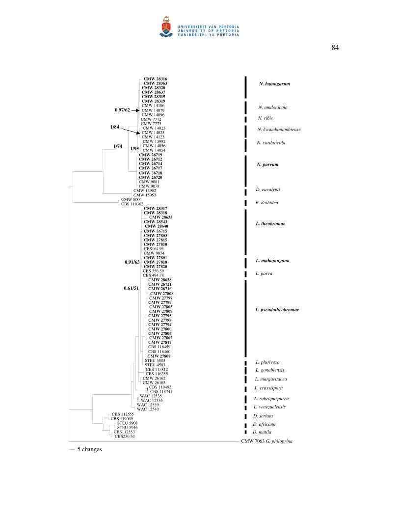
84
CMW 28316CMW 28363
CMW 28320 CMW 28637 CMW 28315 CMW 28319 CMW 14106 CMW 14079 CMW 14096 CMW 7772 CMW 7773 CMW 14023 CMW 14025 CMW 14123 CMW 13992 CMW 14056 CMW 14054
CMW 26719 CMW 26712 CMW 26714 CMW 26717 CMW 26718 CMW 26720 CMW 9081 CMW 9078
CMW 15952 CMW 15953
CMW 8000 CBS 110302
CMW 28317CMW 28318
CMW 28635 CMW 28543CMW 28640CMW 26715CMW 27803CMW 27815CMW 27810CBS164.96 CMW 9074 CMW 27801CMW 27818CMW 27820CBS 356.59 CBS 494.78
CMW 28638CMW 26721CMW 26716CMW 27808CMW 27797 CMW 27799CMW 27805 CMW 27809 CMW 27795CMW 27798CMW 27794CMW 27800CMW 27804CMW 27802 CMW 27817CBS 116459 CBS 116460 CMW 27807
STEU 5803 STEU 4583 CBS 115812 CBS 116355
CMW 26162 CMW 26163
CBS 110492 CBS 118741
WAC 12535 WAC 12536
WAC 12539 WAC 12540
CBS 112555 CBS 119049
STEU 5908 STEU 5946
CBS112553 CBS230.30
CMW 7063 G. philoprina
5 changes
1/74
0.97/62
1/84
1/95
0.61/51
0.91/63
1
1
N. batangarum
N. umdonicola
N. ribis
N. kwambonambiense
N. cordaticola
N. parvum
D. eucalypti
B. dothidea
L. theobromae
L. mahajangana
L. parva
L. pseudotheobromae
L. plurivora
L. gonubiensis
L. margaritacea
L. crassispora
L. rubropurpurea
L. venezuelensis
D. seriata
D. africana
D. mutila

85
Figure 2. One of the most parsimonious trees obtained from Maximum Parsimony analyses of
the combined ITS and tef 1-� sequence data of the representative taxa of the
Botryosphaeriaceae. Posterior probabilities followed by Bootstrap support (%) from 1000
replications are given on the branch (PP/BS). Isolates marked in bold represent those obtained
from T. catappa.

86
CMW 28315 CMW 28363
CMW 28320 CMW 28637 CMW 14106 CMW 14079 CMW 14096
CMW 7772 CMW 7773
CMW 13992 CMW 14056 CMW 14054
CMW 14023 CMW 14025 CMW 14123 CMW 26714 CMW 26717 CMW 26718 CMW 26720 CMW 9081 CMW 9079
CMW 15952 CMW 15953
CMW 8000 CBS 110302
CMW 28317 CMW 28319 CBS164.96 CMW 9074 CMW 27810 CMW 26715
STEU 5803 STEU 4583
CMW 27801 CMW 27818 CMW 27820
CMW 26721 CMW 26716 CMW 27802 CMW 27817 CBS 116459 CBS 447.62
CBS 356.59 CBS 494.78
CMW 26162 CMW 26163
CBS 115812 CBS 116355
CBS 110492 CBS 118741
WAC 12539 WAC 12540
WAC 12535 WAC 12536
CBS 112555 CBS 119049
CBS 112553 CBS 230.30
CMW 7063 G. philoprina
10 changes
0.98/97
0.86/100
1/100
0.94/56
1/75
0.96/100
0.99/98
0.99/85
0.95/87
0.98/100
1/100
0.98/100
0.66/100
0.53/55
0.78/86
1/90
0.98/84
0.76/63
1/100
1/100
0.52/681/99
1/100
1/100
1/100
1/100
1/100
1/990.99/100
1/100
0.93/55
N. batangarum
B. dothidea
N. ribis
N. cordaticola
N. parvum
D. eucalypti
L. plurivora
L. theobromae
N. umdonicola
D.mutila
D. seriata
L. rubropurpurea
L. venezuelensis
L. crassispora
L. gonubiensis
L. margaritacea
L. parva
L. pseudotheobromae
L. mahajangana
N. kwambonambiense

87
Figure 3. One of the most parsimonious unrooted trees inferred from independent analyses of
each data set (A = ITS; B = tef 1-�; C = rpb2; D = �-tub; E = BOTF15; F = combination of
sequences of the five loci) in the Neofusicoccum spp. group of the Botryosphaeriaceae from T.
catappa. Bootstrap support (%) from 1000 replications followed by Posterior probabilities are
given on the branches (BS/PP). Isolates marked in bold represent those obtained from T.
catappa.

88
CMW 7773 N. ribis
CMW 28315
CMW 28363
CMW 28320CMW 28637
CMW 14106
CMW 14079
CMW 14096
CMW 7772 N. ribis
CMW 9079
CMW 26714
CMW 26717
CMW 26718
CMW 26720 CMW 9081
CMW 14123 CMW 14023
CMW 14025
CMW 14054
CMW 13992
CMW 14056
64
65/0.9663/0.97
87/1 88/1
96/1
A
N. umdonicola
N. kwambonambiense
N. cordaticola
N. batangarum
N. parvum
CMW 9079
CMW 26714
CMW 26717
CMW 26718CMW 26720
CMW 9081
CMW 14096 CMW 14079
CMW 14106
CMW 14023
CMW 14025
CMW 14123
CMW 14054
CMW 13992
CMW 14056
CMW 7054 CMW 7772
CMW 28315 CMW 28363
CMW 28320CMW 28637
98/187/0.91
100/0.95
B
N. cordaticola N. batangarum
N. parvum
N. umdonicola
N. kwambonambiense
N. ribis

89
CMW 9081
CMW 26714
CMW 26717
CMW 26718CMW 26720
CMW 9079
CMW 13992 CMW 14056
CMW 14054
CMW 14023
CMW 14025
CMW 14123
CMW 14096
CMW 14106
CMW 14079
CMW 7772 CMW 7772
CMW 28315CMW 28363
CMW 28320 CMW 28637
7268
97/0.98
97/0.99
6165
D
N. umdonicola N. batangarum
N. ribis
N. parvum
N. cordaticola
N. kwambonambiense
CMW 7773 N. ribis
CMW 14106
CMW 14079
CMW 14096
CMW 28315
CMW 28363
CMW 28320
CMW 28637
CMW 7772 N. ribis
CMW 9079
CMW 26714
CMW 26717
CMW 26718CMW 26720 CMW 9081
CMW 14123
CMW 14023
CMW 14025
CMW 14054
CMW 13992
CMW 14056
99/1
63/0.95
96/1 98/1
98/1
C
N. parvum
N. cordaticola
N. kwambonambiense
N. umdonicoola
N. batangarum

90
CMW 7772
CMW 7773
CMW 28315
CMW 28363 CMW 28320
CMW 28637
CMW 14106
CMW 14079
CMW 14096
CMW 9079
CMW 26714 CMW 26717
CMW 26718 CMW 26720
CMW 9081
CMW 13992
CMW 14056 CMW 14054
CMW 14023
CMW 14025
CMW 14123
100/1
100/1
86/1
98/1
100/1 67/0.9
100/1
100/1
N. batangarum
F
N. ribis
N. parvum
N. umdonicola
N. cordaticola
N. kwambonambiense
CMW 14096 N. umdonicola
CMW 28315
CMW 28363
CMW 28320 CMW 28637
CMW 7772 CMW 7773
CMW 14106
CMW 14079
CMW 26720
CMW 9079
CMW 9081
CMW 26714
CMW 26717 CMW 26718
CMW 14123 CMW 14023
CMW 14025
CMW 14054
CMW 13992
CMW 14056 87
63
86
100/0.66
86/0.87
N. batangarum
E N. kwambonambiense
N. umdonicola
N. parvum N. cordaticola
N. ribis
91
Figure 4. Most-parsimonious unrooted trees inferred from independent analyses of each data
set (A = ITS; B = tef 1-�; C = �-tub; D = combination of sequences of the three loci) of the
Lasiodiplodia spp. from T. catappa and related species. Bootstrap support (%) from 1000
replications followed by Posterior probabilities are given on the branch (BS/PP). Isolates
marked in bold represent those obtained from T. catappa.

92
CBS 447.62 CBS 116459
CBS 356.59
CBS 494.78
CMW 27820
CMW 27801 CMW 27818
CMW 9074
CBS 164.96
100/1
100/0.99
90/0.97
100/0.97
96/0.57
L. mahajangana B
L. theobromae
L. pseudotheobromae
L. parva
CMW 27820
CMW 27801
CMW 27818
CMW 9074
CBS 164.96
CBS 116459 CBS 447.62
CBS 356.59
CBS 494.78 63/0.98
61/0.98
95/1
A
L. mahajangana
L. parva
L. pseudotheobromae
L. theobromae

93
CBS 447.62 CBS 116459
CBS 356.59
CBS 494.78
CMW 27820
CMW 27801CMW 27818
CMW 9074
CBS 164.96 100/1
100/1
96/1
100/1
100/1
D L. mahajangana
L. parva
L. pseudotheobromae
L. theobromae
CBS 116459
CBS 164.96
CMW 9074
CBS 447.62
L. pseudotheobromae
CMW 27820
CMW 27801 CMW 28818
CBS 494.78
CBS 356.59
88/1
95/1
88/1
64/0.99
L. mahajangana C
L. theobromae
L. pseudotheobromae
L. parva

94
Figure 5. Lasiodiplodia mahajangana. (a) Pycnidium formed on pine needle in culture. (b)
Paraphyses. (d) Conidiogenous cells with developing conidia. (d) conidia. (e) mature
conidium showing septum. Bars: a = 500 µm; b, c, d, e = 10 µm.

95
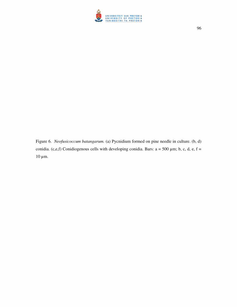
96
Figure 6. Neofusicoccum batangarum. (a) Pycnidium formed on pine needle in culture. (b, d)
conidia. (c,e,f) Conidiogenous cells with developing conidia. Bars: a = 500 µm; b, c, d, e, f =
10 µm.

97

98
Figure 7. Neofusicoccum batangarum culture on MEA. (a) Front plate. (b) Reverse plate.

99

100
Figure 8. Distribution of Botryosphaeriaceae collected from T. catappa per locality.

101

102
Figure 9. Mean lesion lengths (mm) of bark lesions for each Botryosphaeriaceae isolate six
weeks after inoculation on T. catappa (P < 0.0001). L. pseudotheobromae (LPs), N. parvum
(NP), L. theobromae (LT), L. mahajangana (LM), N. batangarum (NB), Control.
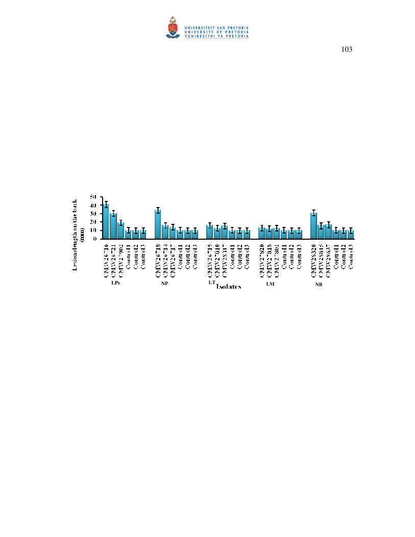
103
LPs NP LT LM NB
104
Figure 10. Mean lesion lengths (mm) on cambial lesions for each Botryosphaeriaceae isolate
six weeks after inoculation on T. catappa (P < 0.0001). L. pseudotheobromae (LPs), N.
parvum (NP), L. theobromae (LT), L. mahajangana (LM), N. batangarum (NB), Control.

105
LPs NP LT LM NB