Fine Structure, Physiology and Biochemistry of Arthrospore Germination in Streptomyces antibioticus
Upload
khangminh22Category
view
1download
0
89
Original Paper
Cell Physiol Biochem 2005;15:89-98 Accepted: September 14, 2004Cellular PhysiologyCellular PhysiologyCellular PhysiologyCellular PhysiologyCellular Physiologyand Biochemistrand Biochemistrand Biochemistrand Biochemistrand Biochemistryyyyy
Copyright © 2005 S. Karger AG, Basel
Fax +41 61 306 12 34E-Mail [email protected]
© 2005 S. Karger AG, Basel1015-8987/05/0154-0089$22.00/0
Accessible online at:www.karger.com/journals/net
Enzymatic Oxidation of Vanillin, Isovanillin andProtocatechuic Aldehyde with Freshly PreparedGuinea Pig Liver Slices
Georgios I. Panoutsopoulos1,2, Christine Beedham1
1Department of Pharmaceutical Chemistry, School of Pharmacy, University of Bradford, Bradford, 2Schoolof Biochemistry and Biotechnology, University of Thessaly, Athens
Dr Georgios I. Panoutsopoulos5 Tenedou Street, Platia Amerikis, Athens 112 57 (Greece)Tel. +30210-8649617, Fax +30210-7462554E-Mail [email protected]
Key WordsAldehyde dehydrogenase • Aldehyde oxidase •Allopurinol • Disulfiram • Isovanillin • Liver slices •Protocatechuic aldehyde • Vanillin • Xanthine oxidase
AbstractBackground/Aims: The oxidation of xenobiotic-derivedaromatic aldehydes with freshly prepared liver sliceshas not been previously reported. The presentinvestigation compares the relative contribution ofaldehyde oxidase, xanthine oxidase and aldehydedehydrogenase activities in the oxidation of vanillin,isovanillin and protocatechuic aldehyde with freshlyprepared liver slices. Methods: Vanillin, isovanillin orprotocatechuic aldehyde was incubated with liverslices in the presence/absence of specific inhibitorsof each enzyme, followed by HPLC. Results: Vanillinwas rapidly converted to vanillic acid. Vanillic acidformation was completely inhibited by isovanillin(aldehyde oxidase inhibitor), whereas disulfiram(aldehyde dehydrogenase inhibitor) inhibited acidformation by 16% and allopurinol (xanthine oxidaseinhibitor) had no effect. Isovanillin was rapidlyconverted to isovanillic acid. The formation ofisovanillic acid was not altered by allopurinol, but
considerably inhibited by disulfiram. Protocatechuicaldehyde was converted to protocatechuic acid at alower rate than that of vanillin or isovanillin. Allopurinolonly slightly inhibited protocatechuic aldehydeoxidation, isovanillin had little effect, whereasdisulfiram inhibited protocatechuic acid formation by50%. Conclusions: In freshly prepared liver slices,vanillin is rapidly oxidized by aldehyde oxidase withlittle contribution from xanthine oxidase or aldehydedehydrogenase. Isovanillin is not a substrate foraldehyde oxidase and therefore it is metabolized toisovanillic acid predominantly by aldehydedehydrogenase. All three enzymes contribute to theoxidation of protocatechuic aldehyde to its acid.
Introduction
Precision-cut liver slices are described as valuabletools for in vitro drug metabolism of xenobiotics andtoxicological studies [1-4]. They are easily prepared fromdifferent animal species, including man, using the samemethod and they closely resemble the organ from which
90
they are derived. Liver slices can retain their normal liverhepatocyte population and their physiological andbiochemical functions for many hours [5, 6]. Successfulexperimentation depends on the maintainance of sliceviability over the designated experimental period. Theslices, however, must be thin enough to allow nutrients,gases and waste exchange to occur [7, 8].
Aldehydes are reactive organic compounds, whichoccur as natural constituents in a wide variety of foodsand food additives [9, 10]. Many drugs, with an aminogroup, can be transformed into aldehydes via the actionof monoamine oxidase. Hydroxylation of an aromaticmethyl group to an alcohol, can be subsequently oxidizedto form an aldehydic group [11]. Other exogenousaldehydes can also occur through the ingestion ofmetabolic precursors such as the primary alcohols.Endogenous aldehydes are produced via oxidativedeamination of compounds such as biogenic aminescatalyzed by monoamine oxidase [3, 12].
Biogenic and xenobiotic aldehydes can either beoxidized to the corresponding acids, which may becatalyzed by aldehyde dehydrogenase (EC 1.2.1.3,aldehyde-NAD(P)+ oxidoreductase), aldehyde oxidase(EC 1.2.3.1, aldehyde-oxygen oxidoreductase) and/orxanthine oxidase (EC 1.2.3.2, xanthine-oxygenoxidoreductase), or reduced by aldehyde reductase and/or alcohol dehydrogenase to the corresponding alcohols.However, the main metabolic route is conversion to acids.
4-Hydroxy-3-methoxybenzaldehyde (vanillin) hasbeen shown to be an excellent substrate of aldehydeoxidase with a lower affinity for xanthine oxidase [13,14]. In contrast, 3,4-dihydroxybenzaldehyde(protocatechuic aldehyde) and 3-hydroxy-4-methoxybenzaldehyde (isovanillin) were shown to bereasonable substrates of xanthine oxidase but potentcompetitive inhibitors of aldehyde oxidase [13, 14].
Vanillin and isovanillin have been previously testedas substrates for aldehyde dehydrogenase, where bothcompounds were found as substrates for guinea pig livermitochondrial aldehyde dehydrogenase [14] and forRhodopseudomonas acidophila M402 aldehydedehydrogenase [15]. Protocatechuic aldehyde was foundas substrate of human [16] and guinea pig [14] livermitochondrial aldehyde dehydrogenase.
Vanillin is found naturally in vanilla beans and issynthesized in the food industry for use as a flavoringagent in confectionery, beverages and foodstuffs [17]. Itis also found in barrel-aged wine [18] and is a by-productof curcumin from turmeric [19].
Vanillin acts as an anticarcinogen by reducing thenumber of small intestinal tumors, which are induced fromseveral carcinogens [20, 21]. It also acts as an antioxidantand pro-oxidant by decreasing the induced damage fromH2O2 [22]. It exerts its antimutagenic activity in damagedcells by promoting recombination and rejoining of DNAat homologous sites [23] and/or by inhibiting DNA repairat the non-homologous end-joining due to DNA-dependantprotein kinase activity inhibition from vanillin [24].Therefore, vanillin might have beneficial antimutageniceffects and the ability to potentiate the effectiveness ofanticancer drugs.
Protocatechuic aldehyde has been demonstrated toinduce apoptotic cell death in cytotoxic T-cells (CTLL-2)either by inhibiting the ornithine decarboxylase activity,which is induced by IL-2, or by interfering with MAPKsignaling pathways [25, 26].
The present investigation examines the oxidation ofthe substituted benzaldehydes vanillin, isovanillin andprotocatechuic aldehyde with freshly prepared precision-cut guinea pig liver slices and followed by HPLC.Incubations were also performed in the presence ofspecific inhibitors for several oxidizing enzymes in orderto determine the relative contribution of each enzyme inthe oxidation of these aldehydes. Guinea pig liver waschosen in this study as aldehyde oxidase has similarsubstrate specificity to the human liver enzyme [27, 28].
Materials and Methods
AnimalsDunkin-Hartley female guinea pigs (450-950 g) were used
for the investigation. Animals were fed with FD1 pelletssupplemented with ascorbic acid and received hay three timesweekly. All animals were allowed food and water ad libitum andmaintained in a strictly controlled temperature (18±1°C), humidity(50 %) and lighting cycle (07.00-19.00 h light, 19.00-07.00 hdark). Animals were handled with humane care in accordancewith the National Institutes of Health guidelines.
Chemicals4-Hydroxy-3-methoxybenzaldehyde (vanillin), 4-hydroxy-
3-methoxybenzylalcohol (vanillyl alcohol), 3,4-dihydroxybenzaldehyde (protocatechuic aldehyde), 3,4-dihydroxybenzoic acid (protocatechuic acid), 3-hydroxy-4-methoxybenzaldehyde (isovanillin), 3-hydroxy-4-methoxybenzoic acid (isovanillic acid), tetraethylthiuramdisulfide (disulfiram), allopurinol, diethylamine from SigmaChemical Company Ltd, 3-hydroxy-4-methoxybenzylalcohol
Panoutsopoulos/BeedhamCell Physiol Biochem 2005;15:89-98
91
(isovanillyl alcohol) from Aldrich Chemical Co Ltd, perchloricacid, 4-hydroxy-3-methoxybenzoic acid (vanillic acid) from BDHChemicals Ltd, and acetonitrile HPLC grade from RathburnChemicals Ltd.
High performance liquid chromatography (HPLC)Reverse-phase HPLC was performed using a system from
Waters Associates (Northwich, Cheshire, UK), which consistedof a single piston reciprocating 501 pump, a WISP 710B auto-injector, a Lambda-Max 481 LC spectrophotometer and a DataModule 740. Compounds were separated with a stainless steelHypersil ODS column 5 µm (25 cm×4.6 mm internal diameter). AWaters Guard Pak pre-column with a C18 insert was fitted tothe HPLC system for protection of the column from the biologicalmaterials. The optimum wavelength was 285 nm and 20 µlsamples were injected for each analysis. HPLC separation wasachieved with a mobile phase containing 15 % acetonitrile and85 % orthophosphate buffer (2.2×10⁻¹M) pH 2.9 containing1.1×10-1M diethylamine at a flow rate of 1.5 ml/min. The mobilephase was filtered and degassed for 10 min under vacuumbefore use.
Preparation of freshly prepared guinea pig liver slicesGuinea pigs were killed by cervical dislocation and their
livers were immediately excised. The livers were placed in ice-cold Krebs-Henseleit solution pH 7.4 containing 2.4×10⁻²Mbicarbonate, which was continuously oxygenated with 95 %O2/5 % CO2. Liver slices were then obtained according to themethod of Panoutsopoulos et al [3] and the freshly preparedliver slices were stored in oxygenated Krebs-Henseleit bufferpH 7.4 containing 2.4×10⁻²M bicarbonate at 4°C until required.
Incubation conditions with liver slicesVanillin, isovanillin or protocatechuic aldehyde (1×10⁻³M)
was incubated with 4 liver slices in a total volume of 3 ml Krebs-Henseleit buffer pH 7.4, the addition of the liver slices indicatedthe beginning of the experiment. Incubation mixtures were
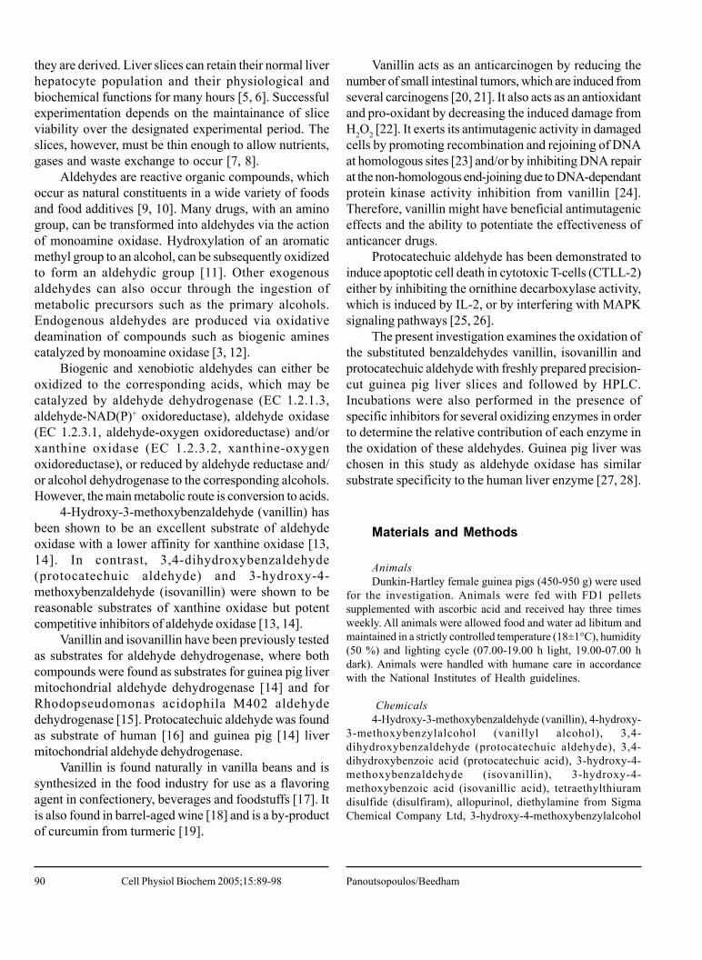
Fig. 1. Metabolism of vanillin by freshlyprepared guinea pig liver slices. Vanillin(1×10⁻³M) was incubated with four guineapig liver slices (147 mg) in 3 ml of Krebs-Heinseleit buffer pH 7.4 at 37°C. Each pointis the mean of five determinations. Valuesare expressed as means±SE.
maintained at 37°C in a shaking water bath. The medium wasoxygenated with 95 % O2/5 % CO2 initially and every subsequenthour for 5 min. Incubations with freshly prepared liver sliceswere also performed in the presence of inhibitors. The inhibitorsused were isovanillin for aldehyde oxidase activity [13, 14],allopurinol for xanthine oxidase activity [29] and disulfiram foraldehyde dehydrogenase activity [30, 31].
Aliquots (0.2 ml) were removed at t= 0 min and at varioustime intervals, added to 0.1 ml of 3.6 % perchloric acid andcentrifuged for 2.5 min at maximum speed on a Beckmanmicrofuge B. The supernatant was then analyzed by HPLC.Control incubations without the liver slices present were alsoperformed. Standard solutions of the aldehydes and theirpossible metabolites were also analyzed by HPLC. Afteranalysis, the slices were blotted dry and weighed to determinethe total weight of liver used. For comparison reasons, theresults on the effect of inhibitors in liver slices have beennormalized per 100 mg of liver.
Results
Metabolism of vanillin with freshly preparedguinea pig liver slicesVanillin (Rt= 13.2±1.0 min, n=7) concentration was
progressively decreased with time, so that after 180 min(Fig. 1) it had been completely transformed into threemetabolites. The major metabolite was vanillic acid (Rt=7.4±0.5 min, n=7), which accounted for approximately82 % conversion of the substrate. The other two minormetabolites were vanillyl alcohol and protocatechuic acid(Rt= 4.3±0.5 min, n=7). Vanillyl alcohol increased to itsmaximum concentrations at 90 min and thereaftergradually disappeared, whereas protocatechuic acid
Aromatic Aldehyde Metabolism in Fresh Liver Slices Cell Physiol Biochem 2005;15:89-98
92
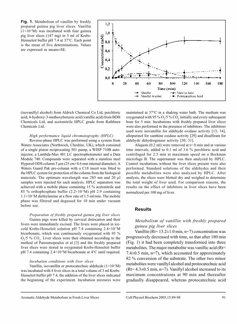
Fig. 2. Effect of isovanillin onvanillin oxidation by freshlyprepared guinea pig liver slices.(i) Vanillin (1×10⁻³M) (a) andisovanillin (1×10⁻³M) (b) wereincubated individually withfour guinea pig liver slices in 3ml of Krebs-Heinseleit bufferpH 7.4 at 37°C. (ii) Vanillin(1×10⁻³M) and isovanillin (1×10-
3M) were incubated togetherwith four guinea pig liver slicesin 3 ml of Krebs-Heinseleitbuffer pH 7.4 at 37°C. Eachpoint is the mean of fivedeterminations. Values areexpressed as means±SE.
accounted for approximately 16 % conversion of vanillinafter 180 min and was thought to arise via an O-demethylation reaction. The total amount of all threemetabolites accounted for all of the vanillin transformed.Control incubations of vanillin, in the absence of guineapig liver slices, showed no such decrease in theconcentration of vanillin and likewise, no metabolites wereformed.
Protocatechuic acid formation in vanillin incubationscould arise via O-demethylation of vanillic acid or fromO-demethylation of vanillin to protocatechuic aldehydeand subsequent oxidation to the corresponding acid.Therefore, in order to elucidate which of these pathwayswas the most likely, vanillic acid (1×10⁻³M) was incubated
with freshly prepared guinea pig liver slices (n=4). A 13% decrease of vanillic acid concentration was observedafter 90 min and a 23 % decrease after 180 min (data notshown). One main metabolite and traces of a number ofother polar metabolites were produced. However, the mainmetabolite was protocatechuic acid, which accounted for8 % conversion of the vanillic acid after 90 min and 16 %after 180 min (data not shown). Thus, it would appearthat microsomal O-demethylation of vanillic acid toprotocatechuic acid occurred in liver slice incubations.
Effect of inhibitors on vanillin oxidation byfreshly prepared guinea pig liver slicesIn the presence of isovanillin (1×10⁻³M), vanillin
Panoutsopoulos/BeedhamCell Physiol Biochem 2005;15:89-98
93
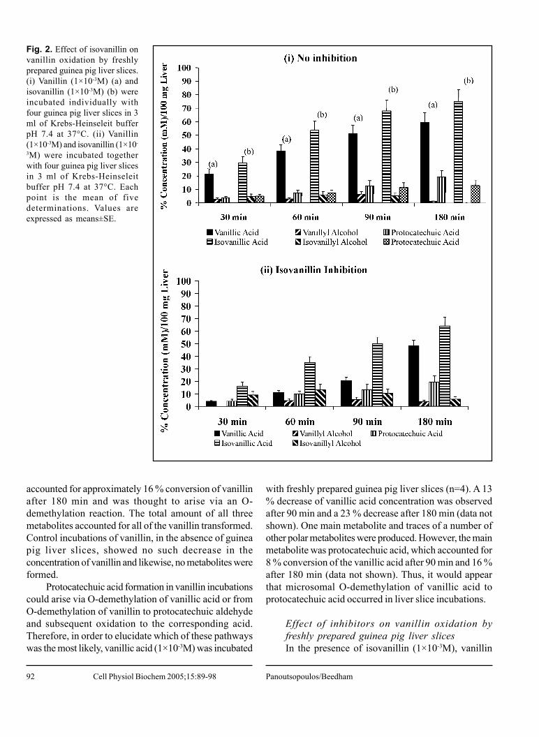
Fig. 3. Effect of inhibitors on vanillinoxidation by freshly prepared guineapig liver slices. Vanillin (1×10⁻³M) wasincubated in the absence ofinhibitors and in the presence of (a)allopurinol (1×10⁻⁴M), (b) disulfiram(1×10⁻⁴M) and (c) disulfiram (1×10-
4M) and allopurinol (1×10⁻⁴M), withfour guinea pig liver slices in 3 ml ofKrebs-Heinseleit buffer pH 7.4 at37°C and t=90 min. Each point is themean of four determinations. Valuesare expressed as means±SE.
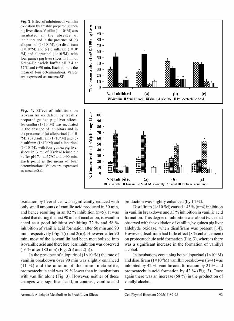
Fig. 4. Effect of inhibitors onisovanillin oxidation by freshlyprepared guinea pig liver slices.Isovanillin (1×10⁻³M) was incubatedin the absence of inhibitors and inthe presence of (a) allopurinol (1×10-
4M), (b) disulfiram (1×10⁻⁴M) and (c)disulfiram (1×10⁻⁴M) and allopurinol(1×10⁻⁴M), with four guinea pig liverslices in 3 ml of Krebs-Heinseleitbuffer pH 7.4 at 37°C and t=90 min.Each point is the mean of fourdeterminations. Values are expressedas means±SE.
oxidation by liver slices was significantly reduced withonly small amounts of vanillic acid produced in 30 min,and hence resulting in an 82 % inhibition (n=5). It wasnoted that during the first 90 min of incubation, isovanillinacted as a good inhibitor exhibiting 72 % and 58 %inhibition of vanillic acid formation after 60 min and 90min, respectively (Fig. 2(i) and 2(ii)). However, after 90min, most of the isovanillin had been metabolized intoisovanillic acid and therefore, less inhibition was observed(16 % after 180 min) (Fig. 2(i) and 2(ii)).
In the presence of allopurinol (1×10⁻⁴M) the rate ofvanillin breakdown over 90 min was slightly enhanced(11 %) and the amount of the minor metabolite,protocatechuic acid was 19 % lower than in incubationswith vanillin alone (Fig. 3). However, neither of thesechanges was significant and, in contrast, vanillic acid
production was slightly enhanced (by 14 %).Disulfiram (1×10⁻⁴M) caused a 43 % (n=4) inhibition
in vanillin breakdown and 33 % inhibition in vanillic acidformation. This degree of inhibition was about twice thatobserved with the oxidation of vanillin, by guinea pig liveraldehyde oxidase, when disulfiram was present [14].However, disulfiram had little effect (8 % enhancement)on protocatechuic acid formation (Fig. 3), whereas therewas a significant increase in the formation of vanillylalcohol.
In incubations containing both allopurinol (1×10⁻⁴M)and disulfiram (1×10⁻⁴M) vanillin breakdown (n=4) wasinhibited by 42 %, vanillic acid formation by 21 % andprotocatechuic acid formation by 42 % (Fig. 3). Onceagain there was an increase (58 %) in the production ofvanillyl alcohol.
Aromatic Aldehyde Metabolism in Fresh Liver Slices Cell Physiol Biochem 2005;15:89-98
94
Metabolism of isovanillin with freshly preparedguinea pig liver slicesThe time profile of isovanillin metabolism (Rt=
12.6±1.1 min, n=5), with freshly prepared guinea pig liverslices, was similar to that observed in vanillin metabolism(Fig. 1). A progressive decrease in aldehyde concentrationwas observed, which continued until it had been completelytransformed during the 180 min incubation time.Simultaneously, three metabolites appeared whichincreased with time and accounted for approximately 100% conversion of the aldehyde after 180 min (data notshown). The major metabolite formed was isovanillic acid(Rt= 8.2±0.6 min), which accounted for 87 % conversionof isovanillin. The other two metabolites wereprotocatechuic acid (13 % conversion) and isovanillylalcohol (Rt= 5.7 min), which achieved maximumconcentrations between 45-60 min and then subsequentlydecreased with no alcohol remaining after 180 min.Control incubations of isovanillin without guinea pig liverslices, did not show any change in isovanillin concentrationand no metabolites were formed.
Due to the unexpected formation of protocatechuicacid in isovanillin incubations with liver slices, incubationsof isovanillic acid with guinea pig liver slices were alsoperformed. A decrease in isovanillic acid concentration(12.5 %) was noted, at t= 180 min, but to a lesser extentthan vanillic acid incubation under similar conditions (datanot shown). The major metabolite observed wasprotocatechuic acid (4.5 %, t= 180 min), whereas theother two minor polar metabolites had retention values of3.7 min and 4.0 min. Neither of these compounds co-
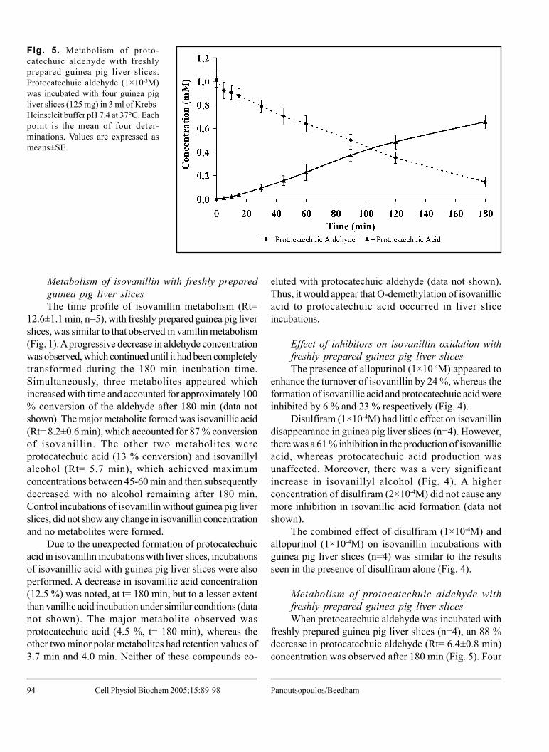
Fig. 5. Metabolism of proto-catechuic aldehyde with freshlyprepared guinea pig liver slices.Protocatechuic aldehyde (1×10⁻³M)was incubated with four guinea pigliver slices (125 mg) in 3 ml of Krebs-Heinseleit buffer pH 7.4 at 37°C. Eachpoint is the mean of four deter-minations. Values are expressed asmeans±SE.
eluted with protocatechuic aldehyde (data not shown).Thus, it would appear that O-demethylation of isovanillicacid to protocatechuic acid occurred in liver sliceincubations.
Effect of inhibitors on isovanillin oxidation withfreshly prepared guinea pig liver slicesThe presence of allopurinol (1×10⁻⁴M) appeared to
enhance the turnover of isovanillin by 24 %, whereas theformation of isovanillic acid and protocatechuic acid wereinhibited by 6 % and 23 % respectively (Fig. 4).
Disulfiram (1×10⁻⁴M) had little effect on isovanillindisappearance in guinea pig liver slices (n=4). However,there was a 61 % inhibition in the production of isovanillicacid, whereas protocatechuic acid production wasunaffected. Moreover, there was a very significantincrease in isovanillyl alcohol (Fig. 4). A higherconcentration of disulfiram (2×10⁻⁴M) did not cause anymore inhibition in isovanillic acid formation (data notshown).
The combined effect of disulfiram (1×10⁻⁴M) andallopurinol (1×10⁻⁴M) on isovanillin incubations withguinea pig liver slices (n=4) was similar to the resultsseen in the presence of disulfiram alone (Fig. 4).
Metabolism of protocatechuic aldehyde withfreshly prepared guinea pig liver slicesWhen protocatechuic aldehyde was incubated with
freshly prepared guinea pig liver slices (n=4), an 88 %decrease in protocatechuic aldehyde (Rt= 6.4±0.8 min)concentration was observed after 180 min (Fig. 5). Four
Panoutsopoulos/BeedhamCell Physiol Biochem 2005;15:89-98
95
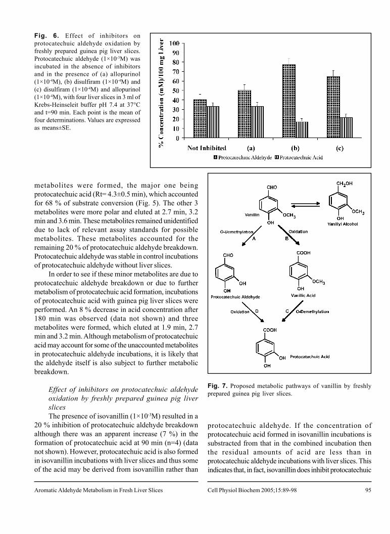
Fig. 6. Effect of inhibitors onprotocatechuic aldehyde oxidation byfreshly prepared guinea pig liver slices.Protocatechuic aldehyde (1×10⁻³M) wasincubated in the absence of inhibitorsand in the presence of (a) allopurinol(1×10⁻⁴M), (b) disulfiram (1×10⁻⁴M) and(c) disulfiram (1×10⁻⁴M) and allopurinol(1×10⁻⁴M), with four liver slices in 3 ml ofKrebs-Heinseleit buffer pH 7.4 at 37°Cand t=90 min. Each point is the mean offour determinations. Values are expressedas means±SE.
metabolites were formed, the major one beingprotocatechuic acid (Rt= 4.3±0.5 min), which accountedfor 68 % of substrate conversion (Fig. 5). The other 3metabolites were more polar and eluted at 2.7 min, 3.2min and 3.6 min. These metabolites remained unidentifieddue to lack of relevant assay standards for possiblemetabolites. These metabolites accounted for theremaining 20 % of protocatechuic aldehyde breakdown.Protocatechuic aldehyde was stable in control incubationsof protocatechuic aldehyde without liver slices.
In order to see if these minor metabolites are due toprotocatechuic aldehyde breakdown or due to furthermetabolism of protocatechuic acid formation, incubationsof protocatechuic acid with guinea pig liver slices wereperformed. An 8 % decrease in acid concentration after180 min was observed (data not shown) and threemetabolites were formed, which eluted at 1.9 min, 2.7min and 3.2 min. Although metabolism of protocatechuicacid may account for some of the unaccounted metabolitesin protocatechuic aldehyde incubations, it is likely thatthe aldehyde itself is also subject to further metabolicbreakdown.
Effect of inhibitors on protocatechuic aldehydeoxidation by freshly prepared guinea pig liverslicesThe presence of isovanillin (1×10⁻³M) resulted in a
20 % inhibition of protocatechuic aldehyde breakdownalthough there was an apparent increase (7 %) in theformation of protocatechuic acid at 90 min (n=4) (datanot shown). However, protocatechuic acid is also formedin isovanillin incubations with liver slices and thus someof the acid may be derived from isovanillin rather than
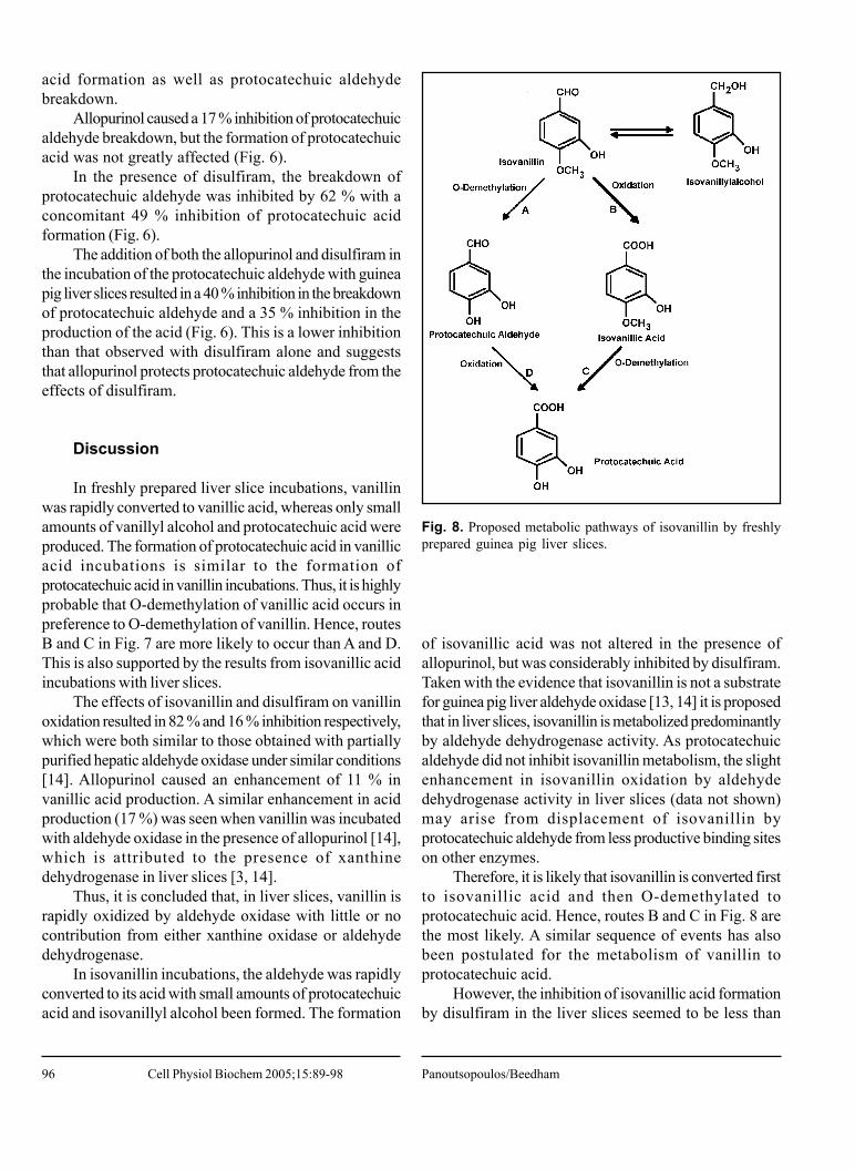
Fig. 7. Proposed metabolic pathways of vanillin by freshlyprepared guinea pig liver slices.
protocatechuic aldehyde. If the concentration ofprotocatechuic acid formed in isovanillin incubations issubstracted from that in the combined incubation thenthe residual amounts of acid are less than inprotocatechuic aldehyde incubations with liver slices. Thisindicates that, in fact, isovanillin does inhibit protocatechuic
Aromatic Aldehyde Metabolism in Fresh Liver Slices Cell Physiol Biochem 2005;15:89-98
96
acid formation as well as protocatechuic aldehydebreakdown.
Allopurinol caused a 17 % inhibition of protocatechuicaldehyde breakdown, but the formation of protocatechuicacid was not greatly affected (Fig. 6).
In the presence of disulfiram, the breakdown ofprotocatechuic aldehyde was inhibited by 62 % with aconcomitant 49 % inhibition of protocatechuic acidformation (Fig. 6).
The addition of both the allopurinol and disulfiram inthe incubation of the protocatechuic aldehyde with guineapig liver slices resulted in a 40 % inhibition in the breakdownof protocatechuic aldehyde and a 35 % inhibition in theproduction of the acid (Fig. 6). This is a lower inhibitionthan that observed with disulfiram alone and suggeststhat allopurinol protects protocatechuic aldehyde from theeffects of disulfiram.
Discussion
In freshly prepared liver slice incubations, vanillinwas rapidly converted to vanillic acid, whereas only smallamounts of vanillyl alcohol and protocatechuic acid wereproduced. The formation of protocatechuic acid in vanillicacid incubations is similar to the formation ofprotocatechuic acid in vanillin incubations. Thus, it is highlyprobable that O-demethylation of vanillic acid occurs inpreference to O-demethylation of vanillin. Hence, routesB and C in Fig. 7 are more likely to occur than A and D.This is also supported by the results from isovanillic acidincubations with liver slices.
The effects of isovanillin and disulfiram on vanillinoxidation resulted in 82 % and 16 % inhibition respectively,which were both similar to those obtained with partiallypurified hepatic aldehyde oxidase under similar conditions[14]. Allopurinol caused an enhancement of 11 % invanillic acid production. A similar enhancement in acidproduction (17 %) was seen when vanillin was incubatedwith aldehyde oxidase in the presence of allopurinol [14],which is attributed to the presence of xanthinedehydrogenase in liver slices [3, 14].
Thus, it is concluded that, in liver slices, vanillin israpidly oxidized by aldehyde oxidase with little or nocontribution from either xanthine oxidase or aldehydedehydrogenase.
In isovanillin incubations, the aldehyde was rapidlyconverted to its acid with small amounts of protocatechuicacid and isovanillyl alcohol been formed. The formation
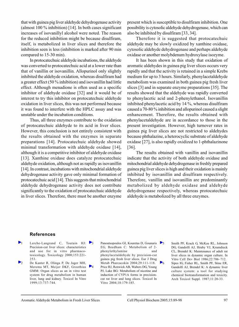
Fig. 8. Proposed metabolic pathways of isovanillin by freshlyprepared guinea pig liver slices.
of isovanillic acid was not altered in the presence ofallopurinol, but was considerably inhibited by disulfiram.Taken with the evidence that isovanillin is not a substratefor guinea pig liver aldehyde oxidase [13, 14] it is proposedthat in liver slices, isovanillin is metabolized predominantlyby aldehyde dehydrogenase activity. As protocatechuicaldehyde did not inhibit isovanillin metabolism, the slightenhancement in isovanillin oxidation by aldehydedehydrogenase activity in liver slices (data not shown)may arise from displacement of isovanillin byprotocatechuic aldehyde from less productive binding siteson other enzymes.
Therefore, it is likely that isovanillin is converted firstto isovanillic acid and then O-demethylated toprotocatechuic acid. Hence, routes B and C in Fig. 8 arethe most likely. A similar sequence of events has alsobeen postulated for the metabolism of vanillin toprotocatechuic acid.
However, the inhibition of isovanillic acid formationby disulfiram in the liver slices seemed to be less than
Panoutsopoulos/BeedhamCell Physiol Biochem 2005;15:89-98
97
References
1 Lerche-Langrand C, Toutain HJ:Precision-cut liver slices: characteristicsand use for in vitro pharmaco-toxicology. Toxicology 2000;153:221-253.
2 De Kanter R, Olinga P, De Jager MH,Merema MT, Meijer DKF, GroothiusGMM: Organ slices as an in vitro testsystem for drug metabolism in humanliver, lung and kidney. Toxicol In Vitro1999;13:737-744.
3 Panoutsopoulos GI, Kouretas D, GounarisEG, Beedham C: Metabolism of 2-phenylethylamine andphenylacetaldehyde by precision-cutguinea pig fresh liver slices. Eur J DrugMetab Pharcacokin 2004;29:111-118.
4 Price RJ, Renwick AB, Walters DG, YoungPJ, Lake BG: Metabolism of nicotine andinduction of CYP1A forms in precision-cut rat liver and lung slices. Toxicol InVitro 2004;18:179-185.
5 Smith PF, Krack G, McKee RL, JohnsonDG, Gandolfi AJ, Hruby VJ, KrumdieckCL, Brendel K: Maintenance of adult ratliver slices in dynamic organ culture. InVitro Cell Dev Biol 1986;22:706–712.
6 Sipes IG, Fisher RL, Smith PF, Stine ER,Gandolfi AJ, Brendel K: A dynamic liverculture system: a tool for studyingchemical biotransformation and toxicity.Arch Toxicol Suppl. 1987;11:20-33.
that with guinea pig liver aldehyde dehydrogenase activity(almost 100 % inhibition) [14]. In both cases significantincreases of isovanillyl alcohol were noted. The reasonfor the reduced inhibition might be because disulfiram,itself, is metabolized in liver slices and therefore theinhibition seen is less (inhibition is marked after 90 mincompared to 15-30 min).
In protocatechuic aldehyde incubations, the aldehydewas converted to protocatechuic acid at a lower rate thanthat of vanillin or isovanillin. Allopurinol only slightlyinhibited the aldehyde oxidation, whereas disulfiram hada greater effect (50 % inhibition) and isovanillin had littleeffect. Although menadione is often used as a specificinhibitor of aldehyde oxidase [32] and it would be ofinterest to try this inhibitor on protocatechuic aldehydeoxidation in liver slices, this was not performed becauseit was found to interfere with the HPLC assay and wasunstable under the incubation conditions.
Thus, all three enzymes contribute to the oxidationof protocatechuic aldehyde to its acid in liver slices.However, this conclusion is not entirely consistent withthe results obtained with the enzymes in separatepreparations [14]. Protocatechuic aldehyde showedminimal transformation with aldehyde oxidase [14],although it is a competitive inhibitor of aldehyde oxidase[13]. Xanthine oxidase does catalyze protocatechuicaldehyde oxidation, although not as rapidly as isovanillin[14]. In contrast, incubations with mitochondrial aldehydedehydrogenase activity gave only minimal formation ofprotocatechuic acid [14]. This suggests that mitochondrialaldehyde dehydrogenase activity does not contributesignificantly to the oxidation of protocatechuic aldehydein liver slices. Therefore, there must be another enzyme
present which is susceptible to disulfiram inhibition. Onepossibility is cytosolic aldehyde dehydrogenase, which canalso be inhibited by disulfiram [33, 34].
Therefore it is suggested that protocatechuicaldehyde may be slowly oxidized by xanthine oxidase,cytosolic aldehyde dehydrogenase and perhaps aldehydeoxidase or another molybdenum hydroxylase isozyme.
It has been shown in this study that oxidation ofaromatic aldehydes in guinea pig liver slices occurs veryrapidly and that the activity is retained in a simple Krebsmedium for up to 3 hours. Similarly, phenylacetaldehydemetabolism was examined in both guinea pig fresh liverslices [3] and in separate enzyme preparations [35]. Theresults showed that the aldehyde was rapidly convertedto phenylacetic acid and 2-phenylethanol. Isovanillininhibited phenylacetic acid by 14 %, whereas disulfiramcaused a 70-80 % inhibition and allopurinol caused a slightenhancement. Therefore, the results obtained withphenylacetaldehyde are in accordance to those in thepresent investigation. However, high turnover rates inguinea pig liver slices are not restricted to aldehydesbecause phthalazine, a heterocyclic substrate of aldehydeoxidase [27], is also rapidly oxidized to 1-phthalazinone[36].
The results obtained with vanillin and isovanillinindicate that the activity of both aldehyde oxidase andmitochondrial aldehyde dehydrogenase in freshly preparedguinea pig liver slices is high and their oxidation is mainlyinhibited by isovanillin and disulfiram respectively.Therefore, vanillin and isovanillin are predominantlymetabolized by aldehyde oxidase and aldehydedehydrogenase respectively, whereas protocatechuicaldehyde is metabolized by all three enzymes.
Aromatic Aldehyde Metabolism in Fresh Liver Slices Cell Physiol Biochem 2005;15:89-98
98
7 Dogterom P: Development of a simpleincubation system for metabolism studieswith precision-cut liver slices. Drug MetabDispos 1993;21:699-704.
8 Fisher RL, Hasal SJ, Sanuik JT, GandolfiAJ, Brendel K: Determination of optimalincubation media and suitable slicediameters in precision-cut liver slices:optimization of tissue slice culture, part2. Toxicol Method 1995;5:115-130.
9 Lindahl R: Aldehyde dehydrogenases andtheir role in carcinogenesis. Crit RevBiochem Mol Biol 1992;27:283-335.
10 Feron VJ, Til HP, de Vrijer F, WoutersenRA, Cassee FR, van Bladeren PJ:Aldehydes: occurrence, carcinogenicpotential, mechanism of action and riskassessment. Mutat Res 1991;259:363-385.
1 1 Washio K, Makaya O, Sasaki H, NishidaK, Nakamura J, Shibasaki J: A new aspectof tolbutamide metabolism in the rabbit:the role of 1-butyl-3-(p-formylphenyl)sulphonylurea. J PharmPharmacol 1993; 45: 231-233.
12 Beedham C, Peet CF, Panoutsopoulos GI,Carter H, Smith JA: Role of aldehydeoxidase in biogenic amine metabolism.Prog Brain Res 1995;106:345-353.
13 Panoutsopoulos GI, Beedham C: Kineticsand specificity of guinea pig liveraldehyde oxidase and bovine milkxanthine oxidase towards substitutedbenzaldehydes. Acta Biochim Polon2004;51:649-663.
14 Panoutsopoulos GI, Kouretas D, BeedhamC: Contribution of aldehyde oxidase,xanthine oxidase and aldehydedehydrogenase on the oxidation ofaromatic aldehydes. Chem Res Toxicol2004; 17: 1368-1376.
15 Yamanaka K, Iino H, Oikawa T:Purification and properties of dye-linkedaldehyde dehydrogenase inRhodopseudomonas acidophila M402.Agric Biol Chem 1991;55:989-996.
16 Klyosov AA: Kinetics and specificity ofhuman liver aldehyde dehydrogenasestoward aliphatic, aromatic, and fusedpolycyclic aldehydes. Biochemistry1996;35:4457-4467.
1 7 Kuroda Y, Inouye T: Antimutagenesis byfactors affecting DNA repair in bacteria.Mutat Res 1988;202:387-391.
18 Spillman PJ, Pollnitz AP, LiacopoulosD, Skouroumounis GK, Sefton MA:Accumulation of vanillin during barrel-aging of white, red, and model wines. JAgric Food Chem 1997;45:2584-2589.
19 Wang YJ, Pan MH, Cheng AL, Lin LI,Ho YS, Hsieh CY, Lin JK: Stability ofcurcumin in buffer solutions andcharacterization of its degradationproducts. J Pharm Biomed Anal1997;15:1867-1876.
20 Akagi K, Hirose M, Hoshiya T,Mizoguchi Y, Ito N, Shrai T: Modulatingeffects of elagic acid, vanillin andquercetin in a rat medium term multi-organ carcinogenesis model. Cancer Lett1995;94:113-121.
21 Tsuda H, Uehara N, Iwahori Y, AsamotoM, Iiogo M, Nagao M, Matsumoto K,Ito M, Hirono J: Chemoprotectiveeffects of beta-carotene, alpha-tocopherol and five naturally occurringantioxidants on initiation ofhepatocarcinogenesis by 2-amino-3-methylimidazo[4,5-f]quinoline in therat. Jpn J Cancer Res 1994;85:1214-1219.
22 Liu J, Mori A: Antioxidant and pro-oxidant activities of p-hydroxybenzylalcohol and vanillin: effects on freeradicals, brain peroxidation anddegradation of benzoate, deoxyribose,amino acids and DNA.Neuropharmacology 1993;32:659-669.
23 Tamai K, Tezuka H, Kuroda Y: Directmodifications by vanillin in cytotoxicityand genetic changes induced by EMS andH2O2 in cultured Chinese hamster cells.Mutat Res 1992;268:231-237.
24 Durant S, Karran P: Vanillins - a novelfamily of DNA-PK inhibitors. NucleicAcids Res 2003;31:5501-5512.
25 Wang Y, Hasuma T, Yano Y, MorishimaY, Matsui-Yuasa I, Otani S: Induction ofapoptosis in CTLL-2 cells byprotocatechualdehyde. Anticancer Res2001;21:1095-1101.
26 Wang Y, Zhu Z, Hasuma T, MorishimaY, Matsui-Yuasa I, Otani S: Modulationof ornithine decarboxylase activity andmitogen-activated protein kinases inprotocatechualdehyde-induced apoptosisof CTLL-2 cells. Anticancer Res2002;22:3389-3394.
27 Beedham C, Bruce SE, Chritchley DJ,Rance DJ: 1-Substituted phthalazines asprobes of the substrate-binding site ofmammalian molybdenum hydroxylases.Biochem Pharmacol 1990;39:1213-1221.
28 Beedham C, Bruce SE, Critchley DJ, Al-Tayib Y, Rance DJ: Species variation inhepatic aldehyde oxidase activity. Eur JDrug Metab Pharmacokinet1987;12:307-310.
29 Peterson GM, Boyle RR, Francis HW,Oliver NWJ, Paterson J, von Witt RJ,Taylor GR: Dosage prescribing and plasmaoxipurinol levels in patients receivingallopurinol therapy. Eur J Pharmacol1990;39:419-421.
30 Lipsky JJ, Shen ML, Naylor S: Overview-In vitro inhibition of aldehydedehydrogenase by disulfiram andmetabolites. Chem-Biol Interact2001;130-132:81-91.
31 Deitrich RA, Erwin VG: Mechanism ofthe inhibition of aldehyde dehydrogenasein vivo by disulfiram anddiethyldithiocarbamate. Mol Pharmacol1971;7:301-307.
32 Rajagopalan KV, Handler P: Hepaticaldehyde oxidase. II. Differentialinhibition of electron transfer to variouselectron acceptors. J Biol Chem1964;239:2022-2026.
33 Lam JP, Mays DC, Lipsky JJ: Inhibitionof recombinant human mitochondrial andcytosolic aldehyde dehydrogenases bytwo candidates for the active metabolitesof disulfiram. Biochemistry1997;36:13748-13754.
34 Vallari RC, Pietruszko R: Human aldehydedehydrogenase: mechanism of inhibitionof disulfiram. Science 1982;216:637-639.
35 Panoutsopoulos GI, Kouretas D, GounarisEG, Beedham C: Enzymatic oxidation of2-phenylethylamine to phenylacetic acidand 2-phenylethanol with specialreference to the metabolism of itsintermediate phenylacetaldehyde. BasicClin Pharmacol Toxicol 2004;95: Inpress.
36 Panoutsopoulos GI, Beedham C:Enzymatic oxidation of phthalazine withfreshly prepared precision-cut guinea pigliver slices. Acta Biochim Polon 2004;51:In press.
Panoutsopoulos/BeedhamCell Physiol Biochem 2005;15:89-98
Copyright © 2022 FDOKUMEN