Cave Biosignature Suites: Microbes, Minerals, and Mars
31
ASTROBIOLOGY Volume 1, Number 1, 2001 Mary Ann Liebert, Inc. Research Paper Cave Biosignature Suites: Microbes, Minerals, and Mars P.J. BOSTON, 1,2 M.N. SPILDE, 3 D.E. NORTHUP, 1 L.A. MELIM, 4 D.S. SOROKA, 5 L.G. KLEINA, 5 K.H. LAVOIE, 6 L.D. HOSE, 7 L.M. MALLORY, 1,8 C.N. DAHM, 1 L.J. CROSSEY, 9 and R.T. SCHELBLE 9 ABSTRACT Earth’s subsurface offers one of the best possible sites to search for microbial life and the characteristic lithologies that life leaves behind. The subterrain may be equally valuable for astrobiology. Where surface conditions are particularly hostile, like on Mars, the subsurface may offer the only habitat for extant lifeforms and access to recognizable biosignatures. We have identified numerous unequivocally biogenic macroscopic, microscopic, and chemical/ geochemical cave biosignatures. However, to be especially useful for astrobiology, we are looking for suites of characteristics. Ideally, “biosignature suites” should be both macroscop- ically and microscopically detectable, independently verifiable by nonmorphological means, and as independent as possible of specific details of life chemistries—demanding (and some- times conflicting) criteria. Working in fragile, legally protected environments, we developed noninvasive and minimal impact techniques for life and biosignature detection/characteriza- tion analogous to Planetary Protection Protocols. Our difficult field conditions have shared limitations common to extraterrestrial robotic and human missions. Thus, the cave/subsur- face astrobiology model addresses the most important goals from both scientific and opera- tional points of view. We present details of cave biosignature suites involving manganese and iron oxides, calcite, and sulfur minerals. Suites include morphological fossils, mineral- coated filaments, living microbial mats and preserved biofabrics, 13 C and 34 S values consis- tent with microbial metabolism, genetic data, unusual elemental abundances and ratios, and crystallographic mineral forms. Key Words: Biosignature suites—Microorganism/mineral as- semblages—Caves—Mars—Life detection. Astrobiology 1, 25–55. 25 1 Biology Department, 3 Institute of Meteoritics, and 9 Department of Earth and Planetary Sciences, University of New Mexico, Albuquerque, NM. 2 Complex Systems Research, Inc., Boulder, CO. 4 Geology Department, Western Illinois University, Macomb, IL. 5 Caves of Tabasco Project, National Speleological Society, Huntsville, AL. 6 SUNY, Plattsburgh, NY. 7 Department of Environmental & Chemical Science, Chapman University, Orange, CA. 8 Biomes, Inc., Amherst, MA.
Transcript of Cave Biosignature Suites: Microbes, Minerals, and Mars

ASTROBIOLOGYVolume 1 Number 1 2001Mary Ann Liebert Inc
Research Paper
Cave Biosignature Suites Microbes Minerals and Mars
PJ BOSTON12 MN SPILDE3 DE NORTHUP1 LA MELIM4 DS SOROKA5
LG KLEINA5 KH LAVOIE6 LD HOSE7 LM MALLORY18 CN DAHM1
LJ CROSSEY9 and RT SCHELBLE9
ABSTRACT
Earthrsquos subsurface offers one of the best possible sites to search for microbial life and thecharacteristic lithologies that life leaves behind The subterrain may be equally valuable forastrobiology Where surface conditions are particularly hostile like on Mars the subsurfacemay offer the only habitat for extant lifeforms and access to recognizable biosignatures Wehave identified numerous unequivocally biogenic macroscopic microscopic and chemicalgeochemical cave biosignatures However to be especially useful for astrobiology we arelooking for suites of characteristics Ideally ldquobiosignature suitesrdquo should be both macroscop-ically and microscopically detectable independently verifiable by nonmorphological meansand as independent as possible of specific details of life chemistriesmdashdemanding (and some-times conflicting) criteria Working in fragile legally protected environments we developednoninvasive and minimal impact techniques for life and biosignature detectioncharacteriza-tion analogous to Planetary Protection Protocols Our difficult field conditions have sharedlimitations common to extraterrestrial robotic and human missions Thus the cavesubsur-face astrobiology model addresses the most important goals from both scientific and opera-tional points of view We present details of cave biosignature suites involving manganeseand iron oxides calcite and sulfur minerals Suites include morphological fossils mineral-coated filaments living microbial mats and preserved biofabrics 13C and 34S values consis-tent with microbial metabolism genetic data unusual elemental abundances and ratios andcrystallographic mineral forms Key Words Biosignature suitesmdashMicroorganismmineral as-semblagesmdashCavesmdashMarsmdashLife detection Astrobiology 1 25ndash55
25
1Biology Department 3Institute of Meteoritics and 9Department of Earth and Planetary Sciences University ofNew Mexico Albuquerque NM
2Complex Systems Research Inc Boulder CO4Geology Department Western Illinois University Macomb IL5Caves of Tabasco Project National Speleological Society Huntsville AL6SUNY Plattsburgh NY7Department of Environmental amp Chemical Science Chapman University Orange CA8Biomes Inc Amherst MA
INTRODUCTION
Earthrsquos subsurface is a rich source of extremelydiverse microorganisms (eg Amy and Hal-
deman 1997 Fliermans and Hazen 1991) Cavessubsurface fissures microcracks and intergrainpore spaces all provide homes for microbial lifeaway from the daily ravages of the surface ordi-nary weather conditions dessication tempera-ture fluctuations UV radiation and grazing byhigher organisms In these protected subterrains microbes carry out metabolism and often experi-ence in vivo lithification and subsequent exten-sive in situ preservation The subsurface offersone of the best of all possible site types to searchfor extant recently alive and long-dead micro-bial lifeforms on Earth and the characteristiclithologies they leave behind (Boston 1999a2000a) This may be equally true for the subsur-face of Mars and other planets Where surfaceconditions are particularly hostile as on Mars thesubsurface may offer the only access to recogniz-able biosignatures or extant lifeforms (Boston etal 1992 Boston 2000a)
Microbiologists studying unusual microorgan-isms in exotic environments often ask the ques-tion ldquoIs it really aliverdquo Paleobiologists geomicro-biologists and mineralogists face an even tougherquestion ldquoWas it ever aliverdquo Microbes often leavevarious traces as detectable evidence of their pres-ence long after death In addition to microfossilsmicrobial mats and biofilms are capable of pro-ducing biofabrics in rocks and large-scale struc-tures such as microbialites that are biological inorigin (cf Westall 1999 Allen et al 2000) Otherby-products of life (eg biominerals biologicallyfractionated isotopic signatures and anomalousconcentrations of elements) are much more diffi-cult to interpret Varying degrees of preservationand postfossilization alteration processes cancomplicate interpretation of lifersquos traces
Certain cave environments those that lackflowing water and are relatively dry and stableover long geological periods can be more benignto preservation of structure and chemistry thaneither the surface or the noncave subsurface bothof which are subject to many deleterious geolog-ical processes like diagenesis One unique aspectof studying microbial processes in caves is the re-markable state of preservation of their biosigna-tures those multifarious traces that life leaves be-hind In our geomicrobiological studies of manygeochemically distinct and unusual caves we
have discovered various structures and mineraltypes that appear to be biological in origin or theindirect result of biological activity (eg Cun-ningham 1991 Cunningham et al 1994 1995Northup et al 1994 1997ab 2000 Rusterholzand Mallory 1994 Mallory et al 1995 Boston1995 1999b Angert et al 1998 Lavoie et al 1998Northup and Mallory 1998 Boston et al 1999ab2000 Hose et al 2000 Melim et al 2001 Spildeet al 2001) Are these traces really biogenic Howcan we establish connections between these ap-parent remains and the organisms that may havecreated them Or equally importantly rule themout as not biogenic We know that even with in-vestigators on site using sophisticated analysesand taking samples back to well-equipped labo-ratories determining the status of a natural ob-ject as now alive once alive or never alive can beequivocal How much more challenging thenwill it be to conduct such studies in environmentsthat are presently inaccessible to direct human in-vestigation like those on Mars
Mineralmicrobial biosignatures
Bacteria on Earth utilize almost any redox cou-ple that yields energy taking advantage of thatenergy while simultaneously transforming thoseelements during the metabolic process (egBoston et al 1992 Ehrlich 1996 Banfield andNealson 1997 Nealson 1997a Boston 1999a)Several investigators have suggested that somespeleothems may be of biological origin (Cox etal 1989 Davis et al 1990 Cunningham 1991Cunningham et al 1995 Gradzinski 1999 MQueen personal communication) A speleothemis defined as a structure or a secondary mineraldeposit formed after the cave itself has been cre-ated (speleogenesis) Until recently there were nosystematic studies addressing the potential for mi-crobial biosignatures to be preserved in caves Ourapproach is to discover the detectable macro-scopic microscopic and chemical characteristicsthat are unequivocally biogenic in a variety ofchemically and physically distinct Earth caves(eg Angert et al 1998 Lavoie et al 1998Northup and Mallory 1998 Boston 1999b Bostonet al 1999ab 2000 Hose et al 2000 Northup etal 2000 Melim et al 2001 Spilde et al 2001)
Because we work in fragile nonrenewable andoften legally protected environments we empha-size the development of noninvasive and minimalimpact analytical techniques for life detection and
BOSTON ET AL26
characterization This is an analogous situation tothe requirement for Planetary Protection Protocolsfor use in the exploration of other planetary bod-ies in our solar system (eg see recent review byRummel 2001) Moreover the physical con-straints of the cave ecosystem impose many of thelimitations common to extraterrestrial robotic andhuman missions (Boston 1999c 2000b) Thus thecavesubsurface model of extraterrestrial lifesearch strategies addresses the most importantgoals of astrobiological inquiry from both scien-tific and operational points of view
In our cave study sites we frequently encounterlithified macroscopic structures and microscopictextures that resemble living microbial matsbiofilms algal fingers layered endolithic desertcommunities and stromatolites Following manylines of evidence (Tables 1 and 2) we sometimesfind extant microorganisms in association withthese materials We often find organic materialmicroscopically visible organisms and evenmacroscopic mats and biofilms containing miner-
als forming in situ (Boston et al 1999a 2000 Hoseet al 2000) In other instances we see only a lithi-fied structure that bears many similarities in over-all appearance internal layered structure or fila-mentous microstructure to the living materialsthat we observe (Melim et al 2001)
We have chosen macroscopically distinguish-able field identification as the primary criterionfor selection of study materials When examiningan exotic Earth environment or planning missionsto other planetary bodies the initial problem tobe faced is ldquoWhat should we study firstrdquo Devel-opment of a body of knowledge about potentialbiosignature suites based first upon relativelyeasy to spot field characteristics could be an in-valuable aid to scientists and mission planners
When considering whether a given lithic ormineral structure or texture is the result of bio-logical activity three questions that must be ad-dressed include the following (1) Is the materialldquoaliverdquo at present That is does it contain activeorganisms interacting with their environment or
CAVE BIOSIGNATURE SUITES 27
TABLE 1 PRIMARY BIOLOGICAL TECHNIQUES EMPLOYED IN ASSESSING BIOSIGNATURES AND
LIVING CAVE MICROBIAL COMMUNITIES
Technique and material analyzed Resultsdata
SEM with EDX Microscale imagingLiving microbesandashc Microscale chemical analysisPreserved microfossilsbndashd
TEM with GIF Nanoscale imagingUnconsolidated materialse Nanoscale chemical analysisLiving material (collected and cultured)e Spatial distribution of C O N S etc
Molecular phylogenetic analysis of microbial communitiesabf Identification of microbial communityBiodiversity measurementPhylogenetic treeNovel strains
In situ exoenzyme studies of microbial communities beg Qualitative identification of activityQuantitative assessment of enzyme activity
Respiratory stains Identification of energy sourcesDAPIae Qualitative assessment of activityAOINTeh
CTCe
Snottite growth measurementsei Response to changing conditionsConventional culturing (in cave and lab) Qualitative assessment of activityIsolationandashcjk Identification of individual microbesIdentificationandashcjk Identification of energy sourcesEnrichment for Mn Fe S etcandashcejk Reproduce biomineralization in lab
Long-term colonization studiese Collect slow-growing culturable organismsReproduce biofilm for SEM studies
Microcosm laboratory simulationse Reproduce in situ conditionsMaintain natural mixed populationsManipulation of individual variables
CTC 5-cyano-23-ditolyltetrazolium chloride GIF Gatan image filteringApplication and methodological details are available in the following references aHose et al (2000) bNorthup et
al (2000) cthis study dMelim et al (2001) eunpublished techniques under development fAngert et al (1998) gHirschet al (1995) hSpilde et al (2000a) iLavoie et al (1998) jBoston (1995) kBoston et al (1999b)
other evidence of biological activity (2) Is thepreserved material the actual or merely perceivedsuccessor to the previously living (or in somecases still living) microbial community (3) Fi-nally how are the living material and its prod-ucts and effects altered to produce the observedstructural geochemical and isotopic biosigna-tures All three of these questions must be an-swered to determine whether a material or struc-ture qualifies as a potential genuine biosignaturewith a traceable ldquopedigreerdquo
Why study biosignatures in caves
Most importantly caves provide an environ-ment in which microbial communities are fre-quently observed to undergo in situ preservationvia mineralization while they are still living (invivo biomineralization) Although the term ldquobio-mineralizationrdquo is widely used its precise defini-tion is context-dependent and can refer to every-thing from the formation of vertebrate teeth to theimpregnation of microorganisms with mineralsby active or passive mechanisms (Omori and
Watabe 1980 Westbroek and de Jong 1983 Mannet al 1989 1990 Kirschvink 1992 de Bolster etal 1997) In this context we use the term ldquobio-mineralizationrdquo to refer to the lithification or min-eralization of microorganisms both internally andexternally as well as their extracellular compo-nents such as biofilm matrix Cave environmentsprovide an opportunity to observe and character-ize directly the transition from life to biosignature
Cave environments can provide a set of pris-tine biosignatures The nature of some cavesserves to protect those biosignatures from theweathering that occurs on the surface These en-vironments also delay the changes imposed byburial diagenesis
Cave environments are a unique subset of thedeep subsurface environment in their own rightWhile they exhibit similarities to the noncavesubsurface they also possess their own specialproperties including constant temperatureshigh humidity air circulation and the resultantexchange of gases large-scale surface area thetotal absence of light sometimes extreme pH val-ues frequently severe nutrient limitation and
BOSTON ET AL28
TABLE 2 GEOCHEMICAL TECHNIQUES EMPLOYED IN ASSESSING BIOSIGNATURES
Technique and material analyzed Resultsdata
SEM with EDX Microscale imagingMineral materialandashd Microscale chemical analysisMineralmicrobe associationsandashd
Transmission electron microscopy (TEM) wElectron Energy Loss Nanoscale imagingSpectroscopy (EELS) Nanoscale crystallinityMineral materiale Valance state of Fe MnLiving material (collected and cultured)ef
Laser ablationstable isotopic analysis Microscale stable isotopic analysisMineral materialbdg
SIMSg Microscale trace element analysisMicroscale isotopic analysis
Gas chromatography Trace element analysis of gasesCave airh Isotopic analysis of gasesEmissions from cave springsiC1 as potential nutrientsi
XRD Mineral identificationMineral materialdj Bulk crystallinity
Bulk chemical analysis Major and trace element analysisParent rockj Cation analysisMineralorganic depositsjk Organic constituentsWater
Ion chromatographyi Organic acidsAnions
Paleomagnetic studiesi Age dating
SIMS secondary ion mass spectrometryApplication and methodological details are available in the following references aHose et al (2000) bMelim et al
(2001) cNorthup et al (2000) dthis study eSpilde et al (2000b) fSpilde et al (2001) gSpilde et al (1999) hSpirakis andCunningham 1991 iunpublished techniques under development jDotson et al (1999) kSpilde et al (2000a)
highly varied secondary mineral assemblagesthat include a wide variety of elements (eg FeMn Ca Si S rare earth elements etc)
Natural caves offer a logistical advantage by al-lowing human researchers direct access to the near(and sometimes deeper) subsurface environmentIn caves it is possible to make observations selectspecific areas for examination and perform on-siteexperiments without resort to cumbersome andexpensive techniques like drilling Additionallythe subsurface of other planets and rocky bodieswill be a biological and geological target of futureresearch and missions in the search for life beyondEarth (Boston et al 1992 Boston 2000ab) It is im-portant to understand as much as possible aboutEarth cave microbiota and biosignatures before at-tempting to discover them elsewhere
Cave distribution variety and microorganisms
Wild caves and their varied mineralogies con-stitute an important and varied class of potentialmicrobial habitats that are distributed globally(Hill and Forti 1997) Contrary to popular opin-ion caves are not rare and they do not all occurin limestone with calcite decorations as do mostldquoshow cavesrdquo open to the public Lava tubes forexample are nonlimestone caves that are some-times extensively colonized by microorganisms(Northup and Welbourn 1997 Leveille et al2000) an attribute that has great relevance to as-trobiology (Boston 2000bc) Other caves form inquartzites sandstones arkoses and metamor-phic materials each possessing distinctive sec-ondary mineralogies and the possibility to sup-port microbial communities (Hill and Forti1997) Caves are commonly thought of as acces-sible from the surface Yet Curl (1966) points outthat at any given time many perhaps most cav-erns do not have openings to the surface Al-though the true volume of Earthrsquos cave-basedsubsurface ecosystems is presently unknown itcould constitute a widely distributed biome ofconsiderable extent
Many types of caves exist each with a varietyof primary and secondary mineral deposits thatprovide habitats for microorganisms A recent re-view of deposition and precipitation influencedby microorganisms in caves is currently in press(Northup and Lavoie 2001) A seminal meetingldquoBreakthroughs in Karst Geomicrobiology andRedox Geochemistryrdquo (Sasowsky and Palmer1994) brought together microbiologists speleol-
ogists and geologists to promote the study ofcave microbial ecosystems and their geologicalmineralogical and atmospheric chemical sur-roundings Additionally recent reviews of the in-teractions of microorganisms and speleothemsdescribe the active and passive roles microbesplay in the formation and weathering of the in-terior lithology of caves (Northup et al 1997aNorthup and Lavoie 2001)
Caves are relatively unstudied as microbialhabitats An early review (Caumartin 1963) re-mains the only general examination of cave mi-crobiology Photosynthetic-based communities arelimited to the twilight region at cave entrances orbeneath occasional skylights Therefore het-erotrophic microbes that rely on high photosyn-thetic productivity often have limited nutritionaloptions Caves receiving a significant amount ofexogenous organic material from the surface arelikely to contain many organisms that also occuron the surface However some caves isolated fromsurficial input for millions of years (Polyak et al1998) are virtually devoid of outside organic car-bon (Sarbu et al 1996 2000) In these caves or-ganisms may scavenge organics from the air orrocks fix their own carbon via chemoautotrophyor live off the scant primary productivity ofchemoautotrophs Caution is warranted regardingassumptions made about whether a cave has beensealed from the outside Turin and Plummer (2000)have recently shown that hydrological rechargezones can behave counterintuitively in some casesproviding relatively rapid (50 years) and unex-pected connections to the surface hydrology
In most oligotrophic caves organisms becomeadept at competition and scavenging (Rusterholzand Mallory 1994) These caves yield large num-bers of novel strains when investigated by eithermolecular techniques (Angert et al 1998 Northupet al 2000) or conventional methods (Rusterholzand Mallory 1994 Mallory et al 1995 Vlasceanuet al 2000 Engel et al 2001)
In a few caves there are auxiliary inputs of ox-idizable hydrogen sulfide (Sarbu et al 1996 2000Southward et al 1996 Lavoie et al 1998 Matti-son et al 1998 Boston et al 1999a 2000 Hose etal 2000) that support a large amount of biomasson the basis of a chemoautotrophic food chainThey are distinct both biologically and chemicallyfrom the surface and some are completely iso-lated from the outer world (Sarbu et al 19962000) In other cases while not physically isolatedfrom the outside they have such large input rates
CAVE BIOSIGNATURE SUITES 29
of H2S that it still dominates the system (South-ward et al 1996 Hose et al 2000)
Ideal extraterrestrial biosignature suite four case studies
We have identified numerous examples of in-dividual biosignatures from various cave envi-ronments ranging from obvious morphologicalstructures to biogenic fabrics in mud (Boston1999a Hose et al 2000 Northup et al 2000 Me-lim et al 2001 Spilde et al 2001) However wepropose that biosignatures especially useful in anastrobiological context be (1) detectable at bothmacroscopic and microscopic scales (2) indepen-dently verifiable by nonmorphological meansand (3) relatively independent of the specific de-tails of the life chemistries of the responsible or-ganisms Four particularly promising candidatesfor ldquobiosignature suitesrdquo from our collective re-search provide a means to evaluate whether ourcriteria are stringent enough for biosignature de-tection They include the following
Filamentous Manganese ldquoSnowrdquo This representsa distinctive deposit of manganese and iron ox-ides and oxyhydroxides and associated microor-ganisms A particularly striking example of fila-mentous and fabric-like manganese ldquosnowrdquodeposits was found in the Southwest Branch ofLechuguilla Cave NM in a small crawlwaynamed ldquoSnowing Passagerdquo (Fig 1A) The crawl-way was named Snowing Passage because of thelarge amount of fluffy material that falls from theceiling at visibly noticeable rates (Spilde et al2001) Filamentous manganese snow deposits areone type of low-density mineral assemblageknown as ldquocorrosion residuerdquo (CR) presumablythe breakdown products of bedrock (Hill andForti 1997) However CR formation may have asmuch to do with the activity of microorganismsas it does with abiotic alteration or corrosionprocesses (Spirakis and Cunningham 1991) Al-though the term CR is potentially misleading byimplying a known causative mechanism it is socommonly used in the speleological literaturethat it is difficult to avoid its use In many partsof Lechuguilla Cave and in nearby Spider CaveNM CR forms fluffy mats of black yellow or-ange and reddish-brown material on the ceilingwalls and blocks of breakdown (some tens of me-ters diameter) that have fallen over geologicaltime onto the floor of passages and cavern rooms(Northup et al 2000 Spilde et al 2000ab 2001)
The mats are particularly striking and occur inlarge colorful patches (Fig 2)
ldquoCriscordquo Moonmilk The deposit is a filamentouscalcitic moonmilk deposit with associated mi-croorganisms Crisco moonmilk in Spider CaveNM (Fig 3A) looks like curds of cottage cheesethat have a slippery greasy feel similar to that ofclay hence the name It is one example of a classof cave deposits collectively known as ldquomoon-milkrdquo (Hill and Forti 1997) Moonmilk deposits ingeneral vary in mineral composition although allconsist of distinctive white gooey and mud-likedeposits that can often cover large areas Moon-milk mineralogy has been studied during the past30 years in Europe and North America (Moore andSullivan 1997) The microscopic grains in themoonmilk of limestone caves consist mainly of cal-cite but in relatively warm caves with magne-sium-rich dolomitic parent rock hydromagnesite[Mg5(CO3)4(OH2) 4H2O] magnesite (MgCO3)huntite [CaMg3(CO3)4] nesquehonite [Mg(HCO3)(OH) 2H2O] and altered dolomite are commonconstituents (Moore and Sullivan 1997) Gypsummoonmilk is common in H2S caves like Cueva deVilla Luz Tabasco Mexico Some workers haveconcluded that moonmilks are biogenic in origin(eg Gradzinski et al 1997) However Borsato etal (2000) have proposed that the Alpine calciticmoonmilk produced at low temperatures by seep-age is a nonbiological form of the material
Lithified U-loops and Living Sulfuric Acid ldquoSnot-titesrdquo Lithified u-shaped structures in an inactivecave (Fig 4A) and living stalactite-shaped bac-terial strings (snottites) containing selenite andgypsum crystals (Fig 4B) In Cueva de Villa LuzTabasco Mexico selenite and gypsum crystals insnottites are precipitated in situ by novelThiobacillus relatives (Figs 4C and 5) The lithi-fied u-loops in Lechuguilla Cave NM are mor-phologically similar to the structures found inCueva de Vila Luz
Davis et al (1990) have speculated upon thepotential biological origin of the puzzling u-loopspeleothems found in Lechuguilla Cave This im-mense and ancient cave in New Mexico formedby a process known as ldquohypogenesisrdquo wherebythe cave passages are enlarged as sulfuric acid(which forms at the interface of rising hydrogensulfide-laden water and oxygenated groundwa-ter) dissolves the rock (Jagnow 1979 Hill 1990Spirakis and Cunningham 1991) LechuguillaCave is many millions of years old (Polyak et al
BOSTON ET AL
1998) and has long since left its chemically ag-gressive origins behind What remains is a rela-tively dry low-nutrient environment long iso-lated from the aboveground world but containingan active microbial ecosystem The physical re-semblance of these structures to demonstrablyliving structures in the hydrogen sulfide-domi-nated cave Cueva de Villa Luz Tabasco Mexico(Figs 4B and 5) has fueled further speculationthat these may be preserved as living examplesof the same structure
The chemically active cave system in TabascoMexico (Cueva de Villa Luz) produces prodi-gious quantities of hydrogen sulfide and showsthe tremendous rate at which a cave dissolved bysulfuric acid can slough off material (Hose andPisarowitz 1997 Hose et al 2000) This cave pro-vides insight into why sulfuric acid caves can be-come so immense (Hill 1990) It also serves as anexample of an environment dominated by veryrapid mineral precipitation (sulfur gypsum se-lenite pyrite and others)
CAVE BIOSIGNATURE SUITES 31
FIG 1 Felted manganese minerals from Snowing Passage Lechuguilla Cave NM A SEM photomicrograph offilamentous Mn-oxide in black CR Scale bar 5 10 mm B XRD patterns from black CR (left) and minerals precipi-tated in Mn-enriched microbial culture C Arrows denote the 10 Aring peak of manganate
MATERIALS AND METHODS
Field methods
Much of the work on biomineralization in-volves careful manipulation of microbes in con-trolled environments in the laboratory (eg Gol-ubev and Gerasimenko 1989 Canfield and DesMarais 1993 Tazaki 1997) However mineral for-mation can be significantly affected by environ-mental conditions in which the microorganisms
BOSTON ET AL32
FIG 2 Brilliantly colored and microbially rich CRs Picasso Rock Lechuguilla Cave NM These high iron- andmanganese-bearing materials also exhibit very high concentrations of various REEs (rare earth elements) and includesuch minerals as phosphates from the breakdown of apatite These residues are rich with manganese- and iron-oxi-dizing bacteria and other microorganisms and their distinctive colors indicate such factors as mineral state presenceof elements (such as iron) and pH variations Photo by V Hildreth-Werker
Pool FingersmdashThe Underground ldquoStromatolitesrdquoThese represent lithified stratified elongate poolstructures with micritic fabrics entombed micro-fossils and biogenic isotopic signatures In thenow dry paleopools of Hidden Cave NM finger-shaped deposits remain (Fig 6A) These forma-tions are common to many caves and they ex-hibit properties that may be indicative of theirbiological origin (Melim et al 2001) The lithifiedstructures however are often devoid of any or-ganic material
live Fewer studies attempt to make the completeconnection between the behavior of organism-in-duced mineralization in the laboratory and thepresence of those minerals in the natural habitat
(eg see Stoltz 1992 Chafetz and Guidry 1999Urzi et al 1999) We have chosen to emphasizestudy of biosignatures and the microbes that pro-duce them in the natural environment thus ex-
CAVE BIOSIGNATURE SUITES 33
FIG 3 Crisco calcitic moonmilk Spider Cave NM M Spilde sampling some of the enormous deposits of Criscoa white pasty calcite deposit representative of a class of materials known to cave science as ldquomoonmilkrdquo Photo byK Ingham Inset SEM photomicrograph of calcite-encrusted filaments secondary etch pits and small ovoid bacter-ial bodies Scale bar 5 2 mm
perimental procedures were conducted in situwhenever feasible Even though some techniqueswere applied in a laboratory setting primary in-formation must be derived and then retested atthe study sites themselves Additional reasons forthis strategy include (1) the high degree of sensi-tivity to perturbation of organisms whose envi-ronment is normally very thermally kineticallyand photically stable and (2) the emphasis of ourwork on operationally detectable biosignatures inthe field by visual or field-implementable tech-niques For an overview and references to specifictechniques see Tables 1 and 2
When sample collection was required the vis-ibly distinguishable nature of the materials oftenallowed direct sample acquisition by conven-tional aseptic techniques For studies requiringobservation of cultured organisms we usedslides machined from the parent rock and con-
ventional glass slides as substrates for coloniza-tion by indigenous microorganisms for later cul-turing in ldquocaptivityrdquo imaging and chemicalanalysis When culturing directly we allowedsignificant time (days to years) for in-cave incu-bation in the new growth medium to minimizeshock and we attempted to maintain the tem-perature humidity and dark conditions of thenative habitat when we brought them out(Rusterholz and Mallory 1994)In situ nondestructive rapid and nonconsump-
tive analytical techniques are essential in environ-ments where the experimental techniques them-selves change the state of the system We havemodified the pioneering methodologies of othersthat addressed these problems (eg Amman et al1995 Hirsch et al 1995 Moser et al 1996 Tobinet al 1999) and have developed new techniques tofit our unique study systems (Tables 1 and 2)
BOSTON ET AL34
FIG 4 Comparison of modern and lithified u-loops and snottites A U-loop speleothems from Lechuguilla CaveNM These possible ldquobiothemsrdquo (speleothems of biological origin) have a strong structural resemblance to recentlydiscovered living bacterial formations shown in Figs 4B and 5 Hanging loops range between 15 and 20 cm Photoby K Cunningham B ldquoSnottitesrdquo from Cueva de Villa Luz Tabasco Mexico These ldquovertical biofilm stringsrdquo knownaffectionately as ldquosnottitesrdquo to cavers are composed of dense bacterial growth (largely novel strains of Thiobaccillus)copious quantities of extracellular polysaccharide slimes and gypsum and selenite crystals that form in situ The snot-tites are saturated and dripping with sulfuric acid from both dissolution and oxidation Active production of sulfu-ric acid by the thiobacilli contributes to pH values as low as 03 in drips from the bottom of the snottites They ap-pear grow and disappear in response to various factors including the external hydrological conditions and nearbyagricultural land disturbance The large snottite in foreground is 20ndash23 cm long Photo by K Ingham Inset SEMphotomicrograph of snottite interior shows crystals and packed bacterial bodies Scale bar 5 5 mm
A
frozen on dry ice for transport to the laboratoryMinerals were collected in sterile plastic collec-tion sleeves
Laboratory methods
We used a variety of geochemical and biologicalanalytical techniques coupled with optical andelectron microscopy Conventional light mi-croscopy was used to examine the overall spatialarrangement of cells and minerals preliminarycharacteristics as revealed by standard stainingtechniques and first-cut morphological detail onbiomineral assemblages We made extensive use ofnatural autofluorescence of both microorganisms
CAVE BIOSIGNATURE SUITES 35
B
FIG 4 Continued
The pH of pool and stream waters microbialslimes water droplets on slimes sulfur massesmineral deposits unconsolidated muds and mi-crobial mats was measured using Twin-CAD pHmeters that can measure the pH of microquanti-ties of residue using two to four drops of buffers(Gerhardt et al 1994) appropriate to the chemi-cal nature of a particular cave habitat These pHmeters were portable and worked well for analy-sis of small quantities of both liquid and nonliq-uid material in the caves
Gases were collected in inert Tedlar collectingbags or in Supelco Gastight syringes Water ma-terials for DNA analyses and living material forvarious chemical analyses were collected and
and minerals as well as fluorescent dyes [eg acri-dine orange (AO) 4969-diamidino-2-phenylindole(DAPI) and 2-(p-iodophenyl)-3-(p-nitrophenyl)-5-phenyltetrazolium chloride(INT)] For generalidentification of minerals and textures present inparent rock and consolidated mineral deposits weused standard petrographic thin sections aug-mented by x-ray diffraction (XRD) analysis of bothparent rock and insoluble residue from carbonatesBulk chemical techniques such as x-ray fluores-cence and atomic absorption spectroscopy wereused to characterize mineral material
Microscopy Scanning electron microscopy wasperformed using a JEOL 5800 scanning electron mi-croscope (SEM) operating at 20 kV and equippedwith a thin window detector and an Oxford en-ergy-dispersive x-ray spectroscopy (EDX) analyzersystem At time of collection biological sampleswere preserved in 2 (volvol) glutaraldehyde orformaldehyde fixatives dehydrated with a gradedethanol series (30 50 70 95 and 100) anddried either by critical point drying or with hexa-methyldisilazane (Dekker et al 1991) Biologicaland mineral samples were sputter-coated with Au-Pd Semiquantitative elemental analysis was ac-quired using the EDX system operating at 20 kV05 nA a beam spot size of 1 mm and an acqui-sition time of 60 seconds
Transmission electron microscopy was per-formed on either a JEOL 2010 transmission elec-tron microscope (TEM) equipped with a thin win-dow detector and an Oxford EDX analyzer or ona JEOL 200-kV field emission TEM equipped withan EDX system (thin window detector) a Gatanelectron energy loss spectrometer (EELS) andhigh-resolution scanning image capabilities Ma-terial to be examined was either embedded inEponate Epoxy (Ted Pella Inc) and ultra-thin-sectioned or ground in acetone and dispersed asa suspension on holey carbon grids We usedEELS to determine the oxidation states of multi-valent elements that may serve as biosignaturesspecifically Fe and Mn following the protocols ofGarvie and Craven (1994)
Chemistry Standard anions and organic acidswere analyzed in the lab on a Dionex 500X IonChromatograph with a GD-40 gradient pumpand an ED-40 detector (Groffman and Crossey1999) An AS-14 analytical column was used withthe isocratic carbonatebicarbonate method forstandard anions For low-molecular-weight or-ganic acids (oxalate propionate pyruvate ac-etate and formate) we used an AS-11 analyticalcolumn with gradient hydroxide elution (Surdamand Crossey 1985 Crossey 1991 Groffman andCrossey 1999)
BOSTON ET AL36
FIG 5 Snottite interior from Cueva de Villa Luz Tabasco Mexico SEM photomicrograph shows the tightly packedbacterial bodies enmeshed and on the surface of exopolysaccharide slime with embedded gypsum and selenite crys-tals that formed in situ in the bacterial mass Scale bar 5 20 mm
Organic carbon was analyzed following themethod of Crossey et al (1986) on a Carlo Erba1106 Elemental Analyzer Inorganic carbon wasdetermined by a coulometric system (Dotson etal 1999) Total organic carbon was analyzed af-ter dissolution of inorganic carbonate with acidand organic carbon was calculated by the differ-ence between the two measurements (Crossey et
al 1986) Hydrogen and nitrogen analyses werealso carried out on the Carlo Erba instrument(Dotson et al 1999)
Isotopic Analyses For isotopic analyses we useda Finnigan Mat Delta Plus XL with extended HDgeometry ultraviolet-CO2 laser system Gas-Bench concentration-purification system ele-
CAVE BIOSIGNATURE SUITES 37
FIG 6 Pool fingers may be underground ldquostromatolitesrdquo A Fossil pool fingers in Hidden Cave NM These or-ganic-looking structures are left in a long-dry cave pool that has extensive rimstone calcite rafts and other traver-tine-like features They are representative of a larger class of speleothems that often have layers reminiscent of stro-matolites but occur in many different shapes Recent work (Melim et al 2001) points strongly to a biological originfor these features even in the absence of any organic remains Ruler is 15 cm Photo by K Ingham B ldquoMicrochollardquoare features of unknown origin that have been seen in both fresh biologically active materials and fossil speleothemsshown here as a feature from fresh CR-like material from Cape Verde Island C A fossilized microcholla acid-etchedfrom pool fingers Hidden Cave NM SEM by K Shinglman Scale bars for B and C 5 5 mm
A
B C
mental analyzer and high-temperature pyrolysisunit A wide range of isotopic analyses were rou-tinely performed in the laboratory including (butnot limited to)
1 carbon and oxygen isotope ratios of carbonatesand phosphates [using both conventional ex-traction lines and in situ using laser ablationand continuous-flow IR mass spectrometry(IRMS)] (Sharp and Cerling 1996 Sharp andAtudorei 1997)
2 carbon and nitogen isotope ratios of organiccompounds [through elemental analyzer-IRMS (EA-IRMS)] (Sharp 1992)
3 oxygen isotope ratios of silicates and oxidesusing both conventional and laser ablation(Sharp 1995 Cerling and Sharp 1996)
4 sulfur isotope ratios of minerals and organiccompounds (EA-IRMS) (Sharp 1992)
5 hydrogen and oxygen isotope ratios of watersand hydrous substances using high-tempera-ture pyrolysisndashcontinuous-flow IRMS (Sharp1995 Cerling and Sharp 1996)
6 carbon oxygen and sulfur isotope ratios ofgaseous phases in continuous-flow IRMS us-ing the GasBench unit (Sharp 1992 1995 Cer-ling and Sharp 1996 Sharp and Cerling 1996Sharp and Atudorei 1997)
Molecular Phylogenetic Techniques To avoid thelimitations of culture-based techniques (Amannet al 1995) we employed molecular phyloge-netic methodology that uses genetic sequencedata from the small subunit rRNA to provide ameasure of biodiversity and nearest relatives ofnovel strains (Barns et al 1994 Reysenbach et al1994) We have modified techniques originallydeveloped at Los Alamos National Laboratory forsoils in order to deal with the difficult and re-fractory materials from caves (Hose et al 2000Northup et al 2000) Nucleic acids were ex-tracted and purified from two 05-g aliquots ofsample by using the bead-mill homogenizationprocedure of Kuske et al (1998) Following bead-mill disruption and centrifugation the super-natant was transferred and the bead pellet waswashed once with 1 ml of TE buffer [10 mM Tris(pH 80) 1 mM EDTA] rehomogenized for 5 sec-onds and centrifuged again This supernatantwas pooled with the original supernatant Weprecipitated nucleic acids from the solution by us-ing a 01 volume of 3 M sodium acetate (pH 52)and 25 volumes of ethanol incubation on ice and
centrifugation for 30 minutes at 12000 g Precip-itated nucleic acids were suspended in TE bufferDNA was purified away from contaminants byusing Elutipreg-d DNA purification columns Wethen precipitated the clear column eluate con-taining DNA and suspended the precipitate in TEbuffer Negative control samples were preparedwith TENS buffer [50 mM Tris (pH 80) 20 mMEDTA 100 mM NaCl 1 sodium dodecyl sul-fate] alone with no sample addition and pro-ceeded with all extraction and purification stepsdescribed above
We used 8F and 23F as forward primers and1492R as the reverse primer (Lane 1991) accord-ing to the protocol of Barns et al (1999) Ampli-fication reaction mixtures contained 30 mM Tris-HCl (pH 83) 50 mM KCl 15 mM MgCl2 5 mgof bovine serum albumin (Boehringer-Mann-heim) 200 mM deoxynucleoside triphosphatesmix 100 pmol of each primer and 5 U of Taqpolymerase (AmpliTaq LD Perkin-Elmer FosterCity CA) in a final reaction volume of 100 mlPolymerase chain reaction (PCR) was conductedwith a Perkin-Elmer 9600 thermal cycler as fol-lows 2 minutes of denaturation at 94degC followedby 35 cycles of 60 seconds annealing at 40degC 60seconds at 72degC (extension) and 5 seconds at94degC (denaturation) with a final 60 seconds of40degC annealing and 5 minutes of 72degC extensionstep after cycling was complete We analyzed 5ml of each reaction mixture on 1 SeaKemagarose gels and visualized the DNA by ethid-ium bromide staining and UV illumination
A clone library of small subunit rRNA genecopies was generated from the sample PCR prod-ucts from 8Fndash1492R and 23Fndash1492R amplificationreactions were ligated according to the manufac-turerrsquos protocols into pGEM-T plasmid vectors(Promega Madison WI) using T4 DNA ligaseand an overnight incubation at 4degC Recombinantplasmids were transformed into Escherichia coliJM109 competent cells (Promega) and coloniescontaining plasmids with inserts were identifiedby bluewhite color selection on LBampi-cillinIPTGXGal agar plates (Sambrook et al1989) Ninety-six colonies were randomly se-lected and stored in glycerol stock preparations
PCR products from 25 clones with inserts of thecorrect size ( 15 kb) were purified with a QIAprep plasmid miniprep kit (Qiagen IncChatsworth CA) Purified DNA (125ndash300 ng)was used as a template in cycle sequencing reac-tions with the Thermo Sequenase dye terminator
BOSTON ET AL38
cycle sequencing premix kit (Amersham Life Sci-ence Inc Cleveland OH) and ABI PRISM dyeterminator cycle sequencing kit (Perkin-Elmer)Primers used for sequencing were T7 and SP6Full-length insert sequences were obtained for asubset of clones by using primers for internal se-quencing (533F 906F 907R and 765F) of therRNA gene
Each sequence was submitted to the CHIMERACHECK program of the Ribosomal Database Pro-ject (RDP Maidak et al 1999 httpwwwcmemsueduRDP) to detect the presence of possiblechimeric artifacts All sequences were initially an-alyzed using BLAST (NCBI Altschul et al 1997)and SIMILARITY RANK (RDP Maidak et al1999) to identify related sequences available in pub-lic databases and to determine phylogenetic group-ings of clone sequences Clone inserts representa-tive of each phylogenetic group identified weresequenced in their entirety
Metabolic Dyes We characterized microbialpopulations with a variety of metabolic dyes To-tal cell numbers were assessed with AO andDAPI which stain both active and inactive bac-teria (Porter and Feig 1980) We then comparedtotal numbers to actively respiring microbes de-termined by INT staining thus yielding the meta-bolically active fraction of the microbial commu-nity Our INT procedure (Rusterholtz andMallory 1994) used bright-field microscopy on aZeiss Axiolab epifluorescent microscope
The determination of microbial activity was es-timated using a modification of the method ofWebster et al (1985) CR samples of 025 cm2
were examined for total microbial density and ac-tivity The density of actively respiring cells wasmeasured on matched pairs of samples that in-cluded a killed control and a treatment sampleThe control received an aliquot of a 2 (volvol)glutaraldehyde solution and was incubated for 30minutes to stop metabolic activity The treatmentsample was stained with 002 (wtvol) INT so-lution (Sigma Chemical St Louis MO) and wasincubated undisturbed for 4 hours at the samplesite Following incubation the staining wasstopped by adding glutaraldehyde as describedpreviously The samples were then transported tothe laboratory
The total cell densities of the INT-stained sam-ples were determined using a (AO) direct countmethod similar to that described by Ghiorse andBalkwill (1983) and Wilson et al (1983) Each INT-
stained sample was mixed divided into two sub-samples and added in a ratio of 1 part sample to9 parts filter-sterilized pyrophosphate buffer(01 wtvol) The pH of the buffer was adjustedto match that of the sample Molten agar (1wtvol 60degC) was added to the sample to makea suspension containing 01 agar Two 5-mlaliquots per subsample were spotted onto 1-cm2
areas on sterile chemically cleaned slides allowedto dry and then stained with a 01 (wtvol) AOsolution Slide samples were also analyzed andstained directly with the 01 AO solution Weobserved cells that fluoresced green (total cellcounts AO-stained) using epifluorescence mi-croscopy using the appropriate filters Green-flu-orescing cells containing dark red formazan crys-tals (total respiring cell counts AOINT-stainedcells) were determined by examining the samefield using bright-field microscopy
ATP Analysis The presence of ATP may be usedas another indication of biomass Samples of bac-terial slimes mats or previously emplaced glassslides (as microbe substrates) were collected andfixed with 2ndash4 (volvol) glutaraldehyde orformaldehyde and transported to the laboratoryor field camp Dry weight (105degC) and organiccontent by ashing (500degC) were determined Othersamples were prepared with a selection of buffers(multiple pKa values with pH optima at 50 70and 90) (Gerhardt et al 1994) sonicated and cen-trifuged at 5000 rpm for 10 minutes The super-natant was microfiltered (02-mm-pore-size mem-brane) and used for analyses of aqueous fractionenzymes organic acids and ATP Pellets were re-suspended in the chosen buffer array microfil-tered again and analyzed for particle-bound en-zymes and ATP We used extraction methods ofPalumbo et al (1987) as modified by Sinsabaughet al (1991) because of their success in applyingthis method to analysis of biofilms The ATP wasanalyzed with conventional luciferinluciferasephotometric techniques (Selan et al 1992 Sira-gusa et al 1995 Kamidate et al 1996)
Exoenzyme Studies Hydrolytic exoenzymeswere investigated using 4-methylumbelliferone(MUF)-labeled substrates The method of Hirschet al (1995) as modified from Pfennig and Wa-gener (1986) was used successfully to detect suchenzymes on the surfaces of rocks We have mod-ified these procedures further and used them suc-cessfully on CR samples sulfur masses Crisco
CAVE BIOSIGNATURE SUITES 39
moonmilk gypsum calcite and parent rock (PJBoston unpublished data) Substrates that can beacted on by various exoenzymes (eg MUF-la-beled acetate stearate butyrate glucoside etc)were impregnated into filter paper (Whatman 4)pressed onto the surfaces of interest weighted(with rocks from the environment or with metaldrapery weights) and left in place from minutesto several hours After exposure and processingwith Na2HCO3 to stop the reaction filters weredried and examined under the fluorescent micro-scope at a 365 nm excitation wavelength Anyspots that have been metabolically active for a par-ticular labeled substrate were visible Other ex-oenzymes we sought included acid and alkalinephosphatases phenol oxidase and peroxidasewhich tended to be particle-bound a variety ofcarbohydrases that tended to remain in solution(Sinsabaugh et al 1991) sulfatase glucosidaseand a selection of fatty acid degradative enzymesThis technique yielded qualitative information
We have begun field testing a volume-basedversion to provide quantitative data This involves1 (wtvol) ldquosloppyrdquo purified agar (Difco) solu-tion that is autoclaved and to which various MUF-labeled substrates are added to final concentra-tions of 01 Mndash001 M The MUF-labeled substratesare dissolved in water gently heated and stirredA drop of Triton X-100 is added to emulsify thefatty acid substrates The pH of various solutionsis adjusted to match the environmental pH of theselected test material (eg CR sulfuric acidthiobacilli slimes etc) Sterile plastic Petri plates(60 mm in diameter) are bottom-poured with aplain solution of stiff (25ndash3 wtvol) agar andallowed to cool Control plates contain only thestiff agar Experimental plates are top-pouredwith the MUF-containing sloppy agar After so-lidifying and drying to remove excess condensa-tion the plates are taken to the field site Much ofthe material we collect is already unconsolidatedand we attempt to perform analyses on visuallyidentifiable patches at millimeter to centimeterscales (eg red CR or pristine white Crisco) Vol-umetrically identical triplicate sets of miniatureEppendorf tubes (05 ml) are packed with uncon-solidated sample at the field site One tube is keptfor later weighing drying dry weighing and ash-ing The second tube is placed into a dilution se-ries (01ndash05 mM Tris depending upon the pH ofthe environment) All dilutions (to 1026 or 1027)are plated out on sets of control and experimen-tal plates Plates are allowed to incubate in the
field for 10 minutes to 10 hours depending uponactivity levels The reactions are then stopped asabove Relative activity levels during incubationare checked at intervals by inspection of sacrifi-cial plates with a small field-portable plate readerwe fabricated The reader has a UV source (360nm) and is fitted with a Wratten filter (460 nm)for visual inspection After reactions are stoppedthe plates are transported to the laboratory andcounted with the Zeiss Axiolab epifluorescent mi-croscope using a standard grid prepared for theplate size Activity levels are expressed in termsof countable sites per original mass of material asdetermined from the paired comparison sampleThe third tube of sample is divided up on site andinoculated into a series of growth and enrichmentmedia selected for the particular microbial envi-ronment of interest (Hose et al 2000)
Microcosm Studies We are conducting long-term (2ndash10-year) experiments in laboratory mi-crocosms mimicking the natural cave environ-ments of interest We have attempted toreproduce biomineralization isotopic fractiona-tion and mineral degradation or pitting that maybe contributing to biosignature production Someexperiments are as simple as exposing sterilizedparent rock in various mineral salts liquid mediato uncharacterized inocula from the sites Theseexperiments are conducted in 38- 3 200-mmscrew-capped test tubes Sterile mineral oil on topof media is used to provide a microaerophilic gra-dient Some microcosm experiments are moreelaborate including flowing water and gas tomimic the habitat of the dense microbial mats lin-ing the springs of our Mexican hydrogen sulfidecave (Boston et al 1999a 2000) These are con-tained in Difco Anaerobic Chambers with inputand outflow valves so that specific atmospherescan be maintained All microcosms are main-tained at 20degC and in the dark
RESULTS
Filamentous manganese ldquosnowrdquo
Total bacterial densities in CR samples fromLechuguilla and Spider Caves were found to besimilar to those in terrestrial soils Our AOINTstudies showed cell densities to be as high as 107
cells cm23 with a 15ndash32 actively respiringpopulation of cells in various parts of Lechuguilla
BOSTON ET AL40
and Spider Caves (Northup et al 2000) Althoughat extremely low level organic carbon is availableto support heterotrophy and numerous het-erotrophs are present in cultures (Boston 1995)Lechuguilla Cave occurs in a Permian age algalreef carbonate complex (Hill 1996) The main por-tion of the cave is in the pure limestone of the reefThe entrance and upper levels of the cave are inthe backreef (or lagoonal) dolostones limestonesand siltstones Organic carbon in reef bedrock(0002ndash0024 wt) in backreef bedrock (0103wt) and in decemented and softened ldquopunkrdquorock (0006ndash0014 wt) is probably residual car-bon from original reef deposition Note that thedescriptor ldquopunk rockrdquo refers to the soft partiallycorroded bedrock that occurs between parent rockand CR It is a transition zone where the rock hasbeen leached of metals and is easily cored byhand Much higher values of organic carbon005ndash163 wt were found in CR (Boston 1995Dotson et al 1999 MN Spilde et al unpublisheddata) presumably the result of microbial produc-tivity ATP analyses yielded biomass numbers inapproximate agreement equivalent to 106ndash107
cells cm23 Weight percent of organic carbon inthe insoluble residue after dissolution in weakacid (10 wt HCl) was 007 wt (Table 3)
Carbon isotopic analyses performed on otherCR specimens from Spider Cave NM were char-acterized by an isotopically light d13C value ofaround 222 As shown in Table 3 we foundthat the d13C values of the parent rock and punkrock was relatively heavy compared with the car-bon isotopic value of the CR sample
SEM investigation of black manganese-rich CRfrom Snowing Passage revealed an amazing
felted and filamentous texture (Fig 1A) not yetseen in CR from other parts of the cave XRDanalysis of the black ldquosnowrdquo showed a predom-inance of todorokite a hydrated manganese min-eral common in this cave (Fig 1B) Cultures ofmanganese-oxidizing organisms were isolatedfrom the black snow using selective enrichmentmedia (Hose et al 2000 Northup et al 2000)The XRD spectrum produced by the manganese-oxidizing isolates of CR includes a low angle peakattributable to the 10 Aring peak of manganate pre-sent data do not allow us to distinguish betweentodorokite [(CaNaK)(MgMn)Mn5O12 H2O] andbuserite [(NaCaK)(MgMn)Mn6O14 5 H2O] (Fig1B) XRD analysis of much younger cultures ini-tiated from the same material reveals the pres-ence of amorphous oxidized manganese with noapparent crystalline manganese oxide (Fig 1C)(Spilde et al 2001) Reduced manganese pro-vided to the bacteria in growth media was oxi-dized and precipitated as amorphous manganeseoxide The initial x-ray amorphous precipitatesubsequently crystallized in the bacterial cul-tures as evidenced by the presence in XRD spec-tra of the 10 Aring peak of manganate the same man-ganese oxide phase found in the cave (Spirakisand Cunningham 1991) Over the same time span(several weeks) that the inoculated medium wasmonitored uninoculated identical medium de-veloped neither the amorphous manganese ox-ides nor the manganate
ldquoCriscordquo moonmilk
Examination of the Crisco material in the SEMrevealed apparently calcitic filaments of bimodal
CAVE BIOSIGNATURE SUITES 41
TABLE 3 CARBON WEIGHT PERCENT AND ISOTOPE DATA CR CRISCO AND
PARENT ROCK TYPES
Crisco Punk WallSample fractions CR moonmilk rock rock
d13C (inorganic)Calcite 369 2784 NA 2045Dolomite 645 NA 671 622
d13C (organic) 22277 22170 22442 22614Wt insoluble 7795 454 404 402Wt organic C in
insoluble fraction 007 140 035 060Total wt organic C 0055 0064 0014 0024
Selected isotopic results are shown from a variety of cave materials from Spider CaveNM
Data are compared with Peedee Belemnite 13C standard
size distribution (large 02 mm in diametersmall 01 mm in diameter) and small ovoid bac-terial bodies that might be responsible for the nu-merous pits visible in the calcite (Fig 3B inset)EDX analysis showed only carbon oxygen andcalcium in elemental abundances consistent withcalcium carbonate Compared with a pure calcite(calcium carbonate) standard we observed asmall excess of carbon perhaps indicating thepresence of organic material Acting on this ten-tative information a sample of the Crisco moon-milk was dissolved with weak acid (3 HCl)which resulted in a remnant transparent glob oforganic material Total mass remaining after dis-solution was 45 wt (Table 3) Of this residuum14 wt was organic carbon Isotopic analyses ofdifferent Crisco fractions showed isotopicallylight d13C values for the organic carbon comparedwith the calcite (Table 3)
Staining of intact Crisco moonmilk with DNA-binding fluorescent dyes (eg AO and DAPI)showed that the calcite appeared to be precipi-tated on the surface of filaments that are or re-cently were living Only the broken ends of thesmall 01-mm-diameter filaments showed stain-ing with these dyes AO was found to be a bet-ter choice for this technique because of the back-ground autofluorescence of calcite at DAPIexcitation wavelengths The larger filaments (02mm in diameter) appeared to be continuous slen-der crystals of calcite Within the same materialthe larger filaments resembled elongate stackedcalcite rhombohedrons The wide range of bacte-ria and fungi that were culturable from this ma-terial demonstrated the presence of an active bi-ological community Long-term (6 months toyears) microcosm studies are underway in an at-tempt to reproduce filament growth and subse-quent calcite mineralization in the laboratory
U-loops and sulfuric acid ldquosnottitesrdquo
Preliminary analyses of lithified ldquou-loopsrdquofrom the Southwest Branch of Lechuguilla Cavedetected no preserved organic material in a bro-ken loop sample No demonstrable microfossilshave been found (JC Bridges personal commu-nication) The mineralogy of the structures is notyet known analysis opportunities were limitedbecause of the need to preserve these rare speleo-thems Broken pieces from the more remote East-ern Branch of the Lechuguilla Cave may be ana-lyzed in the future using methods developed for
our work on the pool finger speleothems (Melimet al 2001) described below
In chemically and speleogenetically active VillaLuz we have measured hydrogen sulfide con-centrations as high as 204 ppm CO concentra-tions as high as 110 ppm and oxygen concentra-tions as low as 96 (mixing ratio) near theoutflows of the many springs Other gases in-clude SO2 elevated CO2 COS and an unknownaldehyde which have been detected but notquantified The stalactite-shaped (ie pendulous)mucous-like bacterial and mineral acidic biofilmsknown as ldquosnottitesrdquo (see Figs 4B and 5) con-tained densely packed bacteria adapted to livingin the presence of sulfuric acid with pH values aslow as 03 We have identified Thiobacillus as theclosest relative of the dominant clone type viamolecular phylogenetic techniques (Hose et al2000 D Northup and S Barns unpublisheddata) EDX analysis revealed numerous gypsumcrystals and elemental sulfur apparently formingin situ within these rubbery structures Similarmineral formations were also identified in othermicrobial mats and stringy structures lining thecave springs under rocks and on other surfacesin Villa Luz
Pool fingersmdashthe underground ldquostromatolitesrdquo
The pool finger formations which are 1ndash4 cmin diameter and between 5 and 50 cm in lengthconsist of dark micritic calcite layers that alter-nate with clear dogtooth spar crystals on a mil-limeter scale A large-scale alternation also occursbetween dense and porous layers on the order of05ndash10 cm (Melim et al 2001)
Some of the layers exhibit various types of fos-sil bacteria including (1) calcified filaments 1 mmin diameter and 5ndash50 mm long and (2) microrods01 3 1ndash2 mm Most filaments are curved rodswith a smooth surface The microrods occur asisolated crystals that grade into dense crystalmeshes (Melim et al 2001) Figure 6B shows anacid-etched microbial fossil of undeterminedidentity that we have dubbed a ldquomicrochollardquofrom its resemblance to the lignified skeletons ofcholla cacti Figure 6C shows a ldquomicrochollardquofrom fresh biologically active material from alava tube cave in the Cape Verde Islands Wehave found these cholla-shaped structures in ma-terials from other caves as have other investiga-tors (Jones 1991 Gradzinski 1999 R Olson per-sonal communication)
BOSTON ET AL42
Carbon isotopic signatures (d13C) are slightlymore negative (by 205 to 210permil) in micritic lay-ers than in the dogtooth spar layers suggestinga greater microbial influence in the micritic lay-ers (Melim et al 2001) There is no consistent dif-ference in the d18O values between the layershowever both oxygen and carbon are depletedand the values are consistent with speleothemdata published from other Guadalupe Mountainscaves (Hill 1987)
DISCUSSION
Filamentous manganese snow
The occurrence in a subaereal environment ofa highly distinctive macroscopic deposit charac-terized by an unusual microscopic fabric and awealth of microbial isolates has made the fila-mentous manganese snow deposits our mostpromising biosignature suite (Spilde et al2000ab 2001)
The presence of insoluble manganese oxidesand oxyhydroxides [Mn2O3 MnO(OH) Mn3O4and MnO2] are a good indicator of microbial ac-tivity (Mann et al 1990) Furthermore laboratoryexperiments indicate that microbial oxidation ofmanganese may produce minerals that are dom-inantly manganese(IV) as opposed to lower oxi-dation states (Tebo et al 1997) Microbial man-ganese metabolism is particularly significant as apotential mechanism for biosignature productionbecause the abiotic oxidation of manganese is ki-netically very slow at neutral pH and normal sur-face oxidation conditions where manganese(II) isthe stable and soluble ion In contrast the oxida-tion of manganese by microbial activity is highlyefficient observed to occur in natural water withmanganese (II) levels as low as 10ndash20 ppb (Littleet al 1997) Microorganisms are responsible formuch of the oxidized manganese observed in ter-restrial environments because of their ability toaccelerate the rate of manganese(II) oxidation(Nealson et al 1988)
On Earth manganese frequently accompaniesiron in mineral suites and together they play crit-ical roles in the lives of many bacterial types invarious environments (eg Ghiorse and Ehrlich1992 Skinner and Fitzpatrick 1992) The man-ganese minerals in Lechuguilla also occur withsuites of iron minerals We have cultured puta-tive iron-oxidizing isolates from most samples on
inorganic mineral salts with iron medium (PJBoston unpublished data) The abiotic oxidationof soluble iron(II) is highly pH-dependent and oc-curs very rapidly at pH 6 Reduction of iron(III)and manganese(IV) on the other hand is not fa-vored thermodynamically under oxic conditionsbut may readily occur under low EhpH condi-tions (Nealson 1983) In neutral environmentsrapid abiotic oxidation does not preclude oxida-tion by bacteria (Canfield et al 1993) Differencesin the relative rates of abiotic and biologically in-duced oxidation however prevent any signifi-cant fraction of biologically precipitated ironfrom accumulating Iron is therefore less satis-factory as a biosignature Iron could be a valu-able biosignature candidate however in acidicenvironments Iron(II) may be stabilized by thechelating action of organic compounds Fe-oxi-dizing bacteria such as Thiobacillus ferrooxidansare acid-tolerant living at pH values of 3 whereiron(II) is stable and soluble (Nealson 1983) Nev-ertheless because we are seeking a distinctivemineral phase or evidence of biologically accel-erated chemistry in near-neutral to alkaline envi-ronments we propose that manganese is a morepromising biosignature
Cultures of manganese oxidizers from man-ganese and iron-rich cave materials producedamorphous manganese oxides over the course ofa few weeks Older cultures (on the order ofmonths to years) are associated with crystallineforms that appear on the basis of their morphol-ogy to be todorokite the predominant man-ganese mineral found in Lechuguilla CaveAmorphous or poorly crystalline precipitates inthe nanometer size range that were found to betightly bound to bacterial surfaces may representnascent bacteriogenic precipitates (S Douglasneeacute Schultze personal communication) The ab-sence of todorokite in uninoculated identical me-dia supports the notion that the presence of thebacteria is instrumental in the production of thismineral At the very least they accelerate the rateof manganese production The progression ofmineral formation in culture is analogous to thatoccurring naturally at our sites The transforma-tion from amorphous manganese oxides to todor-okite could be (1) actively precipitated by the or-ganisms (2) precipitated as a passive result of thepresence of the organisms (eg as nucleation sitesfor crystallization) or (3) irrelevant to the processOn-going long-term experiments on manganeseisolates to pinpoint the exact mechanisms that oc-
CAVE BIOSIGNATURE SUITES 43
cur during the transformation of amorphousmanganese oxides to todorokite in culture are un-derway Coupling crystal transformation mecha-nisms in cultures with TEM and metabolic up-take studies of the natural material in the cavemay help clarify the role microorganisms play inthe processes
Thus the optimal biosignature suite of fila-mentous manganese snow deposits includes
1 Distinctive macroscopic appearance2 Distinctive microscopic appearance3 Potentially distinctive biominerals [eg the
dominance of complex manganese(IV) oxidessuch as todorokite]
The characteristics of modern systems asbiosignature verification include
4 Distinctive difference between biological andabiological rates of manganese oxidation pos-sibly resulting in abundances of manganeseoxides in biologically mediated cases that arenot seen in abiotic instances
5 Mn-oxidizing organisms culturable from theoriginal material
6 Minerals biologically reproducible in culture7 DNA extractable from CR materials8 Exoenzymes detectable in most CR materials
Crisco moonmilk
The biosignature potential for Crisco moon-milk is high given the presence of microscopicallydistinctive calcite-encrusted filaments Large andoften extensive deposits of moonmilk were eas-ily identifiable with the naked eye Filamentousremains inside the smaller calcite filaments couldbe stained and organic material was recoverablefrom the sample The stained interiors indicatedthe presence of nucleic acids in the filamentsThese stained filaments could either be a strainof filamentous organism or perhaps the filamen-tous extensions of organisms whose coccoidalbodies are visible in scanning electron micro-graphs and culturable from the Crisco
If biological (Gradzinski et al 1997) and abio-logical (Borsato et al 2000) formation mecha-nisms are both possible for moonmilks it is crit-ical to distinguish reliably between the two OurCrisco cultures included various Actinomycesspp Bacilli spp Micromonas spp Streptomycesspp and other unidentified isolates Other re-
searchers report bacteria including Macromonasbipunctata actinomycetes and even algae (Danieliand Edington 1983 Moore and Sullivan 1997)The larger stacked rhombohedral calcite fila-ments may have formed as a result of an abioticprecipitation mechanism The small ( 01 mm indiameter) smooth filaments appear to be theproduct of biologically mediated mechanismsWe have not yet been able to separate out the twomorphological types for further detailed analysisTherefore the organic residue could have beenextracted from either or both types of crystalforms However the nucleic acid stains onlystained the material visible at the broken ends ofthe smaller smooth filaments
Unique calcite bundles resembling the stackedrhombohedron filaments in Crisco moonmilkhave been produced in the laboratory by severalworkers (Chafetz and Buczynski 1992 Chafetzand Guidry 1999) Additionally moonmilk re-ported in a Polish cave by Gradzinski et al(1997) resembles the Crisco moonmilk bothmorphologically and chemically Removal ofcalcite in the moonmilk from the Polish cave re-sulted in an organic residue similar to the Criscomoonmilk residue with an organic content of03ndash40 dry wt Several potential mechanismsfor calcite precipitation on the surface of livingfilamentous microorganisms have been pro-posed Microbes may induce alkalization of thelocal environment through metabolic alterationby increasing the bicarbonate concentration oraffecting the pH (Little et al 1997) Calcite crys-tallites may be concentrated from the surround-ing medium by microbial binding Filamentousbacteria may provide nucleation sites for chem-ical precipitation of calcite (Pentecost and Bauld1988) Long-term microcosm studies may helpelucidate whether our culturable isolates are ca-pable of any of these processes Of course thepossibility exists that any organisms responsiblefor moonmilk are not among those we have beenable to culture
When the calcitic Crisco dried out and was ap-parently no longer active it also presented a sim-ilar characteristic fibrous appearance identifiablemacroscopically and with the SEM It is notknown whether any diagenetic changes occurwhen Crisco dries out and apparently ldquodiesrdquoWill it still be a recognizable biosignature capa-ble of wider application when in a nonactivestate and can we identify it in paleokarst depositson Earth
BOSTON ET AL44
Thus the optimal biosignature suite of Criscomoonmilk includes
1 Distinctive macroscopic appearance2 Distinctive microscopic appearance3 Potentially distinctive mineral forms for bio-
logical versus abiological mechanisms (egsmooth filaments versus stacked rhombohe-dral filaments)
4 Organic material present within filament inte-riors stainable with a variety of biologicaldyes
5 Carbon isotopic values consistent with sepa-ration of material into organic and calcitic frac-tions
The characteristics of modern systems asbiosignature verification include
6 Numerous culturable organisms from the orig-inal material
7 Low but detectable exoenzyme levels present
U-loops and acid snottites
The evidence for a connection between lithifiedu-loops and living sulfur bacterial strings is stilllargely circumstantial However similarities inthe speleogenesis of active Villa Luz and long-in-active Lechuguilla lend credence to our hypoth-esis that the modern living structures and thelithified structures are analogs (Jagnow 1979Hill 1990 1996 Spirakis and Cunningham 1991Pisarowicz 1994 Hose and Pisarowicz 19971999 Hose et al 2000) The u-loops have not yetbeen dated but are younger than the 6ndash7 millionyear age of the cave (Polyak et al 1998) Thedemonstration of a thriving sulfur-derived mi-crobial ecosystem can also serve as a model forpossible systems of a similar chemical nature inextraterrestrial situations notably Mars either onthe surface (Clark 1979) or in the subsurface(Boston et al 1992)
We have yet to identify distinctive sulfur min-eralogy from our caves that fit our ldquounequivo-cally biogenicrdquo definition We have made our in-terpretations of biogenicity based on auxiliaryisotopic information and the presence in modernstructures of culturable organisms able to carryout the appropriate mineralizations in the labo-ratory Nevertheless we believe that sulfur sys-tems like these may eventually produce a usablebiosignature suite
Beyond the morphological similarity of snot-tites and u-loops some distinctive sulfur miner-als are known to be biogenic in origin For ex-ample sulfate-reducing bacteria produce ironmonosulfides like mackinawite (FeS1ndashx) andamorphous FeS which are metastable under typ-ical oxic Earth conditions Except for their pres-ence in lunar rocks and meteorites they are rarelyfound outside of anaerobic microbial environ-ments Coupled with sulfur fractionation data astrong case could be made for a biotic origin oftextures and structures within which these min-erals might exist We have not yet found thesemineral biosignatures in our caves
Oxidation of H2S (aq) by bacteria to form ele-mental sulfur is a well-known mechanism for en-ergy production in certain groups of bacteriaMany will carry the oxidation completely to sul-fate while others will stop at elemental sulfurusually under low oxygen conditions We haveisolated organisms that can perform all of thesetransformations and other organisms that utilizethiosulfate iron sulfides etc So far we have notidentified a characteristic form of these mineralsthat would indicate a biological origin
We also know that precipitation of gypsum insurface systems is usually thought to occur abi-otically via evaporation but there are exampleswhere microorganisms have influenced the for-mation of this mineral For example calcium andsulfate-rich water trickled through a microbiallyproduced gel may result in slow evaporationwhen sufficient surface area is exposed to air Themicrobial polymer may then act as a semiperme-able membrane allowing gypsum crystals toform from solution This situation describes theputative mechanism for gypsum-filled cavityproduction in pendant mucilaginous structuresproduced by cyanobacteria in the Bahamas(Braithwaite and Whitton 1987) These structuresbear a remarkable structural resemblance to thesnottites seen in Villa Luz Certainly the micro-scopic gypsum crystals in the interiors of snot-tites (Fig 5) appeared to be forming in situ
Bacteria may also serve as heterogeneous nu-cleation sites for gypsum formation in waters su-persaturated with calcium and sulfate In Fayet-teville Green Lake NY small unicellular cyano-bacteria (Synechococcus) in the water column andin biofilms on the shore directly precipitate gyp-sum on their cell surface This surface is coveredby a proteinaceous paracrystalline layer (an ldquoS-layerrdquo) The S-layers offer a geometrically and
CAVE BIOSIGNATURE SUITES 45
chemically favorable nucleation site that promotesmineral formation (Schultze et al 1992) En-crusted S-layer material is then shed by the cellsand replaced by new material Such bacterialldquomoltingsrdquo might constitute a biosignature them-selves if distinctive enough and well-preservedGypsum crystals have also been observed to formin microbial mats from intertidal locations and hy-persaline alkaline ponds in the Bahamas (S Dou-glas neeacute Schultze personal communication)
Thus the optimal biosignature suite of u-loopsand snottites includes
1 Distinctive macroscopic appearance2 Distinctive microscopic appearance of u-loops
(not yet established)3 Potentially distinctive biogenic mineral forms
(eg mackinawite known from noncave sys-tems)
4 Sulfur isotopic values consistent with micro-bial metabolism
The characteristics of modern systems asbiosignature verification include
5 Numerous culturable organisms from the orig-inal living material
6 DNA extractable from snottites and other sul-fur cave microbial mats
7 Reproduction of mineral forms in culture (egelemental sulfur)
Pool fingersmdashthe underground ldquostromatolitesrdquo
There are several important reasons to pursuethe case of the pool fingers as a potential biosig-nature suite First similar structures exist withinmany caves all over the world Clearly stromato-lite structures are found in a number of differenttypes of microbial ecosystems but are they reallybiological in origin Can the sequence from lifeto mineralization be demonstrated by some com-bination of in situ and laboratory experimentsOr are these pseudobiosignatures Second evenwithin this single speleothem type a mixture ofbiotic and abiotic processes may be at work Wepropose that the biogenic characteristics of poolfingers can be distinguished on the basis of theirmicrostructure mineralogy and isotopic signa-tures and by the presence of fossilized microor-ganisms that contributed to their construction
Based on petrographic and SEM-scale evi-dence pool fingers were found to be closely as-
sociated with bacterial communities We proposethat bacteria contributed significantly to the for-mation of the pool fingers and largely controlledtheir external morphology and internal mi-crostructure This interpretation is based on theconsistent association of a variety of features thatindicate microbial involvement in pool finger de-velopment including (1) internal fabrics similarto stromatolite layers or in some instances sim-ilar to nonlaminated microbialites (see Melim etal 2001 and references therein) (2) fossil bacte-ria and (3) the consistent depletion in d13C val-ues for micritic (putative biogenic) layers versusspar (putative abiogenic) layers
Pool finger spar calcite was likely deposited byan abiotic loss of CO2 similar to the process of de-position that occurs at the dripping end of a sta-lactite There CO2 dissolved in the water out-gases thus raising the pH of the water andcausing the precipitation of calcite Howeversince pool fingers form submerged in cave poolsthe isotopic signature of CO2 loss by outgassingmay not be present The lighter C isotopic valuesin the micrite could have resulted from fraction-ation of C by microbial processes (Melim et al2001) The stable isotope data of our nonbiologi-cal spar layers (Melim et al 2001) are very simi-lar to those for nonbiological calcite depositionreported by Hill (1996) from numerous analysesof speleothems from Carlsbad Caverns NM Theindividual points plot well within the d13C ver-sus d18O field of floating calcite rafts pool de-posits and shelfstone from Carlsbad Caverns(Hill 1996 Melim et al 2001)
Modern surface-deposited microbial mats typi-cally yield d13C values between 26permil and 212permilFossil surface-deposited stromatolites particularlyPrecambrian ones typically yield d13C valuesaround 220permil (Des Marais 1984) In the livingmats a 22permil decrease per centimeter is observedthrough a total of 10 cm of mat Des Marais (1984)has suggested that diagenesis in the lower levelsof the mat may be responsible for the isotopic light-ening with depth Alternatively it may reflect thegradual and continued consumption of organicmatter in the mat until it is completely utilized Inthin carbon-limited microbial mats like the mi-critic layers of the pool fingers the amount ofavailable carbon is already very limited Thus inthis case subsequent diagenesis may not yield thecharacteristic decreasing isotopic signal withdepth However resolution of this question is be-yond the scope of our present results
BOSTON ET AL46
Thus the optimal biosignature suite of poolfingers includes
1 Distinctive stromatolitic macrostructure2 Distinctive stromatolitic microstructure3 Clear morphological microfossils4 Isotopic carbon signatures that indicate bio-
genicity of micritic layers and abiotic forma-tion of spar layers
5 Similarity of macrostructure and microstruc-ture to surface biogenic stromatolites
An additional useful feature of pool fingers in-cludes
6 Similarity to structures in a number of cavesthus a very large study population is avail-able
CONCLUSION
Properties conducive to biosignature production
Like many astrobiologists we seek to arrive atan understanding of the signatures of life and itsworkings that will have broader applicability be-yond the minutiae specific to life on Earth Prop-erties like size (especially its lower limits and uni-versality) have been hotly debated (eg Folk 19931999 Nealson 1997b National Research Council1999) Thermodynamic disequilibria in thechemistries of the atmospheres or lithologies ofother planets (Nealson 1997a) and mineral depo-sition of distinctive forms that might be less preva-lent in abiotic systems (Ferris et al 1988 Douglasand Beveridge 1998) have also been suggested asbiosignatures potentially ldquouniversalrdquo to life
For other planets that could support or havesupported aqueous carbon-based life we cannotforget that wonderful ldquouniversalrdquo chemical wa-ter Strong evidence of an extant hydrological cy-cle or past hydrological cycle can point the wayto potential search sites (McKay and Stoker 1989McKay et al 1992) Beyond kinship at the levelof carbon and water if life in our solar system oc-curs on more planetary bodies than Earth it maybe from either a single origin or multiple originsIf life had a single genesis (whatever the identityof the original planet) then the extremely con-servative nature of 16S DNA and apparent ldquouni-versalrdquo sequence regions might provide a usefulbiosignature for Mars or elsewhere
ldquoEcological universalsrdquo
In addition to the physical and chemical ldquouni-versalsrdquo mentioned above we suggest addingseveral ldquoecological universalsrdquo to the small butimportant armamentum being amassed to tacklethe search for life on other planets The microbialcommunities we have observed at our cave sitesand in surface microbial systems have exhibitedthe near-ubiquitous properties of biofilm forma-tion mat creation filament production and min-eral encrustations in and through all of these lifemodes Along with such predominating tenden-cies in vivo mineralization with subsequent insitu preservation and entombment is a commonaccompaniment (Danielli and Eddington 1983Ferris et al 1988 Chafetz and Buczynski 1992Chafetz et al 1998 Douglas and Beveridge 1998Chafetz and Guidry 1999) Significant isotopicfractionation is present though the precise util-ity of this biosignature to non-Earth applicationsis a subject for debate (Schidlowski 1992)
Cave biofilms and mat lifestyles
Cave biofilms and mats can be very obviouslike the bacterial slimes and centimeter-thickmats in H2S-enriched caves (Boston et al 2000Hose et al 2000) Mat biofilms are similar to thesubaqueous biofilms investigated by Costerton etal (1981 1987 1988 1994) and to the microbialmats and stromatolites investigated by numerousworkers (eg Gebelein 1969 Golubic 1976 Os-borne et al 1982 Awramik 1984 Cohen et al1984 Jannasch 1984 Grey 1989 Cady andFarmer 1996 Ferris et al 1997 Riding 2000) In low-nutrient caves however the concept ofldquobiofilmrdquo is much subtler The high iron andmanganese microbialmineral assemblagesfound in caves may be more akin to the man-ganese and iron bacterial communities of desertvarnish (Dixon and Skinner 1992) and biocor-roding tubercles on various industrial surfaces(Tuovinen et al 1980 Walch 1986)
We suggest that this tendency to form biofilmsand mats provides such valuable services (trap-ping environmental particles changing the im-mediate chemical redox and ionic environmentsequestering needed materials and forming a co-hesive community) to organisms in the size rangeof 50 nmndash100 mm that it may well be an adapta-tion to be expected on other planets with otherbiologies and perhaps with other fundamentalchemistries
CAVE BIOSIGNATURE SUITES 47
There are many nonliving lipidwater systemsthat can mimic cellular structure to create the il-lusion of life (Fox 1991 1993) However evidenceof energy flow and nutrient cycling within suchsystems can be considered truly a property of lifeAdditional clues like the fractionation of stableisotopes may also indicate active catalytic selec-tion exerted by chiral molecules that are lesslikely to be produced by nonliving systems thanare racemic mixtures However the actual originof asymmetry in biological systems is a subject ofextensive debate (eg Goldanskii 1990 Avetisovet al 1991 Bonner 1991ab 1992 1995 Salam1991 Mellersh 1993 Schwartz 1994 Vitkin 1996Stuart 1999 Avalos et al 2000 Bailey 2000)
Mineral and metal accumulation on filaments
Almost all bacterial surfaces have a net negativecharge Bacterial surfaces have many negativelycharged groups exposed within the polymers thatmake up their surface layers The effective adsorp-tion of metal ions from solution on bacterial sur-faces places them among the very best metal ionadsorbents known on a per weight basis (Douglasand Beveridge 1998) It is hardly surprising thatmicroorganisms concentrate metal ions efficientlyin the near-saturated humidity of most caves Thesmall size and high surface-to-volume ratio to-gether with the strong metal-binding capability ofcell surfaces make these organisms very efficientnucleators for mineral formation The nature of themineral will be determined by the rate at which itprecipitates the ionic content of the milieu sur-rounding the cell cell metabolic activity andwhether it changes the chemical environment andthe potential influence of other metal ion-adsorb-ing materials such as the clay mineral particles thatwe commonly find at our study sites Some bacte-ria show extreme selectivity in binding particularmetals while others behave as if they are ion-ex-change resins (Marquis et al 1976) Sometimesbiogenically produced minerals will be those thatare not predicted on the basis of thermodynamicconsiderations to have formed under particularconditions Biogenically produced minerals oftenform because bacterial activity has resulted in ashift away from equilibrium conditions For thoseminerals that are capable of forming abiotically un-der thermodynamically stable conditions addi-tional clues to their biogenicity will be essential
In the cave environment the accumulation ofmetals is not limited to direct contact with the cellwall but rather is associated with the matrix of
the microbial biofilms (MN Spilde unpublishedTEM images) Microorganisms are capable of pro-ducing a network of extracellular polymers thatextend for hundreds of micrometers from the cell(Little et al 1997) attracting metals and precipi-tating or trapping minerals Furthermore nonvi-able and dead cells can be metal adsorbers In factdead cells have a greater binding capacity thanliving cells because cellular degradation increasesthe availability of chemical groups capable ofbinding metals (Ferris et al 1988) Metal ions nolonger have to compete with protons generatedby membrane respiration (Konhauser 1998)
Heterogeneous mineral precipitation on micro-bial-sized objects is universal in environmentswhere solutions are supersaturated Althoughminerals may be nucleated passively or actively bymicroorganisms the details of active bacterial pre-cipitation are chemistry-specific and hence possi-bly of more limited general astrobiological utilityHowever passively nucleated minerals sufferfrom weakness as biosignatures because they areonly loosely tied to the presence of organisms theycould be formed at considerable distance from theorganism and under very minimal influence Nev-ertheless three-dimensional mineral structuremineral growth from a nucleating event and de-position at boundaries between physical states arelikely to be found elsewhere in the universe at tem-peratures of interest to biology Both active andpassive nucleation possibilities may be combinedwithin biosignature suites to provide clues if notdefinitive evidence of life
Isotopic signatures
Isotopic fractionation is a generally useful tech-nique for tracking a variety of biological processesinvolving many elements (C S N and perhapsFe) How generalizable this property may be tonon-Earth life is a subject for debate (M Schid-lowski personal communication) Even on Earthmost materials have a complex fractionation his-tory with multiple iterations through various en-zymatically catalyzed processes each contribut-ing a part of the total fractionation (Canfield andThamdrup 1994) making isotopic signaturesvery hard to interpret
Differentiating abiotic imposters from biosignatures
Finally a particularly important aspect of de-veloping criteria for distinguishing biological
BOSTON ET AL48
from nonbiological mineral and geological fea-tures is ruling out biogenic-appearing materialswhose origins are solely physical and chemical(Little et al 1991 Kirkland et al 1999 Cisar etal 2000) We encounter this difficulty repeatedlyIt is a problem analogous to that of proving thatsomething was once alivemdashthat is proving thatit was never alive These pitfalls exist in all ecosys-tems and must be addressed for any purportedbiological object or biogenically influenced struc-ture The pitfalls are particularly apparent whenwe become overly impressed by morphologicaland imaging data Preparation for SEM TEMand other imaging techniques can result in lossof microbial biomass dehydration andor re-moval of extracellular polymeric structures andmineral alteration (Little et al 1991) We must bevigilant in identifying pseudobiosignatures thatmay be artifacts of preparation as well as thosethat are natural imposters The more often the as-trobiology community can document and publishinstances of dubiofossils and pseudobiosigna-tures the more valuable will be our demonstra-bly biological biosignatures To this end we pro-pose that it is equally important to publish notonly ldquogreat biosignature successesrdquo but alsoldquogreat biosignature impostersrdquo
Biosignature suite field guide
Our studies of microbial life and its by-prod-ucts and macroscopic indicators in chemically di-verse caves on Earth form a unique basis for con-structing a biosignature catalog or field guidedetailing potential metabolic types chemicalprocesses and physical indicators of use for fu-ture astrobiological subsurface exploration andstudy The examples presented here are candi-dates for consideration because they exhibit notonly unique morphology upon microscopic ex-amination but are also highly recognizable to thenaked eye thus providing a biosignature suiteSuch a combination of characteristics could be es-sential in designing robotic artificially intelligenttelepresent and human-crewed extraterrestrialmissions instrumentation and strategies
ACKNOWLEDGMENTS
We dedicate this paper to our late colleagueKimberley I Cunningham a caver perceptivescientist father and great friend and stimulus formuch of the work contained in this body of re-
search He is sorely missed by colleagues friendsand family We thank K Ingham V Hildreth-Werker and the family of K Cunningham forpermission to use photographic images Thanksgo to CC Allen Johnson Space Center HoustonTX for x-ray diffraction analysis of powderedpool finger samples Great thanks go to J CraigUniversity of New Mexico for laboratory sup-port We thank P Swart University of MiamiOH for CO2 analysis via mass spectrometry Wethank S Barns Los Alamos National Laboratoryfor molecular biology support and adviceThanks go to K Dotson for laboratory chemicalanalyses We thank the Institute of Meteoriticsand the Department of Earth and Planetary Sci-ence at the University of New Mexico for the useof many of their analytical facilities Repeatedthanks go to J Werker for assistance with manyengineering needs both scientific and opera-tional We are grateful to the many cavers with-out whose help on expedition teams the oftenbulky heavy yet fragile equipment (and scien-tists) could not reach the destinations We grate-fully acknowledge the support of the Life in Ex-treme Environments Program of the NSF (DEB9809096 C Dahm Principal Investigator) NASAInstitute of Advanced Concepts (P Boston NIACPrincipal Investigator) the National GeographicSociety (L Hose Principal Investigator) KennethIngham Consulting (D Northup Principal In-vestigator) The National Speleological Founda-tion (seed grants to several co-authors) and theWestern Illinois University Research Council (LMelim Principal Investigator) We are grateful tothe reviewer whose astute comments and relent-less pursuit of clarity have greatly improved thispaper We also thank National Park Service cavepersonnel especially Dale Pate Stan Allison Ja-son Richards and Paul Burger for their help andenthusiastic support of our research Thanks alsogo to the US Forest Service for permission tosample in caves under their jurisdiction Thanksalso go to the gracious people of TapijulapaTabasco Mexico who have allowed us to workand live in their midst
ABBREVIATIONS
AO acridine orange CR corrosion residueDAPI 4969-diamidino-2-phenylindole EA-IRMSelemental analyzer-IR mass spectrometry EDXenergy-dispersive x-ray spectroscopy EELS elec-tron energy loss spectrometer INT 2-(p-iodo-
CAVE BIOSIGNATURE SUITES 49
phenyl)-3-(p-nitrophenyl)-5-phenyltetrazoliumchloride IRMS IR mass spectrometry MUF 4-methylumbelliferone PCR polymerase chain re-action RDP Ribosomal Database Project SEMscanning electron microscope TE buffer 10 mMTris (pH 80) 1 mM EDTA TEM transmissionelectron microscope XRD x-ray diffraction
REFERENCES
Allen CC Albert FG Chafetz HS Combie J Gra-ham CR Kieft TL Kivett SJ McKay DS SteeleA Taunton AE Taylor MR Thomas-Keprta KLand Westall F (2000) Microscopic physical biomarkersin carbonate hot springs implications in the search forlife on Mars Icarus 147 49ndash67
Altschul SF Madden TL Schaumlffer AA Zhang JZhang Z Miller W and Lipman DJ (1997) GappedBLAST and PSI-BLAST a new generation of proteindatabase search programs Nucleic Acids Res 25 3389ndash3402
Amman RI Ludwig W and Schleifer K-H (1995) Phy-logenetic identification and in situ detection of indi-vidual microbial cells without cultivation MicrobiolRev 59 143ndash169
Amy PS and Haldeman DL (1997) Microbiology of theTerrestrial Subsurface CRC Lewis Publishers Boca Ra-ton FL
Angert ER Northup DE Reysenbach A-L PeekAS Goebel BM and Pace NR (1998) Molecularphylogenetic analysis of a bacterial community in Sul-phur River Parker Cave Kentucky Am Mineralogist83 1583ndash1592
Avalos M Babiano R and Palacios JC (2000) Chiralautocatalysis where stereochemistry meets the originof life Chem Commun 11 887
Avetisov VA Goldanskii VI and Kuzrsquomin VV (1991)Handedness origin of life and evolution Phys Today44(7) 33
Awramik SM (1984) Ancient stromatolites and micro-bial mats In Microbial Mats Stromatolites edited by YCohen RW Castenholz and H O Halvorsen Alan RLiss New York pp 1ndash22
Bailey J (2000) Chirality and the origin of life Acta As-tronaut 46 627
Banfield JF and Nealson KH eds (1997) Reviews inMineralogy Vol 35 Geomicrobiology Interactions BetweenMicrobes and Minerals Mineralogical Society of Amer-ica Washington DC
Barns SM Fundyga RE Jeffries MW and Pace NR(1994) Remarkable archaeal diversity detected in a Yel-lowstone National Park hot spring environment ProcNatl Acad Sci USA 91 1609ndash1613
Barns SM Takala SL and Kuske CR (1999) Wide dis-tribution and diversity of members of the bacterialkingdom Acidobacterium in the environment Appl En-viron Microbiol 65 1731ndash1737
Bonner WA (1991a) The origin and amplification of bio-molecular chirality Orig Life Evol Biosph 21(2) 59
Bonner WA (1991b) Terrestrial and extraterrestrialsources of molecular homochirality Orig Life EvolBiosph 21(56) 407
Bonner WA (1992) Chirality cosmochemistry and lifeChem Indust 17 640
Bonner WA (1995) Chirality and life Orig Life EvolBiosph 25(13) 175
Borsato A Frisia S Jones B and Van der Borg K(2000) Calcite moonmilk crystal morphology and en-vironment of formation in caves in the Italian Alps JSediment Res 70 1179ndash1190
Boston PJ (1995) Report Preliminary Findings of the MarsUnderground Consortium of Scientists National Park Ser-vice Carlsbad Caverns National Park Carlsbad NM
Boston PJ (1999a) The search for extremophiles on Earthand ldquobeyondrdquo what is extreme here may be just busi-ness-as-usual elsewhere Ad Astra 11(1) 40ndash44
Boston PJ (1999b) A bit of peace and quiet the microbesof Lechuguilla NSS News 57(8) 237
Boston PJ (1999c) Is it life or is it Memorex Why hu-mans are essential for scientific research on Mars a the-oretical and practical analysis of the risks and benefitsof in situ human scientists In Life on Mars edited byJS Hiscox British Interplanetary Society London pp105ndash112
Boston PJ (2000a) Life below and life ldquoout thererdquo Geo-times 45(8) 14ndash17
Boston PJ (2000b) Bubbles in the rocks natural and ar-tificial caves and cavities as life support structures InNASA Technical Memorandum Mars Greenhouses Con-cepts and Challenges Proceedings from a 1999 Workshopedited by RM Wheeler and C Martin-Brennan NASAKennedy Space Center FL pp 9ndash17
Boston PJ (2000c) Scientific exploration and human uti-lization of subsurface extraterrestrial environments Afeasibility assessment of strategies technologies andtest beds Project Overview available at wwwniacusraedu Caves as scientific destination and human re-source available at httpwwwniacusraedufilesli-braryfellows mtgjun00 mtgpdf428Bostonpdf
Boston PJ Ivanov MV and McKay CP (1992) On thepossibility of chemosynthetic ecosystems in subsurfacehabitats on Mars Icarus 95 300ndash308
Boston PJ Kleina LG Soroka DS Lavoie KHNorthup DE Spilde MN and Hose LD (1999a)Cave microbes microbial mats lining hydrogen sulfidesprings Int Symp Subsurf Microbiol Abstr 4 36
Boston PJ Spilde MN and Northup DE (1999b) Itrsquosalive Models of Martian biomarkers derived from ter-restrial cave microbiota [abstract] Geol Soc Am AbstrPrograms 31 A303
Boston PJ Soroka DS Kleina LG Lavoie KHSpilde MN Northup DE and Hose LD (2000) Agarden inside out microbial mats in springs wallmuds and ceiling formations of a sulfur-dominatedcave Cueva de Villa Luz Tabasco Mexico J Cave KarstStudies 62(3) 198ndash199
Braithwaite CJR and Whitton BA (1987) Gypsum and
BOSTON ET AL50
Crossey LJ (1991) Aqueous thermal degradation of ox-alate Geochim Cosmochim Acta 55 1515ndash1527
Crossey LJ Hagen ES LaPoint TW and SurdamRC (1986) Correlation of organic parameters derivedfrom elemental analysis and programmed pyrolysis ofkerogen In Special Publication 38 Roles of Organic Mat-ter in Sediment Diagenesis edited by DL Gautier Soci-ety of Economic Paleontology and Mineralogy TulsaOK pp 35ndash45
Cunningham KI (1991) Organic and inorganic compo-sition of colored corrosion residues Lechuguilla Cavepreliminary report NSS News 49(8) 252ndash254
Cunningham KI Northup DL Pollastro RNLaRock EJ and Wright W (1994) Suspected au-totrophic bacteria in Lechuguilla Cave Carlsbad Cav-erns National Park New Mexico [abstract] In 1st In-ternational Breakthroughs in Karst Geomicrobiology andRedox Geochemistry Conference edited by ID Sasowskyand MV Palmer Karst Waters Institute CharlesTown WV p 13
Cunningham KI Northup DE Pollastro RM WrightW and LaRock EJ (1995) Bacteria fungi and biokarstin Lechuguilla Cave Carlsbad Caverns National ParkNM Environment Geol Special Publication 25 2ndash8
Curl RL (1966) Caves as a measure of karst J Geol 74798ndash830
Danielli HMC and Edington MA (1983) Bacterial cal-cification in limestone caves Geomicrobiol J 3 1ndash15
Davis DG Palmer MV and Palmer AN (1990) Ex-traordinary subaqueous speleothems in LechuguillaCave NM Natl Speleolog Soc Bull 52(2) 70ndash86
de Bolster MWG Cammack R Coucouvanis DRReedijk J and Veeger C (1997) Glossary of Terms Usedin Bioinorganic Chemistry World Wide Web versionprepared by GP Moss Available at httpwwwchemqmwacukiupacbioinorg
Dekker NP Lammel CJ and Brooks GF (1991) Scan-ning electron microscopy of piliated Neisseria gonor-rhoeae processed with hexamethyldisilazane J ElectronMicrosc Techn 19 461ndash467
Des Marais DJ (1984) Evolutionary aspects of microbialmats and their possible global impactmdashdiscussion InMicrobial Mats Stromatolites edited by Y Cohen RWCastenholz and H O Halvorsen Alan R Liss NewYork pp 467ndash470
Dixon JB and Skinner HCW (1992) Manganese min-erals in surface environments In Catena Supplement 21Biomineralization Processes of Iron and Manganese Mod-ern and Ancient Environments edited by HCW Skinnerand RW Fitzpatrick Catena Verlag Cremlingen Ger-many pp 31ndash50
Dotson KE Schelble R T Spilde MN Crossey LJand Northup DE (1999) Geochemistry and mineral-ogy of secondary mineral deposits Lechuguilla andSpider Caves Carlsbad Caverns National Park NMbiogeochemical processes in an extreme environment[abstract] Geol Soc Am Abstr Programs 31 A154
Douglas S and Beveridge TJ (1998) Mineral formationby bacteria in natural microbial communities FEMSMicrob Ecol 26 79ndash88
CAVE BIOSIGNATURE SUITES 51
halite associated with the cyanobacterium Entophysalis Geomicrobiol J 5 43ndash55
Cady S and Farmer J (1996) Fossilization processes insiliceous thermal springs trends in preservation alongthermal gradients CIBA Found Symp 202 150ndash173
Canfield DE and Des Marais DJ (1993) Biogeochemi-cal cycles of carbon sulfur and free oxygen in a mi-crobial mat Geochim Cosmochim Acta 57 3971
Canfield DE and Thamdrup B (1994) The productionof 34S-depleted sulfide during bacterial disproportion-ation of elemental sulfur Science 266 1973ndash1975
Canfield DE Thamdrup B and Hansen JW (1993)The anaerobic degradation of organic matter in Danishcoastal sediments iron reduction manganese reduc-tion and sulfate reduction Geochim Cosmochim Acta57 3867ndash3869
Caumartin V (1963) Review of the microbiology of un-derground environments Natl Speleolog Soc Bull25(1) 1ndash14
Cerling TE and Sharp ZD (1996) Stable carbon andoxygen isotope analysis of fossil tooth enamel usinglaser ablation Paleogeogr Paleoclimatol Paleoecol 126173ndash186
Chafetz HS and Buczynski C (1992) Bacterially inducedlithification of microbial mats Palaios 7 277ndash293
Chafetz HS and Guidry SA (1999) Bacterial shrubscrystal shrubs and ray-crystal shrubs bacterial vs abi-otic precipitation Sediment Geol 126 57ndash74
Chafetz HS Adkim D Julia R and Reid A (1998)Mn- and Fe-rich black travertine shrubs bacterially(and nannobacterially) induced precipitates J Sedi-ment Res 68 404ndash412
Cisar JO Xu D-Q Thompson J Swaim W Hu Land Kopecko DJ (2000) An alternative interpretationof nanobacteria-induced biomineralization Proc NatlAcad Sci USA 97 11511ndash11515
Clark BC (1979) Solar-driven chemical energy sourcesfor a Martian biota Orig Life 9 241ndash249
Cohen Y Castenholz RW and Halvorson HO (1984)The interdisciplinary approach to the study of micro-bial mats perspectives for future researchmdashdiscussionIn Microbial Mats Stromatolites edited by Y CohenRW Castenholz and H O Halvorsen Alan R LissNew York pp 471ndash477
Costerton JW Irvin RT and Cheng K-J (1981) Thebacterial glycocalyx in nature and disease Annu RevMicrobiol 35 299ndash324
Costerton JW Cheng K-J Geesey GG Ladd TINickel JC Dasgupta M and Marrie TJ (1987) Bac-terial biofilms in nature and disease Annu Rev Micro-biol 41 435ndash464
Costerton JW Geesey GG and Jones PA (1988) Bac-terial biofilms in relation to internal corrosion moni-toring and biocide strategies Material Perform 27 49ndash53
Costerton JW Lewandoski Z and DeBeer D (1994)Biofilms the customized microniche J Bacteriol 1762137ndash2142
Cox G James JM Leggett KA and Osborne RAL(1989) Cyanobacterially deposited speleothems sub-aerial stromatolites Geomicrobiol J 7 245ndash252
Ehrlich HL (1996) Geomicrobiology 3rd edit MarcelDekker New York
Engel AS Porter ML Kinkle BK and Kane TC(2001) Ecological assessment and geological signifi-cance of microbial communities from Cesspool CaveVirginia Geomicrobiol J (in press)
Ferris FG Fyfe WS and Beveridge TJ (1988) Metallicion binding by Bacillus subtilis implications for the fos-silization of microorganisms Geology 16 149ndash152
Ferris FG Thompson JB and Beveridge T J (1997)Modern freshwater microbialites from Kelly LakeBritish Columbia Canada Palaios 12 213ndash219
Fliermans CB and Hazen TC eds (1991) Proceedings ofthe First International Symposium on the Microbiology ofthe Deep Subsurface January 15ndash19 1990 Orlando FloridaWSRC Information Services Aiken SC
Folk RL (1993) SEM imaging of bacteria and nannobac-teria in carbonate sediments and rocks J SedimentPetrol 63 990ndash999
Folk RL (1999) Nannobacteria and the precipitation ofcarbonate in unusual environments Sediment Geol 12647ndash55
Fox SW (1991) Synthesis of life in the lab Defining aprotoliving system Q Rev Biol 66 181ndash184
Fox SW (1993) Synthetic molecular evolution and pro-tocells Q Rev Biol 68 79ndash82
Garvie LAJ and Craven AJ (1994) High-resolutionparallel electron energy-loss spectroscopy of Mn L 23-edges in inorganic Mn compounds Phys Chem Miner-als 21 191ndash206
Gebelein CD (1969) Distribution morphology and ac-cretion rate of recent subtidal algal stromatolitesBermuda J Sediment Petrol 39 49ndash69
Gerhardt P Murray RGE Wood WA and KriegNR eds (1994) Methods for General and Molecular Bac-teriology American Society of Microbiology Washing-ton DC
Ghiorse WC and Balkwill DL (1983) Enumeration andmorphological characterization of bacteria indigenousto subsurface sediments Dev Indust Microbiol 24 213ndash224
Ghiorse WC and Ehrlich HL (1992) Microbial biomin-eralization of iron and manganese In Catena Supplement21 Biomineralization Processes of Iron and ManganeseModern and Ancient Environments edited by HCWSkinner and RW Fitzpatrick Catena Verlag Cremlin-gen Germany pp 75ndash89
Goldanskii VI (1990) Chirality and origin of life in spaceand on planets J Br Interplanet Soc 43 31
Golubev SN and Gerasimenko LM (1989) Biomineral-ization in cyanobacteria cultures Microbiology 58 780ndash785
Golubic S (1976) Organisms that build stromatolites InStromatolites edited by MR Walter Elsevier NewYork pp 21ndash30
Gradzinski M (1999) Role of micro-organisms in cavepearls formation In Journal of Conference Abstracts 11thBathurst Conference Vol 4 Cambridge PublicationsCambridge UK p 924
Gradzinski M Szulc J and Smyk B (1997) Microbial
agents of moonmilk calcification In Proceedings of the12th International Congress of Speleology Congress ofSpeleology Berne pp 275ndash278
Grey K (1989) Handbook for the study of stromatolitesand associated structures Stromatolite Newslett 1482ndash171
Groffman A and Crossey LJ (1999) Transient redoxregimes in a shallow alluvial aquifer Chem Geol 161415ndash422
Hill CA (1990) Sulfuric acid speleogenesis of CarlsbadCavern and its relationship to hydrocarbons DelawareBasin New Mexico and Texas Am Assoc Petrol GeolBull 74 1685ndash1694
Hill CA (1996) Geology of the Delaware Basin GuadalupeApache and Glass Mountains New Mexico and West TexasSEPM Tulsa OK
Hill CA (1987) Geology of Carlsbad Cavern and othercaves in the Guadalupe Mountains New Mexico andTexas NM Bureau Mines Min Res Bull 117 1ndash150
Hill CA and Forti P eds (1997) Cave Minerals of theWorld 2nd edit National Speleology Society Hunts-ville AL
Hirsch P Eckhardt FEW and Palmer RJ Jr (1995)Methods for the study of rock-inhabiting microorgan-ismsmdasha mini-review J Microbiol Methods 23 143ndash167
Hose L and Pisarowicz JA (1997) Geologic setting ofCueva de Villa Luzmdasha reconnaissance study of an ac-tive sulfur spring cave In National Speleological SocietyAbstractsNational Speleological Society Sullivan MOp 71
Hose LD and Pisarowicz JA (1999) Cueva de Villa LuzTabasco Mexico reconnaissance study of an active sul-phur spring cave and ecosystem J Cave Karst Studies61(1) 13ndash21
Hose LD Palmer AN Palmer MV Northup DEBoston PJ and Duchene HR (2000) Microbiologyand geochemistry in a hydrogen sulphide-rich karst en-vironment Chem Geol 169 399ndash423
Jagnow DH (1979) Cavern Development in the GuadalupeMountains Cave Research Foundation Columbus OHp 55
Jannasch H (1984) Chemosynthetic microbial mats ofdeep-sea hydrothermal vents In Microbial Mats Stro-matolites edited by Y Cohen RW Castenholz and H O Halvorsen Alan R Liss Inc New York pp121ndash131
Jones B (1991) Genesis of terrestrial oncoids Cayman Is-lands British West Indies Can J Earth Sci 28 382ndash397
Kamidate T Kinlou TT and Watanabe H (1996)DEAE-(dextran enhanced) firefly bioluminescent assayof ATP Chem Lett 3 237ndash240
Kirkland BL Lynch FL Rahnis MA Folk RL Mo-lineux IJ and McLean RJC (1999) Alternative ori-gins for nannobacteria-like objects in calcite Geology 27347ndash350
Kirschvink J (1992) Magnetite biomineralization in thehuman brain Proc Natl Acad Sci USA 89 7683ndash7687
Konhauser KO (1998) Diversity of bacterial iron miner-alization Earth Sci Rev 43 91ndash121
Kuske CR Banton KL Adorada DL Stark PC Hill
BOSTON ET AL52
KK and Jackson PJ (1998) Small-scale DNA samplepreparation method for field PCR detection of micro-bial cells and spores in soil Appl Environ Microbiol 642463ndash2472
Lane DJ (1991) 16S23S rRNA sequencing In NucleicAcid Techniques in Bacterial Systematics edited by EStackebrandt and M Goodfellow Wiley New Yorkpp 115ndash175
Lavoie K Northup D Boston P and Blanco-MonteroC (1998) Preliminary report on the biology of Cuevade Villa Luz Tabasco Mexico J Cave Karst Studies60(3) 180
Leveille RJ Fyfe WS and Longstaffe FJ (2000) Ge-omicrobiology of carbonate-silicate microbialites fromHawaiian basaltic sea caves Chem Geol 169 339ndash355
Little B Wagner P Ray R Pope R and Scheetz R(1991) Biofilms an ESEM evaluation of artifacts intro-duced during SEM preparation J Indust Microbiol 8213ndash221
Little B Wagner P and Lewandowski Z (1997) Spa-tial relationships between bacteria and mineral sur-faces Rev Mineral 35 123ndash159
Maidak BL Cole JR Parker CT Jr Garrity GMLarsen N Li B Lilburn TG McCaughey MJOlsen GJ Overbeck R Pramanik S Schmidt TMTiedje JM and Woese CR (1999) A new version ofthe RDP (Ribosomal Database Project) Nucleic AcidsRes 27 171ndash173
Mallory L Spokas L and Northup D (1995) Numeri-cal taxonomic comparison of two freshwater microbialcommunities in Lechuguilla Cave Carlsbad CavernsNational Park Abstr Gen Meeting Am Soc Microbiol95 442
Mann SN Webb J and Williams RJP eds (1989) Bio-mineralization Chemical and Biochemical PerspectivesVCH Press New York
Mann SN Sparks HC and Wade VJ (1990) Crystal-lochemical control of iron oxide biomineralization InIron Biominerals edited by RB Frankel and R P Blake-more Plenum Press New York pp 21ndash49
Marquis RE Mayzel K and Carstensen EL (1976)Cation exchange in cell walls of gram-positive bacteriaCan J Microbiol 22 975ndash982
Mattison RG Abbiati M Dando PR Fitzsimons MFPratt SM Southward AJ and Southward EC (1998)Chemoautotrophic microbial mats in submarine caveswith hydrothermal sulphidic springs at Cape PalinuroItaly Microb Ecol 35 58ndash71
McKay CP and Stoker CR (1989) The early environ-ment and its evolution on Mars implications for lifeRev Geophys 27(2) 189ndash214
McKay CP Mancinelli RL Stoker CR and WhartonRW Jr (1992) The possibility of life on Mars during awater-rich past In Mars edited by HH Kieffer BMJakosky CW Snyder and MS Matthews Universityof Arizona Press Tucson pp 147ndash183
Melim LA Shinglman KM Boston PJ Northup DESpilde MN and Queen JM (2001) Evidence for mi-crobial involvement in pool finger precipitation Hid-den Cave New Mexico Geomicrobiol J (in press)
Mellersh AR (1993) A model for the prebiotic synthesisof peptides which throws light on the origin of the ge-netic code and the observed chirality of life Orig LifeEvol Biosph 23(4) 261
Moore GW and Sullivan N (1997) SpeleologymdashCaves andthe Cave Environment Cave Books St Louis
Moser DP Brozowski JR and Nealson KH (1996) El-emental analysis for biomass determination in the pres-ence of insoluble substrates J Microbiol Methods 26(3)271ndash278
National Research Council (1999) Size Limits of Very SmallMicroorganisms Proceedings of a Workshop compiled bythe Steering Group for the Workshop on Size Limits ofVery Small Microorganisms National Research Coun-cil National Academy Press Washington DC
Nealson KH (1983) Microbial oxidation and reductionof manganese and iron In Biomineralization and Biolog-ical Metal Accumulation edited by P Westbroek andEW de Jong D Reidel Publishing Dordrecht theNetherlands pp 459ndash479
Nealson KH (1997a) The limits of life on Earth andsearching for life on Mars J Geophys Res Planets 10223675ndash23686
Nealson KH (1997b) Nannobacteria size limits and ev-idence Science 276 1776
Nealson KH Tebo BM and Rosson RA (1988) Oc-currence and mechanisms of microbial oxidation ofmanganese Adv Appl Microbiol 33 279ndash318
Northup DE and Lavoie KH (2001) Microbially influ-enced dissolution and precipitation in caves a reviewGeomicrobiol J (in press)
Northup DE and Mallory LM (1998) Life in the darkmdashan unseen world In Lechuguilla Jewel of the Under-ground revised 2nd edit edited by UF WidmerSpeleo Projects Basel pp 40ndash42
Northup DE and Welbourn WC (1997) Life in the twi-light zonemdashlava-tube ecology NM Bureau Mines MinRes Bull 156 69ndash81
Northup DE Carr DL Crocker MT CunninghamKI Hawkins LK Leonard P and Welbourn WC(1994) Biological investigations in Lechuguilla CaveNSS Bull 56(2) 54ndash63
Northup DE Reysenbach A-L and Pace NR (1997a)Microorganisms and speleothems In Cave Minerals ofthe World 2nd edit edited by C Hill and P Forti Na-tional Speleological Society Huntsville AL pp261ndash266
Northup D Angert E Reysenbach A-L Peek A andPace N (1997b) Microbial communities in SulphurRiver Parker Cave a molecular phylogenetic study InConservation and Protection of the Biota of Karst edited byID Sasowsky DW Fong and EL White Karst Wa-ters Institute Charles Town WV pp 55ndash56
Northup DE Dahm CA Melim LA Spilde MNCrossey LJ Lavoie KH Mallory LM Boston PJCunningham KI and Barns SM (2000) Evidence forgeomicrobiological interactions in Guadalupe caves JCave Karst Studies 62(2) 80ndash90
Omori N and Watabe N eds (1980) The Mechanisms ofBiomineralization in Animals and Plants Proceedings of the
CAVE BIOSIGNATURE SUITES 53
3rd International Biomineralization Symposium TokaiUniversity Press Tokyo p 285
Osborne RH Licari GR and Link MH (1982) Mod-ern lacustrine stromatolites Walker Lake NV Sedi-ment Geol 32 39ndash61
Palumbo AV Mulholland PJ and Elwood JW (1987)Extraction with DMSO to simultaneously measure pe-riphyton photosynthesis chlorophyll and ATP LimnolOceanogr 32 464ndash471
Pentecost A and Bauld J (1988) Nucleation of calcite onthe sheaths of cyanobacteria using a simple diffusioncell Geomicrobiol J 6 129ndash135
Pfennig N and Wagener S (1986) An improved methodfor preparing wet mounts for photomicrographs of mi-croorganisms J Microbiol Methods 4 303ndash306
Pisarowicz JA (1994) Cueva de Villa Luzmdashan active caseof H2S speleogenesis In 1st International Breakthroughsin Karst Geomicrobiology and Redox Geochemistry Confer-ence edited by ID Sasowsky and MV Palmer KarstWaters Institute Charles Town WV 60ndash62
Polyak V McIntosh WC Guven N and Provencio P(1998) Age and origin of Carlsbad Cavern and relatedcaves from 40Ar39Ar of alunite Science 279 1919ndash1922
Porter KG and Feig YS (1980) The use of DAPI foridentifying and counting aquatic microflora LimnolOceanogr 25 943ndash948
Reysenbach A-L Wickham GS and Pace NR (1994)Phylogenetic analysis of the hyperthermophilic pink fil-ament community in Octopus Spring Yellowstone Na-tional Park Appl Environ Microbiol 60 2113ndash2119
Riding R (2000) Microbial carbonates the geologicrecord of calcified bacterial-algal mats and biofilmsSedimentology 47 179ndash214
Rummel JD (2001) Planetary exploration in the time ofastrobiology protecting against biological contamina-tion Proc Natl Acad Sci USA 98 2128ndash2131
Rusterholtz K and Mallory LM (1994) Density activityand diversity of bacteria indigenous to a karstic aquiferMicrobiol Ecol 28 79ndash99
Salam A (1991) The role of chirality in the origin of lifeJ Mol Evol 33(2) 105
Sambrook J Fritsch EF and Maniatis T (1989) Mole-cular Cloning A Laboratory Manual 2nd edit ColdSpring Harbor Laboratory Cold Spring Harbor NY
Sarbu SM Kane TC and Kinkle BK (1996) Chemoau-totrophically based cave ecosystem Science 272 1953ndash1955
Sarbu SM Galdenzi S Menichetti M and Gentile G(2000) Geology and biology of Grotte di Frasassi(Frasassi Caves) in Central Italy an ecological multi-disciplinary study of a hypogenic underground karstsystem In Ecosystems of the World Vol 30 SubterraneanEcosystems edited by H Wilkens DC Culver and WFHumphreys Elsevier Science Amsterdam pp 359ndash378
Sasowsky ID and Palmer MV eds (1994) 1st Interna-tional Breakthroughs in Karst Geomicrobiology and RedoxChemistry Conference Karst Waters Institute CharlesTown WV
Schidlowski M (1992) Stable carbon isotopes possibleclues to early life on Mars Adv Space Res 12(4) 101
Schultze (Douglas) S Harauz G and Beveridge TJ(1992) Participation of a cyanobacterial S-layer in fine-grain mineral formation J Bacteriol 174 7971ndash7981
Schwartz AW (1994) Origin of life the origin of macro-molecular chirality Curr Biol 4 758
Selan L Berlutti F and Passariello C (1992) Reliabilityof a bioluminescence ATP assay for detection of bacte-ria J Clin Microbiol 30 1739ndash1741
Sharp ZD (1992) In situ laser microprobe techniques forstable isotope analysis Isotope Geosci 101 3ndash19
Sharp ZD (1995) Oxygen isotope chemistry of the alu-minum silicate polymorphs Am J Sci 295 1058ndash1076
Sharp ZD and Atudorei VN (1997) Micro Laser-GCIR-MMS stable isotope analysis of biogenic phosphatesand carbonates diagenetic effects and applications topaleoclimate reconstruction [abstract] Geol Soc AmAbstr 29 262
Sharp ZD and Cerling TE (1996) A laser GC-IRMStechnique for in situ stable isotope analyses of carbon-ates and phosphates Geochim Cosmochem Acta 602909ndash2916
Sinsabaugh RL Repert D Weiland T Golladay SWand Linkins AE (1991) Exoenzyme accumulation inepilithic biofilms Hydrobiologia 222 29ndash31
Siragusa G R Cutter CN and Koohmaraie M (1995)Use of a rapid microbial ATP bioluminescence assay todetect contamination on beef and pork carcasses J FoodProt 58 770ndash773
Skinner HCW and Fitzpatrick RW (1992) Iron andmanganese biomineralization In Catena Supplement 21Biomineralization Processes of Iron and Manganese Mod-ern and Ancient Environments edited by HCW Skinnerand RW Fitzpatrick Catena Verlag Cremlingen Ger-many pp 1ndash20
Southward AJ Kennicutt MC II Alcalagrave-Herrera JAbbiati M Airoldi L Cinelli F Bianchi CN MorriC and Southward EC (1996) On the biology of sub-marine caves with sulphur springs appraisal of13C12C ratios as a guide to trophic relations J MarBiol Assoc UK 76 265ndash285
Spilde MN Crossey LJ Dotson KE Schelble RTNorthup DE Barns SM and Dahm CN (2000a)Biogenic influence on mineral corrosion and depositionat Lechuguilla and Spider Caves New Mexico [ab-stract] Geol Soc Am Abstr Programs 32 A256
Spilde MN Brearley AJ and Papike JJ (2000b) Mn-minerals in Lechuguilla Cave NM potential biomark-ers [abstract] First Astrobiology Science ConferenceAvailable at httpwwwastrobiologycomasc2000abstracthtml
Spilde MN Shearer CK and Sharp Z (1999) Biogeniccorrosion of bedrock at Lechuguilla and Spider CavesCarlsbad Caverns National Park a stable isotope andtrace element study [abstract] Geol Soc Am Abstr Pro-grams 31 A155
Spilde MN Brearley AJ and Papike JJ (2001) Mn-ox-ide minerals from a terrestrial cave environment bio-markers for the search for life on Mars [abstract] InProgram of the 32nd Lunar Planetary Science ConferenceLunar and Planetary Institute Houston p 1454
BOSTON ET AL54
Spirakis C and Cunningham KI (1991) Genesis of sul-fur deposits in Lechuguilla Cave Carlsbad Caverns Na-tional Park New Mexico In Native Sulfur Developmentsin Geology and Exploration edited by G Wessel and BWimberley American Institute of Mineralogy Metal-lurgical and Petroleum Engineering pp 139ndash145
Stolz JF (1992) Magnetotactic bacteria biomineraliza-tion ecology sediment magnetism environmental in-dicator In Catena Supplement 21 BiomineralizationProcesses of Iron and Manganese Modern and Ancient En-vironments edited by HCW Skinner and RW Fitz-patrick Catena Cremlingen Germany pp 133ndash145
Stuart C (1999) Polarized starlight and the handednessof life Am Sci 87(4) 336
Surdam RC and Crossey LJ (1985) Organicndashinorganicreactions during progressive burial key toporositypermeability enhancement andor preserva-tion Philos Trans R Soc Lond A 315 135ndash156
Tazaki K (1997) Biomineralization of layer silicates andhydrated FeMn oxides in microbial mats an electronmicroscopical study Clays Clay Min 45 203ndash213
Tebo BM Ghiorse WC van Waasbergen LG SieringPL and Caspi R (1997) Bacterially mediated mineralformation insights into manganese (II) oxidation frommolecular genetic and biochemical studies In Reviewsin Mineralogy Vol 35 Geomicrobiology Interactions Be-tween Microbes and Minerals edited by JF Banfield andKH Nealson Mineralogical Society of America Wash-ington DC pp 225ndash266
Tobin KJ Onstott TC DeFlaun MF Colwell FS andFredrickson J (1999) In situ imaging of microorgan-isms in geological material J Microbiol Methods 37 201ndash213
Tuovinen OH Button KS Vuorinen A Carlson LMair DM and Yut LA (1980) Bacterial chemicaland mineralogical characteristics of tubercles in distri-bution pipelines J Am Water Works Assoc 72 626ndash635
Turin HJ and Plummer MA (2000) Lechuguilla Cave
pool chemistry 1986ndash1999 J Cave Karst Studies 62(2)135ndash143
Urzi C Garcia-Valles M and Pernice A (1999) Bio-mineralization processes on rock and monument sur-faces observed in field and in laboratory conditions Ge-omicrobiol J 16 39ndash45
Vitkin A (1996) Polarized light and the asymmetry oflife Opt Photon News 7(7) 30
Vlasceanu L Sarbu SM Engel AS and Kinkle BK(2000) Acidic cave-wall biofilms located in the FrasassiGorge Italy Geomicrobiol J 17 125ndash139
Walch M (1986) The microbial ecology of metal surfaces[PhD thesis] Harvard University Cambridge MA p110
Webster JJ Hampton GJ Wilson JT Ghiorse WCand Leach FR (1985) Determination of microbial cellnumbers in subsurface samples Ground Water 2317ndash25
Westall F (1999) The nature of fossil bacteria a guide tothe search for extraterrestrial life J Geophys Res Plan-ets 104 16437ndash16442
Westbroek P and de Jong WE eds (1983) Biomineral-ization and Biological Metal Accumulations D Reidel Pub-lishing Co Dordrecht the Netherlands p 283
Wilson JY McNabb JF Balkwill DL and GhiorseWC (1983) Enumeration and characterization of bac-teria indigenous to a shallow water-table aquiferGround Water 21 134ndash142
Address reprint requests toDr PJ Boston
Complex Systems Research IncBox 11320
Boulder CO 80301
E-mail pbostoncomplexorg
CAVE BIOSIGNATURE SUITES 55
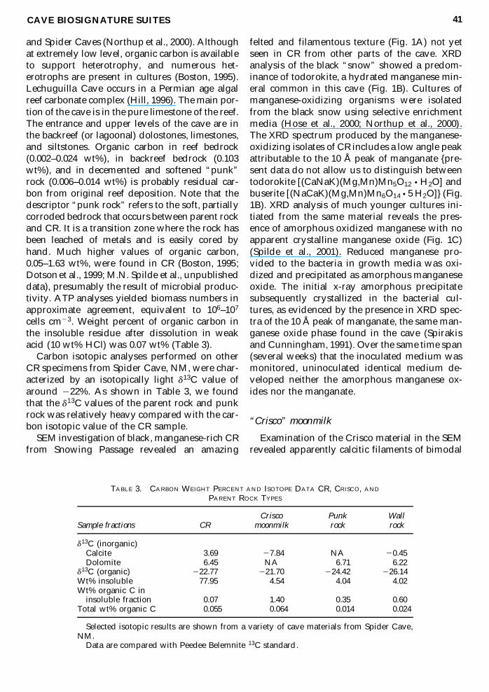