![Colorimetric Detection of Cu[II] Cation and Acetate, Benzoate, and Cyanide Anions by Cooperative Receptor Binding in New α,α‘-Bis-substituted Donor−Acceptor Ferrocene Sensors](https://static.fdokumen.com/doc/165x107/6316233c511772fe4510af34/colorimetric-detection-of-cuii-cation-and-acetate-benzoate-and-cyanide-anions.jpg)
Cation Coordination Induced Modulation of the Anion Sensing Properties of a...
11
Cation Coordination Induced Modulation of the Anion Sensing Properties of a Ferrocene-Imidazophenanthroline Dyad: Multichannel Recognition from Phosphate-Related to Chloride Anions Fabiola Zapata, Antonio Caballero, Arturo Espinosa, Alberto Ta ´rraga,* and Pedro Molina* Departamento de Quı ´mica Orga ´nica, Facultad de Quı ´mica, UniVersidad de Murcia, Campus de Espinardo, E-30100 Murcia, Spain [email protected]; [email protected] ReceiVed February 5, 2008 A new chemosensor molecule 1 based on a ferrocene-imidazophenanthroline dyad, effectively recognizes aqueous hydrogenpyrophosphate and the organic anions ADP and ATP through three different channels. A cathodic shift of the ferrocene/ferrocenium oxidation wave (∆E 1/2 ranging from -130 mV for hydrogenpyrophosphate and fluoride to -40 mV for ADP). A progressive red-shift of the absorption bands and/or appearance of a new low energy band at 314-319 nm. These changes in the absorption spectra are accompanied by color changes from pale yellow to orange or pink, which allow the potential for “naked eye” detection. The emission spectrum (λ exc ) 390 nm) undergoes an important chelation- enhanced fluorescence effect (CHEF ) 50) in the presence of 2.5 equiv of hydrogenpyrophosphate anion and with a large excess of fluoride anion (CHEF ) 114). Interestingly, the emission spectrum obtained at different excitation energy (λ exc ) 340 nm) in the presence of AcOH acid is red-shifted and not only perturbed by the hydrogenpyrophosphate anion (CHEF ) 71) but also with the organic anions ATP (CHEF ) 25), ADP (CHEF ) 15), and the dihydrogenphosphate (CHEF ) 25). The stable heterobimetallic ruthenium (II) complex 2 selectively senses the chloride anion over other anions examined through two channels: cathodic redox shift (∆E 1/2 )-80 mV) of the Fe(II)/Fe(III) redox couple keeping the oxidation wave of the ruthenium (II) center unchanged and a significant red emission enhancement (CHEF ) 30). 1 H and 31 P NMR studies as well as DFT calculations have been carried out to get information about which molecular sites are involved in bonding. About the deprotonation/coordination dualism, the combined electrochemical, absorption, emission, and NMR data strongly support that fluoride anion induces only deprotonation, anions dihydrogenphosphate, ATP, and ADP from hydrogen-bonded complexes and formation of hydrogen-bonded complex between receptor 1 and hydrogenpyrophosphate anion and deprotonation proceed simultaneously. In regards to receptor 2, all available data (electrochemical, absorption, emission, and 1H NMR) strongly support the formation of a [2 · Cl - ] hydrogen-bonded complex. Introduction The design and synthesis of receptors and chemosensors capable of binding and sensing anions selectively has attracted much attention in recent years because of the fundamental role of anions in a wide range of biological and chemical processes. 1 A wide variety of receptors that utilize electrostatic interac- tions, hydrogen bond donor groups, Lewis acid groups, and (1) Sessler, J. L.; Gale, P. A.; Cho, W. S. In Anion Receptor Chemistry; Monographs in Supramolecular Chemistry; Stoddart, J. F., Ed.; RSC: Cambridge, 2006. 10.1021/jo800296c CCC: $40.75 2008 American Chemical Society 4034 J. Org. Chem. 2008, 73, 4034–4044 Published on Web 04/24/2008
Transcript of Cation Coordination Induced Modulation of the Anion Sensing Properties of a...

Cation Coordination Induced Modulation of the Anion SensingProperties of a Ferrocene-Imidazophenanthroline Dyad: Multichannel
Recognition from Phosphate-Related to Chloride Anions
Fabiola Zapata, Antonio Caballero, Arturo Espinosa, Alberto Tarraga,* and Pedro Molina*
Departamento de Quımica Organica, Facultad de Quımica, UniVersidad de Murcia,Campus de Espinardo, E-30100 Murcia, Spain
[email protected]; [email protected]
ReceiVed February 5, 2008
A new chemosensor molecule 1 based on a ferrocene-imidazophenanthroline dyad, effectively recognizesaqueous hydrogenpyrophosphate and the organic anions ADP and ATP through three different channels.A cathodic shift of the ferrocene/ferrocenium oxidation wave (∆E1/2 ranging from -130 mV forhydrogenpyrophosphate and fluoride to -40 mV for ADP). A progressive red-shift of the absorptionbands and/or appearance of a new low energy band at 314-319 nm. These changes in the absorptionspectra are accompanied by color changes from pale yellow to orange or pink, which allow the potentialfor “naked eye” detection. The emission spectrum (λexc ) 390 nm) undergoes an important chelation-enhanced fluorescence effect (CHEF ) 50) in the presence of 2.5 equiv of hydrogenpyrophosphate anionand with a large excess of fluoride anion (CHEF ) 114). Interestingly, the emission spectrum obtainedat different excitation energy (λexc ) 340 nm) in the presence of AcOH acid is red-shifted and not onlyperturbed by the hydrogenpyrophosphate anion (CHEF ) 71) but also with the organic anions ATP(CHEF ) 25), ADP (CHEF ) 15), and the dihydrogenphosphate (CHEF ) 25). The stable heterobimetallicruthenium (II) complex 2 selectively senses the chloride anion over other anions examined through twochannels: cathodic redox shift (∆E1/2 ) -80 mV) of the Fe(II)/Fe(III) redox couple keeping the oxidationwave of the ruthenium (II) center unchanged and a significant red emission enhancement (CHEF ) 30).1H and 31P NMR studies as well as DFT calculations have been carried out to get information aboutwhich molecular sites are involved in bonding. About the deprotonation/coordination dualism, the combinedelectrochemical, absorption, emission, and NMR data strongly support that fluoride anion induces onlydeprotonation, anions dihydrogenphosphate, ATP, and ADP from hydrogen-bonded complexes andformation of hydrogen-bonded complex between receptor 1 and hydrogenpyrophosphate anion anddeprotonation proceed simultaneously. In regards to receptor 2, all available data (electrochemical,absorption, emission, and 1H NMR) strongly support the formation of a [2 ·Cl-] hydrogen-bonded complex.
Introduction
The design and synthesis of receptors and chemosensorscapable of binding and sensing anions selectively has attractedmuch attention in recent years because of the fundamental roleof anions in a wide range of biological and chemical processes.1
A wide variety of receptors that utilize electrostatic interac-tions, hydrogen bond donor groups, Lewis acid groups, and
(1) Sessler, J. L.; Gale, P. A.; Cho, W. S. In Anion Receptor Chemistry;Monographs in Supramolecular Chemistry; Stoddart, J. F., Ed.; RSC: Cambridge,2006.
10.1021/jo800296c CCC: $40.75 2008 American Chemical Society4034 J. Org. Chem. 2008, 73, 4034–4044Published on Web 04/24/2008
hydrophobic interactions have been employed to bind anionicguest species over the intervening years.2 In this context, simplecoordination compounds containing a combination of positivecharge and monodentate ligands featuring hydrogen bond donorgroups provide the basis for powerful molecular sensors foranions and have recently received much interest among re-searchers.3
To date, several different heterocyclic ring systems containinga pyrrolic NH group have been reported in the literature ashydrogen-bond donors to anions, as demonstrated in calixpyr-roles,4 expanded porphyrinoids,5 pyrrole derivatives,6 indoles,7
bisindoles,8 bisimidazoles,9 indolocarbazoles,10 and benzimi-dazoles.11 Surprisingly, although the imidazo[4,5-f]-1,10-phenanthroline moiety has been employed frequently to formruthenium (II) polypyridyl complexes12 using the two nitrogenatoms of the bipyridine fragment, as far as we know no anionreceptors making use of the NH group of the imidazole ringsystem have ever been reported.13
Sensing of a fluoride anion, the smallest anion, has attractedgrowing attention due to its beneficial effects (e.g., preventionof dental caries) and detrimental (e.g., fluorosis) roles.14 Theconventional binding approaches have used either designedhydrogen bonding or the specific strong affinity of a boron atomtoward the fluoride anion. These binding events have beenconverted into an electrochemical15 or fluorescent16 change or,more directly, a colorimetric change detectable by the naked
eye.17 Anions such as pyrophosphate and adenosine triphosphate(ATP) play an important role in energy transduction in organismand control metabolic processes by participation in enzimaticreactions. ATP hydrolysis with the concomitant release ofpyrophosphate is central to many biochemical reactions, suchas DNA polymerization and the synthesis of cyclic adenosinemonophosphate (c-AMP) catalyzed by DNA polymerase andadenylate cyclase, respectively.18 Furthermore, the detection ofreleased pyrophosphate has been examined as a real-time DNAsequencing method,19 and it has also been considered importantin cancer research.20 Therefore, the detection and discriminationof these anions has been the main focus of the effort of severalresearch groups. However, very few examples of effectiveselective fluorescent,21 chromogenic,22 or redox23 chemosensorshave so far been reported.
Only in recent years have alternative mechanisms for severaltypes of anion-receptor interaction been developed.24 If thebasicity of the anion is insufficient to induce deprotonation ofthe receptor, one observes formation of a hydrogen-bondedcomplex manifested in a red-shift of the receptor absorptionband and a downfield shift or often disappearance of NMRsignals of the receptors protons involved in the hydrogenbonding. If the basicity of the anion is high enough todeprotonate the receptor, one observes the appearance of a newintense absorption band in the visible region of the electronicspectrum, the disappearance of NMR signals of abstracted
(2) For reviews, see:(a) Beer, P. D.; Gale, P. A. Angew. Chem., Int. Ed. 2001,40, 486–516. (b) Bondy, C. R.; Loeb, S. J. Coord. Chem. ReV. 2003, 240, 77–99. (c) Choi, K.; Hamilton, A. D. Coord. Chem. ReV. 2003, 240, 101–110. (d)Gale, P. A. Coord. Chem. ReV. 2003, 240, 191–221. (e) Suksai, C.; Tuntulani,T. Chem. Soc. ReV. 2003, 32, 192–202. (f) Bowman-James, C. Acc. Chem. Res.2005, 38, 671–678. (g) Wiskur, S. L.; Ait-Haddou, H.; Lavigne, J. J.; Anslyn,E. Acc. Chem. Res. 2001, 34, 963–972. (h) Sessler, J. L.; Davis, J. M. Acc.Chem. Res. 2001, 34, 989–997. (i) Yoon, J.; Kim, S.; Singh, N. J.; Kim, K. S.Chem. Soc. ReV. 2006, 35, 355–360. (j) Gale, P. A. Acc. Chem. Res. 2006, 39,465–475.
(3) For review, see:(a) Perez, J.; Riera, L. Chem. Commun. 2008, 533–543.(4) (a) Anzenbacher, P.; Jursıkova, K.; Sessler, J. L. J. Am. Chem. Soc. 2000,
122. (b) Gale, P. A.; Anzenbacher, P.; Sessler, J. L. Coord. Chem. ReV. 2001,222, 57102. (c) Nielsen, K. A.; Cho, W.-S.; Jeppesen, J. O.; Lynch, V. M.;Becher, J.; Sessler, J. L. J. Am. Chem. Soc. 2004, 126, 16296–16297. (d) Sessler,J. L.; Katayev, E.; Pantos, G. D.; Scherbakov, P.; Reshetova, M. D.; Khrustalev,V. N.; Lynch, V. M.; Ustynyuk, Y. A. J. Am. Chem. Soc. 2005, 127, 11442–11446.
(5) For reviews, see:(a) Sessler, J. L.; Davis, J. M. Acc. Chem. Res. 2001,34, 989–997. (b) Sessler, J. L.; Camiolo, S.; Gale, P. A. Coord. Chem. ReV.2003, 240, 17–55.
(6) (a) Lin, C.-I.; Selvi, S.; Fang, J.-M.; Chou, P.-T.; Kai, C. H.; Cheng,Y.-Y. J. Org. Chem. 2007, 72, 3537–3542. (b) Sessler, J. L.; Dan Pantos, G.;Gale, P. A.; Light, M. E. Org. Lett. 2006, 8, 1593–1596. For reviews, see: (c)Gale, P. A. Chem. Commun. 2005, 3761–3772.
(7) Pfeffer, F. M.; Lim, K. F.; Sedgwick, Org. Biomol. Chem. 2007, 5, 1795–1799.
(8) Chang, K.-J.; Moon, D.; Lah, M. S.; Jeong, K.-S. Angew. Chem., Int.Ed. 2005, 44, 7926–7929.
(9) Causey, C. P.; Allen, W. E. J. Org. Chem. 2002, 67, 5963–5968.(10) Curiel, D.; Cowley, A.; Beer, P. D. Chem. Commun. 2005, 236–238.(11) Kang, J.; Kim, H. S.; Jang, D. O. Tetrahedron Lett. 2005, 46, 6079–
6082.(12) Han, M.-J.; Gao, L.-H.; Wang, K.-Z. New. J. Chem. 2006, 30, 208–
214.(13) Such as family of heterocyclic compounds has been used in the studies
on the interaction between transition-metal complexes and DNA:(a) Xiong, Y.;Ji, L.-N. Coord. Chem. ReV. 1999, 185-186, 711–733. (b) Ji, L.-N.; Zou, X.-H.; Liu, J.-G. Coord. Chem. ReV. 2001, 216-217, 513–536. Only a ruthenium(II)complex of this ring system has been reported as a selective fluorescent sensorof Mg2+ cations: (c) Liu, Y.; Duan, Z.-Y.; Zhang, H.-Y.; Jiang, X.-L.; Han,J.-R. J. Org. Chem. 2005, 70, 1450–1455.
(14) Kirk, K. L. Biochemistry of the Halogens and Inorganic Halides; PlenumPress: New York, 1991; p 58
(15) (a) Desemund, C.; Sanyadanake, K. R. A. S.; Shinkai, S. J. Chem. Soc.,Chem. Commun. 1995, 333–334. (b) Yamamoto, H.; Ori, A.; Ueda, K.;Desemund, C.; Sinkai, S. Chem. Commun. 1996, 407–408. (c) Oton, F.; Tarraga,A.; Molina, P. Org. Lett. 2006, 8, 2107–2110. (d) Oton, F.; Espinosa, A.; Tarraga,A.; Ramırez de Arellano, C.; Molina, P. Chem.-Eur. J. 2007, 13, 5742–5752.
(16) (a) Anzenbacher, P., Jr.; Try, A. C.; Miyaji, H.; Jursikova, K.; Lynch,V. M.; Marquez, M.; Sessler, J. L. J. Am. Chem. Soc. 2000, 122, 10268–10272.(b) Kima, T.-H.; Swager, T. M. Angew. Chem., Int. Ed. 2003, 42, 4803–4806.(c) Xu, G.; Tarr, M. A. Chem. Commun. 2004, 1050–1051. (d) Wu, J.-S.; Zhou,J.-H.; Wang, P.-F.; Zhang, X.-H.; Wu, S.-K. Org. Lett. 2005, 7, 2133–2136. (e)Melaimi, M.; Gabbai, F. P. J. Am. Chem. Soc. 2005, 127, 9680–9681. (f) Kim,S. K.; Bok, J. H.; Bartsch, R. A.; Lee, J. Y.; Kim, J. S. Org. Lett. 2005, 7,4839–4842. (g) Zhao, Y.-P.; Zhao, C.-C.; Wu, L.-Z.; Zhang, L.-P.; Tung, C.-H.; Pan, Y.-J. J. Org. Chem. 2006, 71, 2143–2149.
(17) (a) Miyaji, H.; Sato, W.; Sessler, J. L. Angew. Chem., Int. Ed. 2000,39, 1777–1780. (b) Miyaji, H.; Sato, W.; Sessler, J. L.; Lynch, M. TetrahedronLett. 2000, 41, 1369–1373. (c) Miyaji, H.; Sessler, J. L. Angew. Chem., Int. Ed.2001, 40, 154–157. (d) Yamaguchi, S.; Akiyama, S.; Tamao, K. J. Am. Chem.Soc. 2001, 123, 11372–11375. (e) Vazquez, M.; Fabbrizzi, L.; Taglietti, A.;Pedrido, R. M.; Gonzalez-Noya, A. M.; Bermejo, M. R. Angew. Chem., Int. Ed.2004, 43, 1962–1695. (f) Watanabe, S.; Seguchi, H.; Yoshida, K.; Kifune, K.;Tadaki, T.; Shiozaki, H. Tetrahedron Lett. 2005, 46, 8827–8829. (g) Lin, Z.-H.;Ou, S.-J.; Duan, C.-Y.; Zhang, B.-G.; Bai, Z.-P. Chem.Commun. 2006, 624–626. (h) Ghosh, T.; Maiya, B. G.; Samanta, A. Dalton Trans. 2006, 795–801.
(18) (a) Lipscombe, W. N.; Strater, N. Chem. ReV. 1996, 96, 2375–2433.(b) Tabary, T.; Lu, L. J. Inmunol. Methods 1992, 156, 55–60. (c) Nyren, P.Anal. Biochem. 1987, 167, 235–238.
(19) Ronaghi, M.; Karamohamed, S.; Petterson, B.; Uhlen, M.; Nyren, P.Anal. Biochem. 1996, 242, 84–89.
(20) Xu, S.; He, M.; Yu, H.; Cai, X.; Tan, X.; Lu, B.; Shu, B. Anal. Biochem.2001, 299, 188–193.
(21) (a) Fabbrizzini, L.; Marcotte, N.; Stomeo, F.; Taglietti, A. Angew. Chem.,Int. Ed. 2002, 41, 3811–3814. (b) Gunnalaugsson, T.; Davis, A. P.; O′Brien,J. E.; Glynn, M. Org. Lett. 2002, 4, 2449–2452. (c) Lee, D. H.; Kim, S. Y.;Hong, J.-I. Angew. Chem., Int. Ed. 2004, 43, 4777–4780. (d) Kanekiyo, Y.;Naganawa, R.; Tao, H. Chem. Commun. 2004, 1006–1007. (e) Cho, H. K.; Lee,D. H.; Hong, J.-I. Chem. Commun. 2005, 1690–1692. (f) Jang, Y. J.; Jun, E. J.;Lee, Y. J.; Kim, Y. S.; Kim, J. S.; Yoon, J. J. Org. Chem. 2005, 70, 9603–9606.(g) McDonough, M. J.; Reynolds, A. J.; Lee, W. Y. G.; Jolliffe, K. A. Chem.Commun. 2006, 2971–2973. (h) Bazzicalupi, C.; Biagini, S.; Bencini, A.; Faggi,E.; Giorgi, C.; Matera, I.; Valtancoli, B. Chem. Commun. 2006, 4087–4089. (i)Lee, H. N.; Swamy, K. M. K.; Kim, S. K.; Kwon, J.-Y.; Kim, Y.; Kim, S.-J.;Yoon, Y. J.; Yoon, J. Org. Lett. 2007, 9, 242–246. (j) Lee, H. N.; Xu, Z.; Kim,S. K.; Swamy, K. M. K.; Kim, Y.; Kim, S.-J.; Yoon, J. J. Am. Chem. Soc. 2007,129, 3828–3829. (k) Swamy, K. M. K.; Kwon, S. K.; Lee, H. N.; Shantha Kumar,S. M.; Kim, J. S.; Yoon, J. Tetrahedron Lett. 2007, 48, 8683–8686.
(22) (a) Lee, D. H.; Im, J. H.; Son, S. U.; Young, K.; Hong, J.-I. J. Am.Chem. Soc. 2003, 125, 7752–7753. (b) Aldakov, D.; Anzenbacher, P., Jr J. Am.Chem. Soc. 2004, 126, 4752–4753. (c) Nishiyabu, R., Jr J. Am. Chem. Soc. 2005,127, 8270–8271.
(23) Anzenbacher, P.; Palacios, M. A.; Kursikova, K.; Marquez, M. Org.Lett. 2005, 7, 5027–5030.
(24) Amendola, V.; Esteban-Gomez, D.; Fabrizzi, L.; Licchelli, M. Acc.Chem. Res. 2006, 39, 343–353.
Multichannel Recognition
J. Org. Chem. Vol. 73, No. 11, 2008 4035

receptor protons, and an upfield shift of the signals of adjacentreceptors protons.25 More interestingly, some detailed studieshave even suggested that mixed processes might occur: initialcomplex formation at low anion-to-receptor ratios, this thenbeing followed by deprotonation at higher anion-to-receptorratios.26 The degree of deprotonation depends on the concentra-tion of the receptor; as a result, a NMR titration may feel onlyhydrogen bonding, whereas a spectrophotometric titration mayclearly show the deprotonation.27 A simple way to measure thebinding constant for the formation of the hydrogen-bondedcomplex is to perform the titration in the presence of addedacid, which suppresses the deprotonation process.
Despite the development of these classical single-signalingaproximations there is a paucity of use of multichannel signalingfluoride and pyrophosphate selective chemosensor molecules.28
Generally, anion recognition motifs are often structurallycomplicated and require an elaborate and sophisticated syntheticprocess. Therefore, the development of simple an easy-to-makechemosensor molecules for anions is strongly desired. From thisperspective, we decided to study a new type of anionophoresby combining the redox activity of the ferrocene moiety withthe strong hydrogen-bonding ability of the imidazole ring andthe photoactive behavior of the phenanthroline ring. Here, wenow report on chemosensor molecules 1 and 2 comprising animidazophenanthroline ring, in which the fluorophore and theanion and cation binding sites are integrated into this ringsystem, which is linked to a redox-active ferrocene moiety togenerate strong fluorescent, optical, and electrochemical signals.
Results and Discussion
Synthesis. Compound 1 was prepared in excellent yield bycondensation of 1,10-phenanthroline-5,6-dione with formylfer-rocene in the presence of NH4OAc, following an improved
modification of the previously reported method for relatedcompounds.29 Heterobimetallic ligand 2 was obtained in 70%yield from the reaction of 1 with cis-dichlorobis(2,2′-bipy-ridine)ruthenium(II) dihydrate and further treatment withNH4PF6 (Scheme 1).
Anion Sensing Properties. The binding and recognitionability of receptors 1 and 2 toward various anions (F-, Cl-,Br-, AcO-, NO3
-, HSO4-, H2PO4
-, and HP2O73-), in the form
of their corresponding tetrabutylammonium salts (TBA+), andthe organic anions ATP and ADP were evaluated by cyclic (CV)and Osteryoung square-wave voltammetry (OSWV).26 The CVresponse of 1 in CH3CN, also containing 0.1 M TBAPF6 assupporting electrolyte, showed a reversible one-electron oxida-tion process at E1/2 ) 0.64 V vs decamethylferrocene (DMFc)redox couple. The heterobimetallic ligand 2 displays a similarreversible oxidation process at E1/2 ) 0.67 V vs DMFc, arisingfrom the one-electron oxidation of the ferrocene unit, and twoirreversible oxidative processes at approximatively 1.40 and 1.45V vs DMFc associated to the oxidations of the Ru center.
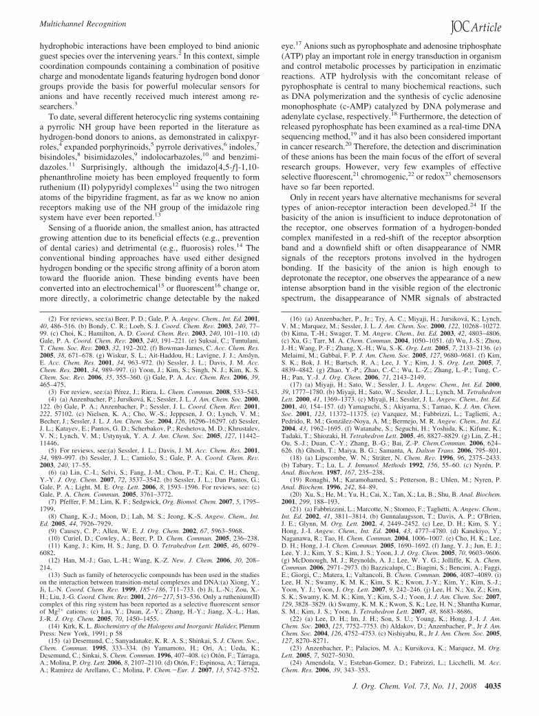
On stepwise addition of F- anion to a solution of receptor 1in CH3CN, a clear evolution of the oxidation wave to E1/2 )0.57 V (∆E1/2 ) -70 mV) was observed with 1 equiv of addedF- anion. The electrochemical shift was accompanied by adecrease in current intensity, a behavior attributed to theformation of a complex with a lower diffusion coefficient.30
OSWV titration also showed a measurable change in the peakcurrent and oxidation potencial (Figure 1a). Titration isothermsobtained from these changes were fitted nicely to a 1:1 bindingmodel. However, in the presence of an excess of F- anion, aremarkable cathodic shift (∆E1/2 ) -340 mV) of the oxidationpotential is observed. Receptor 1 also showed a perturbation ofthe oxidation peak in the presence of HP2O7
3- anion. Upon
(25) (a) Descalzo, A. B.; Rurack, K.; Weisshof, H.; Martinez-Manez, R. M.;Marcos, M. D.; Amoros, P.; Hoffmann, K.; Soto, J. J. Am. Chem. Soc. 2005,127, 184–200. (b) Vazquez, M.; Fabbrizzi, L.; Taglietti, A.; Pedrido, R. M.;Gonzalez-Noya, A. M.; Bermejo, M. R. Angew. Chem., Int. Ed. 2004, 43, 1962–1965. (c) Boiocchi, M.; Dal Boca, L.; Esteban-Gomez, D.; Fabbrizzi, L.; Licchelli,M.; Monzani, E. J. Am. Chem. Soc. 2004, 126, 16507–16514. (d) Esteban-Gomez,D.; Fabbrizzi, L.; Licchelli, M.; Monzani, E. Org. Biomol. Chem. 2005, 3, 1495–1500. (e) Evans, L. S.; Gale, P. A.; Light, M. E.; Quesada, R. Chem. Commun.2006, 965–967. (f) Camiolo, S.; Gale, P. A.; Hursthouse, M. B.; Light, M. E.Org. Biomol. Chem. 2003, 1, 741–744. (g) Caminolo, S.; Gale, P. A.; Hursthouse,M. B.; Light, M. E.; Shi, A. J. Chem. Commun. 2002, 758–759. (h) Gale, P. A.;Navakhun, K.; Camiolo, S.; Light, M. E.; Hursthouse, M. B. J. Am. Chem. Soc.2002, 124, 11228–11229. (i) He, X.; Hu, S.; Liu, K.; Guo, Y.; Xu, J.; Shao, S.Org. Lett. 2006, 8, 333–336. (j) Viruthachalam, T.; Ramamurthy, P.; Thirumalai,D.; Ramakrishman, V. Org. Lett. 2005, 7, 657–660. (k) Caltaginore, C.; Bate,G. W.; Gale, P. A.; Light, M. E. Chem. Commun. 2008, 61–63.
(26) (a) Boiocchi, M.; Del Boca, L.; Esteban-Gomez, D.; Fabbrizzi, L.;Licchelli, M.; Monzani, E. Chem.-Eur. J. 2005, 11, 3097–3104. (b) Amendola,V.; Boiocchi, M.; Fabbrizzi, L.; Palchatti, A. Chem.-Eur. J. 2005, 11, 5648–5660. (c) Esteban-Gomez, D.; Fabbrizzi, L.; Licchelli, M.; Sacchi, D. J. Mater.Chem. 2005, 15, 2670–2675. (d) Esteban-Gomez, D.; Fabbrizzi, L.; Licchelli,M. J. Org. Chem. 2005, 70, 5717–5720. (e) Ros-Lis, J. V.; Martinez-Manez,R.; Sancenon, F.; Soto, J.; Rurack, K.; Weisshoff, H. Eur. J. Org. Chem. 2007,2449–2458. (f) Lin, C.; Simov, V.; Drueckhammer, D. G. J. Org. Chem. 2007,72, 1742–1746.
(27) Perez-Casas, C.; Yatsimirsky, A. K. J. Org. Chem. 2008, 73, 2275–2284.
(28) (a) Cho, E. J.; Moon, J. W.; Ko, S. W.; Lee, J. Y.; Kim, S. K.; Yoon,J.; Nam, K. C. J. Am. Chem. Soc. 2002, 125, 12376–12377. (b) Guunlaugsson,T.; Kruger, P. E.; Lee, T. C.; Parkesh, R.; Pfeffer, F. M.; Hussey, G. M.Tetrahedron Lett. 2003, 44, 6575–6578. (c) Kubo, Y; Yamamoto, M.; Ikeda,M.; Takeuchi, M.; Shinkai, S.; Yamaguchi, S.; Tomao, K. Angew. Chem., Int.Ed. 2003, 42, 2036–2040. (d) Oton, F.; Tarraga, A.; Velasco, M. D.; Espinosa,A.; Molina, P. Chem. Commun. 2004, 1658–1659. (e) Zhang, B.-G.; Xu, J.;Zhao, Y.-G.; Duan, C.-Y.; Cao, X.; Meng, Q.-J. Dalton Trans. 2006, 1271–1276. (f) Oton, F.; Tarraga, A.; Espinosa, A.; Velasco, M. D.; Molina, P J. Org.Chem. 2006, 71, 4590–4598. (g) Jang, Y. J.; Jun, E. J.; Lee, Y. J.; Kim, Y. S.;Kim, J. S.; Yoon, J. J. Org. Chem. 2005, 70, 9603–9606.
(29) Steck, E. A.; Day, A. R. J. Am. Chem. Soc. 1943, 65, 452–456.(30) Kaifer, A. E.; Gomez-Kaifer, M. Supramolecular Electrochemistry;
Wiley-VCH: Weinheim, 1999. (b) Bard, A. J.; Faulkner, L. R. ElectrochemicalMethods, 2nd ed.; Wiley: New York, 2001.
SCHEME 1. Synthesis of Ligands 1 and 2
Zapata et al.
4036 J. Org. Chem. Vol. 73, No. 11, 2008
addition of small amounts of this anion, a new oxidation peakcathodically shifted at 0.30 V (∆E1/2 ) -340 mV), appeared.The current intensity of the new oxidation peak increases witha linear dependence on the equiv of the added anion. Inparticular, this large cathodically shifted oxidation peak reachesthe maximum current intensity value at 3 equiv of anion added,and at this point the peak at 0.64 V disappears (Figure 1b).Titration with the strong base Bu4NOH, which definitely leadsto deprotonation, also induced a remarkable cathodic shift ofthe oxidation peak (∆E1/2 ) -335 mV). A possible way toreveal the formation of hydrogen-bonded complexes underconditions of electrochemical titration is to suppress deproto-nation by adding a small amount of acetic acid. In preliminaryexperiments, we found that addition of up to 20 equiv of aceticacid did not affect neither CV nor OSWV of receptor 1 inCH3CN. Addition of HP2O7
3- anion to an electrochemicalsolution of receptor 1 in CH3CN in the presence of 20 equiv ofAcOH induced a cathodic shift of the oxidation peak (∆E1/2 )-110 mV) considerably smaller than observed in the absenceof acid (∆E1/2 ) -340 mV), whereas the addition of F- anioninduces the same change in the oxidation peak that the observedwith 1 equiv of added F- anion (∆E1/2 ) -70 mV).
Remarkably, the presence of Cl-, Br-, AcO-, NO3-, HSO4
-,and H2PO4
- anions had no effect on the OSWV, even whenpresent in large excess.
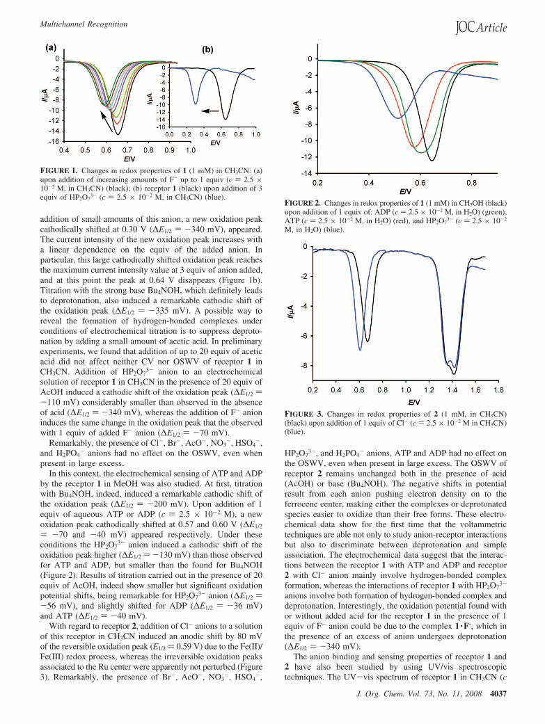
In this context, the electrochemical sensing of ATP and ADPby the receptor 1 in MeOH was also studied. At first, titrationwith Bu4NOH, indeed, induced a remarkable cathodic shift ofthe oxidation peak (∆E1/2 ) -200 mV). Upon addition of 1equiv of aqueous ATP or ADP (c ) 2.5 × 10⁻² M), a newoxidation peak cathodically shifted at 0.57 and 0.60 V (∆E1/2
) -70 and -40 mV) appeared respectively. Under theseconditions the HP2O7
3- anion induced a cathodic shift of theoxidation peak higher (∆E1/2 ) -130 mV) than those observedfor ATP and ADP, but smaller than the found for Bu4NOH(Figure 2). Results of titration carried out in the presence of 20equiv of AcOH, indeed show smaller but significant oxidationpotential shifts, being remarkable for HP2O7
3- anion (∆E1/2 )-56 mV), and slightly shifted for ADP (∆E1/2 ) -36 mV)and ATP (∆E1/2 ) -40 mV).
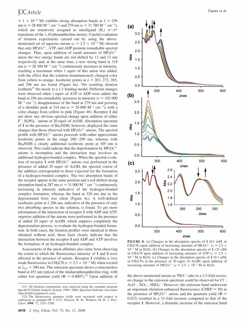
With regard to receptor 2, addition of Cl- anions to a solutionof this receptor in CH3CN induced an anodic shift by 80 mVof the reversible oxidation peak (E1/2 ) 0.59 V) due to the Fe(II)/Fe(III) redox process, whereas the irreversible oxidation peaksassociated to the Ru center were apparently not perturbed (Figure3). Remarkably, the presence of Br-, AcO-, NO3
-, HSO4-,
HP2O73-, and H2PO4
- anions, ATP and ADP had no effect onthe OSWV, even when present in large excess. The OSWV ofreceptor 2 remains unchanged both in the presence of acid(AcOH) or base (Bu4NOH). The negative shifts in potentialresult from each anion pushing electron density on to theferrocene center, making either the complexes or deprotonatedspecies easier to oxidize than their free forms. These electro-chemical data show for the first time that the voltammetrictechniques are able not only to study anion-receptor interactionsbut also to discriminate between deprotonation and simpleassociation. The electrochemical data suggest that the interac-tions between the receptor 1 with ATP and ADP and receptor2 with Cl- anion mainly involve hydrogen-bonded complexformation, whereas the interactions of receptor 1 with HP2O7
3-
anions involve both formation of hydrogen-bonded complex anddeprotonation. Interestingly, the oxidation potential found withor without added acid for the receptor 1 in the presence of 1equiv of F- anion could be due to the complex 1 ·F-, which inthe presence of an excess of anion undergoes deprotonation(∆E1/2 ) -340 mV).
The anion binding and sensing properties of receptor 1 and2 have also been studied by using UV/vis spectroscopictechniques. The UV-vis spectrum of receptor 1 in CH3CN (c
FIGURE 1. Changes in redox properties of 1 (1 mM) in CH3CN: (a)upon addition of increasing amounts of F- up to 1 equiv (c ) 2.5 ×10⁻² M, in CH3CN) (black); (b) receptor 1 (black) upon addition of 3equiv of HP2O7
3- (c ) 2.5 × 10⁻² M, in CH3CN) (blue).FIGURE 2. Changes in redox properties of 1 (1 mM) in CH3OH (black)upon addition of 1 equiv of: ADP (c ) 2.5 × 10⁻² M, in H2O) (green),ATP (c ) 2.5 × 10⁻² M, in H2O) (red), and HP2O7
3- (c ) 2.5 × 10⁻²
M, in H2O) (blue).
FIGURE 3. Changes in redox properties of 2 (1 mM, in CH3CN)(black) upon addition of 1 equiv of Cl- (c ) 2.5 × 10⁻² M in CH3CN)(blue).
Multichannel Recognition
J. Org. Chem. Vol. 73, No. 11, 2008 4037
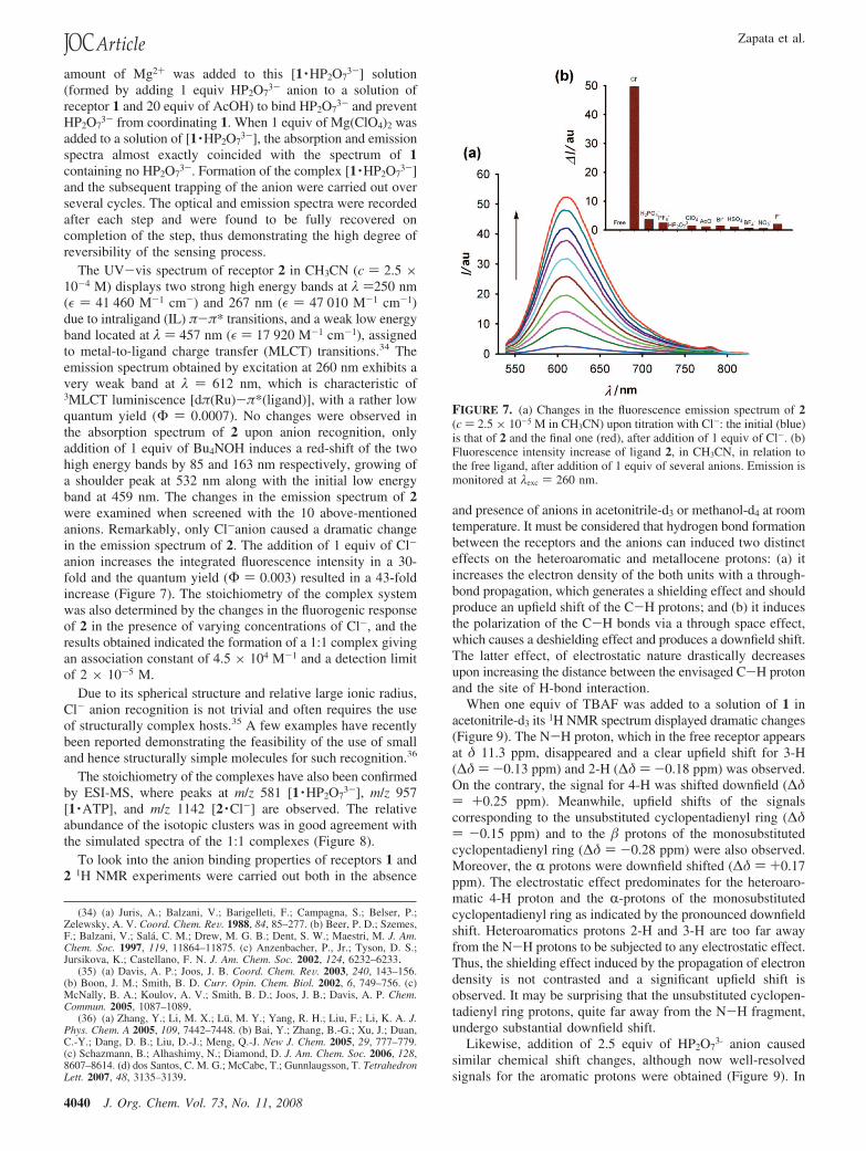
) 1 × 10⁻⁴ M) exhibits strong absorption bands at λ ) 256nm (ε ) 28 860 M-1 cm-1) and 279 nm (ε ) 31 780 M-1 cm-1),which are tentatively assigned as intraligand (IL) π-π*transitions of the 1,10-phenanthroline moiety. Careful evaluationof titration experiments carried out by using the above-mentioned set of aqueous anions (c ) 2.5 × 10⁻² M) showedthat only HP2O7
3-, ATP, and ADP promote remarkable spectralchanges. Thus, upon addition of small amounts of HP2O7
3-
anion the two energy bands are red-shifted by 13 and 12 nmrespectively and, at the same time, a new strong band at 319nm (ε ) 28 300 M-1 cm-1) continuously increases in intensity,reaching a maximum when 1 equiv of this anion was added,with the effect that the solution instantaneously changed colorfrom yellow to orange. Isosbestic points at λ ) 263, 272, 285,and 296 nm are found (Figure 4a). The resulting titrationisotherm31 fits nicely to a 1:1 binding model. Different changeswere observed when 1 equiv of ATP or ADP were added: theband at 256 nm remarkably increases in intensity (ε ) 102 000M-1 cm-1), disappearance of the band at 279 nm and growingof a shoulder peak at 314 nm (ε ) 28 800 M-1 cm-1), with acolor change from yellow to pink (Figure 4b). Receptor 1 didnot show any obvious spectral change upon addition of eitherF-, H2PO4
- anions or 20 equiv of AcOH. Absorption spectrumof 1 in the presence of Bu4NOH, however, displayed the samechanges that those observed with HP2O7
3- anions. The spectralprofile with HP2O7
3- anions proceeds with rather approximateisosbestic points in the range 260-290 nm, whereas withBu4NOH a clearly additional isosbestic point at 305 nm isobserved. This could indicate that the deprotonation by HP2O7
3-
anions is incomplete and the interaction may involves anadditional hydrogen-bonded complex. When the spectral evolu-tion of receptor 1 with HP2O7
3- anions was performed in thepresence of added 20 equiv of AcOH, the spectral course ofthe addition corresponded to those expected for the formationof a hydrogen-bonded complex. The two absorption bands ofthe receptor appear at the same position and a red-shifted strongabsorption band at 287 nm (ε ) 31 000 M-1 cm-1) continuouslyincreasing in intensity indicative of the hydrogen-bondedcomplex formation, whereas the band at 320 nm due to thedeprotonated form was silent (Figure 4c). A well-definedisosbestic point at λ 286 nm, indicative of the presence of onlytwo absorbing species in the solution, is found. To get moreinformation of the interaction of receptor 1 with ADP and ATP,stepwise addition of the anions were performed in the presenceof added 20 equiv of AcOH, which suppress completely thedeprotonation process, to evaluate the hydrogen-bonded forma-tion. In both cases, the titration profiles were identical to thoseobtained without acid, these facts clearly indicate that theinteraction between the receptor 1 and ADP and ATP involvesthe formation of an hydrogen-bonded complex.
Assessments of the anion affinities also came from observingthe extent to which the fluorescence intensity of 1 and 2 wereaffected in the presence of anions. Receptor 1 exhibits a veryweak fluorescence in CH3CN (c ) 2.5 × 10⁻⁵ M) when excitedat λexc ) 390 nm. The emission spectrum shows a structurelessband at 453 nm typical of the imidazophenanthroline ring, withrather low quantum yield (Φ ) 0.0007).32 Upon addition of the above-mentioned anions as TBA+ salts in a 2.5-fold excess,
no change in the emission spectrum could be observed for Cl-,AcO-, NO3
-, HSO4-. However, the emission band underwent
an important chelation-enhanced fluorescence (CHEF ) 50) inthe presence of HP2O7
3- anion, and the quantum yield (Φ )0.022) resulted in a 31-fold increase compared to that of thereceptor 1. However, a dramatic increase of the emission band
(31) All titrations experiments were analyzed using the computer programSpecfit/32 Global Analysis System, 1999-2004, Spectrum Software Associates([email protected]).
(32) The fluorescence quantum yields were measured with respect toanthracene as standard (Φ ) 0.27). Dawson, W. R.; Windsor, M. W. J. Phys.Chem. 1968, 72, 3251–3260.
FIGURE 4. (a) Changes in the absorption spectra of 1 (0.1 mM, inCH3CN) upon addition of increasing amounts of HP2O7
3- (c ) 2.5 ×10⁻² M in H2O). (b) Changes in the absorption spectra of 1 (25 µM)in CH3CN upon addition of increasing amounts of ATP (c ) 2.5 ×10⁻² M in H2O). (c) Changes in the absorption spectra of 1 (0.1 mM,in CH3CN) in the presence of 20 equiv of AcOH, upon addition ofincreasing amounts of HP2O7
3- (c ) 2.5 × 10⁻² M in H2O).
Zapata et al.
4038 J. Org. Chem. Vol. 73, No. 11, 2008

(CHEF ) 170) was obtained in the presence of an excess ofF- anion (25 equiv), and the quantum yield (Φ ) 0.08) resultedin 114-fold compared to that of receptor 1. As was expected,addition of 2.5 equiv of Bu4NOH to a solution of the receptor1 in CH3CN also induced an increase of the emission band(CHEF ) 34). Interestingly, no change in the emission spectrumwas observed after addition of 25 equiv of Cl-, AcO-, NO3
-,HSO4
-, and H2PO4- anions (Figure 5).
When the previous tested anions, but also including ADP andATP, were added in aqueous solution (c ) 2.5 × 10⁻⁵ M) theemission band was also increased with a higher value of thechelation-enhanced fluorescence (CHEF ) 60) for HP2O7
3-
anion, and the quantum yield (Φ ) 0.018) resulted in a 25-foldincreased, even though there was a relatively small response(CHEF ) 6) with H2PO4
- anions (Figure 6).Interestingly, the dual deprotonation/hydrogen-bonded com-
plex formation behavior of the receptor 1 toward the testedanions could be monitored by carefully selecting the excitation
wavelength. Thus, the emission spectrum of 1 when excited atλexc ) 340 nm (Figure 6) displays two very weak and almostoverlapped bands at 393 and 413 nm at the same intensity, witha low quantum yield (Φ ) 0.002). When aqueous HP2O7
3-
anion was added to a CH3CN solution of 1, the fluorescentemission spectrum shifted in a dose-dependent manner towardlonger wavelengths (λemission ) 466 nm). An increase in theHP2O7
3- concentration up to 3 equiv resulted in a 15-foldfluorescence enhancement. The spectral evolution, however,carried out in the presence of added 20 equiv of AcOH displayeda remarkable chelation-enhanced fluorescence (CHEF ) 71) andthe quantum yield (Φ ) 0.048), resulted in a 27-fold increasecompared to that of the receptor 1 under these conditions (Φ )0.002). Taking into account that under these conditions onlythe hydrogen-bonded complex formation has taken place, theJob plot for the binding between receptor 1 and HP2O7
3- anionshow a 1:1 stoichiometry. From the fluorescence titration, theapparent association constant and detection limit33 were ob-served to be 6.2 × 10⁴ M-1 and 5.18 × 10⁻⁶ M-1. The apparentKa has been calculated at different concentrations from 1 × 10⁻⁴
to 5 × 10⁻⁵ M and the found values 5.8 × 10⁴ (c ) 1 × 10⁻⁴);6.3 × 10⁴ (c ) 1 × 10⁻⁵); 6.2 × 10⁴ (c ) 2.5 × 10⁻⁵); 5.9 ×10⁴ (c ) 5 × 10⁻⁵), indicate that the apparent associationconstant of the complex [1 ·HP2O7
3-] is independent of the initialconcentration of the receptor.
It is important to note that the fluorescent response (λexc )340 nm) of the receptor 1 toward the anions H2PO4
-, ATP,and ADP did no change by the presence or absence of addedAcOH. Thus, the addition of 3 equiv of ATP showed a CHEF) 25 accompanied by a 53 nm red-shift of the emission band(λemission ) 406 nm). In the presence of H2PO4
- anion and ADP,the emission band was also red-shifted by 47 nm (λemission )460 nm), with CHEF values of 25 for H2PO4
- anion 15 forADP. Quantum yields of the complexes of receptor 1 with theanions H2PO4
-, ATP, and ADP are 0.013, 0.012, and 0.008,respectively. The Job plots for the binding between receptor 1and the three above-mentioned anions show a 1:1 stoichiometry.From the fluorescence titration, the apparent association con-stants and detection limits were observed to be 5.1 × 10⁴ and2.1 × 10-5 M (H2PO4
-), 4.5 × 10⁵ and 1.8 × 10⁻⁵ M (ATP),and 1.4 × 10⁴ and 1.5 × 10⁻⁵ M (ADP), respectively.
Several trends have surfaced from the spectral emission data.First, the emission band associated to λ exc ) 390 nm could bedue to the deprotonated form, whereas the band associated at λexc ) 340 nm probably is due to the hydrogen-bonded formation.Second, suppression of the deprotonation process by addingexcess of AcOH, allow to monitor only the formation ofhydrogen-bonded complexes. Third, with F- anions only thedeprotonation process is observed, by contrast with H2PO4
-,ATP, and ADP only the formation of hydrogen-bonded com-plexes is achieved. Fourth, emission bands are observed whena solution of the receptor 1 in the presence of HP2O7
3- anionwas excited either at 340 nm (hydrogen-bonded complexformation) or 390 nm (deprotonation); thus, we conclude thatthe formation of hydrogen-bonded complex between receptor1 and HP2O7
3- anion and deprotonation proceed simultaneously.We examined the reversibility of anion sensing. If the sensing
system is reversible, depletion of the anion that coordinates 1must produce a change of either the absorption or emissionspectrum, causing it to revert to the original spectrum. An excess
(33) Shortreed, M.; Kopelman, R.; Kuhn, M.; Hoyland, B. Anal. Chem. 1996,68, 1414–1418.
FIGURE 5. Fluorescence spectra of 1 (25 µM, in CH3CN) in thepresence of 25 equiv of several anions (c ) 1 × 10⁻² M, in H2O) atλexc ) 390 nm.
FIGURE 6. (a) Fluorescence spectra of 1 (25 µM) in CH3CN in thepresence of 3 equiv of several anions (c ) 1 × 10⁻² M in H2O) at λexc
) 340 nm. (b) Fluorescence intensity increase of ligand 1, in CH3CN,in relation to the free ligand, after addition of 3 equiv of several anions.Emission is monitored at λexc ) 340 nm.
Multichannel Recognition
J. Org. Chem. Vol. 73, No. 11, 2008 4039
amount of Mg2+ was added to this [1 ·HP2O73-] solution
(formed by adding 1 equiv HP2O73- anion to a solution of
receptor 1 and 20 equiv of AcOH) to bind HP2O73- and prevent
HP2O73- from coordinating 1. When 1 equiv of Mg(ClO4)2 was
added to a solution of [1 ·HP2O73-], the absorption and emission
spectra almost exactly coincided with the spectrum of 1containing no HP2O7
3-. Formation of the complex [1 ·HP2O73-]
and the subsequent trapping of the anion were carried out overseveral cycles. The optical and emission spectra were recordedafter each step and were found to be fully recovered oncompletion of the step, thus demonstrating the high degree ofreversibility of the sensing process.
The UV-vis spectrum of receptor 2 in CH3CN (c ) 2.5 ×10⁻⁴ M) displays two strong high energy bands at λ )250 nm(ε ) 41 460 M-1 cm-) and 267 nm (ε ) 47 010 M-1 cm-1)due to intraligand (IL) π-π* transitions, and a weak low energyband located at λ ) 457 nm (ε ) 17 920 M-1 cm-1), assignedto metal-to-ligand charge transfer (MLCT) transitions.34 Theemission spectrum obtained by excitation at 260 nm exhibits avery weak band at λ ) 612 nm, which is characteristic of3MLCT luminiscence [dπ(Ru)-π*(ligand)], with a rather lowquantum yield (Φ ) 0.0007). No changes were observed inthe absorption spectrum of 2 upon anion recognition, onlyaddition of 1 equiv of Bu4NOH induces a red-shift of the twohigh energy bands by 85 and 163 nm respectively, growing ofa shoulder peak at 532 nm along with the initial low energyband at 459 nm. The changes in the emission spectrum of 2were examined when screened with the 10 above-mentionedanions. Remarkably, only Cl-anion caused a dramatic changein the emission spectrum of 2. The addition of 1 equiv of Cl-
anion increases the integrated fluorescence intensity in a 30-fold and the quantum yield (Φ ) 0.003) resulted in a 43-foldincrease (Figure 7). The stoichiometry of the complex systemwas also determined by the changes in the fluorogenic responseof 2 in the presence of varying concentrations of Cl-, and theresults obtained indicated the formation of a 1:1 complex givingan association constant of 4.5 × 10⁴ M-1 and a detection limitof 2 × 10⁻⁵ M.
Due to its spherical structure and relative large ionic radius,Cl- anion recognition is not trivial and often requires the useof structurally complex hosts.35 A few examples have recentlybeen reported demonstrating the feasibility of the use of smalland hence structurally simple molecules for such recognition.36
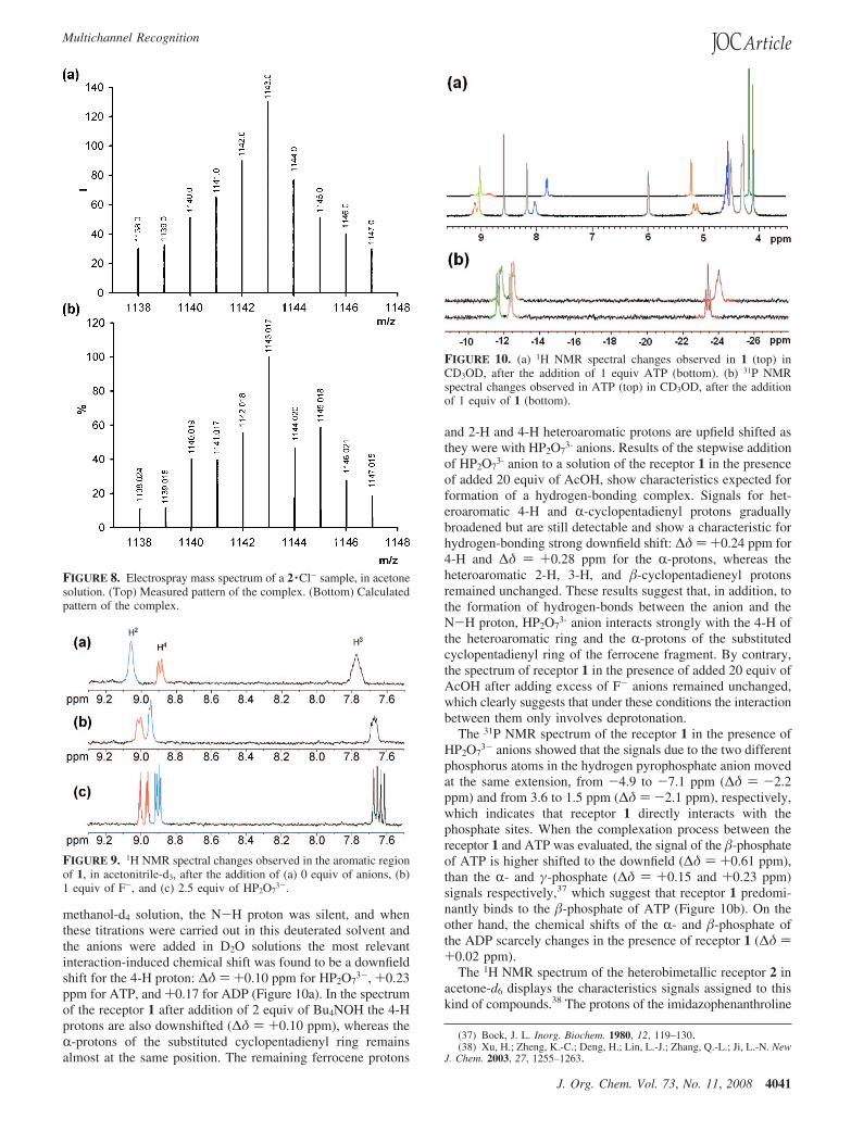
The stoichiometry of the complexes have also been confirmedby ESI-MS, where peaks at m/z 581 [1 ·HP2O7
3-], m/z 957[1 ·ATP], and m/z 1142 [2 ·Cl-] are observed. The relativeabundance of the isotopic clusters was in good agreement withthe simulated spectra of the 1:1 complexes (Figure 8).
To look into the anion binding properties of receptors 1 and2 1H NMR experiments were carried out both in the absence
and presence of anions in acetonitrile-d3 or methanol-d4 at roomtemperature. It must be considered that hydrogen bond formationbetween the receptors and the anions can induced two distincteffects on the heteroaromatic and metallocene protons: (a) itincreases the electron density of the both units with a through-bond propagation, which generates a shielding effect and shouldproduce an upfield shift of the C-H protons; and (b) it inducesthe polarization of the C-H bonds via a through space effect,which causes a deshielding effect and produces a downfield shift.The latter effect, of electrostatic nature drastically decreasesupon increasing the distance between the envisaged C-H protonand the site of H-bond interaction.
When one equiv of TBAF was added to a solution of 1 inacetonitrile-d3 its 1H NMR spectrum displayed dramatic changes(Figure 9). The N-H proton, which in the free receptor appearsat δ 11.3 ppm, disappeared and a clear upfield shift for 3-H(∆δ ) -0.13 ppm) and 2-H (∆δ ) -0.18 ppm) was observed.On the contrary, the signal for 4-H was shifted downfield (∆δ) +0.25 ppm). Meanwhile, upfield shifts of the signalscorresponding to the unsubstituted cyclopentadienyl ring (∆δ) -0.15 ppm) and to the � protons of the monosubstitutedcyclopentadienyl ring (∆δ ) -0.28 ppm) were also observed.Moreover, the R protons were downfield shifted (∆δ ) +0.17ppm). The electrostatic effect predominates for the heteroaro-matic 4-H proton and the R-protons of the monosubstitutedcyclopentadienyl ring as indicated by the pronounced downfieldshift. Heteroaromatics protons 2-H and 3-H are too far awayfrom the N-H protons to be subjected to any electrostatic effect.Thus, the shielding effect induced by the propagation of electrondensity is not contrasted and a significant upfield shift isobserved. It may be surprising that the unsubstituted cyclopen-tadienyl ring protons, quite far away from the N-H fragment,undergo substantial downfield shift.
Likewise, addition of 2.5 equiv of HP2O73- anion caused
similar chemical shift changes, although now well-resolvedsignals for the aromatic protons were obtained (Figure 9). In
(34) (a) Juris, A.; Balzani, V.; Barigelleti, F.; Campagna, S.; Belser, P.;Zelewsky, A. V. Coord. Chem. ReV. 1988, 84, 85–277. (b) Beer, P. D.; Szemes,F.; Balzani, V.; Sala, C. M.; Drew, M. G. B.; Dent, S. W.; Maestri, M. J. Am.Chem. Soc. 1997, 119, 11864–11875. (c) Anzenbacher, P., Jr.; Tyson, D. S.;Jursikova, K.; Castellano, F. N. J. Am. Chem. Soc. 2002, 124, 6232–6233.
(35) (a) Davis, A. P.; Joos, J. B. Coord. Chem. ReV. 2003, 240, 143–156.(b) Boon, J. M.; Smith, B. D. Curr. Opin. Chem. Biol. 2002, 6, 749–756. (c)McNally, B. A.; Koulov, A. V.; Smith, B. D.; Joos, J. B.; Davis, A. P. Chem.Commun. 2005, 1087–1089.
(36) (a) Zhang, Y.; Li, M. X.; Lu, M. Y.; Yang, R. H.; Liu, F.; Li, K. A. J.Phys. Chem. A 2005, 109, 7442–7448. (b) Bai, Y.; Zhang, B.-G.; Xu, J.; Duan,C.-Y.; Dang, D. B.; Liu, D.-J.; Meng, Q.-J. New J. Chem. 2005, 29, 777–779.(c) Schazmann, B.; Alhashimy, N.; Diamond, D. J. Am. Chem. Soc. 2006, 128,8607–8614. (d) dos Santos, C. M. G.; McCabe, T.; Gunnlaugsson, T. TetrahedronLett. 2007, 48, 3135–3139.
FIGURE 7. (a) Changes in the fluorescence emission spectrum of 2(c ) 2.5 × 10⁻⁵ M in CH3CN) upon titration with Cl-: the initial (blue)is that of 2 and the final one (red), after addition of 1 equiv of Cl-. (b)Fluorescence intensity increase of ligand 2, in CH3CN, in relation tothe free ligand, after addition of 1 equiv of several anions. Emission ismonitored at λexc ) 260 nm.
Zapata et al.
4040 J. Org. Chem. Vol. 73, No. 11, 2008
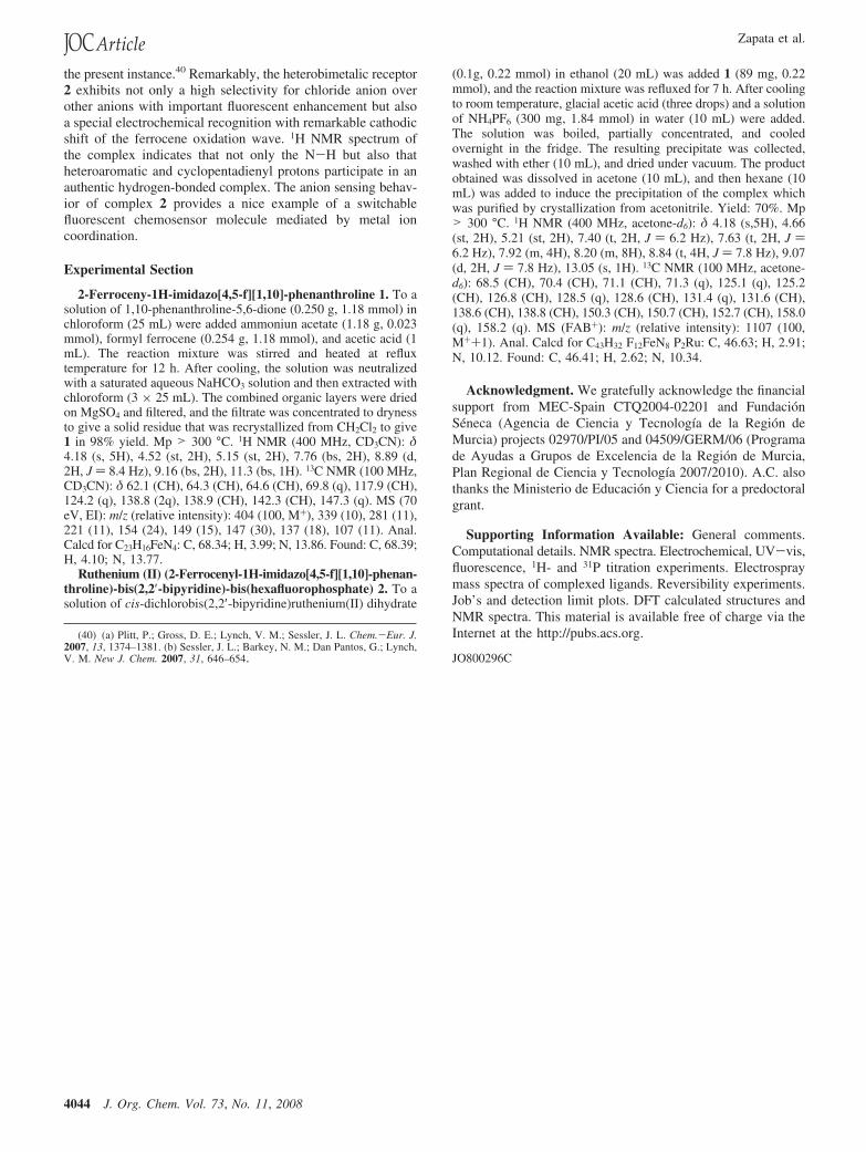
methanol-d4 solution, the N-H proton was silent, and whenthese titrations were carried out in this deuterated solvent andthe anions were added in D2O solutions the most relevantinteraction-induced chemical shift was found to be a downfieldshift for the 4-H proton: ∆δ ) +0.10 ppm for HP2O7
3-, +0.23ppm for ATP, and +0.17 for ADP (Figure 10a). In the spectrumof the receptor 1 after addition of 2 equiv of Bu4NOH the 4-Hprotons are also downshifted (∆δ ) +0.10 ppm), whereas theR-protons of the substituted cyclopentadienyl ring remainsalmost at the same position. The remaining ferrocene protons
and 2-H and 4-H heteroaromatic protons are upfield shifted asthey were with HP2O7
3- anions. Results of the stepwise additionof HP2O7
3- anion to a solution of the receptor 1 in the presenceof added 20 equiv of AcOH, show characteristics expected forformation of a hydrogen-bonding complex. Signals for het-eroaromatic 4-H and R-cyclopentadienyl protons graduallybroadened but are still detectable and show a characteristic forhydrogen-bonding strong downfield shift: ∆δ ) +0.24 ppm for4-H and ∆δ ) +0.28 ppm for the R-protons, whereas theheteroaromatic 2-H, 3-H, and �-cyclopentadieneyl protonsremained unchanged. These results suggest that, in addition, tothe formation of hydrogen-bonds between the anion and theN-H proton, HP2O7
3- anion interacts strongly with the 4-H ofthe heteroaromatic ring and the R-protons of the substitutedcyclopentadienyl ring of the ferrocene fragment. By contrary,the spectrum of receptor 1 in the presence of added 20 equiv ofAcOH after adding excess of F- anions remained unchanged,which clearly suggests that under these conditions the interactionbetween them only involves deprotonation.
The 31P NMR spectrum of the receptor 1 in the presence ofHP2O7
3- anions showed that the signals due to the two differentphosphorus atoms in the hydrogen pyrophosphate anion movedat the same extension, from -4.9 to -7.1 ppm (∆δ ) -2.2ppm) and from 3.6 to 1.5 ppm (∆δ ) -2.1 ppm), respectively,which indicates that receptor 1 directly interacts with thephosphate sites. When the complexation process between thereceptor 1 and ATP was evaluated, the signal of the �-phosphateof ATP is higher shifted to the downfield (∆δ ) +0.61 ppm),than the R- and γ-phosphate (∆δ ) +0.15 and +0.23 ppm)signals respectively,37 which suggest that receptor 1 predomi-nantly binds to the �-phosphate of ATP (Figure 10b). On theother hand, the chemical shifts of the R- and �-phosphate ofthe ADP scarcely changes in the presence of receptor 1 (∆δ )+0.02 ppm).
The 1H NMR spectrum of the heterobimetallic receptor 2 inacetone-d6 displays the characteristics signals assigned to thiskind of compounds.38 The protons of the imidazophenanthroline
(37) Bock, J. L. Inorg. Biochem. 1980, 12, 119–130.(38) Xu, H.; Zheng, K.-C.; Deng, H.; Lin, L.-J.; Zhang, Q.-L.; Ji, L.-N. New
J. Chem. 2003, 27, 1255–1263.
FIGURE 8. Electrospray mass spectrum of a 2 ·Cl- sample, in acetonesolution. (Top) Measured pattern of the complex. (Bottom) Calculatedpattern of the complex.
FIGURE 9. 1H NMR spectral changes observed in the aromatic regionof 1, in acetonitrile-d3, after the addition of (a) 0 equiv of anions, (b)1 equiv of F-, and (c) 2.5 equiv of HP2O7
3-.
FIGURE 10. (a) 1H NMR spectral changes observed in 1 (top) inCD3OD, after the addition of 1 equiv ATP (bottom). (b) 31P NMRspectral changes observed in ATP (top) in CD3OD, after the additionof 1 equiv of 1 (bottom).
Multichannel Recognition
J. Org. Chem. Vol. 73, No. 11, 2008 4041

ring appear as a set of three signals: a doublet centered at 9.07(4-H), a doublet at 8.20 (2-H) and a triplet at 7.92 (3-H) ppmrespectively, whereas the N-H proton appears at δ ) 13.0 ppm.Only the Cl- anion caused a downfield shift of some signals,indicative of an H-bonding interaction. Upon addition of Cl-
anion in the form of TBA salt, the signal of the 4-H protons issplitted into two signals, one of them is shifted to downfield at10.33 ppm (∆δ ) +1.33 ppm) and the second one remains atthe same position. Meanwhile, downfield shift of the signalscorresponding to the R-protons of the monosubstituted cyclo-pentadienyl ring (∆δ ) + 0.43 ppm) was also observed. Anotable feature of this titration is that the resonance correspond-ing to the imidazole N-H proton is downfield shifted (∆δ )+3.16 ppm) over the course of the titration. The fact that thissignal is observable throughout the titration is inconsistent witha deprotonation process involving the receptor, and probablythe formation of a hydrogen-bonded complex has taken place(Figure 11). This assumption is also supported by the fact thataddition of 1 equiv of Bu4NOH to a solution of the receptor 2did not induce any significant change.
Accordingly, the spectroscopic data led us to the conclusionthat the Cl-anions had more effective hydrogen bond interac-tions not only with the N-H proton but also with the aromaticand ferrocenyl protons, which could be another example of N-Hbased receptor in which C-H hydrogens participate in supple-mentary hydrogen bonding with anions.39
Theoretical Calculations. Quantum chemical calculations atthe DFT level of theory (see the Supporting Information) hasallowed satisfactory explanation about binding modes betweenthe receptors and the complexed anions. Under the workingconditions, that is using naked anions (no countercation wasexplicitly included) in the modelization and optimizations beingcarried out in the gas-phase, both F- and HP2O7
3- anionspromote deprotonation of the ligand 1. The obtained structures(Figure 12) are therefore resulting from the complexation ofthe conjugated acids of the anions HF and H2P2O7
3-, by theaction of the conjugated base of ligand 1 still featuring a concave
surface with acidic H atoms directed in a convergent fashion.Thus after initial endergonic acid-base protoin transfer (∆GMeCN
) +11.28 Kcal ·mol-1), complexation of F- is predicted to bemoderately exergonic (∆GMeCN ) -5.44 Kcal ·mol-1) andoccurs upon little deformation of the free-ligand structure (Lstrain
) 1.53 Kcal ·mol-1). A partial deprotonation in 1 ·F- isevidenced by the pyrrolic H atom being almost halfway inbetween nitrogen (dN · · ·H ) 1.279 Å, WBI 0.373) and fluorine(dF · · ·H ) 1.150 Å, WBI 0.378) atoms (angle NHF 178.3°), thelater forming in addition two weak hydrogen bridge bonds withphenanthroline 4-H (dF · · ·4H ) 2.378 Å, WBI 0.019) andferrocenyl HR (dF · · ·HR ) 2.378 Å, WBI 0.017). On the contrary,deprotonation by hydrogenpyrophosphate is expected to beslightly exergonic (∆GMeCN ) -0.53 Kcal ·mol-1), althoughglobal complexation resulted moderately endergonic (∆GMeCN
) +8.20 Kcal ·mol-1), presumably due mainly to highlyunfavorable entropic component, higher ligand deformation(Lstrain ) 6.10 Kcal ·mol-1), and higher relevance of the naked-anion effect in this trianionic substrate. For the calculatedstructure of 1 ·HP2O7
3- complex, partial wrapping of oneH2P2O7
2- phosphate unit by the convergent preorganized cavityof the conjugated base (fully deprotonated) of 1 is observed.One strong hydrogen bridge bond of O-H · · ·N type (dN · · ·H )1.279 Å, WBI 0.373; angle OHN 160.0°) and a weaker onebetween this oxygen atom and the phenanthroline 4-H (dO · · ·4H
) 2.297 Å, WBI 0.010) binds the anion to the imidazophenan-throline part of the receptor, whereas two additional relativelyshort O · · ·H bonds with HR (dO · · ·HR ) 2.235 Å, WBI 0.020)at Cpa (the monosubstituted cyclopentadienyl ring) and oneH-Cpb (Cpb ) unsubstituted cyclopentadienyl ring; dO · · ·HCpb )2.192 Å, WBI 0.022) catch hold of the ferrocenyl part, thushindering rotation around the ferrocene-imidazole bond (seebelow). Then, we assume that the well-resolved signals observedin the 1H NMR of 1 ·HP2O7
3- complex, previously described,could be a consequence of this hindered rotation.
The NBO (Natural Bond Orbital) analysis reveals that theuppermost valence AO (atomic orbitals) at the iron centers inevery molecular system are roughly half-populated e1g-type 3d
(39) (a) Tarraga, A.; Molina, P.; Lopez, J. L.; Velasco, M. D.; Bautista, D.;Jones, P. G. Organometallics 2002, 21, 2055–2065. (b) Lee, C.-H.; Na, H.-K.;Yoon, D.-H.; Cho, W.-S.; Lynch, V. M.; Shevchuck, S. V.; Sessler, J. L. J. Am.Chem. Soc. 2003, 125, 7301–7306. (c) Kwon, J. Y.; Jang, Y. J.; Kim, S. K.;Lee, K.-H.; Kim, J. S.; Yoon, J. J. Org, Chem. 2004, 69, 51555157. (d) Ghosh,S.; Choudhury, A. R.; Row, T. N. G.; Maitra, U. Org. Lett. 2005, 7, 1441–1444. (e) Lu, H. L.; Zhang, D.; Chen, C.; Zhu, D. Org. Lett. 2005, 7, 4629–4632. (f) El Drubi, I.; Gale, P. A.; Light, M. E.; Loeb, S. J. Chem. Commun.2005, 4913–4915. (g) Xu, Z.; Kim, S.; Lee, K.-H.; Yoon, J. Tetrahedron Lett.2007, 48, 3797–3800.
FIGURE 11. 1H NMR spectral changes observed for 2, in acetone-d6,after addition of increasing amounts of Cl- in CD3CN: (a) 0, (b) 0.25,(c) 0.50, (d) 0.75, and (e) 1 equiv.
FIGURE 12. Calculated (B3LYP/aug-6-31G*) structures for com-plexes 1 ·F- (up) and 1 ·HP2O7
3- (down).
Zapata et al.
4042 J. Org. Chem. Vol. 73, No. 11, 2008

AO and their energies increase following the degree of depro-tonation (see above) in the series 1 (- 0.162 au) < 1 ·F- (- 0.040au) < 1 ·HP2O7
3- (+ 0.121 au), in perfect agreement with theincreasing ease of oxidation experimentally shown by their lower(cathodically shifted) oxidation potentials.
Also, the structures of [1 ·Ru(bipy)2]2+ (see the SupportingInformation), that is, 2 without the counteranions, and[1 ·Ru(bipy)2 ·Cl]+ (Figure 13) have been calculated. The NBOanalysis reveals that the uppermost valence AO at Fe are, inthese cases, of a1g symmetry (dz2 type) and also roughtly half-populated. As expected, this AO in [1 ·Ru(bipy)2]2+ has lowerenergy content (-0.286 au) than that in 1, hence its higher(anodically shifted) oxidation potential. Upon complexation ofCl-, the energy of this AO increases (-0.184 au), which is inagreement with the observed cathodic shift experienced by thecorresponding oxidation wave. Complexation of Cl- occursupon little deformation of the free-ligand structure (Lstrain ) 1.38Kcal ·mol-1) and is predicted to be only slightly exergonic(∆GMeCN ) -1.10 Kcal ·mol-1). Selectivity toward otherspherical halide anions has also been studied. For that purpose,the optimized structures of [1 ·Ru(bipy)2 ·F]+ and[1 ·Ru(bipy)2 ·Br]+ (see the Supporting Information) wereobtained. Complexation of F- anion takes place with acomparatively remarkable deformation of the ligand (Lstrain )3.68 Kcal ·mol-1), much higher than that found for 1 ·F-. Liganddeformation is small upon complexation of Br- anion (Lstrain )1.14 Kcal ·mol-1) and, in this case, selectivity may arise fromthe slightly endergonic character (∆GMeCN ) + 0.24Kcal ·mol-1) of the ligand-anion recognition event. Thecalculated structure for the [1 ·Ru(bipy)2 ·Cl]+ complex ischaracterized by strong N-H · · ·Cl bridge bonding with the
pyrrolic H atom (dCl · · ·H ) 1.963 Å, WBI 0.181; angle NHCl169.5°), and the guest Cl- anion forming a complementaryhydrogen bridge bond with phenanthroline 4-H (dCl · · ·4H ) 2.376Å, WBI 0.037), as well as two other weak ones with ferrocenylHR (dCl · · ·HR ) 2.925 Å, WBI 0.004) and H-Cpb (dCl · · ·HCpb )3.155 Å, WBI 0.002).
The calculated spectra for both the receptor [1 ·Ru(bipy)2]2+
and the corresponding complex [1 ·Ru(bipy)2 ·Cl]+ (Table 1)fit nicely with those obtained experimentally (see the SupportingInformation). which supports the theoretical findings concerningthe nature of binding of chloride anions to the receptor. Theremarkable downfield shifting of the N-H and phenanthroline4-H signals constitutes a direct consequence of the hydrogenbridge bonding with the chloride anion guest.
Conclusions
In summary, we describe an easy-to-synthesize ferrocene-based imidazophenanthroline 1 and its stable Ru(II) complex 2receptors and examined their properties toward various guestanions, using electrochemical, spectral and fluorescence tech-niques. Receptor 1 in acetonitrile senses aqueous hydrogenpy-rophosphate and the organic anions ADP and ATP through threedifferent channels: the yellow-to-orange or pink color changewhich allow the potential for “naked eye” detection, a red-shiftof the emission band with an important fluorescent enhancement,and an unprecedent large cathodic shift of the ferroceneoxidation wave (Figure 14). About the deprotonation/coordina-tion dualism, the combined electrochemical, absorption, emis-sion, and NMR data strongly support that fluoride anion inducesonly deprotonation, anions, dihydrogenphosphate, ATP, andADP from hydrogen-bonded complexes and formation ofhydrogen-bonded complex between receptor 1 and hydrogen-pyrophosphate anion and deprotonation proceed simultaneously.As a consequence, selective fluorescent detection of hydrogen-pyrophosphate anions in the presence of the related competitorsdihydrogenphosphate anions, ADP, and ATP could be achievedby carefully selection of the excitation energy. The recognitionevent has also been studied by 1H and 31P NMR spectroscopy,as well as by DFT calculations.
The use of ruthenium(II) fluorophore in receptor 1 has a largeeffect on the resulting function, specifically anion binding in
FIGURE 13. Calculated (B3LYP/aug-6-31G*/SDD-ecp) structure forthe complex [1 ·Ru(bipy)2 ·Cl]+.
TABLE 1. Calculated (GIAO/CPCM/B3LYP/aug-6-311G**//B3LYP/aug-6-31G*) 1H NMR Chemical Shifts (ppm)a for Receptor[1 ·Ru(bipy)2]2+ and Complex [1 ·Ru(bipy)2 ·Cl]+ in Acetone Solution
[1 ·Ru(bipy)2]2+ [1 ·Ru(bipy)2 ·Cl]+
δH δH ∆δH(exp)
Cpa HR 5.25 5.59 0.24 (0.43)H� 4.69 4.65 -0.04 (-0.22)
Cpb 4.26 4.18 -0.08 (0.02)2-H 8.53 8.38 -0.15 (0.00)
Phenan. 3-H 8.37 8.25 -0.12 (-0.08)4-H 9.59 11.85 2.26 (1.23)
9.68 0.09 (0.01)N-H 10.47 16.88 6.41 (3.16)
a Average for related protons.
FIGURE 14. Changes in the color of ligand 1 (up) and in thefluorescence of ligands 1 and 2 (down) upon addition of the corre-sponding anions.
Multichannel Recognition
J. Org. Chem. Vol. 73, No. 11, 2008 4043
the present instance.40 Remarkably, the heterobimetalic receptor2 exhibits not only a high selectivity for chloride anion overother anions with important fluorescent enhancement but alsoa special electrochemical recognition with remarkable cathodicshift of the ferrocene oxidation wave. 1H NMR spectrum ofthe complex indicates that not only the N-H but also thatheteroaromatic and cyclopentadienyl protons participate in anauthentic hydrogen-bonded complex. The anion sensing behav-ior of complex 2 provides a nice example of a switchablefluorescent chemosensor molecule mediated by metal ioncoordination.
Experimental Section
2-Ferroceny-1H-imidazo[4,5-f][1,10]-phenanthroline 1. To asolution of 1,10-phenanthroline-5,6-dione (0.250 g, 1.18 mmol) inchloroform (25 mL) were added ammoniun acetate (1.18 g, 0.023mmol), formyl ferrocene (0.254 g, 1.18 mmol), and acetic acid (1mL). The reaction mixture was stirred and heated at refluxtemperature for 12 h. After cooling, the solution was neutralizedwith a saturated aqueous NaHCO3 solution and then extracted withchloroform (3 × 25 mL). The combined organic layers were driedon MgSO4 and filtered, and the filtrate was concentrated to drynessto give a solid residue that was recrystallized from CH2Cl2 to give1 in 98% yield. Mp > 300 °C. 1H NMR (400 MHz, CD3CN): δ4.18 (s, 5H), 4.52 (st, 2H), 5.15 (st, 2H), 7.76 (bs, 2H), 8.89 (d,2H, J ) 8.4 Hz), 9.16 (bs, 2H), 11.3 (bs, 1H). 13C NMR (100 MHz,CD3CN): δ 62.1 (CH), 64.3 (CH), 64.6 (CH), 69.8 (q), 117.9 (CH),124.2 (q), 138.8 (2q), 138.9 (CH), 142.3 (CH), 147.3 (q). MS (70eV, EI): m/z (relative intensity): 404 (100, M+), 339 (10), 281 (11),221 (11), 154 (24), 149 (15), 147 (30), 137 (18), 107 (11). Anal.Calcd for C23H16FeN4: C, 68.34; H, 3.99; N, 13.86. Found: C, 68.39;H, 4.10; N, 13.77.
Ruthenium (II) (2-Ferrocenyl-1H-imidazo[4,5-f][1,10]-phenan-throline)-bis(2,2′-bipyridine)-bis(hexafluorophosphate) 2. To asolution of cis-dichlorobis(2,2′-bipyridine)ruthenium(II) dihydrate
(0.1g, 0.22 mmol) in ethanol (20 mL) was added 1 (89 mg, 0.22mmol), and the reaction mixture was refluxed for 7 h. After coolingto room temperature, glacial acetic acid (three drops) and a solutionof NH4PF6 (300 mg, 1.84 mmol) in water (10 mL) were added.The solution was boiled, partially concentrated, and cooledovernight in the fridge. The resulting precipitate was collected,washed with ether (10 mL), and dried under vacuum. The productobtained was dissolved in acetone (10 mL), and then hexane (10mL) was added to induce the precipitation of the complex whichwas purified by crystallization from acetonitrile. Yield: 70%. Mp> 300 °C. 1H NMR (400 MHz, acetone-d6): δ 4.18 (s,5H), 4.66(st, 2H), 5.21 (st, 2H), 7.40 (t, 2H, J ) 6.2 Hz), 7.63 (t, 2H, J )6.2 Hz), 7.92 (m, 4H), 8.20 (m, 8H), 8.84 (t, 4H, J ) 7.8 Hz), 9.07(d, 2H, J ) 7.8 Hz), 13.05 (s, 1H). 13C NMR (100 MHz, acetone-d6): 68.5 (CH), 70.4 (CH), 71.1 (CH), 71.3 (q), 125.1 (q), 125.2(CH), 126.8 (CH), 128.5 (q), 128.6 (CH), 131.4 (q), 131.6 (CH),138.6 (CH), 138.8 (CH), 150.3 (CH), 150.7 (CH), 152.7 (CH), 158.0(q), 158.2 (q). MS (FAB+): m/z (relative intensity): 1107 (100,M++1). Anal. Calcd for C43H32 F12FeN8 P2Ru: C, 46.63; H, 2.91;N, 10.12. Found: C, 46.41; H, 2.62; N, 10.34.
Acknowledgment. We gratefully acknowledge the financialsupport from MEC-Spain CTQ2004-02201 and FundacionSeneca (Agencia de Ciencia y Tecnologıa de la Region deMurcia) projects 02970/PI/05 and 04509/GERM/06 (Programade Ayudas a Grupos de Excelencia de la Region de Murcia,Plan Regional de Ciencia y Tecnologıa 2007/2010). A.C. alsothanks the Ministerio de Educacion y Ciencia for a predoctoralgrant.
Supporting Information Available: General comments.Computational details. NMR spectra. Electrochemical, UV-vis,fluorescence, 1H- and 31P titration experiments. Electrospraymass spectra of complexed ligands. Reversibility experiments.Job’s and detection limit plots. DFT calculated structures andNMR spectra. This material is available free of charge via theInternet at the http://pubs.acs.org.
JO800296C
(40) (a) Plitt, P.; Gross, D. E.; Lynch, V. M.; Sessler, J. L. Chem.-Eur. J.2007, 13, 1374–1381. (b) Sessler, J. L.; Barkey, N. M.; Dan Pantos, G.; Lynch,V. M. New J. Chem. 2007, 31, 646–654.
Zapata et al.
4044 J. Org. Chem. Vol. 73, No. 11, 2008