
Catherwood, Dianne F, Edgar, Graham K ORCID: 0000-0003
59
This is a peer-reviewed, post-print (final draft post-refereeing) version of the following published document and is licensed under All Rights Reserved license: Catherwood, Dianne F, Edgar, Graham K ORCID: 0000-0003- 4302-7169, Nikolla, Dritan, Alford, Christopher A, Brookes, David, Baker, Steven ORCID: 0000-0002-3029-8931 and White, Sarah (2014) Mapping Brain Activity During Loss of Situation Awareness: an EEG investigation of a basis for top-down influence on perception. Human Factors, 56 (8). pp. 1428- 1452. doi:10.1177/0018720814537070 Official URL: http://journals.sagepub.com/doi/abs/10.1177/0018720814537070 DOI: http://dx.doi.org/10.1177/0018720814537070 EPrint URI: https://eprints.glos.ac.uk/id/eprint/620 Disclaimer The University of Gloucestershire has obtained warranties from all depositors as to their title in the material deposited and as to their right to deposit such material. The University of Gloucestershire makes no representation or warranties of commercial utility, title, or fitness for a particular purpose or any other warranty, express or implied in respect of any material deposited. The University of Gloucestershire makes no representation that the use of the materials will not infringe any patent, copyright, trademark or other property or proprietary rights. The University of Gloucestershire accepts no liability for any infringement of intellectual property rights in any material deposited but will remove such material from public view pending investigation in the event of an allegation of any such infringement. PLEASE SCROLL DOWN FOR TEXT.
-
Upload
khangminh22 -
Category
Documents
-
view
2 -
download
0
Transcript of Catherwood, Dianne F, Edgar, Graham K ORCID: 0000-0003

This is a peer-reviewed post-print (final draft post-refereeing) version of the following publisheddocument and is licensed under All Rights Reserved license
Catherwood Dianne F Edgar Graham K ORCID 0000-0003-4302-7169 Nikolla Dritan Alford Christopher A Brookes David Baker Steven ORCID 0000-0002-3029-8931 and WhiteSarah (2014) Mapping Brain Activity During Loss of Situation Awareness an EEG investigation of a basis for top-down influence on perception Human Factors 56 (8) pp 1428-1452 doi1011770018720814537070
Official URL httpjournalssagepubcomdoiabs1011770018720814537070DOI httpdxdoiorg1011770018720814537070EPrint URI httpseprintsglosacukideprint620
Disclaimer
The University of Gloucestershire has obtained warranties from all depositors as to their title in the material deposited and as to their right to deposit such material
The University of Gloucestershire makes no representation or warranties of commercial utility title or fitness for a particular purpose or any other warranty express or implied in respect of any material deposited
The University of Gloucestershire makes no representation that the use of the materials will notinfringe any patent copyright trademark or other property or proprietary rights
The University of Gloucestershire accepts no liability for any infringement of intellectual property rights in any material deposited but will remove such material from public view pending investigation in the event of an allegation of any such infringement
PLEASE SCROLL DOWN FOR TEXT
This is a peer-reviewed post-print (final draft post-refereeing) version of the following published document
Catherwood Dianne F and Edgar Graham K and Nikolla Dritan and Alford Christopher A and Brookes David and Baker Steven and White Sarah (2014) Mapping Brain Activity During Loss of Situation Awareness an EEG investigation of a basis for top-down influence on perception Human Factors 56 (8) pp 1428-1452 ISSN 0018-7208
Published in Human Factors and available online at httphfssagepubcomcontent5681428abstract ISSN 0018-7208 We recommend you cite the published (post-print) version The URL for the published version is httphfssagepubcomcontent5681428fullpdf+html ISSN 0018-7208 Disclaimer The University of Gloucestershire has obtained warranties from all depositors as to their title in the material deposited and as to their right to deposit such material The University of Gloucestershire makes no representation or warranties of commercial utility title or fitness for a particular purpose or any other warranty express or implied in respect of any material deposited The University of Gloucestershire makes no representation that the use of the materials will not infringe any patent copyright trademark or other property or proprietary rights The University of Gloucestershire accepts no liability for any infringement of intellectual property rights in any material deposited but will remove such material from public view pending investigation in the event of an allegation of any such infringement
PLEASE SCROLL DOWN FOR TEXT
1
[Human Factors Topic Psychological States and Neuroergonomics]
MAPPING BRAIN ACTIVITY DURING LOSS OF SITUATION AWARENESS
an EEG investigation of a basis for top-down influence on perception
Di Catherwooda Graham K Edgara Dritan Nikollaa Chris Alfordab
David Brookesa Steven Bakera and Sarah Whitea
aCentre for Research in Applied Cognition Knowledge Learning and Emotion (CRACKLE)
University of Gloucestershire UK
and
bDepartment of Health and Social Sciences University of West England UK
PREacuteCIS SHORT ABSTRACT
Loss of SA was enforced in two tasks requiring identification of target items (respectively
abstract concepts and urban ldquothreatrdquo) EEG recording and source-localization with sLORETA
shows rapid co-activity of regions for visual perception and those with high-order duties
This may offer a basis for top-down effects on level 1 SA
Corresponding author Di Catherwood School of Natural amp Social Sciences University of
Gloucestershire Francis Close Hall Cheltenham GL50 4AZ UK dcatherwoodglosacuk
phone +44 (0)1242714808
Running head Brain activity in loss of SA
Keywords QASA Electroencephalography sLORETA SA Source-localization Top-
down Bias
Acknowledgement This research was supported by a grant from the UK Ministry of
Defence under the ldquoCompetition of Ideasrdquo Scheme
Word counts Text 7844 words excluding title page abstract references footnotes tables
figure caption key points preacutecis References 5008 words
EEG Mapping in Loss of SA 2
ABSTRACT
Objective The objective was to map brain activity during early intervals in loss of
Situation Awareness (SA) to examine any co-activity in visual and high-order regions
reflecting grounds for top-down influences on level 1 SA
Background Behavioural and neuroscience evidence indicates that high-order brain
areas can engage before perception is complete Inappropriate top-down messages may
distort perception during loss of SA Evidence of co-activity of perceptual and high-order
regions would not confirm such influence but may reflect a basis for it
Methods SA and Bias were measured using QASA (Quantitative Analysis of
Situation Awareness) and brain activity recorded with 128-channel EEG
(electroencephalography) during loss of SA One task (15 participants) required
identification of a target pattern and another task (10 participants) identification of ldquothreatrdquo in
urban scenes In both the target was changed without warning enforcing loss of SA Key
regions of brain activity were identified using source localization with sLORETA 150-
160msec post-stimulus-onset in both tasks and also 100-110msec in the second task
Results In both tasks there was significant loss of SA and Bias shift (p le 02)
associated at both 150 and 100 msec intervals with co-activity of visual regions and
prefrontal anterior cingulate and parietal regions linked to cognition under uncertainty
Conclusion There was early co-activity in high-order and visual perception regions
that may provide a basis for top-down influence on perception
Application Co-activity in high- and low-order brain regions may explain either
beneficial or disruptive top-down influence on perception affecting level 1 SA in real-world
operations
(word count 250 words)
EEG Mapping in Loss of SA 3
MAPPING BRAIN ACTIVITY DURING LOSS OF SITUATION AWARENESS
an EEG investigation of a basis for top-down influence on perception
Effective interaction with the external environment requires that salient aspects are
processed appropriately to produce or maintain good Situation Awareness (SA) (Adams
Tenney amp Pew 1995 Endsley 1995 2000 2013 Durso amp Sethumadhavan 2008
Parasuraman Sheridan amp Wickens 2008 Patrick amp Morgan 2010 Wickens 2008)
Complementary theoretical approaches define SA either as a ldquostate of knowledgerdquo about a
situation (Endsley 2013) andor in terms of the processes for building that knowledge (Durso
amp Sethumadhavan 2008) encompassing both explicit and implicit understanding (Durso
Rawson amp Girotto 2007) The loss of SA especially in challenging operational
environments such as combat transport fireground or medical situations may precipitate
critical errors with serious consequences (Borghini Astolfi Vecchiato Mattia amp Babiloni
2012 Catherwood Edgar Sallis Medley amp Brookes 2012 Klein Calderwood Clinton-
Sirocco 2010 Schulz Endsley Kochs Gelb amp Wagner 2013) Further investigation of the
psychological dynamics underlying loss of SA is of paramount importance
Understanding of these dynamics has been achieved by the convergence of
behavioural and neuroscience evidence Neuroergonomic research (Parasuraman 2003
Parasuraman ampWilson 2008) using functional Magnetic Resonance Imaging (fMRI) and
Electroencephalography (EEG) has identified brain response linked to behavioural aspects of
SA such as the association between activity in Prefrontal Cortex (PFC) or the theta EEG
band and cognitive workload (Berka et al 2007 Borghini et al 2012 Brookings Wilson
amp Swain 1996 Dussault Jouanin Philippe amp Guezennec 2005 French Clarke Pomeroy
Seymour amp Clark 2007 Lei amp Roetting 2011 Parasuraman Warm amp See 1998 Savage
Potter amp Tatler 2013 Sterman amp Mann 1995 Wilson 2000) EEG activity also reflects
EEG Mapping in Loss of SA 4
aspects of team SA (Stevens Galloway Wang amp Berka 2012) Investigation of behaviour
linked to SA and corresponding brain activity may also provide deeper insights into another
key issue regarding SAndash namely ldquotop-downrdquo influence on perception (level 1 SA) Loss of
SA may be associated with perceptual lapses or impairment due to top-down factors such as
prior memory or expectation (Durso et al 2007 Durso amp Gronlund 1999 Endsley 2013)
Investigation of the associated brain dynamics may advance understanding of this issue for
the following reasons
Most models of SA cite a processing trajectory from perception (level 1 SA) to
cognitive integration (level 2) to projection (level 3) but it is acknowledged that top-down
factors influence all levels of SA (Durso amp Gronlund 1999 Endsley 2013) Individuals
make more errors if situations do not fit expectations (Taylor Endsley amp Henderson 1996)
and loss of SA may involve distortion of perception by faulty top-down processing For
example in the Mt Erebus aircraft disaster the flight-crewrsquos expectation based on faulty
flightpath data may have caused visual information about location to be overlooked (Mahon
1981) and in the Storm King Mountain wildfire tragedy an overworked commander may not
have perceived a shift in wind conditions (Sallis Catherwood Edgar Brookes amp Medley
2013 Useem Cook amp Sutton 2005)
Such cases resonate with neuroscience evidence indicating that information-
processing in the brain does not follow a strictly linear-hierarchical trajectory but may
involve bi-directional (ldquore-entrantrdquo) communication between high-order cortical regions and
low-order perceptual regions There is ongoing discussion about the nature and timing of
these brain dynamics (eg Fu Fedota amp Parasuraman 2012 Rauss Pourtois Vuilleumier
amp Schwartz 2012 Rauss Schwartz amp Pourtois 2011 Theeuwes 2010) but high-order
regions may rapidly transmit ldquopredictive and adaptiverdquo coding about a situation based on
expected input learned contingencies or affective factors that can modulate perceptual
EEG Mapping in Loss of SA 5
response (Bar 2003 Damaraju Huang Barrett amp Pessoa 2009 Dambacher Rolfs Goumlllner
Kliegel amp Jacobs 2009 Delorme Rousselet Maceacute amp Fabre-Thorpe 2004 Furmanski
Schluppeck amp Engel 2004 Gilbert amp Sigman 2007 Hegdeacute 2008 Kelley Rees amp Lavie
2013 Paradiso 2002 Poghosyan amp Ioannides 2008 Rauss et al 2011 Schettino Loeys
Delplanque amp Pourtois 2011 Summerfield amp Egner 2009) Indeed expectation alone can
excite visual cortex (Grill-Spector amp Malach 2004) Such top-down influence on perception
may be critical during loss of SA Establishment of SA or efforts to recoup lost SA may enlist
top-down processing but if premature or inappropriate this could distort perception of the
situation For example faulty expectation may have elicited visual focus on an irrelevant
cockpit signal during loss of a 1972 Eastern Airlines flight (National Transportation Safety
Board 1973) ldquoLooked but fail to seerdquo accidents (Langham Hole Edwards amp OrsquoNeil 2002)
may also be due to poor visual processing of the traffic environment if inconsistent with
expectations
It may thus advance understanding of loss of SA to establish if high-order brain
regions are co-opted while perceptual coding is actively proceeding This issue is addressed
in the current investigation The approach is to build SA then enforce its loss in tasks
requiring decisions about a target item EEG source analysis will reveal if loss of SA is
associated with concurrent activity in brain regions with high-order duties and those for
perceptual (visual) processing Parallel co-activity of these regions is not direct evidence of
their interaction Nevertheless it reflects potential conditions for such interaction compared
to a strictly linear sequence whereby perception subsides before high-order processing
occurs
Combining theoretical approaches (Durso amp Sethumadhavan 2008 Endsley 2013
Patrick amp Morgan 2010 Wickens 2008) SA will be explored as a state of knowledge in
relation to brain activity As noted there is already relevant neuroergonomic evidence on
EEG Mapping in Loss of SA 6
brain response under high cognitive-load conditions associated with loss of SA (Parasuraman
et al 2008) This evidence mostly derives from Event-Related-Potentials (ERPs) andor EEG
spectral analysis (eg P300) (Foxe amp Simpson 2002 Makeig et al 2002 Philiastides amp
Sajda 2006 Rousselet Husk Bennett amp Sekuler 2007) These methods use summated
estimates of brain activity but fMRI studies have more precisely mapped sources of brain
activity associated with SA for example in ACC (anterior cingulate cortex) and PFC
(prefrontal cortex) during driving or aviation performance (Calhoun amp Pearlson 2012
Causse Dehais Peacuteran Sabatini amp Pastor 2013 Causse et al 2013b Peres et al 2000)
fMRI however has slow temporal resolution (Raichle 1998) and may not capture the early
processing dynamics of interest here EEG with source localization is better able to achieve
this (Foxe amp Simpson 2002) and has been used to identify sources of brain activity under
challenging conditions such as microgravity (space) flight (Bruumlmmer et al 2011 de la
Torre et al 2012 Schneider Bruumlmmer Carnahan Dubrowski Askew amp Struumlder 2008)
The current investigation will map brain activity during loss of SA using EEG source
analysis with the sLORETA algorithm (see Method) This method will estimate sources of
brain activity in terms of Brodmann Areas (BAs) useful markers of circuitry for brain
functions (Amunts Schliecher amp Zilles 2007) As in other studies of SA (eg French et al
2007) ERPs (averaged EEG signals) will not be used since as noted above they may mask
the early co-activity of interest here Unaveraged EEG data may more directly reflect the
brain dynamics of interest (Philiastides amp Sajda 2006 Rousselet Husk Bennett amp Sekuler
2007)
Brodmann areas are not isolated modules but typically contribute to wider networks
and are used here in this respect (eg BA17 and BA20 are both on the visual pathways
Table 1) Neuroimaging methods reveal modularity for some brain functions (Downing
EEG Mapping in Loss of SA 7
2008 Downing Liu amp Kanwisher 2001) but it may be more informative to investigate brain
operations on a broader scale that can reveal connectivity of processing (Poldrack 2012) For
example an fMRI study showed that novice pilots display more distributed cortical activity
than experienced pilots reflecting differential expertise (Peres et al 2000) Recording broad-
scale individual maps of brain activity may thus provide clues to global brain dynamics
during loss of SA The aim here is therefore not to simply identify active brain regions during
loss of SA but to examine any co-activity of regions with high-order duties and those for
perception Such co-activity may indicate a basis for top-down influence on perception
ltInsert table 1 about heregt
During loss of SA many brain regions may be engaged with variations across
situations and individuals Nevertheless to address the central question of whether areas with
high-order duties are aroused while perception is progressing the following regions are of
interest based on prior evidence of their roles in these functions (see Table 1a and 1d for
references)
If visual perception is actively occurring multiple regions may be engaged (Grill-
Spector amp Malach 2004 Tootell Hadjikhani Mendola et al 1998) These include primary
visual cortex (V1 or BA17) ventral and dorsal areas for higher-level visual analysis (BAs
5181920 37) retrosplenial cortex (BA29-30) for visual scene integration and perirhinal
cortex (BA35-36) for complex visual binding and figure-ground perception (see Table 1a)
For high-order cognitive processing many brain regions are of potential interest but
efforts to recoup lost SA must employ regions involved in cognitive integration (level 2 SA)
and possibly projection (level 3 SA) The brain activity for these levels is not easily
distinguished but on the basis of prior evidence numerous frontal anterior cingulate and
EEG Mapping in Loss of SA 8
parietal cortical regions may predictably be implicated for both (see Table 1d for a
representative list with references) Loss of SA may especially enlist regions known to be
active with cognitive uncertainty or ambiguity low confidence error and the need to reverse
responses These include BA9 and BA46 (Dorsolateral PFC) BA11 (Orbitofrontal cortex)
BA 47 (Orbitofrontal Ventrolateral frontal cortex) BA24 32 and 33 (ACC) and BA7
(Superior Parietal Lobule) (see Table 1d for references) The link between activity in these
areas and loss of SA has not been specifically examined to date and evidence of their rapid
involvement during active perception could afford new insights into brain conditions
associated with top-down influences during loss of SA
The interval of interest for EEG analysis is that encompassing active perceptual
processing of the situation prior to response on each trial Visual perception involves activity
and co-activity in multiple regions (Table 1a) for up to hundreds of milliseconds (Paradiso
2002) Coarse visual coding is complete by 80-90msec post-stimulus (or sooner) basic
stimulus classification by 75-120msec and registration or identification of the ldquogistrdquo of a
visual context by 150msec (Bar 2003 Delorme et al 2004 Foxe amp Simpson 2002 Rauss
et al 2011 Hegdeacute 2008 Jolij Scholte Gaal Hodgson amp Lamme 2011 Schettino et al
2011 Thorpe Fize amp Marlot 1996) Decision-related visual processing can require 250msec
(van Rullen ampThorpe 2001) and activation of visual cortical areas can continue for 400msec
before a motor response or conscious reporting of visual input (Foxe amp Simpson 2002 Jolij
et al 2011) In consideration of these timeframes 150msec post-stimulus-onset was selected
to represent a phase when the basic visual situation has been ldquosensedrdquo but with ongoing
visual processing to achieve a complete perceptual representation of the situation
corresponding to level 1 SA (Endsley 2000 2013) This provides a justifiable timeframe for
determining whether regions for higher-order operations engage during early perceptual
EEG Mapping in Loss of SA 9
processing in loss of SA Nevertheless if there is evidence of strong arousal of high-order
regions at this interval then an even earlier interval will be explored in Experiment 2
The approach here offers a relatively novel means for assessing brain response during
loss of SA SA will be established to a criterion level and then loss of SA enforced at the
same juncture for all participants This general approach has been used in a recent study of
confusion in reading incongruent text (Durso Geldbach amp Corballis 2012) The current
experiments involve an abstract ldquobaselinerdquo task and a similar task with more real-world
content involving identification of threat in urban scenes In both tasks the ldquosituationrdquo is
defined in terms of target information within a visual field The essential requirements
resemble many real-world situations requiring perceptual and cognitive processing to identify
a target item (eg decisions about whether symptoms indicate disease) The first task was
chosen to be less likely to arouse prior knowledge or expectations than the second which may
invoke knowledge about urban crime This contrast allows assessment of whether high-order
regions are aroused during perceptual processing regardless of whether the task is ldquoabstractrdquo
or more ldquoreal-worldrdquo or instead is more likely in the latter Both tasks however require at
least two levels of SA (Endsley 1995) participants must perceive the visual information
(level 1) and integrate that information to comprehend and hypothesize about the correct
target characteristics (level 2) Activity in brain areas for high-order processing would be
required to achieve level 2 but the question is whether it occurs during activity for level 1
The loss of SA may elicit individual differences in response (perseveration trial-and-
error etc) but regardless of the strategy high-order cognitive functions will be required to
re-attain level 2 SA Discriminating brain patterns for different strategies is an issue for future
investigation The aim here is to answer the basic question of whether there is any brain
activity consistent with high-order cognitive processing concurrent with perceptual
EEG Mapping in Loss of SA 10
processing To this end the analysis will firstly determine if visual perceptual regions are
actively engaged and if so then assess if there is concomitant arousal of regions associated
with cognition under uncertainty It is hypothesized that despite individual differences such
co-activity will be apparent to some extent for all participants in both tasks but possibly to a
greater extent in Experiment 2
EXPERIMENT 1 BASELINE WISCONSIN CATEGORY LEARNING TASK
This task is a variant of the Wisconsin Card Sorting Task (WCST) (Berg 1948 Grant
amp Berg 1948) employed here as a tool for exploring basic processes linked to loss of SA
The ldquosituationrdquo requires detection of a target ldquoconceptrdquo amongst stimulus exemplars
involving combination of features across three visual dimensions (colour shape line
orientation) This requires (a) perception of the elements (colours etc) (level 1 SA) and (b)
cognitive integration of these for generating hypotheses or responses with this involving
memory and processing to link responses to the feedback on each trial (all relevant to level 2
SA) Once the correct category (= SA) is achieved this will be changed without warning
occasioning loss of SA which participants then have to reattain This approach brings all
participants to a common reference point for loss of SA and the need to regain it
METHOD
Sample Participants were right-handed volunteers from the local student and staff
population with no known neurological disorder The experiment was completed by 23
participants but 8 failed to achieve satisfactory SA and were not included in the final analysis
leaving a sample of 15 participants
Design The study was within-participants (all did both phases of the experiment)
Apparatus and stimuli Participants were presented with a series of computer
displays with each either containing examples of a lsquotarget categoryrsquo or not and consisting of
EEG Mapping in Loss of SA 11
four quadrants with one of three possible variations of a visual property The top left quadrant
had one two or three lines the top right quadrant was red green or blue the bottom left
quadrant had vertical horizontal or oblique lines and the bottom right quadrant had a
diamond circle or square (See Figure 1) The lsquotarget categoryrsquo in Phase 1 was ldquothree lines
and redrdquo In Phase 2 the target category was changed without notice to ldquooblique lines and
circlerdquo Phase 2 trials contained the same displays as Phase 1 but the target category was
changed
ltInsert Figure 1 about heregt
Brain activity was recorded with Electrical Geodesics Incorporated (EGI)TM EEG
apparatus consisting of 128-channel HydroCel GeoDesic Sensor Net(s) (with a
referencevertex (Cz) sensor) connected to a wall-mounted NetAmpsTM amplifier The dense
geodesic array of the net (Figure 2) optimises accurate recording The 128 high-impedance
electrodes with sponge inserts (with HydroCel saline electrolyte) include eye-blink and eye-
movement sensors The signals from the 128 sensors were sent to a Macintosh computer
running NetstationTM software for acquiring viewing and navigating the data Further source
localization was performed using the GeoSource 20TM software that has received both US
FDA and European Medical Device Directive clearance (httpwwwegicomhome385-
geosource-fda) See further details below
ltInsert Figure 2 about heregt
Procedure Participants were given instructions about the task and EEG procedure If
consent was given an appropriate-sized EEG net was applied Stimulus arrays and
instructions were presented on a PC monitor via E-primeTM software with presentation
controlled by E-prime and responses made via the PC keyboard The net was accurately
positioned relative to the vertex point pre-marked on the scalp relative to the nasion inion
EEG Mapping in Loss of SA 12
and pre-auricular clefts Prior to testing scalp impedances were adjusted below 50 kΩ
Testing was in low-light with shielding of electrical cables and equipment
There were 10 practice and 104 experimental self-paced trials (52 in random order in
each phase) with no signal that Phase 2 had begun To increase task difficulty there was
overlap between category exemplars each Phase had 26 trials with the correct target and 26
without but for each Phase 16 displays had category 1 16 category 2 10 both categories and
10 neither category Each trial began with a fixation marker for 1 to 15 seconds (randomised
in this range) followed by a display For each display participants had to respond to the
question ldquoThis slide represents a member of the target category TFrdquo Feedback on
correctness of response and reaction time was provided on each trial in both phases
Participants could quickly become aware in Phase 2 that their previously correct response
was now incorrect They still may not identify the new category as there are 6 possible
pairings of the features and may perseverate with the old concept or try other strategies The
key issue however is whether SA is lost with the target change and QASA1 scores (see
below) allow confirmation of this loss
Measures of performance Performance was measured by (a) QASA scores of SA
and Bias (Edgar amp Edgar 2007 Edgar Catherwood Sallis Brookes amp Medley 2012
Rousseau Tremblay Banbury et al 2010) and (b) associated patterns of EEG activity
QASA Analysis QASA is based on a signal detection approach (Green amp Swets
1966 Stanislaw amp Todorov 1999) and calculates (a) Situation Awareness (SA) as
Knowledge (how well true information is discriminated from false) and (b) the Bias applied
to the information (tendency to accept or reject information) Yesno responses on ldquosignalrdquo
trials (with target) and ldquonoiserdquo trials (without target) provide the proportion of hits (correct
target identification) and false alarms (incorrect identification) to calculate (a) SA in terms
EEG Mapping in Loss of SA 13
of Arsquo a ldquoKnowledgerdquo score (corrected for chance or guessing) and (b) Brdquo a Bias score both
re-scaled from -100 to +100 The computation for these scores (see Stanislaw and Todorov
1999 page 142) is
Arsquo = 05 + [ sign (H-F) ]
Brdquo = sign (H-F)
(where H= hit rate and F = false alarm rate and max (HF) = either H or F whichever is
greater) Higher SA scores reflect better SA with scores below zero misguided or false SA
ldquoPositiverdquo Bias scores (above zero) reflect the tendency to reject information (conservative
bias) ldquonegativerdquo scores to accept information (liberal bias) and zero scores no bias Standard
parametric signal detection measures (drsquo and szlig) were not used as the data may not meet the
assumptions Arsquo and Brsquorsquo are more robust and suitable for small samples (Verde Macmillan
amp Rotello 2006) Arsquo is conceptually related to ldquo correctrdquo (Pastore Crawley Berens amp
Skelly 2003) enabling comparison with SA measures such as SAGAT (Endsley 1987)
EEG recording and Source Localization methods Using the 128-channel apparatus
(see above) and Netstation software EEG was recorded at a 250 Hz sampling rate For
subsequent source analysis only one sample at 150msec was needed but a 10msec band
(150-160msec providing 25 samples on average) was employed to increase stability while
still capturing rapid early response
The raw scalp data were initially processed by the software in the following
respective operations filtering (with notch filter centred on 50Hz to eliminate UK mains
noise) segmentation of the recording into epochs linked to the experimental ldquoeventsrdquo
(stimulus onset and offset trials etc) automatic artifact detection (identification of
channels and segments likely to involve eye blinks or movements etc) bad channel
EEG Mapping in Loss of SA 14
(electrode) replacement (replacement of bad channel data with interpolated data from
neighbouring channels) ocular artifact removal (using data from eye blink and eye
movement electrodes to remove affected data segments according to an eyeblink threshold of
14mVms with separate algorithms for eye blinks and movements based on the Eye
Movement Correction Procedure Gratton Coles amp Donchin 1983 Electrical Geodesics
Inc 2006) artifact detection ldquooverwriterdquo of all previous bad channelssegments further
bad channel replacement after this overwrite averaging average re-referencing (using
Polar Average Reference Effect or PARE correction with spherical spline interpolation to
estimate a true zero reference value for the whole brain) and baseline correction with respect
to the level of activity 100msec before stimulus onset Then the processed data were analysed
with GeoSource 20 software (EGI 2011) using the following computations to estimate
sources of brain activity and map these to Brodmann Areas
Source localization employed the computations implemented in the GeoSource 20
software This involved both a forward head model (assumptions about transmission from
the dipolessource locations to the scalp electrodes) and an inverse solution (best estimate for
the sources based on measured scalp activity) Geosource 20 offers a dense dipole set to
represent ldquoaveragerdquo cortical space estimated by Montreal Neurological Institute MRI data
(EGI 2011) (see Figure 3) but the forward-head model for source localization used the
Sun-Stok4-Shell Sphere model representing brain cerebrospinal fluid skull and scalpndash a
commonly used approach for computational efficiency (Michel Murray Lantz Gonzalez
Spinelli amp de Peralta 2004) The inverse solution used the MNLS (minimum norm least
squares) inverse method a mathematical ldquoleast-squaresrdquo procedure providing the best
solution for the sources of the EEG scalp data It has a bias however towards superficial and
weak sources so the sLORETA (standardised low-resolution electrical tomography)
constraint was employed to standardize amplitude (current density) for both superficial and
EEG Mapping in Loss of SA 15
deep sources (Pasqual-Marqui 2002 see below) Finally to remove distortion from small
ldquonoiserdquo variations a further correction the Tikonov (1 x 10 and -2) regularization strategy
was applied The resulting data are an estimate of sources of brain activity (with amplitude
reflected in the standardized current density estimates) The Statistical Extraction Tool in
the Geosource 20 software was employed for mapping to left and right hemisphere
Brodmann Areas and the Hippocampus (90 regions total) and for calculating ldquomean
amplitudesrdquo of activity (mean standardized current density estimates) in these regions on
each trial block
Source localization algorithms can only provide approximations of brain activity
Nevertheless sLORETA is superior to previous algorithms such as LORETA (Pascual-
Marqui 2002) with zero localization error in noise-free simulations (Michel et al 2004
Pasqual-Marqui 2002 Sekihara Sahani amp Nagarajan 2005) and better performance than
other algorithms in the presence of noise (Abe Ogawa Nittono amp Hori 2008 Pascual-
Marqui 2002) provided signals are relatively distinct (Wagner Fuchs amp Kastner 2004)
Further validation comes from convergence of intracranial EEG and sLORETA (and
LORETA) solutions for scalp EEG in localizing epileptogenic zones (Maillard Koessler
Colnat-Coulbois Vignal Louis-Dorr amp Vespignani 2009 Ramatani Cosandier-Rimeacutele
Schulz-Bonhage Maillard Zentner amp Duumlmplemann 2013 Rullmann Anwander
Dannhauer Warfield Duffy amp Walters 2009 Stern et al 2009 Vitacco Brandeis
Pascual-Marqui amp Martin 2002 Zumsteg Friedman Wennberg amp Wieser 2005)
Additional validation for EEG with sLORETA derives from close correspondence with MRI
fMRI and PET data including for deep sources such as the hippocampus (Cannon Kerson amp
Hampshire 2011 Maillard et al 2009 Olbrich Mulert Karch et al 2009 Ramatani et al
2013)
EEG Mapping in Loss of SA 16
sLORETA (and LORETA) are capable of identifying regions active in visual-
perceptual and cognitive processing including visual cortical ACC and frontal areas of
interest here (Lorenzo-Lόpez Amendo Pascual-Marqui amp Cadaveira 2008 Ocklenburg
Guumlntuumlrkuumln amp Beste 2012 Olbrich et al 2009) Of particular value is the capacity of
sLORETA to identify sources for top-down control (Cannon Kerson amp Hampshire 2011
Li Yao amp Yin 2009) and to discriminate early and late visual regions (Kimura Ohira amp
Schroumlger 2010 Schettino Loeys Delplanque amp Pourtois 2011)
These considerations indicate that the EEG methods will allow valid identification of
brain activity in visual cortical regions and frontal-cingulate areas for high-order processing
RESULTS
Firstly the QASA analysis and then the corresponding EEG data are considered
QASA results The QASA analysis provided SA and Bias scores for trial blocks (20
trials per block) The pre-change block at the end of Phase 1 and the change block at the start
of Phase 2 are of most interest to determine if SA was attained during Phase 1 and then lost in
Phase 2
A criterion of ge70 SA by the end of Phase 1 (pre-change block) was applied since
high initial SA was needed to study the outcomes occasioned by its subsequent loss in Phase
2 Eight participants failed to achieve this and were excluded from further analysis The
remaining 15 participants by the end of Phase 1 on the pre-change block showed a mean SA
of 9033 (SD 1056) and Bias scores ranging from -100 to +100 All 15 participants showed
loss of SA on the change block at the start of Phase 2 with mean SA of 4455 (SD 2907) a
significant decline from the pre-change block scores t (14) = 7300 p lt001 d = 231 All
participants also showed a Bias shift on the change block 10 becoming more positive
(conservative) and five more negative (liberal) with a mean percentage change in Bias from
the pre-change block (disregarding direction of change) of 10346 (SD 2501) significant
EEG Mapping in Loss of SA 17
compared to no-change (0) t (14) = 16024 p lt0001 d = 414 The participants thus
showed significant loss of SA and shift in Bias with the target change
The next step was to use the EEG data to map corresponding brain activity during the
loss of SA for these 15 participants
EEG activity with Loss of SA on the Change Block in Phase 2 Each EEG trial block
had 10 trials matched to respective QASA blocks The amplitude of EEG activity with loss of
SA on the change block was estimated in terms of the standardised current density estimates
provided by the Geosource 20 software with sLORETA (see Method) The software
calculated the mean standardised current density estimates for the EEG samples from each
trial and computed the mean of these estimates for the respective trial blocks for each
Brodmann area As noted areas of most interest are those identified from prior research as
having key roles respectively in visual perception and high-order cognitive functions (Table
1a and d respectively)
The first question is whether there was evidence of active visual perception on the
change block All 90 areas (left and right BAs and Hippocampus) were ranked by mean
standardised current density estimates on the change block2 LBA17 andor RBA17 (V1
primary visual cortex) were the most active (highest-ranked) region(s) for six participants and
highly active for others- for example being in the top 20 of ranked areas for 12
participants Other visual areas (BAs 5 18 19 20 29 30 35 36 and 37) also rank highly
(eg in the top 20) (Tables 1 and 2) On this evidence visual perception was robustly in
progress3
ltInsert Table 2 about heregt
The next issue of interest is whether any regions with high-order duties showed
increased activity on the change block while this visual processing was occurring Using
EEG Mapping in Loss of SA 18
mean standard density estimates per se is appropriate for examining visual processing of the
situation because participants were clearly fixating the displays but high-order region activity
need not be linked to the situation participants may have been thinking of unrelated matters
To ensure that this activity was associated with the change block content the percentage
increase in the standardised current density estimates on this block relative to those on the
pre-change block was calculated This reflects the extent to which a region had engaged or
ldquofired uprdquo on the change block and locks the high-order region activity to loss of SA To
further ensure that only the most reactive regions were identified a criterion of at least 50
increase in activity was applied
In these terms there was clear evidence of increased arousal in regions previously
linked to high-order cognitive activity under uncertainty or error (see Table 1 for
references)3 frontal BAs 6 8 9 10 11 44 45 46 47 anteriorposterior cingulate BAs 24
31 32 33 and parietal BAs 7 39 40 (Table 3) 4 There were individual profiles of response
but all participants showed increased activity in some of these high-order areas If the mean
percentage increase for all such areas is calculated for each participant these scores
significantly exceed the criterion of 50 t (14) = 7327 p lt001 d = 189 (mean 1311 SD
429) These data thus confirm rapid and robust arousal of regions associated with high-end
duties during this early processing interval during loss of SA Of additional interest there was
also increased activity in regions for declarative memory (Hippocampus BAs 21 23 27 28
34 38) and affective arousal (BAs 13 and 25) (see Table 1b and 1c respectively for
references)
ltInsert Table 3 about heregt
As noted this arousal of high-order regions occurred when there was also vigorous
activity in visual regions For example for the 12 participants with BA17 amongst their most
EEG Mapping in Loss of SA 19
active regions (consistent with active primary visual processing) there was concurrent
arousal of regions with high-order duties (see all participants except 3 11 15 in Tables 2 and
3) See Figure 3 for a graphic example for one participant of co-activity in both frontal and
visual areas at 150msec on the change block
ltInsert Figure 3 about heregt
CONCLUSIONS EXPERIMENT 1
The important aspect of these results is that during loss of SA there was rapid and
robust engagement of brain regions associated with high-level cognitive processing while
there was also active perceptual processing in Primary Visual Cortex (BA17) and associated
visual regions Even for Participants with BA17 amongst their most vigorous regions there
was evidence of engagement of areas with high-order functions There was also strong
evidence of arousal of regions linked to declarative and working memory with the loss of SA
High-level cognitive and memory operations may thus have been quickly engaged while level
1 visual-perceptual processing was in progress Of additional interest was the arousal of areas
linked to affective processing consistent with emotional response to the loss of SA
Such early co-activity of high- and low-order regions is not direct evidence of their
interaction but may provide a potential basis for it and is consistent with accounts of SA
(Dursoamp Gronlund 1999 Endsley 2013) and neuroscience evidence (Rauss et al 2011)
indicating top-down influence on perceptual response This experiment has clearly confirmed
that during loss of SA there may be rapid arousal of brain areas which contribute to high-
order cognitive processing The next experiment assesses whether this is even more likely in
a task with more associations with natural situations
EXPERIMENT 2 ldquoURBAN THREATrdquo DETECTION EXPERIMENT
EEG Mapping in Loss of SA 20
This experiment is similar to Experiment 1 in methods and analysis except that more
ldquonaturalrdquo stimuli are used with participants having to decide if a ldquoterrorist threatrdquo is present
in photographs of urban scenes (eg an underground carriage) The main aim is again to
determine if during loss of SA brain regions with high-order duties are engaged during active
perceptual (visual) processing This task with more real-world content could produce even
more extensive engagement of cognitive activity based on prior memory of a similar
situation The target information is whether a person in the scene has a bag or not It should
be stressed that this is not simply a perceptual ldquobag-spottingrdquo task but requires cognitive
integration of a range of information to identify the correct target consistent with level 2 SA
Although feedback is given as to correctness of response no specific cues are provided and
participants could focus on irrelevant attributes (ethnicity age etc) Indeed many
participants found the task challenging (see below)
METHOD
Participants Initially 17 right-handed university students with no known neurological
disorder completed the task (different sample to that in Experiment 1) but only 10 met the
criterion for SA and were included in the final sample
Design The design was within-participant (participants doing both phases)
Apparatus and stimuli Each display showed a colour photograph of a person in a
natural urban scene (from open-access sources) (eg city streets) For Phase 1 the target
feature defining the ldquothreatrdquo in each scene was whether a person had a bag of some kind
(more complex categories were piloted but participants failed to identify these) For Phase 2
the target category was whether the person was not carrying a bag
The EEG apparatus was the same as for Experiment 1
EEG Mapping in Loss of SA 21
Procedure The task involved the same basic structure as for Experiment 1 a series
of 114 images were presented on a PC monitor via E-prime software with 10 practice trials
followed by 52 images for Phase 1 and the same 52 for Phase 2 (random image order in each
Phase) Participants were instructed to act in the role of monitoring incoming images from
surveillance cameras in the context of a possible terrorist threat to an urban environment and
to identify whether or not the main subject of the picture constituted a ldquothreatrdquo Each image
was followed by a probe question ldquoThe person in this slide represents a potential threat
TFrdquo Each image remained on the screen until the participant made a response Feedback
on correctness of the response was then presented on-screen To increase cognitive demand
and ldquorealityrdquo a lsquocountrsquo of lives savedlost by the participantrsquos responses (due to identifying or
not identifying the threat) was also displayed based purely on reaction time with faster
responses losing fewer lives Following the practice trials the main trials were presented
without pause between Phase 1 and 2 Participants were then debriefed
QASA was again used to provide SA (Knowledge) and Bias scores During the task
brain activity was recorded with the EEG apparatus using the same procedures for
application of the net acquisition and initial processing of the data and source estimation as
for Experiment 1 The initial analysis of EEG data focussed on the 150-to-160msec post-
stimulus interval on each trial as for Experiment 1 (See Experiment 1 Method for full
details) Additionally however for this experiment further examination of the data at 100 -
110msec was conducted for reasons explained below
RESULTS
QASA results Six participants did not meet criterion SA ge70 on the pre-change block
and another P did not lose SA on the change block so their data were not included for
analysis The remaining 10 participants showed a mean SA score of 9621 (SD 313) on the
EEG Mapping in Loss of SA 22
pre-change block with Bias scores ranging from -100 to +100 Their SA scores declined to a
mean of 6479 (SD 3520) on the change block at the start of Phase 2 with this being a
significant drop from the pre-change block t (9) = 2842 p =019 d = 164 confirming an
overall loss of SA The decline in SA was accompanied by positive Bias shift for 6
participants negative shift for 3 participants and no change for one participant with the
percentage shift (disregarding direction) being significant compared to no-change (0) t (9)
= 7844 p lt001 d = 248 Thus the 10 participants demonstrated significant loss of SA and
shift in Bias on the change block
EEG activity during loss of SA on the Change Block Phase 2 Key areas of interest
are again those for which prior evidence indicates roles in vision and cognition respectively
(see Table 1)The first question is whether there is active visual perception on the change
block As for Experiment 1 all regions were ranked by the mean of their standardised
current density estimates on the change block2 Primary visual cortex (BA17) was amongst
the most active regions for example being amongst the top 20 for all 10 participants
Other higher-order visual perception areas (BAs 5 18 19 20 30 35 36 and 37) were also
well-represented in the top 20 and almost identical to the inventory for Experiment13 (See
Tables 1 and 4) Notably the mean ranks for these top 20 visual areas did not differ from
those for Experiment 1 t (23) = 940 p = 357 d = 37 (Experiment 1 mean rank 86 SD
22 Experiment 2 mean rank 96 SD 32) These data reflect vigorous activity in visual
processing regions comparable to that in Experiment 1
ltInsert Table 4 about heregt
The next question is whether there was concomitant arousal of areas with high-order duties
As for Experiment 1 to ensure that this activity was associated with the current situation
percentage increase in the activity scores on the change block was again calculated and a
EEG Mapping in Loss of SA 23
criterion of ge50 increase in activity was applied to reveal only strongly reactive regions
There were individual differences but over the sample the most reactive areas were almost
identical to those in Experiment 1 Of most interest was the marked increase in activity in
key areas for high-level cognition under uncertainty (frontal BAs 6 8 9 10 11 44 45 46
47 anterior cingulate BAs 24 32 33 posterior cingulate BA31 parietal BAs 7 39 40) (see
Table 5 and Table 1 for references) If the mean percentage increase for all such areas is
calculated for each participant these scores significantly exceed the criterion of 50 t (9) =
398 p lt 003 d = 126 (mean 1322 SD 654) These scores are equivalent to those in
Experiment 1 t (23) lt1 p=96 d = 02 reflecting comparably rapid and robust involvement
of these high-order regions with the loss of SA As for Experiment 1 this high-order region
arousal was concurrent with activity in primary visual cortex (BA17) for all 10 Participants
(see Tables 4 and 5)
ltInsert Table 5 about heregt
Of additional interest there was also arousal of regions for declarative memory
(Hippocampus BAs 21 23 27 28 34 38) and emotionalaffective response (BA 13 25)
(see Table 1b and c)
Additional EEG Analyses at 100msec The engagement of regions linked to high-level
cognitive functions was thus remarkably rapid but it is of interest to determine whether such
arousal is evident at an even earlier interval To this end the EEG records for the 10
participants in this experiment were re-analysed at 100-110msec from stimulus onset in the
same manner as for the 150-160msec data to provide mean estimates of activity
(standardised current density) across all Brodmann Areas and the Hippocampus on the
change block Regions of interest were again those previously implicated in visual perception
and high-order cognition (Table 1)
EEG Mapping in Loss of SA 24
The areas were ranked by their mean standardised current density estimates There
was strong activity in visual regions ndashfor example with BA17 and other visual regions
strongly represented amongst the top 20 (see Table 6) The mean rankings (mean 96 SD
19) do not differ from those at the 150msec interval t (9) = 008 p =994 d = 003 with an
identical overall inventory of regions reflecting similar engagement of visual regions at both
intervals
ltInsert Table 6 about heregt
Areas with high-order duties also showed increased activity on the change block at
this 100msec interval (percentage increase from the pre-change block calculated as before)
Again adopting a conservative criterion of ge50 increase in activity all participants showed
at least four such areas (see Table 7) The mean percentage increase across these areas for
each participant again exceeds the criterion of 50 t (9) = 2852 p = 019 d = 090 (mean
15467 SD 11605) indicating vigorous arousal
ltInsert Table 7 about heregt
CONCLUSIONS EXPERIMENT 2
The results again testify to early engagement of cortical regions associated with high-
order cognitive functions under uncertainty even at 100msec The increased arousal of these
regions is tied to the loss of SA on the change block and is concurrent with active processing
in visual perception regions There is also evidence of engagement of regions for working
and declarative memory and emotional arousal These results closely resemble and extend the
findings of Experiment 1 to include content relevant to natural situations
OVERALL DISCUSSION
EEG Mapping in Loss of SA 25
In both experiments loss of SA was accompanied by concurrent engagement of visual
regions and high-order frontal cingulate and parietal regions associated with cognition under
uncertain conditions There was also strong arousal in regions associated with memory
functions indicating rapid memory operations during loss of SA Although the precise
patterns of brain response varied with individuals and may vary across situations both
experiments show comparably rapid and early co-activity of visual and higher-order regions
for both ldquoabstractrdquo and more real-world content The evidence of such co-activity at 100msec
is remarkable given that decision-related perceptual processing can require 250msec (van
Rullen ampThorpe 2001) and reporting of visual input 400msec (Jolij et al 2011)
Importantly the methods employed ensure that this brain activity is directly linked to
loss of SA The identified high-order regions have been formerly linked to cognition under
uncertainty (Table 1) but this constitutes the first evidence of their association with measured
loss of SA and may offer valuable insights in this regard For example for many
participants with loss of SA orbitofrontal cortex (BA11) was strongly activated This area
receives rapid perceptual and affective input contributing to an ldquoearly warning systemrdquo about
uncertain or affectively-charged contexts (Kveraga Ghumanamp Bar 2007 Table 1) and may
have broadcast early top-down signals about the loss of SA
It must be stressed that co-activity of high-order and visual regions is not evidence of
their interaction Nevertheless it offers more fertile conditions for this than would a linear
processing trajectory where active perception diminishes before high-order areas are
engaged The current evidence is thus critical in confirming the opportunity for early
interaction of high-order and visual-perceptual regions reflecting potential conditions for
rapid top-down influence on perception during loss of SA
EEG Mapping in Loss of SA 26
The timing and mechanisms of any top-down influence on perception have yet to be
decided (eg Fu et al 2012 Rauss et al 2012) but re-entrant feedback from high-order
brain regions can influence visual processing (Bar 2003 Furmanski et al 2004 Poghosyan
amp Ioannides 2008 Rauss et al 2011 etc) Top-down signals are beneficial in
ldquoRecognition-Primed Decision-Makingrdquo (Klein et al 2010) but if flawed or irrelevant may
distort perception In a natural situation resembling that in Experiment 2 top-down
processing (expectation fear) could cause premature judgment that someonersquos bag contained
an explosive device terminating visual scrutinyanalysis that may reveal the bag to be empty
Many cases of loss of SA may be explained by such top-down modulation of perceptual
processing As noted the Mt Erebus Storm King Mountain and Eastern Airlines tragedies
could conceivably have involved such processing scenarios
These results contribute to theoretical understanding of SA Of most importance they
support incorporation of top-down influences (expectation memory etc) in models of SA at
level 1 (Durso amp Gronlund 1999 Endsley 2013) As noted this may be profoundly
important in explaining cases of loss of SA Secondly the results coalesce theoretical
approaches and endorse neuroergonomic methods to studying SA (Parasuraman amp Wilson
2008) by demonstrating correspondence between behavioural product (SA as knowledge) and
process (corresponding brain activity) and so further affirming that despite review (Dekker amp
Hollnagel 2004) the construct of SA has resonance in brain processes as well as behaviour
SA models may also benefit from the evidence here of activity in ventral ACC
(BA25) and the insula possibly reflecting affective arousal (see Table1) Some individuals
may react to loss of SA with negative affect causing perceptual tunnelling (Gable amp
Harmon-Jones 2010) and hampering reattainment of SA (Causse et al 2013a) This may
link to the Bias shifts accompanying loss of SA Most participants showed more conservative
EEG Mapping in Loss of SA 27
bias with loss of SA (tunnelling) although others showed more liberal bias (less exacting)
with potential implications for regaining lost SA It may be important to include emotional
response and bias in models and assessments of SA
Individual differences were apparent in the location and number of active brain
regions possibly reflecting different strategies for recouping level 2 SA Since any such
strategy involves some high-order brain activity this did not detract from the main aim to
observe concurrent engagement of high- and low-order regions Nevertheless dispersion of
brain activity discriminates novices from experts (Peres et al 2000) and may offer a useful
focus for further study
Another issue of interest is that the active areas were often lateralised Asymmetry of
brain processes linked to SA may be a valuable focus for further research For example right
hemisphere dominance occurs in vigilance for simple (but not complex) tasks (Helton Warm
Tripp Matthews Parasuraman amp Hancock 2010) with possible ramifications in operational
contexts where location of items in the visual field is critical
The current results point to the importance of monitoring top-down factors during
real-world operations Even highly trained professionals may be susceptible to perceptual
error due to faulty top-down expectation On the fireground for example well-trained
firefighters may overlook a potential hazard such as a gas cylinder if not anticipating its
presence (Catherwood et al 2012) It is beyond the scope of the current paper to advocate
specific strategies for improving operational risk due to such faulty top-down influences
Augmented cognition (AugCog) systems can however support perception in challenging
situations such as the fireground (Wilson amp Wright 2007) Although currently a distant
possibility the real-time measurement of bias (using EEG or functional near infra-red
spectroscopy ndash fNIRS) could provide a valuable input into an augmented cognition system to
EEG Mapping in Loss of SA 28
give feedback to operators concerning their information-handling bias The amount and type
of information provided to the user could also be adapted as part of an augmented cognition
system able to track user bias
Nevertheless even if feasible such a system should be used cautiously There is no
guarantee that perceptual information delivered by AugCog systems will necessarily be
employed by potential users Such information may also be vulnerable to top-down effects
(expectations memories emotional arousal etc) producing perceptual distortions or
oversights availability of information is not sufficient to ensure accurate perception The
current data show that individuals may vary in the criterion or bias towards employing the
available information Loss of SA here was accompanied by shifts in bias- towards either
rejecting or accepting the available information respectively associated with ldquomissrdquo or ldquofalse
alarmrdquo errors Top-down influences may induce such bias producing failure to make optimal
use of available information It may be feasible to adapt the amount of information presented
and the method for presenting it depending on the operatorrsquos bias but the top-down
influences discussed above may still affect how that information is used
Another possibility is to develop training routines that promote self-monitoring of
such biases and of the nexus between top-down expectations and current perception Such
self-checks could possibly employ a tool such as QASA to provide feedback on bias
tendencies Whatever the adopted strategy however it should clearly acknowledge the
potential for rapid high-end influence on visual perception of the situation
In sum the current investigation reveals rapid co-engagement of visual and high-order
regions during loss of SA While not direct evidence of interaction of these regions this co-
activity indicates a basis for top-down effects on perception during loss of SA that may
EEG Mapping in Loss of SA 29
explain why highly-skilled professionals fly aircraft into visible mountains or overlook
perceptible wind shifts while fighting wildfires
EEG Mapping in Loss of SA 30
Footnotes
1QASA was originally developed as QUASATM (Edgar amp Edgar 2007) based on nominally
non-parametric signal detection measures but has also been implemented using parametric
measures The tool here uses the original nominally non-parametric approach and so is
described as QASA to clarify the distinction in methods
2Standardised current density estimates do not provide measures of absolute activation
(Pascual-Marqui 2002) so the analyses here employ within-participant rankings and
percentage changes
3The regions of high activity occurred in either left or right Brodmann Areas but both left
and right regions are represented over the sample in both experiments
4 BA8 was once considered to house the human frontal eye fields but these are now
considered to be in BA6 (Brignani et al 2007 Grosbras et al 1999 Rosano et al 2002)
but both areas have been linked to task-reversal (see Table 1 for references)
EEG Mapping in Loss of SA 31
KEY POINTS
128-channel EEG data and estimates of Situation Awareness (SA) and Bias using
QASA were obtained during enforced loss of SA in one task requiring identification
of a target pattern (N=15) and in another task of ldquothreatrdquo in urban scenes (N=10)
Participant brain activity was examined 150msec post-stimulus-onset in both tasks
and 100msec in the second task and sources of activity identified with sLORETA
There was significant loss of SA and Bias shifts with associated activity in brain
regions for visual perception concurrent with arousal of cortical regions for cognitive
operations under ambivalent task-switching conditions (eg BA7 9 11 46 47)
The strong co-activity in visual and high-order brain regions may reflect a basis for
rapid top-down influence (expectation memory) on perception (level 1 SA)
EEG Mapping in Loss of SA 32
REFERENCES
Abe T Ogawa K Nittono H amp Hori T (2008) Neural generators of brain potentials before rapid eye
movements during human REM sleep as study using sLORETA Clinical Neurophysiology 119 2044-
2053
Adams M J Tenney Y J amp Pew R W (1995) Situation awareness and the cognitive management of
complex systems Human Factors 37 85-104
Afifi A K amp Bergman R A (2005) Functional neuroanatomy text and atlas NY McGraw-Hill
Amunts K Malikovic A Mohlberg H Schormann T amp Zilles K (2000) Brodmanns areas 17 and 18
brought into stereotaxic space- where and how variable NeuroImage 11 66-84
Amunts K Schleicher A amp Zilles K (2007) Cytoarchitecture of the cerebral cortex- more than localization
NeuroImage 37 1061-1065
Arsalidou M amp Taylor M J (2011) Is 2+2=4 Meta-analyses of brain areas needed for numbers and
calculations NeuroImage 54 2382ndash93
Axmacher N Henseler M M Jensen O Weinreich I Elger C E amp Fell J (2010) Cross-frequency
coupling supports multi-item working memory in the human hippocampus Proceedings of the National
Academy of Sciences USA 107 3228ndash33
Bar M (2003) A cortical mechanism for triggering top-down facilitation in visual object recognition Journal
of Cognitive Neuroscience 15 600ndash9
Bar M amp Aminoff E (2003) Cortical analysis of visual context Neuron 38 347ndash58
Barense M D Ngo J K W Hung L H T amp Peterson M A (2011) Interactions of memory and
perception in amnesia the figure-ground perspective Cerebral Cortex 22 2680-2691
Beer J S Knight R T amp Esposito M D (2006) Controlling the Integration of Emotion and Cognition
Psychological Science 17 448ndash454
Bellgowan P S F Buffalo E Bodurka J amp Martin A (2009) Lateralized spatial and object memory
encoding in entorhinal and perirhinal cortices Learning amp Memory 16 433ndash8
Berg E A (1948) A simple objective technique for measuring flexibility in thinking Journal of General
Psychology 39 15-22
Berka C Levendowski D J Lumicao M N Yau A Davis G Zivkovic V T hellip Craven P L (2007)
EEG correlates of task engagement and mental workload in vigilance learning and memory tasks
Aviation Space and Environmental Medicine 78(Supplement 1) B231ndash44
EEG Mapping in Loss of SA 33
Bhanji J P Beer J S amp Bunge S A (2010) Taking a gamble or playing by the rules Dissociable prefrontal
systems implicated in probabilistic versus deterministic rule-based decisions NeuroImage 49 1810ndash
1819
Blaizot X Mansilla F Insausti a M Constans J M Salinas-Alamaacuten a Proacute-Sistiaga P hellip Insausti R
(2010) The human parahippocampal region I Temporal pole cytoarchitectonic and MRI correlation
Cerebral Cortex 20 2198ndash212
Borghini G Astolfi L Vecchiato G Mattia D amp Babiloni F (2012 in press) Measuring
neurophysiological signals in aircraft pilots and car drivers for the assessment of mental workload fatigue
and drowsiness Neuroscience and Biobehavioral Reviews 1ndash18
httpdxdoiorg101016jneubiorev201210003
Botvinick M M Cohen J D amp Carter C S (2004) Conflict monitoring and anterior cingulate cortex an
update Trends in Cognitive Sciences 8 539-546
Brass M amp von Cramon D Y (2004) Selection for cognitive control a functional magnetic resonance
imaging study on the selection of task-relevant information Journal of Neuroscience 24 8847ndash52
Brignani D Maioli C Maria Rossini P amp Miniussi C (2007) Event-related power modulations of brain
activity preceding visually guided saccades Brain Research 1136 122ndash31
Brookings J B Wilson G F amp Swain C R (1996) Psychophysiological responses to changes in workload
during simulated air traffic control Biological Psychology 42 361ndash77
Bruumlmmer V Schneider SVogt T Struumlder H Carnahan H Askew C D Csuhaj R(2011) Coherence
between Brain Cortical Function and Neurocognitive Performance during Changed Gravity Conditions
Journal of Visualised Experiments 51 e2670 doi1037912670
Bunge S A amp Zelazo P D (2006) A Brain-Based Account of the Development of Rule Use in Childhood
Current Directions in Psychological Science 15 118ndash122
Burgess P W Dumontheil I amp Gilbert S J (2007) The gateway hypothesis of rostral prefrontal cortex (area
10) function Trends in Cognitive Sciences 11 290-298
Bush G Luu P amp Posner M I (2000) Cognitive amd emotional influences in anterior cingulate cortex
Trends in Cognitive Sciences 4 215-221
Bussey T J Saksida L M amp Murray E A (2005) The perceptual-mnemonicfeature conjunction model of
perirhinal cortex function The Quarterly Journal of Experimental Psychology 58B 269-282
EEG Mapping in Loss of SA 34
Calhoun V D amp Pearlson G D (2012) A selective review of simulated driving studies combining
naturalistic and hybrid paradigms analysis approaches and future directions NeuroImage 59 25-35
Cannon R Kerson C amp Hampshire A (2011) sLORETA and fMRI Detection of Medial Prefrontal Default
Network Anomalies in Adult ADHD Journal of Neurotherapy 15 358ndash373
Catherwood D Edgar GK Sallis G Medley A amp Brookes D (2012) Fire alarm or false alarm
Decision-making bias of firefighters in training exercises International Journal of Emergency Services
1 135-158
Cauda F DAgata F Sacco K Duca S Geminiani G amp Vercelli A (2011) The functional connectivity
of the insula in the resting brain NeuroImage 55 8-23
Causse M Dehais F Peacuteran P Sabatini U amp Pastor J (2013a) The effec ts of emotion on pilot decision-
making a neuroergonomic approach to aviation safety Transportation Research Part C 33 272-281
Causse M Peacuteran P Dehais F Caravasso C F Zeffiro T Sabatini U amp Pastor J (2013b) Affective
decision making under uncertainty during a plausible aviation task NeuroImage 71 19-29
Craig a D B (2009) How do you feel--now The anterior insula and human awareness Nature reviews
Neuroscience 10 59ndash70
Crone E Wendelken C Donohue S E amp Bunge S (2006) Neural evidence for dissociable components of
task-switching Cerebral Cortex 16 475ndash86
Curtis C E Rao V Y amp DrsquoEsposito M (2004) Maintenance of spatial and motor codes during oculomotor
delayed response tasks Journal of Neuroscience 24 3944ndash52
Damaraju E Huang Y Feldman L amp Pessoa L (2009) Affective learning enhances activity and functional
connectivity in early visual cortex NeuroPsychologia 47 2480ndash2487
Dambacher M Rolfs M Goumlllner K Kliegl R Jacobs A M (2009) Event-related potentials reveal rapid
verification of predicted visual input PlosONE 4 e5047
De La Torre G G van Baarsen B Ferlazzo F Kanas N Weiss K Schneider S amp Whiteley I (2012)
Future perspectives on space Psychology Recommendations on Psychosocial and neurobehavioural
aspects of human spaceflight Acta Astronautica 81 587ndash599
Delorme A Rousselet G A Maceacute M J-M Fabre-Thorpe M (2004) Interaction of top-down and bottom-
up processing in the fast visual analysis of natural scenes Cognitive Brain Research 19 103-113
Dekker S amp Hollnagel E (2004) Human factors and folk models Cognition Technology amp Work 6(2) 79ndash
86
EEG Mapping in Loss of SA 35
Deppe M Schwindt W Kraumlmer J Kugel H Plassmann H Kenning P amp Ringelstein E B (2005)
Evidence for a neural correlate of a framing effect Bias-specific activity in the ventremedial prefrontal
cortex during credibility judgments Brain Research Bulletin 67 413-421
Dove a Pollmann S Schubert T Wiggins C J amp von Cramon D Y (2000) Prefrontal cortex activation in
task switching an event-related fMRI study Cognitive Brain Research 9 103ndash9
Downing PE (2008) Visual neuroscience a hat-trick for modularity Current Biology 19 160-162
Downing P Liu J Kanwisher N (2001) Testing models of visual attention with fMRI and MEG
NeuroPsychologia 39 1329-1342
Durso F T amp Sethumadhavan a (2008) Situation Awareness Understanding Dynamic Environments
Human Factors 50 442ndash448
Durso F T Geldbach K M amp Corballis P (2012) Detecting confusion using facial electromyography
Human Factors 54 60-69
Durso F D amp Gronlund S D (1999) Situation awareness In F T Durso R Nickerson R Schvaneveldt S
Dumais M Chi amp S Lindsay (Eds) The Handbook of Applied Cognition ( pp 283-314) New York
John Wiley amp Sons
Durso F T Rawson K A amp Girotto S ( 2007 ) Comprehension and situation awareness In F T
Durso R S Nickerson S T Dumais S Lewandowsky amp T Perfect (Eds) Handbook of applied
cognition ( 2nd ed ) (pp 164 ndash 193 ) Hoboken NJ Wiley
Dussault C Jouanin J-C Philippe M amp Guezennec C-Y (2005) EEG and ECG changes during simulator
operation reflect mental workload and vigilance Aviation Space and Environmental Medicine 76 344ndash
51
Edgar G amp Edgar H (2007) Using Signal Detection Theory to Measure Situation Awareness The
Technique The Tool (QUASATM) the TEST the Way Forward In M Cook J Noyes amp Y
Masakowski (Eds) Decision making in complex environments (pp 373-385) Aldershot UK Ashgate
Edgar G K Catherwood D Sallis G Brookes D amp Medley A (2012) I always know whats going on
Assessing the relationship between perceived and actual Situation Awareness across different scenarios
World Academy of Science Engineering and Technology 71 1480-1481
Electrical Geodesics Inc (2006) NetStation Waveform Tools Technical manual Eugene Oregon EGI Inc
Electrical Geodesics Inc (2011) Geosource 20 Technical Manual Eugene Oregon EGI Inc
EEG Mapping in Loss of SA 36
Elliott R amp Dolan R J (1998) Activation of different anterior cingulate foci in association with hypothesis
testing and response selection NeuroImage 8 17ndash29
Elliott R Dolan R J amp Frith C D (2000) Dissociable functions in the medial and lateral orbitofrontal
cortex evidence from human neuroimaging studies Cerebral Cortex 10 308ndash17
Endsley M R (1987) SAGAT A methodology for the measurement of situation awareness Hawthorne CA
Northrop Corporation
Endsley M R (1995) Toward a theory of situation awareness in dynamic systems Human Factors 37 32-
64
Endsley M R (2000) Theoretical underpinnings of situation awareness A critical reviewIn MR Endsley amp
D J Garland (Eds) Situation Awareness Analysis and Measurement (pp 3-32) Mahwah NJ Erlbaum
Endsley M (2013) Situation Awareness In J D Lee amp A Kirlik (eds) Oxford Handbook of Cognitive
Engineeering (pp88-108) Oxford Oxford University Press
Finke C Braun M Ostendorf F Lehmann T-N Hoffmann K-T Kopp U amp Ploner C J (2008) The
human hippocampal formation mediates short-term memory of colour-location associations
NeuroPsychologia 46 614ndash23
Fleck M S Daselaar S M Dobbins I G amp Cabeza R (2006) Role of prefrontal and anterior cingulate
regions in decision-making processes shared by memory and nonmemory tasks Cerebral Cortex 16
1623ndash30
Foxe JJ amp Simpson G V (2002) Flow of activation from V1 to frontal cortex in humans A framework for
defining ldquoearlyrdquo visual processing Experimental Brain Research 142 139-150
French H T Clarke E Pomeroy D Seymour M amp Clark C R (2007) Psycho-physiological measures of
situation awareness In M Cook J Noyes amp Y Masakowski (eds) Decision Making in Complex
Environments(pp291-298) Ashgate Aldershot UK
Fu S Fedota J R amp Parasuraman R (2012) Attentional load is not a critical factor for elicting C1
attentional effect- A reply to Rauss Pourtois Vuilleumier and Schwartz Biological Psychology 91 321-
324
Furmanski C S Schluppeck D Engel S A (2004) Learning Strengthens the Response of Primary Visual
Cortex to Simple Patterns Current Biology 14 573ndash578
Gable P amp Harmon-Jones E (2010) The blues broaden but the nasty narrrows attentional consequences pf
negative affects low and high in emotional intensity Psychological Science 21 211-215
EEG Mapping in Loss of SA 37
Gilbert C D amp SigmanM (2007) Brain states top-down influences in sensory processing Neuron 54 677-
696
Goel V Gold B Kapur S amp Houle S (1998) Neuroanatomical correlates of human reasoning Journal of
Cognitive Neuroscience 10 293ndash302
Grant A D amp Berg A (1948) A behavioral analysis of degree of reinforcement and case of shifting to new
responses in a Weigi-type card-sorting problem Journal of Experimental Psychology 38 404-411
Gratton G Coles MGH amp Donchin E (1983) A new method for off-line removal of ocular artifacts
Electroencephalography and Clinical Neurophysiology 55 468ndash484
Green D M and Swets J A (1966) Signal detection theory and Psychophysics New York Wiley
Grill-Spector K amp Malach R (2004) The human visual cortex Annual Review Neuroscience 27 649-677
Grosbras M-H Lobel E van de Moortele P-F LeBihan D amp Berthoz A (1999) An anatomical landmark
for the supplementary eye fields in humans as revealed with functional magnetic resonance imaging
Cerebral Cortex 9 705- 711
Hanakawa T Honda M Sawamoto N Okada T Yonekura Y Fukuyama H amp Shibasaki H (2002) the
role of rostral Brodmann area 6 in mental-operation tasks an integrative neuroimaging approach
Cerebral Cortex 12 1157-1170
Hassabis D Chu C Rees G Weiskopf NMolyneux PD amp Maguire E A (2009) Decoding neural
assemblies in the human hippocampus Current Biology 19 546ndash554
Hegdeacute J (2008) Time course of visual perception coarse-to-fine processing and beyond Progress in
Neurobiology 84 405ndash39
Helton W S Warm J S Tripp L D Matthews G Parasuraman R amp Hancock P A (2010) Cerebral
lateralization of vigilance a function of task difficulty NeuroPsychologia 48 1683-1688
Henderson J M Larson C L amp Zhu D C (2008) Full scenes produce more activation than close-up scenes
and scene-diagnostic objects in parahippocampal and retrosplenial cortex an fMRI study Brain and
Cognition 66 40ndash9
Jeffery K J (2007) Hippocampus and its interactions within the Medial Temporal Lobe Hippocampus 17
693-696
Jolij J Scholte H S Gaal S Van Hodgson T L amp Lamme V A F (2011) Act Quickly Decide Later
Long-latency Visual Processing Underlies Perceptual Decisions but Not Reflexive Behaviour Journal of
Cognitive Neuroscience 23 3734ndash3745
EEG Mapping in Loss of SA 38
Jung Y-C Park H-J Kim J-J Chun J W Kim H S Kim N W Son A J Oh M-G Lee J D (2008)
Reciproal activation of the orbitofrontal cortex and the ventrolateral prefrontal cortex in processing
ambivalent stimuli Brain Research 1246 136-143
Kastner S amp Ungerleider L G (2000) Mechanisms of visual attention in the human cortex Annual Review of
Neuroscience 23 315-341
Kelley T A Rees G amp Lavie N (2013) The impact of distractor congruency on stimulus processing in
retinotopic visual cortex NeuroImage 81 158ndash163
Kerns J G Cohen J D MacDonald A W Cho R Y Stenger V A amp Carter C S (2004) Anterior
cingulate conflict monitoring and adjustments in control Science 303 1023ndash6
Kimura M Ohira H amp Schroumlger E (2010) Localizing sensory and cognitive systems for pre-attentive visual
deviance detection an sLORETA analysis of the data of Kimura et al (2009) Neuroscience Letters 485
198-203
Klein G Calderwood amp Clinton-Cirocco A (2010) Rapid Decision Making on the Fire Ground The
Original Study Plus a Postscript Journal of Cognitive Engineering and Decision-making 4 186ndash209
Kringelbach M L Rolls E T (2004) The functional neuroanatomy of the human orbitofrontal cortex
evidence from neuroimaging and neuropsychology Progress in Neurobiology 72 341-372
Kroumlger S Rutter B Stark R Windmann S Hermann C Abraham A (2012)Using a shoe as a plant pot
neural correlates of passive conceptual expansion Brain Research 1430 52-61
Kveraga K Ghuman A S amp Bar M (2007) Top-down predictions in the cognitive brain Brain and
Cognition 65 145-168
Langham M Hole G Edwards J amp ONeil C (2002) An analysis of looked but fail to see accidents
involving parked police vehicles Ergonomics 45 167-185
Lee A C H BuckleyM J PegmanS J SpiersH ScahillV L GaffanD BusseyT J et al (2005)
Specialization in the Medial Temporal Lobe for Processing of Objects and Scenes Hippocampus 15
782-797
Lei S amp Roetting M (2011) Influence of task combination on EEG spectrum modulation for driver
workload estimation Human Factors 53 168-179
Li L Yao D amp Yin G (2009) Spatio-temporal dynamics of visual selective attention identified by a
common spatial pattern decomposition method Brain Research 1282 84ndash94
Lorenzo-Loacutepez L Amenedo E Pascual-Marqui R D amp Cadaveira F (2008) Neural correlates of age-
EEG Mapping in Loss of SA 39
related visual search decline a combined ERP and sLORETA study NeuroImage 41 511ndash24
Luu P amp Posner M (2003) Anterior cingulate cortex regualtion of sympathetic activity Brain 126
2119-2120
Maguire E (2001) The retrosplenial contribution to human navigation a review of lesion and neuroimaging
findings Scandavian Journal of Psychology 42 225-238
Maillard L Koessler L Colnat-Coulbois S Vignal J-P Louis-Dorr P-Y Vespignani H (2009)
Combined SEEG and source localization study of temporal lobe schizoencephaly and polymicrogyia
Clinical Neurophysiology 120 1628-1636
Mahon P T (1981) Report of Royal Commission to inquire into the crash on Mt Erebus Antarctica of a DC10
aircraft operated by Air New Zealand Wellington NZ Government printer
(wwwerebusconzInvestigationMahonReportaspx)
Makeig S Westerfield M Jung T-P Enghoff S Townsend J Courchesne E Segnowski T J (2002)
Dynamic brain sources of visual evoked potentials Science 295 690-694
Michel C M Murray M M Lantz G Gonzalez S Spinelli L amp Grave de Peralta R (2004) EEG source
imaging Clinical Neurophysiology 115 2195ndash222
Miller M B Handy T C Cutler J Inati S Wolford G L (2001) Brain activations associated with shifts
in response criterion on a recognition test Canadian Journal of Experimental Psychology 55 162-173
Morgan L K MacEvoy S P Geoffrey K Aguirre G K amp Epstein R A (2011) Distances between Real-
World Locations Are Represented in the Human Hippocampus Journal of Neuroscience 31 1238 ndash
1245
Murray E A amp Richmond B J (2001) Role of perirhinal cortex in object perception memory and
associations Current Opinion in Neurobiology 11 188ndash193
National Transportation Safety Board (1973) Aircraft Accident Report Eastern Airlines Inc L-1011 N310EA
Miami Florida Washington DC
Ocklenburg S Guumlntuumlrkuumln O amp Beste C (2012) Hemispheric asymmetries and cognitive flexibility An
ERP and sLORETA study Brain and Cognition 78 148ndash155
Okuda J Gilbert S Burgess PW Frith C D Simons J S (2011) Looking to the future Automatic
regulation of attention between current performance and future plans NeuroPsychologia 49 2258ndash 2271
Olbrich S Mulert C Karch S Trenner M Leicht G Pogarell O amp Hegerl U (2009) EEG-
vigilance and BOLD effect during simultaneous EEG fMRI measurement NeuroImage 45 319ndash332
EEG Mapping in Loss of SA 40
Olson I R Alan Plotzker A amp Ezzyat Y (2007) The enigmatic temporal pole a review of findings on
social and emotional processing Brain 130 1718 -1731
Oumlnguumlr D amp Price J L (2000) The organization of networks within the medial and prefrontal cortex of rats
monkeys and humans Cerebral Cortex 10 206-219
Paradiso M A (2002) Perceptual and neuronal correspondence in primary visual cortex Current Opinions in
Neurobiology 12 155-161
Parasuraman R (2003) Neuroergonomics Research and practice Theoretical Issues in Ergonomic Science 4
5-20
Parasuraman R amp Wilson G F (2008) Putting the brain to work Neuroergonomics past present and future
Human Factors 50 468-474
Parasuraman R Sheridan T B amp Wickens C D (2008) Situation awareness mental workload and trust in
automation viable empirically supported cognitive engineering constructs Journal of Cognitive
Engineering and Decision Making 2 140-160
Parasuraman R Warm J S amp See J W (1998) Brain systems of vigilance In R Parasuraman (ed) The
attentive brain (pp 221-256) Cambridge MA MIT Press
Park S amp Chun M M (2009) NeuroImage Different roles of the parahippocampal place area ( PPA ) and
retrosplenial cortex ( RSC ) in panoramic scene perception NeuroImage 47 1747ndash1756
Pascual-Marqui R D (2002) Standardised low resolution electromagnetic tomography (sLORETA) technical
details Methods and Findings in Experimental amp Clinical Pharmacology 24 (Supp D) 5-12
Pastore R E Crawley E J Berens M S amp Skelly M A (2003) Nonparametric A and other modern
misconceptions about signal detection theory Psychonomic Bulletin amp Review 10 556-569
Patrick J amp Morgan P L (2010) Approaches to understanding analysing and developing situation
awareness Theoretical Issues in Ergonomics Science 11 41-57
Paus T (2001) Primate anterior cingulate cortex where motor control drive and cognition interfaceNature
Reviews Neuroscience 2 417-424
Peres M van de Moortelle P F Pierard C Lehericy S Sabatin P le Bihan D amp Guezennec C-Y
(2000) Functional magnetic resonance imaging of mental strategy in a simulated aviation performance
task Aviation Space and Environmental Medicine 71 1218-1231
Philiastides M G amp Sajda P (2006) Temporal characterization of the neural correlates of perceptual decision
making in the human brain Cerebral Cortex 16 509-518
EEG Mapping in Loss of SA 41
Piekema C Kessels R P C Mars R B Petersson K M amp Fernaacutendez G (2006) The right hippocampus
participates in short-term memory maintenance of object-location associations NeuroImage 33 374-382
Poghosyan V amp Ioannides A A (2008) Attention modulates earliest responses in the primary auditory and
visual cortices Neuron 58 802-813
Poldrack R A (2012) The future of fMRI in cognitive neuroscience NeuroImage 62 1216-1220
Preston A R amp Gabrieli J D E (1998) Different Functions for Different Medial Temporal Lobe Structures
Learning amp Memory 9 215-217
Raichle M E (1998) Behind the scenes of functional brain imaging a historical and physiological perspective
Proceedings National Academy of Science USA 95 765-772
Ramatani G Cosandier-Rimeacutele D Schulz-Bonhage A Maillard L ZentnerJ Duumlmplemann M (2013)
Source reconstruction based on subdural EEG recordings adds to the surgical evaluation in refractory
frontal lobe epilepsy Clinical Neurophysiology 124 481-491
Rauss K Schwartz S amp Pourtois G (2011) Neuroscience and Biobehavioral Reviews Top-down effects on
early visual processing in humans A predictive coding framework Neuroscience and Biobehavioral
Reviews 35 1237ndash1253
Rauss K Pourtois G Vuilleumier P amp Schwartz S (2012) Voluntary attention reliably influences visual
processing at the level of the C1 component a commentary on Fu Fedota Greenwodd and parasurman
(2010) Biological Psychology 91 325-327
Rosano C Krisky C M Welling J S Eddy W F Luna B Thulborn K R amp Sweeney J A (2002)
Pursuit and Saccadic Eye Movement Subregions in Human Frontal Eye Field A High-resolution fMRI
Investigation Cerebral Cortex 12 107ndash115
Rousseau R Tremblay S Banbury S Breton R amp Guitouni A (2010) The role of metacognition in the
relationship between objective and subjective measures of situation awareness Theoretical Issues in
Ergonomic Science 11 119-130
Rousselet G A Husk J S Bennett P J amp Sekuler A B (2007) Single-trial EEG dynamics of object and
face visual processing NeuroImage 36 843-862
Rullmann M Anwander A Dannhauer M Warfield S K Duffy F H amp Wolters C H (2009) EEG
source analysis of epileptiform activity using a 1mm anisotopic hexhedra finite element head model
NeuroImage 44 399-410
EEG Mapping in Loss of SA 42
Rushworth M F S Behrens T E J Hospital J R Way H amp Ox O (2006) Connection Patterns
Distinguish 3 Regions of Human Parietal Cortex Cerebral Cortex 16 1418-1430
Salimpoor V N Chang C amp Menon V (2009) Neural Basis of Repetition Priming during Mathematical
Cognition Repetition Suppression or Repetition Enhancement Journal of Cognitive Neuroscience 21
790ndash805
Sallis G Catherwood D Edgar G Brookes D amp Medley A (2013) The human brain in fireground
decision-making trustworthy firefighting equipment International Fire Professional 5 21-25
Savage S W Potter D D amp Tatler B W (2013) Does preoccupation impair hazard perception A
simultaneous EEG and Eye Tracking study Transportation Research (part F) 17 52ndash62
Schettino A Loeys T Delplanque S amp Portois G (2011) Brain dynamics of upstream perceptual processes
leading to visual object recognition a high density ERP topographic mapping study NeuroImage 55
1227-1241
Schivde G amp Thompson-Schill S L (2004) Dissociating semantic and phonological maintenance using
fMRI Cognitive Affective amp Behavioral Neuroscience 4 10-19
Schneider S Bruumlmmer VCarnahan H Dubrowski A Askew C D amp Struumlder H K (2008) What
happens to the brain in weightlessness A first approach by EEG tomography NeuroImage 42 1316-
1323
Schnider A Treyer V amp Buck A (2005) The human orbitofrontal cortex monitors outcomes even when no
reward is at stake NeuroPsychologia 43 316-323
Schott B H Niklas C Kaufmann J Bodammer N C Machts J amp Schuumltze H (2011) Fiber density
between rhinal cortex and activated ventrolateral prefrontal regions predicts episodic memory
performance in humans Proceedings National Academy Sciences 108 5408-5413
Schulz C M Endsley MR Kochs E F Gelb A W amp Wagner K J (2013) Situation awareness in
anaesthesia concept and research Anesthesiology 118 729-742
Sekihara K Sahani M amp Nagarajan S S (2005) Localization bias and spatial resolution of adaptive and
non-adaptive spatial filters for MEG source reconstruction NeuroImage 25 1056-1067
Sestieri C Corbetta M Romani G L amp Shulman G L (2011) Episodic Memory Retrieval Parietal
Cortex and the Default Mode Network Functional and Topographic Analyses Journal of Neuroscience
31 4407ndash4420
EEG Mapping in Loss of SA 43
Shah C Erhard K Ortheil H Kaza E Kessler C amp Lotze M (2013) Neural Correlates of Creative
Writing An fMRI Study Human Brain Mapping 34 1088-1101
Silk T J Bellgrove M A Wrafter P Mattingley J B amp Cunnington R (2010) NeuroImage Spatial
working memory and spatial attention rely on common neural processes in the intraparietal sulcus
NeuroImage 53 718ndash724
Simmons W K amp Martin A (2009) The anterior temporal lobes and the functional architecture of semantic
memory Journal of International NeuroPsychological Society 15 645ndash649
Soie E Bellebaum C amp Daum I (2009) Relational and non-relational memory- electrophysiological
correlates of novelty detection repetition detection and subsequent memory European Journal of
Neuroscience 29 388-398
Stanislaw H amp Todorov N (1999) Calculation of signal detection measures Behavior Research Methods
Instruments and Computers 31 137-149
Staresina B P amp Davachi L (2008) Selective and Shared Contributions of the Hippocampus and Perirhinal
Cortex to Episodic Item and Associative Encoding Journal of Cognitive Neuroscience 20 1478ndash1489
Sterman M B amp Mann C A (1995) Concepts and applications of EEG analysis in aviation performance
evaluation Biological Psychology 40 115ndash130
Stern Y Neufeld M Y Kipervasser S Zilberstein A Fried I Teicher M Adi-Japha E (2009) Source
localization of temporal lobe epilepsy using PCA-LORETA analysis on ictal EEG recordings Journal of
Clinical Neurophysiology 26 109-116
Stevens R H Galloways T L Wang P amp Berka C (2012) Cognitive neurophysiologic synchronies what
can they contribute to the study of teamwork Human Factors 54 489-502
Sugiura M Shah N J Zilles K amp Fink G R (2005) Cortical Representations of Personally Familiar
Objects and Places Functional Organization of the Human Posterior Cingulate Cortex Journal of
Cognitive Neuroscience 17 183ndash198
Summerfield C amp Egner T (2009) Expectation (and attention) in visual cognition Trends in Cognitive
Sciences 13 403ndash9
Tanskanen T Saarinen J Parkkonene L amp Hari R (2008) From local toglobal Cortical dynamics of
contour integration Journal of Vision 8 (7) 1-12
EEG Mapping in Loss of SA 44
Taylor R M Endsley M R amp Henderson S (1996) Situational awareness workshop report In B J
Hayward amp A R Lowe (Eds) Applied Aviation Psychology Achievement Change and Challenge
(pp 447-454) Aldershot England Ashgate Publishing Ltd
Theeuwes J (2010) Top-down and bottom-up control of visual selection Acta Psychologia 135 77-99
Thompson W L Slotnick S D Burrage M S amp Kosslyn S M (2009) Two forms of spatial imagery
neuroimaging evidence Psychological Science 20 1245ndash1254
Thorpe S Fize D amp Marlot C (1996) Speed of processing in the human visual system Nature 381 520-
522
Tootell R B H Hadjikhani N K Mendola J d Marrett S amp Dale A M (1998) From retinotopy to
recognition fMRI in human visual cortex Trends in Cognitive Sciences 2 174- 183
Uddin L Q Supekar K Amin H Rykhlevskaia E Nguyen D A amp Greicius M D (2010) Dissociable
Connectivity within Human Angular Gyrus and Intraparietal Sulcus Evidence from Functional and
Structural Connectivity Cerebral Cortex 20 2636--2646
Useem M Cook J amp Sutton L (2005) Developing Leaders for Decision Making under Stress Wildland
Firefighters in the South Canyon Fire and Its Aftermath Academy of Management Learning amp
Education 4 461 ndash 485
Van Rullen R amp Thorpe S J (2001) Is it a bird Is it a plane Ultra-rapid visual categorisation of natural and
artifactual objects Perception 30 655-668
Vann S D Aggleton J P Maguire E A (2009) What does the retrosplenial cortex do Nature Reviews
Neuroscience 10 792-802
Vann S D amp Albasser M M (2011) Hippocampus and neocortex recognition and spatial memory Current
Opinion in Neurobiology 21 440ndash445
Verde M F Macmillan N A amp Rotello C M (2006) Measures of sensitivity based on a single hit rate and
false alarm rate The accuracy precision and robustness of d Az A Perception amp Psychophysics
68(4) 643-654
Visser M Jefferies E amp Lambon Ralph M J (2010) Semantic Processing in the Anterior Temporal Lobes
A Meta-analysis of the Functional Neuroimaging Literature Journal of Cognitive Neuroscience 22
1083-1094
Vitacco D Brandeis D Pascual-Marqui R amp Martin E (2002) Correspondence of Event-Related Potential
Tomography and functional Magnetic Resonance Imaging during language processing Human Brain
EEG Mapping in Loss of SA 45
Mapping 17 4ndash12
Volz K G Schubotz R I amp Cramon D Y Von (2004) Why am I unsure Internal and external attributions
of uncertainty dissociated by fMRI NeuroImage 21 848ndash857
Wagner M Fuchs M amp Kastner J (2004) Evaluation of sLORETA in the presence of noise and multiple
sources Brain Topography 16 277-280
Wenger E Schaefer S Noack H Kuumlhn S Maringrtensson J Heinze H hellip amp Loumlvdeacuten M (2012)
NeuroImage Cortical thickness changes following spatial navigation training in adulthood and aging
NeuroImage 59 3389ndash3397
Wickens C D (2008) Situation Awareness Review of Mica Endsley s 1995 articles on Situation Awareness
theory and measurement Human Factors 50 397ndash403
Wilson G F (2000) Strategies for Psychophysiological assessment of Situation Awareness In M R Endsley
amp D J Garland (eds) Situation Awareness analysis and measurement (pp 158-169) Mahwah N J
Lawrence Erlbaum
Wilson J amp Wright P (2007) Design of monocular head -mounted displays with a case study on fire-fighting
Proceedings of the Institution of Mechanical Engineers part C- Journal of Mechanical Engineering
Science 221 1729-1743
Wittmann M Lovero K L Lane S D amp Paulus M P (2010) Now or Later Striatum and insula
activation to immediate versus delayed rewards Journal of Neuroscience Psychology and Economics 3
15ndash26
Wixted J T amp Squire L R (2010) The role of the human hippocampus in familiarity-based and recollection-
based recognition memory Behavioural Brain Research 215 197ndash208
Woodward T S Metzak P D Meier B amp Holroyd C B (2008) Anterior cingulate cortex signals the
requirement to break inertia when switching tasks a study of the bivalency effect NeuroImage 40 1311ndash
8
Zald D H amp Rauch S L (2006) The Orbitofrontal Cortex Oxford Oxford University Press
Zanolie K Leijenhorst L Van Rombouts S A R B amp Crone E A (2008) Separable neural mechanisms
contribute to feedback processing in a rule-learning task NeuroPsychologia 46 117ndash126
Zumsteg D Friedman Wennberg R A Wieser H G (2005) Source localization of mesial temporal
interictal epileptiform discharges Correlation with intracranial foramen ovale electrode recordings
Clinical Neurophysiology 116 2810-2818
EEG Mapping in Loss of SA 46
FIGURE TITLESCAPTIONS
Figure 1 Example of displays in Experiment 1 (target= 3 lines + red)
Figure 2 Example of the Electrical Geodesics Incorporated 128-channel EEG GeoDesic Sensor Nets used in both Experiment 1 and 2 Figure 3 Experiment 1 GeoSource representation for one Participant of brain activity during loss of SA on the ldquochange blockrdquo at 150msec after stimulus onset averaged across trials Peak activity (crosshairs) is in LBA11 (left orbitofrontal region) but with co-activity in other areas including RBA17 (V1) These displays consist of estimated source data shown as dipoles overlaid on 3D MRI sagittal coronal and axial views of the ldquotypicalrdquo Montreal Neurological Institute brain
EEG Mapping in Loss of SA 47
FIGURES
Figure 1
red
EEG Mapping in Loss of SA 48
Figure 2
EEG Mapping in Loss of SA 49
Figure 3
EEG Mapping in Loss of SA 50
Table 1 References for key Brodmann Areas active in both Tasks and some associated key functions relevant to current tasks1
(a) PRIMARY AND HIGHER-LEVEL VISUAL PROCESSING (LEVEL 1 SA) BA 05 (with BA07 superior parietal lobule) spatial perception and imagery (Thompson et al
2009 Wenger et al 2012) BA17 primary visualstriate cortex (V1) primary visual processing(Amunts Malikovic
Mohlberg et al 2000) BA18 amp BA19 extra-striate occipital cortex (precuneus cuneus lingual gyrus lateral occipital
gyrus) colour shape motion perception contour integration (Tanskanen et al 2009 Thompson et al 2009) figuralspatial reasoning (Goel et al 1998)
BA 20 infero-temporal cortex high-level visual processing and memory including for colour visual categorization (Afifi amp Bergman 2005 Visser et al 2010)
BA29 and BA30 part of retrosplenial cortex episodic memory (Maguire 2001) but also high-level visual scene integration (invariant representation) (Bar amp Aminoff 2003 Henderson et al 2008 Park amp Chun 2009 Vann et al 2009)
BA35 amp BA36 perirhinal cortex object recognition but also complex visual feature binding (colour etc) and discrimination figure-ground perception (Barense et al 2011 Bellgowan et al 2009 Bussey et al 2005 Lee et al 2005 Murray amp Richmond 2001 Staresina amp Davachi 2008)
BA 37 infero- temporal region colour shape binding attention memory amp judgments (Kastner amp Ungerleider 2000 Soie et al 2009)
(b) WORKINGDECLARATIVE MEMORY (ALL LEVELS SA)
BA 21 middle temporal gyrus verbalsemantic working memory ( Schivde amp Thompson-Schill 2004)
BA 23 posterior cingulate cortex episodic memory (Sugiura et al 2005 Salimpoor et al 2009) BA 28 amp BA 34 part of entorhinal cortex declarativeepisodic memory system spatial-object
memory (Bellgowan et al 2009 Jeffery 2007 Preston amp Gabrieli 2002 Schott et al 2011) Hippocampus and BA 27 in hippocampal formation working memory memory consolidation-
binding (Axmacher et al 2010 Finke et al 2008 Hassabis et al 2009 Lee et al 2005 Morgan et al 2011 Piekema et al 2006 Staresina amp Davachi 2009 Vann amp Albasser 2011 Wixted amp Squire 2010)
BA 38 temporal pole declarative memory binds emotion amp perception insight passive conceptualisation (Blaizot et al2010 Kroumlger et al 2012 Olson et al 2007 Simmons amp Martin 2009)
(c) AFFECTIVEEMOTIONAL ROLES (ALL LEVELS SA)
BA13 anterior insula Interoception (Cauda et al 2011) reward-related risky aversive decision-making or learning (Causse et al 2013a Craig 2009 Wittmann et al 2010)
BA 25 anterior cingulate cortex ldquoaffectiverdquo division of ACC (Bush Luu amp Posner2000 Luu amp Posner 2003 Paus 2001)
(d) HIGH-ORDER COGNITION TASK INTEGRATIONAPPRAISALHYPOTHESIS-TESTING SWITCHING (LEVELS 2 AND 3 SA)
BA 06 Supplementary Motor Area (SMA criterion- task- and rule ndashswitching under uncertainty response conflict monitoring ( Dove 2000 Crone et al 2006 Hanakawa et al 2002 Miller et al 2001 Woodward et al 2008) Human FEF (Brignani et al 2007 Rosano et al 2002)
BA07 superior parietal lobuleprecuneus task-switching spatial cognition (Brass amp von Cramon 2004 Crone et al 2006 Curtis et al 2004 Rushworth et al 2002 Thompson et al 2009)
BA8 pre-SMA (pre-supplementary motor area) decisions under uncertaintyconflict visuo-spatial working memory (Bhanji et al 2010Volz et al 2004 Woodward et al 2008) formerly thought to be FEF (but now these considered to be in BA6)
BA 9 dorsolateral PFC useswitch of complex rules hypothesis-testing ambivalence low confidence negative feedback attentionworking memory for new goals (Bunge amp Zelazo 2006 Burgess et al 2007 Causse et al 2013b Crone et al 2006 Fleck et al 2006 Jung et al 2008 Miller 2001 Schnider et al 2005 Zanolie et al 2008)
BA10 frontal polar area mediates stimulus-oriented vs independent attention (Burgess et al 2007 Okuda Gilbert Burgess Frith Simons 2011) other functions as above for BA09
BA 11 orbitofrontal cortex monitors expected S-R contingencies especially if rewarding evaluative affective aspects amp ambivalent conditions and if need to reverse S-R associations (Bunge 2006 Jung et al 2008 Kringelbach amp Rolls 2004Kveraga Ghuman amp Bar 2007 Oumlngur amp Price 2000 Zald amp Rauch 2006)
BA 24 BA32 and BA33 Anterior Cingulate Cortex responds to violations of S-R associations amp uncertainty conflict and error detection (Botvinik Cohen amp Carter 2004 Bush et al 2000 Deppe et al 2005 Kerns et al 2004 Paus 2001 Miller et al 2001)
BA 31 Posterior Cingulate Cortex task-switching involving colour (Dove at al 2000) BA 39 (angular gyrus) Language but also arithmetical cognition object- location working
memory (Sestieri et al 2011 Shah et al 2012) BA40 (supramarginal gyrus ndash with BA39 = Inferior Parietal Lobule) visuo-spatial cognition
divergent problem-solving (Arsalidou amp Taylor 2011Uddin et al 2010 Shah et al 2011 Silk et al 2010)
BA 44-45 Brocarsquos Area2 verbal working memory but also hypothesis-testing task-switching rule-based choices (Bhanji et al 2010 Dove et al 2000 Elliott amp Dolan 1998)
BA46 (part of dorsolateral PFC) rule-switching especially if low confidence visuo-spatial cognition (Bunge amp Zelazo 2006 Crone et al 2006 Fleck et al 2006)
BA47 orbitofrontalventrolateral frontal cortex switching tasks and rules implications of negative events for future actions contextual relevance of emotion in decision-making (Beer et al 2006 Elliott Dolan amp Frith 2000 Kringelbach amp Rolls 2004 Zanolie et al 2008)
1 This is meant to be a general guide to SOME key roles for the Brodmann Areas which can have numerous functions and are often part of larger networks Any area active in cognition under uncertainty has been classified here as ldquohigh-orderrdquo 2BA44 and 45 (Brocarsquos Area) are associated with speech function but also with verbal rehearsal and working memory and hypothesis-testing
EEG Mapping in Loss of SA 51
Table 2 EXPERIMENT 1 WISCONSIN CATEGORY TASK
Brodmann Areas associated with Visual Perception in the top 20 of ranked Brodmann areas for mean activity on Change Block at 150msec Participant
P1 L17 R17 L35 R35 R18 R30 L36 L18 L30 R36 R37 -- rank in top
20 1 2 3 6 7 8 9 10 12 13 18
P2 R37 R18 R20 R36 R30 R17 R35 L17 R19 L36 L20 L35 rank 1 2 3 5 6 7 8 9 11 14 15 16 P3 R35 --
rank 7 P4 L17 R17 R18 L18 L35 R35 L36 R36 L30 --
rank 1 2 3 4 5 6 8 12 13 P5 L17 L36 L35 R17 R18 L18 L37 L20 L30 R30 --
rank 1 2 3 4 7 8 9 11 12 17 P6 L17 R17 R5 L36 L35 R35 --
rank 3 5 13 15 16 17 P7 R35 R36 R20 R18 R30 L17 R37 L36 R17 L35 --
rank 2 3 9 10 11 12 13 14 16 18 P8 L36 L35 R17 L20 R36 R30 R35 R18 --
rank 4 5 8 9 12 14 17 18 P9 L17 R20 R35 R17 R36 L18 L30 --
rank 3 4 6 7 8 13 17 P10 L17 R17 R20 R18 L18 R30 R37 L30 -- rank 1 2 4 5 6 10 11 15 P11 R35 L35 L36 R36 R30 -- rank 1 6 11 12 15 P12 R17 L35 L36 R18 R35 R36 L30 L17 -- rank 1 2 3 9 12 15 16 17 P13 R35 R36 R20 R37 R30 L17 R17 L35 R18 L36 -- rank 1 2 7 9 10 12 13 15 16 18 P14 R17 L17 R18 R30 L18 L30 R35 R19 L36 R36 R29 -- rank 1 2 3 4 5 7 11 14 15 16 17 P15 R35 R36 R20 R37 -- rank 6 8 15 18
L= left Brodmann Area R= right Brodmann Area -- no further regions meeting the criterion
EEG Mapping in Loss of SA 52
Table 3 EXPERIMENT 1 WISCONSIN CATEGORY TASK Brodmann Areas associated with ldquohigh-orderrdquo cognition
that showed at least 50 increase in activity from the pre-change block to the change block (ranked from left by percentage increase) Participant most
increase
P1 R39 L40 R44 -- increase 821 748 593 --
P2 R 46 L 08 L45 L06 R10 L44 R11 R40 L46 -- increase 2255 2203 986 945 924 884 765 651 593 --
P3 L 10 L 44 L 09 L 11 L 45 R 33 L33 R32 R10 R7 L47 L32 L46 -- increase 1738 1605 1596 1481 1479 1294 1244 1173 981 913 849 746 592 --
P4 R 09 L 09 L47 L06 L24 R46 L11 L32 -- increase 1600 1363 899 771 722 697 598 544 --
P5 L06 R 47 R 07 R44 -- increase 3789 2049 1661 822 --
P6 R 07 R 31 L 31 R 39 L 07 R44 L24 R24 R33 R45 -- increase 5792 4341 2041 2031 1815 1127 1025 965 572 565 --
P7 R39 R40 R06 -- increase 897 885 662 --
P8 L 09 L 10 L 11 R 44 L 47 L40 L06 R06 -- increase 2885 2631 1627 1262 1214 915 717 644 --
P9 R 44 R 47 R32 L32 R33 R45 L10 R11 L06 L33 L11 L24 L47 -- increase 2518 2417 1415 1355 1255 1404 1041 991 970 904 756 662 540 --
P10 R 31 L 31 R 39 L24 R24 R11 R47 R33 L33 R07 L08 L07 L11 R06 increase 4646 3953 3191 2797 2536 1309 1277 1158 1130 915 904 731 694 562
P11 R 40 L 11 L 07 R 11 L44 -- increase 1692 1656 1081 1001 710 --
P12 R40 R47 L33 L24 L32 R33 R11 L09 -- increase 1662 1523 1182 934 887 645 534 507 --
P13 R 45 R 44 R47 R11 R09 R33 L44 R06 -- increase 5530 2359 1442 1276 1262 664 622 612 --
P14 L 39 R11 L 11 L 47 R40 L07 R07 R09 -- increase 1983 1421 1319 1135 885 702 672 638 --
P15 R 39 L33 R33 L08 L 24 R24 L32 -- increase 1718 1593 1503 1183 1115 852 631 --
( L= left and R= right Brodmann Area) ( -- no further regions meeting the criterion) (BAs 1234414243 primary somatosensory motor auditory BAs excluded not relevant to task)
EEG Mapping in Loss of SA 53
Table 4 EXPERIMENT 2 URBAN THREAT TASK
Brodmann Areas associated with Visual Perception in the top 20 of ranked Brodmann areas for mean activity on Change Block at 150msec
Participants P1 L20 L17 L36 L35 R35 R5 R19 L5 L18
rank 3 4 5 6 8 9 11 12 18 P2 R5 L5 R20 R36 R17 R35 R18 R19 --
rank 3 4 5 8 9 10 11 14 -- P3 L17 R17 L18 R19 R20 R35 R36 R30 --
rank 1 2 4 7 8 9 14 15 -- P4 R36 R35 R20 L35 L17 L36 --
rank 9 10 13 16 17 18 -- P5 R35 R36 R17 R13 --
rank 5 9 13 14 -- P6 L35 L36 L37 L20 L30 L17 R17 L18 R37
rank 1 2 6 8 9 11 13 14 18 P7 R35 R36 R30 R17 R18 R37 L35 L36 --
rank 1 3 6 7 8 10 15 17 -- P8 R17 L17 L30 R18 L35 L36 L18 L37 --
rank 1 2 4 5 6 7 8 9 -- P9 R35 R36 L17 --
rank 15 16 18 -- P10 R5 L5 L30 R17 -- -- rank 2 6 15 16 -- --
L= left Brodmann Area R = right Brodmann Area -- no further regions meeting the criterion
EEG Mapping in Loss of SA 54
Table 5 EXPERIMENT 2 URBAN THREAT TASK 150msec data Brodmann Areas associated with ldquohigh-orderrdquo cognition
that showed at least 50 increase in activity from the pre-change block to the change block (ranked from left by percentage increase)
P most
increase
P1 L46 R07 L07 R31 -- 1630 1478 1062 809 -- P2 L31 R32 R31 R24 R07 R33 L07 L24 R11 L32 R06 R44 R40 L33 R08 1247 1230 1126 878 874 830 812 789 785 771 759 713 667 653 525 P3 R08 R07 L07 R31 L31 L39 -- 1852 1503 1464 1273 945 783 -- P4 L11 R11 R06 L32 R32 L47 L33 R33 R10 L10 -- 1753 1360 1282 1262 1140 1031 942 932 734 650 -- P5 R46 L09 R45 R09 L08 L44 R08 L46 -- -- 6767 3179 2869 2852 2544 2233 1829 1701 -- -- P6 R09 R10 -- 1139 846 -- P7 L31 R06 L06 L39 R24 R31 L08 R09 L24 R40 R46 R32 R39 -- 2422 2156 1697 1326 892 891 851 777 703 660 538 538 531 -- P8 L10 L31 L11 R06 L47 -- 1854 942 715 630 602 -- P9 L07 R31 L09 -- 1060 1044 532 --
P10 L10 L46 L07 L08 R07 R24 L24 L11 -- 5362 2239 2192 1173 1141 1074 924 509 --
( L= left and R= right Brodmann Area) ( -- no further regions meeting the criterion) (BAs 1234414243 primary somatosensory motor auditory BAs excluded not relevant to task)
EEG Mapping in Loss of SA 55
Table 6
EXPERIMENT 2 URBAN THREAT TASK Brodmann Areas associated with Visual Perception in the top 20 of ranked Brodmann areas for mean activity on Change Block at 100msec
Participant P1 R35 R36 L36 L35 L20 R20 L37 R17 R30 -- rank 2 4 5 11 12 15 16 17 18 -- P2 R20 R35 R36 L17 R5 L5 R17 R37 -- rank 3 4 5 10 11 15 17 18 -- P3 L17 L18 L30 R17 R35 R19 L37 R30 R36 L35 -- rank 1 2 3 5 6 13 14 15 16 18 -- P4 R17 L17 R18 R30 L35 R20 R35 -- rank 1 7 8 12 15 16 17 -- P5 R35 R37 R36 L35 L36 -- rank 5 6 7 14 18 -- P6 L35 R35 L36 R36 -- rank 8 11 13 15 -- P7 R35 R36 R30 R37 L30 L35 L17 R18 -- rank 1 3 6 12 15 16 17 18 -- P8 L17 R17 L30 L18 R35 L35 L36 R37 R19 R30 L19 rank 1 2 3 4 7 8 9 11 12 14 17 P9 R17 L17 L30 R30 L18 L19 R18 R35 -- rank 1 5 6 8 9 10 12 16 -- P10 L20 L36 L35 L37 -- rank 1 4 5 12 -- L= left Brodmann Area R = right Brodmann Area -- no further regions meeting the criterion
EEG Mapping in Loss of SA 56
Table 7 EXPERIMENT 2 URBAN THREAT TASK 100msec data Brodmann Areas associated with ldquohigh-orderrdquo cognition
that showed at least 50 increase in activity from the pre-change block to the change block) (ranked from left by percentage increase)
p most
increase
P1 L40 R10 R46 L08 L46 L09 L33 -- 1401 965 864 815 790 783 542 -- P2 L10 L47 L45 L46 L44 R32 R10 L08 R47 R45 R44 -- 2945 2236 2229 1617 1609 1497 1228 747 696 683 607 -- P3 R33 R24 L31 R31 R07 L24 R11 R40 -- 1143 852 815 802 682 665 632 542 -- P4 L31 R39 R31 R11 -- 2222 937 858 570 -- P5 R46 L46 L45 L44 L09 R45 L08 R06 R08 R44 R39 R09 L47 R10 R47 14461 9983 7881 7163 5264 4919 4333 3535 3513 2409 2084 1846 1526 1440 591 P6 R32 L33 L32 R33 R11 L11 L34 R34 R08 -- 1356 934 874 856 771 742 721 721 597 -- P7 L07 R07 L06 R40 L31 R24 L24 R32 R06 R09 R08 R31 -- 3188 1878 1697 1320 1287 903 862 851 725 669 619 516 -- P8 R32 L39 L32 L31 L07 -- 3448 1180 780 595 518 -- P9 R08 R32 R09 R46 R44 L45 L32 R33 R24 L09 R06 L33 R40 -- 2763 2404 2126 1718 1409 1356 1295 1070 737 710 685 650 549 --
P10 L46 L10 L45 R47 L47 R10 R46 R09 L44 L11 L39 -- 4421 3955 2845 1990 1179 1096 997 920 905 679 610 --
( L= left and R= right Brodmann Area) ( -- no further regions meeting the criterion) (BAs 1234414243 primary somatosensory motor auditory BAs excluded not relevant to task)
EEG Mapping in Loss of SA 57
BIOGRAPHIES
Di Catherwood (Professor) Centre for Research in Applied Cognition Knowledge Learning and
Emotion University of Gloucestershire UK PhD (Psychology) 1979 University of
Queensland Australia
Graham K Edgar (Dr) Centre for Research in Applied Cognition Knowledge Learning and
Emotion University of Gloucestershire UK PhD (Psychology) 1989 Keele University UK
Dritan Nikolla Centre for Research in Applied Cognition Knowledge Learning and Emotion
University of Gloucestershire UK PhD (Psychology)2014 University of Gloucestershire UK
Chris Alford (Dr) Department of Psychology University of West England UK PhD
(Psychology) 1994 Leeds University UK
David Brookes Centre for Research in Applied Cognition Knowledge Learning and Emotion
University of Gloucestershire UKBSc Hons (Applied Biology) 1997 Cardiff University U K
Steven Baker Centre for Research in Applied Cognition Knowledge Learning and Emotion
University of Gloucestershire UK M Sc (Business Psychology) 2005 University of
Gloucestershire UK
Sarah White Centre for Research in Applied Cognition Knowledge Learning and Emotion
University of Gloucestershire UK Graduate Diploma Psychology 2012 University of
GloucestershireUK
- Cover sheet Catherwood eprints no 620
- Mapping Brain Activity post peer review
-
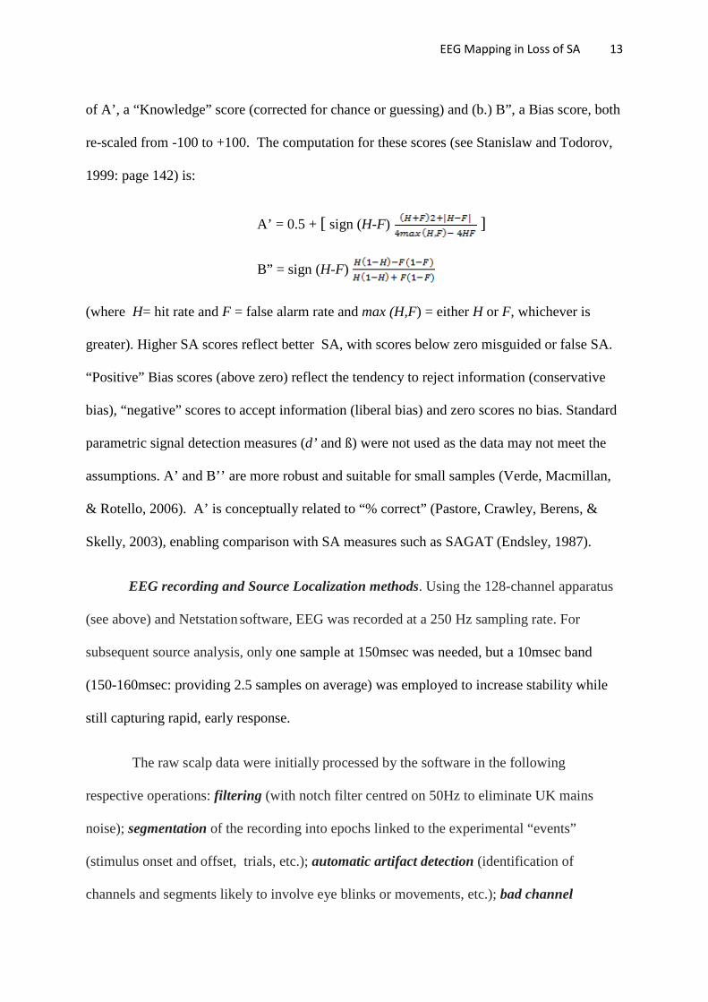