Catechol estrogen quinones as initiators of breast and other human cancers: Implications for...
16
Review Catechol estrogen quinones as initiators of breast and other human cancers: Implications for biomarkers of susceptibility and cancer prevention ☆ Ercole Cavalieri a, ⁎ , Dhubajyoti Chakravarti a , Joseph Guttenplan b , Elizabeth Hart c , James Ingle d , Ryszard Jankowiak e , Paola Muti f , Eleanor Rogan a , Jose Russo g , Richard Santen h , Thomas Sutter i a Eppley Institute for Research in Cancer and Allied Diseases, University of Nebraska Medical Center, 986805 Nebraska Medical Center, Omaha, NE 68198-6805, USA b New York University Dental and Medical Schools, New York, NY 10032, USA c Hart International, Dallas, TX, USA d Mayo Clinic, Rochester, MN, USA e Department of Chemistry, Kansas State University, Manhattan, KS, USA f Italian National Cancer Institute, Rome, Italy g Fox Chase Cancer Center, Philadelphia, PA, USA h University of Virginia Health System, Charlottesville, VA, USA i Center for Genomic Research, University of Memphis, Memphis, TN, USA Received 11 January 2006; received in revised form 14 March 2006; accepted 19 March 2006 Available online 19 April 2006 Abstract Exposure to estrogens is associated with increased risk of breast and other types of human cancer. Estrogens are converted to metabolites, particularly the catechol estrogen-3,4-quinones (CE-3,4-Q), that can react with DNA to form depurinating adducts. These adducts are released from DNA to generate apurinic sites. Error-prone base excision repair of this damage may lead to the mutations that can initiate breast, prostate and other types of cancer. The reaction of CE-3,4-Q with DNA forms the depurinating adducts 4-hydroxyestrone(estradiol) [4-OHE 1 (E 2 )-1-N3Ade and 4-OHE 1 (E 2 )-1- N7Gua. These two adducts constitute more than 99% of the total DNA adducts formed. Increased levels of these quinones and their reaction with DNA occur when estrogen metabolism is unbalanced. Such an imbalance is the result of overexpression of estrogen activating enzymes and/or deficient expression of the deactivating (protective) enzymes. This unbalanced metabolism has been observed in breast biopsy tissue from women with breast cancer, compared to control women. Recently, the depurinating adduct 4-OHE 1 (E 2 )-1-N3Ade has been detected in the urine of prostate cancer patients, but not in urine from healthy men. Mutagenesis by CE-3,4-Q has been approached from two different perspectives: one is mutagenic activity in the lacI reporter gene in Fisher 344 rats and the other is study of the reporter Harvey-ras gene in mouse skin and rat mammary gland. A → G and G → A mutations have been observed in the mammary tissue of rats implanted with the CE-3,4-Q precursor, 4-OHE 2 . Mutations have also been observed in the Harvey-ras gene in mouse skin and rat mammary gland within 6–12 h after treatment with E 2 -3,4-Q, suggesting that these mutations arise by error-prone base excision repair of the apurinic sites generated by the depurinating adducts. Treatment of MCF-10F cells, which are estrogen receptor-α-negative immortalized human breast epithelial cells, with E 2 , 4-OHE 2 or 2-OHE 2 induces their neoplastic transformation in vitro, even in the presence of the antiestrogen ICI-182,780. This suggests that transformation is independent of the estrogen receptor. The transformed cells exhibit specific mutations in several genes. Poorly differentiated adenocarcinomas Biochimica et Biophysica Acta 1766 (2006) 63 – 78 www.elsevier.com/locate/bbacan Abbreviations: AP, apurinic; BB®, Big Blue; BER, base excision repair; BP, benzo[a]pyrene; CE, catechol estrogen; CE-3,4-Q, catechol estrogen-3,4-quinone; COMT, catechol-O-methyltransferase; CYP, cytochrome P450; CYP19, aromatase; E 1 , estrone; E 2 , estradiol; E 2 -3,4-Q, estradiol-3,4-quinone; ER, estrogen receptor; ERKO, estrogen receptor α-knock out; FASS, field amplified sample stacking; GSH, glutathione; H, Harvey; HBEC, human breast epithelial cells; LC/MS/MS, ultraperformance liquid chromatography/tandem mass spectrometry; LOD, limit of detection; LOH, loss of heterozygosity; MAb, monoclonal antibody; OHE 2 , hydroxyestradiol; 4-OHE 1 (E 2 )-1-N3Ade, 4-hydroxyestrone(estradiol)-1-N3Adenine; 4-OHE 1 (E 2 )-1-N7Gua, 4-hydroxyestrone(estradiol)1-N7Guanine; SCID, severe combined immune depressed; TAM, tamoxifen ☆ Dedicated to Joachim G. Liehr (1942–2003), our colleague, collaborator and friend. ⁎ Corresponding author. Tel.: +1 402 559 7237; fax: +1 402 559 8068. E-mail address: [email protected] (E. Cavalieri). 0304-419X/$ - see front matter © 2006 Elsevier B.V. All rights reserved. doi:10.1016/j.bbcan.2006.03.001
Transcript of Catechol estrogen quinones as initiators of breast and other human cancers: Implications for...
Biochimica et Biophysica Acta 1766 (2006) 63–78www.elsevier.com/locate/bbacan
Review
Catechol estrogen quinones as initiators of breast and other human cancers:Implications for biomarkers of susceptibility and cancer prevention☆
Ercole Cavalieri a,⁎, Dhubajyoti Chakravarti a, Joseph Guttenplan b, Elizabeth Hart c, James Ingle d,Ryszard Jankowiak e, Paola Muti f, Eleanor Rogan a, Jose Russo g,
Richard Santen h, Thomas Sutter i
a Eppley Institute for Research in Cancer and Allied Diseases, University of Nebraska Medical Center,986805 Nebraska Medical Center, Omaha, NE 68198-6805, USA
b New York University Dental and Medical Schools, New York, NY 10032, USAc Hart International, Dallas, TX, USAd Mayo Clinic, Rochester, MN, USA
e Department of Chemistry, Kansas State University, Manhattan, KS, USAf Italian National Cancer Institute, Rome, Italy
g Fox Chase Cancer Center, Philadelphia, PA, USAh University of Virginia Health System, Charlottesville, VA, USA
i Center for Genomic Research, University of Memphis, Memphis, TN, USA
Received 11 January 2006; received in revised form 14 March 2006; accepted 19 March 2006Available online 19 April 2006
Abstract
Exposure to estrogens is associatedwith increased risk of breast and other types of human cancer. Estrogens are converted tometabolites, particularlythe catechol estrogen-3,4-quinones (CE-3,4-Q), that can react withDNA to form depurinating adducts. These adducts are released fromDNA to generateapurinic sites. Error-prone base excision repair of this damage may lead to the mutations that can initiate breast, prostate and other types of cancer.
The reaction of CE-3,4-Q with DNA forms the depurinating adducts 4-hydroxyestrone(estradiol) [4-OHE1(E2)-1-N3Ade and 4-OHE1(E2)-1-N7Gua. These two adducts constitute more than 99% of the total DNA adducts formed. Increased levels of these quinones and their reaction withDNA occur when estrogen metabolism is unbalanced. Such an imbalance is the result of overexpression of estrogen activating enzymes and/ordeficient expression of the deactivating (protective) enzymes. This unbalanced metabolism has been observed in breast biopsy tissue from womenwith breast cancer, compared to control women. Recently, the depurinating adduct 4-OHE1(E2)-1-N3Ade has been detected in the urine of prostatecancer patients, but not in urine from healthy men.
Mutagenesis by CE-3,4-Q has been approached from two different perspectives: one is mutagenic activity in the lacI reporter gene in Fisher344 rats and the other is study of the reporter Harvey-ras gene in mouse skin and rat mammary gland. A→G and G→A mutations have beenobserved in the mammary tissue of rats implanted with the CE-3,4-Q precursor, 4-OHE2. Mutations have also been observed in the Harvey-rasgene in mouse skin and rat mammary gland within 6–12 h after treatment with E2-3,4-Q, suggesting that these mutations arise by error-prone baseexcision repair of the apurinic sites generated by the depurinating adducts.
Treatment of MCF-10F cells, which are estrogen receptor-α-negative immortalized human breast epithelial cells, with E2, 4-OHE2 or 2-OHE2
induces their neoplastic transformation in vitro, even in the presence of the antiestrogen ICI-182,780. This suggests that transformation isindependent of the estrogen receptor. The transformed cells exhibit specific mutations in several genes. Poorly differentiated adenocarcinomas
Abbreviations: AP, apurinic; BB®, Big Blue; BER, base excision repair; BP, benzo[a]pyrene; CE, catechol estrogen; CE-3,4-Q, catechol estrogen-3,4-quinone;COMT, catechol-O-methyltransferase; CYP, cytochrome P450; CYP19, aromatase; E1, estrone; E2, estradiol; E2-3,4-Q, estradiol-3,4-quinone; ER, estrogen receptor;ERKO, estrogen receptor α-knock out; FASS, field amplified sample stacking; GSH, glutathione; H, Harvey; HBEC, human breast epithelial cells; LC/MS/MS,ultraperformance liquid chromatography/tandem mass spectrometry; LOD, limit of detection; LOH, loss of heterozygosity; MAb, monoclonal antibody; OHE2,hydroxyestradiol; 4-OHE1(E2)-1-N3Ade, 4-hydroxyestrone(estradiol)-1-N3Adenine; 4-OHE1(E2)-1-N7Gua, 4-hydroxyestrone(estradiol)1-N7Guanine; SCID, severecombined immune depressed; TAM, tamoxifen☆ Dedicated to Joachim G. Liehr (1942–2003), our colleague, collaborator and friend.⁎ Corresponding author. Tel.: +1 402 559 7237; fax: +1 402 559 8068.E-mail address: [email protected] (E. Cavalieri).
0304-419X/$ - see front matter © 2006 Elsevier B.V. All rights reserved.doi:10.1016/j.bbcan.2006.03.001

64 E. Cavalieri et al. / Biochimica et Biophysica Acta 1766 (2006) 63–78
develop when aggressively transformed MCF-10F cells are selected and injected into severe combined immune depressed (SCID) mice. Theseresults represent the first in vitro/in vivo model of estrogen-induced carcinogenesis in human breast epithelial cells.
In other studies, the development of mammary tumors in estrogen receptor-α knockout mice expressing the Wnt-1 oncogene (ERKO/Wnt-1)provides direct evidence that estrogens may cause breast cancer through a genotoxic, non-estrogen receptor-α-mediated mechanism.
In summary, this evidence strongly indicates that estrogens can become endogenous tumor initiators when CE-3,4-Q react with DNA to formspecific depurinating adducts. Initiated cells may be promoted by a number of processes, including hormone receptor stimulated proliferation.These results lay the groundwork for assessing risk and preventing disease.© 2006 Elsevier B.V. All rights reserved.
Keywords: Cancer initiation; Carcinogenicity; Cell transformation; Depurinating estrogen-DNA adduct; Estrogens; Mutations
Contents
1. Introduction . . . . . . . . . . . . . . . . . . . . . . . . . . . . . . . . . . . . . . . . . . . . . . . . . . . . . . . . . . . . . . 642. Estrogens, androgens and breast cancer development—the epidemiological evidence . . . . . . . . . . . . . . . . . . . . . . . . 643. Estrogens as tumor initiators . . . . . . . . . . . . . . . . . . . . . . . . . . . . . . . . . . . . . . . . . . . . . . . . . . . . . 65
3.1. Formation, metabolism and DNA adducts of estrogens . . . . . . . . . . . . . . . . . . . . . . . . . . . . . . . . . . . . 653.2. Imbalance of estrogen homeostasis. . . . . . . . . . . . . . . . . . . . . . . . . . . . . . . . . . . . . . . . . . . . . . . 663.3. Unifying mechanism of tumor initiation by synthetic estrogens . . . . . . . . . . . . . . . . . . . . . . . . . . . . . . . . 673.4. Unifying mechanism of initiation of cancer and other diseases by catechol quinones . . . . . . . . . . . . . . . . . . . . . 67
4. Further evidence for the genotoxicity of estrogen metabolites in the induction of cancer. . . . . . . . . . . . . . . . . . . . . . . 674.1. Tumor incidence in ERKO/Wnt-1 mice . . . . . . . . . . . . . . . . . . . . . . . . . . . . . . . . . . . . . . . . . . . . 674.2. Aromatase-transfected MCF-7 breast cancer cell model . . . . . . . . . . . . . . . . . . . . . . . . . . . . . . . . . . . . 684.3. Implications for estrogen genotoxicity . . . . . . . . . . . . . . . . . . . . . . . . . . . . . . . . . . . . . . . . . . . . . 68
5. Estrogens as mutagens. . . . . . . . . . . . . . . . . . . . . . . . . . . . . . . . . . . . . . . . . . . . . . . . . . . . . . . . . 685.1. In vitro mutagenesis . . . . . . . . . . . . . . . . . . . . . . . . . . . . . . . . . . . . . . . . . . . . . . . . . . . . . . 695.2. Mutagenesis induced by 4-OHE2 and E2-3,4-Q in experimental animals . . . . . . . . . . . . . . . . . . . . . . . . . . . 69
5.2.1. The BB® rat model . . . . . . . . . . . . . . . . . . . . . . . . . . . . . . . . . . . . . . . . . . . . . . . . . . 705.2.2. SENCAR mouse and ACI rat models. . . . . . . . . . . . . . . . . . . . . . . . . . . . . . . . . . . . . . . . . 70
5.3. Conclusions . . . . . . . . . . . . . . . . . . . . . . . . . . . . . . . . . . . . . . . . . . . . . . . . . . . . . . . . . . 716. An in vitro/in vivo model of estrogen-induced carcinogenesis . . . . . . . . . . . . . . . . . . . . . . . . . . . . . . . . . . . . 717. Analysis of possible biomarkers for human prostate cancer . . . . . . . . . . . . . . . . . . . . . . . . . . . . . . . . . . . . . . 738. Overall conclusions . . . . . . . . . . . . . . . . . . . . . . . . . . . . . . . . . . . . . . . . . . . . . . . . . . . . . . . . . . 75Acknowledgements . . . . . . . . . . . . . . . . . . . . . . . . . . . . . . . . . . . . . . . . . . . . . . . . . . . . . . . . . . . . . 75References . . . . . . . . . . . . . . . . . . . . . . . . . . . . . . . . . . . . . . . . . . . . . . . . . . . . . . . . . . . . . . . . . 76
1. Introduction
In this review article, we present major scientificadvancements supporting the hypothesis that specific estro-gen metabolites, namely, catechol estrogen-3,4-quinones (CE-3,4-Q), can initiate breast, prostate and other human cancers.The lines of evidence include epidemiological studies,reaction of the catechol estrogen quinone metabolites withDNA to form specific depurinating adducts, imbalance ofestrogen metabolism in the breast of women with breastcarcinoma, induction of mammary tumors in estrogenreceptor α-knock out mice, in vitro and in vivo mutagenicityinduced by CE-Q, malignant transformation of human breastepithelial cells by catechol estrogen metabolites with resultinggenetic instability, and biomarkers of cancer risk in men andwomen.
2. Estrogens, androgens and breast cancer development—the epidemiological evidence
Until the last decade, epidemiological evidence of an associ-ation between sex steroid hormones and breast cancer risk,
based on a retrospective study design such as case-controlstudies, was generally inconsistent. In spite of the lack ofevidence, prospective cohort studies conducted in the last 10years consistently observed that elevated levels of serumestrogens and androgens preceded the occurrence of breastcancer. Nine research groups have published results fromprospective studies of endogenous hormones and breast cancerin different populations in the world. These studies, based onrecruitment of thousands of healthy women and their epidemi-ological follow-up, are characterized by very different method-ological approaches in terms of population sampling, collecting,processing and storing biological specimens [1–13]. In thepooled analysis, both estrogens and androgens were stronglyassociated with an increase in breast cancer risk, with evidenceof a dose–response relationship [14].
This evidence, mainly found in postmenopausal women, hasbeen corroborated in premenopausal women. Recently, a largecohort study, developed within the European Prospective Inves-tigation into Cancer and Nutrition (EPIC) cohort, has also de-finitively provided evidence for an etiological link betweensex steroids and breast cancer development in premenopausalwomen [15].

65E. Cavalieri et al. / Biochimica et Biophysica Acta 1766 (2006) 63–78
Etiological research conducted in experimental settings andin population and clinical studies shows a remarkable coherenceand consistency of evidence. On-going investigations now needto focus on the origin of cancer, and how and why sex steroidhormones are so closely related to breast cancer development.These investigations need to develop beyond the stochasticmodel based on the paradigm that estrogens bind to estrogenreceptors and stimulate the transcription of genes involved in cellproliferation, creating potential errors in DNA replication andpotential mutations [16,17]. To initiate cancer, these randommutations must occur in specific sites in DNA; this is an ex-tremely unlikely outcome. In contrast, initiation of cancer bygenotoxic estrogen metabolites that generate specific mutationscorrelates with the strong coherence of the results of experi-mental and epidemiological studies, and the consistency oftheir associations across different studies and different popula-tions. The evidence for estrogen genotoxicity and mutagenesis issummarized in this article.
3. Estrogens as tumor initiators
The initial failure to demonstrate that estrogens induce muta-tions in bacterial andmammalian test systems [18–23] resulted inthe classification of estrone (E1) and estradiol (E2) as epigeneticcarcinogens that function by stimulating abnormal cell prolifer-ation via estrogen receptor-mediated processes [16,17,20,24–26]. The stimulated cell proliferation could result in increasedaccumulation of genetic damage, leading to carcinogenesis[16,26,27].
Compelling evidence has led to a new paradigm of cancerinitiation by estrogens. Discovery that specific oxidative meta-bolites of estrogens can react with DNA [28–31] led to and hassupported the hypothesis that these metabolites can becomeendogenous chemical carcinogens. Some of the mutations gen-erated by the specific DNA damage can result in the initiationof cancer in hormone-dependent and -independent tissues[32–35].
Chemical carcinogens covalently bind to DNA to form twotypes of adducts: stable ones that remain in the DNA, unlessremoved by repair, and depurinating ones that are lost from theDNA by destabilization of the glycosyl bond [29,30,36,37].Evidence that depurinating polycyclic aromatic hydrocarbon-and estrogen-DNA adducts play a major role in tumor initiationderives from a correlation between depurinating adducts andoncogenic Harvey (H)-ras mutations in mouse skin papillomas,preneoplastic mouse skin and preneoplastic rat mammary gland(see Section 5 below) [32,33,38,39]. These observations haveprovided the impetus for discovering the estrogen metabolitesthat form depurinating DNA adducts and lead to the mutationsthat can eventually initiate cancer [28–30]. Experiments onestrogen metabolism [40–42], formation of DNA adducts [28–31], carcinogenicity [18,43,44], mutagenicity [32–35] and celltransformation [45–48] have led to and supported the hypothe-sis that reaction of specific estrogen metabolites, namely, CE-3,4-Q and to a much lesser extent, CE-2,3-Q, with DNA cangenerate the critical mutations that initiate breast, prostate andother cancers [30,35].
3.1. Formation, metabolism and DNA adducts of estrogens
Catechol estrogens (CE) are among the major metabolites ofE1 and E2 [49–51]. If these metabolites are oxidized to theelectrophilic CE-Q, they may react with DNA. Specifically, thecarcinogenic 4-OHE1(E2) [18,43,44] are oxidized to E1(E2)-3,4-Q, which can react with DNA to form predominantly depuri-nating adducts [28–30]. These adducts generate apurinic sitesthat may lead to cancer-initiating mutations (see Section 5below) [32–35], which transform cells (see Section 6 below),thereby initiating cancer [45–48]. The extremely weak carcino-gen 2-OHE1(E2) [44] also forms depurinating adducts, but to amuch lesser extent [52]. The depurinating N3Ade and N7Guaadducts are released from DNA at different rates, the formerinstantaneously and the latter with a half-life of 3 h [52,53].
E1 and E2 are formed by aromatization of androstenedioneand testosterone, respectively, catalyzed by cytochrome P450(CYP) 19, aromatase (Fig. 1). E1 and E2 are interconverted bythe enzyme 17β-estradiol dehydrogenase. These estrogens aremetabolized by two major pathways: formation of CE and, to alesser extent, 16α-hydroxylation (not shown in Fig. 1). The CEformed are the 2-OHE1(E2) and 4-OHE1(E2). The 2-OHE1(E2)are generally the major CE formed. Increases in the level ofCYP1B1 and other 4-hydroxylases could render the minor CEmetabolites, 4-OHE1(E2), as the major ones. The CE is generallyinactivated by conjugating reactions such as glucuronidation andsulfation, especially in the liver (not shown in Fig. 1). The mostcommon pathway of conjugation in extrahepatic tissues occurs,however, by O-methylation catalyzed by the ubiquitous cate-chol-O-methyltransferase (COMT) [54]. If conjugation of CEvia methylation becomes insufficient, the competitive catalyticoxidation of CE to CE-Q can occur.
Redox cycling via reduction of CE-Q to semiquinones, cat-alyzed by CYP reductase, and subsequent oxidation back to CE-Q by O2 forms super-anion radicals and then H2O2. In thepresence of Fe2+, H2O2 forms hydroxyl radicals (Fig. 1).
The 4-OHE1(E2) exhibit greater carcinogenic potency thanthe 2-OHE1(E2), which are borderline carcinogens [18,43,44]. Itis difficult to attribute the greater potency of 4-OHE1(E2) to theredox cycling of the 2-OHE1(E2) and 4-OHE1(E2), because theyhave similar redox potentials [55,56]. Instead, one can relate thegreater carcinogenic potency of the 4-OHE1(E2) to the muchhigher level of depurinating DNA adducts formed by E1(E2)-3,4-Q compared to E1(E2)-2,3-Q [52]. Thus, we think the role ofCE-Q in initiating cancer is through formation of depurinatingDNA adducts.
The reactivity of CE-Q with DNA can be prevented byconjugation with glutathione (GSH, Fig. 1). A second inactivat-ing pathway for CE-Q is their reduction to CE by quinonereductase and/or CYP reductase [57,58]. If these inactivatingprocesses are insufficient, CE-Q may react with DNA to formpredominantly depurinating adducts (Fig. 1) [28–31]. Whenmouse skin [32] or rat mammary gland [33] was treated with E2-3,4-Q, the 4-OHE2-1-N3Ade and 4-OHE2-1-N7Gua adductswere formed. In these tissues, E2-3,4-Q induced mainly A to Gmutations in the reporter H-ras gene, presumably because theN3Ade adducts depurinate rapidly, leading to premutagenic

Fig. 1. Formation, metabolism and DNA adducts of estrogens. Activating enzymes and depurinating adducts are in red, and protective enzymes are in green.
66 E. Cavalieri et al. / Biochimica et Biophysica Acta 1766 (2006) 63–78
apurinic (AP) sites, but the N7Gua adducts depurinate relativelyslowly, allowing accurate DNA repair [32,33,52,53]. Thesemutagenicity results suggest that E2-3,4-Q may be the majorcarcinogenic metabolite of estrogens.
3.2. Imbalance of estrogen homeostasis
The above paradigm of cancer initiation by estrogens hinges onestrogenmetabolism that involves a disrupted homeostatic balancebetween activating and deactivating pathways (Fig. 1). Severalfactors can unbalance estrogen homeostasis, namely, the equilib-rium between estrogen activating and deactivating pathways toavert oxidative stress, in particular the formation of endogenouscarcinogenic CE-Q and their reaction with DNA (Fig. 1).
Critical factors in elevating estrogen levels are excessivesynthesis of estrogens by overexpression of CYP19 in targettissues [59–61] and/or the presence of unregulated sulfatase thatconverts excess stored E1-sulfate to E1 [62,63]. The observationthat breast tissue can synthesize E2 in situ suggests that muchmore E2 is present in target tissues than would be predicted from
plasma concentrations [61]. A striking result of in situ E2 pro-duction is that the E2 levels in human breast tissue are similar inpre- and post-menopausal women, even though plasma levelsare 50- to 100-fold lower in postmenopausal women comparedto premenopausal [63,64].
Another critical factor unbalancing estrogen homeostasismay be higher levels of 4-OHE1(E2) due to overexpression ofCYP1B1, which converts E2 predominantly to 4-OHE2 (Fig. 1)[65,66]. This could result in relatively large amounts of 4-OHE1
(E2) and subsequently more oxidation to E1(E2)-3,4-Q. Anadditional factor could be a lack or low level of COMT activity,because of polymorphic variation [67]. If this enzyme activity isinsufficient, 4-OHE1(E2) would not be effectively methylated,but could be oxidized to the ultimate metabolites E1(E2)-3,4-Q.Finally, a low level of GSH, and/or low levels of quinonereductase and/or CYP reductase, could result in a higher level ofE1(E2)-3,4-Q that may react with DNA.
We postulate that unbalanced estrogen homeostasis is acondition that precedes the initiation of breast and prostatecancer. The effects of some of the above factors have already

67E. Cavalieri et al. / Biochimica et Biophysica Acta 1766 (2006) 63–78
been observed in several animal models for estrogen carcino-genesis and in human breast. Imbalances in estrogen homeo-stasis leading to substantial formation of CE-GSH conjugatesand depurinating CE-DNA adducts have been observed in thekidney of male Syrian golden hamsters [40], the prostate ofNoble rats [41] and the mammary gland of female estrogenreceptor-α knockout (ERKO/Wnt-1) mice (see Section 4 below)[68]. A study of breast tissue from women with and withoutbreast cancer provides key evidence in support of unbalancedestrogen homeostasis. In fact, this imbalance was observed inwomen with breast cancer (Fig. 2) [42]. Levels of E1(E2) inwomen with carcinoma were higher than in controls, and thelevels of 4-OHE1(E2) were nearly four times higher in womenwith breast carcinoma than in women without cancer. In womenwith breast carcinoma, 4-OHE1(E2) were three-times moreabundant than 2-OHE1(E2). Levels of CE-Q conjugates inwomen with breast cancer were three-times those in the con-trols, suggesting a greater probability of CE-Q reacting withDNA in the breast tissue of women with breast carcinoma.Levels of 4-OHE1(E2) (Pb0.01) and CE-Q conjugates(Pb0.003) appeared to be significantly associated with breastcancer [42]. One established example of this imbalance is theoverexpression of the estrogen 4-hydroxylase, CYP1B1, intumors of the breast [69–71]. Therefore, the oxidative pathwaythat leads to formation of CE-Q is the result of unbalancing oneor more factors involved in estrogen homeostasis.
3.3. Unifying mechanism of tumor initiation by syntheticestrogens
We have proposed that oxidation of CE to CE-Q is a pathwayto initiate cancer by endogenous estrogens, as well as syntheticestrogens such as the human carcinogen diethylstilbestrol [72]and its hydrogenated derivative hexestrol. These two com-pounds, similarly to the endogenous estrogens, are carcinogenicin the kidney of Syrian golden hamsters [73,74], and the majormetabolites are their catechols [74–77]. These catechols can beeasily oxidized to catechol quinones. Their chemical and bio-chemical properties are similar to those of CE-3,4-Q, namely,
Fig. 2. Relative imbalance of estrogen metabolism in non-tumor breast tissue ofwomen with breast cancer vs. controls. The level of 4-OHE1(E2) was significantlyhigher in cases compared to controls (Pb0.01). Quinone conjugates were 4-OHE1(E2)-2-NAcCys, 4-OHE1(E2)-2-Cys, 2-OHE1(E2)-(1+4)-NAcCys, and 2-OHE1(E2)-(1+4)-Cys. The levels of quinone conjugateswere significantly higher in casesthan in controls (Pb0.003). *Statistically significant differences were determinedusing the Wilcoxon rank sum test.
they specifically form the N7Gua and N3Ade adducts afterreaction with DNA [78–80]. Therefore, the catechol quinonesof hexestrol and diethylstilbestrol appear to be the critical ini-tiators of cancer by these synthetic estrogens. In turn, theseresults support the hypothesis that CE-3,4-Q may be endoge-nous tumor initiators because they react with DNA to formN7Gua and N3Ade adducts. In summary, catechol quinones ofnatural and synthetic estrogens can be initiators of a variety ofhuman cancers, including breast and prostrate.
3.4. Unifying mechanism of initiation of cancer and otherdiseases by catechol quinones
Oxidation of catechols to quinones and semiquinones is notonly a mechanism of tumor initiation by natural and syntheticestrogens, but could be the mechanism of tumor initiation by theleukemogen benzene. In fact, reaction of the benzene catecholquinone with DNA specifically produces N3Ade and N7Guaadducts [81]. The same DNA adducts can be obtained by en-zymatic oxidation of the benzene catechol in the presence ofDNA [81]. The catecholamine neurotransmitter dopamine canundergo an oxidative process that is analogous to the one des-cribed for the catechol of benzene and the natural and syntheticestrogens. In fact, reaction of dopamine quinone with DNAforms specific depurinating N3Ade and N7Gua adducts [81]. Inconclusion, the catechol quinones of natural and syntheticestrogens, benzene and dopamine can react with DNA to formspecific depurinating adducts bonded at the N-7 of Gua or the N-3 of Ade. The apurinic sites formed by these adducts may beconverted by error-prone repair into mutations that can initiatecancer and neurodegenerative diseases. This hypothesized uni-fying mechanism in the induction of these diseases supports themechanism described for natural and synthetic estrogens.
4. Further evidence for the genotoxicity of estrogenmetabolites in the induction of cancer
Experiments using transgenic mice with estrogen receptor-α(ER-α) knocked-out (ERKO/Wnt-1 mice) and metabolism inaromatase (CYP19) overexpressing MCF-7 human breast can-cer cells have provided further important evidence for genotoxiceffects of estrogen metabolites in cancer initiation.
4.1. Tumor incidence in ERKO/Wnt-1 mice
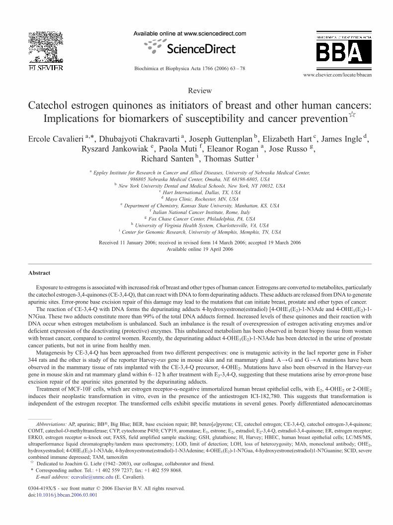
Bocchinfuso and his associates showed that ERKO/Wnt-1mice exhibit a delayed onset of tumor development compared tomice expressing the wild type ER-α. Nonetheless, they ob-served a nearly 100% incidence of mammary tumors in theabsence of ER-α and β [82,83]. To directly determine the effectof E2 in the absence of ER, mice were castrated at 15 days of ageand half were treated with silastic implants containing E2 andthe other half with implants of cholesterol. After 100 weeks ofobservation, the E2-treated mice developed more tumors (12/15vs. 4/10), which appeared earlier than those in the mice re-ceiving cholesterol implants (50% of tumors at 50 weeks ver-sus 25% of tumors at 100 weeks, Pb0.004) (Fig. 3) [84,85].
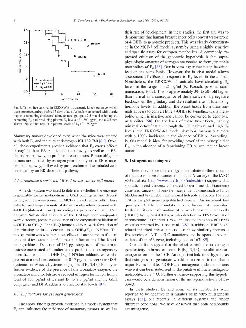
Fig. 3. Tumor-free survival in ERKO/Wnt-1 transgenic knock-out mice, whichwere oophorectomized before 15 days of age. Animals were treated with silasticimplants containing cholesterol alone (control group), a 7.5 mm silastic implantcontaining E2 and producing plasma E2 levels of ∼300 pg/ml and a 2.5 mmsilastic implant that results in plasma levels of E2 of ∼75 pg/ml.
68 E. Cavalieri et al. / Biochimica et Biophysica Acta 1766 (2006) 63–78
Mammary tumors developed even when the mice were treatedwith both E2 and the pure antiestrogen ICI-182,780 [86]. Over-all, these experiments provide evidence that E2 exerts effectsthrough both an ER-α-independent pathway, as well as an ER-dependent pathway, to produce breast tumors. Presumably, thetumors are initiated by estrogen genotoxicity in an ER-α-inde-pendent pathway, followed by proliferation of the initiated cellsmediated by an ER-dependent pathway.
4.2. Aromatase-transfected MCF-7 breast cancer cell model
A model system was used to determine whether the enzymesresponsible for E2 metabolism to GSH conjugates and depuri-nating adducts were present in MCF-7 breast cancer cells. Thesecells formed large amounts of 4-methoxyE2 when cultured with4-OHE2 (data not shown), indicating the presence of the COMTenzyme. Substantial amounts of the GSH-quinone conjugateswere detected, providing evidence of the enzymatic oxidation of4-OHE2 to CE-Q. The CE-Q bound to DNA, with formation ofdepurinating adducts, detected as 4-OHE1(E2)-1-N7Gua. Thenext questionwaswhether these cells could aromatize a sufficientamount of testosterone to E2 to result in formation of the depuri-nating adducts. Detection of 131 pg estrogen/ml of medium intestosterone-treated cells indicated the production of estrogens byaromatization. The 4-OHE1(E2)-1-N7Gua adducts were alsopresent at a total concentration of 0.17 pg/ml, as were the GSH,cysteine, andN-acetylcysteine conjugates of E2-3,4-Q. Finally, asfurther evidence of the presence of the aromatase enzyme, thearomatase-inhibitor letrozole reduced estrogen formation from atotal of 131 pg/ml of E1 and E2 to 2.8 pg/ml and the GSHconjugates and DNA adducts to undetectable levels [87].
4.3. Implications for estrogen genotoxicity
The above findings provide evidence in a model system thatE2 can influence the incidence of mammary tumors, as well as
their rate of development. In these studies, the first aim was todemonstrate that human breast cancer cells convert testosteroneor 4-OHE2 to genotoxic products. This was clearly demonstrat-ed in the MCF-7 cell model system by using a highly sensitiveand specific assay for estrogen metabolites. A commonly ex-pressed criticism of the genotoxic hypothesis is that supra-physiologic amounts of estrogen are needed to form genotoxicmetabolites of E2 [88]. Our in vitro experiments can be critic-ized on the same basis. However, the in vivo model allowsassessment of effects in response to E2 levels in the animal.Nonetheless, the ERKO/Wnt-1 animals have circulating E2
levels in the range of 325 pg/ml (K. Korach, personal com-munication, 2002). This is approximately 30- to 50-fold higherthan normal as a consequence of the absence of E2 negativefeedback on the pituitary and the resultant rise in luteinizinghormone levels. In addition, the breast tissue from these ani-mals appears to convert little 4-OHE2 to 4-methoxyE2, a meta-bolite which is inactive and cannot be converted to genotoxicmetabolites [68]. On the basis of these two effects, namelyminimal detoxification through the CE pathway and high E2
levels, the ERKO/Wnt-1 model develops mammary tumorswith a 100% incidence in the absence of ER-α. According-ly, this model is ideal for providing proof of the principle thatE2, in the absence of a functioning ER-α, can induce breasttumors.
5. Estrogens as mutagens
There is evidence that estrogens contribute to the inductionof mutations in breast cancer in humans. A survey of the IARCp53 database (http://www.iarc.fr/p53/index.html) suggests thatsporadic breast cancers, compared to germline (Li-Fraumeni)cases and cancers in hormone-independent tissues such as lung,bladder and brain, show mutational hotspots at codons 163 and179 in the p53 gene [unpublished results]. An increased fre-quency of A.T to G.C mutations could be seen at these sites.In the in vitro transformation of human breast epithelial cells(HBEC) by E2 or 4-OHE2, a 5-bp deletion in TP53 exon 4 ofchromosome 17 (marker TP53-Dint located in exon 4 of TP53)was also reported by Russo et al. [48]. In addition, BRCA1/2-related inherited breast cancers also show similarly increasedfrequencies of A.T to G.C mutations and hotspots at severalcodons of the p53 gene, including codon 163 [89].
Our studies suggest that the chief contributor to estrogengenotoxicity in breast cancer is E1(E2)-3,4-Q, the ultimate car-cinogenic form of the 4-CE. An important link in the hypothesisthat estrogens are genotoxic would be a demonstration that amajor E2 metabolite, 4-OHE2, is mutagenic under conditionswhere it can be metabolized to the putative ultimate mutagenicmetabolite, E2-3,4-Q. Further evidence supporting this hypoth-esis would be a demonstration of the mutagenic activity of E2-3,4-Q.
In early studies, E2 and some of its metabolites werereported to be negative in a number of in vitro mutagenesisassays [88], but recently in different systems and underdifferent conditions, we have observed that both compoundsare mutagenic.

69E. Cavalieri et al. / Biochimica et Biophysica Acta 1766 (2006) 63–78
5.1. In vitro mutagenesis
The Big Blue® (BB®) rat2 embryonic cell line (Stratagene,La Jolla, CA) was used to detect mutagenesis. This is a ratembryonic cell line transfected with the lambda-LIZ vector. Itenables the host cell to detect mutations in the lacI and/or cIIgenes. The host cells contain approximately 60 copies of thevector per cell. The cII assay was employed.
Initial experiments conducted at doses from 10 to 6800 nM 4-OHE2 failed to detect any significant increase in mutant fractionafter a single 16 hr treatment, and therefore multiple treatmentswere performed. 4-OHE2 induced a dose-dependent increase inmutant fraction up to 200 nM [35]. This was marginally apparentat three treatments and clearer after six treatments. The mutantfraction (in units of mutants/105 pfu) increased from 2.6±1.3 incontrols to 4.5±0.7 and 5.8±0.3 at 100 and 200 nM, respec-tively, after six treatments. After three and six 200-nM treat-ments, the increase over controls was statistically significant.The mutant fraction declined at 400 nM after both three and sixtreatments. Using similar protocols, for single and multipletreatments, it was not possible to detect any induction of muta-genesis over background by 2-OHE2. E2-3,4-Q was also foundto be similarly mutagenic.
The mutational spectra from the 4-OHE2 treated and controlplates were compared, and a major apparent difference betweenthe two groups was the higher percentage of mutations at A:Tbase pairs in the mutants from the 4-OHE2 -treated cells than incontrols (ca. 24% vs. 6%). The mutational spectrum of E2-3,4-Q is currently being analyzed.
Although the mutagenic activity of 4-OHE2 has not beenpreviously reported, 4-OHE2 and its precursor, E2, induce DNAstrand breaks, detectable in the comet assay [90,91]. It is not
Fig. 4. Mutant fractions (MF) in mammary tissue from BB rats treated with 5 mg ofmutational distribution in each group. Brackets refer to mutations at A:T base pairs. M4-OHE2, P=0.06.
known, however, whether these strand breaks actually lead tomutations, as they may be repaired. In an oxidative damagemodification of the comet assay, 4-OHE2 and 2-OHE2 weresimilarly effective in producing strand breaks [90]. In addition,assays on DNA damage in vitro indicate that oxidative damageunder Fenton-like conditions is produced by both 2-OHE2 and4-OHE2 with similar efficiency [92]. Taken together, the resultshere and in the previous studies suggest that 2-OHE2 and 4-OHE2 both induce oxidative damage with similar efficiencies,but these processes do not account for differences in the carcin-ogenic and mutagenic potencies of these compounds. If much ofthe initial oxidative damage to DNA is rapidly and accuratelyrepaired, mutagenesis may result from the mis-repair of AP sitesresulting from depurinating estrogen-DNA adducts, and resid-ual oxidative damage.
4-OHE2 was a weak mutagen and the BB® rat2 cells re-quired multiple treatments to detect mutagenesis. This and theobservation that mutagenesis is only observed over a narrowdose range may explain previous negative reports on mutagen-esis by 4-OHE2 and other estrogens or metabolites. The obser-vation that 4-OHE2, but not 2-OHE2, exhibited mutagenicactivity in BB® rat2 cells correlates with studies on the relativecarcinogenicity [18,43,44] and cell transforming abilities [46]of these compounds, and thus provides additional evidence thatgenotoxicity plays a role in estrogen-induced carcinogenesis.
5.2. Mutagenesis induced by 4-OHE2 and E2-3,4-Q inexperimental animals
Studies in experimental animals have shown that E2 and 4-OHE2 are carcinogenic, whereas 2-OHE2 is only marginallyactive [18,43,44].
4-OHE2+5 mg of E2, 5 mg of 4-OHE2, or 5 mg of E2, or left untreated, and theutagenesis was assayed after 20 weeks. *Pb0.05 vs. control (2 -tailed t-test). For

Table 1Mutations induced by E2-3,4-Q in mouse skin and rat mammary gland
Animal Treatment Mutationsafter6–24 h
Frequency
Mutation/no.of clones
Mutations/totalmutations
SENCAR mouse Control A.TNG.C 1/36 (3%) 1/1skin E2-3,4-Q A.TNG.C 9/59 (15%) 9/13 (69%)
+TDG A.TNG.C 0/74 0/4ACI rat Control A.TNG.C 18/95 (19%) 18/24 (75%)mammary gland E2-3,4-Q A.TNG.C 30/63 (48%) 30/39 (77%)
+TDG A.TNG.C 16/79 (20%) 16/20 (80%)
70 E. Cavalieri et al. / Biochimica et Biophysica Acta 1766 (2006) 63–78
5.2.1. The BB® rat modelThe BB® rat is a Fisher 344 rat that contains about 80 copies
of the Lambda-LIZ vector in every cell of the animal. Thetransgene is not expressed and has no effect on the biochemistryor physiology of the animal. Rats were administered 5 mg of 4-OHE2, 5 mg of E2 or a combination of 5 mg of each in silastictubing, placed in the scapular space. In addition, an untreatedcontrol group was assayed. The rats were euthanized after 20weeks, DNA was extracted from inguinal mammary fat pads,andmutagenesis andmutational spectra in cIIwere then assayed.
The mutant fraction in the three treated groups was abouttwice that in untreated controls and that difference was signifi-cant (Fig. 4), indicating that E2 and 4-OHE2 were mutagenic.
Fig. 5. Induction of A to G mutations from the rapidly-depurinating N3Ade adductsDNA, forming an apurinic site, which undergoes base excision repair. Our studiesgenerating mutations that are initially formed as G.T heteroduplexes. Left: the G.T pabases from G.T heteroduplexes, generating abasic sites that are refractory to PCRfrequency of A.T to G.C mutations.
Based on previous carcinogenesis and cell transformation as-says [18,43,44,46], it was anticipated that E2 would be lessmutagenic than 4-OHE2 and perhaps the combination of theagents would be most effective, if E2 resulted in increasedcell proliferation, which enhanced mutagenesis. However, therewas no significant difference between the mutant frequencies ofthe three treated groups. E2 alone was somewhat toxic, probab-ly as a result of excessive prolactin production in response tothis agent, and only 4 rats in the E2-alone group survived andthe small number of rats in this group may have reduced theaccuracy of the measurement of the mutant frequency for thisgroup.
The mutational spectrum of the groups receiving 4-OHE2
was different from the other groups (Fig. 4). The major differ-ence was the higher fraction of mutations at A:T base pairs,and in particular, AT:GC transitions. As described above, thesemutations are consistent with those expected for the 4-OHE2-1-N3Ade, and the fact that they are more frequent in the groupsreceiving 4-OHE2 is consistent with the hypothesis that 4-OHE2
contributes to mutagenesis in BB® rat mammary tissue.
5.2.2. SENCAR mouse and ACI rat modelsOur studies in SENCAR mouse skin and ACI rat mammary
gland suggest that 4-OHE2 or E2-3,4-Q can induce mutationssimilar to those associated with breast cancer (Table 1). The
generated by E2-3,4-Q treatment. The adducted Ade is spontaneously lost fromsuggest that this repair frequently commits errors (mutation frequency ∼10−4),irs are detected by T.G-DNA glycosylase treatment. This enzyme removes the Tamplification. As a result, the mutation spectra show a drastic reduction in the

71E. Cavalieri et al. / Biochimica et Biophysica Acta 1766 (2006) 63–78
initial study was conducted in the SENCAR mouse model byadministering a single dose (200 nmol in acetone) of E2-3,4-Qand examining the H-ras gene as the target of mutagenesis. Westudied early induction of mutations (12 h–3 d after the treat-ment) to make correlations with DNA adducts [32]. The resultsshowed that E2-3,4-Q induced predominantly A.T to G.C mu-tations. Next, we examined the ACI rat mammary gland, con-sidered to be a model of breast cancer, for mutagenesis by E2-3,4-Q. Similar mutations were again observed (Table 1) [33].These mutations correlated with the rapidly-depurinatingN3Ade adducts, while the slowly-depurinating N7Gua adductsdid not appear to be major sources of mutagenesis in the earlyperiod. These depurinating adducts are spontaneously lost fromDNA, forming apurinic sites. Since the N3Ade adducts depuri-nate rapidly, they will induce a rapid burst of apurinic sites inDNA. Exposure of cells to agents that induce abasic sites resultsin an early, adaptive induction of base excision repair (BER)genes, along with repression of DNA replication [93]. We foundthat coinciding with mutagenesis, E2-3,4-Q treatment of theACI rat mammary gland induces short-patch BER genes [APendonuclease 1, DNA polymerase β, poly (ADP-ribose) phos-phorylase 1 and ligase III] [93]. The abundant formation ofdepurinating adducts and induction of BER genes during muta-genesis suggest that erroneous BER could be the mechanism forinduction of mutations. During BER, a short section of the APsite-containing strand is excised, and the gap is filled by DNAsynthesis. Therefore, errors in BER would generate mutations inthe newly-synthesized strand, i.e., mismatched heteroduplexes.A.T to G.C mutations can form either G.T heteroduplexes (if themutations are A to G) or A.C heteroduplexes (if the mutationsare T to C). Using a glycosylase that is specific for the G.Theteroduplexes, we determined that the A.T to G.C mutationsare initially (6 h–1 d) induced as G.T mispairs, supporting theidea that the mutations are A to G transitions and were inducedfrom the de-adenylated sites produced by the rapidly-depurinat-ing N3Ade adducts. These ideas are described in a cartoon inFig. 5.
5.3. Conclusions
4-OHE2 and E2-3,4-Q have now been assayed in both in vivoand in vitro systems and found to be mutagenic under appropri-ate assay conditions. Themarginally carcinogenic E2metabolite,2-OHE2, was non-mutagenic in BB® rat2 cells under condi-tions in which 4-OHE2 was mutagenic. This result parallels celltransformation assays (see Section 6 below) [46] and providesfurther evidence that estrogens can contribute to carcinogene-sis via a genotoxic pathway. In addition, the proposed ultimatemutagenic metabolite, E2-3,4-Q, was mutagenic in rodent breastand skin, and also in BB® rat2 cells in culture, providing furtherevidence that E2-3,4-Q is the ultimate mutagenic metabolite ofE2. Finally, themutational spectra, both in vivo and in vitro, werealso consistent with those expected from the DNA adductsproduced by 4-OHE2 and E2-3,4-Q. Taken together, the resultsobtained from mutagenesis studies of 4-OHE2 and E2-3,4-Qsupport the hypothesis that estrogens can contribute to carcino-genesis by a genotoxic pathway.
6. An in vitro/in vivo model of estrogen-inducedcarcinogenesis
To fully demonstrate that estrogens are carcinogenic in thehuman breast and for testing potential mechanisms of action, anexperimental system is required in which the natural estrogen E2
by itself or its metabolites, 2-OHE2, 4-OHE2, and 16α-OHE2,respectively, would induce neoplastic transformation of HBECin vitro to a degree at least similar to that induced by thechemical carcinogen benzo[a]pyrene (BP) [94,95]. The trans-forming potential of estrogens on human breast epithelium wasevaluated by treating the spontaneously immortalized ER-αnegative MCF-10F cells with 0.007 nM, 70 nM and 1 μg/ml ofE2, 2-OHE2, 4-OHE2, 16α-OHE2, or cholesterol [94]. Treat-ments with estrogens alone or in the presence of the antiestro-gens tamoxifen (TAM) or ICI-182,780 were carried out for 24 htwice a week for 2 weeks to mimic the intermittent exposureof the breast to endogenous estrogens. At the end of the sec-ond week of treatment, and in successive passages thereafter, thecells were evaluated for assessing the expression of phenotypesindicative of cell transformation [46,94–96], namely, determi-nation of colony formation in agar-methocel, or colony efficien-cy, ductulogenic capacity in collagen matrix, invasiveness in areconstituted basement membrane using the Boyden chamber,genomic analysis by capillary electrophoresis, and tumorigen-ic assay in severe compromised immune-deficient (SCID) mice[97,98].
At all passages tested, MCF-10F cells treated with BP, E2, 2-OHE2, 4-OHE2, or 16α-OHE2 formed colonies in agar-methocelthat were greater than 80 μm in diameter. Cells treated withcholesterol did not form colonies. Colony efficiency was dosedependent and similar in cells treated with BP or E2; 2-OHE2-treated cells had lower colony efficiency than the two previouscompounds, and in 4-OHE2-treated cells colony efficiency wasgreater at the 0.007 nM dose, reaching a plateau at the two higherdoses. Ductulogenesis, which was evaluated by estimating theability of cells plated in collagen to form tubules, revealed thatcholesterol-treated control MCF-10F cells formed ductule-likestructures, mimicking the normal branching pattern of the mam-mary parenchyma. The ductulogenic capacity was lost in BP andE2 treated cells, which formed, instead, solid masses [46]. Themetabolites of estrogen also impaired the formation of ductules.Histological analysis showed that the ductules that control cellsformed in the collagen matrix were lined by a single layer ofcuboidal epithelial cells. The E2-metabolite-treated cells formedspherical masses filled by large cuboidal cells. The invasivecapacity ofMCF-10F cells was significantly increased by BP; E2
or 4-OHE2 treatments increased even further the invasiveness ofthe cells; 2-OHE2 treatment stimulated invasiveness, but to alower degree than BP. Neither TAM nor ICI-182,780 abrogatedthe transforming efficiency of estrogen metabolites [47].
Genomic analysis revealed that MCF-10F cells transformedwith E2 or 4-OHE2, either alone or in combination with ICI-182,780, exhibited loss of heterozygosity (LOH) in the region13q12.3 (D13S893 marker located at approximately 0.8 cMtelomeric to BRCA2) at all the doses tested; 2-OHE2 induced thesame change only at the highest dose used (1 μg/ml) [48].
72 E. Cavalieri et al. / Biochimica et Biophysica Acta 1766 (2006) 63–78
Another significant genomic change observed was a 5-bp dele-tion in TP53 exon 4 of chromosome 17 (marker TP53-Dintlocated in exon 4 of TP53) in cells treated with E2 or 4-OHE2 atthe doses of 0.007 nM or 70 nM and with 2-OHE2 only at ahighest dose used. These changes were considered to be specificfor E2 and its metabolites, since the observedmutations in HBECat D13S893 and TP53 exon 4 loci were not induced by BPtreatment [48]. Injection of 10–15×106 control or treated cellsin the inguinal fat pad of SCIDmice failed to induce tumors up tothe 9th passage. To determine whether more aggressive pheno-types could be selected, cells in their 9th passage after trans-formation with E2 were seeded in a Boyden chamber, and those
Fig. 6. Schematic representation of the genes up- and
cells crossing the membrane were collected, expanded, anddesignated E2-70-B2,B3,B4,B5,C2,C3,C4 and C5 for thosetransformed with 70 nM and 1-B2, 1-B3, 1-B4, 1-B5, 1-C2, 1-C3, 1-C4, and 1-C5 for those transformed with 1 μg/ml E2.These cells were injected in SCIDmice for assay of tumorigenic-ity. Only E2-70-C3 and E2-70-C5 were tumorigenic in 2/12 and9/10 animals injected, respectively [99]. The tumors were poorlydifferentiated adenocarcinomas, ER-α, ER-β and progesteronereceptor negative, expressing immunocytochemically high mo-lecular weight basic keratin (+++), E Cadherin (+) and CAM5.2(+). RNA was collected from E2-70-C3 and E2-70-C5 cells forcDNA microarray analysis. The genomic profile of E2-70-C5
down-regulated in (A) C3 cells and (B) C5 cells.

Fig. 7. Analysis of chromosomal copy number throughout cell transformation.The complete genome view of copy number determined by 100 k SNP analysisof genomic DNA is shown. Three individual samples of each group areidentified at the top of the panel. The chromosomes are identified at the left from1 to 22 and X. Darker areas indicate regions of gain; lighter areas indicateregions of loss. The average copy number is shown to the right. The analysis wasperformed using dCHIP software (Chen Li, Harvard University). DNA wasisolated from: MCF-10F, the immortal, non-transformed HBEC; E2, cellstransformed with 70 nM E2; C5, cells selected for invasive growth using aBoyden chamber (E2-70-C5 in text above); Clones, sub-clones of C5; Tumor,cells established from tumors produced in SCID mice. The changes seen inchromosome 13 were not consistent between replicated samples and did notreach statistical significance.
73E. Cavalieri et al. / Biochimica et Biophysica Acta 1766 (2006) 63–78
cells, which differed from that of E2-70-C3 cells, showed thatthey overexpressed by more than 5-fold genes such as tankyrase(TRF1-interacting ankyrin related ADP ribose polymerase),claudin 1, homeobox C10, and Notch homolog 3; it also ex-hibited down-regulation of telomeric repeat binding factor andtumor metastasis-suppressor gene, all genes that have beenshown to be altered in primary breast cancer (Fig. 6). From the 9tumors obtained from E2-70-C5 cells, four tumoral cell linesdesignated C5-A1-T1,C5-A4-T4, C5-A6-T6 and C5-A8-T8were derived. Fingerprint analysis confirmed that all thesecells originated from MCF-10F cells.
To simultaneously explore copy number abnormalities andloss of heterozygosity occurring throughout the transformationof cells and proceeding from MCF-10F cells to cells derivedfrom tumors in SCID mice, we analyzed genomic DNA fromeach of the cell types using the Affymetrix 100k SNP GeneChipMapping Array set. Shown in Fig. 7 are the progressive changesin the structure of chromosomes 1–22 and X. This analy-sis clearly demonstrates the progression of cancer occurringthroughout cell transformation ofMCF-10F. Gross copy numberabnormalities are rarely observed in the MCF-10F cellstransformed with E2 (Fig. 7, E2 lanes). The earliest event isa gain in chromosome 1p, 1p36.12–1p36.21. In the Boydenchamber-selected cells and its sub-clones (Fig. 7, C5 and Clonelanes) additional gains are seen in chromosome 1p, 1p36.12-1pter, and chromosome 5q, 5q21.1–5q35.3; losses are observedat chromosome 4 and chromosome 8p, 8p11.1–8p23.1. In cellsderived from tumors in SCID mice (Fig. 7, Tumor lanes), addi-tional losses are seen at chromosome 3p, 3p12.1–3p14.1, chro-mosome 9p, 9p22.1–9p24.3, and chromosome 18q, 18q11.2–18q23.
In summary, we have accumulated evidence indicating thatE2 and its metabolites are mutagenic as an early event in theprocess of transformation of the human breast epithelium. Thefact that an antiestrogen did not prevent these mutations indi-cates that the carcinogenic effect of this hormone and its meta-bolites is independent of the receptor pathway.
7. Analysis of possible biomarkers for human prostatecancer
The estrogen metabolites, GSH conjugates and depurinatingDNA adducts provide several possible biomarkers for risk ofdeveloping estrogen-initiated cancers. Spectroscopic studies of4-OHE1-1-N3Ade, 4-OHE1-1-N7Gua, 4-OHE2-1-N3Ade, and4-OHE2-1-N7Gua adduct standards have been performed atdifferent temperatures [100]. Upon high-energy laser excita-tion at 257 nm, the 4-OHE1- and 4-OHE2-derived N7Gua andN3Ade adducts are strongly phosphorescent at liquid nitrogentemperature (T=77 K). No phosphorescence was observed atroom temperature (∼300 K). The limit of detection (LOD) forthe N3Ade and N7Gua adducts, based on phosphorescencemeasurements, is in the low femtomole range (about 10−9 M)[100]. The LOD in capillary electrophoresis with field amplifiedsample stacking (FASS) and absorbance detection is about3×10−8 M [100,101]. We have demonstrated that CE-Q-derived DNA adducts can be identified in tissue extracts from
breast cancer patients [100]. To determine whether this type ofDNA damage can be detected in human urine, urine samplesfrom men with prostate cancer, benign tumors, or benign
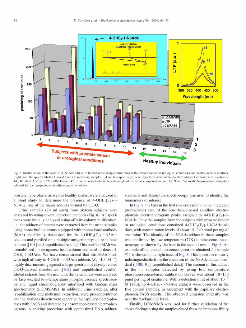
Fig. 8. Identification of the 4-OHE1-1-N3Ade adduct in human urine samples from men with prostate cancer or urological conditions and healthy men as controls.Right inset: the spectra labeled 1, 4 and 6 refer to individual samples 1, 4 and 6, respectively; the red spectrum is that of the standard adduct. Left inset: identification of4-OHE1-1-N3Ade by LC/MS/MS. The m/z 420.1 corresponds to the molecular weight of the parent compound andm/z 135.9 and 296 are the fragmentation daughtersselected for the unequivocal identification of the adduct.
74 E. Cavalieri et al. / Biochimica et Biophysica Acta 1766 (2006) 63–78
prostate hyperplasia, as well as healthy males, were analyzed ina blind study to determine the presence of 4-OHE1(E2)-1-N3Ade, one of the major adducts formed by CE-Q.
Urine samples (20 ml each) from sixteen subjects wereanalyzed by using several detection methods (Fig. 8). All speci-mens were initially analyzed using affinity column purification,i.e., the adducts of interest were extracted from the urine samplesusing home-built columns equipped with monoclonal antibody(MAb) specifically developed for the 4-OHE1(E2)-1-N3Adeadducts and purified on a multiple antigenic peptide resin beadcolumn ([101] and unpublished results). This purified MAb wasimmobilized on an agarose bead column and used to detect 4-OHE1-1-N3Ade. We have demonstrated that this MAb bindswith high affinity to 4-OHE1-1-N3Ade adducts (Ka=10
8 M−1),highly discriminating against a large spectrum of closely relatedCE-Q-derived metabolites ([102] and unpublished results).Eluted extracts from the immunoaffinity columns were analyzedby laser-excited low-temperature phosphorescence spectrosco-py and liquid chromatography interfaced with tandem massspectrometry (LC/MS/MS). In addition, urine samples, afterlyophilization and methanol extraction, were pre-concentratedand the analytes therein were separated by capillary electropho-resis with FASS and detected by absorbance-based electropher-ograms. A spiking procedure with synthesized DNA adduct-
standards and absorption spectroscopy was used to identify thebiomarkers of interest.
In Fig. 8, the bars in the first row correspond to the integrated(normalized) area of the absorbance-based capillary electro-phoresis electropherogram peaks assigned to 4-OHE1(E2)-1-N3Ade. Only the samples from the subjects with prostate canceror urological conditions contained 4-OHE1(E2)-1-N3Ade ad-duct, with concentration levels of about 15–240 pmol per mg ofcreatinine. The identity of the N3Ade adduct in these sampleswas confirmed by low-temperature (77K) luminescence spec-troscopy, as shown by the bars in the second row in Fig. 8. Anexample of the phosphorescence spectrum obtained for sample#11 is shown in the right inset of Fig. 8. This spectrum is nearlyindistinguishable from the spectrum of the N3Ade adduct stan-dard ([100,101], unpublished data)]. The amount of this adductin the 11 samples detected by using low temperaturephosphorescence-based calibration curves was about 10–150pmol per mg of creatinine. With a detection limit of about 10−9
M [100], no 4-OHE1-1-N3Ade adducts were observed in thefive control samples, in agreement with the capillary electro-phoresis/FASS results. The observed emission intensity wasnear the background level.
Finally, LC/MS/MS was used for further validation of theabove findings using the samples eluted from the immunoaffinity

75E. Cavalieri et al. / Biochimica et Biophysica Acta 1766 (2006) 63–78
columns as shown in the third row of Fig. 8. Although similaradduct distribution is observed in all samples using the threedifferent methodologies, the relative adduct concentrationsobserved in eluents from immunoaffinity columns weresomewhat smaller than that observed by capillary electrophore-sis/FASS with absorbance detection. This is not surprising, sincethe recovery from a typical column is 70–80% [100,101]. Anexample of the LC/MS/MS obtained for sample #11 is shown inthe left inset of Fig. 8; the major peak of the LC chromatogramcorresponds to the 4-OHE1-1-N3Ade adduct and indicates thatthe eluent from the immunoaffinity column was relatively pure.The upper spectrum corresponds to the daughters, m/z 135.9and 296.0, which were obtained from fragmentation of the ad-duct parent ion, m/z 420.1. Thus, 4-OHE1-1-N3Ade is excre-ted into the urine of subjects with prostate cancer, suggesting thatthis adduct may be a biomarker for risk of developing prostatecancer.
In addition to the three analytical methods used in the initialstudy of N3Ade adducts in the urine of prostate cancer patientsand control men, other promising methods are being explored.To analyze the estrogen-DNA adducts and/or estrogen-GSHconjugates in human serum or urine, novel MAb-based biosen-sor methodologies and improved microfluidic devices are beingdeveloped. These methodologies offer the promise of clinicallyuseful assays. CE metabolites [42], CE-Q-derived DNA adducts[100] and/or CE-Q-derived conjugates [101,102] could serve asbiomarkers to investigate the hypothesis that metabolically acti-vated endogenous estrogens might be involved in initiatingcancer of both the prostate and the breast. In addition, these CE-Q-derived DNA adducts identified in the urine of breast andprostate cancer patients could serve as biomarkers to assesscancer risk.
8. Overall conclusions
Exposure to estrogens is associated with increased risk ofbreast, prostate and other types of human cancer. Cancer isexpressed as perhaps as many as 200 diseases, but we hypo-thesize that many of the most prevalent, most lethal types ofcancer are initiated by a common, estrogen-initiated mechanism.This mechanism derives from specific estrogen metabolites thatreact with DNA to form predominantly depurinating adducts.These adducts are released from DNA to generate apurinic sites[28–31]. If these sites are in critical oncogenes and/or tumorsuppressor genes, they could initiate breast, prostate and othercancers.
Estrogens may become tumor initiators when estrogen meta-bolism is unbalanced and the CE-Q are formed. These quinonescan react with DNA to form depurinating N3Ade and N7Guaadducts as the predominant adducts (N99%). Analogous dep-urinating adducts are formed by the catechol quinones of thehuman carcinogen diethylstilbestrol, the leukemogen benzene[80] and the neurotransmitter dopamine [80].
Evidence for the role of the CE-Q in the initiation of breast andprostate cancer has been acquired from a variety of studies. A keypoint has been the demonstration of mutagenic activity by theestrogen metabolite 4-OHE2 in a reporter gene in the mammary
gland of BB® rats, as well as BB® rat2 cells [34,35]. The E2-3,4-Q also has been shown to be mutagenic in the H-ras oncogene inmouse skin and rat mammary gland [32,33]. These results de-monstrate the genotoxicity of estrogen metabolites. The muta-tions induced by estrogens could give rise to abnormal cellproliferation that through various processes, including hormonereceptor-mediated ones, promote the development of cancer.
Mammary tumors develop in female transgenic, knock-outERKO/Wnt-1 mice, despite the lack of ER-α [82,83]. The de-velopment of these tumors in ovariectomized ERKO/Wnt-1mice depends on the amount of E2 administered to them [86],and tumor development is not reduced by simultaneous admin-istration of the anti-estrogen ICI-182,780 [86]. Malignant trans-formation of MCF-10F cells by E2 and 4-OHE2 occurs despitethe lack of ER-α in these cells and in the presence of the anti-estrogen ICI-182,780 [45–48]. These transformed MCF-10Fcells accumulate genetic changes, and the aggressively trans-formed MCF-10F cells induce poorly differentiated adenocarci-nomas in SCID mice [48].
Further evidence has been obtained from studies of womenwith and without breast cancer and men with and without pro-state cancer. Non-tumor breast tissue from women with breastcarcinoma contained significantly higher amounts of 4-OHE2
and CE-Q conjugates of GSH, compared to breast tissue fromwomen without breast cancer [42]. In addition, enzymes in-volved in the metabolism of estrogens to CE-Q had higherexpression in breast tissue of women with breast cancer, where-as expression of protective enzymes was lower [71]. Thedepurinating DNA adduct 4-OHE1-1-N3Ade was detected at amuch higher level in breast tissue from a woman with breastcancer, compared to a control woman [100]. Finally, the 4-OHE1-1-N3Ade adduct is excreted in the urine of men withprostate cancer, whereas the level in urine of healthy men isvirtually nil.
These data suggest that some of the estrogen-DNA adducts,estrogen-GSH conjugates and estrogen metabolites may serveas biomarkers for risk of developing breast, prostate and othercancers. We think they would be detected long before tumorsappear. All of these results lay the groundwork for strategies toassess risk and prevent disease.
Acknowledgements
We thank Dr. David Longfellow, Coordinator of the CancerCube, a focus group on estrogen genotoxicity leading to can-cer, for his valuable and extensive advice and support. We alsothank the other members of the Cancer Cube for many valuablescientific discussions and Ms. K. Grotzinger for her outstandingfacilitation of Cancer Cube activities. We thank Drs. G. Balogh,S. Fernandez, N. Gaikwad, Y. Huang, M. Khmelnitsky, Y.Markushin,M. Saeed, S. Singh, B. Trock, D. Venugopal,W. Yue,M. Zahid, and Z.-L. Zhao, Ms. S. Goodwin, Ms. S. Higginbo-tham, and Ms. P. Mailander for their contributions to this re-search. Thisworkwas supported by grants DAMD17-00-1-0247,DAMD17-03-1-0229, P01 CA49210, P20 RR15563. Coresupport at the Eppley Institute was provided by grant P30CA36727 from the National Cancer Institute.

76 E. Cavalieri et al. / Biochimica et Biophysica Acta 1766 (2006) 63–78
References
[1] J.F. Dorgan, C. Longcope, H.E. Stephenson Jr., R.T. Falk, R. Miller, C.Franz, L. Kahle, W.S. Campbell, J.A. Tangrea, A. Schatzkin, Relation ofprediagnostic serum estrogen and androgen levels to breast cancer risk,Cancer Epidemiol. Biomark. Prev. 5 (1996) 533–539.
[2] J.F. Dorgan, F.Z. Stanczyk, C. Longcope, H.E. Stephenson Jr., L. Chang,R. Miller, C. Franz, R.T. Falk, L. Kahle, Relationship of serumdehydroepiandrosterone (DHEA), DHEA sulfate, and 5-androstate-3β,17β-diol to risk of breast cancer in postmenopausal women, CancerEpidemiol. Biomark. Prev. 6 (1997) 177–181.
[3] H.V. Thomas, T.J. Key, D.S. Allen, J.W. Moore, M. Dowsett, I.S.Fentiman, D.Y. Wang, A prospective study of endogenous serumhormone concentrations and breast cancer risk in postmenopausalwomen on the island of Guernsey, Br. J. Cancer 76 (1997) 401–405.
[4] S.E. Hankinson, W.C. Willett, J.E. Manson, G.A. Colditz, D.J. Hunter, D.Spiegelman, R.L. Barbieri, F.E. Speizer, Plasma sex steroid hormonelevels and risk of breast cancer in postmenopausal women, J. Natl. CancerInst. 90 (1998) 1292–1299.
[5] P.G. Toniolo, M. Levitz, A. Zeleniuch-Jacquotte, S. Banerjee, K.L.Koenig, R.E. Shore, P. Strax, B.S. Pasternack, A prospective study ofendogenous estrogens and breast cancer in postmenopausal women,J. Natl. Cancer Inst. 87 (1995) 190–197.
[6] A. Zeleniuch-Jacquotte, P.F. Bruning, J.M. Bonfrer, K.L. Koenig, R.E.Shore, M.Y. Kim, B.S. Pasternack, P. Toniolo, Relation of serum levelsof testosterone and dehydroepiandrosterone sulfate to risk of breastcancer in postmenopausal women, Am. J. Epidemiol. 145 (1997)1030–1038.
[7] F. Berrino, P. Muti, A. Micheli, G. Bolelli, V. Krogh, R. Sciajno, P. Pisani,S. Panico, G. Secreto, Serum sex hormone levels after menopause andsubsequent breast cancer, J. Natl. Cancer Inst. 88 (1996) 291–296.
[8] E. Barrett-Connor, N.J. Friedlander, K.T. Khaw, Dehydroepiandrosteronesulfate and breast cancer risk, Cancer Res. 50 (1990) 6571–6574.
[9] C.F. Garland, N.J. Friedlander, E. Barrett-Connor, K.T. Khaw, Sexhormones and postmenopausal breast cancer: a prospective study in anadult community, Am. J. Epidemiol. 135 (1992) 1220–1230.
[10] M. Kabuto, S. Akiba, R.G. Stevens, K. Neriishi, C.E. Land, Aprospective study of estradiol and breast cancer in Japanese women,Cancer Epidemiol. Biomark. Prev. 9 (2000) 575–579.
[11] J.A. Cauley, F.L. Lucas, L.H. Kuller, K. Stone, W. Browner, S.R.Cummings, Elevated serum estradiol and testosterone concentrations areassociated with a high risk for breast cancer. Study of OsteoporoticFractures Research Group, Ann. Intern. Med. 130 (1999) 270–277.
[12] G.B. Gordon, T.L. Bush, K.J. Helzlsouer, S.R. Miller, G.W. Comstock,Relationship of serum levels of dehydroepiandrosterone and dehydroe-piandrosterone sulfate to the risk of developing postmenopausal breastcancer, Cancer Res. 50 (1990) 3859–3862.
[13] K.J. Helzlsouer, A.J. Alberg, T.L. Bush, C. Longcope, G.B. Gordon,G.W. Comstock, A prospective study of endogenous hormones and breastcancer, Cancer Detec. Prev. 18 (1994) 79–85.
[14] Endogenous Hormones and Breast Cancer Collaborative Group,Endogenous sex hormones and breast cancer in postmenopausalwomen: reanalysis of nine prospective studies, J. Natl. Cancer Inst. 94(2002) 606–616.
[15] R. Kaaks, F. Berrino, T. Key, S. Rinaldi, L. Dossus, C. Biessy, G. Secreto,P. Amiano, S. Bingham, H. Boeing, H.B. Bueno de Mesquita, J. Chang-Claude, F. Clavel-Chapelon, A. Fournier, C.H. van Gils, C.A. Gonzalez,A.B. Gurrea, E. Critselis, K.T. Khaw, V. Krogh, P.H. Lahmann, G. Nagel,A. Olsen, N.C. Onland-Moret, K. Overvad, D. Palli, S. Panico, P. Peeters,J.R. Quiros, A. Roddam, A. Thiebaut, A. Tjonneland, M.D. Chirlaque, A.Trichopoulou, D. Trichopoulos, R. Tumino, P. Vineis, T. Norat, P. Ferrari,N. Slimani, E. Riboli, Serum sex steroids in premenopausal women andbreast cancer risk within the European Prospective Investigation intoCancer and Nutrition (EPIC), J. Natl. Cancer Inst. 97 (2005) 755–765.
[16] H.S. Feigelson, B.E. Henderson, Estrogens and breast cancer, Carcino-genesis 17 (1996) 2279–2284.
[17] R.B. Dickson, G.M. Stancel, Estrogen receptor-mediated processes innormal and cancer cells, in: E. Cavalieri, E. Rogan (Eds.), JNCI
Monograph 27, Estrogens as Endogenous Carcinogens in the Breast andProstate, Oxford Press, 2000, pp. 135–145.
[18] J.G. Liehr, W.F. Fang, D.A. Sirbasku, A. Ari-Ulubelen, Carcinogenicityof catechol estrogens in Syrian hamsters, J. Steroid Biochem. 24 (1986)353–356.
[19] S. Nandi, Role of hormones in mammary neoplasia, Cancer Res. 38(1978) 4046–4049.
[20] J.J. Li, Estrogen carcinogenesis in hamster tissues: update, Endocr. Rev.14 (1993) 94–95.
[21] R. Lang, U. Redmann, Non-mutagenicity of some sex hormones in theSalmonella/microsome test, Mutat. Res. 67 (1979) 361–365.
[22] R. Lang, R. Reiman, Studies for a genotoxic potential of some endogenousand exogenous sex steroids. I. Communication: examination for theinduction of gene mutations using the Ames Salmonella/microsome testand the HGPRT test in V79 cells, Environ. Mol. Mutagen. 21 (1993)272–304.
[23] C. Drevon, C. Piccoli, R. Montesano, Mutagenicity assays of estrogenichormones in mammalian cells, Mut. Res. 89 (1981) 83–90.
[24] J. Furth, Hormones as etiological agents in neoplasia, in: F.F. Becker(Ed.), Cancer. A Comprehensive Treatise. 1. Etiology: Chemical andPhysical Carcinogenesis, Chapt. 4, Cancer Plenum Press, New York, NY,1982, pp. 89–134.
[25] J.J. Li, S.A. Li, Estrogen carcinogenesis in hamster tissues: a criticalreview, Endocr. Rev. 11 (1990) 524–531.
[26] S. Nandi, R.C. Guzman, J. Yang, Hormones and mammary carcinogen-esis in mice, rats and humans: a unifying hypothesis, Proc. Natl. Acad.Sci. U. S. A. 92 (1995) 3650–3657.
[27] W.C. Hahn, R.A. Weinberg, Rules for making tumor cells, N. Engl. J.Med. 347 (2002) 1593–1603.
[28] E.L. Cavalieri, D.E. Stack, P.D. Devanesan, R. Todorovic, I. Dwivedy, S.Higginbotham, S.L. Johansson, K.D. Patil, M.L. Gross, J.K. Gooden, R.Ramanathan, R.L. Cerny, E.G. Rogan, Molecular origin of cancer:catechol estrogen-3,4-quinones as endogenous tumor initiators, Proc.Natl. Acad. Sci. U. S. A. 94 (1997) 10937–10942.
[29] E. Cavalieri, K. Frenkel, J.G. Liehr, E. Rogan, D. Roy, Estrogens asendogenous genotoxic agents: DNA adducts and mutations, in: E.Cavalieri, E. Rogan (Eds.), JNCI Monograph 27: Estrogens asEndogenous Carcinogens in the Breast and Prostate, Oxford Press,2000, pp. 75–93.
[30] E. Cavalieri, E. Rogan, D. Chakravarti, The role of endogenous catecholquinones in the initiation of cancer and neurodegenerative diseases, in:H. Sies, L. Packer (Eds.), Methods in Enzymology, Quinones and Qui-none Enzymes, Part B, vol. 382, Elsevier, Duesseldorf, Germany, 2004,pp. 293–319.
[31] K.-M. Li, R. Todorovic, P. Devanesan, S. Higginbotham, H. Köfeler, R.Ramanathan, M.L. Gross, E.G. Rogan, E.L. Cavalieri, Metabolism andDNA binding studies of 4-hydroxyestradiol and estradiol-3,4-quinone invitro and in female ACI rat mammary gland in vivo, Carcinogenesis 25(2004) 289–297.
[32] D. Chakravarti, P.C. Mailander, K.-M. Li, S. Higginbotham, H.L.Zhang, M.L. Gross, J.L. Meza, E.L. Cavalieri, E.G. Rogan, Evidencethat a burst of DNA depurination in SENCAR mouse skin induceserror-prone repair and forms mutations in the H-ras gene, Oncogene 20(2001) 7945–7953.
[33] D. Chakravarti, P.C. Mailander, S. Higginbotham, E.L. Cavalieri, E.G.Rogan, The catechol estrogen-3,4-quinone metabolite induces mutationsin the mammary gland of ACI rats, Proc. Am. Assoc. Cancer Res. 44(2003) 180.
[34] J.B. Guttenplan, Effects of estradiol, 4-hydroxyestradiol and estradiol2,3- and 3,4-quinones on mutagenesis in vivo and in vitro, U.S. ArmyBreast Cancer Research Program Era of Hope meeting, June 8–11, 2005.
[35] Z. Zhao, W. Kosinska, M. Khmelnitsky, E.L. Cavalieri, E.G. Rogan, D.Chakravarti, P. Sacks, J.B. Guttenplan, Mutagenic activity of 4-hydroxyestradiol, but not 2-hydroxyestradiol, in BB2 rat embryoniccells, and the mutational spectrum of 4-hydroxyestradiol, Chem. Res.Toxicol. 19 (2006) 475–479.
[36] E.L. Cavalieri, E.G. Rogan, Mechanisms of tumor initiation by polycyclicaromatic hydrocarbons in mammals, in: A.H. Neilson (Ed.), The

77E. Cavalieri et al. / Biochimica et Biophysica Acta 1766 (2006) 63–78
Handbook of Environmental Chemistry: PAHs and Related Compounds,vol. 3J, Springer, Heidelberg, Germany, 1998, pp. 81–117.
[37] E.L. Cavalieri, E.G. Rogan, K.-M. Li, R. Todorovic, F. Ariese, R.Jankowiak, N. Grubor, G.J. Small, Identification and quantification of thedepurinatingDNA adducts formed inmouse skin treated with dibenzo[a,l]pyrene (DB[a,l]P) or its metabolites and in rat mammary gland treatedwith DB[a,l]P, Chem. Res. Toxicol. 18 (2005) 976–983.
[38] D. Chakravarti, J.C. Pelling, E.L. Cavalieri, E.G. Rogan, Relatingaromatic hydrocarbon-induced DNA adducts and c-Harvey-ras mutationsin mouse skin papillomas: the role of apurinic sites, Proc. Natl. Acad. Sci.U. S. A. 92 (1995) 10422–10426.
[39] D. Chakravarti, P.C. Mailander, E.L. Cavalieri, E.G. Rogan, Evidencethat error-prone DNA repair converts dibenzo[a,l]pyrene-induceddepurinating lesions into mutations: formation, clonal proliferation andregression of initiated cells carrying H-ras oncogene mutations in earlypreneoplasia, Mutat. Res. 456 (2000) 17–32.
[40] E.L. Cavalieri, S. Kumar, R. Todorovic, S. Higginbotham, A.F.Badawi, E.G. Rogan, Imbalance of estrogen homeostasis in kidneyand liver of hamsters treated with estradiol: implications for estrogen-induced initiation of renal tumors, Chem. Res. Toxicol. 14 (2001)1041–1050.
[41] E.L. Cavalieri, P. Devanesan, M.C. Bosland, A.F. Badawi, E.G. Rogan,Catechol estrogen metabolites and conjugates in different regions of theprostate of Noble rats treated with 4-hydroxyestradiol: implications forestrogen-induced initiation of prostate cancer, Carcinogenesis 23 (2002)329–333.
[42] E.G. Rogan, A.F. Badawi, P.D. Devanesan, J.L. Meza, J.A. Edney, W.W.West, S.M. Higginbotham, E.L. Cavalieri, Relative imbalances inestrogen metabolism and conjugation in breast tissue of women withcarcinoma: potential biomarkers of susceptibility to cancer, Carcinogen-esis 24 (2003) 697–702.
[43] J.J. Li, S.A. Li, Estrogen carcinogenesis in Syrian hamster tissue: role ofmetabolism, Fed. Proc. 46 (1987) 1858–1863.
[44] R.R. Newbold, J.G. Liehr, Induction of uterine adenocarcinoma in CD-1mice by catechol estrogens, Cancer Res. 60 (2000) 235–237.
[45] J. Russo, M.H. Lareef, Q. Tahin, Y.F. Hu, C. Slater, X. Ao, I.H. Russo,17Beta-estradiol is carcinogenic in human breast epithelial cells,J. Steroid Biochem. Mol. Biol. 80 (2002) 149–162.
[46] J. Russo, M.H. Lareef, G. Balogh, S. Guo, I.H. Russo, Estrogen and itsmetabolites are carcinogenic agents in human breast epithelial cells,J. Steroid Biochem. Mol. Biol. 87 (2003) 1–25.
[47] M.H. Lareef, J. Garber, P.A. Russo, I.H. Russo, R. Heulings, J. Russo,The estrogen antagonist ICI-182-780 does not inhibit the transformationphenotypes induced by 17-beta-estradiol and 4-OH estradiol in humanbreast epithelial cells, Int. J. Oncol. 26 (2005) 423–429.
[48] S.V. Fernandez, I.H. Russo, J. Russo, Estradiol and its metabolites 4-hydroxyestradiol and 2-hydroxyestradiol induce mutations in humanbreast epithelial cells, Int. J. Cancer 118 (2006) 1862–1868.
[49] F.P. Guengerich, Characterization of human microsomal cytochrome P-450 enzymes, Annu. Rev. Pharmacol. Toxicol. 29 (1989) 241–264.
[50] C.P. Martucci, J. Fishman, P450 Enzymes of estrogen metabolism,Pharmacol. Ther. 57 (1993) 237–257.
[51] B.T. Zhu, A.H. Conney, Functional role of estrogen metabolism in targetcells: review and perspectives, Carcinogenesis 9 (1998) 1–27.
[52] M. Zahid, E. Kohli, M. Saeed, E. Rogan, E. Cavalieri, The greaterreactivity of estradiol-3,4-quinone versus estradiol-2,3-quinone withDNA in the formation of depurinating DNA adducts. Implications fortumor-initiating activity, Chem. Res. Toxicol. 19 (2005) 164–172.
[53] M. Saeed, M. Zahid, S.J. Gunselman, E. Rogan, E. Cavalieri, Slow loss ofdeoxyribose from the N7deoxyguanosine adducts of estradiol-3,4-quinone and hexestrol-3′,4′-quinone. Implications for mutagenic activity,Steroids 70 (2005) 29–35.
[54] P.T. Männistö, S. Kaakola, Catechol-O-methyltransferase (COMT):biochemistry, molecular biology, pharmacology and clinical efficacy ofthe new selective COMT inhibitors, Pharmacol. Rev. 51 (1999) 593–628.
[55] J.A. Mobley, A.S. Bhat, R.W. Brueggemeier, Measurement of oxidativeDNA damage by catechol estrogens and analogues in vitro, Chem. Res.Toxicol. 12 (1999) 270–277.
[56] E. Cavalieri, Minisymposium on endogenous carcinogens: the catecholestrogen pathway. An introduction, Polycycl. Aromat. Compd. 6 (1994)223–228.
[57] L. Ernester, R.W. Estabrook, P. Hochstein, S. Orrenius (Eds.), DTDiaphorase-A Quinone Reductase with Special Functions in CellMetabolism and Detoxication, Chemica Scripta, vol. 27A, 1987.
[58] D. Roy, J.G. Liehr, Temporary decrease in renal quinone reductaseactivity induced by chronic administration of estradiol to male Syrianhamsters, J. Biol. Chem. 263 (1988) 3646–3651.
[59] W.R. Miller, J. O'Neill, The importance of local synthesis of estrogenwithin the breast, Steroids 50 (1987) 537–548.
[60] E.R. Simpson, M.S. Mahendroo, G.D. Means, M.W. Kilgore, M.M.Hinshelwood, S. Graham-Lorence, B. Amarneh, Y. Ito, C.R. Fisher, M.D.Michael, C.R. Mendelson, S.E. Bulun, Aromatase cytochrome P450, theenzyme responsible for estrogen biosynthesis, Endocr. Rev. 15 (1994)342–355.
[61] C.R. Jefcoate, J.G. Liehr, R.J. Santen, T.R. Sutter, J.D. Yager, W. Yue, S.J.Santner, R. Tekmal, L. Demers, R. Pauley, F. Naftolin, G. Mor, L.Berstein, in: E. Cavalieri, E. Rogan (Eds.), Tissue-Specific Synthesis andOxidative Metabolism of Estrogens, JNCI Monograph 27: Estrogens asEndogenous Carcinogens in the Breast and Prostate, Oxford Press, 2000,pp. 95–112.
[62] S.J. Santner, P.D. Feil, R.J. Santen, In situ estrogen production via theestrone sulfatase pathway in breast tumors: relative importance versus thearomatase pathway, J. Clin. Endocrinol. Metab. 59 (1984) 29–33.
[63] J.R. Pasqualini, G. Chetrite, C. Blacker, M.C. Feinstein, L. Delalonde, M.Talbi, C. Maloche, Concentrations of estrone, estradiol, and estronesulfate and evaluation of sulfatase and aromatase activities in pre- andpostmenopausal breast cancer patients, J. Clin. Endocrinol. Metab. 81(1996) 1460–1464.
[64] G.S. Chetrite, J. Cortes-Prieto, J.C. Philippe, F. Wright, J.R. Pasqualini,Comparison of estrogen concentrations, estrone sulfatase and aromataseactivities in normal, and in cancerous, human breast tissues, J. SteroidBiochem. Mol. Biol. 72 (2000) 23–27.
[65] C.L. Hayes, D.C. Spink, B.C. Spink, J.Q. Cao, N.J. Walker, T.R. Sutter,17β-Estradiol hydroxylation catalyzed by human cytochrome P450 1B1,Proc. Natl. Acad. Sci. U. S. A. 93 (1996) 9776–9781.
[66] D.C. Spink, B.C. Spink, J.Q. Cao, J.A. DePasquale, B.T. Pentecost, M.J.Fasco, Y. Li, T.R. Sutter, Differential expression of CYP1A1 and CYP1B1in human breast epithelial cells and breast tumor cells, Carcinogenesis 19(1998) 291–298.
[67] K. Mitrunen, A. Hirvonen, Molecular epidemiology of sporadic breastcancer. The role of polymorphic genes involved in oestrogen biosynthesisand metabolism, Mutat. Res. 544 (2003) 9–41.
[68] P. Devanesan, R.J. Santen, W.P. Bocchinfuso, K.S. Korach, E.G. Rogan,E. Cavalieri, Catechol estrogen metabolites and conjugates in mammarytumors and hyperplastic tissue from estrogen receptor alpha knock out(ERKO)/Wnt 1 mice: implications for initiation of mammary tumors,Carcinogenesis 22 (2001) 1573–1576.
[69] J.G. Liehr, M.J. Ricci, 4-Hydroxylation of estrogens as marker of humanmammary tumors, Proc. Natl. Acad. Sci. U. S. A. 93 (1996) 3294–3296.
[70] G.I. Murray, M.C. Taylor, M.C. McFayden, J.A. McKay, W.F. Greenlee,M.D. Burke, W.T. Melvin, Tumor-specific expression of cytochromeP450 CYP1B1, Cancer Res. 57 (1997) 3026–3031.
[71] S. Singh, D. Chakravarti, J.A. Edney, R.R. Hollins, P.J. Johnson, W.W.West, S.M. Higginbotham, E.L. Cavalieri, E.G. Rogan, Relativeimbalances in the expression of estrogen-metabolizing enzymes in thebreast tissue of women with breast carcinoma, Oncol. Rep. 14 (2005)1091–1096.
[72] A.L. Herbst, H. Ulfelder, D.C. Poskanzer, Adenocarcinoma of the vagina.Association of maternal stilbestrol therapy with tumor appearance inyoung women, N. Engl. J. Med. 284 (1971) 878–881.
[73] J.J. Li, S.A. Li, J.K. Klicka, J.A. Parsons, L.K. Lam, Relative carcinogenicactivity of various synthetic and natural estrogens in the Syrian hamsterkidney, Cancer Res. 43 (1983) 5200–5204.
[74] J.G. Liehr, A.M. Ballatore, B.B. Dague, A.A. Ulubelen, Carcinogenicityand metabolic activation of hexestrol, Chem.-Biol. Interact. 55 (1985)157–176.
78 E. Cavalieri et al. / Biochimica et Biophysica Acta 1766 (2006) 63–78
[75] H. Haaf, M. Metzler, In vitro metabolism of diethylstilbestrol by hepatic,renal and uterine microsomes of rats and hamsters, Biochem. Pharmacol.34 (1985) 3107–3115.
[76] G. Blaich, M. Gåttlicher, P. Cikryt, M. Metzler, Effects of variousinducers on diethylstilbestrol metabolism, drug-metabolizing enzymeactivities and the aromatic hydrocarbon (Ah) receptor in male Syriangolden hamster livers, J. Steroid Biochem. 35 (1996) 201–204.
[77] M. Metzler, A. McLachlan, Oxidative metabolism of the syntheticestrogens hexestrol and dienestrol indicates reactive intermediates, Adv.Exp. Med. Biol. 136A (1981) 829–837.
[78] S.T. Jan, P.D. Devanesan, D.E. Stack, R. Ramanathan, J. Byun, M.L.Gross, E.G. Rogan, E.L. Cavalieri, Metabolic activation and formation ofDNA adducts of hexestrol, a synthetic nonsteroidal carcinogenicestrogen, Chem. Res. Toxicol. 11 (1998) 412–419.
[79] M. Saeed, S.J. Gunselman, S. Higginbotham, E. Rogan, E. Cavalieri,Formation of the depurinating N3adenine and N7guanine adductsby reaction of DNA with hexestrol-3′,4′-quinone or enzyme-activated3′-hydroxyhexestrol. Implications for a unifying mechanism oftumor initiation by natural and synthetic estrogens, Steroids 70 (2005)37–45.
[80] M. Saeed, E. Rogan, E. Cavalieri, Mechanism of tumor initiation by thehuman carcinogen diethylstilbestrol. The defining link to naturalestrogens, Proc. Am. Assoc. Cancer Res. 46 (2005) 2129.
[81] E.L. Cavalieri, K.-M. Li, N. Balu, M. Saeed, P. Devanesan, S. Higgin-botham, J. Zhao, M.L. Gross, E.G. Rogan, Catechol ortho-quinones:the electrophilic compounds that form depurinating DNA adducts andcould initiate cancer and other diseases, Carcinogenesis 23 (2002)1071–1077.
[82] W.P. Bocchinfuso, K.S. Korach, Mammary gland development andtumorigenesis in estrogen receptor knockout mice, J. Mammary GlandBiol. Neoplasia 2 (1997) 323–334.
[83] W.P. Bocchinfuso, W.P. Hively, J.F. Couse, H.E. Varmus, K.S. Korach, Amouse mammary tumor virus-Wnt1 transgene induces mammary glandhyperplasia and tumorigenesis in mice lacking estrogen receptor-alpha,Cancer Res. 59 (1999) 1869–1876.
[84] W. Yue, R.J. Santen, J.P. Wang, Y. Li, M.F. Verderame, W.P. Bocchinfuso,K.S. Korach, P. Devanesan, R. Todorovic, E.G. Rogan, E.L. Cavalieri,Genotoxic metabolites of estradiol in breast: potential mechanism ofestradiol induced carcinogenesis, J. Steroid Biochem. Mol. Biol. 86(2003) 477–486.
[85] R.J. Santen, W. Yue, W. Bocchinfuso, K. Korach, J.-P. Wang, E. Rogan,Y. Li, E. Cavalieri, J. Russo, P. Devanesan, M. Verderame, Estradiol-induced carcinogenesis via formation of genotoxic metabolites, in: J.N.Ingle, M. Dowsett (Eds.), Advances in Endocrine Therapy of BreastCancer: Proceedings of the 2003 Gleneagles Conference, Marcel Dekker,New York, NY, 2004, pp. 163–177.
[86] W. Yue, J.-P. Wang, P. Fan, E. Rogan, E. Cavalieri, R.J. Santen, Estradiolcan cause breast cancer through a non-receptor mediated mechanism inERα knock-out/Wnt-1 transgenic mice, Proc. Am. Assoc. Cancer Res. 46(2005) 2096.
[87] W. Yue, R. Todorovic, J.-P. Wang, R.J. Santen, E.L. Cavalieri, Aroma-tization of androgens and production of genotoxic estrogen metabolites in
aromatase-transfected MCF-7 human breast cancer cells, Proc. Am.Assoc. Cancer Res. 40 (1999) 46.
[88] J.G. Liehr, Is estradiol a genotoxic mutagenic carcinogen? Endocr. Rev.21 (2000) 40–54.
[89] M.S. Greenblatt, P.O. Chappuis, J.P. Bond, N. Hamel, W.D. Foulkes,TP53 mutations in breast cancer associated with BRCA1 or BRCA2germ-line mutations: distinctive spectrum and structural distribution,Cancer Res. 61 (2001) 4092–4097.
[90] N. Rajapakse, M. Butterworth, A. Kortenkamp, Detection of DNA strandbreaks and oxidized DNA bases at the single-cell level resulting fromexposure to estradiol and hydroxylated metabolites, Environ. Mol.Mutagen. 45 (2005) 397–404.
[91] E. Yared, T.J. McMillan, F.L. Martin, Genotoxic effects of oestrogens inbreast cells detected by the micronucleus assay and the Comet assay,Mutagenesis 17 (2002) 345–352.
[92] P.H. Lin, J. Nakamura, S. Yamaguchi, S. Asakura, J.A. Swenberg,Aldehydic DNA lesions induced by catechol estrogens in calf thymusDNA, Carcinogenesis 24 (2003) 1133–1141.
[93] G.A. Khan, D. Chakravarti, Imbalanced induction of short-patch baseexcision repair genes in dibenzo[a,l]pyrene-treated mouse skin andestradiol-3,4-quinone-treated rat mammary gland is associated withXRCC1 insufficiency, Proc. Am. Assoc. Cancer Res. 46 (2005) 719.
[94] J. Russo, G. Calaf, I.H. Russo, A critical approach to the malignanttransformation of human breast epithelial cells, CRC Crit. Rev. Oncog. 4(1993) 403–417.
[95] G. Calaf, J. Russo, Transformation of human breast epithelial cells bychemical carcinogens, Carcinogenesis 14 (1993) 483–492.
[96] J. Russo, I.H. Russo, Genotoxicity of steroidal estrogens, TrendsEndocrinol. Metab. 15 (2004) 211–214.
[97] P.A. Russo, M.H. Lareef, J. Garber, I.H. Russo, S. Fernandez, R.Fernbaugh, B. Balsara, F. Sheriff, G.A. Balogh, D. Mailo, R. Heulings, J.Russo, An in vitro–in vivo model of estrogen induced human breastcarcinogenesis, Proc. Am. Assoc. Cancer Res. 46 (2005) 2097.
[98] J. Russo, Estrogens as carcinogens in the human breast, Proc. Am. Assoc.Cancer Res. 46 (2005) 1464.
[99] J. Russo, G.A. Balogh, J. Chen, S.V. Fernandez, R. Fernbaugh, R.Heulings, D.A. Mailo, R. Moral, P.A. Russo, F. Sheriff, J.E. Vanegas, R.Wang, I.H. Russo, The concept of stem cell in the mammary gland and itsimplication in morphogenesis, cancer and prevention, Front. Biosci. 11(2006) 151–172.
[100] Y. Markushin, W. Zhong, E.L. Cavalieri, E.G. Rogan, G.J. Small, E.S.Yeung, R. Jankowiak, Spectral characterization of catechol estrogenquinone (CEQ)-derived DNA adducts and their identification in humanbreast tissue extracts, Chem. Res. Toxicol. 16 (2003) 1107–1117.
[101] R. Jankowiak, Y. Markushin, E.L. Cavalieri, G.J. Small, Spectroscopiccharacterization of the 4-hydroxy catechol estrogen quinones-derivedGSH and N-acetylated Cys conjugates, Chem. Res. Toxicol. 16 (2003)304–311.
[102] Y. Markushin, P. Kapke, M. Saeed, H. Zhang, A. Dawoud, E.G. Rogan,E.L. Cavalieri, R. Jankowiak, Development of monoclonal antibodies to4-hydroxyestrogen-2-N-acetylcysteine conjugates: immunoaffinity andspectroscopic studies, Chem. Res. Toxicol. 18 (2005) 1520–1527.