Castration-Resistant Prostate Cancer Bone Metastasis Response Measured by 18F-Fluoride PET After...
32
1 Castration-resistant prostate cancer bone metastasis response measured by 18 F-fluoride PET after treatment with dasatinib and correlation with progression-free survival: Results from ACRIN 6687 Authors: Evan Y. Yu, 1* Fenghai Duan, 2 Mark Muzi, 1 Xuan Deng, 2 Bennett B. Chin, 3 Joshi J. Alumkal, 4* Mary-Ellen Taplin, 5* Jina M. Taub, 1 Ben Herman, 2 Celestia S. Higano, 1* Robert K. Doot, 6 Donna Hartfeil, 7 Philip G. Febbo, 8* and David A. Mankoff 6 Affiliations: 1 University of Washington, Seattle, WA; 2 Department of Biostatistics and Center for Statistical Sciences, Brown University School of Public Health, Providence, RI; 3 Duke University, Durham, NC; 4 Oregon Health & Science University, Portland, OR; 5 Dana-Farber Cancer Institute, Boston, MA; 6 University of Pennsylvania, Philadelphia, PA; 7 American College of Radiology Imaging Network (ACRIN), Philadelphia, PA; 8 University of California San Francisco, San Francisco, CA *These authors are members of the Prostate Cancer Clinical Trials Consortium, sponsored by the Department of Defense Corresponding author: Evan Y. Yu, M.D. Division of Oncology Journal of Nuclear Medicine, published on January 29, 2015 as doi:10.2967/jnumed.114.146936
-
Upload
independent -
Category
Documents
-
view
0 -
download
0
Transcript of Castration-Resistant Prostate Cancer Bone Metastasis Response Measured by 18F-Fluoride PET After...
1
Castration-resistant prostate cancer bone metastasis response measured
by 18F-fluoride PET after treatment with dasatinib and correlation with
progression-free survival: Results from ACRIN 6687
Authors: Evan Y. Yu,1* Fenghai Duan,2 Mark Muzi,1 Xuan Deng,2 Bennett B.
Chin,3 Joshi J. Alumkal,4* Mary-Ellen Taplin,5* Jina M. Taub,1 Ben Herman,2
Celestia S. Higano,1* Robert K. Doot,6 Donna Hartfeil,7 Philip G. Febbo,8* and
David A. Mankoff6
Affiliations: 1University of Washington, Seattle, WA; 2Department of Biostatistics
and Center for Statistical Sciences, Brown University School of Public Health,
Providence, RI; 3Duke University, Durham, NC; 4Oregon Health & Science
University, Portland, OR; 5Dana-Farber Cancer Institute, Boston, MA; 6University
of Pennsylvania, Philadelphia, PA; 7American College of Radiology Imaging
Network (ACRIN), Philadelphia, PA; 8University of California San Francisco, San
Francisco, CA
*These authors are members of the Prostate Cancer Clinical Trials Consortium,
sponsored by the Department of Defense
Corresponding author:
Evan Y. Yu, M.D.
Division of Oncology
Journal of Nuclear Medicine, published on January 29, 2015 as doi:10.2967/jnumed.114.146936
2
Department of Medicine
University of Washington School of Medicine
825 Eastlake Avenue East, G4-836
Box 358081
Seattle, WA 98109
Tel: 206-288-7595
Fax: 206-288-2042
Email: [email protected]
Word count: 5397
Research support: NIH U01 CA080098 (ARRA 2009), U01 CA079778, and
Bristol-Myers Squibb
Running title: CRPC response by 18F-fluoride PET
by UW Health Sciences Library on February 3, 2015. For personal use only. jnm.snmjournals.org Downloaded from
3
ABSTRACT
18F-fluoride PET quantitatively images bone metabolism and may serve as a
pharmacodynamic assessment for systemic therapy like dasatinib, a potent SRC
kinase inhibitor, with activity in bone.
Methods
This was an imaging companion trial (ACRIN 6687) to a multi-center metastatic
castration-resistant prostate cancer (mCRPC) tissue biomarker-guided
therapeutic trial (NCT00918385). Men with bone mCRPC underwent 18F-fluoride
PET prior to and 12 weeks after initiation of dasatinib 100 mg daily. Dynamic
imaging was performed over a 15 cm field of view for trial assessments. The
primary endpoint was to determine if changes in 18F-fluoride incorporation in
tumor and normal bone occur in response to dasatinib. Other endpoints included
differential effect of dasatinib between 18F-fluoride incorporation in tumor and
normal bone, 18F-fluoride transport in bone metastases, correlation with
progression-free survival (PFS), prostate-specific antigen and markers of bone
turnover.
Results
Eighteen participants enrolled and 17 had interpretable baseline 18F-fluoride PET
imaging prior to initiation of dasatinib. Twelve of 17 patients underwent on-
treatment PET imaging. Statistically significant changes in response to dasatinib
were identified by standardized uptake value (SUV)maxavg in bone metastases
(p=0.0002) with a significant differential 18F-fluoride PET response between
tumor and normal bone (p<0.0001). Changes in 18F-fluoride incorporation in bone
by UW Health Sciences Library on February 3, 2015. For personal use only. jnm.snmjournals.org Downloaded from
4
metastases had borderline correlation with PFS by SUVmaxavg (HR=0.91, 95% CI
0.82-1.00; p=0.056). Changes by SUVmaxavg correlated with bone alkaline
phosphatase (p=0.0014) but not PSA (p=0.47).
Conclusions
This trial provides evidence of 18F-fluoride PET ability to delineate treatment
response of dasatinib in CRPC bone metastases with borderline correlation with
PFS.
Keywords: 18F-fluoride PET, bone metastases, dasatinib, metastatic castration-
resistant prostate cancer
by UW Health Sciences Library on February 3, 2015. For personal use only. jnm.snmjournals.org Downloaded from
5
INTRODUCTION
Determination of therapeutic response in prostate cancer bone metastases is
challenging as traditional imaging relies on measuring changes in bone turnover
with bone scintigraphy or bone structure with computed tomography (CT) or
magnetic resonance imaging (MRI). However, these imaging modalities are
limited by a lack of quantitative ability. Prostate-specific antigen (PSA) decline
is also utilized as a treatment response measure, however, PSA does not
differentiate variability in tumor response across different disease sites. PET
imaging is inherently quantitative and offers regional measures of both in vivo
tumor and normal tissue biology using tracers for glucose, lipid, or bone
metabolism, among other processes. 18F-FDG is a radioactive tracer utilized in
routine PET imaging for many malignancies, but it generally lacks sensitivity for
imaging osteoblastic prostate cancer lesions (1).
18F-fluoride offers a quantitative measure of new bone formation and
turnover in both normal bone and bone metastases, making it well–suited for
blastic lesions (2, 3). Recent studies with 18F-fluoride PET show improved
sensitivity over bone scintigraphy for multiple solid tumors, including prostate
cancer (2, 4). Therefore, 18F-fluoride PET offers the ability to image metastatic
lesions with excellent sensitivity while offering quantitative capability for
measuring treatment response, especially for therapeutics with bone remodeling
effects such as dasatinib (5-8).
Dasatinib (SPRYCEL®; Bristol-Myers Squibb) is an oral tyrosine kinase
inhibitor with potent activity against the Src Family Kinases (SFKs), BCR-ABL,
by UW Health Sciences Library on February 3, 2015. For personal use only. jnm.snmjournals.org Downloaded from
6
PDGFR and c-KIT (9). SFKs are overexpressed in prostate cancer and SRC
inhibition results in reduced cancer cell proliferation, invasion, and migration (10,
11). Furthermore, SFKs play an important role in osteoclast and osteoblast
function, with SRC inhibition delaying the appearance and decreasing the size of
bone metastases in murine models of breast cancer (12, 13). Dasatinib treatment
of orthotopic murine bone prostate tumor models have demonstrated decreased
PSA, increased bone mineral density, decreased serum calcium, and potentiated
docetaxel chemotherapy effects (14). Phase 2 trials in patients with metastatic
castration-resistant prostate cancer (mCRPC) showed significant decreases in
bone turnover markers (15, 16). An open-label combination phase 1-2 trial with a
dasatinib/docetaxel combination also confirmed significant bone turnover activity
with impressive anti-tumor effect (17). In a randomized, placebo-controlled,
phase 3 trial, an overall survival (OS) benefit could not be confirmed with the
dasatinib/docetaxel combination over placebo/docetaxel, yet time to first skeletal-
related event (SRE) was in favor of patients who received dasatinib (HR 0.81,
95% CI 0.64-1.02, p=0.08) (18). The discrepancy between clear activity of
dasatinib in bone and anti-tumor endpoints such as OS raises the question
whether the activity of dasatinib is primarily as an osteoclast inhibitor in normal
bone, or whether there is preferential activity on bone metastases.
This imaging trial sought to determine comparative pharmacodynamic
effect of dasatinib in normal bone and bone metastases. Given expression of
SRC both in osteoclasts and in prostate cancer, and the observed clinical activity
on bone turnover markers, 18F-fluoride PET, as a quantitative imaging method
by UW Health Sciences Library on February 3, 2015. For personal use only. jnm.snmjournals.org Downloaded from
7
targeted to bone, was ideally suited for this purpose. Therefore, patients were
imaged with 18F-fluoride PET/CT both at baseline and 12 weeks after initiation of
dasatinib to determine if the nature of the drug effect could be ascertained by
imaging. Specifically, can 18F-fluoride PET/CT discern dasatinib response in
normal bone and bone metastases and identify a preferential drug effect in the
tumor? An exploratory aim was to test the ability of 18F-fluoride PET to measure
clinical outcomes with dasatinib, assessed by progression-free survival (PFS).
MATERIALS AND METHODS
Study Design and Treatments
ACRIN 6687 was a phase 2 trial conducted by the American College of
Radiology Imaging Network (ACRIN) at 4 Prostate Cancer Clinical Trials
Consortium centers: University of Washington, Duke University, Oregon Health
Sciences University and the Dana-Farber Cancer Institute (NCT00936975). Men
with mCRPC were administered dasatinib 100 mg orally QD on a phase 2
companion clinical trial (NCT00918385). This trial selected patients for dasatinib
based on a metastatic biopsy and determination of a 300-gene androgen
receptor (AR) signature. Patients initially found to have an AR high (gene
expression median) signature received nilutamide with dasatinib added at
progression. Patients with an AR low/SRC high signature (19), were treated
initially with dasatinib. Patients receiving dasatinib underwent 18F-fluoride PET
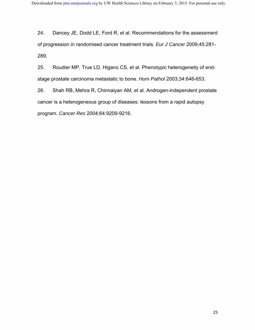
both at baseline and again 12 +/-4 weeks after initiation of dasatinib (Figure 1).
This time point for PET imaging was selected both from prior published bone
by UW Health Sciences Library on February 3, 2015. For personal use only. jnm.snmjournals.org Downloaded from
8
biomarker data (15, 16) with dasatinib and to match with CT and bone scans
from the therapeutic trial.
Patient Eligibility
This trial was reviewed and approved by the institutional review board of
all participating sites, and all patients signed written informed consent before
commencement of study procedures.
Key inclusion criteria for this trial included: males age ≥18 years with
histologically or cytologically proven prostate carcinoma, radiologic evidence of
metastatic bone disease and either biochemical, radiographic or symptomatic
progression of mCRPC with maintained castrate serum testosterone levels (<50
ng/dL). Required treatment withdrawal time frames for were: 30 days from anti-
androgens prior to baseline PSA, 4 weeks from radiation or radiopharmaceutical
treatment to bone, and 4 weeks from granulocyte-macrophage colony stimulating
factor (GM-CSF) or G-CSF prior to first PET scan. Other requirements included
adequate organ function and an ECOG performance status 0–2 with life
expectancy ≥12 weeks.
Key exclusion criteria included: prior receipt of either nilutamide or
dasatinib or amiodorone, lack of recovery to grade 1 toxicity from prior therapy,
history of major cardiac condition, uncorrected hypokalemia or hypomagnesemia,
clinically significant pleural or pericardial effusion, severe respiratory
insufficiency, or any other uncontrolled intercurrent illness. Directly relevant to
PET imaging, patients with poor intravenous access, weight >300 lbs due to
by UW Health Sciences Library on February 3, 2015. For personal use only. jnm.snmjournals.org Downloaded from
9
equipment specifications or those with inability to lie still for imaging were also
excluded.
Imaging Protocol and Analysis
All PET imaging scanners were pre-qualified by the ACRIN Imaging Core
Laboratory using phantom scans with known activity and sample patient image
sets were submitted for qualitative review and approval. PET imaging data was
acquired using either a GE Discovery STE (n=24 scans) or a Seimens Biograph
16 (n=2 scans). After careful consideration of the anticipated biologic impact of
therapy with dasatinib on fluoride delivery and vasculature, dynamic imaging,
limiting us to a single 15 cm field of view (FOV), was considered essential. The
lead nuclear medicine physician from the local study site reviewed both bone and
CT scans to identify the most prominent metastasis site for the dynamic FOV,
Regions in the upper abdomen and thorax were preferred to capture a blood
clearance curve from the heart or aorta. A low dose CT transmission scan was
acquired for attenuation correction, after which a 5.18 MBq/kg intravenous
injection of 18F-fluoride (mean dose 329 MBq, range 282 to 370 MBq) was
administered over 1 minute. At the onset of tracer injection a 60-minute dynamic
3D acquisition imaging protocol (16x5 sec, 7x10 sec, 5x30 sec, 5x60 sec, 5x3
min, 7x5 min) was initiated. A static whole body image from mid-thigh to head
was then obtained for attenuation correction, and a torso survey with emission
scanning was performed at imaging times of 2-5 minutes per position depending
upon the scanner. Image reconstruction corrected for attenuation, decay, scatter,
and random coincidences, using the scanner manufacturers method of 3D
by UW Health Sciences Library on February 3, 2015. For personal use only. jnm.snmjournals.org Downloaded from
10
reconstruction. DICOM header information for each image series was vetted
against ACRIN form information completed by local sites at the time of scanning.
All images were sent to the ACRIN imaging core lab at the University of
Washington for central imaging review. The data presented here are based upon
analysis of the dynamic imaging data; analysis of the static whole-body survey
images will be the subject of a future analysis.
Image Analysis
Dyanamic imaging data served as the primary imaging endpoint in this
trial. A subset of the dynamic imaging data (30-60 min SUV) were summed and
reconstructed, and used to create volumes of interest (VOI) for data extraction
and modeling. Using both the CT and static summed PET emission images,
VOIs were constructed on up to 5 lesions with the greatest 18F uptake in the
dynamic FOV. In the tumor VOI construction procedure, a 1cc VOI was centered
over the region of maximum tumor intensity, on the pixel with the maximum
value. Tumor-matched normal bone regions, identified by both CT and 18F-
fluoride PET, of identical volume were also constructed. SUV and SUVmax values
for each region were obtained from the 30-60 min static summed SUV image,
while time course data were extracted from the dynamic PET series as tissue
time-activity curves (TACs). SUVmax was defined as the SUV for the voxel within
the tumor VOI with the maximal uptake. SUVmaxavg was the average of SUVmax for
up to 5 tumors within the dynamic FOV. To acquire a blood input function for
compartmental modelling analysis, a 1 cm diameter cylindrical VOI was
by UW Health Sciences Library on February 3, 2015. For personal use only. jnm.snmjournals.org Downloaded from
11
constructed on the CT image set covering at least 3 cm of the aorta and applied
to the dynamic PET series to extract an image-derived blood TAC.
Compartmental Modelling
The 2-tissue compartment kinetic model of fluoride metabolism of Hawkins
(5), as modified by Doot (7) was used for parameter optimization of the dynamic
18F tissue TACs using the blood TAC as input to the model. The transfer from
blood into tissue is represented by K1, while the return of 18F from a tissue
compartment representing un-bound 18F back to blood is represented by k2. The
metabolic trapping of 18F through new bone formation is represented by k3, which
is the rate limiting step for the intracellular trapping of 18F in bone. There is some
evidence that 18F can leave the imaging region by bone degradation back to 18F
and subsequent efflux. The loss of image signal through these processes is
adequately described by k4.
The 18F flux is estimated through compartmental model optimization, which
fits model parameters to the tissue TAC data using the 18F blood activity curve as
the input function in a software package designed for PET data analysis (PMOD
v3.408, PMOD group, Zurich, Switzerland). The 18F flux constant, Ki, is
determined by the product of the rates of 18F metabolism (Ki = (K1*k3)/(k2+k3))
and represents the rate of 18F trapping as a quantitative measure of new bone
and fluoride deposition (5, 7). The key parameters for describing 18F uptake in
tissue are the 18F blood-tissue transport rate, K1, and the flux constant, Ki.
by UW Health Sciences Library on February 3, 2015. For personal use only. jnm.snmjournals.org Downloaded from
12
Study Endpoints
The primary study endpoint was to determine if changes in regional
fluoride incorporation, measured by 18F-flouride PET, occur in both CRPC bone
metastases and normal bone in response to treatment with dasatinib. This was
determined, as described above, within the 15 cm dynamic FOV by both SUVmax
and Ki, an indicator of net plasma clearance of fluoride to bone mineral. The
secondary endpoint of the trial determined if changes in 18F-flouride transport, K1,
an indicator of blood flow, and therefore, an indirect marker of angiogenesis,
occurred in both CRPC bone metastases and normal bone in response to
treatment with dasatinib. As a pre-specified exploratory analysis, the difference of
dasatinib treatment effects by SUVmax, Ki and K1 in normal bone was subtracted
from the difference in these measures in tumor bone.
Other exploratory efficacy endpoints compared 18F-fluoride parameters of
SUVmax, Ki and K1 in bone metastases at baseline and change in response to
treatment with dasatinib directly with PFS, as defined by the Prostate Cancer
Working Group 2 (PCWG2) (20). Additionally, changes in SUVm ax, Ki and K1, in
response to dasatinib treatment, were compared with changes in urinary N-
telopeptide (uNTX), bone alkaline phosphatase (BAP) and PSA.
Statistical Analyses
Based on data reported by Frost et al (6), where 18F-fluoride PET found a
15.6% decrease in Ki from baseline to 6-month post-bisphosphonate scan, this
trial was powered to detect a more modest 10% change in Ki or SUVmax from
baseline to post-treatment scan. Under these assumptions, 24 patients were
by UW Health Sciences Library on February 3, 2015. For personal use only. jnm.snmjournals.org Downloaded from
13
required to achieve a 0.05 target significance level and 80% power using a two-
sided paired t-test to compare pre- and post-treatment measurements at the
patient level.
The mean value of the change from pre- to post-dasatinib treatment was
calculated to represent the patient-level change for each uptake parameter (i.e.,
SUVmaxavg, Kiavg, K1avg). To test if the changes were statistically significant, the
generalized estimating equation (GEE) was fitted to analyze the bone-level data
after adjusting for clustering of data within subjects. Specifically, the compound
symmetry was used to denote the correlations among measurements collected
from the same patient.
The association between changes of these parameters and PFS was
evaluated via Cox proportional hazards models. This was done under a
univariate setting due to the limitation of sample sizes and lack of degree
freedom to adjust for other covariates. Spearman rank correlation was used to
examine correlations between changes in SUVmaxavg, Kiavg, and K1avg and
changes in PSA and markers of bone turnover, BAP and uNTX.
RESULTS
Patients and Treatment
Between September 2009 and November 2010, 18 patients were enrolled
in the trial. The goal was 24 patients, however, the companion therapeutic clinical
trial (NCT00918385) closed to accrual prematurely due to regulatory issues
surrounding the biopsy genetic signature. Since this imaging trial related only to
by UW Health Sciences Library on February 3, 2015. For personal use only. jnm.snmjournals.org Downloaded from
14
patients receiving dasatinib, the regulatory issues did not affect this data,
analysis or results, other than limiting the number of patients accrued. Of the 18
patients enrolled, all underwent baseline 18F-fluoride PET imaging. One patient
had PET data that was not interpretable due to technical issues. Thirteen of
these 17 patients underwent the second PET imaging 12 +/-4 weeks after
initiation of dasatinib. Four patients suffered from early clinical progression and
were removed from the trial prior to receiving the on-treatment second PET scan.
Another patient had PET data from the second scan that was not interpretable
due to technical issues. Therefore, 12 patients were evaluable to assess
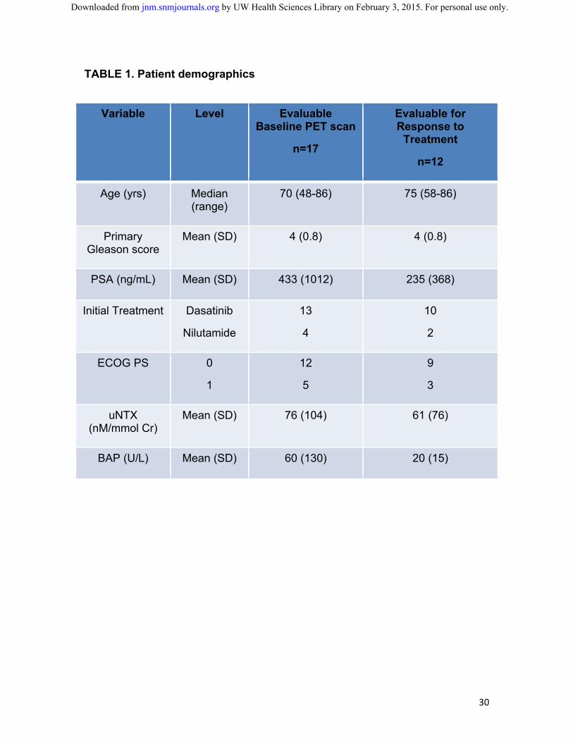
response to dasatinib treatment endpoints. Patient characteristics and
demographic data are presented in Table 1. Median followup for the 17 patients
with interpretable baseline scans and for the 12 patients evaluable for treatment
response was 452 (range 76-815) and 455 (167-645) days, respectively.
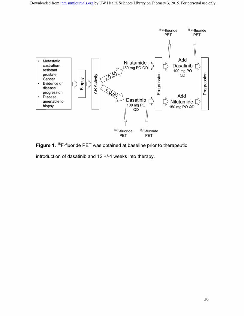
18F-fluoride as a Pharmacodynamic Measure of Dasatinib
A total of 37 pairs of tumor and normal bones were identified from the 12
patients who had both evaluable pre- and post-dasatinib PET images. To assess
the primary endpoint of change in fluoride incorporation into bone, both SUVmax
and Ki were evaluated. Changes in bone metastases in response to dasatinib by
SUVmax were notable with -6.61 decrease of SUVmaxavg (95% CI: -10.07, -3.15,
p=0.0002) versus null hypothesis of no change from GEE) (Figure 2, top panel).
No significant changes in SUVmax of normal bone sites were noted with +0.33
increase in SUVmaxavg (95% CI: -0.32, 0.97, p=0.32). Changes by Ki were not
significant in either tumor or normal bone (Figure 2, middle panel).
by UW Health Sciences Library on February 3, 2015. For personal use only. jnm.snmjournals.org Downloaded from
15
Since SRC inhibition has been shown to potentially decrease
angiogenesis (21), the secondary endpoint of the trial was to evaluate the effect
of dasatinib in bone metastases and normal bone on 18F-fluoride PET radiotracer
flow (K1). Changes in bone metastases and normal bone in response to dasatinib
by K1 were not significant (Figure 2, bottom panel).
A key endpoint of the trial was to determine if there is a differential
response between tumor and normal bone (Figure 2). This was significant for
SUVmax with a difference of -6.98 (95% CI: -10.30, -3.66, p<0.0001). There was
no differential response between tumor and normal bone by Ki. Although
previously, there was no difference in K1 in response to treatment with dasatinib
by either tumor or normal bone, there is a trend towards significance in the
differential response between tumor and normal bone with an absolute change of
-0.04 (95% CI: -0.082, 0.0002, p=0.051).
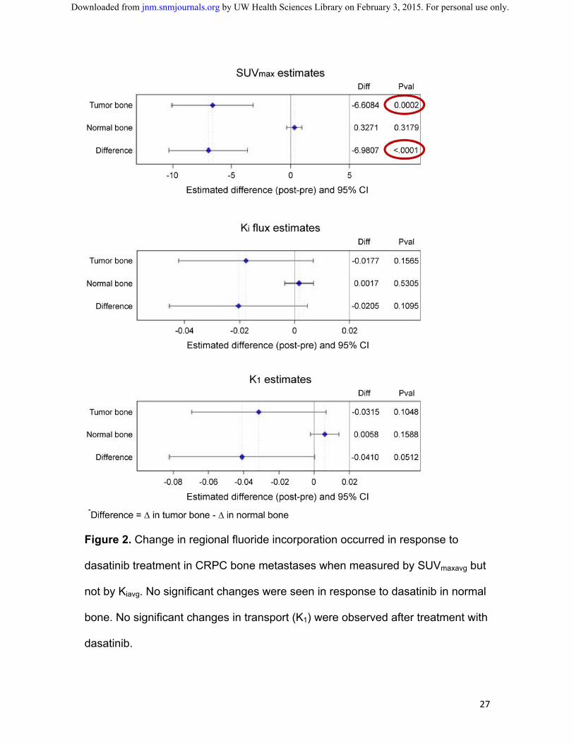
Associations Between 18F-fluoride PET and PFS
As exploratory endpoints, both baseline PET parameters and change in
response to dasatinib by 18F-fluoride PET in bone metastases were compared
with PCWG2-defined PFS. All patients developed progression and were
evaluable for PFS, therefore n=17 for baseline PET parameters and n=12 for
change from baseline to post-dasatinib PET. PFS was measured from the
initiation of dasatinib to an event or censoring.
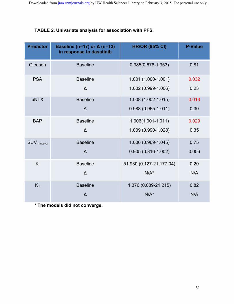
Results of the PFS association analysis are presented in Table 2. Other
parameters such as Gleason, PSA, uNTX and BAP were also evaluated, and
although baseline PSA, uNTX and BAP had association with PFS, changes in
by UW Health Sciences Library on February 3, 2015. For personal use only. jnm.snmjournals.org Downloaded from
16
these parameters did not correlate with PFS. Although baseline SUVmaxavg, Kiavg,
and K1avg did not correlate with PFS, changes in response to treatment with
dasatinib has borderline correlation with PFS for SUVmaxavg (HR 0.91, 95% CI
0.82-1.00; p=0.056). Interestingly, the HR was <1 implying that patients with
smaller decreases or even increases in uptake of 18F-fluoride had longer PFS,
rather than shorter PFS. For detailed granularity, individual change in SUVmaxavg
in relation to PFS is shown in Figure 3. This finding was contradictory our original
hypothesis and will be further addressed below in the discussion.
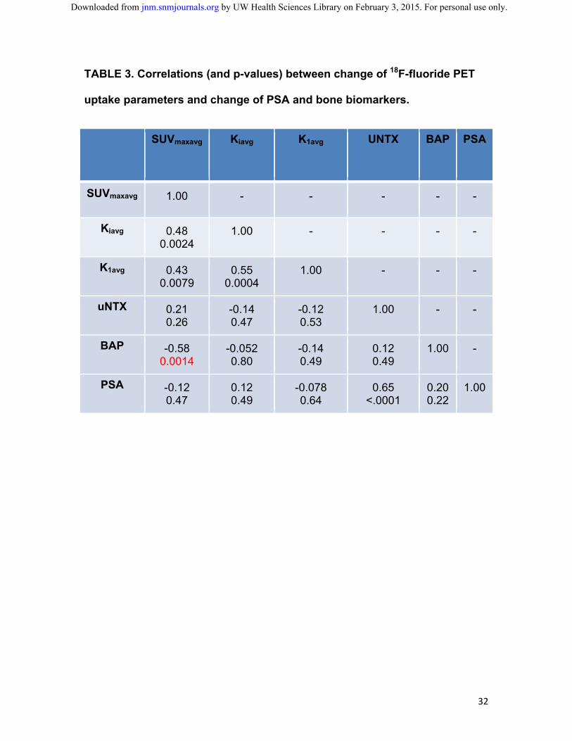
18F-fluoride PET Correlation with Bone Biomarkers and PSA
Other exploratory endpoints compared changes in 18F-fluoride PET
parameters in response to dasatinib in bone metastases with changes in PSA
and bone biomarkers (Table 3). Specifically, change in BAP had a significant
negative correlation with change 18F-fluoride PET by SUVmaxavg. Change in uNTX
and PSA had no correlation with changes by 18F-fluoride PET.
DISCUSSION
Prostate cancer clinical research is challenged by lack of validated disease
response endpoints for bone metastases. Bone scintigraphy is not a quantitative
measure and response to therapy is impossible to describe outside of the
detection of new lesions. As a result, prostate cancer trials have focused on
endpoints such as OS and radiographic PFS, rather than response to therapy
(20). These endpoints require significant patient numbers and follow-up and may
by UW Health Sciences Library on February 3, 2015. For personal use only. jnm.snmjournals.org Downloaded from
17
not be practical for widespread use in the clinic due to inability to offer a real-time
assessment of treatment response to the patient.
For these reasons, we embarked on this multicenter, cooperative group,
prospective imaging biomarker trial to determine response to therapy by PET.
The selection of 18F-fluoride as the radiotracer was a purposeful coupling with a
bone-dominant disease and a therapeutic agent with effects in bone. The goal of
demonstrating bone metastatic changes in response to dasatinib and differential
change for normal bone compared to bone metastases was successfully
demonstrated. Specifically, there was a significant difference in the change in
18F-fluoride SUVmaxavg with dasatinib for bone metastases versus normal bone,
with bone metastases, but not normal bone, having a significant decline in
uptake. To lend this finding further credence, we confirmed that changes 18F-
fluoride uptake in bone metastases correlated with accepted criteria for
radiographic PFS.
It is perhaps surprising that Ki was not a better indicator of 18F-fluoride
incorporation than SUVmax or SUVmaxavg, however, we noted anecdotally that the
18F-fluoride curves had statistical noise, suggesting more limited precision in the
kinetic estimates. SUV measures reporting the hottest pixel from a high
resolution 30 min image showed less variation than a 1cc VOI from a tumor
region that may include a distribution of voxels over a wide range of intensity
levels. Correcting for partial volume effect or segmenting the tumor volume may
have helped reduce the variability. This will require further study and confirmation
in more detailed analyses.
by UW Health Sciences Library on February 3, 2015. For personal use only. jnm.snmjournals.org Downloaded from
18
We were surprised at the nature of the borderline correlation found
between changes in 18F-fluoride incorporation by PET and PFS. Notably, patients
with the largest decrease in radiotracer incorporation in bone in response to
dasatinib had the worst outcomes, which were unexpected for predominantly
blastic prostate cancer bone metastases, where we might expect treatment to
decrease blastic activity at the site of metastasis. Those with a lower decline or
even an increase in blastic activity, had the longest durations until progression.
Dasatinib has been shown previously to promote osteoblast differentiation (22)
and mineralization that could lead to a relative activation and increase in bone
mineralization, somewhat similar to osteoblast activation accompanying a healing
“flare” seen on bone scan and 18F-fluoride PET (23), where early increases in
uptake, indicative of a healing or reparative response, may occur in patients with
PSA declines and/or other evidence of response to systemic therapy. Further
mechanistic studies will be needed to test this hypothesis and rule out the
possibility that this finding was obtained by chance.
Although not a validated endpoint for drug approval, PSA is commonly
utilized in the clinic to assess response to therapy. Dasatinib has had minimal
effect on PSA in early clinical trials (15, 16), and given the mechanism of action
on SRC, it might not be expected to have as much effect on PSA as agents that
inhibit the androgen axis. Nevertheless, we evaluated correlation between PSA
with 18F-fluoride PET parameters and found none, e.g., SUVmaxavg (p=0.47), Kiavg
(p=0.49), and K1avg (p=0.64).
by UW Health Sciences Library on February 3, 2015. For personal use only. jnm.snmjournals.org Downloaded from
19
Our trial had a number of limitations. Selection (or attrition) bias introduced
by informative censoring is a common issue for cancer treatment trials with PFS
as the study endpoint (24). Another potential bias could result from the 4 patients
who exited the trial due to rapid disease progression before the second on-
treatment PET study. Therefore, findings of this trial are limited to those patients
with disease indolent enough to allow acquisition of a second on-treatment PET
scan. Unfortunately, the small sample size precluded application of efficient
methods to correct for these types of biases or to perform multivariable analyses.
A number of variables were evaluated as potential predictors of radiographic
PFS, therefore, the borderline correlations between SUVmaxavg and radiographic
PFS could be due to chance. Finally, the timing for the second 18F-fluoride PET
scan had a large window to accommodate the complexity of the therapeutic trial,
and this may have contributed some variability in findings.
Heterogeneity of 18F-fluoride PET radiotracer uptake, and heterogeneity of
changes in response to dasatinib treatment were observed (Figure 4). This
heterogeneity is not surprising, as prostate cancer has been demonstrated to
have significant diversity from one metastasis to another (25, 26). The ability to
identify such heterogeneity emphasizes one of the strengths of PET imaging, as
PSA only offers a summed overview and molecular characterization by biopsy
and assay of a metastasis only offers data from that specific lesion. Given the
mechanism of action of dasatinib, it was felt that certain dynamic measures such
as Ki and K1 might effectively capture the biologic activity of the drug. For this
reason, we were limited in analysis to the dynamic FOV, and it is possible that
by UW Health Sciences Library on February 3, 2015. For personal use only. jnm.snmjournals.org Downloaded from
20
important data outside the dynamic FOV may not have been captured. Although
outside of the scope of this manuscript, more information may be gained by
evaluating the whole-body static surveys, and this future analysis will focus on
heterogeneity of disease response.
Larger trials must be performed to confirm the interesting findings in this
small, prospective trial. Although the development of dasatinib is unlikely to
proceed as a prostate cancer therapeutic, imaging biomarker studies of this sort
may help future development of novel agents by offering pharmacodynamic
assessments to aid in selection of patients most likely to benefit. Other 18F-
fluoride PET trials are underway, and it is now being tested with current standard
of care agents (NCT01516866).
CONCLUSION
18F-fluoride PET is capable of identifying dasatinib treatment response in CRPC
bone metastases and these changes may correlate with PFS. Validation of these
findings from larger, prospective trials with other therapeutic agents is required.
by UW Health Sciences Library on February 3, 2015. For personal use only. jnm.snmjournals.org Downloaded from
21
DISCLOSURES:
Evan Y. Yu: Research – Bristol-Myers Squibb
Fenghai Duan: Paid Consultant - WorldCare Clinical, LLC
ACKNOWLEDGMENTS
The study was supported by ACRIN, which receives funding from the National
Cancer Institute through U01 CA080098, under the American Recovery and
Reinvestment Act of 2009 (ARRA), U01 CA079778, and Bristol-Myers Squibb.
Imaging analysis was supported by the Quantitative Imaging Network U01
CA148131. All patients were accrued at DoD Prostate Cancer Clinical Trials
Consortium (UW W81XWH-09-1-0144, Duke W81XWH-09-1-0152, OHSU
W81XWH-09-1-0140, DFCI W81XWH-09-1-0150) and Prostate Cancer
Foundation Therapy Consortium sites.
by UW Health Sciences Library on February 3, 2015. For personal use only. jnm.snmjournals.org Downloaded from
22
REFERENCES
1. Cook GJ, Fogelman I. Detection of bone metastases in cancer patients by
18F-fluoride and 18F-fluorodeoxyglucose positron emission tomography. Q J Nucl
Med 2001;45:47-52.
2. Beheshti M, Langsteger W, Fogelman I. Prostate cancer: role of SPECT
and PET in imaging bone metastases. Semin Nucl Med 2009;39:396-407.
3. Schoder H, Larson SM. Positron emission tomography for prostate,
bladder, and renal cancer. Semin Nucl Med 2004;34:274-292.
4. Schirrmeister H, Guhlmann A, Elsner K, et al. Sensitivity in detecting
osseous lesions depends on anatomic localization: planar bone scintigraphy
versus 18F PET. J Nucl Med 1999;40:1623-1629.
5. Hawkins RA, Choi Y, Huang SC, et al. Evaluation of the skeletal kinetics
of fluorine-18-fluoride ion with PET. J Nucl Med 1992;33:633-642.
6. Frost ML, Cook GJ, Blake GM, Marsden PK, Benatar NA, Fogelman I. A
prospective study of risedronate on regional bone metabolism and blood flow at
the lumbar spine measured by 18F-fluoride positron emission tomography. J Bone
Miner Res 2003; 18:2215-2222.
7. Doot RK, Muzi M, Peterson LM, et al. Kinetic analysis of 18F-fluoride PET
images of breast cancer bone metastases. J Nucl Med 2010;51:521-527.
8. Cook G Jr, Parker C, Chua S, Johnson B, Aksnes AK, Lewington VJ. 18F-
fluoride PET: changes in uptake as a method to assess response in bone
metastases from castrate-resistant prostate cancer patients treated with 223Ra-
chloride (Alpharadin). EJNMMI Res 2011;1:4.
by UW Health Sciences Library on February 3, 2015. For personal use only. jnm.snmjournals.org Downloaded from
23
9. Nam S, Kim D, Cheng JQ, et al. Action of the Src family kinase inhibitor,
dasatinib (BMS-354825), on human prostate cancer cells. Cancer Res
2005;65:9185–9189.
10. Fizazi K. The role of Src in prostate cancer. Ann Oncol 2007;18:1765–
1773.
11. Summy JM, Gallick GE. Src family kinases in tumor progression and
metastasis. Cancer Metastasis Rev 2003;22:337–358.
12. Myoui A, Nishimura R, Williams PJ, et al. C-SRC tyrosine kinase activity is
associated with tumor colonization in bone and lung in an animal model of
human breast cancer metastasis. Cancer Res 2003;63:5028–5033.
13. Rucci N, Recchia I, Angelucci A, et al. Inhibition of protein kinase c-Src
reduces the incidence of breast cancer metastases and increases survival in
mice: implications for therapy. J Pharmacol Exp Ther 2006;318:161–172.
14. Koreckij T, Nguyen H, Brown LG, Yu EY, Vessella RL, Corey E. Dasatinib
inhibits the growth of prostate cancer in bone and provides additional protection
from osteolysis. Br J Cancer 2009;101:263–268.
15. Yu EY, Wilding G, Posadas M, et al. Phase 2 study of dasatinib in patients
with metastatic castration-resistant prostate cancer. Clin Cancer Res
2009;15:7421-7428.
16. Yu EY, Massard C, Gross M, et al. Once-daily dasatinib: Expansion of a
phase 2 study evaluating the safety and efficacy of dasatinib in patients with
metastatic castration-resistant prostate cancer. Urology 2011;77:1166-1171.
by UW Health Sciences Library on February 3, 2015. For personal use only. jnm.snmjournals.org Downloaded from
24
17. Araujo JC, Mathew P, Armstrong AJ, et al. Dasatinib combined with
docetaxel for castration-resistant prostate cancer: results from a phase 1-2 study.
Cancer 2012;118:63-71.
18. Araujo JC, Trudel GC, Saad F, et al. Randomized, double-blind, placebo-
controlled phase 3 trial of docetaxel and dasatinib in men with metastatic
castration-resistant prostate cancer. Lancet Oncol 2013;14:1307-1316.
19. Mendiratta P, Mostaghel E, Guinney J, et al. Genomic strategy for
targeting therapy in castration-resistant prostate cancer. J Clin Oncol
2009;27:2022–2029.
20. Scher HI, Halabi S, Tannock I, et al. Design and end points of clinical trials
for patients with progressive prostate cancer and castrate levels of testosterone:
recommendations of the Prostate Cancer Clinical Trials Working Group. J Clin
Oncol 2008;26:1148–1159.
21. Inoue S, Branch CD, Gallick GE, Chada S, Ramesh R. Inhibition of Src
kinase activity by Ad-mda7 suppresses vascular endothelial growth factor
expression in prostate carcinoma cells. Mol Ther 2005;12:707-715.
22. Lee YC, Huang CF, Murshed M, et al. Src family kinase/abl inhibitor
dasatinib suppresses proliferation and enhances differentiation of osteoblasts.
Oncogene 2010; 29:3196-3207.
23. Ryan CJ, Shah S, Efstathiou E, et al. Phase II study of abiraterone acetate
in chemotherapy-naive metastatic castration-resistant prostate cancer displaying
bone flare discordant with serologic response. Clin Cancer Res 2011;17:4854-
4861.
by UW Health Sciences Library on February 3, 2015. For personal use only. jnm.snmjournals.org Downloaded from
25
24. Dancey JE, Dodd LE, Ford R, et al. Recommendations for the assessment
of progression in randomised cancer treatment trials. Eur J Cancer 2009;45:281-
289.
25. Roudier MP, True LD, Higano CS, et al. Phenotypic heterogeneity of end-
stage prostate carcinoma metastatic to bone. Hum Pathol 2003;34:646-653.
26. Shah RB, Mehra R, Chinnaiyan AM, et al. Androgen-independent prostate
cancer is a heterogeneous group of diseases: lessons from a rapid autopsy
program. Cancer Res 2004;64:9209-9216.
by UW Health Sciences Library on February 3, 2015. For personal use only. jnm.snmjournals.org Downloaded from
Figu
introd
ure 1. 18F-flu
duction of d
uoride PET
dasatinib an
T was obtain
nd 12 +/-4 w
ned at base
weeks into
eline prior to
therapy.
o therapeut
2
tic
26
by UW Health Sciences Library on February 3, 2015. For personal use only. jnm.snmjournals.org Downloaded from
27
Figure 2. Change in regional fluoride incorporation occurred in response to
dasatinib treatment in CRPC bone metastases when measured by SUVmaxavg but
not by Kiavg. No significant changes were seen in response to dasatinib in normal
bone. No significant changes in transport (K1) were observed after treatment with
dasatinib.
by UW Health Sciences Library on February 3, 2015. For personal use only. jnm.snmjournals.org Downloaded from
Figu
dasa
ure 3. Indivi
atinib show
dual patien
less chang
nt changes
ge correlate
in SUVmaxav
es with long
vg in respon
er PFS.
nse to treat
2
ment with
28
by UW Health Sciences Library on February 3, 2015. For personal use only. jnm.snmjournals.org Downloaded from
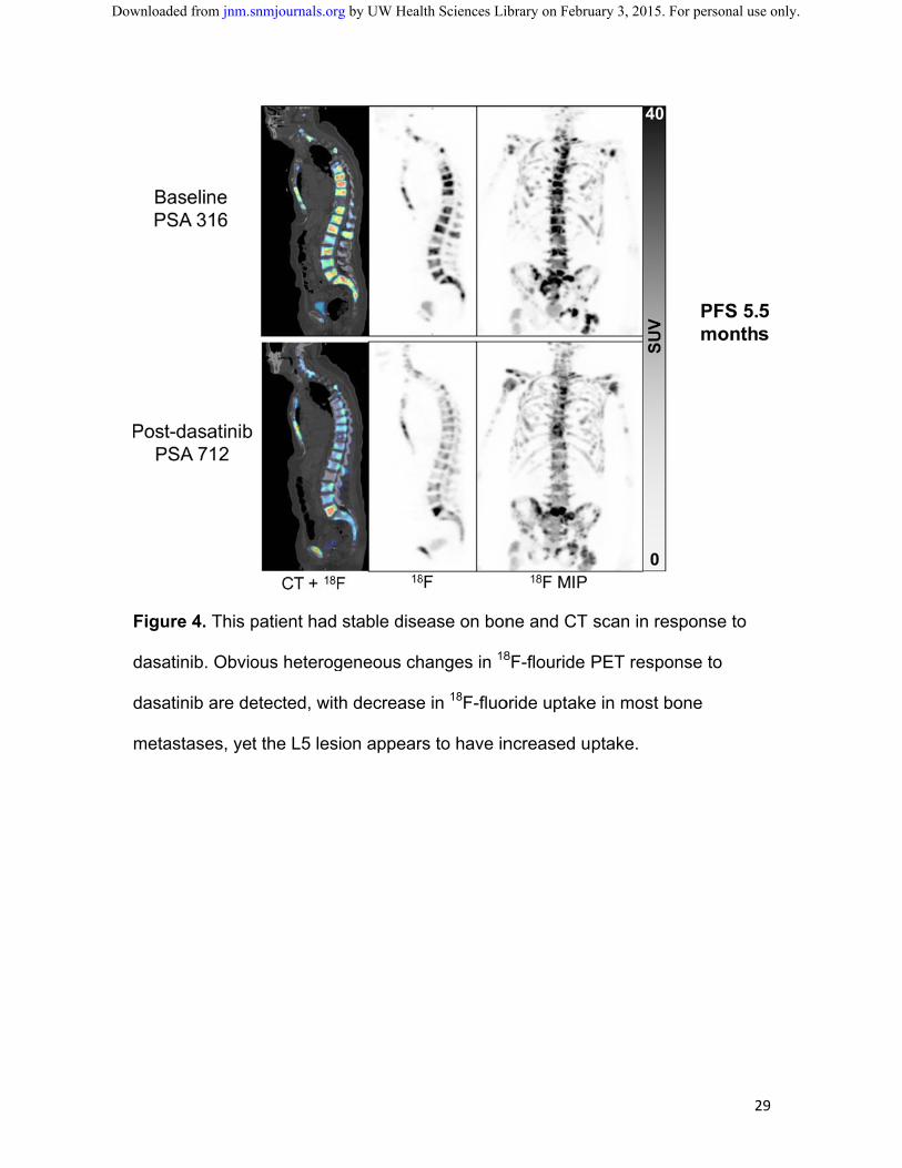
Figu
dasa
dasa
meta
ure 4. This p
atinib. Obvio
atinib are de
astases, yet
patient had
ous heterog
etected, wit
t the L5 les
stable dise
geneous ch
th decrease
sion appear
ease on bon
hanges in 18
e in 18F-fluo
rs to have in
ne and CT
8F-flouride
oride uptake
ncreased u
scan in res
PET respo
e in most bo
ptake.
2
sponse to
nse to
one
29
by UW Health Sciences Library on February 3, 2015. For personal use only. jnm.snmjournals.org Downloaded from
30
TABLE 1. Patient demographics
Variable Level Evaluable Baseline PET scan
n=17
Evaluable for Response to
Treatment
n=12
Age (yrs) Median (range)
70 (48-86) 75 (58-86)
Primary Gleason score
Mean (SD) 4 (0.8) 4 (0.8)
PSA (ng/mL) Mean (SD) 433 (1012) 235 (368)
Initial Treatment Dasatinib
Nilutamide
13
4
10
2
ECOG PS 0
1
12
5
9
3
uNTX (nM/mmol Cr)
Mean (SD) 76 (104) 61 (76)
BAP (U/L) Mean (SD) 60 (130) 20 (15)
by UW Health Sciences Library on February 3, 2015. For personal use only. jnm.snmjournals.org Downloaded from
31
TABLE 2. Univariate analysis for association with PFS.
Predictor Baseline (n=17) or ∆ (n=12) in response to dasatinib
HR/OR (95% CI) P-Value
Gleason Baseline 0.985(0.678-1.353) 0.81
PSA Baseline
∆
1.001 (1.000-1.001)
1.002 (0.999-1.006)
0.032
0.23
uNTX Baseline
∆
1.008 (1.002-1.015)
0.988 (0.965-1.011)
0.013
0.30
BAP Baseline
∆
1.006(1.001-1.011)
1.009 (0.990-1.028)
0.029
0.35
SUVmaxavg Baseline
∆
1.006 (0.969-1.045)
0.905 (0.816-1.002)
0.75
0.056
Ki Baseline
∆
51.930 (0.127-21,177.04)
N/A*
0.20
N/A
K1 Baseline
∆
1.376 (0.089-21.215)
N/A*
0.82
N/A
* The models did not converge.
by UW Health Sciences Library on February 3, 2015. For personal use only. jnm.snmjournals.org Downloaded from
32
TABLE 3. Correlations (and p-values) between change of 18F-fluoride PET
uptake parameters and change of PSA and bone biomarkers.
SUVmaxavg Kiavg K1avg UNTX BAP PSA
SUVmaxavg 1.00 - - - - -
Kiavg 0.48 0.0024
1.00
- - - -
K1avg 0.43 0.0079
0.55 0.0004
1.00
- - -
uNTX 0.21 0.26
-0.14 0.47
-0.12 0.53
1.00
- -
BAP -0.58 0.0014
-0.052 0.80
-0.14 0.49
0.12 0.49
1.00
-
PSA -0.12 0.47
0.12 0.49
-0.078 0.64
0.65 <.0001
0.20 0.22
1.00
by UW Health Sciences Library on February 3, 2015. For personal use only. jnm.snmjournals.org Downloaded from
![Regional distribution and kinetics of haloperidol binding in human brain: A pet study with [18F]haloperidol](https://static.fdokumen.com/doc/165x107/6344f2dd596bdb97a908b31a/regional-distribution-and-kinetics-of-haloperidol-binding-in-human-brain-a-pet.jpg)


![Multi-GBq Production of the Radiotracer [18F]Fallypride in a ...](https://static.fdokumen.com/doc/165x107/63218aff117b4414ec0b81c7/multi-gbq-production-of-the-radiotracer-18ffallypride-in-a-.jpg)


![Development of N-[3-(2′,4′-dichlorophenoxy)-2-18F-fluoropropyl]-N-methylpropargylamine (18F-fluoroclorgyline) as a potential PET radiotracer for monoamine oxidase-A](https://static.fdokumen.com/doc/165x107/63364f54a1ced1126c0b2979/development-of-n-3-24-dichlorophenoxy-2-18f-fluoropropyl-n-methylpropargylamine.jpg)


![Quantitative Receptor-Based Imaging of Tumor Proliferation with the Sigma-2 Ligand [18F]ISO-1](https://static.fdokumen.com/doc/165x107/6335c37c64d291d2a3029c11/quantitative-receptor-based-imaging-of-tumor-proliferation-with-the-sigma-2-ligand.jpg)