Carpals and tarsals of mule deer, black bear and human
114
Western Washington University Western Washington University Western CEDAR Western CEDAR WWU Graduate School Collection WWU Graduate and Undergraduate Scholarship 2009 Carpals and tarsals of mule deer, black bear and human: an Carpals and tarsals of mule deer, black bear and human: an osteology guide for the archaeologist osteology guide for the archaeologist Tamela S. Smart Western Washington University Follow this and additional works at: https://cedar.wwu.edu/wwuet Part of the Anthropology Commons Recommended Citation Recommended Citation Smart, Tamela S., "Carpals and tarsals of mule deer, black bear and human: an osteology guide for the archaeologist" (2009). WWU Graduate School Collection. 19. https://cedar.wwu.edu/wwuet/19 This Masters Thesis is brought to you for free and open access by the WWU Graduate and Undergraduate Scholarship at Western CEDAR. It has been accepted for inclusion in WWU Graduate School Collection by an authorized administrator of Western CEDAR. For more information, please contact [email protected].
-
Upload
khangminh22 -
Category
Documents
-
view
1 -
download
0
Transcript of Carpals and tarsals of mule deer, black bear and human

Western Washington University Western Washington University
Western CEDAR Western CEDAR
WWU Graduate School Collection WWU Graduate and Undergraduate Scholarship
2009
Carpals and tarsals of mule deer, black bear and human: an Carpals and tarsals of mule deer, black bear and human: an
osteology guide for the archaeologist osteology guide for the archaeologist
Tamela S. Smart Western Washington University
Follow this and additional works at: https://cedar.wwu.edu/wwuet
Part of the Anthropology Commons
Recommended Citation Recommended Citation Smart, Tamela S., "Carpals and tarsals of mule deer, black bear and human: an osteology guide for the archaeologist" (2009). WWU Graduate School Collection. 19. https://cedar.wwu.edu/wwuet/19
This Masters Thesis is brought to you for free and open access by the WWU Graduate and Undergraduate Scholarship at Western CEDAR. It has been accepted for inclusion in WWU Graduate School Collection by an authorized administrator of Western CEDAR. For more information, please contact [email protected].


MASTER'S THESIS
In presenting this thesis in partial fulfillment of the requirements for a master's degree at Western Washington University, I grant to Western Washington University the non-exclusive royalty-free right to archive, reproduce, distribute, and display the thesis in any and all forms, including electronic format, via any digital library mechanisms maintained by WWu.
I represent and warrant this is my original work, and does not infringe or violate any rights of others. I warrant that I have obtained written permissions from the owner of any third party copyrighted material included in these files.
I acknowledge that I retain ownership rights to the copyright of this work, including but not limited to the right to use all or part of this work in future works, such as articles or books.
Library users are granted permission for individual, research and non-commercial reproduction of this work for educational purposes only. Any further digital posting of this document requires specific permission from the author.
Any copying or publication of this thesis for commercial purposes, or for financial gain, is not allowed without my permission.
Signature I4me.iA ~+-Date 6 /I--'.=t--'-I_CYf----'----___
-

CARPALS AND TARSALS OF MULE DEER, BLACK BEAR
AND HUMAN: AN OSTEOLOGY GUIDE FOR
THE ARCHAEOLOGIST
A Thesis
Presented to
The Faculty of
Western Washington University
In Partial Fulfillment
of the Requirements for the Degree
Master of Arts
by
Tamela S. Smart
May 2009

iv
ABSTRACT
Existing osteological literature often lacks descriptions and illustrations of the smaller
elements, such as hand and foot bones, of animals commonly found in the archaeological record.
Black bear (Ursus americanus) and mule deer (Odocoileus hemionus) are both cosmopolitan species
and important resources for indigenous peoples, resulting in their widespread presence in faunal
assemblages. Additionally, the carpal and tarsal elements of these two mammalian taxa can be
difficult to distinguish from human elements because of their similarities in size and shape. Proper
identification of faunal and human remains is paramount to responsible cultural resource management
(CRM). This thesis presents a textual and photographic osteological guide of black bear and mule
deer carpals and tarsals and provides the means for distinguishing these elements from their human
counterparts.

v
ACKNOWLEDGEMENTS
I am grateful to my thesis committee Dr. Sarah K. Campbell, Dr. Joan C. Stevenson and
Dr. Michael A. Grimes for their insight and support. This thesis would not have been possible
without access to the mammalogy collection at The Burke Museum of Natural History and
Culture, the faunal collection at the Western Washington University Archaeology Laboratory, the
osteological collection at the Western Washington University Biological Anthropology
Laboratory, the faunal collection at the Western Washington University Geology Laboratory, and
the faunal collection at the Equinox Research and Consulting International, Inc (ERCI)
Laboratory. Further, I extend thanks to Sarah Willis, Gregory Willis and Elizabeth Ellis for their
donations to my private comparative collection. Finally, I would like to thank Kelly R. Bush and
Alyson M. Rollins for their continual guidance and encouragement throughout this project.

vi
TABLE OF CONTENTS
Abstract .......................................................................................................................................... iv Acknowledgements ......................................................................................................................... v List of Tables .................................................................................................................................. ix List of Figures ................................................................................................................................. x Chapter I: Introduction .................................................................................................................... 1 Chapter II: Natural History and the Cultural Importance of Mule Deer and Black Bear ................ 8
Natural History of the Mule Deer ............................................................................................... 8 Natural History of the Black Bear ............................................................................................... 9 Cultural Importance of Mule Deer and Black Bear to Prehistoric Peoples ............................... 10
Chapter III: Methods ..................................................................................................................... 13 Chapter IV: The Morphology of the Wrist .................................................................................... 17 Chapter IV: The Morphology of the Wrist .................................................................................... 17
The Carpals ............................................................................................................................... 18 Scaphoid .................................................................................................................................... 19
Deer Scaphoid ....................................................................................................................... 20 Bear Scapho-lunar ................................................................................................................ 22
Lunate ....................................................................................................................................... 25 Deer Lunate .......................................................................................................................... 25 Bear Scapho-lunar ................................................................................................................ 27
Triquetral ................................................................................................................................... 28 Deer Triquetral (Cuneiform) ................................................................................................. 28 Bear Triquetral ...................................................................................................................... 30
Trapezium ................................................................................................................................. 34 Bear Trapezium .................................................................................................................... 34
Trapezoid .................................................................................................................................. 37 Deer Trapezoid-magnum ...................................................................................................... 37 Bear Trapezoid...................................................................................................................... 40
Capitate ..................................................................................................................................... 42 Deer Trapezoid-magnum ...................................................................................................... 42 Bear Capitate ........................................................................................................................ 43
Hamate ...................................................................................................................................... 46 Deer Hamate (Unciform) ...................................................................................................... 46

vii
Bear Hamate ......................................................................................................................... 48 Pisiform ..................................................................................................................................... 50
Deer Pisiform ........................................................................................................................ 50 Bear Pisiform ........................................................................................................................ 52
Chapter V: The Morphology of the Ankle .................................................................................... 55 The Tarsals ................................................................................................................................ 56 Astragalus.................................................................................................................................. 57
Deer Astragalus .................................................................................................................... 57 Bear Astragalus ..................................................................................................................... 60
Calcaneus .................................................................................................................................. 64 Deer Calcaneus ..................................................................................................................... 64 Bear Calcaneus ..................................................................................................................... 66
Navicular ................................................................................................................................... 69 Deer Naviculo-cuboid ........................................................................................................... 69 Bear Navicular ...................................................................................................................... 71
Medial Cuneiform ..................................................................................................................... 74 Deer Medial Cuneiform ........................................................................................................ 74 Bear Medial Cuneiform ........................................................................................................ 76
Intermediate Cuneiform ............................................................................................................ 77 Deer Intermediate/Lateral Cuneiform ................................................................................... 77 Bear Intermediate Cuneiform ............................................................................................... 79
Lateral Cuneiform ..................................................................................................................... 81 Deer Intermediate/ lateral Cuneiform ................................................................................... 81 Bear Lateral Cuneiform ........................................................................................................ 81
Cuboid ....................................................................................................................................... 84 Deer Cuboid .......................................................................................................................... 84 Bear Cuboid .......................................................................................................................... 84
Lateral Malleolus ...................................................................................................................... 88 Deer Lateral Malleolus ......................................................................................................... 88 Bear Lateral Malleolus ......................................................................................................... 89
Chapter VI: Conclusions ............................................................................................................... 90 References ..................................................................................................................................... 92 Appendix 1: Bear and Deer Specimens ......................................................................................... 99

viii
Appendix 2: Human Specimens .................................................................................................. 101

ix
LIST OF TABLES
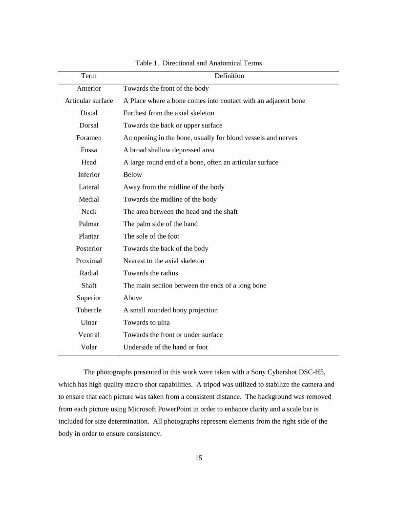
Table 1. Directional and Anatomical Terms ................................................................................ 15
Table 2. Carpal Name Synonyms ................................................................................................. 19
Table 3. Tarsal Name Synonyms .................................................................................................. 57

x
LIST OF FIGURES
Figure 1. Schematic drawing of bear, human and deer right carpals. ........................................... 18
Figure 2. Right deer scaphoid ventral and dorsal surfaces. ........................................................... 20
Figure 3. Right deer scaphoid lateral and medial surfaces. ........................................................... 21
Figure 4. Right bear scapho-lunar anterior and posterior surfaces. ............................................... 22
Figure 5. Right human navicular anterior surface and right bear scapho-lunar posterior surface. 23
Figure 6. Right human navicular posterior surface and right bear scapho-lunar anterior surface. 24
Figure 7. Right deer lunate dorsal, posterior and ventral surfaces. ............................................... 26
Figure 8. Right deer lunate lateral and medial surfaces. ............................................................... 27
Figure 9. Right deer cuneiform anterior and medial surfaces. ...................................................... 28
Figure 10. Right deer cuneiform posterior and lateral surfaces. ................................................... 29
Figure 11. Right human hamate view from the triquetral and right bear cuneiform lateral surface.
.............................................................................................................................................. 29
Figure 12. Right bear triquetral anterior and posterior surfaces. ................................................... 30
Figure 13. Right bear triquetral medial surface. ............................................................................ 31
Figure 14. Right human scaphoid view from the capitate and right bear triquetral anterior surface.
.............................................................................................................................................. 32
Figure 15. Right human scaphoid view from the radius and right bear triquetral posterior surface.
.............................................................................................................................................. 33
Figure 16. Right bear trapezium anterior and lateral surfaces. ...................................................... 34
Figure 17. Right bear trapezium posterior and medial surfaces. ................................................... 35
Figure 18. Right human lunate view from the capitate and right bear trapezium anterior view. .. 36
Figure 19. Right deer trapezoid-magnum ventral and dorsal surfaces. ......................................... 38
Figure 20. Right deer trapezoid-magnum lateral and medial surfaces. ......................................... 39
Figure 21. Right bear trapezoid anterior and posterior surfaces. ................................................... 40
Figure 22. Right bear trapezoid lateral and medial surfaces. ........................................................ 41
Figure 23. Right human triquetral view from the hamate and right bear trapezoid medial surface.
.............................................................................................................................................. 41
Figure 24. Right bear capitate anterior and posterior surfaces. ..................................................... 43
Figure 25. Right bear capitate lateral and medial surfaces. ........................................................... 44
Figure 26. Right human capitate view from the scaphoid and right bear capitate posterior surface.
.............................................................................................................................................. 45

xi
Figure 27. Right human and bear capitate distal ends. .................................................................. 45
Figure 28. Right deer unciform dorsal and posterior surface. ....................................................... 46
Figure 29. Right deer unciform ventral and medial surfaces. ........................................................ 47
Figure 30. Right bear hamate anterior and posterior surfaces. ...................................................... 48
Figure 31. Right bear hamate lateral and medial surfaces. ............................................................ 49
Figure 32. Right human cuboid ventral surface and right bear hamate lateral surface. ................ 49
Figure 33. Right deer pisiform anterior and medial surfaces. ....................................................... 50
Figure 34. Right human and deer pisiforms lateral surfaces. ........................................................ 51
Figure 35. Right bear pisiform anterior surface. ........................................................................... 52
Figure 36. Right bear pisiform ventral and dorsal surfaces. .......................................................... 53
Figure 37. Right human MT 1 lateral surface and right bear pisiform dorsal surface. .................. 54
Figure 38. Schematic drawing of bear, human and deer right tarsals. ........................................... 56
Figure 39. Right deer astragalus anterior and posterior surfaces. .................................................. 58
Figure 40. Right deer astragalus lateral surface. ........................................................................... 59
Figure 41. Right bear astragalus dorsal and ventral surfaces. ....................................................... 60
Figure 42. Right bear astragalus lateral surface. ........................................................................... 61
Figure 43. Right human talus superior surface and right bear astragalus dorsal surface. ............. 62
Figure 44. Right human talus inferior surface and right bear astragalus ventral surface. ............. 63
Figure 45. Right deer calcaneus anterior and medial surfaces. ..................................................... 65
Figure 46. Right human calcaneus superior surface and right deer calcaneus anterior surface. ... 66
Figure 47. Right bear calcaneus dorsal and ventral views. ........................................................... 67
Figure 48. Right human calcaneus superior view and right bear calcaneus dorsal view. ............. 68
Figure 49. Right deer naviculo-cuboid dorsal, ventral and anterior surfaces. ............................... 70
Figure 50. Right bear navicular anterior, posterior and lateral surfaces. ....................................... 71
Figure 51. Right human navicular anterior surface and right bear navicular anterior surface. ..... 72
Figure 52. Right human navicular posterior surface and right bear navicular posterior surface. .. 73
Figure 53. Right deer medial cuneiform dorsal and medial surfaces. ........................................... 74
Figure 54. Right human pisiform lateral surface and right deer medial cuneiform medial surface.
.............................................................................................................................................. 75
Figure 55. Right bear medial cuneiform anterior, posterior and lateral surfaces. ......................... 76
Figure 56. Right deer intermediate/lateral cuneiform dorsal, ventral and medial surfaces. .......... 77
Figure 57. Right bear intermediate cuneiform medial and lateral surfaces and right deer
intermediate/lateral cuneiform medial surface. ..................................................................... 78

xii
Figure 58. Right bear intermediate cuneiform anterior and posterior surfaces. ............................ 79
Figure 59. Right bear intermediate cuneiform medial and lateral surfaces. .................................. 80
Figure 60. Right bear lateral cuneiform anterior and posterior surfaces. ...................................... 82
Figure 61. Right bear lateral cuneiform lateral and medial surfaces. ............................................ 83
Figure 62. Right bear cuboid anterior and ventral surfaces. .......................................................... 85
Figure 63. Right bear cuboid posterior and medial surfaces. ........................................................ 86
Figure 64. Right human cuboid ventral surface and right bear cuboid ventral surface. ................ 87
Figure 65. Right deer lateral malleolus medial and ventral surfaces. ............................................ 88
Figure 66. Right lateral malleolus dorsal and lateral surfaces. ...................................................... 89

CHAPTER I: INTRODUCTION
The ability to distinguish human skeletal remains from bones of other animals is
becoming increasingly important for professional archaeologists in North America because of
their ethical and legal responsibilities when human remains are discovered. Archaeologists
conducting shovel testing, permitted excavation, or monitoring of construction excavation must
often make quick decisions about whether to stop or continue work in order to comply with
procedures mandated by law, contract, or permit. Most protocols for handling inadvertent
discovery of human remains require leaving them in place and causing no further disturbance
while the appropriate authorities are contacted. While many archaeologists have some training in
human osteology or comparative mammalian osteology, few have extensive experience, and even
those elements that are generally recognizable may present challenges if they are fragmented or
only partially exposed. Thus archaeologists commonly rely on illustrated osteological guides to
confirm field identifications, and to recognize less common or less distinctive elements. Despite
the number of available osteological guides, there are still gaps in coverage. Human bone
anatomy texts and virtual guides assist in identifications (Bass 1995; Kappelman 2008; Schwartz
1995; White 2000; White and Folkens 2005); however most of these sources provide little
comparison of human and faunal anatomy. Most animal osteological guides (eg., Brown and
Gustafson 1990; Digital Morphology Group and UTCT 2002; Gilbert 1990; Kasper 1980;
Schmid 1972) focus on crania or larger skeletal elements and ignore smaller ones, such as the
bones of the hands and feet. This is unfortunate as these bones tend to preserve well and be
relatively well represented in archaeological sites. Carpals and tarsals are morphologically
distinct yet less familiar than long bones to the non-specialist and can be readily misidentified
between animals of comparable size.
This thesis helps to fill this gap by providing a guide for identifying and distinguishing
human elements from the carpals and tarsals of Ursus americanus (black bear) and Odocoileus
hemionus (mule deer), mammals which are commonly represented in North American
archaeological sites (Ubelaker 2006). These elements are similar enough in size and morphology
to commonly be confused with human bones by untrained observers.
Given the pace of development in the United States, and increasing legislative protection
for burials, archaeologists frequently find themselves working in situations where discovery of
human remains requires specific procedural responses, and where failure to comply can have
significant negative consequences for individuals, companies, and the professional community.

2
Recent federal legislation provides more explicit protection for burials, and many states
have followed suit. Federal law mandates that human remains, funerary objects, sacred objects or
objects of cultural patrimony that are in the possession of the federal government, or a federally
funded institution or agency, must be repatriated to tribes that can show genetic or cultural
affiliation (See Native American Graves Protection and Repatriation Act – NAGPRA, 1990).
NAGPRA also regulates the inadvertent discovery and intentional archaeological excavation of
human remains on Federal land (43 CFR 10). Before the late 1980s most states had legislation
protecting graves in clearly established cemeteries which, however, did not apply to unmarked
Native American burials (Hutt 1998). A 1989 National Geographic article brought public
attention to the destruction of more than 800 burial sites on a Kentucky farm and the lack of
government response. Within a couple of years of this publication most states had amended their
laws to include Native American burials (Arden 1989; Hutt 1998). Washington State reflects this
nationwide trend. Here, a number of recent cases involving ancient burials have brought this
issue to the forefront. The most well known case concerns the dispute over the disposition of
Kennewick Man’s remains (Boxberger 2009; Bruning 2006; Chatters 2000). Also, within the
last ten years two large scale construction projects, the Hood Canal Bridge Graving Dock in Port
Angeles and the Blaine Waste Water Treatment Plant on the Semiahmoo Spit, were stopped due
to the discovery of unmarked burials and disagreements over management of the unearthed
human remains (Davila 1999; WSDOT 2004).
State laws concerning burials vary a great deal; Washington state law is discussed here as
an example. Three statutes pertain to the discovery of human remains in Washington State. The
first is the Notice to Coroner (Revised Code of Washington [RCW] 68.50) (1987) which states
that “…it shall be the duty of every person who knows of the existence and location of a dead
body coming under the jurisdiction of the coroner…to notify the coroner thereof in the most
expeditious manner possible…”. Those that do not follow this action are guilty of a
misdemeanor. The second applicable statute is the Indian Graves and Records Act (RCW 27.44)
(1989) which requires that
Any person who knowingly removes, mutilates, defaces, injures, or destroys any cairn or grave of any native Indian or any glyptic or painted record of any tribe or peoples is guilty of a class C felony punishable under chapter 9A.20RCW. Persons disturbing Native American graves through inadvertence…shall reinter the human remains under the supervision of the appropriate Indian tribe.

3
The third statute is the Abandoned and Historic Cemeteries and Historic Graves Act
(RCW 68.60) (1989) which makes it is a class C felony to knowingly remove, mutilate, deface,
injure or destroy an historic grave. The three statutes described above have recently (in 2008)
been strengthened with the passing of Washington State House Bill 2624-S2.E which adds new
sections to the existing grave protection legislation. Further, it has resulted in the creation of a
State Physical Anthropologist position under the Department of Archaeology and Historic
Preservation (DAHP) and has made this person responsible for determining whether non-forensic
remains are Indian or non-Indian and for reporting their findings to the affected tribes (DAHP
n.d.).
When human remains are inadvertently discovered during an initial investigation,
monitoring project, permitted excavation or during lab work the applicable laws should be
followed. How laws governing human remains are implemented is dependent upon the policy of
the agency with jurisdiction (Hutt 1988; King 2008). Generally, when human remains are
encountered the archaeologist should notify the property owner or on-site superintendent. All
work in the immediate area should stop and the area should be secured, with all equipment moved
to a safe distance. The local law enforcement and medical examiner should be contacted in order
to determine if the remains are forensic in nature. If they are non-forensic the State Physical
Anthropologist determines if they are Indian or Non-Indian and reports the findings to the
affected tribes (DAHP n.d.). If a federal agency is not involved the DAHP conducts all
consultation with the affected parties regarding future preservation, excavation and the remains
disposition (DAHP n.d.).
The task of differentiating human remains from faunal remains is complicated by
numerous taphonomic processes (Aslan and Behrensmeyer 1996; Behrensmeyer 1978;
Behrensmeyer et al. 2000; Haglund and Sorg 1997; Lyman 1994; Whyte 2001). First, the way in
which a skeleton enters the archaeological record greatly affects its appearance and preservation.
Human remains that are intentionally buried are easily identified by most people because of their
anatomical positioning and completeness (Whyte 2001). However, certain mortuary practices
such as secondary burials and cremations can greatly change the positioning and appearance of
elements (Whyte 2001). In comparison, animal carcasses are rarely buried intentionally and are
often processed by humans for a wide variety of reasons. Processing results in bone
fragmentation and incomplete and disarticulated skeletons; all of which make identification more
difficult (Davis 1987; Lyman 1994; Reitz and Wing 2008). For example, if a carcass is being

4
processed in the field with the intention of reducing it to transportable parts then it would likely
exhibit cut marks associated with skinning and disarticulation. It would then be brought back to
camp in its entirety minus the bone fragments related to its division (Lyman 1978 and 1994;
Nokes 2004).
Finally, post depositional disturbances can greatly impact skeletal remains and their
identification. These disturbances can be divided into two groups, abiotic and biotic. Abiotic
disturbances include inanimate forces such as rain, earthquakes, floods, waves, wind, sun,
freezing and thawing, as well as soil properties (Ceci 1984; Gordon and Buikstra 1981; Reitz and
Wing 2008). For example, forests soils are generally highly acidic, which accelerates the rate of
decomposition of organics such as bone (Gordon and Buikstra 1981). Bones that are naturally
decomposing may no longer exhibit key identifying landmarks. In contrast, shell midden
deposits increase the alkalinity of the soil and result in a slower rate of bone decomposition.
Biotic disturbances are associated with plants and animals. Bioturbation, the movement of soil by
plants and animals, is a common biotic disturbance (Ceci 1984). Others include scavenging,
trampling and gnawing (Lyman 1994; Reitz and Wing 2008). Humans should also be considered
one of the main causes of post depositional disturbance. Agricultural activities, land clearing,
damming of rivers, road construction and maintenance, installation of utilities and construction of
buildings are just a few of the land modifications that can impact archaeological remains (Ceci
1984; Reitz and Wing 2008).
Regardless of the condition of skeletal remains archaeologists need to be able to identify
whether they are human or faunal. In the laboratory the most effective way to make a positive
identification is with the use of a comparative collection. As this resource is not feasible for use
in the field, osteological guidebooks are essential. A number of human skeletal guides provide
detailed descriptions and illustrations of adult elements (Bass 1995; Flower 1885; Schwartz 1995;
White 2000; White and Folkens 2005). In addition, a few guides emphasize human juvenile
osteology and the development of the skeletal system (Baker et al. 2005; Scheuer and Black
2000; Scheuer et al. 2008). Compared to human osteological guides, faunal guides generally do
not provide the same level of detail, especially in regards to smaller elements such as wrist and
ankle bones. For example, Brown and Gustafson (1990) is a guide to the postcranial skeletal
elements of cattle/bison, elk and horse, yet there are no carpal illustrations and only three tarsals
are included (the calcaneus, the astragulus and the naviculo-cuboid). In addition, Schmid’s
(1972) comparative guide illustrates only the calcaneus and astragulus of selected mammalian
taxa. This unfortunate bias is not new in the field of archaeology. Cornwall (1964), one of the

5
more in-depth human and faunal skeletal guides (geared towards archaeologists), exemplifies this
bias in stating:
For the archaeologist the carpal bones are generally of no great importance. Owing to their usually small size and irregular shape, they are frequently but poorly preserved and may easily be overlooked by excavators since, in the earth; they are not readily recognizable as bones [146].
The major exceptions to this trend are Flower (1885), Gilbert 1990, Post (2004a and 2004b), and
Sandefur (1977), which do emphasize carpal and tarsal elements. Of these works, Sandefur
(1977), Post (2004a) and Gilbert (1990) were the most helpful in the creation of this guide.
Sandefur (1977) provides in depth descriptions of mule deer carpals, however, the illustrations
are lacking in detail. Gilbert (1990) also provides illustrations of mule deer carpals, as well as
drawings of the calcaneus, astragulus, and naviculo-cuboid for a number of selected taxa, but
lacks labels and descriptions of key landmarks. Post (2004a) provides detailed illustrations of
black bear wrist and ankle elements from a number of different views, unfortunately the views are
not labeled and a description of key landmarks is not provided.
Guides that illustrate ankle and wrist bones are important because these elements
commonly preserve in the archaeological record. This is due in large part to their small spherical
shape and relatively high structural density (Darwent and Lyman 2001; Sobolik 2003). In
addition, carpals and tarsals (especially those of artiodactyls) exhibit minimal interstitial space,
thus contain limited grease and marrow and are not likely to be fragmented by humans during
processing (Binford 1978; Darwent and Lyman 2001). The common preservation of these
elements makes them ideal for quantifying the minimum number of individuals (MNI) of a given
taxa in an assemblage. MNI is defined as the fewest number of individuals necessary to account
for all of the elements present in an assemblage (Lyman 1994; White 2000). White (2000) states
that this method of quantifying individuals should take into consideration the following: element,
side, age, sex, occlusion, articulation and antimeric partners (White 2000). Carpal and tarsal
counts are ideal for estimating MNI because these elements preserve well and each individual has
only one left and one right. Alyson M. Rollins, the Lummi Indian Nation Physical
Anthropologist who works on repatriation issues including unanticipated discoveries of human
remains, often uses carpal and tarsal elements to assist in determining MNI (personal
communication 2009). In addition, carpals and tarsals of human can also be used for stature,

6
age and sex determinations (Bidmos 2006; Bidmos and Asala 2003; Bidmos and Asala 2004;
Cameriere and Ferrante 2008; Cameriere et al. 2007; Hsieh et al. 2007; Sulzmann et al. 2008)
There are no comprehensive osteological guidebooks that emphasize comparative carpal
and tarsal morphology. The goal of this thesis is to fill this gap by providing information needed
to identify these elements for two mammals commonly found at archaeological sites: the black
bear (Ursus americanus) and the mule deer (Odocoileus hemionus) (Ubelaker 2006). In addition,
this work will highlight the characteristics of these elements that distinguish them from human.
Black bear and mule deer were selected for three primary reasons: 1) potential for
misidentification due to size and morphological similarity to human wrist and ankle bones, 2)
their importance in the local economies of prehistoric peoples, and 3) overall ubiquity at
archaeological sites. Many black bear and mule deer skeletal elements are similar in both size
and morphology to their human counterparts and thus are commonly misidentified by non-
specialists. Carcasses of mule deer and less frequently black bear are routinely discovered on
roadways or trails after predation by other animals and humans, and after collisions with motor
vehicles. In Michigan State alone there are approximately 65,000 deer-vehicle collisions each
year (Riley and Marcoux 2006). Disarticulated animal remains can easily be mistaken for human.
In these cases, local law enforcement, coroners, medical examiners and physical anthropologists
are often contacted for identification (Klepinger 2006; Orcholl and Hudson 2001; Owsley and
Mann 1990; Stewart 1959; Sims 2007). Owsley and Mann (1990) estimate that about 15% to
25% of the medicolegal cases involving forensic anthropologists are misidentified animal
remains, most commonly deer, pig, dog and bear.
Robert Bishop, Coroner for Island County in Washington State is often brought skeletal
remains by the public and stated “I have had a bear paw mistaken for a human hand once, but it
still had soft tissue attached. I was interested in how similar it appeared, but of course it did not
have an opposable thumb…In the past 14 years I would suggest that deer make up the majority of
the bones presented to this office” (personal communication 2009). Alyson M. Rollins and Guy
Tasa the Washington State Physical Anthropologist for DAHP have had similar experiences with
archaeological skeletal remains. Rollins says that she has worked on a number of repatriations in
which deer, bear and dog elements were identified as human and human elements were identified
as faunal (personal communication 2009). Guy Tasa believes that deer skeletal elements and to a
lesser extent bear, cow, sheep, wapiti, seal, sea lion and pig bones are commonly misidentified as
human (personal communication 2009).

7
Identification of mule deer and black bear bones is important not just as an aid to
recognizing human remains correctly, but for their value in contributing to analysis of human life
ways and their use of animal resources. Both mule deer and black bear had great importance as
resources for indigenous peoples of North America, which is evidenced by both the
archaeological record and ethnographic literature (Ubelaker 2006). Both of these taxa have a
wide geographic range (Mackie et al. 1982; Nowak and Paradiso 1983; Pelton 1982), and were
hunted throughout their range.
In this osteological guide emphasis is placed on descriptions and photographs of
Odocoileus hemionus and Ursus americanus wrist and ankle elements. First, an overview of the
focal species is provided in Chapter II. Discussed in this chapter are the animals’ taxonomy,
distribution, ecology, social behavior, morphology and their importance for prehistoric peoples.
Chapter III describes the methods used in creating this guide. Chapters IV and V provide an
overview of mammalian hand and foot morphology, as well as a detailed discussion of
Odocoileus hemionus and Ursus americanus carpals and tarsals. Further, these chapters highlight
the key characteristics that distinguish mule deer and black bear wrist and ankle bones from their
human counterparts. The final chapter discusses the importance of osteological guides to
archaeologists and suggests where additional work is needed.

8
CHAPTER II: NATURAL HISTORY AND THE CULTURAL IMPORTANCE OF MULE
DEER AND BLACK BEAR
Mule deer and black bear are two of the most widely dispersed and recognizable
terrestrial mammals in North America. Further, these species play an important role in both the
local ecology and in the cultures of the indigenous peoples. Due to the importance of mule deer
and black bear as a resource to the indigenous peoples and their common presence in the
archaeological record these animals are often encountered by archaeologists in the field. In this
chapter an overview of the natural history of mule deer and black bear is discussed including their
taxonomy, geographic range, social behavior, and physical attributes. A discussion of the
importance of these animals to prehistoric peoples in the Pacific Northwest follows.
Natural History of the Mule Deer
The mule deer (Odocoileus hemionus) is an extant species of the taxonomic Order
Artiodactyla, which includes a group of mammals whose weight is positioned on their third and
fourth toes. Deer are commonly referred to as cervids, due to their inclusion in the family
Cervidae. The other members of this family that inhabit North America include white-tailed deer
(Odocoileus virginianus), wapiti (Cervus elaphus), moose (Alces alces) and caribou (Rangifer
tarandus). The species Odocoileus hemionus consists of seven generally agreed upon subspecies,
including both mule deer and black tailed deer (Mackie et al 1982). The specimens utilized in
this project were identified only to the species level.
The geographic range of this species covers most of temperate North America from the
Pacific coast eastward to the 100th meridian. They are found as far north as the southern Yukon
in the west and Manitoba in the east. Their southern limits extend into Baja California and
northern Mexico (Mackie et al 1982; Nowak and Paradiso 1983).
Within this geographic range mule deer are found in habitats that vary widely in terms of
climatic and vegetation zones as well as elevation. They frequently endure temperatures
averaging -15° C in the winter and 30° C in the summer. This species commonly inhabits open
forests, brushy areas, and shrub-lands associated with rough terrain. They are highly adapted to
high elevation mountainous regions, foothills and will also frequent prairies along river drainages
(Mackie et al 1982; Nowak and Paradiso 1983).
The social behavior of Odocoileus hemionus varies greatly depending on the following
factors: subspecies, sex, and season. For the majority of the year they are widely dispersed with

9
the basic social unit consisting of an adult female, her female yearling, and two fawns. Males are
usually alone or in small groups. In certain cases however, this species will aggregate around
resources and groups as large as several hundred individuals will form. Mule deer are
polygamous and during mating season males compete with each other for females in estrus
(Mackie et al 1982; Nowak and Paradiso 1983).
The physical appearance of this species is quite distinctive. Adult individuals molt twice
each year. In the winter their upper body is brownish gray and their under belly is lighter in
color. In the summer they exhibit a reddish brown coat. The tail is somewhat small and is either
white or black on the proximal end and black on the distal end. Odocoileus hemionus exhibits
sexual dimorphism with males weighing on average 74 kg and females weighing 59 kg. In
addition, males are longer, averaging 152 cm, while females average 142 cm. Also, males grow
deciduous antlers, which females do not (Mackie et al 1982; Nowak and Paradiso 1983).
As previously mentioned, all members of the family Artiodactyla exhibit highly adapted
morphology. The third and fourth toes bear all of the body weight and this adaptation is
manifested not only in the morphology and orientation of the animal’s digits but in the entire
morphology of the manus and pes. The individual bones of the hands and feet will be discussed
at length in the succeeding chapters.
Natural History of the Black Bear
The black bear (Ursus americanus) is an extant species of the taxonomic Family Ursidae.
Two additional members of this Family inhabit North America, the Grizzly bear (Ursus arctos)
and the Polar bear (Ursus maritimus). Of these three species, black bears are by far the most
common.
Black bears have a substantial geographic range. They inhabit the forested areas across
much of North America. To the north, they are found in Alaska and Northern Canada, while their
southern boundary extends into Mexico. It should be noted that their density varies greatly within
this range due to limited suitable habitat and human population size (Nowak and Paradiso 1983;
Pelton 1982). Black bears are primarily found in forested areas with a thick under-story and
mountainous terrain. Along the Pacific coast they inhabit forests where the dominant over-story
includes Douglas fir (Pseudotsuga taxifolia), Ponderosa Pine (Pinus ponderosa), Lodgepole pine
(Pinus contorta), Redwood (Sequoia sempervierns), Sitka spruce (Picea sitchensis), and Hemlock
10
(Tsuga sp.). In addition to forested areas, black bears also live in meadows, riparian zones,
brushlands and tidelands (Pelton 1982).
Black bears are largely solitary animals. Breeding occurs in the summer and females
give birth during their dormancy period in the winter. A litter usually consists of two cubs,
however females have been observed with as many as four. The cubs generally stay with their
mother until they are yearlings when they disperse during the spring or summer months (Pelton
1982).
The outward appearance of Ursus americanus is quite impressive. Body lengths range
from one to two meters and weight ranges from 40 to 70 kg in females and 60 to 140 kg in males.
These animals are most commonly black, chocolate brown or cinnamon brown. In the Pacific
Northwest white and blue phases do occur, but are rare. Black bears can also be differentiated
from other bears by their small, round ears, short tail and brown muzzle (Nowak and Paradiso
1983; Pelton 1982).
The hand and foot morphology of the black bear is similar to many mammals in that they
exhibit five digits (pentadactyl) and have a plantigrade stance (Pelton 1982). The anatomy of
their carpals and tarsals will be discussed at length in the succeeding chapters.
Cultural Importance of Mule Deer and Black Bear to Prehistoric Peoples
Mule deer inhabit most of the western half of North America, while their close cousin,
the white tailed deer, is found throughout the continental United States, southern Canada,
Mexico, Central America and northern portions of South America. Black bear are also very
widespread, ranging from the Pacific to the Atlantic Ocean and from Northern Alaska down into
Mexico (Mackie et al. 1982; Nowak and Paradiso 1983; Pelton 1982). These animals were
hunted to varying degrees throughout these geographic ranges (Ubelaker 2006). It is beyond the
scope of this project to provide a detailed discussion of traditional use of these two animals across
North America, therefore, archaeological and ethnographic evidence from the Pacific Northwest
is provided as an example of the importance of these animals to indigenous peoples.
The Pacific Northwest includes both the Northwest Coast culture area and the interior
Plateaus. The former is defined by Suttles (1990) as extending approximately 1,500 miles from
the Copper River Delta on the Gulf of Georgia south to the Winchuck River near Oregon’s
southern border. West to east it spans from the coastal islands to the Chugach and St. Elias
Ranges in Alaska, the Coast Mountain Ranges in British Columbia and the Cascade Range in

11
Washington and Oregon. East of the Cascade Range are the Columbia and Fraser Plateaus,
which have generally been treated as a distinct culture area (Walker 1989). Ames and Maschner
(1999) argue for treating these areas together, and term the combined area Cascadia. The hunter-
fisher-gatherer peoples that inhabited both sides of mountain were connected by ties of
linguistics, trade, kinship, and the exploitation of anadromous salmon, although the coastal areas
were distinguished by more complex social organization (Ames 1994; Ames and Maschner
1999).
Researchers have placed great emphasis on the role of salmon to explain the cultural
complexity and population density of the Pacific Northwest (Ames 1994; Butler and Campbell
2006; Driver 1993; Hodgetts and Rahemtulla 2001). More recently, archaeologists are paying
greater attention to the importance of non-marine resources. Butler and Campbell (2006)
analyzed faunal data from 65 Plateau and Northwest Coast archaeological sites that date from the
last 10,000 years and confirmed that over time their was a gradual change from broad spectrum
foraging to organized collecting strategies and intensified exploitation of salmon. However, this
intensification did not result in an increased use of salmon relative to other resources. Rather,
some terrestrial mammals such as deer (Odocoileus spp.) and wapiti (Cervus elaphus) were
utilized to a greater extent over time (Butler and Campbell 2006). Further, artiodactyls
(especially deer and wapiti) are the most ubiquitous terrestrial mammals found in sites throughout
this region. Black bear do not comprise a large component of faunal assemblages like deer, but
they are found at numerous Pacific Northwest sites (Butler and Campbell 2006; Cannon 1991;
Imamoto 1976; Lyman 1995; Hodgett and Rahemtulla 2001; Huelsbeck 1994; Montgomery
1979; Nokes 2004).
An examination of selected archaeological sites from both the coastal region and the
plateau clearly reflects this trend (Butler and Campbell 2006; Nokes 2004; Livingston 1985).
Odocoileus hemionus (number of identified specimens [NISP] 239) is the most abundant mammal
with Castor canadensis (beaver) (NISP 193) as the second and Cervus elaphus (elk) (NISP 169)
as the third at 45WH34 (Ferndale), a site located in the Gulf of Georgia. Black bear are also
present at this site but comprise only a small portion of the assemblage (NISP 24, MNI 1).
Interestingly, the bear elements present at the site are primarily hand and foot bones and include
one calcaneus, three metapodials, two metacarpals, four first phalanges, six second phalanges,
and one third phalanx (Nokes 2004). Deer and wapiti elements at the Ferndale site also follow
this trend with distal leg and manus and pes elements being the most common (Nokes 2004). In
the plateau region, artiodactyls are again the predominant terrestrial taxa in the faunal assemblage

12
(Butler and Campbell 2006). For example, artiodactyls comprise 90% of the identified elements
at 45OK258 (a site located along the right bank of the Columbia River). Odocoileus spp. (3,452
elements) including mule deer and white tailed deer were the most abundant of the artiodactyls
and Ovis canadensis (mountain sheep) (511 elements), Cervus elaphus (14 elements), and
Antilocapra americana (pronghorn antelope) (42 elements) were also present but in much smaller
numbers. An examination of the distribution of the elements indicates that all parts of the
artiodactyls were brought back to the site and utilized. A total of four Ursus spp. (grizzly bear
and black bear) elements were identified at this site and all were from the paw region and include
two metatarsal fragments, one metacarpal and one metapodial (Livingston 1985).
The ethnographic literature also indicates that mule deer and black bear were important
sources of food and raw material for Pacific Northwest peoples. “Deer and elk meat were
considered the best varieties and dried with special care, the meat was cut in pieces and hung on a
frame. Fires were built on three sides and the meat was thoroughly roasted” (Haeberlin and
Gunther 1930: 21). Deer were also valued for their bone, antlers, hooves and hides which were
used to make tools, clothes and ceremonial and decorative items (Ruby and Brown 1976;
Underhill 1945). In May of 1792, George Vancouver during his exploration of Port Discovery
writes the following in his journal “Their native woollen garment was most in fashion, next to it
the skins of deer, bear, etc.; a few wore dresses manufactured from bark, which, like their woolen
ones, were very neatly wrought” (Meany 1942:122).
Bears were utilized as a food source, for their hide, and to make regalia for ceremonies
(Haeberlin and Gunther 1930; Ruby and Brown 1976; Suttles 1990; Underhill 1945). They were
hunted by bow and arrow, trapped and chased out of caves with smoke and killed (Suttles 1990).
Haeberlin and Gunther (1930) provide a detailed description of a bear trap.
A trap for bears consisted of two poles about ten feet high erected over some bear tracks. These carried a heavy horizontal pole to which a rope was attached. This rope was also tied to brush that covered up a four to five foot hole dug underneath the horizontal pole. When the bear stepped on the brush and fell into the hole, he pulled down the horizontal pole which fell on him [25].
13
CHAPTER III: METHODS
The differentiation between human skeletal elements and those of other mammalian taxa
is complicated by many factors, the most notable of which is similarity in body size. Second,
humans exhibit a generalized mammal form resulting in elements that are morphologically
similar to other generalized taxa, such as bear. Third, ubiquity is an important factor. Black bear
and mule deer were important resources for North American indigenous peoples resulting in their
widespread presence in archaeological sites (Ubelaker 2006). Based on these factors, mule deer
and black bear were chosen for this osteological study.
Specimen selection was based on age of the individual, completeness of the forelimbs
and hind-limbs and the overall condition of individual elements. Age determination was based on
epiphyseal fusion and preference was given to mature individuals. All of the specimens
photographed in this work were adult except for #UA1, an adolescent bear. Additionally,
elements that were highly fragmented, exhibited injury or severe degenerative disease were
excluded.
The faunal specimens used for photographing and comparison were borrowed from the
Burke Museum of Natural History and Culture at the University of Washington, the Western
Washington University Geology Lab, the Western Washington University Archaeology lab, and
the Equinox Research and Consulting International, Inc. laboratory. Additional specimens were
donated by private individuals and added to the author’s personal collection. Seven Ursus
americanus specimens were used in this thesis: three of the specimens were complete
individuals; one specimen was partially complete with an articulated lower limb and ankle; one
specimen was an articulated foot; and two of the specimens were disarticulated hand and foot
bones. In addition, 26 Odocoileus hemionus specimens were examined: two of the specimens
were complete individuals; one specimen was largely complete minus the head; two specimens
were articulated lower limbs and feet; and 21 of the specimens were represented by single carpal
or tarsal elements. A complete list of the faunal specimens is presented in Appendix 1. The six
human specimens used in this guide are teaching specimens housed at the Western Washington
University Biological Anthropology Lab. All of the human specimens were either largely
complete or complete skeletons and they are listed in Appendix 2.
In addition to comparative collections, guide books were a key resource in the
construction of this guide. The following sources provided valuable illustrations/photographs and
textual descriptions that were consulted throughout this work: Cornwall (1964), Flower (1885),

14
Gilbert (1990), Post (2004a), Sandefur (1977), White (2000) and White and Folkens (2005).
Further, the organization of this guide is patterned largely after White (2000) and White and
Folkens (2005).
In order to provide a comprehensive guide to bear and deer carpals and tarsals, both
written descriptions and black and white photographs are provided. The written descriptions
detail the general morphology of each element, their location within the hand or foot and the key
identifying landmarks. These landmarks are labeled with an alphabetical delineation and are
identified in the corresponding photographs. Photographed specimens were selected based on
their completeness and lack of any soft tissue that might obscure landmarks. All specimens are
photographed in anatomical position, which results in differences in element orientation.
Specimens that were not photographed were used for comparative purposes. Multiple views of
each element are presented in order to show all of the key landmarks and to enhance the reader’s
ability to identify both complete elements and fragments.
Many bear and deer carpals and tarsals exhibit a morphological resemblance to human
elements. In cases where possible confusion may arise, a discussion outlining how to make a
positive determination between the species is included. Comparisons are not limited to
homologous (the same bones in different animals) elements, rather elements that are similar in
size and morphology are compared. For example the deer medial cuneiform is compared to the
human pisiform. Most of the discussions do highlight human carpals and tarsals; however the
bear pisiform is compared to the human first metacarpal, as they are similar in appearance.
Photographs of the necessary views of the human elements are provided, as well. Key landmarks
on the human elements were given numerical delineations that correspond to the descriptions in
the text.
The osteological terminology utilized by human and faunal specialists often varies. To
assist the reader a list of important directional and anatomical terms are listed in Table 1. The
terms are compiled from the following sources: Clemente (1985), Cornwall (1964), Flower
(1885), Scheuer and Black (2000), and White (2000).
15
Table 1. Directional and Anatomical Terms
Term Definition
Anterior Towards the front of the body
Articular surface A Place where a bone comes into contact with an adjacent bone
Distal Furthest from the axial skeleton
Dorsal Towards the back or upper surface
Foramen An opening in the bone, usually for blood vessels and nerves
Fossa A broad shallow depressed area
Head A large round end of a bone, often an articular surface
Inferior Below
Lateral Away from the midline of the body
Medial Towards the midline of the body
Neck The area between the head and the shaft
Palmar The palm side of the hand
Plantar The sole of the foot
Posterior Towards the back of the body
Proximal Nearest to the axial skeleton
Radial Towards the radius
Shaft The main section between the ends of a long bone
Superior Above
Tubercle A small rounded bony projection
Ulnar Towards to ulna
Ventral Towards the front or under surface
Volar Underside of the hand or foot
The photographs presented in this work were taken with a Sony Cybershot DSC-H5,
which has high quality macro shot capabilities. A tripod was utilized to stabilize the camera and
to ensure that each picture was taken from a consistent distance. The background was removed
from each picture using Microsoft PowerPoint in order to enhance clarity and a scale bar is
included for size determination. All photographs represent elements from the right side of the
body in order to ensure consistency.

16
Photographing the fine details on bones can be difficult with inadequate lighting, thus a
light box was constructed to sufficiently illuminate the specimens during photographing sessions.
Instructions for light box construction were found at the Our Media: Channels of Creativity (n.d.)
website. The materials used in its assembly included five pieces of white foam board, strips of 2”
wide masking tape, one straight edge knife, one ruler, three metal clip-on shop lights and three
60Hz full spectrum light bulbs.

17
CHAPTER IV: THE MORPHOLOGY OF THE WRIST
The bones of the mammalian hand (manus) are divided into three groups: carpals,
metacarpals and phalanges. Carpals are small wrist bones, largely cubical in shape, which form
from single primary ossification centers. They articulate through synovial joints which allow for
limited mobility (Flower 1885). Proximal to the carpals are the ulna and radius (the forearm
bones) and distal to the carpals are five tubular shaped bones called metacarpals.
Metacarpals (MC) are numbered one through five (from medial to lateral). They form
from two centers of ossification with the epiphysis at the distal end (in all but the first) (Flower
1885). The proximal ends of the metacarpals articulate with the carpals at largely immobile
joints and their distal ends articulate through hinge joints with the phalanges. Deer are an
exception to this pattern, as they exhibit two small vestigial metacarpals (second and fifth) and
one large metacarpal formed from the fused third and fourth elements (Cornwall 1964; Flower
1885).
Phalanges comprise the fingers or digits of the hand. The generalized mammalian form
is comprised of five digits, as represented in both human and bear. The digits are referred to as
follows: I (thumb or pollex), II (index finger), III (middle finger), IV (ring finger), V (little
finger). Each of the digits has three phalanges including the proximal, intermediate and distal
(ungual) with the exception of the pollex which is comprised of only two phalanges (Cornwall
1964; Flower 1885; Scheuer and Black 2000; White 2000). It is important to emphasize that in
some of the more specialized mammal species digits may be reduced in size or absent. For
example, in deer the pollex is absent (Flower 1885).
Proximal to the carpals are the bones of the forearm, which include the radius and ulna.
The radius and ulna are long bones positioned side by side in the lower arm. In humans these
elements are relatively similar in overall size and robusticity, however in most non-human
mammals the radius is generally the more robust of the two (Flower 1885). In deer and horses,
for example, the ulna exhibits a substantial proximal end and a reduced sliver-like distal end. The
degree of movement between these two elements also varies greatly. In some mammals,
including deer, the ulna and radius are fused, resulting in a limited range of motion. Whereas in
humans, they remain unfused, resulting in a greater range of motion that allows for the palm to be
oriented up or down (Flower 1885; White 2000).

18
The Carpals
The full complement of carpals in mammals includes eight bones, which are generally
arranged in two transverse rows, a proximal and a distal, with one additional bone occupying a
centered position among them, the central (Cornwall 1964). Interestingly, the central is often
absent, as is the case with the species included in this guide. The proximal row of carpals (from
radial to ulnar) is comprised of the scaphoid, the lunate, and the triquetral. The distal row (from
radial to ulnar) includes the trapezium, the trapezoid, the capitate and the hamate. Two sesamoid
bones, located on either side of the carpals within the tendons of the flexor muscles, are also
common within the wrist. The first is the pisiform which is common and often highly developed.
The second is the radial sesamoid, which is frequently absent (Cornwall 1964; Flower 1885)
(Figure 1).
Figure 1. Schematic drawing of bear, human and deer right carpals. Key: A, scaphoid/scapho-lunar; B, lunate; C, triquetral/cuneiform; D, trapezium; E, trapezoid/ trapezoid-magnum; F, capitate; G, hamate/unciform; H, pisiform.

19
There are several systems for carpal bone nomenclature; synonyms are presented in
Table 2 (Bass 1995; Cornwall 1964; Flower 1885; White 2000).
Table 2. Carpal Name Synonyms
Carpal Names Synonyms Scaphoid Radial, Navicular, Scapho-lunar (when fused with the lunate)
Lunate
Lunar, Intermedium, Semilunar, Lunatum, Scapho-lunar (when fused with
the scaphoid)
Triquetral Ulnare, Cuneiform, Triquetrum, Pyramidale
Central Centrale, Intermedium
Trapezium Greater Multangular, Multangulum majus, Carpal 1
Trapezoid
Lesser Multangular, Multangulum minus, Carpal 2, Trapezoid-magnum
(when fused with the capitate)
Capitate
Capitol, Magnum, Carpal 3, Trapezoid-magnum (when fused with the
trapezoid)
Hamate Hamate, Uncinatum, Unciform, Carpal 4 & 5
Pisiform Ulnar sesamoid bone
Radial Sesamoid Sesamoid
Each carpal is morphologically distinctive. An understanding of their characteristic
landmarks can help distinguish both within and between species. The following pages provide
detailed descriptions and photographs of each of the deer and bear carpals. Key landmarks are
noted using alphabetical notation and are described in detail. Human elements that share a
common morphology with deer and bear carpals are discussed in order to identify their
distinguishing features. These comparisons are not limited to homologous (the same bones in
different animals) elements. Also, the views presented for each of the compared elements may
differ, resulting from either differences in anatomical positioning or because different views were
more similar in appearance.
Scaphoid
The scaphoid is one of the larger carpal bones and its name stems from the Greek word
skaphidion meaning “a small ship” (Sanderfur 1977). Due to its boat like shape it is commonly
referred to as the navicular of the hand. This element is located in the proximal row of the carpal

20
block on the radial side. In some mammals, including bear, the scaphoid and lunate are fused,
forming the scapho-lunar. The overall shape of this element varies greatly between mammals.
Deer Scaphoid
The deer scaphoid is an irregular shaped bone that is located on the superior-medial side
of the carpal block (Figure 2 and Figure 3). It articulates with three other bones including the
radius (superiorly), the trapezoid-magnum (inferiorly) and the lunate (laterally).
Figure 2. Right deer scaphoid ventral and dorsal surfaces. Key: a, trapezoid magnum articular surface; b, distal radial articular surface.
a. The trapezoid magnum articular surface is a large landmark that covers most of the
elements ventral surface. It exhibits four non-distinct facets.
b. The distal radial articular surface covers the entire dorsal surface of the element and
exhibits a saddle-like appearance.

21
Figure 3. Right deer scaphoid lateral and medial surfaces. Key: c, lunate articular facets; d, medial side.
c. There are two articular facets for the lunate located on the lateral side of the scaphoid.
They run horizontally and are separated by a deep groove.
d. The medial side of the scaphoid is a semi rough rectangular surface with no articular
facets. It exhibits a distinctive rectangular shape with a pinched-in midline.

22
Bear Scapho-lunar
The scapho-lunar is the largest carpal found in the bear and also one of the most
distinctive (Figure 4). Its overall shape is rectangular with a large inferior projection extending
from its medial corner. The scapho-lunar articulates with six elements, including the trapezium,
trapezoid, capitate, hamate, triquetral (anteriorly), and radius (posteriorly).
Figure 4. Right bear scapho-lunar anterior and posterior surfaces. Key: a, trapezium articular surface; b, trapezoid articular surface; c, capitate articular facet; d, hamate articular surface; e, triquetral articular surface; f, articular surface for radius; g, scapho-lunar process.
a. The trapezium articular surface is a small triangular facet located on the medial side of
the anterior surface.
b. The trapezoid articular surface is located just lateral to the trapezium articular surface
and it is the larger of the two facets.
c. The capitate articular facet is a long concave rectangular facet located along the midline
of the anterior surface.
d. The hamate articular surface is located along the lateral side of the anterior surface of
the scapho-lunar. It is an elongated concave facet.
e. The triquetral articular surface is located just inferior to the hamate articular surface on
the lateral side of the anterior surface. It is small and semi-circular in shape.
f. The articular surface for the radius is a large rectangular convex facet that comprises
2/3 of the elements posterior surface.

23
g. The scapho-lunar process extends from the medio-inferior quadrant of the element and
serves as an attachment site for soft tissue.
Possible confusion with human elements:
The bear scapho-lunar is similar in morphology to the human navicular (Figure 5 and
Figure 6). Both have a large flattened body with a process or tubercle extending from their
medial sides. In addition, the posterior side of the bear scapho-lunar and the anterior side of the
human navicular both exhibit a large convex articular surface.
To distinguish between these two elements look for the following distinctive
characteristics.
Figure 5. Right human navicular anterior surface and right bear scapho-lunar posterior surface. Key: 1, medial cuneiform articular surface; 2, intermediate cuneiform articular surface; 3, lateral cuneiform articular surface; f, articular surface for the radius; g, scapho-lunar process. The human navicular is generally smaller than the bear scapho-lunar.
The anterior side of the human navicular is oval in shape and its articular surface can be
divided into three, sometimes four sections, where it articulates with the medial
cuneiform (1), the intermediate cuneiform (2), the lateral cuneiform (3) and sometimes
the cuboid. In contrast the articular surface on the posterior side of the bear scapho-lunar
is rectangular in shape and exhibits a single facet where it articulates with the radius (f).

24
Figure 6. Right human navicular posterior surface and right bear scapho-lunar anterior surface. Key: 4, talar facet; a, trapezium articular surface; b, trapezoid articular surface; c, capitate articular facet; d, hamate articular surface; e, triquetral articular surface. The opposing sides of the elements are very different. The anterior side of the bear
scapho-lunar has three deep depressions separated by two ridges, while the posterior side
of the human navicular has a single large rounded facet for the head of the talus (talar
facet) (4).

25
Lunate
The lunate is named for its distinctive crescent shape, which is clearly exhibited in the
human form. This characteristic however, is absent in many taxa, including deer and bear. The
lunate is generally positioned along the midline of the carpal block between the scaphoid and the
triquetral. As previously mentioned, the scaphoid and lunate are fused in many mammals, such
as bear, and in these cases it is referred to as the scapho-lunar.
Deer Lunate
Though the lunate was named for its resemblance to the moon, it exhibits a cuboidal
shape in the deer (Figure 7 and Figure 8). The lunate is located in the middle of the proximal
carpal row and it articulates with the following five elements: radius (superiorly), scaphoid
(medially), cuneiform (laterally), trapezoid-magnum (inferio-medially) and unciform (inferio-
laterally). The deer lunate can be identified by its twisted appearance and its very distinct angular
surfaces. It is also the largest carpal in the deer (Sandefur 1977).

26
Figure 7. Right deer lunate dorsal, posterior and ventral surfaces. Key: a, articular facets for the radius; b, trapezoid magnum articular facet; c, unciform articular facet.
a. There are two articular facets for the radius. The larger of the two covers the entire
dorsal surface of the element. Its overall shape is irregular but it does bare some
resemblance to the letter “J”. The smaller articular facet for the radius is located on the
posterior surface, in the posterio-lateral corner.
b. The trapezoid magnum articular facet is located on the medial side of the ventral
surface adjacent to the articular facet for the unciform. This landmark is rectangular in
shape, has a twisted appearance and exhibits a convex anterior portion and a concave
posterior portion.
c. The unciform articular facet is located on the lateral side of the ventral surface and is
shaped like the adjacent trapezoid magnum articular facet. It is the larger of the two facets
on this surface.

27
Figure 8. Right deer lunate lateral and medial surfaces. Key: d, articular facets for the cuneiform; e, scaphoid articular facets.
d. There are two articular facets for the cuneiform on the lateral surface. The first is
positioned along the inferior margin and runs the length of the element. The second is
triangular in shape and is located on the anterio-superior corner. These two landmarks are
separated by a large groove.
e. There are three scaphoid articular facets located on the medial surface. The first is
positioned along the superior border and forms a long narrow strip that extends the length
of the element. The two remaining facets are located on the anterior and posterior corners
of the inferior border.
Bear Scapho-lunar
The bear scaphoid and lunate are fused forming the scapho-lunar. See the above
scaphoid section for photographs and descriptions of this element.
28
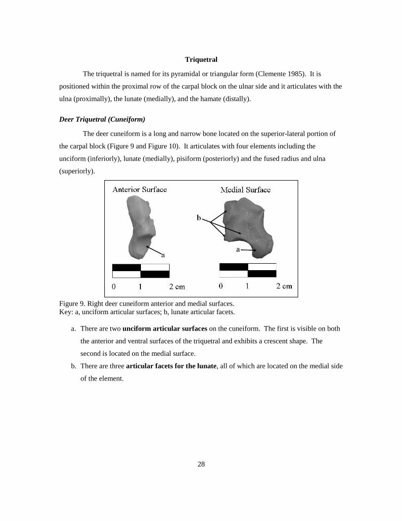
Triquetral
The triquetral is named for its pyramidal or triangular form (Clemente 1985). It is
positioned within the proximal row of the carpal block on the ulnar side and it articulates with the
ulna (proximally), the lunate (medially), and the hamate (distally).
Deer Triquetral (Cuneiform)
The deer cuneiform is a long and narrow bone located on the superior-lateral portion of
the carpal block (Figure 9 and Figure 10). It articulates with four elements including the
unciform (inferiorly), lunate (medially), pisiform (posteriorly) and the fused radius and ulna
(superiorly).
Figure 9. Right deer cuneiform anterior and medial surfaces. Key: a, unciform articular surfaces; b, lunate articular facets.
a. There are two unciform articular surfaces on the cuneiform. The first is visible on both
the anterior and ventral surfaces of the triquetral and exhibits a crescent shape. The
second is located on the medial surface.
b. There are three articular facets for the lunate, all of which are located on the medial side
of the element.

29
Figure 10. Right deer cuneiform posterior and lateral surfaces. Key: c, pisiform articular surface; d, articular surface for the fused ulna and radius.
c. The pisiform articular surface is located on the posterior surface and exhibits a crescent
shape.
d. The articular surface for the fused ulna and radius is a large facet that can be seen on
both the dorsal and lateral surfaces.
Possible confusion with human elements:
The deer cuneiform shares a minor resemblance to the human hamate (Figure 11). Both
elements exhibit an irregular body and a large hook-like process. The process on the human
hamate is referred to as the hamulus (1). Though similar these elements can be easily
distinguished due to the cuboidal shape of the human hamate versus the flatted appearance of the
deer cuneiform. In addition, their articular surfaces vary greatly in shape and size.
Figure 11. Right human hamate view from the triquetral and right bear cuneiform lateral surface. Key: 1, hamulus; d, articular surface for the fused ulna and radius.

30
Bear Triquetral
The bear triquetral is a flattened, irregularly shaped bone (Figure 12 and Figure 13). It is
positioned on the lateral side of the manus and articulates with the hamate (anteriorly), the
pisiform (posteriorly) and the scapho-lunar (medially).
Figure 12. Right bear triquetral anterior and posterior surfaces. Key: a, hamate articular facet; b, triquetral process; c, ulnar articular facet; d, pisiform articular facet.
a. The hamate articular facet is located on the anterior surface. This landmark is concave
and triangular in shape.
b. The triquetral process is a roughened non-articular projection that extends laterally. It
serves as an attachment site for soft tissue.
c. The ulnar articular facet is located on the posterior surface of the element just superior
to the articular facet for the pisiform. This landmark is concave and has a semicircular
shape.
d. The pisiform articular facet is located on the posterior surface and exhibits a kidney
bean shape. This landmark is smaller than the superiorly placed ulnar articular facet.

31
Figure 13. Right bear triquetral medial surface. Key: e, scapho-lunar articular surface.
e. The scapho-lunar articular surface is a small semicircular facet located on the inferior
portion of the medial surface.
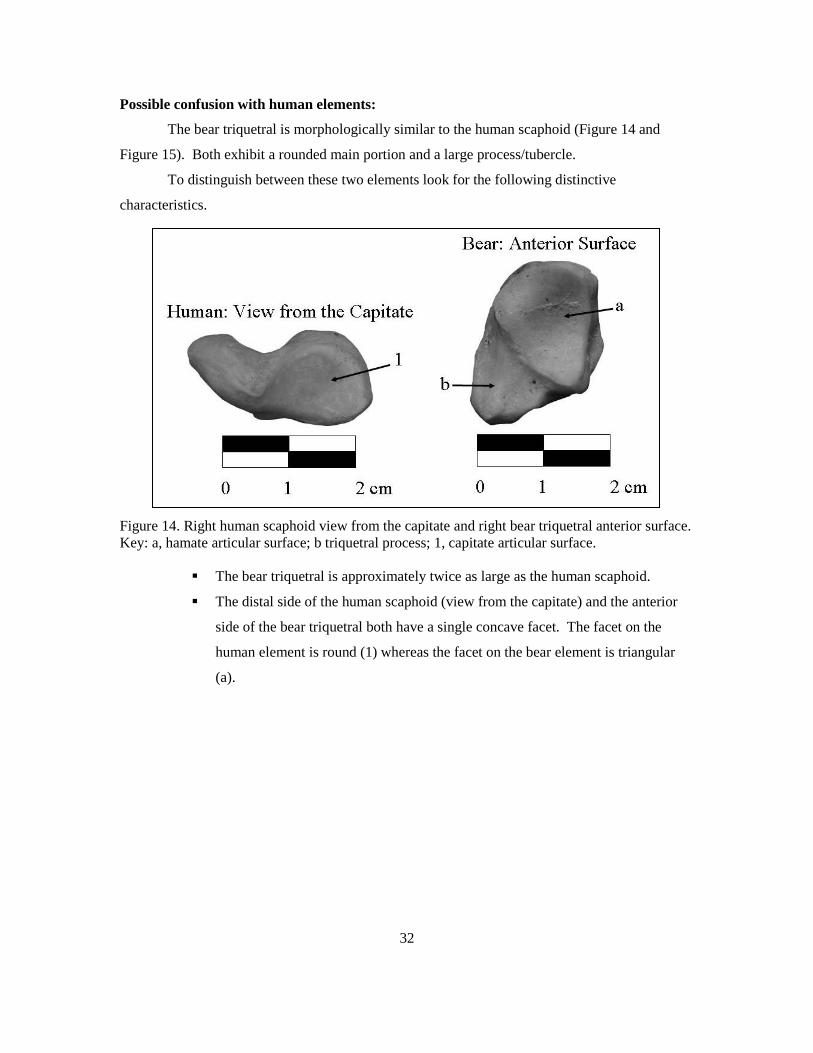
32
Possible confusion with human elements:
The bear triquetral is morphologically similar to the human scaphoid (Figure 14 and
Figure 15). Both exhibit a rounded main portion and a large process/tubercle.
To distinguish between these two elements look for the following distinctive
characteristics.
Figure 14. Right human scaphoid view from the capitate and right bear triquetral anterior surface. Key: a, hamate articular surface; b triquetral process; 1, capitate articular surface.
The bear triquetral is approximately twice as large as the human scaphoid.
The distal side of the human scaphoid (view from the capitate) and the anterior
side of the bear triquetral both have a single concave facet. The facet on the
human element is round (1) whereas the facet on the bear element is triangular
(a).

33
Figure 15. Right human scaphoid view from the radius and right bear triquetral posterior surface. Key: b, triquetral process; c, ulnar articular facet; d, pisiform articular facet; 2, radial facet.
The proximal side of the human scaphoid (view from the radius) and the
posterior surface of the bear triquetral look similar with both exhibiting a large
articular surface. However, the proximal surface on the human scaphoid has a
single facet for the radius (2), which is convex. Whereas the posterior surface of
the bear element has two facets (c and d) and both are concave.

34
Trapezium
The term trapezium stems from the Greek word trapezion which literally means “small
table” (Sandefur 1977). In the generalized mammal form this element is located on the radial
side within the distal row of the carpal block, and is therefore situated between metacarpal I and
the scaphoid. As previously mentioned, this element is not present in the deer.
Bear Trapezium
The bear trapezium is a wedge-shaped bone, located on the medial side of the manus
(Figure 16 and Figure 17). It articulates with the scapho-lunar (posteriorly), the trapezoid
(laterally) and the first metacarpal (anteriorly).
Figure 16. Right bear trapezium anterior and lateral surfaces. Key: a, MC 1 articular surface; b, trapezoid articular surface.
a. The articular surface for MC 1 comprises the entire anterior surface of the element. It is
a large vertical facet that exhibits a slightly concave appearance.
b. The trapezoid articular surface is shaped like a lower case “r” and is located on the
lateral surface of the trapezium.

35
Figure 17. Right bear trapezium posterior and medial surfaces. Key: c, scapho-lunar articular surface.
c. The scapho-lunar articular surface is located on the posterior and medial surfaces. It is
slightly concave and has an elliptical shape.

36
Possible confusion with human elements:
The bear trapezium is very similar in size and morphology to a human lunate (Figure 18).
Morphologically, they both exhibit a crescent shape with concave articular facets.
Figure 18. Right human lunate view from the capitate and right bear trapezium anterior view. Key: 1. capitate articular facet; a. MC 1 articular facet. Despite these similarities the two species can easily be distinguished.
The human lunate is cuboidal in shape while the bear trapezium has a more
flattened and slightly twisted appearance.
The human lunate exhibits a large concave articular facet where it comes into
contact with the capitate (1). This landmark resembles the bear trapezium
articular surface for MC 1 (a). To distinguish between these landmarks note that
the facet on the human element is more concave and exhibits a less distinct
border.

37
Trapezoid
Like the trapezium, the term trapezoid also stems from the Greek word trapezion which
literally means “small table” (Sandefur 1977). In some mammal forms, such as the mule deer,
the trapezoid is fused with the capitate forming an element called the trapezoid-magnum. The
term magnum comes from the Latin word magnus meaning “great” (Sandefur 1977). In the
generalized mammal form the trapezoid is positioned towards the radial side of the distal row of
the carpal block.
Deer Trapezoid-magnum
The deer trapezoid-magnum is positioned on the medial side of the carpal block in the
distal row and is one of the primary weight bearing carpals in the deer (Sandefur 1977) (Figure 19
and Figure 20). It articulates on the dorsal and posterior surfaces with the scaphoid and lunate,
laterally with the unciform, and ventrally with the metacarpal.

38
Figure 19. Right deer trapezoid-magnum ventral and dorsal surfaces. Key: a, MC articular surface; b, scaphoid articular surface; c, lunate articular surface.
a. The MC articular surface comprises most of the ventral surface of the trapezoid
magnum and its shape resembles a backwards “C.”
b. The scaphoid articular surface is located on the medial side of the dorsal surface,
adjacent to the lunate articular surface. It exhibits an “S” shape with a concave anterior
portion and a convex posterior portion.
c. The lunate articular surface is located on the lateral side of the dorsal surface. It is
adjacent to and smaller than the scaphoid articular surface. This landmark is long and
narrow and exhibits a concave anterior portion and a convex posterior portion.

39
Figure 20. Right deer trapezoid-magnum lateral and medial surfaces. Key: d, unciform articular facets; e, medial surface.
d. There are three articular facets for the unciform and they are located on the lateral
surface of the element. All three exhibit a semicircular appearance and they are separated
by a concave roughened area.
e. The medial surface of the trapezoid-magnum is rough with no articular facets; however,
it can be easily identified by its “S” shape.
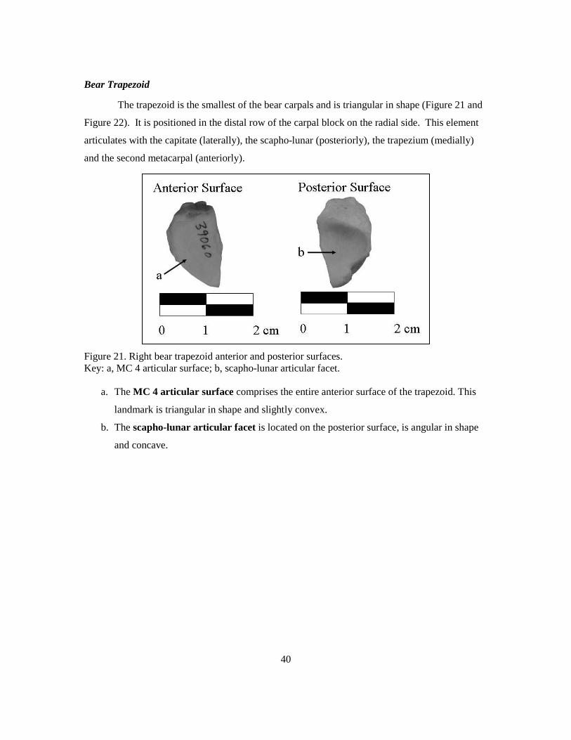
40
Bear Trapezoid
The trapezoid is the smallest of the bear carpals and is triangular in shape (Figure 21 and
Figure 22). It is positioned in the distal row of the carpal block on the radial side. This element
articulates with the capitate (laterally), the scapho-lunar (posteriorly), the trapezium (medially)
and the second metacarpal (anteriorly).
Figure 21. Right bear trapezoid anterior and posterior surfaces. Key: a, MC 4 articular surface; b, scapho-lunar articular facet.
a. The MC 4 articular surface comprises the entire anterior surface of the trapezoid. This
landmark is triangular in shape and slightly convex.
b. The scapho-lunar articular facet is located on the posterior surface, is angular in shape
and concave.

41
Figure 22. Right bear trapezoid lateral and medial surfaces. Key: c, capitate articular facet; d, trapezium articular surface.
c. The capitate articular facet is located on the superior portion of the lateral surface. It is
small and rectangular in shape.
d. The trapezium articular surface is located on the superior portion of the medial surface,
and is small and narrow.
Possible confusion with human elements:
The bear trapezoid is similar in size to the human triquetral and both exhibit an angular
shape (Figure 23). However, the two elements can be easily distinguished as the human triquetral
has a pyramidal shape where as the bear trapezoid has an overall flattened appearance.
Figure 23. Right human triquetral view from the hamate and right bear trapezoid medial surface. Key: d, trapezium articular surface.

42
Capitate
The name of this element stems from the Latin word capitātus which means “having a
head.” This descriptive name refers to the element’s large rounded portion or head where it
articulates with the scaphoid and lunate. The base of the capitate is square, morphology dictated
by its articulation with the third metatarsal. The capitate is located in the distal row of the carpal
block towards the midline of the hand.
Deer Trapezoid-magnum
As previously mentioned the deer capitate is fused with the trapezoid and is commonly
referred to as the trapezoid-magnum. For further discussion see the above trapezoid section.

43
Bear Capitate
The bear capitate lives up to its name, as it possesses a distinctive large rounded head
(Figure 24 and Figure 25). This element articulates with the scapho-lunar (posteriorly), the
hamate (laterally) the trapezoid and fourth metacarpal (medially) and the third metacarpal
(anteriorly). It is located in the distal row of the carpal block, along the midline.
Figure 24. Right bear capitate anterior and posterior surfaces. Key: a, MC 3 articular surface; b, head.
a. The articular surface for the proximal end of MC 3 is a rectangular concave facet that
comprises the entire anterior surface of the capitate. Both its medial and lateral sides
pinch in towards the midline.
b. The head of the capitate articulates with the scapho-lunar (posteriorly). This landmark is
a long and narrow vertically oriented feature with a smooth convex surface.
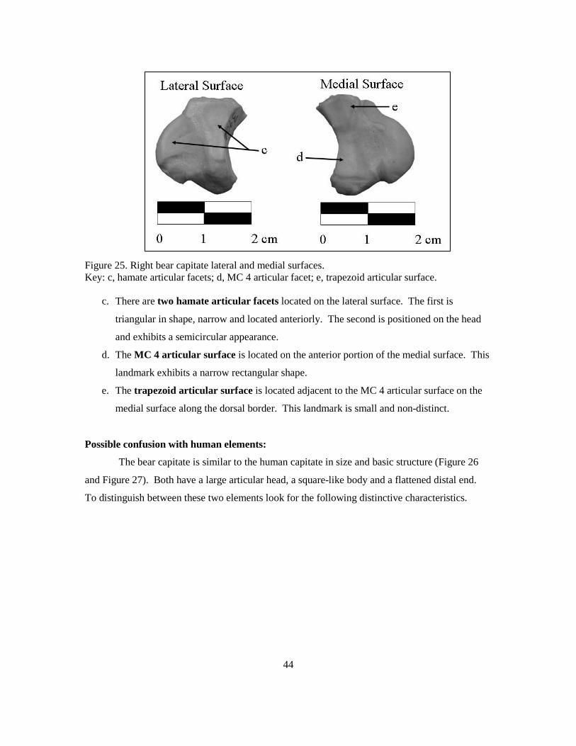
44
Figure 25. Right bear capitate lateral and medial surfaces. Key: c, hamate articular facets; d, MC 4 articular facet; e, trapezoid articular surface.
c. There are two hamate articular facets located on the lateral surface. The first is
triangular in shape, narrow and located anteriorly. The second is positioned on the head
and exhibits a semicircular appearance.
d. The MC 4 articular surface is located on the anterior portion of the medial surface. This
landmark exhibits a narrow rectangular shape.
e. The trapezoid articular surface is located adjacent to the MC 4 articular surface on the
medial surface along the dorsal border. This landmark is small and non-distinct.
Possible confusion with human elements:
The bear capitate is similar to the human capitate in size and basic structure (Figure 26
and Figure 27). Both have a large articular head, a square-like body and a flattened distal end.
To distinguish between these two elements look for the following distinctive characteristics.
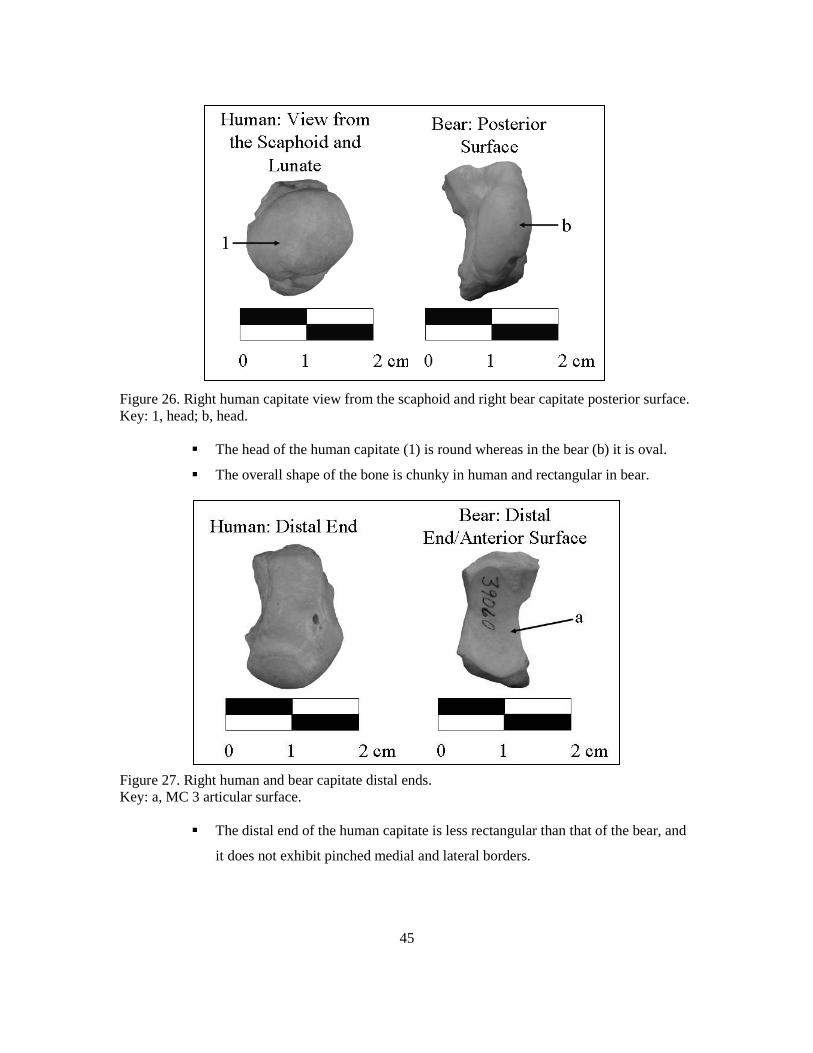
45
Figure 26. Right human capitate view from the scaphoid and right bear capitate posterior surface. Key: 1, head; b, head.
The head of the human capitate (1) is round whereas in the bear (b) it is oval.
The overall shape of the bone is chunky in human and rectangular in bear.
Figure 27. Right human and bear capitate distal ends. Key: a, MC 3 articular surface.
The distal end of the human capitate is less rectangular than that of the bear, and
it does not exhibit pinched medial and lateral borders.

46
Hamate
The hamate, which is also commonly referred to as the unciform, is located in the distal
row of the carpal block. It is named for and easily distinguished by its hamulus or hook-shaped
process (Sandefur 1977; White 2000).
Deer Hamate (Unciform)
The deer unciform is positioned in the distal row of the carpal block on the lateral side
(Figure 28 and Figure 29). It articulates with the lunate and the cuneiform (superiorly), the
trapezoid-magnum (medially) and the metacarpal (inferiorly). The unciform can most easily be
identified by its stair stepped appearance when viewed posteriorly (Figure 28).
Figure 28. Right deer unciform dorsal and posterior surface. Key: a, cuneiform articular surface; b, lunate articular surface.
a. The cuneiform articular surface is located on the lateral portion of the dorsal surface and
continues onto the posterior surface. It is adjacent to the lunate articular surface from
which it is separated by a slight ridge.
b. The lunate articular surface is located on the medial side of the unciform’s dorsal
surface, adjacent to the articular surface for the cuneiform.

47
Figure 29. Right deer unciform ventral and medial surfaces. Key: c, MC articular surface; d, lunate articular facets.
c. The MC articular surface composes the entire ventral surface of the unciform. It is flat
and resembles a kidney bean in shape.
d. There are three articular facets for the lunate located on the medial surface of the
unciform. The two posterior facets are circular where as the anterior facet is more
rectangular.

48
Bear Hamate
The bear hamate is cuboidal in shape and is located on the lateral side of the distal row in
the carpal block (Figure 30 and Figure 31). It articulates with five other elements including the
scapho-lunar (posteriorly), the triquetral (laterally), the capitate (medially) and the fourth and fifth
metacarpals (anteriorly).
Figure 30. Right bear hamate anterior and posterior surfaces. Key: a, MC 4 articular surface; b, MC 5 articular surface; c, triquetral articular surface; d, scapho-lunar articular surface.
a. The MC 4 articular surface composes the medial half of the anterior surface of the
hamate. This facet is slightly concave and adjacent to the MC 5 articular surface, from
which it is separated by a very slight depression.
b. The MC 5 articular surface composes the lateral half of the anterior surface of the
hamate. This facet is slightly concave and adjacent to the MC 4 articular surface.
c. The triquetral articular surface is a large convex facet that can be seen on both the
posterior and lateral surfaces. It is positioned adjacent to the scapho-lunar articular
surface from which it is separated by a groove.
d. The scapho-lunar articular surface can be seen from both the posterior and medial
surfaces. It is large, irregularly shaped and extends inferio-medially.

49
Figure 31. Right bear hamate lateral and medial surfaces. Key: c, triquetral articular surface; d, scapho-lunar articular surface; e, capitate articular surface.
e. The capitate articular facet is positioned along the anterior border of the medial surface.
This landmark is slightly convex and angular.
Possible confusion with human elements:
The overall shape of the bear hamate could be confused with the human cuboid, as both
are very irregular (Figure 32). However, the human cuboid is about twice the size of the bear
hamate and also exhibits a large number of foramina. In addition, the human cuboid has
primarily flat articular facets, where as the facets on the bear hamate are more rounded.
Figure 32. Right human cuboid ventral surface and right bear hamate lateral surface.

50
Pisiform
The pisiform is a sesamoid bone that is often highly developed. In humans it is
approximately the size of a pea and its single articular surface is its primary distinctive feature.
The size and shape of the pisiform varies greatly among mammals.
Deer Pisiform
The deer pisiform is roughly the size and shape of a kidney bean and it articulates with
only one element, the triquetral (anteriorly) (Figure 33). It is situated in the upper region of the
carpal block and is positioned on the posterior side.
Figure 33. Right deer pisiform anterior and medial surfaces. Key: a, triquetral articular surface; b, non-articular body.
a. The triquetral articular surface is slightly convex and circular in shape. It is located on
the anterior surface.
b. The non-articular body of the pisiform is rough in texture and serves as an attachment
site for soft tissue.

51
Possible confusion with human elements:
The deer pisiform could be confused with the human pisiform as they are both small
rounded elements with a single articular surface for the triquetral (Figure 34). However, the deer
pisiform is more elongated and flattened than that of the human.
Figure 34. Right human and deer pisiforms lateral surfaces.

52
Bear Pisiform
The bear pisiform is a large sesamoid bone with an elongated, tubular shape ( Figure 35
and Figure 36). Its most distinctive feature is its bifurcated anterior surface which has two
articular facets for the ulna and triquetral.
Figure 35. Right bear pisiform anterior surface. Key: a, ulnar articular surface; b, triquetral articular surface.
a. The ulnar articular surface is located on the anterior surface, just superior to the
articular facet for the triquetral. It is horizontally oriented and concave. This landmark is
also visible from the dorsal view of the element.
b. The triquetral articular surface is located on the inferior portion of the anterior surface.
It is horizontally oriented, concave and is longer than the ulnar articular facet which it is
adjacent to. This landmark is also visible from the ventral view of the element.
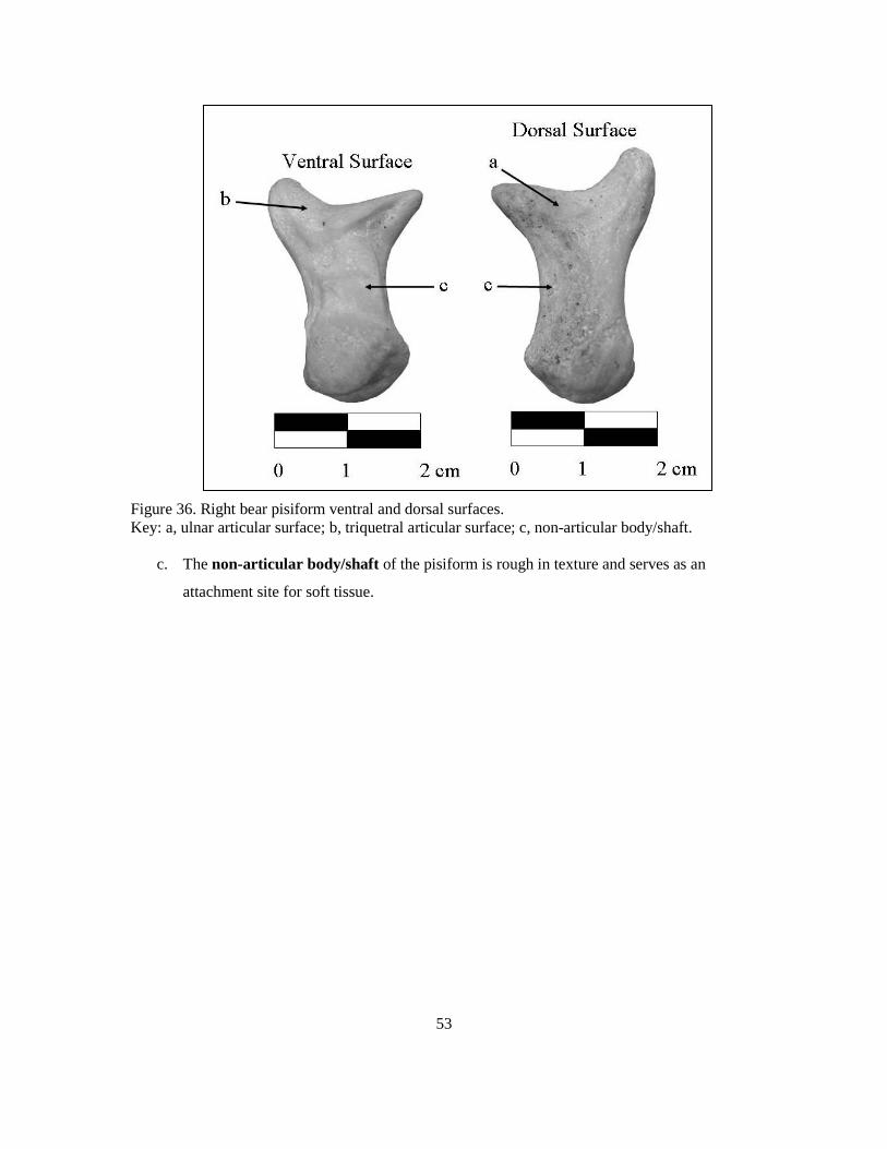
53
Figure 36. Right bear pisiform ventral and dorsal surfaces. Key: a, ulnar articular surface; b, triquetral articular surface; c, non-articular body/shaft.
c. The non-articular body/shaft of the pisiform is rough in texture and serves as an
attachment site for soft tissue.
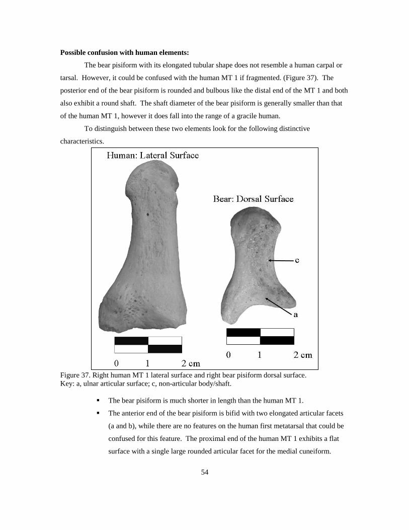
54
Possible confusion with human elements:
The bear pisiform with its elongated tubular shape does not resemble a human carpal or
tarsal. However, it could be confused with the human MT 1 if fragmented. (Figure 37). The
posterior end of the bear pisiform is rounded and bulbous like the distal end of the MT 1 and both
also exhibit a round shaft. The shaft diameter of the bear pisiform is generally smaller than that
of the human MT 1, however it does fall into the range of a gracile human.
To distinguish between these two elements look for the following distinctive
characteristics.
Figure 37. Right human MT 1 lateral surface and right bear pisiform dorsal surface. Key: a, ulnar articular surface; c, non-articular body/shaft.
The bear pisiform is much shorter in length than the human MT 1.
The anterior end of the bear pisiform is bifid with two elongated articular facets
(a and b), while there are no features on the human first metatarsal that could be
confused for this feature. The proximal end of the human MT 1 exhibits a flat
surface with a single large rounded articular facet for the medial cuneiform.

55
CHAPTER V: THE MORPHOLOGY OF THE ANKLE
The mammalian foot (pes) generally has 26 bones, which are divided into three groups:
tarsals, metatarsals and phalanges. The lower hind-limb with which the foot articulates is
composed of the tibia and fibula.
Tarsals are small irregularly shaped bones that form from single primary ossification
centers. The one exception to this rule is the calcaneus, which forms from two (Flower 1885;
Scheuer and Black 2000). Tarsals articulate with each other through synovial joints, resulting in
very limited mobility. Further, the most proximal tarsal, the astragulus articulates with the lower
leg elements (tibia and fibula) and the distal carpals (medial, intermediate, and lateral cuneiforms
and the cuboid) articulate with the metatarsals (MT).
Generalized mammals have five metatarsals, numbered one through five (medial to
lateral) (Flower 1885). Deer however, exhibit a more derived anatomy with the loss of foot
elements typical of the even toed artiodactyls. They exhibit vestigial 2nd and 5th metatarsals and
one large metatarsal formed from the fused 3rd and 4th metatarsals (Cornwall 1964; Flower 1885).
Metatarsals are tubular bones that form from two centers of ossification. All metatarsals have
epiphyses at the distal end except the first, which has an epiphysis at the proximal end. All of the
metatarsals are positioned in a parallel arrangement and have limited mobility between them.
The metatarsals articulate proximally with the tarsals and distally with the corresponding
proximal pedal phalanges.
The phalanges of the foot, like in the hand, comprise five digits. Deer are again an
exception with only four digits and two of which are reduced. Each digit has a proximal, an
intermediate, and a distal (ungual) phalanx. The one exception is the first digit, the hallux, which
is comprised of only two phalanges, a proximal and a distal. Phalanges all form from two
ossification centers with epiphyses at the proximal end.

56
The Tarsals
In the general mammalian form there are a total of seven tarsal bones that are arranged in
two (proximal and distal) transverse rows. The proximal row includes the astragalus (talus in
humans) and the calcaneus. The distal row includes the medial, intermediate, and lateral
cuneiforms and the cuboid. An additional tarsal, the navicular, is positioned between the
proximal and distal rows on the medial side (Figure 38).
Figure 38. Schematic drawing of bear, human and deer right tarsals. Key: A, talus/astragulus; B, calcaneus; C, medial cuneiform; D, intermediate cuneiform; E, lateral cuneiform; F, cuboid; G, navicular/naviculo-cuboid; H, lateral malleolus.
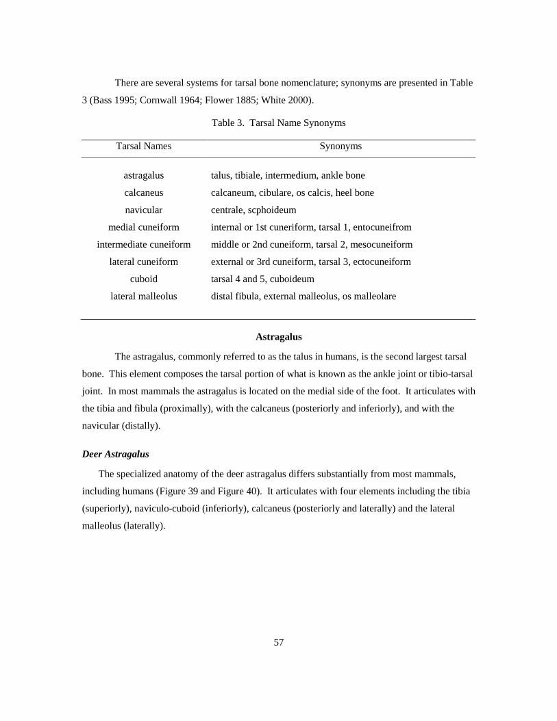
57
There are several systems for tarsal bone nomenclature; synonyms are presented in Table
3 (Bass 1995; Cornwall 1964; Flower 1885; White 2000).
Table 3. Tarsal Name Synonyms
Tarsal Names Synonyms
astragalus talus, tibiale, intermedium, ankle bone
calcaneus calcaneum, cibulare, os calcis, heel bone
navicular centrale, scphoideum
medial cuneiform internal or 1st cuneriform, tarsal 1, entocuneifrom
intermediate cuneiform middle or 2nd cuneiform, tarsal 2, mesocuneiform
lateral cuneiform external or 3rd cuneiform, tarsal 3, ectocuneiform
cuboid tarsal 4 and 5, cuboideum
lateral malleolus distal fibula, external malleolus, os malleolare
Astragalus
The astragalus, commonly referred to as the talus in humans, is the second largest tarsal
bone. This element composes the tarsal portion of what is known as the ankle joint or tibio-tarsal
joint. In most mammals the astragalus is located on the medial side of the foot. It articulates with
the tibia and fibula (proximally), with the calcaneus (posteriorly and inferiorly), and with the
navicular (distally).
Deer Astragalus
The specialized anatomy of the deer astragalus differs substantially from most mammals,
including humans (Figure 39 and Figure 40). It articulates with four elements including the tibia
(superiorly), naviculo-cuboid (inferiorly), calcaneus (posteriorly and laterally) and the lateral
malleolus (laterally).
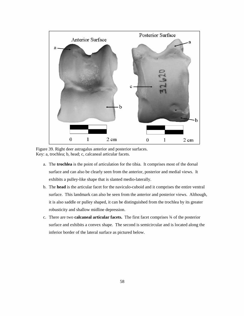
58
Figure 39. Right deer astragalus anterior and posterior surfaces. Key: a, trochlea; b, head; c, calcaneal articular facets.
a. The trochlea is the point of articulation for the tibia. It comprises most of the dorsal
surface and can also be clearly seen from the anterior, posterior and medial views. It
exhibits a pulley-like shape that is slanted medio-laterally.
b. The head is the articular facet for the naviculo-cuboid and it comprises the entire ventral
surface. This landmark can also be seen from the anterior and posterior views. Although,
it is also saddle or pulley shaped, it can be distinguished from the trochlea by its greater
robusticity and shallow midline depression.
c. There are two calcaneal articular facets. The first facet comprises ¾ of the posterior
surface and exhibits a convex shape. The second is semicircular and is located along the
inferior border of the lateral surface as pictured below.

59
Figure 40. Right deer astragalus lateral surface. Key: c, calcaneal articular facet; d, lateral malleolar articular facet.
d. The lateral malleolar articular facet is positioned along the superior portion of the
lateral surface. It exhibits an elongated curved shape.

60
Bear Astragalus
The bear astragalus, unlike that of the deer, exhibits a less derived structure, with a
cubical-shaped body and large anterior projection known as the head (Figure 41 and Figure 42). It
articulates with five elements including the tibia (superiorly), navicular (anteriorly), cuboid
(anterio-laterally), fibula (laterally) and calcaneus (inferiorly).
Figure 41. Right bear astragalus dorsal and ventral surfaces. Key: a, head; b, body; c, neck; d, trochlea; e, calcaneal articular facets; f, sulcus tali.
a. The head is a convex smooth oval-shaped articular surface that projects anteriorly on the
medial side of the bone. It is comprised largely of the navicular articular surface.
b. The body is the square shaped main portion of the element located posterior to the head.
c. The neck is the portion of the element that connects the head and the body.
d. The trochlea is a saddle-shaped articular surface on the dorsal surface of the body. It
articulates with the distal tibia.
e. There are three calcaneal articular facets located on the ventral surface of the body. The
smallest facet is located in the medial posterior corner of the element and it is vertically
oriented. The second is round and flat and is located along the medial border of the
element, just posterior to the head. The largest of the three is located along the lateral
border and is concave and oval in shape.
f. The sulcus tali is a deep groove on the ventral surface that runs between the calcaneal
facets.

61
Figure 42. Right bear astragalus lateral surface. Key: a, head; c, neck; g, fibula articular surface; h, cuboid articular surface.
g. The articular surface for the fibula is located on the lateral side of the body and exhibits
a concave crescent shape.
h. The cuboid articular surface is a flat rounded facet located on the latero-inferior portion
of the head where it meets the neck.

62
Possible confusion with human elements:
The bear astragalus is morphologically similar to the human talus (Figure 43 and Figure
44). Both exhibit a large cuboidal body with a trochlear surface on the superior side. On their
inferior surfaces both exhibit three calcaneal facets. Further, they each have a large articular
head on the medial side of their anterior surfaces and finally, both possess large articular facets
for the fibula on their lateral surfaces.
Figure 43. Right human talus superior surface and right bear astragalus dorsal surface. Key: 1, trochlea; 2; neck; 3, head; a, head; b, body; c, neck; d, trochlea. To distinguish between these two elements look for the following distinctive characteristics.
The human talus is more bulbous than that of the bear. Further, the bear talus
exhibits a more flattened appearance.
The trochlea on the bear astragalus (d) is concave along the mid-sagittal plane,
which is not seen in the human element (1).
The neck of the bear astragalus (c) is more pinched than the neck of the human
talus (2).
The head (3) is horizontally oriented in the bear and round in humans.

63
Figure 44. Right human talus inferior surface and right bear astragalus ventral surface. Key: 4, calcaneal articular facets; a, head; b, body; c, neck; e, calcaneal articular facets; f, sulcus tali.
Both human and bear exhibit three calcaneal articular facets (4 & e) however,
they vary greatly in their positioning.

64
Calcaneus
The calcaneus is generally the largest, longest and most distinct tarsal. In humans this
element is commonly referred to as the heel, while it is called the hock in many non-human
mammals. One of the most notable characteristics of the mammalian calcaneus is the elongated
shape that runs anterior to posterior. This element is also unique because it is the only carpal or
tarsal bone that often has two primary centers of ossification (Scheuer and Black 2000). In
addition, an epiphyseal line can often be seen at the most posterior portion of the bone, which
marks the point of fusion of a secondary ossification center, the calcaneal tuberosity (Flower
1885).
Deer Calcaneus
The deer calcaneus is notably long and narrow compared to humans and many other
mammalian species (Figure 45). The positioning of the calcaneus, in a nearly vertical plane, is
also quite different than that of either bear or human, which results from differences in their
locomotor behaviors.

65
Figure 45. Right deer calcaneus anterior and medial surfaces. Key: a, articular facets for the astragalus; b, lateral malleolar articular facet; c, calcaneal tuberosity; d, naviculo-cuboid articular facet.
a. There are two articular facets for the astragalus, both positioned on the anterior portion
of the element. The first is round, slightly convex and faces inferiorly. It articulates with
the posterior side of the astragalus. The second facet is a long narrow landmark that faces
medially and articulates with the lateral side of the astragalus.
b. The lateral malleolar articular facet is a small bulbous projection located on the lateral
side of the anterior portion of the element.
c. The calcaneal tuberosity is a large non-articular process on the posterior portion of the
bone that serves as an attachment site for the calcaneal tendon.
d. The naviculo-cuboid articular facet exhibits an elongated curved appearance and is
located on the inferior portion of the posterior surface.

66
Possible confusion with human elements:
The deer calcaneus has the same basic morphology as the human calcaneus; both have
roughened and bulbous calcaneal tuberosities, shaft-like bodies and anterior ends exhibiting
several articular surfaces (Figure 46). The views provided of each animal are not the same due to
differences in the species’ anatomical positioning.
Figure 46. Right human calcaneus superior surface and right deer calcaneus anterior surface. Key: 1, calcaneal tuberosity; a, articular facets for the astragalus; b, lateral malleolar articular facet; c, calcaneal tuberosity.
To distinguish between these two elements look for the following characteristics.
The deer calcaneus is much narrower and more angular than the human form.
The calcaneal tuberosity is round in humans (1), while in deer (c) it is oblong and
slightly bifid on its inferior aspect.
The anterior portions of the elements share little resemblance. Note the
differences in the location and shape of their articular facets.
Bear Calcaneus
The bear calcaneus is more robust and shorter than that of the deer (Figure 47). Its
positioning in the body is much like that of the human form, with the astragalus superior to it and

67
the remainder of the tarsals positioned anteriorly. The calcaneus of the bear makes up the heel, as
it does in humans.
Figure 47. Right bear calcaneus dorsal and ventral views. Key: a, sustentaculum tali; b, astragalus articular facets; c, lateral projection; d, cuboid articular surface; e, calcaneal tuberosity.
a. The sustentaculum tali is a triangular shelf-like structure that protrudes from the
element’s medial side. This landmark is primarily responsible for supporting the medial
half of the astragalus and exhibits an articular facet.
b. There are two articular facets for the astragalus. The first (middle articular surface) is a
rounded superior facing feature and is located on the sustentaculum tali. The second
(posterior articular surface) is positioned just anterior to the shaft. This facet is a smooth
convex elongated surface that slopes down from the midline of the bone laterally.
c. The lateral projection is a round, shelf like landmark that serves as an attachment site for
soft tissues.
d. The cuboid articular surface is a large slightly concave rounded facet positioned on the
anterior surface of the element.
e. The calcaneal tuberosity at the posterior end of the bone is a large round billowing
landmark that serves as the point of insertion for the calcaneal tendon.

68
Possible confusion with human elements:
The bear calcaneus is morphologically similar to the human calcaneus, with its
roughened bulbous posterior end, shaft-like main portion and anterior end exhibiting several
articular surfaces (Figure 48).
Figure 48. Right human calcaneus superior view and right bear calcaneus dorsal view. Key: 1, sustentaculum tali; 2, astragalus articular facets; a, sustentaculum tali; b, astragalus articular facets; c, lateral projection; d, cuboid articular facet; e, calcaneal tuberosity.
To distinguish between the bear and human calcaneus look for the following
characteristics.
Overall, the human calcaneus is more bulbous and robust, while the bear calcaneus is
more angular.
In humans, the sustentaculum tali (1) is more vertically oriented, while it is more
horizontal in the bear (a).
The human calcaneus exhibits three articular facets for the talus (anterior, middle and
posterior) (2) whereas the bear form exhibits two distinct facets (middle and posterior)
(b).

69
Navicular
The navicular was originally named for its large proximal articular surface, which
resembles a boat. This element is located in the proximal row of the tarsal block. It articulates
with the cuneiforms (distally), the talus/astragalus (proximally) and the cuboid (laterally). In
some taxa, such as infraorder Pecora (which deer fall into), the navicular and the cuboid are
fused. In such cases, this element is commonly referred to as the naviculo-cuboid (Flower 1885).
Deer Naviculo-cuboid
The deer naviculo-cuboid is a thick square shaped element (Figure 49). It articulates
superiorly with the astragalus and calcaneus and inferiorly with the fused intermediate/lateral
cuneiform, the medial cuneiform and the fused MT.

70
Figure 49. Right deer naviculo-cuboid dorsal, ventral and anterior surfaces. Key: a, articular surface for the astragalus; b, calcaneal articular surface; c, MT articular facets; d, articular facet for the intermediate/lateral cuneiform; e, medial cuneiform articular surface.
a. The articular surface for the astragalus comprises the majority of the dorsal surface.
This landmark has two depressed areas in which the head of the astragalus sits.
b. The calcaneal articular surface is an elongated slightly convex facet located on the
lateral side of the element adjacent to the articular surface for the astragalus.
c. There are two MT articular facets on the ventral surface of the naviculo-cuboid. The
first, located on the lateral portion of the element is tear drop shaped. The second is a
small facet positioned along the lateral side of the posterior border.
d. The articular facet for the fused intermediate/lateral cuneiform is positioned on the
ventral surface in the anterior-medial quadrant. It is oval in shape and recessed.
e. The medial cuneiform articular surface is a very small circular facet located on the
ventral surface just posterior to the articular facet for the intermediate/lateral cuneiform.

71
Bear Navicular
The bear navicular lives up to its name as it is boat-like in appearance (Figure 50).
Positioned on the medial side of the foot, the navicular articulates with the cuboid (laterally), the
astragalus (posteriorly) and the medial, intermediate and lateral cuneiforms (anteriorly).
Figure 50. Right bear navicular anterior, posterior and lateral surfaces. Key: a, medial cuneiform articular surface; b, intermediate cuneiform articular surface; c, lateral cuneiform articular surface; d, articular surface for the head of the astragalus; e, cuboid articular facet.
a. The medial cuneiform articular surface is located on the medial side of the anterior
surface, adjacent to the articular surface for the intermediate cuneiform. This facet is
slightly convex and exhibits a non distinct shape.
b. The intermediate cuneiform articular surface is located on the anterior surface
between the medial and lateral cuneiform articular facets and is the largest of the three.
c. The lateral cuneiform articular surface is located on the lateral side of the anterior
surface, adjacent to the articular surface for the intermediate cuneiform. It is slightly
convex and exhibits a non distinct shape.
d. The articular surface for the head of the astragalus is a large oval concave facet on the
posterior surface of the navicular.
e. The cuboid articular facet is located on the lateral surface and exhibits a narrow
crescent shaped appearance.

72
Possible confusion with human elements:
The bear navicular is morphologically quite similar to the human navicular (Figure 51
and Figure 52). In both cases the element has a flattened appearance with a large singular facet
on the posterior side and an articular surface on the anterior side that can be divided into three
facets. To distinguish between these two elements look for the following distinctive
characteristics.
Figure 51. Right human navicular anterior surface and right bear navicular anterior surface. Key: 1, medial cuneiform facet; 2, lateral cuneiform facet; 3, intermediate cuneiform facet; a, medial cuneiform articular surface; b, intermediate cuneiform articular surface; c, lateral cuneiform articular surface. The human navicular is significantly larger and thicker than that of the bear.
The human navicular is oval, whereas the bear navicular is triangular.
The articular facets on the anterior side of the human navicular are more distinct than
those of the bear. In human, the lateral and medial cuneiform facets (1 and 2) are round
and the facet for the intermediate cuneiform (3) is triangular.

73
Figure 52. Right human navicular posterior surface and right bear navicular posterior surface. Key: 4, articular surface for the head of the talus; 5, tubercle; d, articular surface for the head of the talus. The posterior surface of both elements exhibits an oval-shaped facet for the head of the
talus (4 and d). In the human form however, a large distinct tubercle (5) is present just
medial to this landmark.

74
Medial Cuneiform
The term cuneiform stems from the Latin word cuneus, which means wedge-form. This
descriptive term holds true for all three of the cuneiform bones of the foot. Of these, the medial
cuneiform is the largest. It is positioned on the medial side of the tarsal block in the distal row. It
articulates with the navicular (proximally), the intermediate cuneiform (laterally) and with the
first metatarsal (distally).
Deer Medial Cuneiform
The deer medial cuneiform is spherical in form and noticeably small, measuring less than
1 cm in diameter (Figure 53). Due to this small size, it is often missing in comparative
collections. Within the tarsal block this element is positioned on the medial side of the distal
row, just posterior to the fused intermediate/lateral cuneiform. It exhibits a single articular facet
where it contacts the naviculo-cuboid (superiorly).
Figure 53. Right deer medial cuneiform dorsal and medial surfaces. Key: a, naviculo-cuboid articular surface.
a. The naviculo-cuboid articular surface covers the entire dorsal surface of the medial
cuneiform. This landmark can be identified by its small round shape.

75
Possible confusion with human elements:
The deer medial cuneiform is very similar to the human pisiform, a sesamoid bone
located in the hand (Figure 54). In both cases the elements are small and round and exhibit a
singular round articular surface.
Figure 54. Right human pisiform lateral surface and right deer medial cuneiform medial surface.
The human pisiform can be easily distinguished from the deer medial cuneiform based on
its larger size.

76
Bear Medial Cuneiform
The bear medial cuneiform is shaped like a parallelogram and oriented diagonally on the
medial side of the foot (Figure 55). It articulates with three elements including the first
metatarsal (anteriorly), the navicular (posteriorly) and the intermediate cuneiform (laterally).
Figure 55. Right bear medial cuneiform anterior, posterior and lateral surfaces. Key: a, MT 1 articular surface; b, navicular articular surface; c, intermediate cuneiform articular facet.
a. The MT 1 articular surface covers the entire anterior surface of the medial cuneiform.
This facet is concave and similar in size and shape to a kidney bean.
b. The navicular articular surface covers the entire posterior surface of the element. It is
slightly concave and is narrower than the MT 1 articular facet.
c. The intermediate cuneiform articular facet is a small rounded landmark located on the
anterior border of the element’s lateral surface.

77
Intermediate Cuneiform
The intermediate cuneiform is the smallest of the three cuneiform bones and is located
between the other two. It articulates with the navicular (proximally), the medial cuneiform
(medially), with the lateral cuneiform (laterally) and with the second metatarsal (distally). In
some animals, such as in the group Pecora, an infraorder of the order Artiodactyla, the
intermediate cuneiform and the lateral cuneiform are fused. For lack of a better term, this
element is referred to herein as the intermediate/lateral cuneiform. Common animals that fall
within this grouping include: deer, sheep, goats and cattle.
Deer Intermediate/Lateral Cuneiform
The deer intermediate/lateral cuneiform is the size and shape of a kidney bean (Figure
56). It articulates with two elements including the naviculo-cuboid (superiorly) and the fused MT
(inferiorly). The other surfaces are void of articular facets.
Figure 56. Right deer intermediate/lateral cuneiform dorsal, ventral and medial surfaces. Key: a, naviculo-cuboid articular surface; b, MT articular surface.
a. The naviculo-cuboid articular surface covers almost the entire dorsal surface of the
element. It is concave and kidney bean shaped.
b. The articular surface for the fused MT is positioned on the ventral surface. It is very
similar in size and shape to the naviculo-cuboid articular surface with the exception of
being convex.

78
Possible confusion with other elements:
The deer intermediate/lateral cuneiform does not resemble the human cuneiforms due in
large part to its block-like shape (Figure 57). However, there are morphological similarities
between the deer intermediate/lateral cuneiform and the bear intermediate cuneiform. They share
a common overall shape and size, as well as a small boney projection. To distinguish between
these two elements look for the following distinctive characteristics.
Figure 57. Right bear intermediate cuneiform medial and lateral surfaces and right deer intermediate/lateral cuneiform medial surface. The bear intermediate cuneiform exhibits articular facets on its medial and lateral sides,
which are not seen in deer.

79
Bear Intermediate Cuneiform
The intermediate cuneiform is the smallest of the three cuneiforms in the bear and is
shaped like a flattened rectangle, with a small projection on its inferior medial corner (Figure 58
and Figure 59). It articulates with four elements including the navicular (posteriorly), the lateral
cuneiform (laterally), the second metatarsal (anteriorly) and the medial cuneiform (medially).
Figure 58. Right bear intermediate cuneiform anterior and posterior surfaces. Key: a, MT 2 articular facet; b, navicular articular facet.
a. The MT 2 articular facet comprises most of the intermediate cuneiforms anterior
surface. This landmark is slightly concave and rectangular in shape.
b. The navicular articular facet comprises most of the posterior surface of the intermediate
cuneiform and looks very similar to the MT 2 articular facet. The navicular articular
facet however, is more concave.

80
Figure 59. Right bear intermediate cuneiform medial and lateral surfaces. Key: c, medial cuneiform articular facet; b, lateral cuneiform articular facet
c. The medial cuneiform articular facet is semicircular and is located on the inferior
portion of the medial surface along the posterior border.
d. The lateral cuneiform articular facet is a non-distinct elongated landmark positioned
along the lateral side of the element.
Possible confusion with other elements:
The bear intermediate cuneiform does not resemble a human tarsal due to its diminutive
size and block-like shape. The human cuneiforms are all wedge-shaped and generally larger.
However, there are morphological similarities between the bear intermediate cuneiform and the
deer intermediate/lateral cuneiform. For further discussion on how to distinguish between these
elements see the above deer intermediate/lateral cuneiform section.

81
Lateral Cuneiform
In the generalized mammal form the lateral cuneiform is larger than the intermediate
cuneiform and smaller than the medial cuneiform. It is positioned in the distal row of the tarsal
block between the intermediate cuneiform and the cuboid. The lateral cuneiform articulates with
the navicular proximally and with the third metatarsal distally.
Deer Intermediate/ lateral Cuneiform
As previously mentioned, the intermediate and lateral cuneiforms are fused in the deer.
For further discussion of this element see the intermediate cuneiform section above.
Bear Lateral Cuneiform
The lateral cuneiform is the largest of the three cuneiforms in the bear foot (Figure 60 and
Figure 61). It is located on the lateral side of the tarsal block and is oriented diagonally. The
lateral cuneiform articulates with four elements including the navicular (posteriorly), the cuboid
(laterally), the intermediate cuneiform and the second metatarsal (medially) and with the third
metatarsal (anteriorly).
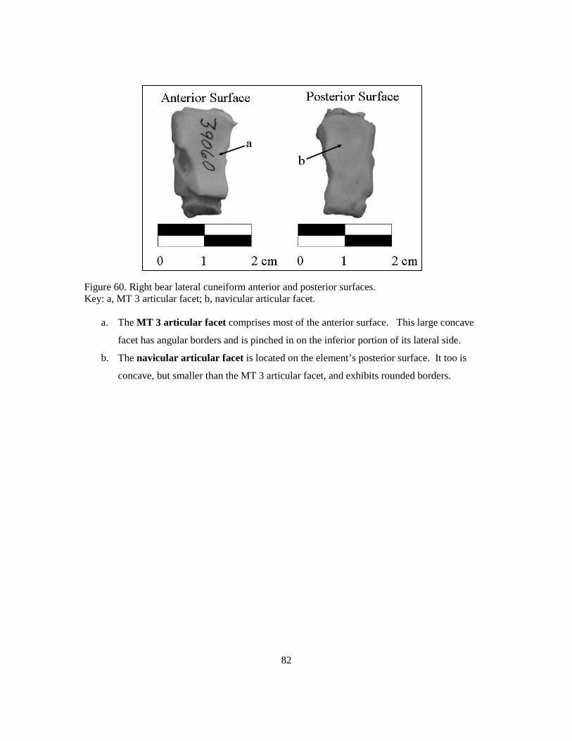
82
Figure 60. Right bear lateral cuneiform anterior and posterior surfaces. Key: a, MT 3 articular facet; b, navicular articular facet.
a. The MT 3 articular facet comprises most of the anterior surface. This large concave
facet has angular borders and is pinched in on the inferior portion of its lateral side.
b. The navicular articular facet is located on the element’s posterior surface. It too is
concave, but smaller than the MT 3 articular facet, and exhibits rounded borders.

83
Figure 61. Right bear lateral cuneiform lateral and medial surfaces. Key: c, three cuboid articular facets; d, MT 2 articular facets; e, intermediate cuneiform articular facet.
c. There are three cuboid articular facets on the lateral surface. Two of the facets are
located along the elements’ anterior border and both are generally semi-circular in shape.
The third and largest facet is convex, kidney bean shaped, and located along the
element’s posterior border.
d. There are two MT 2 articular facets positioned on the elements’ medial surface. Both
are similar in size and are positioned along the elements anterior border.
e. The intermediate cuneiform articular facet on the posterior border of the medial
surface is long and narrow in shape.
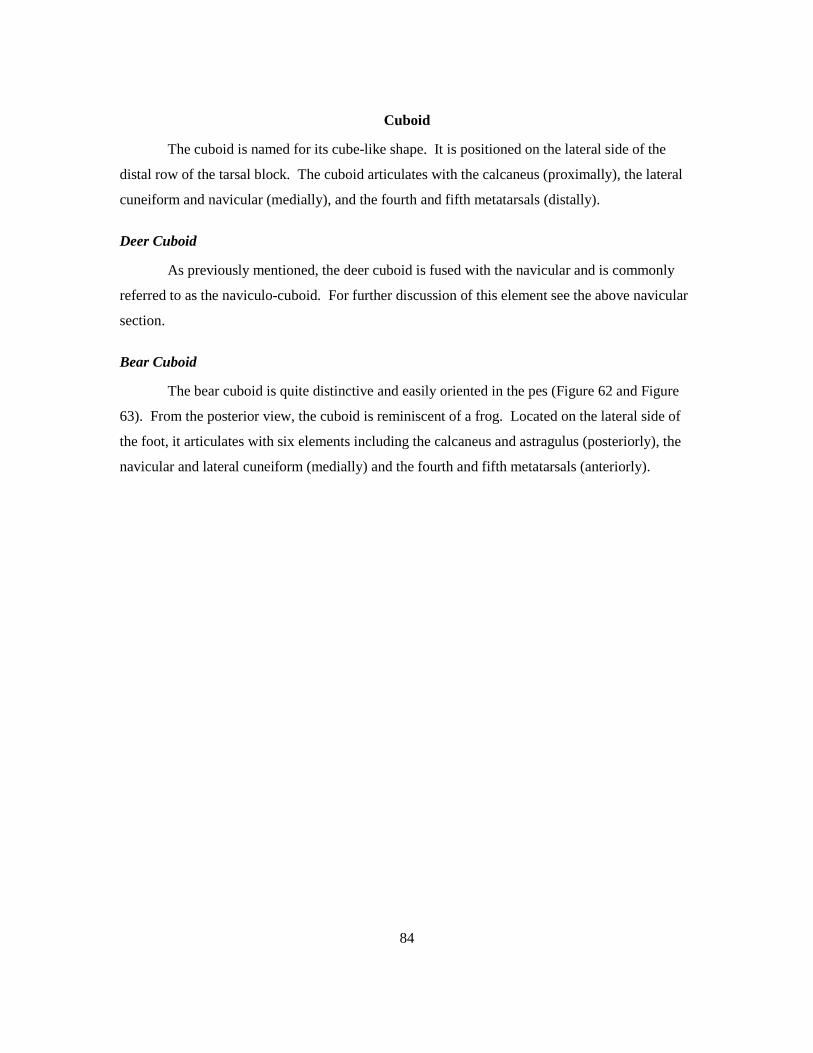
84
Cuboid
The cuboid is named for its cube-like shape. It is positioned on the lateral side of the
distal row of the tarsal block. The cuboid articulates with the calcaneus (proximally), the lateral
cuneiform and navicular (medially), and the fourth and fifth metatarsals (distally).
Deer Cuboid
As previously mentioned, the deer cuboid is fused with the navicular and is commonly
referred to as the naviculo-cuboid. For further discussion of this element see the above navicular
section.
Bear Cuboid
The bear cuboid is quite distinctive and easily oriented in the pes (Figure 62 and Figure
63). From the posterior view, the cuboid is reminiscent of a frog. Located on the lateral side of
the foot, it articulates with six elements including the calcaneus and astragulus (posteriorly), the
navicular and lateral cuneiform (medially) and the fourth and fifth metatarsals (anteriorly).

85
Figure 62. Right bear cuboid anterior and ventral surfaces. Key: a, MT 5 articular surface; b, MT 4 articular surface; c, cuboid groove; d, cuboid tuberosity.
a. The MT 5 articular surface on the lateral side of the anterior surface is a small round
facet that is positioned adjacent to the articular surface for MT 4.
b. The MT 4 articular surface is located on the medial side of the anterior surface. It is
rectangular in shape and is larger than the articular surface for MT 5 to which it is
adjacent.
c. The cuboid groove is located on the anterior surface just inferior to the articular facets for
MT4 and MT 5. It runs at a slight diagonal from the medial to lateral border.
d. The cuboid tuberosity is located on the ventral surface. This large billowing surface
serves as a soft tissue attachment site.

86
Figure 63. Right bear cuboid posterior and medial surfaces. Key: e, calcaneal articular surface; f, astragalus articular surface; g, navicular articular surface; h, lateral cuneiform articular facet.
e. The calcaneal articular surface is rectangular in shape and slightly convex. It is located
on the dorsal surface of the element.
f. The articular surface for the astragalus is located on the element’s dorsal and medial
surfaces. It is small, triangular in shape and slightly concave.
g. The navicular articular surface is located on the medial side of the element just inferior
to the articular surface for the astragalus. It exhibits a circular shape.
h. The lateral cuneiform articular facet is located on the medial surface just anterior to the
navicular articular surface.

87
Possible confusion with human elements:
The bear cuboid and the human cuboid are both chunky and cuboidal, but beyond this
basic characteristic they share little resemblance to each other (Figure 64).
Figure 64. Right human cuboid ventral surface and right bear cuboid ventral surface. Key: d, cuboid tuberosity. The human cuboid is approximately twice as large as the bear cuboid and their facets
differ greatly.

88
Lateral Malleolus
In many mammals the tibia and fibula are fused. In such cases however, the fibula never
becomes completely unrecognizable. For example, in the group ruminatia a suborder of the order
Artiodactyla the only recognizable part of the fibula is its most distal portion, which is known as
the lateral malleolus (Klein and Cruz-Uribe 1984; Parker 1907). In this form the lateral malleolus
is a small irregular element located lateral to the tarsal block. This element articulates with the
tibia, the astragalus and the calcaneus. Though this element is technically not a tarsal it has been
included due to its small irregular size, position adjacent to the tarsal block and its potential of
being confused with wrist or ankle elements of other animals.
Deer Lateral Malleolus
The deer lateral malleolus is a narrow irregular bone (Figure 65 and Figure 66). It is
located on the lateral side of the tarsal block and articulates with the tibia (superiorly), the
calcaneus (inferiorly) and the astragalus (medially).
Figure 65. Right deer lateral malleolus medial and ventral surfaces. Key: a, astragalus articular surface; b, calcaneal articular surface.
a. The astragalus articular surface is located on the medial surface of the element. It
exhibits a large curved groove in which the astragalus sits.
b. The calcaneal articular surface is an oval shaped concave facet that comprises the
majority of the ventral surface.
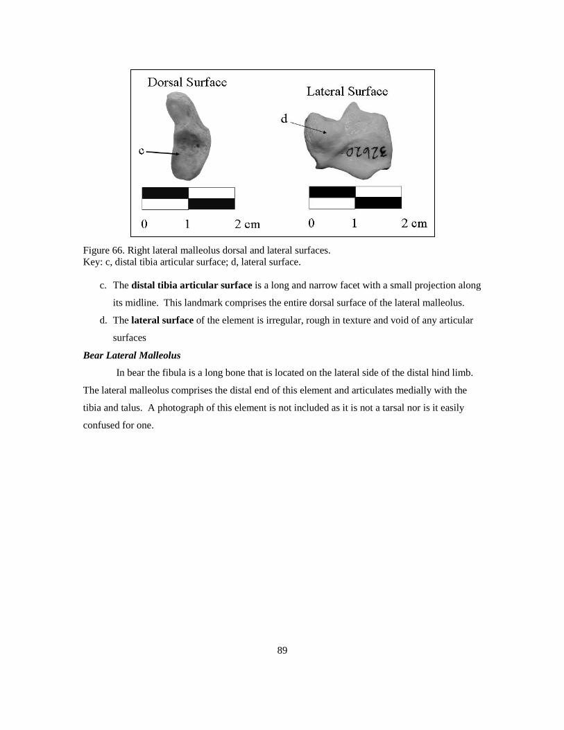
89
Figure 66. Right lateral malleolus dorsal and lateral surfaces. Key: c, distal tibia articular surface; d, lateral surface.
c. The distal tibia articular surface is a long and narrow facet with a small projection along
its midline. This landmark comprises the entire dorsal surface of the lateral malleolus.
d. The lateral surface of the element is irregular, rough in texture and void of any articular
surfaces
Bear Lateral Malleolus
In bear the fibula is a long bone that is located on the lateral side of the distal hind limb.
The lateral malleolus comprises the distal end of this element and articulates medially with the
tibia and talus. A photograph of this element is not included as it is not a tarsal nor is it easily
confused for one.

90
CHAPTER VI: CONCLUSIONS
The most effective way to accurately identify skeletal elements is through the use of a
comparative collection. This method is not feasible for all projects; for this reason a
comprehensive guidebook is the best alternative. Through the presentation of multiple views of
an element (either drawings or photographs) as well as written descriptions, key morphological
characteristics on even the smallest elements can be clearly distinguished. This is imperative as
many elements of different species share size and morphological similarities. Furthermore, in
archaeological sites where elements are often incomplete the ability to identify distinguishing
landmarks is critical.
Many archaeologists are not experts in the identification of mammal and human bones
and are forced to rely on osteological guidebooks when skeletal elements are encountered. Wrist
and ankle elements are often overlooked in such guidebooks, which is unfortunate as their shape,
density and lack of grease and marrow make them likely to preserve in archaeological sites. In an
attempt to fill this gap in the literature, this thesis has provided a detailed description of the
distinguishing features of the carpals and tarsals of two mammals commonly found in
archaeological sites, as well as highlighted the elements that may be confused with
morphologically similar human elements. As described above four of the deer elements
(triquetral, pisiform, calcaneus and medial cuneiform) and eleven of the bear elements (scaphoid,
triquetral, trapezium, trapezoid, capitate, hamate, pisiform, astragalus, calcaneus, navicular and
cuboid) share many similar features with their human counterparts, and could potentially be
confused by the non-specialist.
This thesis was a learning process in regards to both the osteological content and the
methods involved in constructing a useable guidebook. The format of this work went through
many revisions before this final draft was completed. Originally side by side photographs of the
different species were not included, which made visual comparisons more difficult. In addition,
the inclusion of a key directly beneath each series of photographs made landmark identification
easier. Also, each bone was originally photographed on a black background which was replaced
with the white background in the current version. This change greatly enhanced the clarity and
contrast of the landmarks on each bone. These revisions are listed here to assist others in the
construction of future osteological guides.
The field of archaeology would benefit greatly from the addition of a variety of
osteological guides. Specifically, pictures and descriptions that focus on smaller elements of the

91
hand, foot, wrist, and ankle would be a great contribution. Through personal communication with
physical anthropologists it has become clear that Phoca vitulina (harbor seal), Castor canadensis
(beaver), Canis familiaris (dog), (Ovis canadensis) mountain sheep, and Cervus elaphus (wapiti)
may also be useful in a comparative mammalian osteological guide because of their ubiquity and
morphological similarity to the human skeleton. In addition, some animals introduced to North
America during historic times such as the pig (Sus domestica) share osteological similarities and
overlap in size with human and the indigenous fauna. The inclusion of such animals in
osteological guides would assist the archaeologist in both correctly identifying the specimen and
in determining the presence of an historic component. Finally, relatively few osteological works
provide information on non-human sub-adults making their positive identification difficult. This
is unfortunate as immature elements of domesticated animals such as dogs are commonly found
in archaeological sites and are difficult to differentiate from human (Alyson Rollins, personal
communication 2009; Davis 1987; Nokes 2004; Reitz and Wing 2008)
The above thesis, in guidebook form, is intended to fill a gap in osteological literature
and to reduce the number of bones that are considered unidentifiable, which often results in their
omission from site reconstruction. It also provides archaeologists with a means of distinguishing
between human and faunal elements so that they can manage the remains appropriately.

92
REFERENCES
Ames, Kenneth, M. 1994 The Northwest Coast: Complex Hunters-Gatherers, Ecology and Social Evolution.
Annual Review of Anthropology 23: 209-229. Ames, Kenneth, M. and Herbert D.G. Maschner 1999 Peoples of the Northwest Coast: Their Archaeology and Prehistory. Thames and Hudson
Ltd, London. Arden, Harvey 1989 Who Owns Our Past? National Geographic 175(3):376-393. Aslan, Andres and Anna K. Behrensmeyer 1996 Taphonomy and Time Resolution of Bone Assemblages in a Contemporary Fluvial
System: The East Fork River, Wyoming. Palaios 11(5): 411-421. Baker, Brenda J., Tosha L. Dupras and Matthew W. Tocheri 2005 Osteology of Infants and Children. Texas A & M University Press, College Station, Texas. Bass, William M. 1995 Human Osteology: A Laboratory and Field Manual. 4th ed. Missouri Archaeological
Society, Columbia, Missouri. Behrensmeyer, Anna K. 1978 Taphonomy and ecologic information from bone weathering. Paleobiology 4(2): 150-
162. Behrensmeyer, Anna K., Susan M. Kidwell and Robert A. Gastaldo 2000 Taphonomy and Paleobiology. Paleobiology 26(4): 103-147. Bidmos, Mubarak 2006 Adult Stature Reconstruction From the Calcaneus of South Africans of European
Descent. Journal of Clinical Forensic Medicine 13(5)247-252. Bidmos, Mubarak A. and Samuel A. Asala 2003 Discriminant Function Sexing of the Calcaneus of The South African Whites. Journal of
Forensic Sciences 48(6)1213-1218. 2004 Sexual Dimorphism of the Calcaneus of South African Blacks. Journal of Forensic
Sciences 49(3):446-50. Binford, Lewis R. 1978 Nunamiut Ethnoarchaeology. Academic Press, New York

93
Boxberger, Daniel 2009 Objectivity and Reality in Science of the American Indian: Lessons learned from the
Ancient One. Presented at Western Washington University: Turning Points Faculty Speaker Series, Bellingham, WA.
Brown, Christopher L., and Carl E. Gustafson 1990 A Key to Postcranial Skeletal Remains of Cattle/Bison, Elk and Horse. Reports of
Investigations, No. 57. Department of Anthropology, Washington State University, Pullman. Bruning, Susan 2006 Complex Legal Legacies: The Native American Graves Protection and Repatriation Act,
Scientific Study and Kennewick Man. American Antiquity. 71(3):501-521. Butler, Virgina L. and Sarah K. Campbell 2006 Northwest Coast and Plateau Animals. In Environment, Origins and Population, edited by Douglas H. Ubelaker, pp. 263-273. Handbook of North American Indians, Vol. 3, William C. Sturtevant, general editor, Smithsonian Institution, Washington, D.C. Cameriere, Roberto and Luigi Ferrante 2008 Age Estimation in Children by Measurement of Carpals and Epiphyses of Radius and
Ulna Open Apices in Teeth: A Pilot Study. Forensic Science International 147(1) 60-3. Cameriere, Roberto, Luigi Ferrante, Branko Ermenc and Dora Mirtella 2007 Age Estimation Using Carpals: Study of a Slovenia Sample to Test Cameriere’s Method.
Forensic Science International 174(2-3) 178-81. Cannon, Aubrey 1991 The Economic Prehistory of Namu: Patterns in Vertebrate Fauna. Archaeology Press, Simon Fraser university, Burnaby, B.C. Ceci, L. 1984 Shell Midden Deposits as Coastal Resources. World Archaeology 16:62-74. Clemente, Carmine D. (ed.) 1985 Gray’s Anatomy. 30th ed. Lea and Feibiger, Philadelphia. Chatters, James C. 2000 The Recovery and First Analysis of an Early Holocene Human Skeleton from
Kennewick, Washington. American Antiquity 65(2): 291-316 Cornwall, I.W. 1964 Bones for the Archaeologist. Phoenix House, London. Darwent, Christyann M. and R. L. Lyman 2001 Detecting the Postburial Fragmentation of Carpals, Tarsals and Phalanges. In Advances
in Forensic Taphonomy: Method Theory and Archaeological Perspective, edited by William D. Haglund and Marcella H. Sorg. pp. 355-378. Boca Raton, Fl.

94
Davis, Simon J.M. 1987 The Archaeology of Animals. Yale University Press, New Haven. Davila, Florangela 1999 Battle over Semiahmoo Bones: Lummi Spiritual Heritage Vies with Modern Municipal
Necessity on a Spit of Land in Blaine. The Seattle Times 19 December. Seattle, Washington.
Digital Morphology Group and University of Texas High Resolution X-Ray Computed Tomography Facility (UTCT) 2002 Digital Morphology: A National Science Foundation Digital Library at the University of
Texas at Austin. electronic document, http://digimorph.org/. accessed April 12, 2009. Driver, Jonathan 1993 Zooarchaeology in British Columbia. BC Studies 99: 77-105. Flower, William Henry 1885 An Introduction to the Osteology of the Mammalia. 3rd ed. MacMillan and Co., London. Gilbert, B. Miles 1990 Mammalian Osteology. Missouri Archaeological Society, Columbia, Missouri. Gordon, C.C. and J. E. Buikstra 1981 Soil Ph, bone preservation and sampling bias at mortuary sites. American Antiquity
46:566-571. Haeberlin, Hermann and Erna Gunther 1930 Indians of Puget Sound. University of Washington Press, Seattle, Washington. Haglund, William D. and Marcella H. Sorg (editors) 1997 Forensic Taphonomy: The Post Mortem Fate of Human Remains. CRC Press, Boca
Raton, Florida. Hodgetts, Lisa and Farid Rahemtulla 2001 Land and Sea: Use of Terrestrial Mammal Bones in Coastal Hunter-Gatherer Communities. Antiquity 75(287): 56-62. Hsieh, Chi-Wen, Tai-Lang Jong and Chui Mei Tiu 2007 Bone Age Estimation Based on Phalanx Information With Fuzzy Constrain of Carpals.
Medical and Biological Engineering and Computing 45(3):283-295. Huelsbeck, David R. 1994 Mammals and Fish. In The Subsistence Economy of Ozette. In
Ozette Archaeology Project Research Reports, Volume II, Fauna. Edited by Samuels, Stephan R., pp.17-91. Reports of Investigation 66. Department of Anthropology, Washington State University, Pullman, and the National Park
Service, Pacific Northwest Regional Office, Seattle.

95
Hutt, Sherry 1998 Native American Cultural Property Law: Human Rights Legislation. electronic
document, http://www.myazbar.org/AZAttorney/Archives/Jan98/1-98a3.htm. accessed April 16, 2009.
Imamoto, Shirley 1976 Chapter 2: An Analysis of the Glenrose Faunal Remains. In The Glenrose Cannery Site, edited by R.G. Matson, pp 21-41. National Museum of Man Mercury Series:
Archaeological Survey of Canada Paper No. 52, Ottawa, Canada. Kappleman, John 2008 The eSkeletons Project. electronic document, http://www.eskeletons.org/. accessed
April 12, 2009. Kasper, Jan, C. 1980 Skeletal Identification of California Sea Lions and Harbor Seals for Archaeologists. Ethnic Technology Notes No. 17. San Diego Museum of Man, San Diego. King, Thomas F. 2008 Cultural Resource Laws and Practice 3rd ed. Altamira Press, Lanham, Maryland. Klepinger, Linda L. 2006 Fundamentals of Forensic Anthropology. John Wiley & Sons, Inc., Hoboken, New Jersey. Klein, Richard G. and Kathryn Cruz-Uribe 1984 The Analysis of Animal Bones from Archaeological Sites. The University of Chicago Press, Chicago. Livingston, Stephanie 1985 Faunal Analysis. In Archaeological Investigations at Site 45OK-258, Chief Joseph Dam
Project, Washington. Edited by Manfred E.W. Jaehnig, pp.117-132. Submitted to U.S. Army Corps of Engineers, Seattle District, Contract No. DACW67-78-C-0106. Copies available from U.S. Army Corps of Engineers, Seattle District.
Lyman, R. L. 1978 Prehistoric Butchering Techniques in the Lower Granite Reservoir, Southeastern
Washington. Tebiwa. Miscellaneous Paper of the Idaho State University Museum of Natural History 13. Pocatello. 1994 Vertebrate Taphonomy. Cambridge University Press, New York. 1995 Mammalian and Avian Zooarchaeology of the West Point, Washington Archaeology Sites (45KI428 and 45KI429). In The Archaeology of West Point, Seattle, Washington 4,000 Years of Hunter-Fisher-Gatherer Land Use in Southern Puget Sound. Edited by Lynn L. Larson and Dennis E. Lewarch, pp. 9- 1 to 9-52. Submitted to King County Department of Metropolitan Services.

96
Mackie, R.J., Kenneth L. Hamlin and David F. Pack 1982 Chapter 44: Mule Deer (Odocoileus hemionus). In Wild Mammals of North America: Biology, Management and Economics. edited by Joseph A. Chapman and George A. Feldhamer, pp. 862-877. The Johns Hopkins University Press, Baltimore, Maryland. Meany, Edmond Stephen 1942 Vancouver's discovery of Puget Sound. Binfords & Mort, Portland, Oregon. Montgomery, Jackie A. 1979 Prehistoric Subsistence at Semiahmoo Spit 45WH17. Unpublished Masters Thesis, Department of Anthropology, Western Washington University, Bellingham. Nokes, Randolph David 2004 Mid-Holocene Terrestrial Animal Use In the Gulf of Georgia Region: A Case
Study from The Ferndale Site, Lower Nooksack River, Washington. Unpublished Masters Thesis, Department of Anthropology, Western Washington University, Bellingham.
Nowak, Ronald and John L. Paradiso 1983 Walker’s Mammals of the World, Vol 2. 4th ed. The Johns Hopkins University Press, Baltimore, Maryland. Orcholl, Jackie L. and Jean Hudson 2001 Diagnostic Criteria for the Comparison of Human and American Black Bear Skeletal
Elements. Electronic document, http://www.uwm.edu/Dept/Grad_Sch/McNair /2001/jackieorcholl.htm>, accessed 2006.
Our Media: Channels of Creativity n.d. Do-it-Yourself Light Box, http://www.ourmedia.org/node/253481, accessed November 2006. Owsley, Douglas W. and Robert W. Mann 1990 Medicolegal Case Involving A Bear Paw. Journal of the American Podiatric Medical Association. 80:623-625. Parker, W.N. (editor) 1907 Comparative Anatomy of Vertebrates. 3rd ed. Macmillan and Co., London Pelton, Michael R. 1982 Black Bear (Ursus americanus). In Wild Mammals of North America: Biology,
Management and Economics. edited by Joseph A. Chapman and George A. Feldhamer, pp. 504-514. The Johns Hopkins University Press, Baltimore, Maryland
Post, Lee 2004a Building Bear Bones: A Guide to Preparing and Assembling a Bear Skeleton or
Plantigrade Possibilities. Vol 7. Lee Post. 2004b Pinniped Projects: Articulating Seal and Sea Lion Skeletons. Vol. 4. Lee Post.

97
Reitz, Elizabeth J. and Elizabeth S. Wing 2008 Zooarchaeology. 2nd ed. Cambridge University Press, Cambridge. Riley, Shawn J. and Alix Marcoux 2006 Deer-Vehicle Collisions: An Understanding of Accident Characteristics and Drivers
Attitudes, Awareness and Involvement. Department of Fisheries and Wildlife, Michigan State University. Submitted to Michigan Department of Transportation Construction & Technology Research Administration. Copies available from the Michigan Department of Transportation, Lansing.
Ruby, Robert H. and John A. Brown 1976 Myron Eells and the Puget Sound Indians. Superior Publishing Company. Seattle,
Washington. Sandefur, Elsie 1977 Archaeology and the Mule Deer: A Guide to Carpal Identification. Occasional Paper #10. The Archaeological Survey Association. La Verne, California. Scheuer, Louise and Sue Black 2000 Developmental Juvenile Osteology. Elsevier Academic Press, Amsterdam. Scheuer, Louise, Sue Black and Maureen C. Schaefer 2008 Juvenile Osteology: A Laboratory and Field Manual. Academic Press, Burlington, MA. Schmid, Elisabeth 1972 Atlas of Animal Bones: For Prehistorians, Archaeologists and Quaternary Geologists.
Elsevier Publishing Company. Amsterdam. Schwartz, Jeffrey H. 1995 Skeleton Keys: An Introduction to Human Skeletal Morphology, Development and Analysis. Oxford University Press, New York. Sobolik, Kristin Dee 2003 Archaeobiology. AltaMira Press, Lanham MD. Sims, M.E. 2007 Comparison of Black Bear Paws to Human Hands and Feet. Identification Guides for Wildlife Law Enforcement. 11: 1-5. Stewart, T. D. 1959 Bear Paw Remains Closely Resemble human bones. FBI Law Enforcement Bulletin.
25(11): 18-22. Sulzmann, C. E., J. L. Buckberry and R. F. Pastor 2008 The Utility of Carpals for Sex Assessment: A Preliminary Study. American Journal of
Physical Anthropology 135(3)252-62.

98
Suttles, Wayne 1990 Central Coast Salish. In Northwest Coast, edited by Wayne Suttles, pp. 453-475. Handbook of North American Indians, Vol. 7, William C. Sturtevant, general editor, Smithsonian Institution, Washington, D.C. Ubelaker, Douglas 2006 Environment, Origins and Population, edited by Douglas H. Ubelaker, Handbook of
North American Indians, Vol. 3, William C. Sturtevant, general editor, Smithsonian Institution, Washington, D.C.
Underhill, Ruth 1945 Indians of the Pacific Northwest. United States Department of the Interior. Bureau of
Indian Affairs. Washington D.C. Walker, Deward E. Jr. 1989 Environment, Origins and Population, edited by Douglas H. Ubelaker, Handbook of
North American Indians, Vol. 12, William C. Sturtevant, general editor, Smithsonian Institution, Washington, D.C.
Washington State Department of Archaeology and Historic Preservation (DAHP) n.d. Guidelines for the Discovery of Human Remains, electronic document http://www.dahp.wa.gov/pages/Archaeology/documents/Guidelinesforthe
DiscoveryofHumanRemains.pdf, accessed April 17, 2009. Washington State Department of Transportation (WSDOT) 2004 Hood Canal Bridge-Statement about Termination of Work at the Port Angels Graving
Dock Site, electronic document, http://www.wsdot.wa.gov/Accountability/HCB GravingDock/statement.htm, accessed April 15, 2009.
White, Tim D. 2000 Human Osteology. 2nd ed. Academic Press, San Diego. White, Tim D. and Pieter Arend Folkens 2005 The Human Bone Manual. Elsevier Academic Press, San Diego, CA. Whyte, Thomas R. 2001 Distinguishing Remains of Human Cremations from Burned Animal Bones. Journal of
Field Archaeology. 28(3/4): 437-448.

99
APPENDIX 1: BEAR AND DEER SPECIMENS
Species Number Type Collection Sex Age Comments
Ursus americanus 12950 Complete Skeleton Burke Museum Female Adult Comparative
Ursus americanus 39060 Complete Skeleton Burke Museum Male Adult Photographed
Ursus americanus 33259 Complete Skelton Burke Museum Male Adult Comparative
Ursus americanus NA Articulated Foot Burke Museum Male Juvenile Comparative
Ursus americanus UA1 Hands and Feet Personal Collection Male Sub-adult Photographed
Ursus americanus UA2 Hands and Feet Personal Collection Male Adult Photographed
Ursus americanus NA Partially Complete Skeleton ERCI Unknown Adult Comparative
Odocoileus hemionus OH1 Largely Complete Skeleton Personal Collection Unknown Adult Photographed
Odocoileus hemionus 32620 Complete Skeleton Burke Museum Female Adult Photographed
Odocoileus hemionus 33346 Articulated Foot Burke Museum Male Adult Comparative
Odocoileus hemionus 34272 Complete Skeleton Burke Museum Female Adult Photographed
Odocoileus hemionus NA Articulated Leg and Foot WWU Geology Lab Unknown Sub-adult Comparative
Odocoileus hemionus NA Left Calcaneus WWU Archaeology Lab Unknown Adult Comparative
Odocoileus hemionus NA Right Calcaneus WWU Archaeology Lab Unknown Adult Photographed
Odocoileus hemionus NA Right Talus WWU Archaeology Lab Unknown Adult Comparative
Odocoileus hemionus NA Left Talus WWU Archaeology Lab Unknown Adult Comparative
Odocoileus hemionus NA Right Naviculo-cuboid WWU Archaeology Lab Unknown Adult Comparative
Odocoileus hemionus NA Right Naviculo-cuboid WWU Archaeology Lab Unknown Adult Comparative
Odocoileus hemionus NA Right Scaphoid WWU Archaeology Lab Unknown Adult Comparative
Odocoileus hemionus NA Right Trapezoid-magnum WWU Archaeology Lab Unknown Adult Comparative

100
Species Number Type Collection Sex Age Comments
Odocoileus hemionus NA Left Unciform WWU Archaeology Lab Unknown Adult Comparative
Odocoileus hemionus OH2 Left Naviculo-cuboid Personal Collection Unknown Adult Comparative
Odocoileus hemionus OH3 Right Lunate Personal Collection Unknown Adult Comparative
Odocoileus hemionus OH4 Right Scaphoid Personal Collection Unknown Adult Comparative
Odocoileus hemionus OH5 Right Lunate Personal Collection Unknown Adult Comparative
Odocoileus hemionus OH6 Left Calcaneus Personal Collection Unknown Adult Comparative
Odocoileus hemionus OH7 Right Lateral Malleolus Personal Collection Unknown Adult Comparative
Odocoileus hemionus OH8 Right Lateral Malleolus Personal Collection Unknown Adult Comparative
Odocoileus hemionus OH9 Right Trapezoid-magnum Personal Collection Unknown Adult Comparative
Odocoileus hemionus OH10 Right Trapezoid-magnum Personal Collection Unknown Adult Comparative
Odocoileus hemionus OH11 Right Unciform Personal Collection Unknown Adult Comparative
Odocoileus hemionus OH12 Right Cuneiform Personal Collection Unknown Adult Comparative
Odocoileus hemionus OH13 Right Scaphoid Personal Collection Unknown Adult Comparative

101
APPENDIX 2: HUMAN SPECIMENS
Species Number Type Collection Sex Age Ancestry Comments
Homo sapiens NA Complete Skeleton WWU Bio Anth Lab Female Adult South Asian (India) Comparative
Homo sapiens NA Complete Skeleton WWU Bio Anth Lab Male Adult South Asian (India) Photographed
Homo sapiens NA Complete Skeleton WWU Bio Anth Lab Male Adult South Asian (India) Photographed
Homo sapiens NA Complete Skeleton WWU Bio Anth Lab Female Adult Unknown Comparative
Homo sapiens NA Largely Complete Skeleton WWU Bio Anth Lab Male Adult Unknown Comparative
Homo sapiens NA Largely Complete Skeleton WWU Bio Anth Lab Male Adult Euro-American Comparative