Cardiac gene expression and systemic cytokine profile are complementary in a murine model of...
20
Cardiac gene expression and systemic cytokine profile are complementary in a murine model of post-ischemic heart failure S. Lachtermacher 1,* , B.L.B. Esporcatte 1,* , F. Montalvão 1 , P.C. Costa 1 , D.C. Rodrigues 1 , L. Belem 2 , A. Rabischoffisky 2 , H.C.C. Faria Neto 3 , R. Vasconcellos 4 , S. Iacobas 5 , D.A. Iacobas 5 , H.F.R. Dohmann 2 , D.C. Spray 5 , R.C.S. Goldenberg 1 , and A.C. Campos-de- Carvalho 1,5 1 Instituto de Biofísica Carlos Chagas Filho, Universidade Federal do Rio de Janeiro, Rio de Janeiro, RJ, Brasil 2 PROCEP, Centro de Ensino e Pesquisa, Hospital Pró-Cardíaco, Rio de Janeiro, RJ, Brasil 3 Departamento de Fisiologia e Farmacodinâmica, Fundação Oswaldo Cruz, Rio de Janeiro, RJ, Brasil 4 Departamento de Imunobiologia, Instituto de Imunologia, Universidade Federal Fluminense, Niterói, RJ, Brasil 5 Albert Einstein College of Medicine, Bronx, NY, USA Abstract After myocardial infarction (MI), activation of the immune system and inflammatory mechanisms, among others, can lead to ventricular remodeling and heart failure (HF). The interaction between these systemic alterations and corresponding changes in the heart has not been extensively examined in the setting of chronic ischemia. The main purpose of this study was to investigate alterations in cardiac gene and systemic cytokine profile in mice with post-ischemic HF. Plasma was tested for IgM and IgG anti-heart reactive repertoire and inflammatory cytokines. Heart samples were assayed for gene expression by analyzing hybridization to AECOM 32k mouse microarrays. Ischemic HF significantly increased the levels of total serum IgM (by 5.2-fold) and total IgG (by 3.6-fold) associated with a relatively high content of anti-heart specificity. A comparable increase was observed in the levels of circulating pro-inflammatory cytokines such as IL-1β (3.8X) and TNF-α (6.0X). IFN-γ was also increased by 3.1-fold in the MI group. However, IL-4 and IL-10 were not significantly different between the MI and sham-operated groups. Chemokines such as MCP-1 and IL-8 were 1.4- and 13-fold increased, respectively, in the plasma of infarcted mice. We identified 2079 well annotated unigenes that were significantly regulated by post-ischemic HF. Complement activation and immune response were among the most up- regulated processes. Interestingly, 21 of the 101 quantified unigenes involved in the inflammatory response were significantly up-regulated and none were down-regulated. These data indicate that post-ischemic heart remodeling is accompanied by immune-mediated mechanisms that act both systemically and locally. Correspondence: A.C. Campos-de-Carvalho, Instituto de Biofísica Carlos Chagas Filho, UFRJ, CCS, Bloco G, 21949-900 Rio de Janeiro, RJ, Brasil. Fax: +55-21-2280-8193. [email protected]. * These authors contributed equally to this study. NIH Public Access Author Manuscript Braz J Med Biol Res. Author manuscript; available in PMC 2011 February 2. Published in final edited form as: Braz J Med Biol Res. 2010 April ; 43(4): 377–389. NIH-PA Author Manuscript NIH-PA Author Manuscript NIH-PA Author Manuscript
Transcript of Cardiac gene expression and systemic cytokine profile are complementary in a murine model of...

Cardiac gene expression and systemic cytokine profile arecomplementary in a murine model of post-ischemic heart failure
S. Lachtermacher1,*, B.L.B. Esporcatte1,*, F. Montalvão1, P.C. Costa1, D.C. Rodrigues1, L.Belem2, A. Rabischoffisky2, H.C.C. Faria Neto3, R. Vasconcellos4, S. Iacobas5, D.A.Iacobas5, H.F.R. Dohmann2, D.C. Spray5, R.C.S. Goldenberg1, and A.C. Campos-de-Carvalho1,51Instituto de Biofísica Carlos Chagas Filho, Universidade Federal do Rio de Janeiro, Rio deJaneiro, RJ, Brasil2PROCEP, Centro de Ensino e Pesquisa, Hospital Pró-Cardíaco, Rio de Janeiro, RJ, Brasil3Departamento de Fisiologia e Farmacodinâmica, Fundação Oswaldo Cruz, Rio de Janeiro, RJ,Brasil4Departamento de Imunobiologia, Instituto de Imunologia, Universidade Federal Fluminense,Niterói, RJ, Brasil5Albert Einstein College of Medicine, Bronx, NY, USA
AbstractAfter myocardial infarction (MI), activation of the immune system and inflammatory mechanisms,among others, can lead to ventricular remodeling and heart failure (HF). The interaction betweenthese systemic alterations and corresponding changes in the heart has not been extensivelyexamined in the setting of chronic ischemia. The main purpose of this study was to investigatealterations in cardiac gene and systemic cytokine profile in mice with post-ischemic HF. Plasmawas tested for IgM and IgG anti-heart reactive repertoire and inflammatory cytokines. Heartsamples were assayed for gene expression by analyzing hybridization to AECOM 32k mousemicroarrays. Ischemic HF significantly increased the levels of total serum IgM (by 5.2-fold) andtotal IgG (by 3.6-fold) associated with a relatively high content of anti-heart specificity. Acomparable increase was observed in the levels of circulating pro-inflammatory cytokines such asIL-1β (3.8X) and TNF-α (6.0X). IFN-γ was also increased by 3.1-fold in the MI group. However,IL-4 and IL-10 were not significantly different between the MI and sham-operated groups.Chemokines such as MCP-1 and IL-8 were 1.4- and 13-fold increased, respectively, in the plasmaof infarcted mice. We identified 2079 well annotated unigenes that were significantly regulated bypost-ischemic HF. Complement activation and immune response were among the most up-regulated processes. Interestingly, 21 of the 101 quantified unigenes involved in the inflammatoryresponse were significantly up-regulated and none were down-regulated. These data indicate thatpost-ischemic heart remodeling is accompanied by immune-mediated mechanisms that act bothsystemically and locally.
Correspondence: A.C. Campos-de-Carvalho, Instituto de Biofísica Carlos Chagas Filho, UFRJ, CCS, Bloco G, 21949-900 Rio deJaneiro, RJ, Brasil. Fax: +55-21-2280-8193. [email protected].*These authors contributed equally to this study.
NIH Public AccessAuthor ManuscriptBraz J Med Biol Res. Author manuscript; available in PMC 2011 February 2.
Published in final edited form as:Braz J Med Biol Res. 2010 April ; 43(4): 377–389.
NIH
-PA Author Manuscript
NIH
-PA Author Manuscript
NIH
-PA Author Manuscript

KeywordsExperimental post-ischemic heart failure; Anti-heart antibodies; Cytokines; Immunoarray; RNAm- Microarray
IntroductionMyocardial infarction (MI) is the most frequent cause of heart failure (HF), and the leadingcause of death in Western countries (1). Several factors, including inflammation, apoptosisand immune-mediated mechanisms, culminate in adaptive changes of ventricular size, shapeand function known as ventricular remodeling, which leads to HF (2,3). The inflammatoryreaction after acute MI (AMI) is coordinated by the activity of a series of cytokines, which isa prerequisite for healing and scar formation (4-6). IFN-γ, a typical cytokine involved in theTh1-type response (7), is significantly increased in patients after AMI (8). The pro-inflammatory cytokines IL-1 (9) and TNF-α (10) have also been implicated in theinflammatory response after AMI. Chemokine expression is markedly up-regulated in AMIand may play an important role in regulating leukocyte infiltration and activity and inmodulating infarct angiogenesis as well as fibrous tissue deposition (11). Although theimmune-inflammatory response has been well characterized after AMI (12), far fewerstudies have examined this response in the setting of healed myocardial infarctions leadingto chronic heart failure (13,14). Furthermore, there have been few publications correlatingthe systemic immune-inflammatory response to changes in gene expression in thechronically ischemic myocardium. Therefore, in the present study, we have characterizedthe immune and inflammatory profile persisting 7 weeks after the induction of ischemicheart failure in mice by determining serum levels of cytokines and of the anti-heart reactiveIgM and IgG antibody repertoire and by concomitantly determining the cardiac geneactivation profile by microarray analysis.
Material and MethodsThis investigation conforms to the Guide for the Care and Use of Laboratory Animals(Institute of Laboratory Animal Research, Commission on Life Science, National ResearchCouncil, USA) and was approved by the Animal Committee of UFRJ.
AnimalsMale and female C57BL/6 mice aged 8-10 weeks (20.5-25.5 g) were obtained from theAnimal Facility of the Federal University of Rio de Janeiro. Mice were housed at controlledtemperature (23°C) with a 12:12-h light-dark cycle and received standard mouse chow andwater ad libitum.
Ischemic heart failurePermanent myocardial infarcts were produced by ligation of the descending branch of theleft coronary artery. Animals were anesthetized by an intraperitoneal injection of ketamine(40 mg/kg) and xylazine (80 mg/kg), placed in the supine position and intubated. Mice wereventilated with a volume-cycled ventilator (100 cycles/min; Harvard Apparatus, USA) witha volume sufficient to adequately expand but not overexpand the lungs. After a left anteriorthoracotomy, the heart was exposed and the suture (8-0 mononylon) was passed under theartery at a position ~1 mm from the tip of the normally positioned left auricle. The chest wasthen closed and the skin sutured with 5-0 nylon. Sham operations were carried out by thesame method but without tying the suture on the left anterior descending artery (15).
Lachtermacher et al. Page 2
Braz J Med Biol Res. Author manuscript; available in PMC 2011 February 2.
NIH
-PA Author Manuscript
NIH
-PA Author Manuscript
NIH
-PA Author Manuscript

ElectrocardiographyBriefly, 24 h before infarction mice were anesthetized and electrodes were implantedsubcutaneously in each limb and extended to the back of the animal where a three-pronglead was exteriorized in order to obtain a ventral plane 3-lead ECG in awake animals. ECGwere recorded continuously (Power Laboratory, ADInstruments, USA), and the parametersanalyzed were: heart rate, ECG intervals [PR, QRS, QT, and QTc using Bazett's methods(QTc = QT/RR1/2)], ECG wave amplitude and duration (P, QRS, T), ST amplitude, andpresence of Q wave in L1, which were recorded starting 24 h after surgery and thereafter.
Myocardial damage markersBlood was obtained from the caudal vein before and 3 days after surgery. The distal one-halfcentimeter of the tail was clipped and a capillary pipette was used to collect two samples of80 μL each from the bleeding surface. Immediately after collection, the cut surface of thetail was cauterized with styptic powder. Serum cardiac troponin I (cTnI) was measuredusing the ADVIA Centaur® immunoassay (Bayer Diagnostics, Germany), which has asensitivity and assay range of 0.1-50 ng/mL. Serum creatine kinase isoenzyme MB (CK-MB) was measured by chemiluminescence immunoassay using the IMMULITE System(Diagnostic Products Corporation, Germany).
EchocardiographyWe used an echocardiography color-system (GE vivid 7 Megas) equipped with a 12-MHzelectronic-phased-array transducer at a rate of 100 frames/s. All images were acquired at adepth setting of 6 mm using direct chest contact. Under ketamine and xylazine anesthesia,the animal's chest was shaved and the animal was maintained either in left lateral decubitusor in the supine position. Subcutaneous electrodes were taped to the mice allowing ECG forobservation of heart rate. Images were obtained from the left parasternal window. Short-axis2-dimensional views of the left ventricle (LV) were taken at the level of the papillarymuscles to obtain the M-mode recordings. Anterior and posterior end-diastolic and end-systolic wall thickness, LV, left atrium and aorta internal dimensions, and shorteningfraction were measured according to the leading-edge method of the American Society ofEchocardiography (ASE) (16). Transmitral and aortic flow pulsed Doppler measurementswere acquired in the parasternal long-axis view. Mice were followed sequentially bycomparing pre- and post-procedure measurements in the same animal. Normalization foranthropometric measures did not show significant differences. All post-infarction analyseswere performed at 49 days after surgery by the same echocardiographist, who was blind togroup allocation (sham-operated or MI).
Evaluation of serum immunoglobulin (Ig) concentrationsBriefly, 96-well flat-bottom plates were coated overnight at 4°C with 3 μg/mL purified goatanti-mouse IgM or IgG (Southern Biotech, USA), diluted in 50 mM phosphate buffer, pH8.0. After blocking nonspecific binding sites with 0.5% gelatin-PBS (Sigma ChemicalCompany, USA), serum samples were plated in serial dilutions and incubated for 2 h at37°C. Following extensive washing with 0.1% Tween 20 PBS, the plates were incubatedwith horseradish peroxidase-labeled goat anti-mouse IgM or IgG (Southern Biotech) for 1 hat 37°C. After several washes in PBS-0.1% Tween 20, the reaction was developed with 3,3′,5,5′-tetra-methyl benzidine substrate (Sigma). The enzymatic reaction was stopped withH2SO4 and the plates were read at 450 nm on a microplate spectrophotometer (SOFTmaxPro; Molecular Devices, USA). Unlabeled purified IgM and IgG (Southern Biotech) wereused as standards. Serum IgM or IgG concentrations are reported as μg/mL.
Lachtermacher et al. Page 3
Braz J Med Biol Res. Author manuscript; available in PMC 2011 February 2.
NIH
-PA Author Manuscript
NIH
-PA Author Manuscript
NIH
-PA Author Manuscript

Assessment of serum anti-heart Ig repertoireMurine hearts (at least 5 hearts per group) were mechanically disrupted with a homogenizer(Polytron; Brinkmann Instruments, USA) with appropriate buffer (2% SDS, 5% 2-mercaptoethanol and 62.5 mM Tris/HCl, pH 6.8) on ice, as described previously (17). Theextract was sonicated at maximum power for 2 min and then boiled for 10 min. The sampleswere cooled to 4°C and centrifuged, the supernatant was collected and filtered (5-μm pores),and protein concentration was measured by spectrophotometry at 280/260 nm. Aliquots ofheart extract were stored at −80°C and defrosted only once, prior to use. Heart extract (600μg) was submitted to electrophoresis on 10% polyacrylamide gels under reducing conditionsin a Mighty Small II SE 250 apparatus (Hoefer Scientific Instruments, USA). Proteins weretransferred onto nitrocellulose membranes (0.2 μm; Schleicher and Schull, Germany) usinga Semi-Dry Electroblotter B (Ancos, Denmark) and blocked overnight with PBS-0.2%Tween 20. Incubation of sera from infarcted, sham-operated and control mice (50 μL 1:20dilution in PBS-0.2% Tween 20) with the blotted membrane was performed using a 28-channel Miniblot System (Immunetics Inc., USA). After 4-h incubation at roomtemperature, the membranes were extensively washed and incubated with the secondaryantibodies coupled to alkaline phosphatase specific for mouse IgM or IgG antibodies(Southern Biotech) for 90 min. After washings, immunoreactivities were revealed withnitroblue tetrazolium/bromo-chloro-indolyl phosphate substrate (Promega, USA) inappropriate buffer (100 mM Tris/HCl, pH 9.5, 100 mM NaCl, 5 mM MgCl2) and thereaction was stopped after 3-5 min with distilled water. Immunoreactivities were quantifiedby optical scanning of the membranes in the reflective mode, followed by computer-baseddensitometry. Total protein profiles were acquired after staining of the blotted proteins withcolloidal gold (Protogold), and then submitted to a second densitometric quantitation. Dataanalysis was done on MacIntosh computers (Apple Computer Inc., USA) with the IGORsoftware (Wavemetrics, USA) and special macros written for the analysis of Western blots.Protein and immunoreactivity profiles were superimposed after migration distortions werecorrected to allow the semi-quantitative comparison of immunoreactivity by multivariatestatistics - principal component analysis (PCA) (18).
Cytokine analysisBlood samples were collected (from at least 5 animals per group) between 10:00 and 12:00am 49 days after surgery as described for the myocardial damage markers. Blood was placedon ice and plasma was collected by centrifugation at 800 g for 15 min at 4°C, aliquoted andstored at −80°C until the day of analysis. A Multiplex cytokine kit [IFN-γ, TNF-α, IL-1β,IL-4, IL-6, KC (IL-8), IL-10, IL-12 (p40), MCP-1] was used and the assay performedaccording to manufacturer instructions (Bio-Rad, USA). Briefly, the appropriate cytokinestandards and samples (50 μL) diluted in plasma dilution buffer were added to the wells of afiltered plate. The samples were incubated with 50 μL of the antibody-coupled microsphereset (2000 beads/well) at room temperature for 30 min on a plate shaker (set to 300 rpm) inthe dark and filter washed three times with 100 μL wash buffer. Freshly diluted secondary/detection antibody (25 μL/well) was added to the wells and then incubated at roomtemperature on a plate shaker for 30 min in the dark and filter washed three times with 100-μL wash buffer. Fifty microliters of streptavidin-phycoerythrin (16 μg/mL in assay buffer)was added to the wells and incubation at room temperature continued for the first 10 min ona plate shaker. Unbound analytes were filtered through the wells using the vacuum manifoldand the bound beads washed three times with 100-μL wash buffer. Following the last washstep, 125-μL assay buffer was added to each well and the plate placed for 1 min on a plateshaker set at 500 rpm, followed by reduced speed to 300 rpm for 3 min. Fifty microliters ofsample was analyzed on the Bio-Plex system (Bio-Rad) according to manufacturerinstructions. Data analyses of all assays were performed with the Bio-Plex Managersoftware (19).
Lachtermacher et al. Page 4
Braz J Med Biol Res. Author manuscript; available in PMC 2011 February 2.
NIH
-PA Author Manuscript
NIH
-PA Author Manuscript
NIH
-PA Author Manuscript

Microarray analysisWe compared RNA samples extracted from whole hearts of control (N = 4) and healedinfarcted (N = 4) mice by analyzing hybridization to AECOM 32k mouse microarrays(https://www.ncbi.nlm.nih.gov/geo/query/acc.cgi?acc=GPL5371) spotted with Operonversion 3.0 70-mer oligonucleotides. The hybridization protocol, the slide type and thescanner settings were uniform throughout the entire experiment to minimize the technicalnoise. Thirty micrograms total RNA extracted in Trizol from each heart was reversetranscribed in the presence of fluorescent Alexa Fluor® 647-aha-dUTPs to obtain “red”-labeled cDNA. Eight samples, each consisting of 30 μg total RNA, of our universalreference (20) prepared from 10 adult mouse tissues (aorta, brain, heart, kidney, liver, lung,ovary/testicle, spleen, and stomach - equal amounts from males and females) were reversetranscribed in the presence of fluorescent Alexa Fluor® 555-aha-dUTPs to obtain “green”-labeled cDNA. On each microarray slide, co-hybridization of a red-labeled heart sample anda green-labeled reference sample was performed overnight at 50°C. After washing (0.1%SDS and 1% SSC) to remove the non-hybridized cDNAs, each array was scanned at 750 V(635 nm) and 670 V (532 nm) with an Axon 4000B dual-laser scanner. Locally corruptedand saturated spots, as well as those for which the foreground median fluorescence did notexceed double the median local background fluorescence on one slide, were eliminated fromthe analysis of all slides, with four independent measurements (in biological replicas) thusbeing obtained for each gene in each condition. Microarray data were processed as indicatedin previous studies (20,21). Thus, we used a normalization algorithm that alternated intra-chip and inter-chip normalization until the residual error was below 5% in subsequent steps.The spots probing the same gene were organized into redundancy groups to which weapplied Bonferroni adjustment. Considering variance among the 4 control and 4 MI hearts(using the Student heteroscedastic t-test of equality of the means of the distributions with aBonferroni-type adjustment), all target genes that showed statistically significant (P < 0.05)changes of at least 1.5-fold were considered to be differentially expressed. GenMapp (22)and MappFinder softwares (www.genmapp.org) (23) and databases were used to identify themost affected Gene Ontology categories.
Real-time RT-PCRThe level of mRNA expression of control (N = 4) and infarcted (N = 4) mouse hearts wasmeasured by real-time RT-PCR (qRT-PCR) to validate the microarray experiment. Genesinvolved in the inflammatory response, Hif1a, Ifnar1, Nos2, Mmp23, and Tlr4, were chosen.The same total RNA preparation was used in the microarray and real-time experiments. ThecDNA was prepared using a High Capacity cDNA Reverse Transcription kit (AppliedBiosystems, USA) following manufacturer instructions. The forward and reverse primersequences used in the qRT-PCR assay are listed below. qRT-PCR amplifications wereperformed in a 96-well plate of the ABI Prism 7500 Fast Sequence Detector (AppliedBiosystems) in a reaction mixture of 25 μL, which contained 1 μL 100X diluted cDNA, 12.5μL 2X Power SYBR Master Mix (Applied Biosystems) and 150 nM of each primer. Actb(Gene bank accession number NC_000071): Forward (5′-3′)CATCACTATTggCAACgAgCg, Reverse (5′-3′) ATGGATGCCACAGGATTCCA. Hif1a(Gene bank accession number NM_010431): Forward (5′-3′) TCAAgTCAgCAACgTggAAg, Reverse (5′-3′) TATCgAggCTgTgTCg ACTg. Ifnar1 (Gene bankaccession number NC_000082): Forward (5′-3′) TCCCCgCAgTATTgATgAgT, Reverse(5′-3′) CTggTCTgTgAgCTgTACTT. Mmp23 (Gene bank accession number NC_00070.5):Forward (5′-3′) CCAC GGTGGCATTCACTTTGATGA, Reverse (5′-3′) CAATgTggCATTgAggTgCATgAg. Nos2 (Gene bank accession number NM_010927): Forward(5′-3′) TCCCTgATgACATT CCTTCTT, Reverse (5′-3′) CATTggAAgTgAAgCgTTTCg.Tlr4 (Gene bank accession number NM_021297): Forward (5′-3′)CAgCTgggCTgTACAAACCTT, Reverse (5′-3′) TgAg CCACATTgAgTTTCTTTA.
Lachtermacher et al. Page 5
Braz J Med Biol Res. Author manuscript; available in PMC 2011 February 2.
NIH
-PA Author Manuscript
NIH
-PA Author Manuscript
NIH
-PA Author Manuscript

The amplification program was 55°C for 2 min, 95°C for 10 min, followed by 40 cycles at95°C for 15 s and annealing at 60°C for 1 min. The qRT-PCR melting curve data werecollected to check PCR specificity. Each cDNA sample was tested in triplicate, and acorresponding non-reverse transcriptase reaction was included as a control for DNAcontamination. In addition, the expression of the chosen genes was normalized to that of β-actin as an internal control.
The relative quantities of gene-specific mRNA expression were determined by thecomparative CT method expressed by the formula 2-(ΔCt) where Ct refers to the “thresholdcycle” and is determined for each plate with 7500 real-time PCR System SequenceDetection Software (Applied Biosystems). ΔCt is the difference between the Ct of the targetmRNA and the Ct of the endogenous control (β-actin). The fold-change in the expression ofthe target genes in response to the infarct was calculated as follows: mean ± SD of 2-(ΔCt)for each group followed by the determination of the ratio between infarcted and normal.
Statistical analysisAnimals were divided into three experimental groups: non-operated control (N = 25), sham-operated (N = 7), and infarcted mice (N = 30). All groups were analyzed together byinvestigators blind to which animals were sham-operated or infarcted. All data are reportedas means ± SD. Differences between the values obtained for control, sham-operated andinfarcted groups were evaluated by one-way analysis of variance (ANOVA) followed by theBonferroni post-test. Nonparametric variables were evaluated by the Kruskal-Wallis test andthe Dunn post-test. Statistical significance was defined by P < 0.05.
ResultsMyocardial infarction and assessment of heart failure
Surgical mortality was 23% (7/30) and during the first 2 weeks post-infarction 43% (10/23)of the animals died, with no further death until the end of the study at 7 weeks. No sham-operated mice (N = 7) died during the same period. Interruption of left anterior coronaryperfusion led to the development of ST-segment elevation and “Q” waves after 24 h (datanot shown). Infarcted mice also developed atrial and ventricular conduction alterations suchas widened P, QRS complex and longer rate-corrected QT intervals (Table 1). Myocardialinfarction was confirmed by CK-MB and cTnI levels (data not shown), which showed asignificant increase 3 days after surgery compared to sham-operated and control groups (P <0.001). cTnI levels (>0.3 ng/mm3) were increased in 95% of these animals. No sham-operated animal had a cTnI concentration above 0.3 ng/mm3 after surgery.
There was a significant impairment of global LV function measured 7 weeks after MI (Table2). The shortening fraction decreased from 36 to 22% (P < 0.05) and diastolic LV diametersincreased by 11%. A representative M-mode image of the LV of an MI mouse (Figure 1B)shows anterior wall thinning associated with hypokinesia when compared with sham-operated and control groups and with the posterior intact wall. The HF group developeddiastolic dysfunction confirmed by an elevated E/A ratio (4.51 ± 1.09 for HF vs 3.25 ± 0.78for control animals). There was no significant difference in heart rate (HR) under aketamine/xylazine anesthesia regimen (Table 2). Myocardial infarction sizes were about 20to 30% of the LV in the chronic state (data not shown). During sacrifice the majority ofinfarcted mice exhibited pleural effusion consistent with HF.
Lachtermacher et al. Page 6
Braz J Med Biol Res. Author manuscript; available in PMC 2011 February 2.
NIH
-PA Author Manuscript
NIH
-PA Author Manuscript
NIH
-PA Author Manuscript

Post-ischemic heart failure results in a sustained increase in serum IgM and IgGreactivities directed at heart auto-antigens
Sera of post-ischemic HF mice were analyzed for total IgM and IgG levels by quantitativeELISA. While non-manipulated or sham-operated mice presented comparable levels of bothIgM (Figure 2A) and IgG (Figure 2B), a dramatic increase in both immunoglobulin isotypeswas identified in the serum of infarcted mice 7 weeks later. To evaluate the impact of post-ischemic HF on the serum levels of anti-heart reactive antibodies we applied a semi-quantitative immunoblot technique. When serum IgM and IgG from individual mice wereassayed for reactivity against syngeneic heart extracts we observed that the reactive patternswere similar in all animals of the same experimental group. As expected, both control andsham-operated mice exhibited comparable anti-heart serum IgM or IgG repertoires,characterized by a natural antibody repertoire weakly reactive to heart antigens. In contrast,mice submitted to experimental post-ischemic HF exhibited remarkably higher levels ofanti-heart reactive IgM and IgG in their sera 7 weeks after surgical intervention (Figure 3).
Western blot densitometry profiles displayed extensive increases in the anti-heart profiles ofboth IgM and IgG reactivities. This alteration reflects not only the enhancement of existingnatural antibody specificities, but also the emergence of new IgM and IgG anti-heartspecificities in the serum of HF mice (Figure 3). When the scores obtained fromdensitometry profiles of anti-heart reactivity were subdivided taking into account individualreactivity for each mouse from control and HF groups and submitted to a classicalmultivariate statistical treatment, i.e., PCA, the differences between these groups wereprominent. The first two principal components of both IgM and IgG reactivities clearlysegregated HF and control mice into non-overlapping areas of the two-dimensional spacedefined by the first two PCA factors. These principal components are related to theimpressive increase of serum IgM and IgG specificities for heart antigens occurring andpersisting after the establishment of HF (Figure 3B, inset). There were no significantdifferences between control and sham-operated groups (data not shown).
Effect of ischemic heart failure on circulating cytokine profileIn an independent time course experiment, we observed a slight but significant increase inplasma concentrations of all cytokines tested shortly after MI or sham surgery (data notshown), reflecting alterations induced by the surgical procedures. When the assessment wasperformed 7 weeks post-surgery we observed that most cytokines had returned to controllevels in the sham-operated group. One exception was IL-12, which showed persistentelevation in the serum of the sham-operated mice even after 7 weeks. In contrast, post-ischemic HF mice exhibited persistently elevated levels of circulating cytokines (Figure 4).IL-1β (P = 0.033) and TNF-α (P = 0.004), pro-inflammatory cytokines, were significantlyhigher in plasma 7 weeks after MI compared to control. Moreover, INF-γ, a typical Th1response mediator, was higher (P = 0.031) in the MI group than in the other groups. Incontrast, the anti-inflammatory cytokines IL-4 (P = 0.17) and IL-10 (P = 0.19) did not differsignificantly among groups (Figure 4B). Importantly, a highly significant increase wasobserved in the plasma levels of the chemokines KC (IL-8 murine ortholog) (P = 0.008) andMCP-1 (Ccl2; P = 0.018) in the infarcted group compared to control.
Differential gene expression profile in post-ischemic heart failure mouse heartsWhole heart tissues from HF (7 weeks post-MI) or control mice were sampled to examinechanges in gene expression associated with the disease. The experiment was performedaccording to Minimum Information about a Microarray Experiment (MIAME) standards andthe results were deposited in http://www.ncbi.nlm.nih.gov/sites/entrez?db=geo as seriesGSE18703. A total of 14,755 distinct genes with known protein products were probed in thisexperiment. Of these, 1989 (13.48%) were significantly up-regulated and 90 (0.61%) were
Lachtermacher et al. Page 7
Braz J Med Biol Res. Author manuscript; available in PMC 2011 February 2.
NIH
-PA Author Manuscript
NIH
-PA Author Manuscript
NIH
-PA Author Manuscript

down-regulated in post-ischemic HF hearts. Using the GenMapp software to categorizeGene Ontology terms, we examined whether certain cellular pathways encoded by alteredgenes were prominently affected in the infarcted myocardium. Down-regulated genespredominantly represented G-protein signaling, cell communication, respiratory chaincomplex IV, regulation of secretion, z disc, sarcomere and sarcoplasmic reticulum,gluconeogenesis, and voltage gated chloride channel gene products. Up-regulated genespredominantly encoded molecules involved in complement activation, immune response,protein degradation (cysteine-type and ubiquitin protease as well as aminopeptidase activity,modification-dependent protein catabolism), tumor necrosis factor binding, Ca2+ transport,β-catenin binding, cytoskeleton organization, and biogenesis gene products. Known geneswere sorted on the basis of their functions such as immune and inflammatory response,apoptosis, cell proliferation and differentiation. We validated by real-time PCR five of thegenes that were found by the microarray analysis to be either up- (Hif1a, Ifnar1, Mmp23,and Tlr4) or not regulated (Nos2). These results, which quantitatively agreed with those ofthe microarray analysis, are illustrated in Figure 5.
Gene expression patterns indicative of an immune inflammatory responseA specific objective of this study was to identify late changes in myocardial transcription ofimmune-mediated inflammatory response genes after the establishment of post-ischemicHF. Of the 184 quantified genes known to be related to the inflammatory/immune/defenseresponse (see Table 3 and Supplementary Tables 1A and 1B for all quantified genes in thiscategory), we found that 38 (20.7%; Table 3) were significantly (>1.5-fold, P < 0.05) up-regulated after chronic MI. In addition, 37 other genes had a change exceeding 1.5-fold butincreases were not significant (P > 0.05; see Supplementary Table 1A), so that 41% of allsampled genes related to the inflammatory/immune/defense response showed >1.5-fold up-regulation. Among the significantly up-regulated genes there were 3 chemokines (Ccl8,Ccl9, and Cxcl14) and a chemokine-like receptor, 3 interferon receptors (Ifnar1, Ifngr1,Ifngr2), 6 interferon activated genes (Ifi204, Ifi205, Isg20, Icsbp1, Ifih1, Ifi1), 2 interleukins(Il13, Il1f9), 4 interleukin receptors (Il1r1, Il13ra1, Il2rg, Il22ra1), 4 interleukin-1 receptor-associated kinases (Irak1, Irak2, Irak3, Irak4), and 7 tumor necrosis factor receptors(Tnfrsf1a, Tnfrsf1b, Tnfrsf10b, Tnfrsf12a, Tnfrsf19l, Tnfrsf22, Tnfrsf25). As expected, noinflammation-related gene was found to be significantly down-regulated, although thechange in the expression of 5 (i.e., <3%) of them (Cklfsf2a, Cxcl12, Il17e, Il17f, Tnfsf15)exceeded 1.5-fold. The fact that several cytokine and chemokine genes weretranscriptionally elevated indicates pro-inflammatory properties within chronically injuredmyocardial tissue.
The majority of receptor genes regulated after chronic MI were related to TNF-α, which hasa critical role in diverse cellular events, including cell proliferation, differentiation andapoptosis. In agreement with systemic measurements of protein levels, we did not observetranscriptional regulation of expression of IL-4, IL-6 or IL-10 or their receptors in cardiactissue 7 weeks after MI. In contrast, other interleukin and interleukin receptors and Toll-likereceptors were up-regulated in the post-ischemic healed heart tissue. We also identified asignificant up-regulation of IgG Fc receptor (FcgR3), related to antibody-dependent cellularcytotoxicity (x = 2.70, P = 0.02). In addition, chemokine monocyte chemo-attractant protein(Ccl8), also known as monocyte chemo-attractant protein-2 (MCP-2), was highly expressedin the post-ischemic heart tissue, achieving a 9.7-fold increase (Table 3).
DiscussionAfter myocardial necrosis, sequestered cardiac antigens are exposed and presented to theimmune system. This triggers both cellular and humoral immune responses consisting ofactivation of cytotoxic T cells and production of anti-heart antibodies (5,24-26). We report
Lachtermacher et al. Page 8
Braz J Med Biol Res. Author manuscript; available in PMC 2011 February 2.
NIH
-PA Author Manuscript
NIH
-PA Author Manuscript
NIH
-PA Author Manuscript

here that C57BL/6 mice submitted to MI surgery presented remarkably higher levels ofcirculating IgMs and IgGs with increased auto-reactivity towards heart proteins, whichpersisted during the chronic stage of the disease. In acute MI, various proteins have beenidentified as targets of antibodies, including myosin (27), troponin I (16) and actin (28).
We report that a broad array of anti-heart antibody specificities are increased in the serum ofpost-ischemic HF mice. Our data suggest that cardiac-specific antigens may trigger theexpansion and differentiation of disease-related auto-reactive B cells, which are still activeeven after the healing process has occurred. However, the relationship between thespecificity of the secreted antibodies and the outcome of heart disease remains to bedetermined. Remarkably, the increased expression of FcgR3 genes in the healed heart wasalso identified. This suggests an active function of antibodies mediating myocyte destructionby cytotoxicity and/or apoptosis, even 7 weeks after MI.
Sensitization to heart antigens after acute MI can be correlated with the increase in plasmacytokines and chemokines. These alterations can play an active role in the initiation,progression and maintenance of inflammatory responses by promoting cell activation andinflux into the injured tissue, which lead to myocardial dysfunction (29). We demonstratedthat persistently increased levels of circulating cytokines/ chemokines course withconcomitant up-regulation of cardiac genes encoding cytokine/chemokine receptors after theestablishment of post-ischemic HF. The imbalance between pro- and anti-inflammatorycytokines may determine the outcome of the healing and cardiac remodeling after acute MI.Pro-inflammatory cytokines such as IL-1 and TNF-α are increased in plasma after AMI (12)and we have now shown that these cytokines remain elevated even 7 weeks after MI whenHF has developed. At first, these cytokines are determinant for the healing process andrestoration of cardiac function through activation of matrix metalloproteinases, inhibition ofcollagen formation, and promotion of cell differentiation (30). However, these cytokines canalso promote unfavorable events leading to chronic dilation, and finally lethal HF (14). Theincrease in serum INF-γ and its cardiac receptor gene suggests that the Th1 response plays asignificant role in the evolution of HF. Anti-inflammatory cytokines such as IL-10 and TGF-β would provide the counter-activation signals restricting the excessive inflammatoryresponse. In our study, however, cardiac gene expression of these cytokines did not showsignificant differences among the experimental groups. Indeed, altered expression of eitheranti-inflammatory cytokines or their receptor genes could not be detected in the heart afterMI.
The evaluation of cardiac gene expression profile in response to myocardial ischemicdysfunction highlighted the activation of damage-repair and remodeling processes, asreflected by overexpression of several heat shock protein and Toll-like receptor-relatedgenes. In addition, our analysis revealed an increase of pro-apoptotic signaling-related genesin the healed infarcted heart in agreement with published data (31). Thus, the response tochronic ischemic insult is associated with altered repair and remodeling processes, metabolicreprogramming and enhanced apoptosis, as previously observed by others in acute MI (32).
Furthermore, we showed that immune/inflammatory process-related gene receptors are up-regulated in the chronic infarction model, emphasizing that remodeling leading to HF isaccompanied by alterations in immune-mediated processes. The importance of immune-mediated inflammation in heart disease has been demonstrated in cardiotropic viralinfections, immunization with cardiac antigens or exposure to other pathogens. Under thesecircumstances, cardiomyocyte injury occurs leading to cardiomyopathy, with theconcomitant development of cardiac antibodies and cytotoxic T cell mediators (33-36),which are independent predictors of fatal cardiac events (37). In the present study, we
Lachtermacher et al. Page 9
Braz J Med Biol Res. Author manuscript; available in PMC 2011 February 2.
NIH
-PA Author Manuscript
NIH
-PA Author Manuscript
NIH
-PA Author Manuscript

demonstrated that in post-ischemic heart failure changes in the expression of cardiac genesoccur along with robust and persistent cytokine and antibody systemic responses.
Although the observed alterations have been associated with cardiac impairment in thesetting of acute MI, our results suggest that persistence of the systemic and local pro-immune/inflammatory repertoire may be important for the development of post-ischemicheart failure. Future experiments involving knockout mice are expected to reveal a moremechanistic view of the involvement of these immune/ inflammatory processes in thecardiac remodeling leading to heart failure after ischemia.
Supplementary MaterialRefer to Web version on PubMed Central for supplementary material.
AcknowledgmentsThis study is part of the MD-PhD Program of the Federal University of Rio de Janeiro. The authors are grateful toEdson Rondinelli (IBCCF, Universidade Federal do Rio de Janeiro, Rio de Janeiro, RJ, Brazil) for discussing qRT-PCR experiments, to Roberto Esporcatte (Hospital Pró-Cardíaco) for helpful comments after reading themanuscript, and to Debora Ornellas (Universidade Federal do Rio de Janeiro) and Marcia Urban Maldonado (AlbertEinstein College of Medicine) for excellent technical support. Research supported by grants from FAPERJ,Instituto do Milênio de Bioengenharia Tecidual-MCT, NIH (RO1 HL73732-01), CAPES/MEC, Ministerio daSaúde/Decit and CNPq.
References1. Volpe M, Tocci G. Integrated cardiovascular risk management for the future: lessons learned from
the ASCOT trial. Aging Clin Exp Res 2005;17:46–53. [PubMed: 16640173]2. Anversa P, Li P, Zhang X, Olivetti G, Capasso JM. Ischaemic myocardial injury and ventricular
remodelling. Cardiovasc Res 1993;27:145–157. [PubMed: 8472264]3. Sutton MG, Sharpe N. Left ventricular remodeling after myocardial infarction: pathophysiology and
therapy. Circulation 2000;101:2981–2988. [PubMed: 10869273]4. Entman ML, Smith CW. Postreperfusion inflammation: a model for reaction to injury in
cardiovascular disease. Cardiovasc Res 1994;28:1301–1311. [PubMed: 7954637]5. Frangogiannis NG, Youker KA, Rossen RD, Gwechenberger M, Lindsey MH, Mendoza LH, et al.
Cytokines and the microcirculation in ischemia and reperfusion. J Mol Cell Cardiol 1998;30:2567–2576. [PubMed: 9990529]
6. Mehta JL, Li DY. Inflammation in ischemic heart disease: response to tissue injury or apathogenetic villain? Cardiovasc Res 1999;43:291–299. [PubMed: 10536659]
7. Williams JG, Jurkovich GJ, Maier RV. Interferon-gamma: a key immunoregulatory lymphokine. JSurg Res 1993;54:79–93. [PubMed: 8429643]
8. Cheng X, Liao YH, Ge H, Li B, Zhang J, Yuan J, et al. TH1/TH2 functional imbalance after acutemyocardial infarction: coronary arterial inflammation or myocardial inflammation. J Clin Immunol2005;25:246–253. [PubMed: 15981090]
9. Byrne CE, Fitzgerald A, Cannon CP, Fitzgerald DJ, Shields DC. Elevated white cell count in acutecoronary syndromes: relationship to variants in inflammatory and thrombotic genes. BMC MedGenet 2004;5:13. [PubMed: 15171792]
10. Torre-Amione G, Kapadia S, Benedict C, Oral H, Young JB, Mann DL. Proinflammatory cytokinelevels in patients with depressed left ventricular ejection fraction: a report from the Studies of LeftVentricular Dysfunction (SOLVD). J Am Coll Cardiol 1996;27:1201–1206. [PubMed: 8609343]
11. Frangogiannis NG, Entman ML. Chemokines in myocardial ischemia. Trends Cardiovasc Med2005;15:163–169. [PubMed: 16165012]
12. Frangogiannis NG, Smith CW, Entman ML. The inflammatory response in myocardial infarction.Cardiovasc Res 2002;53:31–47. [PubMed: 11744011]
Lachtermacher et al. Page 10
Braz J Med Biol Res. Author manuscript; available in PMC 2011 February 2.
NIH
-PA Author Manuscript
NIH
-PA Author Manuscript
NIH
-PA Author Manuscript

13. Mari D, Di Berardino F, Cugno M. Chronic heart failure and the immune system. Clin Rev AllergyImmunol 2002;23:325–340. [PubMed: 12402415]
14. Jahns R, Boivin V, Krapf T, Wallukat G, Boege F, Lohse MJ. Modulation of beta1-adrenoceptoractivity by domain-specific antibodies and heart failure-associated autoantibodies. J Am CollCardiol 2000;36:1280–1287. [PubMed: 11028484]
15. Gould KE, Taffet GE, Michael LH, Christie RM, Konkol DL, Pocius JS, et al. Heart failure andgreater infarct expansion in middle-aged mice: a relevant model for postinfarction failure. Am JPhysiol Heart Circ Physiol 2002;282:H615–H621. [PubMed: 11788410]
16. Gao XM, Dart AM, Dewar E, Jennings G, Du XJ. Serial echocardiographic assessment of leftventricular dimensions and function after myocardial infarction in mice. Cardiovasc Res2000;45:330–338. [PubMed: 10728353]
17. Eriksson S, Halenius H, Pulkki K, Hellman J, Pettersson K. Negative interference in cardiactroponin I immunoassays by circulating troponin autoantibodies. Clin Chem 2005;51:839–847.[PubMed: 15718489]
18. Nobrega A, Stransky B, Nicolas N, Coutinho A. Regeneration of natural antibody repertoire aftermassive ablation of lymphoid system: robust selection mechanisms preserve antigen bindingspecificities. J Immunol 2002;169:2971–2978. [PubMed: 12218111]
19. Szodoray P, Alex P, Brun JG, Centola M, Jonsson R. Circulating cytokines in primary Sjogren'ssyndrome determined by a multiplex cytokine array system. Scand J Immunol 2004;59:592–599.[PubMed: 15182255]
20. Iacobas DA, Iacobas S, Urban-Maldonado M, Spray DC. Sensitivity of the brain transcriptome toconnexin ablation. Biochim Biophys Acta 2005;1711:183–196. [PubMed: 15955303]
21. Iacobas DA, Iacobas S, Li WE, Zoidl G, Dermietzel R, Spray DC. Genes controlling multiplefunctional pathways are transcriptionally regulated in connexin43 null mouse heart. PhysiolGenomics 2005;20:211–223. [PubMed: 15585606]
22. Dahlquist KD, Salomonis N, Vranizan K, Lawlor SC, Conklin BR. GenMAPP, a new tool forviewing and analyzing microarray data on biological pathways. Nat Genet 2002;31:19–20.[PubMed: 11984561]
23. Doniger SW, Salomonis N, Dahlquist KD, Vranizan K, Lawlor SC, Conklin BR. MAPPFinder:using Gene Ontology and GenMAPP to create a global gene-expression profile from microarraydata. Genome Biol 2003;4:R7. [PubMed: 12540299]
24. Lange LG, Schreiner GF. Immune mechanisms of cardiac disease. N Engl J Med 1994;330:1129–1135. [PubMed: 8133856]
25. Smith SC, Allen PM. The role of T cells in myosin-induced autoimmune myocarditis. ClinImmunol Immunopathol 1993;68:100–106. [PubMed: 8358855]
26. Varda-Bloom N, Leor J, Ohad DG, Hasin Y, Amar M, Fixler R, et al. Cytotoxic T lymphocytes areactivated following myocardial infarction and can recognize and kill healthy myocytes in vitro. JMol Cell Cardiol 2000;32:2141–2149. [PubMed: 11112990]
27. Matache C, Stefanescu M, Ivanov D, Szegli G, Popescu P, Filip Z. Presence and significance ofsome antibodies/autoantibodies in patients with acute myocardial infarction and idiopathic dilatedcardiomyopathy. Roum Arch Microbiol Immunol 1992;51:197–203. [PubMed: 1284800]
28. Melguizo C, Prados J, Velez C, Aranega AE, Marchal JA, Aranega A. Clinical significance ofantiheart antibodies after myocardial infarction. Jpn Heart J 1997;38:779–786. [PubMed:9486930]
29. Hosenpud JD, Campbell SM, Mendelson DJ. Interleukin-1-induced myocardial depression in anisolated beating heart preparation. J Heart Transplant 1989;8:460–464. [PubMed: 2614547]
30. Siwik DA, Chang DL, Colucci WS. Interleukin-1beta and tumor necrosis factor-alpha decreasecollagen synthesis and increase matrix metalloproteinase activity in cardiac fibroblasts in vitro.Circ Res 2000;86:1259–1265. [PubMed: 10864917]
31. Lyn D, Liu X, Bennett NA, Emmett NL. Gene expression profile in mouse myocardium afterischemia. Physiol Genomics 2000;2:93–100. [PubMed: 11015587]
32. Nanni L, Romualdi C, Maseri A, Lanfranchi G. Differential gene expression profiling in geneticand multifactorial cardiovascular diseases. J Mol Cell Cardiol 2006;41:934–948. [PubMed:17020763]
Lachtermacher et al. Page 11
Braz J Med Biol Res. Author manuscript; available in PMC 2011 February 2.
NIH
-PA Author Manuscript
NIH
-PA Author Manuscript
NIH
-PA Author Manuscript

33. Alvarez FL, Neu N, Rose NR, Craig SW, Beisel KW. Heart-specific autoantibodies induced byCoxsackievirus B3: identification of heart autoantigens. Clin Immunol Immunopathol1987;43:129–139. [PubMed: 3030591]
34. Matsui S, Fu ML, Katsuda S, Hayase M, Yamaguchi N, Teraoka K, et al. Peptides derived fromcardiovascular G-protein-coupled receptors induce morphological cardiomyopathic changes inimmunized rabbits. J Mol Cell Cardiol 1997;29:641–655. [PubMed: 9140822]
35. Matsumori A, Kawai C. An experimental model for congestive heart failure afterencephalomyocarditis virus myocarditis in mice. Circulation 1982;65:1230–1235. [PubMed:6280889]
36. Neu N, Rose NR, Beisel KW, Herskowitz A, Gurri-Glass G, Craig SW. Cardiac myosin inducesmyocarditis in genetically predisposed mice. J Immunol 1987;139:3630–3636. [PubMed:3680946]
37. Rupprecht HJ, Blankenberg S, Bickel C, Rippin G, Hafner G, Prellwitz W, et al. Impact of viraland bacterial infectious burden on long-term prognosis in patients with coronary artery disease.Circulation 2001;104:25–31. [PubMed: 11435333]
Lachtermacher et al. Page 12
Braz J Med Biol Res. Author manuscript; available in PMC 2011 February 2.
NIH
-PA Author Manuscript
NIH
-PA Author Manuscript
NIH
-PA Author Manuscript

Figure 1.M-mode echocardiography. Representative parasternal short axis views 3 weeks aftersurgery of sham-operated (A) and infarcted (B) mice. Observe the wall thinning anddecrease in anterior wall motion (arrow) in the infarcted mouse.
Lachtermacher et al. Page 13
Braz J Med Biol Res. Author manuscript; available in PMC 2011 February 2.
NIH
-PA Author Manuscript
NIH
-PA Author Manuscript
NIH
-PA Author Manuscript

Figure 2.Increased levels of serum IgM and IgG in mice with post-ischemic heart failure. Serum IgM(A) and IgG (B) concentrations in post-ischemic heart failure (HF) or sham-operated (Sh)mice were tested by ELISA 7 weeks after surgery. Data for non-manipulated age-matchedcontrol C57BL/10 mice are also presented (Ct). Data are reported as means ± SD for at least5 mice per group. *P < 0.0001, HF mice vs Ct and Sh mice (Kruskal-Wallis test).
Lachtermacher et al. Page 14
Braz J Med Biol Res. Author manuscript; available in PMC 2011 February 2.
NIH
-PA Author Manuscript
NIH
-PA Author Manuscript
NIH
-PA Author Manuscript

Figure 3.Increased anti-heart antibodies in mice with post-ischemic heart failure. A, Semi-quantitativeimmunoblot of IgM and IgG reactivity to autologous heart extract was scored for individualcontrol (Ct), sham-operated (Sh) and heart failure (HF) mice 7 weeks after surgery. B,Densitometric profiles of serum IgM and IgG immunoreactivities of individual control(black lines) and HF (red lines) mice to heart extracts are shown. Reactivity intensities(absorbance) and migration distances are reported as arbitrary units. The values of theindividual IgM or IgG immunoreactivity intensity (peak value) were submitted to principalcomponent analysis. Scores of the resulting first two principal components of the individualCt and HF mice are shown in two-dimensional plots in the insets: control (open squares) andheart failure mice (black squares). Data are reported as means ± SD of at least 5 mice pergroup. P < 0.05, HF mice vs control mice (Kruskal-Wallis test).
Lachtermacher et al. Page 15
Braz J Med Biol Res. Author manuscript; available in PMC 2011 February 2.
NIH
-PA Author Manuscript
NIH
-PA Author Manuscript
NIH
-PA Author Manuscript
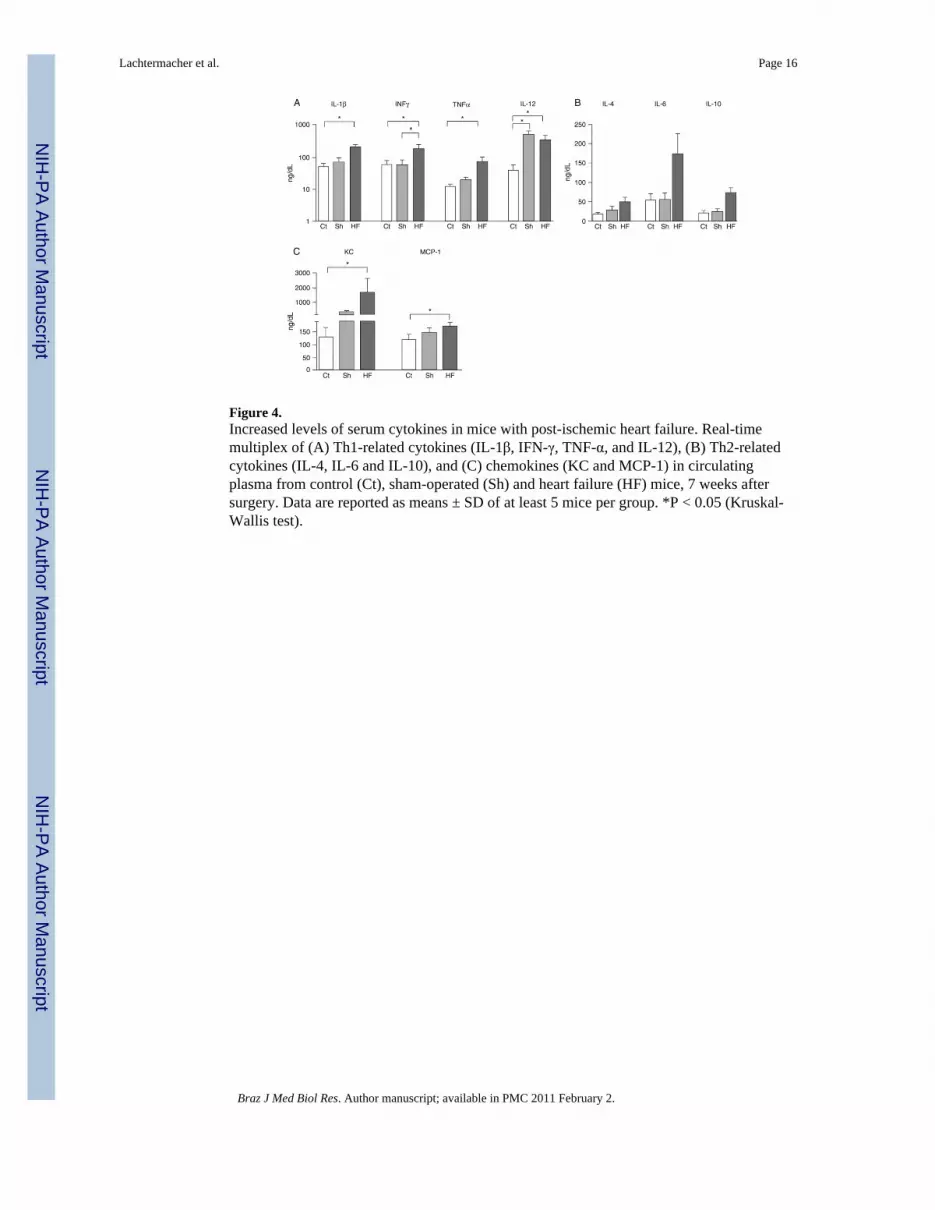
Figure 4.Increased levels of serum cytokines in mice with post-ischemic heart failure. Real-timemultiplex of (A) Th1-related cytokines (IL-1β, IFN-γ, TNF-α, and IL-12), (B) Th2-relatedcytokines (IL-4, IL-6 and IL-10), and (C) chemokines (KC and MCP-1) in circulatingplasma from control (Ct), sham-operated (Sh) and heart failure (HF) mice, 7 weeks aftersurgery. Data are reported as means ± SD of at least 5 mice per group. *P < 0.05 (Kruskal-Wallis test).
Lachtermacher et al. Page 16
Braz J Med Biol Res. Author manuscript; available in PMC 2011 February 2.
NIH
-PA Author Manuscript
NIH
-PA Author Manuscript
NIH
-PA Author Manuscript

Figure 5.Real-time PCR validation of changes in the expression of five genes that were found to bealtered in microarray analysis (Hif1a, Ifnar1, Mmp23, Nos2, and Tlr4). Fold changes foreach gene indicate that expression changes determined by each method are positivelycorrelated for these sampled genes. Linear regression curve with dotted line as 95%confidence interval (Pearson R = 0.968, P = 0.0066).
Lachtermacher et al. Page 17
Braz J Med Biol Res. Author manuscript; available in PMC 2011 February 2.
NIH
-PA Author Manuscript
NIH
-PA Author Manuscript
NIH
-PA Author Manuscript

NIH
-PA Author Manuscript
NIH
-PA Author Manuscript
NIH
-PA Author Manuscript
Lachtermacher et al. Page 18
Table 1
Electrocardiographic analysis of C57BL/6 mice.
Control(N = 25)
Sham-operated(N = 7)
Myocardial infarction(N = 10)
Heart rate (bpm) 592 ± 105 679 ± 80 532 ± 78+
Amplitude
P wave (mV) 0.088 ± 0.033 0.118 ± 0.036 0.147 ± 0.068
Q wave (mV) 0.003 ± 0.014 0.031 ± 0.045 0.221 ± 0.276*+
QRS complex (mV) 0.608 ± 0.254 0.760 ± 0.392 0.357 ± 0.316+
ST segment (mV) 0.004 ± 0.003 0.084 ± 0.087 0.397 ± 0.363*+
T wave (mV) 0.106 ± 0.063 −0.024 ± 0.200 −0.140 ± 0.270*
Duration
P wave (ms) 11 ± 2 12 ± 1 14 ± 2*
PR interval (ms) 34 ± 7 32 ± 5 37 ± 6
QRS (ms) 11 ± 2 14 ± 3 15 ± 2*
QT interval (ms) 22 ± 6 39 ± 13 68 ± 49*+
QTc 71 ± 13 177 ± 147 201 ± 143*
Data are reported as means ± SD. QTc = QT corrected interval.
*P < 0.05 vs control;
+P < 0.05 vs sham-operated (one-way ANOVA).
Braz J Med Biol Res. Author manuscript; available in PMC 2011 February 2.

NIH
-PA Author Manuscript
NIH
-PA Author Manuscript
NIH
-PA Author Manuscript
Lachtermacher et al. Page 19
Table 2
Echocardiographic analysis of mice with myocardial infarction.
Control(N = 25)
Sham-operated(N = 7)
Myocardial infarction(N = 10)
Heart rate (bpm) 250 ± 31 298 ± 56 249 ± 41
Flux
Peak A (m/s) 0.14 ± 0.05 0.12 ± 0.02 0.13 ± 0.02
Peak E (m/s) 0.43 ± 0.09 0.39 ± 0.07 0.53 ± 0.13*+
E/A 3.25 ± 0.78 3.29 ± 0.67 4.51 ± 1.09*+
Mct (ms) 158 ± 43 96 ± 43 149 ± 68
Ajt (ms) 94 ± 18 90 ± 22 80 ± 7*
Diameters
Awthd (cm) 0.08 ± 0.01 0.09 ± 0.02 0.06 ± 0.01*+
Pwthd (cm) 0.09 ± 0.01 0.11 ± 0.03 0.08 ± 0.04+
LVDd (cm) 0.36 ± 0.03 0.32 ± 0.11 0.40 ± 0.05*+
LVDs (cm) 0.23 ± 0.03 0.21 ± 0.11 0.32 ± 0.05*+
FS (%) 36 ± 6 37 ± 12 22 ± 7*+
Data are reported as means ± SD. A = transmitral late flling velocity; E = transmitral early flling velocity; Mct = mitral closure time; Ajt = aorticejection time; Awthd = diastolic anterior wall thickness; Pwthd = diastolic posterior wall thickness; LVDd = diastolic left ventricle (LV)dimension; LVDs = systolic LV dimension; FS% = fractional shortening.
*P < 0.05 vs control;
+P < 0.05 vs sham-operated (one-way ANOVA).
Braz J Med Biol Res. Author manuscript; available in PMC 2011 February 2.

NIH
-PA Author Manuscript
NIH
-PA Author Manuscript
NIH
-PA Author Manuscript
Lachtermacher et al. Page 20
Table 3
Signifcant regulation of genes involved in the infammatory/immune defense response of infarcted mice.
Name Symbol X P
Chemokine (c-c motif) ligand 8 Ccl8 9.70 0.024
Chemokine (c-c motif) ligand 9 Ccl9 2.31 0.040
Chemokine (c-x-c motif) ligand 14 Cxcl14 2.31 0.027
Chemokine-like receptor 1 Cmklr1 1.82 0.004
Complement component 3 C3 2.68 0.015
Gamma-glutamyltransferase-like activity 1 Ggtla1 2.16 0.026
Immunity-related gtpase family, m Irgm 1.81 0.009
Interferon (alpha and beta) receptor 1 Ifnar1 1.51 0.016
Interferon activated gene 204 If204 3.21 0.000
Interferon activated gene 205 If205 5.06 0.001
Interferon consensus sequence binding protein 1 Icsbp1 1.55 0.025
Interferon gamma receptor 1 Ifngr1 2.02 0.013
Interferon gamma receptor 2 Ifngr2 2.01 0.015
Interferon induced with helicase c domain 1 Ifh1 1.58 0.006
Interferon-induced protein 35 If35 1.54 0.039
Interferon-stimulated protein Isg20 1.58 0.012
Interleukin 1 family, member 9 Il1f9 1.69 0.018
Interleukin 1 receptor, type 1 Il1r1 2.53 0.024
Interleukin 13 Il13 2.11 0.017
Interleukin 13 receptor, alpha 1 Il13ra1 1.93 0.005
Interleukin 2 receptor, gamma chain Il2rg 1.91 0.047
Interleukin 22 receptor, alpha 1 Il22ra1 2.17 0.047
Interleukin 6 signal transducer Il6st 2.71 0.018
Interleukin-1 receptor-associated kinase 1 Irak1 1.57 0.038
Interleukin-1 receptor-associated kinase 2 Irak2 3.27 0.000
Interleukin-1 receptor-associated kinase 3 Irak3 2.95 0.030
Interleukin-1 receptor-associated kinase 4 Irak4 1.61 0.015
Nuclear factor of activated t-cells, cytoplasmic, calcineurin-dependent 4 Nfatc4 1.94 0.015
Toll interacting protein Tollip 1.50 0.048
Toll-like receptor 2 Tlr2 1.65 0.005
Toll-like receptor 4 Tlr4 3.15 0.001
Tumor necrosis factor receptor superfamily, member 10b Tnfrsf10b 1.68 0.043
Tumor necrosis factor receptor superfamily, member 12a Tnfrsf12a 2.37 0.001
Tumor necrosis factor receptor superfamily, member 19-like Tnfrsf19l 1.61 0.010
Tumor necrosis factor receptor superfamily, member 1a Tnfrsf1a 1.52 0.008
Tumor necrosis factor receptor superfamily, member 1b Tnfrsf1b 1.92 0.006
Tumor necrosis factor receptor superfamily, member 22 Tnfrsf22 1.80 0.039
Tumor necrosis factor receptor superfamily, member 25 Tnfrsf25 1.80 0.025
X = fold difference (negative for down-regulation). The Student heteroscedastic t-test was used for the comparison of heart failure with control.
Braz J Med Biol Res. Author manuscript; available in PMC 2011 February 2.