Candidate Polyurethanes Based on Castor Oil ... - MDPI
31
-
Upload
khangminh22 -
Category
Documents
-
view
3 -
download
0
Transcript of Candidate Polyurethanes Based on Castor Oil ... - MDPI


molecules
Article
Candidate Polyurethanes Based on Castor Oil(Ricinus communis), with Polycaprolactone Dioland Chitosan Additions, for Use inBiomedical Applications
Yomaira L. Uscátegui 1,2 , Luis E. Díaz 3 , José A. Gómez-Tejedor 4,5 , Ana Vallés-Lluch 4 ,Guillermo Vilariño-Feltrer 4 , María A. Serrano 4 and Manuel F. Valero 2,*
1 Doctoral Program of Biosciences, Universidad de La Sabana, Chía 140013, Colombia;[email protected]
2 Energy, Materials and Environment Group, Faculty of Engineering, Universidad de La Sabana,Chía 140013, Colombia
3 Bioprospecting Research Group, Faculty of Engineering, Universidad de La Sabana, Chía 140013, Colombia;[email protected]
4 Centre for Biomaterials and Tissue Engineering, Universitat Politècnica de València, Camino de Vera, s/n,46022 Valencia, Spain; [email protected] (J.A.G.-T.); [email protected] (A.V.-L.); [email protected] (G.V.-F.);[email protected] (M.A.S.)
5 Biomedical Research Networking Center in Bioengineering, Biomaterials, and Nanomedicine (CIBER-BBN),46022 Valencia, Spain
* Correspondence: [email protected]; Tel.: +57-1-8615555 (ext. 25224)
Received: 14 December 2018; Accepted: 4 January 2019; Published: 10 January 2019�����������������
Abstract: Polyurethanes are widely used in the development of medical devices due to theirbiocompatibility, degradability, non-toxicity and chemical versatility. Polyurethanes were obtainedfrom polyols derived from castor oil, and isophorone diisocyanate, with the incorporation ofpolycaprolactone-diol (15% w/w) and chitosan (3% w/w). The objective of this research was toevaluate the effect of the type of polyol and the incorporation of polycaprolactone-diol and chitosanon the mechanical and biological properties of the polyurethanes to identify the optimal ones forapplications such as wound dressings or tissue engineering. Polyurethanes were characterized bystress-strain, contact angle by sessile drop method, thermogravimetric analysis, differential scanningcalorimetry, water uptake and in vitro degradation by enzymatic processes. In vitro biologicalproperties were evaluated by a 24 h cytotoxicity test using the colorimetric assay MTT and theLIVE/DEAD kit with cell line L-929 (mouse embryonic fibroblasts). In vitro evaluation of the possibleinflammatory effect of polyurethane-based materials was evaluated by means of the expressionof anti-inflammatory and proinflammatory cytokines expressed in a cellular model such as THP-1cells by means of the MILLIPLEX® MAP kit. The modification of polyols derived from castor oilincreases the mechanical properties of interest for a wide range of applications. The polyurethanesevaluated did not generate a cytotoxic effect on the evaluated cell line. The assessed polyurethanes aresuggested as possible candidate biomaterials for wound dressings due to their improved mechanicalproperties and biocompatibility.
Keywords: castor oil; biomedical devices; polyurethanes; polycaprolactone-diol; chitosan
1. Introduction
Polyurethanes (PUs) are widely used in the preparation of medical devices due to theirbiocompatibility, degradability, and non-toxicity when compared to polymers such as polylactic
Molecules 2019, 24, 237; doi:10.3390/molecules24020237 www.mdpi.com/journal/molecules

Molecules 2019, 24, 237 2 of 30
acid (PLA), polycarbonate, polycaprolactone, among others [1–4]. Examples of PU applications inthe biomedical field are implants, artificial heart valves, sutures, catheters, artificial heart, vascularprostheses, wound coatings, blood compatible coatings, drug delivery systems, porous supports fortissue regeneration, among others [5–9].
Since the mechanical, thermal, chemical and biological properties of PUs can be varied duringthe synthesis process [10–14], the addition of polymers, such as polycaprolactone diol (PCL) orchitosan (Ch) can modify the properties of PUs such as biocompatibility [15] and the antimicrobialactivity. PCL is an attractive polymer for the development of biomaterials due to its properties suchas biocompatibility, biodegradability, ease in the processing of biomaterials, among others [16]. Ch isa polysaccharide that is obtained from renewable sources because it is part of the structure of somecrustaceans. Ch is mainly characterized by being biocompatible, biodegradable, bioadhesive, non-thetoxic, and has antimicrobial properties, among others [15,17,18]. These properties have allowed PCLand Ch to be used in some applications such as wound dressings, surgical sutures, scaffolds in tissueengineering, among others [19]. The use of Ch and PCL is expected to increase the biocompatibility ofthe PUs synthesized with polyols derived from castor oil. Additionally, the filler effect is expected toincrease the mechanical properties such as tensile strength. And when using the mixture of chitosanwith polycaprolactone, it is sought to evaluate if there is a possible synergistic effect or not to obtainbiocompatible materials with antimicrobial properties, or if both mechanical and biological propertiesare affected.
Biocompatibility is interpreted as a series of interactions that occur at the tissue/material interface,allowing the identification of those materials with surface characteristics and/or more biocompatiblepolymer chemistry; these interactions are influenced by the intrinsic characteristics of the material.Some biocompatibility tests involve analytical tests or observations of physiological phenomena,reactions or surface properties attributable to a specific application [12].
Cell cultures are ideal systems for the study and observation of a specific cell type under specificconditions since these systems do not have the complexity that an in vivo system entails, due to alarge number of variables that interact. In vitro tests assess the morphology, cytotoxicity and secretoryfunctions of different cell types. The tests can be by direct contact of the cells and the material orindirect, adding an extract of the material to the cell culture [20,21].
Monocytes and macrophages are part of the innate immune system because they are cellsthat are involved in inflammatory processes with the ability to synthesize and secrete pro andanti-inflammatory cytokines [22]. Cytokines correspond to a diverse group of extracellular,water-soluble proteins, which influence the production and activity of other cytokines by increasing(proinflammatory) or decreasing (anti-inflammatory) the inflammatory response [23].
The human monocyte cell line THP-1 is widely used in research thanks to the ability of monocytesto differentiate into macrophages [24]. THP-1 monocytes have a round shape in suspension; when theydifferentiate upon stimulation by phorbol 12-myristate-13-acetate (PMA), the cells adhere to the cultureplates, gaining phenotypic and functional characteristics similar to primary human macrophages [25,26].The immune response is assessed by measuring cytokines in the cell culture medium [22].
The inflammatory response of macrophages is activated by invading pathogens, particles,lipopolysaccharides (LPSs), and other stimuli [27]. LPSs are part of the outer membrane ofGram-negative bacteria and can cause tissue damage and the release of multiple pro-inflammatorycytokines [27]. Therefore, when an inflammatory response is induced, pro-inflammatory cytokines,such as interleukin-1 beta (IL-1β), tumor necrosis factor alpha (TNF-α), and interleukin-6 (IL-6),can be released. Likewise, anti-inflammatory cytokines, such as interleukin-10 (IL-10) [28], can bereleased. Biomaterials, such as high molecular weight polyethylene, can activate macrophages tosecrete pro-inflammatory cytokines, including TNF-α and IL-1β, among others, as a response tomaterial implantation [29].
PU applications in biomedicine are diverse due to Pus’ various properties. Using aliphaticchains derived from vegetable oils creates flexible PUs, and cyclic diisocyanates provide greater

Molecules 2019, 24, 237 3 of 30
mechanical strength [30]. Therefore, it is necessary to specifically characterize each synthesizedmaterial to determine its functionality and suitability for biomedical devices. The aim of this researchwas to determine the physicochemical, mechanical, morphological, biodegradability, and in vitrobiocompatibility characteristics of the PUs, in addition to the possible inflammatory effects of thematerials synthesized with castor oil polyols, depending on segment structure and PU cross-linkingdensity. In this study, different PUs were synthesized with castor oil (chemically modified or not) andisophorone diisocyanate (IPDI) by adding polycaprolactone diol (PCL) (15% w/w) and chitosan (Ch)(3% w/w). In vitro degradation was determined in acidic and basic media, and enzymatic degradationwas carried out with pig liver esterase. The in vitro cell viability was determined using L929 mousefibroblasts (ATCC® CCL-1), human fibroblasts (MRC-5) (ATCC® CCL-171™), and adult human dermalfibroblasts (HDFa) (ATCC® PCS-201-012™) with the PUs. The viability was also determined by alive/dead kit for the L929 mouse fibroblasts. Pro- and anti-inflammatory responses were evaluated bycytokine expression (IFN-γ, IL-1β, IL-2, IL-4, IL-5, IL-6, IL-8, IL-10, and TNF-α) of the THP-1 cells withand without stimulation by LPS. The present paper serves as a screening of the immunomodulatoryeffects of PU materials synthesized with castor oil.
2. Results and Discussion
2.1. Obtaining Polyols
The reaction to obtaining polyols derived from the castor oil is presented in Scheme 1. Thehydroxyl number of the polyols transesterified with pentaerythritol was determined. The values ofthe hydroxyl index for each polyol (P.1, P.2 and P.3) were 160, 191 and 236 mg KOH per g of castoroil sample, respectively. According to the results of the hydroxyl index, it is noted that the chemicalmodification of the castor oil increases as the pentaerythritol content increases. The increment ofthe hydroxyl index can be related to the increase of the crosslinking reactions and to the gain of thebulk density of the polymeric materials. An increase in the crosslink density would generate animprovement in the mechanical properties of the polymer matrices.
Molecules 2019, 24, x FOR PEER REVIEW 3 of 29
PU applications in biomedicine are diverse due to Pus’ various properties. Using aliphatic chains
derived from vegetable oils creates flexible PUs, and cyclic diisocyanates provide greater mechanical
strength [30]. Therefore, it is necessary to specifically characterize each synthesized material to
determine its functionality and suitability for biomedical devices. The aim of this research was to
determine the physicochemical, mechanical, morphological, biodegradability, and in vitro
biocompatibility characteristics of the PUs, in addition to the possible inflammatory effects of the
materials synthesized with castor oil polyols, depending on segment structure and PU cross-linking
density. In this study, different PUs were synthesized with castor oil (chemically modified or not)
and isophorone diisocyanate (IPDI) by adding polycaprolactone diol (PCL) (15% w/w) and chitosan
(Ch) (3% w/w). In vitro degradation was determined in acidic and basic media, and enzymatic
degradation was carried out with pig liver esterase. The in vitro cell viability was determined using
L929 mouse fibroblasts (ATCC® CCL-1), human fibroblasts (MRC-5) (ATCC® CCL-171™), and adult
human dermal fibroblasts (HDFa) (ATCC® PCS-201-012™) with the PUs. The viability was also
determined by a live/dead kit for the L929 mouse fibroblasts. Pro- and anti-inflammatory responses
were evaluated by cytokine expression (IFN-γ, IL-1β, IL-2, IL-4, IL-5, IL-6, IL-8, IL-10, and TNF-α) of
the THP-1 cells with and without stimulation by LPS. The present paper serves as a screening of the
immunomodulatory effects of PU materials synthesized with castor oil.
2. Results and Discussion
2.1. Obtaining Polyols
The reaction to obtaining polyols derived from the castor oil is presented in Scheme 1. The
hydroxyl number of the polyols transesterified with pentaerythritol was determined. The values of
the hydroxyl index for each polyol (P.1, P.2 and P.3) were 160, 191 and 236 mg KOH per g of castor
oil sample, respectively. According to the results of the hydroxyl index, it is noted that the chemical
modification of the castor oil increases as the pentaerythritol content increases. The increment of the
hydroxyl index can be related to the increase of the crosslinking reactions and to the gain of the bulk
density of the polymeric materials. An increase in the crosslink density would generate an
improvement in the mechanical properties of the polymer matrices.
HC
H2C
H2C
C
C
O
O
O
O
C
O
O
R
R
R
+ COHHO
HO OH
COHO
HO OH
PbO
CR
O
COHO
HO O
CR
O
CR
O
HC
H2C
H2C
OH
OH
O C
O
R
HC
H2C
H2C
C
O
O
OH
C
O
O
R
R
(21.2%)
(8.5%)
(40.5%)
(20.8%)
(H2C)7 CH
CH
H2C CH
OH
(CH2)5 CH3R=
Castor oil Pentaerythritol
Scheme 1. Reaction scheme for obtaining polyols.
2.2. Mechanical Properties of PUs
Aromatic diisocyanates are the most commonly used in the synthesis of PUs due to their
mechanical properties, but they can produce carcinogenic and mutagenic diamines upon
degradation [31], therefore, an aliphatic diisocyanate was used in this research to avoid the side
effects of the raw materials on living tissues.
Scheme 1. Reaction scheme for obtaining polyols.
2.2. Mechanical Properties of PUs
Aromatic diisocyanates are the most commonly used in the synthesis of PUs due totheir mechanical properties, but they can produce carcinogenic and mutagenic diamines upondegradation [31], therefore, an aliphatic diisocyanate was used in this research to avoid the sideeffects of the raw materials on living tissues.

Molecules 2019, 24, 237 4 of 30
Mechanical testing is essential to establish the use of a biomaterial because it allows obtaining theload parameters required for a tissue of interest [32]. The mechanical properties of 12 PU matrices wereevaluated by determining the stress-strain curves from which the tensile strength and the elongationat the break were obtained. Figure 1 shows the results of the mechanical properties depending on thepolyol used in the synthesis.
Molecules 2019, 24, x FOR PEER REVIEW 4 of 29
Mechanical testing is essential to establish the use of a biomaterial because it allows obtaining
the load parameters required for a tissue of interest [32]. The mechanical properties of 12 PU matrices
were evaluated by determining the stress-strain curves from which the tensile strength and the
elongation at the break were obtained. Figure 1 shows the results of the mechanical properties
depending on the polyol used in the synthesis.
Figure 1. Mechanical properties of the synthesized PUs. (a) Maximum stress of PUs; (b) Percent
elongation of PUs. The data are expressed as the mean ± SD (n = 3). Bars with different letters (a–h)
indicate significant differences (p < 0.05) between the polyols.
Physical and mechanical properties depend on the atomic and molecular structure of the
materials used in the synthesis. The nature of the bonds and subunits of the structure affects the
mechanical properties and therefore the stress-strain properties, which are of interest for the
evaluation of biomaterials [32]. The highest tensile strength (16.86 MPa) was obtained with polyol P.3
(P.3-0%Ch-0%PCL). Figure 1a shows an increase in the tensile strength of all the materials
synthesized with polyol P.3, which has the highest cross-linking. This is how the chemical
modification of the polyols increases the maximum stress—via the increase of physical cross-linking
of the polymer matrix [33].
The mechanical properties of PUs are attributed to presence of hard and soft segment domains
[34]. The hard segment generally refers to the combination of the chain extender and the diisocyanate
components, while the soft segments refer to polymeric diols. Depending on the structure of the hard
and soft segments, crystalline and amorphous domains can be formed, which determine the stiffness
and stability of the material [35]. Hydrogen bond cause strong interactions, so the polar nature of the
hard segment causes a strong attraction, forming the domains [36]. Therefore, when using polyol P.3
in the synthesis, the values of the mechanical properties increased because the soft segments had a
higher number of hydroxyl groups, increasing the cross-linking density, and the hard segments
formed a ring, providing greater resistance.
Regarding the percent elongation at break (Figure 1b), the analysis showed significant
differences (p < 0.05) of polyol P.1 compared with the other polyols (P.2 and P.3), with the percentage
increasing as the polyol was modified. The mechanical properties of PUs depend on many factors,
including molecular weight, chemical bonds, cross-linking, crystallinity of the polymer, and the size,
shape, and interactions of the hard segment present in the structure [37]. Thus, PUs with a higher
degree of cross-linking have higher values of tensile strength and percent elongation at break.
Increased cross-linking produces a more compact structure [37]. An increase in strength is attributed
to the content of intermolecular hydrogen bonds and cross-linking density [38].
When analyzing the influence of the additives used in the synthesis on the mechanical properties,
significant differences are found when 3% Ch was added to polyol P.2 (P.2-3%Ch-0%PCL), obtaining
a higher value than the other materials synthesized with P.1 and P.3. For polyol P.3, the additives
decreased the maximum stress compared with the material without additives.
Chen et al. (2018) synthesized PUs with PCL as a polyol, IPDI, and polylactic acid (PLA),
obtaining tensile strength values between 41 and 60 MPa when using PLA- and PCL-based PU ratios
in the range of 80/20 to 95/5. The authors attributed the decrease in tensile strength as the ratio of PCL
Figure 1. Mechanical properties of the synthesized PUs. (a) Maximum stress of PUs; (b) Percentelongation of PUs. The data are expressed as the mean ± SD (n = 3). Bars with different letters (a–h)indicate significant differences (p < 0.05) between the polyols.
Physical and mechanical properties depend on the atomic and molecular structure of thematerials used in the synthesis. The nature of the bonds and subunits of the structure affectsthe mechanical properties and therefore the stress-strain properties, which are of interest for theevaluation of biomaterials [32]. The highest tensile strength (16.86 MPa) was obtained with polyol P.3(P.3-0%Ch-0%PCL). Figure 1a shows an increase in the tensile strength of all the materials synthesizedwith polyol P.3, which has the highest cross-linking. This is how the chemical modification of thepolyols increases the maximum stress—via the increase of physical cross-linking of the polymermatrix [33].
The mechanical properties of PUs are attributed to presence of hard and soft segment domains [34].The hard segment generally refers to the combination of the chain extender and the diisocyanatecomponents, while the soft segments refer to polymeric diols. Depending on the structure of the hardand soft segments, crystalline and amorphous domains can be formed, which determine the stiffnessand stability of the material [35]. Hydrogen bond cause strong interactions, so the polar nature of thehard segment causes a strong attraction, forming the domains [36]. Therefore, when using polyol P.3 inthe synthesis, the values of the mechanical properties increased because the soft segments had a highernumber of hydroxyl groups, increasing the cross-linking density, and the hard segments formed a ring,providing greater resistance.
Regarding the percent elongation at break (Figure 1b), the analysis showed significant differences(p < 0.05) of polyol P.1 compared with the other polyols (P.2 and P.3), with the percentage increasingas the polyol was modified. The mechanical properties of PUs depend on many factors, includingmolecular weight, chemical bonds, cross-linking, crystallinity of the polymer, and the size, shape,and interactions of the hard segment present in the structure [37]. Thus, PUs with a higher degreeof cross-linking have higher values of tensile strength and percent elongation at break. Increasedcross-linking produces a more compact structure [37]. An increase in strength is attributed to thecontent of intermolecular hydrogen bonds and cross-linking density [38].
When analyzing the influence of the additives used in the synthesis on the mechanical properties,significant differences are found when 3% Ch was added to polyol P.2 (P.2-3%Ch-0%PCL), obtaininga higher value than the other materials synthesized with P.1 and P.3. For polyol P.3, the additivesdecreased the maximum stress compared with the material without additives.
Chen et al. (2018) synthesized PUs with PCL as a polyol, IPDI, and polylactic acid (PLA), obtainingtensile strength values between 41 and 60 MPa when using PLA- and PCL-based PU ratios in the

Molecules 2019, 24, 237 5 of 30
range of 80/20 to 95/5. The authors attributed the decrease in tensile strength as the ratio of PCLincreased to the plasticizing effect of certain non-cross-linked PCL polyols and to a possible decreasein compatibility as the PCL content increased [39].
A similar effect was observed for percent elongation at break because it decreased when PCL andCh were added to polyol P.3 (P.3-3%Ch-15%PCL). The other materials did not differ with the additivesused from the material without additives. The flexibility of PU may be due to the long oil hydrocarbonchain present in the polymer chain [37]. This agrees with the results reported by Park et al. (2013),who synthesized PUs with polycaprolactone, hexamethylene diisocyanate, and isosorbide, with silkadded, and determined that a higher silk content increased the stiffness and decreased the maximumstress. The authors stated that the design of flexible and soft polymers allows for the production of awide range of biomaterials to regenerate soft tissues such as muscles and ligaments [6]. Additionally,Vannozzi et al. reported that in general, soft and deformable substrates are key features for skeletalmuscle tissue engineering [33].
2.3. Fourier-Transform Infrared Spectroscopy (FTIR)
FTIR was used to determine the efficiency of the synthesis process by the identification ofcharacteristic functional groups of PU and the absence of characteristic peaks of the monomersused in the synthesis process. Figure 2 shows the infrared spectra of the synthesized PUs.
Molecules 2019, 24, x FOR PEER REVIEW 5 of 29
increased to the plasticizing effect of certain non-cross-linked PCL polyols and to a possible decrease
in compatibility as the PCL content increased [39].
A similar effect was observed for percent elongation at break because it decreased when PCL
and Ch were added to polyol P.3 (P.3-3%Ch-15%PCL). The other materials did not differ with the
additives used from the material without additives. The flexibility of PU may be due to the long oil
hydrocarbon chain present in the polymer chain [37]. This agrees with the results reported by Park
et al. (2013), who synthesized PUs with polycaprolactone, hexamethylene diisocyanate, and
isosorbide, with silk added, and determined that a higher silk content increased the stiffness and
decreased the maximum stress. The authors stated that the design of flexible and soft polymers allows
for the production of a wide range of biomaterials to regenerate soft tissues such as muscles and
ligaments [6]. Additionally, Vannozzi et al. reported that in general, soft and deformable substrates
are key features for skeletal muscle tissue engineering [33].
2.3. Fourier-Transform Infrared Spectroscopy (FTIR)
FTIR was used to determine the efficiency of the synthesis process by the identification of
characteristic functional groups of PU and the absence of characteristic peaks of the monomers used
in the synthesis process. Figure 2 shows the infrared spectra of the synthesized PUs.
Figure 2. FTIR spectra of the synthesized PUs.
Figure 2 shows that all of the FTIR spectra had similar peaks, independent of the polyol or
additive used in the synthesis, and the peaks observed corresponded to the expected PU matrices.
The absence of the stretching peak of the −N=C=O bond of the diisocyanates at 2250 cm−1 [31],
indicates there were no unreacted free isocyanate groups in the synthesized PU matrices, showing
that the reaction was complete.
Spectral peaks characteristic of PUs can be seen in the spectra. Thus, around 3330 cm−1, the
characteristic bands for the stretching vibrations of the −N–H bonds are observed [40] from which it
can be inferred that they correspond to the urethane bonds present in the matrices. Near 2923 cm−1,
the stretching peak of the methyl group can be observed, and at around 2855 cm−1, the symmetric
stretching of the C–H bond is present. At around 1700 cm−1, an intense band is present due to the C=O
bond stretching [40] thus indicating the formation of the urethane group. Around 1250 cm−1, the C–
N bond stretching is observed; at about 1140 cm−1, the stretching vibrations of the C–O bond
appear [37].
400900140019002400290034003900
Tra
nsm
itta
nce
(%
)
Wavenumber (cm−1)
P.1-0%Ch-0%PCL P.1-3%Ch-15%PCL P.2-0%Ch-0%PCL
P.2-3%Ch-15%PCL P.3-0%Ch-0%PCL P.3-3%Ch-15%PCL
Figure 2. FTIR spectra of the synthesized PUs.
Figure 2 shows that all of the FTIR spectra had similar peaks, independent of the polyol or additiveused in the synthesis, and the peaks observed corresponded to the expected PU matrices. The absenceof the stretching peak of the −N=C=O bond of the diisocyanates at 2250 cm−1 [31], indicates therewere no unreacted free isocyanate groups in the synthesized PU matrices, showing that the reactionwas complete.
Spectral peaks characteristic of PUs can be seen in the spectra. Thus, around 3330 cm−1, thecharacteristic bands for the stretching vibrations of the −N–H bonds are observed [40] from which itcan be inferred that they correspond to the urethane bonds present in the matrices. Near 2923 cm−1,the stretching peak of the methyl group can be observed, and at around 2855 cm−1, the symmetricstretching of the C–H bond is present. At around 1700 cm−1, an intense band is present due to theC=O bond stretching [40] thus indicating the formation of the urethane group. Around 1250 cm−1,the C–N bond stretching is observed; at about 1140 cm−1, the stretching vibrations of the C–O bondappear [37].
Molecules 2019, 24, 237 6 of 30
2.4. Thermal Analysis
2.4.1. Thermogravimetric Analysis
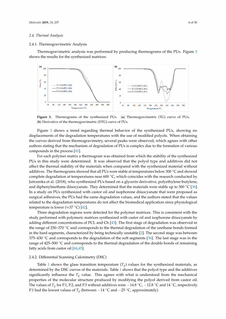
Thermogravimetric analysis was performed by producing thermograms of the PUs. Figure 3shows the results for the synthesized matrices.
Molecules 2019, 24, x FOR PEER REVIEW 6 of 29
2.4. Thermal Analysis
2.4.1. Thermogravimetric Analysis
Thermogravimetric analysis was performed by producing thermograms of the PUs. Figure 3
shows the results for the synthesized matrices.
Figure 3. Thermograms of the synthesized PUs. (a) Thermogravimetric (TG) curve of PUs; (b)
Derivative of the thermogravimetric (DTG) curve of PUs.
Figure 3 shows a trend regarding thermal behavior of the synthesized PUs, showing no
displacements of the degradation temperatures with the use of modified polyols. When obtaining the
curves derived from thermogravimetry, several peaks were observed, which agrees with other
authors stating that the mechanism of degradation of PUs is complex due to the formation of various
compounds in the process [41].
For each polymer matrix a thermogram was obtained from which the stability of the synthesized
PUs in this study were determined. It was observed that the polyol type and additives did not affect
the thermal stability of the materials when compared with the synthesized material without additives.
The thermograms showed that all PUs were stable at temperatures below 300 °C and showed
complete degradation at temperatures near 600 °C, which coincides with the research conducted by
Jutrzenka et al. (2018), who synthesized PUs based on a glycerin derivative, polyethylene-butylene,
and diphenylmethane diisocyanate. They determined that the materials were stable up to 300 °C [36].
In a study on PUs synthesized with castor oil and isophorone diisocyanate that were proposed as
surgical adhesives, the PUs had the same degradation values, and the authors stated that the values
related to the degradation temperatures do not affect the biomedical application since physiological
temperature is lower (≈37 °C) [42].
Three degradation regions were detected for the polymer matrices. This is consistent with the
study performed with polymeric matrices synthesized with castor oil and isophorone diisocyanate
by adding different concentrations of PCL and Ch [43]. The first stage of degradation was observed
in the range of 250–370 °C and corresponds to the thermal degradation of the urethane bonds formed
in the hard segments, characterized by being technically unstable [2]. The second stage was between
375–430 °C and corresponds to the degradation of the soft segments [36]. The last stage was in the
range of 425–500 °C and corresponds to the thermal degradation of the double bonds of remaining
fatty acids from castor oil [44,45].
2.4.2. Differential Scanning Calorimetry (DSC)
Table 1 shows the glass transition temperature (Tg) values for the synthesized materials, as
determined by the DSC curves of the materials. Table 1 shows that the polyol type and the additives
significantly influence the Tg value. This agrees with what is understood from the mechanical
properties of the molecular structure produced by modifying the polyol derived from castor oil. The
values of Tg for P.1, P.2, and P.3 without additives were −14.8 °C, −12.8 °C and 14 °C, respectively. P.1
had the lowest values of Tg (between −14 °C and −25 °C, approximately).
Figure 3. Thermograms of the synthesized PUs. (a) Thermogravimetric (TG) curve of PUs;(b) Derivative of the thermogravimetric (DTG) curve of PUs.
Figure 3 shows a trend regarding thermal behavior of the synthesized PUs, showing nodisplacements of the degradation temperatures with the use of modified polyols. When obtainingthe curves derived from thermogravimetry, several peaks were observed, which agrees with otherauthors stating that the mechanism of degradation of PUs is complex due to the formation of variouscompounds in the process [41].
For each polymer matrix a thermogram was obtained from which the stability of the synthesizedPUs in this study were determined. It was observed that the polyol type and additives did notaffect the thermal stability of the materials when compared with the synthesized material withoutadditives. The thermograms showed that all PUs were stable at temperatures below 300 ◦C and showedcomplete degradation at temperatures near 600 ◦C, which coincides with the research conducted byJutrzenka et al. (2018), who synthesized PUs based on a glycerin derivative, polyethylene-butylene,and diphenylmethane diisocyanate. They determined that the materials were stable up to 300 ◦C [36].In a study on PUs synthesized with castor oil and isophorone diisocyanate that were proposed assurgical adhesives, the PUs had the same degradation values, and the authors stated that the valuesrelated to the degradation temperatures do not affect the biomedical application since physiologicaltemperature is lower (≈37 ◦C) [42].
Three degradation regions were detected for the polymer matrices. This is consistent with thestudy performed with polymeric matrices synthesized with castor oil and isophorone diisocyanate byadding different concentrations of PCL and Ch [43]. The first stage of degradation was observed inthe range of 250–370 ◦C and corresponds to the thermal degradation of the urethane bonds formedin the hard segments, characterized by being technically unstable [2]. The second stage was between375–430 ◦C and corresponds to the degradation of the soft segments [36]. The last stage was in therange of 425–500 ◦C and corresponds to the thermal degradation of the double bonds of remainingfatty acids from castor oil [44,45].
2.4.2. Differential Scanning Calorimetry (DSC)
Table 1 shows the glass transition temperature (Tg) values for the synthesized materials, asdetermined by the DSC curves of the materials. Table 1 shows that the polyol type and the additivessignificantly influence the Tg value. This agrees with what is understood from the mechanicalproperties of the molecular structure produced by modifying the polyol derived from castor oil.The values of Tg for P.1, P.2, and P.3 without additives were −14.8 ◦C, −12.8 ◦C and 14 ◦C, respectively.P.1 had the lowest values of Tg (between −14 ◦C and −25 ◦C, approximately).

Molecules 2019, 24, 237 7 of 30
Table 1. DSC thermograms of the synthesized PUs depending on the polyol used.
Polymeric MaterialTg (◦C)
P.1 P.2 P.3
0%Ch-0%PCL −14.8 −12.8 14.83%Ch-0%PCL −13.3 −1.0 13.7
0%Ch-15%PCL −25.3 −10.2 4.73%Ch-15%PCL −25.4 −14.8 2.8
The observed trend is a decrease in Tg as the hydroxyl index decreases, which agrees with thecross-linking of the synthesized PUs, indicating that greater energy is needed for reordering thestructure by a change in intermolecular forces. This may be due to secondary interactions resultingfrom the hyperbranched structure [46]. The thermal properties of PUs depend on the number ofurethane bonds present in the structure because they can tolerate a considerable amount of heat [47].Saénz-Pérez et al. (2016) synthesized PUs with polytetramethylene glycol and diphenylmethane ortoluene diisocyanates, and butanediol. In the determination of Tg, they found that the values increasedas the amount of chain extender increased. Therefore, the authors state that the increase in Tg wascaused by the reduction in the mobility of chain segments due to the increase of hard segments [48].
The ricinoleic acid triglyceride from castor oil used as a polyol contains an ordered structurein which hydroxyl groups are uniformly distributed within the chain, helping to obtain a PU witha uniform cross-linked structure, achieving high mechanical properties and thermal stability [30].In general, all of the synthesized matrices had a single value of Tg, indicating that all the materialsshowed homogeneous segment dispersion. The Tg values were similar to those reported for PUsbased on polyethylene glycol, poly (ε-caprolactone-co-D,L-lactide), and diurethane diisocyanate (withhexamethylene diisocyanate and butanediol), where the authors found values near −33 ◦C. Likewise,the authors did not find exothermic peaks because the materials were amorphous [49]. With the aboveinformation, it can be generalized that Pus are thermally stable and that they can be used in variousbiomedical applications, for example, as materials for non-absorbable sutures.
2.5. Hydrophilic Character
2.5.1. Contact Angle
To evaluate the hydrophilic nature of PUs, the water contact angle on their surfaces wasdetermined. Figure 4 shows the results of the synthesized PUs.
Molecules 2019, 24, x FOR PEER REVIEW 7 of 29
Table 1. DSC thermograms of the synthesized PUs depending on the polyol used.
Polymeric Material Tg (°C)
P.1 P.2 P.3
0%Ch-0%PCL −14.8 −12.8 14.8
3%Ch-0%PCL −13.3 −1.0 13.7
0%Ch-15%PCL −25.3 −10.2 4.7
3%Ch-15%PCL −25.4 −14.8 2.8
The observed trend is a decrease in Tg as the hydroxyl index decreases, which agrees with the
cross-linking of the synthesized PUs, indicating that greater energy is needed for reordering the
structure by a change in intermolecular forces. This may be due to secondary interactions resulting
from the hyperbranched structure [46]. The thermal properties of PUs depend on the number of
urethane bonds present in the structure because they can tolerate a considerable amount of heat [47].
Saénz-Pérez et al. (2016) synthesized PUs with polytetramethylene glycol and diphenylmethane or
toluene diisocyanates, and butanediol. In the determination of Tg, they found that the values
increased as the amount of chain extender increased. Therefore, the authors state that the increase in
Tg was caused by the reduction in the mobility of chain segments due to the increase of hard
segments [48].
The ricinoleic acid triglyceride from castor oil used as a polyol contains an ordered structure in
which hydroxyl groups are uniformly distributed within the chain, helping to obtain a PU with a
uniform cross-linked structure, achieving high mechanical properties and thermal stability [30]. In
general, all of the synthesized matrices had a single value of Tg, indicating that all the materials
showed homogeneous segment dispersion. The Tg values were similar to those reported for PUs
based on polyethylene glycol, poly (ɛ-caprolactone-co-D,L-lactide), and diurethane diisocyanate (with
hexamethylene diisocyanate and butanediol), where the authors found values near −33 °C. Likewise,
the authors did not find exothermic peaks because the materials were amorphous [49]. With the
above information, it can be generalized that Pus are thermally stable and that they can be used in
various biomedical applications, for example, as materials for non-absorbable sutures.
2.5. Hydrophilic Character
2.5.1. Contact Angle
To evaluate the hydrophilic nature of PUs, the water contact angle on their surfaces was
determined. Figure 4 shows the results of the synthesized PUs.
Figure 4. Contact angle of the synthesized PUs. The data are expressed as the mean ± SD (n = 10). Bars
with different letters (a–c) indicate significant differences (p < 0.05).
a
bc
ca
a a aa aa
a
50
60
70
80
90
100
110
0%Ch-0%PCL 3%Ch-0%PCL 0%Ch-15%PCL 3%Ch-15%PCL
Co
nta
ct a
ng
le [°]
Type of polyurethane
P.1 P.2 P.3
Figure 4. Contact angle of the synthesized PUs. The data are expressed as the mean ± SD (n = 10).Bars with different letters (a–c) indicate significant differences (p < 0.05).
According to the statistical analysis in Figure 4, it is observed that for polyols P.2 and P.3, thereare no significant differences in contact angle values. For P.1, significant differences are observed by

Molecules 2019, 24, 237 8 of 30
using additives because there is a reduction of angle values, indicating a decrease in the hydrophiliccharacter. Values near 100 degrees were found, so it can be deduced that the materials tend to behydrophobic. Mi et al. determined the contact angles of PCL-based thermoplastic PUs and differentchain extenders; the values were near 90 degrees. They attributed the results of the contact angle to thehydrophilic functional groups present in the chain extenders that were used. The authors state thatalthough PUs maintain the same chemical structure, monomer variation in the synthesis can affect thewettability and therefore vary the degradation behavior [50].
Likewise, Gossart et al. evaluated the contact angle of PUs synthesized with L-lysine diisocyanate,hydroxyethyl methacrylate, and poly (hexamethylene-carbamate) and found values above 80 degrees.The authors state that the common values reported for PU matrices were in the range of 80 to 90degrees depending on the structure of the PU and interactions with the surfaces [51]. The results ofthe contact angle shown in Figure 4 are greater than those reported; this possibly results from thecross-linked network generated by the monomers used in the synthesis, as the materials tended tobe hydrophobic.
2.5.2. Water Absorption Rate
Figure 5 shows the rates of water absorption for the synthesized PUs during 72 h of testing.
Molecules 2019, 24, x FOR PEER REVIEW 8 of 29
According to the statistical analysis in Figure 4, it is observed that for polyols P.2 and P.3, there
are no significant differences in contact angle values. For P.1, significant differences are observed by
using additives because there is a reduction of angle values, indicating a decrease in the hydrophilic
character. Values near 100 degrees were found, so it can be deduced that the materials tend to be
hydrophobic. Mi et al. determined the contact angles of PCL-based thermoplastic PUs and different
chain extenders; the values were near 90 degrees. They attributed the results of the contact angle to
the hydrophilic functional groups present in the chain extenders that were used. The authors state
that although PUs maintain the same chemical structure, monomer variation in the synthesis can
affect the wettability and therefore vary the degradation behavior [50].
Likewise, Gossart et al. evaluated the contact angle of PUs synthesized with L-lysine
diisocyanate, hydroxyethyl methacrylate, and poly (hexamethylene-carbamate) and found values
above 80 degrees. The authors state that the common values reported for PU matrices were in the
range of 80 to 90 degrees depending on the structure of the PU and interactions with the surfaces [51].
The results of the contact angle shown in Figure 4 are greater than those reported; this possibly results
from the cross-linked network generated by the monomers used in the synthesis, as the materials
tended to be hydrophobic.
2.5.2. Water Absorption Rate
Figure 5 shows the rates of water absorption for the synthesized PUs during 72 h of testing.
Figure 5. Water absorption rate over 72 h. Absorption results are expressed as the mean ± SD (n = 3).
Bars with different letters (a–d) indicate significant differences (p < 0.05).
The weights of the materials were monitored until constant weight over 24, 48, 72, and 144 h,
but the data are not shown because no significant differences were found after 48 h. The results show
that the water absorption rates range between 0.5 and 1.5%. As seen in Figure 5, adding PCL and Ch
increases the rate of absorption compared to the material without additives, although the difference
in rates was not greater than one. It is likely that by increasing the amount of additive more functional
groups became available to interact with the medium, which is also polar. However, by increasing
the functionality of the polyol, the effect generated is inverse, showing a reduction in the rate of
absorption compared to PUs without the additive. Internal interactions (hydrogen bonds) increase
the barrier effect by preventing fluid diffusion. In addition, the additive function causes the chains to
reorganize, showing a reduction of volumetric defects or voids in which the water can be
deposited [32].
Marques et al. evaluated a bioadhesive synthesized from lactic acid in which the water
absorption was 10%. The authors noted that moderate rates of water absorption improved the
hemostatic character of the materials [52].
a
b
e
c
a
c
e
d
a
d
a
c
0.00
0.50
1.00
1.50
2.00
0%Ch-0%PCL 3%Ch-0%PCL 0%Ch-15%PCL 3%Ch-15%PCL
Wat
er a
bso
rpti
on
(%
)
Type of polyurethane
P.1 P.2 P.3
Figure 5. Water absorption rate over 72 h. Absorption results are expressed as the mean ± SD (n = 3).Bars with different letters (a–d) indicate significant differences (p < 0.05).
The weights of the materials were monitored until constant weight over 24, 48, 72, and 144 h, butthe data are not shown because no significant differences were found after 48 h. The results showthat the water absorption rates range between 0.5 and 1.5%. As seen in Figure 5, adding PCL and Chincreases the rate of absorption compared to the material without additives, although the difference inrates was not greater than one. It is likely that by increasing the amount of additive more functionalgroups became available to interact with the medium, which is also polar. However, by increasing thefunctionality of the polyol, the effect generated is inverse, showing a reduction in the rate of absorptioncompared to PUs without the additive. Internal interactions (hydrogen bonds) increase the barriereffect by preventing fluid diffusion. In addition, the additive function causes the chains to reorganize,showing a reduction of volumetric defects or voids in which the water can be deposited [32].
Marques et al. evaluated a bioadhesive synthesized from lactic acid in which the water absorptionwas 10%. The authors noted that moderate rates of water absorption improved the hemostatic characterof the materials [52].
The contact angle and the rate of absorption provide information abouthydrophobicity/hydrophilicity and could be an indirect indicator of surface molecular mobility.

Molecules 2019, 24, 237 9 of 30
Surface wettability can affect protein adsorption on the surface and biocompatibility [53]. This is onereason why it is indispensable to perform a study of material biocompatibility to determine its possibleacceptance by the human body. Therefore, the PUs synthesized in this study can be consideredsuitable for biomedical applications, such as materials for non-absorbable sutures, considering thewater absorption rates under the test conditions.
2.6. Density Determination
Figure 6 shows the density of the materials synthesized with IPDI and the additives (PCL and Ch).
Molecules 2019, 24, x FOR PEER REVIEW 9 of 29
The contact angle and the rate of absorption provide information about
hydrophobicity/hydrophilicity and could be an indirect indicator of surface molecular mobility.
Surface wettability can affect protein adsorption on the surface and biocompatibility [53]. This is one
reason why it is indispensable to perform a study of material biocompatibility to determine its
possible acceptance by the human body. Therefore, the PUs synthesized in this study can be
considered suitable for biomedical applications, such as materials for non-absorbable sutures,
considering the water absorption rates under the test conditions.
2.6. Density Determination
Figure 6 shows the density of the materials synthesized with IPDI and the additives (PCL
and Ch).
Figure 6. Density of the synthesized PUs. The data are expressed as the mean ± SD (n = 3). Bars with
different letters (a–d) indicate significant differences (p < 0.05).
As seen in Figure 6, the density of PUs depends on the type of polyol, which agrees with the
results shown for the mechanical and thermal properties. As the cross-linking density increased, the
density of the resulting polymeric material significantly increased, although the difference between
the highest and lowest values was less than 5%. This agrees with the results of Conejero-García et al.
who synthesized polyglycerol sebacate, with a different degree of cross-linking, as a material for
various applications in tissue engineering. The density values reported by the authors ranged
between 1.13 and 1.14 g mL−1 [54].
The high densities of polymeric materials can be related to a higher hydroxyl (OH) content due
to increased cross-linking reactions [55]. In a study conducted by Carriço et al. they found that
increasing the castor oil content in the formulation of foam increased the apparent density, suggesting
that the polymer chains were more packed, with less free volume and smaller cells, increasing the
stiffness of these materials [55].
2.7. Dynamo-Mechanical Thermal Analysis (DMTA)
Figure 7 shows the dynamic behavior, DMTA, in a tension mode of PUs corresponding to the
evolution of the modulus and the loss factor versus temperature. With the variation of the storage
modulus and the loss factor, it was possible to observe the displacements suffered by Tg for the
evaluated materials. It can be seen that Tg increases when polyols P.2 and P.3 are used; this is probably
due to the stiffness of the structure and greater generation of hydrogen bonds because of the high
hydroxyl index present in the polyol [38,39]. According to the above results, it can be inferred that
material compatibility decreases when the polyol without modifications, and therefore with less
cross-linking, is used.
ab b
bb
c c cc
d
cd
0.98
0.99
1.00
1.01
1.02
1.03
1.04
1.05
1.06
1.07
0%Ch-0%PCL 3%Ch-0%PCL 0%Ch-15%PCL 3%Ch-15%PCL
Den
sity
(g
/cm
3)
Type of polyurethane
P.1 P.2 P.3
Figure 6. Density of the synthesized PUs. The data are expressed as the mean ± SD (n = 3). Bars withdifferent letters (a–d) indicate significant differences (p < 0.05).
As seen in Figure 6, the density of PUs depends on the type of polyol, which agrees with theresults shown for the mechanical and thermal properties. As the cross-linking density increased, thedensity of the resulting polymeric material significantly increased, although the difference between thehighest and lowest values was less than 5%. This agrees with the results of Conejero-García et al. whosynthesized polyglycerol sebacate, with a different degree of cross-linking, as a material for variousapplications in tissue engineering. The density values reported by the authors ranged between 1.13and 1.14 g mL−1 [54].
The high densities of polymeric materials can be related to a higher hydroxyl (OH) content due toincreased cross-linking reactions [55]. In a study conducted by Carriço et al. they found that increasingthe castor oil content in the formulation of foam increased the apparent density, suggesting that thepolymer chains were more packed, with less free volume and smaller cells, increasing the stiffness ofthese materials [55].
2.7. Dynamo-Mechanical Thermal Analysis (DMTA)
Figure 7 shows the dynamic behavior, DMTA, in a tension mode of PUs corresponding to theevolution of the modulus and the loss factor versus temperature. With the variation of the storagemodulus and the loss factor, it was possible to observe the displacements suffered by Tg for theevaluated materials. It can be seen that Tg increases when polyols P.2 and P.3 are used; this is probablydue to the stiffness of the structure and greater generation of hydrogen bonds because of the highhydroxyl index present in the polyol [38,39]. According to the above results, it can be inferred thatmaterial compatibility decreases when the polyol without modifications, and therefore with lesscross-linking, is used.

Molecules 2019, 24, 237 10 of 30
Molecules 2019, 24, x FOR PEER REVIEW 10 of 29
Figure 7. DMTA thermograms of the synthesized PUs. (a) Loss factor; (b) Storage modulus.
Results with a similar trend were found by Chen et al. on PU matrices synthesized with PCL as
a polyol, IPDI, and PLA. The authors reported that when the mobility of the chain decreased, the
value of Tg increased. Therefore, they observed that phase compatibility decreased when the PCL
content increased due to a possible plasticizing effect of non-cross-linked material [39].
A similar behavior occurred with the modulus results. The lowest values corresponded to the
materials synthesized with P.1 (polyol without modification) compared with polyols P.2 and P.3,
which have a higher content of hydroxyl groups. An increase in hydroxyl groups results in an
increase in cross-linking, hindering polymer chain mobility and thereby increasing the storage
modulus. A decrease in polymer chain mobility can limit energy transfer and diffusion, which could
decrease the absorption capacity of impact resistance and deformation [39].
The relationship between hard and soft segments is important as they simultaneously act as a
physical cross-linking agent and as a high-modulus filler. When there is an organization of the hard
and soft segments in the respective domains, pre-polymers tend to have two Tg values. One
temperature will be negative corresponding to the soft segments, while the other will be positive,
corresponding to the hard segments [38]. When a single event of Tg occurs, it could be inferred that
there is a homogeneous phase distribution [38]. According to the results obtained by DSC, it can be
observed that the trend of Tg is similar, that is, a single value of Tg is present, increasing as the polyol
is modified. The presence of a single transition can be related to the existence of a dominant phase,
so it can be inferred that there is a uniform distribution of the components [56]. The differences in Tg
between the polyols used may be due to the cross-linking density because this would cause a greater
compatibility between hard and soft segments [38].
2.8. Field-Emission Scanning Electron Microscopy (FESEM)
Figure 8 shows the morphology of PUs synthesized with IPDI as a function of the polyol. The
FESEM micrographs showed a uniform distribution of PUs, but it was not possible to differentiate
the hard segments from the soft segments. Similarly, no differences were observed related to the type
of polyol used for the polymeric matrix. These results can be correlated with the calorimetric results
because if there is only a single Tg, it is probable that there is a homogeneous phase distribution. This
agrees with the results reported by Thakur et al. on PUs synthesized with castor oil and toluene
diisocyanate for the coating of materials [47].
Figure 7. DMTA thermograms of the synthesized PUs. (a) Loss factor; (b) Storage modulus.
Results with a similar trend were found by Chen et al. on PU matrices synthesized with PCL as apolyol, IPDI, and PLA. The authors reported that when the mobility of the chain decreased, the valueof Tg increased. Therefore, they observed that phase compatibility decreased when the PCL contentincreased due to a possible plasticizing effect of non-cross-linked material [39].
A similar behavior occurred with the modulus results. The lowest values corresponded to thematerials synthesized with P.1 (polyol without modification) compared with polyols P.2 and P.3, whichhave a higher content of hydroxyl groups. An increase in hydroxyl groups results in an increasein cross-linking, hindering polymer chain mobility and thereby increasing the storage modulus. Adecrease in polymer chain mobility can limit energy transfer and diffusion, which could decrease theabsorption capacity of impact resistance and deformation [39].
The relationship between hard and soft segments is important as they simultaneously act asa physical cross-linking agent and as a high-modulus filler. When there is an organization of thehard and soft segments in the respective domains, pre-polymers tend to have two Tg values. Onetemperature will be negative corresponding to the soft segments, while the other will be positive,corresponding to the hard segments [38]. When a single event of Tg occurs, it could be inferred thatthere is a homogeneous phase distribution [38]. According to the results obtained by DSC, it can beobserved that the trend of Tg is similar, that is, a single value of Tg is present, increasing as the polyolis modified. The presence of a single transition can be related to the existence of a dominant phase,so it can be inferred that there is a uniform distribution of the components [56]. The differences in Tg
between the polyols used may be due to the cross-linking density because this would cause a greatercompatibility between hard and soft segments [38].
2.8. Field-Emission Scanning Electron Microscopy (FESEM)
Figure 8 shows the morphology of PUs synthesized with IPDI as a function of the polyol. TheFESEM micrographs showed a uniform distribution of PUs, but it was not possible to differentiatethe hard segments from the soft segments. Similarly, no differences were observed related to thetype of polyol used for the polymeric matrix. These results can be correlated with the calorimetricresults because if there is only a single Tg, it is probable that there is a homogeneous phase distribution.This agrees with the results reported by Thakur et al. on PUs synthesized with castor oil and toluenediisocyanate for the coating of materials [47].

Molecules 2019, 24, 237 11 of 30Molecules 2019, 24, x FOR PEER REVIEW 11 of 29
Figure 8. FESEM micrographs of PUs synthesized at 100×. (a) P.1; (b) P.2; (c) P.3.
2.9. In Vitro Biodegradability Assays
The biodegradability of PUs was evaluated in the presence of different media (HCl, NaOH, and
enzymatic) over a specific period of time, the results are shown in Figure 9.
Figure 9. Degradation rate of PUs in different media. (a) Acidic medium (0.1 M HCl) for 90 days; (b)
Basic medium (0.1 M NaOH) for 90 days; (c) Enzyme medium (esterase) for 21 days. The data are
expressed as the mean ± SD (n = 3). Bars with different letters (a–f) indicate significant differences (p
< 0.05) between the polyols.
PCL was used as a positive control in enzymatic degradation, showing a degradation of 9.08 ±
0.30% after 21 days. It can be inferred that the linear structure of PCL facilitates the diffusion of the
cleaved chains throughout the polymer and their release into the medium, producing a higher
degradation rate. For the synthesized PUs, characterized by being cross-linked materials, the chain
mobility is lower, thus hindering the diffusion of the cleaved chains.
Figure 8. FESEM micrographs of PUs synthesized at 100×. (a) P.1; (b) P.2; (c) P.3.
2.9. In Vitro Biodegradability Assays
The biodegradability of PUs was evaluated in the presence of different media (HCl, NaOH, andenzymatic) over a specific period of time, the results are shown in Figure 9.
Molecules 2019, 24, x FOR PEER REVIEW 11 of 29
Figure 8. FESEM micrographs of PUs synthesized at 100×. (a) P.1; (b) P.2; (c) P.3.
2.9. In Vitro Biodegradability Assays
The biodegradability of PUs was evaluated in the presence of different media (HCl, NaOH, and
enzymatic) over a specific period of time, the results are shown in Figure 9.
Figure 9. Degradation rate of PUs in different media. (a) Acidic medium (0.1 M HCl) for 90 days; (b)
Basic medium (0.1 M NaOH) for 90 days; (c) Enzyme medium (esterase) for 21 days. The data are
expressed as the mean ± SD (n = 3). Bars with different letters (a–f) indicate significant differences (p
< 0.05) between the polyols.
PCL was used as a positive control in enzymatic degradation, showing a degradation of 9.08 ±
0.30% after 21 days. It can be inferred that the linear structure of PCL facilitates the diffusion of the
cleaved chains throughout the polymer and their release into the medium, producing a higher
degradation rate. For the synthesized PUs, characterized by being cross-linked materials, the chain
mobility is lower, thus hindering the diffusion of the cleaved chains.
Figure 9. Degradation rate of PUs in different media. (a) Acidic medium (0.1 M HCl) for 90 days; (b)Basic medium (0.1 M NaOH) for 90 days; (c) Enzyme medium (esterase) for 21 days. The data areexpressed as the mean ± SD (n = 3). Bars with different letters (a–f) indicate significant differences (p <0.05) between the polyols.
PCL was used as a positive control in enzymatic degradation, showing a degradation of 9.08± 0.30% after 21 days. It can be inferred that the linear structure of PCL facilitates the diffusion ofthe cleaved chains throughout the polymer and their release into the medium, producing a higher

Molecules 2019, 24, 237 12 of 30
degradation rate. For the synthesized PUs, characterized by being cross-linked materials, the chainmobility is lower, thus hindering the diffusion of the cleaved chains.
The highest value of the degradation rate after 90 days of testing (acidic and basic media) wasobtained with the acidic medium under the test conditions for the material synthesized with polyol P.1by adding the additives (3%Ch-15%PCL). FTIR of the degraded materials (Figure 10) helped determinewhether the degradation corresponded to one of the functional groups of the material.
Molecules 2019, 24, x FOR PEER REVIEW 12 of 29
The highest value of the degradation rate after 90 days of testing (acidic and basic media) was
obtained with the acidic medium under the test conditions for the material synthesized with polyol
P.1 by adding the additives (3%Ch-15%PCL). FTIR of the degraded materials (Figure 10) helped
determine whether the degradation corresponded to one of the functional groups of the material.
Figure 10. FTIR spectra of PUs degraded in acidic, basic, and enzymatic media.
Figure 10 shows that the functional groups characteristic of PUs are conserved compared with
the undegraded material, therefore, it can be inferred that degradation occurs at the surface level.
Surface images of the degraded materials were also taken using FESEM, observing a possible surface
degradation of the materials (Figure 11). When observing that the degradation rate is less than 4% in
the evaluated media, it can be said this is due to the hydrophobic character of PUs, which agrees with
the results found for the rate of water absorption and contact angle.
Figure 11. FESEM micrographs of the PU synthesized with P.2-3%Ch-15%PCL after the degradation
process in different media (10,000×). (a) Undegraded material; (b) Acidic medium (0.1 M HCl) after
90 days; (c) Basic medium (0.1 M NaOH) after 90 days; (d) Enzyme medium (esterase) after 21 days.
400900140019002400290034003900
Tra
nsm
itta
nce
(%
)
Wavenumber (cm−1)Undegraded Enzymatic degradation
Degradation in acidic medium Degradation in basic medium
P.1-3%Ch-15%PCL
Figure 10. FTIR spectra of PUs degraded in acidic, basic, and enzymatic media.
Figure 10 shows that the functional groups characteristic of PUs are conserved compared withthe undegraded material, therefore, it can be inferred that degradation occurs at the surface level.Surface images of the degraded materials were also taken using FESEM, observing a possible surfacedegradation of the materials (Figure 11). When observing that the degradation rate is less than 4% inthe evaluated media, it can be said this is due to the hydrophobic character of PUs, which agrees withthe results found for the rate of water absorption and contact angle.
Molecules 2019, 24, x FOR PEER REVIEW 12 of 29
The highest value of the degradation rate after 90 days of testing (acidic and basic media) was
obtained with the acidic medium under the test conditions for the material synthesized with polyol
P.1 by adding the additives (3%Ch-15%PCL). FTIR of the degraded materials (Figure 10) helped
determine whether the degradation corresponded to one of the functional groups of the material.
Figure 10. FTIR spectra of PUs degraded in acidic, basic, and enzymatic media.
Figure 10 shows that the functional groups characteristic of PUs are conserved compared with
the undegraded material, therefore, it can be inferred that degradation occurs at the surface level.
Surface images of the degraded materials were also taken using FESEM, observing a possible surface
degradation of the materials (Figure 11). When observing that the degradation rate is less than 4% in
the evaluated media, it can be said this is due to the hydrophobic character of PUs, which agrees with
the results found for the rate of water absorption and contact angle.
Figure 11. FESEM micrographs of the PU synthesized with P.2-3%Ch-15%PCL after the degradation
process in different media (10,000×). (a) Undegraded material; (b) Acidic medium (0.1 M HCl) after
90 days; (c) Basic medium (0.1 M NaOH) after 90 days; (d) Enzyme medium (esterase) after 21 days.
400900140019002400290034003900
Tra
nsm
itta
nce
(%
)
Wavenumber (cm−1)Undegraded Enzymatic degradation
Degradation in acidic medium Degradation in basic medium
P.1-3%Ch-15%PCL
Figure 11. FESEM micrographs of the PU synthesized with P.2-3%Ch-15%PCL after the degradationprocess in different media (10,000×). (a) Undegraded material; (b) Acidic medium (0.1 M HCl) after 90days; (c) Basic medium (0.1 M NaOH) after 90 days; (d) Enzyme medium (esterase) after 21 days.

Molecules 2019, 24, 237 13 of 30
The results obtained in this research show a similar trend with those reported by Thakur etal., who evaluated the chemical resistance of PUs in acidic and basic media, finding that in general,PUs show chemical resistance, and PUs based on castor oil showed higher resistance to the basicmedium because the oil contains hydrolyzable functional groups [37]. This may be the reason why thebiodegradation of the PUs in basic medium was lower than in acidic medium.
The degradation of poly (ester-urethane) occurs mainly by a hydrolytic attack of the ester andurethane bond [31]. Das et al. attributed the degradation in acidic and basic media to different types ofstrong interactions in the structures of PUs [57]. The authors stated that low resistance to alkalis is dueto the hydrolyzable ester bonds in the monoglyceride residues and PCL of polymers [57]. After 90 daysof testing, it was observed that the highest values of the degradation rate under the test conditionswere relatively low (3.9% in acidic medium and 2.3% in basic medium). These results indicate that thepolymeric materials are resistant to degradation due to the structure of PUs, that is, to the cross-linkedmatrix with high mechanical properties and a hydrophobic character.
Regarding enzymatic degradation (Figure 12c), the highest rate observed was 1.6% after 21 days.This degradation process was greater than those obtained with the other treatments evaluated. Thisbehavior can be attributed to an increase of hydroxyl groups in the polyols, producing an increasein the physical cross-linking of the polymer, and therefore, an increase in urethane groups. Urethanebonds are similar to amides and may be hydrolyzed by enzymes such as the esterase used in thisresearch [58,59]. This agrees with what was stated by Gogoi et al. who reported that amide and ureabonds present in the branched polymer structure facilitate degradation [60]. According to Cherng etal., the degradation of PUs is due to cleavage of the hydrolytically weak bonds that are characteristicof the soft segments; therefore, they concluded that the in vitro degradation rate depends mainly onthe type of polyol used in the synthesis due to the ester bonds in the structure [58].
Molecules 2019, 24, x FOR PEER REVIEW 13 of 29
The results obtained in this research show a similar trend with those reported by Thakur et al.,
who evaluated the chemical resistance of PUs in acidic and basic media, finding that in general, PUs
show chemical resistance, and PUs based on castor oil showed higher resistance to the basic medium
because the oil contains hydrolyzable functional groups [37]. This may be the reason why the
biodegradation of the PUs in basic medium was lower than in acidic medium.
The degradation of poly (ester-urethane) occurs mainly by a hydrolytic attack of the ester and
urethane bond [31]. Das et al. attributed the degradation in acidic and basic media to different types
of strong interactions in the structures of PUs [57]. The authors stated that low resistance to alkalis is
due to the hydrolyzable ester bonds in the monoglyceride residues and PCL of polymers [57]. After
90 days of testing, it was observed that the highest values of the degradation rate under the test
conditions were relatively low (3.9% in acidic medium and 2.3% in basic medium). These results
indicate that the polymeric materials are resistant to degradation due to the structure of PUs, that is,
to the cross-linked matrix with high mechanical properties and a hydrophobic character.
Regarding enzymatic degradation (Figure 12c), the highest rate observed was 1.6% after 21 days.
This degradation process was greater than those obtained with the other treatments evaluated. This
behavior can be attributed to an increase of hydroxyl groups in the polyols, producing an increase in
the physical cross-linking of the polymer, and therefore, an increase in urethane groups. Urethane
bonds are similar to amides and may be hydrolyzed by enzymes such as the esterase used in this
research [58,59]. This agrees with what was stated by Gogoi et al. who reported that amide and urea
bonds present in the branched polymer structure facilitate degradation [60]. According to Cherng et
al., the degradation of PUs is due to cleavage of the hydrolytically weak bonds that are characteristic
of the soft segments; therefore, they concluded that the in vitro degradation rate depends mainly on
the type of polyol used in the synthesis due to the ester bonds in the structure [58].
2.10. In Vitro Cell Viability Assay by the MTT Method
As part of the in vitro biological evaluation of PUs, the percentage of cell viability was
determined on three fibroblast cell lines. The results are shown in Figure 12.
Figure 12. Percentage of cell viability at 24 h. (a) L929 Mouse fibroblasts; (b) Human fibroblasts (MRC-
5); (c) Human dermal fibroblasts (HDFa). The data are expressed as the mean ± SD (n = 3). Bars with
different letters (a–d) indicate significant differences (p < 0.05) between the polyols.
Figure 12. Percentage of cell viability at 24 h. (a) L929 Mouse fibroblasts; (b) Human fibroblasts(MRC-5); (c) Human dermal fibroblasts (HDFa). The data are expressed as the mean ± SD (n = 3). Barswith different letters (a–d) indicate significant differences (p < 0.05) between the polyols.

Molecules 2019, 24, 237 14 of 30
2.10. In Vitro Cell Viability Assay by the MTT Method
As part of the in vitro biological evaluation of PUs, the percentage of cell viability was determinedon three fibroblast cell lines. The results are shown in Figure 12.
Figure 12 shows that all polymers had a cell viability of greater than 70% for the three cell typesevaluated. According to the ISO/CD 10993-5 standard, values greater than 70% can be consideredsuitable for biomaterials since they would be non-cytotoxic. As a control material, polypropylene(PP) was used because it is a biocompatible and non-absorbable biomaterial. As a negative control,doxorubicin (Doxo) was used. Figure 12a shows the cell viability results of the PUs on L929 fibroblasts.The percent viability of all matrices, except for the PU synthesized with P.3 without additive, did notshow significant differences (p < 0.05) compared to PP. For the PU synthesized with P.3, significantdifferences were observed with the polymer synthesized with P.1 and with the control, but the cellviabilities are suitable to propose the synthesized materials as possible biomaterials. For the L929fibroblasts, as the cross-linking of PU increased, the percentage of cell viability decreased. It is likelythat the high degree of cross-linking produces a low availability of functional groups, such as −OHgroups, in the cross-linked polymer. These groups would decrease the cell adhesion to the surface [33]mediated by proteins from the medium.
Bakhshi et al. evaluated PUs synthesized by adding quaternary ammonium salts in theepoxidation of soybean oil and found that PUs show a cell viability between 78–108% with L929mouse fibroblasts, indicating that there was no toxicity of the synthesized polymers, coinciding withthe results of this study [2].
Similar results were found by Calvo-Correas et al. in a preliminary study of in vitro cytotoxicitywith murine L929 cells. These authors determined that PUs synthesized from castor oil and lysinediisocyanate had a cell viability greater than 100% in the first 24 h of the assay when following the ISO10993-12 standard, indicating that the synthesized PUs were non-toxic and could have a potential usein biomedical applications [61]. Likewise, the results from the current study agree with those reportedby Reddy et al., who synthesized a PU from lysine diisocyanate, PCL, and 1,4-butane-diamide andfound that the polymers did not show toxicity when in contact with NIH/3T3 mouse fibroblasts [62].
Figure 12b shows the cell viability results of the polymers on human fibroblasts (MRC-5). Forthis cell line, no trend was observed related to the polyol type used in the synthesis of the PUs. Theviability was decreased compared to that of the control material (PP); nevertheless, the materials aresuitable for use in the design of biomaterials because they show a cell viability of greater than 80%.
Figure 12c shows the cell viability results of PUs on human fibroblasts (HDFa), and the materialsshow greater than 80% cell viability. Here, there were no statistically significant differences (p < 0.05)between the polyols used in the synthesis of PUs. Similarly, there were no significant differencesbetween the synthesized materials and the PP control material; therefore, it can be inferred that thesynthesized PUs can be considered suitable for the design of biomaterials for non-absorbable sutures.
The above results are in accordance with those reported by Coakley et al., who evaluated HDFacells in vitro from a construct derived from porcine urinary bladder as a potential scaffold in tissueengineering. The study demonstrated the viability of the human dermal fibroblasts. The resultsshowed cell viability values of greater than 90% [63].
Cytotoxicity may be related to the relative cell viability of controls, where values lower than 30%indicate severe toxicity of the materials, values between 30 and 60% indicate moderate toxicity, valuesbetween 60 and 90% indicate slight toxicity, and values above 90% indicate that the materials arenon-toxic [64]. Therefore, the fibroblast cell viability results shown in Figure 12 demonstrate that thePUs are non-toxic for all of the lines evaluated.

Molecules 2019, 24, 237 15 of 30
2.11. Immunocytochemical Techniques
2.11.1. In Vitro Cell Viability Assay by a Live/Dead Kit
L929 cells that were cultured directly on the material and evaluated by the MTT method showedno toxic effects. When evaluating the cells by a live/dead viability kit, it was possible to observe thatthe cells adhered to the material, showed viability, and proliferated during the exposure time withthe material. This kit allows determination of the number of live and dead cells during the test. Inthis experiment, viable cells were determined. Figure 13 shows the fluorescence units of the viabilityresults as evaluated by the live/dead kit.
Molecules 2019, 24, x FOR PEER REVIEW 15 of 29
this experiment, viable cells were determined. Figure 13 shows the fluorescence units of the viability
results as evaluated by the live/dead kit.
Figure 13. Fluorescence unit results of the in vitro cell viability test after 48 h with the live/dead kit.
Polystyrene (PS) positive control; Latex negative control. The data are expressed as the mean ± SD (n
= 3). Bars with different letters (a–f) indicate significant differences (p < 0.05) between polyols.
The results shown in Figure 13 indicate that PUs synthesized with modified polyols by
transesterification with and without the addition of Ch show values of living cells similar to and
higher than the material used as a positive control, PS. The materials synthesized with modified
polyols by adding Ch showed statistically significant differences compared to the other PUs after 48
h of testing. Likewise, all the materials showed higher values than the negative control used in the
test.
All the materials showed statistically significant differences compared to the negative control
(latex). When comparing to the results of the positive control (PS), no significant differences were
observed between the materials synthesized with P.3 without additive and by adding 3% Ch. This
indicated that these materials had a similar cell viability behavior to that obtained with the PS
reference material, which is widely used to grow and proliferate cells in vitro. Similarly, it was
observed that one material evaluated showed significant differences compared to PS, with the
viability values being higher than the positive control. This PU corresponds to the one synthesized
with polyol P.2 with 3% Ch. These results indicate that Ch has a positive effect because it improves
the biocompatibility of the PU.
Chitosan has a primary amino group and two free hydroxyl groups for each glucose unit, which
is beneficial for biomedical applications [15]. The presence of free amino groups causes an increase
in the positive charge of the polymer; therefore, there is a greater interaction between Ch and the
cells [65].
These results agree with those reported by Laube et al., who performed a live/dead staining
assay of NIH/3T3 fibroblasts grown on PU foams that were synthesized with lysine diisocyanate.
They found that the cells were viable and proliferated, showing an increased cell density over time.
Therefore, the authors proposed these materials as possible substitutes for soft tissue fillers [31].
2.11.2. Fixation and Morphological Analysis with Phalloidin and DAPI
In this assay, the adhesion and proliferation of L929 cells were observed by morphological
analysis and were compared to the latex negative control. PS was used as a positive control, which is
a common biomaterial used for cell culturing. Figure 14 shows the images obtained from the cells
fixed and stained after 24 h of contact with the PUs by polyol type.
Figure 13. Fluorescence unit results of the in vitro cell viability test after 48 h with the live/dead kit.Polystyrene (PS) positive control; Latex negative control. The data are expressed as the mean ± SD(n = 3). Bars with different letters (a–f) indicate significant differences (p < 0.05) between polyols.
The results shown in Figure 13 indicate that PUs synthesized with modified polyols bytransesterification with and without the addition of Ch show values of living cells similar to and higherthan the material used as a positive control, PS. The materials synthesized with modified polyols byadding Ch showed statistically significant differences compared to the other PUs after 48 h of testing.Likewise, all the materials showed higher values than the negative control used in the test.
All the materials showed statistically significant differences compared to the negative control(latex). When comparing to the results of the positive control (PS), no significant differences wereobserved between the materials synthesized with P.3 without additive and by adding 3% Ch. Thisindicated that these materials had a similar cell viability behavior to that obtained with the PS referencematerial, which is widely used to grow and proliferate cells in vitro. Similarly, it was observed thatone material evaluated showed significant differences compared to PS, with the viability values beinghigher than the positive control. This PU corresponds to the one synthesized with polyol P.2 with 3%Ch. These results indicate that Ch has a positive effect because it improves the biocompatibility ofthe PU.
Chitosan has a primary amino group and two free hydroxyl groups for each glucose unit, which isbeneficial for biomedical applications [15]. The presence of free amino groups causes an increase in thepositive charge of the polymer; therefore, there is a greater interaction between Ch and the cells [65].
These results agree with those reported by Laube et al., who performed a live/dead stainingassay of NIH/3T3 fibroblasts grown on PU foams that were synthesized with lysine diisocyanate.They found that the cells were viable and proliferated, showing an increased cell density over time.Therefore, the authors proposed these materials as possible substitutes for soft tissue fillers [31].

Molecules 2019, 24, 237 16 of 30
2.11.2. Fixation and Morphological Analysis with Phalloidin and DAPI
In this assay, the adhesion and proliferation of L929 cells were observed by morphological analysisand were compared to the latex negative control. PS was used as a positive control, which is a commonbiomaterial used for cell culturing. Figure 14 shows the images obtained from the cells fixed andstained after 24 h of contact with the PUs by polyol type.Molecules 2019, 24, x FOR PEER REVIEW 16 of 29
Figure 14. Optical microscopy images of phalloidin (green) and DAPI (blue) staining for
representative PUs evaluated on L929 fibroblasts for 24 h. C(+) polystyrene positive control, C(−) latex
negative control. Images were analyzed by ImageJ software.
This technique stains the F-actin fibers (a main component of the cytoskeleton) with phalloidin,
producing a green color [66], and DAPI staining produces a blue color for the fibroblast nuclei. As
observed in the images, the structure of the L929 fibroblasts was conserved when compared with the
positive control. Likewise, varying type of polyol or additive did not produce conformational
changes in cell structure. As seen in the images, fibroblasts could adhere to the polymeric material,
as evaluated after 24 h of contact. Cell morphology can indicate substrate-cell interactions. Flat and
extended cell forms indicate strong adhesion from cell to substrate, while a rounded morphology
generally means that the cells have difficulty in adhering to the substrate [67]. With the above, it can
be inferred that the PUs evaluated in direct contact with cells allow cell proliferation and do not affect
the cell morphology after 24 h of testing. Therefore, the synthesized PUs in this research may be
suitable as materials for the design of non-absorbable sutures.
2.12. Evaluation of Inflammatory Processes
Twelve materials synthesized with castor oil polyols and IPDI with 3% Ch and 15% PCL were
evaluated to determine the in vitro inflammation processes produced after contact with the materials.
Inflammation was evaluated by the release of different cytokines to the medium of THP-1 cells that
were differentiated into macrophages, with and without LPS stimulation, and in contact with the PUs.
Nine inflammatory markers were analyzed, including pro- and anti-inflammatory cytokines (IFN-γ,
IL-1β, IL-2, IL-4, IL-5, IL-6, IL-8, IL-10, and TNF-α). The first test group (group A) was used to
determine the possible effect of PUs on the inflammatory process of macrophages.
Figure 15 shows the concentrations of anti-inflammatory cytokines expressed by the
macrophages after 24 h of exposure to the PUs. Figure 16 shows the concentration of pro-
inflammatory cytokines expressed in the culture medium. The reference material was a
polypropylene (PP) biomaterial used in non-absorbable biomedical sutures. The control was the
culture medium without polymer. Figures 15 and 16 show cytokine expression in the monocytes (the
cells not treated with PMA). The second test group (group B) was formed to determine if the
synthesized materials had anti-inflammatory activity against cells in which inflammation had been
stimulated by LPS before direct contact with the PUs.
Figure 14. Optical microscopy images of phalloidin (green) and DAPI (blue) staining for representativePUs evaluated on L929 fibroblasts for 24 h. C(+) polystyrene positive control, C(−) latex negativecontrol. Images were analyzed by ImageJ software.
This technique stains the F-actin fibers (a main component of the cytoskeleton) with phalloidin,producing a green color [66], and DAPI staining produces a blue color for the fibroblast nuclei. Asobserved in the images, the structure of the L929 fibroblasts was conserved when compared with thepositive control. Likewise, varying type of polyol or additive did not produce conformational changesin cell structure. As seen in the images, fibroblasts could adhere to the polymeric material, as evaluatedafter 24 h of contact. Cell morphology can indicate substrate-cell interactions. Flat and extended cellforms indicate strong adhesion from cell to substrate, while a rounded morphology generally meansthat the cells have difficulty in adhering to the substrate [67]. With the above, it can be inferred that thePUs evaluated in direct contact with cells allow cell proliferation and do not affect the cell morphologyafter 24 h of testing. Therefore, the synthesized PUs in this research may be suitable as materials forthe design of non-absorbable sutures.
2.12. Evaluation of Inflammatory Processes
Twelve materials synthesized with castor oil polyols and IPDI with 3% Ch and 15% PCL wereevaluated to determine the in vitro inflammation processes produced after contact with the materials.Inflammation was evaluated by the release of different cytokines to the medium of THP-1 cells thatwere differentiated into macrophages, with and without LPS stimulation, and in contact with thePUs. Nine inflammatory markers were analyzed, including pro- and anti-inflammatory cytokines(IFN-γ, IL-1β, IL-2, IL-4, IL-5, IL-6, IL-8, IL-10, and TNF-α). The first test group (group A) was used todetermine the possible effect of PUs on the inflammatory process of macrophages.
Figure 15 shows the concentrations of anti-inflammatory cytokines expressed by the macrophagesafter 24 h of exposure to the PUs. Figure 16 shows the concentration of pro-inflammatory cytokinesexpressed in the culture medium. The reference material was a polypropylene (PP) biomaterial used innon-absorbable biomedical sutures. The control was the culture medium without polymer. Figures 15and 16 show cytokine expression in the monocytes (the cells not treated with PMA). The second testgroup (group B) was formed to determine if the synthesized materials had anti-inflammatory activityagainst cells in which inflammation had been stimulated by LPS before direct contact with the PUs.

Molecules 2019, 24, 237 17 of 30
Molecules 2019, 24, x FOR PEER REVIEW 17 of 29
Figure 15. Anti-inflammatory cytokines without LPS stimulation. (a) IL-10; (b) IL-5; (c) IL-4. PP:
polypropylene as a reference material; Control: culture medium without PUs; THP-1: monocyte cell
line. The concentration of cytokines is expressed as the mean ± SD (n = 3).
Figure 16. Pro-inflammatory cytokines without LPS stimulation. (a) INF-γ; (b) TNF-α; (c) IL-1β; (d)
IL-8; (e) IL-6; (f) IL-2. PP: polypropylene as a reference material; Control: culture medium without
PUs; THP-1: monocyte cell line. The concentration of cytokines is expressed as the mean ± SD (n = 3).
Figure 15. Anti-inflammatory cytokines without LPS stimulation. (a) IL-10; (b) IL-5; (c) IL-4. PP:polypropylene as a reference material; Control: culture medium without PUs; THP-1: monocyte cellline. The concentration of cytokines is expressed as the mean ± SD (n = 3).
Molecules 2019, 24, x FOR PEER REVIEW 17 of 29
Figure 15. Anti-inflammatory cytokines without LPS stimulation. (a) IL-10; (b) IL-5; (c) IL-4. PP:
polypropylene as a reference material; Control: culture medium without PUs; THP-1: monocyte cell
line. The concentration of cytokines is expressed as the mean ± SD (n = 3).
Figure 16. Pro-inflammatory cytokines without LPS stimulation. (a) INF-γ; (b) TNF-α; (c) IL-1β; (d)
IL-8; (e) IL-6; (f) IL-2. PP: polypropylene as a reference material; Control: culture medium without
PUs; THP-1: monocyte cell line. The concentration of cytokines is expressed as the mean ± SD (n = 3).
Figure 16. Pro-inflammatory cytokines without LPS stimulation. (a) INF-γ; (b) TNF-α; (c) IL-1β;(d) IL-8; (e) IL-6; (f) IL-2. PP: polypropylene as a reference material; Control: culture medium withoutPUs; THP-1: monocyte cell line. The concentration of cytokines is expressed as the mean ± SD (n = 3).

Molecules 2019, 24, 237 18 of 30
The results of the concentration of pro- and anti-inflammatory cytokines from test B are shown inFigures 17 and 18, respectively. PP was tested as a reference material, and culture medium withoutpolymer was used as a control.
As observed in the results of groups A and B, there were no significant differences between theconcentrations of the cytokines expressed by the cells that were stimulated with LPS and those thatwere not stimulated. This may be related to the protocol that was used. A standardized protocol todifferentiate THP-1 monocytes into macrophages with PMA is not yet available. Therefore, a widevariation of PMA concentrations is found—between 6 and 600 nM. The time of stimulation with PMAcan vary from 3 to 72 h, and the recovery periods vary between 0 and 10 days [25,68]. It is probablethat high concentrations of PMA (higher than 100 ng mL−1) increase the levels of expression of genesassociated with inflammation and cause an increase in the secretion of pro-inflammatory cytokines,such as TNF or IL-8 [25].
As stated by Park et al. (2007), high concentrations of PMA can induce the expression of somegenes during the differentiation process, which can mask the effect of post-differentiation stimuli [69].The above results indicate that the concentrations of PMA used during the differentiation process ofTHP-1 monocytes into macrophages triggered the release of pro- and anti-inflammatory cytokines,and this process was prior to exposing the cells to LPS stimulation.
The results obtained for group A (Figures 15 and 16) show that the PUs do not produce aninflammatory process in THP-1 macrophages. This is based on the control results (medium withoutPU) and the PP reference material, which showed higher values (although not always significant) inthe concentrations of the evaluated pro- and anti-inflammatory cytokines.
An acute inflammation in an organism occurs due to a protective response against tissuedamage caused by infectious or foreign agents. The first cytokines formed in response to bacteriallipopolysaccharides, tissue injury or infection are TNF-α and IL-1β, which act directly on specificreceptors to trigger a cascade of other effectors, such as cytokines and chemokines, amongothers [23,25,70]. TNF-α and IL-1β have a synergic effect on inflammation, also promoted by IFN-γthrough the increase of TNF-α [70]. Tumor necrosis factor (TNF-α) is a pro-inflammatory cytokine thatis stimulated early in the inflammatory response [23].
One cytokine evaluated was interleukin-4 (IL-4), characterized by having a potentanti-inflammatory activity and the capacity to inhibit the synthesis of pro-inflammatory cytokines [71].It acts on activated macrophages to reduce the effects of cytokines IL-1, TNF-α, IL-6, and IL-8 [23].Therefore, by expressing a pro- or anti-inflammatory cytokine, the expression of its opposite isinhibited. Figure 17 shows IL-4 values near 5 pg mL−1, and its opposites such as TNF-α, IL-6,and IL-8 show values over 20,000 pg mL−1, as observed in Figure 15. Interleukin-10 (IL-10), whichexerts anti-inflammatory actions on monocytes or macrophages, was also evaluated [71]. It inhibitsthe pro-inflammatory cytokines IL-1, TNF-α, and IL-6, stimulating the endogenous production ofanti-inflammatory cytokines [23].
TNF-α plays an important role in the inflammatory response of an organism, as previouslymentioned. Upon LPS stimulation, a systemic inflammatory state occurs that is characterizedby increased levels of the pro-inflammatory cytokines TNF-α and IL-1β, and decreased levels ofanti-inflammatory cytokines, such as IL-10 [26]. This behavior was observed in the values obtainedfrom group B because in Figure 18a, the values obtained for TNF-α showed concentrations near30,000 pg mL−1 and IL-1β had values of 25,000 pg mL−1, but for IL-10, mean values of 80 pg mL−1
were found.Other evaluated cytokines were interleukins 2 and 6. Interleukin-2 (IL-2) is a pro-inflammatory
cytokine characterized by the generation and propagation of the antigen-specific immune response [23].Interleukin-6 (IL-6) is a cytokine with pro-inflammatory properties, and high levels are associated withseverity in septic processes. IL-6 is sometimes considered an anti-inflammatory cytokine because itcan induce beneficial proteins in septic shock [71].

Molecules 2019, 24, 237 19 of 30
For the two groups (A and B), the cytokines of the THP-1 monocytes that were not transformed tomacrophages were evaluated to observe the basal expression levels of each cytokine. According toChanput et al., the basal levels of cytokines of THP-1 monocytes and macrophages have values near 20and 30 pg mL−1 [22]. For the study performed with PUs, cytokines IFN GAMA, IL-2, IL-5, and IL-6had values that were within the range mentioned by the study of Chanput et al. for THP-1 monocytes,as observed in Figures 15–18. After the differentiation of monocytes into macrophages, it was observed(Figures 17 and 18) that the expression levels of all cytokines increased compared to the basal levels ofthe monocytes. Anti-inflammatory cytokines IL-4, IL-5, and IL-10 had increases of 55, 65, and 35%,respectively. Pro-inflammatory cytokines IL-2, IL-6, IL-8, IL-1β, IFN-γ, and TNF-α had increases of 65,122, 29, 100, 70, and 97%, respectively. Regarding the results of the macrophages stimulated with LPS,the percent increases in the concentrations of the cytokines were similar to these.
According to the results from the reference biomaterial (PP) and the medium without PU, thebehavior does not show statistically significant differences of the PUs evaluated in group B. Therefore,it can be inferred that the PUs do not have anti-inflammatory activity.
The results show that, for the differentiation of THP-1 monocytes into macrophages, in vitrostandardization and optimization is necessary to achieve adequate cytokine expression results. THP-1monocytes and macrophages could be an adequate and reliable model to evaluate the inflammatoryresponse before conducting a more detailed study with human-derived cells [22].
Molecules 2019, 24, x FOR PEER REVIEW 19 of 29
6 had values that were within the range mentioned by the study of Chanput et al. for THP-1
monocytes, as observed in Figures 15–18. After the differentiation of monocytes into macrophages, it
was observed (Figures 17 and 18) that the expression levels of all cytokines increased compared to
the basal levels of the monocytes. Anti-inflammatory cytokines IL-4, IL-5, and IL-10 had increases of
55, 65, and 35%, respectively. Pro-inflammatory cytokines IL-2, IL-6, IL-8, IL-1β, IFN-γ, and TNF-α
had increases of 65, 122, 29, 100, 70, and 97%, respectively. Regarding the results of the macrophages
stimulated with LPS, the percent increases in the concentrations of the cytokines were similar to these.
According to the results from the reference biomaterial (PP) and the medium without PU, the
behavior does not show statistically significant differences of the PUs evaluated in group B. Therefore,
it can be inferred that the PUs do not have anti-inflammatory activity.
The results show that, for the differentiation of THP-1 monocytes into macrophages, in vitro
standardization and optimization is necessary to achieve adequate cytokine expression results. THP-
1 monocytes and macrophages could be an adequate and reliable model to evaluate the inflammatory
response before conducting a more detailed study with human-derived cells [22].
Figure 17. Anti-inflammatory cytokines with LPS stimulation. (a) IL-10; (b) IL-5; (c) IL-4. PP:
polypropylene as a reference material; Control: culture medium without PUs; THP-1: monocyte cell
line. The concentration of cytokines is expressed as the mean ± SD (n = 3).
Figure 17. Anti-inflammatory cytokines with LPS stimulation. (a) IL-10; (b) IL-5; (c) IL-4. PP:polypropylene as a reference material; Control: culture medium without PUs; THP-1: monocytecell line. The concentration of cytokines is expressed as the mean ± SD (n = 3).
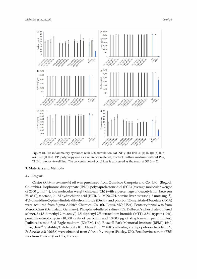
Molecules 2019, 24, 237 20 of 30
Molecules 2019, 24, x FOR PEER REVIEW 20 of 29
Figure 18. Pro-inflammatory cytokines with LPS stimulation. (a) INF-γ; (b) TNF-α; (c) IL-1β; (d) IL-8;
(e) IL-6; (f) IL-2. PP: polypropylene as a reference material; Control: culture medium without PUs;
THP-1: monocyte cell line. The concentration of cytokines is expressed as the mean ± SD (n = 3).
3. Materials and Methods
3.1. Reagents
Castor (Ricinus communis) oil was purchased from Químicos Campota and Co. Ltd. (Bogotá,
Colombia). Isophorone diisocyanate (IPDI), polycaprolactone diol (PCL) (average molecular weight
of 2000 g mol−1), low molecular weight chitosan (Ch) (with a percentage of deacetylation between 75–
85%), n-octane, 0.1 M hydrochloric acid (HCl), 0.1 M NaOH, porcine liver esterase (18 units mg−1),
4′,6-diamidino-2-phenylindole dihydrochloride (DAPI), and phorbol 12-myristate-13-acetate (PMA)
were acquired from Sigma-Aldrich Chemical Co. (St. Louis, MO, USA). Pentaerythritol was from
Merck KGaA (Darmstadt, Germany). Phosphate-buffered saline (PBS: Dulbecco’s phosphate-
buffered saline), 3-(4,5-dimethyl-2-thiazolyl)-2,5-diphenyl-2H-tetrazolium bromide (MTT), 2.5%
trypsin (10), penicillin-streptomycin (10,000 units of penicillin and 10,000 μg of streptomycin per
milliliter), Dulbecco’s modified Eagle medium (DMEM, 1), Roswell Park Memorial Institute (RPMI)
1640, Live/dead® Viability/Cytotoxicity Kit, Alexa Flour™ 488 phalloidin, and lipopolysaccharide
(LPS, Escherichia coli 026:B6) were obtained from Gibco/Invitrogen (Paisley, UK). Fetal bovine serum
(FBS) was from Eurobio (Les Ulis, France).
3.2. Biological Material
Human acute monocytic leukemia cell line, THP-1 (ATCC® TIB-202™). Mouse subcutaneous
connective tissue fibroblasts L929 (ATCC® CCL-1). Human lung fibroblasts MRC-5 (ATCC® CCL-
Figure 18. Pro-inflammatory cytokines with LPS stimulation. (a) INF-γ; (b) TNF-α; (c) IL-1β; (d) IL-8;(e) IL-6; (f) IL-2. PP: polypropylene as a reference material; Control: culture medium without PUs;THP-1: monocyte cell line. The concentration of cytokines is expressed as the mean ± SD (n = 3).
3. Materials and Methods
3.1. Reagents
Castor (Ricinus communis) oil was purchased from Químicos Campota and Co. Ltd. (Bogotá,Colombia). Isophorone diisocyanate (IPDI), polycaprolactone diol (PCL) (average molecular weightof 2000 g mol−1), low molecular weight chitosan (Ch) (with a percentage of deacetylation between75–85%), n-octane, 0.1 M hydrochloric acid (HCl), 0.1 M NaOH, porcine liver esterase (18 units mg−1),4′,6-diamidino-2-phenylindole dihydrochloride (DAPI), and phorbol 12-myristate-13-acetate (PMA)were acquired from Sigma-Aldrich Chemical Co. (St. Louis, MO, USA). Pentaerythritol was fromMerck KGaA (Darmstadt, Germany). Phosphate-buffered saline (PBS: Dulbecco’s phosphate-bufferedsaline), 3-(4,5-dimethyl-2-thiazolyl)-2,5-diphenyl-2H-tetrazolium bromide (MTT), 2.5% trypsin (10×),penicillin-streptomycin (10,000 units of penicillin and 10,000 µg of streptomycin per milliliter),Dulbecco’s modified Eagle medium (DMEM, 1×), Roswell Park Memorial Institute (RPMI) 1640,Live/dead® Viability/Cytotoxicity Kit, Alexa Flour™ 488 phalloidin, and lipopolysaccharide (LPS,Escherichia coli 026:B6) were obtained from Gibco/Invitrogen (Paisley, UK). Fetal bovine serum (FBS)was from Eurobio (Les Ulis, France).
Molecules 2019, 24, 237 21 of 30
3.2. Biological Material
Human acute monocytic leukemia cell line, THP-1 (ATCC® TIB-202™). Mouse subcutaneousconnective tissue fibroblasts L929 (ATCC® CCL-1). Human lung fibroblasts MRC-5 (ATCC®
CCL-171™). Adult human dermal fibroblast HDFa (ATCC® PCS-201-012™). All biological materialswere acquired from the strain library of the Universidad de La Sabana (Chía, Colombia).
3.3. Obtaining Polyols
The polyols were derived from castor oil (P.1, P.2, and P.3). P.1 corresponded to commercialunmodified castor oil. Polyols P.2 and P.3 were obtained by transesterification with pentaerythritol [72].For the reaction, the castor oil temperature was raised to 120 ◦C for 10 min. The temperature was thenfurther increased to 210 ◦C, adding pentaerythritol (1.32% and 2.64% mol pentaerythritol/mol castoroil for P.2 and P.3, respectively) and 0.05% lead oxide as a catalyst. The mixture was maintained at210 ◦C for 2 h. After the reaction time, the catalyst was removed by decantation for 24 h and filtration.The hydroxyl number of the polyols were determined according to ASTM D1957-86 [73].
3.4. Synthesis of Polyurethanes
The PUs were synthesized by the pre-polymer method. The polyol was brought to 60 ◦C, andthe diisocyanate was added the reactor at a constant NCO:OH ratio (1:1) [74], and maintained at 300rpm for 5 min. Them the PCL (15% w/w of oil polyol weight) and Ch (3% w/w of oil polyol weight)were then added and maintained at 300 rpm for 5 min additional. The formation of the PU sheetswas carried out by pouring the prepolymer into a steel mold. Twelve PU matrices were synthesizedin sheets (15 cm × 9 cm × 0.3 cm (length × width × height)). Curing was performed at 110 ◦C for12 h [72]. The synthesized PUs are identified with the following nomenclature: Pn-xCh-yPCL, where nrepresents the polyol used (1 for unmodified castor oil, 2 for castor oil with 1.32% pentaerythritol/mol,and 3 for the castor oil with 2.64% pentaerythritol/mol), and x and y represent the percentage of Chand PCL, respectively.
3.5. Mechanical Tests
A universal testing machine EZ-LX (Shimadzu, Kyoto, Japan) was used to determine maximumstress, percent elongation, and Young’s modulus of the polyurethanes (following the ASTM D638-10standard). A load cell of 5 kN with a crosshead speed of 25 mm min−1 was used [75,76]. Three samplesof 40 mm × 6 mm × 3 mm (length × width × thickness) were tested.
3.6. Fourier-Transform Infrared Spectroscopy (FTIR)
Chemical structures were evaluated by an ATR-FTIR spectrometer (Bruker Alpha, Billerica, MA,USA), in the range from 400 to 4000 cm−1. The spectrum corresponds to the average of 24 scans at aspectral resolution of 4 cm−1 [40,77].
3.7. Thermal Analysis
3.7.1. Thermogravimetric Analysis
Thermal behavior was evaluated in a TGA/DCS1 thermogravimetric analyzer coupled to DSC(Mettler-Toledo Inc., Schwerzenbach, Switzerland). According to ASTM D6370, the conditions usedwere as follows: heating speed of 25 ◦C min−1, a temperature range of 25–600 ◦C, nitrogen atmosphere,and samples of 15 ± 2 mg [48].

Molecules 2019, 24, 237 22 of 30
3.7.2. Differential Scanning Calorimetry
Glass transition temperatures were determined by a DSC 3+ analyzer (Mettler-Toledo, Columbus,OH, USA). The conditions were as follows: temperature range from −70 ◦C to 150 ◦C, nitrogenatmosphere with 20 mL min−1 flow, and sample weights of 10 ± 2 mg [78].
3.8. Hydrophilic Character
3.8.1. Contact Angle
A “Drop Shape Analysis—DSA” device (GH11, Krüss, Hamburg, Germany) was used to measurethe contact angle. According to ASTM-D7334-08 (2013), it was measured with the sessile drop methodusing 10 µL of distilled water at 20 ◦C [79]. Ten measurements of each material were performed.
3.8.2. Water Absorption Rate
To remove uncross-linked chains and monomer residues after synthesis, the PU materials werewashed with ethanol for two days, renewing the ethanol every day. The solvent was then replaced bydeionized water for two more days. The materials were dried in a vacuum chamber (0.01 mm Hg) at37 ◦C for 24 h [54]. The rate of water absorption was determined by immersing the sample in distilledwater until constant weight. The residual water was then removed from the samples with dry filterpaper, and the samples were weighed [80]. All tests were performed in triplicate. The rate of waterabsorption at equilibrium was calculated by comparing the mass of the sample (mt) after obtainingconstant weight with the initial mass (mi) of the sample using Equation (1):
% Water absortion of PUs = (mi −mt)/mi (1)
3.9. Determination of Density
The material density was determined using the Archimedes immersion technique using a MettlerAX 205 balance (Mettler-Toledo Inc.) with a sensitivity of 0.01 mg and a Mettler ME-33360 densitydetermination kit. Dry samples of 5 mm × 5 mm × 3 mm were used to calculate the density intriplicate. The PUs were weighed in the air (mair) and then immersed in n-octane with a known density(ρn-octane), and the immersion weight (mn-octane) was obtained at 20 ◦C. The density was calculated bythe following Equation (2) [54]:
Density of PUs = (maire × ρn−octano)/(maire −mn−octano) (2)
3.10. Dynamic Mechanical Thermal Analysis (DMTA)
The effect of temperature on the mechanical properties was evaluated by a DMTA test. A DMA8000 thermomechanical analyzer (Perkin-Elmer, Waltham, MA, USA) was used at a frequency of 1 Hz,a deformation of 0.1%, and a temperature program between −90 ◦C and 150 ◦C, with a heating rate of5 ◦C min−1. The storage modulus and the loss factor, tanδ [38], were determined.
3.11. Field Emission Scanning Electron Microscopy (FESEM)
Morphological characterization of the PUs was performed with a field-emission scanning electronmicroscope (FESEM, ZEISS ULTRA 55 from Oxford Instruments (Abingdon, UK). The PUs werewashed with ethanol for two days, renewing the ethanol every day. The solvent was then replaced bydeionized water for two more days. The materials were dried in a vacuum chamber (0.01 mm Hg) at37 ◦C for 24 h [54]. The samples were coated with platinum to observe the morphology of the materialswith an accelerating voltage of 5 kV [81].

Molecules 2019, 24, 237 23 of 30
3.12. In Vitro Biodegradability Tests
Biodegradability tests were performed in independent tests in triplicate following ASTM F1635-11.To remove uncross-linked chains and monomer residues, the PUs were washed with ethanol for twodays, renewing the ethanol each day. The solvent was then replaced by deionized water for two moredays. The materials were dried in a vacuum chamber at 37 ◦C for 24 h [54]. Samples of 5 mm × 5 mm× 3 mm were placed in the biodegradation media and incubated at 37 ◦C for three months, exceptfor the enzyme medium (10 units mg−1 solid), which was for 21 days. After this time, the sampleswere washed with distilled water, dried in a vacuum chamber, and weighed. The reagents usedwere: 0.1 M HCl medium, 0.1 M NaOH medium, and porcine liver esterase enzymatic medium. Thebiodegradation rate was calculated by comparing the dry weight (wf) of the sample after degradationduring the predetermined time with the initial dry weight (wi) of the sample using Equation (3) [81,82]:
% Degradation of PUs = (wi −wt)/wi (3)
3.13. In Vitro Cell Viability Assay by the MTT Method
L929 mouse fibroblasts and MRC-5 and HDFa human fibroblasts were cultured in DMEMsupplemented with 10% FBS and 1% penicillin-streptomycin in T-75 cell culture flasks. They weregrown at 37 ◦C and 5% CO2. The cell culture medium was changed every 48 h [83]. At 100% confluence,the cells were trypsinized (trypsin-EDTA) for viability analysis.
The effect of polyurethanes on cell viability was evaluated by the MTT method defined byISO/CD 10993-5. Cells were seeded in 96-well plates at a concentration of 4.0 × 10⁴ cells per wellin supplemented medium and cultured at 37 ◦C and 5% CO2 for 24 h. Subsequently, PU cylindersof 3 mm × 2 mm (diameter × height) (previously sterilized under ultraviolet UV light (260 nm) for30 min on each side [33]) were placed in 100 µL of supplemented medium. The materials were leftin contact with the polymers for 24 h at 37 ◦C and 5% CO2. The supernatant and polymers werethen removed, and the MTT solution (12 mM in PBS) was added to a total volume of 100 µL andincubated for 4 h at 37 ◦C. The supernatant was removed, and 100 µL of dimethyl sulfoxide wasadded and incubated for 15 min at 37 ◦C. The optical density was then determined in a plate reader(Bio-Tek ELx800 Microplate Reader, Highland Park, Winooski, VT, USA) at 570 nm. A polypropylenebiomaterial (PP) was used as a positive control for cell viability, and doxorubicin (DOXO) was used asa negative control. All tests were performed in triplicate. Cell viability was determined according toEquation (4):
% Cell viability of PUs = Abssample/Abscontrol (4)
where Abssample corresponds to the absorbance value of the cells after treatment with the PU, andAbscontrol corresponds to the cells without treatment.
3.14. Immunocytochemical Techniques
In vitro cell viability assay by the live/dead kit. Cell viability was also evaluated by the live/deadkit based on plasmatic membrane integrity and intracellular esterase activity [84]. L929 mousefibroblasts were cultured in RPMI supplemented with 10% FBS and 1% penicillin-streptomycin in T-75cell culture flasks at 37 ◦C and 5% CO2. The cell culture medium was changed every 48 h [83]. At 100%confluence, the cells were trypsinized (trypsin-EDTA) for viability analysis.
Prior to the assay, the PUs were washed with ethanol and water for 3 days and dried in a vacuumchamber at 37 ◦C for 24 h [54]. PU sheets with a diameter of 4 mm (previously sterilized under UVlight (260 nm) for 30 min on each side) were placed in 96-well plates [33]. Five microliters of cells,at a concentration of 1.0 × 10⁴ cells mL−1, were then seeded onto each material using supplementedmedium and incubated at 37 ◦C and 5% CO2 for 30 min. Next, 95 µL of supplemented medium wasadded and incubated for 24 h at 37 ◦C and 5% CO2 [54]. The supernatant was removed, and 2 µL ofcalcein AM (live cell marker) staining solution and 4 µL of ethidium homodimer (dead cell marker)
Molecules 2019, 24, 237 24 of 30
were added and incubated for 20 min in the dark with stirring [84]. Fluorescence measurement wasperformed for calcein in a Victor 1420 Multilabel Counter spectrophotometer (Perkin-Elmer, Waltham,MA, USA) at 485 nm to determine the concentration of living cells. PS was used as a positive controland latex as a negative control. All tests were performed in triplicate.
Fixation and morphological analysis with phalloidin and DAPI. L929 fibroblasts in contact with thematerial after 24 h of incubation were washed with PBS and fixed for 20 min with paraformaldehyde4% w/v at 20 ◦C. They were then washed twice with PBS at 4 ◦C. Phalloidin (Alexa Flour™ 488phalloidin) was added and incubated for 20 min to reveal F-actin. The samples were then washedtwice with PBS. The cell nuclei were stained for 5 min with DAPI at a 1/5000 dilution in PBS, followedby washing twice with PBS. Next, 0.05% sodium azide was added. The cell structure was observedusing an Eclipse 80i optical microscope (Nikon, Tokyo, Japan) equipped with a Nikon IntensilightIlluminator [54]. The images were analyzed by ImageJ software version 1.45k (Bethesda, MD, USA).
3.15. Differentiation into Macrophages and Inflammation Stimulation
A human acute monocytic leukemia cell line, THP-1, was used. The cells were cultured at 37 ◦Cand 5% CO2 in RPMI medium supplemented with 10% FBS and 1% penicillin-streptomycin in T-75cell culture flasks. For cell differentiation into macrophages, the cells were centrifuged at 2000 rpm for10 min, resuspended in 3 mL of RPMI culture medium and seeded into 24-well plates [69] by adding200 mM of PMA and incubating for 48 h [27]. The differentiated cells (macrophages) were washedtwice with PBS to remove undifferentiated cells [26]. The macrophages that had been differentiatedwith PMA were stimulated with 700 ng mL−1 of LPS for 3 h [22].
3.16. Evaluation of Inflammatory Processes
With the THP-1 cell line and PUs, two independent tests were performed to evaluate theinflammatory processes of the materials in contact with the cells. The first test (group A) consisted inthe direct contact of PUs on the cells differentiated into macrophages with PMA, for 24 h, to determinewhether the PUs caused inflammation after contact with the cell line. The next test group (group B)consisted of PUs in contact for 24 h with macrophages differentiated with PMA and stimulated withLPS to determine whether the materials had anti-inflammatory activity.
3.17. Immunoassay
The MILLIPLEX® MAP kit for flow cytometry was used to evaluate cytokine production (IFN-γ,IL-1β, IL-2, IL-4, IL-5, IL-6, IL-8, IL-10, and TNF-α) and release to the supernatant of the macrophagescultured in contact with PUs for 24 h, following the instructions provided by the manufacturers.A mixture of beads with different fluorescence intensities, coated with antibodies for the mentionedcytokines, was pre-incubated in the dark for one hour on an orbital shaker. Concentration standardsof 3.2, 16, 80, 400, 2000 and 10,000 pg mL−1 were used to determine the concentration curve of eachanalyte. In a 96-well plate, 200 µL of wash buffer was added, the plate was shaken for 10 min, andthe supernatant was discarded. Twenty-five microliters of the standards, 25 µL of running buffer,25 µL of the stock solution, 25 µL of supernatant from cells in the appropriate wells, and 25 µL of thebead mixture were added. The plate was incubated overnight with shaking at 4 ◦C. The supernatantwas then removed, and the samples were washed twice with 200 µL of wash buffer. Next, 25 µLof antibody was added for detection, and the samples were incubated for 1 h in the dark and withshaking. Twenty-five microliters of the streptavidin phycoerythrin binding protein was then addedand incubated for 30 min with shaking in the dark. The supernatant was discarded, and the wells werewashed twice with 200 µL of wash buffer. One hundred and fifty microliters of the coating liquid werethen added, and the plate was read on a MAGPIX flow cytometer from MILLIPLEX MAP (Darmstadt,Germany). The results of the samples were analyzed using the xPONENT MAGPIX software (Madison,WI, USA).
Molecules 2019, 24, 237 25 of 30
3.18. Statistical Analysis
The results were expressed as mean values ± standard deviation (SD). The data were analyzedby means of an analysis of variance (ANOVA) and the significant differences were determined forp < 0.05. For the comparison between samples the t-Student test was used with the SPSS StatisticsSoftware v.23 (IBM, Armonk, NY, USA).
4. Conclusions
PUs synthesized with castor oil polyols, isophorone diisocyanate, 15% w/w polycaprolactoneand 3% w/w chitosan were used to evaluate their mechanical, physicochemical, morphological,biodegradability, and biocompatibility characteristics, along with their possible inflammatory effects.The type of polyol and additive showed a significant impact on the maximum stress, percent elongation,and contact angle of the material. Chemical modification of the polyols improves the evaluatedproperties due to the increased cross-linking of the resulting materials. The resulting mechanicalproperties of the PUs verified their dependence on the presence of hydrogen bonds, the numberof hydroxyl groups in the polyols, and the interactions between the hard and soft segments of thematrix. The polyol with the highest hydroxyl index showed the highest values for the mechanicaland thermal properties. The percent elongation, with a maximum of 265%, can be used to obtainresistant materials with flexibility, which allows designing biomaterials that do not cause injuries tosoft tissues. The degradation rates under the study conditions showed values of less than 4%, so itcan be inferred that cross-linking of the synthesized PUs hinders the degradation process. In vitrocell viability was determined using L929 mouse fibroblasts, human fibroblasts (MRC-5) and adulthuman dermal fibroblast (HDFa). The cell viability results of the PUs in contact with the three cell linesdemonstrated that there was no effect on cell viability; therefore, it is likely that these materials couldbe used for direct contact with the skin without causing damage to surrounding cells. Cell viabilitywas also determined by the live/dead kit for the L929 mouse fibroblasts. Due to the biocompatibleproperties of the PUs evaluated in this study, it can be inferred that they are suitable for use inbiomedical applications as materials for non-absorbable biomedical sutures.
Author Contributions: Conceptualization, M.F.V., Y.L.U., L.E.D., J.A.G.-T., and A.V.-L.; Methodology, Y.L.U.;Formal analysis, Y.L.U.; Investigation, Y.L.U., G.V.-F. and M.A.S.; Resources, M.F.V.; Writing—original draftpreparation, Y.L.U.; Visualization, Y.L.U.; Supervision, M.F.V. and L.E.D.; Project administration, M.F.V.; Fundingacquisition, M.F.V. and L.E.D.
Funding: This research was funded by the UNIVERSIDAD DE LA SABANA, grant number ING-202-2018 andby COLCIENCIAS under scholarship grant 617-2-2014. CIBER-BBN is an initiative funded by the VI NationalR&D&I Plan 2008–2011, Iniciativa Ingenio 2010, Consolider Program. CIBER Actions are financed by the Institutode Salud Carlos III with assistance from the European Regional Development Fund. J.A.G.-T. and A.V.-LL.acknowledge the support of the Spanish Ministry of Economy and Competitiveness (MINECO) through projectDPI2015-65401-C3-2-R (including FEDER financial support).
Acknowledgments: The authors thank the Universidad de La Sabana for financing research project ING-202-2018which is part of this research, Colciencias for the doctoral scholarship under grant 617-2 of 2014, and the UniversitatPolitècnica de València for assistance and advice with the equipment.
Conflicts of Interest: The authors declare no conflict of interest.
References
1. Alishiri, M.; Shojaei, A.; Abdekhodaie, M.J.; Yeganeh, H. Synthesis and characterization of biodegradableacrylated polyurethane based on poly(ε-caprolactone) and 1,6-hexamethylene diisocyanate. Mater. Sci.Eng. C 2014, 42, 763–773. [CrossRef] [PubMed]
2. Bakhshi, H.; Yeganeh, H.; Yari, A.; Nezhad, S.K. Castor oil-based polyurethane coatings containing benzyltriethanol ammonium chloride: Synthesis, characterization, and biological properties. J. Mater. Sci. 2014, 49,5365–5377. [CrossRef]

Molecules 2019, 24, 237 26 of 30
3. Kucinska-Lipka, J.; Gubanska, I.; Janik, H.; Sienkiewicz, M. Fabrication of polyurethane and polyurethanebased composite fibres by the electrospinning technique for soft tissue engineering of cardiovascular system.Mater. Sci. Eng. C Mater. Biol. Appl. 2015, 46, 166–176. [CrossRef] [PubMed]
4. Tsai, M.-C.; Hung, K.-C.; Hung, S.-C.; Hsu, S. Evaluation of biodegradable elastic scaffolds made of anionicpolyurethane for cartilage tissue engineering. Colloids Surf. B Biointerfaces 2015, 125, 34–44. [CrossRef][PubMed]
5. Rocco, K.A.; Maxfield, M.W.; Best, C.A.; Dean, E.W.; Breuer, C.K. In vivo applications of electrospuntissue-engineered vascular grafts: A review. Tissue Eng. Part B 2014, 20, 628–640. [CrossRef]
6. Park, H.; Gong, M.-S.; Park, J.-H.; Moon, S.-I.; Wall, I.B.; Kim, H.-W.; Lee, J.H.; Knowles, J.C. Silkfibroin-polyurethane blends: Physical properties and effect of silk fibroin content on viscoelasticity,biocompatibility and myoblast differentiation. Acta Biomater. 2013, 9, 8962–8971. [CrossRef]
7. Rajan, K.P.; Al-ghamdi, A.; Parameswar, R.; Nando, G.B. Blends of thermoplastic polyurethane andpolydimethylsiloxane rubber: Assessment of biocompatibility and suture holding strength of membranes.Int. J. Biomater. 2013, 2013. [CrossRef]
8. Rodríguez-Galán, A.; Franco, L.; Puiggal, J. Biodegradable Polyurethanes and Poly(ester amide)s. InHandbook of Biodegradable Polymers: Synthesis, Characterization and Applications; Lendlein, A., Sisson, A., Eds.;John Wiley & Sons: Hoboken, NJ, USA, 2011; pp. 133–154.
9. Adolph, E.J.; Pollins, A.C.; Cardwell, N.L.; Davidson, J.M.; Guelcher, S.A.; Nanney, L.B. Biodegradablelysine-derived polyurethane scaffolds promote healing in a porcine full-thickness excisional wound model.J. Biomater. Sci. Polym. Ed. 2014, 25, 1973–1985. [CrossRef]
10. Shourgashti, Z.; Khorasani, M.T.; Khosroshahi, S.M.E. Plasma-induced grafting of polydimethylsiloxane ontopolyurethane surface: Characterization and in vitro assay. Radiat. Phys. Chem. 2010, 79, 947–952. [CrossRef]
11. Qiu, H.; Li, D.; Chen, X.; Fan, K.; Ou, W.; Chen, K.C.; Xu, K. Synthesis, characterizations, and biocompatibilityof block poly(ester-urethane)s based on biodegradable poly(3-hydroxybutyrate-co-4-hydroxybutyrate)(P3/4HB) and poly(ε-caprolactone). J. Biomed. Mater. Res. A 2013, 101, 75–86. [CrossRef]
12. Morral-Ruíz, G.; Melgar-Lesmes, P.; García, M.L.; Solans, C.; García-Celma, M.J. Polyurethane andpolyurea nanoparticles based on polyoxyethylene castor oil derivative surfactant suitable for endovascularapplications. Int. J. Pharm. 2014, 461, 1–13. [CrossRef] [PubMed]
13. Dulinska-Molak, I.; Lekka, M.; Kurzydłowski, K.J. Surface properties of polyurethane composites forbiomedical applications. Appl. Surf. Sci. 2013, 270, 553–560. [CrossRef]
14. Chan-Chan, L.H.; Solis-Correa, R.; Vargas-Coronado, R.F.; Cervantes-Uc, J.M.; Cauich-Rodríguez, J.V.;Quintana, P.; Bartolo-Pérez, P. Degradation studies on segmented polyurethanes prepared with HMDI, PCLand different chain extenders. Acta Biomater. 2010, 6, 2035–2044. [CrossRef] [PubMed]
15. Usman, A.; Zia, K.M.; Zuber, M.; Tabasum, S.; Rehman, S.; Zia, F. Chitin and chitosan based polyurethanes: Areview of recent advances and prospective biomedical applications. Int. J. Biol. Macromol. 2016, 86, 630–645.[CrossRef] [PubMed]
16. Wu, C.-S. Enhanced antibacterial activity, antioxidant and in vitro biocompatibility of modifiedpolycaprolactone-based membranes. Int. J. Polym. Mater. Polym. Biomater. 2016, 65, 872–880. [CrossRef]
17. Anirudhan, T.S.; Nair, S.S.; Nair, A.S. Fabrication of a bioadhesive transdermal device from chitosan andhyaluronic acid for the controlled release of lidocaine. Carbohydr. Polym. 2016, 152, 687–698. [CrossRef]
18. Kaur, G.; Mahajan, M.; Bassi, P. Derivatized Polysaccharides: Preparation, characterization, and applicationas bioadhesive polymer for drug delivery. Int. J. Polym. Mater. 2013, 62, 475–481. [CrossRef]
19. Wu, H.; Williams, G.R.; Wu, J.; Wu, J.; Niu, S.; Li, H.; Wang, H.; Zhu, L. Regenerated chitin fibers reinforcedwith bacterial cellulose nanocrystals as suture biomaterials. Carbohydr. Polym. 2018, 180, 304–313. [CrossRef]
20. Aranguren, M.I.; González, J.F.; Mosiewicki, M.A. Biodegradation of a vegetable oil based polyurethane andwood flour composites. Polym. Test. 2012, 31, 7–15. [CrossRef]
21. Guelcher, S.; Srinivasan, A.; Dumas, J. Synthesis, mechanical properties, biocompatibility, and biodegradationof polyurethane networks from lysine polyisocyanates. Biomaterials 2008, 29, 1762–1775. [CrossRef]
22. Chanput, W.; Mes, J.; Vreeburg, R.A.M.; Savelkoul, H.F.J.; Wichers, H.J. Transcription profiles ofLPS-stimulated THP-1 monocytes and macrophages: A tool to study inflammation modulating effectsof food-derived compounds. Food Funct. 2010, 1, 254–261. [CrossRef] [PubMed]
23. Oliveira, C.M.; Sakata, R.K.; Issy, A.M.; Gerola, L.R. Citocinas y dolor. Rev. Bras. Anestesiol. 2011, 61, 137–142.[CrossRef]
Molecules 2019, 24, 237 27 of 30
24. Small, A.; Lansdown, N.; Al-Baghdadi, M.; Quach, A.; Ferrante, A. Facilitating THP-1 macrophage studiesby differentiating and investigating cell functions in polystyrene test tubes. J. Immunol. Methods 2018, 461,73–77. [CrossRef] [PubMed]
25. Lund, M.E.; To, J.; O’Brien, B.A.; Donnelly, S. The choice of phorbol 12-myristate 13-acetate differentiationprotocol influences the response of THP-1 macrophages to a pro-inflammatory stimulus. J. Immunol. Methods2016, 430, 64–70. [CrossRef] [PubMed]
26. Ballerini, P.; Diomede, F.; Petragnani, N.; Cicchitti, S.; Merciaro, I.; Cavalcanti, M.F.X.B.; Trubiani, O.Conditioned medium from relapsing-remitting multiple sclerosis patients reduces the expression andrelease of inflammatory cytokines induced by LPS-gingivalis in THP-1 and MO3.13 cell lines. Cytokine 2017,96, 261–272. [CrossRef] [PubMed]
27. Dreskin, S.C.; Thomas, G.W.; Dale, S.N.; Heasley, L.E. Isoforms of Jun kinase are differentially expressedand activated in human monocyte/macrophage (THP-1) cells. J. Immunol. 2001, 166, 5646–5653. [CrossRef][PubMed]
28. Dash, B.C.; Thomas, D.; Monaghan, M.; Carroll, O.; Chen, X.; Woodhouse, K.; Brien, T.O.; Pandit, A.An injectable elastin-based gene delivery platform for dose- dependent modulation of angiogenesis andinflammation for critical limb ischemia. Biomaterials 2015, 65, 126–139. [CrossRef] [PubMed]
29. Lin, T.H.; Yao, Z.; Sato, T.; Keeney, M.; Li, C.; Pajarinen, J.; Yang, F.; Egashira, K.; Goodman, S.B. Suppressionof wear-particle-induced pro-inflammatory cytokine and chemokine production in macrophages via NF-κBdecoy oligodeoxynucleotide: A preliminary report. Acta Biomater. 2014, 10, 3747–3755. [CrossRef] [PubMed]
30. Zhang, C.; Garrison, T.F.; Madbouly, S.A.; Kessler, M.R. Recent advances in vegetable oil-based polymersand their composites. Prog. Polym. Sci. 2017, 71, 91–143. [CrossRef]
31. Laube, T.; Weisser, J.; Berger, S.; Börner, S.; Bischoff, S.; Schubert, H.; Gajda, M.; Bräuer, R.; Schnabelrauch, M.In situ foamable, degradable polyurethane as biomaterial for soft tissue repair. Mater. Sci. Eng. C 2017, 78,163–174. [CrossRef]
32. Temenoff, J.S.; Mikos, A.G. Biomaterials: The Intersection of Biology and Materials Science, 8th ed.; Prentice Hall,Inc.: Upper Saddle River, NJ, USA, 2008.
33. Vannozzi, L.; Ricotti, L.; Santaniello, T.; Terencio, T.; Oropesa-Nunez, R.; Canale, C.; Borghi, F.; Menciassi, A.;Lenardi, C.; Gerges, I. 3D porous polyurethanes featured by different mechanical properties: Characterizationand interaction with skeletal muscle cells. J. Mech. Behav. Biomed. Mater. 2017, 75, 147–159. [CrossRef][PubMed]
34. Chashmejahanbin, M.R.; Daemi, H.; Barikani, M.; Salimi, A. Noteworthy impacts of polyurethane-ureaionomers as the efficient polar coatings on adhesion strength of plasma treated polypropylene. Appl. Surf. Sci.2014, 317, 688–695. [CrossRef]
35. Braun, U.; Lorenz, E.; Weimann, C.; Sturm, H.; Karimov, I.; Ettl, J.; Meier, R.; Wohlgemuth, W.A.; Berger, H.;Wildgruber, M. Mechanic and surface properties of central-venous port catheters after removal: A comparisonof polyurethane and silicon rubber materials. J. Mech. Behav. Biomed. Mater. 2016, 64, 281–291. [CrossRef][PubMed]
36. Jutrzenka Trzebiatowska, P.; Santamaria Echart, A.; Calvo Correas, T.; Eceiza, A.; Datta, J. The changes ofcrosslink density of polyurethanes synthesised with using recycled component. Chemical structure andmechanical properties investigations. Prog. Org. Coat. 2018, 115, 41–48. [CrossRef]
37. Thakur, S.; Karak, N. Castor oil-based hyperbranched polyurethanes as advanced surface coating materials.Prog. Org. Coat. 2013, 76, 157–164. [CrossRef]
38. Gurunathan, T.; Mohanty, S.; Nayak, S.K. Isocyanate terminated castor oil-based polyurethane prepolymer:Synthesis and characterization. Prog. Org. Coat. 2015, 80, 39–48. [CrossRef]
39. Chen, H.; Yu, X.; Zhou, W.; Peng, S.; Zhao, X. Highly toughened polylactide (PLA) by reactive blending withnovel polycaprolactone-based polyurethane (PCLU) blends. Polym. Test. 2018, 70, 275–280. [CrossRef]
40. Shah, S.A.A.; Imran, M.; Lian, Q.; Shehzad, F.K.; Athir, N.; Zhang, J.; Cheng, J. Curcumin incorporatedpolyurethane urea elastomers with tunable thermo-mechanical properties. React. Funct. Polym. 2018, 128,97–103. [CrossRef]
41. Cakic, S.M.; Ristic, I.S.; Cincovic, M.M.; Nikolic, N.C.; Nikolic, L.; Cvetinov, M.J. Synthesis and propertiesbiobased waterborne polyurethanes from glycolysis product of PET waste and poly (caprolactone) diol.Prog. Org. Coat. 2017, 105, 111–122. [CrossRef]

Molecules 2019, 24, 237 28 of 30
42. Ferreira, P.; Pereira, R.; Coelho, J.F.J.; Silva, A.F.M.; Gil, M.H. Modification of the biopolymer castor oil withfree isocyanate groups to be applied as bioadhesive. Int. J. Biol. Macromol. 2007, 40, 144–152. [CrossRef][PubMed]
43. Arévalo, F.; Uscategui, Y.L.; Diaz, L.; Cobo, M.; Valero, M.F. Effect of the incorporation of chitosan onthe physico-chemical, mechanical properties and biological activity on a mixture of polycaprolactone andpolyurethanes obtained from castor oil. J. Biomater. Appl. 2016, 31, 708–720. [CrossRef] [PubMed]
44. Corcuera, M.A.; Rueda, L.; Fernandez d’Arlas, B.; Arbelaiz, A.; Marieta, C.; Mondragon, I.; Eceiza, A.Microstructure and properties of polyurethanes derived from castor oil. Polym. Degrad. Stab. 2010, 95,2175–2184. [CrossRef]
45. Uscátegui, Y.L.; Arévalo-Alquichire, S.J.; Gómez-Tejedor, J.A.; Vallés-Lluch, A.; Díaz, L.E.; Valero, M.F.Polyurethane-based bioadhesive synthesized from polyols derived from castor oil (Ricinus communis) andlow concentration of chitosan. J. Mater. Res. 2017, 32, 3699–3711. [CrossRef]
46. Saikia, A.; Karak, N. Renewable resource based thermostable tough hyperbranched epoxy thermosets assustainable materials. Polym. Degrad. Stab. 2017, 135, 8–17. [CrossRef]
47. Thakur, S.; Hu, J. Polyurethane: A Shape Memory Polymer (SMP). In Aspects of Polyurethanes; Yilmaz, F., Ed.;InTechOpen: London, UK, 2017; pp. 53–71.
48. Sáenz-Pérez, M.; Lizundia, E.; Laza, J.M.; García-Barrasa, J.; Vilas, J.L.; León, L.M. Methylene diphenyldiisocyanate (MDI) and toluene diisocyanate (TDI) based polyurethanes: Thermal, shape-memory andmechanical behavior. RSC Adv. 2016, 6, 69094–69102. [CrossRef]
49. Hou, Z.; Zhang, H.; Qu, W.; Xu, Z.; Han, Z. Biomedical segmented polyurethanes based on polyethyleneglycol, poly(ε-caprolactone-co-D,L-lactide), and diurethane diisocyanates with uniform hard segment:Synthesis and properties. Int. J. Polym. Mater. Polym. Biomater. 2016, 65, 947–956. [CrossRef]
50. Mi, H.Y.; Jing, X.; Hagerty, B.S.; Chen, G.; Huang, A.; Turng, L.S. Post-crosslinkable biodegradablethermoplastic polyurethanes: Synthesis, and thermal, mechanical, and degradation properties. Mater. Des.2017, 127, 106–114. [CrossRef]
51. Gossart, A.; Battiston, K.G.; Gand, A.; Pauthe, E.; Santerre, J.P. Mono vs multilayer fibronectin coatingson polar/hydrophobic/ionic polyurethanes: Altering surface interactions with human monocytes. ActaBiomater. 2018, 66, 129–140. [CrossRef]
52. Marques, D.S.; Santos, J.M.C.; Ferreira, P.; Correia, T.R.; Correia, I.J.; Gil, M.H.; Baptista, C.M.S.G.Photocurable bioadhesive based on lactic acid. Mater. Sci. Eng. C 2016, 58, 601–609. [CrossRef]
53. Sheikh, Z.; Khan, A.S.; Roohpour, N.; Glogauer, M.; Rehman, I.U. Protein adsorption capability onpolyurethane and modified-polyurethane membrane for periodontal guided tissue regeneration applications.Mater. Sci. Eng. C 2016, 68, 267–275. [CrossRef]
54. Conejero-García, Á.; Gimeno, H.R.; Sáez, Y.M.; Vilariño-Feltrer, G.; Ortuño-Lizarán, I.; Vallés-Lluch, A.Correlating synthesis parameters with physicochemical properties of poly(glycerol sebacate). Eur. Polym. J.2017, 87, 406–419. [CrossRef]
55. Carriço, C.S.; Fraga, T.; Pasa, V.M.D. Production and characterization of polyurethane foams from a simplemixture of castor oil, crude glycerol and untreated lignin as bio-based polyols. Eur. Polym. J. 2016, 85, 53–61.[CrossRef]
56. Fuentes, L.E.; Pérez, S.; Martínez, S.I.; García, Á.R. Redes poliméricas interpenetradas de poliuretano apartir de aceite de ricino modificado y poliestireno: Miscibilidad y propiedades mecánicas en función de lacomposición. Revisata Ion. 2011, 24, 45–50.
57. Das, B.; Konwar, U.; Mandal, M.; Karak, N. Sunflower oil based biodegradable hyperbranched polyurethaneas a thin film material. Ind. Crops Prod. 2013, 44, 396–404. [CrossRef]
58. Cherng, J.Y.; Hou, T.Y.; Shih, M.F.; Talsma, H.; Hennink, W.E. Polyurethane-based drug delivery systems.Int. J. Pharm. 2013, 450, 145–162. [CrossRef] [PubMed]
59. Spontón, M.; Casis, N.; Mazo, P.; Raud, B.; Simonetta, A.; Ríos, L.; Estenoz, D. Biodegradation study byPseudomonas sp. of flexible polyurethane foams derived from castor oil. Int. Biodeterior. Biodegradation 2013,85, 85–94. [CrossRef]
60. Gogoi, S.; Barua, S.; Karak, N. Biodegradable and thermostable synthetic hyperbranchedpoly(urethane-urea)s as advanced surface coating materials. Prog. Org. Coat. 2014, 77, 1418–1427. [CrossRef]

Molecules 2019, 24, 237 29 of 30
61. Calvo-Correas, T.; Santamaria-Echart, A.; Saralegi, A.; Martin, L.; Valea, Á.; Corcuera, M.A.; Eceiza, A.Thermally-responsive biopolyurethanes from a biobased diisocyanate. Eur. Polym. J. 2015, 70, 173–185.[CrossRef]
62. Reddy, T.T.; Kano, A.; Maruyama, A.; Takahara, A. Synthesis, characterization and drug release ofbiocompatible/biodegradable non-toxic poly(urethane urea)s based on poly(epsilon-caprolactone)s andlysine-based diisocyanate. J. Biomater. Sci. Polym. Ed. 2010, 21, 1483–1502. [CrossRef] [PubMed]
63. Coakley, D.N.; Shaikh, F.M.; O’Sullivan, K.; Kavanagh, E.G.; Grace, P.A.; McGloughlin, T.M. In vitroevaluation of acellular porcine urinary bladder extracellular matrix—A potential scaffold in tissue engineeredskin. Wound Med. 2015, 10–11, 9–16. [CrossRef]
64. Shahrousvand, M.; Sadeghi, G.M.M.; Shahrousvand, E.; Ghollasi, M.; Salimi, A. Superficial physicochemicalproperties of polyurethane biomaterials as osteogenic regulators in human mesenchymal stem cells fates.Colloids Surf. B Biointerfaces 2017, 156, 292–304. [CrossRef] [PubMed]
65. Aranaz, I.; Mengíbar, M.; Harris, R.; Paños, I.; Miralles, B.; Acosta, N.; Galed, G.; Heras, Á. Functionalcharacterization of chitin and chitosan. Curr. Chem. Biol. 2009, 3, 203–230.
66. Ortuno-Lizarán, I.; Vilarino-Feltrer, G.; Martinez-Ramos, C.; Pradas, M.M.; Vallés-Lluch, A. Influence ofsynthesis parameters on hyaluronic acid hydrogels intended as nerve conduits. Biofabrication 2016, 8, 1–12.[CrossRef] [PubMed]
67. Jing, X.; Mi, H.Y.; Huang, H.X.; Turng, L.S. Shape memory thermoplastic polyurethane(TPU)/poly(ε-caprolactone) (PCL) blends as self-knotting sutures. J. Mech. Behav. Biomed. Mater. 2016, 64,94–103. [CrossRef] [PubMed]
68. Starr, T.; Bauler, T.J.; Malik-Kale, P.; Steele-Mortimer, O. The phorbol 12-myristate-13-acetate differentiationprotocol is critical to the interaction of THP-1 macrophages with Salmonella Typhimurium. PLoS ONE 2018,13, 1–13. [CrossRef] [PubMed]
69. Park, E.K.; Jung, H.S.; Yang, H.I.; Yoo, M.C.; Kim, C.; Kim, K.S. Optimized THP-1 differentiation is requiredfor the detection of responses to weak stimuli. Inflamm. Res. 2007, 56, 45–50. [CrossRef] [PubMed]
70. Gómez Estrada, H.A.; González Ruiz, K.N.; Medina, J.D. Actividad antiinflamatoria de productos naturales.Bol. Latinoam. Caribe Plantas Med. Aromat. 2011, 10, 182–217.
71. González, R.; Zamora, Z.; Alonso, Y. Citocinas anti-inflamatorias y sus acciones y efectos en la sepsis y elchoque séptico. REDVET Rev. Electrónica Vet. 2009, 10, 1–11.
72. Valero, M.F.; Ortegón, Y. Polyurethane elastomers-based modified castor oil and poly(e-caprolactone) forsurface-coating applications: Synthesis, characterization, and in vitro degradation. J. Elastomers Plast. 2015,47, 360–369. [CrossRef]
73. Valero, M.F.; Pulido, J.E.; Ramírez, Á.; Cheng, Z. Determinación de la densidad de entrecruzamiento depoliuretanos obtenidos a partir de aceite de ricino modificado por transesterificación. Polímeros 2009, 19,14–21. [CrossRef]
74. Valero, M.F. Poliuretanos elastoméricos obtenidos a partir de aceite de ricino y almidón de yuca original ymodificado con anhidrido propiónico: Síntesis, propiedades fisicoquímicas y fisicomecánicas. Quim. Nov.2010, 33, 850–854. [CrossRef]
75. Simón-Allué, R.; Pérez-López, P.; Sotomayor, S.; Peña, E.; Pascual, G.; Bellón, J.M.; Calvo, B. Short- andlong-term biomechanical and morphological study of new suture types in abdominal wall closure. J. Mech.Behav. Biomed. Mater. 2014, 37, 1–11. [CrossRef] [PubMed]
76. Yoshida, K.; Jiang, H.; Kim, M.J.; Vink, J.; Cremers, S.; Paik, D.; Wapner, R.; Mahendroo, M.; Myers, K.Quantitative evaluation of collagen crosslinks and corresponding tensile mechanical properties in mousecervical tissue during normal pregnancy. PLoS ONE 2014, 9. [CrossRef] [PubMed]
77. Mekewi, M.A.; Ramadan, A.M.; ElDarse, F.M.; Abdel Rehim, M.H.; Mosa, N.A.; Ibrahim, M.A. Preparationand characterization of polyurethane plasticizer for flexible packaging applications: Natural oils affirmedaccess. Egypt. J. Pet. 2017, 26, 9–15. [CrossRef]
78. Hormaiztegui, M.E.V.; Aranguren, M.I.; Mucci, V.L. Synthesis and characterization of a waterbornepolyurethane made from castor oil and tartaric acid. Eur. Polym. J. 2018, 102, 151–160. [CrossRef]
79. Kanmani, P.; Rhim, J.-W. Physical, mechanical and antimicrobial properties of gelatin based activenanocomposite films containing AgNPs and nanoclay. Food Hydrocoll. 2014, 35, 644–652. [CrossRef]
80. Członka, S.; Bertino, M.F.; Strzelec, K. Rigid polyurethane foams reinforced with industrial potato protein.Polym. Test. 2018, 68, 135–145. [CrossRef]

Molecules 2019, 24, 237 30 of 30
81. Basak, P.; Adhikari, B. Effect of the solubility of antibiotics on their release from degradable polyurethane.Mater. Sci. Eng. C 2012, 32, 2316–2322. [CrossRef]
82. Wang, Y.; Yu, Y.; Zhang, L.; Qin, P.; Wang, P. One-step surface modification of polyurethane using affinitybinding peptides for enhanced fouling resistance. J. Biomater. Sci. Polym. Ed. 2015, 26, 459–467. [CrossRef]
83. Rezvanain, M.; Ahmad, N.; Mohd Amin, M.C.I.; Ng, S.F. Optimization, characterization, and in vitroassessment of alginate-pectin ionic cross-linked hydrogel film for wound dressing applications. Int. J.Biol. Macromol. 2017, 97, 131–140. [CrossRef]
84. Vilariño Feltrer, G.; Martínez Ramos, C.; Monleon De La Fuente, A.; Vallés Lluch, A.; Moratal Pérez, D.; BarciaAlbacar, J.; Monleón Pradas, M. Schwann-cell cylinders grown inside hyaluronic-acid tubular scaffolds withgradient porosity. Acta Biomater. 2016, 30, 199–211. [CrossRef] [PubMed]
Sample Availability: Samples of the compounds are not available from the authors.
© 2019 by the authors. Licensee MDPI, Basel, Switzerland. This article is an open accessarticle distributed under the terms and conditions of the Creative Commons Attribution(CC BY) license (http://creativecommons.org/licenses/by/4.0/).