Cancer classification using the Immunoscore: a worldwide task force
10
Immuno score Naples, Italy Graz, Austria Erlangen, Germany Stockholm, Sweden Paris, France Umea, Sweden Nijmegen, Netherlands Madrid, Spain Doha, Qatar Milan, Italy Bern, Switzerland Siena, Italy Melbourne, Australia Sapporo, Japan Xi'an, China Dorchester, UK Toronto, ON, Canada Houston, TX, USA Rochester, MN, USA Portland, OR, USA Tokyo, Japan Ahmedabad, India Cancer classification using the Immunoscore: a worldwide task force Galon et al. Galon et al. Journal of Translational Medicine 2012, 10:205 http://www.translational-medicine.com/content/10/1/205
-
Upload
independent -
Category
Documents
-
view
0 -
download
0
Transcript of Cancer classification using the Immunoscore: a worldwide task force
Immuno score
Naples, Italy
Graz, Austria
Erlangen, Germany
Stockholm, Sweden
Paris, France
Umea, Sweden
Nijmegen, Netherlands
Madrid, Spain
Doha, Qatar
Milan, Italy
Bern, Switzerland
Siena, Italy
Melbourne, Australia
Sapporo, Japan
Xi'an, China
Dorchester, UK
Toronto, ON, Canada
Houston, TX, USA
Rochester, MN, USA
Portland, OR, USA
Tokyo, Japan
Ahmedabad, India
Cancer classification using the Immunoscore: aworldwide task forceGalon et al.
Galon et al. Journal of Translational Medicine 2012, 10:205http://www.translational-medicine.com/content/10/1/205
Galon et al. Journal of Translational Medicine 2012, 10:205http://www.translational-medicine.com/content/10/1/205
REVIEW Open Access
Cancer classification using the Immunoscore: aworldwide task forceJérôme Galon1,2,3,4,5*, Franck Pagès1,2,3,4, Francesco M Marincola5,6, Helen K Angell1,2,3, Magdalena Thurin7,Alessandro Lugli8, Inti Zlobec8, Anne Berger4, Carlo Bifulco9, Gerardo Botti10, Fabiana Tatangelo10,Cedrik M Britten11, Sebastian Kreiter11, Lotfi Chouchane12, Paolo Delrio13, Hartmann Arndt14, Martin Asslaber15,Michele Maio16, Giuseppe V Masucci17, Martin Mihm18, Fernando Vidal-Vanaclocha19, James P Allison20,Sacha Gnjatic20, Leif Hakansson21, Christoph Huber11, Harpreet Singh-Jasuja22, Christian Ottensmeier23,Heinz Zwierzina24, Luigi Laghi25, Fabio Grizzi25, Pamela S Ohashi26, Patricia A Shaw27, Blaise A Clarke27,Bradly G Wouters27, Yutaka Kawakami28, Shoichi Hazama29, Kiyotaka Okuno30, Ena Wang6, Jill O'Donnell-Tormey31,Christine Lagorce32, Graham Pawelec33, Michael I Nishimura34, Robert Hawkins35, Réjean Lapointe36,Andreas Lundqvist37, Samir N Khleif38, Shuji Ogino39, Peter Gibbs40, Paul Waring41, Noriyuki Sato42,Toshihiko Torigoe42, Kyogo Itoh43, Prabhu S Patel44, Shilin N Shukla44, Richard Palmqvist45, Iris D Nagtegaal46,Yili Wang47, Corrado D'Arrigo48, Scott Kopetz49, Frank A Sinicrope50, Giorgio Trinchieri51, Thomas F Gajewski5,52,Paolo A Ascierto53,54 and Bernard A Fox5,55,56
Abstract
Prediction of clinical outcome in cancer is usually achieved by histopathological evaluation of tissue samplesobtained during surgical resection of the primary tumor. Traditional tumor staging (AJCC/UICC-TNM classification)summarizes data on tumor burden (T), presence of cancer cells in draining and regional lymph nodes (N) andevidence for metastases (M). However, it is now recognized that clinical outcome can significantly vary amongpatients within the same stage. The current classification provides limited prognostic information, and does notpredict response to therapy. Recent literature has alluded to the importance of the host immune system incontrolling tumor progression. Thus, evidence supports the notion to include immunological biomarkers,implemented as a tool for the prediction of prognosis and response to therapy. Accumulating data, collected fromlarge cohorts of human cancers, has demonstrated the impact of immune-classification, which has a prognosticvalue that may add to the significance of the AJCC/UICC TNM-classification. It is therefore imperative to begin toincorporate the ‘Immunoscore’ into traditional classification, thus providing an essential prognostic and potentiallypredictive tool. Introduction of this parameter as a biomarker to classify cancers, as part of routine diagnostic andprognostic assessment of tumors, will facilitate clinical decision-making including rational stratification of patienttreatment. Equally, the inherent complexity of quantitative immunohistochemistry, in conjunction with protocolvariation across laboratories, analysis of different immune cell types, inconsistent region selection criteria, andvariable ways to quantify immune infiltration, all underline the urgent requirement to reach assay harmonization. Inan effort to promote the Immunoscore in routine clinical settings, an international task force was initiated. Thisreview represents a follow-up of the announcement of this initiative, and of the J Transl Med. editorial from January(Continued on next page)
* Correspondence: [email protected], U872, Laboratory of Integrative Cancer Immunology, Paris F-75006,France2Université Paris Descartes, Paris, FranceFull list of author information is available at the end of the article
© 2012 Galon et al.; licensee BioMed Central Ltd. This is an Open Access article distributed under the terms of the CreativeCommons Attribution License (http://creativecommons.org/licenses/by/2.0), which permits unrestricted use, distribution, andreproduction in any medium, provided the original work is properly cited.

Galon et al. Journal of Translational Medicine 2012, 10:205 Page 2 of 9http://www.translational-medicine.com/content/10/1/205
(Continued from previous page)
2012. Immunophenotyping of tumors may provide crucial novel prognostic information. The results of thisinternational validation may result in the implementation of the Immunoscore as a new component for theclassification of cancer, designated TNM-I (TNM-Immune).
BackgroundConventional clinical and pathological risk prediction incancer patients is usually achieved by histopathologicalevaluation of tissue samples obtained during surgical re-moval of the primary tumor. The histopathological char-acteristics used can include: the size of the tumor; tissueintegrity; atypical cell morphology; histological grade; ab-errant expression of protein and genetic markers; evi-dence of malignant transformation, senescence andproliferation; characteristics of the invasive margin (IM);depth of invasion; and the extent of vascularization. Inaddition, histological or radiological analyzes of tumor-draining and regional lymph nodes, as well as of distantorgans, are carried out looking to identify evidence ofmetastases. In accordance with this classification system,the evaluation of cancer progression is performed longi-tudinally and then applied to estimate patient prognosis.The parameters used to predict disease-free (DFS),disease-specific (DSS) and overall (OS) survival are takenfrom statistical analysis of patients with similar diseaseprogression characteristics and corresponding clinicaloutcome. Tumor staging (AJCC/UICC-TNM classifica-tion) summarizes data on the extent of the tumor bur-den (T), presence of cancer cells in draining andregional lymph nodes (N) and evidence of metastases(M). This classification, based only on tumor invasionparameters, has been shown to be valuable in estimatingthe outcome of patients with a variety of cancers [1-3].However, these traditional classification tools provide
limited information in estimating patient post-operativeoutcome. It is well known that clinical outcome can sig-nificantly vary among patients within the same histo-logical tumor stage [4]. In some patients, advanced stagecancer can remain stable for years, and although rare,partial or full regression of metastatic tumors can occurspontaneously [5]. In contrast, relapse, rapid tumor pro-gression and patient death is associated with approxi-mately 20-25% of TNM I/II stage patients, despitecomplete surgical resection and no evidence of residualtumor burden or distant metastasis [5].The predictive accuracy of this traditional staging sys-
tem relies on the assumption that tumor progression islargely a cell-autonomous process. The focus of thisclassification is solely on the tumor cells and fails toconsider and incorporate the effects of the host immuneresponse [6]. Histopathological analysis of tumors hasrevealed the infiltration of inflammatory and lympho-cytic cells [7]. Detailed intra-tumor analysis illustrates
that these immune infiltrates are not randomly distribu-ted. Tumor-infiltrating immune cells appear to be loca-lized and organized within dense infiltrates in the centerof the tumor (CT), at the IM of tumoral nests and in ad-jacent tertiary lymphoid structures (TLS). The presenceof immune cells may reflect a distinct underlying biologyof the tumor, as gene expression profiling and otherassays have revealed the presence of a broad signature ofinflammation. This signature includes evidence for in-nate immune activation, chemokines for innate andadaptive cell recruitment, immune effector molecules,and expression of immunoregulatory factors [8-10]. Im-mune infiltrates are heterogeneous between tumor types,and are diverse from patient to patient. All immune celltypes may be found in a tumor, including macrophages,dendritic cells (DC), mast cells, natural killer (NK) cells,naïve and memory lymphocytes, B cells and T lympho-cytes (which include various subsets of T cell: TH1, TH2,TH17, regulatory T cells (TREGS), T follicular helper cells(TFH) and cytotoxic T cells). The analysis of the location,density and functional orientation of different immunecell populations (termed the immune contexture [11,12])in large collections of annotated human tumors hasallowed the identification of components that are benefi-cial for patients and those that are deleterious [6,9,12-14].Nonetheless, to implement any new tumor biomarker in-cluding immune infiltrates for routine clinical use, carefulevaluation of its laboratory validity and clinical utility isessential [15].Since tumor molecular features and immune reactions
are inter-related, a comprehensive assessment of thesefactors is critical [16]. Examining the effects of tumor-host interactions on clinical outcome and prognosisclearly represents an evolving interdisciplinary field ofmolecular pathological epidemiology, the paradigm ofwhich has recently been established [6,11,17,18]. Patho-logical immunity evaluation may provide novel informa-tion on prognosis and help identify patient cohorts morelikely to benefit from immunotherapy.
A new classification of cancer based on the tumormicroenvironmentIncreasing literature [9,11,13,14,19] and meeting reports[20-22] support the hypothesis that cancer developmentis influenced by the host immune system. A commontheme has emerged, emphasizing the critical need toevaluate systemic and local immunological biomarkers.It is in agreement that this may offer powerful
Galon et al. Journal of Translational Medicine 2012, 10:205 Page 3 of 9http://www.translational-medicine.com/content/10/1/205
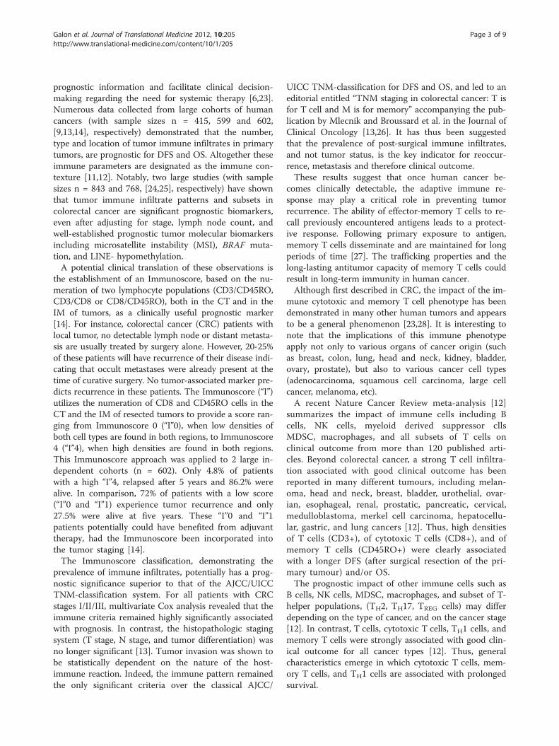
prognostic information and facilitate clinical decision-making regarding the need for systemic therapy [6,23].Numerous data collected from large cohorts of humancancers (with sample sizes n = 415, 599 and 602,[9,13,14], respectively) demonstrated that the number,type and location of tumor immune infiltrates in primarytumors, are prognostic for DFS and OS. Altogether theseimmune parameters are designated as the immune con-texture [11,12]. Notably, two large studies (with samplesizes n = 843 and 768, [24,25], respectively) have shownthat tumor immune infiltrate patterns and subsets incolorectal cancer are significant prognostic biomarkers,even after adjusting for stage, lymph node count, andwell-established prognostic tumor molecular biomarkersincluding microsatellite instability (MSI), BRAF muta-tion, and LINE- hypomethylation.A potential clinical translation of these observations is
the establishment of an Immunoscore, based on the nu-meration of two lymphocyte populations (CD3/CD45RO,CD3/CD8 or CD8/CD45RO), both in the CT and in theIM of tumors, as a clinically useful prognostic marker[14]. For instance, colorectal cancer (CRC) patients withlocal tumor, no detectable lymph node or distant metasta-sis are usually treated by surgery alone. However, 20-25%of these patients will have recurrence of their disease indi-cating that occult metastases were already present at thetime of curative surgery. No tumor-associated marker pre-dicts recurrence in these patients. The Immunoscore (“I”)utilizes the numeration of CD8 and CD45RO cells in theCT and the IM of resected tumors to provide a score ran-ging from Immunoscore 0 (“I”0), when low densities ofboth cell types are found in both regions, to Immunoscore4 (“I”4), when high densities are found in both regions.This Immunoscore approach was applied to 2 large in-dependent cohorts (n = 602). Only 4.8% of patientswith a high “I”4, relapsed after 5 years and 86.2% werealive. In comparison, 72% of patients with a low score(“I”0 and “I”1) experience tumor recurrence and only27.5% were alive at five years. These “I”0 and “I”1patients potentially could have benefited from adjuvanttherapy, had the Immunoscore been incorporated intothe tumor staging [14].The Immunoscore classification, demonstrating the
prevalence of immune infiltrates, potentially has a prog-nostic significance superior to that of the AJCC/UICCTNM-classification system. For all patients with CRCstages I/II/III, multivariate Cox analysis revealed that theimmune criteria remained highly significantly associatedwith prognosis. In contrast, the histopathologic stagingsystem (T stage, N stage, and tumor differentiation) wasno longer significant [13]. Tumor invasion was shown tobe statistically dependent on the nature of the host-immune reaction. Indeed, the immune pattern remainedthe only significant criteria over the classical AJCC/
UICC TNM-classification for DFS and OS, and led to aneditorial entitled “TNM staging in colorectal cancer: T isfor T cell and M is for memory” accompanying the pub-lication by Mlecnik and Broussard et al. in the Journal ofClinical Oncology [13,26]. It has thus been suggestedthat the prevalence of post-surgical immune infiltrates,and not tumor status, is the key indicator for reoccur-rence, metastasis and therefore clinical outcome.These results suggest that once human cancer be-
comes clinically detectable, the adaptive immune re-sponse may play a critical role in preventing tumorrecurrence. The ability of effector-memory T cells to re-call previously encountered antigens leads to a protect-ive response. Following primary exposure to antigen,memory T cells disseminate and are maintained for longperiods of time [27]. The trafficking properties and thelong-lasting antitumor capacity of memory T cells couldresult in long-term immunity in human cancer.Although first described in CRC, the impact of the im-
mune cytotoxic and memory T cell phenotype has beendemonstrated in many other human tumors and appearsto be a general phenomenon [23,28]. It is interesting tonote that the implications of this immune phenotypeapply not only to various organs of cancer origin (suchas breast, colon, lung, head and neck, kidney, bladder,ovary, prostate), but also to various cancer cell types(adenocarcinoma, squamous cell carcinoma, large cellcancer, melanoma, etc).A recent Nature Cancer Review meta-analysis [12]
summarizes the impact of immune cells including Bcells, NK cells, myeloid derived suppressor cllsMDSC, macrophages, and all subsets of T cells onclinical outcome from more than 120 published arti-cles. Beyond colorectal cancer, a strong T cell infiltra-tion associated with good clinical outcome has beenreported in many different tumours, including melan-oma, head and neck, breast, bladder, urothelial, ovar-ian, esophageal, renal, prostatic, pancreatic, cervical,medulloblastoma, merkel cell carcinoma, hepatocellu-lar, gastric, and lung cancers [12]. Thus, high densitiesof T cells (CD3+), of cytotoxic T cells (CD8+), and ofmemory T cells (CD45RO+) were clearly associatedwith a longer DFS (after surgical resection of the pri-mary tumour) and/or OS.The prognostic impact of other immune cells such as
B cells, NK cells, MDSC, macrophages, and subset of T-helper populations, (TH2, TH17, TREG cells) may differdepending on the type of cancer, and on the cancer stage[12]. In contrast, T cells, cytotoxic T cells, TH1 cells, andmemory T cells were strongly associated with good clin-ical outcome for all cancer types [12]. Thus, generalcharacteristics emerge in which cytotoxic T cells, mem-ory T cells, and TH1 cells are associated with prolongedsurvival.
Galon et al. Journal of Translational Medicine 2012, 10:205 Page 4 of 9http://www.translational-medicine.com/content/10/1/205
The Immunoscore as a new approach for theclassification of cancerConsidering the important role of the host immune signa-ture in controlling tumor progression, it is now imperativeto initiate the incorporation of the Immunoscore as acomponent of cancer classification [13,14] and a prognos-tic tool [23]. This strategy has a dual advantage: firstly, itappears to be the strongest prognostic factor for DFS andOS, particularly in early stage cancers and secondly, itcould allude to potential targets for novel therapeuticapproaches, including immunotherapy. Current immuno-histochemical technologies allow the application of suchanalyses by laboratories concerned with routine diagnosticand prognostic assessment of tumors.The inherent complexity of immunohistochemistry, in
conjunction with protocol variability, analysis of differ-ent immune cell types, inconsistent tissue region selec-tion criteria, combined with differences in conjunctionwith qualitative and semi-quantitative criteria to meas-ure immune infiltration, all contribute to the variabilityof the results obtained, and raise the concern that spe-cialized protocols and training may be required. It istherefore essential to pursue assay uniformity to reducethese limitations. Many markers, signatures, and meth-ods have been described to evaluate the prognosis ofcancer patients. Yet, very few such markers and labora-tory assays are used in clinical practice. Thus, we believethat harmonization of an assay evaluating the “inflam-mation”, i.e. the Immunoscore of the tumor is essential.Analytical and clinical validation of the assay is requiredbefore the Immunoscore will reach clinical applicabilityfor individual patients. However, current immunohisto-chemical technologies allow the application and cross-validation of such analysis in laboratories performingroutine diagnostic and prognostic assessment of tumors.In order to be able to compare results in the future, andfor the development of more effective prognostic andpredictive markers to improve clinical decision-making,it is important to perform a standardized set of experi-ments. Assay harmonization should minimize data vari-ability and allow worldwide correlations of Immunoscoreresults with clinical outcomes. Harmonization guidelinesresulting from this process are expected to be simple toimplement and will improve assay performance. Effectivelarge-scale assay harmonization efforts have already beenconducted for commonly used immunological assays ofperipheral blood immune cell populations [29,30].A fundamental parameter to determine the Immuno-
score will include the immune cell density, calculated bynumerical quantification of two lymphocyte populations,cytotoxic and memory T cells at the CT and the IM oftumors. This core criterion will establish prognosis ofpatient clinical outcome, regardless of the absence ofother cancer associated prognostic markers, such as in
early tumor stage (I/II) patients [14]. In human cancers,a high density of TH1/cytotoxic memory T lymphocytes,located both in the CT and IM of the primary tumor, isassociated with long DFS and OS, in addition to low riskof relapse and metastasis. This was particularly illu-strated in CRC [5,9,13,14,19], and should be applicableto most human tumors [23]. Thus, this Immunoscoreclassification may help identify the high-risk patientswho would benefit the most from adjuvant therapy.
Impact on response to cancer therapiesWhether the immune contexture of the primary tumorpredicts therapeutic responses is of paramount import-ance for patient clinical management. Data based on im-mune signatures have established that a strong immunecomponent is predictive of good response to chemother-apy in breast cancer [31-33], a tumor in which a highlymphocyte infiltrate is associated with higher responserate in neo-adjuvant therapy [34,35]. In hepatic metastasesof CRC, high CD8 infiltrates in the IM predicts better re-sponse to chemotherapy and prolonged survival [36]. Inmelanoma, an immune signature displaying high expres-sion of TH1 and cytotoxicity-associated genes, correlateswith favorable clinical outcome to several different thera-peutic vaccines [8]. In addition, high numbers of CD8 Tcell infiltrates within metastatic melanoma correlated withprolonged survival [37]. However, the high TH1 and cyto-toxic immune response associated with prolonged survivalin patients receiving adjuvant therapies may not be a pre-diction of response to the therapy, but rather the fact thatthe host-immune response within the tumor protects thepatient and prolongs patient life. To assess the impact ofthe Immunoscore as a predictive marker, it should beevaluated prospectively in randomized clinical trials.
An open access call for a broad participation to thedevelopment of a task force dedicated to the evaluationof the Immunoscore in cancer patientsOver the past few years, the area of immune regulation atthe level of the tumor microenvironment has gained aforefront position in cancer research, in CRC [9,12-14], inmelanoma [38] and all other cancer types [6]. The Immu-noscore was initially described several years ago [9], andmore recently advances have been made in the develop-ment of the Immunoscore as a prognostic factor [13,14]that could be used in routine testing [39]. In an effort topromote the utilization of such Immunoscore in routineclinical settings worldwide, the Society for Immunother-apy of Cancer (SITC), the European Academy of TumorImmunology (EATI), and “La Fondazione MelanomaOnlus”, initiated a task force on “Immunoscoring as aNew Possible Approach for the Classification of Cancer”that took place in Naples, Italy, February 13th, 2012 [39].This perspective represents a follow-up on this initiative,
Galon et al. Journal of Translational Medicine 2012, 10:205 Page 5 of 9http://www.translational-medicine.com/content/10/1/205
originally announced in a J Transl Med. editorial in Janu-ary 2012 [39]. The working group, composed of inter-national expert pathologists and immunologists, identifieda strategy for the organization of worldwide participationby various groups for the validation of the Immunoscore.The objectives of the meeting included discussing: the roleof immune system in cancer; a review of the AJCC/UICC-TNM classification of CRC; the role of the microenviron-ment in melanoma biology; the review of the AJCCclassification of melanoma; the relevance of HLA-A2 incancer prognosis and tumor malignancy; data utilizing theImmunoscore and a proposal for standardizing the operat-ing procedures for the Immunoscore quantification. Fur-thermore, the international working group evaluated thefeasibility of using the Immunoscore for the classificationof cancer. Evidence-based selection of specific markersand their combinations for the Immunoscore was dis-cussed including biological rationale, clinical use, syntheticmeta-analysis of the Immunoscore, analytical perform-ance, reagents availability and testing, metrics for decisionmaking, cross-laboratory validation of methodology andidentification of potential problems during developmentof other markers. Practical aspects of the validation of theassay by participating centers were proposed includingconsideration of cancer types, cancer stages, and the def-inition of a working group of pathologists for the valid-ation phase.CRC has been most comprehensively studied and the
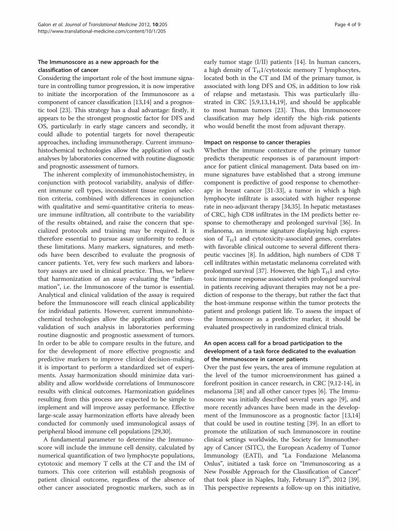
prognostic significance of immunologic parameters hasbeen best validated, thus special emphasis will be placed inthis disease for this formal validation. As neo-adjuvanttreatments are nowadays recommended for rectal cancer, itmay be advisable to separate the validation of colon cancersand rectal cancers. Other cancer types, including melanomaand breast cancers were additionally discussed and theirvalidation will follow. An independent international con-sensus panel of expert laboratories discussed cross-laboratory assay validation for the development of anImmunoscore prognostic method. As evaluation of cyto-toxic memory CD8+ Tcells (CD3+, CD8+, CD45RO+, Gran-zyme B+ (GZMB)) provides the best method to
CD
CD
CT
IM
Tumor regions (CT & IM) Immunos
Digital PFigure 1 Immunoscore definition and method.
discriminate patient outcome, any combination of two ofthese aforementioned markers should have similar statis-tical power. Because of technical difficulties including back-ground noise (CD45RO) and granular staining (GZMB), itwas decided to employ the two easiest membrane stains,CD3 and CD8. Thus, the combination of two markers(CD3+ and CD8+) in two regions (CT and IM) was agreedfor validation in standard clinical practice. Precise quantifi-cation will be performed on whole slide sections (Figure 1).For harmonization of the assay and reproducibility of themethod, all laboratories agreed to test the prognostic valueof specific immune cell infiltration following the recom-mended initial guidelines. The inherent complexity ofquantitative immunohistochemistry underscored the urgentneed to reach assay harmonization. The components of theImmunoscore are listed in Table 1. Additional markerscould be added subsequently to refine the methodologyeven further if required. After worldwide validation, a con-sensus detailed protocol will be available.To be used globally in a routine manner, evaluation of
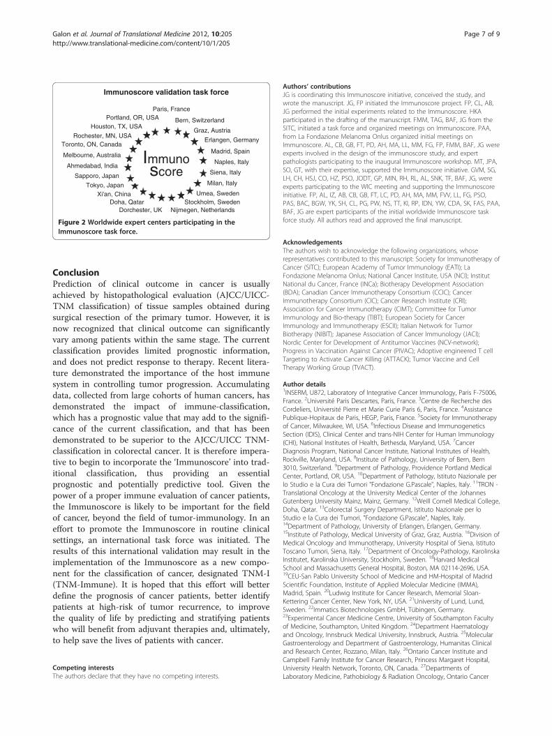
a novel marker should have the following characteristics:pathology-based, feasible in routine settings, simple, in-expensive, rapid, robust, reproducible, quantitative, stan-dardized, and powerful. The Immunoscore fulfills allthese keys aspects summarized in Table 2.The purpose of the Immunoscore worldwide task force
is to validate these points.The goals of the first ongoing initiative are the
following:
1) to demonstrate the feasibility and reproducibility ofthe Immunoscore.
2) to validate the major prognostic power of theImmunoscore in routine settings for patients withcolon cancer stage I/II/III.
3) to demonstrate the utility of the Immunoscore topredict stage II colon cancer patients with high riskof recurrence.
Thus, the benefit of the Immunoscore worldwidestudy would be to validate the feasibility, reproducibility,
3
8
Immunoscore (CT+IM)
Hi
Hi Hi
Hi Hi Hi
Hi Hi Hi Hi
tainings
athology
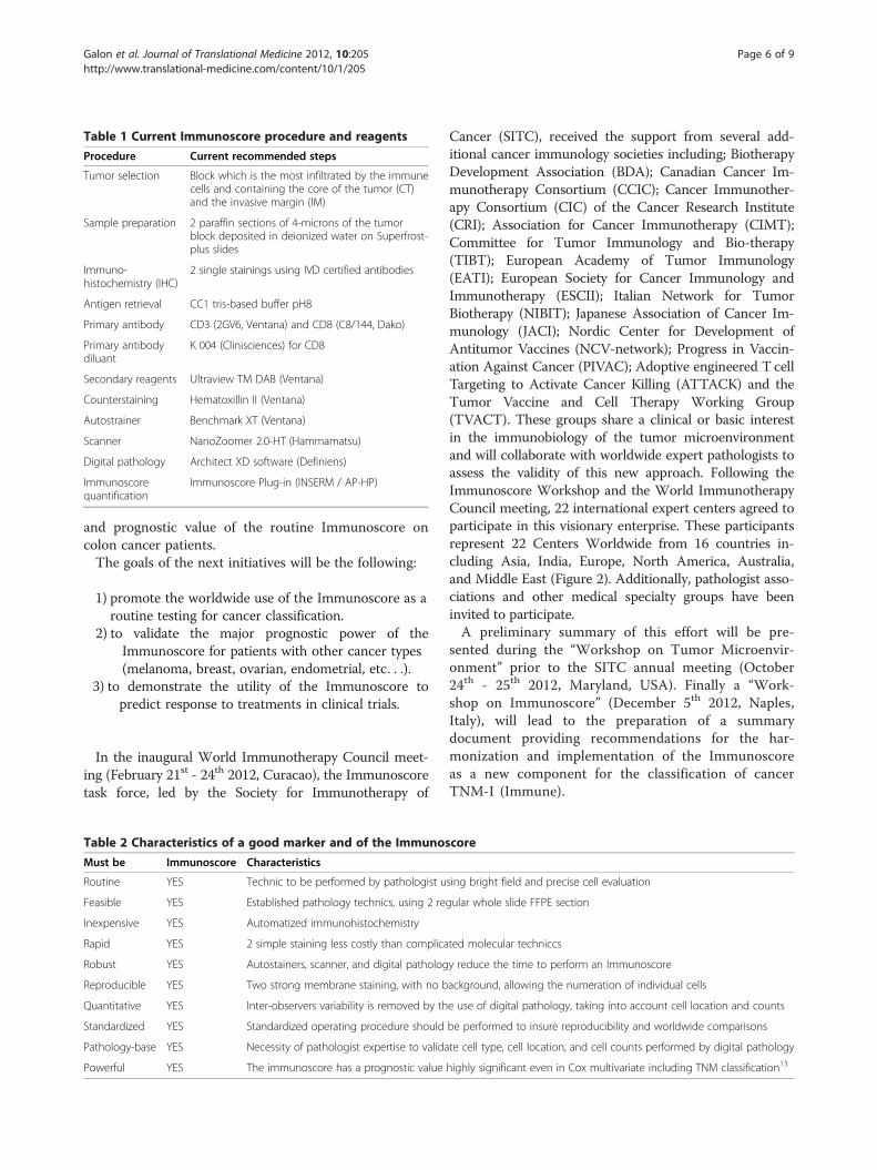
Table 1 Current Immunoscore procedure and reagents
Procedure Current recommended steps
Tumor selection Block which is the most infiltrated by the immunecells and containing the core of the tumor (CT)and the invasive margin (IM)
Sample preparation 2 paraffin sections of 4-microns of the tumorblock deposited in deionized water on Superfrost-plus slides
Immuno-histochemistry (IHC)
2 single stainings using IVD certified antibodies
Antigen retrieval CC1 tris-based buffer pH8
Primary antibody CD3 (2GV6, Ventana) and CD8 (C8/144, Dako)
Primary antibodydiluant
K 004 (Clinisciences) for CD8
Secondary reagents Ultraview TM DAB (Ventana)
Counterstaining Hematoxillin II (Ventana)
Autostrainer Benchmark XT (Ventana)
Scanner NanoZoomer 2.0-HT (Hammamatsu)
Digital pathology Architect XD software (Definiens)
Immunoscorequantification
Immunoscore Plug-in (INSERM / AP-HP)
Galon et al. Journal of Translational Medicine 2012, 10:205 Page 6 of 9http://www.translational-medicine.com/content/10/1/205
and prognostic value of the routine Immunoscore oncolon cancer patients.The goals of the next initiatives will be the following:
1) promote the worldwide use of the Immunoscore as aroutine testing for cancer classification.
2) to validate the major prognostic power of theImmunoscore for patients with other cancer types(melanoma, breast, ovarian, endometrial, etc. . .).
3) to demonstrate the utility of the Immunoscore topredict response to treatments in clinical trials.
In the inaugural World Immunotherapy Council meet-ing (February 21st - 24th 2012, Curacao), the Immunoscoretask force, led by the Society for Immunotherapy of
Table 2 Characteristics of a good marker and of the Immunos
Must be Immunoscore Characteristics
Routine YES Technic to be performed by pathologist u
Feasible YES Established pathology technics, using 2 re
Inexpensive YES Automatized immunohistochemistry
Rapid YES 2 simple staining less costly than complica
Robust YES Autostainers, scanner, and digital patholog
Reproducible YES Two strong membrane staining, with no b
Quantitative YES Inter-observers variability is removed by th
Standardized YES Standardized operating procedure should
Pathology-base YES Necessity of pathologist expertise to valida
Powerful YES The immunoscore has a prognostic value
Cancer (SITC), received the support from several add-itional cancer immunology societies including; BiotherapyDevelopment Association (BDA); Canadian Cancer Im-munotherapy Consortium (CCIC); Cancer Immunother-apy Consortium (CIC) of the Cancer Research Institute(CRI); Association for Cancer Immunotherapy (CIMT);Committee for Tumor Immunology and Bio-therapy(TIBT); European Academy of Tumor Immunology(EATI); European Society for Cancer Immunology andImmunotherapy (ESCII); Italian Network for TumorBiotherapy (NIBIT); Japanese Association of Cancer Im-munology (JACI); Nordic Center for Development ofAntitumor Vaccines (NCV-network); Progress in Vaccin-ation Against Cancer (PIVAC); Adoptive engineered T cellTargeting to Activate Cancer Killing (ATTACK) and theTumor Vaccine and Cell Therapy Working Group(TVACT). These groups share a clinical or basic interestin the immunobiology of the tumor microenvironmentand will collaborate with worldwide expert pathologists toassess the validity of this new approach. Following theImmunoscore Workshop and the World ImmunotherapyCouncil meeting, 22 international expert centers agreed toparticipate in this visionary enterprise. These participantsrepresent 22 Centers Worldwide from 16 countries in-cluding Asia, India, Europe, North America, Australia,and Middle East (Figure 2). Additionally, pathologist asso-ciations and other medical specialty groups have beeninvited to participate.A preliminary summary of this effort will be pre-
sented during the “Workshop on Tumor Microenvir-onment” prior to the SITC annual meeting (October24th - 25th 2012, Maryland, USA). Finally a “Work-shop on Immunoscore” (December 5th 2012, Naples,Italy), will lead to the preparation of a summarydocument providing recommendations for the har-monization and implementation of the Immunoscoreas a new component for the classification of cancerTNM-I (Immune).
core
sing bright field and precise cell evaluation
gular whole slide FFPE section
ted molecular techniccs
y reduce the time to perform an Immunoscore
ackground, allowing the numeration of individual cells
e use of digital pathology, taking into account cell location and counts
be performed to insure reproducibility and worldwide comparisons
te cell type, cell location, and cell counts performed by digital pathology
highly significant even in Cox multivariate including TNM classification13
Immunoscore validation task force
Immuno score
Naples, Italy
Graz, Austria
Erlangen, Germany
Stockholm, Sweden
Paris, France
Umea, Sweden
Nijmegen, Netherlands
Madrid, Spain
Doha, Qatar
Milan, Italy
Bern, Switzerland
Siena, Italy
Melbourne, Australia
Sapporo, Japan
Xi'an, China
Dorchester, UK
Toronto, ON, Canada
Houston, TX, USA
Rochester, MN, USA
Portland, OR, USA
Tokyo, Japan
Ahmedabad, India
Figure 2 Worldwide expert centers participating in theImmunoscore task force.
Galon et al. Journal of Translational Medicine 2012, 10:205 Page 7 of 9http://www.translational-medicine.com/content/10/1/205
ConclusionPrediction of clinical outcome in cancer is usuallyachieved by histopathological evaluation (AJCC/UICC-TNM classification) of tissue samples obtained duringsurgical resection of the primary tumor. However, it isnow recognized that clinical outcome can significantlyvary among patients within the same stage. The currentclassification provides limited prognostic information,and does not predict response to therapy. Recent litera-ture demonstrated the importance of the host immunesystem in controlling tumor progression. Accumulatingdata, collected from large cohorts of human cancers, hasdemonstrated the impact of immune-classification,which has a prognostic value that may add to the signifi-cance of the current classification, and that has beendemonstrated to be superior to the AJCC/UICC TNM-classification in colorectal cancer. It is therefore impera-tive to begin to incorporate the ‘Immunoscore’ into trad-itional classification, thus providing an essentialprognostic and potentially predictive tool. Given thepower of a proper immune evaluation of cancer patients,the Immunoscore is likely to be important for the fieldof cancer, beyond the field of tumor-immunology. In aneffort to promote the Immunoscore in routine clinicalsettings, an international task force was initiated. Theresults of this international validation may result in theimplementation of the Immunoscore as a new compo-nent for the classification of cancer, designated TNM-I(TNM-Immune). It is hoped that this effort will betterdefine the prognosis of cancer patients, better identifypatients at high-risk of tumor recurrence, to improvethe quality of life by predicting and stratifying patientswho will benefit from adjuvant therapies and, ultimately,to help save the lives of patients with cancer.
Competing interestsThe authors declare that they have no competing interests.
Authors’ contributionsJG is coordinating this Immunoscore initiative, conceived the study, andwrote the manuscript. JG, FP initiated the Immunoscore project. FP, CL, AB,JG performed the initial experiments related to the Immunoscore. HKAparticipated in the drafting of the manuscript. FMM, TAG, BAF, JG from theSITC, initiated a task force and organized meetings on Immunoscore. PAA,from La Fondazione Melanoma Onlus organized initial meetings onImmunoscore. AL, CB, GB, FT, PD, AH, MA, LL, MM, FG, FP, FMM, BAF, JG wereexperts involved in the design of the immunoscore study, and expertpathologists participating to the inaugural Immunoscore workshop. MT, JPA,SO, GT, with their expertise, supported the Immunoscore initiative. GVM, SG,LH, CH, HSJ, CO, HZ, PSO, JODT, GP, MIN, RH, RL, AL, SNK, TF, BAF, JG, wereexperts participating to the WIC meeting and supporting the Immunoscoreinitiative. FP, AL, IZ, AB, CB, GB, FT, LC, PD, AH, MA, MM, FVV, LL, FG, PSO,PAS, BAC, BGW, YK, SH, CL, PG, PW, NS, TT, KI, RP, IDN, YW, CDA, SK, FAS, PAA,BAF, JG are expert participants of the initial worldwide Immunoscore taskforce study. All authors read and approved the final manuscript.
AcknowledgementsThe authors wish to acknowledge the following organizations, whoserepresentatives contributed to this manuscript: Society for Immunotherapy ofCancer (SITC); European Academy of Tumor Immunology (EATI); LaFondazione Melanoma Onlus; National Cancer Institute, USA (NCI); InstitutNational du Cancer, France (INCa); Biotherapy Development Association(BDA); Canadian Cancer Immunotherapy Consortium (CCIC); CancerImmunotherapy Consortium (CIC); Cancer Research Institute (CRI);Association for Cancer Immunotherapy (CIMT); Committee for TumorImmunology and Bio-therapy (TIBT); European Society for CancerImmunology and Immunotherapy (ESCII); Italian Network for TumorBiotherapy (NIBIT); Japanese Association of Cancer Immunology (JACI);Nordic Center for Development of Antitumor Vaccines (NCV-network);Progress in Vaccination Against Cancer (PIVAC); Adoptive engineered T cellTargeting to Activate Cancer Killing (ATTACK); Tumor Vaccine and CellTherapy Working Group (TVACT).
Author details1INSERM, U872, Laboratory of Integrative Cancer Immunology, Paris F-75006,France. 2Université Paris Descartes, Paris, France. 3Centre de Recherche desCordeliers, Université Pierre et Marie Curie Paris 6, Paris, France. 4AssistancePublique-Hopitaux de Paris, HEGP, Paris, France. 5Society for Immunotherapyof Cancer, Milwaukee, WI, USA. 6Infectious Disease and ImmunogeneticsSection (IDIS), Clinical Center and trans-NIH Center for Human Immunology(CHI), National Institutes of Health, Bethesda, Maryland, USA. 7CancerDiagnosis Program, National Cancer Institute, National Institutes of Health,Rockville, Maryland, USA. 8Institute of Pathology, University of Bern, Bern3010, Switzerland. 9Department of Pathology, Providence Portland MedicalCenter, Portland, OR, USA. 10Department of Pathology, Istituto Nazionale perlo Studio e la Cura dei Tumori "Fondazione G.Pascale", Naples, Italy. 11TRON -Translational Oncology at the University Medical Center of the JohannesGutenberg University Mainz, Mainz, Germany. 12Weill Cornell Medical College,Doha, Qatar. 13Colorectal Surgery Department, Istituto Nazionale per loStudio e la Cura dei Tumori, "Fondazione G.Pascale", Naples, Italy.14Department of Pathology, University of Erlangen, Erlangen, Germany.15Institute of Pathology, Medical University of Graz, Graz, Austria. 16Division ofMedical Oncology and Immunotherapy, University Hospital of Siena, IstitutoToscano Tumori, Siena, Italy. 17Department of Oncology-Pathology, KarolinskaInstitutet, Karolinska University, Stockholm, Sweden. 18Harvard MedicalSchool and Massachusetts General Hospital, Boston, MA 02114-2696, USA.19CEU-San Pablo University School of Medicine and HM-Hospital of MadridScientific Foundation, Institute of Applied Molecular Medicine (IMMA),Madrid, Spain. 20Ludwig Institute for Cancer Research, Memorial Sloan-Kettering Cancer Center, New York, NY, USA. 21University of Lund, Lund,Sweden. 22Immatics Biotechnologies GmbH, Tübingen, Germany.23Experimental Cancer Medicine Centre, University of Southampton Facultyof Medicine, Southampton, United Kingdom. 24Department Haematologyand Oncology, Innsbruck Medical University, Innsbruck, Austria. 25MolecularGastroenterology and Department of Gastroenterology, Humanitas Clinicaland Research Center, Rozzano, Milan, Italy. 26Ontario Cancer Institute andCampbell Family Institute for Cancer Research, Princess Margaret Hospital,University Health Network, Toronto, ON, Canada. 27Departments ofLaboratory Medicine, Pathobiology & Radiation Oncology, Ontario Cancer
Galon et al. Journal of Translational Medicine 2012, 10:205 Page 8 of 9http://www.translational-medicine.com/content/10/1/205
Institute/Princess Margaret Cancer Centre, Toronto, ON, Canada. 28Division ofCellular Signaling, Institute for Advanced Medical Research, Keio UniversitySchool of Medicine, Tokyo, Japan. 29Department of Digestive Surgery andSurgical Oncology, Yamaguchi University, Graduate School of Medicine,Yamaguchi, Japan. 30Department of Surgery, Kinki University, School ofMedicine, Osaka-sayama, Japan. 31Cancer Research Institute, New York, NY,USA. 32Department of Pathology, Avicenne Hospital, AP-HP, Bobigny, France.33Center for Medical Research, University of Tuebingen, Tuebingen, Germany.34Oncology Institute, Loyola University Medical Center, Cardinal BernardinCancer Center, Maywood, IL, USA. 35School of Cancer and Imaging Sciences,University of Manchester, Christie Hospital NHS Trust, Manchester, UK.36Research Center, University Hospital, Université de Montréal (CRCHUM),Montréal, Québec, Canada ; Institut du Cancer de Montréal, Montréal,Québec, Canada. 37Karolinska Institutet Department of Oncology-Pathology,Stockholm, Sweden. 38Georgia Health Sciences University Cancer Center,Augusta, GA, USA. 39Department of Pathology, Brigham and Women'sHospital and Harvard Medical School, Boston, MA, USA; Department ofMedical Oncology, Dana-Farber Cancer Institute, Boston, MA, USA.40Department of Medical Oncology, Royal Melbourne Hospital, Melbourne,Australia. 41Department of Pathology, The University of Melbourne,Melbourne, Australia. 42Department of Pathology, Sapporo Medical UniversitySchool of Medicine, Sapporo, Japan. 43Department of Immunology andImmunotherapy, Kurume University School of Medicine, Kurume, Japan.44The Gujarat Cancer & Research Institute, Asarwa, Ahmedabad, India.45Department of Medical Biosciences, Pathology, Umea University, Umea,Sweden. 46Pathology Department, Radboud University Nijmegen MedicalCenter, Nijmegen, The Netherlands. 47Institute for Cancer Research, Center ofTranslational medicine, Xi’an Jiaotong university, Xian, China. 48Departmentof Histopathology, Dorset County Hospital, DCHFT, NHS, Dorchester, UK.49MD Anderson Cancer Center, Houston, TX, USA. 50Mayo Clinic and MayoCollege of Medicine, Rochester, MN 55905, USA. 51Cancer InflammationProgram, Center for Cancer Research, National Cancer Institute and Trans-NIHCenter for Human Immunology (CHI), National Institutes of Health, Frederickand Bethesda, Maryland, USA. 52University of Chicago, Chicago, IL, USA.53Medical Oncology and Innovative Therapies Unit, Istituto Nazionale per loStudio e la Cura dei Tumori, "Fondazione G. Pascale", Napoli, Italy.54Fondazione Melanoma Onlus, Napoli, Italy. 55Laboratory of Molecular andTumor Immunology, Earle A. Chiles Research Institute, Robert W. FranzCancer Center, Providence Portland Medical Center, Portland, OR, USA.56Department of Molecular Microbiology and Immunology, Oregon Healthand Science University, Portland, OR, USA.
Received: 6 July 2012 Accepted: 19 September 2012Published: 3 October 2012
References1. Locker GY, Hamilton S, Harris J, Jessup JM, Kemeny N, Macdonald JS,
Somerfield MR, Hayes DF, Bast RC Jr: ASCO 2006 update ofrecommendations for the use of tumor markers in gastrointestinalcancer. J Clin Oncol 2006, 24:5313–5327.
2. Sobin L, Wittekind C: TNM classification of malignant tumors. New York:Wiley-Liss; 2002.
3. Weitz J, Koch M, Debus J, Hohler T, Galle PR, Buchler MW: Colorectalcancer. Lancet 2005, 365:153–165.
4. Nagtegaal ID, Quirke P, Schmoll HJ: Has the new TNM classification forcolorectal cancer improved care? Nat Rev Clin Oncol 2011, 9:119–123.
5. Mlecnik B, Bindea G, Pages F, Galon J: Tumor immunosurveillance inhuman cancers. Cancer Metastasis Rev 2011, 30:5–12.
6. Bindea G, Mlecnik B, Fridman WH, Pages F, Galon J: Natural immunity tocancer in humans. Curr Opin Immunol 2010, 22:215–222.
7. Finn OJ: Cancer immunology. N Engl J Med 2008, 358:2704–2715.8. Gajewski TF, Louahed J, Brichard VG: Gene signature in melanoma
associated with clinical activity: a potential clue to unlock cancerimmunotherapy. Cancer J 2010, 16:399–403.
9. Galon J, Costes A, Sanchez-Cabo F, Kirilovsky A, Mlecnik B, Lagorce-Pages C,Tosolini M, Camus M, Berger A, Wind P, et al: Type, density, and location ofimmune cells within human colorectal tumors predict clinical outcome.Science 2006, 313:1960–1964.
10. Wang E, Miller LD, Ohnmacht GA, Mocellin S, Perez-Diez A, Petersen D,Zhao Y, Simon R, Powell JI, Asaki E, et al: Prospective molecular profiling of
melanoma metastases suggests classifiers of immune responsiveness.Cancer Res 2002, 62:3581–3586.
11. Galon J, Fridman WH, Pages F: The adaptive immunologicmicroenvironment in colorectal cancer: a novel perspective. Cancer Res2007, 67:1883–1886.
12. Fridman WH, Pages F, Sautes-Fridman C, Galon J: The immune contexturein human tumours: impact on clinical outcome. Nat Rev Cancer 2012,12:298–306.
13. Mlecnik B, Tosolini M, Kirilovsky A, Berger A, Bindea G, Meatchi T, Bruneval P,Trajanoski Z, Fridman WH, Pages F, Galon J: Histopathologic-basedprognostic factors of colorectal cancers are associated with the state ofthe local immune reaction. J Clin Oncol 2011, 29:610–618.
14. Pages F, Kirilovsky A, Mlecnik B, Asslaber M, Tosolini M, Bindea G, Lagorce C,Wind P, Marliot F, Bruneval P, et al: In situ cytotoxic and memory T cellspredict outcome in patients with early-stage colorectal cancer. J ClinOncol 2009, 27:5944–5951.
15. Febbo PG, Ladanyi M, Aldape KD, De Marzo AM, Hammond ME, Hayes DF,Iafrate AJ, Kelley RK, Marcucci G, Ogino S, et al: NCCN Task Force report:evaluating the clinical utility of tumor markers in oncology. J Natl ComprCanc Netw 2011, 9(Suppl 5):S1–S32. quiz S33.
16. Ogino S, Galon J, Fuchs CS, Dranoff G: Cancer immunology–analysis ofhost and tumor factors for personalized medicine. Nat Rev Clin Oncol2011, 8:711–719.
17. Ogino S, Chan AT, Fuchs CS, Giovannucci E: Molecular pathologicalepidemiology of colorectal neoplasia: an emerging transdisciplinary andinterdisciplinary field. Gut 2011, 60:397–411.
18. Ogino S, Stampfer M: Lifestyle factors and microsatellite instability incolorectal cancer: the evolving field of molecular pathologicalepidemiology. J Natl Cancer Inst 2010, 102:365–367.
19. Pages F, Berger A, Camus M, Sanchez-Cabo F, Costes A, Molidor R, MlecnikB, Kirilovsky A, Nilsson M, Damotte D, et al: Effector memory T cells, earlymetastasis, and survival in colorectal cancer. N Engl J Med 2005,353:2654–2666.
20. Butterfield LH, Disis ML, Fox BA, Lee PP, Khleif SN, Thurin M, Trinchieri G,Wang E, Wigginton J, Chaussabel D, et al: A systematic approach tobiomarker discovery; preamble to "the iSBTc-FDA taskforce onimmunotherapy biomarkers". J Transl Med 2008, 6:81.
21. Butterfield LH, Palucka AK, Britten CM, Dhodapkar MV, Hakansson L, JanetzkiS, Kawakami Y, Kleen TO, Lee PP, Maccalli C, et al: Recommendations fromthe iSBTc-SITC/FDA/NCI Workshop on Immunotherapy Biomarkers. ClinCancer Res 2011, 17:3064–3076.
22. Tahara H, Sato M, Thurin M, Wang E, Butterfield LH, Disis ML, Fox BA, LeePP, Khleif SN, Wigginton JM, et al: Emerging concepts in biomarkerdiscovery; the US-Japan Workshop on Immunological Molecular Markersin Oncology. J Transl Med 2009, 7:45.
23. Pages F, Galon J, Dieu-Nosjean MC, Tartour E, Sautes-Fridman C, FridmanWH: Immune infiltration in human tumors: a prognostic factor thatshould not be ignored. Oncogene 2010, 29:1093–1102.
24. Nosho K, Baba Y, Tanaka N, Shima K, Hayashi M, Meyerhardt JA, GiovannucciE, Dranoff G, Fuchs CS, Ogino S: Tumour-infiltrating T-cell subsets,molecular changes in colorectal cancer and prognosis: cohort study andliterature review. J Pathol 2010, 222:350–366.
25. Ogino S, Nosho K, Irahara N, Meyerhardt JA, Baba Y, Shima K, Glickman JN,Ferrone CR, Mino-Kenudson M, Tanaka N, et al: Lymphocytic reaction tocolorectal cancer is associated with longer survival, independent oflymph node count, microsatellite instability, and CpG island methylatorphenotype. Clin Cancer Res 2009, 15:6412–6420.
26. Broussard EK, Disis ML: TNM staging in colorectal cancer: T is for T celland M is for memory. J Clin Oncol 2011, 29:601–603.
27. Sallusto F, Geginat J, Lanzavecchia A: Central memory and effectormemory T cell subsets: function, generation, and maintenance. Annu RevImmunol 2004, 22:745–763.
28. Ascierto ML, De Giorgi V, Liu Q, Bedognetti D, Spivey TL, Murtas D, UccelliniL, Ayotte BD, Stroncek DF, Chouchane L, et al: An immunologic portrait ofcancer. J Transl Med 2011, 9:146.
29. Fox BA, Schendel DJ, Butterfield LH, Aamdal S, Allison JP, Ascierto PA, AtkinsMB, Bartunkova J, Bergmann L, Berinstein N, et al: Defining the CriticalHurdles in Cancer Immunotherapy. J Transl Med 2011, 9:214.
30. van der Burg SH, Kalos M, Gouttefangeas C, Janetzki S, Ottensmeier C,Welters MJ, Romero P, Britten CM, Hoos A: Harmonization of immunebiomarker assays for clinical studies. Sci Transl Med 2011, 3:108–ps144.
Galon et al. Journal of Translational Medicine 2012, 10:205 Page 9 of 9http://www.translational-medicine.com/content/10/1/205
31. Desmedt C, Haibe-Kains B, Wirapati P, Buyse M, Larsimont D, Bontempi G,Delorenzi M, Piccart M, Sotiriou C: Biological processes associated withbreast cancer clinical outcome depend on the molecular subtypes. ClinCancer Res 2008, 14:5158–5165.
32. Iwamoto T, Bianchini G, Booser D, Qi Y, Coutant C, Shiang CY, Santarpia L,Matsuoka J, Hortobagyi GN, Symmans WF, et al: Gene pathways associatedwith prognosis and chemotherapy sensitivity in molecular subtypes ofbreast cancer. J Natl Cancer Inst 2011, 103:264–272.
33. Sotiriou C, Pusztai L: Gene-expression signatures in breast cancer. N Engl JMed 2009, 360:790–800.
34. Andre F, Berrada N, Desmedt C: Implication of tumor microenvironmentin the resistance to chemotherapy in breast cancer patients. Curr OpinOncol 2010, 22:547–551.
35. Denkert C, Loibl S, Noske A, Roller M, Muller BM, Komor M, Budczies J, Darb-Esfahani S, Kronenwett R, Hanusch C, et al: Tumor-associated lymphocytesas an independent predictor of response to neoadjuvant chemotherapyin breast cancer. J Clin Oncol 2010, 28:105–113.
36. Halama N, Michel S, Kloor M, Zoernig I, Benner A, Spille A, Pommerencke T,von Knebel DM, Folprecht G, Luber B, et al: Localization and density ofimmune cells in the invasive margin of human colorectal cancer livermetastases are prognostic for response to chemotherapy. Cancer Res2011, 71:5670–5677.
37. Erdag G, Schaefer JT, Smolkin ME, Deacon DH, Shea SM, Dengel LT,Patterson JW, Slingluff CL Jr: Immunotype and immunohistologiccharacteristics of tumor-infiltrating immune cells are associated withclinical outcome in metastatic melanoma. Cancer Res 2012, 72:1070–1080.
38. Ascierto PA, De Maio E, Bertuzzi S, Palmieri G, Halaban R, Hendrix M,Kashani-sabet M, Ferrone S, Wang E, Cochran A, et al: Future perspectivesin melanoma research. Meeting report from the "Melanoma Research: abridge Naples-USA. Naples, December 6th-7th 2010". J Transl Med 2011,9:32.
39. Galon J, Pages F, Marincola FM, Thurin M, Trinchieri G, Fox BA, Gajewski TF,Ascierto PA: The immune score as a new possible approach for theclassification of cancer. J Transl Med 2012, 10:1.
doi:10.1186/1479-5876-10-205Cite this article as: Galon et al.: Cancer classification using theImmunoscore: a worldwide task force. Journal of Translational Medicine2012 10:205.
Submit your next manuscript to BioMed Centraland take full advantage of:
• Convenient online submission
• Thorough peer review
• No space constraints or color figure charges
• Immediate publication on acceptance
• Inclusion in PubMed, CAS, Scopus and Google Scholar
• Research which is freely available for redistribution
Submit your manuscript at www.biomedcentral.com/submit