BYC, an atypical aspartic endopeptidase from Rhipicephalus (Boophilus) microplus eggs
Upload
independentCategory
view
0download
0
Calcium modulates endopeptidase 24.15 (EC 3.4.24.15)membrane association, secondary structure and substratespecificityVitor Oliveira1, Paula A. G. Garrido2, Claudia C. Rodrigues2, Alison Colquhoun2,Leandro M. Castro2, Paulo C. Almeida3, Claudio S. Shida3, Maria A. Juliano4, Luiz Juliano4,Antonio C. M. Camargo5, Stephen Hyslop6, James L. Roberts7, Valerie Grum-Tokars8,Marc J. Glucksman8 and Emer S. Ferro2
1 Laboratorio de Neurociencias, Universidade da Cidade de Sao Paulo, Brazil
2 Departamento de Biologia Celular e Desenvolvimento, Programa de Biologia Celular, Instituto de Ciencias Biomedicas, Universidade de
Sao Paulo, Brazil
3 Universidade de Mogi das Cruzes, Mogi das Cruzes, Brazil
4 Departamento de Biofısica, Universidade Federal de Sao Paulo, Brazil
5 Centro de Toxinologia Aplicada (CAT), Instituto Butantan, Brazil
6 Departamento de Farmacologia, Faculdade de Ciencias Medicas, Universidade Estadual de Campinas, Campinas, Brazil
7 Sam and Ann Barshop Institute for Longevity and Aging Studies, University of Texas Health Science Center, San Antonio, TX, USA
8 Midwest Proteome Center, Department of Biochemistry & Molecular Biology, Rosalind Franklin University of Medicine and Science ⁄Chicago Medical School, USA
Keywords
calcium; membrane binding; peptide
metabolism; protease; thimet oligopeptidase
Correspondence
E. S. Ferro, Laboratorio de Comunicacao
Celular, Avenida Prof. Lineu Prestes1524
Sala 431, Sao Paulo, 05508-900, SP, Brazil
E-mail: [email protected]
Note
V. Oliveira and P.A.G. Garrido contributed
equally to this work.
(Received 9 February 2005, revised 24
March 2005, accepted 31 March 2005)
doi:10.1111/j.1742-4658.2005.04692.x
The metalloendopeptidase 24.15 (EP24.15) is ubiquitously present in the
extracellular environment as a secreted protein. Outside the cell, this
enzyme degrades several neuropeptides containing from 5 to 17 amino
acids (e.g. gonadotropin releasing hormone, bradykinin, opioids and neuro-
tensin). The constitutive secretion of EP24.15 from glioma C6 cells was
demonstrated to be stimulated linearly by reduced concentrations of extra-
cellular calcium. In the present report we demonstrate that extracellular
calcium concentration has no effect on the total amount of the extracellular
(cell associated + medium) enzyme. Indeed, immuno-cytochemical analyses
by confocal and electron microscopy suggested that the absence of calcium
favors the enzyme shedding from the plasma membrane into the medium.
Two putative calcium-binding sites on EP24.15 (D93 and D159) were
altered by site-directed mutagenesis to investigate their possible contribu-
tion to binding of the enzyme at the cell surface. These mutated recombin-
ant proteins behave similarly to the wild-type enzyme regarding enzymatic
activity, secondary structure, calcium sensitivity and immunoreactivity.
However, immunocytochemical analyses by confocal microscopy consis-
tently show a reduced ability of the D93A mutant to associate with the
plasma membrane of glioma C6 cells when compared with the wild-type
enzyme. These data and the model of the enzyme’s structure as determined
by X-ray diffraction suggest that D93 is located at the enzyme surface and
is consistent with membrane association of EP24.15. Moreover, calcium
was also observed to induce a major change in the EP24.15 cleavage site
on distinctive fluorogenic substrates. These data suggest that calcium may
be an important modulator of ep24.15 cell function.
Abbreviations
EP24.15; metalloendopeptidase 24.15; G6PD, glucose-6-phosphate dehydrogenase; PFA, paraformaldehyde.
2978 FEBS Journal 272 (2005) 2978–2992 ª 2005 FEBS

The metalloendopeptidase EC 3.4.24.15 (EP24.15) rep-
resents a distinctive peptide-metabolizing enzyme with
size-selectivity for peptides ranging from 8 to 17 amino
acids [1–3]. EP24.15 is a monomeric, soluble, 77-kDa
endopeptidase, thiol-activated, with an isoelectric point
of 5.6 and a pH optimum of activity of 7.4, first isola-
ted from the soluble fraction of the rat brain [4]. This
enzyme is distributed in all mammalian tissues so far
examined, with high levels in testis, brain, kidney and
pituitary [5]. In the brain, at the electron microscopic
level, EP24.15-like immunoreactivity is observed over
the nuclei, cytoplasm, cross-sectioned dendrites, myeli-
nated and unmyelinated axons, axon terminals, and in
both astrocytes and oligodendrocytes [6]. Recent stud-
ies have shown that EP24.15 could interact with
peptides generated by the multicatalytic complex pro-
teasome [7,8]. Thus, the high concentration of the
enzyme observed inside the cells could be somewhat
related to the degradation of catabolic products gener-
ated by the proteasome [7,9]. EP24.15 has been mainly
implicated in extracellular metabolism of neuropep-
tides [5]. Supporting an extracellular function of
EP24.15, cell fractionation studies have shown that
EP24.15 is present in a minor form in the particulate
subcellular fractions from the central nervous tissue
[10] and AtT-20 cells [11]. Moreover, we previously
reported that EP24.15 is secreted from distinctive cells
lines, such as the rat glioma C6 [12] and mouse AtT20
anterior pituitary [13]. Studies in the neuroendocrine
cell line AtT-20 have shown that EP24.15 was enriched
in the regulated secretory pathway [14] and could be
either constitutively released in the extracellular space
or upon stimulation with corticotrophin releasing hor-
mone as well as calcium ionophore A23187 [13]. How-
ever, EP24.15 secretion from AtT20 cells is partially
blocked by brefeldin A or nocodazole, suggesting that
this enzyme could be secreted by a pathway different
from that described for other secreted proteins or neu-
ropeptides, such as b-endorphin [13]. EP24.15 secretion
from glioma C6 cells is even more unusual, as it is sti-
mulated upon incubation of the cells in the absence of
calcium [12].
We have previously shown that EP24.15 is released
from glioma C6 cells with low calcium concentrations
[12]. Using these cells as a model we now show that
extracellular EP24.15 activity (enzyme associated to
cells and present in the medium) was not affected by
the presence or absence of calcium. In fact, we have
shown that previously membrane-associated EP24.15 is
shed into the medium in the absence of calcium. Site-
directed mutagenesis on a putative calcium binding
motif revealed a role for D93 in EP24.15–membrane
association. The location of this residue is on the
surface, and it can bind calcium as corroborated by
molecular modeling by the EP24.15 derived X-ray dif-
fraction data. Moreover, calcium was also observed to
affect EP24.15 cleavage site on specific fluorogenic
peptides.
Results
The rate of shedding of EP24.15 from C6 glioma cells
is inversely proportional to the calcium concentration
present in the medium (Fig. 1A). To analyze if this
increment in EP24.15 activity into the medium was
due to secretion, the following experiments were con-
ducted: soluble EP24.15 activity was determined in the
medium and subtracted from the total activity
observed in the presence of cells, the latter correspond-
ing to cells + medium activity (Fig. 1B). Total extra-
cellular EP24.15 activity (cells + media) remains the
same with respect to the presence or absence of cal-
cium (Fig. 1B). However, there is a higher EP24.15
activity in the medium of cells incubated in calcium-
free conditions compared with those incubated in
calcium-containing medium (Fig. 1B). The viability of
glioma C6 cells was determined by the Trypan blue
dye exclusion method, and suggested that more than
99.5 ± 5% of cells were intact. In the same experi-
ments, the activity of the cytosolic glucose-6-phosphate
dehydrogenase (G6PD), an indicator of cell death, in
the medium was <0.01 ± 0.01 mU per 106 cells (n ¼5), from a total activity in the whole cell homogenates
of 2.9 ± 0.3 mU per 106 cells (n ¼ 5), further suggest-
ing that cells were intact after incubation in calcium-
free medium.
The rabbit anti-EP24.15 serum recognized a single
protein band of approximately 78 kDa in the crude
homogenate of glioma C6 cells (Fig. 2A), suggesting
the specificity against this enzyme [15]. Importantly,
the rabbit anti-EP24.15 antiserum used here has been
previously characterized not to show cross-reactivity
with the related endopeptidase EP24.16 [6]. EP24.15
immunostaining revealed the presence of a diffuse
labeling throughout the whole cell that includes an
intense nuclear labeling (Fig. 2B–E). The intensity of
this immunolabeling differed from cell to cell indica-
ting a variable expression of EP24.15 in these cells. No
major differences in the specific immunostaining could
be observed after preincubating the cells in the pres-
ence (Fig. 2B) or absence (Fig. 2C) of calcium. The
presence of Triton X100 (0.1%) during the immuno-
cytochemical procedures (Fig. 2B and C) clearly
increases the nuclear labeling for EP24.15, compared
to similar experiments conducted in the absence of the
detergent (Fig. 2D and E). These data suggest that cell
V. Oliveira et al. EP24.15 membrane association and calcium sensitivity
FEBS Journal 272 (2005) 2978–2992 ª 2005 FEBS 2979

permeabilization with detergent is an important issue
regarding the nuclear staining for EP24.15. However,
detergent and calcium removal was not sufficient to
avoid intracellular EP24.15 staining (Fig. 2D and E).
Moreover, in control experiments, neither somatic nor
nuclear labeling was observed when the anti-EP24.15
antiserum was preadsorbed with the recombinant
EP24.15 (100 lgÆmL)1; Fig. 2C).
On the other hand, immunocytochemistry performed
with postfixation of the cells in the absence of Triton
X100, produces a clear cell-surface immunostaining for
EP24.15, with different intensities among the cells
(Fig. 3A). Under these latter conditions, previous incu-
bation of the cells in calcium-free medium strongly
affects the intensity of EP24.15 extracellular immuno-
staining (Fig. 3B). These data suggest the shedding of
EP24.15 from the plasma membrane after calcium
removal. Immunocytochemistry for electron micro-
scopy further confirmed the presence of EP24.15 in the
extracellular face of the plasma membrane of glioma
C6 cells (Fig. 4).
We then searched for putative calcium-binding
motifs on the surface of EP24.15 that could contribute
to the enzyme’s membrane association. At least two
possible calcium-binding motifs containing an aspartic
acid were identified on these structural analyses (D93
and D159), which were mutated to alanine by site-
directed mutagenesis producing the mutants D93A,
D159A and the double mutant, D93 ⁄ 159A. The corres-
ponding recombinant proteins were expressed in bac-
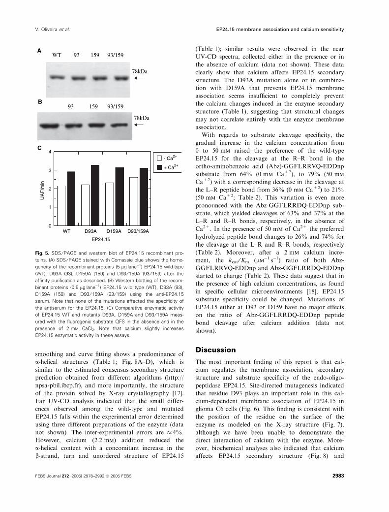
teria and purified to homogeneity (Fig. 5A). Western
blot analyses show that such point mutations have no
effect on antiserum recognition (Fig. 5B). The follow-
ing experiments were performed in an attempt to corre-
late a possible effect of the above point mutations with
the calcium-dependent EP24.15 membrane association.
The homogeneously purified EP24.15 recombinant pro-
teins (wild-type or mutant) were incubated with glioma
C6 cells and the putative membrane binding was ana-
lyzed by confocal microscopy. Incubation of either
wild-type EP24.15 or the D159A mutant with the
glioma C6 cells produced a clear increment in the
immunoreactivity of EP24.15 associated with the extra-
cellular surface of these cells (Fig. 6A and B) when
compared with control performed without adding any
of the recombinant EP24.15 (Fig. 6E). On the other
hand, incubation of the glioma C6 cells with either
D93A or D93 ⁄ 159A mutated proteins was not able to
produce a similar increment in the extracellular immu-
noreactivity related to EP24.15 (Fig. 6C and D). Con-
trol experiments under these conditions show that
without the addition of EP24.15 the basal immunoreac-
tivity was greatly reduced (Fig. 6E). Taken together,
these data suggest the D93 residue of EP24.15 within
the putative calcium-binding motif 89SPNKD93 is a
possible mediator of enzyme–membrane association.
To examine a structure–function correlate of the
D93–membrane association and the location of the resi-
due within the three-dimensional realm of the enzyme,
Fig. 1. Calcium-dependent EP24.15 released into the medium.
EP24.15 activity in intact cells was determined using b-lipotro-
pin(61–69) (YGGFMTSEK; 100 lM) as substrate. b-lipotropin(61–69)
fragments were separated by reverse phase HPLC and the
EP24.15 activity was based on the generation of [Met5]enkephalin
calculated by comparing the peak areas of each of those products
with the corresponding peak area of the same synthetic peptides
of known concentrations. The EP24.15 enzymatic activity released
into the medium was determined fluorimetrically using the
substrate Abz-GGFLRRV-EDDnp. To discern peptidolytic activity
exclusively due to EP24.15, the inhibitors N-(1-(R,S)-carboxyl-
2-phenylethyl)-AAF-p-amino-benzoate (CFP-AAF-pAB) and ⁄ or the
dipeptide Pro–Ile were used. All enzymatic determinations were
conducted under linear conditions where product formation was
directly proportional to enzyme concentration, and obeys zero-order
kinetic parameters with < 10% of total substrate consumed during
the course of the assay. Results are means of three independent
determinations ± SE. (A) EP24.15 release into the glioma C6 cells’
medium is inversely proportional to the extracellular calcium con-
centration. (B) Total extracellular EP24.15 activity is not affected by
calcium, however, the amount of enzyme that is released in the
medium is largely increased after incubating the glioma C6 cells in
medium that lacks calcium.
EP24.15 membrane association and calcium sensitivity V. Oliveira et al.
2980 FEBS Journal 272 (2005) 2978–2992 ª 2005 FEBS

a hypothetical intermolecular interaction using calcium
as a ligand was performed (Fig. 7). The calcium is
modeled in green and is hydrogen bonded to two
amino acids. The first amino acid is D93, the object of
study in this work. Using an ensemble of coordinates
of well studied Asp–calcium interactions, the carboxy
groups of the Asp are within hydrogen bonding dis-
tance (2.5–3 A) for calcium. The coordinates for Fig. 7
were obtained from the crystal structure of thimet
oligopeptidase deposited at the Research Collaboratory
for Structural Bioinformatics (Protein Data Bank ID
#1S4B). The figure was drawn with the computer
modeling program spock and then subsequently ren-
dered using raster 3d [16]. To ensure that electro-
statics were properly accounted for, calculations were
performed and slightly negative potentials were
obtained for the D93.
Furthermore, we investigated the calcium effects on
EP24.15 secondary structure, enzymatic activity and
substrate specificity. Enzymatic activity of the mutated
Fig. 2. EP24.15 immunoreactivity in glioma C6 cells. EP24.15 presence in glioma C6 cells was further confirmed by immunochemical reac-
tions. (A) The antiserum raised against the rat testis recombinant EP24.15 recognized in the whole homogenate of glioma C6 cells a single
protein band corresponding to the expected Mr of EP24.15. For details see Experimental procedures. (B–E) Prior to each immunocytochem-
istry experiment the DMEM culture medium was removed and cells were rinsed three times with fresh NaCl ⁄ Pi, with (B, D) or without
(C, E) 1.2 mM of calcium, and incubated for 30 min in 4% formaldehyde, pH 7.4. After three cycles of NaCl ⁄ Pi washing (15 min each) cells
were incubated for 2 h in the presence of rabbit anti-EP24.15 serum (1 : 4000) diluted in NaCl ⁄ Pi containing 3% normal goat serum, 1%
BSA, in the presence (B, C) or absence (D, E) of 0.1% of Triton X100. Labeled cells were examined under a Zeiss laser confocal microscope
(CLSM 410) equipped with an Axiovert 100 inverted microscope and an Argon ⁄ Krypton laser. Cy3-tagged molecules were excited at a wave-
length of 568 nm. Images were acquired sequentially as single transcellular optical sections and averaged over 32 scans per frame. They
were then processed using the Carl Zeiss CLSM software (version 3.1) and stored on Jazz disks for further retrieval and editing. Final com-
posites were prepared using Adobe PHOTOSHOP without modifying the spectral characteristics of the original signal. Control experiments were
performed as described; no specific cell labeling could be observed (data not shown). The data presented are representative of three experi-
ments performed independently and exhibiting similar results.
V. Oliveira et al. EP24.15 membrane association and calcium sensitivity
FEBS Journal 272 (2005) 2978–2992 ª 2005 FEBS 2981

proteins either in the presence or absence of calcium
was not different from the wild-type EP24.15
(Fig. 5C). The superimposable CD spectra indicates
that there are no gross perturbations in expression of
the mutant proteins with respect to folding. CD spec-
tra of wild-type EP24.15 and mutants without
Fig. 4. Extracellular EP24.15 immunogold
labeling observed by electron microscopy.
Cells incubated in the presence of calcium
were processed for electron microscopy
using a pre-embedding procedure as des-
cribed. (A) Lower magnification of an entire
glioma C6 cell observed in the electron
microscope and immunolabeled at the
extracellular surface for EP24.15 (arrows).
(B) Higher magnification of the cell shown
in (A) immunolabeled for EP24.15 (arrows).
Scale bar: A, 140 nm; B, 500 nm.
Fig. 3. The effects of calcium on the extracellular EP24.15 immunoreactivity in glioma C6 cells. (A, B) To observe exclusively the extracellular
EP24.15 immunoreactivity in glioma C6 cells, cells were incubated for 15 min at 4 �C in the presence of rabbit anti-EP24.15 serum (1 : 500),
diluted in NaCl ⁄ Pi containing 3% normal goat serum and 1% BSA, prior to fixation with 4% formaldehyde pH 7.4. Labeled cells were exam-
ined under a Zeiss laser confocal microscope (CLSM 410) equipped with an Axiovert 100 inverted microscope and an Argon ⁄ Krypton laser.
Cy3-tagged molecules were excited at a wavelength of 568 nm. Images were acquired sequentially as single transcellular optical sections
and averaged over 32 scans per frame. They were then processed using the Carl Zeiss CLSM software (version 3.1) and stored on Jazz disks
for further retrieval and editing. Final composites were prepared using Adobe PHOTOSHOP without modifying the spectral characteristics of
the original signal. Immunocytochemistry reactions were conducted after incubation of the glioma C6 cells in the presence (A) or absence
(B) of calcium for 1 h. Note that cells previously incubated in medium lacking calcium (B) show a reduced extracellular labeling for EP24.15
compared to cells incubated in the presence of calcium (A). The intensity of the immunostaining varies from cell to cell but is consistently
stronger in cells previously incubated in the presence of calcium compared to those incubated in the absence of calcium. (1) Confocal regu-
lar image; (2) nomarski; (3) glow scale. The immunolabeling intensity is shown in the graphic (intensity grows from green to red). No immu-
nolabeling is observed after preabsorbing the anti-EP24.15 serum with recombinant EP24.15 (100 lgÆmL)1; data not shown).
EP24.15 membrane association and calcium sensitivity V. Oliveira et al.
2982 FEBS Journal 272 (2005) 2978–2992 ª 2005 FEBS
smoothing and curve fitting shows a predominance of
a-helical structures (Table 1; Fig. 8A–D), which is
similar to the estimated consensus secondary structure
prediction obtained from different algorithms (http://
npsa-pbil.ibcp.fr), and more importantly, the structure
of the protein solved by X-ray crystallography [17].
Far UV-CD analysis indicated that the small differ-
ences observed among the wild-type and mutated
EP24.15 falls within the experimental error determined
using three different preparations of the enzyme (data
not shown). The inter-experimental errors are � 4%.
However, calcium (2.2 mm) addition reduced the
a-helical content with a concomitant increase in the
b-strand, turn and unordered structure of EP24.15
(Table 1); similar results were observed in the near
UV-CD spectra, collected either in the presence or in
the absence of calcium (data not shown). These data
clearly show that calcium affects EP24.15 secondary
structure. The D93A mutation alone or in combina-
tion with D159A that prevents EP24.15 membrane
association seems insufficient to completely prevent
the calcium changes induced in the enzyme secondary
structure (Table 1), suggesting that structural changes
may not correlate entirely with the enzyme membrane
association.
With regards to substrate cleavage specificity, the
gradual increase in the calcium concentration from
0 to 50 mm raised the preference of the wild-type
EP24.15 for the cleavage at the R–R bond in the
ortho-aminobenzoic acid (Abz)-GGFLRRVQ-EDDnp
substrate from 64% (0 mm Ca+2), to 79% (50 mm
Ca+2) with a corresponding decrease in the cleavage at
the L–R peptide bond from 36% (0 mm Ca+2) to 21%
(50 mm Ca+2; Table 2). This variation is even more
pronounced with the Abz-GGFLRRDQ-EDDnp sub-
strate, which yielded cleavages of 63% and 37% at the
L–R and R–R bonds, respectively, in the absence of
Ca2+. In the presence of 50 mm of Ca2+ the preferred
hydrolyzed peptide bond changes to 26% and 74% for
the cleavage at the L–R and R–R bonds, respectively
(Table 2). Moreover, after a 2 mm calcium incre-
ment, the kcat ⁄Km (lm)1Æs)1) ratio of both Abz-
GGFLRRVQ-EDDnp and Abz-GGFLRRDQ-EDDnp
started to change (Table 2). These data suggest that in
the presence of high calcium concentrations, as found
in specific cellular microenvironments [18], EP24.15
substrate specificity could be changed. Mutations of
EP24.15 either at D93 or D159 have no major effects
on the ratio of Abz-GGFLRRDQ-EDDnp peptide
bond cleavage after calcium addition (data not
shown).
Discussion
The most important finding of this report is that cal-
cium regulates the membrane association, secondary
structure and substrate specificity of the endo-oligo-
peptidase EP24.15. Site-directed mutagenesis indicated
that residue D93 plays an important role in this cal-
cium-dependent membrane association of EP24.15 in
glioma C6 cells (Fig. 6). This finding is consistent with
the position of the residue on the surface of the
enzyme as modeled on the X-ray structure (Fig. 7),
although we have been unable to demonstrate the
direct interaction of calcium with the enzyme. More-
over, biochemical analyses also indicated that calcium
affects EP24.15 secondary structure (Fig. 8) and
AWT 93 159 93/159
4- Ca2+
+ Ca2+
3
2
1
0WT D93A D159A D93/159A
EP24.15
UA
F/m
in
93 159 93/159
78kDa
78kDa
B
C
Fig. 5. SDS ⁄ PAGE and western blot of EP24.15 recombinant pro-
teins. (A) SDS ⁄ PAGE stained with Comassie blue shows the homo-
geneity of the recombinant proteins (5 lgÆlane)1) EP24.15 wild-type
(WT), D93A (93), D159A (159) and D93 ⁄ 159A (93 ⁄ 159) after the
affinity purification as described. (B) Western blotting of the recom-
binant proteins (0.5 lgÆlane)1) EP24.15 wild type (WT), D93A (93),
D159A (159) and D93 ⁄ 159A (93 ⁄ 159) using the anti-EP24.15
serum. Note that none of the mutations affected the specificity of
the antiserum for the EP24.15. (C) Comparative enzymatic activity
of EP24.15 WT and mutants D93A, D159A and D93 ⁄ 159A meas-
ured with the fluorogenic substrate QFS in the absence and in the
presence of 2 mM CaCl2. Note that calcium slightly increases
EP24.15 enzymatic activity in these assays.
V. Oliveira et al. EP24.15 membrane association and calcium sensitivity
FEBS Journal 272 (2005) 2978–2992 ª 2005 FEBS 2983

Fig. 6. Immunocytochemical staining of glioma C6 cells after incubation with the recombinant EP24.15 proteins. Glioma C6 cells were prein-
cubated at 4 �C in the presence (A–D) or absence (E) of 2 lg of either EP24.15 wild-type (A), D159A (B), D93A (C) or D93 ⁄ 159A (D), for
30 min, in DMEM containing BSA 0.05%. The medium was then removed and cells were rinsed three times with DMEM and incubated at
4 �C for 30 min in the presence of the anti-EP24.15 serum (1 : 500). The excess of primary antiserum was removed rinsing the cells three
times in 1 mL of NaCl ⁄ Pi containing 5% BSA, before fixing the cells in PFA 4%. The secondary anti-(rabbit Cy3) Ig (1 : 250; Sigma) was
used to develop the immunoreaction. Note that the intensity of the EP24.15-related immunostaining is reduced for either the D93A or
D93 ⁄ 159A proteins compared to the wild-type and D159A.
Fig. 7. Molecular modeling of the surface EP24.15 D93 residue complexed with a calcium ion. On the left a diagram of the side chain of
D93 and a bound calcium (gray sphere) mapped onto the surface of the recently solved EP24.15 structure ([40] PDB ID #1S4B). On the right
is a magnified view focusing on the D93 side chain and interaction with calcium. The EP24.15 model with the calcium ion was energy mini-
mized using Molecular Operating Environment software, and the figure was generated using SPOCK.
EP24.15 membrane association and calcium sensitivity V. Oliveira et al.
2984 FEBS Journal 272 (2005) 2978–2992 ª 2005 FEBS

changes the enzyme substrate specificity (Table 2).
These data suggest that calcium is an important medi-
ator of EP24.15 biological function.
It has been previously shown that in the brain
EP24.15 is present both in neurons and glial cells [5,6].
Glial cells are at least 10 times more abundant than
neurons in the brain [19] and during the past years
they have been recognized to be more than a connect-
ive tissue responsible for neuronal support [20]. Glial
cells are now identified as excitable cells capable of
modulating neuronal stimulus [19]. As for neurons,
glial cells can also produce growth factors, pro-hor-
mone processing enzymes [21], receptors [22], neuro-
transmitters [19] and neuropeptides [23]. However, in
contrast to neurons, which process peptides within the
secretory pathway, glial cells secrete immature peptide
precursors [24,25]. Glial post-translational processing
of opioid peptides precursor (e.g. proenkephalin) is
therefore thought to occur in the extracellular environ-
ment, as demonstrated by the presence of protease
inhibitors in the cultured medium of striatal astrocytes
that prevented Met-enkephalin generation from secre-
ted proenkephalin [25–27]. Therefore, the present
results suggest that in physiological conditions
EP24.15 would be able to associate with the extracellu-
lar surface of glial cells and could participate in the
metabolism of a specific set of peptides released by
these cells and ⁄or from neurons. Thus, further charac-
terization of peptide-processing enzymes such as
EP24.15 in the extracellular milieu of glial cells is of
neurobiological significance.
Table 1. Calcium effects on the secondary structure of EP24.15.
a-Helix b-Strand Turn Unordered
TBS
WT 42.4% 13.9% 14.3% 26.9%
D93A 44.3% 14.7% 12.6% 25.8%
D159A 57.5% 10.6% 11.4% 22.3%
D93 ⁄ 159A 50.3% 12.3% 12.6% 24.5%
TBS + 2.2 mM Ca2+
WT 38.7% 15.6% 14.4% 27.6%
D93A 40.2% 15.9% 13.5% 27.2%
D159A 49.1% 12.7% 12.8% 25.1%
D93 ⁄ 159A 47.1% 14.6% 12.3% 25.6%
Fig. 8. Far UV-CD spectra of the EP24.15 WT (A), D93A (B), D159A (C) and D93 ⁄ 159A (D) mutants both in the absence (n) or in the pres-
ence of CaCl2 2 mM (h). Note that calcium addition reduces the a-helical content of all EP24.15s analyzed (Table 1).
V. Oliveira et al. EP24.15 membrane association and calcium sensitivity
FEBS Journal 272 (2005) 2978–2992 ª 2005 FEBS 2985

In a previous study we reported that EP24.15 secre-
tion from glioma C6 cells was increased linearly by
reduced extracellular calcium concentrations [12].
These data were paradoxical to many other secretory
systems in which extracellular calcium is required for
secretion [28]; therefore, the mechanism behind this
calcium effect on EP24.15 secretion was investigated
further. It is known that extracellular calcium is not
required for constitutive protein secretion [29] and
reduces the release of parathyroid hormone [28]. Bio-
chemical observations shown here indicated that
extracellular calcium concentrations could not change
the overall rate of peptide metabolism from intact gli-
oma C6 cells in culture (Fig. 1). These data suggest
that the overall extracellular EP24.15 enzymatic activ-
ity was not affected by altered calcium concentra-
tions. Consequently, we conclude that calcium
removal was not really affecting EP24.15 secretion
from glioma C6 cells. Another possibility was that
low extracellular calcium concentration compromised
the EP24.15 association with the E face of the plasma
membrane. The presence of EP24.15 associated with
the E face of the plasma membrane in glioma C6
cells was confirmed by immunocytochemistry experi-
ments both at the light and electron microscopy
levels. Indeed, immunocytochemistry analyses shown
here suggested that EP24.15 extracellular immuno-
reactivity is deeply affected when extracellular calcium
is removed from glioma C6 cells. Thus, it seems clear
that a deficiency in extracellular calcium affects the
shedding of EP24.15 from the plasma membrane of
glioma C6 cells into the medium, suggesting that
EP24.15–membrane association within cells can be
dynamically modulated. While further investigation
would be necessary, this mechanism could be relevant
for the intracellular traffic and secretion of EP24.15
[13,30], since it could dynamically locate the enzyme
in specific cell membranes, such as the endoplasmic
reticulum [6], Golgi apparatus, secretory vesicles [14]
and plasma membrane [11].
Interestingly, EP24.15 contains neither a leader pep-
tide sequence nor any other hydrophobic domains to
mediate its interaction with biological membranes [31]
and the mechanism responsible for the enzyme mem-
brane association in neuroendocrine cells remains
unknown [6,11]. The EP24.15 crystal structure was
recently solved [17] showing that the enzyme surface
contains 80% of all charged residues. Among these
amino acid residues D93 and D159 were investigated
as possible mediators of EP24.15–membrane associ-
ation. The D93 amino acid residue is located within
the EP24.15 89SPNKD93 sequence, which recalls the
calcium-binding motif DXSXS previously described
[32]. On the other hand, D159 residue is the EP24.15
corresponding metal divalent-binding site residue iden-
tified in the homologue endopeptidase EP24.16; both
D93 and D159 residues are located at the external sur-
face of the enzyme [17,33]. The D93A or D93 ⁄ 159Adouble mutations were shown to affect the calcium-
dependent association of EP24.15 to the plasma
membrane of glioma C6 cells, while the D159A point
mutation had no such effect. The molecular modeling
of the interaction of calcium and D93 on the surface
of the enzyme has the correct electrostatic potential.
Additionally, the calcium is far enough from the active
site zinc that no direct interaction can be postulated
for direct effects on catalysis. Changes appear to be
transduced through changes in secondary structure.
Circular dichroism experiments reported here clearly
shows that calcium affects EP24.15 secondary struc-
ture, reducing the a-helical content by 3–8%. It is well
known that calcium affects the conformational struc-
ture of several proteins [34–37], and that could provide
a possible mechanistic explanation for the data shown
above. The D93A or D93 ⁄ 159A mutations that
strongly affected EP24.15 membrane association were
not sufficient to prevent the secondary structure altera-
tions of EP24.15 induced by calcium. Hence, it is
plausible to suggest that the structural changes induced
by calcium on EP24.15 secondary structure are not
correlated with the membrane association. Therefore,
the exact role of D93 in EP24.15 membrane associ-
ation remains unknown. A possible explanation that
needs further attention is that D93 mediates the inter-
action of EP24.15 with a yet unidentified membrane
protein. Molecular modeling corroborates that the resi-
due is on the surface.
Table 2. Influence of calcium concentration on EP24.15 peptide
bond cleavage.
[Ca2+]
(mM)
Cleaved bond (%)
Abz-GGFLRRVQ-EDDnp Abz-GGFLRRDQ-EDDnp
L–Rb R–Rb
kcat ⁄ Kma
(lM)1.s)1) L–Rb R–Rb
kcat ⁄Kma
(lM)1.s)1)
0 36 64 2.0 63 37 1.3
0.5 – – – 62 38 1.3
1 – – – 61 39 1.3
2 – – – 56 44 1.3
5 31 69 1.5 52 48 1.4
10 28 72 1.2 45 55 1.3
50 21 79 0.8 26 74 0.7
a The SD were < 5% for any of the obtained kinetic parameters. b In
three independent assays conduced in triplicate the errors obtained
in the percentage of hydrolyzed peptide bond was less than 3%.
EP24.15 membrane association and calcium sensitivity V. Oliveira et al.
2986 FEBS Journal 272 (2005) 2978–2992 ª 2005 FEBS

A previous report demonstrated the influence of cal-
cium and other divalent cations activating EP24.15
enzyme activity, in a substrate dependent manner [38]
consistent with our data. Another recent study has also
shown the influence of calcium on the acid limb of a
pH-dependence activity curve of EP24.15 [39]. Fluoro-
genic substrates have also been used previously to
show the influence of different salts on the EP24.15
activity [40]. Our present data confirm these previous
studies as calcium affected the kcat ⁄KM ratio of at least
two fluorogenic substrates hydrolyzed by EP24.15
(Table 2). In addition, we have shown here that
calcium affect the ratio of peptide bond (L–R ⁄R–R)
cleavage observed with the fluorogenic substrates Abz-
GGFLRRDQ-EDDnp and Abz-GGFLRRVQ-EDDnp
assayed. These effects were more pronounced using the
substrate Abz-GGFLRRDQ-EDDnp, which contains
an aspartic acid residue that could interact directly
with calcium. However, the effect was also verified
with the substrate Abz-GGFLRRVQ-EDDnp that
does not contain any negatively charged residues to
bind calcium directly, thus suggesting that the struc-
tural changes induced by calcium on EP24.15 secon-
dary structure could modulate its substrate specificity,
even altering the cleavage site on specific peptides.
Moreover, the results obtained with the substrate Abz-
GGFLRRDQ-EDDnp seem to indicate the presence
of a positive residue at the S3¢ subsite. Thus, if the
kcat ⁄KM ratio obtained from the kinetics with the sub-
strate Abz-GGFLRRDQ-EDDnp containing a negat-
ive change at P3¢ is decomposed into the cleavage sites,
it is possible to verify that the cleavage at the R–R
bond was activated while the cleavage at the L–R
bond was inhibited at higher calcium concentra-
tions. Similar analyses using the substrate Abz-
GGFLRRVQ-EDDnp, which that does not contain a
negative change at P3¢, shows inhibition of both R–R
and L–R cleaved sites. These observations suggest that
EP24.15 contains a positively charged residue at the
S3¢ subsite. This is in agreement with previously pub-
lished reports [3,40], and could be of importance in the
development of new specific substrates and ⁄or inhibi-
tors for the enzyme, as the detailed architecture of the
enzyme and its interactions with substrates are deci-
phered.
The mutations D93A and D159A neither prevented
the effect of calcium ions on the cleaved peptide
bond ratio (L–R ⁄R–R) nor changed the kcat ⁄KM
ratio for these substrates (data not shown). These
mutations that could not prevent the structural chan-
ges induced by calcium on EP24.15 structure as
observed with the far UV-CD assays suggest the
possible existence of other calcium binding site(s) in
the EP24.15 structure, which have not yet been des-
cribed.
In conclusion, the present report provides evidence
that EP24.15 is able to associate to the extracellular
face of the plasma membrane in a calcium dependent
fashion. Calcium was also shown to change the secon-
dary structure by reduction of a-helical content and to
change the substrate specificity of EP24.15. Together,
these new findings are useful to understand the com-
plex organization of peptide metabolism within cells
mediated by divalent cation by altering and regulating
enzyme localization within subcellular compartments
in the cell.
Experimental procedures
Cell culture
Glioma C6 cells were cultured in Dulbecco’s modified
Eagle’s medium (DMEM; Invitrogen, Carlsbad, CA, USA)
containing 10% fetal calf serum (Invitrogen), 50 UÆmL)1
penicillin G, and 50 lgÆmL)1 streptomycin sulfate, as previ-
ously described [12]. The cells were maintained in 12-well
plates (5.2 · 105 cellsÆcm)2 density of saturation), at 37 �Cin a humidified atmosphere consisting of 5% CO2 and 95%
air. Prior to the experiments, the culture medium from 60–
80% confluent cells was removed and cells were rinsed
three times with 1 mL Henk’s buffer (136 mm NaCl,
3.8 mm KCl, 1.2 mm CaCl2, 0.8 mm MgSO4, 1.1 mm
Na2HPO4, 0.48 mm KH2PO4, 2.0 mm NHCO3, 4 gÆL)1
d-glucose, final pH 7.4) equilibrated at 37 �C. For the cal-
cium-free experiments, cells were previously incubated for
1 h in fresh NaCl ⁄Pi (0.2 m phosphate buffer pH 7.4, con-
taining 0.15 m NaCl), at 37 �C in a humidified atmosphere
consisting of 5% CO2 and 95% air.
Determination of EP24.15 activity of intact glioma
C6 cells by HPLC
Total extracellular EP24.15 activity was determined using
b-lipotropin(61–69) (YGGFMTSEK) [12]. Experiments
were conducted in 12-well plates (� 5.2 · 105 cellsÆcm)2)
containing 0.5 mLÆwell)1 Henk’s media. The extent of
hydrolysis was consistently <10% during all the determi-
nations. The enzymatic reactions were terminated by addi-
tion of 5 lL 8% H3PO4. Peptide fragments were separated
by reverse phase HPLC using a C18 lBondapak column
(4.6 · 250 mm; Millipore Corp., Danvers, MA, USA) with
a linear gradient of 5–35% acetonitrile in 0.085% H3PO4
for 15 min at a flow rate of 2 mLÆmin)1. The rate of the
reaction was evaluated by determining the amount of
[Met5]enkephalin formed from b-lipotropin(61–69). Absorb-
ance was monitored at a wavelength of 214 nm.
[Met5]enkephalin concentrations were calculated by
V. Oliveira et al. EP24.15 membrane association and calcium sensitivity
FEBS Journal 272 (2005) 2978–2992 ª 2005 FEBS 2987

comparing the peak areas of each of those products with
the corresponding peak area of the same synthetic peptides
of known concentrations. The EP24.15 enzymatic activity
released into the medium was determined fluorimetrically
using the substrate Abz-GGFLRRV-EDDnp (QF7) [16].
To discern peptidolytic activity exclusively due to EP24.15,
the inhibitors N-(1-(R,S)-carboxyl-2-phenylethyl)-AAF-p-
amino-benzoate (CFP-AAF-pAB) and ⁄ or the dipeptide
Pro–Ile were used. All enzymatic determinations were con-
ducted under linear conditions where product formation
was directly proportional to enzyme concentration, with
<10% of total substrate consumed during the course of
the assay. The Abz-GGFL fluorescent product was used as
a calibration standard. Enzyme assays were conducted in a
final volume of 100 lL containing 10–50 lL cell extra-
ct ⁄media, 13 lm QF7, 1 mm 2-mercaptoethanol, and TBS
(0.025 m Tris ⁄HCl pH 7.4, 0.125 m NaCl). After 30 min,
the reactions were terminated by addition of 1.9 mL 80 mm
sodium formate pH 4.0. One milli-unit EP24.15 activity is
defined as the amount of enzyme (inhibited by CFP-
AAF-pAB but not by Pro–Ile), able to hydrolyze 1 nmol
QF7Æmin)1ÆmL)1, at 37 �C pH 7.5, in TBS containing
1 mm b-mercaptoethanol. Results were expressed as the
mean ± SD of five independent determinations.
Determination of soluble G6PD enzyme activity
Incubation media from C6 cells were centrifuged at
14 000 g for 1 min and filtered through a 0.2-lm filter (Mil-
lipore), prior to determination of the extracellular EP24.15
and G6PD enzyme activities. Intact C6 cells from each well
(� 5.2 · 105 cellsÆcm)2) were re-suspended and homogen-
ized in 1 mL ice cold TBS using a Potter–Elvehjem homo-
genizer (10 · 30 s at 900 r.p.m). The samples were then
centrifuged (105 000 g, 60 min) and the supernatant (cyto-
solic fraction) was used to determine both EP24.15 and
G6PD enzyme activities. G6PD enzyme activity was deter-
mined as previously described [13], in a 1 mL (final volume)
reaction mixture of 0.1 m Tris ⁄HCl pH 8.0, 0.1 mm glu-
cose-6-phosphate, 1 mm NADP+, and an amount of
sample able to alter 0.05–0.1 units of absorbance at
340 nmÆmin)1 at 25 �C, in the linear portion of the enzyme
kinetics. One milli-unit of G6PD was defined as the amount
of enzyme able to increase 0.01 units of absorbance at
340 nmÆmin)1 at 25 �C. Results were expressed as the
mean ± SD of five independent determinations.
Trypan blue exclusion assay
After experiments, cells were rinsed twice with Henk’s
media and treated with 0.2% Trypan blue solution in
NaCl ⁄Pi for 2 min. Dye excess was removed from the cell
supernatant after a brief centrifugation (1000 g, 5 min),
and cells were counted using a Neubauer chamber
(Hirschmann). Cells that did not incorporate the dye
were considered intact. Results were expressed as the
mean ± SD of three independent determinations.
Western blots
After electrophoretic separation by SDS ⁄PAGE on an 8%
acrylamide gel, proteins from the glioma C6 cells crude
homogenate (50 lg per lane) were transferred to nitrocellu-
lose membranes and western blots were performed using the
rabbit anti-EP24.15 serum (1 : 2000), diluted in NaCl ⁄Pi
containing 5% nonfat dry milk (w ⁄ v) as described pre-
viously [18]. For controls, the antiserum was replaced by
serum preadsorbed with 100 lgÆmL)1 of recombinant
(EP24.15). After 2 h incubation at room temperature in the
presence of the primary antiserum, the membranes were
rinsed with TBS containing 0.1% Tween 20 and 5% nonfat
milk and incubated with peroxidase-conjugated goat anti-
rabbit IgGs (1 : 5000; GE Healthcare, Amersham, UK) for
1 h. Immunoreactive bands on the nitrocellulose membranes
were visualized using an ECL kit (GE Healthcare) according
to the manufacturer’s instructions.
Immunocytochemistry for light and electron
microscopy
C6 glioma cells were cultured as described above under
cover slips. For the calcium-free experiments cells were pre-
viously incubated for 1 h in fresh NaCl ⁄Pi, at 37 �C in a
humidified atmosphere consisting of 5% CO2 and 95% air.
Prior to each immunocytochemistry experiment, culture
medium was removed and cells were rinsed three times with
fresh NaCl ⁄Pi and incubated for 30 min in 4% formalde-
hyde pH 7.4. After three cycles of NaCl ⁄Pi washing
(15 min each) cells were incubated 2 h in the presence of
rabbit anti-EP24.15 serum (1 : 4000) diluted in NaCl ⁄Pi
containing 3% normal goat serum, 1% BSA and 0.01% or
0.1% of Triton X100. Triton X100 was omitted in some
experiments as indicated in the figure legend.
To analyze the extracellular EP24.15 immunoreactivity,
experiments were conducted by incubating the cells for
15 min at 4 �C in the presence of rabbit anti-EP24.15 serum
(1 : 500), diluted in NaCl ⁄Pi containing 3% normal goat
serum, 1% BSA, prior to the addition of the 4% formalde-
hyde, pH 7.4. After three cycles of 15 min wash in
NaCl ⁄Pi, anti-(rabbit Cy3-conjugated) Ig (1 : 250; Sigma)
diluted in NaCl ⁄Pi containing 0.01% Triton X100, 3% nor-
mal goat serum and 1% BSA was added to the cells, and
incubated for 2 h at room temperature. Controls for the
EP24.15 immunocytochemical reaction consisted of both
preabsorbing the anti-EP24.15 serum with recombinant
EP24.15 (100 lgÆmL)1) and incubation of the preimmune
serum (1 : 4000) instead of the anti-EP24.15 serum. Follow-
ing several cycles of washing in NaCl ⁄Pi, the cover slips
were mounted over slide glass using glycerol. The immuno-
fluorescent staining was observed and photographed on
EP24.15 membrane association and calcium sensitivity V. Oliveira et al.
2988 FEBS Journal 272 (2005) 2978–2992 ª 2005 FEBS

either a microscope equipped for epifluorescence with the
corresponding filters (data not shown), or on a confocal
microscope as described below. Labeled cells were exam-
ined under a Zeiss laser confocal microscope (CLSM 410)
equipped with an Axiovert 100 inverted microscope and an
Argon ⁄Krypton laser. Cy3-tagged molecules were excited at
a wavelength of 568 nm. Images were acquired sequentially
as single transcellular optical sections and averaged over 32
scans per frame. They were then processed using the Carl
Zeiss clsm software (version 3.1) and stored for further
retrieval and editing. Final composites were prepared using
Adobe’s photoshop without modifying the spectral charac-
teristics of the original signal.
For electron microscopy, cells were processed using a
pre-embedding procedure [6]. Briefly, cells were fixed at
4 �C for 1 h in 3.75% acrolein and 4% paraformaldehyde
(PFA) in 0.1 m phosphate buffer, pH 7.4. After that, cells
were washed extensively in phosphate buffer, reacted with
sodium borohydride, washed again in phosphate buffer,
and cryoprotected for 30 min by immersion in a mixture of
6% glycerol and 25% sucrose in 0.05 m phosphate buffer.
They were then rapidly frozen in isopentane at )80 �C,transferred to liquid nitrogen for 1 min, and thawed in
phosphate buffer at room temperature. Thawed cells were
incubated for 30 min in TBS containing 3% of normal goat
serum, and then for 18 h at 4 �C in rabbit anti-EP24.15
serum diluted 1 : 1000 in TBS containing 0.5% normal goat
serum. Cells were rinsed in 0.01 m NaCl ⁄Pi (0.01 m phos-
phate buffer pH 7.4, containing 0.9% NaCl), incubated for
2 h in a 1 : 50 dilution of colloidal gold (1 nm)-conjugated
goat anti-rabbit IgGs (Amersham, Arlington Heights, IL,
USA) diluted in 0.01 m NaCl ⁄Pi containing 0.2% gelatin
and 0.8% BSA, washed again in 0.01 m NaCl ⁄Pi, and fixed
for 10 min in 2% glutaraldehyde in 0.01 m NaCl ⁄Pi. After
several washes in 0.2 m citrate buffer, pH 7.4, the immuno-
gold was silver-enhanced by incubating the sections for
7 min with IntenSE M silver solution (Amersham). The
reaction was stopped by washing in citrate buffer, and post-
fixed in 2% osmium tetroxide in phosphate buffer for
40 min, dehydrated in graded ethanols and flat-embedded
in Polybed 812 (Polysciences, Inc., Warrington, Pennsylva-
nia, USA). Samples were blocked off from the bottom of
the plate, glued to the tip of a polymerized Polybed chuck,
and cut at 80-nm thickness on an ultramicrotome. Ultra-
thin sections were counterstained with lead citrate and
uranyl acetate and examined with a JEOL 1010 TEM. Neg-
atives from electron microscopic photomicrographs were
scanned at 1200 dpi resolution on an AGFA Duoscan
T1200 scanner and final composites were prepared using
Adobe’s photoshop without modifying the original data.
Site-direct mutagenesis and protein expression
Double-stranded site-directed mutagenesis of rat EP24.15
was performed on a pGEX-4t2 (Amersham-Pharmacia
Biotech Inc.) using the protocols described by the manufac-
tory of the Quick-change Site-directed mutagenesis kit (Strat-
agene, Inc). Oligonucleotide primers 5¢-CGTGTCTCCGAA
CAAGGCAATCCGCGCAGC-¢3 (sense) and 5¢-GCTGCG
CGGATTGCCTTGTTCGGAGACACG-3¢ (antisense) and
5¢-CCACTTACCTCAGGCAACACAGGAGAAGATCAAG-
3¢ (sense) and 5¢-CTTGATCTTCTCCTGTGTTGCCTG
AGGTAAGTGG-3¢ (antisense) were used to introduce the
D93A and D159A point mutations on EP24.15, respectively;
double mutant was obtained from the D93A mutated cDNA
using the D159A oligos. The plasmid DNA was purified
(Mini-Prep, Promega Corp., Madison, WI, USA) and muta-
tions screened by automatic DNA sequencing, using a
MegaBace machine (GE Healthcare). Plasmid DNA contain-
ing the desired mutation was purified (Mini-Prep, Promega
Corp.) and transformed into electrocompetent DH5a bacter-
ial cells and plated overnight on plates containing ampicillin
to yield single colonies. Expression and purification of the
mutant proteins for biochemical characterization were per-
formed as described [15]. Purification to homogeneity was
assessed by SDS ⁄PAGE, and protein was quantified by the
Bradford assay [41]. Yields of expressed protein were similar
for all of the mutations and proper folding was verified by
CD (described below). Aliquots were stored at )80 �C for
subsequent studies.
CD
Circular dichroism experiments were performed in a Jobin
Yvon CD6 spectropolarimeter. Calibration was made
using d-10-camphosulfonic acid. CD spectra were collected
in the wavelength range of 195–260 nm (far UV-CD) or
240–350 nm (near UV-CD) at 0.5 nm resolution, 3 s
response, eight scans, 0.01 or 0.02 cm path length cells,
and temperature 20–22 �C. Ellipticity is reported as mean
residue molar ellipticity [h] (deg cm2Ædmol)1). The control
baseline was obtained with solvent and all the components
(TBS) without the proteins. All the data were obtained
with three different solutions of the proteins. Secondary
structure estimation of the proteins was performed using
data in the wavelength range of 195–260 nm using SEL-
CON algorithm [42].
Kinetic assays
The hydrolysis of the quenched fluorescence peptide sub-
strates at 37 �C in TBS was followed by measuring the
fluorescence at kem ¼ 420 nm and kex ¼ 320 nm in a Hita-
chi F-2000 spectrofluorometer. A buffer solution containing
EP24.15 and dithiotheitol 0.5 mm (to assure maximum
enzyme activation), was added to a 1-cm path-length
cuvette to a final volume of 2 mL. The cuvette was placed
in a thermostatically controlled cell compartment for 5 min
before the substrate solution was added. The increase in
fluorescence was recorded continuously with time. The
V. Oliveira et al. EP24.15 membrane association and calcium sensitivity
FEBS Journal 272 (2005) 2978–2992 ª 2005 FEBS 2989
concentration of peptide solutions was obtained by
colorimetric determination of the 2,4-dinitrophenyl group
(17 300 m)1Æcm)1, extinction coefficient at 365 nm). The
inner-filter effect was corrected using an empirical equation
as previously described [43]. All the obtained data were
fitted to nonlinear least square equations, using grafit
(Version 3.0, Erithacus Software Ltd, Staines, UK).
Specificity rate constants (kcat ⁄Km) were determined
under first-order conditions, with substrates concentrations
10-fold less than Km. The obtained first-order rate constants
were divided by the total enzyme concentration to provide
kcat ⁄Km. As the products Abz-GGFL, Abz-GGFLR and
their respective C-terminal fragments, were resistant to
hydrolysis by EP24.15, we could determine the specificity
rate constants (kcat ⁄Km) under first-order conditions, even
for the peptides hydrolyzed at two peptide bonds, as previ-
ously published [3]. All experiments were conducted in trip-
licate for at least three independent times. The results
varied less than 5% within these independent experiments.
Determination of cleaved bonds
The cleaved bonds were identified by isolation of the frag-
ments by HPLC, comparing the retention times of the
products fragments with synthetic peptides encompassing
the expected hydrolysis products and ⁄ or by molecular
weight. The molecular weights were determined by MS
using a MALDI-TOF (Shimadzu Tokyo, Japan). All deter-
minations were conducted in triplicates for at least three
times independently. The results varied less than 3% within
these independent experiments.
Protein assay
Protein determinations were carried out using the method
described by Bradford [41] using BSA as a standard. For
CD experiments, protein concentration was determined
according to the method described by Gill and von Hippel
[43].
Molecular modeling
The D93 mutation was modeled onto the X-ray crystallo-
graphic structure of the recently solved human EP24.15
[17]. We utilized the just released coordinates from the
Protein Data Bank of the Research Collaboratory for
Structural Bioinformatics (ID 1S4B). The atomic coordi-
nates of this structure served as a template for the D93, a
conserved residue. The in silico substitution of the mutated
alanine, as well as model building, were calculated and
rendered with the computer software suites quanta,
insight ii (Accelrys, San Diego, CA, USA), or o, for
molecular visualization [44]. The model was energy mini-
mized using Molecular Operating Environment software
(Montreal, Canada), and the graphics rendered with spock
and raster3d [45].
Acknowledgements
Thanks are due to Dr Ian A. Smith (Baker Institute)
who provided substrate and inhibitors for EP24.15. The
technical support of Sandra Regina S. Lascosck, Edson
Rocha de Oliveira, Gaspar Ferreira de Lima and Rober-
to Cabado Modia Junior is gratefully acknowledged.
This work was supported by the Sao Paulo State
Research Foundation (FAPESP; grants 96 ⁄05904-8,97 ⁄ 10831-2, 99 ⁄ 01983-9, 00 ⁄ 04297-8, 01 ⁄ 07544-9 and
04 ⁄ 04933-2), CNPq and CAPES, and NIH NS39892
and RR19325 (to MJG). PAGG and CCR were suppor-
ted by studentships from FAPESP (02 ⁄09861-4 and
97 ⁄ 05500-7, respectively). This work is part of the PhD
thesis of PAGG.
References
1 Camargo AC, Gomes MD, Reichl AP, Ferro ES,
Jacchieri S, Hirata IY & Juliano L (1997) Structural
features that make oligopeptides susceptible substrates
for hydrolysis by recombinant thimet oligopeptidase.
Biochem J 324, 517–522.
2 Montiel JL, Cornille F, Roques BP & Noble F (1997)
Nociceptin ⁄ orphanin FQ metabolism: role of aminopepti-
dase and endopeptidase 24.15. J Neurochem 68, 354–361.
3 Oliveira V, Campos M, Melo RL, Ferro ES, Camargo
AC, Juliano MA & Juliano L (2001) Substrate specifi-
city characterization of recombinant metallo oligo-
peptidases thimet oligopeptidase and neurolysin.
Biochemistry 40, 4417–4425.
4 Orlowski M, Michaud C & Chu TG (1983) A soluble
metalloendopeptidase from rat brain. Purification of the
enzyme and determination of specificity with synthetic
and natural peptides. Eur J Biochem 135, 81–88.
5 Chu TG & Orlowski M (1985) Soluble metalloendopep-
tidase from rat brain: action on enkephalin-containing
peptides and other bioactive peptides. Endocrinology
116, 1418–1425.
6 Fontenele-Neto JD, Massarelli EE, Gurgel Garrido PA,
Beaudet A & Ferro ES (2001) Comparative fine struc-
tural distribution of endopeptidase 24.15 (EC3.4.24.15)
and 24.16 (EC3.4.24.16) in rat brain. J Comp Neurol
438, 399–410.
7 Silva CL, Portaro FC, Bonato VL, de Camargo AC &
Ferro ES (1999) Thimet oligopeptidase (EC 3.4.24.15), a
novel protein on the route of MHC class I antigen pre-
sentation. Biochem Biophys Res Commun 255, 591–595.
8 Portaro FC, Gomes MD, Cabrera A, Fernandes BL,
Silva CL, Ferro ES, Juliano L & de Camargo AC
EP24.15 membrane association and calcium sensitivity V. Oliveira et al.
2990 FEBS Journal 272 (2005) 2978–2992 ª 2005 FEBS

(1999) Thimet oligopeptidase and the stability of MHC
class I epitopes in macrophage cytosol. Biochem Biophys
Res Commun 255, 596–601.
9 Kim S, Pabon A, Swanson TA & Glucksman MJ (2003)
Regulation of cell-surface major histocompatibility com-
plex class I expression by the endopeptidase EC
3.4.24.15 (thimet oligopeptidase). Biochem J 375, 111–
120.
10 Acker GR, Molineaux C & Orlowski M (1987) Synap-
tosomal membrane-bound form of endopeptidase-24.15
generates Leu-enkephalin from dynorphin1–8, alpha-
and beta-neoendorphin, and Met-enkephalin from Met-
enkephalin-Arg6-Gly7-Leu8. J Neurochem 48, 284–292.
11 Crack PJ, Wu TJ, Cummins PM, Ferro ES, Tullai JW,
Glucksman MJ & Roberts JL (1999) The association of
metalloendopeptidase EC 3.4.24.15 at the extracellular
surface of the AtT-20 cell plasma membrane. Brain Res
835, 113–124.
12 Ferro ES, Tambourgi DV, Gobersztejn F, Gomes MD,
Sucupira M, Armelin MC, Kipnis TL & Camargo AC
(1993) Secretion of a neuropeptide-metabolizing enzyme
similar to endopeptidase 22. 19 by glioma C6 cells.
Biochem Biophys Res Commun 191, 275–281.
13 Ferro ES, Tullai JW, Glucksman MJ & Roberts JL
(1999) Secretion of metalloendopeptidase 24.15 (EC
3.4.24.15). DNA Cell Biol 18, 781–789.
14 Garrido PA, Vandenbulcke F, Ramjaun AR, Vincent B,
Checler F, Ferro E & Beaudet A (1999) Confocal
microscopy reveals thimet oligopeptidase (EC 3.4.24.15)
and neurolysin (EC 3.4.24.16) in the classical secretory
pathway. DNA Cell Biol 18, 323–331.
15 Rioli V, Kato A, Portaro FC, Cury GK, te KK, Vin-
cent B, Checler F, Camargo AC, Glucksman MJ, Rob-
erts JL, Hirose S & Ferro ES (1998) Neuropeptide
specificity and inhibition of recombinant isoforms of the
endopeptidase 3.4.24.16 family: comparison with the
related recombinant endopeptidase 3.4.24.15. Biochem
Biophys Res Commun 250, 5–11.
16 Merritt EA & Bacon DJ (1997) Raster3D: photorealistic
molecular graphics. Methods Enzymol 277, 505–524.
17 Ray K, Hines CS, Coll-Rodriguez J & Rodgers DW
(2004) Crystal structure of human thimet oligopeptidase
provides insight into substrate recognition, regulation,
and localization. J Biol Chem 279, 20480–20489.
18 Carafoli E, Santella L, Branca D & Brini M (2001)
Generation, control, and processing of cellular calcium
signals. Crit Rev Biochem Mol Biol 36, 107–260.
19 Gomes FC, Spohr TC, Martinez R, Moura N & V
(2001) Cross-talk between neurons and glia: highlights
on soluble factors. Braz J Med Biol Res 34, 611–620.
20 Parpura V, Basarsky TA, Liu F, Jeftinija K, Jeftinija S
& Haydon PG (1994) Glutamate-mediated astrocyte-
neuron signalling. Nature 369, 744–747.
21 Giulian D, Vaca K & Johnson B (1988) Secreted
peptides as regulators of neuron-glia and glia–glia
interactions in the developing nervous system. J Neuro-
sci Res 21, 487–500.
22 Klein RS, Das B & Fricker LD (1992) Secretion of car-
boxypeptidase E from cultured astrocytes and from
AtT-20 cells, a neuroendocrine cell line: implications for
neuropeptide biosynthesis. J Neurochem 58, 2011–2018.
23 Eriksson PS, Hansson E & Ronnback L (1990) Delta
and kappa opiate receptors in primary astroglial cul-
tures from rat cerebral cortex. Neurochem Res 15, 1123–
1126.
24 Spruce BA, Curtis R, Wilkin GP & Glover DM (1990)
A neuropeptide precursor in cerebellum: proenkephalin
exists in subpopulations of both neurons and astrocytes.
EMBO J 9, 1787–1795.
25 Benda P, Lightbody J, Sato G, Levine L & Sweet W
(1968) Differentiated rat glial cell strain in tissue culture.
Science 161, 370–371.
26 Batter DK, Vilijn MH & Kessler J (1991) Cultured
astrocytes release proenkephalin. Brain Res 563, 28–32.
27 Yoshikawa K & Sabol SL (1986) Expression of the
enkephalin precursor gene in C6 rat glioma cells: regula-
tion by beta-adrenergic agonists and glucocorticoids.
Brain Res 387, 75–83.
28 Nemeth EF & Scarpa A (1987) Rapid mobilization of
cellular Ca2+ in bovine parathyroid cells evoked by
extracellular divalent cations. Evidence for a cell surface
calcium receptor. J Biol Chem 262, 5188–5196.
29 Burgess TL & Kelly RB (1987) Constitutive and regu-
lated secretion of proteins. Annu Rev Cell Biol 3, 243–
293.
30 Yamamoto M, Chikuma T, Yamashita A, Yamaguchi
M, Hojo H, Ozeki Y, Ahmed M & Kato T (2003) Ante-
rograde axonal transport of endopeptidase 24.15 in rat
sciatic nerves. Neurochem Int 42, 231–237.
31 Pierotti A, Dong KW, Glucksman MJ, Orlowski M &
Roberts JL (1990) Molecular cloning and primary struc-
ture of rat testes metalloendopeptidase EC 3.4.24.15.
Biochemistry 29, 10323–10329.
32 Lee JO, Rieu P, Arnaout MA & Liddington R (1995)
Crystal structure of the A domain from the alpha sub-
unit of integrin CR3 (CD11b ⁄CD18). Cell 80, 631–
638.
33 Ray K, Hines CS & Rodgers DW (2002) Mapping
sequence differences between thimet oligopeptidase and
neurolysin implicates key residues in substrate recogni-
tion. Protein Sci 11, 2237–2246.
34 Goto K, Toyama A, Takeuchi H, Takayama K, Saito
T, Iwamoto M, Yeh JZ & Narahashi T (2004) Ca2+
binding sites in calmodulin and troponin C alter inter-
helical angle movements. FEBS Lett 561, 51–57.
35 Baneres JL, Roquet F, Green M, LeCalvez H &
Parello J (1998) The cation-binding domain from the
alpha subunit of integrin alpha5 beta1 is a minimal
domain for fibronectin recognition. J Biol Chem 273,
24744–24753.
V. Oliveira et al. EP24.15 membrane association and calcium sensitivity
FEBS Journal 272 (2005) 2978–2992 ª 2005 FEBS 2991

36 Deng L, Markova SV, Vysotski ES, Liu ZJ, Lee J, Rose
J & Wang BC (2004) Crystal structure of a Ca2+-dis-
charged photoprotein: Implications for the Mechanisms
of the Calcium Trigger and the Bioluminescence. J Biol
Chem 279, 33647–33652.
37 Li J, Bigelow DJ & Squier TC (2004) Conformational
changes within the cytosolic portion of phospholamban
upon release of Ca-ATPase inhibition. Biochemistry 43,
3870–3879.
38 Wolfson AJ, Shrimpton CN, Lew RA & Smith AI
(1996) Differential activation of endopeptidase EC
3.4.24.15 toward natural and synthetic substrates by
metal ions. Biochem Biophys Res Commun 229, 341–348.
39 Sigman JA, Edwards SR, Pabon A, Glucksman MJ &
Wolfson AJ (2003) pH dependence studies provide
insight into the structure and mechanism of thimet oli-
gopeptidase (EC 3.4.24.15). FEBS Lett 545, 224–228.
40 Oliveira V, Gatti R, Rioli V, Ferro ES, Spisni A, Cam-
argo AC, Juliano MA & Juliano L (2002) Temperature
and salts effects on the peptidase activities of the recom-
binant metallooligopeptidases neurolysin and thimet oli-
gopeptidase. Eur J Biochem 269, 4326–4334.
41 Bradford MM (1976) A rapid and sensitive method for
the quantitation of microgram quantities of protein util-
izing the principle of protein-dye binding. Anal Biochem
72, 248–254.
42 Sreerama N & Woody RW (1993) A self-consistent
method for the analysis of protein secondary structure
from circular dichroism. Anal Biochem 209, 32–44.
43 Gill SC & von Hippel PH (1989) Calculation of protein
extinction coefficients from amino acid sequence data.
Anal Biochem 182, 319–326.
44 Jones TA, Zou JY, Cowan SW & Kjeldgaard A (1991)
Improved methods for building protein models in elec-
tron density maps and the location of errors in these
models. Acta Crystallogr A 47, 110–119.
45 Merritt EA & Bacon DJ (1997) Raster3D: photorealistic
molecular graphics. Methods Enzymol 277, 505–524.
EP24.15 membrane association and calcium sensitivity V. Oliveira et al.
2992 FEBS Journal 272 (2005) 2978–2992 ª 2005 FEBS
Copyright © 2022 FDOKUMEN