C1q and the glomerulonephritides: therapeutic approaches for the treatment of complement-mediated...
241
-
Upload
independent -
Category
Documents
-
view
3 -
download
0
Transcript of C1q and the glomerulonephritides: therapeutic approaches for the treatment of complement-mediated...


Series Editor
Prof. Michael J. Parnham, PhDSenior Scientific AdvisorPLIVA Research Institute Ltd.Prilaz baruna Filipovica 29HR-10000 ZagrebCroatia
Progress in Inflammation Research
Forthcoming titles:Toll-like Receptors in Inflammation, L.A.J. O’Neill, E. Brint (Editors), 2006Chemokine Biology: Basic Research and Clinical Application, Volume I: Immunobiology of
Chemokines, K. Neote, L. G. Letts, B. Moser (Editors), 2005Chemokine Biology: Basic Research and Clinical Application, Volume II: Pathophysiology
of Chemokines, K. Neote, L. G. Letts, B. Moser (Editors), 2005The Hereditary Basis of Rheumatic Diseases, R. Holmdahl (Editor), 2006
(Already published titles see last page.)
Advisory Board
G. Z. Feuerstein (Wyeth Research, Collegeville, PA, USA)M. Pairet (Boehringer Ingelheim Pharma KG, Biberach a. d. Riss, Germany)W. van Eden (Universiteit Utrecht, Utrecht, The Netherlands)

Birkhäuser VerlagBasel · Boston · Berlin
Complement and Kidney Disease
Peter F. Zipfel
Editor

ISBN-10: 3-7643-7166-8 Birkhäuser Verlag, Basel – Boston – BerlinISBN-13: 978-3-7643-7166-1 Birkhäuser Verlag, Basel – Boston – Berlin
The publisher and editor can give no guarantee for the information on drug dosage and administration contained in thispublication. The respective user must check its accuracy by consulting other sources of reference in each individual case.The use of registered names, trademarks etc. in this publication, even if not identified as such, does not imply thatthey are exempt from the relevant protective laws and regulations or free for general use.
This work is subject to copyright. All rights are reserved, whether the whole or part of the material is concerned,specifically the rights of translation, reprinting, re-use of illustrations, recitation, broadcasting, reproduction on micro-films or in other ways, and storage in data banks. For any kind of use, permission of the copyright owner must be obtained.
© 2006 Birkhäuser Verlag, P.O. Box 133, CH-4010 Basel, SwitzerlandPart of Springer Science+Business MediaPrinted on acid-free paper produced from chlorine-free pulp. TCF ∞Cover design: Markus Etterich, BaselCover illustration: HUS – Renal biopsy with typical glomerular changes of HUS including segmental thickening ofglomerular basement membranes with double contours, congestion of capillaries and increase in extracellular matrix(Masson trichrome stain) (see p. 173)Printed in GermanyISBN-10: 3-7643-7166-8 e-ISBN: 3-7643-7428-4ISBN-13: 978-3-7643-7166-1
9 8 7 6 5 4 3 2 1 www.birkhauser.ch
Editor
Peter F. ZipfelDepartment of Infection BiologyLeibniz Institute for Natural Product Research and Infection BiologyHans Knoell InstituteBeutenbergstr. 11aD-07745 JenaGermany
Library of Congress Cataloging-in-Publication Data
Complement and kidney disease / Peter F. Zipfel, editor.p. ; cm. -- (Progress in inflammation research)
Includes bibliographical references and index.ISBN-13: 978-3-7643-7166-1 (alk. paper)ISBN-10: 3-7643-7166-8 (alk. paper) 1. Kidneys--Diseases--Etiology. 2. Complement (Immunology). I. Zipfel, Peter F. II. PIR (Series)
[DNLM: 1. Kidney Diseases--etiology. 2. Complement--physiology. 3. Hemolytic-Uremic Syndrome. WJ 300 C7373 2005]RC903.C63 200561636’1--dc22
2005053631
Bibliographic information published by Die Deutsche BibliothekDie Deutsche Bibliothek lists this publication in the Deutsche Nationalbibliografie; detailed bibliographic data is available in the internet at http://dnb.ddb.de

Abbreviations . . . . . . . . . . . . . . . . . . . . . . . . . . . . . . . . . . . . . . . . . . . . . . . . . . . . . . . . . . . . . . . . . . . . . . . vii
List of contributors . . . . . . . . . . . . . . . . . . . . . . . . . . . . . . . . . . . . . . . . . . . . . . . . . . . . . . . . . . . . . . . . . ix
Poem. . . . . . . . . . . . . . . . . . . . . . . . . . . . . . . . . . . . . . . . . . . . . . . . . . . . . . . . . . . . . . . . . . . . . . . . . . . . . . . . . xiii
Preface . . . . . . . . . . . . . . . . . . . . . . . . . . . . . . . . . . . . . . . . . . . . . . . . . . . . . . . . . . . . . . . . . . . . . . . . . . . . . . . xv
Momir Macanovic and Peter Lachmann The complement system in renal diseases . . . . . . . . . . . . . . . . . . . . . . . . . . . . . . . . . . . . . . . . . 1
Wuding Zhou and Steven H. SacksComplement in renal transplantation . . . . . . . . . . . . . . . . . . . . . . . . . . . . . . . . . . . . . . . . . . . . . . 19
Stefan P. Berger, Tom W.L. Groeneveld, Anja Roos and Mohamed R. DahaC1q and the glomerulonephritides: therapeutic approaches for the treatment of complement-mediated kidney diseases. . . . . . . . . . . . . . . . . . . . . . . . . . . . . . 37
Joshua M. Thurman and V. Michael HolersComplement deficient mice as model systems for kidney diseases . . . . . . . . . . . . . . . 49
Marina Noris and Giuseppe RemuzziNon-Shiga toxin-associated hemolytic uremic syndrome . . . . . . . . . . . . . . . . . . . . . . . . . 65
Christine Skerka and Mihály Józsi Role of complement and Factor H in hemolytic uremic syndrome . . . . . . . . . . . . . . . 85
Timothy H.J. Goodship, Véronique Frémeaux-Bacchi, John P. AtkinsonGenetic testing in atypical HUS and the role of membrane cofactor protein(MCP;CD46) and Factor I . . . . . . . . . . . . . . . . . . . . . . . . . . . . . . . . . . . . . . . . . . . . . . . . . . . . . . . . . . 111
Maren Salzmann, Michael Hoffmann, Gisa Schluh, Peter Riegler, Markus Cybulla, Hartmut P.H. NeumannTowards a new classification of hemolytic uremic syndrome . . . . . . . . . . . . . . . . . . . . . 129
Contents

Reinhard Würzner and Lothar B. ZimmerhacklTherapeutic strategies for atypical and recurrent hemolytic uremic syndromes (HUS). . . . . . . . . . . . . . . . . . . . . . . . . . . . . . . . . . . . . . . . . . . . . . . . . . . . . . . . . . . . . . . . . . . . 149
Christoph Licht and Bernd HoppeComplement defects in children which result in kidney diseases: diagnosis and therapy. . . . . . . . . . . . . . . . . . . . . . . . . . . . . . . . . . . . . . . . . . . . . . . . . . . . . . . . . . . . . . . 165
Peter F. Zipfel, Richard J.H. Smith and Stefan HeinenThe role of complement in membranoproliferative glomerulonephritis . . . . . . . . . . 199
Pearl L. LewisThe experience of a patient advocacy group . . . . . . . . . . . . . . . . . . . . . . . . . . . . . . . . . . . . . . 223
Index . . . . . . . . . . . . . . . . . . . . . . . . . . . . . . . . . . . . . . . . . . . . . . . . . . . . . . . . . . . . . . . . . . . . . . . . . . . . . . . . 233

ADAMTS13 an integrin-like and metalloproteinase with thrombospondin motifAPC antigen presenting cellsBSH British Society for HaematologyCCP complement control protein module also termed short consensus
repeat (SCR)CCE cholesterol crystal emboliCR1 complement receptor 1, also termed CD 36DAF decay accelerating Factor, also termed CD 59DDD dense deposit diseasesDHPLC denaturing high performance liquid chromatographyESRD end stage renal diseaseI/R ischemic reperfusion injuryGBM glomerular basement membraneHUS hemolytic uremic syndromemAB monoclonal antibodyMAC membrane attack complex, the terminal effector system of comple-
ment which generates holes in the membrane of the target cellMCP membrane cofactor protein also termed CD 46MN membranous nephropathyMPGN membranoproliferative glomerulonephritisMLPA multiplex ligation-dependent probe amplificationRCA regulators of complement gene cluster on human chromosome 1,
includes the genes coding for Factor H, MCP, CR1 and DAFPE plasma exchangePBMC peripheral blood mononuclear cellsSLE systemic lupus erythematosusStx Shiga like toxinSSCP single strand conformation polymorphismTTP thrombotic thrombocytic purpuraTMA thrombotic microangiopathiesvWF von Willebrand Factor
Abbreviations

ix
John P. Atkinson, Division of Rheumatology, Washington University School of Med-icine, St. Louis, MO 63110, USA; e-mail: [email protected]
Stefan P. Berger, Department of Nephrology, C3-P25, Leiden University Medical Cen-ter, P.O. Box 9600, 2300 RC Leiden, The Netherlands; e-mail: [email protected]
Markus Cybulla, Department of Nephrology, Albert-Ludwigs-University, Hugstet-ter Straße 55, 79106 Freiburg, Germany; e-mail: [email protected]
Mohamed R. Daha, Department of Nephrology, C3-P25, Leiden University MedicalCenter, P.O. Box 9600, 2300 RC Leiden, The Netherlands; e-mail: [email protected]
Véronique Frémeaux-Bacchi, Hôpital Européen Georges Pompidou, 20 rue Leblanc,75908 Paris Cedex 15, France; e-mail: [email protected]
Tim Goodship, Institute of Human Genetics, University of Newcastle upon Tyne,Newcastle upon Tyne, NE1 3BZ, United Kingdom; e-mail: [email protected]
T.W.L. Groeneveld, Department of Nephrology, C3-P25, Leiden University MedicalCenter, P.O. Box 9600, 2300 RC Leiden, The Netherlands; e-mail: [email protected]
Stefan Heinen, Department of Infection Biology, Leibniz Institute for Natural Prod-uct Research and Infection Biology, Hans Knoell Institute, Beutenbergstr. 11a,07745 Jena, Germany; e-mail: [email protected]
V. Michael Holers, Division of Rheumatology, B-115, University of Colorado,Health Sciences Center, 4200 East 9th Avenue, Denver Co 80262, USA; e-mail: [email protected]
List of contributors
x
Michael Hoffmann, Department of Laboratory Medicine, Albert-Ludwigs-Universi-ty, Hugstetter Str. 55, 79106 Freiburg, Germany; e-mail: [email protected]
Bernd Hoppe, Children’s Hospital of the University of Cologne, Pediatric Nephrol-ogy, Kerpener Str. 62, 50937 Cologne, Germany; e-mail: [email protected]
Mihály Józsi, Department of Infection Biology, Leibniz Institute for Natural Prod-uct Research and Infection Biology, Hans Knoell Institute, Beutenbergstr. 11a,07745 Jena, Germany; e-mail: [email protected]
Pearl L. Lewis, Maryland Patient Advocacy Group, Foundation for Children withAtypical HUS, Maryland Renal Advocate 9131, 2530 Kensington Gardens #104,Ellicott City, MD 21043, USA; e-mail: [email protected]
Peter Lachmann, Centre for Veterinary Science, Madingley Road, Cambridge, CB30ES, UK; e-mail: [email protected]
Christoph Licht, Children’s Hospital of the University of Cologne, PediatricNephrology, Kerpener Str. 62, 50937 Cologne, Germany; e-mail: [email protected]
Momir Macanovic, Centre for Veterinary Science, Madingley Road, Cambridge,CB3 0ES, UK; e-mail: [email protected]
Hartmut P.H. Neumann, Department of Nephrology, Albert-Ludwigs-University,Hugstetter Str. 55, 79106 Freiburg, Germany; e-mail: [email protected]
Marina Noris, Chem Pharm D, Transplant Research Center “Chiara Cucchi deAlessandri e Gilberto Crespi”, Mario Negri Institute for Pharmacological Research,Villa Camozzi, Via Camozzi 3, 24020, Ranica (BG), Italy; e-mail: [email protected]
Giuseppe Remuzzi, Chem Pharm D, Transplant Research Center “Chiara Cucchi deAlessandri e Gilberto Crespi”, Mario Negri Institute for Pharmacological Research,Villa Camozzi, Via Camozzi 3, 24020, Ranica (BG), Italy; and Department of Med-icine and Transplantation, Ospedali Riuniti di Bergamo, Largo Barozzi 1, 24128,Bergamo, Italy; e-mail: [email protected]
List of contributors

Peter Riegler, Department of Nephrology, General Hospital, Lorenz-Böhler-Str. 5,39100 Bolzano, Italy; e-mail: [email protected]
Anja Roos, of Nephrology, C3-P25, Leiden University Medical Center, P.O. Box9600, 2300 RC Leiden, The Netherlands; e-mail: [email protected]
Steven H. Sacks, Department of Nephrology and Transplantation, Guy’s, King’s andSt. Thomas’ School of Medicine, King’s College of London, London SE1 9RT, UK;e-mail: [email protected]
Maren Salzmann, Department of Nephrology, Albert-Ludwigs-University, Hugstet-ter Straße 55, 79106 Freiburg, Germany; e-mail: [email protected]
Gisa Schluh, Department of Nephrology, Albert-Ludwigs-University, HugstetterStraße 55, 79106 Freiburg, Germany; e-mail: [email protected]
Christine Skerka, Department of Infection Biology, Leibniz Institute for NaturalProduct Research and Infection Biology, Hans Knoell Institute, Beutenbergstr. 11a,07745 Jena, Germany; e-mail: [email protected]
Richard J.H. Smith, Department of Otolaryngology, 200 Hawkins Drive, 21151PFP, The University of Iowa, Iowa City, IA 52242, USA; e-mail: [email protected]
Joshua M. Thurman, Division of Nephrology and Hypertension, B-115, Universityof Colorado Health Sciences Center, 4200 East 9th Avenue, Denver, CO 80262,USA; e-mail: [email protected]
Reinhard Würzner, Department of Paediatrics, Innsbruck Medical University,Anichstr. 35, 6020 Innsbruck, Austria
Wuding Zhou, Department of Nephrology and Transplantation, Guy's, King’s andSt. Thomas’ School of Medicine, King’s College of London, London SE1 9RT, UK;e-mail: [email protected]
Lothar Bernd Zimmerhackl, Department of Paediatrics, Innsbruck Medical Univer-sity, Anichstr. 35, 6020 Innsbruck, Austria; e-mail: [email protected]
Peter F. Zipfel, Department of Infection Biology, Leibniz Institute for Natural Prod-uct Research and Infection Biology, Hans Knoell Institute, Beutenbergstr. 11a,07745 Jena, Germany, and Friedrich-Schiller-University Jena, Professor for InfectionBiology; e-mail: [email protected]
xi
List of contributors

In Dämmerschein liegt schon die Welt erschlossen,Der Wald ertönt von tausendstimmigem Leben,Tal aus, Tal ein ist Nebelstreif ergossen,Doch senkt sich Himmelsklarheit in die Tiefen,...
Faust II, Johann Wolfgang v. Goethe

Complement is a central and important immune defense system that acts at theinterface between innate and adaptive immunity. Aimed at maintaining the integri-ty of the human organism, this system is – when activated – highly toxic, destruc-tive and inflammatory. Actually, the action of the highly efficient and toxic activa-tion components is desired and favorable on foreign surfaces such as microbes, orsurfaces of modified self cells, but at the same time absolutely unfavorable on thesurface of host cells. Consequently, activation of this defense system must berestricted in terms of time and space. Complement activation is initiated either spon-taneously by the alternative pathway, or is induced by antibodies via the classicalpathway. Regardless of the pathway of initiation, in order to prevent attack anddamage host cells and tissues must continuously restrict and block amplification ofthe complement cascade. Uncontrolled activation and defective control results inautoimmune diseases.
It has been known for a long time that there is an association between the com-plement system and renal disease. Complement components are involved in the ini-tiation, pathophysiology and progression of glomerular diseases in many ways. Therecent years have witnessed an impressive development in defining the role of com-plement and individual complement components in glomerular disease. In severalcases the defects have been identified on a molecular level by describing novelmutations in individual complement components, and by understanding the patho-physiology of the diseases, e.g., by defining how mutations of single componentsrelate to glomerular damage. This information reveals a pivotal role of complementin renal disease and – in several cases – allowed a novel understanding of the dis-ease mechanisms or a description of the pathophysiology in molecular terms. Inaddition to reports on the general role of complement in glomerular disease, thisvolume highlights the role of individual complement components in kidney dis-eases. In particular, several kidney diseases such as the atypical form of hemolyticuremic syndrome, membranoproliferative glomerulonephritis and systemic lupuserythematosus are covered in detail from the clinical side and from the side of basicresearch.
xv
Preface

This book is aimed at bridging the broad scope between basic research, diseasemechanisms and clinical aspects, and also include a report from the patients’ side.Several distinguished researchers, clinicians and a patient advocate, who have madesignificant contributions to the field of complement and glomerular disease duringthe last years review the latest findings in this field and give an in depth overviewon the current situation. The understanding of the relation of complement andglomerular diseases has substantially improved in recent years, due to the reports ofgene mutations in single effector proteins or regulators, detailed functional charac-terization of single mutated complement components and the description howdefective function relates to disease. Through these various concepts, novel diag-nostic regimens have been established and new ways paved for therapeutic inter-vention and treatment.
Based on the broad range of topics reviewed and covered, this book is aimed foranyone with an interest in glomerular diseases and in the role of complement inautoimmunity, particularly clinicians, researchers and students who wish to under-stand the role of complement in the pathogeneses of renal and autoimmune diseases.
Jena, September 2005 Peter F. Zipfel
xvi
Preface

1
The complement system in renal diseases
Momir Macanovic and Peter Lachmann
Centre for Veterinary Science, Madingley Road, Cambridge, CB3 0ES, UK
Complement and Kidney Disease, edited by Peter F. Zipfel© 2006 Birkhäuser Verlag Basel/Switzerland
Introduction
The association between the complement system and renal disease has been appre-ciated for a long time. The complement system may be involved in the initiation,pathogenesis and resolution of glomerulonephritis (GN) in many ways. The activa-tion of complement and its deleterious consequences have been observed in manyrenal diseases: some primary forms of GN, GN associated with systemic diseases,acute and chronic humoral rejection of renal transplant, cholesterol renal emboli,hemodialysis and others.
Understanding of relations of complement and GN evolved from the initialobservation of depletion of serum complement in some forms of GN and identifi-cation of complement deposits in kidney biopsy specimens. Varying glomeru-lonephritides are associated with alterations in the serum concentration of specificcomplement components, the presence of complement breakdown products in thecirculation, glomerular complement deposits and circulating complement-activatingfactors.
The evidence for the role of complement in enhancing renal injury comes alsofrom experimental data obtained by depletion of complement with cobra venom.Studies utilizing cobra venom factor to produce generalized complement depletionin animals have shown that most of the morphological and functional changeswhich occur in some forms of GN are complement dependent [1]. More recently,molecular biology techniques of genetically manipulated mice, administration ofrecombinant complement regulatory proteins [2, 3], monoclonal anti-C5 antibodiesthat block or reduce kidney damage [4–6], have provided additional evidence formultiple and important roles of complement in glomerular diseases and implicationsfor complement-targeted therapy to control ongoing inflammation in the kidney.
Hypocomplementemia in GN
Evidence of complement activation in GN comes from characteristic patterns of adecrease in the serum concentrations of specific components, some of which are vir-

2
Momir Macanovic and Peter Lachmann
tually diagnostic of certain nephritides. Hypocomplementemia is used as a markerfor diagnosis and monitoring efficacy of treatment. The components most frequent-ly measured in clinical practice are C3, C4, C2 and total hemolytic complement(CH50). Several commercial antisera are available for each, and rapid nephelomet-ric assays are in widespread use. The CH50 is a functional measurement, based onthe ability of serum to support complement-mediated lysis of sensitized erythro-cytes.
In chronic bacteremic states or in lupus nephritis, circulating or depositedimmune complexes may act as classical pathway activators [7]. These disorders arefrequently accompanied by reduction of the classical pathway activation proteinsC1q, C2, C4 along with C3 [8, 9]. Typical patients with acute post-streptococcalGN have reductions of C3, C5 and properdin. Normalization of C3 usually occurswithin 6–8 weeks [10].
The other major group of hypocomplementic GN, membranoproliferative GN(MPGN), is somewhat more variable. Type II MPGN usually is associated with areduction in C3 only. Evidence of either classical pathway activation or terminalcomponent depression is seen in type I MPGN.
In all these situations, depression of serum complement component concentra-tions is assumed to represent activation, although synthetic defects are occasionallysuggested.
Complement activation fragments in circulation
Complement activation, whether in the fluid phase or on surface, often results in thegeneration of soluble fragments, which can be detected in circulation. The detectionof these complement fragments during the course of glomerular injury providesadditional evidence of ongoing complement activation. The fragments most oftenassayed are C3a and C5a. Commercial assays are available for both, and elevatedlevels appear to be compatible with ongoing complement activation. Neither, how-ever, is currently in wide clinical usage. These tests are frequently used in testing bio-compatibility of dialysis membranes.
Complement activation in hemodialysis patients occurs due to the bioincompat-ibility of some types of dialysis membranes [11]. Complement activation proceedsvia the alternate pathway during hemodialysis [12–14]. New cuprophane mem-branes activate complement to the greatest degree [15]. The hydroxyl group on thesurface of the cuprophane membrane is thought to promote the deposition of C3band the association of C3b with factor B; this is followed by the activation of fac-tor B by factor D, eventually resulting in formation of the C3 convertase C3bBb andthe C5 convertase. There are several sequelae of complement activation onhemodialysis membranes: release of anaphylatoxins (C3a and C5a), formation ofthe membrane attack complex (C5b-9) and activation of neutrophils and mono-

cytes. C3a and C5a are potent, biologically active agents capable of producingintense vascular smooth muscle contraction, increased vascular permeability, andthe release of histamines from mast cells [16].
Several types of membranes result in specific patterns of complement activation.Complement activation is greatest with cuprophane, intermediate with celluloseacetate and minimal with polyacrylonitrile [17].
Complement deposits in glomeruli
Deposition of complement, visualized by immunofluorescence, immunochemistry orelectron microscopy is a frequent feature of GN, usually in parallel with antibodydeposition [2, 8, 18]. Complement deposits may be dominant, for example inMPGN, or characteristic such as in post-streptococcal GN, IgA nephropathy, andmembranous nephropathy (MN). In class IV lupus nephritis and some forms ofrapidly progressive GN, immune deposits including complement are generally foundin mesangial and subendothelial distribution. Complement deposits generally con-tain either both C4 and C3, corresponding to the plasma patterns of classical com-plement activation, or C3 without the early components, corresponding to alterna-tive pathway of complement activation [19].
Immune complex diseases and complement
Multiple relationships exist between complement system and immune-mediatednephropathies. Most forms of glomerulonephritides are associated with immunecomplex deposition. Under physiological conditions, complement promotes theclearance of immune complexes, both modifying the immune complex size andfavoring the physiological clearance by the erythrocyte transport system [20–22].Deposition of complement proteins (C3bi) on the surface of immune complexesfacilitates clearance through interaction with erythrocyte-bound CR1 and the retic-uloendothelial system.
However, depending on circumstances, complement may be not a friend, it canbe a foe. If immune complexes cannot be eliminated, then complement becomeschronically activated and can incite inflammation. Chronic infections can perpetu-ate the formation of immune complexes, which in hepatitis C infection and bacter-ial endocarditis cause relentless activation and consumption of complement.
Immune complexes in glomeruli, either deposited from the circulation or gener-ated in situ, can activate the complement system. Active products of this systeminclude the anaphylatoxins C3a and C5a, C3b, and the C5b-9 membrane attackcomplex. The outcomes of this are glomerular changes: either increased permeabil-ity of glomerular basement membrane (GBM) or inflammation. The pattern of
3
The complement system in renal diseases
glomerular injury seen in immune complex-mediated glomerular diseases is relatedto the site of formation of immune deposits. If complement is activated at a siteaccessible to blood constituents, such as subendothelial and mesangial regions, thegenerated C3a, C5a and C3b can interact with circulating or intrinsic glomerularcells bearing relevant receptors, and striking changes seen by light microscopy occur,particularly hypercellularity, which may reflect infiltration by circulating inflamma-tory cells such as neutrophils and monocytes, or proliferation of glomerular cells,particularly mesangial cells. The histological result is proliferative GN (focal prolif-erative, diffuse proliferative, proliferative and exudative, membranoproliferative,rapidly progressive) and clinically active urine sediment (red cells, white cells, andcellular and granular casts), proteinuria, and often an acute decline in renal func-tions. The examples are post-infectious GN, IgA nephropathy, rapidly progressiveGN, lupus nephritis, and MPGN.
In a privileged site such as subepithelial space, immune deposits can also activatecomplement, but there is no influx of inflammatory cells, since the chemoattractantsare separated from the circulation by the GBM. Thus, injury is limited to the GBMand glomerular epithelial cells and the primary clinical manifestation is proteinuria,which is often in the nephrotic range. Histologically, these patients most commonlyhave MN [3, 21, 22].
A plethora of recent studies establishes that both circulating inflammatory cellsand resident glomerular cells can mediate glomerular injury acutely by release ofoxidants, proteases and probably other chemoattractants and GBM-degrading mol-ecules. Chronic injury is also augmented by release of various cytokines and growthfactors, which results in increased deposition of extracellular matrix leading to scar-ring and sclerosis.
Deficiencies of complement components and GN
Deficiencies or polymorphism of certain complement components are also associat-ed with disease, especially with infections but also with autoimmune and renal dis-eases. The detailed description of this topic is presented in separate chapters of thisbook (see chapters by Goodship et al., Skerka and Józsi, and Zipfel et al.).
Our understanding of some of the roles of complement derived from the studiesof individuals or experimental animals deficient in some of complement compo-nents. Several of these deficiencies are associated with renal disease, either in pri-mary renal diseases or in systemic disease with renal involvement, such as systemiclupus erythematosus (SLE). Complement deficiencies can cause renal disease byuncontrolled complement activation, or aberrant handling of immune complexes orsecondary to the development of SLE.
Primary C3 deficiencies result in increased susceptibility to infections and inMPGN [21–24]. The renal disease in these patients may be explained by an aber-
4
Momir Macanovic and Peter Lachmann

rant handling of immune complexes in the absence of C3. A rare relation betweenC9 deficiency and renal disease has been described [3]. One report relates C9 defi-ciency to immune complex GN [25], and another report to IgA nephropathy [26].How C9 deficiency contributes to renal disease is unknown. Mice deficient in clus-terin, a fluid-phase complement regulator, which inhibits the incorporation of C5b-9, also develop renal disease [27].
A direct interaction between complement and renal injury was concluded fromfamilies with a Factor H deficiency [3, 28, 29]. Factor H deficiency results in lowplasma levels of Factor B, unhindered activation of fluid-phase C3, severe deple-tion of plasma C3 and in consumption of the terminal complement componentsC5–C9 (see chapter by Zipfel et al.). Continuous activation and turnover of C3 inthe vicinity of the GBM may cause C3b to bind to glomeruli and incite inflamma-tion.
Both MPGN and idiopathic hemolytic uremic syndrome are associated with Fac-tor H deficiency (see chapters by Skerka and Józsi, and Zipfel et al.). A high indexof suspicion for Factor H deficiency is needed in patients with reduced levels of C3and recurrent hemolytic–uremic syndrome or MPGN. Factor H-deficient pigs [30,31] and Factor H knockout mice [32] develop spontaneous renal disease and dis-play MPGN-like symptoms, confirming the importance of Factor H in complementregulation. In pigs, Factor H deficiency leads to the development of MPGN withdense deposits, similar to human type II MPGN observed in patients with nephriticfactor (NeF) and in some very rare patients with Factor H deficiency [29]. Theadministration of purified Factor H to pigs prevents the formation of immunedeposits and subsequent glomerular damage, and, even when administered later,allows regression of nephritis. Factor H deficit/mutation carries a great risk(21–65%) of recurrence in the living donor renal transplant (see chapter by Good-ship et al.).
Factor I deficiency results in uncontrolled C3 activation and recurrent infections.Some of these patients exhibit focal segmental GN [33], indicating that uncontrolledactivation of C3 results in the generation of active C3 fragments and renal disease.
Antibodies against complement components
In autoimmune individuals an antibody response against complement componentscan occur. Some of these antibodies show such a high degree of correlation withrenal disease that the term NeF was introduced to indicate this activity. Anti-com-plement autoantibodies can contribute to renal disease by deregulating complementactivation or influencing immune complex deposition in glomeruli. Several autoan-tibodies directed against complement components have been identified in patients[3], some of which are related to renal disease directly: C3 nephritic factor (C3NeF),C4NeF, C3NeF:P, anti-Factor H, and anti-C1q.
5
The complement system in renal diseases

One of the first demonstrations of complement activation by circulating factorwas the recognition that serum of some patients with type II MPGN was capable ofactivating C3. This activation has been shown to result from an immunoglobulin,C3NeF, which binds to and stabilizes C3bBb, the alternative pathway C3 conver-tase, and prolongs its half-life, resulting in ongoing complement activation [8,34–37].
The association of MPGN with disparate disorders such as type II MPGN,partial lipodystrophy, sickle cell disease, complement deficiencies, cryoglobuline-mia, and infections with either hepatitis B or C suggests that this disorder is nota single pathogenic entity [18]. The recent recognition of a causal relationbetween hepatitis C infection and MPGN has led to the suggestion that this virusmay be responsible for as many as 60% of cases previously deemed to be idio-pathic [38].
C3NeF is an IgG autoantibody that prolongs the enzymatic half-life of the C3convertase C3bBb, thus producing continuous C3 activation in plasma. Patientswith positive C3NeF develop MPGN in 50% of cases. Although clinical manifesta-tions, characterized by heavy proteinuria, progressive loss of renal function, hyper-tension, are similar, two major types of idiopathic MPGN have been recognized onthe basis of difference in ultrastructural morphology: type I, characterized by suben-dothelial deposits, and type II (dense deposit disease), characterized by the deposi-tion of dense deposits within the GBM.
In type I MPGN immunofluorescence microscopy reveals granular deposits ofC3 in the mesangium and in peripheral capillary loops in all patients. Deposits ofimmunoglobulins and other complement components in capillary loops are presentin some patients.
Type II MPGN differs from type I histologically because the dense deposits arelocalized homogeneously within GBM. C3 and other complement components aredetected in the mesangium and capillary loops, but immunoglobulins are generallynot seen on immunofluorescence microscopy. Electron microscopy of kidneys affect-ed by type II MPGN reveals electron-dense deposits of unknown composition with-in the GBM.
Type I MPGN may be mediated by the deposition of immune complexes capa-ble of activating complement both systemically and within the kidney. Serum com-plement concentrations tend to fluctuate. However, serial determinations reveal atleast an intermittent decrease in the concentrations of C3, C1q, and C4 in the vastmajority of patients, suggesting activation of complement through both the classicand alternative pathways.
In patients with type II MPGN serum concentrations of C3 tend to be persis-tently low, while concentrations of early components of the classic pathway are usu-ally normal, suggesting that complement activation occurs primarily through thealternative pathway. Virtually all patients with type II MPGN have high serum con-centration of C3NeF [39].
6
Momir Macanovic and Peter Lachmann

Partial lipodystrophy is a disfiguring condition that affects the body from thewaist upward but spares the legs. Mathieson et al. [40] provided an explanation forthe loss of fat in this condition. Adipose cells are the main source of Factor D, whichcompletes the formation of the C3 convertase enzyme C3bBb by cleaving Factor Bbound to C3b. There is a gradient in the concentration of Factor D in the fat cellsof the body; more is present in the upper than the lower half of the body, whichcould explain the distribution of the fat loss. It is likely that the C3 nephritic anti-body in partial lipodystrophy stabilizes the C3bBb C3 convertase that forms in theimmediate vicinity of adipocytes. The abnormally stabilized enzyme may then cleaveenough C3 to allow assembly of the membrane attack complex, which lysesadipocytes.
C3NeF is not usually connected with other conditions, but it is present in a fewpatients with SLE [41], post-streptococcal acute GN [42], and even in clinicallyhealthy individual [43]. What triggers the production of C3NeF is unknown. It hasbeen shown that about 50% of the patients positive for NeF do develop MPGN [3,44, 45].
C3NeF:P has been found in sera from patients with types I and II MPGN and itdisplays the properties of properdin and IgG. C3NeF:P has been observed inpatients with reduced serum concentrations of C3 and terminal complement com-ponents [46, 47].
An autoantibody with specificity to classical pathway convertase C4b2a, calledC4NeF, stabilizes this convertase and prolongs the half-life [3, 36, 37, 47, 48]. It hasbeen found in the sera of patients with SLE, MPGN and acute post-streptococcalGN. Chronic infections can perpetuate the formation of immune complexes, whichin hepatitis C infection and bacterial endocarditis cause relentless activation andconsumption of complement.
Defective regulation of C3 typically associated with GN may be due not only toC3NeF, which increases the stability of the C3 convertase enzymes, but also toreduced function of Factor H or Factor I. Other autoantibodies influencing the func-tion of complement regulation have been described in relation to renal disease. Anti-factor H autoantibodies bind to Factor H and inhibit its function of enzymatic inac-tivation of C3b [49]. The alternative pathway is activated and depleted, leading tosecondary C3 deficiency causing MPGN.
Anti-C1q autoantibodies have been described to be related to nephritis in SLEpatients [50–52]. About one third of patients with SLE have high titers of autoan-tibodies against C1q. The presence of these autoantibodies is indicative of severedisease; they are strongly associated with severe consumptive hypocomple-mentemia and lupus nephritis. A rise in the titer of these anti-C1q autoantibodieshas been reported to predict a flare of nephritis [53]. There also seems to be noovert nephritis in anti-C1q-negative SLE patients [54]. Immune deposits elutedfrom postmortem kidneys of SLE patients reveal the accumulation of these anti-C1q autoantibodies [55]. All these facts point to a pathological role of these
7
The complement system in renal diseases

autoantibodies. However, not all patients with anti-C1q autoantibodies developrenal disease, and some healthy individuals also have low titer of anti-C1q autoan-tibodies [56].
Trouw et al. [3] found that anti-C1q antibodies deposit in the glomerulus,together with C1q, after injection of these antibodies in healthy mice, indicating thateven in the absence of pre-existing immune complexes, as in SLE, these autoanti-bodies can target C1q to the glomerulus. The origin of these autoantibodies isunknown, but if C1q forms a molecular association with tissue debris, then it mayitself become part of an autoantigenic complex [57].
Complement in SLE and lupus nephritis
There are many other observations that suggest the important role of complementin SLE. In human lupus nephritis, there is a large amount and diversity of immunereactants in glomerular deposits including IgG, IgA, C1q, C4, C3, C5b-9 (“fullhouse” pattern). In addition, there is systemic consumption of complement withreduced concentrations of some components and the appearance of complementactivation products in sera. Low serum concentrations of C3 and C4, low totalhemolytic complement activity and elevated levels of antibodies to DNA or antinu-clear antibody have been reported to correlate with the presence of active GN; sero-logical evidence of increasing disease activity may precede the development of seri-ous renal inflammation by months.
In addition, a number of mouse strains spontaneously develop lupus nephritiswith immune complex and complement deposition in glomeruli, similar to thehuman disease, such as the New Zealand Black/White (NZB/W F1) and MRL/lprstrains.
Deficiencies of complement components are associated with renal disease, sec-ondary to the development of SLE [58]. Deficiency of C1q, C4 and C2 predisposesstrongly to the development of SLE via mechanisms relating to defective clearanceof apoptotic material. In many of these SLE patients lupus nephritis occurs, charac-terized by the deposition of immune complexes. These immune complexes are pri-marily composed of anti-DNA antibodies and nucleosomes as antigen [59].
It has been widely accepted that the activation of complement by immune com-plexes is an important contributor to tissue injury in patients with SLE. The strengthof the association of complement deficiency with SLE itself and with the severity ofthe disease is inversely correlated with the position of the deficient protein in theactivation sequence of the classical pathway. Thus, hereditary homozygous defi-ciencies of C1q, C1r and C1s, and C4 are each strongly associated with susceptibil-ity to SLE, with respective prevalence of 93%, 57% (since deficiencies of C1r andC1s are usually inherited together), and 75%. By contrast, the prevalence of SLEamong persons with C2 deficiency is about 10%.
8
Momir Macanovic and Peter Lachmann

There is also an association between SLE, hereditary angioedema and GN [60,61]. In patients with hereditary angioedema, excessive cleavage of C4 and C2 byC1s, caused by a heterozygous deficiency of C1 inhibitor, leads to an acquired defi-ciency of C4 and C2 that is sufficient to increase susceptibility to SLE and lupusnephritis.
IgA nephropathy and anti-mesangial cell proliferation
In IgA nephopathy serum complement levels are normal. On immunofluorescenceof kidney biopsies, in addition to typical mesangial IgA deposits, C3 and C5b-9 inpredominantly mesangial distribution are present, whereas C1 and C4 are uncom-mon, suggesting that IgA-mediated activation of the alternative complement path-way may be involved in the pathogenesis of this form of mesangioproliferative GN[62].
Much attention in recent years has focused on the role of the mesangial cellmediation of immune types of glomerular injury. Mesangial cell proliferation is aprominent feature of glomerular disease, including IgA nephropathy, lupus nephri-tis, some types of steroid-resistant nephritic syndrome and other lesions. In vitrostudies demonstrate activation of mesangial cells by C5b-C9 [20].
Complement fixing IgG or IgA antibodies to mesangial cells have been reportedin IgA nephropathy [63]. IgA can activate complement by the alternative pathway[64], and C5b-9 deposits are prominent in idiopathic IgA GN [65].
To test the role of C5-9 in immune diseases of the mesangium, anti-thymocyteserum model of mesangioproliferative GN was induced in C-6-deficient rats. Therewas a marked reduction in glomerular mesangiolysis, platelet infiltration, mesangialcell proliferation, macrophage infiltration and matrix expansion in C-6-deficientrats compared to control animals [66].
Nangaku et al. [67] compared a model of antibody to glomerular endothelialcell-induced thrombotic microangiopathy and found marked reduction in intracap-illary thrombi and fibrin deposits, glomerular endothelial cell proliferation andmacrophage infiltration in C6-deficient animals.
GN associated with infection
The prototype of this form of GN is the nephritis that follows infection with nephri-togenic strains of group-A hemolytic streptococci by 14–21 days. Complementabnormalities include a large reduction in CH50 and C3 concentrations in manycases with normal C4, suggesting complement activation primarily via the alterna-tive pathway [68]. Coarse granular pattern of deposits of C3 are seen by immuno-fluorescence in the mesangium and along capillary walls accompanied by lesser
9
The complement system in renal diseases

amounts of IgG, and suggest that immune complex formation (either circulating orformed in situ) is involved [38].
Subendothelial immune deposits are probably responsible for local influx ofinflammatory cells, but they are rapidly cleared and may not be seen on renal biop-sy specimens obtained relatively late in the course of disease. Large subepithelialimmune deposits referred as “humps” are best seen on electron microscopy duringthe first 2 weeks of the disease and tend to diminish by weeks 4–8.
Serial measurements of complement components can be helpful in the diagnosisof this disorder. Total hemolytic complement activity and C3 concentration aredepressed early in the course of the disease and, in most cases, return to normal by6–8 weeks [69]. The finding of persistently low concentrations of C3 more than 8weeks after presentation should alert clinician to the possibility of lupus nephritis orMPGN.
Membranous nephropathy
MN disease is mediated primarily by the humoral immune response, which leadsto deposition of IgG and complement on the outer, subepithelial surface of theGBM [70]. Although small complexes can cross the GBM and deposit at this site,experimental studies suggest that passive glomerular trapping of preformedimmune complexes directly from the circulation is unlikely. Deposits are formed insitu by accretion of an antibody to intrinsic or planted antigens on the epithelialside of GBM.
Based on studies in animal models, the mechanism by which damage to theglomerular filtration barrier occurs that is sufficient to cause proteinuria appears toinvolve sublytic effects of complement C5b-9 on the glomerular epithelial cell. Com-plement activation and cleavage of C5 generates chemotactic factor C5a, which pre-sumably is flushed by filtration forces into the urinary space and does not movebackwards across the GBM to attract circulating inflammatory cells. The otherproduct of C5 cleavage C5b combines with C6 to form a lipophilic complex thatinserts into the lipid bilayer of the glomerular epithelial cells where C7, C8 and mul-tiple C9 molecules are added to create a pore-forming complex C5b-9 [20].
Because the deposits form only on the outer, or subepithelial surface of theglomerular capillary wall, complement and immunoglobulin-derived chemotacticand immune adherence proteins are nor interactive with circulating cells, probablyaccounting for the non-inflammatory nature of the lesion. However, proteinuria iscomplement dependent and appears to be mediated primarily by the C5b-9 mem-brane attack complex of complement. In addition to other proteins, urinary excre-tion of C5-9 correlates to the immunological activity of disease [71].
Membrane insertion of C5b-9, although insufficient to cause cell lysis, doesinduce cell activation and signal transduction, with increased production of multi-
10
Momir Macanovic and Peter Lachmann

ple potentially nephritogenic molecules, including oxidants, proteases, cytokines,growth factors, vasoactive molecules, and extracellular matrix. This appears tooccur in part through upregulation of glomerular epithelial cells production oftransforming growth factor-β (TGF-β) isoforms II and III, as well as increasedexpression of TGF-β receptors in response to C5b-9. Glomerular injury mediated byC5b-9 induces a nonselective proteinuria through loss of both the size and thecharge-selective properties of the glomerular capillary wall. Increased excretion ofC5b-9 can be detected in urine.
Characteristic immunofluorescence or immunohistochemistry finding in MN areextensive subepithelial deposits of antibodies and complement components includ-ing C3 and C5-9. In 50% of cases, C3 deposits accompany deposits of IgG. Theyare of the similar diffuse granular pattern and are also localized subepithelially. Pos-itive C3 staining (C3c) reflects active ongoing immune deposit formation and com-plement activation at the time of the biopsy. Staining for C5b-9 is also present, andC1 and C4 are often absent.
While the nature of the deposited antibody in human MN has not yet been estab-lished, many other aspects of the immunopathogenesis of MN are now understoodbased on studies of the Heymann nephritis models in rats, which closely simulatehuman lesion.
Tubulointerstitial disease and complement system
Proteinuria is now accepted to be not only a sign of glomerular lesions but also acontributory factor to the development of progressive tubulointerstitial changes.Excellent correlations between the degree of proteinuria and rate of decline ofglomerular filtration rate have been demonstrated [72].
The complement system is being increasingly implicated in the pathogenesis ofprogressive renal disease resulting from persistent proteinuria. Under normal cir-cumstances glomeruli (GBM and layers of endothelial and epithelial cells) restrictprotein (including complement proteins) passage into urine and the tubular lumen.This barrier is impaired in GN and tubular cells are exposed to protein-containingurine. Activation of the complement system contributes to tubulointerstitial damagethat invariably accompany glomerular diseases [73].
Data are now accumulating that proteins, including complement componentsderived from leak into the urine in the course of glomerular lesion, cause comple-ment activation on the luminal surface of tubular epithelial cells. Damage of thesecells results in fibrotic and inflammatory changes, leading to end-stage renal disease[74].
The complement system has a role in mediating these lesions. Most cells ofhuman body are protected from autologous complement attack by expression ofseveral membrane-bound complement regulatory proteins. The expression of com-
11
The complement system in renal diseases

plement regulators is low in the tubular epithelial cells. Tubular epithelial cellsexpress these complement regulatory proteins on their baso-lateral side and not onthe luminal side [75, 76].
It has recently been established that renal tubular cells can produce complementproteins, activate complement, and respond to complement activation. Proteins inurine may cause direct activation of the tubular cells to overexpress complementproteins and contribute to local tissue injury [73]. Renal C3 production, mainly atthe tubular level, may be induced by urinary excretion of C5-9 in idiopathic mem-branous glomerulopathy and may have a pathogenetic role in the tubulointerstitialdamage. Local synthesis of complement components in the kidney may have a roleboth in host defense and in the promotion of interstitial inflammation and scaring.The most consistent and dramatic local expression of effectors molecules (e.g., C3)occurs in the proximal tubule. Such tubular complement may be mediator of inter-stitial damage [9].
Several bacteria have the capacity to colonize the urinary tract and cause infec-tion. Most of these infections do not reach higher than the bladder but some are ableto reach kidney and cause pyelonephritis. Various studies indicate that complementplays a role in the defense against bacteria, probably at all levels of infection. How-ever, local production of C3 by tubular epithelial cells is very important in the col-onization of the upper urinary tract, since C3-deficient mice exhibited less severeinfections compared to C3-sufficient mice [77].
Recent data indicate that complement produced locally is very important in rela-tion to transplant rejection and ischemia-reperfusion injury, since renal transplantsfrom C3-deficient mice into wild-type C3-sufficient mice were not rejected, where-as C3-sufficient kidneys were rejected in this setting [78].
Complement, renal transplant and ischemia/reperfusion injury
Ischemia/reperfusion injury of the kidney is best explained by hypoxia damage oftubular epithelial cells that activate complement by alternative pathway. Tubularepithelial cells produce all the proteins of the alternative pathway. Most of the localinjury is due to the assembly of the membrane attack complex [18].
The complement system plays a role in the rejection of xenotransplants [79].Hyperacute rejection can be attributed to the reactivity of natural antibodies andactivation of the complement system. Strategies to prevent or reduce complementactivation include complement depletion or inhibition in the recipient or expressionof natural complement regulatory proteins, such as decay-accelerating factor andmembrane cofactor protein on donor cells, making them resistant to activation ofrecipient complement [80].
C4d deposits are found in 83% of patients with chronic allograft nephropathy.They are deposited mainly along peritubular capillaries, but they can be localized on
12
Momir Macanovic and Peter Lachmann

glomeruli as well [81]. The exact pathogenetic significance of these deposits isunknown, but they may indicate ongoing humoral immune reaction.
Complement activation in cholesterol crystal emboli disease
Cholesterol crystal emboli (CCE) is still an under-diagnosed condition causing renaldysfunction to the degree of acute renal insufficiency. Clinical observation of lowserum complement, peripheral blood eosinophilia and eosinophiluria suggest thatactivation of complement system participate in inflammatory changes seen on his-tology specimens in patients with renal CCE [82, 83]. Activation of complement invivo on trapped cholesterol crystals may result in complement cleavage products(C3a and C5a) causing chemotaxis for polymorphonuclear leukocytes andeosinophils with inflammation in small blood vessels.
References
1 Yamamoto T, Wilson CB (1987) Quantitative and qualitative studies of antibody-induced mesangial cell damage in the rat. Kidney Int 32: 514–525
2 Quigg RJ (2003) Complement and the kidney. J Immunol 171: 3319–33243 Trouw LA, Seelen MA, Daha MR (2003) Complement and renal disease. Mol Immunol
40: 125–1344 Wang Y, Hu Q, Madri AA, Rollins SA, Chodera A, Matis LA (1996) Amelioration of
lupus-like autoimmune disease in NZB/W F1 mice after treatment with a blocking mon-oclonal antibody specific for complement component C5. Proc Natl Acad Sci USA 93:8563–8568
5 Thomas TC, Rollins SA, Rother RP, Giannoni MA, Hartman SL, Elliott EA, Nye SH,Matis LA, Squinto SP, Evans MJ (1996) Inhibition of complement activity by human-ized anti-C5 antibody and single-chain Fv. Mol Immunol 33: 1389–1401
6 Ravirajan CT, Wang Y, Matis LA, Papadaki L, Griffiths MH, Latchman DS, IsenbergDA (2004) Effect of neutralizing antibodies to Il-10 and C5 on the renal damage causedby a pathogenic human anti-dsDNA antibody. Rheumatology (Oxford) 43: 442–447
7 Edberg JC, Tosic L, Wright EL, Sutherland WM, Taylor RP (1988) Quantitativeanalyses of the relationship between C3 consumption, C3b capture, and immuneadherence of complement-fixing antibody/DNA immune complexes. J Immunol 141:4258–4265
8 West CD (1989) The complement profile in clinical medicine: Inherited and acquiredconditions lowering the serum concentrations of complement component and controlproteins. Complement Inflamm 6: 49–64
9 Welch TR, Beischel LS, Witte DP (1993) Differential expression of complement C3 andC4 in the human kidney. J Clin Invest 92: 1451–1458
13
The complement system in renal diseases

10 Wyatt RJ, Forristal J, West CD, Sugimoto S, Curd JG (1988) Complement profiles inacute post-streptococcal glomerulonephritis. Pediatr Nephrol 2: 219–223
11 Deppisch R, Ritz E, Hansch GM, Schols M, Rauterberg EW (1994) Bioincompatibility-perspectives in 1993. Kidney Int 44 (Suppl): S77–S84
12 Hauser AC, Derefler K, Stockenhuber F, Janata O, Balcke P (1990) Generation of themembrane attack complex during haemodialysis: impact of classical and alternativepathway components. Clin Sci (Lond) 79: 471–476
13 Innes A, Farrell AM, Burden RP, Morgan AG, Powell RJ (1994) Complement activationby cellulosic dialysis membranes. J Clin Pathol 47: 155–158
14 Lhotta K, Wurzner R, Kroenenberg F, Oppermann M, Konig P (1998) Rapid activationof the complement system by cuprophane depends on complement component C4. Kid-ney Int 53: 1044–1051
15 Brunet P, Jaber K, Berland Y, Baz M (1992) Anaphylactoid reactions during hemodial-ysis and hemofiltration: role of associating AN69 membrane and angiotensin I- con-verting enzyme inhibitors. Am J Kidney Dis 19: 444–447
16 Varela MP, Kimmel PL, Phillips TM, Mishkin GJ, Lew SQ, Bosch JP (2001) Biocom-patibility of hemodialysis membranes: interrelations between plasma complement andcytokine levels. Blood Purif 19: 370–379
17 Chenoweth DE (1984) Complement activation during hemodialysis: clinical observa-tions, proposed mechanisms, and theoretical implications. Artif Organ 8: 281–290
18 Inal JM, Pascual M, Lesavre P, Schifferli JA (2003) Complement inhibition in renal dis-ease. Nephrol Dial Transplant 18: 237–240
19 Kashgarian M, Hayslett JP (1989) Renal involvement in systemic lupus erythematosus.In: C Tischer, B Brenner (eds): Renal Pathology. Lippincott, Philadelphia, 389
20 Couser WG (1998) Pathogenesis of glomerular damage in glomerulonephritis. NephrolDial Transplant 13 (Suppl 1): 10–15
21 Walport MJ (2001) Complement. First of two parts. N Engl J Med 344: 1058–106622 Walport MJ (2001) Complement. Second of two parts. N Engl J Med 344: 1140–114423 Pussell BA, Bourke E, Nayef M, Morris S, Peters DK (1980) Complement deficiency and
nephritis. A report of family. Lancet 1 (8170): 675–67724 Roord JJ, van Diemen-van, Stennvorde RA, Shuurman HJ, Rijkers GT, Zegers BJ,
Gmelig Meyling FH, Stoop JW (1989) Membranoproliferative glomerulonephritis in apatient with congenital dericiency of the third component of complement: effect of treat-ment with plasma. Am J Kidney Dis 205: 595–609
25 Hironaka K, Makino H, Amano T, Ota Z (1993) Immune complex glomerulonephritisin a pregnant woman with congenital C9 deficiency. Intern Med 32: 806–809
26 Yoshioka K, Takemura T, Akano N, Okada M, Yagi K, Maki S, Inai S, Akita H,Koitabashi Y, Takekoshi Y (1992) IgA nephropathy in patients with congenital C9 defi-ciency. Kidney Int 42: 1253–1258
27 Rosenberg ME, Girton R, Finkel D, Chmielewski D, Barrie IIIA, Witte DP, Zhu G,Bissler JJ, Harmony JA, Aronow BJ (2002) Apolipoprotein J/clusterin prevents a pro-gressive glomerulopathy of aging. Mol Cell Biol 22: 1893–1902
14
Momir Macanovic and Peter Lachmann

28 Ault BH (2000) Factor H and the pathogenesis of renal diseases. Pediatr Nephrol 14:1045–1053
29 Levy M, Halbach-Mecarelli L, Gubler MC, Kohout G, Bensenouci A, Niaudet P, Haupt-mann G, Lesavre P (1986) H deficiency in two brothers with atypical dense intramem-branous deposit disease. Kidney Int 30: 949–956
30 Hogasen K, Jansen JH (1995) Porcine membranoproliferative glomerulonephritis type IIis caused by factor H deficiency. J Clin Invest 95: 1054–1061
31 Jansen JH, Hogasen K, Harboe M, Hovig T (1998) In situ complement activation inporcine membranoproliferative glomerulonephritis type II. Kidney Int 53: 331–349
32 Pickering MC, Cook HT, Warren J, Bygrave AER, Moss J, Walport MJ, Botto M (2002)Uncontrolled C3 activation causes membranoproliferative glomerulonephritis in micedeficient in complement factor H. Nat Genet 31:424–428
33 Sadallah S, Gudat F, Laissue JA, Spath PJ, Schifferli JA (1999) Glomerulonephritis in apatient with complement factor I deficiency. Am J Kidney Dis 33: 1153–1157
34 Daha MR, Fearon DT, Austen KF (1976) C3 nephritic factor (C3NeF): stabilization offluid phase and cell-bound alternative pathway convertase. J Immunol 116: 1–7
35 Daha MR, van Es LA (1979) Further evidence for the antibody nature of C3 nephriticfactor (C3Nef). J Immunol 123: 755–758
36 Daha MR, van Es LA (1980) Relative resistance of the F-42 stabilized classical pathwayC3 convertase to inactivation by C4-binding protein. J Immunol 125: 2051–2054
37 Halbwachs L, Leveille M, Lesavre P, Wattel S, Leibowitch J (1980) Nephritic factor ofthe classical pathway of complement: immunoglobulin G autoantibody directed againstthe classical pathway C3 convertase enzyme. J Clin Invest 65: 1249–1256
38 Hrick DE, Chung-Park M, Sedor JR (1998) Glomerulonephritis. N Engl J Med 339:888–899
39 Varade WS, Forristal J, West CD (1990) Patterns of complement activation in idiopath-ic membranoproliferative glomerulonephritis, types I, II and III. Am J Kidney Dis 16:196–206
40 Mathieson PW, Wurzner R, Oliveria DB, Lachmann PJ, Peters DK (1993) Complement-mediated adipocyte lysis by nephritic factor sera. J Exp Med 177: 1827–1831
41 Jasin HE (1979) Systemic lupus erythematosus, partial lipodistrophy and hypocomple-mentemia. J Rheumatol 6: 43–50
42 Fremeaux-Bacchi V, Weiss L, Demouchy C, May A, Palomera S, Kazatchkine MD(1995) Hypocomplementaemia of poststreptococcal acute glomerulonephritis is associ-ated with C3 nephritic factor (C3NeF) IgG autoantibody activity. Nephrol Dial Trans-plant 10: 1782–1783
43 Gewurz AT, Imherr SM, Strauss S, Gewurz H, Mold C (1983) C3 nephritic factor andhypocomplementemia in a clinically healthy individual. Clin Exp Immunol 54: 253–258
44 Sissons JG, West RJ, Fallows J, Williams DG, Boucher BJ, Amos N, Peters DK (1976)The complement abnormalities of lipodystrophy. N Engl J Med 294: 461–465
45 Spitzer RE, Stitzel AE, Tsokos G (1992) On the origin of C3 nephritic factor (antibody
15
The complement system in renal diseases

to alternative pathway C3 convertase): evidence for the Adam and Eve concept ofautoantibody production. Clin Immunol Immunopathol 64: 177–183
46 Mollnes TE, Ng YC, Peters DK, Lea T, Tshopp J, Harboe M (1986) Effect of nephriticfactor on C3 and on the terminal pathway of complement in vivo and in vitro. Clin ExpImmunol 65: 73–79
47 Tanuma Y, Ohi H, Hatano M (1990) Two types of C3 nephritic factor: properdin-dependent C3NeF and properdin-independent C3NeF. Clin Immunol Immunopathol56: 226–238
48 Ohi H, Yasugi T (1994) Occurrence of C3 nephritic factor and C4 nephritic factor inmembranoproliferative glomerulonephritis (MPGN). Clin Exp Immunol 95: 316–321
49 Jokiranta TS, Solomon A, Pangburn MK, Zipfel PF, Meri S (1999) Nephritogenic lamb-da light chain dimmer: a unique human miniautoantibody against complement factor H.J Immunol 163: 4590–4596
50 Horvath L, Czirjak L, Fekete B, Jakab L, Pozsonyi T, Kalabay L, Romics MR, MiklosK, Varga L, Prohaszka Z et al (2001) High levels of antibodies against C1q are associ-ated with disease actibvity and nephritis but not with other organ manifestations in SLEpatients. Clin Exp Rheumatol 19: 667–672
51 Siegert C, Daha M, Westedt ML, van der Voort E, Breedveld F (1991) IgG autoanti-bodies against C1q are correlated with nephritis, hypocomplementemia and dsDNAantibodies in systemic lupus erythematosus. J Rheumatol 18: 230–234
52 Siegert CE, Daha MR, Halma C, van der Voort EA, Breedveld FC (1992) IgG and IgAautoantibodies to C1q in systemic and renal diseases. Clin Exp Rheumatol 10: 19–23
53 Coremans IE, Spronk PE, Bootsma H, DSaha MR, van der Voort EA, Kater L, Breed-veld FC, Kallenberg CG (1995) Changes in antibodies to C1q predict renal relapses insystemic lupus erythematosus. Am J Kidney Dis 26: 595–601
54 Trendelenburg M, Marfurt J, Gerber I, Tyndall A, Schifferli JA (1999) Lack of occur-rence of severe lupus nephritis among anti-C1q autoantibody negative patients. Arthri-tis Rheumatol 42: 187–188
55 Mannik M, Werner MH (1997) Deposition of antibodies to the collagen-like region ofC1q in renal glomeruli of patients with proliferative lupus glomerulonephritis. ArthritisRheum 40: 1504–1511
56 Siegert CE, Daha MR, Swaak AJ, van der Voort EA, Breedveld FC (1993) The relation-ship between serum titers of autoantibodies to lupus erythematosus. Clin ImmunolImmunopathol 67: 204–209
57 Pickering MC, Botto M, Taylor PR, Lachmann PJ, Walport MJ (2000) Systemic lupuserythematosus, complement deficiency, and apoptosis. Adv Immunol 76: 227–324
58 Walport MJ, Davies KA, Morley BJ, Botto M (1997) Complement deficiency andautoimmunity. Ann NY Acad Sci 815: 267–281
59 Lachmann PJ (1961) An attempt to characterize the lupus erythematosus cell antigen.Immunology 4: 153–163
60 Pan CG, Strife CF, Ward MK, Spitzer RE, McAdams AJ (1992) Long-term follow-up of
16
Momir Macanovic and Peter Lachmann

non-systemic lupus erythematosus glomerulonephritis in patients with hereditaryangioedema: report of four cases. Am J Kidney Dis 19: 526–531
61 Wisnieski JJ, Emancipator SN, Korman NJ, Lass JH, Zaim TM, McFadden ER (1994)Hypocomplementemic urticarial vasculitis syndrome in identical twins. Arthritis Rheum37: 1105–1111
62 Emancipator SN (1994) IgA nephropathy: morphologic expression and pathogenesis.Am J Kidney Dis 23: 451–462
63 O’Donoghue DJ, Darvill A, Baillardie W (1991) Mesangial cell autoantigens inimmunoglobulin A nephropathy and henoch-Schonlein Purpura. J Clin Invest 88:1522–1530
64 Rits M, Hiemstra PS, Bazin H, Van Es LA, Voerman JP, Daha MR (1988) Activation ofrat complement by soluble and insoluble rat IgA immune complexes. Eur J Immunol 18:1873–1880
65 Rauterberg EW, Lieberknecht HM, Wingen AM, Ritz E (1987) Complement membraneattack (MAC) in idiopathic IgA-glomerulonephritis. Kidney Int 31: 820–829
66 Brandt J, Pippin J, Shulze M, Mansche GM, Alpers CE, Johnson RJ, Gordon K, CouserWG (1996) Role of the complement membrane attack complex (C5-9) in mediatingexperimental mesangioproliferative glomerulonephritis. Kidney Int 49: 335–343
67 Nangaku M, Pippin J, Shankland SJ, Johnson RJ, Couser WG (1997) Renal microvas-cular injury induced by the antibody to glomerular endothelial cells is mediated by C5b-9. Kidney Int 52: 1570–1578
68 Couser WG (1999) Glomerulonephritis. Lancet 353: 1509–151569 Cameron JS, Vick RM, Ogg CS, Seymour WM, Chantler C, Turner DR (1973) Plasma
C3 and C4 concentrations in management of glomerulonephritis. Brit Med J 3: 668–67270 Floege J, Feehally J (2000) IgA nephropathy: recent developments. J Am Soc Nephrol
11: 2395–240371 Montinaro V, Lopez A, Monno R, Cappiello V, Manno C, Gesualdo L, Schena FP
(2000) Renal C3 synthesis in idiopathic membranous nephropathy: correlation to uri-nary C5b-9 excretion. Kidney Int 57: 137–146
72 Newman DJ, Thakkar H, Gallagher H (2000) Progressive renal disease: does the quali-ty of the proteinuria matter or only the quantity? Clin Chim Acta 297: 43–54
73 Tang S, Lai KN, Sacks SH (2002) Role of complement in tubulointerstitial injury fromproteinuria. Kidney Blood Press Res 25: 120–126
74 Van Kooten C, Langers AM, Bruijn JA, Daha MR (1999) Role of tubular cells in pro-gressive renal disease. Kidney Blood Press Res 22: 53–61
75 Ischida S, Yuzawa Y, Okada H, Yoshioka K, Matsuo S (1994) Localization of the com-plement regulatory proteins in the normal human kidney. Kidney Int 46: 89–96
76 Nangaku M (1998) Complement regulatory proteins in glomerular diseases. Kidney Int54: 1419–1428
77 Springall T, Sheerin NS, Abe K, Holers VM, Wan H, Sacks SH (2001) Epithelial secre-tion of C3 promotes colonization of the upper urinary tract by Escherichia coli. NatMed 7: 801–806
17
The complement system in renal diseases

78 Pratt JR, Basheer SA, Sacks SH (2002) Local synthesis of complement component C3regulates acute renal transplant rejection. Nat Med 8: 582–587
79 Dooldeniya MD, Warrens AN (2003) Xenotransplantation: where are we today? J RSoc Med 96: 111
80 Coopper DK, Gollackner B, Sach DH (2002) Will pig solve the transplantation backlog?Annu Rev Med 53: 133
81 Mauiyyedi S, Pelle PD, Saidman S, Collins AB, Pascual M, Tolkoff-Rubin NE, WilliamsWW, Cosimi AA, Schneeberger EE, Colvin RB (2001) Chronic humoral rejection: iden-tification of antibody-mediated chronic renal allograft rejection by C4d deposits in per-itubular capillaries. J Am Soc Nephrol 12: 574–582
82 Wilson DM, Salazer TL, Farkouh ME (1991) Eosinophiluria in atheroembolic renal dis-ease. Am J Med 91: 186–189
83 Cosio FG, Zager RA, Sharma HM (1985) Atheroembolic renal disease causes hypocom-plementemia. Lancet 2: 118–121
18
Momir Macanovic and Peter Lachmann

19
Complement in renal transplantation
Wuding Zhou and Steven H. Sacks
Department of Nephrology and Transplantation, Guy’s, King’s and St. Thomas’ School ofMedicine, King’s College of London, London, SE1 9RT, UK
Complement and Kidney Disease, edited by Peter F. Zipfel© 2006 Birkhäuser Verlag Basel/Switzerland
Introduction
Over the past two decades, renal transplantation has become a highly successfultreatment for end-stage kidney disease, with 85% of kidney grafts still functioningat the end of the first year. Nonetheless, a significant number of grafts undergo dam-age in the early period, and the incidence of late graft loss due to chronic rejectionhas not substantially changed [1]. It is thought that both alloantigen-dependent and-independent mechanisms contribute to this late graft loss. It is, therefore, vital thatall potential mechanisms of graft injury are considered, especially where the mech-anism of injury may not be controlled by current immunosuppressive drug regi-mens, such as injuries caused by intrinsic stimuli including complement activationin the kidney, ischemia/reperfusion (I/R) and surgical procedure. In this chapter, wediscuss the complement system in renal transplantation and potential contributionsof locally produced complement in the kidney to renal graft injury or rejection.
Complement and renal transplantation
The complement system is pivotal in the regulation of inflammation and hostdefense (reviewed in [2]). The complement system consists of a set of distinct plas-ma proteins that react in a cascade manner to opsonize pathogens, and induce aseries of inflammatory responses. It can be activated through three pathways, theclassical pathway, the mannose-binding lectin (MBL) pathway and the alternativepathway, which are triggered by a number of stimuli including bound antibodies,pathogen surfaces and spontaneously hydrolyzed C3. The function of the comple-ment system in the regulation of inflammation and host defense is achieved by com-plement split products (e.g., C3b, C4b, C3a and C5a) in cooperation with antibodyand phagocytes, as well as the formation complement membrane attack complex(MAC). Complement activation is well controlled by numerous complementinhibitors and regulators under normal circumstances. However, in the event of

20
Wuding Zhou and Steven H. Sacks
inappropriate or excessive activation, complement can cause tissue injury. In addi-tion to their role in host defense and stimulation of a nonspecific inflammatoryresponse, complement also participates in the regulation of the antigen-specificimmune response including T cells and B cells (reviewed in [2, 3]).
Renal transplantation is an intricate procedure; the organ graft could suffer I/Rinjury, hyperacute rejection, acute rejection and chronic rejection as well as infec-tion. All of these events are associated with the development of nonspecific inflam-matory injury and antigen-specific immune response in the organ graft and recipi-ent. Given the functions in both the innate and adaptive immune responses, com-plement may play an important role in renal transplantation.
Role of complement in renal I/R injury
I/R injury is an important form of injury that occurs upon reperfusion of vascular-ized tissue after an extended period of ischemia. It is an unavoidable event in organtransplantation. Numerous clinical and experimental studies have shown that renalI/R injury has a major impact on short- and long-term graft survival after organtransplantation [4–6]. During I/R insult, the depletion of ATP causes intracellularaccumulation of ions and water, resulting in cell swelling. Subsequently, multipleenzyme systems associated with the metabolism of oxygen, lipid, phospholipid,nucleotide are activated, leading to cytoskeleton disruption, membrane damage,DNA degradation and eventually cell death. This injury becomes manifest throughthe participation of a number of components such as complement activation, mole-cular oxygen, neutrophils and adhesion molecules such as P-selectin [7–9]. Morerecently, T cells and B cells have also been considered as components contributingto the process of reperfusion injury [10–12].
Complement activation is an early event in the course of reperfusion injury,which is evident by detecting activated complement product on renal tubules asearly as 30 min after reperfusion [13]. The generation of complement effector mol-ecules may influence the function of other factors, such as free radicals, neutrophilsand the products of activated endothelium [14]. Thus, activation of complement isan important event in the setting of I/R injury. Complement activation after hypox-ia and reperfusion causes vascular and parenchymal cell injury, which may be medi-ated through a variety of effector products. Complement activation releases a num-ber of biologically active products, several of which possess pro-inflammatory activ-ity in vitro. The early products C4a, C3a and C5a, the anaphylatoxins, can inducesmooth muscle contraction, cause the release of histamine, and lead to increasedvascular permeability [15]. In addition, C5a can act directly on neutrophils, pro-moting chemotaxis and activation, and can act on both neutrophils and endotheli-um to up-regulate cell adhesion molecules such as CD11b/CD18 and intercellularadhesion molecule (ICAM-1) [15, 16]. The MAC, C5b-9, inserts into the membrane

of target cells, directly causing cell injury and necrosis [17, 18]. Sub-lethal amountsof C5b-9 can activate neutrophils as well as endothelium and epithelium by up-reg-ulating adhesion molecules, and promoting the release of cell stimulants such ashydrolytic enzymes, reactive oxygen species, arachidonic acid metabolites andcytokines [19–22]. In addition, C5b-9 can enhance the pro-coagulant properties ofendothelium [23].
Although the role of complement in I/R injury has been widely studied in a num-ber of organs such as heart, lung, brain, intestine and muscle [24–27], less wasknown in renal I/R injury until recently. Using various complement-deficient mice,we demonstrated that complement plays an important pathogenic role in renal I/Rinjury [28]. Our study showed that renal injury is reduced up to 50% in C3-defcientmice compared with their wild-type counterparts. Because a similar degree of pro-tection against renal I/R injury is achieved in C3- and C5- or C6-deficient mice, thissuggests that products derived from the terminal pathway of complement activationis an important factor in pathogenesis of renal I/R injury. Furthermore, C4 wasfound to be dispensable for the development of renal I/R injury, indicating that com-plement activation in the mouse model occurs through the alternative pathway, andis less or not dependent on the classical or lectin pathways. Subsequently, renal I/Rinjury has been studied in mice with deficiency in Factor B, an essential componentof the alternative pathway [29]. These studies found that Factor B-deficient micedevelop substantially less renal I/R injury, confirming that complement activationduring renal I/R injury occurs mainly via the alternative pathway. Renal I/R injuryhas also been studied in mice with deficiency in complement inhibitors CD55 (decayaccelerating factor, DAF) and CD59 [30, 31]. CD55 inhibits complement activationby promoting the breakdown of activated C3 and accelerating decay of the C3 con-verting enzyme complexes; CD59 protects against complement-mediated cell injuryby blocking the formation of the MAC. These two studies showed that mice lack-ing either CD55 or CD59 are highly susceptible to renal I/R injury [30, 31]. More-over, double deficiency of CD55/CD59 significantly exacerbates the injury [30].Thus, complement regulatory proteins may be equally important as complementcomponents in renal I/R injury. This is supported by the previous observations thatcomplement regulatory proteins have reduced or diminished expression in I/R-injured tissue and in hypoxic cells [32, 33]. A more recent study using P-selectininhibition in C3-deficient mice found that treatment with anti-P-selectin antibody toreduce the neutrophil-mediated renal I/R injury was equally effective in the absenceof C3, suggesting that complement- and P-selectin-mediated pathways of renal I/Rinjury are mutually independent [13]. Thus, complement and P-selectin may posedistinct targets for therapy.
Besides investigation in complement-deficient animals, renal I/R injury has alsobeen studied using complement inhibitory reagents, which include a membrane-tar-geted portion of the complement regulator CR1, C5aR antagonist, anti-C5 mAband Crry-Ig. Perfusion and incubation of donor kidney with membrane-targeted
21
Complement in renal transplantation

complement regulator CR1 significantly protected transplanted tissue from I/Rinjury, and improved isograft function in the short- and long-term after transplan-tation [34]. C5a blockade with C5a receptor antagonist prevented many of the fea-tures of renal injury in rats and mice, suggesting that C5a is an important patho-genic product in renal I/R injury, and that C5aR antagonist may be a useful thera-peutic reagent for preventing such injury. The results with C5aR antagonists in micealso suggest that involvement of C5a in the pathogenesis of renal I/R injury is bothneutrophil dependent and neutrophil independent, indicating that C5a receptor onthe target organ may also be an important mediator of I/R injury [35, 36]. Treat-ment with anti-C5-mAb, which inhibits the generation of C5a and the formation ofC5b-9, prevented C5b-9 formation on the tubule epithelium and coordinatelyreduced renal I/R injury [37]. Treatment with Crry-Ig, which inhibits the enzymesactivating C3 and C5, did not show significant reduction of renal I/R injury,although reduction of renal I/R injury was observed in C3–/– mice in the same study[38, 39]. The reason for this discrepancy is not clear, but may reflect the importanceof successful tissue penetration of therapeutics in renal I/R injury.
In general, utilizing complement inhibitory reagents in renal I/R injury seemspromising. However, to achieve more effective therapeutic complement inhibition inthe kidney, further studies are needed to better understand the precise mechanism ofrenal I/R injury. Because the precise mechanism of I/R injury may vary from oneorgan to another, this may give a different slant regarding the approach to therapy.The classical paradigm for complement-mediated reperfusion injury is seen in theheart, gut and muscle. In these organs, post-ischemic injury initially may cause smallvessel thrombosis, vessel wall inflammatory cell infiltration and direct endothelialmembrane injury. Tissue infarction appears to be downstream event followingblood vessel damage. Ig deposition on damaged vasculature may favor classicalpathway activation, with secondary amplification by the alternative pathway loop.In contrast to theses organs, renal post-ischemic injury of mild and moderate sever-ity appears to involve primary damage of the renal tubules, those worst affectedbeing in the hypoxia-sensitive region of the corticomedullary junction [7, 28]. Asmentioned earlier, MAC appears to cause a significant amount of tubule damage.C5a may have a direct action on the renal tubule, in addition to an effect on infil-trating neutrophils [36]. Therefore, reagents with good tissue penetration that areable access the tubulointerstitial space, and reagents only targeting the alternativepathway, or more specifically, the terminal pathway, should be considered in thedesign of a therapeutic strategy for the prevention of renal I/R injury.
Complement in hyperacute rejection
Currently, a major difficulty facing the transplant community is the shortage ofdonor organs. A promising solution at the moment is xenotransplantation, using
22
Wuding Zhou and Steven H. Sacks
organs from species discordant to man, such as pig kidney (reviewed in [40]). How-ever, the earliest and most striking immunological obstacle in xenotransplantationis hyperacute rejection (reviewed in [41, 42]). Similarly, in pre-sensitized recipients,hyperacute allograft rejection is an important potential source of graft loss [43, 44].Rapid rejection of an organ graft may occur within several minutes of revascular-ization, and is characterized pathologically by vascular thrombi and congestion,vascular endothelial damage, interstitial hemorrhage, edema and infarction(reviewed in [45, 46]) Complement activation is a critical mediator of hyperacuterejection [47–49]. It is thought that complement activation is mainly triggered bypre-existing antibody through the classical pathway [50], although the alternativepathway may also be involved in the initiation of complement activation [51–53].Products of complement activation, such as MAC, C3a and C5a could contribute tohyperacute rejection directly and/or indirectly [54, 55].
In addition, complement regulatory proteins play a critical role in determiningthe intensity of hyperacute rejection. These regulatory proteins include CD55,CD46 (membrane co-factor protein, MCP) and CD59, which control complementactivation at different levels of the complement cascade. However, these regulatoryproteins often show homologous restriction, which means they may only regulatecomplement activation in their own species. With great advances in molecular biol-ogy, a number of transgenic pigs expressing human DAF (hDAF), human MCP(hCD46) or human CD59 (hCD59) have been generated [56–60]. Several studies,transplanting donor kidney from complement regulator-transgenic pig to baboon,have shown that inhibition of complement activation on donor organs effectivelyreduced hyperacute rejection. The xenografts, by transplanting kidney from hDAF-pig to baboon, with different protocols of immunosuppression, survived between 1and 31 days [61]. Baboons, receiving kidneys from double transgenic pigs express-ing hCD55 and hCD59 without immunosuppression, survived for 5 and 6 days(versus baboon transplanted with non-transgenic kidneys surviving 2 days) [62]. Intransplantion of hCD46 transgenic pig kidney into adult baboon, where recipientsdid not receive immunosuppression, the xenografts showed significantly reducedmacroscopic damage compared with normal pig renal graft [63]. These studiesdemonstrated that the expression of complement regulator on pig organs providessome protection against complement-mediated hyperacute rejection. This protec-tion, however, is short-lived, the longest median time for organ survival being undera month. Many other obstacles remain in xenotransplantation, such as acutehumoral rejection and the potential risk of transmission of animal infections.
Complement in allograft rejection
Although it is well established that complement activation plays a significant role inmediating renal I/R injury and hyperacute rejection, less is known about the role of
23
Complement in renal transplantation

the complement system in unsensitized allograft rejection. Recent studies havedrawn attention to the role of complement in acute allograft rejection. Analysis ofhuman tissue biopsy sepecimens provides strong evidence of complement activationfollowing kidney transplantation. The demonstration of C3 split product in thetubular basement membrane and C4 split product in capillaries of allografts under-going early graft dysfunction suggests an association between complement activa-tion and graft outcome [64, 65].
Animal models of complement deficiency and complement inhibition have beenwidely used in transplantation studies. These studies showed that recipients with adeficiency in different complement components or those treated with complementinhibitors demonstrate prolonged survival of skin, heart, lung, as well as kidneyallografts [54, 66–70]. Early studies in rat renal allografts (a fully MHC-mis-matched strain combination, Lewis to DA), using the soluble inhibitor complementreceptor-1 to treat recipient rats but with no other treatment, have found that inhi-bition of complement has a beneficial effect on graft outcomes. These studiesshowed that complement inhibition significantly improves the outcome of renalallografts, prolongs renal allograft survival and reduces the anti-donor T cellresponse. In addition, the deposition of C3 and MAC on tubules and vessels isalmost completely abolished in complement-inhibited rats [69]. Furthermore, com-plement-inhibited rats display marked impairment of antibody production againstdonor antigen [70]. In addition, a study in a mouse skin allograft model by Marshet al. [67] showed that mice with complement deficiency either of C3 or C4, have asignificantly reduced titers of alloantibody and a failure of Ig class switching tohigh-affinity IgG. More recently, another study using a membrane-targeted comple-ment regulator derived from CR1, which has been shown to be effective in pre-venting renal I/R injury as mentioned above, also showed that rat renal allograftrejection was significantly reduced [34].
Together, these studies provide support that complement activation is associatedwith renal allograft rejection. Complement inactivation results in prolonged graftsurvival, reduced nonspecific inflammatory injury of the organ graft and impairedT cell and B cell responses against donor antigen.
Locally synthesized C3 in allograft rejection
Increasing recognition that extrahepatic tissue may be a source of complement rais-es the question of whether kidney is able to produce complement components, andif local production participates in the pathogenesis of renal injury. Since the 1980s,when Colten and colleagues [71] first detected complement synthesis in murine kid-ney, and Feucht et al. [72] subsequently detected C4 mRNA in normal human kid-ney tissue, many studies have been carried out to investigate intrarenal synthesis ofcomplement. This subject has been extensively reviewed elsewhere [73]. In brief,
24
Wuding Zhou and Steven H. Sacks

renal tissue has the capacity to synthesize most components of the classical andalternative activation pathways of complement. At least four types of resident kid-ney cells (mesangial, endothelial, glomerular and tubular epithelial) are capable ofsynthesizing multiple complement proteins either spontaneously or in response tocytokine stimulation. It thus appears that extravascular production of complementis sufficient for activation of the complement cascade.
Gene expression studies on human kidney biopsy material revealed that expres-sion of C3 and C4 mRNA is markedly up-regulated in a variety of inflammatoryconditions [74–76]. This is especially so in rejecting kidney allografts [76]. Using anELISA that is specific for the donor C3, Tang et al. [77] found that a single donorkidney contributes 4–5% of the total circulating C3, which increases during allo-graft rejection. Using a donor-specific antibody staining method, Andrews at al. [78]found that renal tubular epithelium is likely to be the most abundant source of C3in inflamed human kidney. These studies together suggest a strong linkage betweenthe local synthesis of C3 in human kidney grafts and graft rejection.
However, the functional significance of local production of C3 needs to be clari-fied. The emergence of complement-deficient mice and the improvement of micro-surgery technique for mouse kidney transplantation in the last decade, allowed us toclarify the functional significance of local production of C3 in allograft rejection [79,80]. Our studies found that C3-deficient mouse kidneys (C57BL/6, H-2b), whentransplanted into allogeneic wild-type mice (B10Br, H-2k), mostly survived for atleast 100 days, compared to only 14 days for C3-sufficient donor mouse kidney [81].This suggests that locally produced C3 plays an important role in transplant rejec-tion. Specifically, the results suggested that C3-producing cells in the donor kidneyhad a significant effect on up-regulating the alloantigen-specific immune response.
Complement regulates the immune response in allograft rejection
How complement, particularly the locally synthesized C3 produced by donor kid-ney, affects allograft rejection is unclear. There are several possible explanations.Firstly, local production of C3 might enhance the complement-mediated nonspecif-ic inflammatory response, such as direct injury on endothelium and epithelium, theinfiltration of inflammatory cells and the secretion of pro-inflammatory cytokines,oxygen free radicals and vasoactive substrates from injured cells [15–21]. Conse-quently, this nonspecific inflammatory response might enhance the organ graftimmunogenicity [82]. That is, local inflammation mediated by complement activa-tion could increase the capacity of antigen-presenting cells (APCs) to stimulatealloreactive T cells. This could involve an increase in the number of activated APCsmigrating from donor kidney to the recipient secondary lymphoid tissues, and/or anincrease in the expression of MHC and co-stimulatory molecules by APCs, includ-ing donor and recipient APCs.
25
Complement in renal transplantation

Secondly, local production of C3 might have a direct effect on the T cellresponse. Complement effector products, such C3a and C5a, can bind to comple-ment receptors on T cells, which leads to T cell activation [83, 84]. C3 split prod-ucts (C3b or C3d), either deposited on APCs or bound to antigen, can potentiallyenhance antigen uptake, processing and presentation [85–87]. In addition, deposi-tion of C3 split product on tubular epithelial cells can significantly enhance the stim-ulation of alloreactive T cells [88].
Thirdly, based on observations that C3-positive APCs including dendritic cells(DCs), macrophages and epithelial cells stimulate alloreactive T cells more vigorous-ly than C3-negative APCs ([81] and our unpublished data), it is possible that endoge-nous C3 produced by donor APCs might have an effect on antigen presentation bythese cells and enhance the potency of APCs to stimulate alloreactive T cells.In addition to an effect on the T cell response, complement also regulates the B cellresponse in allograft rejection [67, 70]. The mechanism of complement-mediatedstimulation of the B cell response appears to involve at least two processes. Co-liga-tion of B cell antigen receptor and co-receptor (complement receptor 2, CR2) byC3d-coated antigen results in an enhanced signal for B cell activation, which marked-ly lowers the threshold for the B cell response [89]. Secondly, C3d-coated antigen ismore effectively retained in the germinal centers of secondary lymphoid tissues,where rapid proliferation and differentiation of B cells occur. This enhanced trappingof opsonized antigen appears to be the result of binding with complement receptor(CR1/2) expressed on follicular DCs in the germinal centers (reviewed in [90]).
C4d in allograft rejection
Recently, the Banff classification of allograft rejection has been revised by incorpo-rating the presence of deposits of C4d in the interstitial capillaries (with or withoutthe presence of circulating anti-donor antibodies) into the definition of acutehumoral rejection [91]. C4 is cleaved during classical pathway activation, and formsa covalent bond with the cell membrane close to the site of antibody attachment.Bound C4b is rapidly degraded by proteolytic cleavage, leaving C4d stably attachedto the cell membrane.
Capillary deposition of C4d in renal grafts was first described in 1993 by Feuchtand colleagues [65]. They found that in the setting of delayed graft function, espe-cially when recipients were pre-sensitized by donor antigen, peritubular capillaryC4d is present in 50% of biopsy specimens. More recently, in a growing number oftransplant studies, immunohistological detection of C4d in the interstitial capillar-ies has become recognized as a sensitive index of antibody-mediated alloreaction[92–97].
The largest of these studies is from Basle [94]. In a cohort of 398 renal trans-plant biopsies, peritubular capillary C4d was present in 30% of samples. Its pres-
26
Wuding Zhou and Steven H. Sacks

ence was indicative of acute humoral rejection, and correlated with a high fre-quency of panel-reactive antibodies in the recipient serum. Other studies havereported similar findings, and, in addition, have shown an adverse effect of C4d-positive staining on graft outcome [95, 96]. A group in Boston has found that thespecificity and sensitivity of peritubular capillary C4d for correctly identifyingrecipients with anti-donor antibodies was greater than 90% [97]. Others havereported a lower frequency (about 50%) of C4d-positive staining, and it is clearthat there is a need for prospective data collection, ideally on unselected cases [92].In addition to the presence of C4d in grafts with acute humoral rejection, a studyof 213 late renal transplant biopsy specimens, from Vienna, showed that the pres-ence of peritubular capillary C4d (in 34%) is associated with chronic transplantglomerulopathy and cellular infiltration [98]. Consequently, C4d detection looksset to become a new tool with which to re-evaluate the role of antibodies in chron-ic rejection [93].
Clinical implications
Although studies examining the clinical relevance of these observations are in theirinfancy, several general observations can be made. First, complement activationdoes occur in human renal allografts, although this may not have been so wellappreciated before the application of immunohistological reagents specific for sta-ble tissue-bound activation products, such as the metabolites C3d and C4d [99].Second, as mentioned above, donor kidney production of complement may be soapparent in the inflamed human transplant that changes are easily detected in therecipient circulation [76, 77]. Third, an earlier suggestion that the donor kidneyexpression of C3 may have an influence on graft outcome [100] has recently beenconfirmed in a large cohort of transplant recipients where the influence of the donorC3 allotypes has been again studied (our unpublished results). Fourth, the involve-ment of the complement cascade may serve as a marker for previously unrecognizedhumoral rejection in chronic as well as acute graft damage, as inferred above. Fifth,with the growing interest in ABO incompatible transplants, as a measure to over-come the donor organ shortage, protection from complement-mediated endothelialdamage can be seen as a growing priority [101].
These observations make a persuasive argument for the development of thera-peutic complement inhibitors and their application in renal transplantation. It is notcertain that existing agents used to prevent graft rejection have an effect on com-plement-mediated injury. In vitro studies suggest that calcineurin inhibitors, in ther-apeutic doses, have no effect on local synthesis of complement [102]. Moreover, theeffect of prednisolone on complement production and complement-mediatedinflammation may be variable [103–105]. Therefore, specific complement inhibitionis likely to be needed. It is beyond the scope of this chapter to discuss in detail the
27
Complement in renal transplantation

approaches currently being undertaken. In general terms, therapeutic complementinhibitors fall into two main categories: soluble and membrane targeted. Solublecomplement inhibitors/regulators are likely to be more effective at blocking theeffects of circulating complement, for example in antibody-mediated endothelialinflammation. Tissue-targeted inhibitors, on the other hand, may be more appro-priate for preventing injury in the extracapillary space. One such approach usedextracorporeal treatment of donor kidney with membrane-targeted complementregulator [34]. This avoided systemic complement depletion, yet provided effectivereduction of injury of the renal tubule inflammation subsequent to transplantation.A further consideration is whether to target the early or late part of the complementcascade. Prevention of the immunostimulatory functions of complement wouldrequire inhibition at the level of C3, whereas terminal cascade inhibition would pro-vide selective avoidance of membrane injury.
Conclusions
Complement poses a relatively new target for the prevention of renal injury in solidorgan transplantation. However, the feasibility and efficacy of such treatmentremains to be assessed in transplant patients. Based on observation in animal stud-ies, complement inhibition, at the very least, may be expected to lessen I/R injury ofthe transplanted kidney, including downstream effects on acute and late graft loss.A potential attraction is that by reducing the proinflammatory and immunoregula-tory effects of complement, the development of donor-specific immunity might bereduced. Fuller understanding of the mechanisms and sites of action of complementin solid organ transplantation will enable a more logical approach regarding thechoice, timing and method of delivery of therapeutic inhibitors.
References
1 Cecka JM (2001) The UNOS renal transplant registry. Clin Transpl 1–182 Walport MJ (2001) Complement. First of two parts. N Engl J Med 344: 1058–10663 Walport MJ (2001) Complement. Second of two parts. N Engl J Med 344: 1140–11444 Troppmann C, Gillingham KJ, Benedetti E, Almond PS, Gruessner RW, Najarian JS,
Matas AJ (1995) Delayed graft function, acute rejection, and outcome after cadaverrenal transplantation. The multivariate analysis. Transplantation 59: 962–968
5 Pagtalunan ME, Olson JL, Tilney NL, Meyer TW (1999) Late consequences of acuteischemic injury to a solitary kidney. J Am Soc Nephrol 10: 366–373
6 Herrero-Fresneda I, Torras J, Cruzado JM, Condom E, Vidal A, Riera M, Lloberas N,Alsina J, Grinyo JM (2003) Do alloreactivity and prolonged cold ischemia cause differ-ent elementary lesions in chronic allograft nephropathy? Am J Pathol 162: 127–137
28
Wuding Zhou and Steven H. Sacks

7 Bonventre JV (1993) Mechanisms of ischemic acute renal failure. Kidney Int 43:1160–1178
8 Sutton TA, Molitoris BA (1998) Mechanisms of cellular injury in ischemic acute renalfailure. Semin Nephrol 18: 490–497
9 Padanilam BJ (2003) Cell death induced by acute renal injury: a perspective on the con-tributions of apoptosis and necrosis. Am J Physiol Renal Physiol 284: F608–F627
10 Rabb H, Daniels F, O'Donnell M, Haq M, Saba SR, Keane W, Tang WW (2000) Patho-physiological role of T lymphocytes in renal ischemia-reperfusion injury in mice. Am JPhysiol Renal Physiol 279: F525–F531
11 Burne MJ, Daniels F, El Ghandour A, Mauiyyedi S, Colvin RB, O’Donnell MP, Rabb H(2001) Identification of the CD4(+) T cell as a major pathogenic factor in ischemic acuterenal failure. J Clin Invest 108: 1283–1290
12 Burne-Taney MJ, Ascon DB, Daniels F, Racusen L, Baldwin W, Rabb H (2003) B celldeficiency confers protection from renal ischemia reperfusion injury. J Immunol 171:3210–3215
13 Farrar CA, Wang Y, Sacks SH, Zhou W (2004) Independent pathways of p-selectin andcomplement-mediated renal ischemia/reperfusion injury. Am J Pathol 164: 133–141
14 Kilgore KS, Friedrichs GS, Homeister JW, Lucchesi BR (1994) The complement systemin myocardial ischaemia/reperfusion injury. Cardiovasc Res 28: 437–444
15 Hugli TE (1986) Biochemistry and biology of anaphylatoxins. Complement 3: 111–12716 Foreman KE, Glovsky MM, Warner RL, Horvath SJ, Ward PA (1996) Comparative
effect of C3a and C5a on adhesion molecule expression on neutrophils and endothelialcells. Inflammation 20: 1–9
17 Biancone L, David S, Della P, V, Montrucchio G, Cambi V, Camussi G (1994) Alterna-tive pathway activation of complement by cultured human proximal tubular epithelialcells. Kidney Int 45: 451–460
18 David S, Biancone L, Caserta C, Bussolati B, Cambi V, Camussi G (1997) Alternativepathway complement activation induces proinflammatory activity in human proximaltubular epithelial cells. Nephrol Dial Transplant 12: 51–56
19 Kilgore KS, Schmid E, Shanley TP, Flory CM, Maheswari V, Tramontini NL, CohenH, Ward PA, Friedl HP, Warren JS (1997) Sublytic concentrations of the membraneattack complex of complement induce endothelial interleukin-8 and monocytechemoattractant protein-1 through nuclear factor-kappa B activation. Am J Pathol150: 2019–2031
20 Hansch GM, Seitz M, Betz M (1987) Effect of the late complement components C5b-9on human monocytes: release of prostanoids, oxygen radicals and of a factor inducingcell proliferation. Int Arch Allergy Appl Immunol 82: 317–320
21 Seeger W, Suttorp N, Hellwig A, Bhakdi S (1986) Noncytolytic terminal complementcomplexes may serve as calcium gates to elicit leukotriene B4 generation in human poly-morphonuclear leukocytes. J Immunol 137: 1286–1293
22 Campbell AK, Morgan BP (1985) Monoclonal antibodies demonstrate protection ofpolymorphonuclear leukocytes against complement attack. Nature 317: 164–166
29
Complement in renal transplantation

23 Wiedmer T, Esmon CT, Sims PJ (1986) Complement proteins C5b-9 stimulate procoag-ulant activity through platelet prothrombinase. Blood 68: 875–880
24 Weisman HF, Bartow T, Leppo MK, Marsh HC Jr, Carson GR, Concino MF, Boyle MP,Roux KH, Weisfeldt ML, Fearon DT (1990) Soluble human complement receptor type1: in vivo inhibitor of complement suppressing post-ischemic myocardial inflammationand necrosis. Science 249: 146–151
25 Eppinger MJ, Deeb GM, Bolling SF, Ward PA (1997) Mediators of ischemia-reperfusioninjury of rat lung. Am J Pathol 150: 1773–1784
26 Huang J, Kim LJ, Mealey R, Marsh HC, Jr., Zhang Y, Tenner AJ, Connolly ES Jr, Pin-sky DJ (1999) Neuronal protection in stroke by an sLex-glycosylated complementinhibitory protein. Science 285: 595–599
27 Ikai M, Itoh M, Joh T, Yokoyama Y, Okada N, Okada H (1996) Complement plays anessential role in shock following intestinal ischaemia in rats. Clin Exp Immunol 106:156–159
28 Zhou W, Farrar CA, Abe K, Pratt JR, Marsh JE, Wang Y, Stahl GL, Sacks SH (2000)Predominant role for C5b-9 in renal ischemia/reperfusion injury. J Clin Invest 105:1363–1371
29 Thurman JM, Ljubanovic D, Edelstein CL, Gilkeson GS, Holers VM (2003) Lack of afunctional alternative complement pathway ameliorates ischemic acute renal failure inmice. J Immunol 170: 1517–1523
30 Yamada K, Miwa T, Liu J, Nangaku M, Song WC (2004) Critical protection from renalischemia reperfusion injury by CD55 and CD59. J Immunol 172: 3869–3875
31 Turnberg D, Botto M, Lewis M, Zhou W, Sacks SH, Morgan BP, Walport MJ, Cook HT(2004) CD59a deficiency exacerbates ischemia-reperfusion injury in mice. Am J Pathol165: 825–832
32 Vakeva A, Morgan BP, Tikkanen I, Helin K, Laurila P, Meri S (1994) Time course ofcomplement activation and inhibitor expression after ischemic injury of rat myocardi-um. Am J Pathol 144: 1357–1368
33 Vakeva A, Meri S (1998). Complement activation and regulator expression after anox-ic injury of human endothelial cells. APMIS 106: 1149–1156
34 Pratt JR, Jones ME, Dong J, Zhou W, Chowdhury P, Smith RA, Sacks SH (2003) Non-transgenic hyperexpression of a complement regulator in donor kidney modulates trans-plant ischemia/reperfusion damage, acute rejection, and chronic nephropathy. Am JPathol 163: 1457–1465
35 Arumugam TV, Shiels IA, Strachan AJ, Abbenante G, Fairlie DP, Taylor SM (2003) Asmall molecule C5a receptor antagonist protects kidneys from ischemia/reperfusioninjury in rats. Kidney Int 63: 134–142
36 de Vries B, Kohl J, Leclercq WK, Wolfs TG, van Bijnen AA, Heeringa P, Buurman WA(2003) Complement factor C5a mediates renal ischemia-reperfusion injury independentfrom neutrophils. J Immunol 170: 3883–3889
37 de Vries B, Matthijsen RA, Wolfs TG, van Bijnen AA, Heeringa P, Buurman WA (2003)
30
Wuding Zhou and Steven H. Sacks

Inhibition of complement factor C5 protects against renal ischemia-reperfusion injury:inhibition of late apoptosis and inflammation. Transplantation 75: 375–382
38 Park P, Haas M, Cunningham PN, Alexander JJ, Bao L, Guthridge JM, Kraus DM,Holers VM, Quigg RJ (2001) Inhibiting the complement system does not reduce injuryin renal ischemia reperfusion. J Am Soc Nephrol 12: 1383–1390
39 Park P, Haas M, Cunningham PN, Bao L, Alexander JJ, Quigg RJ (2002) Injury in renalischemia-reperfusion is independent from immunoglobulins and T lymphocytes. Am JPhysiol Renal Physiol 282: F352–F357
40 Cooper DK (2003) Clinical xenotransplantion--how close are we? Lancet 362: 557–55941 Lawson JH, Platt JL (1996) Molecular barriers to xenotransplantation. Transplantation
62: 303–31042 Auchincloss H, Jr., Sachs DH (1998) Xenogeneic transplantation. Annu Rev Immunol
16: 433–47043 Kissmeyer-Nielsen F, Olsen S, Petersen VP, Fjeldborg O (1966) Hyperacute rejection of
kidney allografts, associated with pre-existing humoral antibodies against donor cells.Lancet 2: 662–665
44 Monteiro F, Buelow R, Mineiro C, Rodrigues H, Kalil J (1997) Identification of patientsat high risk of graft loss by pre- and posttransplant monitoring of anti-HLA class I IgGantibodies by enzyme-linked immunosorbent assay. Transplantation 63: 542–546
45 Platt JL, Vercellotti GM, Dalmasso AP, Matas AJ, Bolman RM, Najarian JS, Bach FH(1990) Transplantation of discordant xenografts: a review of progress. Immunol Today11: 450–456
46 Perper RJ, Najarian JS (1966) Experimental renal heterotransplantation. I. In widelydivergent species. Transplantation 4: 377–388
47 Leventhal JR, Dalmasso AP, Cromwell JW, Platt JL, Manivel CJ, Bolman RM, III, MatasAJ (1993) Prolongation of cardiac xenograft survival by depletion of complement.Transplantation 55: 857–865
48 Miyagawa S, Shirakura R, Matsumiya G, Fukushima N, Nakata S, Matsuda H, Mat-sumoto M, Kitamura H, Seya T (1993) Prolonging discordant xenograft survival withanticomplement reagents K76COOH and FUT175. Transplantation 55: 709–713
49 Baldwin WM, III, Pruitt SK, Brauer RB, Daha MR, Sanfilippo F (1995) Complement inorgan transplantation. Contributions to inflammation, injury, and rejection. Transplan-tation 59: 797–808
50 Platt JL, Fischel RJ, Matas AJ, Reif SA, Bolman RM, Bach FH (1991) Immunopatholo-gy of hyperacute xenograft rejection in a swine-to-primate model. Transplantation 52:214–220
51 Leventhal JR, Matas AJ, Sun LH, Reif S, Bolman RM, III, Dalmasso AP, Platt JL (1993)The immunopathology of cardiac xenograft rejection in the guinea pig-to-rat model.Transplantation 56: 1–8
52 Johnston PS, Wang MW, Lim SM, Wright LJ, White DJ (1992) Discordant xenograftrejection in an antibody-free model. Transplantation 54: 573–576
53 Zhow XJ, Niesen N, Pawlowski I, Biesecker G, Andres G, Brentjens J, Milgram F (1990)
31
Complement in renal transplantation

Prolongation of survival of discordant kidney xenografts by C6 deficiency. Transplan-tation 50: 896–898
54 Brauer RB, Baldwin WM, III, Ibrahim S, Sanfilippo F (1995) The contribution of ter-minal complement components to acute and hyperacute allograft rejection in the rat.Transplantation 59: 288–293
55 Pruitt SK, Baldwin WM, III, Sanfilippo F (1996) The role of C3a and C5a in hyperacuterejection of guinea pig-to-rat cardiac xenografts. Transplant Proc 28: 596
56 Rosengard AM, Cary NR, Langford GA, Tucker AW, Wallwork J, White DJ (1995) Tis-sue expression of human complement inhibitor, decay-accelerating factor, in transgenicpigs. A potential approach for preventing xenograft rejection. Transplantation 59:1325–1333
57 Diamond LE, Quinn CM, Martin MJ, Lawson J, Platt JL, Logan JS (2001) A humanCD46 transgenic pig model system for the study of discordant xenotransplantation.Transplantation 71: 132–142
58 Diamond LE, McCurry KR, Martin MJ, McClellan SB, Oldham ER, Platt JL, Logan JS(1996) Characterization of transgenic pigs expressing functionally active human CD59on cardiac endothelium. Transplantation 61: 1241–1249
59 Zhou CY, McInnes E, Parsons N, Langford G, Lancaster R, Richards A, Pino-ChavezG, Dos Santos CG, Copeman L, Carrington C, Thompson S (2002) Production andcharacterization of a pig line transgenic for human membrane cofactor protein. Xeno-transplantation 9: 183–190
60 Byrne G, McCurry K, Martin M, Platt J, Logan J (1996) Development and analysis oftransgenic pigs expressing the human complement regulatory protein CD59 and DAF.Transplant Proc 28: 759
61 Gonzalez MM, Garcia BJ, Alonso HA, Centeno CA, Lopez PE, Vazquez ME, MosqueraRJ, Requejo I, I, Manez MR (2004) [Renal xenotransplantation from hDAF pig tobaboon. Experience and review]. Actas Urol Esp 28: 161–174
62 Menoret S, Plat M, Blancho G, Martinat-Botte F, Bernard P, Karam G, Tesson L,Renaudin K, Guillouet P, Weill B et al (2004) Characterization of human CD55 andCD59 transgenic pigs and kidney xenotransplantation in the pig-to-baboon combina-tion. Transplantation 77: 1468–1471
63 Loveland BE, Milland J, Kyriakou P, Thorley BR, Christiansen D, Lanteri MB, Regens-burg M, Duffield M, French AJ, Williams L et al (2004) Characterization of a CD46transgenic pig and protection of transgenic kidneys against hyperacute rejection in non-immunosuppressed baboons. Xenotransplantation 11: 171–183
64 Mathew M, Bolton WK (1988) Linear C3 deposits on the tubular basement membranein renal allograft biopsies. Am J Kidney Dis 12: 121–125
65 Feucht HE, Schneeberger H, Hillebrand G, Burkhardt K, Weiss M, Riethmuller G, LandW, Albert E (1993) Capillary deposition of C4d complement fragment and early renalgraft loss. Kidney Int 43: 1333–1338
66 Caren LD, Rosenberg LT (1965) Complement in skin grafting in mice. Immunology 9:359–364
32
Wuding Zhou and Steven H. Sacks

67 Marsh JE, Farmer CK, Jurcevic S, Wang Y, Carroll MC, Sacks SH (2001) The allogene-ic T and B cell response is strongly dependent on complement components C3 and C4.Transplantation 72: 1310–1318
68 Nakashima S, Qian Z, Rahimi S, Wasowska BA, Baldwin WM, III (2002) Membraneattack complex contributes to destruction of vascular integrity in acute lung allograftrejection. J Immunol 169: 4620–4627
69 Pratt JR, Hibbs MJ, Laver AJ, Smith RA, Sacks SH (1996) Effects of complement inhi-bition with soluble complement receptor-1 on vascular injury and inflammation duringrenal allograft rejection in the rat. Am J Pathol 149: 2055–2066
70 Pratt JR, Harmer AW, Levin J, Sacks SH (1997) Influence of complement on the allospe-cific antibody response to a primary vascularized organ graft. Eur J Immunol 27:2848–2853
71 Passwell J, Schreiner GF, Nonaka M, Beuscher HU, Colten HR (1988) Local extrahep-atic expression of complement genes C3, factor B, C2, and C4 is increased in murinelupus nephritis. J Clin Invest 82: 1676–1684
72 Feucht HE, Zwirner J, Bevec D, Lang M, Felber E, Riethmuller G, Weiss EH (1989)Biosynthesis of complement C4 messenger RNA in normal human kidney. Nephron 53:338–342
73 Zhou W, Marsh JE, Sacks SH (2001) Intrarenal synthesis of complement. Kidney Int59:1227–1235
74 Welch TR, Beischel LS, Witte DP (1993) Differential expression of complement C3 andC4 in the human kidney. J Clin Invest 92: 1451–1458
75 Sacks SH, Zhou W, Andrews PA, Hartley B (1993) Endogenous complement C3 syn-thesis in immune complex nephritis. Lancet 342: 1273–1274
76 Andrews PA, Pani A, Zhou W, Sacks SH (1994) Local transcription of complement C3in human allograft rejection. Evidence for a pathogenic role and correlation to histologyand outcome. Transplantation 58: 637–640
77 Tang S, Zhou W, Sheerin NS, Vaughan RW, Sacks SH (1999) Contribution of renalsecreted complement C3 to the circulating pool in humans. J Immunol 162: 4336–4341
78 Andrews PA, Finn JE, Lloyd CM, Zhou W, Mathieson PW, Sacks SH (1995) Expressionand tissue localization of donor-specific complement C3 synthesized in human renalallografts. Eur J Immunol 25: 1087–1093
79 Wessels MR, Butko P, Ma M, Warren HB, Lage AL, Carroll MC (1995) Studies of groupB streptococcal infection in mice deficient in complement component C3 or C4 demon-strate an essential role for complement in both innate and acquired immunity. Proc NatlAcad Sci USA 92: 11490–11494
80 Zhang Z, Schlachta C, Duff J, Stiller C, Grant D, Zhong R (1995) Improved techniquesfor kidney transplantation in mice. Microsurgery 16: 103–109
81 Pratt JR, Basheer SA, Sacks SH (2002) Local synthesis of complement component C3regulates acute renal transplant rejection. Nat Med 8: 582–187
82 Takada M, Nadeau KC, Shaw GD, Marquette KA, Tilney NL (1997) The cytokine-
33
Complement in renal transplantation

adhesion molecule cascade in ischemia/reperfusion injury of the rat kidney. Inhibition bya soluble P-selectin ligand. J Clin Invest 99: 2682–2690
83 Schraufstatter IU, Trieu K, Sikora L, Sriramarao P, DiScipio R (2002) Complement c3aand c5a induce different signal transduction cascades in endothelial cells. J Immunol169: 2102–2110
84 Nataf S, Davoust N, Ames RS, Barnum SR (1999) Human T cells express the C5a recep-tor and are chemoattracted to C5a. J Immunol 162: 4018–4023
85 Arvieux J, Yssel H, Colomb MG (1988) Antigen-bound C3b and C4b enhance antigen-presenting cell function in activation of human T-cell clones. Immunology 65: 229–235
86 Erdei A, Kohler V, Schafer H, Burger R (1992) Macrophage-bound C3 fragments asadhesion molecules modulate presentation of exogenous antigens. Immunobiology 185:314–326
87 Kerekes K, Prechl J, Bajtay Z, Jozsi M, Erdei A (1998) A further link between innate andadaptive immunity: C3 deposition on antigen-presenting cells enhances the proliferationof antigen-specific T cells. Int Immunol 10: 1923–1930
88 Li K, Patel H, Farrar CA, Hargreaves RE, Sacks SH, Zhou W (2004) Complement acti-vation regulates the capacity of proximal tubular epithelial cell to stimulate alloreactiveT cell response. J Am Soc Nephrol 15: 2414–2422
89 Dempsey PW, Allison ME, Akkaraju S, Goodnow CC, Fearon DT (1996) C3d of com-plement as a molecular adjuvant: bridging innate and acquired immunity. Science 271:348–350
90 Carroll MC (2004) The complement system in regulation of adaptive immunity. NatImmunol 5: 981–986
91 Racusen LC, Halloran PF, Solez K (2004) Banff 2003 meeting report: new diagnosticinsights and standards. Am J Transplant 4: 1562–1566
92 Lederer SR, Kluth-Pepper B, Schneeberger H, Albert E, Land W, Feucht HE (2001)Impact of humoral alloreactivity early after transplantation on the long-term survival ofrenal allografts. Kidney Int 59: 334–341
93 Mauiyyedi S, Pelle PD, Saidman S, Collins AB, Pascual M, Tolkoff-Rubin NE, WilliamsWW, Cosimi AA, Schneeberger EE, Colvin RB (2001) Chronic humoral rejection: iden-tification of antibody-mediated chronic renal allograft rejection by C4d deposits in per-itubular capillaries. J Am Soc Nephrol 12: 574–582
94 Nickeleit V, Zeiler M, Gudat F, Thiel G, Mihatsch MJ (2002) Detection of the comple-ment degradation product C4d in renal allografts: diagnostic and therapeutic implica-tions. J Am Soc Nephrol 13: 242–251
95 Bohmig GA, Exner M, Habicht A, Schillinger M, Lang U, Kletzmayr J, Saemann MD,Horl WH, Watschinger B, Regele H (2002) Capillary C4d deposition in kidney allo-grafts: a specific marker of alloantibody-dependent graft injury. J Am Soc Nephrol 13:1091–1099
96 Herzenberg AM, Gill JS, Djurdjev O, Magil AB (2002) C4d deposition in acute rejec-tion: an independent long-term prognostic factor. J Am Soc Nephrol 13: 234–241
97 Mauiyyedi S, Crespo M, Collins AB, Schneeberger EE, Pascual MA, Saidman SL,
34
Wuding Zhou and Steven H. Sacks

Tolkoff-Rubin NE, Williams WW, Delmonico FL, Cosimi AB, Colvin RB (2002) Acutehumoral rejection in kidney transplantation: II. Morphology, immunopathology, andpathologic classification. J Am Soc Nephrol 13: 779–787
98 Regele H, Bohmig GA, Habicht A, Gollowitzer D, Schillinger M, Rockenschaub S,Watschinger B, Kerjaschki D, Exner M (2002) Capillary deposition of complement splitproduct C4d in renal allografts is associated with basement membrane injury in per-itubular and glomerular capillaries: a contribution of humoral immunity to chronic allo-graft rejection. J Am Soc Nephrol 13: 2371–2380
99 Feucht HE (2003) Complement C4d in graft capillaries -- the missing link in the recog-nition of humoral alloreactivity. Am J Transplant 3: 646–652
100 Andrews PA, Finn JE, Mathieson PW, Sacks SH (1995) Molecular analysis of C3 allo-types related to transplant outcome in human renal allografts. Transplantation 60:1342–1346
101 Takahashi K, Saito K, Takahara S, Okuyama A, Tanabe K, Toma H, Uchida K,Hasegawa A, Yoshimura N, Kamiryo Y (2004) Excellent long-term outcome of ABO-incompatible living donor kidney transplantation in Japan. Am J Transplant 4:1089–1096
102 Hong Y, Zhou W, Li K, Sacks SH (2002) Triptolide is a potent suppressant of C3, CD40and B7h expression in activated human proximal tubular epithelial cells. Kidney Int 62:1291–1300
103 Zach TL, Hill LD, Herrman VA, Leuschen MP, Hostetter MK (1992) Effect of gluco-corticoids on C3 gene expression by the A549 human pulmonary epithelial cell line. JImmunol 148: 3964–3969
104 Strunk RC, Tashjian AH, Jr., Colten HR (1975) Complement biosynthesis in vitro by rathepatoma cell strains. J Immunol 114: 331–335
105 Lemercier C, Julen N, Coulpier M, Dauchel H, Ozanne D, Fontaine M, Ripoche J(1992) Differential modulation by glucocorticoids of alternative complement proteinsecretion in cells of the monocyte/macrophage lineage. Eur J Immunol 22: 909–915
35
Complement in renal transplantation
37
C1q and the glomerulonephritides: therapeutic approaches forthe treatment of complement-mediated kidney diseases
Stefan P. Berger, Tom W.L. Groeneveld, Anja Roos and Mohamed R. Daha
Department of Nephrology, C3-P25, Leiden University Medical Center, P.O. Box 9600, 2300 RC Leiden, The Netherlands
Complement and Kidney Disease, edited by Peter F. Zipfel© 2006 Birkhäuser Verlag Basel/Switzerland
Introduction
Numerous forms of glomerulonephritis are associated with the deposition ofimmunoglobulins in the glomerulus. Immunoglobulin deposition can lead to theinteraction of C1q with its ligands, e.g., IgG- or IgM-containing immune complex-es. C1q is a large multimeric molecule consisting of six trimeric subunits and is asso-ciated with the serine proteases C1r and C1s in a large pro-enzymatic complex. TheC1q molecule is characterized by a head and a tail subunit. The head portion is aglobular domain that recognizes ligands, such as antigen-antibody complexes. Thecollagenous tail binds to C1q receptors and interacts with C1r and C1s. Upon acti-vation by ligand binding, these enzymes cleave C4 and C2, leading to the formationof the classical pathway C3-convertase that may result in the release of the chemoat-tractive anaphylatoxins C3a and C5a, and the activation of the terminal pathway ofcomplement. The pathological effect of antibody-mediated damage depends on thesite of complement activation. In membranous nephropathy immunoglobulin depo-sition occurs subepithelially. The resulting co-deposition of C3 does not result in theinflux of inflammatory cells, presumably because this site is not accessible for cellsresiding in the blood compartment. The complement products produced in this dis-ease are most probably released in the pre-urine. If on the other hand immune com-plexes accumulate in a subendothelial location, products of complement activationlike C5a will be released into the bloodstream and lead to chemotaxis and activa-tion of inflammatory cells. Examples of this kind of damage include post-infectiousnephritis, anti-glomerular basement membrane (GBM) nephritis and certain formsof lupus nephritis. The third site of immune complex deposition is the mesangium.It is accessible without crossing the GBM. Immune complex deposition at this sitecan lead to mesangial proliferation and occasionally to extracapillary proliferation.
38
Stefan P. Berger et al.
C1q deposition patterns in human glomerulonephritis
Interestingly, C1q deposition is only found in a limited number of glomeru-lonephritides associated with immunoglobulin deposition. The "full house" patternof immunofluorescence including IgG, IgM, IgA, C3 and C1q is the hallmark ofglomerulonephritis in patients with systemic lupus erythematosus (SLE). The role ofC1q and specifically that of autoantibodies against C1q in this disease is discussedlater in this review. In idiopathic membranous nephritis, granular staining for IgGand C3 along the GBM is seen, whereas C1q deposition is usually not found. If C1qis detected in the presence of a morphological and immunofluorescence pattern oth-erwise compatible with membranous nephritis, this will lead to the suggestion ofWHO class V lupus nephritis or C1q nephropathy [1, 2]. Anti-GBM disease, aninflammatory form of glomerulonephritis caused by antibodies directed against theα-3 chain of collagen type VI, is characterized by a linear deposition of IgG and C3along the basement membrane. C1q deposition is an unusual finding in anti-GBMglomerulonephritis. In membranoproliferative glomerulonephritis type I, comple-ment deposition is dominated by C3, though C1q deposition is regularly found. Anoverview of site and character of complement deposition in various forms ofglomerulonephritis is summarized in Table 1.
The absence of detectable C1q in the presence of glomerular staining for IgGand C3 does not exclude that the classical pathway is responsible for the comple-ment deposition in these kidneys. Since C1q does not undergo covalent binding toits ligands, in contrast to C4b and C3b, it may have a short half-life and can bedifficult to detect. In line with this explanation, the frequent detection of C1q inlupus nephritis may be related to a local stabilizing effect of anti-C1q antibodies[3].
Complement-mediated damage in animal models of glomerulonephritis
Numerous animal models have been studied to explore the role of complement inrenal damage. Early studies of antibody-induced glomerular injury in rats linkedcomplement-associated damage to the influx of neutrophils induced by C5a [4].Complement depletion by administration of human IgG prior to the treatment withnephrotoxic serum markedly diminished neutrophil influx and renal damage in thismodel. Later it was shown that depletion of the complement system is beneficial inthe non-inflammatory passive Heymann nephritis model, which morphologicallyresembles membranous nephropathy in humans [5]. These findings suggested neu-trophil-independent mechanisms of complement-mediated damage. Studies utilizingC6-depleted or C6-deficient rats more specifically demonstrated the importance ofthe terminal pathway of complement in various models of renal disease. In the anti-thymocyte serum model of mesangioproliferative glomerulonephritis, the absence of

C6 was associated with a marked reduction in various parameters of renal damage[6]. The effect was comparable to the administration of cobra venom factor. C6depletion using an anti-C6 antibody abolished proteinuria in the passive Heymannnephritis model [7]. These data established the relative importance of C5b-9 in com-plement-mediated renal damage. C5b-9 has also been linked to interstitial fibrosisand inflammation in a non-immune-mediated model of proteinuric renal diseaseinduced with the aminonucleoside puromycin [8].
C5b-9 may exert its effect in the Heymann nephritis model by increasing the for-mation of oxygen radicals by podocytes [9, 10]. Similarly C5b-9 has been shown toactivate endothelial and mesangial cells [11, 12]. These reactive oxygen species maythen cause damage to matrix proteins and lipid peroxidation. Activation of com-plement in proteinuric urine has been proposed as an important mediator of chron-ic progressive renal damage irrespective of the underlying glomerular disease [13].
Taken together, there is clear evidence for a pro-inflammatory and pathogenicrole of complement activation products in renal disease. However, the pathway thatis responsible for complement activation is in most cases not established. Recentdata suggested a role for the lectin pathway in complement activation in IgAnephropathy [14, 15] and in lupus nephritis [16]. However, direct identification ofmechanisms of complement activation has been hampered by the lack of appropri-ate animal models for complement-dependent renal disease.
39
C1q and the glomerulonephritides: therapeutic approaches for the treatment of complement-mediated kidney diseases
Table 1 - Distribution of complement and immunoglobulins in glomerulonephritides
Disease Glomerular deposition
Focal segmental glomerulosclerosis (FSGS) Focal IgM and C3
Membranoproliferative glomerulonephritis I Granular C3, IgG, C1q along capillary wall
(MPGN I) and in mesangium
Membranoproliferative glomerulonephritis II C3 along capillary wall and in mesangium
(MPGN II)
IgA nephropathy IgA, IgG, IgM, C3 and MBL in mesangium
Membranous glomerulonephropathy Granular IgG and C3 along GBM
Post-streptococcal glomerulonephritis Granular IgG and C3 along capillary wall
and in mesangium
Goodpasture syndrome Linear IgG and C3 and occasionally C1q
in GBM pattern
Lupus nephritis 'Full house' pattern of IgG, IgM, IgA, C1q,
C4 and C3 along capillary wall and
in mesangium

C1q in glomerulonephritis
Few tools are available to study the relative importance of C1q in complement-mediated renal damage. As described earlier, C1q deposition is regularly found inlupus nephritis, but not in most other forms of glomerulonephritis. In humansthere is a strong association between the homozygous deficiency of the early com-ponents of complement activation (C1q, C4 and C2) and the development of SLE[17]. It should be noted that the effects of C1q and C4 deficiency are muchstronger than the effect of C2 deficiency. Gene-targeted C1q-deficient mice havebeen recently used to study the role of the classical pathway in SLE.
In mice the effect of C1q deficiency strongly depends on the genetic back-ground. Both C1q- and C4-deficient mice with a 129_C57BL/6 genetic back-ground develop glomerulonephritis associated with autoantibodies and accumu-lation of apoptotic cells [18, 19]. No evidence of glomerulonephritis was foundin C1q-deficient C57BL/6 mice. Similarly, C1q deficiency does not lead to a sig-nificant change in the severity of glomerulonephritis in the MLR/lpr mouse [20].Both the high incidence of SLE with severe glomerulonephritis in C1q-deficienthumans, and the aggravation of lupus nephritis in certain C1q-deficient mousestrains, show that activation of the classical pathway is not an essential compo-nent in the development of renal damage in lupus nephritis. In fact, C1q seems toplay a protective role in the setting of SLE. This may be explained by the role ofC1q in the clearance of immune complexes [21] and apoptotic cells [22–24].Absence of C1q may lead to defective clearance of apoptotic cells, leading toincreased exposure of the immune system to autoantigens. This concept is under-scored by the finding that high concentrations of the autoantigens identified inSLE are found in apoptotic blebs [25].
This beneficial role of C1q may not only apply to SLE. Robson et al. [26] stud-ied the effect of C1q deficiency in the accelerated nephrotoxic serum model ofglomerulonephritis in mice, using injection of heterologous rabbit anti-GBM anti-bodies in mice that were immunized with rabbit IgG. C1q-deficient mice devel-oped more severe glomerular thrombosis compared to wild-type mice. This exac-erbation of disease was associated with increased IgG deposits, enhanced influxof neutrophils and increased numbers of apoptotic cells. Defective processing ofimmune complexes seems to result in increased influx of neutrophils, with Fc-receptors being more important mediators of damage than complement in thismodel. Using a different model, in which primary injury was studied followinginjection of rabbit anti-mouse GBM antibodies in mice, Sheerin et al. [27] showedthat the development of prompt renal injury in this model was C3 dependent.Although the classical pathway is very likely to be responsible for complementactivation in this model, the role of C1q in such a model has not yet been stud-ied.
40
Stefan P. Berger et al.

Anti-C1q antibodies
Autoantibodies reacting with the collagenous portion of the C1q molecule [28] havebeen described in patients with various autoimmune diseases. In SLE, anti-C1q anti-bodies are detectable in 30–40% of the patients, whereas this incidence is almost100% in the hypocomplementemic urticarial vasculitis syndrome (HUVS). Inpatients with SLE, the presence of anti-C1q correlates with active renal disease.Anti-C1q antibodies have been reported to correlate with lupus nephritis with a sen-sitivity of 87% and a specificity of 92% [29], and a rise in the anti-C1q antibodytiter has been suggested to predict renal flares [29, 30]. A number of studies haveshown that patients with a negative anti-C1q titer generally do not develop lupusnephritis [31]. Obviously, these data suggest an important role of anti-C1q anti-bodies in the development of lupus nephritis. Several animal models have beendeveloped to study whether C1q antibodies actually are pathogenic.
Treatment of naïve mice with rabbit polyclonal antibodies directed againstmouse C1q resulted in glomerular deposition of C1q and anti-C1q antibodies.Although this treatment resulted in stable deposition of C1q and anti-C1q antibod-ies, these mice did not develop overt glomerulonephritis [32]. The injection of anti-C1q antibodies did not result in the glomerular deposition of C1q and anti-C1q inIgG-deficient Rag2–/– mice [33], suggesting that minor amounts of IgG associatedwith the GBM are necessary for the deposition of C1q and anti-C1q in the glomeru-lus.
Antibodies directed against C1q have also been described in murine models ofSLE. To study whether the disease associations of anti-C1q antibodies in mice arecomparable with human SLE, the course of the anti-C1q titer in relation to renaland non-renal manifestations of SLE in MLR-lpr mouse has been described [34].With increasing age of the mice, a rise in the anti-C1q titer and a decrease of theC1q concentration was observed. Worsening glomerulonephritis with signs of com-plement activation, cell influx and loss of renal function paralleled the increase inanti-C1q.
The generation of homologous mouse anti-mouse C1q antibodies provided atool to acquire experimental evidence for a pathogenic role of C1q antibodies [3].These antibodies were generated by immunizing C1q-deficient mice with purifiedmouse C1q. An antibody recognizing the tail region of C1q was used in the in vivoexperiments. Administration of this antibody to healthy wild-type C57BL/6 miceresulted in depletion of circulating C1q and in glomerular deposition of these anti-bodies together with C1q. This was not accompanied by evidence of renal damage.However, if mice were pre-treated with a subnephritogenic dose of rabbit anti-GBMantibodies, administration of anti-C1q antibodies resulted in increased deposition ofimmunoglobulins and complement as well as strong renal damage.
Figure 1 shows increased staining for IgG and C1q in this model. These findingsimply that glomerular deposition of C1q-containing immune complexes provide the
41
C1q and the glomerulonephritides: therapeutic approaches for the treatment of complement-mediated kidney diseases

indispensable condition for anti-C1q antibodies to cause renal damage. The pro-posed sequence of events is illustrated in Figure 2. This model was also applied tomice genetically deficient for C4, C3 or all three Fc-γ receptors. These experimentsshowed that renal damage was dependent on both complement activation and Fc-γreceptor-mediated influx of inflammatory cells.
Therapeutic options
As described above, the contribution of C1q in the pathogenesis of renal disease isactually not well established. C1q deposition is detected in a limited number of renalafflictions with lupus nephritis being the most prominent one. However, from thepresent data it is clear that C1q plays a dual role in SLE. On one hand, C1q is
42
Stefan P. Berger et al.
Figure 1Anti-C1q antibodies enhance glomerular deposition of C1qMice (C57BL/6) received a single injection of rabbit anti-GBM antibodies alone (A, C) orfollowed by an mouse anti-mouse C1q antibody (B, D). After 24 h, kidney sections werestained for rabbit IgG (A, B) and C1q (C, D). Magnification × 400.

involved in the clearance of immune complexes and apoptotic cells, and C1q defi-ciency is involved in a loss of self tolerance, resulting in systemic autoimmunity andlupus-like renal disease both in mice and in humans. On the other hand, the classi-cal pathway is likely to contribute to the local antibody-mediated complement acti-vation in lupus nephritis. As described above, recent data in a mouse model supporta role for the classical pathway of complement in conjunction with anti-C1q anti-bodies for the development of renal injury. Nevertheless, in view of this dual role ofcomplement and early complement proteins in SLE, there is at present no supportfor a complement-inhibitory approach in lupus nephritis.
Inhibition of the earliest step of classical pathway activation may offer severalattractive advantages that would not be achieved by an intervention further down-stream in the complement pathway. First of all, such an approach would allowinnate defense via the alternative pathway and the lectin pathway to protect theindividual against microbial attack. Furthermore, an early intervention would blockthe pro-inflammatory effects of early complement factors, such as C1q and C4a. Forexample, the interaction of endothelial cells with C1q leads to the production of IL-8 and IL-6 and expression of adhesion molecules [35, 36]. However, for such anapproach, more research is required for the identification of the role of C1q in com-plement-dependent renal injury. In this respect, the generation of C1q gene-targetedmice and the recent availability of C1q-inhibitory monoclonal antibodies [3] allowmore in depth studies in relevant experimental models.
Several options are conceivable for therapeutic inhibition of C1q (reviewed in[37]). Anti-C1q therapeutics could be directed at either inhibiting the interaction of
43
C1q and the glomerulonephritides: therapeutic approaches for the treatment of complement-mediated kidney diseases
Figure 2Proposed cascade of events in experimental lupus nephritis(A) Immune complexes deposit or form locally in the GBM. (B) C1q molecules from the cir-culation bind to the immune complexes deposited in the GBM. (C) When sufficient anti-C1qautoantibodies bind the C1q attached to the immune complexes, inhibition by complementregulators will be overruled, which leads to complement activation, inflammation and sub-sequently renal damage.

the C1q globular head with target molecules or at impairing complement activationand interaction with complement receptors by the binding to the collagenous tail.C1q-inhibitory monoclonal antibodies and C1q-binding peptides have beendescribed which are able to interfere in the interaction between C1q andimmunoglobulin [38, 39]. Furthermore, natural C1q-binding proteins have beendescribed that inhibit C1q function [40]. Also the natural regulatory protein C1inhibitor could be useful in this respect, although this protein would also inhibit thelectin pathway of complement. In previous studies from our group, it has beenshown that preparations of intravenous immunoglobulin, especially those that con-sist mainly of IgM, were effective in the inhibition of complement-mediated renalinjury in an acute model of glomerulonephritis mediated by the classical comple-ment pathway [41].
Next to the necessity of proof of principle of C1q inhibition in relevant animalmodels for renal disease, it should be considered whether chronic antibody-mediat-ed renal diseases could be treated on the long-term with complement inhibition,both from a logistic and a medical point of view. In view of the clear pathologicalconsequences of complement deficiencies, chronic complement inhibition may alsoconfer risks for infections and autoimmunity, although this has not yet been stud-ied. It is conceivable that such a therapy would be limited to acute and early phas-es of disease.
References
1 Jennette JC, Hipp CG (1985) C1q nephropathy: a distinct pathologic entity usuallycausing nephrotic syndrome. Am J Kidney Dis 6: 103–110
2 Sharman A, Furness P, Feehally J (2004) Distinguishing C1q nephropathy from lupusnephritis. Nephrol Dial Transplant 19: 1420–1426
3 Trouw LA, Groeneveld TW, Seelen MA, Duijs JM, Bajema IM, Prins FA, Kishore U,Salant DJ, Verbeek JS, van Kooten C et al (2004) Anti-C1q autoantibodies deposit inglomeruli but are only pathogenic in combination with glomerular C1q-containingimmune complexes. J Clin Invest 114: 679–688
4 Cochrane CG, Unanue ER, Dixon FJ (1965) A role of polymorphonuclear leukocytesand complement in nephrotoxic nephritis. J Exp Med 122: 99–116
5 Salant DJ, Belok S, Madaio MP, Couser WG (1980) A new role for complement inexperimental membranous nephropathy in rats. J Clin Invest 66: 1339–1350
6 Brandt J, Pippin J, Schulze M, Hansch GM, Alpers CE, Johnson RJ, Gordon K, CouserWG (1996) Role of the complement membrane attack complex (C5b-9) in mediatingexperimental mesangioproliferative glomerulonephritis. Kidney Int 49: 335–343
7 Baker PJ, Ochi RF, Schulze M, Johnson RJ, Campbell C, Couser WG (1989) Depletionof C6 prevents development of proteinuria in experimental membranous nephropathyin rats. Am J Pathol 135: 185–194
44
Stefan P. Berger et al.

8 Nangaku M, Pippin J, Couser WG (1999) Complement membrane attack complex(C5b-9) mediates interstitial disease in experimental nephrotic syndrome. J Am SocNephrol 10: 2323–2331
9 Shah SV (1988) Evidence suggesting a role for hydroxyl radical in passive Heymannnephritis in rats. Am J Physiol 254: F337–F344
10 Neale TJ, Ojha PP, Exner M, Poczewski H, Ruger B, Witztum JL, Davis P, Kerjaschki D(1994) Proteinuria in passive Heymann nephritis is associated with lipid peroxidationand formation of adducts on type IV collagen. J Clin Invest 94: 1577–1584
11 Benzaquen LR, Nicholson-Weller A, Halperin JA (1994) Terminal complement proteinsC5b-9 release basic fibroblast growth factor and platelet-derived growth factor fromendothelial cells. J Exp Med 179: 985–992
12 Couser WG, Pippin JW, Shankland SJ (2001) Complement (C5b-9) induces DNA syn-thesis in rat mesangial cells in vitro. Kidney Int 59: 905-912
13 Hsu SI and Couser WG (2003) Chronic progression of tubulointerstitial damage in pro-teinuric renal disease is mediated by complement activation: a therapeutic role for com-plement inhibitors? J Am Soc Nephrol 14: S186–S191
14 Endo M, Ohi H, Ohsawa I, Fujita T, Matsushita M, Fujita T (1998) Glomerular depo-sition of mannose-binding lectin (MBL) indicates a novel mechanism of complementactivation in IgA nephropathy. Nephrol Dial Transplant 13: 1984–1990
15 Roos A, Bouwman LH, Gijlswijk-Janssen DJ, Faber-Krol MC, Stahl GL, Daha MR(2001) Human IgA activates the complement system via the mannan-binding lectinpathway. J Immunol 167: 2861–2868
16 Lhotta K, Wurzner R, Konig P (1999) Glomerular deposition of mannose-binding lectinin human glomerulonephritis. Nephrol Dial Transplant 14: 881–886
17 Manderson AP, Botto M, Walport MJ (2004) The role of complement in the develop-ment of systemic lupus erythematosus. Annu Rev Immunol 22: 431–456
18 Botto M, Dell'Agnola C, Bygrave AE, Thompson EM, Cook HT, Petry F, Loos M, Pan-dolfi PP, Walport MJ (1998) Homozygous C1q deficiency causes glomerulonephritisassociated with multiple apoptotic bodies. Nat Genet 19: 56–59
19 Paul E, Pozdnyakova OO, Mitchell E, Carroll MC (2002) Anti-DNA autoreactivity inC4-deficient mice. Eur J Immunol 32: 2672–2679
20 Mitchell DA, Pickering MC, Warren J, Fossati-Jimack L, Cortes-Hernandez J, Cook HT,Botto M, Walport MJ (2002) C1q deficiency and autoimmunity: the effects of geneticbackground on disease expression. J Immunol 168: 2538–2543
21 Nash JT, Taylor PR, Botto M, Norsworthy PJ, Davies KA, Walport MJ (2001) Immunecomplex processing in C1q-deficient mice. Clin Exp Immunol 123: 196–202
22 Taylor PR, Carugati A, Fadok VA, Cook HT, Andrews M, Carroll MC, Savill JS, Hen-son PM, Botto M, Walport MJ (2000) A hierarchical role for classical pathway com-plement proteins in the clearance of apoptotic cells in vivo. J Exp Med 192: 359–366
23 Roos A, Xu W, Castellano G, Nauta AJ, Garred P, Daha MR, van Kooten C (2004)Mini-review: A pivotal role for innate immunity in the clearance of apoptotic cells. EurJ Immunol 34: 921–929
45
C1q and the glomerulonephritides: therapeutic approaches for the treatment of complement-mediated kidney diseases

24 Nauta AJ, Castellano G, Xu W, Woltman AM, Borrias MC, Daha MR, van Kooten C,Roos A (2004) Opsonization with C1q and mannose-binding lectin targets apoptoticcells to dendritic cells. J Immunol 173: 3044–3050
25 Casciola-Rosen LA, Anhalt G, Rosen A (1994) Autoantigens targeted in systemic lupuserythematosus are clustered in two populations of surface structures on apoptotic ker-atinocytes. J Exp Med 179: 1317–1330
26 Robson MG, Cook HT, Botto M, Taylor PR, Busso N, Salvi R, Pusey CD, Walport MJ,Davies KA (2001) Accelerated nephrotoxic nephritis is exacerbated in C1q-deficientmice. J Immunol 166: 6820–6828
27 Sheerin NS, Springall T, Abe K, Sacks SH (2001) Protection and injury: the differingroles of complement in the development of glomerular injury. Eur J Immunol 31:1255–1260
28 Uwatoko S, Mannik M (1988) Low-molecular weight C1q-binding immunoglobulin Gin patients with systemic lupus erythematosus consists of autoantibodies to the collagen-like region of C1q. J Clin Invest 82: 816–824
29 Moroni G, Trendelenburg M, Del Papa N, Quaglini S, Raschi E, Panzeri P, Testoni C,Tincani A, Banfi G, Balestrieri G et al (2001) Anti-C1q antibodies may help in diagnos-ing a renal flare in lupus nephritis. Am J Kidney Dis 37: 490–498
30 Marto N, Bertolaccini ML, Calabuig E, Hughes GR, Khamashta MA (2005) Anti-C1qantibodies in nephritis: correlation between titres and renal disease activity and positivepredictive value in systemic lupus erythematosus. Ann Rheum Dis 64: 444–448. Epub2004 Jul 29
31 Trendelenburg M, Marfurt J, Gerber I, Tyndall A, Schifferli JA (1999) Lack of occur-rence of severe lupus nephritis among anti-C1q autoantibody-negative patients. Arthri-tis Rheum 42: 187–188
32 Trouw LA, Seelen MA, Duijs JM, Benediktsson H, van Kooten C, Daha MR (2003)Glomerular deposition of C1q and anti-C1q antibodies in mice following injection ofantimouse C1q antibodies. Clin Exp Immunol 132: 32–39
33 Trouw LA, Duijs JM, van Kooten C, Daha MR (2003) Immune deposition of C1q andanti-C1q antibodies in the kidney is dependent on the presence of glomerular IgG. MolImmunol 40: 595–602
34 Trouw LA, Seelen MA, Visseren R, Duijs JM, Benediktsson H, de Heer E, Roos A, vanKooten C, Daha MR (2004) Anti-C1q autoantibodies in murine lupus nephritis. ClinExp Immunol 135: 41–48
35 van den Berg RH, Faber-Krol MC, Sim RB, Daha MR (1998) The first subcomponentof complement, C1q, triggers the production of IL-8, IL-6, and monocyte chemoattrac-tant peptide-1 by human umbilical vein endothelial cells. J Immunol 161: 6924–6930
36 Lozada C, Levin RI, Huie M, Hirschhorn R, Naime D, Whitlow M, Recht PA, GoldenB, Cronstein BN (1995) Identification of C1q as the heat-labile serum cofactor requiredfor immune complexes to stimulate endothelial expression of the adhesion molecules E-selectin and intercellular and vascular cell adhesion molecules 1. Proc Natl Acad SciUSA 92: 8378–8382
46
Stefan P. Berger et al.

37 Roos A, Ramwadhdoebe TH, Nauta AJ, Hack CE, Daha MR (2002) Therapeutic inhi-bition of the early phase of complement activation. Immunobiology 205: 595–609
38 Hoekzema R, Martens M, Brouwer MC, Hack CE (1988) The distortive mechanism forthe activation of complement component C1 supported by studies with a monoclonalantibody against the “arms” of C1q. Mol Immunol 25: 485–494
39 Roos A, Nauta AJ, Broers D, Faber-Krol MC, Trouw LA, Drijfhout JW, Daha MR(2001) Specific inhibition of the classical complement pathway by C1q-binding pep-tides. J Immunol 167: 7052–7059
40 Roos A, Ramwadhdoebe TH, Nauta AJ, Hack CE, Daha MR (2002) Therapeutic inhi-bition of the early phase of complement activation. Immunobiology 205: 595–609
41 Rieben R, Roos A, Muizert Y, Tinguely C, Gerritsen AF, Daha MR (1999) Immunoglob-ulin M-enriched human intravenous immunoglobulin prevents complement activation invitro and in vivo in a rat model of acute inflammation. Blood 93: 942–951
47
C1q and the glomerulonephritides: therapeutic approaches for the treatment of complement-mediated kidney diseases

49
Complement deficient mice as model systems for kidney diseases
Joshua M. Thurman and V. Michael Holers
Division of Rheumatology, B-115, University of Colorado Health Sciences Center, 4200 East 9th Avenue, Denver, CO 80262, USA
Complement and Kidney Disease, edited by Peter F. Zipfel© 2006 Birkhäuser Verlag Basel/Switzerland
Introduction
The development of mice deficient in one or more of the components of the com-plement system has facilitated our ability to examine the role of complement in var-ious disease models. As with all studies utilizing mice, those that utilize complement-deficient mice must be regarded with some skepticism. Mice that under- or overex-press various complement components may have compensatory changes in theexpression of other components [1]. Furthermore, there are a number of importantdifferences between the murine and human immune systems [2], complicating theextrapolation from mouse models to human disease.
In spite of these caveats, complement-deficient mice are a powerful tool withwhich to investigate the role of complement in renal disease. As discussed here, micedeficient in the activation pathways develop significantly milder renal disease in anumber of different models, strongly implicating complement activation in thepathogenesis of disease. Several nephrologists have proposed that complementinhibitors should effectively treat various forms of renal disease [3, 4], and pre-clin-ical studies utilizing complement deficient mice are critical to the establishment ofcomplement inhibition as a viable therapy for renal disease.
Mouse strains with targeted deletion of complement components
Targeted disruption of numerous complement genes has been performed, and somerodent strains have been identified with spontaneous inactivating mutations (Fig. 1).Gene-targeted mice now exist with selective disruption of the activation pathways,regulators of complement activation (RCAs), and complement receptors. These micehave been effectively used to study the role of complement activation in variousmodels, and several studies have compared responses among strains deficient in dif-ferent complement proteins to compare and contrast the specific roles of comple-ment components [5–7] in the pathogenesis of disease. For example, studies havecompared disease severity in C3-deficient and C4-deficient mice to determine the

50
Joshua M. Thurman and V. Michael Holers
relative contributions of the classical and alternative pathways to renal ischemia/reperfusion (I/R) injury [7]. Other strains have been used to corroborate previousresults. For example, Factor D-deficient mice have been used to confirm that theresults in Factor B-deficient mice are the result of a non-functioning alternativepathway [8, 9], and not some other unforeseen effect of Factor B or a linked genethat was also affected during the gene-targeting process.
Mice lacking essential components of the activation cascades are useful forstudying the role of complement activation in particular disease models. For exam-ple, C3-deficient mice, which have been created by two laboratories [10, 11], effec-tively block activation by all three pathways. In addition, C4-deficient mice havebeen created that lack activation of C3 by both the classical and lectin pathways[12]. Gene-targeted mice have been generated to selectively target the classical [13],alternative [14, 15], and lectin pathways [16].
Figure 1Complement components that have been targeted for germ-line disruption in mice. In thisscheme of the complement system, components that have been selectively disrupted inexisting mouse strains are indicated in red. Targeted components include factors necessaryfor each of the three activation pathways, the anaphylatoxin receptors, and inhibitors ofcomplement activation.
Mice deficient in downstream components of the complement system have per-mitted investigators to study the contribution of specific complement-activationproducts to the pathogenesis of disease. Specific disruption of the C3a and C5a ana-phylatoxin receptors has been accomplished [17, 18], allowing specific investigationof the downstream effects of these moieties. A mutation-prone mouse strain with adeficiency of C6 has also been identified [19], rendering these mice unable to formthe membrane attack complex (MAC), but permitting activation of the differentpathways and generation of the C3a and C5a anaphylatoxins. Experiments withthese mice can reveal the inflammatory effects of the MAC without interfering withanaphylatoxin function.
Mice that are deficient in the various RCAs have been developed. The threemembrane-bound complement inhibitors expressed within the mouse kidney aredecay-accelerating factor (DAF), CD59, and complement receptor 1 (CR1)-relatedgene y (Crry). Each of these genes has individually been targeted [20–23], and micedeficient in more than one of these inhibitors have also been studied [22]. The sol-uble inhibitor of alternative pathway complement activation, Factor H, has alsobeen disrupted [24]. Autologous injury to host tissues by complement activation isaccentuated in mice lacking one or more of the complement regulators, highlightingthe importance of endogenous complement inhibition in regulating the inflammato-ry response [24–27].
Phenotypes of complement-deficient mice
Similar to humans, mice deficient in classical pathway components may be suscep-tible to bacterial infection [10]. Mice with C4 deficiency also have an impaired T-dependent humoral immune response [12]. Deficiency in classical pathway compo-nents may exacerbate the loss of self tolerance in models of autoimmunity, and C1q-deficient mice (which have abnormal clearance of apoptotic bodies) developspontaneous autoimmune disease [13]. Although comparison of C3- and C4-defi-cient mice suggests some role for the alternative pathway in antibody-mediatedclearance of some bacteria [10], factor B-deficient mice have not been demonstrat-ed to have greater susceptibility to bacterial infection [14]. Mice deficient in the C5areceptor have impaired clearance of intrapulmonary Pseudomonas aeruginosa [17],and have diminished intraperitoneal and intradermal reverse passive Arthus reac-tions compared to wild-type controls [16].
Homozygous deficiency of Crry is lethal in utero; thus only mice with heterozy-gous deficiency of this protein survive [20]. This finding revealed the importancecontrolling complement activation within the placenta. Even in wild-type mice thealternative pathway appears to be activated at a low level within the placenta [28],and clearly control of complement activation is critical to the survival of the fetus.Activation of the alternative pathway also occurs within the normal renal tubuloin-
51
Complement deficient mice as model systems for kidney diseases

terstitium in mice and humans [29], where the only membrane-bound inhibitorexpressed by the tubular epithelium is Crry [21, 30, 31]. To our knowledge, theseare the only two sites of readily detectable C3 deposition in normal mice. BecauseCrry-deficient mice do not survive to birth, it is not possible to assess the effects ofunfettered alternative pathway activation within the tubulointerstitium of Crry-defi-cient mice. The exogenous administration of an inhibitor of Crry causes renal tubu-lar injury [32], however, demonstrating that, as with the placenta, the renal tubu-lointerstitium is dependent upon the expression of Crry to prevent autologousinjury.
The phenotype of Factor H-deficient mice is particularly informative. The alter-native pathway is continuously activated at a low level through a process called“tickover”, and Factor H is the only fluid-phase inhibitor of this pathway. Congen-ital deficiency of Factor H in humans and pigs has been associated with the devel-opment of membranoproliferative glomerulonephritis (MPGN) [33, 34] (see chap-ter by Zipfel et al.). Mice lacking Factor H also spontaneously develop a form ofrenal injury histologically similar to MPGN [24]. The introduction of a secondmutation in Factor B (an essential protein for alternative pathway activation) res-cues the phenotype. The Factor H-deficient mice are also more susceptible thanwild-type mice to immune complex (IC)-mediated renal disease [24].
Models of renal disease in which complement-deficient mice have beenutilized
Lupus nephritis
Mice deficient in various complement components have been particularly useful forunraveling the complex role of complement activation in the development of lupusnephritis. It has long been known that complement-activation products are deposit-ed in the glomeruli of patients with lupus nephritis, and that active lupus nephritisis associated with perturbations in the systemic levels of C3 and C4 [35]. It has alsobeen demonstrated, however, that people with congenital or acquired deficiency ofclassical pathway components are predisposed to the development of lupus andlupus nephritis.
These seemingly paradoxical observations have been largely reconciled in recentmodels of lupus that incorporate both the pro- and anti-inflammatory functions ofthe complement system. In these models, lack of a functional classical pathwayimpedes the clearance of cellular debris such as chromatin released by apoptoticcells [36]. Thus, as the theory holds, inability to clear this debris fosters an immuneresponse to autoantigens that otherwise would be rapidly removed. Another func-tion of the complement system is to aid in the solubilization and clearance of ICs[37, 38]. Patients with deficiency of early classical pathway components such as C1q
52
Joshua M. Thurman and V. Michael Holers

[36], C1s [39, 40], and C4 [41] may therefore be predisposed to mount an autoim-mune response to nuclear antigens released by dying cells. Furthermore, the com-plement-deficient host may be predisposed to deposit the ICs generated during thisresponse into tissue because of an impaired ability to clear them [42].
Studies in complement-deficient mice have strongly supported this contradictoryrole played by the complement system in the development of lupus nephritis. C1qa-deficient mice develop autoantibodies and renal disease [43]. When lupus-pronemouse strains have been bred onto a complement-deficient background, mice withdeficiency of C1q [44] and C4 [45, 46] have developed more aggressive renal dis-ease than complement-sufficient controls. Interestingly, C3-deficient mice on alupus-prone background develop similar disease severity as their complement-suffi-cient controls [47]. The authors of this study noted that the C3-deficient mice hadgreater deposition of IgG in their glomeruli, highlighting the role of complement inIC removal.
Interestingly, deficiency of alternative pathway components appears to be pro-tective when crossed onto lupus-prone strains [8, 9]. Although lupus nephritis isgenerally thought of as an IC disease that activates complement through the classi-cal pathway, activation of the alternative pathway (either directly or perhaps via theamplification loop) clearly contributes to the development of renal injury. Othermouse strains have further demonstrated that the downstream effects of comple-ment activation. For example, MRL/lpr mice that transgenically overexpress Crry[48], or have been treated with an exogenous form of soluble Crry [49] are pro-tected from renal disease. Furthermore, circulating autoantibody levels andglomerular IgG deposition were similar in the transgenic mice compared to non-transgenic controls [48].
Studies comparing the development of autoimmune nephritis in complement-deficient and -sufficient animals have thus helped to disentangle the sometimes con-tradictory role of complement in the development of autoimmune IC glomeru-lonephritis. These studies have deepened our understanding of the function of com-plement in vivo. They have also demonstrated that inhibition of complementactivation as a therapy for lupus nephritis will likely be more effective if it is thealternative pathway or downstream complement-activation products (such as theproducts of C5 cleavage or C3a alone) that are targeted.
Renal I/R injury
Studies from several different laboratories have demonstrated that complement acti-vation contributes to the development of ischemic acute renal failure [7, 29, 50–54].The use of mice deficient in several different complement components has shed lightonto the mechanisms of complement activation after I/R injury, as well as the mech-anisms of complement-mediated injury.
53
Complement deficient mice as model systems for kidney diseases

Although I/R injury of the heart, intestine, and skeletal muscle activates comple-ment through the classical pathway, renal I/R injury appears to be unique in thatcomplement activation occurs predominantly (if not exclusively) via the alternativepathway [7, 29]. Furthermore, there is evidence of alternative pathway activationalong the tubular basement membrane of unmanipulated mice [29]. The only mem-brane-bound complement inhibitor expressed by the proximal tubular epithelium isCrry, and Crry is polarized to the basolateral aspect of the tubular epithelial cells(unpublished observation). Alternative pathway activation along the tubular base-ment membrane must therefore overwhelm the inhibitory activity of Crry at thislocation.
Studies in wild-type mice have revealed that the expression and localization ofCrry by proximal tubular epithelial cells is rapidly altered after I/R injury. Loss ofinhibition by Crry at the basolateral surface of the epithelium, therefore, rendersthese cells deficient of any membrane-bound complement inhibitor, permitting acti-vation of the alternative pathway on the cell surface. Studies with immortalized prox-imal tubular epithelial cells have also shown that treatment with an inhibitor of Crryin the presence of mouse serum induces injury to the cells in an alternative pathway-dependent manner. Consistent with these findings, mice that are heterozygous defi-cient for Crry are more sensitive to mild ischemic insults (unpublished observation).
Interestingly, mice that are deficient in DAF or doubly deficient for DAF andCD59 are also more susceptible to ischemic acute renal failure (ARF) [25]. Comple-ment activation in these mice occurs intravascularly [25], as would be expected bythe localization of these proteins. In contrast, wild-type mice do not demonstrateintravascular complement deposition after I/R [7]. These findings in DAF-,DAF/CD59-, and Crry-deficient mice reveal that the different structures in the kid-ney are critically dependent upon the different membrane-bound RCAs after I/R. I/Rrenders the proximal tubular epithelium effectively deficient in expression of Crry;however, it allows alternative pathway activation to occur. Studies in complement-deficient mice have therefore helped in the discovery of this novel means of comple-ment activation after I/R.
Studies in complement-deficient mice have also helped to reveal the mechanismsof complement-mediated injury after I/R. A study by Zhou et al. [7] demonstratedthat mice deficient in C6 (and therefore unable to form the MAC) were protectedfrom ischemic ARF. Administration of C5a receptor antagonists has also been shownto protect rodents from ischemic ARF [51, 52]. Thus, both the membrane attackcomplex and the anaphylatoxin C5a likely contribute to renal inflammation after I/R.
Renal transplantation
A series of elegant studies by the Sacks laboratory has demonstrated that synthesisof complement components by the kidney after allogeneic transplantation in rodents
54
Joshua M. Thurman and V. Michael Holers

contributes to loss of the allograft [54–57] (see also the chapter by Zhou and Sacks).Kidneys from C3-deficient mice transplanted into wild-type mice showed betterfunction than wild-type kidneys transplanted into C3-deficient recipients [56]. Thisfinding specifically implicates C3 synthesized by the allograft in post-transplantinjury to the kidney.
Complement activation may adversely affect transplant outcomes by several dif-ferent mechanisms. As discussed above, complement deficiency reduces I/R injuryincurred during the transplantation. The allospecific IgG response to transplanted tis-sue may depend on an intact classical pathway [55]. Complement activation may alsoinfluence the response of T cells to the allogeneic tissue [54]. Although many ques-tions remain, these studies highlight the important role of complement in modulatingthe adaptive immune response. Given this interdependence of the complement systemand the adaptive immune response, studies of immune-mediated injury in which com-plement-deficient mice are protected must be interpreted cautiously. Activation ofcomplement influences the adaptive immune response, the solubilization and clear-ance of ICs, and the effector function of both innate and acquired immunity.
IC-mediated glomerulonephritis
The suspicion that complement activation contributes to the development of IC-mediated glomerular disease is founded upon the detection of complement-activa-tion products in the glomeruli of affected individuals, perturbations in systemic lev-els of complement components, and the protection of complement-deficient animalsin IC disease models [35]. ICs may deposit in the subendothelial, mesangial, andsubepithelial spaces. The resulting injury depends upon the localization of the ICswithin the glomerulus and the access of complement-activation fragments to theblood stream [58].
Complement-deficient mice have been tested in numerous models of antibody-mediated glomerular disease [5, 59]. The complement dependence of acute (het-erologous) nephrotoxic nephritis illustrates that complement activation likely has animportant role as a direct effector of antibody-mediated renal injury [6, 60]. Com-parison of C4- and C3-deficient mice suggests an important role for the alternativepathway [6]. As in lupus, then, the relative protection offered by mouse strains withdeletions of classical and alternative pathway components has revealed that thealternative pathway may be more important to the development of injury than tra-ditionally thought [61].
DAF-deficient mice develop disease with subnephritogenic doses of the nephro-toxic serum [27], and they develop more severe disease than wild-type controls withnephritogenic doses of nephrotoxic serum [26]. Thus, although endogenousinhibitors of complement are overwhelmed in this and other models of IC-mediat-ed injury, the development of disease is clearly more severe in their absence.
55
Complement deficient mice as model systems for kidney diseases
Models of IC-mediated glomerulonephritis in which the immune response of themouse is important to the development of disease have shown that the classicalpathway is not simply an effector mechanism of injury [5, 62, 63]. In these models,as exemplified by models of lupus nephritis, complement likely has both pro- andanti-inflammatory activity (see above). Traditional models of glomerulonephritisthat emphasize the injurious consequences of complement activation on the surfaceof resident glomerular cells [64] must, therefore, be modified to incorporate the pro-tean effects of complement activation, including its role in IC clearance and theadaptive immune response (Fig. 2).
In the accelerated form of (autologous) nephrotoxic nephritis, the nature of theglomerular injury and the role of complement depend upon the mouse strain used[65]. This likely reflects the tendency of a given strain to mount a Th1 or Th2immune response [65]. An intact classical pathway may help clear apoptotic bodiesand ICs in this model, and C1q-deficient mice develop more severe disease than wild-type controls [66]. H2-Bf/C2-deficient mice did not show this exacerbation of dis-ease, suggesting that it is proteins proximal to C2 that are responsible for protectionfrom IC-mediated injury [66], although it is possible that concurrent deficiency of thealternative pathway was protective and masked the effect of C2 deficiency. IncreasedIgG immune deposits as well as increased numbers of apoptotic cells were seen in theglomeruli of the C1q-deficient mice, suggesting that impaired clearance of these enti-ties was the cause of more severe disease. Another study that examined the effects ofC3 deficiency on the development of disease after injection with nephrotoxic serumdemonstrated that C3-deficient mice were protected from injury in the early (het-erologous) phase of injury, but were more susceptible than wild-type controls in thelater (autologous) phase of injury [62]. Thus, in the heterologous phase, complementpresumably functions primarily as a proinflammatory mediator of injury. As themouse mounts an immune response to the planted antigen, however, the anti-inflam-matory functions of the complement system predominate.
Progressive renal disease
Injury to the renal tubulointerstitium in progressive renal disease may occur throughthe filtration of complement components into the tubular lumen [67], through localsynthesis of complement components [68, 69], or through activation by increasedinterstitial concentrations of ammonia in diseases causing reduced renal mass [70].Because injury by these mechanisms should occur independently of IC activation ofcomplement, they are best tested by non-IC models of progressive renal injury.Available models, such as the 5/6 nephrectomy and aminonucleoside nephrosis, aremost commonly performed in rats [71, 72]. The refinement of such models in miceshould permit an improved evaluation of the role of complement in progressiverenal disease.
56
Joshua M. Thurman and V. Michael Holers

Conclusions
Complement-deficient mice, particularly those lacking factors necessary for the acti-vation pathways, have demonstrated that complement activation contributes to thedevelopment of disease in several models. Complement-deficient mice have shownprotection in models of IC glomerulonephritis, progressive renal disease, renal I/Rinjury, as well as mismatched allogeneic renal transplantation. The unexpected
57
Complement deficient mice as model systems for kidney diseases
Figure 2Role of complement in IC-mediated tissue injury. IC activation of the complement systemresults in the generation of pro-inflammatory complement-activation fragments. Comple-ment activation also functions to solubilize IC and clear apoptotic cells (grey arrows indicatethat these functions are anti-inflammatory). Complement-activation fragments also modu-late the adaptive immune response of B cells and possibly T cells.

importance of the alternative pathway in the development of lupus nephritis under-scores the importance of in vivo experiments utilizing multiple available knockoutstrains.
Mice deficient in the various RCAs have demonstrated the importance of theseproteins in preventing spontaneous renal injury [24], as well as greater susceptibili-ty to complement-mediated injury [27]. These studies confirm the necessity for con-stant inhibition by the host to prevent complement-mediated injury either sponta-neously or as the result of bystander injury. To the degree that all complement-medi-ated injury to the host is a “failure” of RCAs, future studies should address themechanisms whereby these proteins are overwhelmed.
The studies examining the role of complement in the development of renal dis-ease in lupus-prone mice demonstrate the complexities of complement function.Models in which complement deficiency has proven protective (or not protective)may warrant detailed investigation using several of the available knockout strainsto confirm a purely pro-inflammatory role for complement activation in thedevelopment of disease. Furthermore, our emerging understanding of the impor-tance of the complement system in the clearance of apoptotic cells and in tissueregeneration [73] means that the numerous available knockouts (as well as agrowing list of exogenous complement inhibitors) will be necessary to define allof the downstream consequences of complement activation. It seems likely thatcomplement activation occurs and triggers both inflammatory and anti-inflam-matory complement-dependent processes in most models in which this activationoccurs.
References
1 Kang HJ, Bao L, Xu Y, Quigg RJ, Giclas PC, Holers VM (2004) Increased serum C3 lev-els in Crry transgenic mice partially abrogates its complement inhibitory effects. ClinExp Immunol 136: 194–199
2 Mestas J, Hughes CC (2004) Of mice and not men: differences between mouse andhuman immunology. J Immunol 172: 2731–2738
3 Couser WG (2003) Complement inhibitors and glomerulonephritis: are we there yet? JAm Soc Nephrol 14: 815–818
4 Quigg RJ (1999) We need to inhibit complement in glomerular proteinuria. Kidney Int56: 2314–2315
5 Quigg RJ, Lim A, Haas M, Alexander JJ, He C, Carroll MC (1998) Immune complexglomerulonephritis in C4- and C3-deficient mice. Kidney Int 53: 320–330
6 Sheerin NS, Springall T, Carroll MC, Hartley B, Sacks SH (1997) Protection againstanti-glomerular basement membrane (GBM)-mediated nephritis in C3- and C4-deficientmice. Clin Exp Immunol 110: 403–409
7 Zhou W, Farrar CA, Abe K, Pratt JR, Marsh JE, Wang Y, Stahl GL, Sacks SH (2000)
58
Joshua M. Thurman and V. Michael Holers

Predominant role for C5b-9 in renal ischemia/reperfusion injury. J Clin Invest 105:1363–1371
8 Elliott MK, Jarmi T, Ruiz P, Xu Y, Holers VM, Gilkeson GS (2004) Effects of comple-ment factor D deficiency on the renal disease of MRL/lpr mice. Kidney Int 65: 129–138
9 Watanabe H, Garnier G, Circolo A, Wetsel RA, Ruiz P, Holers VM, Boackle SA, ColtenHR, Gilkeson GS (2000) Modulation of renal disease in MRL/lpr mice genetically defi-cient in the alternative complement pathway factor B. J Immunol 164: 786–794
10 Wessels MR, Butko P, Ma M, Warren HB, Lage AL, Carroll MC (1995) Studies of groupB streptococcal infection in mice deficient in complement component C3 or C4 demon-strate an essential role for complement in both innate and acquired immunity. Proc NatlAcad Sci USA 92: 11490–11494
11 Circolo A, Garnier G, Fukuda W, Wang X, Hidvegi T, Szalai AJ, Briles DE, VolanakisJE, Wetsel RA, Colten HR (1999) Genetic disruption of the murine complement C3 pro-moter region generates deficient mice with extrahepatic expression of C3 mRNA.Immunopharmacology 42: 135–149
12 Fischer MB, Ma M, Goerg S, Zhou X, Xia J, Finco O, Han S, Kelsoe G, Howard RG,Rothstein TL et al (1996) Regulation of the B cell response to T-dependent antigens byclassical pathway complement. J Immunol 157: 549–556
13 Botto M (1998) C1q knock-out mice for the study of complement deficiency in autoim-mune disease. Exp Clin Immunogenet 15: 231–234
14 Matsumoto M, Fukuda W, Circolo A, Goellner J, Strauss-Schoenberger J, Wang X, Fuji-ta S, Hidvegi T, Chaplin DD, Colten HR (1997) Abrogation of the alternative comple-ment pathway by targeted deletion of murine factor B. Proc Natl Acad Sci USA 94:8720–8725
15 Xu Y, Ma M, Ippolito GC, Schroeder HW Jr, Carroll MC, Volanakis JE (2001) Com-plement activation in factor D-deficient mice. Proc Natl Acad Sci USA 98: 14577–14582
16 Shi L, Takahashi K, Dundee J, Shahroor-Karni S, Thiel S, Jensenius JC, Gad F, HamblinMR, Sastry KN, Ezekowitz RA (2004) Mannose-binding lectin-deficient mice are sus-ceptible to infection with Staphylococcus aureus. J Exp Med 199: 1379–1390
17 Hopken UE, Lu B, Gerard NP, Gerard C (1996) The C5a chemoattractant receptormediates mucosal defence to infection. Nature 383: 86–89
18 Kildsgaard J, Hollmann TJ, Matthews KW, Bian K, Murad F, Wetsel RA (2000) Cuttingedge: targeted disruption of the C3a receptor gene demonstrates a novel protective anti-inflammatory role for C3a in endotoxin-shock. J Immunol 165: 5406–5409
19 Orren A, Wallance ME, Horbart MJ, Lachmann PJ (1989) C6 polymorphism and C6deficiency in site strains of the mutation-prone Peru-Coppock mice. ComplementInflamm 6: 295–296 (abstr)
20 Xu C, Mao D, Holers VM, Palanca B, Cheng AM, Molina H (2000) A critical role formurine complement regulator crry in fetomaternal tolerance. Science 287: 498–501
21 Lin F, Fukuoka Y, Spicer A, Ohta R, Okada N, Harris CL, Emancipator SN, Medof ME(2001) Tissue distribution of products of the mouse decay-accelerating factor (DAF)
59
Complement deficient mice as model systems for kidney diseases

genes. Exploitation of a Daf1 knock-out mouse and site-specific monoclonal antibodies.Immunology 104: 215–225
22 Miwa T, Zhou L, Hilliard B, Molina H, Song WC (2002) Crry, but not CD59 and DAF,is indispensable for murine erythrocyte protection in vivo from spontaneous comple-ment attack. Blood 99: 3707–3716
23 Holt DS, Botto M, Bygrave AE, Hanna SM, Walport MJ, Morgan BP (2001) Targeteddeletion of the CD59 gene causes spontaneous intravascular hemolysis and hemoglo-binuria. Blood 98: 442–449
24 Pickering MC, Cook HT, Warren J, Bygrave AE, Moss J, Walport MJ, Botto M (2002)Uncontrolled C3 activation causes membranoproliferative glomerulonephritis in micedeficient in complement factor H. Nat Genet 31: 424–428
25 Yamada K, Miwa T, Liu J, Nangaku M, Song WC (2004) Critical Protection from RenalIschemia Reperfusion Injury by CD55 and CD59. J Immunol 172: 3869–3875
26 Lin F, Emancipator SN, Salant DJ, Medof ME (2002) Decay-accelerating factor confersprotection against complement-mediated podocyte injury in acute nephrotoxic nephri-tis. Lab Invest 82: 563–569
27 Sogabe H, Nangaku M, Ishibashi Y, Wada T, Fujita T, Sun X, Miwa T, Madaio MP,Song WC (2001) Increased susceptibility of decay-accelerating factor deficient mice toanti-glomerular basement membrane glomerulonephritis. J Immunol 167: 2791–2797
28 Girardi G, Berman J, Redecha P, Spruce L, Thurman JM, Kraus D, Hollmann TJ, CasaliP, Caroll MC, Wetsel RA et al (2003) Complement C5a receptors and neutrophils medi-ate fetal injury in the antiphospholipid syndrome. J Clin Invest 112: 1644–1654
29 Thurman JM, Ljubanovic D, Edelstein CL, Gilkeson GS, Holers VM (2003) Lack of afunctional alternative complement pathway ameliorates ischemic acute renal failure inmice. J Immunol 170: 1517–1523
30 Li B, Sallee C, Dehoff M, Foley S, Molina H, Holers VM (1993) Mouse Crry/p65. Char-acterization of monoclonal antibodies and the tissue distribution of a functional homo-logue of human MCP and DAF. J Immunol 151: 4295–4305
31 Qin X, Miwa T, Aktas H, Gao M, Lee C, Qian YM, Morton CC, Shahsafaei A, SongWC, Halperin JA (2001) Genomic structure, functional comparison, and tissue distrib-ution of mouse Cd59a and Cd59b. Mamm Genome 12: 582–589
32 Nomura A, Nishikawa K, Yuzawa Y, Okada H, Okada N, Morgan BP, Piddlesden SJ,Nadai M, Hasegawa T, Matsuo S (1995) Tubulointerstitial injury induced in rats by amonoclonal antibody that inhibits function of a membrane inhibitor of complement. JClin Invest 96: 2348–2356
33 Levy M, Halbwachs-Mecarelli L, Gubler MC, Kohout G, Bensenouci A, Niaudet P,Hauptmann G, Lesavre P (1986) H deficiency in two brothers with atypical denseintramembranous deposit disease. Kidney Int 30: 949–956
34 Hogasen K, Jansen JH, Mollnes TE, Hovdenes J, Harboe M (1995) Hereditary porcinemembranoproliferative glomerulonephritis type II is caused by factor H deficiency. JClin Invest 95: 1054–1061
35 Volanakis JE, Frank MM (eds) (1998) The Human Complement System in Health and
60
Joshua M. Thurman and V. Michael Holers
Disease. New York: Marcel Dekker, Inc.36 Manderson AP, Botto M, Walport MJ (2004) The role of complement in the develop-
ment of systemic lupus erythematosus. Annu Rev Immunol 22: 431–45637 Takahashi M, Tack BF, Nussenzweig V (1977) Requirements for the solubilization of
immune aggregates by complement: assembly of a factor B-dependent C3-convertase onthe immune complexes. J Exp Med 145: 86–100
38 Vivanco F, Munoz E, Vidarte L, Pastor C (1999) The covalent interaction of C3 withIgG immune complexes. Mol Immunol 36: 843–852
39 Pickering MC, Botto M, Taylor PR, Lachmann PJ, Walport MJ (2000) Systemic lupuserythematosus, complement deficiency, and apoptosis. Adv Immunol 76: 227–324
40 Dragon-Durey MA, Quartier P, Fremeaux-Bacchi V, Blouin J, de Barace C, Prieur AM,Weiss L, Fridman WH (2001) Molecular basis of a selective C1s deficiency associatedwith early onset multiple autoimmune diseases. J Immunol 166: 7612–7616
41 Rupert KL, Moulds JM, Yang Y, Arnett FC, Warren RW, Reveille JD, Myones BL, Blan-chong CA, Yu CY (2002) The molecular basis of complete complement C4A and C4Bdeficiencies in a systemic lupus erythematosus patient with homozygous C4A and C4Bmutant genes. J Immunol 169: 1570–1578
42 Walport MJ, Lachmann PJ (1988) Erythrocyte complement receptor type 1, immunecomplexes, and the rheumatic diseases. Arthritis Rheum 31: 153–158
43 Botto M, Dell'Agnola C, Bygrave AE, Thompson EM, Cook HT, Petry F, Loos M, Pan-dolfi PP, Walport MJ (1998) Homozygous C1q deficiency causes glomerulonephritisassociated with multiple apoptotic bodies. Nat Genet 19: 56–59
44 Mitchell DA, Pickering MC, Warren J, Fossati-Jimack L, Cortes-Hernandez J, Cook HT,Botto M, Walport MJ (2002) C1q deficiency and autoimmunity: the effects of geneticbackground on disease expression. J Immunol 168: 2538–2543
45 Prodeus AP, Goerg S, Shen LM, Pozdnyakova OO, Chu L, Alicot EM, Goodnow CC,Carroll MC (1998) A critical role for complement in maintenance of self-tolerance.Immunity 9: 721–731
46 Einav S, Pozdnyakova OO, Ma M, Carroll MC (2002) Complement C4 is protective forlupus disease independent of C3. J Immunol 168: 1036–1041
47 Sekine H, Reilly CM, Molano ID, Garnier G, Circolo A, Ruiz P, Holers VM, BoackleSA, Gilkeson GS (2001) Complement component C3 is not required for full expressionof immune complex glomerulonephritis in MRL/lpr mice. J Immunol 166: 6444–6451
48 Bao L, Haas M, Boackle SA, Kraus DM, Cunningham PN, Park P, Alexander JJ, Ander-son RK, Culhane K, Holers VM et al (2002) Transgenic expression of a soluble com-plement inhibitor protects against renal disease and promotes survival in MRL/lpr mice.J Immunol 168: 3601–3607
49 Bao L, Zhou J, Holers VM, Quigg RJ (2003) Excessive matrix accumulation in the kid-neys of MRL/lpr lupus mice is dependent on complement activation. J Am Soc Nephrol14: 2516–2525
50 De Vries B, Matthijsen RA, Van Bijnen AA, Wolfs TG, Buurman WA (2003) Lysophos-
61
Complement deficient mice as model systems for kidney diseases

phatidic Acid prevents renal ischemia-reperfusion injury by inhibition of apoptosis andcomplement activation. Am J Pathol 163: 47–56
51 De Vries B, Kohl J, Leclercq WK, Wolfs TG, Van Bijnen AA, Heeringa P, Buurman WA(2003) Complement factor C5a mediates renal ischemia-reperfusion injury independentfrom neutrophils. J Immunol 170: 3883–3889
52 Arumugam TV, Shiels IA, Strachan AJ, Abbenante G, Fairlie DP, Taylor SM (2003) Asmall molecule C5a receptor antagonist protects kidneys from ischemia/reperfusioninjury in rats. Kidney Int 63: 134–142
53 Park P, Haas M, Cunningham PN, Alexander JJ, Bao L, Guthridge JM, Kraus DM,Holers VM, Quigg RJ (2001) Inhibiting the complement system does not reduce injuryin renal ischemia reperfusion. J Am Soc Nephrol 12: 1383–1390
54 Pratt JR, Jones ME, Dong J, Zhou W, Chowdhury P, Smith RA, Sacks SH (2003) Non-transgenic hyperexpression of a complement regulator in donor kidney modulates trans-plant ischemia/reperfusion damage, acute rejection, and chronic nephropathy. Am JPathol 163: 1457–1465
55 Marsh JE, Farmer CK, Jurcevic S, Wang Y, Carroll MC, Sacks SH (2001) The allogene-ic T and B cell response is strongly dependent on complement components C3 and C4.Transplantation 72: 1310–1318
56 Pratt JR, Basheer SA, Sacks SH (2002) Local synthesis of complement component C3regulates acute renal transplant rejection. Nat Med 8: 582–587
57 Pratt JR, Abe K, Miyazaki M, Zhou W, Sacks SH (2000) In situ localization of C3 syn-thesis in experimental acute renal allograft rejection. Am J Pathol 157: 825–831
58 Trouw LA, Seelen MA, Daha MR (2003) Complement and renal disease. Mol Immunol40: 125–134
59 Quigg RJ (2003) Complement and the kidney. J Immunol 171: 3319–332460 Quigg RJ, He C, Lim A, Berthiaume D, Alexander JJ, Kraus D, Holers VM (1998)
Transgenic mice overexpressing the complement inhibitor crry as a soluble protein areprotected from antibody-induced glomerular injury. J Exp Med 188: 1321–1331
61 Holers VM, Thurman JM (2004) The alternative pathway of complement in disease:opportunities for therapeutic targeting. Mol Immunol 41: 147–152
62 Sheerin NS, Springall T, Abe K, Sacks SH (2001) Protection and injury: the differingroles of complement in the development of glomerular injury. Eur J Immunol 31:1255–1260
63 Muhlfeld AS, Segerer S, Hudkins K, Farr AG, Bao L, Kraus D, Holers VM, Quigg RJ,Alpers CE (2004) Overexpression of complement inhibitor Crry does not prevent cryo-globulin-associated membranoproliferative glomerulonephritis. Kidney Int 65:1214–1223
64 Couser WG (1985) Mechanisms of glomerular injury in immune-complex disease. Kid-ney Int 28: 569–583
65 Huang XR, Holdsworth SR, Tipping PG (1997) Th2 responses induce humorally medi-ated injury in experimental anti-glomerular basement membrane glomerulonephritis. JAm Soc Nephrol 8: 1101–1108
62
Joshua M. Thurman and V. Michael Holers

66 Robson MG, Cook HT, Botto M, Taylor PR, Busso N, Salvi R, Pusey CD, Walport MJ,Davies KA (2001) Accelerated nephrotoxic nephritis is exacerbated in C1q-deficientmice. J Immunol 166: 6820–6828
67 Sheerin NS, Sacks SH (2002) Leaked protein and interstitial damage in the kidney: iscomplement the missing link? Clin Exp Immunol 130: 1–3
68 Tang S, Lai KN, Chan TM, Lan HY, Ho SK, Sacks SH (2001) Transferrin but not albu-min mediates stimulation of complement C3 biosynthesis in human proximal tubularepithelial cells. Am J Kidney Dis 37: 94–103
69 Tang S, Sheerin NS, Zhou W, Brown Z, Sacks SH (1999) Apical proteins stimulate com-plement synthesis by cultured human proximal tubular epithelial cells. J Am SocNephrol 10: 69–76
70 Nath KA, Hostetter MK, Hostetter TH (1985) Pathophysiology of chronic tubulo-inter-stitial disease in rats. Interactions of dietary acid load, ammonia, and complement com-ponent C3. J Clin Invest 76: 667–675
71 Rangan GK, Pippin JW, Couser WG (2004. C5b-9 regulates peritubular myofibroblastaccumulation in experimental focal segmental glomerulosclerosis. Kidney Int 66:1838–1848
72 Abbate M, Zoja C, Rottoli D, Corna D, Perico N, Bertani T, Remuzzi G (1999) Antipro-teinuric therapy while preventing the abnormal protein traffic in proximal tubule abro-gates protein- and complement-dependent interstitial inflammation in experimentalrenal disease. J Am Soc Nephrol 10: 804–813
73 Markiewski MM, Mastellos D, Tudoran R, DeAngelis RA, Strey CW, Franchini S, Wet-sel RA, Erdei A, Lambris JD (2004) C3a and C3b activation products of the third com-ponent of complement (C3) are critical for normal liver recovery after toxic injury. JImmunol 173: 747–754
63
Complement deficient mice as model systems for kidney diseases
65
Non-Shiga toxin-associated hemolytic uremic syndrome
Marina Noris1 and Giuseppe Remuzzi1,2
1Transplant Research Center “Chiara Cucchi de Alessandri e Gilberto Crespi”, Mario NegriInstitute for Pharmacological Research, Villa Camozzi, via Camozzi, 3, 24020 Ranica, Bergamo, Italy; 2Department of Medicine and Transplantation, Ospedali Riuniti di Bergamo,Largo Barozzi 1, 24128, Bergamo, Italy
Complement and Kidney Disease, edited by Peter F. Zipfel© 2006 Birkhäuser Verlag Basel/Switzerland
Definition
Hemolytic uremic syndrome (HUS) is a rare disease of microangiopathic hemolyticanemia, low platelet count and renal impairment [1]. Anemia is severe, non-immune(Coombs negative) and microangiopathic in nature, with fragmented red blood cells(schistocytes) in the peripheral smear, high serum lactate dehydrogenase (LDH), cir-culating free hemoglobin and reticulocytes. Platelet count is lower than 60,000/mm3
in most cases [1], and platelet survival time is reduced, reflecting enhanced plateletdisruption in the circulation.
In children, the disease is most commonly triggered by certain strains of E. colithat produce powerful exotoxins, the Shiga-like toxins (Stx-E. coli) [2–6], and man-ifests with diarrhea (D+HUS), often bloody. Cases of Stx-E. coli HUS – around 25%[7] – that do not present with diarrhea have also been reported [8]. Acute renal fail-ure manifests in 55–70% of cases [9–11]; however, renal function recovers in mostof them (up to 70% in various series) [1, 8, 11, 12]. The overall incidence of Stx-HUS is estimated to be 2 cases per 100 000 persons/year, with a peak in summermonths.
Non-Shiga toxin-associated HUS (non-Stx-HUS) comprises a heterogeneousgroup of patients in whom an infection by Stx-producing bacteria could be exclud-ed as cause of the disease. It can be sporadic or familial (i.e., more than one mem-ber of a family affected by the disease and exposure to Stx-E. coli excluded). Col-lectively, non-Stx-HUS forms have a poor outcome. Up to 50% of cases progress toend-stage renal disease (ESRD) or have irreversible brain damage, and 25% may dieduring the acute phase of the disease [13–15].
Pathology
The common microvascular lesion of HUS, defined by the term thromboticmicroangiopathy, consists of vessel wall thickening (mainly arterioles and capillar-
66
Marina Noris and Giuseppe Remuzzi
ies) with endothelial swelling and detachment, which allows accumulation of pro-teins and cell debris in the subendothelial layer, creating a space between endothe-lial cells and the underlying basement membrane of affected microvessels [1, 8](Fig. 1). Both the widening of the subendothelial space and intraluminal plateletthrombi lead to a partial or complete obstruction of the vessel lumina. It is proba-bly because of the partial occlusion of the lumen that erythrocytes are disrupted bymechanical trauma, which explains the Coombs-negative hemolysis and finding offragmented and distorted erythrocytes in the blood smear.
In children younger than 2 years of age the lesion is mainly confined to theglomerular tuft and is noted in an early phase of the disease. Glomerular capillarylumina are reduced or occluded. In patent glomerular capillaries packed with redblood cells and fibrin, thrombi are occasionally seen. Examination of biopsy speci-mens taken several months after the disease onset showed that most glomeruli werenormal (an indication of the reversibility of the lesions), whereas 20% eventuallybecame sclerotic [16, 17]. Arterial thrombosis does occur but is uncommon, andappears to be a proximal extension of the glomerular lesion [16, 17]. In adults andolder children, glomerular changes are different and more heterogeneous than ininfants. The classical pattern of thrombotic microangiopathy is less evident. In thesepatients glomeruli mainly show ischemic changes with retraction of glomerulartufts, expansion of the urinary space, and thickening and wrinkling of the capillarywall (Fig. 2) [18]. When the renal biopsy is performed in the early phase of the dis-ease, changes may be reminiscent of those of younger children with typical thicken-ing of the glomerular capillary wall and swelling of the endothelium. Thrombi,packed and fragmented red blood cells are observed in the capillary lumina. Occa-sionally, glomeruli have necrotic areas or segmental extracapillary proliferation.Late in the course of the disease, thickening of the glomerular basement membraneand intraglomerular thrombi are less frequent and the ischemic changes prevail [17].
Mesangiolysis is observed in the early phase of the disease, characterized byenlargement of glomerular capillary loops with elongated and ectatic segments (Fig.2). In subsequent phases, mesangiolysis is replaced by a form of nodular sclerosiscalled pale sclerosis, the nature of which remains to be established [17].
In the acute phase, tubular changes include foci of necrosis of proximal tubularcells, and the presence of red blood cells and eosinophilic casts in the lumina of dis-tal tubules. Occasionally, fragmented red blood cells can be detected in the distaltubular lumina.
In children with a predominantly glomerular pattern, arteriolar lesions are ratherheterogeneous: mild arteriolar lesions with some vessels not even affected by themicroangiopathy were reported in a study [19], but arteriolar thrombosis was fre-quently detected in other childhood series [20, 21] with similar glomerular pattern.In adults, changes in the arterioles and arteries are common with severe narrowingof the lumina due to expansion of the subendothelial space. Fibrin and plateletthrombi and myointimal proliferation also are frequent (Figs 3, 4). On occasion,

necrosis of the arteriolar wall can be detected. Arterial changes primarily affect theinterlobular arteries. Vascular lumina often are congested with sludged and frag-mented red blood cells. [16, 17]. Red blood cells and fibrin also may infiltrate thewalls of arteries. Intimal proliferation has been regarded as a response to intimalswelling and to the accumulation of red blood cells and fibrin. When specimens areexamined in the late phase of the disease, intimal thickening is replaced by denseintimal fibroplasias which leads to marked narrowing of the lumina.
At immunofluorescence, glomeruli almost invariably have fibrinogen depositsalong the capillary wall and in the mesangium in a diffuse granular pattern. Stain-ing is generally strong. Often fibrinogen antibodies stain large masses in glomerularcapillaries that correspond to thrombi in the capillary lumina [19]. Arterioles andarteries are also positive for fibrinogen, which localizes both in the vessel wall andin thrombi. Granular deposits of IgM, C1q and C3 (see below) along the capillaryloops of glomeruli were frequent in some series [19, 22]. Deposits of IgG and IgAwere seldom detected [17].
67
Non-Shiga toxin-associated hemolytic uremic syndrome
Figure 1Electron micrograph of a glomerular capillary from a patient with HUS. The endothelium isdetached from the glomerular basement membrane, the subendothelial space is widenedand occupied by electron-lucent fluffy material and cell debris. The capillary lumen ismarkedly narrowed. Podocyte foot processes are focally effaced (magnification × 4400).

Non-Stx-HUS
Epidemiology
Non-Stx-HUS is less common than Stx-HUS, and accounts for only 5–10% of allcases of the disease [1, 23]. It may manifest at all ages, but is more frequent inadults. According to a recent US study the incidence of non-Stx-HUS in children isapproximately one tenth that of Stx-HUS [15], corresponding to approximately 2cases per year per 1 000 000 total population. At variance with Stx-HUS, there isno clear etiologic agent or seasonal pattern. The onset may be preceded by featuresof the nephrotic syndrome. A diarrhea prodrome is rarely observed (diarrhea-nega-tive HUS, D–HUS) [1, 8, 15, 24]. Non-Stx-HUS can occur sporadically or in fami-lies.
68
Marina Noris and Giuseppe Remuzzi
Figure 2Glomerulus from a patient with HUS. A marked mesangiolysis (dissolution of mesangialmatrix/anchor with dilated capillary lumina) and wrinkling of the capillary wall are evident(silver stain, magnification × 290).

Sporadic non-Stx-HUS
A wide variety of triggers for sporadic non-Stx-HUS have been identified includingvarious non-enteric infections, viruses, drugs, malignancies, transplantation, preg-nancy and other underlying medical conditions (scleroderma, anti-phospholipidsyndrome, lupus, malignant hypertension) (Tab. 1).
Infection caused by Streptococcus pneumoniae accounts for 40% of non-Stx-HUS and 4.7% of all causes of HUS in children in US [15]. Neuroaminidase pro-duced by Streptococcus pneumoniae, by removing sialic acids from the cell mem-branes, exposes Thomsen-Friedenreich antigen to preformed circulating IgM anti-bodies, which bind to this neoantigen on platelets and endothelial cells and causeplatelet aggregation and endothelial damage [25, 26]. The clinical picture is usuallysevere, with respiratory distress, neurological involvement and coma. The mortalityrate is 50% [26].
69
Non-Shiga toxin-associated hemolytic uremic syndrome
Figure 3Small artery of a patient with HUS. Changes include mucoid intimal hyperplasia with severenarrowing of the lumen. (Periodic acid-Schiff; magnification × 290).
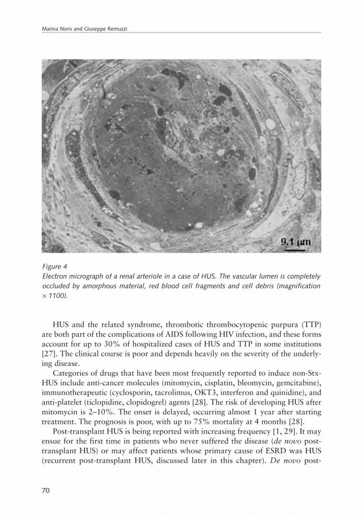
HUS and the related syndrome, thrombotic thrombocytopenic purpura (TTP)are both part of the complications of AIDS following HIV infection, and these formsaccount for up to 30% of hospitalized cases of HUS and TTP in some institutions[27]. The clinical course is poor and depends heavily on the severity of the underly-ing disease.
Categories of drugs that have been most frequently reported to induce non-Stx-HUS include anti-cancer molecules (mitomycin, cisplatin, bleomycin, gemcitabine),immunotherapeutic (cyclosporin, tacrolimus, OKT3, interferon and quinidine), andanti-platelet (ticlopidine, clopidogrel) agents [28]. The risk of developing HUS aftermitomycin is 2–10%. The onset is delayed, occurring almost 1 year after startingtreatment. The prognosis is poor, with up to 75% mortality at 4 months [28].
Post-transplant HUS is being reported with increasing frequency [1, 29]. It mayensue for the first time in patients who never suffered the disease (de novo post-transplant HUS) or may affect patients whose primary cause of ESRD was HUS(recurrent post-transplant HUS, discussed later in this chapter). De novo post-
70
Marina Noris and Giuseppe Remuzzi
Figure 4Electron micrograph of a renal arteriole in a case of HUS. The vascular lumen is completelyoccluded by amorphous material, red blood cell fragments and cell debris (magnification × 1100).

transplant HUS might occur in patients receiving renal transplants and otherorgans, as a consequence of the use of calcineurin inhibitors or due to humoral(C4b positive) rejection. In renal transplant patients treated with cyclosporin, theincidence of the disease is 5–15%; a lower incidence, approximately 1%, is foundin patients receiving tacrolimus [30]. A peculiar form of de novo post-transplantHUS affects about 6% of the recipients of a bone marrow transplant (BMT), usu-ally in the setting of a graft-versus-host disease (GVHD) or of intensive GVHDprophylaxis [30].
Non-Stx-HUS may also be triggered by pregnancy. Pregnancy-associated HUSmay occasionally develop as a complication of pre-eclampsia. Some patientsprogress to a life-threatening variant of pre-eclampsia with severe thrombocy-topenia, microangiopathic hemolytic anemia, renal failure and liver involvement(HELLP syndrome). HUS or its complications in pregnancy are always an indi-cation for prompt delivery that is usually followed by complete remission [31].Post-partum HUS is another complication of pregnancy that manifests within 3months of delivery in most cases. The outcome is usually poor with 50–60%mortality; residual renal dysfunction and hypertension are the rule in survivingpatients [32].
Of note, in about 50% of cases of sporadic non-Stx-HUS no clear triggering con-ditions could be found (idiopathic HUS) [1]. The outcome is variable, it may followa progressive course to ESRD; however, many patients recover completely [1].
71
Non-Shiga toxin-associated hemolytic uremic syndrome
Table 1 - Classification and treatment of different forms of Non-Stx-HUS
Disease Causes Treatment
Non-Stx-HUSSporadic Bacteria (Streptococcus pneumoniae) Antibiotics, no plasma
Viruses (HIV) PlasmaDrugs (antineoplastic, antiplatelet Drug withdrawal, plasma
immunosuppressive)Pregnancy-associated Delivery, plasmaPost-partum PlasmaSystemic diseases
- lupus Steroids, plasma- scleroderma BP control- anti-phospholipid syndrome Oral anticoagulants
Idiopathic PlasmaGenetic (factor H, MCP, factor I) Plasma
Familial Genetic (factor H, MCP, factor I) Plasma

Familial non-Stx-HUS
Familial forms accounts for fewer than 3% of all cases of HUS. Reports of familialoccurrence of HUS in children date back to 1965, when Campbell and Carre [33]described the development of hemolytic anemia and azotemia in concordantmonozygous twins. Since then, familial forms of HUS in children and, less fre-quently, in adults, have been reported [34]. Although some reports have been in sib-lings, suggesting autosomal recessive transmission [35], there have also been othersdescribing affected members across two [36–38] or three [35] successive genera-tions, suggesting an autosomal dominant mode of inheritance. In a kindred [39],late onset in the grandmother and early onset in the grandson may be indicative ofthe phenomenon known as anticipation, in which clinical manifestations appear atan earlier age and with increasing severity in succeeding generations.
In autosomal recessive HUS, the onset is usually early in childhood. The prog-nosis is poor, with a mortality rate of 60–70%. Recurrences are very frequent.
The onset of symptoms in autosomal dominant HUS varies (early in childhoodin some cases, but it can manifests in adult life), and is often triggered by precipi-tating events such as bacterial and viral infections or pregnancy [35]. The use of oralcontraceptives and the intake of estrogens have also been implicated as triggeringfactors [35]. Malignant hypertension is a frequent complication and the prognosisis poor, with a cumulative incidence of death or ESRD of 50% [40, 41] to 90% [35].In a large series of patients with familial history of HUS, identified through the data-base of the International Registry of Familial and Recurrent HUS/TTP, 54% haddied because of the disease. Among survivors, 38% were on chronic dialysis [40].Disease recurrences were reported in more than one half of the cases. Conditionspredisposing to disease onset were recognized in 54% of cases, and included upperrespiratory tract infections, pregnancy and consumption of birth control pills [40].
Complement abnormalities
Reduced serum levels of the third component (C3) of complement have been report-ed since 1974 in patients with both Stx-HUS and non-Stx-HUS, during the acutephase of the disease [37, 42]. In patients suffering from episodes of Stx-HUS C3abnormalities consistently subside with remission of the disease. In contrast, in casesof familial non-Stx-HUS and in patients suffering from recurrences, serum C3 lev-els were consistently and remarkably low, even during remission of the disease [40,43, 44]. However, levels of the C4 are usually normal. Persistently low C3 levels inpatients with familial HUS, even in the unaffected relatives, suggested a geneticdefect [40]. Low C3 levels most likely reflect complement activation and C3 con-sumption, which is consistent with increased levels of C3 activation and breakdownproducts, C3b, C3c and C3d, in the plasma of these patients [45–47]. Granular C3
72
Marina Noris and Giuseppe Remuzzi

deposits in glomeruli and arterioles of kidney biopsies taken during the acute phaseof the disease [22, 48–51] further reflect activation of the complement system andC3 consumption in the renal microvasculature.
The complement system consists of several plasma and membrane-associatedproteins that are organized in three distinct activation patterns. These include clas-sical, lectin and alternative pathways [52, 53] that, once activated on the surface ofmicroorganisms, form protease complexes, collectively known as C3 convertases,which serve to cleave C3 generating C3b. The classic/lectin convertases are formedby C2 and C4 fragments, while the generation of the alternative pathway conver-tase requires the cleavage of C3 and Factor B, but not of C4. Thus, low C3 levels inpatients with HUS in the presence of normal C4 indicate a selective activation of thealternative pathway. Reduction of Factor B levels and, particularly, the appearanceof Factor B (Ba and Bb) breakdown products have been also reported in HUSpatients [46], confirming the activation of the alternative pathway of complement.Upon generation C3b deposits on bacterial surfaces, which leads to opsonization forphagocytosis by neutrophils and macrophages. C3b also participates in the forma-tion of the C5 convertases that cleaves C5 and initiates assembly of the membraneattack complex (MAC), causing cell lysis. The human complement system is highlyregulated to prevent nonspecific damage to host cells and to limit deposition of C3bto the surface of pathogens. This fine regulation is based on a number of membrane-anchored (CR1, DAF, MCP and CD59) and fluid-phase (Factor H and Factor I) reg-ulators that protect host tissues. Foreign surfaces that either lack membrane-boundregulators or cannot bind soluble regulators are attacked by complement.
Evidence is now emerging that the activation of the alternative pathway of com-plement in familial forms of non-Stx-HUS may derive from genetic abnormalities ofsome of the molecules, mentioned above, that are involved in the regulation of thecomplement system. Similar genetic abnormalities have been found in sporadic non-Stx-HUS as well. Thus, more than 50 different Factor H mutations have been iden-tified in 80 patients who had familial and sporadic forms of HUS [38, 41, 54–59].The Factor H mutation frequency is up to 40% in familial forms and 13–17% insporadic forms. In addition, mutations for the cell-bound membrane complementregulator, membrane cofactor protein (MCP), have been reported in familial andsporadic non-Stx-HUS, accounting for around 10% mutation frequency [45, 60].Finally, three mutations in the gene encoding for Factor I have been reported in threepatients with the sporadic form [61], which supports the idea that non-Stx-HUS isa disease of complement dysregulation.
Complement regulatory proteins are essential to prevent nonspecific damage ofhost endothelial cells and limit deposition of C3b to the surface of pathogens [52,53]. Thus, in patients with genetic alterations causing defective activity of comple-ment regulatory proteins, exposure to an agent that activates complement results inC3b deposition on vascular endothelial cells and in the formation of the C3 and C5convertase complexes. The proteolysis of C3 and C5 by convertases causes the
73
Non-Shiga toxin-associated hemolytic uremic syndrome

release of chemotactic anaphylatoxins (C3a and C5a) that recruit inflammatorycells toward the endothelial layer. On the other hand, the deposition of C3b is fol-lowed by the formation of MAC, which leads to endothelial cells injury, withincreased expression of adhesion molecules and tissue factor, and endothelialdetachment. Under these conditions, platelets from the microcirculation adhere andaggregate, and the formation of thrombin and of fibrin polymers occurs.
Of note, not all patients with non-Stx-HUS have lower than normal C3 serumconcentrations, even in the presence of genetic alterations of complement regulators.Thus, the incidence of hypocomplementemia ranges between 31% and more than90% in patients with Factor H mutations and between 0% and 51% in patientswith no Factor H mutations, according to different series [41, 54–56, 62, 63]. Sim-ilarly, 33–50% of patients with MCP mutations [45, 60] and two-thirds of patientswith Factor I mutations [61] have lower than normal serum C3 levels. Thus, nor-mal C3 levels in patients with non-Stx-HUS do not necessarily exclude a geneticdeficiency of complement regulation. More sensitive markers of complements acti-vation status are increased plasma levels of C3 breakdown products and anincreased C3d/C3 ratio, which have been found altered in most patients with HUSeven in the face of normal serum C3 concentrations [45–47]. Another consistentmarker of complement activation could be the presence of C3 deposits in renal biop-sies. Significant amounts of C3 deposits were found in the glomeruli of renal biop-sy specimens from eight children with renal sequelae following a severe HUSepisode, despite serum C3 levels being normal [49]. C3 deposits appeared as nodu-lar pattern along the capillary walls. Immunohistology evaluation of renal biopsyfrom two siblings with familial HUS and normal C3 serum levels evidenced the pres-ence of C3 deposits in renal arterioles and arteries [64]. In another report, exami-nation of kidney biopsy material from two siblings of a big Bedouin family with tenaffected infants due to an homozygous Factor H mutation, revealed strong C3 andMAC formation and C5b-9 deposition in glomerular capillary walls [65]. Similarfindings were reported in the kidney biopsy material from another familial case withan adult onset and an heterozygous Factor H mutation [41] (Fig. 5).
Treatment of non-Stx-HUS
Despite the fact that non-Stx-HUS has a rather poor prognosis, after plasma manip-ulation was introduced, the mortality rate has dropped from 50% to 25% [66–68](Tab. 1). However, debate still exists on whether plasma is or is not effective in thetreatment of acute episodes [69–72]. Published observations [67, 73–75] and ourown experience indicate that a consistent number of patients with non-Stx-HUSrespond to plasma treatment. It has been proposed that plasma exchange might berelatively more effective than plasma infusion since it might remove potentially toxicsubstances from the patient’s circulation (see chapter by Würzner & Zimmerhackl).
74
Marina Noris and Giuseppe Remuzzi

That this may not be the case is documented by data that, in a patient with relaps-ing thrombotic microangiopathy [76], normalization of the platelet count wasinvariably obtained by plasma exchange or infusion, whereas plasma removal andsubstitution with albumin and saline never raised the platelet count. However, in sit-uations such as renal insufficiency or heart failure that limit the amount of plasmathat can be provided with infusion alone, plasma exchange should be considered asfirst-choice therapy [1]. Plasma infusion or exchange has been used in patients withHUS and Factor H mutations, with the rationale to provide the patients with nor-mal Factor H to correct the genetic deficiency. Some patients did not respond at alland died or developed ESRD [65]. Others remained chronically ill [77, 78] orrequired infusion of plasma at weekly intervals to rise Factor H plasma levelsenough to maintain remission [79]. Stratton et al. [80] were able to induce sustainedremission in a patient with Factor H mutation who developed an acute episode ofHUS and required hemodialysis. After 3 months of weekly plasma exchange in con-junction with intravenous immunoglobulins, the patient regained renal function,dialysis was withdrawal and plasma therapy stopped. At 1 year after stopping plas-ma therapy, the patient remained disease-free and dialysis independent.
75
Non-Shiga toxin-associated hemolytic uremic syndrome
Figure 5Glomerulus from a patient with HUS carrying a heterozygous mutation (delA1494-1496,causing a premature interruption of the protein at CCP 8) in Factor H gene, stained with afluorescent anti-C3 antibody. Strong staining of capillaries, and many subendothelial granu-lar deposits are evident (magnification × 400).

Plasma therapy is contraindicated in patients with HUS induced by Streptococ-cus pneumoniae, because adult plasma contains antibodies against the Thomsen-Friedenreich antigen, which may exacerbate the disease (Tab. 1).
In those few patients with extensive microvascular thrombosis at renal biopsy,refractory hypertension and signs of hypertensive encephalopathy, and for whomconventional therapies including plasma manipulation are not enough to control thedisease (i.e., persistent severe thrombocytopenia and hemolytic anemia), bilateralnephrectomy has been performed with excellent follow-up in some patients [81].Other treatments including anti-platelet agents, prostacyclin, heparin or fibrinolyt-ic agents, steroids and intravenous immunoglobulins have been attempted, with noconsistent benefit [1].
Those patients who develop HUS upon challenge with cyclosporin or tacrolimushave to stop the medication. Sirolimus has been used as an alternative in occasion-al patients with encouraging results [82].
Kidney transplantation in non-Stx-HUS
Less than 15% of children with Stx-HUS progress to ESRD, but when it happensrenal transplantation can be safely performed. Thus, in children with Stx-HUSdisease recurrence on the graft ranges from 0% to 10% [83, 84], and graft sur-vival at 10 years is even better than in control transplanted children with otherdiseases [85]. On the other hand, 50% (in sporadic forms) to 60% (in familialforms) of patients with non-Stx-HUS [1, 41] progress to ESRD. Renal transplan-tation is not necessarily an option for non-Stx-HUS, and whether kidney trans-plantation is feasible in these patients is still a debated issue. Actually, recurrenceoccurred in over 50% of the patients who had a renal transplant, with graft fail-ure developing in around 90% of them despite any treatment. [84, 86]. Recur-rences occur at a median time of 30 days after transplant (range 0 days–16 years).[1, 84]. Neither avoidance of calcineurin inhibitors as part of the immunosup-pressive therapy nor bilateral nephrectomy before transplantation preventedrecurrence of HUS and graft loss. Patients who lost the first kidney graft forrecurrence should not be re-transplanted, since the disease recurs in around 90%of the subsequent grafts. Live-related renal transplant should also be avoided, inthat it carries the additional risk to precipitate the disease onset in the healthydonor relative, as recently reported in two families [87]. New knowledge fromgenetic studies would hopefully allow the risk of recurrence to be more accurate-ly predicted. In patients with Factor H mutations, the recurrence rate ranges from30% to 100% according to different surveys [41, 54, 55], and is significantlyhigher than in patients without Factor H mutations [41]. In view of the fact thatFactor H is a plasma protein mainly of liver origin, a kidney transplant does notcorrect the Factor H genetic defect [38, 58].
76
Marina Noris and Giuseppe Remuzzi

Simultaneous kidney and liver transplant has been recently performed in twoyoung children with non-Stx-HUS and Factor H mutations, with the objective ofcorrecting the genetic defect and prevent disease recurrences [88, 89]. However, forreasons that are currently under evaluation and that possibly involve an increasedliver susceptibility to ischemic or immune injury related to uncontrolled comple-ment activation, both cases treated with this procedure were complicated by pre-mature irreversible liver failure. In the first patient, a humoral rejection of the livergraft manifested by the day 26 after transplantation and in a few days the childdeveloped hepatic encephalopathy and coma, and recovered with a second, unevent-ful, liver transplant [88]. The second case was complicated by a fatal, primary non-function of the liver graft followed by multi-organ failure and patient’s death [89].Thus, despite its capacity of correcting the genetic defect, combined kidney and livertransplant for non-Stx-HUS associated with Factor H mutations, should not be per-formed unless a patient is at imminent risk of life-threatening complications.
On the other hand, 25–50% of patients with non-Stx-HUS but no evidence ofFactor H mutations, who received a kidney transplant, lost the graft within 1 year[41, 54]. A remarkable exception are possibly those forms associated with MCPmutations. Four of these patients have been transplanted successfully with no dis-ease recurrence [60] (and our unpublished data). There is a strong biochemicalrationale for that: at variance with Factor H, which is a circulating soluble inhibitor,MCP is a membrane-bound protein highly expressed in the kidney. The transplan-tation of a normal kidney in the latter condition not surprisingly corrects the genet-ic defect. The case of Factor I, again a soluble and circulating inhibitor of liver ori-gin, should not be different from the case of Factor H. Actually, the occurrence oftwo graft failures in a recently published case with a Factor I mutation [61] seemsto be consistent with this reasoning.
Thus, genetic testing should be performed in all patients with ESRD due to non-Stx-HUS awaiting transplantation. Renal transplantation is strongly contraindicat-ed in those patients with Factor H mutations due to the high risk of disease recur-rence. At variance, graft outcome is good in patients with mutations in MCP gene,and renal transplantation should be recommended in these patients.
Despite recent advances, the underlying genetic alteration remains unknown inmore than one half of non-Stx-HUS patients, and further studies are required tofully clarify the possible role of additional candidate genes encoding for proteinsinvolved in the regulation of complement, which would hopefully allow in thefuture a more tailored clinical management of these patients.
AcknowledgementsThis work has been partially supported by grants from Comitato 30 ore per la vita,from Telethon (grants GPP02161 and GPP02162) and from Foundation for chil-dren with Atypical HUS along with The Nando Peretti Foundation.
77
Non-Shiga toxin-associated hemolytic uremic syndrome

References
1 Ruggenenti P, Noris M, Remuzzi G (2001) Thrombotic microangiopathy, hemolytic ure-mic syndrome, and thrombotic thrombocytopenic purpura. Kidney Int 60: 831–846
2 Thorpe CM (2004) Shiga toxin-producing Escherichia coli infection. Clin Infect Dis 38:1298–1303
3 Karmali MA, Steele BT, Petric M, Lim C (1983) Sporadic cases of haemolytic-uraemicsyndrome associated with faecal cytotoxin and cytotoxin-producing Escherichia coli instools. Lancet 1: 619–620
4 Riley LW, Remis RS, Helgerson SD, McGee HB, Wells JG, Davis BR, Hebert RJ, OlcottES, Johnson LM, Hargrett NT, Blake PA, Cohen ML (1983) Hemorrhagic colitis asso-ciated with a rare Escherichia coli serotype. N Engl J Med 308: 681–685
5 Mead PS, Griffin PM (1998) Escherichia coli O157:H7. Lancet 352: 1207–12126 Jackson MP, Newland JW, Holmes RK, O’Brien AD (1987) Nucleotide sequence analy-
sis of the structural genes for Shiga-like toxin I encoded by bacteriophage 933J fromEscherichia coli. Microb Pathog 2: 147–153
7 Gianviti A, Rosmini F, Caprioli A, Corona R, Matteucci MC, Principato F, Luzzi I, Riz-zoni G (1994) Haemolytic-uraemic syndrome in childhood: surveillance and case-con-trol studies in Italy. Italian HUS Study Group. Pediatr Nephrol 8: 705–709
8 Kaplan BS, Meyers KE, Schulman SL (1998)The pathogenesis and treatment of hemolyt-ic uremic syndrome. J Am Soc Nephrol 9: 1126–1133
9 Tonshoff B, Sammet A, Sanden I, Mehls O, Waldherr R, Scharer K (1994) Outcome andprognostic determinants in the hemolytic uremic syndrome of children. Nephron 68:63–70
10 Banatvala N, Griffin PM, Greene KD, Barrett TJ, Bibb WF, Green JH, Wells JG(2001)The United States National Prospective Hemolytic Uremic Syndrome Study:microbiologic, serologic, clinical, and epidemiologic findings. J Infect Dis 183:1063–1070
11 Milford D (1992) The hemolytic uremic syndromes in the United Kingdom. In: BSTRKaplan, JL Moake (eds): Hemolytic Uremic Syndrome and Thrombotic Thrombocy-topenic Purpura. Marcel Dekker Inc., New York, 39–59
12 Van Dyck M, Proesmans W, Depraetere M (1988) Hemolytic uremic syndrome in child-hood: renal function ten years later. Clin Nephrol 29: 109–112
13 Schieppati A, Ruggenenti P, Cornejo RP, Ferrario F, Gregorini G, Zucchelli P, Rossi E,Remuzzi G (1992) Renal function at hospital admission as a prognostic factor in adulthemolytic uremic syndrome. The Italian Registry of Haemolytic Uremic Syndrome. JAm Soc Nephrol 2: 1640–1644
14 Taylor CM, Chua C, Howie AJ, Risdon RA (2004) Clinico-pathological findings in diar-rhoea-negative haemolytic uraemic syndrome. Pediatr Nephrol 19: 419–425
15 Constantinescu AR, Bitzan M, Weiss LS, Christen E, Kaplan BS, Cnaan A, TrachtmanH (2004) Non-enteropathic hemolytic uremic syndrome: causes and short-term course.Am J Kidney Dis 43: 976–982
78
Marina Noris and Giuseppe Remuzzi

16 Taylor CM, Howie AJ, Williams JM (1999) No common final pathogenetic pathway inhaemolytic uraemic syndromes. Nephrol Dial Transplant 14: 1100–1102
17 Remuzzi G, Ruggenenti P (1994) Thrombotic microangiopathies. In: C Tisher, B Bren-ner (eds): Renal Pathology, 2nd ed. J.B. Lippincott, Philadelphia, 1154–1184
18 Kincaid-Smith P (1975) Participation of intravascular coagulation in the pathogenesis ofglomerular and vascular lesions. Kidney Int 7: 242–253
19 Levy M, Gagnadoux M, Habib R (1980) Pathology of hemolytic uremic syndrome inchildren. In: G Remuzzi, G Mecca, G dG (eds): Hemostasis, Prostaglandins and RenalDisease. Raven Press, New York, 383–397
20 Vitsky BH, Suzuki Y, Strauss L, Churg J(1969) The hemolytic-uremic syndrome: a studyof renal pathologic alterations. Am J Pathol 57: 627–647
21 Proia AD, Harden EA, Silberman HR (1984) Mitomycin-induced hemolytic-uremic syn-drome. Arch Pathol Lab Med 108: 959–962
22 Hammar SP, Bloomer HA, McCloskey D (1978) Adult hemolytic uremic syndrome withrenal arteriolar deposition of IgM andC3. Am J Clin Pathol 70: 434–439
23 Hall SM, Glickman M (1988) The British Paediatric Surveillance Unit. Arch Dis Child63: 344–346
24 Renaud C, Niaudet P, Gagnadoux MF, Broyer M, Habib R (1995) Haemolytic uraemicsyndrome: prognostic factors in children over 3 years of age. Pediatr Nephrol 9: 24–29
25 Erickson LC, Smith WS, Biswas AK, Camarca MA, Waecker NJ Jr (1994) Streptococ-cus pneumoniae-induced hemolytic uremic syndrome: a case for early diagnosis. PediatrNephrol 8: 211–213
26 Novak RW, Martin CR, Orsini EN (1983) Hemolytic-uremic syndrome and T-cryptanti-gen exposure by neuraminidase-producing pneumococci: an emerging problem? PediatrPathol 1: 409–413
27 Leaf AN, Laubenstein LJ, Raphael B, Hochster H, Baez L, Karpatkin S (1988)Throm-botic thrombocytopenic purpura associated with human immunodeficiency virus type 1(HIV-1) infection. Ann Intern Med 109: 194–197
28 Dlott JS, Danielson CF, Blue-Hnidy DE, McCarthy LJ (2004) Drug-induced thromboticthrombocytopenic purpura/hemolytic uremic syndrome: a concise review. Therap ApherDial 8: 102–111
29 Reynolds JC, Agodoa LY, Yuan CM, Abbott KC (2003) Thrombotic microangiopathyafter renal transplantation in the United States. Am J Kidney Dis 42: 1058–1068
30 Ruggenenti P (2002) Post-transplant hemolytic-uremic syndrome. Kidney Int 62:1093–1104
31 Remuzzi G, Ruggenenti P (1995)The hemolytic uremic syndrome. Kidney Int 48: 2–1932 George JN (2003) The association of pregnancy with thrombotic thrombocytopenic
purpura-hemolytic uremic syndrome. Curr Opin Hematol 10: 339–34433 Campbell S, Carre IJ (1965)Fatal haemolytic uraemic syndrome and idiopathic hyperli-
paemia in monozygotic twins. Arch Dis Child 40: 654–65834 Kaplan BS, Chesney RW, Drummond KN (1975) Hemolytic uremic syndrome in fami-
lies. N Engl J Med 292: 1090–1093
79
Non-Shiga toxin-associated hemolytic uremic syndrome

35 Berns JS, Kaplan BS, Mackow RC, Hefter LG (1992) Inherited hemolytic uremic syn-drome in adults. Am J Kidney Dis 19: 331–334
36 Noris M, Azzollini N, Mister M, Pezzotta A, Piccinini G, Casiraghi F, Cugini D, PericoN, Orisio S, Remuzzi G (1999) Peripheral donor leukocytes prolong survival of rat renalallografts. Kidney Int 56: 1101–1112
37 Carreras L, Romero R, Requesens C, Oliver AJ, Carrera M, Clavo M, Alsina J (1981)Familial hypocomplementemic hemolytic uremic syndrome with HLA-A3,B7 haplotype.Jama 245: 602–604
38 Warwicker P, Goodship TH, Donne RL, Pirson Y, Nicholls A, Ward RM, Turnpenny P,Goodship JA (1988) Genetic studies into inherited and sporadic hemolytic uremic syn-drome. Kidney Int 53: 836–844
39 Petermann A, Offermann G, Distler A, Sharma AM (1998) Familial hemolytic-uremicsyndrome in three generations. Am J Kidney Dis 32: 1063–1067
40 Noris M, Ruggenenti P, Perna A, Orisio S, Caprioli J, Skerka C, Vasile B, Zipfel PF,Remuzzi G (1999) Hypocomplementemia discloses genetic predisposition to hemolyticuremic syndrome and thrombotic thrombocytopenic purpura: role of factor H abnor-malities. Italian Registry of Familial and Recurrent Hemolytic Uremic Syndrome/Thrombotic Thrombocytopenic Purpura. J Am Soc Nephrol 10: 281–293
41 Caprioli J, Castelletti F, Bucchioni S, Bettinaglio P, Bresin E, Pianetti G, Gamba S,Brioschi S, Daina E, Remuzzi G, Noris M (2003) Complement factor H mutations andgene polymorphisms in haemolytic uraemic syndrome: the C-257T, the A2089G and theG2881T polymorphisms are strongly associated with the disease. Hum Mol Genet 12:3385–3395
42 Stuhlinger W, Kourilsky O, Kanfer A, Sraer JD (1974) Letter: Haemolytic-uraemic syn-drome: evidence for intravascular C3 activation. Lancet 2: 788–789
43 Bogdanovic R, Cvoric A, Nikolic V, Sindjic M (1988) Recurrent haemolytic-uraemicsyndrome with hypocomplementaemia: a case report. Pediatr Nephrol 2: 236–238
44 Pichette V, Querin S, Schurch W, Brun G, Lehner-Netsch G, Delage JM (1994) Familialhemolytic-uremic syndrome and homozygous factor H deficiency. Am J Kidney Dis 24:936–941
45 Noris M, Brioschi S, Caprioli J, Todeschini M, Bresin E, Porrati F, Gamba S, RemuzziG (2003) Familial haemolytic uraemic syndrome and an MCP mutation. Lancet 362:1542–1547
46 Kim Y, Miller K, Michael AF (1977) Breakdown products of C3 and factor B inhemolytic-uremic syndrome. J Lab Clin Med 89: 845–850
47 Monnens L, Molenaar J, Lambert PH, Proesmans W, van Munster P (1980) The com-plement system in hemolytic-uremic syndrome in childhood. Clin Nephrol 13: 168–171
48 Gonzalo A, Mampaso F, Gallego N, Bellas C, Segui J, Ortuno J (1981) Hemolytic ure-mic syndrome with hypocomplementemia and deposits of IgM and C3 in the involvedrenal tissue. Clin Nephrol 16: 193–199
49 Cossio PM, Laguens RP, Pantin DJ, de Bracco MM, Molinas F, Voyer LE, Arana RM
80
Marina Noris and Giuseppe Remuzzi
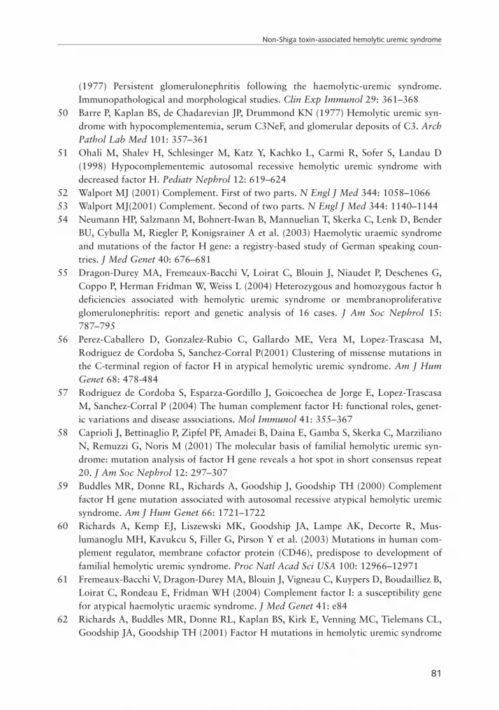
(1977) Persistent glomerulonephritis following the haemolytic-uremic syndrome.Immunopathological and morphological studies. Clin Exp Immunol 29: 361–368
50 Barre P, Kaplan BS, de Chadarevian JP, Drummond KN (1977) Hemolytic uremic syn-drome with hypocomplementemia, serum C3NeF, and glomerular deposits of C3. ArchPathol Lab Med 101: 357–361
51 Ohali M, Shalev H, Schlesinger M, Katz Y, Kachko L, Carmi R, Sofer S, Landau D(1998) Hypocomplementemic autosomal recessive hemolytic uremic syndrome withdecreased factor H. Pediatr Nephrol 12: 619–624
52 Walport MJ (2001) Complement. First of two parts. N Engl J Med 344: 1058–106653 Walport MJ(2001) Complement. Second of two parts. N Engl J Med 344: 1140–114454 Neumann HP, Salzmann M, Bohnert-Iwan B, Mannuelian T, Skerka C, Lenk D, Bender
BU, Cybulla M, Riegler P, Konigsrainer A et al. (2003) Haemolytic uraemic syndromeand mutations of the factor H gene: a registry-based study of German speaking coun-tries. J Med Genet 40: 676–681
55 Dragon-Durey MA, Fremeaux-Bacchi V, Loirat C, Blouin J, Niaudet P, Deschenes G,Coppo P, Herman Fridman W, Weiss L (2004) Heterozygous and homozygous factor hdeficiencies associated with hemolytic uremic syndrome or membranoproliferativeglomerulonephritis: report and genetic analysis of 16 cases. J Am Soc Nephrol 15:787–795
56 Perez-Caballero D, Gonzalez-Rubio C, Gallardo ME, Vera M, Lopez-Trascasa M,Rodriguez de Cordoba S, Sanchez-Corral P(2001) Clustering of missense mutations inthe C-terminal region of factor H in atypical hemolytic uremic syndrome. Am J HumGenet 68: 478-484
57 Rodriguez de Cordoba S, Esparza-Gordillo J, Goicoechea de Jorge E, Lopez-TrascasaM, Sanchez-Corral P (2004) The human complement factor H: functional roles, genet-ic variations and disease associations. Mol Immunol 41: 355–367
58 Caprioli J, Bettinaglio P, Zipfel PF, Amadei B, Daina E, Gamba S, Skerka C, MarzilianoN, Remuzzi G, Noris M (2001) The molecular basis of familial hemolytic uremic syn-drome: mutation analysis of factor H gene reveals a hot spot in short consensus repeat20. J Am Soc Nephrol 12: 297–307
59 Buddles MR, Donne RL, Richards A, Goodship J, Goodship TH (2000) Complementfactor H gene mutation associated with autosomal recessive atypical hemolytic uremicsyndrome. Am J Hum Genet 66: 1721–1722
60 Richards A, Kemp EJ, Liszewski MK, Goodship JA, Lampe AK, Decorte R, Mus-lumanoglu MH, Kavukcu S, Filler G, Pirson Y et al. (2003) Mutations in human com-plement regulator, membrane cofactor protein (CD46), predispose to development offamilial hemolytic uremic syndrome. Proc Natl Acad Sci USA 100: 12966–12971
61 Fremeaux-Bacchi V, Dragon-Durey MA, Blouin J, Vigneau C, Kuypers D, Boudailliez B,Loirat C, Rondeau E, Fridman WH (2004) Complement factor I: a susceptibility genefor atypical haemolytic uraemic syndrome. J Med Genet 41: e84
62 Richards A, Buddles MR, Donne RL, Kaplan BS, Kirk E, Venning MC, Tielemans CL,Goodship JA, Goodship TH (2001) Factor H mutations in hemolytic uremic syndrome
81
Non-Shiga toxin-associated hemolytic uremic syndrome

cluster in exons 18-20, a domain important for host cell recognition. Am J Hum Genet68: 485–490
63 Rougier N, Kazatchkine MD, Rougier JP, Fremeaux-Bacchi V, Blouin J, Deschenes G,Soto B, Baudouin V, Pautard B, Proesmans W et al (1998) Human complement factorH deficiency associated with hemolytic uremic syndrome. J Am Soc Nephrol 9:2318–2326
64 Pirson Y, Lefebvre C, Arnout C, van Ypersele de Strihou C (1987) Hemolytic uremicsyndrome in three adult siblings: a familial study and evolution. Clin Nephrol 28:250–255
65 Landau D, Shalev H, Levy-Finer G, Polonsky A, Segev Y, Katchko L (2001) Familialhemolytic uremic syndrome associated with complement factor H deficiency. J Pediatr138: 412–417
66 Hollenbeck M, Kutkuhn B, Aul C, Leschke M, Willers R, Grabensee B (1998)Haemolytic-uraemic syndrome and thrombotic-thrombocytopenic purpura in adults:clinical findings and prognostic factors for death and end-stage renal disease. NephrolDial Transplant 13: 76–81
67 Lara PN Jr, Coe TL, Zhou H, Fernando L, Holland PV, Wun T(1999) Improved survivalwith plasma exchange in patients with thrombotic thrombocytopenic purpura-hemolyt-ic uremic syndrome. Am J Med 107: 573–579
68 Morel-Maroger L, Kanfer A, Solez K, Sraer JD, Richet G (1979)Prognostic importanceof vascular lesions in acute renal failure with microangiopathic hemolytic anemia(hemolytic-uremic syndrome): clinicopathologic study in 20 adults. Kidney Int 15:548–558
69 Dundas S, Murphy J, Soutar RL, Jones GA, Hutchinson SJ, Todd WT (1999) Effective-ness of therapeutic plasma exchange in the 1996 Lanarkshire Escherichia coli O157:H7outbreak. Lancet 354: 1327–1330
70 Tsai HM (2003) Is severe deficiency of ADAMTS-13 specific for thrombotic thrombo-cytopenic purpura? Yes. J Thromb Haemost 1: 625–631
71 Tsai HM (2003) Deficiency of ADAMTS-13 in thrombotic and thrombocytopenic pur-pura. J Thromb Haemost 1: 2038–2040; discussion 2040–2035
72 Remuzzi G (2003): Is ADAMTS-13 deficiency specific for thrombotic thrombocytopenicpurpura? No. J Thromb Haemost 1: 632–634
73 Clark WF, Rock GA, Buskard N, Shumak KH, LeBlond P, Anderson D, Sutton DM(1999)Therapeutic plasma exchange: an update from the Canadian Apheresis Group.Ann Intern Med 131: 453–462
74 Vesely SK, George JN, Lammle B, Studt JD, Alberio L, El-Harake MA, Raskob GE(2003) ADAMTS13 activity in thrombotic thrombocytopenic purpura-hemolytic uremicsyndrome: relation to presenting features and clinical outcomes in a prospective cohortof 142 patients. Blood 102: 60–68
75 Tostivint I, Mougenot B, Flahault A, Vigneau C, Costa MA, Haymann JP, Sraer JD,Rondeau E (2002) Adult haemolytic and uraemic syndrome: causes and prognostic fac-tors in the last decade. Nephrol Dial Transplant 17: 1228–1234
82
Marina Noris and Giuseppe Remuzzi

76 Ruggenenti P, Galbusera M, Cornejo RP, Bellavita P, Remuzzi G (1993) Thromboticthrombocytopenic purpura: evidence that infusion rather than removal of plasmainduces remission of the disease. Am J Kidney Dis 21: 314–318
77 Filler G, Radhakrishnan S, Strain L, Hill A, Knoll G, Goodship TH (2004) Challengesin the management of infantile factor H associated hemolytic uremic syndrome. PediatrNephrol 19: 908–911
78 Gerber A, Kirchhoff-Moradpour AH, Obieglo S, Brandis M, Kirschfink M, Zipfel PF,Goodship JA, Zimmerhackl LB (2003) Successful (?) therapy of hemolytic-uremic syn-drome with factor H abnormality. Pediatr Nephrol 18: 952–955
79 Nathanson S, Fremeaux-Bacchi V, Deschenes G (2001) Successful plasma therapy inhemolytic uremic syndrome with factor H deficiency. Pediatr Nephrol 16: 554–556
80 Stratton JD, Warwicker P (2002) Successful treatment of factor H-related haemolyticuraemic syndrome. Nephrol Dial Transplant 17: 684–685
81 Remuzzi G, Galbusera M, Salvadori M, Rizzoni G, Paris S, Ruggenenti P (1996) Bilat-eral nephrectomy stopped disease progression in plasma-resistant hemolytic uremic syn-drome with neurological signs and coma. Kidney Int 49: 282–286
82 Franco A, Hernandez D, Capdevilla L, Errasti P, Gonzalez M, Ruiz JC, Sanchez J (2003)De novo hemolytic-uremic syndrome/thrombotic microangiopathy in renal transplantpatients receiving calcineurin inhibitors: role of sirolimus. Transplant Proc 35:1764–1766
83 Loirat C, Niaudet P (2003) The risk of recurrence of hemolytic uremic syndrome afterrenal transplantation in children. Pediatr Nephrol 18: 1095–1101
84 Artz MA, Steenbergen EJ, Hoitsma AJ, Monnens LA, Wetzels JF (2003) Renal trans-plantation in patients with hemolytic uremic syndrome: high rate of recurrence andincreased incidence of acute rejections. Transplantation 76: 821–826
85 Ferraris JR, Ramirez JA, Ruiz S, Caletti MG, Vallejo G, Piantanida JJ, Araujo JL, SojoET(2002) Shiga toxin-associated hemolytic uremic syndrome: absence of recurrenceafter renal transplantation. Pediatr Nephrol 17: 809–814
86 Miller RB, Burke BA, Schmidt WJ, Gillingham KJ, Matas AJ, Mauer M, Kashtan CE(1997) Recurrence of haemolytic-uraemic syndrome in renal transplants: a single-centrereport. Nephrol Dial Transplant 12: 1425–1430
87 Donne RL, Abbs I, Barany P, Elinder CG, Little M, Conlon P, Goodship TH (2002)Recurrence of hemolytic uremic syndrome after live related renal transplantation asso-ciated with subsequent de novo disease in the donor. Am J Kidney Dis 40: E22
88 Remuzzi G, Ruggenenti P, Codazzi D, Noris M, Caprioli J, Locatelli G, Gridelli B (2002)Combined kidney and liver transplantation for familial haemolytic uraemic syndrome.Lancet 359: 1671–1672
89 Remuzzi G, Ruggenenti P, Colledan M, Gridelli B, Bertani A, Bettinaglio P, Bucchioni S,Sonzogni A, Bonanomi E, Sonzogni V et al. (2005) Hemolytic uremic syndrome: a fataloutcome after kidney and liver transplantation performed to correct factor H genemutation. Am J Transplant 5: 1146–1150
83
Non-Shiga toxin-associated hemolytic uremic syndrome
85
Role of complement and Factor H in hemolytic uremic syndrome
Christine Skerka and Mihály Józsi
Department of Infection Biology, Leibniz Institute for Natural Product Research and InfectionBiology, Hans Knoell Institute, Beutenbergstr. 11a, 07745 Jena, Germany
Complement and Kidney Disease, edited by Peter F. Zipfel© 2006 Birkhäuser Verlag Basel/Switzerland
Introduction
Defective complement control is a cause for atypical hemolytic uremic syndrome(aHUS). A number of recent studies have identified mutations of several comple-ment regulatory genes in aHUS patients. Mutations have been identified in the genesencoding the fluid-phase complement regulator Factor H, the serine protease FactorI and the surface-bound regulator membrane cofactor protein (MCP/CD46). Allthree proteins control the activity of the central alternative pathway amplificationconvertase C3bBb. A total of 65 mutations have been identified in the Factor H gene(Tab. 1), 4 in the Factor I gene [1, 2] and 4 mutations in the gene coding for MCP[3, 4]. In addition, autoantibodies that bind and inactivate the function of theimmune regulator Factor H have been reported [5].
The functional analyses of the mutant proteins have defined a protective role ofFactor H, Factor I and MCP for the integrity of host endothelial cells. These analy-ses are also indicative for the disease mechanism of aHUS, as defective alternativepathway complement regulation causes generation of aggressive complement-acti-vation products that relate to endothelial cell damage.
The majority of the identified Factor H gene mutations are clustered in the C ter-minus of the protein, which includes the cell surface recognition region (reviewed in[6]). This clustering of mutations in the C terminus indicates a central role of thecell surface recognition domain for pathophysiology of aHUS. Mutations of addi-tional complement regulatory genes, whose products control the activity of the cen-tral amplification convertase C3bBb, are associated with aHUS. These scenariosindicate that defective local inhibition of complement, occurring most likely uponan inflammatory reaction, results in endothelial cell damage. Here we discuss howmutated, defective or inactivated complement regulatory proteins relate to the dis-ease aHUS and focus on complement Factor H.

86
Christine Skerka and Mihály Józsi
Tabl
e 1
- M
utat
ions
in F
acto
r H
gen
e as
soci
ated
wit
h at
ypic
al H
US
No.
Mut
atio
nC
CP
Mut
atio
nR
esul
tC
ase
Age
Fact
or H
C
3 C
omm
ent
Ref
eren
ceno
.on
set
70–1
30%
(650
–23
5–75
0 18
50m
g/L
mg/
L)
11
115
5 de
l 4bp
32st
opSi
ngle
cas
ehe
36 y
222
mg/
L0.
4St
op[1
7]2
21
A30
5GR
78G
101
he–
––
[33]
33
244
5 de
l 25b
p13
6 st
opG
cas
e 9
he18
mo
45%
430
Stop
[20]
44
7C
1271
AQ
400K
N c
ase
16he
birt
h–
–[2
0]5
58
1494
del
A47
9 st
opF4
0he
adul
t56
9 m
g/L
680
Stop
[19]
66
11T1
963G
C63
0WC
ase
1he
–77
4 m
g/L
876
C[3
2]7
711
G20
91A
C67
3YJ
case
12
he34
41%
453
C[2
0]8
812
C22
14G
S714
sto
pC
ase
2he
–61
9 m
g/L
562
Stop
[32]
99
1323
73+
A77
4 st
opF
case
8he
16 m
o35
%31
0St
op[2
0]10
1014
G26
21A
E850
KC
ase
3he
–92
7 m
g/L
2000
Cha
rge
[32]
1111
14G
2776
TV
835L
Sing
le c
ase
heN
eum
ann
unpu
b.12
1215
G27
42T
S890
IF4
5he
22 y
830
mg/
L62
0+
AD
AM
TS[1
0]13
mut
1313
15A
2751
GH
893R
I cas
e 11
he9
mo
30%
240
Cha
rge
[20]
1414
15T2
770A
Y89
9sto
pSi
ngle
cas
eho
8 m
o<
1%26
0St
op[2
1]15
15T2
770A
Y89
9sto
pA
cas
e1,2
ho7
mo
<1%
50St
op[2
0]16
1515
T281
6AC
915S
H c
ase
10he
2645
%27
0C
[20]
1716
15C
2846
TQ
924s
top
E ca
se 7
he6
mo
50%
580
Stop
[20]
1817
15G
2850
TC
926F
Sing
le c
ase
he30
mo
Low
520
C[8
2]19
1816
G29
23T
+T2
924C
Q95
0H+
Y95
1H11
8he
––
Low
Cha
rge
[33]
2019
16T2
924C
Y95
1H08
7he
––
Low
[33]
2120
16C
2940
TT9
56M
HU
S12
he13
y82
1 m
g/L
1350
[70]
2221
16G
3007
TW
978C
Cas
e 4
he–
703
mg/
L75
4C
[32]
2322
17A
3032
GT9
87A
Sing
le c
ase
––
––
Neu
man
n un
pub.
2423
17A
3135
T+
Y10
21F
+
Cas
e 16
he–
787
mg/
L74
9C
, cha
rge
[32]
+20
C37
01T
R12
10C

87
Role of complement and Factor H in hemolytic uremic syndrome
2524
17T3
200C
C10
43R
Cas
e 5
he–
560
mg/
L74
8C
, cha
rge
[32]
2625
18C
3299
GQ
1076
EC
ase
6he
–51
1 m
g/L
1820
Cha
rge
[32]
2726
18/1
9C
3299
G,
Q10
76E
Pat.
1he
––
–St
op[3
]35
59 d
elA
1168
sto
p28
2719
A34
29G
D11
19G
Pat.
2he
–N
orm
alN
orm
alC
harg
e[3
]29
2819
T347
4GV
1134
GC
ase
7he
–83
5 m
g/L
835
[32]
3029
19T3
497G
Y11
42D
Cas
e 8
he–
1007
mg/
L42
5C
harg
e[3
2]31
3019
T354
2CW
1157
RC
ase
9he
–89
4 m
g/L
508
Cha
rge
[32]
3231
19C
3562
GC
1163
WR
087
he–
–lo
wC
[33]
3332
1935
66 +
1 G
/ASp
lice
defe
ct
Cas
e 10
he–
729
mg/
L82
6St
op[3
2]34
3320
G35
87T
E117
2 st
op02
2he
––
Mod
low
Stop
[33]
3520
G35
87T
E117
2 st
op08
1he
––
mod
. low
Stop
[33]
3620
E117
2 st
opR
043
he–
–St
op
Hei
nen
unpu
b.37
3420
G36
19T
R11
82S
Sing
le c
ase
Cha
rge
Neu
man
n un
pub.
3835
20T3
620C
W11
83R
Cas
e 11
he–
619
mg/
l89
0C
harg
e[3
2]39
3620
T362
0AW
1183
RSi
ngle
cas
ehe
2 y
norm
alno
rmal
Cha
rge
[83]
4037
20G
3621
TW
1183
LL
case
14
he4
mo
94%
470
[20]
4120
G36
21T
W11
83L
HU
S2he
23 y
1160
mg/
l 71
0R
ed b
i C3b
[70]
4238
20C
3624
GT1
184R
Pat.
4he
–no
rmal
–C
harg
e[7
1]43
3920
C36
38T
L118
9FH
US
37[8
4]44
4020
T363
9GL1
189R
HU
S11
he–
1225
mg/
l16
70 m
g/l
Cha
rge
[70]
4541
20C
3645
GS1
191W
HU
S 30
[84]
4642
20C
3645
T, T
3663
CS1
191L
; V11
97A
Pat.
5he
––
–[3
]47
4320
G36
54A
G11
94D
130
he–
–m
od. l
owC
harg
e[3
3]48
20G
1194
Dsi
ngle
cas
e–
––
–C
harg
e[6
0]49
4420
G36
54 +
36
G11
94+
12
aaSi
ngle
cas
ehe
12 m
052
0 m
g/l
800
mg/
lD
efor
m.
[72]
5045
20T3
663C
V11
97A
056
he–
––
[33]
5146
20T3
663C
V11
97A
+N
ull a
ll.H
US3
he53
y11
0 m
g/l
590
Red
bi C
3b[7
0]
5247
20A
3666
CE1
198A
152
he–
––
Cha
rge
[33]
5348
20T3
669C
F119
9SK
cas
e 13
he29
y37
%38
2[2
0]54
4920
C37
01T
R12
10C
HU
S29
he14
mo
198
mg/
l E
710
Red
bi C
3b[7
4]

88
Christine Skerka and Mihály Józsi
Tabl
e 1
– co
ntin
ued
No.
Mut
atio
nC
CP
Mut
atio
nR
esul
tC
ase
Age
Fact
or H
C
3 C
omm
ent
Ref
eren
ceno
.on
set
70–1
30%
(650
–23
5–75
0 18
50m
g/L
mg/
L)
5520
R12
10C
Sing
le c
ase
[60]
5620
C37
01T
R12
10C
Cas
e 12
he–
580
mg/
l12
50[3
2]57
20C
3701
TR
1210
CF1
06he
we
–8 y
711
mg/
l61
0[1
9]58
20C
3701
TR
1210
CR
16he
48 y
731
mg/
l12
20[1
9]59
20C
3701
TR
1210
C06
9he
––
mod
. low
[33]
6050
20C
3716
GR
1215
G2a
IV12
––
486
mg/
l75
0C
harg
e[1
7]61
5120
G37
17A
R12
15Q
F34
hew
e –8
y56
1 m
g/l
690
Stop
[19]
6252
2037
19 d
el A
CA
1216
del
TC
ase
13he
––
–[3
2]63
5420
C37
49T
P122
6SC
ase
14he
–13
00 m
g/l
1020
Red
bin
d[3
2]64
5320
A37
47Td
el 2
4bp
1228
stop
F85
Bedo
uin
how
e –8
y<
70 m
g/l
100
Stop
[19]
6555
2037
68 d
el A
GA
AFr
ames
hift
Cas
e 15
he–
503
mg/
l54
0Lo
nger
[32]
+ 3
7 aa
tran
scrip
t
he, h
eter
ozyg
ous
mut
atio
n; h
o, h
omoz
ygou
s m
utat
ion;
A, a
lani
ne; C
, cys
tein
e; E
, glu
tam
ic a
cid;
F, p
heny
lala
nine
; G, g
lyci
ne; H
, his
tidi
ne; K
, lys
ine;
L,
leuc
ine;
Q, g
luta
min
e; R
, arg
inin
e; S
, ser
ine;
T, t
hreo
nine
; V, v
alin
e; W
, try
ptop
han;
Y, t
yros
ine;
red
bi, r
educ
ed b
indi
ng. N
umbe
ring
of b
ase
pair
s/am
ino
acid
s is
ada
pted
fro
m R
ipoc
he e
t al
198
8 [3
0]. F
acto
r H
and
C3
leve
ls a
re o
rigi
nal d
ata
as p
ublis
hed
by t
he a
utho
rs.

HUS: the disease
HUS is characterized by microangiopathic hemolytic anemia, thrombocytopeniaand acute renal failure, and was first described by Gasser and co-workers in 1955[7]. Three forms of HUS can be differentiated: an epidemic, sporadic and familialform of HUS.
The epidemic form of HUS is the most common and usually manifests by infec-tions with toxin-producing Escherichia coli of the serotype 0157:H7 or Shigelladysenteria serotype 1. The virulence of these microbes is associated with the pro-duction of shiga toxin or shiga-like toxins SLT1 and SLT2 that inhibit protein syn-thesis at the ribosomes, and have a direct toxic effect on renal epithelial cells [8].This form of HUS is common in children, and is often associated with diarrhea and,therefore, also termed D+HUS or Shiga toxin HUS. Most of the patients show acuterenal failure; however, renal function recovers frequently [9].
The second and third form, the atypical form (aHUS) can be sporadic and famil-ial occurring HUS cases, which usually are not associated with diarrhea and are alsotermed D–HUS or non-shiga toxin HUS. The sporadic form can be triggered bygenetic defects, neuraminidase produced by S. pneumoniae, or by other factors, suchas pregnancy, drugs like cyclosporin A, diseases such as systemic lupus erythemato-sus (SLE) or infection with the human immune deficiency virus (HIV).
The familial form is a rare disease but carries a significant burden of morbidityand mortality. Patients often suffer from end-stage renal failure and depend on con-tinuous dialysis [10].
Complement and aHUS
Reduced plasma levels of the central complement serum protein C3 were reportedalready in 1975 in both sporadic and familial forms of aHUS [11]. In addition, thepresence of C3 cleavage products have been identified in plasma of aHUS patients.These parameters are indicative either of defective complement control, or of aninherited defect in C3 protein synthesis. The role of defective complement was under-lined by reduced C3 levels between relapses [12]. Granular C3 deposits were alsoobserved in glomeruli and arterioles in aHUS patients and enhanced levels of com-plement cleavage products (C3a and C5a) in serum were indicative of uncontrolledcomplement activation due to defective control. In 1981 Thompson and Winterborn[13] described a young aHUS patient with very low C3 plasma levels and low levelsof complement Factor H. This study identified a defective complement control andFactor H as disease causing factor, and initiated further investigations. Several addi-tional studies showed decreased levels or abnormal mobilities of the Factor H pro-tein in SDS-PAGE, or decreased C3 plasma levels and/or complement functionalactivities for patients with familial and sporadic HUS [12, 14–16].
89
Role of complement and Factor H in hemolytic uremic syndrome

By genetic linkage analysis in three large pedigrees of familial aHUS, Warwickerand co-workers [17] identified in 1998 the regulators of complement activation(RCA) gene cluster on chromosome 1 as a hot spot that segregated with aHUS. Con-sequently, the authors reported mutations in the Factor H gene in patients withaHUS [18]. Several genetic studies reported mutations in the Factor H gene in aHUSpatients, and genetic analyses identified mutations in two other genes coding forcomplement regulatory proteins, i.e., the serine protease Factor I and the mem-brane-bound regulator MCP. The three gene products Factor H, MCP and Factor Idirectly control or affect the activity of the alternative complement pathway ampli-fication convertase C3bBb.
aHUS-associated Factor H gene mutations
Up to now 65 mutations have been identified in the Factor H gene that are associ-ated with aHUS (Fig. 1 and Tab. 1). The identified mutations represent either non-sense mutations that introduce a premature stop codon, or missense mutations thatresult in single amino acid exchanges, deletions or insertions. The vast majority ofthe mutations are heterozygous, thus the patients have one defective and one intactallele. Three exceptions to this rule exists. One was described for a Bedouin family,which has a premature stop codon in complement control protein (CCP) domain 20,that causes deletion of the four C-terminal amino acids [19]. A truncated Factor Hprotein was expressed and the potentially functionally defective protein was found
90
Christine Skerka and Mihály Józsi
Figure 1Domain structure of complement Factor H and location of HUS-associated mutations(A) Factor H is composed of 20 CCP (or short consensus repeats) domains. The individualdomains are numbered consecutively. The N-terminal complement regulatory region (repre-sented by CCPs 1–4) is shown in yellow and the C-terminal recognition region in blue. Thelocation of the C3 and heparin/polyanion binding regions are shown above the structure byhorizontal lines. The position of the reported Factor H gene mutations in HUS are givenbelow the structure. Mutations that introduce premature stop codons are shown in white let-ters with a black background. Mutations that cause exchange of single amino acids, or thatintroduce amino acid deletions are shown with black letters. (B) Folding of a single CCPdomain, here the sequence of the most C-terminal domain (CCP 20) is shown. The con-served cysteine residues (C) are shown in blue and the disulfide bonds that are formedbetween Cys I and Cys III and between Cys II and Cys IV are indicated by the blue line. Thisarrangement forms three loop regions which are termed loop I, loop II and loop III. (C)Sequence of CCP region 19 and 20. The conserved cysteine residues are shown in white let-ters on a gray background. Amino acids that are mutated in HUS patients are shown in red.

91
Role of complement and Factor H in hemolytic uremic syndrome

in severely reduced levels in patients plasma (below 70 mg/L). The other homozy-gous mutations have both a premature stop in CCP 15 of Factor H, and are associ-ated with low Factor H plasma levels [20, 21].
Structure and function of Factor H
Factor H genetics
The human Factor H gene (CFH) is located on the long arm of chromosome 1, with-in the RCA gene cluster in 1q32 [22, 23]. This cluster includes two groups of com-plement regulatory genes that are separated by a stretch of DNA coding for non-complement genes. The individual RCA genes are probably derived from one ances-tral gene, and they have exons coding for a so-called CCP module, also known asshort consensus repeat. These modules are mostly encoded by symmetric, phase 1-1 exons, which can be duplicated, inserted, deleted and shuffled. The two groups ofRCA genes reflect their evolutionary relationship [24]. The FH gene is found in agroup that contains six Factor H family genes: Factor H, the alternatively splicedFactor H gene product FHL-1, and five genes coding for Factor H-related proteins(CFHL1–CFHL5) [25].
The Factor H gene spans 94 kb and contains 23 exons. In general each CCPdomain of Factor H is encoded by single exons, with the exception of CCP 2, whichrepresents a split exon and which is encoded by exons III and IV. In addition, thesignal peptide and the unique C-terminal region of FHL-1 are encoded by uniqueexons. The Factor H gene encodes two proteins: Factor H and the Factor H like pro-tein 1 (FHL-1). FHL-1 is derived from an alternatively spliced transcript that coversCCPs 1–7, and an unique C terminus that encodes four amino acids and the 3’untranslated region and that is encoded by exon X [26–28].
Several single nucleotide polymorphisms in the Factor H gene sequence havebeen observed, both silent ones and those leading to amino acid exchanges [29–33].Some of these polymorphisms have been associated with aHUS as they are foundmore often in patients than in healthy controls [20, 33–35].
The Factor H protein
Factor H is a single-chain, 150-kDa plasma glycoprotein. The secreted protein is1213 amino acids long and is exclusively composed of 20 individual CCP domains(Fig. 1A) [30, 36]. A single CCP domain is about 60 amino acids long, and is sta-bilized by two disulfide bridges. The four conserved framework cysteine residuesform disulfide bridges in a Cys I-Cys III and Cys II-Cys IV manner and allow a glob-ular structure with six β-strands and variable exposed loops [37]. The consecutive
92
Christine Skerka and Mihály Józsi

CCP domains are linked by inter domain regions, that are 3–8 amino acids long(residues counted between the conserved Cys IV and Cys I residues). The typicalCCP-type folding generates three loop regions (Fig. 1B). The longest loop, i.e., loopI is formed between Cys I and Cys II, loop II represents the amino acid stretchbetween Cys II and Cys III and loop III is formed between Cys III and Cys IV. Inaddition to the central framework Cys residues, additional non-framework aminoacid residues are conserved in the various CCP domains [27]. These conservedresidues are considered relevant for the structure and architecture of an individual-ly folding CCP unit. In contrast, the variable residues, particularly those located inthe loop regions, are considered surface exposed and responsible for interactionwith the specific ligands. This composition of conserved amino acid residues thatform the framework or scaffold of a CCP unit and the variable ligand interactingresidues defines the complex architecture of a CCP unit. Such an architectureexplains why single amino acid mutations as observed in patients with aHUS [andin membranoproliferative glomerulonephritis (MPGN) patients] can seriously affectboth the structure and the function of a CCP composed protein such as the FactorH and the MCP protein.
Factor H functions
Factor H is an essential fluid-phase regulator of the alternative pathway of comple-ment [38–41] and the plasma protein (i) competes with factor B for C3b binding,i.e., prevents the formation of alternative pathway convertases C3bBb (Fig. 2A); (ii)promotes the disassembly of the alternative pathway convertases C3bBb andC3bBbC3b (decay-accelerator activity) (Fig. 2B); and (iii) acts as a cofactor for theinactivation of C3b by the serine protease Factor I (Fig. 2C). This capacity to blockthe C3b amplification convertase makes Factor H a potent inhibitor of the alterna-tive cascade.
Factor H has two important functional domains that are located at the oppositeends of the protein: The N-terminal recognition region, i.e., CCPs 1–4 include thecomplement regulatory activities, and the C-terminal recognition domain is con-tained within CCP domains 19 and 20. Factor H acts as fluid-phase complementregulator and, in addition, with its C-terminal domain, Factor H binds to cell andtissue surfaces, and thus mediates its protective role also on host cell surfaces(Fig. 1) [42–44].
Factor H ligands and ligand binding
Factor H is a multifunctional protein and mediates its regulatory role by interactionwith several ligands such as C3b/C3d [45–47], heparin/glycosaminoglycans [48–55],
93
Role of complement and Factor H in hemolytic uremic syndrome

94
Christine Skerka and Mihály Józsi
Figu
re 2
Func
tion
s of
com
plem
ent
Fact
or H
in c
ontr
ol o
f th
e am
plif
icat
ion
conv
erta
se C
3bB
bFa
ctor
H c
ontr
ols
the
acti
vity
and
sta
bilit
y of
the
alt
erna
tive
com
plem
ent
path
way
con
vert
ase
C3b
Bb.
Fac
tor
H p
reve
nts
form
atio
n of
the
C3
conv
erta
se b
y co
mpe
ting
wit
h Fa
ctor
B f
or C
3b b
indi
ng (
A);
Fac
tor
H f
acili
tate
s th
e de
cay
of t
he C
3 co
nver
tase
by
disp
laci
ngbo
und
Bb
(B),
and
Fac
tor
H a
cts
as o
ne o
f th
e Fa
ctor
I co
fact
ors
to d
egra
de C
3b (
C)
as s
how
n he
re f
or t
he a
ctiv
ity
in f
luid
pha
se.

adrenomedullin [56], C-reactive protein [57], and multiple bacterial and microbialligands (reviewed in [58, 59]) (Fig. 1). Apparently, C3b and heparin represent impor-tant ligands of Factor H, as the Factor H protein has three binding sites for both.Heparin binding regions are localized to CCP 7, to a region around CCP 13 and toCCP 20. The domains represented by CCP 19 and CCP 20 is of special interest andintensively investigated as it includes several binding regions like that for binding tocell surfaces. Homology modeling [60], mutational analysis and peptide spot analy-sis identified amino acids relevant for heparin binding and the visualization of a sur-face-exposed contact region. A first biochemical-based mutational analysis identifiedresidues K1108 in CCP 19 and K1202, R1203, R1206, R1210, K1222, K1230 andR1231 in CCP 20 as central elements for heparin binding (Fig. 1C) [61]. A recom-binant Factor H 15-20 fragment with mutated residues (R1203E, R1206E, R1210S,K1230S and R1231A) showed severely reduced binding to heparin, thus confirmingthat these positively charged amino acids mediated binding to heparin. Ganesh et al.[62] determined the structure of vaccinia virus control protein (VCP) complexedwith a heparin decamer fragment, and used this structure for identification of aminoacids relevant for Factor H–heparin interaction. Upon superimposition of CCP 20 ofFactor H with CCP 4 of VCP, again the basic residues R1182, K1202, R1203,R1206, R1210, R1215, K1230 and R1231 were in contact with heparin. Hybridiza-tion experiments with a fluorescently labeled heparin molecule to short peptides rep-resenting the complete sequence of CCP 19 and 20 identified four linear heparinbinding sites, which include the positively charged residues identified in the formerstudies: region 1 is located in CCP 19 (aa 1136–1157), region 2 (aa 1178–1187) andregion 3 (aa 1202–1215) in CCP 20 and in the C-terminal region of Factor H (aa1230 and 1231) [63].
The C3 binding sites have been mapped within the Factor H protein to CCPs1–4, to CCPs 11–14 (C3b and C3c) and to CCPs 19 and 20 (C3b and C3d) [61,64–66]. The C-terminal binding sites of Factor H for C3b and C3d overlap with theheparin binding site. This has been demonstrated using heparin as an inhibitor forbinding of Factor H fragment 15–20 to C3b [61] and by peptide spot analysis,showing overlapping binding sites for all three ligands, heparin, C3b and C3d [63].
Factor H mutations
In general terms, the reported aHUS-associated mutations in the Factor H protein[19–21, 67–72] can be grouped into three categories (Fig. 3): (i), nonsense mutations(Fig. 3: scenario I), (ii) missense mutations (Fig. 3: scenarios II and III); and (iii) dele-tion, insertion or duplication of one or more nucleotides.
(i) Nonsense mutations: In seven cases (i.e., 11%) nonsense mutations that causeintroduction of a premature stop codon have been identified. These mutations
95
Role of complement and Factor H in hemolytic uremic syndrome

96
Christine Skerka and Mihály Józsi
have been reported for CCP 15 (three separate mutations) and CCP 20 (CCP12, three separate mutations) (Fig. 1 and Tab. 1). It is currently unclear whetherthese truncated proteins are expressed and secreted. Plasma levels of Factor Hof these patients are mostly reduced.
(ii) Missense mutations: The vast majority of mutations represent single aminoacid exchanges (70%). These mutations cluster in the C terminus of the FactorH protein and are located in the recognition domain of the protein in CCP 19and 20 (46%). Based on results of the plasma levels and of Factor H gene muta-tions reported and characterized in MPGN (see Chapter by Zipfel, Smith andHeinen) it is hypothesized that these single amino acid mutations either affectsecretion of the defective allelic product (a), or result in secretion of a func-tionally defective protein (b).(a) Mutations of both framework and non-framework residues cause a block
of protein secretion as has been clearly shown for MPGN type II. In thesecases the mutated protein is expressed intracellularly and is retained in thecytoplasm. As the vast majority of aHUS patients are heterozygous for themutation, it is difficult to assess defective protein secretion. Factor H plas-ma levels vary in neonates 170–400 µg/mL, in children 225–1600 µg/mLand in adults 235–750 µg/mL [73]. In addition, various laboratories usedifferent methods for the determination of Factor H concentrations inplasma, and therefore it is currently impossible to decide which of thereported mutations is associated with a defect in protein secretion.
(b) The second type of missense mutations produce a secreted protein productthat has a single amino acid exchanged. Several of these mutant Factor Hproteins can be identified by their altered mobility by SDS-PAGE in com-bination with Western blot analysis [43, 74]. Based on the unique structureof the CCP domain, amino acid residues can have a role as scaffold ele-ments and maintain the proper architecture of the domain, or if the over-
97
Role of complement and Factor H in hemolytic uremic syndrome
Figure 3Different scenarios of Factor H mutations and modifications that are associated with HUSThe vast majority of Factor H gene mutations are heterozygous, the patients have one defec-tive and one intact allele. The mutations have different effects. Scenario I: The majority ofnonsense mutations that introduce premature stop codons seem to result in non-expressedalleles (exception to this are patients who have a premature stop codon in domain 20 suchas the Bedouin family). Scenario II: Missense mutations can either cause defective proteinsecretion or result in a secreted but functionally defective protein (scenario III). Additionalscenarios, such as the presence of autoantibodies result in normal Factor H protein, normalsecretion and normal Factor H plasma levels, but the function of the protein is impaired (sce-nario IV).
all structure is intact exchanged amino acids may be located in accessibleregion within a ligand binding site. Recent studies with recombinant Fac-tor H fragments underline the sensitivity of minor amino acid changes onthe complement regulatory functions of Factor H and the importance oflinker regions [63, 75, 76].
(iii) Deletion, insertion or duplication of one or more amino acids are described fornine aHUS patients (15%). The mutations are spread over the complete FactorH gene and in six cases resulted in a premature stop of translation. The remain-ing three mutations presumably create Factor H molecules with additionalamino acids, which may interfere with normal regulatory functions.
Clustering of mutations in CCP 19 and 20
The mutations identified in the Factor H gene are distributed over the Factor H genebut most of the mutations cluster in the C terminus, particularly in CCP domain 19and 20 of Factor H, which represent a hot spot. Interestingly nine mutations at fivepositions have been identified in several unrelated individuals (Tab. 1).
A total of 39 of the reported 65 aHUS-associated mutations in the Factor H gene(i.e., 60%) are located in CCP 19 or CCP 20, and represent either single amino acidmutations, deletions, premature Stop codons or insertions (Tab. 1, Fig. 1A). Thisclustering of mutations within the two most C-terminal CCP domains of the flexi-ble Factor H protein highlights the role of the C-terminal recognition region for Fac-tor H physiology.
The importance of the C-terminal recognition region for proper Factor H func-tions on cell surfaces was shown experimentally using mutant Factor H purifiedfrom plasma of aHUS patients, such as Factor H E1172Stop [43, 77] or in studieswith recombinantly expressed fragments of Factor H that carry the aHUS-associat-ed mutations R1210C, R1215G [43, 74], W1157R, W1183L, V1197A and P1226S[63, 74], or recombinant Factor H proteins deleted of the C-terminal CCPs (Fig. 1C)[42]. These studies demonstrated severely reduced binding of the mutated proteinsto cell surfaces, and suggest that reduced or defective complement control on cellsurfaces is associated with aHUS.
16 different single amino acid exchanges in CCP 20 of Factor H have been iden-tified in aHUS patients. Thus, it is of interest to understand how these differentmutations result into the same functional defect. Considering a role as compositor-ial scaffold residues, which maintain the overall architecture, or a role as directlyinteracting residues, exchanges of single amino acids can have different effects. Theymay either affect the architecture of a single CCP domain, or, if the architectureremains intact, the exchange may affect ligand binding regions and, thus, directlyinterfere with ligand binding, e.g., C3b/C3d and heparin/glycosaminoglycan. This
98
Christine Skerka and Mihály Józsi

scenario was confirmed in a detailed comparison and localization of a number ofaHUS-associated mutations [63]. Amino acid residues in CCP 20 of Factor H thatare conserved among all members of the Factor H protein family seem responsiblefor the architecture of the globular structure. Mutations of these amino acids mayaffect the general architecture, and therefore influence the orientation of a completeloop. Thus, a complete binding region may be affected and become inverted, tiltedand inaccessible. However, if the overall structure remains intact, an exchange of anamino acid located within a central surface-exposed position may directly interferewith ligand interaction. Consequently, ligand binding such as C3b/C3d orheparin/glycosaminoglycans is impaired. In addition, as shown for the Factor Hprotein with the R1210C mutation, the mutant protein changes its characteristicsand interacts with other proteins like albumin [74].
The currently available data show that CCP 19 and 20 of Factor H mediate acentral role in recognition and bind heparin and C3b/C3d, and represent the majorbinding domain for cell surfaces [42–44, 61, 63, 74, 77, 78]. The C-terminalheparin binding region is rather sensitive for mutations and modifications, and sofar no polymorphic variants have been described for this region. Consequently, itappears that any mutation within this region of Factor H does directly interfere withligand and/or cell binding. Defective cell binding blocks the protective role of Fac-tor H on the surface of cells [44, 77, 78].
Factor H mutations and C3b/d binding sites
In addition to affecting heparin binding, mutations in Factor H also change thebinding characteristics to C3b and C3d. Several binding domains for C3b/d havebeen described in Factor H. The complement regulatory domain in CCP 1–4 har-bors a C3b binding site [75], but also CCP 11–14 and CCP 19/20 can bind C3b/d[61, 64, 65, 79]. Mutations in CCP 19/20 also affect C3b and C3d binding, asdemonstrated in binding studies with recombinant mutant proteins [43, 63, 74].These data are in agreement with overlapping binding sites for heparin and C3b/ddetermined in the C terminus [61, 63].
Autoantibodies to Factor H in aHUS patients
In addition, autoantibodies against Factor H have been identified in plasma of threepatients with recurrent aHUS. These antibodies affected Factor H function andcaused decreased Factor H activity. This observation provides evidence for anacquired functional Factor H deficiency, supporting the fact that aHUS occurs in acontext of an autoimmune disease (Fig. 3: scenario IV) [5].
99
Role of complement and Factor H in hemolytic uremic syndrome

Mutations in other complement regulatory proteins: Factor I and MCP(CD46)
Serine protease Factor I
Human complement Factor I is a serine protease that in the presence of appropriatecofactors cleaves the α chain of C3b and inhibits the alternative pathway amplifi-cation loop. Thus, recently identified mutations in the gene coding for Factor I inpatients suffering from aHUS confirm the role of impaired complement regulationin the pathogenesis of this disease [1, 2].
Membrane cofactor protein
Complement control particularly at the cell surface is mediated by multiple regula-tors of the fluid-phase and the integral membrane regulators such as MCP (CD46),complement receptor 1 (CR1/CD35) and decay-accelerating factor (DAF/CD55).Recently mutations in the gene coding for the MCP have been identified in four fam-ilies with aHUS [3, 4, 80].
MCP is a surface-bound membrane regulator, is widely expressed on almostevery human cell, and has four CCP domains that mediate the complement regula-tory cofactor activity of the protein (reviewed in [81]). The protein also contains aserine/proline rich domain, a transmembrane and an intracellular cytoplasmicdomain. So far, eight aHUS cases from four families have been reported with amutated MCP gene. These mutations include a deletion of two amino acids (D237and S238 in CCP domain 4; two individuals from family I) and an exchange ofS206P in CCP domain 4 (two patients of family II are heterozygous, and twopatients of family III are homozygous for this mutation) [3]. In another family, aframe shift results in an exchange of three amino acids (X233, Y234 and Z235) anda premature stop codon that causes the loss of the transmembrane domain [4]. Allthese mutations caused reduced surface expression, reduced binding of C3b and/orreduced function of MCP at the cell membrane. Thus, the mutant protein causesreduced complement control at the surface of the cells. MCP is expressed in highlevels at glomerular cells (MCP-associated mutations are discussed in detail in thechapter by Goodship et al.)
Conclusions
The alternative pathway of complement is spontaneously and continuously activat-ed, and clinical, genetic and molecular data show that defective complement controlis associated with aHUS. Particularly on host cells and tissue surfaces, activation of
100
Christine Skerka and Mihály Józsi

the amplification convertase C3bBb is strongly controlled and continuously inhibit-ed. The activity of the initial convertase and that of the amplification convertase istightly controlled and regulated by a combination of surface-attached regulatorssuch as Factor H and FHL-1, and by integral membrane proteins such as CR1, MCPand DAF. The importance of the regulation and inhibition of the aggressive com-plement activation products is demonstrated by the severity of diseases that occurwhen the activity of these regulators is unbalanced. Under situations of stress, acombined local action of the multiple functionally identical regulators is required tomaintain integrity of host cell surfaces. Particularly during stress and high-levelcomplement activation, a reduced activity or a defect of one single regulator mayshift the balance from protection to damage. Such a scenario explains why the alter-native pathway regulators Factor H and MCP as well as the serine protease FactorI, which in the presence of a cofactor inactivates the newly formed surface-deposit-ed C3b molecule, are all associated with aHUS. In contrast, a complete loss of Fac-tor H functions by homozygous mutations that cause autosomal dominant muta-tions lead to MPGN type II early in life (see chapter by Zipfel et al).
Although considerable clinical and genetic evidence shows an association ofcomplement regulation with aHUS and MPGN type II, the molecular mechanismsof how these functional disturbances induce cell damage, particularly of the renalendothelial cell, and cause the diseases are not fully understood. In addition, the ini-tial triggers that lead to complement activation are not clearly identified. The fol-lowing aspects support the view that the damage to the endothelium by complementis a cause for the development of aHUS:(i) Factor H displays its complement regulatory functions in both the fluid phase
and on self cell surfaces (Figs 2 and 4).(ii) A total of 60% of the reported aHUS mutations in the Factor H gene cluster
are located in the C terminus of the protein and impair the recognition func-tion of Factor H. A mutant Factor H protein may show normal complementregulatory activity in fluid phase. However, the impaired cell binding activitymay lead to reduced protection at the cell surface, e.g., endothelial cells.
(iii) The remaining 27% of the reported aHUS mutations in the Factor H gene arelocated in CCPs 1–18. Presumably these mutations interfere directly withproper complement regulatory functions as these mutations, for instance,replace cysteines that are important for the structure of the molecule, or intro-duce charged amino acid residues into the sequence. Thus, these Factor Hmutant proteins might bind to cell surfaces but have impaired complement reg-ulatory functions.
(iv) Reduced Factor H plasma levels by autoantibodies against complement FactorH or a defect in secretion reduce plasma protein levels of Factor H, and resultin a reduced protection of endothelial cells.
(v) Mutations in the membrane-bound complement regulator MCP result inreduced protection of the cell surfaces.
101
Role of complement and Factor H in hemolytic uremic syndrome

102
Christine Skerka and Mihály Józsi
Figu
re 4
Alt
erna
tive
com
plem
ent
path
way
act
ivat
ion
on v
ario
us s
urfa
ces
The
alte
rnat
ive
path
way
of
com
plem
ent
is a
ctiv
ated
spo
ntan
eous
ly a
nd c
onti
nuou
sly.
If t
he p
athw
ay is
not
inhi
bite
d, a
ctiv
atio
n pr
o-ce
eds
and
a po
wer
ful
ampl
ific
atio
n co
nver
tase
is
form
ed,
e.g.
, on
an
acti
vato
r su
rfac
e su
ch a
s m
icro
be (
righ
t pa
nel)
. In
con
tras
t, o
nho
st c
ells
tha
t re
pres
ent
a no
n-ac
tiva
tor
surf
ace,
the
cas
cade
is b
lock
ed a
nd C
3b is
inac
tiva
ted
by t
he s
erin
e pr
otea
se F
acto
r I.
Fact
orH
act
s as
a m
ajor
cof
acto
r fo
r th
is p
rote
ase
and
also
act
s on
the
sur
face
of
host
cel
ls.

(vi) Mutations in serine protease Factor I lead to complement activation on cellsurfaces, as Factor H and membrane-bound complement regulators requireFactor I for degradation of the C3 convertase C3bBb.
(vii) Verotoxin-producing E. coli which might induce the typical form of HUS actdirectly on endothelial cells and cause damages of the cell lining.
(viii) A complement-mediated damage of the endothelial cell lining can induce theformation of thrombi that leads to microangiopathies.
As the alternative complement pathway is continuously activated at a very lowlevel, endothelial cells always need to be protected by efficient regulators like Fac-tor H or MCP. Plasma Factor H is recruited to the cell surfaces and fulfils twomajor functions, binding to nonactivator surfaces together with a proper comple-ment regulatory function. These properties are even more important in cases ofinfections when microorganisms enhance local complement activation, which pro-duces damaging activation products that need to be properly controlled and inac-tivated on the surface of bystander cells. In this situation the endothelial cell needsmaximum protection and actually exploits a concert of several complement regu-lators.
Apparently having a half normal level of intact Factor H or MCP is not sufficientto protect endothelial kidney cells in patients with heterozygous mutations. In addi-tion, several individual exist who carry the mutations but are healthy and show nosigns of the disease. Thus, most likely an additional factor like stress, microbialinfection, drugs or injuries may initiate an inflammatory response that causesendothelial cell damage and may eventuate to aHUS.
References
1 Fremeaux-Bacchi V, Dragon-Durey MA, Blouin J, Vigneau C, Kuypers D, Boudailliez B,Loirat C, Rondeau E, Fridman WH (2004) Complement factor I: a susceptibility genefor atypical haemolytic uraemic syndrome. J Med Genet 41: e84
2 Kavanagh D, Kemp EJ, Mayland E, Winney RJ, Duffield JS, Warwick G, Richards A,Ward R, Goodship JA, Goodship TH (2005) Mutations in complement Factor I predis-pose to development of atypical hemolytic uremic syndrome. J Am Soc Nephrol 16:2150–2155
3 Richards A, Kemp EJ, Liszewski MK, Goodship JA, Lampe AK, Decorte R, Mus-lumanoglu MH, Kavukcu S, Filler G, Pirson Y et al (2003) Mutations in human com-plement regulator, membrane cofactor protein (CD46), predispose to development offamilial hemolytic uremic syndrome. Proc Natl Acad Sci USA 100: 12966–12971
4 Noris M, Brioschi S, Caprioli J, Todeschini M, Bresin E, Porrati F, Gamba S, RemuzziG; International Registry of Recurrent and Familial HUS/TTP (2003) Familialhaemolytic uraemic syndrome and an MCP mutation. Lancet 362: 1542–1547
103
Role of complement and Factor H in hemolytic uremic syndrome

5 Dragon-Durey MA, Loirat C, Cloarec S, Macher MA, Blouin J, Nivet H, Weiss L, Frid-man WH, Fremeaux-Bacchi V (2005) Anti-Factor H autoantibodies associated withatypical hemolytic uremic syndrome. J Am Soc Nephrol 16: 555–563
6 Zipfel PF, Neumann HP, Józsi M (2003) Genetic screening in haemolytic uraemic syn-drome. Curr Opin Nephrol Hypertens 12: 653–657
7 Gasser C, Gautier E, Steck A, Siebenmann RE, Dechslin R (1955) Hämolytisch-urämis-che syndrome bilaterale Nierenrindennekrosen bei akuten erworbenen hämolytischenAnämien. Schweiz Med Wschr 85: 905–909
8 Moake JL (2001) Thrombotic Microangiopathies. N Engl J Med 347: 589–6009 Siegler R, Oakes R (2005) Hemolytic uremic syndrome; pathogenesis, treatment, and
outcome. Curr Opin Pediatr 17: 200–20410 Noris M, Remuzzi G (2005) Hemolytic uremic syndrome. J Am Soc Nephrol
16:1035–105011 Kaplan BS, Chesney RW, Drummond KN (1975) Hemolytic uremic syndrome in fami-
lies. N Engl J Med 292: 1090–109312 Noris M, Ruggenenti P, Perna A, Orisio S, Caprioli J, Skerka C, Vasile B, Zipfel PF,
Remuzzi G (1999) Hypocomplementemia discloses genetic predisposition to hemolyticuremic syndrome and thrombotic thrombocytopenic purpura: role of factor H abnor-malities. Italian Registry of Familial and Recurrent Hemolytic Uremic Syndrome/Thrombotic Thrombocytopenic Purpura. J Am Soc Nephrol 10: 281–293
13 Thompson RA, Winterborn MH (1981) Hypocomplementemia due to a genetic defi-ciency of b1H globulin. Clin Exp Immunol 46: 110–119
14 Pichette V, Querin, S, Schürich W, Brun G, Lehner-Netsch G, Delaghe J-M (1994) Famil-ial hemolytic-uremic syndrome and homozygous factor H deficiency. Am J Kidney Dis26: 936–941
15 Rougier N, Kazatchkine MD, Rougier JP, Fremeaux-Bacchi V, Blouin J, Deschenes G,Soto B, Baudouin V, Pautard B, Proesmans W et al (1998) Human complement factorH deficiency associated with hemolytic uremic syndrome. J Am Soc Nephrol 9:2318–2326
16 Ohali M, Shalev H, Schlesinger M, Katz Y, Kachko L, Carmi R, Sofer S, Landau D(1998) Hypocomplementemic autosomal recessive hemolytic uremic syndrome withdecreased factor H. Pediatr Nephrol 12: 619–624
17 Warwicker P, Goodship TH, Donne RL, Pirson Y, Nicholls A, Ward RM, Turnpenny P,Goodship JA (1998) Genetic studies into inherited and sporadic hemolytic uremic syn-drome. Kidney Int 53: 836–844
18 Warwicker P, Donne RL, Goodship JA, Goodship TH, Howie AJ, Kumararatne DS,Thompson RA, Taylor CM (1999) Familial relapsing haemolytic uraemic syndrome andcomplement factor H deficiency. Nephrol Dial Transplant 14: 1229–1233
19 Caprioli J, Bettinaglio P, Zipfel PF, Amadei B, Daina E, Gamba S, Skerka C, MarzilianoN, Remuzzi G, Noris M; Italian Registry of Familial and Recurrent HUS/TTP (2001)The molecular basis of familial hemolytic uremic syndrome: mutation analysis of factorH gene reveals a hot spot in short consensus repeat 20. J Am Soc Nephrol 12: 297–307
104
Christine Skerka and Mihály Józsi

20 Dragon-Durey MA, Fremeaux-Bacchi V, Loirat C, Blouin J, Niaudet P, Deschenes G,Coppo P, Herman Fridman W, Weiss L (2004) Heterozygous and homozygous factor Hdeficiencies associated with hemolytic uremic syndrome or membranoproliferativeglomerulonephritis: report and genetic analysis of 16 cases. J Am Soc Nephrol 15:787–795
21 Licht C, Weyersberg A, Heinen S, Stapenhorst L, Devenge J, Beck B, Waldherr R,Kirschfink M, Zipfel PF, Hoppe B (2005) Successful plasma therapy for atypicalhemolytic uremic syndrome caused by factor H deficiency owing to a novel mutation inthe complement cofactor protein domain 15. Am J Kidney Dis 45: 415–421
22 Rodríguez de Córdoba S, Díaz-Guillén MA, Heine-Suñer D (1999) An integrated mapof the human regulator of complement activation (RCA) gene cluster on 1q32. MolImmunol 36: 803–808
23 Díaz-Guillén MA, Rodríguez de Córdoba S, Heine-Suñer D (1999) A radiation hybridmap of complement factor H and factor H-related genes. Immunogenetics 49: 549–552
24 Krushkal J, Bat O, Gigli I (2000) Evolutionary relationships among proteins encoded bythe regulator of complement activation gene cluster. Mol Biol Evol 17: 1718–1730
25 Józsi M, Richter H, Loschmann J, Skerka C, Buck F, Beisiegel U, Erdei A, Zipfel PF(2005) FHR-4A: a new factor H-related protein is encoded by the human FHR-4 gene.Eur J Hum Genet 13: 321–329
26 Schwaeble W, Zwirner J, Schulz TF, Linke RP, Dierich MP, Weiss EH (1987) Humancomplement factor H: expression of an additional truncated gene product of 43 kDa inhuman liver. Eur J Immunol 17: 1485–1489
27 Zipfel PF, Skerka C (1994) Complement factor H and related proteins: an expandingfamily of complement-regulatory proteins? Immunol Today 15: 121–126
28 Friese MA, Hellwage J, Jokiranta TS, Meri S, Peter HH, Eibel H, Zipfel PF (1999) FHL-1/reconectin and factor H: two human complement regulators which are encoded by thesame gene are differently expressed and regulated. Mol Immunol 36: 809–818
29 Rodríguez de Córdoba S, Rubinstein P (1984) Genetic polymorphism of human factorH (β1H). J Immunol 132: 1906–1908
30 Ripoche J, Day AJ, Harris TJ, Sim RB (1988) The complete amino acid sequence ofhuman complement factor H. Biochem J 249: 593–602
31 Warwicker P, Goodship THJ, Goodship JA (1997) Three new polymorphisms in thehuman complement factor H gene and promoter region. Immunogenetics 46: 437–438
32 Neumann HP, Salzmann M, Bohnert-Iwan B, Mannuelian T, Skerka C, Lenk D, BenderBU, Cybulla M, Riegler P, Konigsrainer A et al (2003) Haemolytic uraemic syndromeand mutations of the factor H gene: a registry-based study of German speaking coun-tries. J Med Genet 40: 676–681
33 Caprioli J, Castelletti F, Bucchioni S, Bettinaglio P, Bresin E, Pianetti G, Gamba S,Brioschi S, Daina E, Remuzzi G, Noris M (2003) International Registry of Recurrentand Familial HUS/TTP. Complement factor H mutations and gene polymorphisms inhaemolytic uraemic syndrome: the C-257T, the A2089G and the G2881T polymor-phisms are strongly associated with the disease. Hum Mol Genet 12: 3385–3395
105
Role of complement and Factor H in hemolytic uremic syndrome

34 Fremeaux-Bacchi V, Kemp EJ, Goodship JA, Dragon-Durey MA, Strain L, Loirat C,Deng HW, Goodship TH (2005) The development of atypical HUS is influenced by sus-ceptibility factors in factor H and membrane cofactor protein- evidence from two inde-pendent cohorts. J Med Genet 2005 Mar 22; [Epub ahead of print]
35 Esparza-Gordillo J, Goicoechea de Jorge E, Buil A, Carreras Berges L, Lopez-TrascasaM, Sanchez-Corral P, Rodriguez de Cordoba S (2005) Predisposition to atypicalhemolytic uremic syndrome involves the concurrence of different susceptibility alleles inthe regulators of complement activation gene cluster in 1q32. Hum Mol Genet 14:703–712
36 Kristensen T, Tack BF (1986) Murine protein H is comprised of 20 repeating units, 61amino acids in length. Proc Natl Acad Sci USA 83: 3963–3967
37 Norman DG, Barlow PN, Baron M, Day AJ, Sim RB, Campbell D (1991) Three dimen-sional structure of a complement control protein module in solution. J Mol Biol 219:717–725
38 Nilsson UR, Müller-Eberhard HJ (1965) Isolation of β1F-globulin from human serumand its characterization as the fifth component of complement. J Exp Med 122:277–298
39 Whaley K, Ruddy S (1976) Modulation of the alternative complement pathway by beta1H globulin. J Exp Med 144: 1147–1163
40 Weiler JM, Daha MR, Austen KF, Fearon DT (1976) Control of the amplification con-vertase of complement by the plasma protein beta1H. Proc Natl Acad Sci USA 73:3268–3272
41 Pangburn MK, Schreiber RD, Müller-Eberhard HJ (1977) Human complement C3binactivator: isolation, characterization, and demonstration of an absolute requirementfor the serum protein beta1H for cleavage of C3b and C4b in solution. J Exp Med 146:257–270
42 Pangburn MK (2002) Cutting edge: localization of the host recognition functions ofcomplement factor H at the carboxyl-terminal: implications for hemolytic uremic syn-drome. J Immunol 169: 4702–4706
43 Manuelian T, Hellwage J, Meri S, Caprioli J, Noris M, Heinen S, Józsi M, Neumann HP,Remuzzi G, Zipfel PF (2003) Mutations in factor H reduce binding affinity to C3b andheparin and surface attachment to endothelial cells in hemolytic uremic syndrome. JClin Invest 111: 1181–1190
44 Józsi M, Manuelian T, Heinen S, Oppermann M, Zipfel PF (2004) Attachment of thesoluble complement regulator factor H to cell and tissue surfaces: relevance for pathol-ogy. Histol Histopathol 19: 251–258
45 Conrad DH, Carlo JR, Ruddy S (1978) Interaction of beta1H globulin with cell-boundC3b: quantitative analysis of binding and influence of alternative pathway componentson binding. J Exp Med 147: 1792–1805
46 Jokiranta TS, Zipfel PF, Hakulinen J, Kuhn S, Pangburn MK, Tamerius JD, Meri S(1996) Analysis of the recognition mechanism of the alternative pathway of complement
106
Christine Skerka and Mihály Józsi

by monoclonal anti-factor H antibodies: evidence for multiple interactions between Hand surface bound C3b. FEBS Lett 393: 297–302
47 Pangburn MK, Pangburn KL, Koistinen V, Meri S, Sharma AK (2000) Molecular mech-anisms of target recognition in an innate immune system: interactions among factor H,C3b, and target in the alternative pathway of human complement. J Immunol 164:4742–4751
48 Fearon DT (1978) Regulation by membrane sialic acid of beta1H-dependent decay-dis-sociation of amplification C3 convertase of the alternative complement pathway. ProcNatl Acad Sci USA 75: 1971–1975
49 Kazatchkine MD, Fearon DT, Austen KF (1979) Human alternative complement path-way: membrane-associated sialic acid regulates the competition between B and beta1 Hfor cell-bound C3b. J Immunol 122: 75–81
50 Meri S, Pangburn MK (1990) Discrimination between activators and nonactivators ofthe alternative pathway of complement: Regulation via a sialic acid/polyanion bindingsite on factor H. Proc Natl Acad Sci USA 87: 3982–3986
51 Pangburn MK, Atkinson MA, Meri S (1991) Localization of the heparin-binding site oncomplement factor H. J Biol Chem 266: 16847–16853
52 Meri S, Pangburn MK (1994) Regulation of alternative pathway complement activationby glycosaminoglycans: specificity of the polyanion binding site on factor H. BiochemBiophys Res Commun 198: 52–59
53 Blackmore TK, Sadlon, TA, Ward, HM, Lublin DM, Gordon, DL (1996) Identificationof a heparin binding domain in the seventh short consensus repeat of complement fac-tor H. J Immunol 157: 5422–5427
54 Blackmore TK, Hellwage J, Sadlon TA, Higgs N, Zipfel PF, Ward HM, Gordon DL(1998) Identification of the second heparin-binding domain in human complement fac-tor H. J Immunol 160: 3342–3348
55 Ram S, Sharma AK, Simpson SD, Gulati S, McQuillen DP, Pangburn MK, Rice PA(1998) A novel sialic acid binding site on factor H mediates serum resistance of sialy-lated Neisseria gonorrhoeae. J Exp Med 187: 743–752
56 Pio R, Martinez A, Unsworth EJ, Kowalak JA, Bengoechea JA, Zipfel PF, Elsasser TH,Cuttitta F (2001) Complement factor H is a serum-binding protein for adrenomedullin,and the resulting complex modulates the bioactivities of both partners. J Biol Chem 276:12292–12300
57 Jarva H, Jokiranta TS, Hellwage J, Zipfel PF, Meri S (1999) Regulation of complementactivation by C-reactive protein: targeting the complement inhibitory activity of factorH by an interaction with short consensus repeat domains 7 and 8-11. J Immunol 163:3957–3962
58 Zipfel PF, Hellwage J, Friese MA, Hegasy G, Jokiranta ST, Meri S (1999) Factor H anddisease: a complement regulator affects vital body functions. Mol Immunol 36: 241–248
59 Lindahl G, Sjobring U, Johnsson E (2001) Human complement regulators: a major tar-get for pathogenic microorganisms. Curr Opin Immunol 12: 576–582
60 Perkins SJ, Goodship TH (2002) Molecular modelling of the C-terminal domains of fac-
107
Role of complement and Factor H in hemolytic uremic syndrome

tor H of human complement: a correlation between haemolytic uraemic syndrome anda predicted heparin binding site. J Mol Biol 316: 217–224
61 Hellwage J, Jokiranta TS, Friese MA, Wolk TU, Kampen E, Zipfel PF, Meri S (2002).Complement C3b/C3d and cell surface polyanions are recognized by overlapping bind-ing sites on the most carboxyl-terminal domain of complement factor H. J Immunol169: 6935–6944
62 Ganesh VK, Smith SA, Kotwal GJ, Murthy KH. (2004) Structure of vaccinia comple-ment protein in complex with heparin and potential implications for complement regu-lation. Proc Natl Acad Sci USA 101: 8924–8929
63 Jozsi M, Heinen S, Hartmann A, Ostrowicz CD, Hälbich S, Richter H, Kunert A, LichtC, Saunders RE, Perkins SJ et al (2005) Factor H and atypical hemolytic uremic syn-drome: mutations in the C-terminus cause structural changes and defective recognitionfunctions. JASN; in press
64 Sharma AK, Pangburn MK (1996) Identification of three physically and functionallydistinct binding sites for C3b in human complement factor H by deletion mutagenesis.Proc Natl Acad Sci USA 93: 10996–11001
65 Jokiranta TS, Hellwage J, Koistinen V, Zipfel PF, Meri S (2000) Each of the three bind-ing sites on complement factor H interacts with a distinct site on C3b. J Biol Chem 275:27657–27662
66 Oppermann M, Manuelian T, Józsi M, Brandt E, Hellwage J, Jokiranta TS, Meri S,Skerka C, Götze O, Zipfel PF (2005) The C-terminus of complement regulator FactorH is central for target recognition: Evidence for a compact conformation of the nativeprotein. Submitted
67 Ault BH, Schmidt BZ, Fowler NL, Kashtan CE, Ahmed AE, Vogt BA, Colten HR (1997)Human factor H deficiency. J Biol Chem 272: 25168–25175
68 Ying L, Katz Y, Schlesinger M, Carmi R, Shalev H, Haider N, Beck G, Sheffield VC,Landau D (1999) Complement factor H gene mutation associated with autosomal reces-sive atypical hemolytic uremic syndrome. Am J Hum Genet 65: 1538–1546
69 Buddles MR, Donne RL, Richards A, Goodship J, Goodship TH (2000) Complementfactor H gene mutation associated with autosomal recessive atypical hemolytic uremicsyndrome. Am J Hum Genet 66: 1721–1722
70 Perez-Caballero D, Gonzalez-Rubio C, Gallardo ME, Vera M, Lopez-Trascasa M,Rodriguez de Cordoba S, Sanchez-Corral P (2001) Clustering of missense mutations inthe C-terminal region of factor H in atypical hemolytic uremic syndrome. Am J HumGenet 68: 478–484
71 Richards A, Buddles MR, Donne RL, Kaplan BS, Kirk E, Venning MC, Tielemans CL,Goodship JA, Goodship TH (2001) Factor H mutations in hemolytic uremic syndromecluster in exons 18-20, a domain important for host cell recognition. Am J Hum Genet68: 485–490
72 Filler G, Radhakrishnan S, Strain L, Hill A, Knoll G, Goodship TH (2004) Challengesin the management of infantile factor H associated hemolytic uremic syndrome. PediatrNephrol 19: 908–911
108
Christine Skerka and Mihály Józsi

73 de Paula PF, Barbosa JE, Junior PR, Ferriani VP, Latorre MR, Nudelman V, Isaac L(2003) Ontogeny of complement regulatory proteins - concentrations of factor H, fac-tor I, C4b-binding protein, properdin and vitronectin in healthy children of differentages and in adults. Scand J Immunol 58: 572–577
74 Sanchez-Corral P, Perez-Caballero D, Huarte O, Simckes AM, Goicoechea E, Lopez-Trascasa M, Rodriguez de Cordoba S (2002) Structural and functional characterizationof factor H mutations associated with atypical hemolytic uremic syndrome. Ann J HumGenet 71: 1285–1295
75 Kühn S, Skerka C, Zipfel PF (1995) Mapping of the complement regulatory domains inthe human factor H-like protein 1 and in factor H1. J Immunol 155: 5663–5670
76 Lehtinen MJ, Meri S, Jokiranta TS (2004) Interdomain contact regions and anglesbetween adjacent short consensus repeat domains. J Mol Biol 344: 1385–1396
77 Heinen S, Józsi M, Noris M, Remuzzi G, Zipfel PF (2006) An HUS associated mutantFactor H protein that lacks domain 20 shows reduced cofactor activity on the surfaceof endothelial cells. Mol Immunol 43: 165–166 [abstract]
78 Sanchez-Corral P, Gonzalez-Rubio C, Rodriguez de Cordoba S, Lopez-Trascasa M(2004) Functional analysis in serum from atypical hemolytic uremic syndrome patientsreveals impaired protection of host cells associated with mutations in factor H. MolImmunol 41: 81–84
79 Jokiranta TS, Westin J, Nilsson UR, Nilsson B, Hellwage J, Lofas S, Gordon DL, EkdahlKN, Meri S (2001) Complement C3b interactions studied with surface plasmon reso-nance technique. Int Immunopharmacol 1: 495–506
80 Goodship TH, Liszewski MK, Kemp EJ, Richards A, Atkinson JP (2004) Mutations inCD46, a complement regulatory protein, predispose to atypical HUS. Trends Mol Med10: 226–231
81 Liszewski MK, Farries TC, Lublin DM, Rooney IA, Atkinson JP (1996) Control of thecomplement system. Adv Immunol 61: 201–283
82 Cheong HI, Lee BS, Kang HG, Hahn H, Suh KS, Ha IS, Choi Y (2004) Attempted treat-ment of factor H deficiency by liver transplantation. Pediatr Nephrol 19: 454–458
83 Remuzzi G, Ruggenenti P, Codazzi D, Noris M, Caprioli J, Locatelli G, Gridelli B (2002)Combined kidney and liver transplantation for familial haemolytic uraemic syndrome.Lancet 359: 1671–1672
84 Rodriguez de Cordoba S, Esparza-Gordillo J, Goicoechea de Jorge E, Lopez-TrascasaM, Sanchez-Corral P (2004) The human complement factor H: functional roles, genet-ic variations and disease associations. Mol Immunol 41: 355–367
109
Role of complement and Factor H in hemolytic uremic syndrome

111
Genetic testing in atypical HUS and the role of membranecofactor protein (MCP; CD46) and Factor I
Timothy H.J. Goodship1, Véronique Frémeaux-Bacchi2, John P. Atkinson3
1Institute of Human Genetics, University of Newcastle upon Tyne, Newcastle upon Tyne,NE1 3BZ, UK; 2Hôpital Européen Georges Pompidou, 20 rue Leblanc, 75908 Paris Cedex 15,France; 3Division of Rheumatology, Washington University School of Medicine, St Louis, MO 63110, USA
Complement and Kidney Disease, edited by Peter F. Zipfel© 2006 Birkhäuser Verlag Basel/Switzerland
Introduction
There has been a dramatic increase in our understanding of the molecular mecha-nisms responsible for those diseases that are characterized by a thrombotic microan-giopathy, including atypical hemolytic uremic syndrome (aHUS). In aHUS, the find-ing of mutations in the plasma complement regulatory protein Factor H [1] has nowbeen extended by the description of mutations in the transmembrane complementregulator, membrane cofactor protein (MCP, CD46) [2] and the serine protease Fac-tor I [3]. These three proteins work in concert to regulate the complement cascadeat the step of C3 activation, and thus implicate this system in the pathophysiologyof HUS. Understanding the molecular mechanisms of these diseases now affords theopportunity to develop logical and more targeted treatment. In this chapter we focuson recent observations pertaining to MCP and Factor I, and discuss the role ofgenetic testing in aHUS.
Atypical HUS
HUS consists of the triad of a microangiopathic hemolytic anemia, thrombocytope-nia and acute renal failure. Most commonly this syndrome is associated with a pre-ceding diarrheal illness usually due to infection with Escherichia coli O157. Less fre-quently, there is no preceding diarrheal illness, and hence this form of the disease isknown as “atypical HUS”. This can either be sporadic or, when more than onemember of a family is affected, familial. In both sporadic and familial HUS the dis-ease tends to be recurrent. The clinical features of the syndrome are due to the devel-opment of platelet-rich microthrombi in small vessels. This particularly affects theglomeruli of the kidney causing acute renal failure. Recovery of renal function isuncommon in aHUS, and many patients require long-term renal replacement thera-py. While dialysis is often successful, recurrence of the disease in renal alllografts isfrequently seen.

112
Timothy H.J. Goodship et al.
Membrane cofactor protein
MCP was identified in 1985 during an examination of C3b binding proteinsexpressed by human peripheral blood mononuclear cells (PBMCs) [4]. MCP isexpressed on almost every human cell except erythrocytes [5–9]. It was initiallycalled gp45-70 because of its broad two-band electrophoretic mobility pattern. Sub-sequently, it was discovered to be a cofactor for the liver-synthesized serine proteaseFactor I and renamed [10]. Together with the serine protease Factor I, it degradesC3b and C4b bound to the same cell surface on which MCP is expressed [10–15].This proteolytic inactivation event is known as cofactor activity.
MCP consists of four alternatively spliced isoforms that co-exist on most cells(Fig. 1) [16–18]. On gel electrophoresis, MCP appears as two broad, heterogeneousbands [4, 10]. The extracellular domain is composed of four complement controlprotein modules (CCPs). Following these repeats is an O-glycosylated region rich inserines, threonines and prolines, the “STP region”, which is alternatively spliced.There are either 14 or 29 amino acids in the STP region, and this depends on thesplicing in or out of STP exon B. This results in a characteristic structure of MCPon SDS-PAGE with two species of 51–58 kDa (“lower band”) and 59–68 kDa(“upper band”). The presence of STP-B produces the higher and its absence thelower molecular mass isoform [17]. The STP region is followed by a juxtamembra-nous region of 12 amino acids of unknown function followed by a hydrophobictransmembrane domain and a charged cytoplasmic anchor. The cytoplasmic tail ofMCP is also alternatively spliced (the 16 amino acid CYT-1 and the 23 amino acidCYT-2), and mediates signaling events [16, 19]. There are four commonly expressedisoforms: MCP-BC1, MCP-BC2, MCP-C1 and MCP-C2, where B and C refer to theSTP region and 1 or 2 the cytoplasmic tail. The MCP gene is located within the reg-ulators of complement activation (RCA) cluster on the long arm of chromosome 1at 3.2 and consists of 14 exons spanning ~43 kb.
Function of MCP
The human complement system promotes the inflammatory response and modifiesmembranes through opsonization and lytic activity. It is part of both the innate andadaptive immune systems. Three pathways of complement activation lead to gener-ation of C3b, which deposits on the target (Fig. 2). That nearly half of the proteinsin the system are regulators emphasizes the importance of controlling the system. Inmost cases, foreign surfaces usually lack membrane-bound regulators and areunable to bind soluble regulators, so they are attacked by complement. Host cellsare protected from complement activation by regulatory proteins that inactivateC3b deposited on their surface [20, 21]. As noted above, they accomplish thisthrough a cofactor protein which must bind C3b or C4b before Factor I can cleave.

There are five principle regulators of complement activation at the steps of C3and C5 cleavage; the enzymes involved in these reactions are known as convertases.C4 binding protein (C4BP) and Factor H are plasma proteins that regulate the clas-sical and alternative pathway, respectively, by both decay-accelerating activity andcofactor activity. Decay-accelerating factor (DAF, CD55), MCP (CD46) and com-plement receptor 1 (CR1, CD35) are membrane bound and regulate both the clas-sical and alternative pathways. DAF possess only decay-accelerating and MCP onlycofactor activity (Fig. 3), while the much less widely expressed CR1 possesses both.DAF and MCP are synergistic in their mode of action [22]. Studies with deletionmutants and monoclonal antibodies (mAbs) have shown that ligand binding andcofactor activity are contained in CCPs 2, 3 and 4 of MCP [23, 24]. Besides its
113
Genetic testing in atypical HUS and the role of membrane cofactor protein (MCP; CD46) and Factor I
1
2
C3b and C4b binding
—— O-CHO
—— N -CHO
—— O-CHO—— O-CHO
CHO-O ——
NCH-O ——
NCH-O ——
CHO-O ——CHO-O ——
3
4
B
C
Figure 1Schematic diagram of the structure of MCPThe amino portion (shown in blue) consists of four complement control protein repeats, eachabout 60 amino acids in length, that contains three N-linked sugars. This is followed by adomain bearing O-linked sugars termed B and C. The exon encoding the B region is alter-natively spliced. A transmembrane hydrophobic domain and one of two possible alterna-tively spliced cytoplasmic tails completes the protein at its C terminus (reproduced from [54]with permission from Elsevier) .

cofactor and ligand binding activity, MCP is a receptor for the measles virus, Strep-tococcus pyogenes, pathogenic Neisseria, herpes virus 6 and some of the aden-oviruses [25, 26].
MCP and HUS
In an early study we established linkage in three HUS families to the RCA clusteron chromosome at 1q32 [27]. At that time the first candidate gene we examined wasFactor H because of previous case reports associating Factor H deficiency with HUS[28–30]. However, in only one of the three families did we find a Factor H muta-tion. Because MCP lies within the RCA cluster on chromosome 1, we looked at itas a candidate gene in the other two families. In one family we detected in affectedindividuals a heterozygous 6 base pair deletion (GACAGT) in the exon coding forCCP 4, resulting in the loss of two amino acids (∆D237/S238). In this family threemale siblings were affected at the ages of 27, 31 and 35 years [31]. C3 levels at pre-sentation were normal, and there was no recovery of renal function. Subsequently,all three received a cadaveric renal transplant with no recurrence of HUS in the allo-graft. Flow cytometric analysis of PBMCs from an affected individual showedreduced expression of MCP, and C3b binding studies of PBMC cell lysates revealed~50% of expected C3b binding activity. Mutant MCP constructs were evaluatedafter transfection into CHO cells. Western blot, flow cytometry, cell surface label-
114
Timothy H.J. Goodship et al.
Figure 2The three pathways of complement activationDeposition of clusters of C3b on a target is the primary goal. As shown by the broken line,the alternative pathway also serves as a feedback loop to amplify C3b deposition (repro-duced from [54] with permission from Elsevier) .

ing (biotinylation) and pulse chase analysis all showed that the mutant protein wasretained intracellularly. Noris et al. [32] have also described an HUS patient with aheterozygous mutation in the exon coding for CCP 4 that results in a prematurestop. This was associated with reduced expression of MCP on PBMCs.
The finding of the MCP deletion mutation (∆D237/S238) led us to examine apanel of other HUS families in whom we had not found a Factor H mutation. Intwo families, we found a transition (T822C) resulting in a serine to proline change,S206P, in the exon coding for CCP 4. In one family two male siblings were affectedat the ages of 8 and 15 years. In both renal function recovered. The two affectedindividuals in this family were heterozygous for the S206P substitution. In the otherfamily, one male and one female sibling were affected. Both again made a completerecovery. The affected individuals in this family were homozygous for the substitu-tion, their parents being first degree relatives. In the affected individuals from both
115
Genetic testing in atypical HUS and the role of membrane cofactor protein (MCP; CD46) and Factor I
Figure 3Complement regulatory function of MCPMCP binds to C3b or C4b attached to the same cell on which it is expressed. The plasmaserine protease factor I can then cleave these proteins, thereby preventing further comple-ment activation. This regulatory process is known as cofactor activity. Factor I returns to theplasma, while MCP remains on the membrane ready to bind to more deposited C3b. FactorH possesses cofactor activity C3b (reproduced from [54] with permission from Elsevier).

families, flow cytometry showed normal MCP expression levels in PBMCs. Howev-er, C3b binding was ~50% reduced in affected heterozygotes and nearly unde-tectable in affected homozygotes.
The functional consequences of the S206P substitution were studied in CHOcells expressing this mutant. All the studies undertaken showed that the mutant pro-tein’s ability to interact with C3b was severely impaired (~7% of wild type). C3bcofactor activity was similarly impaired. In addition, cells transfected with thismutation and then challenged with a complement activation process, demonstratedless inhibition of C3b deposition. However, C4b binding and cofactor activity wereunaffected. This activity profile strongly suggests a major defect in alternative path-way regulation that predisposes an individual to aHUS.
We have previously generated MCP mutants to identify areas in the CCP domainsthat are important for C3b/C4b binding and cofactor activity [24]. A 205–212 blockpeptide deletion exhibited a partial decrease in C3b binding, abrogation of cofactoractivity and loss of reactivity to the function blocking mAb GB24. Alterations ofindividual amino acids (changed to alanines) also altered ligand binding and cofac-tor activity. S206A, however, had no loss of C3b/C4b binding but ~75% decrease inC3b cofactor activity. The specific change in C3b binding and cofactor activityobserved with S206P demonstrates how disease-associated mutations can enhanceour understanding of the functional sites of molecules such as MCP.
Thus, these mutations in MCP, particularly S206P, suggest that a failure to inac-tivate or regulate C3b mediates disease development. In particular, both Factor Hand MCP bind C3b and possess cofactor activity for C3b [16]. Moreover, the onlyfunction of Factor I is to serve as a protease in this reaction. Therefore, we hypoth-esize that Factor H, Factor I and MCP protect against C3 activation on vascular tis-sue. MCP provides protection due to its expression on endothelial cells. As theglomerular capillary bed is a fenestrated endothelium, the exposed basement mem-brane supplies a surface rich in polyanions for Factor H binding. Once bound tosuch a substrate, Factor H may then function like MCP to protect the tissue.Although not yet published, data from the Spanish, French and Italian cohorts ofaHUS patients confirm that 10–20% of patients have MCP mutations which eitherlead to reduced expression or impaired function.
Factor I
Besides abnormalities of soluble and membrane-bound regulators of complementactivation, mutations in Factor I have recently been described in aHUS. Factor I isa soluble regulatory serine protease of the complement system and cleaves three pep-tide bonds in the α-chain of C3b and two bonds in the α-chain of C4b thereby inac-tivating these proteins. The protein is a heterodimer of about 88 000 Da, which con-sists of a non-catalytic heavy chain of 50,000 Da, which is linked by a disulfide
116
Timothy H.J. Goodship et al.

bond to a catalytic light chain of 38,000 Da. The protein is synthesized as a singlechain precursor of 565 amino acids, predominantly in the liver. Four basic aminoacids are then excised from the precursor prior to secretion of the heterodimer. Likemany of the complement proteins, Factor I has a modular structure (Fig. 4). Theheavy chain contains two low-density lipoprotein receptor (LDLr) domains, a CD5domain, and a module found only in Factor I and complement proteins C6 and C7.The Factor I gene is located on chromosome 4q25 and spans 63 kb [33]. It com-prises 13 exons, and there is a strong correlation between the exonic organizationof the gene and the modular structure of the protein. The light chain of Factor I,which is the serine proteinase (catalytic) region of the molecule, is encoded in 5exons. The genomic organization of the enzymic part of Factor I is similar to thatof trypsin. The gene structure is unusual though, in that the first exon is small, 86bp, and it is separated from the rest of the gene by a large first intron of 36 kb.Homozygous Factor I deficiency has been described previously in approximately 30kindreds. Because of a secondary C3 deficiency, these patients usually present at anearly age with pyogenic infections [34, 35]. Fremeaux-Bacchi et al. [3] have recent-ly described three heterozygous mutations in Factor I in aHUS. They screened thecoding sequence of Factor I in six familial and 19 sporadic cases of aHUS. In twocases, heterozygous nonsense mutations led to a premature stop codon (G456X andW528X). Both of these had approximately half normal antigenic Factor I levels. Inthe third that had normal Factor I levels there was a heterozygous missense muta-tion in exon 13 leading to the substitution of an aspartic acid residue by valine(D506V). D506 is a highly conserved residue in the catalytic site of Factor I and itssubstitution is likely to be functionally significant.
Implications of the association between MCP, Factor I and HUS
The finding of mutations in two further CCPs in aHUS provides further evidencethat this is a disease of complement dysregulation (Tab. 1). It is likely that othercomplement genes will be implicated in those patients and families in whom a Fac-tor H, MCP or Factor I mutation has not yet been found. Other candidate genesinclude DAF, CR1, CD59, C3 and Factor B.
Do mutations in MCP and Factor I directly cause aHUS? We favor the hypoth-esis that the mutations predispose to, rather than directly cause, a thromboticmicroangiopathy. In this situation endothelial activation secondary to injury ismaintained by excessive complement activation [36]. Prothrombotic effects of com-plement activation on endothelial cells include:(i) The formation of C3b clusters that serve as ligands for complement receptors
on phagocytic cells (PMNs and monocytes).(ii) Local liberation of C3a and C5a that bind to C3a and C5a receptors on
endothelial cells. This attracts inflammatory cells and activates endothelial cells
117
Genetic testing in atypical HUS and the role of membrane cofactor protein (MCP; CD46) and Factor I

including the upregulation of procoagulant membrane proteins such as adhesinmolecules.
(iii) Formation of the membrane attack complex on the endothelial cell, leading toeither cell lysis or sublytic membrane perturbation.
A characteristic feature of Factor H-, MCP- and Factor I-associated HUS is variablepenetrance and inheritance. The penetrance of the disease phenotype is approxi-mately 50%. One possible explanation for this is that genetic variation in comple-ment regulatory genes may modify disease penetrance in patients with Factor H,MCP and Factor I mutations. For instance, Caprioli et al. [37] have reported anassociation between Factor H alleles and HUS. This may also explain why HUS hasnot previously been described in other families with Factor I deficiency.
The MCP and Factor I findings have clinical consequences. Many patients withaHUS require renal replacement therapy. Although peritoneal dialysis andhemodialysis are usually successful in aHUS patients, renal transplantation is com-plicated by recurrence of the disease in the allograft in approximately 50% ofpatients [38]. This figure is higher in patients known to have a Factor H abnormal-ity (Tab. 2). Our observation that three patients from the same family with an MCPmutation have been successfully transplanted without recurrence suggests thatdetermination of the genetic defect underlying HUS may facilitate selection of ther-apy. Based on the finding of mutations in three complement components, we sug-
118
Timothy H.J. Goodship et al.
Figure 4Domain structure of Factor I(Adapted from [21] with permission from Elsevier).

gest that there is sufficient evidence to undertake a clinical trial in some HUSpatients of complement inhibitors that block the activation of C3 [39, 40].
Clinical evaluation of the patient with aHUS
The diagnosis of HUS should be suspected in any patient presenting with the afore-mentioned triad of a microangiopathic hemolytic anemia, thrombocytopenia andacute renal failure. The same triad is also a feature of thrombotic thromboctyopenicpurpura (TTP), a syndrome in which neurological and systemic manifestations suchas fever are also seen at presentations [41]. Distinction of the two is based on thepredominant presenting feature; renal involvement in HUS versus neurologicalinvolvement in TTP. The HUS phenotype is associated with abnormalities of com-plement regulators, whereas TTP is associated with abnormalities of the von-Wille-brand Factor cleaving protease ADAMTS13. However, there is overlap between thetwo. In HUS the complement regulatory abnormality is usually secondary to a pri-mary gene change, while autoimmunity has only been described in three cases withFactor H antibodies [42]. In contrast, the abnormality in ADAMTS13 is usually dueto autoantibodies in adults [43] with primary gene changes being described less fre-quently [44]. The triad of presenting features can also be seen as a secondary man-ifestation of systemic lupus erythematosus, systemic sclerosis, anti-phospholipid
119
Genetic testing in atypical HUS and the role of membrane cofactor protein (MCP; CD46) and Factor I
Table 1 - Abnormalities of complement regulators in aHUS
Regulator Size (kDa) Site of Function Heterozygoussynthesis mutations in
aHUS (%)
Factor H 150,000 Liver Binds C3b, CA and 20–30(CFH) DAA for convertases
containing C3MCP 55,000/65,000 Most cells Binds C3b and C4b 10–20(CD46) (two size variants – (transmembrane has CA for both of
arise via alternative protein) these ligandssplicing)
Factor I 88,000 (two chains) Liver Serine protease – 10–20(IF) requires a cofactor
protein to cleave C3b or C4b
CA, cofactor activity; DAA, decay accelerating activity

antibody syndrome, malignant hypertension, cobalamin C disease and HIV infec-tion. Appropriate investigations should be undertaken to exclude these. An evalua-tion of complement activation should be undertaken, including measurement inserum of C3, C4, APH50, CH50, Factor I and Factor H. However, it has beenshown that Factor H and MCP mutations can be present without evidence by thesetypes of tests of complement activation. Consideration should therefore be given toundertaking mutation screening of the genes for Factor H, MCP and Factor I.
Molecular evaluation of Factor H, MCP and Factor I
Table 3 summarizes the methods that can be used to detect mutations within or nearthe genes for Factor H, MCP and Factor I. The two techniques most frequently usedfor mutation scanning are denaturing high-performance liquid chromatography(DHPLC) and single-strand conformation polymorphism (SSCP). DHPLC is basedon heteroduplex analysis where mutant and wild-type sequences present in a PCR
120
Timothy H.J. Goodship et al.
Table 2 - Reported outcome of transplantation in patients known to have either Factor Hmutations and/or Factor H deficiency
Reference n Outcome Recurrence
Caprioli et al. [37] 5 All five failed to HUS recurrence within first year 5/5Olie [47] 1 Two transplants both failed due to recurrence 2/2
of HUS Donne et al. [48] 2 Two transplants failed due to recurrent HUS 2/2(and unpublished)Fortin [49] 1 Two transplants both failed due to thrombotic 2/2
microangiopathy/acute allograft glomerulopathyNeumann et al. [38] 6 Eight out of ten grafts lost in first year of transplant 8/10Warwicker et al. [50] 1 No recurrence up to 11 days. Kidney lost due to 0/1
cortical rupture Warwicker et al. [27] 3 One graft with recurrence on day 24 1/3(and unpublished) No recurrence at 7 and 8 years Roodhooft et al. [51] 1 HUS recurrence day 27 1/1Dragon-Durey et al. 5 No recurrence at 2, 4 and 10 years 2/5[52] Recurrence at 25 days and 18 months Caprioli et al. [53] 3 Recurrence in all three 3/3Newcastle cohort 3 Recurrence in all three 3/3(unpublished)Total 29/37 (78%)

product are heated and allowed to re-anneal slowly. This results in the formation offour species of DNA; two heteroduplexes and two homoduplexes. These havealtered mobility compared to the wild-type sequence and this is seen as a time shiftor variant pattern on DHPLC. In SSCP, the PCR amplicon is denatured, snap-cooledand electrophoresed in a non-denaturing polyacrylamide gel. The single-stranded
121
Genetic testing in atypical HUS and the role of membrane cofactor protein (MCP; CD46) and Factor I
Table 3 - Methods for mutation detection
Method Advantages Disadvantages
Southern blot, hybridize Only way to determine Laborious, expensiveto cDNA probe major deletions and Needs several µg DNA
rearrangementsSequencing Detects all changes Expensive
Mutations fully characterizedHeteroduplex gel Very simple Sequences <200 bp onlymobility Cheap Limited sensitivity
Does not reveal position of changeDHPLC Quick, high throughput Expensive equipment
Quantitative Does not reveal position of changeSSCP Simple, cheap Sequences < 200 bp only
Does not reveal position of changeDGGE High sensitivity Choice of primers is critical
Expensive primersDoes not reveal position of change
Chemical mismatch High sensitivity Toxic chemicalscleavage Shows position of change Experimentally difficultPTT High sensitivity for chain Chain terminating mutations only
terminating mutations Expensive difficult techniqueShows position of change Usually needs RNA
Quantitative PCR Detects heterozygous Expensivedeletions
Microarrays Quick ExpensiveHigh throughput Limited range of genesMight detect and define all changes
DHPLC, denaturing high performance liquid chromatography; SSCP, single-strand conforma-tion polymorphism; DGGE, denaturing gradient gel electrophoresis; PTT, protein truncationtest. Reproduced with permission from 3rd Edition of Human Molecular Genetics by T. Stra-chan and A.P. Read, Garland Science, London.

DNA assumes a 3D conformation that is dependent on the primary sequence andsecondary structure, and this results in altered migration patterns when comparedto the wild-type pattern. The disadvantages of these scanning methods are that nei-ther can detect homozygous changes and the sensitivity is not greater than 95%. Inaddition, all variant patterns have to be sequenced to fully characterize the natureof the change. Sequencing PCR amplicons directly without prior mutation scanningcan be costly but the sensitivity of detection approaches 100%. With the wide avail-ability of automated fluorescence sequencers and robotic liquid handling, directsequencing is increasingly becoming the method of choice for mutation detection. Ifthe exon-specific primers have a 5’ N13 (N13 forward GTAGCGCGACG-GCCAGT, N13 reverse CAGGGCGCAGCGATGAC) (modified M13) tag then allsequencing reactions can be carried out using a common forward and a commonreverse primer. Genomic DNA is sequenced with each exon being amplified withapproximately 20 bp of flanking intron. Care has to be taken in designing primersto amplify the exons coding for the C-terminal exons of Factor H because of theclose homology shared with the Factor H-related proteins [45]. Genomic DNA isextracted from peripheral blood by standard methods and then amplified by PCRusing exon-specific primers and annealing temperatures as reported previously[1–3]. PCR products are purified using magnetic microparticles (AMPure, Agen-court Biosciences) to remove unincorporated dNTPs, primers and salts. Sequencingreactions are carried out by dye terminator cycle sequencing using either exon-spe-cific primers or N13 primers, purified using magnetic microparticles (CleanSeq,Agencourt Biosciences) and then electrophoresed on a fluorescence 16 capillarysequencer (Beckman CEQ 8000). The resulting electropherograms are checked forhomozygous and heterozygous base changes using an automated sequence analysispackage (Mutation SurveyorTM, http://www.softgenetics.com). This program has atrace difference component in which the test electopherogram is subtracted from thenormal electropherogram and base changes are seen as peaks rising above the back-ground. One disadvantage of both sequencing and mutation scanning, however, isthat large deletions and duplications or other large scale rearrangements will not bedetected and, therefore, some method of dosage analysis is required. Multiplex lig-ation-dependent probe amplification (MLPA) is a relatively new method for detect-ing deletions and duplications in a simple two-stage procedure and gives dosageinformation for up to 45 exons in one test [46]. Conventional dosage analysis canalso be carried out using a fluorescence multiplex PCR but a limited number ofexons can be tested in this way.
Summary
Mutations in Factor H, MCP and Factor I have established that aHUS is a diseaseof complement dysregulation. These findings give hope that complement inhibition
122
Timothy H.J. Goodship et al.
may be of therapeutic benefit in this condition. In the near future, we expect muta-tions to be found in other complement genes. This work has major implications forhow we think about syndromes involving essentially any type of tissue injury wherethe complement cascade is activated (autoantibodies, immune complexes,ischemia/reperfusion injury, sepsis and many others). For a given degree of injury,individuals with defects in Factor H, MCP and Factor I, even if heterozygous, willlikely be at increased risk for more severe tissue damage because they cannot appro-priately regulate C3 activation and amplification.
AcknowledgementsT.H.J.G. is supported by the National Kidney Research Fund, the Northern Coun-ties Kidney Research Fund, the Peel Medical Research Trust and the Robin DaviesTrust. J.P.A. is supported by National Institutes of Health Grant R01 AI37618.
References
1 Richards A, Buddles MR, Donne RL, Kaplan BS, Kirk E, Venning MC, Tielemans CL,Goodship JA, Goodship THJ (2001) Factor H mutations in hemolytic uremic syn-drome cluster in exons 18-20, a domain important for host cell recognition. Am JHum Genet 68: 485–490
2 Richards A, Kemp EJ, Liszewski MK, Goodship JA, Lampe AK, Decorte R, Mus-lumanoglu MH, Kavukcu S, Filler G, Pirson Y et al (2003) Mutations in human com-plement regulator, membrane cofactor protein (CD46), predispose to development offamilial hemolytic uremic syndrome. Proc Natl Acad Sci USA 100: 12966–12971
3 Fremeaux-Bacchi V, Dragon-Durey M, Blouin J, Vigneau C, Kuypers D, Boudailliez B,Loirat C, Rondeau E, Fridman W (2004) Complement Factor I: a susceptibility genefor atypical hemolytic-uremic syndrome. J Med Genet 41: e84
4 Cole JL, Housley GA Jr, Dykman TR, MacDermott RP, Atkinson JP (1985) Identifi-cation of an additional class of C3-binding membrane proteins of human peripheralblood leukocytes and cell lines. Proc Natl Acad Sci USA 82: 859–863
5 Endoh M, Yamashina M, Ohi H, Funahashi K, Ikuno T, Yasugi T, Atkinson JP, OkadaH (1993) Immunohistochemical demonstration of membrane cofactor protein (MCP)of complement in normal and diseased kidney tissues. Clin Exp Immunol 94: 182–188
6 Ichida S, Yuzawa Y, Okada H, Yoshioka K, Matsuo S (1994) Localization of the com-plement regulatory proteins in the normal human kidney. Kidney Int 46: 89–96
7 McNearney T, Ballard L, Seya T, Atkinson JP (1989) Membrane cofactor protein ofcomplement is present on human fibroblast, epithelial, and endothelial cells. J ClinInvest 84: 538–545
8 Nakanishi I, Moutabarrik A, Hara T, Hatanaka M, Hayashi T, Syouji T, Okada N,Kitamura E, Tsubakihara Y, Matsumoto M et al (1994) Identification and characteri-
123
Genetic testing in atypical HUS and the role of membrane cofactor protein (MCP; CD46) and Factor I

zation of membrane cofactor protein (CD46) in the human kidneys. Eur J Immunol24: 1529–1535
9 Johnstone RW, Loveland BE, McKenzie IF (1993) Identification and quantification ofcomplement regulator CD46 on normal human tissues. Immunology 79: 341–347
10 Seya T, Turner JR, Atkinson JP (1986) Purification and characterization of a mem-brane protein (gp45-70) that is a cofactor for cleavage of C3b and C4b. J Exp Med163: 837–855
11 Barilla-LaBarca ML, Liszewski MK, Lambris JD, Hourcade D, Atkinson JP (2002)Role of membrane cofactor protein (CD46) in regulation of C4b and C3b depositedon cells. J Immunol 168: 6298–6304
12 Devaux P, Christiansen D, Fontaine M, Gerlier D (1999) Control of C3b and C5bdeposition by CD46 (membrane cofactor protein) after alternative but not classicalcomplement activation. Eur J Immunol 29: 815–822
13 Kojima A, Iwata K, Seya T, Matsumoto M, Ariga H, Atkinson JP, Nagasawa S (1993)Membrane cofactor protein (CD46) protects cells predominantly from alternativecomplement pathway-mediated C3-fragment deposition and cytolysis. J Immunol 151:1519–1527
14 Oglesby TJ, Allen CJ, Liszewski MK, White DJ, Atkinson JP (1992) Membrane cofac-tor protein (CD46) protects cells from complement-mediated attack by an intrinsicmechanism. J Exp Med 175: 1547–1551
15 Seya T and Atkinson JP (1989) Functional properties of membrane cofactor protein ofcomplement. Biochem J 264: 581–588
16 Liszewski MK, Farries TC, Lublin DM, Rooney IA, Atkinson JP (1996) Control of thecomplement system. Adv Immunol 61: 201–283
17 Post TW, Liszewski MK, Adams EM, Tedja I, Miller EA, Atkinson JP (1991) Mem-brane cofactor protein of the complement system: alternative splicing of serine/threo-nine/proline-rich exons and cytoplasmic tails produces multiple isoforms that correlatewith protein phenotype. J Exp Med 174: 93–102
18 Russell SM, Sparrow RL, McKenzie IF, Purcell DF (1992) Tissue-specific and allelicexpression of the complement regulator CD46 is controlled by alternative splicing. EurJ Immunol 22: 1513–1518
19 Riley-Vargus RC and Atkinson JP (2003) Expression of membrane cofactor protein(MCP; CD46) on spermatozoa: Just a complement inhibitor? Modern Aspects ofImmunobiology 3: 75–78
20 Parker CJ (eds) (1992) Membrane Defenses Against Attack by Complement and Per-forins. Springer, Berlin
21 Morgan BP, Harris CL (eds) (1999) Complement Regulatory Proteins. AcademicPress, London
22 Brodbeck WG, Mold C, Atkinson JP, Medof ME (2000) Cooperation between decay-accelerating factor and membrane cofactor protein in protecting cells from autologouscomplement attack. J Immunol 165: 3999–4006
124
Timothy H.J. Goodship et al.

23 Adams EM, Brown MC, Nunge M, Krych M, Atkinson JP (1991) Contribution of therepeating domains of membrane cofactor protein (CD46) of the complement system toligand binding and cofactor activity. J Immunol 147: 3005–3011
24 Liszewski MK, Leung M, Cui W, Subramanian VB, Parkinson J, Barlow PN, Man-chester M, Atkinson JP (2000) Dissecting sites important for complement regulatoryactivity in membrane cofactor protein (MCP; CD46). J Biol Chem 275: 37692–37701
25 Segerman A, Atkinson JP, Marttila M, Dennerquist V, Wadell G, Arnberg N (2003)Adenovirus type 11 uses CD46 as a cellular receptor. J Virol 77: 9183–9191
26 Gaggar A, Shayakhmetov DM, Lieber A (2003) CD46 is a cellular receptor for groupB adenoviruses. Nat Med 9: 1408–1412
27 Warwicker P, Goodship THJ, Donne RL, Pirson Y, Nicholls A, Ward RM, GoodshipJA (1998) Genetic studies into inherited and sporadic haemolytic uraemic syndrome.Kidney Int 53: 836–844
28 Thompson RA and Winterborn MH (1981) Hypocomplementaemia due to a geneticdeficiency of beta-1H globulin. Clin Exp Immunol 46: 110–119
29 Ohali M, Shalev H, Schlesinger M, Katz Y, Kachko L, Carmi R, Sofer S, Landau D(1998) Hypocomplementemic autosomal recessive hemolytic uremic syndrome withdecreased factor H – Original article. Pediatr Nephrol 12: 619–624
30 Pichette V, Querin S, Schurch W, Brun G, Lehnernetsch G, Delage JM (1994) Familialhemolytic-uremic syndrome and homozygous factor-H deficiency. Am J Kidney Dis24: 936–941
31 Pirson Y, Lefebvre C, Arnout C, Van Ypersele de Strihou C (1987) Hemolytic uremicsyndrome in three adult siblings: a family study and evolution. Clin Nephrol 28:250–255
32 Noris M, Brioschi S, Caprioli J, Todeschini M, Bresin E, Porrati F, Gamba S, RemuzziG (2003) Familial haemolytic uraemic syndrome and an MCP mutation. Lancet 362:1542–1547
33 Vyse TJ, Bates GP, Walport MJ, Morley BJ (1994) The organization of the humancomplement factor I gene (IF): a member of the serine protease gene family. Genomics24: 90–98
34 Vyse TJ, Spath PJ, Davies KA, Morley BJ, Philippe P, Athanassiou P, Giles CM, Wal-port MJ (1994) Hereditary complement factor I deficiency. QJM 87: 385–401
35 Vyse TJ, Morley BJ, Bartok I, Theodoridis EL, Davies KA, Webster AD, Walport MJ(1996) The molecular basis of hereditary complement factor I deficiency. J Clin Invest97: 925–933
36 Ballermann BJ (1998) Endothelial cell activation. Kidney Int 53: 1810–182637 Caprioli J, Castelletti F, Bucchioni S, Bettinaglio P, Bresin E, Pianetti G, Gamba S,
Brioschi S, Daina E, Remuzzi G et al (2003) Complement factor H mutations and genepolymorphisms in haemolytic uraemic syndrome: the C-257T, the A2089G and theG2881T polymorphisms are strongly associated with the disease. Hum Mol Genet 12:3385–3395
125
Genetic testing in atypical HUS and the role of membrane cofactor protein (MCP; CD46) and Factor I

38 Neumann HP, Salzmann M, Bohnert-Iwan B, Mannuelian T, Skerka C, Lenk D, Ben-der BU, Cybulla M, Riegler P, Konigsrainer A et al. (2003) Haemolytic uraemic syn-drome and mutations of the factor H gene: a registry-based study of German speakingcountries. J Med Genet 40: 676–681
39 Kirschfink M (2001) Targeting complement in therapy. Immunol Rev 180: 177–18940 Lambris JD, Holers VM (eds) (2002) Therapeutic Interventions in the Complement
System. Humana Press, Towata, New Jersey41 Moake JL (2002) Thrombotic microangiopathies. N Engl J Med 347: 589–60042 Dragon-Durey MA, Loirat C, Cloarec S, Macher MA, Blouin J, Nivet H, Weiss L,
Fridman WH, Fremeaux-Bacchi V (2004) Anti-Factor H autoantibodies associatedwith atypical hemolytic uremic syndrome. J Am Soc Nephrol 16: 555–563
43 Tsai H-M, Lian ECY (1998) Antibodies to Von Willebrand factor-cleaving protease inacute thrombotic thrombocytopenic purpura. N Engl J Med 339: 1585–1594
44 Levy GG, Nichols WC, Lian EC, Foroud T, McClintick JN, McGee BM, Yang AY,Siemieniak DR, Stark KR, Gruppo R et al (2001) Mutations in a member of theADAMTS gene family cause thrombotic thrombocytopenic purpura. Nature 413:488–494
45 Zipfel PF, Jokiranta TS, Hellwage J, Koistinen V, Meri (1999) The factor H proteinfamily. Immunopharmacology 42: 53–60
46 Schouten JP, McElgunn CJ, Waaijer R, Zwijnenburg D, Diepvens F, Pals G (2002) Rel-ative quantification of 40 nucleic acid sequences by multiplex ligation-dependentprobe amplification. Nucleic Acids Res 30: e57
47 Olie KH, Florquin S, Groothoff JW, Verlaak R, Strain L, Goodship TH, Weening JJ,Davin JC (2004) Atypical relapse of hemolytic uremic syndrome after transplantation.Pediatr Nephrol 19: 1173–1176
48 Donne RL, Abbs I, Barany P, Elinder CG, Little M, Conlon P, Goodship THJ (2002)Recurrence of hemolytic uremic syndrome after live related renal transplantation asso-ciated with subsequent de novo disease in the donor. Am J Kidney Dis 40: E22
49 Fortin MC, Schurch W, Cardinal H, Hebert MJ (2004) Complement factor H defi-ciency in acute allograft glomerulopathy and post-transplant hemolytic uremic syn-drome. Am J Transplant 4: 270–273
50 Warwicker P, Donne RL, Goodship JA, Goodship THJ, Howie AJ, Kumararatne DS,Thompson RA, Taylor CM (1999) Familial relapsing haemolytic uraemic syndromeand complement factor H deficiency. Nephrol Dial Transplant 14: 1229–1233
51 Roodhooft AM, McLean RH, Elst E, Van Acker KJ, Vanacker KJ (1990) Recurrenthemolytic uremic syndrome and acquired hypomorphic variant of the third compo-nent of complement. Pediatr Nephrol 4: 597–599
52 Dragon-Durey MA, Fremeaux-Bacchi V, Loirat C, Blouin J, Niaudet P, Deschenes G,Coppo P, Herman FW, Weiss L (2004) Heterozygous and homozygous factor H defi-ciencies associated with hemolytic uremic syndrome or membranoproliferative
126
Timothy H.J. Goodship et al.

glomerulonephritis: report and genetic analysis of 16 cases. J Am Soc Nephrol 15:787–795
53 Caprioli J, Bettinaglio P, Zipfel PF, Amadei B, Daina E, Gamba S, Skerka C, Marzil-iano N, Remuzzi G, Noris M (2001) The molecular basis of familial hemolytic uremicsyndrome: Mutation analysis of factor H gene reveals a hot spot in short consensusrepeat 20. J Am Soc Nephrol 12: 297–307
54 Goodship THJ, Liszewski MK, Kemp EJ, Richards A, Atkinson JP (2004) Mutationsin CD46, a complement regulatory protein, predispose to atypical HUS. Trends MolMed 10: 226–231
127
Genetic testing in atypical HUS and the role of membrane cofactor protein (MCP; CD46) and Factor I
129
Towards a new classification of hemolytic uremic syndrome
Maren Salzmann1, Michael Hoffmann2, Gisa Schluh1, Peter Riegler3, Markus Cybulla1,Hartmut P.H. Neumann1
1Department of Nephrology, Albert-Ludwigs-University, Hugstetter Straße 55, 79106Freiburg, Germany; 2Department of Laboratory Medicine, Albert-Ludwigs-University, Hug-stetter Str. 55, 79106 Freiburg, Germany; 3Department of Nephrology, General Hospital,Lorenz-Böhler-Str. 5, 39100 Bolzano, Italy
Complement and Kidney Disease, edited by Peter F. Zipfel© 2006 Birkhäuser Verlag Basel/Switzerland
Introduction
Hemolytic uremic syndrome (HUS) is worldwide an important group of renal dis-orders. The criteria for the diagnosis of HUS are Coombs test-negative hemolyticanemia (with fragmented red cells), thrombocytopenia (thrombotic microangiopa-thy, TMA) and impairment of renal function, and a high risk for end-stage renal fail-ure. The clinical features of HUS and thrombotic thrombocytopenic purpura (TTP)overlap: in general, nephrologists refer to the syndrome as HUS, hematologists asTTP. HUS can be classified by clinical and histopathological characteristics: infec-tion, drug- or treatment-induced, genetic forms, pregnancy associated, acquired vonWillebrand protease deficiency, superimposed on existing disorders or idiopathic.Genetical research has identified the involvement of three genes with multiple muta-tions, which allows a molecular classification of HUS and related disorders: factorH, membrane cofactor protein (MCP/CD46) and ADAMTS13. Current under-standing is that the genetic status is only a predisposition and a “second event” isneeded for manifestation of disease.
Clinical features
Clinical presentation
HUS presents with non-immunogenic anemia, low platelet count and acute renalinsufficiency. Laboratory abnormalities include elevated lactate dehydrogenase,decreased haptoglobin, fragmentocytes in blood smear and negative Coombs test. Inkidney biopsy specimens, TMA is evident by endothelial damage, glomerularmicrothrombi, necrosis and infiltrates of leukocytes. Renal impairment is mostlyrapidly progressive, leading to end-stage renal failure within days or few weeks.There is an overlap of HUS with TTP, which is dominated by central nervous sys-tem symptoms.
130
Maren Salzmann et al.
Classical HUS
The classical or typical HUS occurs mainly in childhood. Most patients experiencean episode of intestinal infection with diarrhea (D+). Diarrhea-associated (D+)HUS is caused by bacterial infections with production of Shiga-like verotoxins [1].
Table 1 - A classification of HUS, TTP, and related disorders [6]
InfectionShiga toxin and verocytotoxin-/Shiga-like toxin-producing organism; i.e. Shigella dysenteriae
type 1 and enterohemorrhagic E. coliNeuraminidase-producing organisms; red cell polyagglutination syndrome; i.e. Streptococcus
pneumoniaeHuman immunodeficiency virusDrug- and treatment-inducedOral contraceptivesMitomycin C, 5-Fluorouracil, Gemcitabine, Bleomycin, DeoxycoformycinCyclosporin A, Tacrolimus, SirolimusQuinineTiclopidine, ClopidogrelInterferon-α“Crack” CocaineGenetic formsHemolytic complement abnormalities, e.g. Factor Hvon Willebrand proteinase deficiencyCobalamin metabolic disordersAutosomal recessive not part of the aboveAutosomal dominant not part of the abovePregnancy-associatedPregnancy-associatedPuerperium-associatedHELLP syndromeAcquired von Willebrand protease deficiencyAuto-antibody against vWF proteaseSuperimposed on existing disorderAssociated with malignancyAssociated with bone marrow transplantationAssociated with vasculitisAssociated with glomerulonephritisAccelerated phase hypertensionCurrently unknown, idiopathic

Of central importance are E. coli infections, mainly the 0157:H7 strain [2]. InIndia and Africa Shigella dysenteriae type 1 is a major cause [3]. Globatrioasylce-ramide (Gb3) receptor-mediated endothelial damage is predominantly found in therenal cortex [4], but Shiga-like toxins may also directly affect proximal tubularcells [5].
Clinical classification of HUS
A clinical classification aims towards etiology of HUS. Among several proposals, weprefer to use the suggested listing of Taylor and Nield [6] (Tab. 1). A major groupis HUS associated with infections. In this group have to be mentioned also Strepto-coccus pneumoniae, Aeromonas and HIV. Another group is medication- und treat-ment-associated HUS. Noteworthy is that a number of immunosuppressive drugsused, among other instances, for prevention of kidney transplant rejection have beenreported in context to HUS, such as cyclosporin A [7–10], tacrolimus [11], tra-pamycin [12, 13] azothioprine [14] and OKT 3 [15]. Some chemotherapeutics suchas gemcitabine [16–21] and mitomycin [22, 23] have to be listed here. Other drugsare oral anticonceptiva, ticlopidin [24–28], clopidogrel [29] and quinine [30, 31].Pregnancy-associated HUS may occur before childbirth or in the puerperium. Close-ly related are HELLP syndrome and HUS [32]. Associated diseases include systemiclupus erythematosus [33, 34], sclerodermia and Sjögren's syndrome, vasculitis andglomerulonephritis. Finally, malignancies and status after bone marrow transplan-tation [35] are well-known HUS-related conditions. With all these associations, onemay wonder why only a minority of patients develop frank HUS but most do not.It can be speculated that these subjects may be carriers of a predisposition, andsuperimposition of such disorders, drugs etc. lead to clinical manifestation of HUS.Fascinating reports are those with familial occurrence of HUS. Both modes of trans-mission, autosomal dominant and autosomal recessive have been observed. Anoth-er interesting feature is relapsing HUS, which has been repeatedly found [36]. In anumber of patients none of these conditions can be seen. For such patients the termidiopathic HUS is often used.
Pathogenesis
Complement alterations in HUS
Derangements of the complement system are known features in HUS. Namely, adecrease has been found of C3, Factor B and CH50 [37–39]. These data suggestan activation of the alternative complement activation. In 1994, Pichette et al.[40] reported a decrease of Factor H in familial HUS down to 5%. These findings
131
Towards a new classification of hemolytic uremic syndrome

were confirmed in 1999 by Noris [41]. Factor H is a central regulator protein ofthe alternative complement activation pathway. It regulates C3b by blocking Fac-tor B and regulation of the C3bBb complex stability. Factor H also acts as a cofac-tor of Factor I for inactivation of C3b to iC3b [42, 43]. Factor H consists of 20repetitive protein domains, so-called complement control protein modules (CCPs)or short consensus repeats (SCRs). The three-dimension structure is globular dueto four highly conserved cysteines, and thus disulfide binding areas. The comple-ment regulatory element has been attributed to CCP 1–4 [44–46]. Factor H hasseveral complement regulatory functions including cell adhesion, receptor bindingand interaction of cellular surfaces [47–50]. Other domains for C3 binding arelocated in CCP 6–15 and CCP 15–20 [51, 52]. Factor H is best characterizedamong the factor H protein family to which five Factor H-related proteins (FHR1–5) and factor H-like protein (FHL1) also belong [53]. Deficiency of Factor Hcan be partial or complete. Absence of Factor H activity is associated with mem-branoproliferative glomerulonephritis type 2 [54]. This was found also in pigs[55] and in association with Factor H antibodies [56]. Another interesting regu-lator of the alternative pathway of complement activation is MCP, also calledCD46. Together with Factor I, MCP degrades C3b and C4b bound to the cell sur-face [57]. MCP consists of four extracellular contiguous modules (SCRs) followedby a serine-threonine-proline-rich region, the transmembrane domain and a cyto-plasmic tail.
Coagulation abnormalities
In contrast to global coagulation tests which reveal normal results in HUS and TTP,von Willebrand factor (vWF) is altered. It is produced by endothelial cells and storedthere as well as in platelets. vWF is released after endothelium damage in very largemultimers, but normally cleaved by a protease [58]. This process is enhanced byshear stress [59]. In TTP unusual large vWF multimers have been found in chronicrelapsing and in familial disease [60]. A vWF-specific metalloproteinase cleavesvWF in the A2 domain between Tyr 1605 and Met 1606. Furlan [61] demonstrat-ed, in non-familial TTP, an inhibitor of vWF cleaving protease and described a defi-ciency of the protease in familial TTP. The inhibitors are IgG antibodies [34].Unusual large vWF multimers have not been reported in HUS. vWF cleaving pro-tease activity can be measured by collagen binding affinity of degraded vWF [62],and thus HUS and TTP differentiated. The protease has been shown to be a mem-ber of the ADAMTS family (a disintegrin-like and metalloproteinase with throm-bospondin motif) and is named ADAMTS13. It makes sense that TTP induced byvWF protease antibodies can effectively be treated by membrane plasmapheresis, aprocess which removes this antibody by substitution of antibody-free fresh frozenplasma.
132
Maren Salzmann et al.

Familial reports of HUS and TTP
Literature before 1998 comprises several reports of familial HUS including thosefrom Bergstein [63], Farr [64], Edelsten and Tuck [65] and others [40, 66–72]. Inmost families, several siblings are affected, but the parents are not affected. Thesefeatures have been interpreted mostly as autosomal recessive, although autosomaldominant transmission with incomplete penetrance is an alternative explanation. Aclear autosomal dominant inheritance was given in the reports of Farr [64], Edel-stein and Tucks [65] and Bergstein [63]. In these families age at onset was in adult-hood, whereas in families with affected siblings only, age at onset varied from child-hood to early adulthood. Recurrence of HUS after kidney transplantation was notrare [73, 74].
HUS-associated mutations
HUS associated with mutations of the FH1 gene
The major break through was successfully performed by the Newcastle-upon-Tynegroup of Dr. Goodship [36]. Using genetic linkage studies in three HUS families,they identified the chromosomal locus at the long arm of chromosome 1, band 32(1q32). The gene of Factor H is located in this area. Consequently, they analyzedthe Factor H gene and found a point mutation, a missense mutation (Arg→Gly) inexon 20 (R1197G). The two other families did not show a FH1 mutation. They alsoanalyzed a case with sporadic recurrent HUS, and found a deletion of four basepairs in exon 1. These findings gave evidence that sporadic HUS may apparently bea genetic disorder. In addition, astonishingly, serum Factor H levels were within nor-mal range in the affected familial cases. FH1 consists of 23 exons encoding the FH1protein with 1232 amino acids. So far extended studies have been reported from fivecenters from Newcastle (England) by Richards and Perkins [75, 76], from Bergamo(Italy) by Caprioli and Remuzzi [77, 78], from Madrid (Spain) by Sanchez-Corral[79], from Freiburg (Germany) by Neumann [80] and from Paris (France) by Drag-on-Durey [81]. These series contain 7 cases from England, 4 from Italy, 4 fromSpain, 17 from Germany and 10 from France. The 11 index cases are familial and30 are sporadic cases affected by HUS. Ten relatives also showed the mutation ofthe index case in the given family but without disease. The total of the differentmutations was 36 including 8 truncating mutations, 1 deletion of 1 amino acid and27 missense mutations (Tab. 1). Interestingly, 22 of these mutations affect CCP16–20, and another 7, located in CCP 1–15, produce proteins without CCP 16–20(Fig. 1). C3 and Factor H levels of these cases are shown in Table 2. Of note, thereis no clear correlation of C3 and Factor H, and many of the patients have normalC3 and normal or even elevated Factor H.
133
Towards a new classification of hemolytic uremic syndrome

134
Maren Salzmann et al.
Tabl
e 2
- M
utat
ions
of
the
FH1
Gen
e as
soci
ated
wit
h H
US
Mut
atio
nEx
onC
onse
quen
ceC
CP
C3
Fact
or H
Fam
ilyR
ef.
(cD
NA
Nuc
leot
ide)
(am
ino
acid
)
Cas
es w
ith
one
mut
atio
n15
5-15
8 de
l 4 b
p2
Prem
atur
e st
op
1lo
wlo
wsp
orad
ic[3
6]44
3 de
l (25
bp)
3Pr
emat
ure
stop
2
low
low
spor
adic
[81]
1271
C/A
940
0 G
ln→
Lys
7no
rmal
low
spor
adic
[81]
1494
del
A11
Fram
eshi
ft
8lo
wno
rmal
fam
ilial
[77]
1963
T/G
1463
0 C
ys→
Trp
11no
rmal
norm
alsp
orad
ic (
carr
ier:
fat
her)
[80]
2091
G/A
1467
3 C
ys→
Tyr
11lo
wlo
wsp
orad
ic[8
1]22
14 C
/G15
714
Ser→
Stop
12lo
wno
rmal
spor
adic
(ca
rrie
r: m
othe
r)[8
0]23
73 in
s A
16Pr
emat
ure
stop
13
low
low
spor
adic
[81]
2621
G/A
1785
0 G
lu→
Lys
14hi
ghhi
ghsp
orad
ic[8
0]27
51 A
/G18
893
His
→A
rg15
low
low
spor
adic
[81]
2770
T/A
a18
899
Tyr→
Stop
15lo
wlo
wfa
mili
al[8
1]28
16 T
/A18
915
Cys
→Se
r15
low
low
spor
adic
[81]
2846
C/T
1892
4 G
ln→
Stop
15lo
wlo
wsp
orad
ic[8
1]29
40 C
/T
1995
6 Th
r→M
et
16no
rmal
high
spor
adic
(ca
rrie
r m
othe
r)[9
7]30
07 G
/T19
978
Trp→
Cys
16no
rmal
norm
alfa
mili
al (
carr
iers
: mot
her,
gran
dmot
her)
[8
0]32
00 T
/C20
1043
Cys
→A
rg17
norm
alno
rmal
spor
adic
[8
0]32
99 C
/G21
1076
Gln
→G
lu18
norm
alno
rmal
spor
adic
(ca
rrie
r fa
ther
)[8
0]34
29 A
/G
2211
19 A
sp→
Gly
19
norm
alno
rmal
fam
ilial
[82]
3474
T/G
2211
34 V
al→
Gly
19no
rmal
high
spor
adic
(ca
rrie
r: m
othe
r)[8
0]34
97 T
/G22
1142
Tyr
→A
sp19
low
high
spor
adic
[80]
3542
T/C
2211
57 T
rp→
Arg
19lo
whi
ghsp
orad
ic[8
0]35
66 +
1 G
/A22
Splic
e de
fect
19no
rmal
high
spor
adic
[8
0]

135
Towards a new classification of hemolytic uremic syndrome
3620
C/T
2311
83 T
rp→
Arg
20no
rmal
norm
alfa
mili
al[8
0]36
21 G
/T
2311
83 T
rp→
Leu
20lo
whi
ghsp
orad
ic[9
7]n.
r.[7
8]fa
mili
al[8
1]36
24 C
/G
2311
84 T
hr→
Arg
20
low
norm
alsp
orad
ic[8
2]36
39 T
/G
2311
89 L
eu→
Arg
20
norm
alhi
ghfa
mili
al
[97]
3654
G/A
2311
94 G
ly→
Asp
20n.
r.n.
r.n.
r.[7
6]36
63 T
/C23
1197
Val
→A
la
20lo
wlo
wsp
orad
ic[7
7]36
69 T
/C23
1199
Phe
→Se
r20
low
low
spor
adic
[81]
3701
C/T
23
1210
Arg
→C
ys
20no
rmal
norm
alsp
orad
ic[8
0]37
16 C
/G
2312
15 A
rg→
Gly
20
norm
alno
rmal
spor
adic
[73,
82]
3717
C/A
23
1215
Arg
→G
ln
20lo
wno
rmal
spor
adic
(ca
rrie
rs: f
athe
r an
d gr
andf
athe
r)[7
7]37
19 d
el A
CA
2312
16 d
el T
hr20
low
norm
alsp
orad
ic[8
0]37
49 C
/T23
1226
Pro
→Se
r20
norm
alhi
ghsp
orad
ic
[80]
3768
-71
delA
GA
A23
fram
eshi
ft20
low
norm
alsp
orad
ic[8
0]de
l 24
bpa
23Pr
emat
ure
stop
20
low
low
spor
adic
[77]
Cas
es w
ith
two
mut
atio
ns31
35 A
/T a
nd20
1021
Tyr
→Ph
e17
norm
alno
rmal
spor
adic
(ca
rrie
rs: f
athe
r, m
othe
r)[8
0]37
01 C
/T23
1210
Arg
→C
ys20
3299
C/G
and
2110
76 G
ln→
Glu
18n.
r.n.
r.sp
orad
ic[8
2]35
59 d
el A
22
Fram
e sh
ift
1936
45 C
/Tb
and
2311
91 S
er→
Leu
20n.
r.n.
r.fa
mili
al[8
2]36
63 T
/Cb
2311
97 V
al→
Ala
20
3663
T/C
aan
d23
1197
Val
→A
la
20lo
wlo
wn.
r.[9
7]Pa
rtia
l del
etio
nc
a Hom
ozyg
ous;
b Mut
atio
ns o
n th
e sa
me
alle
le; c O
ther
chr
omos
ome;
n.r.
, not
rep
orte
d

Mutations of CD46 (MCP) in HUS patients
MCP is a complement regulator (see above) and its gene is localized in a chromo-somal area containing a cluster of complement-related genes. The gene is located inthe same band as FH1, namely on chromosome 1, 1q32. The group in Newcastle-upon-Tyne described linkage of HUS to this area, but only in 1 of the used 3 fami-lies was a mutation of FH1 found. Therefore, in a new attempt, they tested 30 FH1-negative HUS families and detected MCP mutations in 3 families [82]. In one fam-ily, a heterozygote 6-base pair deletion causing deletion of two amino acids (D237,S238) was found. The other two families, one from Germany and one from Turkeyshowed the same mutation. The missense mutation 822 T/C (numbering fromcDNA X59410) resulting in a serine to proline change was detected [82]. A relationof the two families was unknown and the DNA variant was not seen in 112 con-trols. In one family, the DNA variant was transmitted by the mother, and bothpatients showed a heterozygous mutation. In the other family, both patients showedthe DNA variant on both alleles (homozygous mutation). MCP serum levels werereduced in the del DS 237-8 mutation carriers, but normal in the S206P carriers.The heterozygous S206P carriers showed 50% C3b binding, the homozygous casescomplete lack of C3b binding. Noris et al. [83] reported, in a familial case of HUS,a deletion of 2 base pairs in codon 233, leading to a stop codon 236. This was alsodetected in the healthy mother. The expression of MCP protein was reduced byabout 50% in all three mutation carriers (Tab. 3).
Mutations of the ADAMTS13 gene in TTP
ADAMTS13 plays a critical pathophysiological role for TTP (see above) [84]. Levy[85] performed a genome wide linkage scan in four families with familial or earlyonset of TTP. They identified an area on the long arm of chromosome 9, 9q34, asthe candidate region of a susceptibility gene in a recessive model. After further nar-rowing the candidate area, they detected a protease or a protease cofactor candidategene C90RF8, which exhibited homology to the ADAMTS family, now namedADAMTS13. This gene spans 29 exons, encompassing about 37 kb, encoding a pro-tein of 1427 amino acids. Analyses of DNA sequences identified mutations withinthe ADAMTS13 gene in all four affected pedigrees studied. In addition, mutationswere found in three TTP patients of other families. In total, they detected 12 differ-ent mutations, and found that affected individuals of one family had a homozygousmutation. Thus, in six families a homozygous or compound heterozygousADAMTS13 mutation was shown, leaving one family (with the only splice sitemutation) having only one heterozygous mutation. Further studies by Fujikawa [86]and Gerritsen et al. [87] confirmed the identity of ADAMTS13 as the vWF cleavingprotease. Subsequently, nine other reports on ADAMTS13 mutations have been
136
Maren Salzmann et al.

137
Towards a new classification of hemolytic uremic syndrome
Tabl
e 3
- M
utat
ions
of
the
MC
P (C
D46
) ge
ne a
ssoc
iate
d w
ith
HU
S
Inde
x H
omoz
ygou
s/N
ucle
otid
eEx
on
Con
sequ
ence
MC
P ac
tivi
tyA
ge o
f on
set
Ref
.ca
sehe
tero
zygo
usam
ino
acid
cha
nge
(yea
rs)
1he
tero
zygo
us6-
bp d
el (
GA
CA
GT)
6lo
ss o
f D
237
+ S
238
redu
ctio
n of
50%
27
; 31;
35a
[82]
in C
3b b
indi
ng2
hete
rozy
gous
T822
C t
rans
6S2
06P
chan
gere
duct
ion
of 5
0%
8; 1
5a[8
2]in
C3b
bin
ding
3ho
moz
ygou
sT8
22 C
tra
ns6
S206
P ch
ange
no d
etec
tabl
e bi
ndin
g9;
17a
[82]
4he
tero
zygo
us2-
bp A
843-
C84
4 6
stop
-cod
on a
t 23
6re
duct
ion
of a
roun
d 50
%
1.3;
9a
[83]
del i
n co
don
233
a Mor
e th
an o
ne p
atie
nt in
the
fam
ily

published [84, 88–96]. These are listed in Table 4. Summarizing these reports, near-ly all cases show mutations of both alleles: the index cases of 8 families showhomozygous mutations, and 22 families show compound heterozygous mutations.In 2 cases only one heterozygous mutation was found [85, 93]. Remarkable is thereport of Antoine [84] on mutation analysis of the ADAMTS13 gene in two broth-ers affected by TTP and their parents. On the paternal allele in exon 2, they foundthe ADAMTS13 c. 130 C>T mutation, resulting in Q44X (Gln44stop). Two mis-sense variants, c. 2195 C>T in exon 18 and c. 4006 C>T in exon 28 on the mater-nal allele, were not found in the 115 controls, leaving open whether one may rep-resent a rare polymorphism and, if so, which. The brothers suffered from congeni-tal deficiency of vWF cleaving protease (activity >3%), whereas the parents hadabout 50% activity and were asymptomatic. Where reported, ADAMTS13 activitywas reduced lower than 7% in all patients.
Remarks to genetics and phenotype
Clinically, HUS and TTP show an overlap in a considerable number of patients. Thepathogenesis of TTP currently appears to be better understood. Patients with TTPeither harbor a genetic inactivation of both ADAMTS13 alleles of 9q34, and thuslack, or show minimal activity of, the specific ADAMTS13 proteinase. The other TTPpatients obviously have an acquired disease with IgG antibodies against the pro-teinase. For HUS and FH1 gene mutations, only the Paris study has reported adecrease of Factor H by 50% in heterozygous and to lower than 5% in homozygouscarriers. The latter all show membranoproliferative glomerulonephritis type 2 [81].Difficult to understand are compound heterozygous carriers including the FH1 c.3663 T/C mutation [75, 96]; similarly each of the mutations FH1 c. 3701 C/T andFH1 c. 3299 C/G have been observed in compound heterozygous and heterozygouscarriers [75, 80]. The current understanding is that the genetic status is only a pre-disposition. A “second event” is needed for manifestation of the disease. In addition,18 healthy mutation carriers have been detected in 11 families, suggesting a low life-long risk for a “second event”. This is in contrast to the risk for recurrent HUS afterkidney transplantation, which seems to be about 50–80% in carriers and non-carri-ers of FH1 mutations [80], and about 60% in formally autosomal recessive HUS [97].
FH1 and MPC (CD46) are seemingly only two of several genes which contributeto the pathophysiology of HUS. At least 12 reported families with HUS are lackinga mutation in these genes [82, 83]. The importance of mutations in candidate genescan only be shown in a combined three-dimensional approach using moleculargenetic DNA analysis and functional studies of the proteins as shown, but also usinglarge national or international registries of clinically well-characterized index casesand relatives. Currently, there seems to exist only one such registry exceeding 100cases [80].
138
Maren Salzmann et al.

139
Towards a new classification of hemolytic uremic syndrome
Tabl
e 4
- M
utat
ions
of
the
AD
AM
TS13
Gen
e as
soci
ated
wit
h TT
P
Inde
x H
omoz
ygou
s/N
ucle
otid
eEx
on
Con
sequ
ence
Prot
ease
act
ivit
yA
ge o
f R
ef.
case
hete
rozy
gous
amin
o ac
id c
hang
eno
rmal
ran
geon
set
(60–
150%
(yea
rs)
1ho
moz
ygou
s20
74C
/T17
692
Arg
→C
ys2–
7%
nr[8
5]2
Com
poun
d 28
51T/
G22
951C
ys→
Gly
2–7%
nr
[85]
hete
rozy
gous
286C
/G3
96 H
is→
Asp
3C
ompo
und
1582
A/G
1352
8 A
rg→
Gly
2–7%
nr
[85]
hete
rozy
gous
3769
-377
0ins
T27
Fram
eshi
ft4
Com
poun
d 11
93G
/A10
398
Arg
→H
is2–
7%
nr[8
5]he
tero
zygo
us30
70T/
G24
1024
Cys
→G
ly5
Com
poun
d 30
4C/T
310
2 A
rg→
Cys
2–7%
nr
[85]
hete
rozy
gous
587C
/T6
196
Thr→
Ile6
Com
poun
d 23
76-2
401∆
19Fr
ames
hift
2–7%
nr
[85]
hete
rozy
gous
3638
G/A
26C
1213
Cys
→Ty
r7
hete
rozy
gous
1584
+5G
/A13
Splic
e2–
7%
nr[8
5]8
Com
poun
d 41
43 in
s A
29Fr
ames
hift
≤1
%at
birt
h[8
8]he
tero
zygo
us31
00A
/T N
onse
nse
2410
34 A
rg→
Stop
9C
ompo
und
1523
G/A
1350
8 C
ys→
Tyr
nrnr
[89]
hete
rozy
gous
803G
/C7
268
Arg
→Pr
o10
hom
ozyg
ous
1345
C/T
1244
9 G
ln→
stop
nrnr
[89]
11ho
moz
ygou
sTT
del
etio
n15
Fram
eshi
ft a
fter
His
594
< 0
.1 U
/mL
At
birt
h[9
0]12
Hom
ozyg
ous
932G
/A
831
1 C
ys→
Tyr
decr
ease
da1.
9[9
1]13
hom
ozyg
ous
703G
/C
723
5 A
sp→
His
nr1.
8; 1
[91]
14C
ompo
und
1058
C/T
935
3 Pr
o→Le
unr
1.7
[91]
hete
rozy
gous
1370
C/T
1245
7 Pr
o→Le
u

140
Maren Salzmann et al.
Tabl
e 4
- co
ntin
ued
Inde
x H
omoz
ygou
s/N
ucle
otid
eEx
on
Con
sequ
ence
Prot
ease
act
ivit
yA
ge o
f R
ef.
case
hete
rozy
gous
amin
o ac
id c
hang
eno
rmal
ran
geon
set
(60–
150%
(yea
rs)
15C
ompo
und
718-
724∆
del G
+C
7fr
ames
hift
nr0.
3; 0
.8 d
[91]
hete
rozy
gous
2728
C/T
2191
0 A
rg→
stop
16ho
moz
ygou
s22
79de
lG
19Fr
ames
hift
sto
p A
A77
6nr
0.6
[91]
17ho
moz
ygou
s41
43-4
144i
nsA
29
Fram
eshi
ft s
top
AA
1386
nr1.
5; 5
.3[9
1]18
Com
poun
d 78
8C/G
726
3 Se
r→C
ys<
2–5
%A
t bi
rth
[92]
hete
rozy
gous
4143
insA
29fr
ames
hift
19C
ompo
und
1058
C/T
935
3 Pr
o→Le
unr
2[9
2]he
tero
zygo
us27
28C
/T21
910
Arg
→St
op20
Com
poun
d 41
43in
sA29
fram
eshi
ft<
2%
At
birt
h[9
2]he
tero
zygo
us31
00A
/T24
1034
Arg
→St
op21
hom
ozyg
ous
695T
/A
723
2 Le
u→G
ln2–
6%A
t bi
rth
[92]
22C
ompo
und
1169
G/A
1039
0 Tr
p→St
opnr
At
birt
h[9
2]he
tero
zygo
us25
49-2
550d
elAT
20fr
ames
hift
23C
ompo
und
1058
C/T
935
3 Pr
o→Le
u<
2–6
%4
[92]
hete
rozy
gous
4143
insA
29fr
ames
hift
24C
ompo
und
27
28 C
/T21
910
Arg
→St
op<
2%
3[9
2]he
tero
zygo
us41
43in
sA29
fram
eshi
ft25
Com
poun
d 13
0C/T
1873
2 A
la→
Val
< 3
%nr
[84]
hete
rozy
gous
2195
C/T
+ 4
006C
/Tb
2813
36 A
rg→
Trp
26he
tero
zygo
us41
4 +
1G
/AIn
tron
4sp
lice
< 3
%A
t bi
rth
[93]
27C
ompo
und
2017
A/T
1716
73 Il
e→Ph
enr
At
birt
h[9
3]he
tero
zygo
us27
23G
/A21
908
Cys
→Ty
r

141
Towards a new classification of hemolytic uremic syndrome
28C
ompo
und
577
C/T
619
3 A
rg→
Trp
nrA
t bi
rth
[93]
hete
rozy
gous
1244
+ 2
T/G
Intr
on 1
0sp
lice
29C
ompo
und
686
+ 1
G/A
Intr
on 6
splic
enr
At
birt
h[9
3]he
tero
zygo
us33
67C
/T25
1123
Arg
→C
ys30
Com
poun
d 41
4 +
1G
/AIn
tron
4sp
lice
< 3
%A
t bi
rth
[93]
hete
rozy
gous
2017
A/T
1716
73 Il
e→Ph
e31
Com
poun
d A
250V
mis
sens
e7
250
Ala
→Va
l<
3%
46[9
4]he
tero
zygo
us5G
>A
Intr
on 3
32C
ompo
und
1170
G/C
1039
0 Tr
p→C
ys<
3%
10[9
5]he
tero
zygo
us37
35G
/A27
1245
Trp
→st
op
a May
be d
ue t
o st
rept
ococ
cal s
epti
cem
ia; b U
ncle
ar w
hich
of
the
2 va
rian
ts is
a p
atho
geni
c m
utat
ion;
c No
mut
atio
n on
the
oth
er a
llele
foun
d;d T
wo
pati
ents
in t
he f
amily
; nr,
not
repo
rted

References
1 Calderwood S B, Acheson DWK, Keusch G T and Barrett T J (1996) Proposed newnomencalature for SLT (VT) family. ASM News 62: 118–119
2 Wong CS, Jelacic S, Habeeb RL, Watkins SL, Tarr PI (2000) The risk of the hemolytic-uremic syndrome after antibiotic treatment of Escherichia coli O157:H7 infections. NEngl J Med 342: 1930–1936
3 Bhimma R, Rollins NC, Coovadia HM, Adhikari M (1997) Post-dysenteric hemolyticuremic syndrome in children during an epidemic of Shigella dysentery in Kwazulu/Natal. Pediatr Nephrol 11:560–564
4 Louise CB, Obrig TG (1995) Specific interaction of Escherichia coli O157:H7-derivedShiga-like toxin II with human renal endothelial cells. J Infect Dis 172: 1397–1401
5 Hughes AK, Stricklett PK, Kohan DE (1998) Cytotoxic effect of Shiga toxin-1 on humanproximal tubule cells.[see comment]. Kidney Intern 54: 426–437
6 Taylor CM, Neild GH (2005) Acute renal failure associated with microangiopathy(haemolytic-uremic syndrome and thrombotic thrombocytopenic purpura). In: AMDavison, JS Cameron, JP Grünfeld, C Ponticelli, E Ritz, CG Winearls, C van Ypersele(eds): Oxford Textbook of Clinical Nephrology. Oxford University Press, Oxford
7 Wiener Y, Nakhleh RE, Lee MW, Escobar FS, Venkat KK, Kupin WL, Mozes MF (1987)Prognostic factors and early resumption of cyclosporin A in renal allograft recipientswith thrombotic microangiopathy and hemolytic uremic syndrome. Clin Transplant 11:157–162
8 Neild GH, Reuben R, Hartley RB, Cameron JS (1985) Glomerular thrombi in renal allo-grafts associated with cyclosporin treatment. J Clin Pathol 38: 253–258
9 Shulman H, Striker G, Deeg HJ, Kennedy M, Storb R, Thomas ED (1981) Nephrotox-icity of cyclosporin A after allogeneic marrow transplantation: glomerular thrombosesand tubular injury. N Engl J Med 305: 1392–1395
10 Langer RM, Van Buren CT, Katz SM, Kahan BD (2002) De novo hemolytic uremic syn-drome after kidney transplantation in patients treated with cyclosporine-sirolimus com-bination. Transplantation 73: 756–760
11 Lin CC, King KL, Chao YW, Yang AH, Chang CF, Yang WC (2003) Tacrolimus-associ-ated hemolytic uremic syndrome: a case analysis. J Nephrol 16: 580–585
12 Heering P, Deppe CE, Farokhzad F, Helmchen U, Grabensee B (2002) Hemolytic uremicsyndrome after renal transplantation: immunosuppressive therapy with rapamycin.Nephron 91: 177
13 Franco A, Hernandez D, Capdevilla L, Errasti P, Gonzalez M, Ruiz JC, Sanchez J; HUS-Sirolimus Spanish Study Group (2003) De novo hemolytic-uremic syndrome/thrombot-ic microangiopathy in renal transplant patients receiving calcineurin inhibitors: role ofsirolimus. Transplant Proc 35: 1764–1766
14 Grino JM, Caralps A, Carreras L, Nogues R, Castelao AM, Romero R, Carreras M,Alsina J (1988) Apparent recurrence of hemolytic uremic syndrome in azathioprine-treated allograft recipients. Nephron 49: 301–304
142
Maren Salzmann et al.

15 Goodman DJ, Walker RG, Birchall IE, d'Apice AJ, Powell HR, Kincaid-Smith P (1991)Recurrent haemolytic uraemic syndrome in a transplant recipient on orthoclone (OKT3). Pediatr Nephrol 5: 240–241
16 Fung MC, Storniolo AM, Nguyen B, Arning M, Brookfield W, Vigil J (1999) A reviewof hemolytic uremic syndrome in patients treated with gemcitabine therapy. Cancer 85:2023–2032
17 Serke S, Riess H, Oettle H, Huhn D (1999) Elevated reticulocyte count--a clue to thediagnosis of haemolytic-uraemic syndrome (HUS) associated with gemcitabine therapyfor metastatic duodenal papillary carcinoma: a case report. Br J Cancer 79: 1519–1521
18 Gross M, Hiesse C, Kriaa F, Goldwasser F (1999) Severe hemolytic uremic syndrome inan advanced ovarian cancer patient treated with carboplatin and gemcitabine. Anti-Cancer Drugs 10: 533–536
19 Muller S, Schutt P, Bojko P, Nowrousian MR, Hense J, Seeber S, Moritz T (2005)Hemolytic uremic syndrome following prolonged gemcitabine therapy: report of fourcases from a single institution. Ann Hematol 84: 110–114
20 Venat-Bouvet L, Ly K, Szelag JC, Martin J, Labourey JL, Genet D, Tubiana-Mathieu N(2003) Thrombotic microangiopathy and digital necrosis: two unrecognized toxicities ofgemcitabine. Anti-Cancer Drugs 14: 829–832
21 Walter RB, Joerger M, Pestalozzi BC (2002) Gemcitabine-associated hemolytic-uremicsyndrome. Am J Kidney Dis 40: E16
22 Cattell V (1985) Mitomycin-induced hemolytic uremic kidney. An experimental modelin the rat. Am J Pathol 121: 88–95
23 Verweij J, van der Burg ME, Pinedo HM (1987) Mitomycin C-induced hemolytic ure-mic syndrome. Six case reports and review of the literature on renal, pulmonary and car-diac side effects of the drug. Radiother Oncol 8: 33–41
24 Andersohn F, Hagmann FG, Garbe E (2004) Thrombotic thrombocytopenicpurpura/haemolytic uraemic syndrome associated with clopidogrel: report of two newcases. Heart 90: e57
25 Manor SM, Guillory GS, Jain SP (2004) Clopidogrel-induced thrombotic thrombocy-topenic purpura-hemolytic uremic syndrome after coronary artery stenting. Pharma-cotherapy 24: 664–667
26 Moy B, Wang JC, Raffel GD, Marcoux JP (2000) 2nd Hemolytic uremic syndrome asso-ciated with clopidogrel: a case report. Arch Intern Med 160: 1370–1372
27 Chinnakotla S, Leone JP, Fidler ME, Hammeke MD, Tarantolo S (2000) Clopidogrel-associated thrombotic thrombocytopenic purpura/hemolytic uremic syndrome in a kid-ney/pancreas transplant recipient. Transplantation 70: 550–552
28 Evens AM, Kwaan HC, Kaufman DB, Bennett CL (2002) TTP/HUS occurring in asimultaneous pancreas/kidney transplant recipient after clopidogrel treatment: evidenceof a nonimmunological etiology. Transplantation 74: 885–887
29 Bennett CL, Connors JM, Carwile JM, Moake JL, Bell WR, Tarantolo SR, McCarthyLJ, Sarode R, Hatfield AJ, Feldman MD et al (2000) Thrombotic thrombocytopenicpurpura associated with clopidogrel. N Engl J Med 342: 1773–1777
143
Towards a new classification of hemolytic uremic syndrome

30 Glynne P, Salama A, Chaudhry A, Swirsky D, Lightstone L (1999) Quinine-inducedimmune thrombocytopenic purpura followed by hemolytic uremic syndrome. Am J Kid-ney Dis 33: 133–137
31 Kojouri K, Vesely SK, George JN (2001) Quinine-associated thrombotic thrombocy-topenic purpura-hemolytic uremic syndrome: frequency, clinical features, and long-termoutcomes. Ann Intern Med 135: 1047–1051
32 McMinn JR, George JN (2001) Evaluation of women with clinically suspected throm-botic thrombocytopenic purpura-hemolytic uremic syndrome during pregnancy. J ClinApheresis 16: 202–209
33 Kawasaki Y, Suzuki J, Nozawa R, Suzuki S, Suzuki H (2002) A 12-year-old girl withhemolytic uremic syndrome as initial symptom of systemic lupus erythematosus and aliterature review. Am J Nephrol 22: 576–580
34 Tsai HM, Lian EC (1998) Antibodies to von Willebrand factor-cleaving protease inacute thrombotic thrombocytopenic purpura. N Engl J Med 339: 1585–1594
35 Rabinowe SN, Soiffer RJ, Tarbell NJ, Neuberg D, Freedman AS, Seifter J, Blake KW,Gribben JG, Anderson KC, Takvorian T et al (1991) Hemolytic-uremic syndrome fol-lowing bone marrow transplantation in adults for hematologic malignancies. Blood 77:1837–1844
36 Warwicker P, Goodship TH, Donne RL, Pirson Y, Nicholls A, Ward RM, Turnpenny P,Goodship JA (1998) Genetic studies into inherited and sporadic hemolytic uremic syn-drome. Kidney Int 53: 836–844
37 Cameron JS, Vick R (1973) Letter: Plasma-C3 in haemolytic-uraemic syndrome andthrombotic thrombocytopenic purpura. Lancet 2: 975
38 Kaplan BS, Thomson PD, MacNab GM (1973) Letter: Serum-complement levels inhaemolytic-uraemic syndrome. Lancet 2: 1505–1506
39 Monnens L, Molenaar J, Lambert PH, Proesmans W, van Munster P (1980)The com-plement system in hemolytic-uremic syndrome in childhood. Clin Nephrol 13: 168–171
40 Pichette V, Querin S, Schurch W, Brun G, Lehner-Netsch G, Delage JM (1994) Familialhemolytic-uremic syndrome and homozygous factor H deficiency. Am J Kidney Dis 24:936–941
41 Noris M, Ruggenenti P, Perna A, Orisio S, Caprioli J, Skerka C, Vasile B, Zipfel PF,Remuzzi G (1999) Hypocomplementemia discloses genetic predisposition to hemolyticuremic syndrome and thrombotic thrombocytopenic purpura: role of factor H abnor-malities. Italian Registry of Familial and Recurrent Hemolytic Uremic Syndrome/Thrombotic Thrombocytopenic Purpura. J Am Soc Nephrol 10: 281–293
42 Whaley K, Ruddy S (1976) Modulation of C3b hemolytic activity by a plasma proteindistinct from C3b inactivator. Science 193: 1011–1013
43 Pangburn MK, Schreiber RD, Muller-Eberhard HJ (1977) Human complement C3binactivator: isolation, characterization, and demonstration of an absolute requirementfor the serum protein beta1H for cleavage of C3b and C4b in solution. J Exp Med 146:257–270
144
Maren Salzmann et al.

44 Gordon DL, Kaufman RM, Blackmore TK, Kwong J, Lublin DM (1995) Identificationof complement regulatory domains in human factor H. J Immunol 155: 348–356
45 Kuhn S, Skerka C, Zipfel PF (1995) Mapping of the complement regulatory domains inthe human factor H-like protein 1 and in factor H1. J Immunol 155: 5663–5670
46 Kuhn S, Zipfel PF (1996) Mapping of the domains required for decay acceleration activ-ity of the human factor H-like protein 1 and factor H. Eur J Immunol 26: 2383–2387
47 Hellwage J, Kuhn S, Zipfel PF (1997) The human complement regulatory factor-H-likeprotein 1, which represents a truncated form of factor H, displays cell-attachment activ-ity. Biochem J 326: 321–327
48 DiScipio RG (1992) Ultrastructures and interactions of complement factors H and I. JImmunol 149: 2592–2599
49 Blackmore TK, Sadlon TA, Ward HM, Lublin DM, Gordon DL (1996) Identification ofa heparin binding domain in the seventh short consensus repeat of complement factorH. J Immunol 157: 5422–5427
50 Blackmore TK, Hellwage J, Sadlon TA, Higgs N, Zipfel PF, Ward HM, Gordon DL(1998) Identification of the second heparin-binding domain in human complement fac-tor H. J Immunol 160: 3342–3348
51 Sharma AK, Pangburn MK (1996) Identification of three physically and functionallydistinct binding sites for C3b in human complement factor H by deletion mutagenesis.Proc Natl Acad Sci USA 93: 10996–11001
52 Zipfel PF (2001) Complement factor H: physiology and pathophysiology. SeminThromb Hemost 27: 191–199
53 Zipfel PF, Skerka C (1994) Complement factor H and related proteins: an expandingfamily of complement-regulatory proteins? Immunol Today 15: 121–126
54 Ault BH, Schmidt BZ, Fowler NL, Kashtan CE, Ahmed AE, Vogt BA, Colten HR (1997)Human factor H deficiency. Mutations in framework cysteine residues and block in Hprotein secretion and intracellular catabolism. J Biol Chem 272: 25168–25175
55 Jansen JH, Hogasen K, Harboe M, Hovig T (1998) In situ complement activation inporcine membranoproliferative glomerulonephritis type II. Kidney Int 53: 331–349
56 Meri S, Koistinen V, Miettinen A, Tornroth T, Seppala IJ (1992) Activation of the alter-native pathway of complement by monoclonal lambda light chains in membranoprolif-erative glomerulonephritis. J Exp Med 175: 939–950
57 Liszewski MK, Leung M, Cui W, Subramanian VB, Parkinson J, Barlow PN, Manches-ter M, Atkinson JP (2000) Dissecting sites important for complement regulatory activi-ty in membrane cofactor protein (MCP; CD46). J Biol Chem 275: 37692–37701
58 Dent JA, Berkowitz SD, Ware J, Kasper CK, Ruggeri ZM (1990) Identification of acleavage site directing the immunochemical detection of molecular abnormalities in typeIIA von Willebrand factor.[erratum appears in Proc Natl Acad Sci USA Dec; 87(23):9508]. Proc Natl Acad Sci USA 87: 6306–6310
59 Tsai HM, Sussman II, Nagel RL (1994) Shear stress enhances the proteolysis of vonWillebrand factor in normal plasma. Blood 83: 2171–2179
60 Moake JL, Rudy CK, Troll JH, Weinstein MJ, Colannino NM, Azocar J, Seder RH,
145
Towards a new classification of hemolytic uremic syndrome

Hong SL, Deykin D (1982) Unusually large plasma factor VIII:von Willebrand factormultimers in chronic relapsing thrombotic thrombocytopenic purpura. N Engl J Med307: 1432–1435
61 Furlan M, Robles R, Galbusera M, Remuzzi G, Kyrle PA, Brenner B, Krause M, Schar-rer I, Aumann V, Mittler U, Solenthaler M, Lammle B (1998) von Willebrand factor-cleaving protease in thrombotic thrombocytopenic purpura and the hemolytic-uremicsyndrome. N Engl J Med 339: 1578–1584
62 Gerritsen HE, Turecek PL, Schwarz HP, Lammle B, Furlan M (1999) Assay of von Wille-brand factor (vWF)-cleaving protease based on decreased collagen binding affinity ofdegraded vWF: a tool for the diagnosis of thrombotic thrombocytopenic purpura (TTP).Thromb Haemost 82: 1386–1389
63 Bergstein JM, Riley M, Bang NU (1992) Role of plasminogen-activator inhibitor type 1in the pathogenesis and outcome of the hemolytic uremic syndrome. N Engl J Med 327:755–759
64 Farr MJ, Roberts S, Morley AR, Dewar DF, Uldall PR (1975) The haemolytic uraemicsyndrome – a family study. Q J Med 44: 161–188
65 Edelsten AD, Tuck S (1978) Familial haemolytic uraemic syndrome. Arch Dis Child 53:255–256
66 Thompson RA, Winterborn MH (1981) Hypocomplementaemia due to a genetic defi-ciency of beta 1H globulin. Clin Exp Immunol 46: 110–119
67 Merrill RH, Knupp CL, Jennette JC (1985) Familial thrombotic microangiopathy. Q JMed 57: 749–759
68 Pirson Y, Lefebvre C, Arnout C, van Ypersele de Strihou C (1987) Hemolytic uremicsyndrome in three adult siblings: a familial study and evolution. Clin Nephrol 28:250–255
69 Mattoo TK, Mahmood MA, al-Harbi MS, Mikail I (1989) Familial, recurrent hemolyt-ic-uremic syndrome. J Pediatr 114: 814–816
70 Roodhooft AM, McLean RH, Elst E, Van Acker KJ (1990) Recurrent haemolyticuraemic syndrome and acquired hypomorphic variant of the third component of com-plement. Pediatr Nephrol 4: 597–599
71 Kaplan BS, Kaplan P (1992) Hemolytic uremic syndrome in families. In: BS Kaplan, RSTrompeter, JL Moake (eds): Hemolytic uremic Syndrome and Thrombotic Thrombocy-topenic Purpura. Marcel Dekker Inc., New York, 213–225
72 Kaplan BS, Leonard MB (2000) Autosomal dominant hemolytic uremic syndrome: vari-able phenotypes and transplant results. Pediatr Nephrol 14: 464–468
73 Kaplan BS, Chesney RW, Drummond KN (1975) Hemolytic uremic syndrome in fami-lies. N Engl J Med 292: 1090–1093
74 Kaplan BS, Papadimitriou M, Brezin JH, Tomlanovich SJ, Zulkharnain (1997) Renaltransplantation in adults with autosomal recessive inheritance of hemolytic uremic syn-drome. Am J Kidney Dis 30: 760–765
75 Richards A, Buddles MR, Donne RL, Kaplan BS, Kirk E, Venning MC, Tielemans CL,Goodship JA, GoodshipTH (2001) Factor H mutations in hemolytic uremic syndrome
146
Maren Salzmann et al.

cluster in exons 18-20, a domain important for host cell recognition. Am J HumanGenet 68: 485–490
76 Perkins SJ, Goodship TH (2002) Molecular modelling of the C-terminal domains of fac-tor H of human complement: a correlation between haemolytic uraemic syndrome anda predicted heparin binding site. J Mol Biol 316: 217–224
77 Caprioli J, Bettinaglio P, Zipfel PF, Amadei B, Daina E, Gamba S, Skerka C, MarzilianoN, Remuzzi G, Noris M and Italian Registry of Familial and Recurrent HUS/TTP (2001)The molecular basis of familial hemolytic uremic syndrome: mutation analysis of factorH gene reveals a hot spot in short consensus repeat 20. J Am Soc Nephrol 12: 297–307
78 Remuzzi G, Ruggenenti P, Codazzi D, Noris M, Caprioli J, Locatelli G, Gridelli B (2002)Combined kidney and liver transplantation for familial haemolytic uraemic syndrome.Lancet 359: 1671–1672
79 Sanchez-Corral P, Perez-Caballero D, Huarte O, Simckes AM, Goicoechea E, Lopez-Trascasa M, de Cordoba SR (2002) Structural and functional characterization of factorH mutations associated with atypical hemolytic uremic syndrome. Am J Human Genet71: 1285–1295
80 Neumann HP, Salzmann M, Bohnert-Iwan B, Mannuelian T, Skerka C, Lenk D, BenderBU, Cybulla M, Riegler P, Konigsrainer A et al (2003) Haemolytic uraemic syndromeand mutations of the factor H gene: a registry-based study of German speaking coun-tries. J Med Genet 40: 676–681
81 Dragon-Durey MA, Loirat C, Cloarec S, Macher MA, Blouin J, Nivet H, Weiss L, Frid-man WH, Fremeaux-Bacchi V (2005) Anti-factor H autoantibodies associated withatypical hemolytic uremic syndrome. J Am Soc Nephrol 16: 555–563
82 Richards A, Kemp EJ, Liszewski MK, Goodship JA, Lampe AK, Decorte R, Mus-lumanoglu MH, Kavukcu S, Filler G, Pirson Y et al (2003) Mutations in human com-plement regulator, membrane cofactor protein (CD46), predispose to development offamilial hemolytic uremic syndrome. Proc Natl Acad Sci USA 100: 12966–12971
83 Noris M, Brioschi S, Caprioli J, Todeschini M, Bresin E, Porrati F, Gamba S, RemuzziG and International Registry of Recurrent and Familial (2003) Familial haemolyticuraemic syndrome and an MCP mutation. Lancet 362: 1542–1547
84 Antoine G, Zimmermann K, Plaimauer B, Grillowitzer M, Studt JD, Lammle B, Schei-flinger F (2003) ADAMTS13 gene defects in two brothers with constitutional throm-botic thrombocytopenic purpura and normalization of von Willebrand factor-cleavingprotease activity by recombinant human ADAMTS13. Br J Haematol 120: 821–824
85 Levy GG, Nichols WC, Lian EC, Foroud T, McClintick JN, McGee BM, Yang AY,Siemieniak DR, Stark KR, Gruppo R et al (2001) Mutations in a member of theADAMTS gene family cause thrombotic thrombocytopenic purpura. Nature 413:488–494
86 Fujikawa K, Suzuki H, McMullen B, Chung D (2001) Purification of human von Wille-brand factor-cleaving protease and its identification as a new member of the metallo-proteinase family. Blood 98: 1662–1666
147
Towards a new classification of hemolytic uremic syndrome

87 Gerritsen HE, Robles R, Lammle B, Furlan M (2001) Partial amino acid sequence ofpurified von Willebrand factor-cleaving protease. Blood 98: 1654–1661
88 Kentouche K, Budde U, Furlan M, Scharfe V, Schneppenheim R, Zintl F (2002) Remis-sion of thrombotic thrombocytopenic purpura in a patient with compound heterozy-gous deficiency of von Willebrand factor-cleaving protease by infusion of solvent/deter-gent plasma.[see comment]. Acta Paediatrica 91: 1056–1059
89 Kokame K, Matsumoto M, Soejima K, Yagi H, Ishizashi H, Funato M, Tamai H, KonnoM, Kamide K, Kawano Y et al (2002) Mutations and common polymorphisms inADAMTS13 gene responsible for von Willebrand factor-cleaving protease activity. ProcNatl Acad of Sci USA 99: 11902–11907
90 Savasan S, Lee SK, Ginsburg D, Tsai HM (2003) ADAMTS13 gene mutation in con-genital thrombotic thrombocytopenic purpura with previously reported normal VWFcleaving protease activity. Blood 101: 4449–4451
91 Assink K, Schiphorst R, Allford S, Karpman D, Etzioni A, Brichard B, van de Kar N,Monnens L,van den Heuvel L (2003) Mutation analysis and clinical implications of vonWillebrand factor-cleaving protease deficiency. Kidney Int 63: 1995–1999
92 Schneppenheim R, Budde U, Oyen F, Angerhaus D, Aumann V, Drewke E, HassenpflugW, Haberle J, Kentouche K, Kohne E et al. (2003) von Willebrand factor cleaving pro-tease and ADAMTS13 mutations in childhood TTP. Blood 101: 1845–1850
93 Matsumoto M, Kokame K, Soejima K, Miura M, Hayashi S, Fujii Y, Iwai A, Ito E, TsujiY, Takeda-Shitaka M et al (2004) Molecular characterization of ADAMTS13 genemutations in Japanese patients with Upshaw-Schulman syndrome. Blood 103:1305–1310
94 Uchida T, Wada H, Mizutani M, Iwashita M, Ishihara H, Shibano T, Suzuki M, Mat-subara Y, Soejima K, Matsumoto M et al and Research Project on Genetics of Throm-bosis (2004) Identification of novel mutations in ADAMTS13 in an adult patient withcongenital thrombotic thrombocytopenic purpura. Blood 104: 2081–2083
95 Licht C, Stapenhorst L, Simon T, Budde U, Schneppenheim R, Hoppe B (2004) Twonovel ADAMTS13 gene mutations in thrombotic thrombocytopenic purpura/hemolytic-uremic syndrome (TTP/HUS). Kidney Int 66: 955–958
96 Perez-Caballero D, Gonzalez-Rubio C, Gallardo ME, Vera M, Lopez-Trascasa M,Rodriguez de Cordoba S, Sanchez-Corral P (2001) Clustering of missense mutations inthe C-terminal region of factor H in atypical hemolytic uremic syndrome. Am J HumGenet 68: 478–484.
97 Loirat C, Niaudet P (2003) The risk of recurrence of hemolytic uremic syndrome afterrenal transplantation in children. Pediatr Nephrol 18: 1095–1101
148
Maren Salzmann et al.

149
Therapeutic strategies for atypical and recurrent hemolyticuremic syndromes (HUS)
Reinhard Würzner and Lothar B. Zimmerhackl
Departments of Pediatrics and Hygiene, Microbiology and Social Medicine, Innsbruck Medical University, Anichstr. 35, A–6020 Innsbruck, Austria
Complement and Kidney Disease, edited by Peter F. Zipfel© 2006 Birkhäuser Verlag Basel/Switzerland
Introduction
In this review we use the term hemolytic uremic syndrome (HUS) for patients inwhom the classical triad is present with hemolytic anemia (hemoglobin <10 mg/dl),thrombocytopenia (platelet count <150 000) and acute renal failure (serum creati-nine above the 97th percentile and/or oliguria defined as urine output <0.5mL/kg/h). The term thrombotic microangiopathy is often used as a nomenclature com-bining the two entities, HUS and thromboctyopenic purpura [1]. However, since thepathogenetic pathways and the therapeutic options are divergent one should use thisterm with some caution.
The original description of HUS was given by Gasser, an internist, Gautier, apediatrician, and other members of the University Hospital at Bern in 1955 [2].Already in the original publication, Gasser did not talk about HUS but the plural:syndromes. Today both nomenclature and pathogenesis of HUS are not uniform.Several different diseases cause the same phenotype or clinical presentation, respec-tively. For clinical and practical purposes, the most frequently used differentiation isD+ and D–. In patients with D+HUS, an association with Shiga toxin-induced gas-troenteritis is found in over 90% of cases, usually as initial diarrhea (D+, for diar-rhea present) [3, 4]. Atypical HUS (aHUS) is thus often used as description forpatients without prodromal diarrhea (D–, for diarrhea absent). Among these dis-eases some have familial forms, some have recurrent disease, and others an “atypi-cal” clinical presentation, whereby ‘atypical’ still comprises different causes. Severeneurological symptoms are sometimes considered as high-risk predisposing to long-term sequelae and death in the first episode [1, 3, 5, 6]. Positive family history orrecurrent disease are also not associated with diarrhea and are therefore usuallytermed ‘atypical’. These patients have a significantly worse prognosis. Renal seque-lae in particular arterial hypertension and chronic renal failure are more frequent inatypical patients compared to the “classical HUS” (Fig. 1) [3, 6].
For this chapter, the therapeutic strategies are restricted to the description ofaHUS and recurrent (D–) HUS. A further narrowing is made by focusing on known

150
Reinhard Würzner and Lothar B. Zimmerhackl
complement disorders. It should be emphasized that using that description only aportion of patients with HUS is characterized. According to recent data of the Euro-pean research group for aHUS and related disorders, complement is involved in50–70% of these patients (unpublished).
The disease has also been considered as thrombotic microangiopathy. Here theterm thrombotic thrombocytopenic purpura (TTP, Morbus Moschcowitz) and HUSare considered as subgroups [1]. TTP, a disease preferentially of adults, and adultHUS have more in common and are often related to diseases of the von WillebrandtFactor (vWF), in particular to acquired diseases caused by antibodies to the vWFprotease or inherited gene defects of the ADAMTS13 gene [5, 7]. In children, mostpatients have a gene defect and are symptomatic early in life. This subgroup ofpatients will not be in focus here.
Figure 1Outcome in patients with recurrent HUSData were obtained 1 year after initial presentation. RTx, renal transplantation; GFR,glomerular filtration rate; GFR in mL/min/1.73 m2; rec, recurrent).

Alternative hypothesis of Factor H involvement
Discussion of therapy concepts is difficult in the light of the unknown pathogenesesof HUS. Before these different options are discussed, we would like to present analternative hypothesis as this may be relevant for the choice of therapy for at leasta portion of affected patients.
Specific dysfunctional complement regulation predisposes to HUS, predomi-nantly due to the lack of host cell protection from complement attack (see chaptersby Skerka and Józsi, and Zipfel et al.). This is illustrated by the fact that a smallfraction of Factor H gene mutations appear in the N-terminal part of the protein,which mediates the complement regulatory functions, but most of the mutations areclustered particularly in the C-terminal part of the molecule, which facilitates Fac-tor H attachment, targeting Factor H action to the surface of the host cell (chaptersby Noris and Remuzzi, and Zipfel et al.). Furthermore, lack of other cell-boundcomplement regulators also predisposes to aHUS (chapter by Goodship et al.).
The striking fact now is that by far most of the Factor H mutations in patientsare heterozygous and are often associated with normal plasma levels, which is insharp contrast to patients with dense-deposit disease (DDD, or membranoprolifer-ative glomerulonephritis type II (MPGN type II)), who are usually homozygous defi-cient with consecutive absent or, in the case of a subtotal deficiency, very low levels[8, 9]. The most plausible explanation for this paradox is that the concentration ofprotective molecules is usually sufficient, but in certain (e.g., inflammatory) condi-tions higher concentrations would be required for protection, i.e., the defect predis-poses rather than causes disease. However, normally nature allows heterozygosityfor most molecules without predisposing to disease (and must allow this for the sakeof preservation of species), provided the other half is (only!) absent and not inter-fering. This is in particular true for complement, as an important executor of innateimmune defense [10, 11]. It is, therefore, somewhat strange to believe that as muchas 50% of functionally active Factor H molecules, even in states of marked com-plement activation, should not appear to be sufficient.
We, therefore, propose that in some patients it is not the absence of 50% of func-tionally active molecules which predisposes to HUS, but rather the presence of dys-functional molecules. A possible mechanism would be that the dysfunctional mole-cules attach to the host cell like their functionally active counterparts, not only lack-ing protection, but also preventing attachment of the active molecules as well. Thishypothesis is corroborated by the fact that the quality of the mutations, and not theactual levels of the complement proteins, appear to be responsible for disease sus-ceptibility and mortality rate, and that certain allotypes (not necessarily deficiencymutations!) are more often found predisposing to HUS [12, 13]. The resultant effectwould even be more marked, if the affinity of the abnormal molecules is higher, andit would be interesting to test whether some of these aberrant molecules indeed pos-sess a higher affinity for host cells [14]. This hypothesis should in particular be con-
151
Therapeutic strategies for atypical and recurrent hemolytic uremic syndromes (HUS)

sidered when plasma exchange (partially removing dysfunctional proteins) or plas-ma infusion (only slightly diluting these aberrant molecules) appear to be equal ther-apy options. As discussed below, the former option is often more successful, cor-roborating our hypothesis. Obviously, acquired autoimmune Factor H defectscaused by autoaggressive antibodies, e.g., against Factor H itself [15, 16], will evenmore likely benefit more from plasma exchange, removing the autoaggressive parti-cles, and/or immunosuppressive drugs, rather than plasma infusion alone.
Recommendations for therapy of recurrent/aHUS
In adult HUS/TTP, plasma exchange or plasma infusion have been recommendedfrom the early 1990s onwards. At that time the pathophysiological rationale was(and is still) somewhat unclear, but survival in these patients dramatically improvedwith these regimen [10, 17]. In pediatric HUS, these recommendations were usedalso to improve outcome in severe HUS. Nevertheless, the effect of this procedurewas not proven, but has been later copied for patients with recurrent HUS, for atyp-ical and familial HUS [7, 17].
In adult TTP, plasma exchange is preferred to plasma infusion [7, 17, 18]. Inadult HUS, the approach is similar. In pediatric HUS, no valid data are at presentavailable and only anecdotal recommendations have been reported so far. For adultTTP, the plasma volume to be exchanged is generally estimated to be 40–60 mL/kgper session. Usually fresh frozen plasma (FFP) or FFP with 5% albumin have beenused. Applying this regimen, the mortality dropped from >80% to below 10% inadults [7, 19].
For pediatric recurrent/atypical HUS, the recommendations are different. Thereis at present no differentiation for patients with coagulation disorders detectable bylow C3 or C3d alone. As discussed before, different causes (mutations) maymarkedly affect the choice of therapy. The most relevant questions are:(i) is there a difference between patients lacking Factor H and patients that have
mutations in the Factor H gene with normal plasma concentrations?(ii) should homozygous mutations be treated differently to heterozygous mutations?
These questions cannot be answered with the necessary evidence based on clinicalstudies, and we cannot differentiate the therapy at present. However, all theseunderlying mutations have to be considered in the future. Nowadays, the recom-mendations are based on more empirical approaches. Initially, the patient’s clinicaland especially his/her renal situation will influence the choice of therapy. Assumingthe defect is causing increased complement turnover, usually determined by low C3levels, but more accurately assessed by increased C3d or terminal complement com-plex (TCC) concentrations, the recommendations are plasma exchange usually1–1.5 times the circulating plasma volume. In Figure 2 the therapies initiated in
152
Reinhard Würzner and Lothar B. Zimmerhackl

recurrent HUS from members of the European Research Group on aHUS are dis-played. As noted, many colleagues start with plasma exchange and switch to plas-ma infusion in further recurrent episodes. The plasma volume used, assuming theabove-mentioned approach, corresponds to 40–65 mL/kg body weight perexchange. Initially, the procedure should be performed daily or every other day untilsigns of hemolysis disappear: no fragmentocytes detectable (anymore), normaliza-tion of platelet counts, normalization of renal function [18, 20–23]. It should benoted that, under plasma therapy, the determination of LDH or haptoglobin asparameters of hemolysis may lead to false conclusions.
Prophylactic use of plasma either as exchange or as an infusion is, at present,even more empirical. Most pediatric nephrologists use 10–20 mL/kg per sessionbetween twice weekly to one infusion every 3 weeks. Some patients show particularsigns of deterioration during periods of infections. Thus, one practical approach isto start with plasma exchange and try plasma infusion for prophylaxis. Most cen-ters prefer a central line (Broviac, Permcath, etc.) or a Cimino fistula for long-termtherapy.
153
Therapeutic strategies for atypical and recurrent hemolytic uremic syndromes (HUS)
Figure 2Present therapy of patients with recurrent HUSNote: at first episode plasma exchange is used in 1/3 of patients (total number of patientsin parentheses). In recurrent episodes (Rec) plasma infusion is the preferred therapy. PI, plas-ma infusion; PPh, plasmapheresis; EryTRans, transfusion of erythrocytes; Thrombo, transfu-sion of thrombocytes.

Recommendations for mode of plasma therapy
At present there is no valid information available on whether plasmapheresis orplasma infusion are comparable modes of infusion. The rationale of giving plasmais that in some patients Factor H is missing or very low. In these situations plasmainfusion is considered to be a replacement strategy [3, 23–25]. Assuming that thereare no cryptic proteins present, infusion of plasma with normal Factor H concen-trations should be a rationale treatment. For an assessment of the frequency of theseinfusions one has to know details on the half-life of Factor H.
Half-life of Factor H
Only few case reports on the half-life of Factor H have been published. In a patientwith aHUS caused by complete Factor H deficiency, it was concluded that the half-life of Factor H is 6 days [24]. In a patient with HUS and heterozygous Factor Hdeficiency, we calculated a half-life of approximately 2.5 days (Fig. 3). However, inpatients with heterozygous mutations with normal Factor H levels two differentproteins are likely in the circulation [20, 26]. In the initial publication by Ault andco-workers [27], it was demonstrated that the mutated protein interfered with theregular secretion of normal Factor H. Thus, an uncertainty remains, and at presentwe do not know how the mutated protein is interfering with the C3b binding siteand how this interacts with the inhibition of complement activation. Under theassumption that the mutated protein binds to the C3b binding site and is not actingas inhibitor, a therapy that removes the mutated protein would be favorable. In thiscase, plasmapheresis would be the more logical approach. Under the assumptionthat the mutated protein has no effect, plasma infusion should be sufficient.
Recommendations for plasma volume
The amount of plasma used and the route of application are variably discussed in theliterature. In particular, it is not clear which volume is sufficient for therapy and evenless is known on the amount necessary for prophylaxis. In this respect, Factor H lev-els may not really act as a surrogate marker for the reasons detailed above, but maynevertheless give a rough estimation. Using the above-mentioned half-lifes of FactorH of 2.5 and 6 days (in Fig. 3) the plasma volume substitutions can be calculated: Aconcentration of Factor H of lower than 200 mg/L would be reached between 70 and130 h (4–7 days) after transfusion. Assuming no protein is present, our recommen-dation is to transfuse 60 mL/kg plasma per week. With this regimen a maximum ofapproximately 500 mg/L Factor H is reached shortly after transfusion and, follow-ing the decay, this calculation demands a regular transfusion of at least once weekly,
154
Reinhard Würzner and Lothar B. Zimmerhackl

but two transfusions of 25 mL/kg plasma per week may be as effective. However, allmathematical models will be different for each patient and, in fact, regular plasmainfusions in 2-week intervals stopped the decline of kidney function in the homozy-gous deficient 2-year-old boy with the half-life of 6 days [24].
155
Therapeutic strategies for atypical and recurrent hemolytic uremic syndromes (HUS)
Figure 3Half-life of Factor HHalf-life of Factor H in a patient with complete Factor H deficiency (quadrangle) and apatient with a heterozygous Factor H defect (triangle). Half-life was calculated from fiveplasmapheresis sessions from the decay to the next plasmapheresis. After plasmapheresis,factor H values were in the normal range. Half-life was calculated from the values using expo-nential regression. The Factor H concentration at 0 days was assumed to be 500 mg/L. Half-life was calculated as one compartment method using exponential regression (y=a.x, e–dt).

Choice of plasma
FFP, cryosupernatant, or cryoprecipitate may be used for transfusion [7, 17, 18].Although these products will likely differ with regard to their Factor H contents,these differences in the various preparations are not known. Unfortunately, there arealso no concentrations for Factor I reported. At present therefore, there is no evi-dence-based recommendation available for either one of the known preparations.FFP from units of whole blood or from plasmapheresis are comparable in view oftherapeutic effectiveness. Plasma preparations may have several risks that have tobe taken into account. In particular allergic reactions and anaphylaxis, transfusion-related acute lung injury and hemolysis to blood group antigens, especially A and B,have to be considered [18].
Risk of pathogen transmission due to plasma therapy
Risk of transmission of pathogens is small. The British Society for Haematology(BSH) estimates the risk of pathogen transmission for FFP to be in the range of 1 in10 million for HIV, 0.2 in 10 million for hepatitis C, and 0.83 in 10 million forhepatitis B. Pathogen-reduced plasma (Octaplas®) has been proposed to be safer inthis regard (see also below).
Patients likely to receive plasma products for long time should be considered forvaccination against hepatitis A and B. These patients with need for long-term plas-ma therapy may benefit from a reduced donor pool as used by Landau and co-work-ers [23] in their patients (preferentially single donor source), or pathogen-reducedFFP preparations. For children, the BSH recommends that only pathogen-reducedFFP (Octaplas®) should be used! If this demand cannot be met, the BSH recom-mends the donor to be at least male [18].
Risks of plasma infusion
There is considerable reservation regarding plasma infusion in children. In additionto the major risk factor, namely the transmission of infections, the chance of immu-nization of the patient is also of considerable importance. However, the patient’srisk to generate HLA-related antibodies is not specifically known for patients withHUS. In general, the risk will increase with the number of plasma donors in thepool or the number of pools used consecutively in a patient. Since there is an immi-nent risk of developing end-stage renal disease with the need for kidney (and/orliver) transplantation in the future, any additional prior immunization should beavoided. Furthermore, infusion of plasma may increase the protein concentrationin the patient’s plasma and hence plasma viscosity. At present we are not aware of
156
Reinhard Würzner and Lothar B. Zimmerhackl

a published study on this topic. From our own experience and in particular from apersonal communication (Dr. Guido Filler, Ontario, Canada), we conclude that thevolume of plasma infusion should be limited. Filler reported that he has to performplasma exchange once a month to maintain the patient’s total protein concentra-tion at least at 10 g/L. In addition to increased viscosity a high protein concentra-tion causes plasma expansion and may therefore aggravate arterial hypertension.Since almost all patients have elevated blood pressure and more than a third havesevere hypertension (> 30 mmHg above the 95th percentile of age) and require upto four different anti-hypertensive medications, this issue is of importance. Thisexperience is comparable to the use of plasma in adult patients with TTP/HUS [7,18, 19].
Risks of plasma exchange
Plasma exchange is a special dialysis technique using high blood flow vascularaccess. Thus, the risk of plasma exchange is comparable to side effects during renalreplacement therapy. In general, during plasma exchange vascular reactions includ-ing shock symptoms are possible. The risk of shock is, however, considerably low-ered by a tightly balanced volume exchange. In addition, the risk of bleeding orblood loss caused by the procedure itself has to be considered. Therefore, this ther-apy should be restricted to centers with experience with this therapy mode. Ana-phylactic reactions are similar to those caused by plasma infusion [18].
Blood pressure control
As mentioned above, increased blood pressure is common in patients with recur-rent/aHUS (see Fig. 1). In general, long time sequelae of increased blood pressureare a major problem in all children with renal disease. In particular, arteriosclerosisand myocardial infarction are responsible for death, morbidity or reduced qualityof life in the long run. At present the aim should be to maintain arterial blood pres-sure in the normal range, preferentially below the 50th percentile. This should rep-resent a supportive measure for all patients with recurrent/aHUS [1, 7, 28].
High viscosity
As mentioned above, plasma infusion may cause increased plasma viscosity.Increased viscosity aggravates arterial blood pressure and may induce thromboticevents. In these patients plasma exchange may be preferable to plasma infusion [7,28, 29].
157
Therapeutic strategies for atypical and recurrent hemolytic uremic syndromes (HUS)
Coagulation
Coagulation disorders may interfere with the coagulation system. Despite theimportance of this issue, the influence of the coagulation system in recurrent/aHUSis not well known. Future research should also focus on the interaction of comple-ment disorders and coagulation.
Transplantation
Usually the differentiation between the two entities TTP and HUS is used regardingthe recommendation for solid organ transplantation in recurrent/aHUS. Risk ofrecurrence of recurrent/aHUS is high and recommendations vary [28, 30, 31]. Indi-cations for kidney transplantation according to Chiurchiu, Ruggenenti and Remuzziare as as shown in Table 1 [21].
CD46-associated HUS
In addition to soluble factors, membrane-bound complement inhibitors, such asMCP (CD46) may be involved in HUS and renal failure. At present, only a fewpatients have been reported [32]. In theory, membrane-bound complement defectswould cause cell damage preferentially in the organs that normally express thisreceptor protein. Since the kidney is a main organ expressing CD46, a renal trans-plantation should not be hampered by recurrence of the disease. However, CD46 isnot restricted to the kidney and this may represent a residual risk of recurrence dueto mechanisms yet to discover, when more patients with CD46 disorders are char-acterized [32].
Transplantation of liver and combined simultaneous liver and kidneytransplantation
Under the assumption that Factor H is produced preferentially by the liver, attemptshave been made to replace the normal Factor H by liver transplantation in patientswith terminal renal failure. At present three patients are known. Although trans-plantation was initially successful, the long-term course was fatal; all three patientsdied. In these three patients unexplained coagulation problems were described.Therefore, this therapy option has not been performed by other groups to ourknowledge. Is there a reason, why liver transplantation does not correct the FactorH disorder? Was the underlying deterioration forcing a liver transplantation somarked that this was the main reason for the fatal outcome? The pathogenesis is still
158
Reinhard Würzner and Lothar B. Zimmerhackl
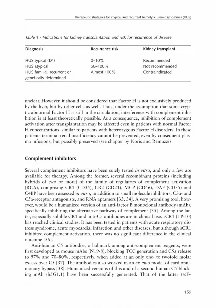
unclear. However, it should be considered that Factor H is not exclusively producedby the liver, but by other cells as well. Thus, under the assumption that some cryp-tic abnormal Factor H is still in the circulation, interference with complement inhi-bition is at least theoretically possible. As a consequence, inhibition of complementactivation after transplantation may be affected even in patients with normal FactorH concentrations, similar to patients with heterozygous Factor H disorders. In thesepatients terminal renal insufficiency cannot be prevented, even by consequent plas-ma infusions, but possibly preserved (see chapter by Noris and Remuzzi)
Complement inhibitors
Several complement inhibitors have been solely tested in vitro, and only a few areavailable for therapy. Among the former, several recombinant proteins (includinghybrids of two or more) of the family of regulators of complement activation(RCA), comprising CR1 (CD35), CR2 (CD21), MCP (CD46), DAF (CD55) andC4BP have been assessed in vitro, in addition to small molecule inhibitors, C3a- andC5a-receptor antagonists, and RNA aptamers [33, 34]. A very promising tool, how-ever, would be a humanized version of an anti-factor B monoclonal antibody (mAb),specifically inhibiting the alternative pathway of complement [35]. Among the lat-ter, especially soluble CR1 and anti-C5 antibodies are in clinical use. sCR1 (TP-10)has reached clinical studies. It has been tested in patients with acute respiratory dis-tress syndrome, acute myocardial infarction and other diseases, but although sCR1inhibited complement activation, there was no significant difference in the clinicaloutcome [36].
Anti-human C5 antibodies, a hallmark among anti-complement reagents, werefirst developed as mouse mAbs (N19-8), blocking TCC generation and C5a releaseto 97% and 70–80%, respectively, when added at an only one- to twofold molarexcess over C5 [37]. The antibodies also worked in an ex vivo model of cardiopul-monary bypass [38]. Humanized versions of this and of a second human C5-block-ing mAb (h5G1.1) have been successfully generated. That of the latter (scFv
159
Therapeutic strategies for atypical and recurrent hemolytic uremic syndromes (HUS)
Table 1 - Indications for kidney transplantation and risk for recurrence of disease
Diagnosis Recurrence risk Kidney transplant
HUS typical (D+) 0–10% RecommendedHUS atypical 50–100% Not recommendedHUS familial; recurrent or Almost 100% Contraindicatedgenetically determined
h5G1.1) has reached clinical trials as pexelizumab [39] and may represent an inter-esting therapy option for patients suffering from aHUS.
Conclusions
Therapy of recurrent/aHUS is not evidence based. However, several reports in theliterature and the personal experience of the authors suggests that many patientswith complement disorders respond well to any form of plasma therapy. Most cen-ters prefer plasma exchange in the beginning and continue with plasma infusionas mainstay or prophylaxis. Despite this active and vigorous regimen manypatients succumb to end-stage renal disease. Transplantation of either kidneyalone or combined kidney/liver transplantation bear a high risk of recurrence.New therapeutic strategies are therefore absolutely mandatory. Hopefully, newinsights in the mechanisms of disease and perhaps new complement inhibitors(such as humanized anti C5 antibodies) may improve treatment and outcome ofthese poor patients.
References
1 Zimmerhackl LB (1998) Epidemiology, pathogenesis and therapeutic modalities inhemolytic-uremic syndrome. Kidney Blood Press Res 21: 290–292
2 Gasser C, Gautier E, Steck A, Siebenmann Re, Oechslin R (1955) Hemolytic-uremic syn-dromes: bilateral necrosis of the renal cortex in acute acquired hemolytic anemia.Schweiz Med Wochenschr 85: 905–909
3 Gerber A, Karch H, Allerberger F, Verweyen HM, Zimmerhackl LB (2002) Clinicalcourse and the role of shiga toxin-producing Escherichia coli infection in the hemolyt-ic-uremic syndrome in pediatric patients, 1997–2000, in Germany and Austria: aprospective study. J Infect Dis 186: 493–500
4 Zimmerhackl LB (2000) E. coli, antibiotics, and the hemolytic-uremic syndrome. NEngl J Med 342: 1990–1991
5 Sutor AH, Thomas KB, Prufer FH, Grohmann A, Brandis M, Zimmerhackl LB (2001)Function of von Willebrand factor in children with diarrhea-associated hemolytic-ure-mic syndrome (D+ HUS). Semin Thromb Hemost 27: 287–292
6 Sautter S (2004) Klinischer Verlauf und Veränderungen im Komplementsystem beiKindern mit rekurrierendem Hämolytisch-urämischem Syndrom. Doctoral thesis. Med-ical University Innsbruck
7 Ruggenenti P, Remuzzi G (1998) Pathophysiology and management of thromboticmicroangiopathies. J Nephrol 11: 300–310
8 Dragon-Durey MA, Fremeaux-Bacchi V, Loirat C, Blouin J, Niaudet P, Deschenes G,Coppo P, Herman Fridman W, Weiss L (2004) Heterozygous and homozygous factor H
160
Reinhard Würzner and Lothar B. Zimmerhackl

deficiencies associated with hemolytic uremic syndrome or membranoproliferativeglomerulonephritis: report and genetic analysis of 16 cases. J Am Soc Nephrol 15:787–795
9 Appel G, Cook TH, Hageman G, Jennette JC, Kashgarian M, Kirschfink M, Lambris J,Lanning L, Lutz HU, Meri S et al. (2005) Disease of the month: Membranoproliferativeglomerulonephritis type II (dense deposit disease): An update. J Am Soc Nephrol 16:1392–1403
10 Ross SC, Densen P (1984) Complement deficiency states and infection: epidemiology,pathogenesis and consequences of neisserial and other infections in an immune defi-ciency. Medicine (Balt) 63: 243–273
11 Würzner R, Orren A, Potter P, Morgan BP, Ponard D, Spath P, Brai M, Schulze M,Happe L, Götze O (1991) Functionally active complement proteins C6 and C7 detectedin C6- and C7-deficient individuals. Clin Exp Immunol 83: 430–437
12 Caprioli J, Castelletti F, Bucchioni S, Bettinaglio P, Bresin E, Pianetti G, Gamba S,Brioschi S, Daina E, Remuzzi G, Noris M (2003) Complement factor H mutations andgene polymorphisms in haemolytic uraemic syndrome: the C-257T, the A2089G and theG2881T polymorphisms are strongly associated with the disease. Hum Mol Genet 12:3385–3395
13 Esparza-Gordillo J, Goicoechea de Jorge E, Buil A, Carreras Berges L, Lopez-TrascasaM, Sanchez-Corral P, Rodriguez de Cordoba S (2005) Predisposition to atypicalhemolytic uremic syndrome involves the concurrence of different susceptibility alleles inthe regulators of complement activation gene cluster in 1q32. Hum Mol Genet 14:703–712
14 Quigg RJ (2003) Complement and the kidney. J Immunol 171: 3319–332415 Dragon-Durey MA, Loirat C, Cloarec S, Macher MA, Blouin J, NIvet H, Weiss L, Frid-
man WH, Fremaux-Bacchi V (2005) Anti-Factor H Autoantibodies associated withatypical hemolytic uremic syndrome. J Am Soc Nephrol 16: 555–563
16 Dragon-Durey MA, Fremeaux-Bacchi V, Loirat C, Blouin J, Niaudet P, Deschenes G,Coppo P, Herman Fridman W, Weiss L (2004) Heterozygous and homozygous factor hdeficiencies associated with hemolytic uremic syndrome or membranoproliferativeglomerulonephritis: report and genetic analysis of 16 cases. J Am Soc Nephrol 15:787–795
17 (2002) Guidelines on the diagnosis and management of the thrombotic microangio-pathic hemolytic anemias. Br J Haematol 120: 556–573
18 O’Shaughnessy DF, Atterbury C, Bolton Maggs P, Murphy M, Thomas D, Yates S,Williamson LM (2004) British Committee for Standards in Haematology, Blood Trans-fusion Task Force. Guidelines for the use of fresh-frozen plasma, cryoprecipitate andcryosupernatant. Br J Haematol 126: 11–28
19 Rock A, Shumak KH, Buskard NA (1991) Comparison of plasma exchange with plas-ma infusion in the treatment of thrombotic thrombocytopenic purpura. CanadianApheresis Study Group. N Engl J Med 325: 393–397
20 Caletti MG, Lejarraga H, Kelmansky D, Missoni M (2004) Two different therapeutic
161
Therapeutic strategies for atypical and recurrent hemolytic uremic syndromes (HUS)

regimes in patients with sequelae of hemolytic-uremic syndrome. Pediatr Nephrol 19:1148–1152
21 Chiurchiu C, Ruggenenti P, Remuzzi G (2002) Thrombotic microangiopathy in renaltransplantation. Annals of Transplantation 7: 28–33
22 Gerber A, Kirchhoff-Moradpour AH, Obieglo S, Brandis M, Kirschfink M, Zipfel PF,Goodship JA, Zimmerhackl LB (2003) Successful (?) therapy of hemolytic-uremic syn-drome with factor H abnormality. Pediatr Nephrol 18: 952–955
23 Landau D. Shalev H, Levy-Finer G, Polonsky A, Segev Y, Katchko L (2001) Familialhemolytic uremic syndrome associated with complement factor H deficiency. J Pediatr138: 412–417
24 Licht C, Weyersberg A, Heinen S, Stapenhorst L, Devenge J, Beck B, Waldherr R,Kirschfink M, Zipfel PF, Hoppe B (2005) Successful plasma therapy for atypicalhemolytic uremic syndrome caused by factor H deficiency owing to a novel mutation inthe complement cofactor protein domain 15. Am J Kidney Dis 45: 415–421
25 Nathanson S, Fremeaux-Bacchi V, Deschenes G (2001) Successful plasma therapy inhemolytic uremic syndrome with factor H deficiency. Pediatr Nephrol 16: 554–556
26 Manuelian T, Hellwage J, Meri S, Caprioli J, Noris M, Heinen S, Jozsi M, Neumann HP,Remuzzi G, Zipfel PF (2003) Mutations in factor H reduce binding affinity to C3b andheparin and surface attachment to endothelial cells in hemolytic uremic syndrome. JClin Invest 111: 1181–1190
27 Ault BH, Schmidt BZ, Fowler NL, Kashtan CE, Ahmed AE, Vogt BA, Colten HR (1997)Human factor H deficiency Mutations in framework cysteine residues and block in Hprotein secretion and intracellular catabolism. J Biol Chem 272: 25168–25175
28 Loirat C, Niaudet P (2003) The risk of recurrence of hemolytic uremic syndrome afterrenal transplantation in children. Pediatr Nephrol 18: 1095–11101
29 Filler G, Radhakrishan S, Strain L, Hill Andrew, Knoll G, Goodship TH (2004) Chal-lenges in the management of infantile factor H associated hemolytic uremic syndrome.Pediatr Nephrol 19: 908–911
30 Artz MA, Steenbergen EJ, Hoitsma AJ, Monnens LAH, Wetzels JFM (2003) Renaltransplantation in patients with hemolytic uremic syndrome: high rate of recurrence andincreased incidence of acute rejection. Transplantation 76: 821–826
31 Pirson Y, Lefebvre C, Lambert C, Warwicker P, Goodship T (2004) Familial hemolyticuremic syndrome: a privileged observation. Bull Mem Acad R Med Belg 159: 191–194
32 Richards A, Kemp EJ, Liszewski MK, Goodship JA, Lampe AK, Decorte R, Mus-lumanoglu MH, Kavukcu S, Filler G, Pirson Y et al (2003) Mutations in human com-plement regulator, membrane cofactor protein (CD46), predispose to development offamilial hemolytic uremic syndrome. Proc Natl Acad Sci USA 100: 12966–12971
33 Mollnes TE (2004) Therapeutic manipulation in the complement system. In: J Szebeni(ed): The Complement System. Novel roles in health and disease. Kluwer, Norwell, Dor-drecht, 483–516
34 Morgan BP, Harris CL (2003) Complement therapeutics; history and current progress.Mol Immunol 40: 159–170
162
Reinhard Würzner and Lothar B. Zimmerhackl

35 Thurman JM, Lucia MS, Ljubanovic D, Holers VM (2005) Acute tubular necrosis ischaracterized by activation of the alternative pathway of complement. Kidney Int 67:524–530
36 Zimmerman JL, Dellinger RP, Straube RC, Levin JL (2000) Phase I trial of the recom-binant soluble complement receptor 1 in acute lung injury and acute respiratory distresssyndrome. Crit Care Med 28: 3149–3154
37 Würzner R, Schulze M, Happe L, Franzke A, Bieber FA, Oppermann M, Götze O(1991) Inhibition of terminal complement complex formation and cell lysis by mono-clonal antibodies. Complement Inflamm 8: 328–340
38 Rinder CS, Rinder HM, Smith BR, Fitch JC, Smith MJ, Tracey JB, Matis LA, SquintoSP, Rollins SA (1995) Blockade of C5a and C5b-9 generation inhibits leukocyte andplatelet activation during extracorporeal circulation. J Clin Invest 96: 1564–1572
39 Fitch JC, Rollins S, Matis L, Alford B, Aranki S, Collard CD, Dewar M, Elefteriades J,Hines R, Kopf G et al (1999) Pharmacology and biological efficacy of a recombinant,humanized, single-chain antibody C5 complement inhibitor in patients undergoing coro-nary artery bypass graft surgery with cardiopulmonary bypass. Circulation 100:2499–2506
163
Therapeutic strategies for atypical and recurrent hemolytic uremic syndromes (HUS)

165
Complement defects in children which result in kidney diseases: diagnosis and therapy
Christoph Licht and Bernd Hoppe
Children’s Hospital of the University of Cologne, Pediatric Nephrology, Kerpener Str. 62,50937 Cologne, Germany
Complement and Kidney Disease, edited by Peter F. Zipfel© 2006 Birkhäuser Verlag Basel/Switzerland
Introduction
In recent years, several kidney diseases have been linked to defective complementregulation, namely to mutations of genes located within the “regulators of comple-ment activation (RCA)” gene cluster on chromosome 1q32 [1]. First candidategenes in this region were Factor H (Factor H protein family) [2–5], and membranecofactor protein (MCP, CD46) [6–8]. The clinical spectrum includes renal diseaseslike atypical hemolytic uremic syndrome (aHUS), membranoproliferative glomeru-lonephritis type II (MPGN II), isolated microscopic hematuria, collagen type IIIglomerulopathy, and IgA nephropathy [9]. However, diseases with a mainlyextrarenal manifestation have also been linked to the complement system: systemiclupus erythematosus (SLE) [10, 11], increased susceptibility to meningococcal infec-tions resulting in meningococcal meningitis [12–14], recurrent microbial infectionsand chronic inflammatory conditions such as rheumatoid arthritis [15, 16], andimmune evasion of tumor cells [17, 18], as well as disorders in the coagulation sys-tem [19–21].
A key finding in patients with complement-based renal diseases is the activationof the alternative complement pathway, in the majority of cases caused by het-erozygous or homozygous Factor H deficiency [9]. However, not only deficiency ofFactor H but also mutations affecting protein function but not plasma availabilityare identified as causing disease (aHUS [22–25], MPGN II [26]). Dragon-Durey etal. [27] recently described the presence of anti-Factor H autoantibodies as a newpathomechanism of functional Factor H deficiency also causing aHUS. In addition,deficiency in membrane-anchored MCP and Factor I are now also known to causeaHUS [6–8, 28].
By contrast, the paroxysmal nocturnal hemoglobinuria (PNH) is not based onFactor H or MCP but on a defect of the decay-accelerating factor (DAF, CD55).DAF is a glycophosphatidylinositol-linked membrane protein expressed on endothe-lial cells and erythrocytes. A defect of this protein-lipid linkage results in the failureto express DAF and causes PNH. Unregulated complement activation on the surface

166
Christoph Licht and Bernd Hoppe
of erythrocytes causes recurrent intravascular hemolysis and – in the long run –leads to chronic hemolytic anemia and venous thrombosis [19, 29–32].
In principal, all of these diseases can be found in children. Thus, consequentscreening for defects in the regulation of the complement system will allow the actu-al incidence/prevalence of the (complement-based subtypes of the) above-mentioneddiseases to be identified. In the following chapter, however, we focus on aHUS andMPGN II, which are so far the most common known complement-based renal dis-eases in children [9].
The complement system
Introduction
The complement system is part of the innate immune system. It represents one ofthe major recognition systems of innate immunity and an important effector mech-anism of humoral (antibody-mediated) adaptive immunity. The central physiologi-cal functions of the complement system are:(i) Defense against pyogenic microbial infections(ii) Bridging innate and adaptive immunity(iii) Disposing of immune complexes and products of inflammatory injury.
The complement system consists of a family of multiple plasma and surface-boundproteins, which interact with one another and other proteins of the immune systemin a tightly regulated fashion. Activated complement proteins (e.g., central comple-ment component C3/C3b) bind, e.g., to the surface of microbes or to antibodieswithin antigen-antibody complexes and induce the activation of the late steps of thecomplement cascade, which are (1) opsonization and phagocytosis, (2) induction ofinflammation, and (3) formation of the membrane attack complex (MAC) andcytolysis [29–31].
Microbes on which complement is activated become coated with C3b or C4b(opsonization), and are phagocytosed by the binding of these proteins to specificreceptors on macrophages and neutrophils. C5a, C4a, and C3a, also called ana-phylatoxins, are proteolytic fragments of C5, C4, and C3, which are generatedwhen these complement components are activated. They induce acute inflammationby acting on mast cells and neutrophils. Finally, binding of C3b to microbes andactivation of the late components of the complement cascade (C5b-C9, terminalcomplement complex, TCC) leads to the formation of the MAC, which causesosmotic lysis of the microbes [29–31].
With few exceptions, host cells are protected against activation of the comple-ment system on their surface by cell membrane-anchored inhibitors. Microbial cells,by contrast, lack these inhibitors. Thus, the complement system is able to “differ-

entiate” between “self” and “non-self”, which guarantees the directed attackagainst invading microbial cells and prevents a generalized self attack with fatal con-sequences for the host [29–31].
Regulation of complement activation
Complement activation occurring on foreign cells such as invading microbes is partof a vital defense mechanism of the body, providing the possibility of immediateimmune reaction with the final goal of attacking and eliminating these microbes. Asa consequence, complement activation on “self cells” must be tightly regulated andcontrolled by both soluble, e.g., Factor H, and cell-membrane anchored, e.g., MCP,DAF, regulators to avoid fatal self attack [33–36].
Activation of the complement system occurs via one of the following pathways:(i) Alternative pathway(ii) Classical pathway(iii) Lectin pathway.
The two main pathways of complement activation are the alternative pathway,which is – at low level – continuously activated with the early steps of the comple-ment cascade occurring indiscriminately on any surface in the absence of antibody,and the classical pathway, which is activated by antigen-antibody complexes[29–31, 37]. The lectin pathway is activated by microbial polysaccharides and cir-culating lectin molecules and follows, except for the different induction, the classi-cal pathway. Both the classical and the alternative pathway merge at the level of C3resulting in the generation of C3b (Fig. 1) [29–31].
Under physiological conditions, there is constant low rate of C3 cleavage in plas-ma, generating C3b – a process called “C3 tick over”. A small amount of C3bbecomes covalently bound to the surfaces of cells, including host cells and microbes.Surface-attached C3b binds Factor B, which is then cleaved by Factor D to generateFactor Bb. Factor Bb remains attached to C3b, resulting in the alternative pathwayC3 convertase C3bBb. Properdin binds to and stabilizes C3bBb. The function ofC3bBb is to cleave more C3 molecules, thereby setting in motion the activation loopof C3 within the alternative pathway of the complement system [29–31].
On mammalian (“self”) cells, C3bBb becomes rapidly degraded by several sur-face-attached and membrane-anchored regulatory proteins. On microbial cells,however, in the absence of such regulators, activation of the (alternative pathway ofthe) complement system proceeds. Some of the C3b molecules generated by the C3convertase bind to C3bBb, thereby forming the alternative pathway C5 convertaseC3bBbC3b [29–31].
Within the classical pathway, activation of C3 occurs through the action of theclassical pathway C3 convertase: C1 binds to antigen-antibody complexes and
167
Complement defects in children which result in kidney diseases: diagnosis and therapy
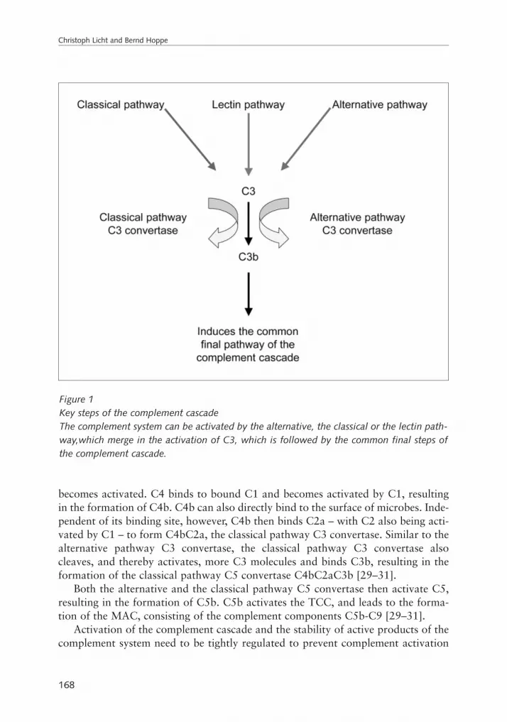
becomes activated. C4 binds to bound C1 and becomes activated by C1, resultingin the formation of C4b. C4b can also directly bind to the surface of microbes. Inde-pendent of its binding site, however, C4b then binds C2a – with C2 also being acti-vated by C1 – to form C4bC2a, the classical pathway C3 convertase. Similar to thealternative pathway C3 convertase, the classical pathway C3 convertase alsocleaves, and thereby activates, more C3 molecules and binds C3b, resulting in theformation of the classical pathway C5 convertase C4bC2aC3b [29–31].
Both the alternative and the classical pathway C5 convertase then activate C5,resulting in the formation of C5b. C5b activates the TCC, and leads to the forma-tion of the MAC, consisting of the complement components C5b-C9 [29–31].
Activation of the complement cascade and the stability of active products of thecomplement system need to be tightly regulated to prevent complement activation
168
Christoph Licht and Bernd Hoppe
Figure 1Key steps of the complement cascadeThe complement system can be activated by the alternative, the classical or the lectin path-way,which merge in the activation of C3, which is followed by the common final steps ofthe complement cascade.

on normal host cells, and to limit the duration of complement activation even onmicrobial cells and antigen-antibody complexes. Complement activation is regulat-ed by a family of humoral or membrane-bound proteins summarized as RCA. Mem-bers of this family are C1 inhibitor (C1 INH), Factor I, Factor H, Factor H like pro-tein-1 (FHL-1), C4 binding protein (C4BP), MCP, type 1 complement receptor(CR1, CD35), DAF, and CD59 [29–31].
C1 INH inhibits the proteolytic activity of C1. MCP, CR1 and DAF inhibit thebinding of regulatory proteins to C3b and C4b. Furthermore, C3b is bound by Fac-tor H (alternative pathway), and C4b is bound by C4BP (classical pathway). Bybinding to C3b and C4b, Factor H and C4BP competitively inhibit the binding ofother components of the C3 convertase, such as Bb (alternative pathway) and C2a(classical pathway), thus inhibiting further activation of the complement cascade.C3b and C4b are proteolytically degraded by Factor I. MCP, Factor H, C4BP, andCR1 serve as cofactors for Factor I-mediated cleavage of C3b and C4b. CD59inhibits the formation of the MAC [29–31].
The differential attachment and presence of these regulators on mammalian(“self”) but not on microbial cells results in selective inhibition of the complementsystem on host cells and allows complement activation to proceed on microbial cells[29–31].
The complement system and disease
The complement system is involved in diseases in two general ways: deficiencies inany of the protein components of the complement cascade may lead to abnormalcomplement activation resulting in either deficient or unrestricted activation of thecomplement system (defect of regulatory components) with too much complementactivation at the wrong time or the wrong site [29–31]. With respect to renal dis-eases, however, in most cases deficiency results in unrestricted activation of the com-plement cascade, especially of the alternative pathway [38].
As described above, there is a redundant system of factors, which are in chargeof a fine-tuned regulation of the activation of the complement cascade, the RCA.The functional redundancy of these factors ensures that the activation of the com-plement cascade occurs specifically “at the right time and at the right site” and selfattack and damage of self surfaces of the mammalian organism are avoided. Fur-thermore, the distribution of the proteins that regulate the activation of the com-plement system directs the action of the complement system specifically to the sur-face of the target cells, e.g., invading microbes. While, in general, microbial cellslack surface-bound regulators for the activation of the complement system, mam-malian cells, i.e., host cells, are protected by the presence of such regulatory proteins(e.g., MCP, CR1 and DAF). In addition, a group of humoral factors (e.g., Factor H,FHL-1, Factor I) acts complementary to these cell membrane-bound proteins. These
169
Complement defects in children which result in kidney diseases: diagnosis and therapy

factors are primarily present as circulating plasma proteins, which bind and attachto host cell surfaces. The serine protease Factor I cleaves C3b and C4b using FactorH and other factors, e.g., MCP, C4BP, and CR1, as cofactors [29–31].
However, it has been shown that Factor H, to become functionally active, needsto bind to cell surfaces [21, 37, 39]. Factor H binds with high affinity to cell mem-branes rich in sialic acid, as found in mammalian cells different from microbial cells,which again directs the action of the complement system to attack invadingmicrobes and to protect mammalian host cells [21, 37, 39]. When bound to a cellmembrane, Factor H functions as cofactor of Factor I in mediating the cleavage ofcell membrane-bound C3b and C4b. Furthermore, Factor H binds C3b, thereby pre-venting the binding of other factors to C3b, e.g., of Bb, which would result in theformation of the alternative pathway C3 convertase C3bBb – or even displacingalready bound Bb from C3b, thereby dissociating and inactivating the alternativepathway C3 convertase [29–31].
A central step within the sequential events of complement activation is the acti-vation of the third complement component – C3 – resulting in the generation ofC3b. Responsible for the activation of C3, however, are the classical and/or thealternative pathway C3 convertases C3bBb and C4bC2a. All previously describedregulatory mechanisms controlling the activation of the complement system arepositioned around this center: they either cleave C3b or C4b themselves, or inhibitthe formation of the C3 convertase complex or dissociate the already formed com-plex [29–31].
In addition, there is another humoral factor that is assumed to stabilize the(alternative pathway) C3 convertase, called C3 nephritic factor (C3NeF). C3NeF issupposed to be an IgG antibody, which covalently binds to C3bBb, thereby pre-venting Factor H-induced dissociation of this complex. With respect to the finalresult, the presence of C3NeF displays a comparable effect to any other situation inwhich deficiency or defect of the previously described regulatory proteins lead tounrestricted activation of the complement system (Fig. 2) [29–31, 40, 41].
In summary, each defect – either quantitative deficiency or functional defect – ofone or more of the proteins forming this tight and redundant network of control ofcomplement activation (alternative and classical pathway), or the presence ofC3NeF (alternative pathway), unavoidably leads to unrestricted complement acti-vation, which bears the potential of causing disease [29–31].
Kidney diseases as result of complement defects in children
Atypical hemolytic uremic syndrome
In 1952, Symmers [42] defined the disease thrombotic microangiopathy (TMA) asa “lesion of vessel wall thickening (mainly affecting arterioles and capillaries) with
170
Christoph Licht and Bernd Hoppe

swelling or detachment of the endothelial cells from the basement membrane, accu-mulation of fluffy material in the subendothelial space, intraluminal platelet throm-bi, and partial or complete obstruction of the vessel lumina”. Depending on the dis-tribution of the microvascular lesions and the age of the patient, different diseasesubtypes can develop: while in adults neurological symptoms, e.g., transientischemic attacks (TIA), prevail as a sign of cerebral affection, acute renal failure rep-resents the main clinical complication in children, indicating that the kidney is thepreferred disease target in this age group [43]. The TMA subtype found in adults iscalled thrombotic thrombocytopenic purpura (TTP) or Morbus Moshcowitz,whereas the subtype found in children is called hemolytic uremic syndrome (HUS)[43–45].
HUS is defined by the triad microangiopathic hemolytic anemia with the appear-ance of fragmentocytes/schistocytes and thrombopenia followed by acute renal fail-ure (uremia), which was first described by Gasser et al. in 1955 [44]. HUS occurswith a frequency of 5–10/100,000 children and represents the major cause of acute
171
Complement defects in children which result in kidney diseases: diagnosis and therapy
Figure 2Unrestricted activation of (alternative pathway) C3 convertaseBoth presence of C3NeF and deficiency or functional defect of Factor H, Factor I and MCPresult in unrestricted activation of (alternative pathway) C3 convertase and subsequently ofC3.

renal failure in childhood [46]. There are two main forms of HUS: typical D+HUSor Shiga toxin HUS (Stx HUS), and atypical D–HUS. Stx HUS usually follows anepisode of gastroenteritis caused by verocytotoxin- or Shiga (like) toxin-producingenterohemorrhagic Escherichia coli (EHEC), mostly of the serotype O157:H7 [46].It affects male and female patients with the same frequency. Relapses are extremelyrare, and compared to aHUS, prognosis is usually good with end-stage renal failure(ESRF) in <10% [46]. Atypical D–HUS, however, can occur in a sporadic or famil-ial (inherited) fashion, is characterized by the absence of antecedent diarrhea, thetendency to pre- and post-transplant relapse (about 50% after renal transplantationin children) and often a poor outcome with development of ESRD or death. Atypi-cal D–HUS accounts for 5–10% of all HUS cases and prefers male patients(male:female, 2:1) [46–50].
Pathogenesis of typical and atypical HUS merge in a common final pathway:damage of blood vessel endothelial cells causes exposure of the basement mem-brane, which activates both the complement and the coagulation systems. Activa-tion of the coagulation system causes formation of platelet-rich thrombi, which arecarried with the bloodstream and later occlude peripheral small vessels (Fig. 3A andB) [42, 46]. In children, this process mainly affects the kidneys and causes transientor permanent loss of organ function [42, 46]. It is still unclear why childhood HUS,both the typical and the atypical type of the disease, mainly affects the kidneysbefore other organ systems. A possible explanation for Stx HUS is the high expres-sion level of Gb3 receptors in endothelial cells of renal vessels, resulting in anincreased Stx binding and thereby higher susceptibility to Stx-mediated damage [51,52].
In recent years, there has been a remarkable increase in the understanding of themolecular pathomechanisms responsible for TMA like TTP (Morbus Moshcowitz)and aHUS [42, 46, 53]. For aHUS, defective control of the activation of the alter-native complement pathway by Factor H deficiency caused by Factor H gene muta-tions or autoantibodies against Factor H [34, 37, 46, 54–59], or by MCP deficien-cy caused by MCP gene mutations [6–8] has been identified as key pathomech-anism.
Before deficiency or functional defects of Factor H (or other complement regu-lators) were proven to be crucial for the development of aHUS, hypocomple-
172
Christoph Licht and Bernd Hoppe
Figure 3HUSRenal biopsy with typical glomerular changes of HUS including segmental thickening ofglomerular basement membranes with (A) double contours, congestion of capillaries andincrease in extracellular matrix (Masson trichrome stain, original magnification × 100) and(B) thrombemboli occluding the lumen of the glomerular arterioles (EM by Rüdiger Wald-herr, Heidelberg, Germany).

173
Complement defects in children which result in kidney diseases: diagnosis and therapy
A
B

mentemia with low serum levels of the central complement component C3 inpatients with familial (atypical) HUS/TTP had been demonstrated [5, 60–62]. In alarge study, Noris et al. [62] found an association between (low) C3 serum levelsand HUS/TTP within affected families. Furthermore, affected individuals and theirhealthy relatives showed significantly more Factor H abnormalities than healthycontrol individuals, and, within families, Factor H abnormalities were significantlycorrelated with (low) C3 serum levels.
Patients with familial (atypical) HUS showed both autosomal-recessive [63] andautosomal-dominant [1] modes of inheritance. Subsequent studies in patients withfamilial (atypical) HUS showed mutations in the Factor H gene in some patients,and these mutations were associated with both the spontaneous and the recurrentform of the disease [1, 22, 55, 62–71]. The frequency of Factor H gene mutationswas described in patients with sporadic and familial aHUS in a registry of Germanspeaking countries: 14% of 111 patients had Factor H gene mutations including 2of 8 patients with familial aHUS [49]. In contrast to these data, Wyatt et al. [72]report an allelic frequency of factor H deficiency of 1:326 healthy Caucasian con-trols from Kentucky, allowing the calculation of the prevalence of homozygous fac-tor H deficiency as 1:425,104 in the United States [9, 72]. The vast majority of themutations found in Factor H-associated HUS is heterozygous, resulting in one nor-mal and one defective Factor H allele. Factor H plasma levels of these patients canbe normal, but also reduced, or even increased [73]. Only very few patients withFactor H-associated HUS carry (genetically or functionally) homozygous mutationswith usually no or very little plasma Factor H [65, 66, 68, 69].
Recent progress in the understanding of Factor H protein function revealed therole of Factor H mutations for endothelial cell damage, which is the central featureof the pathogenesis of aHUS. The secreted Factor H protein is composed of 20repetitive domains termed short consensus repeats (SCR) or complement controlproteins (CCP). Structure-function analysis demonstrated that N-terminal SCRs 1–4are required for complement regulatory activities (“regulatory region”) and that C-terminal SCRs 19–20, which are highly conserved within the Factor H protein, playa crucial role for protein function (“recognition region”): they include overlappingheparin and C3b binding domains, and are the binding domain for microbial sur-face proteins. In addition, Factor H has three C3 binding domains (SCRs 1–4 bindC3b; SCRs 12–14 bind C3c; and SCRs 16–20 bind C3b/C3d) and three heparinbinding domains (SCRs 7, 13 and 20) [35, 73, 74].
Factor H gene mutations identified in Factor H-associated HUS cluster in the Cterminus of Factor H protein, mainly SCR 20, identifying this domain as a hot spot,but they also occur in almost all of the other SCRs [34, 49, 65, 66, 68–70, 73, 75,76].
Reflecting the role of Factor H as a complement regulator and the functionalorganization of the Factor H protein, the current understanding of the pathogenesisof Factor H-associated HUS can be summarized as follows. Endothelial cells coat-
174
Christoph Licht and Bernd Hoppe

ing the inner surface of blood vessels are in direct contact with plasma and are –under physiological conditions – coated with Factor H. The interaction of Factor Hwith cell surface structures is mediated via the C terminus, and the initial cell sur-face contact exposes the complement regulatory N-terminal domain of Factor Hprotein [37, 76–78]. Thus, in addition to its activity in fluid phase, Factor H con-tributes to the assembly of complement control systems on cell surfaces consistingof two layers: the inner layer activity on the cell membrane level is mediated bymembrane-anchored regulators (e.g., MCP, DAF), the outer layer activity in about20–140 nm (defined by molecule dimensions) distance from the cell membrane bysurface-attached soluble regulators such as Factor H [37].
However, Factor H proteins with mutations within SCR 20 – purified frompatient plasma or designed as recombinant mutant proteins – show reduced affini-ty to the central complement component C3b/C3d, to heparin and to endothelialcells, and are inactive [21, 37, 39]. This is in agreement with recent three-dimen-sional structure studies of the Factor H protein, which permitted the molecular(functional) interpretation of mutations within SCR 20 [79]. In consequence, localactivation of the complement system occurs, resulting in a vicious circle of damageof endothelial cells – exposure of the basement membrane – and continued activa-tion of the complement (and the coagulation) system [34, 37, 59].
Under physiological conditions, the concerted action of membrane-anchored andsurface-attached fluid-phase regulators provides sufficient complement control andprotection of host cells. During situations of “biological stress”, like inflammationor microbial infection, and other triggering events like pregnancy, vaccination, orthe action of drugs (e.g., calcineurin inhibitors), complement activation occursrapidly and heavily, and needs to be restricted. Under these conditions the activityof the endogenous, membrane-anchored regulators must be supplemented by sur-face-attached, fluid-phase regulators to provide sufficient protection of host cells. Ifsuch additional regulators are not available, e.g., Factor H deficiency, unrestrictedcomplement activation and subsequently damage occurs [37, 42, 46]. According toa recent report of Dragon-Durey et al. [27], anti-Factor H autoantibodies can alsocause defective Factor H function, and can therefore also be responsible for thedevelopment of aHUS.
In summary, either reduced availability or functional impairment of Factor Hprotein critically increases the vulnerability of blood vessel endothelial cells withrespect to an attack of the complement system, finally resulting in aHUS.
By contrast, we recently described a patient who developed aHUS on the back-ground of a novel homozygous Factor H mutation in SCR 15. This mutation allowsintracellular expression of a truncated Factor H protein, which cannot be secreted,resulting in very low Factor H plasma levels of < 1 µg/mL (normal: about 500 µg/mL). This case is of special interest since despite missing plasma Factor H the patientdeveloped aHUS and not MPGN II, as one would have expected from current expe-riences and knowledge [68].
175
Complement defects in children which result in kidney diseases: diagnosis and therapy

However, not only Factor H deficiency but also deficiency in MCP (CD46) caus-es aHUS, as has recently been shown in patients with a family history of aHUS onthe background of complement activation but normal Factor H [6–8]. Like solublecomplement regulator Factor H, transmembrane complement regulator MCPinhibits complement activation by inactivating the C3b, which is deposited on tar-get surfaces [6]. Different MCP gene mutations (deletion resulting in a prematurestop codon and loss of the C terminus [7]; deletion resulting in intracellular reten-tion of the mutant protein; substitution resulting in the expression of a functionallyimpaired protein [8]) result in reduced protein expression or impaired protein func-tion were found. These MCP mutations have the same functional consequences asFactor H deficiency: unrestricted activation of the alternative complement pathway[6] (see also chapter by Goodship et al.).
Appendix
Besides deficiency of factors regulating the activation of the alternative complementpathway, e.g., complement Factor H and MCP, there is a second known cause forthe development of aHUS: deficiency of von Willebrand factor-cleaving protease(vWF-CP), a factor of the coagulation system [1, 7, 46, 53, 63]. VWF-CP is a vWF-specific metalloprotease of the ADAMTS (a disintegrin and metalloprotease, withthrombospondin-1-like domains) family, which physiologically cleaves vWF aftersynthesis and secretion by endothelial cells. If vWF-CP is deficient, highly reactiveultra-large vWF multimers (ULvWFM) persist, and mediate adhesion and aggrega-tion of circulating platelets mainly at sites of high intravascular shear stress. As aresult, large, potentially occlusive platelet-fibrin-rich thrombi appear, which are car-ried with the bloodstream to ultimately occlude the peripheral microvasculature,i.e., arterioles and capillaries [46, 80, 81].
A severe decrease in activity below detection level (normal: 50–150%) of vWF-CP (ADAMTS13) has been described in patients with TTP [81–83]. VWF-CP defi-ciency can be caused by IgG autoantibodies against vWF-CP, resulting in the (spo-radically occurring) acquired form [84–86], or by homozygous or compound het-erozygous mutations of the ADAMTS13 gene, resulting in the (recessively) inheritedform [82, 83, 87, 88].
Main target of TTP is the central nervous system with neurological symptomscaused by cerebral ischemia, but different organ systems like the gastrointestinaltract and the kidneys can also be affected. In this case the disease is called TTP. TTPis a severe disease with a mortality rate of > 90% without treatment. When treatedwith plasma infusion or plasma exchange, however, 60–90% of the patients survivethe acute episodes. Relapses occur in about 30–35% of the patients who achieveremission, and a subset of patients develops chronic TTP requiring long-term plas-
176
Christoph Licht and Bernd Hoppe

ma exchange. If, however, the kidneys are the main target, with renal failure as pre-dominant clinical feature, the disease is called aHUS [46].
Clinical distinction between TTP and aHUS is not always clear-cut: renal abnor-malities in patients with a presumed diagnosis TTP, and extrarenal manifestationsin patients with a presumed diagnosis aHUS do occur [46]. Some authors, therefore,suggest the term TTP/aHUS for this group of diseases [42, 46]. By contrast, othersreject this summarizing term, since assay of ADAMTS13 activity distinguishes TTPfrom aHUS and other types of TMA [53]. With respect to future pathophysiology-based classifications, it is important that Noris et al. recently described TTP as aresult of two heterozygous ADAMTS13 gene mutations in two siblings, and, inaddition, chronic renal failure in one sibling, possibly caused by a heterozygous Fac-tor H gene mutation [89]. This finding might open the door for an understandingof the pathophysiology of aHUS in which different concepts of explanation – vWF-CP deficiency and RCA deficiency – possibly merge to the concept of one funda-mental pathomechanism of endothelial cell damage.
Membranoproliferative glomerulonephritis type II
Even before a link with aHUS was appreciated, dysregulation of the (alternativepathway of the) complement system had been associated with the development ofMPGN II [54]. MPGN II, also termed “dense-deposit disease”, is a rare disease,which is characterized by complement containing dense deposits within the base-ment membrane of the glomerular capillary wall, and followed by capillary wallthickening, mesangial cell proliferation and glomerular fibrosis (Fig. 4) [9, 90].
Besides MPGN II, there are two other MPGN subtypes, MPGN I and MPGN III.All three subtypes are characterized by mesangial cell proliferation and increase inmesangial matrix combined with a thickening of the glomerular capillary walls(MPGN I: interposition of mesangial cells and matrix between basement membraneand endothelial cells, resulting in the formation of a double layer structure of suben-dothelial electron-dense deposits; MPGN III: subendothelial and subepithelial elec-tron-dense deposits). Deposits in all subtypes contain C3 and other complement fac-tors [90]. In some patients combination of MPGN with extrarenal manifestationslike lipodystrophy and retina alterations can be found [90, 91].
MPGN mainly affects older children and adolescents (median age at onset of dis-ease: about 10 years). Of the patients, 50% present with nephrotic syndrome, theothers with mild proteinuria, 20% with macrohematuria; 30% of the patients devel-op hypertension at onset of disease. Children with MPGN have an unfavorable lateprognosis, and develop ESRD after about 8–16 years (MPGN I: 15.3 years; MPGNII: 8.7 years; MPGN III: 15.9 years) [90, 92].
The development of MPGN II has been linked to the dysregulation of the (alter-native pathway of the) complement system caused by the following five different
177
Complement defects in children which result in kidney diseases: diagnosis and therapy

conditions [9]: (1) congenital absence of Factor H in plasma [5, 10, 13, 66, 72,93–95]; (2) functional defect of Factor H protein caused by a mutation, e.g., in theregulatory domain [26]; (3) inactivation of Factor H caused by a circulatinginhibitor [96]; (4) presence of the IgG autoantibody C3 nephritic Factor (C3NeF)[40, 41, 97, 98]; and (5) mutation in C3 altering its Factor H binding site [99].
Absence of Factor H in plasma as the cause of MPGN II has been observed inhumans [5, 10, 13, 66, 72, 93–95], in naturally mutant Factor H-deficient pigs [100,101], and in genetically engineered Factor H-knockout mice [102]. All mutations sofar known result in a block of protein secretion. The mutant protein is expressed inhepatocytes, but is retained in the endoplasmic reticulum, thereby accumulating inthe cytoplasm. The absence of Factor H in plasma causes sustained activation of thealternative complement pathway, reflected by consumption of C3 and accumulationof the C3 degradation product C3d in plasma [103].
We recently described siblings with MPGN II who showed activation of thealternative pathway of the complement system despite normal plasma Factor H lev-
178
Christoph Licht and Bernd Hoppe
Figure 4MPGN IIElectron micrograph with segmental osmiophilic transformation of the glomerular basementmembrane (“dense deposits”) (EM by Rüdiger Waldherr, Heidelberg, Germany).

els. Genetic analyses revealed a novel mutation of the Factor H gene (cDNA743–745) causing deletion of Lys in position 224 (∆K224) located within SCR 4 inboth patients. Testing of mutant Factor H protein function revealed normal C-ter-minal (i.e., binding to heparin, C3d, and endothelial cells), but impaired N-terminal(i.e., cofactor and decay-accelerating activity, binding to central complement C3b)activities [26]. Thus, with respect to complement activation and development ofMPGN II functional defect of Factor H protein proves to be of equal importancecompared to plasma Factor H deficiency.
Meri et al. [96] isolated a factor from serum and urine of a patient withhypocomplementemic MPGN II (atypical histology with additional subendothelialdeposits: intermediate type between MPGN I and II) that was associated with dys-function of the alternative complement pathway. When mixed with fresh normalserum, the patient’s serum induced almost complete conversion of C3. This activitywas due to a circulating factor, which was different from C3NeF, but was observedto interact directly with Factor H and therefore can be considered as Factor H anti-body.
However, not only the absence (Factor H mutations and Factor H knockout) orinactivation (Factor H antibodies) of Factor H, but also the presence of the IgGautoantibody C3NeF in plasma is linked to MPGN II [40, 41]: 54% of adult and82% of pediatric MPGN II patients are positive for C3NeF [104]. However, C3NeFis observed not only in patients with MPGN II, but also in patients with the MPGNsubtypes I and III*, partial lipodystrophy, retina alterations, meningococcal menin-gitis and even in healthy individuals [91, 104]. Moreover, it is possible that the samepatient is C3NeF positive at one time point, and becomes negative at a different timepoint [104]. Both Factor H and C3NeF control the same enzyme, the alternativepathway C3 convertase C3bBb. However, while Factor H dissociates the C3bBbcomplex and – together with Factor I – inactivates the convertase, C3NeF displaysthe opposite effect: C3NeF stabilizes the convertase and increases its half-lifeapproximately tenfold, a defect which also causes unrestricted activation of the(alternative pathway of the) complement system (Fig. 2) [40, 105].
Marder et al. [99] described a mutation in C3 that alters its Factor H bindingsite, and renders Factor H unable to dissociate the alternative pathway convertaseC3bBb. In consequence, this defect, called Marder’s disease, also results in unre-stricted activation of the (alternative pathway of the) complement system.
In summary, quite distinct abnormalities can cause dysregulation of the activa-tion of the (alternative pathway of the) complement system. A redundant system ofsoluble and membrane-anchored RCA prevents unrestricted progression of the acti-vation of the complement system. At particular sites, however, tissue surfaces lack
179
Complement defects in children which result in kidney diseases: diagnosis and therapy
* Distribution of C3NeF in serum depending on different histological types of MPGN: MPGN II >MPGN I > MPGN III [92].

such additional membrane-anchored regulators so that these structures depend onsoluble regulators like Factor H. The glomerular basement membrane representssuch a sensitive structure because it lacks endogenous, membrane-anchored regula-tors [37, 100]. Lack or functional inactivation of Factor H results in continued C3deposition within the “lamina densa” of the glomerular basement membrane,resulting in “dense-deposit disease” (MPGN II) with thickening of the glomerularbasement membrane, impairment of the glomerular filter, hematuria and protein-uria, and, finally, progressive loss of renal function and development of ESRD.
Diagnosis and therapy
Diagnosis
Based on the explanation of aHUS and MPGN II as complement based diseases, thedetailed evaluation of the complement system in a stepwise approach is necessarywhenever the diagnosis aHUS or MPGN II (or of another disease which is linked tothe complement system) is suspected or diagnosed, and other causes – especially foraHUS – are excluded.
In a first step, C3, C3d and APH50 and CH50 [tests of the activation status ofthe alternative (APH50) and of the classical (CH50) pathway of the complementsystem] are measured. Low C3 and high C3d levels accompanied by low values forAPH50 and CH50 indicate the activation of the (alternative and the classical path-way of the) complement system. If the complement system is activated, plasma Fac-tor H, the major known complement regulator, should be examined. If Factor H isnot detectable in plasma or if its level is severely decreased, the Factor H gene shouldbe sequenced.
If, however, the concentration of plasma Factor H is normal despite activationof the (alternative pathway of the) complement system, function of Factor H pro-tein should be examined. If function of Factor H protein is normal, it is likely thatone of the other complement regulators, e.g., Factor I or MCP, is responsible forcomplement activation and, thus, possibly responsible for the disease. If, however,function of Factor H protein is defective, sequencing of Factor H gene should beperformed to detect possible Factor H gene mutations, which affect proper functionof Factor H protein. Figure 5 summarizes this stepwise approach and provides adiagnostic algorithm for complement based renal diseases.
Whenever unrestricted activation of the (alternative pathway of the) complementsystem caused by deficiency or defect of one or more complement regulators is iden-tified as cause for aHUS or MPGN II, substitution of this missing or functionallydefective factor, e.g., via plasma infusion, is one possible therapeutic option inde-pendent of the disease caused by the complement defect.
180
Christoph Licht and Bernd Hoppe

Therapy of aUHS
While for classical HUS there is no treatment of proven efficacy besides supportivemeasures and, if needed, renal replacement therapy, there are several treatmentoptions for complement based, especially Factor H related, aHUS.
Since Factor H is physiologically synthesized by the liver, liver transplantationtheoretically represents a causative treatment in patients with Factor H deficiency.In fact, there have been three patients with Factor H-associated aHUS worldwidewho underwent (combined kidney and) liver transplantation [106]. However, noneof these patients survived and at least one of them developed acute liver failure forsevere thrombosis of the liver veins [107].
Restoration of Factor H plasma concentration and/or Factor H function byeither plasma exchange or repetitive plasma infusions, however, represents the treat-ment concept, which is currently accepted [23, 24, 42, 108–110]. However, so farthere is rather little secured information about therapy regimens available in the lit-erature, and most authors stress the empirical character of the chosen treatment reg-imen. Plasma exchange (1–2 plasma volumes/session) or plasma infusion (10–20 to30–40 mL/kg) is considered first-line therapy. Depending on the need of the patient,
181
Complement defects in children which result in kidney diseases: diagnosis and therapy
Figure 5Diagnostic algorithm for complement based renal diseases

plasma infusion is given daily in the beginning, afterwards every 7–14 days. In somepatients, daily plasma infusions are required even for a longer period of time [23,42]. As alternative to fresh frozen plasma (FFP), cryosupernatant – in patients whodo not respond to plasma – or solvent detergent-treated plasma – to minimize therisk of viral contamination – can be used [23, 42, 111].
We recently described a patient with complete Factor H deficiency who devel-oped aHUS [68]. Genetic analysis revealed a point mutation of the Factor H genewhich introduced a premature stop codon in SCR 15, preventing Factor H proteinfrom being secreted by liver cells. In a time course investigation of this patient fol-lowing plasma infusion (20 mL/kg), Factor H half-life of about 6 days was deter-mined (Fig. 6A). Based on this result, the patient was treated with regular plasmainfusions in cycles of 10–14 days, which is used in the majority of patients reportedin the literature. Following plasma infusion, plasma Factor H and C3 levelsincreased, while C3d levels decreased (Fig. 6A), and in parallel APH50 and CH50levels increased (Fig. 6B). In summary, these findings reflect successful inhibition ofthe unrestricted activation of the complement system. The long-term observation ofindicators for disease activity and renal function showed that both lactate dehydro-genase (LDH) and creatinine (Crea) in serum decreased upon periodical plasmainfusion of 20 mL/kg every 14 days, indicating that this treatment was able to stopdisease activity and progression (Fig. 7). The patient recovered completely and kid-ney function remained normal [68]. Shortly after initial aHUS manifestation and ini-tiation of regular treatment with plasma infusion, a minor aHUS relapse with com-plement activation, signs of hemolysis, and decrease of renal function occurred afterpneumococcal vaccination but could be controlled by immediate plasma infusion.
This observation is in accordance with the literature: conditions of “biologicalstress”, e.g., vaccinations and infections, are reported to be triggering events for newaHUS crises [46, 65]. Thus, in addition to the long-term regimen of regular plasmainfusions, the patient was prophylactically treated with additional plasma infusionswhenever he developed severe airway or gastrointestinal infections. The typicalcombination of low C3, high C3d, and low APH50 levels indicated activation of thecomplement system in each of these situations. Plasma infusions (20 mL/kg/day)were then given daily for the period with fever > 38.5°C. By this regimen unre-stricted activation of the complement system could be stopped (Fig. 8A), and devel-opment of new aHUS crises could be prevented, which, in the long run, preservedrenal function (Fig. 8B) [68].
As rescue therapy for extremely sick patients with frequent relapses or require-ment of large amounts of plasma, splenectomy might be considered [42, 112].
182
Christoph Licht and Bernd Hoppe

183
Complement defects in children which result in kidney diseases: diagnosis and therapy
Figure 6Factor H and complement profile before and following plasma infusion [68](A) Factor H, C3 and C3d expressed as ratio of value before plasma infusion. Factor H, C3and C3d were measured before, and 2, 8, 16, 24, 32, 40 and 48 h after plasma infusion(arrow) by ELISA. The decrease of Factor H in plasma was paralleled by a decrease of C3 andan increase of the C3 degradation product C3d. The patient does not secrete Factor H. There-fore, this decline can be extrapolated to estimate half-life of Factor H in plasma to about 6days. (B) Complement profile of both the alternative pathway (APH50) and the classicalpathway (CH50) expressed as % of normal mean value (=100%) following plasma infusion.Both APH50 and CH50 increased after plasma infusion and decreased gradually afterwards.This indicates complement activation before plasma infusion and proves inhibition of com-plement activation through plasma infusion (arrow).
A
B
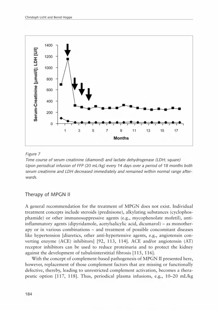
Therapy of MPGN II
A general recommendation for the treatment of MPGN does not exist. Individualtreatment concepts include steroids (prednisone), alkylating substances (cyclophos-phamide) or other immunosuppressive agents (e.g., mycophenolate mofetil), anti-inflammatory agents (dipyridamole, acetylsalicylic acid, dicumarol) – as monother-apy or in various combinations – and treatment of possible concomitant diseaseslike hypertension [diuretics, other anti-hypertensive agents, e.g., angiotensin con-verting enzyme (ACE) inhibitors] [92, 113, 114]. ACE and/or angiotensin (AT)receptor inhibitors can be used to reduce proteinuria and to protect the kidneyagainst the development of tubulointerstitial fibrosis [115, 116].
With the concept of complement-based pathogenesis of MPGN II presented here,however, replacement of those complement factors that are missing or functionallydefective, thereby, leading to unrestricted complement activation, becomes a thera-peutic option [117, 118]. Thus, periodical plasma infusions, e.g., 10–20 mL/kg
184
Christoph Licht and Bernd Hoppe
Figure 7Time course of serum creatinine (diamond) and lactate dehydrogenase (LDH; square)Upon periodical infusion of FFP (20 mL/kg) every 14 days over a period of 18 months bothserum creatinine and LDH decreased immediately and remained within normal range after-wards.

every 14 days similar to the treatment of aHUS, seem an adequate regimen [26].This approach is supported by the observation of the beneficial effect of the infu-sion of porcine plasma to piglets with MPGN II caused by Factor H deficiency [100,
185
Complement defects in children which result in kidney diseases: diagnosis and therapy
Figure 8Plasma infusion decreases complement activation in a patient with aHUS on the backgroundof Factor H deficiency triggered by an airway infectionPlasma infusion (arrow) results in (A) an increase of Factor H (triangle) and C3 (diamond),and a decrease of C3d (square), reflecting limitation of the activation of the complement sys-tem, and (B) a decrease of both serum creatinine (diamond) and LDH (square), reflectingrecovery of renal function.
A
B

103], and by recently published results obtained in a mouse model of chronic serumsickness, indicating a disease limiting effect of Factor H substitution [119].
In accordance with this concept, we recently published the treatment of two sib-lings with MPGN II with mild hematuria and proteinuria caused by functionaldefect of Factor H protein. In these patients complement activation (low C3, highC3d, low APH50 levels) was present despite normal plasma Factor H levels. Patientswere treated with regular plasma infusions (20 mL/kg/14 days) to substitute thepatient with functionally intact Factor H. Figure 9 demonstrates the immediateeffect of this treatment on two serum parameters (C3 and LDH), which reflect thedegree of complement activation (Fig. 9A and B) [26]. Acutely, plasma infusionreduced complement activation. Chronically, however, plasma infusion preventeddisease progression with development of ESRD.
Similar to aHUS on the background of Factor H deficiency, conditions of “bio-logical stress” like airway or gastrointestinal infections caused complement activa-tion, resulting in a more severe hematuria and proteinuria. Complement activationcould successfully be stopped by additional plasma infusions, which were given forthe period of fever > 38.5°C (Fig. 9a and b).
Besides replacement of deficient or defective complement regulatory factors,plasmapheresis might provide a therapeutic option for cases in which MPGN II iscaused by the presence of C3NeF. In support of this approach, Kurtz et al. [120]reported a child with rapidly progressive recurrent MPGN II due to of the presenceof C3NeF. In this patient disease progression towards onset of chronic renal failurewas delayed by periodical plasmapheresis.
Summary
We here describe aHUS and MPGN II as complement based renal diseases. Theincreasing number of published case reports now allows a better and more detailedinsight into the pathomechanism of both diseases. The general concept consists in acongenital or acquired defect in the regulation of the activation of the alternativecomplement pathway, leading to self attack and damage of specific structures, i.e.,tissue surfaces, endothelial cells of the vasculature, and basement membranes (espe-cially glomerular basement membrane). Besides the known defects in Factor H,MCP, and Factor I, there are more candidate proteins which play a role in comple-ment regulation, e.g., C4BP, Factor B, CR1, DAF, etc., which might in the nearfuture also become identified as factors causing unrestricted alternative complementpathway activation when defective.
In aHUS, complement activation causes lesions of the endothelial cell lineageespecially of renal vessels, resulting in thrombembolic occlusion of small vessels fol-lowed by the loss of organ function. In MPGN II, however, complement activationcauses deposition of complement factors in the “lamina densa” of the glomerular
186
Christoph Licht and Bernd Hoppe

basement membrane (dense-deposit disease), which results in thickening of the base-ment membrane and impairs the function of the glomerular filter.
187
Complement defects in children which result in kidney diseases: diagnosis and therapy
Figure 9Plasma infusion (arrow) decreases complement activation in a patient with MPGN II on thebackground of functional Factor H defectPlasma infusion limits complement activation reflected by an immediate increase in (A) C3(diamond) and (B) LDH (square). Serum creatinine (diamond) of this patient was normal andwas not affected by plasma infusion.
A
B

While the individual chains of events explaining the pathogenesis of complement-based aHUS and MPGN II are consistent, it is still unclear why different patientswith the same defect, e.g., Factor H deficiency, do not develop the same disease but,as in most cases, either aHUS or MPGN II. The two pathologies might be linked byshared pathogenetic mechanisms, or one might be able to evolve into the other [54].In support of this hypothesis, Mathieson et al. [54] described a family with a fatherand his son who both developed aHUS in infancy. Both patients showed sustainedalternative complement pathway activation. Renal biopsy of the father during adultlife showed MPGN (subtype not specified) but it remained open whether the son’scourse of disease would follow the same pattern.
The plasma levels of alternative complement pathway regulator Factor H mightalso be of importance. Based on the literature, patients with heterozygous Factor Hmutations show reduced plasma levels of Factor H and are prone to develop aHUS,while patients with complete plasma Factor H deficiency on the background of ahomozygous Factor H mutation, or patients with loss in protein function, usuallydevelop MPGN II. By contrast, Dragon-Durey et al. [66] identified overlapsbetween these two groups, describing patients carrying homozygous mutations withno detectable plasma Factor H who develop aHUS and not MPGN II. This obser-vation was confirmed by our own results: we also described a patient with completeFactor H deficiency on the background of a homozygous Factor H mutation whodeveloped aHUS and not MPGN II [68].
Furthermore, not only Factor H deficiency but also functional defects of FactorH cause complement activation, and finally result in disease. Accordingly, wedescribed two siblings with MPGN II with complement activation despite normallevels of plasma Factor H. Genetic analysis identified a novel Factor H gene muta-tion in the regulatory domain SCR 4 of Factor H, and functional analysis of mutantFactor H protein revealed impaired N-terminal function (“regulation”), while C-ter-minal function (“activity”) was normal [26].
Finally, the variability in the manifestation of these different diseases might alsobe explainable by the existence of “cofactors”, e.g., additional proteins or certainbiological stress constellations with catalytic function for disease manifestation.
Substitution of the missing or defective factors (e.g., Factor H) by means of acuteor chronic (periodical) plasma infusion represents a therapeutic option for thisgroup of patients with the potential to gain successful recovery from acute renal fail-ure, to maintain normal renal function, and to prevent development of ESRD withthe necessity of renal replacement therapy and transplantation.
Regular plasma infusions were, except few mild allergic reactions, usually welltolerated. Hypervolemia and protein overload cause problems only in smallerpatients or in situations which require more frequent plasma infusions or the infu-sion of higher plasma volumes than usual (20 mL/kg) [23].
However, drugs containing pure Factor H protein isolated from plasma orrecombinantly synthesized might become a therapeutic instrument for the future.
188
Christoph Licht and Bernd Hoppe

The use of these drugs would help to reduce the risk of infections and would maketreatment of patients more convenient by offering the possibility of subcutaneous orintramuscular application as compared to regular visits to the outpatient clinic forintravenous plasma infusions as currently required.
References
1 Warwicker P, Goodship THJ, Donne RL, Pirson Y, Nicholls A, Ward RM, Turnpenny P,Goodship JA (1998) Genetic studies into inherited and sporadic hemolytic uremic syn-drome. Kidney Int 53: 836–844
2 Ohali M, Shalev H, Schlesinger M, Katz Y, Kachko L, Carmi R, Sofer S, Landau D(1998) Hypocomplementemic autosomal recessive hemolytic uremic syndrome withdecreased factor H. Pediatr Nephrol 12: 619–624
3 Pichette V, Quérin S, Schürch W, Brun G, Lehner-Netsch G, Delâge JM (1994) Familialhemolytic-uremic syndrome and homozygous factor H deficiency. Am J Kidney Dis 24:936–941
4 Roodhooft AM, McLean RH, Elst E, Van Acker KJ (1990) Recurrent haemolyticuraemic syndrome and acquired hypomorphic variant of the third component of com-plement. Pediatr Nephrol 4: 597–599
5 Thompson RA, Winterborn MH (1981) Hypocomplementaemia due to a genetic defi-ciency of beta 1H globulin. Clin Exp Immunol 46:110–119
6 Goodship TH, Liszewski MK, Kemp EJ, Richards A, Atkinson JP (2004) Mutations inCD46, a complement regulatory protein, predispose to atypical HUS. Trends Mol Med10: 226–231
7 Noris M, Brioschi S, Caprioli J, Todeschini M, Bresin E, Porrati F, Gamba S, RemuzziG (2003) Familial haemolytic uraemic syndrome and an MCP mutation. Lancet 362:1542–1547
8 Richards A, Kemp EJ, Liszewski MK, Goodship JA, Lampe AK, Decorte R, Mus-lumanoglu MH, Kavukcu S, Filler G, Pirson Y et al (2003) Mutations in human com-plement regulator, membrane cofactor protein (CD46), predispose to development offamilial hemolytic uremic syndrome. Proc Natl Acad Sci USA 100: 12966–12971
9 Ault BH (2000) Factor H and the pathogenesis of renal disease. Pediatr Nephrol 14:1045–1053
10 Brai M, Misiano G, Maringhini S, Cutaja I, Hauptmann G (1988) Combined homozy-gous factor H and heterozygous C2 deficiency in an Italian family. J Clin Immunol 8:50–56
11 Fijen C, Kuijper E, Holdrinet A, Daha M, Dankert J (1992) Factor H deficiency in aDutch family. Immunobiology 184: 427
12 Lopez-Larrea C, Dieguez M, Enguix A, Dominguez O, Marin B, Gomez F (1987) Afamilial deficiency of complement factor H. Biochem Soc Trans 15: 648–649
189
Complement defects in children which result in kidney diseases: diagnosis and therapy
13 Nielsen HE, Christensen KC, Koch C, Thomas BS, Heegard NH, Tranum-Jensen J(1989) Hereditary, complete deficiency of complement factor H associated with recur-rent meningococcal disease. Scand J Immunol 30: 711–718
14 Rougier N, Kazatchkine MD, Rougier JP, Fremeaux-Bacchi V, Blouin J, Deschenes G,Soto B, Baudouin V, Pautard B, Proesmans W et al (1998) Human complement factorH deficiency associated with hemolytic uremic syndrome. J Am Soc Nephrol 9:2318–2326
15 Friese MA, Hellwage J, Jokiranta TS, Meri S, Müller-Quernheim HJ, Peter HH, EibelH, Zipfel PF (2000) Different regulation of factor H and FHL-1/reconectin by inflam-matory mediators and expression of the two proteins in rheumatoid arthritis (RA). ClinExp Immunol 121: 406–415
16 Friese MA, Manuelian T, Junnikkala S, Hellwage J, Meri S, Peter HH, Gordon DL, EibelH, Zipfel PF (2003) Release of endogenous anti-inflammatory complement regulatorsFHL-1 and factor H protects synovial fibroblasts during rheumatoid arthritis. Clin ExpImmunol 132: 485–495
17 Junnikkala S, Hakulinen J, Jarva H, Manuelian T, Bjorge L, Butzow R, Zipfel PF, MeriS (2002) Secretion of soluble complement inhibitors factor H and factor H-like protein(FHL-1) by ovarian tumor cells. Br J Cancer 87: 1119–1127
18 Junnikkala S, Jokiranta TS, Friese MA, Jarva H, Zipfel PF, Meri S (2000) Exceptionalresistance of human H2 glioblastoma cells to complement-mediated killing by expres-sion and utilization of factor H and factor H-like protein 1. J Immunol 164: 6075–6081
19 Devine DV, Siegel RS, Rosse WF (1987) Interactions of the platelets in paroxysmal noc-turnal hemoglobinuria with complement. Relationship to defects in the regulation ofcomplement and to platalet survival in vivo. J Clin Invest 79: 131–137
20 DiScipio RG, Daffern PJ, Schraufstatter IU, Sriramarao P (1998) Human polymor-phonuclear leukocytes adhere to complement factor H through an interaction thatinvolves alphaMbeta2 (CD11b/CD18). J Immunol 160: 4057–4066
21 Manuelian T, Hellwage J, Meri S, Caprioli J, Noris M, Heinen S, Jozsi M, NeumannHPG, Remuzzi G, Zipfel PF (2003) Mutations in factor H reduce binding affinity toC3b and heparin and surface attachment to endothelial cells in hemolytic uremic syn-drome. J Clin Invest 111: 1181–1190
22 Berner R, Krause MF, Gordjani N, Zipfel PF, Boehm N, Krueger M, Brandis M, Zim-merhackl LB (2002) Hemolytic uremic syndrome due to an altered factor H triggered byneonatal pertussis. Pediatr Nephrol 17: 190–192
23 Filler G, Radhakrishnan S, Strain L, Hill A, Knoll G, Goodship TH (2004) Challengesin management of infantile factor H associated haemolytic uremic syndrome. PediatrNephrol 19: 908–911
24 Gerber A, Kirchhoff-Moradpour AH, Obieglo S, Brandis M, Kirschfink M, Zipfel PF,Goodship JA, Zimmerhackl LB (2003) Successful (?) therapy of hemolytic-uremic syn-drome with factor H abnormality. Pediatr Nephrol 18: 952–955
25 Olie KH, Florquin S, Groothoff JW, Verlaak R, Strain L, Goodship THJ, Weening JJ,
190
Christoph Licht and Bernd Hoppe
Davin JC (2004) Atypical relapse of hemolytic uremic syndrome after transplantation.Pediatr Nephrol 19: 1173–1176
26 Licht C, Heinen S, Józsi M, Löschmann I, Saunders RE, Perkins SJ, Waldherr R, SkerkaC, Kirschfink M, Hoppe B, Zipfel PF (2005) Deletion of a single amino acid (K224) inthe regulatory domain of factor H reveals a novel pathomechanism for membranopro-liferative glomerulonephritis type II. Submitted
27 Dragon-Durey MA, Loirat C, Cloarec S, Macher MA, Blouin J, Nivet H, Weiss L, Frid-man WH, Frémeaux-Bacchi V (2005) Anti-factor H autoantibodies associated withatypical hemolytic uremic syndrome. J Am Soc Nephrol 16: 555–563
28 Kavanagh D, Kemp EJ, Mayland E, Winney RJ, Duffield JS, Warwick G, Richards A,Ward R, Goddship JA, Goddship TH (2005) Mutations in complement factor I predis-pose to development of atypical hemolytic uremic syndrome. J Am Soc Nephrol 16:2150-2155
29 Abbas AK, Lichtman AH, Pober JS (2000) Effector mechanisms of humoral immunity.In: Cellular and Molecular Immunology, 4th edition. WB Saunders Company, Philadel-phia, 309–339
30 Walport MJ (2001) Complement (first of two parts). N Engl J Med 344: 1058–106631 Walport MJ (2001) Complement (second of two parts). N Engl J Med 344: 1140–114432 Young NS (2005) Paroxysmal nocturnal hemoglobinuria: current issues in pathophysi-
ology and treatment. Curr Hematol Rep 4: 103–10933 Zipfel PF (2001) Complement factor H: physiology and pathophysiology. Semin
Thromb Hemost 27: 191–19934 Zipfel PF (2001) Hemolytic uremic syndrome: how do factor H mutants mediate
endothelial damage? Trends Immunol 22: 345–34835 Zipfel PF, Jokiranta TS, Hellwage J, Koistinen V, Meri S (1999) The factor H protein
family. Immunopharmacology 42: 53–6036 Zipfel PF, Skerka C (1999) FHL-1/reconectin: a human complement and immune regu-
lator with cell-adhesive function. Immunol Today 20: 135–14037 Jószi M, Manuelian T, Heinen S, Oppermann M, Zipfel PF (2004) Attachment of the
soluble complement regulator factor H to the cell and tissue surfaces: relevance forpathology. Histol Histopathol 19: 251–258
38 Quigg RJ (2003) Complement and the kidney. J Immunol 171: 3319–332439 Sánchez-Corral P, Pérez-Caballero D, Huarte O, Simckes AM, Goicoechea E, López-
Trascasa M, Rodríguez de Córdoba S (2002) Structural and functional characterizationof factor H mutations associated with atypical hemolytic uremic syndrome. Am J HumGenet 71: 1285–1295
40 Daha MR, Fearon DT, Austen KF (1976) C3nephritic factor (C3NeF): stabilization offluid phase and cell-bound alternative pathway convertase. J Immunol 116: 1–7
41 Mathieson PW, Würzner R, Oliviera DBG, Lachmann PJ, Peters DK (1993) Comple-ment-mediated adipocyte lysis by nephritic factor sera. J Exp Med 177: 1827–1831
42 Ruggenenti P, Noris M, Remuzzi G (2001) Thrombotic microangiopathy, hemolytic ure-mic syndrome, and thrombotic thrombocytopenic purpura. Kidney Int 60: 831–846
191
Complement defects in children which result in kidney diseases: diagnosis and therapy
43 Symmers WS (1952) Thrombotic microangiopathic haemolytic anemia (thromboticmicroangiopathy). Br Med J 2: 897–903
44 Gasser C, Gautier E, Steck A, Siebenmann RE, Oechslin R (1955) Hemolytic-uremicsyndrome: bilateral necrosis of the renal cortex in acute acquired hemolytic anemia.Schweiz Med Wochenschr 85: 905–909
45 Moschcowitz E (1924) Hyaline thrombosis of the terminal arterioles and capillaries: ahitherto undescribed disease. Proc NY Pathol Soc 24: 21–24
46 Moake JL (2002) Thrombotic microangiopathies. N Engl J Med 347: 589–60047 Donne RL, Abbs I, Barany P, Elinder CG, Little M, Conlon P, Goodship THJ (2002)
Recurrence of hemolytic uremic syndrome after live related renal transplantation asso-ciated with subsequent de novo disease in the donor. Am J Kidney Dis 40: E22
48 Loirat C, Niaudet P (2003) The risk of recurrence of hemolytic uremic syndrome afterrenal transplantation in children. Pediatr Nephrol 18: 1095–1101
49 Neuman HPH, Salzmann M, Bohnert-Iwan B, Mannuelian T, Skerka C, Lenk D, Ben-der BU, Cybulla M, Riegler P, Königsrainer A et al (2003) Haemolytic uraemic syn-drome and mutations of the factor H gene: a registry-based study of German speakingcountries. J Med Genet 40: 676–681
50 Taylor CM, Chua C, Howie AJ, Ridson RA (2004) Clinico-pathological findings in diar-rhoea negative haemolytic uraemic syndrome. Pediatr Nephrol 19: 419–425
51 Hughes AK, Ergonul Z, Stricklett PK, Kohan DE (2002) Molecular basis for high renalcell sensitivity to the cytotoxic effects of shigatoxin-1: upregulation of globotriaosylce-ramide expression. J Am Soc Nephrol 13:2239-2245
52 Hughes AK, Stricklett PK, Schmid D, Kohan DE (2000) Cytotoxic effect of Shiga toxin-1 on human glomerular epithelial cells. Kidney Int 57:2350-2359
53 Tsai HM (2003) Advances in the pathogenesis, diagnosis, and treatment of thromboticthrombocytopenic purpura. J Am Soc Nephrol 14: 1072–1081
54 Mathieson P (2002) Complement factor H and haemolytic uraemic syndrome. Lancet359: 801–802
55 Richards A, Goodship JA, Goodship TA (2002) The genetics and pathogenesis ofhaemolytic uraemic syndrome and thrombotic thrombocytopenic purpura. Curr OpinNephrol Hypertens 11: 431–435
56 Taylor CM (2001) Complement factor H and the haemolytic uraemic syndrome. Lancet358: 1200–1202
57 Taylor CM (2001) Hemolytic-uremic syndrome and complement factor H deficiency:clinical aspects. Semin Thromb Hemost 27: 185–190
58 Zipfel PF, Hellwage J, Friese MA, Hegasy G, Jokiranta ST, Meri S (1999) Factor H anddisease: a complement regulator affects vital body function. Mol Immunol 36: 241–248
59 Zipfel PF, Skerka C, Caprioli J, Manuelian T, Neumann HH, Noris M, Remuzzi G(2001) Complement factor H and hemolytic uremic syndrome. Int Immunopharmacol1: 461–468
60 Kaplan BS, Chesney RW, Drummond KN (1975) Hemolytic uremic syndrome in fami-lies. N Engl J Med 292: 1090–1093
192
Christoph Licht and Bernd Hoppe

61 Kaplan BS, Meyers KE, Schulman SL (1998) The pathogenesis of haemolytic uremicsyndrome. J Am Soc Nephrol 9: 1126–1133
62 Noris M, Ruggenenti P, Perna A, Orisio S, Caprioli J, Skerka C, Vasile B, Zipfel PF,Remuzzi G (1999) Hypocomplementemia discloses genetic predisposition to hemolyticuremic syndrome and thrombotic thrombocytopenic purpura: role of factor H abnor-malities. J Am Soc Nephrol 10: 281–293
63 Ying L, Katz Y, Schlesinger M, Carmi R, Shalev H, Haider N, Beck G, Sheffield VC,Landau D (1999) Complement factor H gene mutation associated with autosomal reces-sive atypical haemolytic uremic syndrome. Am J Hum Genet 65: 1538–1546
64 Buddles MRH, Donne RL, Richards A, Goodship J, Goodship THJ (2000) Complementfactor H gene mutation associated with autosomal recessive atypical hemolytic uremicsyndrome. Am J Hum Genet 66: 1721–1722
65 Caprioli J, Bettinaglio P, Zipfel PF, Amadei B, Daina E, Gamba S, Skerka C, MarzilianoN, Remuzzi G, Noris M (2001) The molecular basis of familial hemolytic uremic syn-drome: mutation analysis of factor H gene reveals a hot spot in short consensus repeat20. J Am Soc Nephrol 12: 297–307
66 Dragon-Durey MA, Frémeaux-Bacchi V, Loirat C, Blouin J, Niaudet P, Deschenes G,Coppo P, Fridman WH, Weiss L (2004) Heterozygous and homozygous factor H defi-ciencies associated with hemolytic uremic syndrome or membranoproliferative glomeru-lonephritis: report and genetic analysis of 16 cases. J Am Soc Nephrol 15: 787–795
67 Landau D, Shalev H, Levy-Finer G, Polonsky A, Segev Y, Katchko L (2001) Familialhemolytic uremic syndrome associated with complement factor H deficiency. J Pediatr138: 412–417
68 Licht C, Weyersberg A, Heinen S, Stapenhorst L, Devenge J, Beck B, Waldherr R,Kirschfink M, Zipfel PF, Hoppe B (2005) Successful plasma therapy for atypicalhemolytic uremic syndrome (aHUS) caused by factor H deficiency due to a mutation inthe complement cofactor protein (CCP) domain 15. Am J Kidney Dis 45: 415–421
69 Pérez-Caballero D, González-Rubio C, Gallardo ME, Vera M, López-Trascasa M,Rodríguez de Córdoba S, Sánchez-Corral P (2001) Clustering of missense mutations inthe C-terminal region of factor H in atypical haemolytic uremic syndrome. Am J HumGenet 68: 478–484
70 Richards A, Buddles MR, Donne RL, Kaplan BS, Kirk E, Venning MC, Tielemans CL,Goodship JA, Goodship THJ (2001) Factor H mutations in hemolytic uremic syndromecluster in exons 18-20, a domain important for host cell recognition. Am J Hum Genet68:485–490
71 Warwicker P, Donne RL, Goodship JA, Goodship THJ, Howie AJ, Kumararatne DS,Thompson RA, Taylor CM (1999) Familial relapsing haemolytic uraemic syndrome andcomplement factor H deficiency. Nephrol Dial Transplant 14: 1229–1233
72 Wyatt RJ, Julian BA, Weinstein A, Rothfield NF, McLean RH (1982) Partial H (beta1H) deficiency and glomerulonephritis in two families. J Clin Immunol 2: 110–117
73 Zipfel PF, Neumann HPH, Jozsi M (2003) Genetic screening in haemolytic uraemic syn-drome. Curr Opin Nephrol Hypertens 12: 653–657
193
Complement defects in children which result in kidney diseases: diagnosis and therapy

74 Zipfel PF, Skerka C, Hellwage J, Jokiranta ST, Meri S, Brade V, Kraiczy P, Noris M,Remuzzi G (2002) Factor H family proteins: on complement, microbes and human dis-eases. Biochem Soc Trans 30: 971–978
75 Caprioli J, Castelletti F, Bucchioni S, Bettinaglio P, Bresin E, Pianetti G, Gamba S,Brioschi S, Daina E, Remuzzi G, Noris M (2003) Complement factor H mutations andgene polymorphisms in haemolytic uraemic syndrome: the C-257T, the A2089G and theG2881T polymorphisms are strongly associated with the disease. Human MolecularGenetics 12: 3385–3395
76 Pangburn MK (2002) Localization of the host recognition functions of complement fac-tor H at the carboxyl-terminal: implications for hemolytic uremic syndrome. J Immunol169: 4702–2706
77 Hellwage J, Jokiranta TS, Friese MA, Wolk TU, Kampen E, Zipfel PF, Meri S (2002)Complement C3b/C3d and cell surface polyanions are recognized by overlapping bind-ing sites on the most carboxyl-terminal domain of complement factor H. J Immunol169: 6935–6944
78 Sánchez-Corral P, González-Rubio C, Rodríguez de Córdoba S, López-Trascasa M(2004) Functional analysis in serum from atypical hemolytic uremic syndrome patientsreveals impaired protection of host cells associated with mutations in factor H. MolImmunol 41: 81–84
79 Perkins SJ, Goodship THJ (2002) Molecular modelling of the C-terminal domains offactor H of human complement: a correlation between haemolytic uraemic syndromeand a predicted heparin binding site. J Mol Biol 316: 217–224
80 Furlan M, Laemmle B (2000) Haemolytic-uraemic syndrome and thrombotic thrombo-cytopenic purpura – new insights into underlying biochemical mechanisms. NephrolDial Transplant 15: 1112–1114
81 Furlan M, Robles R, Galbusera M, Remuzzi G, Kyrle PA, Brenner B, Krause M, Schar-rer I, Aumann V, Mittler U et al (1998) Von Willebrand factor-cleaving protease inthrombotic-thrombocytopenic purpura and the hemolytic-uremic syndrome. N Engl JMed 339: 1578–1584
82 Furlan M, Robles R, Solenthaler M, Wassmer M, Sandoz P, Lammle B (1997) Deficientactivity of von Willebrand factor-cleaving protease in chronic relapsing thromboticthrombocytopenic purpura. Blood 89: 3097–3103
83 Schneppenheim R, Budde U, Oyen F, Angerhaus D, Aumann V, Drewke E, HassenpflugW, Häberle J, Kentouche K, Kohne E et al (2003) Von Willebrand factor cleaving pro-tease and ADAMTS13 mutations in childhood TTP. Blood 101: 1845–1850
84 Furlan M, Robles R, Solenthaler M, Lammle B (1998) Acquired deficiency of von Wille-brand factor-cleaving protease in a patient with thrombotic thrombocytopenic purpura.Blood 91: 2839–2846
85 Rieger M, Mannucci PM, Hovinga JA, Herzog A, Gerstenbauer G, Konetschny C, Zim-mermann K, Scharrer I, Peyvandi F, Galbusera M et al (2005) ADAMTS13 autoanti-bodies in patients with thrombotic microangiopathies and other immunomediated dis-eases. Blood May 17 [Epub ahead of print]
194
Christoph Licht and Bernd Hoppe

86 Tsai HM, Lian ECY (1998) Antibodies to von Willebrand factor-cleaving protease inacute thrombotic thrombocytopenic purpura. N Engl J Med 339: 1585–1594
87 Levy GG, Nichols WC, Lian EC, Foroud T, McClintick JN, Mc Gee BM, Yang AY,Siemieniak DR, Stark KR, Gruppo R et al (2001) Mutations in a member of theADAMTS gene family cause thrombotic thrombocytopenic purpura. Nature 413:488–494
88 Licht C, Stapenhorst L, Simon T, Budde U, Schneppenheim R, Hoppe B (2004) Twonovel ADAMTS13 gene mutations in thrombotic thrombocytopenic purpura/hemolytic-uremic syndrome (TTP/HUS). Kidney Int 66: 955–958
89 Noris M, Bucchioni S, Galbusera M, Donadelli R, Bresin E, Castelletti F, Caprioli J,Brioschi S, Scheiflinger F, Remuzzi G (2005) Complement factor H mutation in familialthrombotic thrombocytopenic purpura with ADAMTS13 deficiency and renal involv-ment J Am Soc Nephrol 16: 1177–1183
90 Schärer K (2002) Membranoproliferative Glomerulonephritis. In: K Schärer, O Mehls(eds): Pädiatrische Nephrologie. Springer-Verlag, Berlin, 230–232
91 Levy Y, George J, Yona E, Shoenfeld Y (1998) Partial lipodystrophy, mesangiocapillaryglomerulonephritis, and complement dysregulation. An autoimmune phenomenon.Immunol Res 18: 55–60
92 Schwertz R, de Jong R, Gretz N, Kirschfink M, Anders D, Schärer K and Arbeitsge-meinschaft Pädiatrische Nephrologie (1996) Outcome of idiopathic membranoprolifer-ative glomerulonephritis in children. Acta Paediatr 85: 308–312
93 Fijen CA, Kuijper EJ, Te Bulte M, van de Heuvel MM, Holdrinet AC, Sim RB, DahaMR, Dankert J (1996) Heterozygous and homozygous factor H deficiency states in aDutch family. Clin Exp Immunol 105: 511–516
94 Levy M, Halbwachs-Mecarelli L, Gubler MC, Kohout G, Bensenouci A, Niaudet P,Hauptmann G, Lesavre P (1986) H deficiency in two brothers with atypical denseintramembranous deposit disease. Kidney Int 30: 949–956
95 Vogt BA, Wyatt RJ, Burke BA, Simonton SC, Kashtan CE (1995) Inherited factor H defi-ciency and collagen type III glomerulopathy. Pediatr Nephrol 9: 11–15
96 Meri S, Koistinen V, Miettinen A, Tornroth T, Seppala IJ (1992) Activation of the alter-native pathway of complement by monoclonal lambda light chains in membranoprolif-erative glomerulonephritis. J Exp Med 175: 939–950
97 West CD (1994) Nephritic factors predispose to chronic glomerulonephritis. Am J Kid-ney Dis 24: 956–963
98 West CD, McAdams AJ (1999) The alternative pathway C3 convertase and glomerulardeposits. Pediatr Nephrol 13: 448–453
99 Marder HK, Coleman TH, Forristal J, Beischel L, West CD (1983) An inherited defectin the C3 convertase, C3b,Bb, associated with glomerulonephritis. Kidney Int 23:749–758
100 Hogasen K, Jansen JH, Mollnes TE, Hovdenes J, Harboe M (1995) Hereditary porcinemembranoproliferative glomerulonephritis type II is caused by factor H deficiency. JClin Invest 95: 1054–1061
195
Complement defects in children which result in kidney diseases: diagnosis and therapy

101 Jansen JH, Hogasen K, Grondahl AM (1995) Porcine membranoproliferative glomeru-lonephritis type II: an autosomal recessive deficiency of factor H. Veterinary Record137: 240–244
102 Pickering MC, Cook HT, Warren J, Bygrave AE, Moss J, Walport MJ, Botto M (2002)Uncontrolled C3 activation causes membranoproliferative glomerulonephritis in micedeficient in complement factor H. Nat Genet 31: 424–428
103 Hegasy GA, Manuelian T, Hogasen K, Jansen JH, Zipfel PF (2002) The molecular basisfor hereditary porcine membranoproliferative glomerulonephritis type II. Am J Pathol161: 2027–2034
104 Schwertz R, Rother U, Anders D, Gretz N, Schärer K, Kirschfink M (2001) Complementanalysis in children with idiopathic membranoproliferative glomerulonephritis: a long-term follow-up. Pediatr Allergy Immunol 12: 166–172
105 Spitzer RE, Stitzel AE, Tsokos GC (1990) Human antiidiotypic antibody responses toautoantibody against alternative pathway C3 convertase. Clin Immunol Immunopathol57: 19–32
106 Remuzzi G, Ruggenenti P, Codazzi D, Noris M, Caprioli J, Locatelli G, Gridelli B (2002)Combined kidney and liver transplantation for familial haemolytic uraemic syndrome.Lancet 359: 1671–1672
107 Remuzzi G, Ruggenenti P, Colledan M, Gridelli B, Bertani A, Bettinaglio P, Bucchioni S,Sonzogni A, Bonanomi E, Sonzogni V et al (2005) Hemolytic uremic syndrome: a fataloutcome after kidney and liver transplantation performed to correct factor H genemutation. Am J Transplant 5: 1146–1150
108 Nathanson S, Frémeaux-Bacchi V, Deschênes G (2001) Successful plasma therapy inhemolytic uremic syndrome with factor H deficiency. Pediatr Nephrol 16: 554–556
109 Özcakar ZB, Yalcinkaya F, Derelli E, Acar B, Yüksel S, Tulunay Ö, Ekim M (2004)Favorable outcome of haemolytic uremic syndrome with factor H deficiency. PediatrNephrol 19: 815–816
110 Stratton JD, Warwicker P (2002) Successful treatment of factor H-related haemolytic-uraemic syndrome. Nephrol Dial Transplant 17: 684–685
111 George JN (2000) How I treat patients with thrombotic thrombocytopenic purpura-hemolytic uremic syndrome. Blood 96: 1223–1229
112 Hayward CP, Sutton DM, Carter WH, Campbell ED, Scott JG, Francombe WH, Shu-mak KH, Baker MA (1994) Treatment outcomes in patients with adult thromboticthrombocytopenic purpura-hemolytic uremic syndrome. Arch Intern Med 154: 982–987
113 Jones G, Juszczak M, Kingdon E, Harber M, Sweny P, Burns A (2004) Treatment ofidiopathic membranoproliferative glomerulonephritis with mycophenolate mofetil andsteroids. Nephrol Dial Transplant 19: 3160–3164
114 Levin A (1999) Management of membranoproliferative glomerulonephritis: evidence-based recommendations. Kidney Int 55: S41–S46
115 Gross O, Beirowski B, Koepke ML, Kuck J, Reiner M, Addicks K, Smyth N, Schulze-Lohoff E, Weber M (2003) Preemptive ramipril therapy delays renal failure and reduces
196
Christoph Licht and Bernd Hoppe

renal fibrosis in COL4A3-knockout mice with Alport syndrome. Kidney Int 63:438–446
116 Gross O, Schulze-Lohoff E, Koepke ML, Beirowski B, Addicks K, Bloch W, Smyth N,Weber M (2004) Antifibrotic, nephroprotective potential of ACE inhibitor vs AT1antagonist in a murine model of renal fibrosis. Nephrol Dial Transplant 19: 1716–1723
117 Appel GB, Cook HT, Hageman G, Jennette JC, Kashgarian M, Kirschfink M, LambrisJD, Lanning L, Lutz HU, Meri S et al (2005) Membranoproliferative glomerulonephri-tis type II (dense deposit disease): an update. J Am Soc Nephrol 16: 1392–1403
118 Cunningham PN, Quigg RJ (2005) Contrasting roles of complement activation and itsregulation in membranous nephropathy. J Am Soc Nephrol 16: 1214–1222
119 Alexander JJ, Pickering MC, Haas M, Osawe I, Quigg RJ (2005) Complement factor Hlimits immune complex deposition and prevents inflammation and scarring in glomeruliof mice with chronic serum sickness. J Am Soc Nephrol 16: 52–57
120 Kurtz KA, Schlueter AJ (2002) Management of membranoproliferative glomeru-lonephritis type II with plasmapheresis. J Clin Apheresis 17: 135–137
197
Complement defects in children which result in kidney diseases: diagnosis and therapy

199
The role of complement in membranoproliferative glomerulonephritis
Peter F. Zipfel1, Richard J.H. Smith2 and Stefan Heinen1
1Department of Infection Biology, Leibniz Institute for Natural Products Research and Infection Biology, Hans Knoell Institute, Beutenbergstr. 11a, 07745 Jena, Germany; 2Department of Otolaryngology, 200 Hawkins Drive, 21151 PFP, The University of Iowa,Iowa City, IA 52242, USA
Complement and Kidney Disease, edited by Peter F. Zipfel© 2006 Birkhäuser Verlag Basel/Switzerland
Introduction
Membranoproliferative glomerulonephritis (MPGN) is a rare disease, and is char-acterized by complement-containing dense deposits within the basement membraneof the glomerular capillary wall. These changes are followed by capillary wall thick-ening, mesangial cell proliferation and glomerular fibrosis. This non-immune com-plex-mediated form of glomerulonephritidis is caused by defective complement con-trol, and is associated with hypocomplementemia due to continuous uncontrolledcomplement activation. Hypercatabolism of C3 is caused by quite distinct scenariosincluding: (i) the absence of individual complement components, (ii) an increasedturnover of the cascade, or (iii) defective complement control, particularly caused bythe absence or functional inactivation of the fluid-phase regulator Factor H or thepresence of C3 nephritic factor. Here we discuss the role of complement, particu-larly of the alternative pathway of complement, in the pathomechanisms of MPGN.We describe and summarize how and why apparently distinct scenarios that affectthe activity of the central alternative pathway convertase cause damage to theglomerular basement membrane (GBM) of the kidney.
Membranoproliferative glomerulonephritis: the disease
Complement defects result in kidney diseases, and dysregulation of the alternativepathway, particularly at the level of the amplification convertase C3bBb, causesMPGN. MPGN is a form of glomerulonephritis that is associated with changes ofthe GBM and mesangial cell proliferation [1, 2]. The disease accounts for about 5%of all cases of idiopathic glomerulopathies, and is caused by an abnormal immuneresponse, with complement and/or immune deposits of antibodies formed in the kid-ney [3]. MPGN is most common in people under the age of 30 years and both sexes
200
Peter F. Zipfel et al.
are affected. Complement and immune deposits are formed within the capillaries ofthe kidney, and cause thickening of the capillaries and impairment of kidney filtra-tion. There is a gradual decline in renal function with 50% of patients reaching end-stage renal failure within 10 years [1, 3].
The three major forms of MPGN are identified by microscopy and immunoflu-orescence staining, and are differentiated due to their distinct morphology, the siteof damage or deposit formation. The most common variant is MPGN type I(MPGN I, also called mesangiocapillary glomerulonephritis) in which deposits areformed within the subendothelial layer of the glomerular membrane. MPGN type II(MPGN II, also called dense deposit disease) is associated with deposits in the GBM.The term MPGN type III is used when deposits are formed in the subepithelial zone.
Type I membranoproliferative (mesangiocapillary) glomerulonephritis
In MPGN I, periodic acid-Schiff (PAS) staining normally shows doubling or com-plex replication of the basement membrane, a thickening of the capillary walls andmesangial matrix expansion. Hypercellularity can be explained by mesangial cellproliferation and capillary wall thickening, which may cause hypersegmentation.The glomerular membrane disruption causes a change in urine filtration, making theglomerulus permeable to protein and blood cells. In many patients with MPGN I,C3 is identified as a component in the deposits, especially in the idiopathic child-hood variant.
Type II membranoproliferative glomerulonephritis
MPGN II is a rare disease and is characterized by the deposition of electron-densematerial within the GBM of the kidney. MPGN II is considered a non-immune com-plex-mediated glomerulonephritis. Frequently, hypocomplementemia is associatedwith this disease, and the hyperactive C3 catabolism can be caused by inherited oracquired deficiency of the complement inhibitor Factor H, or by an autoantibodytermed C3 nephritic factor (C3NeF), which binds to and stabilizes the alternativepathway convertase C3bBb [4].
Electron microscopy is useful for differentiating between type I and type IIMPGN. In MPGN II, there is hypercellularity and thickening of capillary walls [5].Some patients with this disease have thick capillary walls, but no hypercellularity,therefore the term dense deposit disease is frequently used. Histologically stainedsections show a thickening of the basement membrane and also of the capillary wall.In most cases the dense deposits form within the basement membrane, but rarely inthe subepithelial or subendothelial region. Immunofluorescence microscopy shows

normally intense staining for C3, with almost no staining for immunoglobulins. Thecapillary wall staining is linear or bilinear. Electron microscopy often reveals spher-ical or ring-shaped mesangial dense deposits. MPGN II has a higher frequency ofhypocomplementemia and of the presence of C3NeF than MPGN I.
It is difficult to differentiate between MPGN I and MPGN II by light andimmunofluorescence microscopy. Immunofluorescence glomerular staining for C3and lack of staining for immunoglobulin are common for MPGN II, but have alsoreported for MPGN I. Similarly hypocomplementemia is common for MPGN II andis also observed in MPGN I.
Based on the initial diagnosis of the various Factor H-deficient cases, severalpatients with defective Factor H were initially diagnosed as MPGN I and/or MPGNII. The reasons for this difference in diagnosis are currently unclear. They mayreflect different disease states, or various time from the initial onset, they might evencorrelate with the type of Factor H gene mutation or reflect difficulties in the diag-nosis.
The alternative pathway as an essential part of complement
Complement is part of innate immunity and represents a central defense system ofthe vertebrate host [6–8]. This cascade system is activated by three pathways andfunctions in:(i) the elimination of foreign microbes or modified self cells(ii) induction of a potent inflammatory response(iii) the enhancement of the adaptive immune reactions(iv) the clearance of immune complexes.
The alternative pathway of complement is the most ancient pathway, which is aimedat identifying, recognizing and eliminating microbes or modified tissue cells. Uponentry of a microbe into an immunocompetent host, the alternative pathway is direct-ly initiated and amplified.
The activated complement system has disastrous effects, which are desirable onforeign surfaces, such as microbes, but are highly undesirable for host cells. Theeffector functions of the activated complement system include the generation andrelease of the potent anaphylatoxins C3a and C5a. The activated complement sys-tem causes surface deposition of a large number of C3b molecules. This process istermed opsonization, as it facilitates and enhances phagocytosis of the target parti-cle. In addition, the activated system forms the terminal complement membraneattack complex, C5b-9 (MAC). Host integrity requires tight control and continuousdownregulation of these toxic reactions, and when left uncontrolled damage alsooccurs to host cells and results in autoimmune diseases.
201
The role of complement in membranoproliferative glomerulonephritis

Default setting and activation: formation of the amplification convertase
The alternative pathway of complement represents a true safeguard system of thehuman host. Its central component C3 is found in high concentration in the plasma(1.4 mg/mL) and is distributed with blood and body fluids. Consequently, this sys-tem acts everywhere [6].
The initiation reaction—the initial phase
C3 initiates the alternative pathway by undergoing a spontaneous conformationalchange. This transient form of C3 is termed C3(H2O), is generated in the fluid phaseat a low level, has a very short half-life of milliseconds and is highly reactive. Uponbinding of Factor B, the C3(H2O)B complex is cleaved by Factor D. The Ba frag-ment is released, and the C3(H2O)Bb complex forms the first and initial C3 con-vertase. These initiation reactions are rather slow, but trigger the alternative path-way and set the amplification cascade in motion (Fig. 1) [9].
The amplification reaction—the second phase
The initial and first convertase is formed in fluid phase and can activate C3. Uponcleavage of C3, the soluble anaphylatoxin C3a is generated and, in the newlyformed C3b molecule, the highly reactive thioester is accessible. This initial highlyreactive form of C3b* has an exposed thioester that immediately reacts with anymolecule in its neighborhood, and binds preferentially to surfaces. The surface-bound C3b molecule can bind Factor B and upon cleavage by Factor D forms thesurface-bound amplification convertase of the alternative pathway C3bBb. Thisactive enzyme generates more C3b, and C3b is deposited to the surface and thenumber of new amplification convertases expands explosively. Thus, a potentamplification loop is generated, which acts like a chain reaction, and which canlocally deposit around 106–108 C3b molecules at the site of action within 5–15 min[8, 10]. If activation proceeds, an active C5 convertase C3bBbC5b is generated. Thisconvertase pathway initiates the terminal complement and forms the terminal com-plement complex C5b-C9, which inserts into membranes and forms pores, causingcell lysis.
In its default setting, i.e., when left uncontrolled, activation proceeds unrestrict-ed, and the cascade expands explosively (Fig. 1). Due to its indiscriminatory nature,an activated complement forms a local toxic environment on any surface and caus-es damage. This kind of activation and damage is favorable on foreign surfaces andresults in the elimination of a microbe. However, on host cells the same response ishighly unfavorable. Therefore, host cells need to actively and continuously down-
202
Peter F. Zipfel et al.

regulate the amplification cascade, particularly during the initial phase. Host cellsuse membrane-integrated inhibitors and attach plasma inhibitors to their surface toefficiently and competently inactivate the convertase and inhibit this process.
Regulation and regulators
The activity of the amplification convertase C3bBb is tightly controlled both in thefluid phase and on the surface of host cells or tissues. Multiple regulators exist, allof which prevent formation of the alternative pathway amplification convertase andwhich directly inactivate the convertase. The soluble regulators Factor H and theFactor H-like protein 1 (FHL-1), an alternatively spliced product of the human Fac-tor H gene, are present in plasma and body fluids [11, 12]. The two regulators con-trol complement in fluid phase and, in addition, attach to cells, where at the cell sur-face they form a second layer of control [11, 13]. The three integral membrane reg-ulators complement receptor 1 (CR1, CD35), membrane co-factor protein (MCP,CD46) and decay-accelerating factor (DAF, CD55) are expressed on the surface ofhost cells [14–16].
The five human regulators have overlapping and redundant activities, and they:(i) control and inhibit formation and assembly of the initial and the amplification
C3 convertase. Thus, in the presence of a single regulator no amplification con-vertase is formed.
(ii) inactivate the newly generated surface-deposited C3b molecule. The regulatorsdisplay cofactor activity for the serine protease Factor I. Factor I inactivates andcleaves a newly generated C3b protein and requires an appropriate cofactorprotein. The two fluid-phase regulators Factor H, FHL-1 and the membrane-bound inhibitors CR1 and MCP share cofactor activity.
(iii) destroy an existing C3bBb convertase. The two fluid-phase regulators FactorH, FHL-1, and the two membrane-inserted regulators CR1 and DAF havedecay-accelerating activity. Each protein individually accelerates the decay ofthe convertase complex.
Site of action and regulation
The alternative pathway convertase C3bBb is generated spontaneously and every-where in the body. Due to the toxic effects of the activated complement system, theactive convertase needs to be controlled anywhere and at any time. Host cells andmost host tissue surfaces are equipped with use membrane-bound regulators and inaddition surface-attached regulators to control this step.
The five host regulators show wide distribution and each cell or tissue utilizes aunique pattern and a different combination (mixture) of regulators. Some host cells
203
The role of complement in membranoproliferative glomerulonephritis

204
Peter F. Zipfel et al.

express a single membrane-integrated regulator in high copy number, other cellsexpress several integral regulators and also attach soluble fluid-phase regulators totheir surface. In addition, some tissues lack endogenous regulators and dependentexclusively on soluble regulators.
Role of Factor H
Factor H and FHL-1 are soluble regulators distributed with plasma and body fluidsthat inhibit complement activation. Recent work has identified a role of Factor Has a cell surface binding protein. Factor H binds to endothelial cells via its C-termi-nal recognition site. When attached to the cell surface Factor H forms a second,additional layer that controls complement activation [13, 17]. In addition, forimmune evasion, pathogenic microbes mimic host surface characteristics andexpress unique surface molecules for acquisition of host complement regulators Fac-tor H and FHL-1 [18].
Expression of complement regulators on kidney cells and surfaces
Kidney cells express integral membrane-inserted complement regulators and, inaddition, some cells also attach soluble complement inhibitors to their surface
205
The role of complement in membranoproliferative glomerulonephritis
Figure 1Complement activation via the alternative pathwayAlternative complement activation is initiated in the fluid phase by the spontaneous con-formational change of the component C3. The resulting protein C3(H2O) binds Factor B,and, upon cleavage by Factor D, forms the initial fluid-phase alternative pathway convertaseC3(H2O)Bb. This initial convertase can convert fluid-phase C3 to an active C3b* molecule,which has a highly reactive thioester exposed. This exposed thioester can immediately formcovalent bounds with any moiety in its direct neighborhood. In addition, the soluble ana-phylactic peptide C3a is generated and released. Bottom panel: The activated C3b* mole-cule can interact and bind to surface molecules. Following covalent attachment Factor B canbind and, upon cleavage by Factor D, the Ba fragment of Factor B is released and the C3bBbmolecule forms the active alternative pathway amplification convertase. This molecule ini-tiates the amplification cascade and generates further C3b molecules in its vicinity, whichattach to the surface in direct vicinity of the enzyme and can form additional active enzymes,which perpetuate the amplification of the complement cascade. The activated system resultsin deposition of a large number of C3b molecules to the surface, (a process termed opsoniza-tion) or initiates the terminal complement reactions that forms the MAC or C5b-C9. See alsothe text for details.

206
Peter F. Zipfel et al.
12 R
127L
Com
ple
men
tR
egul
ator
yR
egio
nR
egul
ator
yR
egio
n
1.2
3 6,7
4 5 8
C43
1S
∆K22
4
C51
8RC
673S
L493
VP
igs
I116
6R
C94
1Y
34
56
78
910
1112
1314
1516
1718
1920
Figu
re 2
Stru
ctur
e of
Fac
tor
H a
nd lo
caliz
atio
n of
the
rep
orte
d Fa
ctor
H g
ene
mut
atio
ns le
adin
g to
MPG
NFa
ctor
H is
org
aniz
ed in
20
cons
ecut
ive
dom
ains
ter
med
com
plem
ent
cont
rol p
rote
in m
odul
es (
CC
Ps)
or s
hort
con
sens
us r
epea
ts. T
heN
-ter
min
al c
ompl
emen
t re
gula
tory
dom
ain
of F
acto
r H
is c
onta
ined
in t
he f
irst
4 C
CP
dom
ains
and
is s
how
n in
yel
low
and
the
C-t
er-
min
al r
ecog
niti
on d
omai
n, t
hat
inte
ract
s w
ith
cell
surf
ace
stru
ctur
es a
re c
onta
ined
in C
CPs
19,
20
are
show
n in
gre
en.

(Tab. 1). Endothelial and mesangial cells express MCP and DAF, but do notstain positive for CR1 [19, 20]. Epithelial cells, such as the podocytes express acombination of CR1, MCP, DAF and CD59. In addition, kidney cells alsoexpress and secrete soluble complement inhibitors; mesangial cells and proximaltubular cells secrete the soluble regulator Factor H. For cultured glomerularepithelial cells and glomeruli, expression of Factor H has been shown at both themRNA and the protein level [21]. Expression is upregulated in membranousnephropathy [22]. Thus, it is likely that glomerular cells respond to complementattack and inflammation by inducing Factor H expression as a protectiveresponse. The newly secreted Factor H protein acts in an autocrine fashion;upon secretion the newly formed inhibitor binds directly to the secreting and toneighboring cells.
The existence, different distribution and simultaneous expression of several reg-ulators explains why, under normal conditions, the absence or functional inactiva-tion of a single regulator can be tolerated at most sites. The remaining proteinswhich display redundant activity can in combination control the activated comple-ment system at a local site.
The GBM of the kidney is unique in its composition and structure, and is distinctform the adjacent endothelial and epithelial cells, as it lacks endogenous membrane-inserted regulators [23]. This tissue has a highly negatively charged surface, whichbinds, attaches, or absorbs regulators from plasma. Apparently, Factor H, but notthe FHL-1, binds to the GBM, and the attached regulator is essential for local com-plement control.
This scenario or set up explains why either the absence of Factor H or a dereg-ulated amplification convertase causes local damage, specifically of the GBM. As theadjacent endothelial cells and epithelial podocytes express membrane-bound regu-
207
The role of complement in membranoproliferative glomerulonephritis
Table 1 - Expression of complement regulators in kidney cells
Complement regulatorSoluble Membrane inserted
Cell type Factor H FHL-1 CR1 MCP DAF Protectin(CD35) (CD46) (CD55) CD59
Endothelial cells ND ND – + + +Mesangial cells + ND – + + +Epithelial cells (podocytes) ND + + + +Proximal tubular cells + ND – + + +/–Distal tubular cells ND – + + +Collecting duct ND – + + +

lators, it is hypothesized that these membrane-inserted regulators are sufficient toinhibit local complement control at the surface of these cells.
Dysregulation of the alternative pathway results in membranoproliferativeglomerulonephritis
MPGN and hypocomplementemia has been reported in patients who:
(i) lack Factor H in plasma(ii) express a defective Factor H protein(iii) have a soluble inhibitor of Factor H(iv) are positive for an autoantibody to the amplification convertase C3NeF.
Hypocomplementemia due to continuous C3 degradation is evidenced in plasma bylow APH50 and CH50 values, low C3 and low Factor B levels, and the appearanceof C3 degradation product C3d. Thus, it is hypothesized that a defective regulationof the amplification convertase causes pathophysiology [24].
Defective Factor H expression or deregulated function of Factor H
Complement regulation is essential and a proper control of the amplification con-vertase is particularly essential for integrity of kidney cells and particularly the base-ment membrane of the kidney.
Factor H gene mutations associated with MPGN
Factor H deficiencies have been reported in 29 individuals from 12 families([25–32], reviewed in [33]); in addition, 2 patients from 1 family have beenreported which express Factor H in their plasma [34]. So far, genetic Factor Hmutations have been reported in a relative small number of patients; however,the emerging data allow a better and detailed insight in the mechanisms of thedisease. Genetic defects in the Factor H gene of patients with MPGN II havebeen reported in 7 patients in 5 families and in two animal models (Fig. 2), Fac-tor H-deficient pigs and Factor H-knockout mice provide valuable systems toanalyze the pathomechanisms of the disease (Tab. 2). Both alleles of the FactorH gene are affected in MPGN patients. The patients may, therefore, represent ahomozygous or a compound heterozygous scenario. This is further confirmed bythe fact that heterozygous knockout mice are healthy and do not develop MPGN(see below).
208
Peter F. Zipfel et al.

Factor H gene mutations observed in patients with defective secretion
Cases 1 and 2
Two brothers with MPGN show a homozygous mutations in complement controlprotein module (CCP) 2 of the Factor H protein, which has the non-frameworkarginine residue at position 127 changed to leucine (R127L). At the first diagnosisthe patients were 12 and 14 years old; Factor H was undetectable in their plasma,hypocomplementemia and complement activation were evidenced by low CH50value (< 10% in both patients), low C3 (170 and 100 mg/L) and low Factor B plas-ma levels (70 and 50 mg/L) [35].
Case 3
Another patient, originally diagnosed with MPGN I and nephritic syndrome, showshomozygous exchange of the framework cysteine residue 431 to serine (C431S).This patient was 6 years old at the initial diagnosis, and plasma of this patient
209
The role of complement in membranoproliferative glomerulonephritis
Table 2 - Factor H gene mutations in patients with MPGN
Patient Diagnosis Factor H CH50 C3 Amino Domain Comment Refs
in plasma acid
1, 2 MPGN Absent <10 low R127L CCP 2 2 patients, [35]
1 family
3 MPGN I Absent <10 low C431 S CCP 7 [35]
4 MPGN I Absent nd nd C518 R CCP 9 FHL-1 [36]
collagen type III low C941Y CCP16 present
glomerulopathy
5 MPGN I Absent <10 low C637 S CCP 11 [35]
nephrotic
syndrome
6, 7 MPGN II Present <10 nd Delta CCP 4 2 patients, [34]
K227 1 family
8 MPGN I Present nd nd Auto- CCP 3 [39]
and MPGN II antibody
Pigs MPGN II Absent nd nd L493V CCP 9 [38]
I1166R CCP 20
Mice MPGN II Absent nd nd Knockout [42]
knockout

lacked Factor H, and complement activation was evidenced by low CH50 (<10) andlow C3 (< 40 mg/L) and low Factor B levels (12 mg/L) [35].
Case 4
A 13-month-old native American patient with Factor H deficiency and hypocomple-mentemic hypertensive renal diseases was initially diagnosed as collagen type IIIglomerulopathy. Renal biopsy showed changes consistent with MPGN deposition oftype III collagen and segmental complement C3 deposition in capillary loops [32]. Inplasma, Factor H was absent and C3 and Factor B levels were decreased. Geneticanalyses revealed a compound heterozygous deficiency with mutations of conservedcysteine residues in CCP 9 (C518R) and CCP 16 (C991Y). Both mutations caused aprofound block in protein secretion. The mutant protein was expressed intracellularly,it was identified in the cytoplasm, but secretion across the cell membrane was blocked.The mutant protein accumulated in the endoplasmic reticulum and degraded relative-ly slowly. This patient expressed the related FHL-1 at about a 2.5-fold-increased level.Although Factor H and FHL-1 have similar functions in vitro, at least in this patientthe FHL-1 protein did not substitute for the defective Factor H protein [36, 37].
Case 5
An additional patient diagnosed with MPGN I and nephritic syndrome also showeda homozygous mutation of the Factor H gene, which within CCP domain 11 con-verted a framework cysteine to a serine residue (C637S). Factor H was absent in theplasma of this patient and complement activation was indicated by low CH50 lev-els (< 10), low C3 (<40 mg/L) and low Factor B levels (17 mg/L) [35].
A comparison of the available data shows that a defective Factor H secretion iscaused by mutations of central framework cysteine residues. Disulfide bond forma-tion is required for the proper folding of an individual CCP domain, thus explainingwhy the lack of one single cysteine residue results in protein misfolding, the mutantprotein accumulates in the Golgi complex and causes defective secretion. As indicat-ed for patients 1 and 2 and for the deficient pigs, mutations of non-frameworkresidues, such as R127L and I1166R, also result in defective secretion [35, 38].
Mutations that result in a secreted but functionally defective protein
Cases 6 and 7
Two patients, diagnosed as having MPGN II, who come from one family, expressed
210
Peter F. Zipfel et al.

Factor H in plasma. Complement activation was evidenced by low APH50 andCH50 plasma levels, low values for C3b and Factor B, and increased values of C3degradation product C3d. Genetic analysis of the Factor H gene revealed the samehomozygous deletion of a lysine residue at position 224 (K224) in both patients. Thisdeleted amino acid residue is located within the complement regulatory region inCCP 4, and is relevant for pathophysiology. The mutant Factor H protein was puri-fied from plasma from both patients and showed defective complement regulatoryactivity, i.e., severely reduced cofactor, as well as reduced decay-accelerating activi-ties and reduced binding to the central complement component C3b. Regular infu-sion of fresh frozen plasma (FFP) stopped disease progression in both patients andpreserved kidney function. To our knowledge, this study reports the first mutantFactor H protein expressed in plasma of two MPGN II patients that displays defec-tive complement control [34].
Autoantibodies that inactivate the regulatory function of Factor H
Case 8
Autoantibodies that bind to Factor H and that inhibit the regulatory functions ofthe protein represent an additional cause for MPGN. In a 57-year-old woman whodeveloped hypocomplementemia and MPGN, extensive glomerular deposits of C3were observed in the kidney and mesangial matrix and thickening of the capillarywalls was evident. The GBM was thickened and showed dense intramembranoussubendothelial deposits. Immunohistology analyses of the patient's kidney revealedan atypical form of MPGN with both subendothelial (type I) and dense intramem-branous deposits (type II) [39]. Subsequently, a circulating factor was purified fromthe plasma and urine of the patient, which was identified as a 46-kDa monoclonalimmunoglobulin lambda light chain dimer. This lambda light chain activated thealternative pathway in a dose- and ion-dependent manner, and bound to Factor H,but not to the amplification convertase C3bBb. The binding site of the lambda lightchain was localized to CCP 3 of Factor H, which represents the regulatory regionof the protein. Binding of the autoantibody inhibited cofactor and decay accelera-tion activity. Binding of this autoantibody blocked Factor H binding to C3b, andthus, by inhibiting the complement regulator, caused uncontrolled activation of thealternative pathway [40].
Animal models
Two animal models for MPGN II have been reported: Factor H-deficient pigs rep-resent natural mutants [41] and Factor H-knockout mice were genetically designed
211
The role of complement in membranoproliferative glomerulonephritis

[42]. The pigs and mice have provided, and continue to provide valuables systemsfor analyzing the pathomechanisms of MPGN II.
Factor H-deficient pigs
An initial report showed a significant loss among Norwegian Yorkshire pigs due tokidney defects [42]. The pigs developed MPGN II (dense deposit disease) early in lifeand died at about 1–2 month after birth. Glomerular disease was evidenced bydeposition of complement components, which began in utero, such as C3b and ter-minal complement complex (TCC) in the GBM [41, 43]. Serum analyses showed theabsence of Factor H in plasma, and genetic analyses identified homozygous muta-tions of non-framework residues L493V and I1166R that are located within CCP 9and CCP 20 [38]. The mutant protein is expressed, accumulates intracellularly andis absent in plasma.
A detailed analysis combining light microscopy, immunohistology and electronmicroscopy followed the alterations in the glomeruli of the kidneys of the animalsin a chronological manner [44]. C3 and TCC staining of the glomeruli were identi-fied already 4 days after birth and the intensity of staining increased with time. C3deposition preceded the appearance of subendothelial electron-dense deposits andthe structural changes of the GBM. The process starts at the subendothelial site, thesite being exposed to Factor H-deficient plasma. Heterozygous pigs, which showhalf normal plasma concentration of Factor H are healthy and are indistinguishablefrom wild-type animals.
Factor H-knockout mice
Factor H-knockout mice provide a valuable model system for studying the patho-mechanisms of MPGN [42]. Factor H-deficient mice were generated on a C57BL/6background. Factor H is absent in plasma of these mice and the animals developMPGN II, relatively late in life. At 8 month of age the knockout mice display amortality of 25%; thus, mice are older than the pigs when they develop the dis-ease [45]. However, similar to deficient pigs, the knockout mice develop MPGNspontaneously and are hypersensitive for developing renal injury. These analysesconfirmed the absolute requirement of uncontrolled C3 activation in the patho-genesis of MPGN. Uncontrolled C3 activation in vivo is associated with deficien-cy, or functionally inactivity of Factor H [42]. The knockout animals showreduced levels of C3 in plasma. When assayed at 4 days of age, there was deposi-tion of C3 and C9, seen by immunofluorescence, in capillary wall of the glomeruli,but histology of the kidney was normal. At 2 month of age, C3 and C9 were stilldetectable and at this time linear subendothelial electron-dense deposits were pre-
212
Peter F. Zipfel et al.

sent in the capillary wall. These alterations demonstrate that uncontrolled com-plement activation leads to deposition of C3 and C9. These deposits are presentand can be identified for a long period before glomerulonephritis develops. Inaddition, the Factor H-knockout mice show increased susceptibility to experi-mental nephritis.
C3 nephritic factor, an autoantibody that inactivates the alternative complement pathway convertase C3bBb
C3NeF is an autoantibody to the alternative pathway convertase C3bBb. It is agamma globulin with an estimated molecular mass of 150 kDa, and there is(increasing) evidence for an autoantibody IgG nature of C3NeF. The native C3 con-vertase is rather labile and has an in vivo half-life of 4 min. C3NeF stabilizes theC3bBb complex by protecting it from Factor I inactivation. Binding of C3NeFincreases the half-life of the enzyme about tenfold to 45–60 min [46–49]. As a con-sequence a C3NeF-stabilized amplification convertase consumes more C3b, result-ing in amplified C3 cleavage in serum, enhanced local complement activation andincreased C3 deposition.
Between 54% of adult and 82% of pediatric MPGN patients are positive forC3NeF, which binds to the C3bBb. The presence of C3NeF is associated withMPGN, similar as Factor H deficiency. However, C3NeF is also observed in patientswith partial lipid dystrophy, menigococcal meningitis, and anti-phospholipid syn-drome, and even in healthy individuals, who show no clinical signs of glomeru-lonephritis [50–53].
The reported examples demonstrate that distinct scenarios, which all affect theamplification convertase of the alternative pathway of complement, lead to MPGN.In addition, based on the understanding of the situation in hemolytic uremic syn-drome (see chapters by Skerka and Józsi, and Goodship et al.), it is hypothesizedthat additional types of mutation in the Factor H gene exist in patients with MPGN.If these mutations occur in a homozygous scenario, they will also lead to MPGN:(i) point mutation in the Factor H gene that cause misfolding and a block of pro-
tein secretion and result in the absence of Factor H in plasma (cases 1–5)(ii) point mutations that affect central functions of the secreted protein. e.g., the
regulatory N-terminal region (cases 6 and 7)(iii) autoantibodies to Factor H that bind to central functional domains, i.e., the N-
terminal regulatory and the C-terminal recognition domain (case 8)(iv) nonsense mutations that introduce a premature stop codon and cause a block
of protein synthesis are likely to exist and cause the same effect (not shown sofar)
(v) autoantibodies, termed C3NeF that bind to the C3 convertase and thereby sta-bilize the enzyme and increase enzymatic activity.
213
The role of complement in membranoproliferative glomerulonephritis

All scenarios affect or modify the activity of the alternative pathway amplificationconvertase and consequently increase the activity of this enzyme and result in patho-physiology.
Pathomechanisms
Quite distinct scenarios that all cause dysregulation of the amplification convertaselead to glomerulonephritis. Deregulation of the amplification convertase of comple-ment occurs systemically and results in excessive C3b consumption. This systemiceffect is evidenced in plasma by low APH50 activity, reduced C3 and Factor B plas-ma levels, and by the generation of C3 degradation product C3d.
Although the deregulation is systemic and occurs everywhere in the body, thepathological changes occur locally and appear specifically at the GBM. This localdamage is explained by the unique structure and specific composition of the GBM.This membrane lacks endogenous membrane-inserted regulators and requires exclu-sively Factor H for protection. This requirement is distinct from that of directlyadjacent endothelial and epithelial cells, which express membrane-bound regulators.If Factor H is absent, and as the GBM depends exclusively on Factor H, defects inform of local complement activation, induction of the amplification convertase andC3b deposition occur specifically at the site of the GBM. In contrast, as the adjacentendothelial and epithelial cells express membrane-integrated regulators, activationis controlled and stopped at the cell surface. As has been shown in the Factor H-deficient pigs complement deposition starts at the subendothelial side of the GBM,which is the side being exposed to blood through the fenestrated endothelial of theglomerular capillaries.
The structure and composition of the GBM provide further clues for the damag-ing effects [54]. Once complement is activated on a cell, apoptosis, phagocytosis orlysis is induced and the damaged host cell is eliminated and consequently disappears.However, when complement activation proceeds on a tissue surface, like the base-ment membrane, this surface can not undergo apoptosis or lysis. Consequently, thesestructures can neither be removed by phagocytosis nor can lysis or MAC formationoccur. Under these conditions a defective control results in continuous, local, com-plement activation and the process becomes chronic. Immunofluorescence analysesof the dense deposits, which stain positive for C3b and MAC are thus indicative ofa chronic complement attack. It is currently unclear how the glomerular membraneresponds to this chronic attack, which last for months or even years. Most likelyrepair processes are induced, which, in the situation of continuous complement acti-vation and C3 deposition, result in interruptions of the glomerular membrane, whichexplains the gap formation that develops into the double contour structures.
It has been known that patients with defective complement are at risk to devel-op age-related macular degeneration [55]. Extracellular deposits are formed
214
Peter F. Zipfel et al.

between the retinal pigmented epithelium and choroids. Immunohistologicalanalyses identified several complement components such as C5 and C5b-9 com-plexes in all type of drusen, thus indicating a defective complement control at thissite [56]. Recent genetic data have shown that a previously identified polymor-phism of the Factor H gene, which causes a H402Y exchange, is strongly associ-ated with age-related macular degeneration [57–59]. These recent results from thehuman genome showed that eye diseases are also caused by defective complementcontrol.
Outlook: diagnosis, treatment and prognosis for transplanted kidneys
Diagnosis
Activation of complement in patients can be monitored as a depressed total com-plement hemolytic activity, indicated as APH50 or CH50. Decreased levels of indi-vidual proteins, which are consumed during the continuous activation process,e.g., C3 and Factor B, and similarly the presence of activation split products, e.g.,C3a and C3d, can be directly assayed in plasma, and are clear indication for com-plement activation. Further, the level of the individual regulators such as Factor H,or of the serine protease Factor I and the levels of autoantibodies for the individ-ual regulators and for C3NeF should be assayed in plasma (see chapters by Norisand Remuzzi, Licht and Hoppe, and Würzner and Zimmerhackl). The presence ofmembrane-bound regulators such as MCP and DAF can be assayed in blood cellsby means of flow cytometry. Although these assays and plasma tests are valuabletools, they do not cover all mutations as, e.g., normal serum levels for C3 and forFactor H do not exclude functional defects. Therefore, genetic testing for the var-ious relevant genes should be considered in addition (see also chapter by Goodshipet al.).
Treatment
In several cases, a deregulation of the complement system caused by a defective ormissing regulator such as Factor H is treated by plasma infusion and/or exchange.In a Factor H-deficient patient the half-life of Factor H was shown to be 6.5 days,thus giving a rationale for plasma administration in 2-week intervals [61]. In mostcases, regular plasma infusion are well tolerated. In addition, it has recently beensuggested that the complement system of these patients could be modulated usingcomplement inhibitors [61, 62] (see chapters by Licht and Hoppe, and Würzner andZimmerhackl).
215
The role of complement in membranoproliferative glomerulonephritis

Prognosis for transplanted kidneys
In case of defective systemic complement deregulation, there is a poor prognosis fora kidney transplant. Two recent reviews show that patients with Factor H deficien-cy have a high risk for diseases recurrence, and in addition a high frequency for lossof the grafted kidney [63, 64]. This situation is different for patients, who showmutations of integral membrane regulators such as MCP: upon transplantation thedefect is repaired, as the transplant expresses an intact regulator. Therefore, this sit-uation is likely to exclude diseases recurrence. In addition, the combined liver andkidney transplantation that have been reported recently [65, 66] have been unsuc-cessful in their long-term outcome.
Conclusions
The alternative pathway of complement mediates immune defense and regulatestissue integrity. The recent characterization of the role of this complement acti-vation pathway in distinct diseases, such as MPGN, atypical form of hemolyt-ic uremic syndrome, age-related macular degeneration of the eye and in micro-bial immune evasion, highlight the importance of this branch of complementfor the integrity of the human body. Proper regulation maintains tissue integri-ty, and improper regulation results in local defects which eventuate in severediseases. The detailed understanding of the individual reactions and of the localrole of the individual regulators defines how this part of complement maintainstissue integrity and how defects result in diseases. This understanding is essen-tial to define proper tools and approaches for diagnosis and all kind of treat-ments which modulate the action of the complement system systemically andlocally.
AcknowledgementThe work of the authors is funded by the Deutsche Forschungsgemeinschaft, theKidneeds program, Cedar Rapids, IA, USA and the Foundation for Children withAtypical HUS.
References
1 Davison AM, Cameron JS, Grünfeld JP et al (2005) Oxford Textbook of ClinicalNephrology. Oxford University Press, Oxford
2 Appel GB, Cook HT, Hageman G, Jennette JC, Kashgarian M, Kirschfink M, Lambris
216
Peter F. Zipfel et al.

JD, Lanning L, Lutz HU, Meri S et al (2005) Membranoproliferative glomerulonephri-tis Type II (dense deposit disease): an update. J Am Soc Nephrol 16: 1392–13403
3 Orth SR, Ritz E (1998) The nephrotic syndrome. N Engl J Med 338: 1202–12114 Habib R, Gubler MC, Loriat C, Maiz HB, Levy M (1975) Dense deposit disease. A vari-
ant of membranoproliferative glomerulonephritis. Kidney Int 7: 204–2155 Cameron JS, Turner Dr, Heaton J, Gwyn Williams D, Ogg CS, Chantler C, Haycock GB,
Hicks J (1983) Idiopathic mesangiocapillary glomerulonephritis. Comparison of types Iand II in children and adults and long-term prognosis. Am J Med 74: 175–192
6 Walport MJ (2001) Complement. First of two parts. N Engl J Med 344: 1058–10667 Walport MJ (2001) Complement. Second of two parts. N Engl J Med 344: 1140–11448 Muller Eberhard HJ (1988) Molecular organization and function of the complement
system. Annu Rev Biochem 57: 321–3479 Pangburn MK (1998) Alternative Pathway: Activation and Regulation. In: K Rother,
GO Till, GM Hänsch (eds): The Complement System. Springer, Berlin, 93–11510 Pangburn MK, Muller-Eberhard HJ (1986) The C3 convertase of the alternative path-
way of human complement. Biochem J 235: 723–73011 Zipfel PF, Jokiranta TS, Hellwage J, Koistinen V, Meri S (1999) The factor H protein
family. Immunopharmacology 42: 53–6012 Rodríguez de Córdoba S, Esparza-Gordillo J, Goicoechea de Jorge E, Lopez-Trascasa
M, Sánchez-Corral P (2004) The human complement factor H: functional roles, genet-ic variations and disease associations. Molecular Immunol 41: 355–367
13 Józsi M, Manuelian T, Heinen S, Oppermann M, Zipfel PF (2004) Attachment of thesoluble complement regulator factor H to cell and tissue surfaces: relevance for pathol-ogy. Histol Histopathol 19: 251–258
14 Hourcade D, Liszewski MK, Krych-Goldberg M, Atkinson JP (2000) Functionaldomains, structural variations and pathogen interactions of MCP, DAF and CR1.Immunopharmacology 49: 103–116
15 Krych-Goldberg M, Atkinson JP (2001) Structure-function relationships of complementreceptor type 1. Immunol Rev 180: 112–122
16 Liszewski MK, Atkinson JP (1998) Regulatory proteins of complement. In: JE Volon-akis, MM Frank (eds): The Human Complement System in Health and Disease. M.Dekker, Inc., New York
17 Manuelian T, Hellwage J, Meri S, Caprioli J, Noris M, Heinen S, Jozsi M, Neumann HP,Remuzzi G, Zipfel PF (2003 Mutations in factor H reduce binding affinity to C3b andheparin and surface attachment to endothelial cells in hemolytic uremic syndrome. JClin Invest 111: 1181–1190
18 Zipfel, PF, Skerka C, Hellwage J, Jokiranta ST, Meri S, Brade V, Kraiczy P, Noris M,Remuzzi G (2002) Factor H family proteins: on complement, microbes and human dis-eases. Biochem Soc Trans 30: 971–978
19 van den Dobbelsteen ME, Verhasselt V, Kaashoek JG, Timmerman JJ, Schroeijers WE,Verweij CL, van der Woude FJ, van Es LA, Daha MR (1994) Regulation of C3 and fac-
217
The role of complement in membranoproliferative glomerulonephritis

tor H synthesis of human glomerular mesangial cells by IL-1 and interferon-gamma.Clin Exp Immunol 95: 173–180
20 Timmerman JJ, van der Woude FJ, van Gijlswijk-Janssen DJ, Verweij CL, van Es LA,Daha MR (1996) Differential expression of complement components in human fetal andadult kidneys. Kidney Int 49: 730–740
21 Nangaku M (1998) Complement regulatory proteins in glomerular diseases. Kidney Int54: 1419–1428
22 Bao L, Spiller OB, St John PL, Haas M, Hack BK, Ren G, Cunningham PN, Doshi M,Abrahamson DR, Morgan BP, Quigg RJ (2002) Decay-accelerating factor expression inthe rat kidney is restricted to the apical surface of podocytes. Kidney Int 62: 2010–2021
23 Pavenstädt H, Kriz W, Kretzler M (2003) Cell Biology of the glomerular podocyte. Phys-iol Rev 83: 253–307
24 Cunningham PN, Qigg RJ (2005) Contrasting role of complement activation and its reg-ulation in membranous nephropahty. J Am Soc Nephrol 16: 1214–1222
25 Thompson RA, Winterborn MH (1981) Hypocomplementaemia due to a genetic defi-ciency of beta 1H globulin. Clin Exp Immunol 46: 110–119
26 Wyatt RJ, Julian BA, Weinstein A, Rothfield NF, McLean RH (1982) Partial H beta 1H)deficiency and glomerulonephritis in two families. J Clin Immunol 2: 110–117
27 Levy M, Halbwachs-Mecarelli L, Gubler MC, Kohout G, Bensenouci A, Niaudet P,Hauptmann G, Lesavre P (1986) H deficiency in two brothers with atypical denseintramembranous deposit disease. Kidney Int 30: 949–956
28 Nielsen HE, Christensen KC, Koch C, Thomsen BS, Heegaard NH, Tranum-Jensen HE(1989) Hereditary, complete deficiency of complement factor H associated with recur-rent meningococcal disease. Scand J Immunol 30: 711–718
29 Rougier N, Kazatchkine MD, Rougier JP, Fremeaux-Bacchi V, Blouin J, Deschenes G,Soto B, Baudouin V, Pautard B, Proesmans W et al (1998) Human complement factorH deficiency associated with hemolytic uremic syndrome. J Am Soc Nephrol 9:2318–2326
30 Fijen CA, Kuijper EJ, Te Bulte M, van de Heuvel MM, Holdrinet AC, Sim RB, DahaMR, Dankert J (1996). Heterozygous and homozygous factor H deficiency states in aDutch family. Clin Exp Immunol 105: 511–516
31 Brai M, Misiano G, Maringhini S, Cutaja I, Hauptmann G (1988) Combined homozy-gous factor H and heterozygous C2 deficiency in an Italian family. J Clin Immunol 8:50–56
32 Vogt BA, Wyatt RJ, Burke BA, Simonton SC, Kashtan CE (1995) Inherited factor H defi-ciency and collagen type III glomerulopathy. Pediatr Nephrol 9: 11–15
33 Kirschfink M, Binder R (1998) Deficiencies in the Alternative pathway: Factors I and H.In: K Rother, GO Till, GM Hänsch (eds): The Complement System. Springer, Berlin,420–427
34 Licht C, Heinen S, Józsi M, Löschmann I, Saunders RE, Perkins SJ, Waldherr R, SkerkaC, Kirschfink M, Hoppe B, Zipfel PF (2004) Deletion of a single amino acid (K224) in
218
Peter F. Zipfel et al.

the regulatory domain of the immune inhibitor Factor H reveals a novel pathomech-anism for membranoproliferative glomerulonephritis type II. Immunobiol 209: 328
35 Dragon-Durey MA, Fremeaux-Bacchi V, Loirat C, Blouin J, Niaudet P, Deschenes G,Coppo P, Herman Fridman W, Weiss L (2004) Heterozygous and homozygous factor Hdeficiencies associated with hemolytic uremic syndrome or membranoproliferativeglomerulonephritis: report and genetic analysis of 16 cases. J Am Soc Nephrol 15:787–795
36 Ault BH, Schmidt BZ, Fowler NL, Kashtan CE, Ahmed AE, Vogt BA, Colten HR (1997)Human factor H deficiency. Mutations in framework cysteine residues and block in Hprotein secretion and intracellular catabolism. J Biol Chem 272: 25168–25175
37 Schmitt Schmidt BZ, Fowler NL, Hidvegi T, Perlmutter DH, Colten HR (1999) Disrup-tion of disulfide bonds is responsible for impaired secretion in human complement fac-tor H deficiency. J Biol Chem 274: 11782–11788
38 Hegasy GA, Manuelian T, Hogasen K, Jansen JH, Zipfel PF (2002) The molecular basisfor hereditary porcine membranoproliferative glomerulonephritis type II. Am J Pathol161: 2027–2034
39 Meri S, Koistinen V, Miettinen A, Tornroth T, Seppala IJ (1992) Activation of the alter-native pathway of complement by monoclonal lambda light chains in membranoprolif-erative glomerulonephritis. J Exp Med 175: 939–950
40 Jokiranta TS, Solomon A, Pangburn MK, Zipfel PF, Meri S (1999) Nephritogenic l lightchain dimer: A unique human miniautoantibody against complement factor H. JImmunol 163: 4590–4596
41 Hogasen K, Jansen JH (1995) Porcine membranoproliferative glomerulonephritis type IIis caused by factor H deficiency. J Clin Invest 95: 1054–1061
42 Pickering MC, Cook HT, Warren J, Bygrave AER, Moss J, Walport MJ, Botto M (2002)Uncontrolled C3 activation causes membranoproliferative glomerulonephritis in micedeficient in complement factor H. Nat Genet 31: 424–428
43 Jansen JH, Hogasen K, Mollnes TE (1993) Extensive complement activation in heredi-tary porcine membranoproliferative glomerulonephritis type II (porcine dense depositdisease). Am J Pathol 143: 1356–1365
44 Jansen JH, Hogasen K, Harboe M, Hovig T (1998) In situ complement activation inporcine membranoproliferative glomerulonephritis type II. Kidney Int 53: 331–349
45 Alexander JJ, Pickering MC, Haas M, Osawe I, Quigg RJ (2005) Complement factor hlimits immune complex deposition and prevents inflammation and scarring in glomeruliof mice with chronic serum sickness. J Am Soc Nephrol 16: 52–57
46 Daha MR, Van Es LA (1981) Stabilization of homologous and heterologous cell-boundamplification convertases, C3bBb, by C3 nephritic factor. Immunol 43: 33–38
47 Daha MR (1998) C3 Nephritic Factor. In: K Rother, GO Till, GM Hänsch (eds): TheComplement System. Springer, Berlin, 456–462
48 Weiler JM, Daha MR, Austen KF, Fearon DT (1976) Control of the amplification con-vertase of complement by the plasma protein b1H. Proc Natl Acad Sci USA 73:3268–3272
219
The role of complement in membranoproliferative glomerulonephritis

49 West CD, Witte DP, McAdams JA (2001) Composition of Nephritic Factor-GeneratedGlomerular Deposits in Membranoproliferative Glomerulunephritis Type 2. Am J Kid-ney Dis 37: 1120–1130
50 West CD (1994) Nephritic factors predispose to chronic glomerulonephritis. Am J Kid-ney Dis 24: 956–963
51 West C (1998) Complement and glomerular Diseases. In: JE Volonakis, MM Frank(eds): The Human Complement System in Health and Disease. M. Dekker, Inc., NewYork, 571–596
52 Mathieson PW, Wurzner R, Oliveria DB, Lachmann PJ, Peters DK (1993) Complement-mediated adipocyte lysis by nephritic factor sera. J Exp Med 177: 1827–1831
53 Schwertz R, Rother U, Anders D, Gretz N, Scharer K, Kirschfink M (2001) Complementanalysis in children with idiopathic membranoproliferative glomerulonephritis: A long-term follow-up. Pediatr Allergy Immunol 12: 166–172
54 Mason RM, Wahab NA (2003) Extracellular matrix metabolism in diabetic nephropa-thy. J Am Soc Nephrol 14: 1358–1373
55 Leys A, Proesmans W, Van Damme-Lombaerts R, Van Damme B (1991) Specific eyefundus lesions in type II membranoproliferative glomerulonephritis. Pediatr Nephrol 5:189–192
56 Mullins RF, Russell SR, Anderson DH, Hageman GS (2000) Drusen associated withaging and age-related macular degeneration contain proteins common to extracellulardeposits associated with atherosclerosis, elastosis, amyloidosis, and dense deposit dis-ease. FASEB J 14: 835–846
57 Klein RJ, Zeiss C, Chew EY, Tsai JY, Sackler RS, Haynes C, Henning AK, SangiovanniJP, Mane SM, Mayne ST et al (2005) Complement Factor H polymorphism in age-relat-ed macular degeneration. Science 308: 385–389
58 Haines JL, Hauser MA, Schmidt S, Scott WK, Olson LM, Gallins P, Spencer KL, KwanSY, Noureddine M, Gilbert JR, Schnetz-Boutaud N et al (2005) Complement Factor Hvariant increases the risk of age-related macular degeneration. Science 308: 419–421
59 Edwards AO, Ritter R 3rd, Abel KJ, Manning A, Panhuysen C, Farrer LA (2005) Com-plement Factor H polymorphism and age-related macular degeneration. Science 308:421–424
60 Hageman GS, Anderson DH, Johnson LV, Hancox LS, Taiber AJ, Hardisty LI, HagemanJL, Stockman HA, Borchardt JD, Gehrs KM et al (2005) A common haplotype in thecomplement regulatory gene factor H (HF1/CFH) predisposes individuals to age-relatedmacular degeneration. Proc Natl Acad Sci USA 102: 7227–7232
61 Licht C, Weyersberg A, Heinen S, Stapenhorst L, DevengeJ, Beck B, Waldherr R,Kirschfink M, Zipfel PF, Hoppe B (2005) Factor H deficiency and atypical hemolyticuremic syndrome successful plasma therapy for atypical hemolytic uremic syndrome(aHUS) caused by Factor H deficiency due to a mutation in the complement cofactorprotein (CCP) domain 15. Am J Kidney Dis 45: 415–421
62 Gerber A, Kirchhoff-Moradpour AH, Obieglo S, Brandis M, Kirschfink M, Zipfel PF,
220
Peter F. Zipfel et al.

Goodship JA, Zimmerhackl, LB (2003) Successful (?) therapy of hemolytic-uremic syn-drome with factor H abnormality. Pediatr Nephrol 18: 52–55
63 Loirat C, Niaudet P (2003) The risk of recurrence of hemolytic uremic syndrome afterrenal transplantation in children. Pediatr Nephrol 18: 1095–1101
64 Neumann HP, Salzmann M, Bohnert-Iwan B, Mannuelian T, Skerka C, Lenk D, BenderBU, Cybulla M, Riegler P, Konigsrainer A et al (2003) Haemolytic uraemic syndromeand mutations of the factor H gene: a registry-based study of German speaking coun-tries. J Med Genet 40: 676–681
65 Remuzzi G, Ruggenenti P, Codazzi D, Noris M, Caprioli J, Locatelli G, Gridelli B (2002)Combined kidney and liver transplantation for familial haemolytic uraemic syndrome.Lancet 359: 1671–1672
66 Remuzzi G, Ruggenenti P, Colledan M, Gridelli B, Bertani A Bettinaglio P, Bucchioni S,Sonzogni A, Bonanomi E, Sonzogni V et al (2005) Hemolytic uremic syndrome: a fataloutcome after kidney and liver transplantation performed to correct factor H genemutation. Am J Transplant 5: 1146–1150
221
The role of complement in membranoproliferative glomerulonephritis

223
The experience of a patient advocacy group
Pearl L. Lewis
Maryland Patient Advocacy Group, Foundation for Children with Atypical HUS, MarylandRenal Advocate 9131, 2530 Kensington Gardens #104, Ellicott City, MD 21043, USA
Complement and Kidney Disease, edited by Peter F. Zipfel© 2006 Birkhäuser Verlag Basel/Switzerland
Introduction
What is a Patient Advocacy Group? It is so much more than the definition implies.The best of the best address patient, professional and public education; lobby onboth the state and federal levels to assure citizens with the specific condition ofaccess to medically necessary healthcare, raise funds for research and encouragepromising researchers to address the disease to identify the cause, better treatmentand cure for the condition.
The Foundation for Children with Atypical HUS
The Foundation for Children with Atypical HUS was founded in 2001, as mostfoundations are, by family members of someone with the disease. In this case theindividual with the disease was the son of the founding person. With a disease asrare as this there was no where to turn for information or support. The foundingfamily took the initial steps to create what exists today – a non-profit foundationthat provides education, and support, and which has funded research projects inItaly and Germany (http://www.atypicalhus.50megs.com/). The Foundation forChildren with Atypical HUS combines its efforts with The Nando Peretti Founda-tion (http://www.nandoperettifound.org) to support any activities and research thatwill lead to identifying the cause, and to better treatment and cure for atypical HUS.
Advocacy, education and support
In over 21 years as a patient advocate and interaction with thousands of patients,one thing is evident, patients who receive education and support from patient advo-cacy groups – comprised jointly of patients and physicians working together toencourage and foster a therapeutic alliance between physician and patient – lead

224
Pearl L. Lewis
much fuller, healthier lives than those who have not. The cornerstone of this effortis to encourage and foster a therapeutic alliance between physician and patient.Patient advocacy groups have proven to be effective in (i) encouraging and raisingfunds for research; (ii) molding public policy; (iii) providing patient education andsupport, and (iv) educating the public.
Our world
The words of Albert Schweitzer best convey the impact of chronic illness as well asthe belief in humanity’s ability to hold fast to our dreams. “….life take(s) from usour belief in the good and the true and our enthusiasm for them. We need not sur-render our dreams and ideals. Ideals, when brought into contact with reality, are fora time, crushed by facts. We, however, are not bound to capitulate to the facts, butmerely to the fact that our ideals and dreams, for that moment, are on hold.”
Those of us who live each day with chronic illness have had to wrestle with thesefact innumerable times. If we are strong enough, introspective enough, we are ableto put our lives on hold, and, when medical reality allows, we once again move for-ward reaching for the goals we have had to adjust time and again. Not everyone isable to manage this; in the wake of chronicity lives are crushed, dreams are shat-tered. Worldwide those with chronic illness, especially those with chronic and end-stage renal disease exist medical appointment to appointment, treatment to treat-ment. Time that was once filled with school, career, and family activities are nowfilled with painful imposing medical reality. What is it that can allow one to retain“normalcy” in the face of this new reality? Can one hold on alone?
While the power within is incalculable; something must happen to activate thatpower. It is available to all who seek it; it exists within the experience of those of uswho have traveled the self-same path and devoted our lives to sharing what weknow to be true, that there exists within us the strength to overcome that whichafflicts our physical being. That is the role patient advocacy plays – we are there toextend our hand to those who are just beginning the journey we have successfullytraveled.
The process of internalizing one’s experience, coming to grips with that experi-ence, and once doing so, transforming that knowledge into concrete interventionthat will allow one to achieve optimal psychosocial function, is not an easy task. Todevelop a delivery system that will impart that knowledge to others is an even moredifficult task, but such systems have been developed, they do exist, and they mustbe used in the care of patients with chronic disease.
Rabbi Harold Kushner, in his book When Bad Things Happen to Good People,shared his experiences with us as the father of a terminally ill child. In doing so, hehas untangled for many parents the web of guilt, anger, and rage they feel. As aphysician, you cannot explain the cause or cure for disease any more than you can

explain life itself. You cannot tell parents why their child is ill, but as Kushner says,you can help them rise above the question “why” and move on to ask the question,“what do I do now that it has happened?” Stand back and for a moment put your-self in that parent’s shoes. Imagine what that parent is feeling and will feel each andevery day of his or her life.
Although the worst fear we envision is death for ourselves or our child, whenkidney disease is at its worst this thought appears for a moment in the guise of “itmight offer relief for us all.” What kind of person or parents are we to allow thisthought to even cross our minds? Our reality and permanent source of anxiety, thischronic, often unremitting entity, has taken hold of our child and family.
“The issue is not death; death is too final, too spiritual to grand. Chronic illnessis an unsung presence that hovers, fluttering like an uncertain bird of prey. We arecaught in a deep chasm between remote rims of acute illness and non-illness – thereis no health. There are constant perturbations of sensation: these do not evendeserve the word pain, but they are transformed by our imagination into agony.
There are waves of hope and despair with constant fatigue, irritability, and anendless preoccupation with numbers on lab reports and how one feels. There arewaves of hope and despair with restless fingers probing for remembered aches.There is waiting in doctor’s offices, laboratories, x-ray departments, drug stores,and for and in hospital beds. Health is less than a memory, more of a mirage – thin,unreal, longed for, elusive. Guilt runs rampant, “Why did I? Why didn’t I?” How inthe world can you smile while he/she/I am/is suffering?”
What fiscally driven medicine does at times is fragment this systemic disaster intofragments of internal organs, each of which is separately and uniquely treated withhigh technology and ineffective medicine, and in that reality that most precious giftis lost – humanity.
The physicians have the ability to interject humanity into that mix by making theconnection between your patient and the education and support he/she so desper-ately needs. You cannot do it all. The patient whether parent or child needs sympa-thy and compassion, the reassurance that each is a good person, cherished as aworthwhile individual. They need help in keeping mind and spirit strong, so thatthey can look forward to a future in which they will be able to plan in spite of kid-ney disease. Not only is the individual a victim of his or her disease (just as is thechild), but in the case of parents they often feel responsible and has a self-image asa bad person who had this coming to him or her. As a consequence, the individualdrives people away who try to come close and help. Too often, in their pain and con-fusion, patients/parents do not feel they deserve to be helped, so they let guilt, anger,and self-imposed loneliness makes a bad situation worse. Just when we need othersthe most, we push them away. A physician with a life-threatening disease wrote:“The patient has been blamed for his illness, has been handed back his questionsunopened, and has been left feeling rejected, abandoned. This is classical in longchronic disease.” Today, this statement must not be allowed to be anyone’s reality.
225
The experience of a patient advocacy group
Support system rationale
I cannot stress enough the need to initiate a process that will allow a person to inter-nalize that which is necessary to live successfully, either as a person with chronickidney disease or the parent of a child with kidney disease, and in turn facilitatetransfer of this knowledge to his or her child. This process will allow the buildingnot only of a therapeutic alliance between the family and the professional, but alsoa support system that will restore much that has been lost to both the individualwith the disease as well as the family – that which is central to truly living, ratherthan just coping.
The characteristics or formulas that assure psychological survival are communi-cation, control, conviction, clearness of conscience, and an identified life’s goal,whether short or long term, and the belief in its attainment. For the young child, itmight be going to summer camp, or joining the baseball or swimming team; for theadolescent, it might be college or a career, for the adult it might be maintaining one’semployment. Those of us who work with kidney disease patients recognize these ascommon characteristics in those who function successfully in all aspects of theirlives.
A support group need only consist of two people with a common need and thewillingness to communicate experience, and thus methods of coping. It need not bea formal group, held regularly, or be problem specific. Just as there are differencesin your patients and their families, there are also differences in the type of supportthey will accept. For some, connection to a formal group setting is of utmost impor-tance; for others, the phone is their lifeline.
Research shows that a solid stable connection to a social group or to humanityin general improves one’s resistance to illness because the individual is valuable as amember of the group. This may be one of the reasons why almost all societiesemphasize caring for others, being generous to others, and serving them. Perhapsdoing so is helpful not only to the entire community, but also to the health of therecipient and donor alike. Thus, it may be true that the isolation one feels duringmany lengthy hospital stays and living with a chronic disease is not only unhealthypsychologically, but also has an adverse physiological impact.
Physicians know that a sense of independence and self responsibility reflects ahealthy psychological adjustment to chronic disease. Dr. Kenneth Terkelson(Rochester, NY) states that “people suffering from a disease may have the expertisethey can share with others who have the same disease. We haven’t thought of sup-port organizations as a place to turn to help patients. We have been ignoring a valu-able resource.” Dr. Terkelson also states that “support groups, while ‘therapeutic,’are not therapy groups but rather are based on the assumption that people have theability to cope with their own psychosocial problems if they have enough informa-tion, positive role models, additional support and encouragement, and some help inlearning to deploy all available resources.”
226
Pearl L. Lewis

In chronic diseases we deal with tertiary prevention. Although we cannot preventthe disease, in many instances, we can prevent exacerbations and complications byproviding information, and by inculcating positive attitudes and the belief that evenin a life touched by chronic disease there can be wellness and productivity. Disease-specific support, such as that provided by the Foundation for Children with Atypi-cal HUS, can provide the day-to-day coping mechanisms that give the patient asense of control – actual, not illusory. Actual control – having concrete things to doto help in the process of recuperation and adjustment – was found to be beneficialto the patient and parent, whereas illusory control – information without a way toact on it – was found to have an adverse effect.
It is true that some things are best incorporated into one’s knowledge basethrough personal experience. It is also true that not everyone can do this alone.There are various ways a parent can cope, and as a former adolescent with inflam-matory bowel disease who had the additional pain of watching my own parents suf-fer as I suffered, I know that a burden shared is a burden lessened.
How, then, does one manage the emotional aspects of the disease? The patient isthe one with the physical symptoms. You, the doctor prescribe treatments, medica-tions but what else can you do? You can let them know that they are not alone. Youcan suggest that they get in touch with support groups and give them the names ofother patients and parents in the same situation. You can let them know that yourmedical treatment is not the sole key to dealing with this disease. The patient, or theparent of the chronically ill child, must be able to renew their own strength to pro-vide strength for themselves and their children. However, parents and children maynot recognize the need for a support system. In this case both the physician and thefamily must recognize that seeking support outside the doctor’s office is not anadmission of defeat or weakness on the part of the physician, on the part of thepatient, or on the part of the parents.
The child’s/adolescent’s reality
Kristina, a 13-year-old with a chronic disease, writes: “I remember when I was 9years old; I was going through a very hard time. I thought it was not fair. I remem-ber when my mother told me I had … disease and how upset she was … I wasscared. I had to take prednisone. It made me get very heavy and a lot of people atschool made fun of me. It hurt so badly. The teachers did not understand andwouldn’t allow me to go to the bathroom and one day I went in class. Through allof this I needed a friend, someone to talk to who understood.”
Laurie K., whose father was diagnosed as having a chronic disease at age 18, isa young woman who herself was found to have the same disease at 17. As both apatient and a family member, she has a unique perspective to share, “The physicaldrama itself cannot touch us until someone points out its spiritual sense”, (Antoine
227
The experience of a patient advocacy group

de St. Exupéry, Wind, Sand and Stars). Ms. K. points out that, as the “expert,” theone to whom the patient and parents turn (or to whom they are dragged, finally,kicking and screaming) in the face of chronic illness, the physician can do far morethan talk at those involved. She or he is the initial source of information for thepatient and his or her family, and can do more than translate the bewildering med-ical terms into plain, frightening English.
By the time a child has been diagnosed with a chronic illness, both the child andthe parents may be exhausted, confused, angry. There are feelings of helplessnessand hopelessness, guilt. Parents want to combat the “thing” that has attacked theirchild. Medical intervention is indeed an important part of “managing” a diseaselike chronic or end-stage kidney disease. However, medical management is notenough.
Ms. K. also notes that “once a chronic illness has been diagnosed, there is a feel-ing of relief; temporarily the symptoms that have disrupted normalcy for the childand the family have finally been labeled. But then comes the same feelings as before.This disease is not curable. It can be managed, but it will never be cured. Parentsmust be made a vital part of this management team just as the physician prescribesmedication to relieve symptoms; parents are providers of emotional comfort. Theyare the ones on whom the children vent their frustration, their anger, and their tears.But the child is not the only one feeling these things. The parents have the additionalburden of their own fears, their own frustrations, and their own tears. To do theirbest for their child and themselves in order for the family of the chronically ill childnot just to manage the illness, but to live, the parents must have somewhere to turnthemselves. As the symptoms cause incredible pain and disability for the child, boththe child and the parent deal with incredible emotional pain. The physical pain andsymptoms can be managed medically, but what about the emotional pain? Just asmany diseases cannot be cured, there is no quick cure for the frustration, the hope-lessness, helplessness, fear, and anger that go along with chronic disease. Educationand support can lessen these feelings and their impact.
Support system
A support group does not have to be a large group of people exposing themselvesand their fears to every one else as Ms. Koven’s points out. It does not have to beled by a professional or be incorporated. A support group can consist of one or twoother people; it can take the form of a book, keeping a journal, or talking to God.When the patient or parent is in the midst of a crisis, or if everyone is worn downby a long struggle with the illness, the patient/parent may not have the energy to getinvolved in anything formal.
When things are going well, they don’t want to be involved in something thatreminds them of their ties to the disease and everything awful that goes along with
228
Pearl L. Lewis

it. Nevertheless, without some support system, formal or informal, one cannot sus-tain the energy needed to facilitate the healing of mind, body, and spirit of their fam-ily unit.
The parent speaks
The words below have been adapted from those of several parents.A mother of a 5-year-old writes: “Eight months ago our son was found to have
atypical HUS. We naturally felt – why us, God? We were told to accept. We had noother choice but to do just that, but we weren’t sure what it was we were supposedto accept. We knew nothing of atypical HUS and few others did either. Our doctorstreated our son expertly, but they are busy people who sometimes do not realizehow ill-informed lay people are; certainly they are too busy to educate whole fami-lies, yet whole families are affected.
How often I wished that we could talk to someone about our son’s conditionwho would understand. Then we heard of the Foundation for Children withAtypical HUS. Other people were coping with our same problems. We spoke toother parents and shared concerns and knowledge. We spoke to strangers who werestrangers no longer and, best of all we were informed.”
To raise a resilient child
Dr. Jules Segal advises that a parent (or professional):(i) Give youngsters a sense of independence and control over their own destinies.
Allow them to make choices as soon as they are able, and show them how theycan affect their environments so they will not feel helpless.
(ii) Discourage self blame. Children (and parents) tend to hold themselves respon-sible for events well beyond their control, and they can be helped to accept thefact that they are not responsible for their illness or the stresses it causes theirparents or siblings.
(iii) Do all within the given parameters to allow the child and family to maintaintheir normal routine.
(iv) Most importantly, communicate. Impress upon the parent that it is vital to openup and that you the professional are there to listen and respond and, if neces-sary, to act as his or her advocate.
One parent of a child with kidney disease said “When we got together and discussedhow to handle teachers, friends, siblings, and other family members, we discoveredpractical steps we could take to actively address the situation at hand. Our frustra-tion was no longer ‘our’ frustration.”
229
The experience of a patient advocacy group

Support groups serve as a means of channeling tension and frustration that mustnot be held inside; anxiety turned inward only results in depression. Working as ateam, whether it is a team of 2 or 20, pools knowledge and provides direction andself confidence. For example, inflammatory bowel disease, at times, necessitates re-evaluation of previously set goals. That is how we survive life as a whole individual– by being flexible. Only by understanding and hearing this from those who possessa validity that only one who has been there possesses can that message be transmit-ted. In support groups, the members with validity and empathy say “that it’s OK,your feelings are normal, are shared, and can be coped with – have done the same”.
Professional lifeline
You are a patient’s parent’s lifeline, thus your patient’s lifeline. By providing a phonenumber or the name of another parent in your practice who has traveled thechronic disease road for a while longer and, through education and support, hasachieved optimal wellness for his or her family unit, you can provide a successfulrole model, a mentor support. As one physician, himself afflicted with chronic life-threatening disease, put it, “Patient (parent) to patient (parent) support is the life-line to others who have not as yet traveled life’s unpredictable road.” Those havingthe best chance of living well with chronic disease, indeed are those with faith inthemselves and the physician, connection to a support system, and with the powerwithin. We heard of the National Kidney Foundation. Other people were copingwith our same problems. We met other patients and parents, shared concerns andknowledge. We met strangers who were strangers no longer and, best of all, we wereinformed.”
A multidimensional approach to the process of education – true education thatfacilitates behavior change, and thus adherence to a recommended treatment regi-men – can be achieved through the utilization of written material, audiovisual aids,individual counseling, and parent-to-parent and patient-to-patient support. Allow-ing a parent to view “through that support system looking glass” those parents andchildren who have succeeded and are whole will allow you to achieve our commongoals.
I think back to an incident somewhere in the midst of a 5-month hospitalization.My surgeon came in and found me crying and asked if I was in pain. I replied thatI was crying for what could have been and what would never be. It is at that point,as well as at the time your “families” are diagnosed or are in the midst of the cur-rent crisis, that they cannot see any positive amidst so much negative. It is educa-tion and support that has the power to allow one to tap into that strength within.It is education and support systems – formal and informal – that enable those afflict-ed with inflammatory bowel disease and other diseases to share methods of coping,
230
Pearl L. Lewis

thus reinstating the parent and entire family into society as a participant and not anobserver.
One parent of a child with a chronic disease said, “When we got together anddiscussed how to handle teachers, friends, siblings, and other family members, wediscovered practical steps we could take to actively address the situation at hand.Our frustration was no longer ‘our’ frustration.” Support groups serve as a meansof channeling tension and frustration that must not be held inside; anxiety turnedinward becomes depression.
In conclusion
As a person who has lived with a chronic disease for over 40 years, as the motherof four with chronic illnesses, two with atypical HUS, I have learned that central toliving well with chronic illness is control. Control can only be gained by educatingourselves about the disease, its complications, treatments and prognosis. We haveno control over what disease we have or the level of medical knowledge available atthat time. We cannot navigate this effort alone; we must reach out to others livingsuccessfully despite the disease.
As a physician you must be confident enough to realize that a patient who feelshe/she has no control over his/her life will never be successful in life, will feel vic-timized by his/her disease. You have the ability to provide your patient with the edu-cation and support necessary to foster growth of spirit despite living with the day today constraints of disease.
We are flesh and bone, mind and body, spirit and soul; you cannot treat onewithout the other. As a physician you cannot successfully treat the human being inhis/her entirety, much less the whole family in its entirety by yourself. You mustview yourself as part of a continuum; primary care physician, specialist, nurse,social worker, dietitian, health educator and patient advocacy group. We must allbe part of the “team” working together towards the same goal – that of allowingthose with chronic illness to engage life with the education and support necessaryto be successful in each of our roles – as an individual, family member and mem-ber of society.
231
The experience of a patient advocacy group

233
Index
ABO incompatible transplant 27
activation of the complement system 169
activation of the complement system, surface-
bound regulators 169
acute (heterologous) nephrotoxic nephritis 55
acute rejection 20, 27
ADAMTS13 gene 136, 176
adaptive immune response 20, 55
alloantigen-dependent and -independent mecha-
nism 19
alloantigen-specific immune response 25
allogeneic transplantation 54
allograft rejection 23
alloreactive T cell 25
alternative complement pathway 19, 165, 169,
201, 208
alternative pathway convertase 213
amplification convertase C3bBb 203
anaphylatoxin 2, 3
angioedema, hereditary 9
antibodies against complement components 5–8
antibody-mediated glomerular disease 55
anti-C1q antibody 41, 42
antigen-presenting cell (APC) 25
anti-mesangial cell proliferation 9
APH50 180
Arthus reaction 51
atypical D-HUS 172
atypical hemolytic uremic syndrome (aHUS) 85,
165, 170, 223
autoantibody 211
autoantibodies to Factor H 99
B cell response 26
bacterial endocarditis 3, 7
basement membrane of the glomerular capillary
wall 177
C1q 37, 40
C1q deficiency 40
C1q inhibition 44
C1q nephropathy 38
C2 deficiency 40
C3 25, 170, 180, 202
C3a 20
C3b 19
C3 convertase C3bBb 170
C3 convertase C4bC2a 170
C3d 27, 180
C3(H2O) 202
C3 mutation, altering its Factor H binding site
178
C3 nephritic factor (C3NeF) 170, 178, 213
C3-, C5- and C6-deficient mice 21
C4 deficiency 40
C4a 20
C4b 19
C4d 26
C5a 20
C5a receptor 21
C5b-9 20, 21, 39
C5b-9, sub-lethal amounts 21
capillary deposition of C4d 26
CD46 (membrane co-factor protein, MCP) 23
CD55 21, 203
CD59 21
cellulose acetate 3
CH50 2, 180
cholesterol crystal emboli (CCE) disease 13
chronic allograft nephropathy 12
chronic disease 224-228, 230, 231
chronic disease, support 226, 227
chronic infection 3, 20
circulating inhibitor 178
classical pathway 19, 38
classical pathway, inhibition of 43
cobra venom 1
complement abnormalities 72
complement activation 19
complement control 85
complement fragment 2
complement inhibitor 19
complement regulator 19
complement regulator Factor H 85
complement split product 19
complement-deficient mice 25
congenital absence of Factor H in plasma 177
co-stimulatory molecules 25
CR1 21, 169, 203
CR2 26
Crry-Ig 21
cryoglobulinemia 6
cuprophane 3
cuprophane membrane 2
death 225
decay-accelerating factor (DAF/CD55) 21, 169,
203
defective Factor H 208
deficiencies of complement components 4, 5
dense-deposit disease 177
deposit 3, 6–10, 177
deposit, complement 3
deposit, glomerular 3, 8
donor C3 allotypes 27
dysregulation of the alternative pathway 208
dysregulation of the complement system 177
emotional comfort 228
emotional pain 228
endocarditis 3, 7
endothelial kidney cells 25
endothelial swelling and detachment 66
Factor B 5, 6, 21, 50
Factor B-deficient mice 21
Factor D 7, 50
Factor H 5, 7, 111, 113–120, 122, 123, 151,
152, 154–156, 158, 159, 169, 170, 177,
178, 203, 206, 208
Factor H deficiency 5, 208
Factor H function 93
Factor H gene 133
Factor H gene mutation 85, 208
Factor H half-life 182
Factor H ligand 93
Factor H mutations 208
Factor H, inactivation 178
Factor H-deficient pig 212
Factor H-knockout mice 208, 212
Factor H-like protein 1 (FHL-1) 169, 203
Factor I 7, 111, 112, 115–120, 122, 123, 165,
169
familial non-Stx-HUS 72
Fc-receptor 40
fetus 51
follicular DC 26
Foundation for Children with Atypical HUS
223, 227, 229
fresh frozen plasma (FFP) 211
functional defect of Factor H protein 177
glomerular basement membrane (GBM) 3–6,
10, 11, 177, 207
glomerular kidney cells 25
glomeruli, complement deposit 3, 8
234
Index

glomerulonephritis 1–10, 37, 51, 165, 177, 199
Harold Kushner 224
hemolytic complement (CH50) 2, 180
hemolytic uremic syndrome (HUS) 5, 65, 68, 70,
72, 76, 85, 111, 114, 115, 117–120, 129,
165, 170, 172, 223, 227, 229
hepatitis C infection 3, 6, 7
Heymann nephritis 38, 39
Heymann nephritis model 11
humanity 225
humoral factor 169
hyperacute rejection 20
hypocomplementemia 1, 2, 7, 74
hypocomplementemia in GN 1, 2
hypocomplementemic urticarial vasculitis
syndrome (HUVS) 41
idiopathic hemolytic uremic syndrome 5
idiopathic membranous glomerulopathy 12
IgA nephropathy 4, 5, 9, 39
immune complex 3–5, 8, 37, 40, 43
infusion 211
innate immune response 20
innate immunity 201
ischemia/reperfusion (I/R) 19
ischemia/reperfusion injury 12
ischemic acute renal failure 53
kidney disease, defective complement regulation
165
kidney transplant 216
kidney transplantation in non-Stx-HUS 76
knockout mice, Factor H 5
lectin pathway 39
lipodystrophy, partial 6, 7
local production of C3 25
lupus nephritis 2–4, 7–10, 39–41, 53
MAC 20
mannose-binding lectin (MBL) pathway 19
membrane attack complex 2, 12
membrane co-factor protein (MCP) 23,
111–118, 120, 122, 129, 165, 169, 203
membranoproliferative glomerulonephritis
(MPGN) 2–7, 10, 51, 199, 200
membranoproliferative glomerulonephritis type
II (MPGN II) 165, 177, 200
membranous nephropathy (MN) 3, 4, 10, 11,
38
mesangial kidney cells 25
mesangioproliferative GN 9
MHC 25
MLR/lpr mouse 40
Nando Peretti Foundation 223
National Kidney Foundation 230
nephritic factor (NeF) 5, 7, 170, 178, 213
nephrotoxic nephritis 56
non-Shiga toxin-associated HUS (non-Stx-HUS)
65, 68, 69, 74
organ graft immunogenicity 25
parenchymal cell injury 20
patient advocacy 224
Patient Advocacy Group, definition 223
periodical plasma infusion 186
pig 5, 212
placenta 51
plasma exchange 75, 182
plasma infusion 75
plasma manipulation 74
plasma therapy 153, 154, 156, 160
plasmapheresis 153–156
polyacrylonitrile 3
post-transplant HUS 70
prednisone 227
pregnancy-associated HUS 71
pre-sensitized recipients 23
progressive renal disease 56
properdin 2
proteinuria 4, 6, 10, 11, 39
235
Index

P-selectin 21
pyelonephritis 12
regulator of complement activation (RCA) 165,
169
regulators complement receptor 1 (CR1, CD 35)
203
regulatory domain 178
regulatory protein 169
regulatory protein CR1 169
regulatory protein DAF 169
regulatory protein MCP 169
renal I/R injury 20
renal insufficiency 159
renal transplantation 19
repititive plasma infusion 182
resident kidney cell, endothelial 25
resident kidney cell, glomerular 25
resident kidney cell, mesangial 25
resident kidney cell, tubular epithelial 25
resilient child, raise a 229, 230
Schweitzer, Albert 224
Shiga toxin HUS (Stx HUS) 172
sickle cell disease 6
soluble complement inhibitors/regulators 28
sporadic non-Stx-HUS 69
streptococci 9
sub-lethal amounts of C5b-9 21
support group 228, 230
support system 228
support system rationale at chronic disease 226,
227
systemic lupus erythematosus (SLE) 7–9, 38
T cell response 26
therapeutic alliance 226
third complement component C3 170
thrombotic thrombocytopenic purpura (TTP)
129
tickover 51, 52
tissue-targeted inhibitor 28
transforming growth factor-β (TGF-β) 11
transplantation 118, 120, 150, 158, 159
treatment of non-Stx-HUS 74
tubular basement membrane 54
tubular epithelial kidney cells 25
tubulointerstitial disease 11, 12
tubulointerstitium 56
Type I membranoproliferative (mesangio-
capillary) glomerulonephritis (MPGN I)
200
Type II membranoproliferative glomerulo-
nephritis (MPGN II) 200
typical D+HUS 172
unrestricted activation of the complement
cascasde 169
von Willebrand factor-cleaving protease
(vWF-CP) 176
vWF-CP (ADAMTS13) 176
xenotransplant 12
xenotransplantation 23
236
Index

The PIR-SeriesProgress in Inflammation Research
Homepage: http://www.birkhauser.ch
Up-to-date information on the latest developments in the pathology, mechanisms andtherapy of inflammatory disease are provided in this monograph series. Areas covered inclu-de vascular responses, skin inflammation, pain, neuroinflammation, arthritis cartilage andbone, airways inflammation and asthma, allergy, cytokines and inflammatory mediators, cellsignalling, and recent advances in drug therapy. Each volume is edited by acknowledgedexperts providing succinct overviews on specific topics intended to inform and explain. Theseries is of interest to academic and industrial biomedical researchers, drug developmentpersonnel and rheumatologists, allergists, pathologists, dermatologists and other cliniciansrequiring regular scientific updates.
Available volumes:T Cells in Arthritis, P. Miossec, W. van den Berg, G. Firestein (Editors), 1998Chemokines and Skin, E. Kownatzki, J. Norgauer (Editors), 1998Medicinal Fatty Acids, J. Kremer (Editor), 1998Inducible Enzymes in the Inflammatory Response,
D.A. Willoughby, A. Tomlinson (Editors), 1999Cytokines in Severe Sepsis and Septic Shock, H. Redl, G. Schlag (Editors), 1999Fatty Acids and Inflammatory Skin Diseases, J.-M. Schröder (Editor), 1999Immunomodulatory Agents from Plants, H. Wagner (Editor), 1999Cytokines and Pain, L. Watkins, S. Maier (Editors), 1999In Vivo Models of Inflammation, D. Morgan, L. Marshall (Editors), 1999Pain and Neurogenic Inflammation, S.D. Brain, P. Moore (Editors), 1999Anti-Inflammatory Drugs in Asthma, A.P. Sampson, M.K. Church (Editors), 1999Novel Inhibitors of Leukotrienes, G. Folco, B. Samuelsson, R.C. Murphy (Editors), 1999Vascular Adhesion Molecules and Inflammation, J.D. Pearson (Editor), 1999Metalloproteinases as Targets for Anti-Inflammatory Drugs,
K.M.K. Bottomley, D. Bradshaw, J.S. Nixon (Editors), 1999Free Radicals and Inflammation, P.G. Winyard, D.R. Blake, C.H. Evans (Editors), 1999Gene Therapy in Inflammatory Diseases, C.H. Evans, P. Robbins (Editors), 2000New Cytokines as Potential Drugs, S. K. Narula, R. Coffmann (Editors), 2000High Throughput Screening for Novel Anti-inflammatories, M. Kahn (Editor), 2000Immunology and Drug Therapy of Atopic Skin Diseases,
C.A.F. Bruijnzeel-Komen, E.F. Knol (Editors), 2000Novel Cytokine Inhibitors, G.A. Higgs, B. Henderson (Editors), 2000Inflammatory Processes. Molecular Mechanisms and Therapeutic Opportunities,
L.G. Letts, D.W. Morgan (Editors), 2000

Backlist
Cellular Mechanisms in Airways Inflammation, C. Page, K. Banner, D. Spina (Editors), 2000Inflammatory and Infectious Basis of Atherosclerosis, J.L. Mehta (Editor), 2001Muscarinic Receptors in Airways Diseases, J. Zaagsma, H. Meurs, A.F. Roffel (Editors), 2001TGF-β and Related Cytokines in Inflammation, S.N. Breit, S. Wahl (Editors), 2001Nitric Oxide and Inflammation, D. Salvemini, T.R. Billiar, Y. Vodovotz (Editors), 2001Neuroinflammatory Mechanisms in Alzheimer’s Disease. Basic and Clinical Research,
J. Rogers (Editor), 2001Disease-modifying Therapy in Vasculitides,
C.G.M. Kallenberg, J.W. Cohen Tervaert (Editors), 2001Inflammation and Stroke, G.Z. Feuerstein (Editor), 2001NMDA Antagonists as Potential Analgesic Drugs,
D.J.S. Sirinathsinghji, R.G. Hill (Editors), 2002Migraine: A Neuroinflammatory Disease? E.L.H. Spierings, M. Sanchez del Rio (Editors), 2002Mechanisms and Mediators of Neuropathic pain, A.B. Malmberg, S.R. Chaplan (Editors),
2002Bone Morphogenetic Proteins. From Laboratory to Clinical Practice,
S. Vukicevic, K.T. Sampath (Editors), 2002The Hereditary Basis of Allergic Diseases, J. Holloway, S. Holgate (Editors), 2002Inflammation and Cardiac Diseases, G.Z. Feuerstein, P. Libby, D.L. Mann (Editors), 2003Mind over Matter – Regulation of Peripheral Inflammation by the CNS,
M. Schäfer, C. Stein (Editors), 2003Heat Shock Proteins and Inflammation, W. van Eden (Editor), 2003Pharmacotherapy of Gastrointestinal Inflammation, A. Guglietta (Editor), 2004Arachidonate Remodeling and Inflammation, A.N. Fonteh, R.L. Wykle (Editors), 2004Recent Advances in Pathophysiology of COPD, P.J. Barnes, T.T. Hansel (Editors), 2004Cytokines and Joint Injury, W.B. van den Berg, P. Miossec (Editors), 2004Cancer and Inflammation, D.W. Morgan, U. Forssmann, M.T. Nakada (Editors), 2004Bone Morphogenetic Proteins: Bone Regeneration and Beyond, S. Vukicevic, K.T. Sampath
(Editors), 2004Antibiotics as Anti-Inflammatory and Immunomodulatory Agents, B.K. Rubin, J. Tamaoki
(Editors), 2005Antirheumatic Therapy: Actions and Outcomes, R.O. Day, D.E. Furst, P.L.C.M. van Riel,
B. Bresnihan (Editors), 2005Regulatory T-Cells in Inflammation, L. Taams, A.N. Akbar, M.H.M Wauben (Editors), 2005 Sodium Channels, Pain, and Analgesia, K. Coward, M. Baker (Editors), 2005Turning up the Heat on Pain: TRPV1 Receptors in Pain and Inflammation, A.B Malmberg,
K.R. Bley (Editors), 2005The NPY Family of Peptides in Immune Disorders, Inflammation, Angiogenesis and Cancer,
Zofia Zukowska, Giora Z. Feuerstein (Editors), 2005