C-X Bond Activation by Low-Valent First-Row Transition Metal ...
183
C-X Bond Activation by Low-Valent First-Row Transition Metal Centers A DISSERTATION SUBMITTED TO THE FACULTY OF THE GRADUATE SCHOOL OF THE UNIVERSITY OF MINNESOTA BY Elodie Eléonore Julie Marlier IN PARTIAL FUFILLMENT OF THE REQUIREMENTS FOR THE DEGREE OF DOCTOR OF PHILOSOPHY Connie C. Lu, Co-advisor Kristopher McNeill, Co-advisor October, 2011
-
Upload
khangminh22 -
Category
Documents
-
view
1 -
download
0
Transcript of C-X Bond Activation by Low-Valent First-Row Transition Metal ...
C-X Bond Activation by Low-Valent First-Row Transition Metal Centers
A DISSERTATION
SUBMITTED TO THE FACULTY OF THE GRADUATE SCHOOL
OF THE UNIVERSITY OF MINNESOTA BY
Elodie Eléonore Julie Marlier
IN PARTIAL FUFILLMENT OF THE REQUIREMENTS
FOR THE DEGREE OF
DOCTOR OF PHILOSOPHY
Connie C. Lu, Co-advisor
Kristopher McNeill, Co-advisor
October, 2011
© Elodie Eléonore Julie Marlier 2011

i
Acknowledgements
First, I would like to thank my two advisors, Kris McNeill and Connie Lu, who have
been great scientific mentors. While my path through graduate school was not a
traditional one, I am so thankful and feel incredibly lucky that I have had the chance to
work with both of you and be part of your research groups. The transition between
research projects could have not been as easy if it had not been for your support and
encouragement.
Numerous thanks go to my amazing coworkers. It has been a pleasure working with and
getting to know each of you. I especially would like to thank the inorganic girls of the
McNeill group who I am indebted to. Each of you has taught me so much in the lab, in
the classroom and in life. I also would like to thank my undergraduate mentee Bridget
Ulrich for her dedication to work and overall fun attitude. A very special thanks goes to
my Lu group members who welcomed me with open arms and made my last year a
productive and pleasant one.
During my many lab moves, I came to rely on the wonderful staff in the chemistry
department. All of you have been a great help, especially Jason Radde and Ben Geisbauer
who helped numerous times when it came to moving the glovebox.
To the instrument facility staff: thank you for your continuous willingness to help, it has
been greatly appreciated. I would especially like to thank Dr. Letitia Yao for sharing with
me some great conversations, fantastic Asian food, plenty of candy and a place to hide
when I needed it.
I also would like to thank the members of the volleyball crew for some epic Wednesday
nights. It has been great playing with all of you.
I would also like to thank the people outside of the department that have supported me
through my five years. To Emma and Daniel, thank you for listening and being a source
to the outside world and to Jean and Darrel who have never failed to ask me how school
was going and always showed great interest in my research.
I would like to thank my parents who have always been there to help me and have shown
me great support, especially during the past five years. Thank you for wanting to
understand what I was working on, for motivating me and for believing in me.
Finally, I would like to thank Andy for being there every day. I can’t imagine having
accomplished this goal without you. Thank you.
ii
Table of Contents
Acknowledgements………………………………………………………………………...i
Table of Contents………………………………………………………….……….……...ii
List of Tables……………………………………………………………………………...v
List of Figures……………………………………………………………………………vii
List of Schemes……………………………………………………………………………x
List of Charts…………………………………………………………………………..…xi
List of Symbols and Abbreviations……………………………………………………...xii
PART I: Development of Cobalamin Model Complexes for the Study of Reductive
Dehalogenation……………………………………………………………………………1
Chapter One Introduction………………………………………………………2
1.1 Chlorinated solvents in the environment...………………...……………...3
1.2 Remediation of chlorinated ethylenes by cobalamin...…………….……...6
1.3 Important characteristics of Co(I)balamin...……..………………………..9
1.4 Current models for cobalamin...……………………….…………...……12
1.5 Scope of thesis...…………………………………………………...…….14
iii
Chapter Two. Metal Ion Size and Coordination Mode in Complexes of a β-
Diketiminate Ligand with Pendant Quinoline Arms…………15
2.1 Overview..…….………………………………………………………….16
2.2 Introduction...………………………………………………………….…17
2.3 Results & discussion...…………………………………………………...19
2.4 Conclusions...……………………………………………………………36
2.5 Experimental procedures...………………………………………………37
Chapter Three Synthesis and Reactivity of an Isolable Cobalt (I) Complex
Containing a β-diketiminate based Acyclic Tetradentate
Ligand…………………………………………………………...47
3.1 Overview...……………………………………………………………….48
3.2 Introduction...…………………………………………………………….49
3.3 Results & discussion...…………………………………………………...52
3.4 Conclusions...…………………………………………………………….68
3.5 Experimental procedures...………………………………………………69
PART II: Investigation into the Wide Bite-Angle Diphosphine iPr
DPDBFphos through
Preparation of First-Row Transition Metal Complexes and Catalysis with
(iPr
DPDBFphosNi)Cl……………………………………………………………………78
Chapter Four Introduction……………………………………………………..79
4.1 Wide bite-angle diphosphines...………………………………………….80
iv
4.2 Catalysis with nickel diphosphines...…………………………………….86
4.3 Scope of thesis...…………………………………………………………88
Chapter Five First-Row Transition-Metal Complexes of the Wide Bite-Angle
Diphosphine iPr
DPDBFphos……………………………………89
5.1 Overview...……………………………………………………………….90
5.2 Introduction…...…………………………………………………………91
5.3 Results & discussion...…………………………………………………...93
5.4 Conclusions...…………………………………………………...………113
5.5 Experimental procedures...………………………………………......…114
Chapter Six Reactivity and Catalysis of (iPr
DPDBFphos)NiCl.…………..120
6.1 Overview...……………………………………………………………...121
6.2 Introduction...…………………………………………………………...122
6.3 Results & discussion...………………………………………………….123
6.4 Conclusions...…………………………………………………………...135
6.5 Experimental procedures...……………………………………………..136
Bibliography…………………………………………………………………………...141
v
List of Tables
Chapter One
1.1 Calculated reduction potentials (vs. SHE) for chlorinated ethylenes…..…………9
Chapter Two
2.1 Crystallographic data for BDIQQ
CuCl (4) & BDIQQ
ZnCl (5)……………...….…30
2.2 Experimental bond distances and angles for BDIQQ
CuCl (4) & BDIQQ
ZnCl (5)..31
2.3 Ligand distortion energy (LDE) values from DFT calculations and their Shannon
ionic radii for Group 1, 2, 3 and 4 metals.....………………………………….…33
2.4 1H shift assignment from COSY experiments…………………………………...38
2.5 13
C shift assignment from HMQC and HMBC experiments…………………….38
Chapter Three
3.1 Crystallographic data for Tol-BDI(2-pp)2
ZnI & [Tol-BDI(2-pp)
ZnI]2………………58
3.2 Experimental bond distances and angles for Tol-BDI(2-pp)2
ZnI &
[Tol-BDI(2-pp)
ZnI]2……………………………………………….………………59
3.3 13
C NMR assignment from HMQC and HMBC experiments…………………...70
Chapter Five
5.1 Crystallographic data for 1 – 5...….………………………………………….....100
5.2 Experimental bond distances (Å) and angles (deg) for 1 – 5…………………...101
5.3 Shortest non-bonded contact distances near the Cl atoms in the two independent
molecules of 4………………………………………………………………..…108
5.4 Comparison of calculated and experimental geometric parameters of 4……….110
vi
Chapter Six
6.1 Crystallographic data for 2 and 5……………………………………………….124
6.2 Comparison of selected bond lengths (Å) and angles (°) for 2 and (dtbpe)NiNHR
where R is 2,6-(CHMe2)2C6H3.………………………………………………...126
6.3 Kumada cross-coupling reactions of vinyl chloride and PhMgBr……………...134
vii
List of Figures
Chapter One
1.1 Occurrence of VOC in drinking water wells……………………………………...4
1.2 Sequential removal of a chlorine atom from PCE to ethylene…………………….5
1.3 Cofactors used in reductive dechlorination………………………………………..6
1.4 Mechanism of cobalamin-mediated dechlorination of TCE and PCE via caged
radical intermediates………………………………………………………………8
1.5 Definition of the nucleophilicity constant nCH3I…………………………………10
1.6 Cobalamin model complexes…………………………………………………….12
Chapter Two
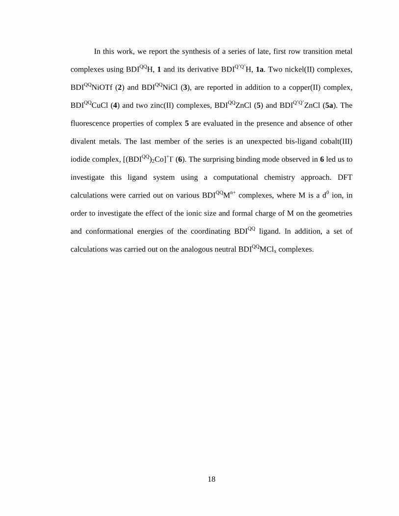
2.1 Synthetic route to complexes 2 and 3...……………………………………….…19
2.2 Synthetic route to complexes 4 and 5...……………………………………….…20
2.3 Synthetic route to complex 6...…………………………………………………..21
2.4 Synthetic route to compounds 1a and 5a...………………………………………23
2.5 UV-vis spectrum of complexes 2-5...……………………………………………24
2.6 Quantum yield determination plot...……………………………………………..26
2.7 Fluorescence emission spectra of complex 5...…………………………………..27
2.8 Fluorescence emission spectra of complex 1...…………………………………..28
2.9 ORTEP diagram of 4 and 5...…………………………………………………….29
2.10 Different binding modes observed in the metal complexes series using 1 as a
ligand……………………………………………………………………………..32
2.11 Ligand distortion energy versus Shannon effective 6-coordinate ionic radii of
various d0 metal ions...…………………………………………………………...34
viii
Chapter Three
3.1 1H NMR spectrum of Tol-BDI
(2-pp)2H, 1…..…………………………………..…53
3.2 1H NMR spectrum of rearranged ligand, 2 (Tol-BDI
(2-pp)(4-pp)H)………………..54
3.3 ORTEP diagram of [Tol-BDI(2-pp)
ZnI]2………………………………………….55
3.4 1H NMR spectrum of Tol-BDI
(2-pp)2ZnI, 3……………………………………….56
3.5 Electronic absorption spectrum of compounds 1 and 3………………………….57
3.6 ORTEP diagram of Tol-BDI(2-pp)2
ZnI, 3…………………………………………57
3.7 Electronic absorption spectrum of complexes 4 and 5…………………………..59
3.8 1H NMR spectrum of Tol-BDI
(2-pp)2Co, 5………………………………………..60
3.9 Cyclic voltammograms of BDI(2-pp)2
ZnI (3) & BDI(2-pp)2
CoCl (4)………………61
3.10 Cyclic voltammograms of 4 at various scan rates……………………………….62
3.11 1H NMR spectrum of Tol-BDI
(2-pp)2Co(CH3)I, 6………………………………...63
3.12 Electronic absorption spectrum of complexes 6 and 7…..………………………64
3.13 1H NMR spectrum of Tol-BDI
(2-pp)2Co(C2Si(CH3)3)I, 7…………………………65
3.14 1H NMR spectrum of Tol-BDI
(2-pp)2Co(CH=CH(CH3))Br, 8……………………66
Chapter Four
4.1 Examples of wide bite angle diphosphines…………………..…………………..81
4.2 Simplified catalytic cycle of the nickel-catalyzed hydrocyanation reaction…….82
4.3 Possible mechanism for the cis-trans isomerization of Xantphos……………….84
Chapter Five
5.1 Electronic absorption spectra of Zn 1, Co 2, Ni 3 and iPr
DPDBFphos...………...94
ix
5.2 Electronic absorption spectra of Zn 1, Co 2 and Ni 3.…………………...………94
5.3 1H NMR spectrum of [(
iPrDPDBFphos)ZnCl2] 1…………..………….…………95
5.4 Variable-temperature 1H NMR spectra of [trans-(
iPrDPDBFphos)NiCl2] 3...…..96
5.5 1H NMR spectrum of [(
iPrDPDBFphos)Fe(CO)3] 5...…………….……..…..…...98
5.6 Solid-state structures of [(iPr
DPDBFphos)ZnCl2] 1 and
[(iPr
DPDBFphos)CoCl2] 2…………………………..……………………………99
5.7 Solid-state structure of [(iPr
DPDBFphos)NiCl2] 3………….……………....…..102
5.8 Solid-state structure of [(iPr
DPDBFphos)Fe(CO)3] 5………..……………….....104
5.9 Cyclic voltammograms of (iPr
DPDBFphos)MCl2 1 – 4………………..……….105
5.10 Electronic absorption spectra of the nickel compounds 3 and 4………………..106
5.11 Solid-state structures of [(iPr
DPDBFphos)NiCl] 4………….………....………..106
5.12 X-band EPR spectrum (dX´´/dB) of [(iPr
DPDBFphos)NiCl] 4………………....109
5.13 Qualitative MO diagram of the d-orbital manifold derived from a DFT-BP86
calculations of [(iPr
DPDBFphos)NiCl] 4……………………………………….111
Chapter Six
6.1 Solid-state structure of the [(iPr
DPDBFphos)Ni{NH(2,6-(CHMe2)2C6H3)}] 2...125
6.2 31
P NMR spectrum of [(iPr
DPDBFphos)Ni(H)Cl] (4) from a crude reaction of 1
with H2(g)…...…………………………………………………………………..128
6.3 UV-vis spectrum of [(iPr
DPDBFphos)Ni(CHCH2)Cl], 5……………….……....131
6.4 Solid-state structure of [(iPr
DPDBFphos)Ni(CH=CH2)Cl] 5…………………...131
6.5 Stacked 1H NMR spectra of [(
iPrDPDBFphos)Ni(CHCH2)Cl], 5;
[(iPr
DPDBFphos)NiCl2], 3; and, the two-product mixture in the reaction of vinyl chloride
and [(iPr
DPDBFphos)NiCl]……………………………………………………………..132
x
List of Schemes
Chapter Three
3.1 Synthetic route to Tol-BDI(2-pp)2
H, 1…………………………………………….52
3.2 Synthetic routes to Tol-BDI(2-pp)2
Zn and Co complexes, 3-8…………………...55
Chapter Four
4.1 Synthetic route to diphosphine ligand iPr
DPDBFphos…………………………...85
Chapter Five
5.1 Synthetic routes to coordination complexes 1 to 4………………………………93
5.2 Fluxional processes in 3………………………………………………………...103
Chapter Five
6.1 Synthetic route for [(iPr
DPDBFphos)Ni{NH(2,6-(CHMe2)2C6H3)}] 2…...…….123
6.2 Synthetic route for [(iPr
DPDBFphos)Ni{NH(2,6-(CHMe2)2C6H3)}][PF6] 3…...126
6.3 Reaction of [(iPr
DPDBFphos)NiCl] 1 and vinyl chloride………………………133
xi
List of Charts
Chapter Five
3.1 Tetradentate monoanionic ligands with the current ligand shown in inset………51
Chapter Five
5.1 Ligands DBFphos and iPr
DPDBFphos…………………………………………...91
xii
List of Symbols and Abbreviations
1,1-DCE 1,2-dichloroethylene
Å Ångström
A hyperfine coupling constant
AgNO3 silver nitrate
atm atmosphere
BDI β-diketiminate
(BDIQQ
)H 1,3-bis(quinolyl)propenediimine or Zatka ligand
(BDIQ(SO3H)Q(SO3H)
)H 1,3-bis(quinolyl-5-sulfonic acid) propenediimine
BISBI 2,2’-bis(diphenylphosphinomethyl)biphenyl
br broad
C Celsius
cDCE cis-dichloroethylene
CE chlorinated ethylene
CDCl3 chloroform
CH2Cl2 methylene chloride
CD2Cl2 deuterated methylene chloride
CH3CN acetonitrile
CD3CN deuterated acetonitrile
CD3I deuterated methyl iodide
cm-1
wavenumber
COD cycloocta-1-5-diene
COSY correlation spectroscopy
CV cyclic voltammetry
d doublet
D2O deuterated water
DBFphos 4,6-bis(diphenylphopshino)dibenzofuran
Dc calculated density
DCE dichloroethylene
dd doublet of doublets
deg degree
DFT density functional theory
d6-DMSO deuterated dimethylsulfoxide
DMSO dimethylsulfoxide
dppe 1,2-bis(diphenylphosphino)ethane
dppf 1,1’-bis(diphenylphosphino)ferrocene
dt doublet of triplets
dtbpe 1,2-bis(di-tert-butylphosphino)ethane
δ chemical shift in ppm
EDTA ethylenediaminetetraacetic acid
EPA Environmental Protection Agency
EPR electron paramagnetic resonance
ESI-TOF MS electrospray ionization time of flight mass spectrometry
Et2O diethyl ether
xiii
eV electron volt
ε molar absorptivity
Fc ferrocene
g g factor
g gram
G Gauss
GC gas chromatograph
GC-EI-MS gas chromatography-electron impact-mass spectrometry
GC-MS gas chromatography-mass spectrometry
GHz gigahertz
GOOF goodness of fit
HCl hydrochloric acid
H3PO4 phosphoric acid
HMBC heteronuclear multiple bond correlation
HMQC heteronuclear multiple quantum correlation
HRMS high-resolution mass spectrometry
Hz hertz iPr
DPDBFphos 4,6-bis(3-diisopropylphosphinophenyl)dibenzofuran
IR infrared
J coupling constant
K Kelvin
KC8 potassium graphite
kcal kilocalorie
L liter
λex excitation wavelength
λmax wavelength absorbance maximum
LDE ligand distortion energy
M metal
M molar
m multiplet
MCL maximum contaminant level
Me methyl
MeOH methanol
Mg milligram
MHz megahertz
mL milliliter
MLCT metal to ligand charge transfer
mM millimolar
mmol millimole
MN-GFM minnesota gaussian functional module
mol mole
MO molecular orbital
mV millivolt
mW milliwatt
m/z mass-to-charge ratio
μB Bohr magneton

xiv
μeff effective magnetic moment
μS.O. spin-only magnetic moment
μ bridging
μL microliter
NaOH sodium hydroxide
[nBu4N]PF6 tetrabutylammonium hexafluorophophate
nm nanometer
NMR nuclear magnetic resonance
ORTEP Oak Ridge thermal ellipsoid plot
PCE perchloroethylne
PEG polyethylene glycol
PFK perfluorokerosene
PPG polypropylene glycol
ppm parts per million
θ degrees of data collection
π pi bonding orbital
r radius
reflns reflections
s singlet
S spin
sh shoulder
SHE standard hydrogen electode
SOMO singly occupied molecular orbital
SPANphos 8,8’-bis(diphenylphosphino)-4,4,4’,4’,6,6’-
hexamethylspiro-2,2’-bichroman
ζ sigma bonding orbital
t triplet
TCE trichloroethylene
tDCE trans-dichloroethylene
d8-THF deuterated tetrahydrofuran
THF tetrahydrofuran
Tol toluene
(Tol-BDI(2-pp)2
)H 2-(4-tolyl)-1,3-bis(2-isopropylpyridyl)propenediimine
TRANSphos 2,11-bis(diphenylphosphinomethyl) benzophenanthrene
UV ultraviolet
UV-vis ultraviolet-visible
V volt
V volume
VC vinyl chloride
VOC volatile organic compound
W line width
Xantphos 9,9-dimethyl-4,6-bis(diphenylphosphino)xanthene
Z number of independent structures in unit cell
1
-PART I-
Development of Cobalamin Model Complexes for the
Study of Reductive Dehalogenation

2
-Chapter One-
Introduction
3
1.1 Chlorinated solvents in the environment
Since the introduction of the Clean Water Act in 1977, the Environmental
Protection Agency (EPA) has been committed to ensuring the quality of our water
supply. Now, three decades later, this commitment has been renewed with aims to clean
up the water supply of contaminants. In January 2011, a new strategic goal was
implemented where contaminants were to be addressed as a group rather than individual
compounds. The first compounds to be targeted under this new initiative are carcinogenic
volatile organic compounds (VOCs). This came as no surprise, considering VOCs have
plagued our groundwater supply in abundance, due to their presence in such household
products as paints, adhesives, cleaning supplies, and plastics.1 In 2007, Squillace and co-
workers tested for the presence of 55 VOCs in groundwater from more than 5000 wells
across the country and found that two VOCs stood out in terms of their occurrences at
concentrations greater than or near their Maximum Contaminant Levels (MCLs).
Perchloroethylene (PCE) and trichloroethylene (TCE) were not only detected in 11% and
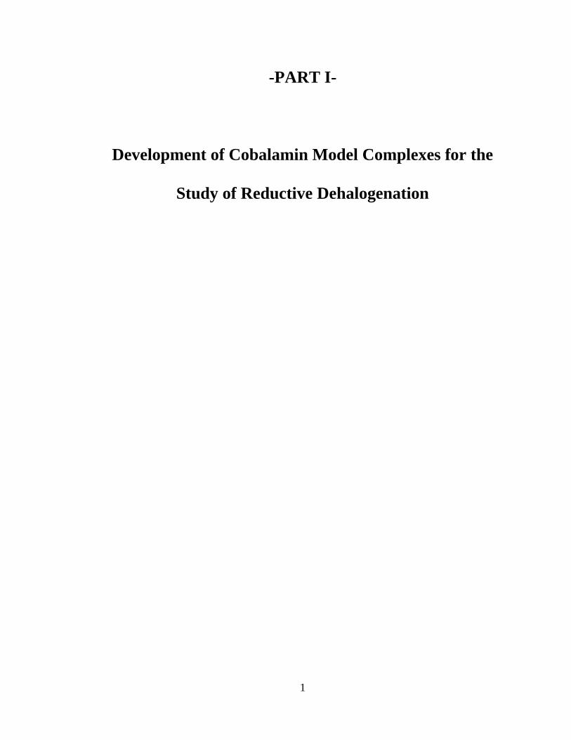
5% of the wells, respectively, but more importantly, they were ranked 1 and 3 in terms of
the frequency of concentration greater than MCLs (Figure 1.1).2
4
Figure 1.1. Occurrence of VOC in drinking water wells, relative to their MCLs.
Figure is taken from reference 2.
Although PCE and TCE were identified as priority pollutants by the EPA in 1977,
they have been an environmental nuisance for nearly a century, with records showing
cattle poisoning dating back to the early 1920s.3-4
While both chemicals have had a wide
range of uses, PCE was mainly used as a cleaning fluid in the dry-cleaning and textile
processing industries, and TCE’s most prominent uses were as a vapor degreasing solvent
in the metal industry and as an extraction solvent for natural fats and oils.3-4
Both
chemicals are still in use today as chemical precursors for the production of
fluorocarbons and hydrofluorocarbons, but their production and use have dramatically
declined.5 At their peak, PCE and TCE production reached 763 and 600 million pounds
5
respectively.3-4
In 2002, the annual production of PCE was down to 430 million pounds
while TCE has seen a larger decline to 330 million pounds in 2005.5
While the production and use of PCE and TCE have decreased, the damage
caused by chlorinated ethylenes (CEs) will take decades to repair. Both PCE and TCE are
anticipated to be human carcinogens.5-8
Human studies are showing incidences of liver
and kidney cancers and non-Hodgkin’s lymphoma from occupational exposure to TCE.5-8
For PCE there is limited data due to the exposure of dry-cleaning workers to a multitude
of solvents; however there is some evidence connecting esophageal cancer, cervical
cancer, and non-Hodgkin’s lymphoma to PCE exposure.5-7
Additionally, cancer studies in
animals show that PCE exposure causes benign and malignant liver and kidney tumors,
as well as mononuclear-cell leukemia.5 Further health concerns over PCE and TCE water
contamination are directed toward their less-chlorinated daughter products, such as the
known carcinogen vinyl chloride (VC).5, 9
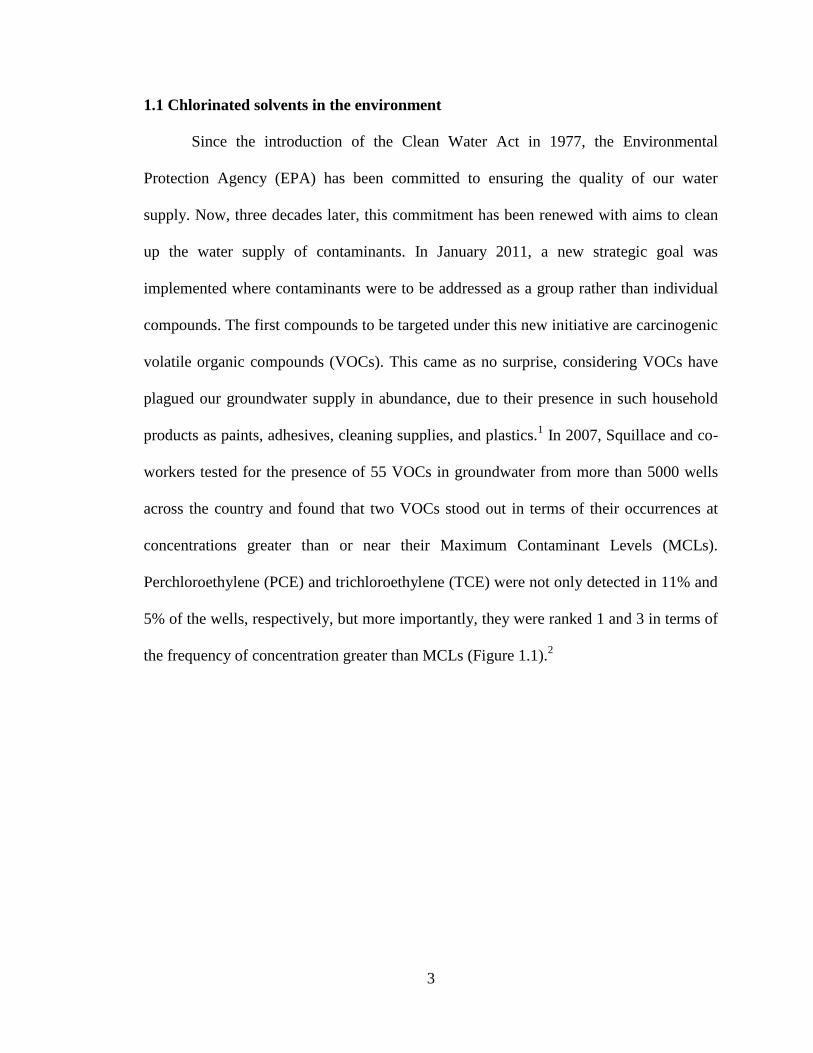
While partial reductive dechlorination of PCE
and TCE can lead to carcinogenic species, complete removal of the chlorine atoms results
in the production of the benign compound ethylene (Figure 1.2).
Figure 1.2. Sequential removal of chlorine atoms from PCE to ethylene.
6
1.2 Remediation of chlorinated ethylenes by cobalamin
As a result of their considerable health concerns and widespread contamination,
remediation strategies for water affected by CEs have been implemented. One
remediation strategy has focused on natural attenuation using microorganisms.10
Early
studies of microorganisms revealed the participation of metallocofactors, specifically
corrinoids, in the reductive dehalogenation of a variety of substrates.10-11
In 1991,
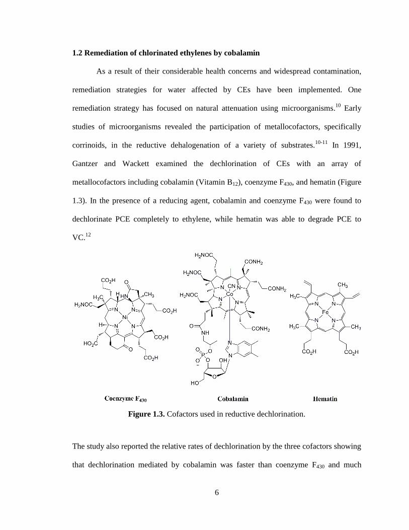
Gantzer and Wackett examined the dechlorination of CEs with an array of
metallocofactors including cobalamin (Vitamin B12), coenzyme F430, and hematin (Figure
1.3). In the presence of a reducing agent, cobalamin and coenzyme F430 were found to
dechlorinate PCE completely to ethylene, while hematin was able to degrade PCE to
VC.12
Figure 1.3. Cofactors used in reductive dechlorination.
The study also reported the relative rates of dechlorination by the three cofactors showing
that dechlorination mediated by cobalamin was faster than coenzyme F430 and much
7
faster than hematin.12
Recently it was revealed that all reductive dehalogenases, the
enzymes responsible for reductive dehalogenation, contain a corrinoid with the exception
of one that employs a heme.13
This observation further affirmed the role of cobalamin in
the process of reductive dehalogenation.
Subsequent studies have examined the kinetics of reductive dechlorination by
cobalamin and found discrepancies in the rates of dechlorination and distribution of
products. The rates of dechlorination seem highly dependent on the experimental
conditions such as pH, temperature, and the concentrations of bulk reagents and
cobalamin, resulting in a difficulty in comparing the various studies.14-20
While the rates
of dechlorination may vary between studies, the trends of dechlorination appear the same.
Higher chlorinated substrates react fastest, and rates slow down as the chlorine content
decreases, with the exception of the fast conversion of vinyl chloride to ethylene.12, 14-18
The slowest reaction is the dechlorination of cis-DCE to VC, resulting in a build-up of
cis-DCE in the environment.12, 14-18
In addition to rates of degradation, the products of dechlorination mediated by
cobalamin have also been highly debated, with the detection of acetylene products in
some studies and the different distributions of DCE products observed for the
dechlorination of TCE.18
These conflicting rates and products have led to an interest in
the mechanism of reductive dehalogenation mediated by cobalamin and have contributed
to vastly different proposed mechanisms, including inner vs. outer-sphere electron
transfer, the presence of radicals, and the formation of organocobalt intermediates.21-33
A
recent review by Kliegman and McNeill evaluated the mechanistic studies on cobalamin-
mediated dehalogenation using both cobalamin and its model complexes.18
Seeing that
8
the review highlighted the importance of the cobalt-vinyl intermediate and the lack of
understanding on the involvement of radicals, it prompted a study on the role of free
radicals investigated by radical traps.18
Interestingly, the study showed that the reaction
did not solely proceed by an inner or outer-sphere mechanism and hypothesized the
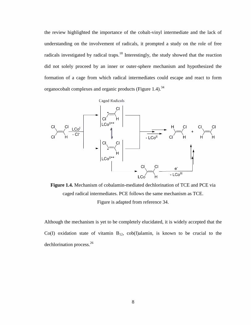
formation of a cage from which radical intermediates could escape and react to form
organocobalt complexes and organic products (Figure 1.4).34
Figure 1.4. Mechanism of cobalamin-mediated dechlorination of TCE and PCE via
caged radical intermediates. PCE follows the same mechanism as TCE.
Figure is adapted from reference 34.
Although the mechanism is yet to be completely elucidated, it is widely accepted that the
Co(I) oxidation state of vitamin B12, cob(I)alamin, is known to be crucial to the
dechlorination process.26
9
1.3 Important characteristics of cob(I)alamin
Reductive dehalogenation of halogenated substrates depends on the reducing
capacity of the transition metal complex. The potential intermediates formed and the
distribution of products are all influenced by this parameter. Furthermore, it has been
shown that the reduction potential, when correlated with product ratio in the reduction of
TCE, can be used as a probe for the mechanism.22
As previously mentioned, cob(I)alamin has been found to play a central role in the
process of reductive dehalogenation. The CoI/II
couple observed for cobalamin is -0.59 V
(vs. SHE), making cob(I)alamin a strong reductant.35-39
This is one characteristic that
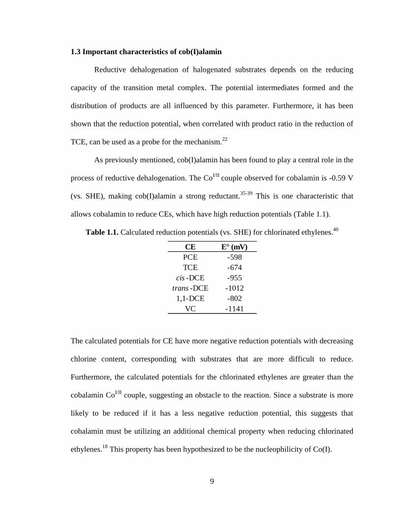
allows cobalamin to reduce CEs, which have high reduction potentials (Table 1.1).
Table 1.1. Calculated reduction potentials (vs. SHE) for chlorinated ethylenes.40
The calculated potentials for CE have more negative reduction potentials with decreasing
chlorine content, corresponding with substrates that are more difficult to reduce.
Furthermore, the calculated potentials for the chlorinated ethylenes are greater than the
cobalamin CoI/II
couple, suggesting an obstacle to the reaction. Since a substrate is more
likely to be reduced if it has a less negative reduction potential, this suggests that
cobalamin must be utilizing an additional chemical property when reducing chlorinated
ethylenes.18
This property has been hypothesized to be the nucleophilicity of Co(I).
CE E° (mV)
PCE -598
TCE -674
cis -DCE -955
trans -DCE -1012
1,1-DCE -802
VC -1141
10
Due to its high nucleophilicity, the cobalt center in cob(I)amin has been referred
to as a “supernucleophile.”41-42
Nucleophilicity has been quantified by Pearson et al. and
is defined as the log of the second order rate constant for the attack of the substrate by a
specific nucleophile divided by the second order rate constant for the attack of MeOH by
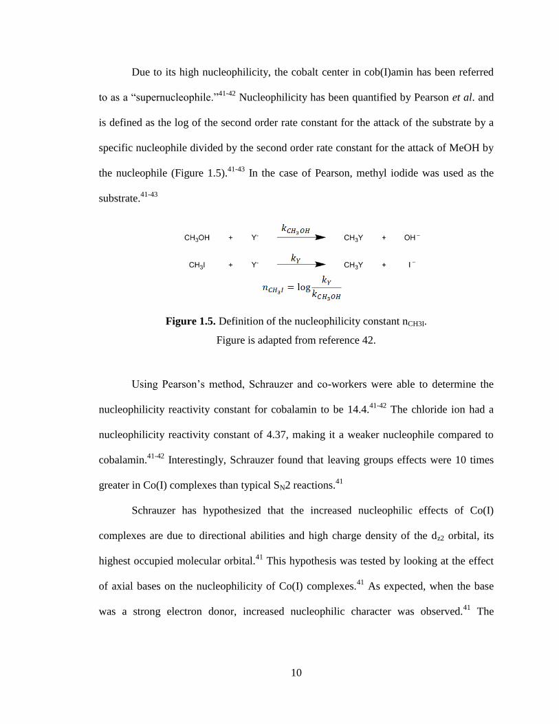
the nucleophile (Figure 1.5).41-43
In the case of Pearson, methyl iodide was used as the
substrate.41-43
Figure 1.5. Definition of the nucleophilicity constant nCH3I.
Figure is adapted from reference 42.
Using Pearson’s method, Schrauzer and co-workers were able to determine the
nucleophilicity reactivity constant for cobalamin to be 14.4.41-42
The chloride ion had a
nucleophilicity reactivity constant of 4.37, making it a weaker nucleophile compared to
cobalamin.41-42
Interestingly, Schrauzer found that leaving groups effects were 10 times
greater in Co(I) complexes than typical SN2 reactions.41
Schrauzer has hypothesized that the increased nucleophilic effects of Co(I)
complexes are due to directional abilities and high charge density of the dz2 orbital, its
highest occupied molecular orbital.41
This hypothesis was tested by looking at the effect
of axial bases on the nucleophilicity of Co(I) complexes.41
As expected, when the base
was a strong electron donor, increased nucleophilic character was observed.41
The
11
nucleophilic character of cob(I)alamin is believed to work in tandem with its
electrochemical properties to allow for the reduction of chlorinated ethylenes.
12
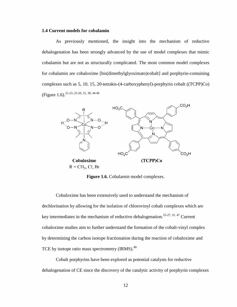
1.4 Current models for cobalamin
As previously mentioned, the insight into the mechanism of reductive
dehalogenation has been strongly advanced by the use of model complexes that mimic
cobalamin but are not as structurally complicated. The most common model complexes
for cobalamin are cobaloxime [bis(dimethylglyoximato)cobalt] and porphyrin-containing
complexes such as 5, 10, 15, 20-tetrakis-(4-carboxyphenyl)-porphyrin cobalt ((TCPP)Co)
(Figure 1.6).21-23, 25-26, 31, 38, 44-46
Figure 1.6. Cobalamin model complexes.
Cobaloxime has been extensively used to understand the mechanism of
dechlorination by allowing for the isolation of chlorovinyl cobalt complexes which are
key intermediates in the mechanism of reductive dehalogenation.25-27, 31, 47
Current
cobaloxime studies aim to further understand the formation of the cobalt-vinyl complex
by determining the carbon isotope fractionation during the reaction of cobaloxime and
TCE by isotope ratio mass spectrometry (IRMS).48
Cobalt porphyrins have been explored as potential catalysts for reductive
dehalogenation of CE since the discovery of the catalytic activity of porphyrin complexes
13
toward the dechlorination of chloromethanes.49
Dror and Schlautmann followed with the
dechlorination of PCE using water-soluble cobalt, iron and nickel porphyrins.44, 50
This
prompted a more detailed kinetic and mechanistic study by Fritsch and McNeill,
investigating the dechlorination of PCE, TCE, cDCE, and tDCE by a survey of cobalt
porphyrins complexes, including (TCPP)Co.45-46
Remarkably, (TCPP)Co was found to
have dechlorination rates superior to those of cobalamin.45
While cobaloxime and cobalt porphyrin complexes have been crucial to
understanding the mechanism of reductive dehalogenation and developing better
catalysts, both systems mimic the tetraaza coordination environment of the corrin but do
not match its monoanionic charge. This leaves room for improvement in the development
of a cobalamin model complex.
14
1.5 Scope of thesis
The objective of the first part of this dissertation is to synthesize a cobalamin
model complex that can mimic both the monoanionic charge and the tetraaza
coordination environment of the cobalamin corrin while demonstrating reactivity toward
C-X bonds. In the second chapter, the monoanionic tetradentate β-diketiminate ligand
(BDIQQ
H) is evaluated as a possible corrin model. Through the preparation of a series of
first row transition metal complexes and DFT calculations, it is shown that the ligand is
poorly suited to be model for the corrin due to its inflexibility, preventing it from
accommodating different ionic radii and thus stabilizing the different oxidation states of
cobalt. The third chapter is dedicated to the synthesis and characterization of an
improved, more flexible β-diketiminate ligand, Tolyl-BDI(2-pp)2
H. An isolable Co(I)
complex is prepared in this chapter and is shown to be comparable to cobalamin(I) both
in terms of its electrochemistry and reactivity toward C-X bonds.
15
-Chapter Two-
Metal Ion Size and Coordination Mode in Complexes of a
β-Diketiminate Ligand with Pendant Quinoline Arms
In part from:
Marlier, E. E.; Sadowsky, D.; Cramer, C.; McNeill, K. Inorg. Chim. Acta, 2011, 369,
173-179.
16
2.1 Overview
A suite of late first row transition metal complexes has been synthesized using a
monoanionic nitrogen donor β-diketiminate ligand with quinolyl pendant arms, BDIQQ
H
(1). BDIQQ
NiOTf (2), BDIQQ
CuCl (4), BDIQQ
ZnCl (5) were prepared from the reaction of
1 with Ni(OTf)2, CuCl2·2H2O and ZnCl2 respectively. BDIQQ
NiCl (3) was synthesized
from an anion exchange of 2 with nBu4NCl. Reaction of 1 and CoI2 afforded the
unexpected [(BDIQQ
)2Co]+I- (6). To increase the water solubility of these complexes, a
derivative of the BDIQQ
H ligand, BDIQ’Q’
H (1a) was synthesized by substituting a
hydrogen on each quinolyl pendant arm with a sulfonic acid group. The derivative ligand
was used to synthesize BDIQ’Q’
ZnCl (5a) from 1a and zinc chloride. Through density
functional theory (DFT) calculations, ligand geometries in BDIQQ
complexes were
investigated and it was found that smaller ionic radius and higher charge destabilize 1:1
metal-ligand complexes relative to alternative 1:2 complexes like 6 owing to significant
conformational strain in 1:1 complexes involving metals with small ionic radii. Synthesis
and characterization of these complexes, including crystal structures of 4 and 5, are
reported, in addition to the results of DFT calculations.
17
2.2 Introduction
In recent years, reports of β-diketiminate (BDI) complexes have increased
dramatically, spanning the entire periodic table from main group elements to
lanthanides.1 While most BDI examples are monoanionic bidentate ligands, there are
only a handful of examples of monoanionic tridentate and tetradenate BDIs. For example,
nitrogen and mixed atom donor monoanionic tridentate BDIs have been employed in
applications involving polymer synthesis, such as the ring opening polymerization of
lactide.2-3
The current repertoire of monoanionic tetradentate nitrogen donor BDI ligands
is far less expansive.4 The first example of which we are aware appeared in 1971 when
Zatka and co-workers prepared a ligand with quinolyl groups as pendant arms on a
simple β-diketiminate backbone.5 Roesky and co-workers also used a β-diketiminato
derivative with substituted ethylene diamine pendant arms to prepare a variety of
lanthanides and early transition metal complexes.6-9
We previously reported a group of
divalent metal complexes using a similar ligand to Zatka, differing by the presence of a
tolyl group on the central carbon of the BDI backbone.10
Our prolonged interest in monoanionic tetradentate nitrogen donor BDI ligands
stems from their potential as models for the corrin in vitamin B12 and the corphin in
cofactor F430. BDI ligands with pendant arms are potentially well suited for this task due
to their ability to mimic and simplify the coordination environment of these two
macrocycles as well as offer flexibility via facile modification of their pendant arms. In
an effort to understand their utility as models, we have investigated the metal binding
properties of the previously mentioned Zatka ligand, BDIQQ
H, by preparing a suite of
metal complexes.
18
In this work, we report the synthesis of a series of late, first row transition metal
complexes using BDIQQ
H, 1 and its derivative BDIQ’Q’
H, 1a. Two nickel(II) complexes,
BDIQQ
NiOTf (2) and BDIQQ
NiCl (3), are reported in addition to a copper(II) complex,
BDIQQ
CuCl (4) and two zinc(II) complexes, BDIQQ
ZnCl (5) and BDIQ’Q’
ZnCl (5a). The
fluorescence properties of complex 5 are evaluated in the presence and absence of other
divalent metals. The last member of the series is an unexpected bis-ligand cobalt(III)
iodide complex, [(BDIQQ
)2Co]+I- (6). The surprising binding mode observed in 6 led us to
investigate this ligand system using a computational chemistry approach. DFT
calculations were carried out on various BDIQQ
Mn+
complexes, where M is a d0 ion, in
order to investigate the effect of the ionic size and formal charge of M on the geometries
and conformational energies of the coordinating BDIQQ
ligand. In addition, a set of
calculations was carried out on the analogous neutral BDIQQ
MClx complexes.
19
2.3 Results & discussion
Synthesis and Characterization of Metal Complexes
Compound 1, BDIQQ
H was synthesized according to a literature procedure by
Zatka et al. in 99% yield.5 Compound 1 was fully characterized using
1H NMR,
13C
NMR, UV-vis spectroscopy and high resolution mass spectrometry (HRMS). The 1H
NMR spectrum contains a doublet at 12.65 ppm, indicative of the amine proton and eight
distinct resonances in the aromatic region. The elemental composition of 1 was
confirmed by HRMS using a polyethylene glycol (PEG) exact mass internal standard.
Compound 1 was reacted with late first row metals to give a suite of metal complexes.
Compound 2, BDIQQ
NiOTf was synthesized by reacting compound 1 with
potassium bis(trimethylsilyl)amide, followed by addition of nickel triflate under inert
atmosphere. The complex was isolated in 25% yield (Figure 2.1).
Figure 2.1. Synthetic route to complexes 2 and 3.
An anion exchange with tetrabutylammonium chloride was carried out to give compound
3, BDIQQ
NiCl, in 98% yield. Both nickel complexes were characterized by HRMS and
1H NMR,
13C NMR, IR and UV-vis spectroscopy. The elemental composition for 2 and 3
was verified by HRMS using a PEG exact mass internal standard. Both 1H NMR spectra
showed the disappearance of the amine peak and an upfield shift (from 7.13 ppm to 5.70
ppm) of the resonance corresponding to the proton on the central carbon of the BDI

20
backbone. The IR spectrum of 2 revealed four bands associated with a triflate,
specifically 1267cm-1
(SO3 asym. stretch), 1219 cm-1
(CF3 sym. stretch), 1141 cm-1
(CF3
asym. stretch) and 1027 cm-1
(SO3 sym. stretch). The strong asymmetric stretch for the
sulfonyl group at 1267 cm-1
indicates the presence of an ionic triflate, as it falls within the
expected ionic triflate range of 1235 cm-1
to 1288 cm-.1 11-12
Conductivity measurements
of 2 and 3 revealed that both complexes were weak electrolytes, leading us to
hypothesize that the triflate ion is weakly coordinated. Considering the similarity between
the UV-vis absorbance and 1H NMR spectra of 3 and those of an analogous zinc complex
5 (described below), we believe the chlorine is bound in the case of 3.
The syntheses of 4, BDIQQ
CuCl, and 5, BDIQQ
ZnCl, were carried out in identical
manner. Ligand 1 was reacted with either copper chloride or zinc chloride in the presence
of three equivalents of 1.0 M sodium hydroxide (Figure 2.2).
Figure 2.2. Synthetic route to complexes 4 and 5.
Compound 4 was isolated in 40% yield and was characterized by HRMS, UV-vis
spectroscopy and X-ray crystallography. The elemental composition of 4 was confirmed
by HRMS using a PEG exact mass internal standard. X-ray quality crystals of 4 were
grown from layering a methanol solution of 4 with diethyl ether. The geometry around
copper was found to be distorted square pyramidal with a tau value of 0.39.13
Compound
5 was isolated in 73% yield and was characterized by the same techniques as 4 as well as
21
1H NMR and
13C NMR spectroscopy. The elemental composition of 5 was verified by
HRMS using a PEG exact mass internal standard. Crystals of 5 suitable for X-ray
crystallography were grown using the same method as 4 and also showed a similar
geometry where 5 can be described as a distorted square pyramid with a tau value of
0.36.13
In the 1H NMR spectrum of 5, the disappearance of the resonance for the amine
proton and a nearly 2 ppm upfield shift of the resonance belonging to the proton of the
central carbon of the BDI backbone can be observed.
To complete the series of metal complexes, we attempted to synthesize a
cobalt(II) chloride complex. Although HRMS indicated the presence of this complex
using the same method as 4 and 5, we were unable to isolate it in pure form. When
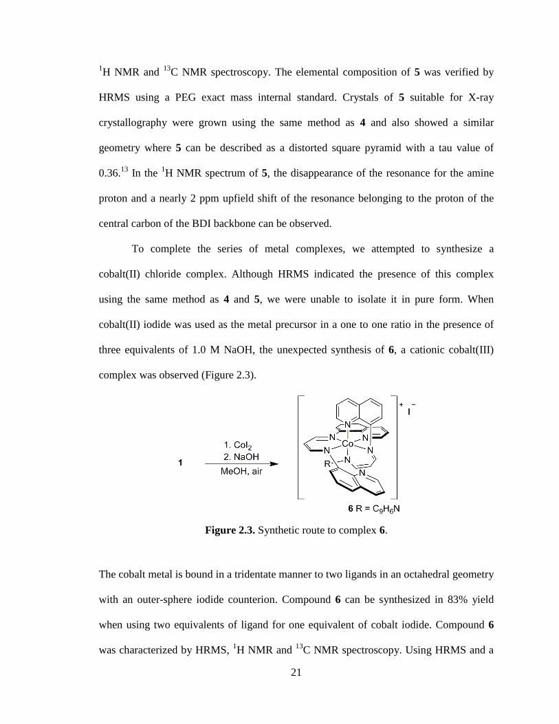
cobalt(II) iodide was used as the metal precursor in a one to one ratio in the presence of
three equivalents of 1.0 M NaOH, the unexpected synthesis of 6, a cationic cobalt(III)
complex was observed (Figure 2.3).
Figure 2.3. Synthetic route to complex 6.
The cobalt metal is bound in a tridentate manner to two ligands in an octahedral geometry
with an outer-sphere iodide counterion. Compound 6 can be synthesized in 83% yield
when using two equivalents of ligand for one equivalent of cobalt iodide. Compound 6
was characterized by HRMS, 1H NMR and
13C NMR spectroscopy. Using HRMS and a
22
PEG exact mass internal standard, the elemental composition of 6 was verified. In the 1H
NMR spectrum, a total of 13 distinct and two overlapping peaks were present caused by
the break of symmetry due to the bound and unbound quinolines. An upfield triplet at
5.21 ppm was observed for the proton on the central carbon of the BDI backbone.
Considering the spectroscopic similarity between bound and unbound quinolines, 2D
correlation spectroscopy (COSY) was used to further verify the assignment of each shift
to a specific proton. These assignments are presented in the experimental procedure
section. An X-ray diffraction experiment was conducted with crystals of 6 that were
obtained from a concentrated THF solution. While the data set was of insufficient quality
for publication (R = 0.11), the connectivity was rigorously established.
In an effort to increase the water solubility of the metal complexes, a modification
on the BDIQQ
H ligand was introduced. A sulfonic acid group was substituted on each
pendant quinolyl groups to create the ligand derivative BDIQ’Q’
H (1a) (Figure 2.4).
Compound 1a was synthesized in quantitative yield by reacting 8-aminoquinoline-5-
sulfonic acid with 1,1,3,3-tetramethoxypropane in a 1:1 mixture of MeOH and HCl. It
was characterized by 1H NMR spectroscopy and HRMS. In the
1H spectrum, a total eight
resonances are observed including a doublet at 12.64 ppm, belonging to the amine proton.
The elemental composition of 1a was verified by HRMS using a polyethylene glycol
(PEG) exact mass internal standard. Compound 1a was reacted with zinc chloride to give
BDIQ’Q’
ZnCl (5a) at near quantitative yield, which was characterized by HRMS, 1H
NMR and UV-vis spectroscopy (Figure 2.4).
23
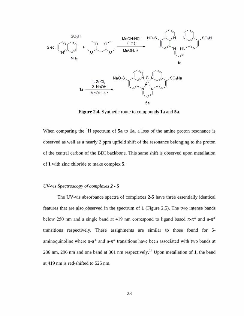
Figure 2.4. Synthetic route to compounds 1a and 5a.
When comparing the 1H spectrum of 5a to 1a, a loss of the amine proton resonance is
observed as well as a nearly 2 ppm upfield shift of the resonance belonging to the proton
of the central carbon of the BDI backbone. This same shift is observed upon metallation
of 1 with zinc chloride to make complex 5.
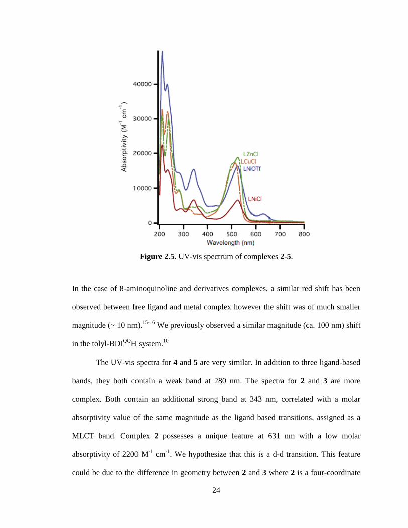
UV-vis Spectroscopy of complexes 2 - 5
The UV-vis absorbance spectra of complexes 2-5 have three essentially identical
features that are also observed in the spectrum of 1 (Figure 2.5). The two intense bands
below 250 nm and a single band at 419 nm correspond to ligand based π-π* and n-π*
transitions respectively. These assignments are similar to those found for 5-
aminoquinoline where π-π* and n-π* transitions have been associated with two bands at
286 nm, 296 nm and one band at 361 nm respectively.14
Upon metallation of 1, the band
at 419 nm is red-shifted to 525 nm.
24
Figure 2.5. UV-vis spectrum of complexes 2-5.
In the case of 8-aminoquinoline and derivatives complexes, a similar red shift has been
observed between free ligand and metal complex however the shift was of much smaller
magnitude (~ 10 nm).15-16
We previously observed a similar magnitude (ca. 100 nm) shift
in the tolyl-BDIQQ
H system.10
The UV-vis spectra for 4 and 5 are very similar. In addition to three ligand-based
bands, they both contain a weak band at 280 nm. The spectra for 2 and 3 are more
complex. Both contain an additional strong band at 343 nm, correlated with a molar
absorptivity value of the same magnitude as the ligand based transitions, assigned as a
MLCT band. Complex 2 possesses a unique feature at 631 nm with a low molar
absorptivity of 2200 M-1
cm-1
. We hypothesize that this is a d-d transition. This feature
could be due to the difference in geometry between 2 and 3 where 2 is a four-coordinate
25
cation with a weakly coordinating anion and 3 is hypothesized to be a neutral five-
coordinate complex similar to 4 and 5. Lastly, the molar absorptivity values for the
visible absorbances of these complexes are in the range of 104 M
-1 cm
-1. Although these
are intense, these values are expected for aminoquinoline complexes.10, 15, 17
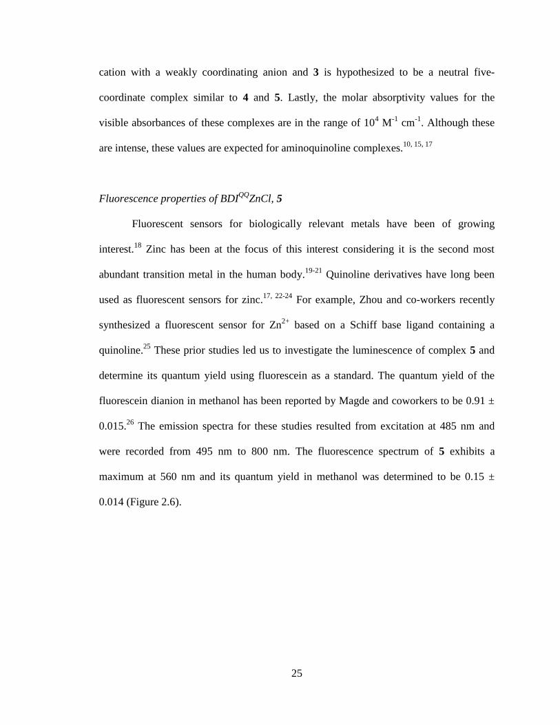
Fluorescence properties of BDIQQ
ZnCl, 5
Fluorescent sensors for biologically relevant metals have been of growing
interest.18
Zinc has been at the focus of this interest considering it is the second most
abundant transition metal in the human body.19-21
Quinoline derivatives have long been
used as fluorescent sensors for zinc.17, 22-24
For example, Zhou and co-workers recently
synthesized a fluorescent sensor for Zn2+
based on a Schiff base ligand containing a
quinoline.25
These prior studies led us to investigate the luminescence of complex 5 and
determine its quantum yield using fluorescein as a standard. The quantum yield of the
fluorescein dianion in methanol has been reported by Magde and coworkers to be 0.91 ±
0.015.26
The emission spectra for these studies resulted from excitation at 485 nm and
were recorded from 495 nm to 800 nm. The fluorescence spectrum of 5 exhibits a
maximum at 560 nm and its quantum yield in methanol was determined to be 0.15 ±
0.014 (Figure 2.6).
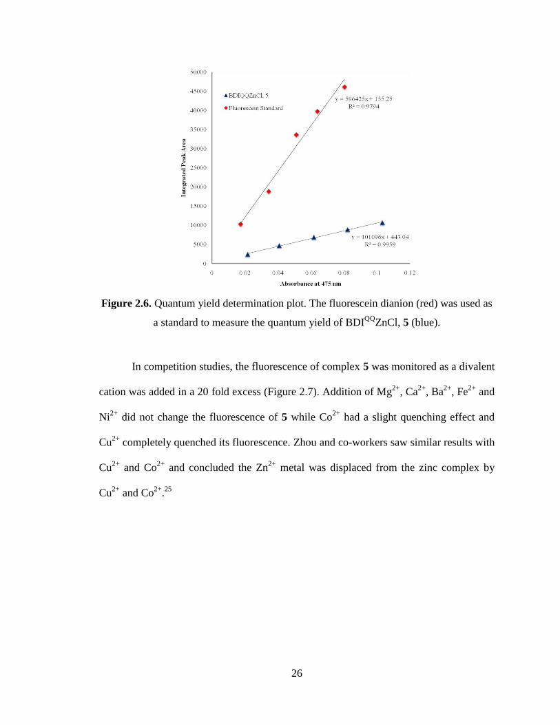
26
Figure 2.6. Quantum yield determination plot. The fluorescein dianion (red) was used as
a standard to measure the quantum yield of BDIQQ
ZnCl, 5 (blue).
In competition studies, the fluorescence of complex 5 was monitored as a divalent
cation was added in a 20 fold excess (Figure 2.7). Addition of Mg2+
, Ca2+
, Ba2+
, Fe2+
and
Ni2+
did not change the fluorescence of 5 while Co2+
had a slight quenching effect and
Cu2+
completely quenched its fluorescence. Zhou and co-workers saw similar results with
Cu2+
and Co2+
and concluded the Zn2+
metal was displaced from the zinc complex by
Cu2+
and Co2+
.25
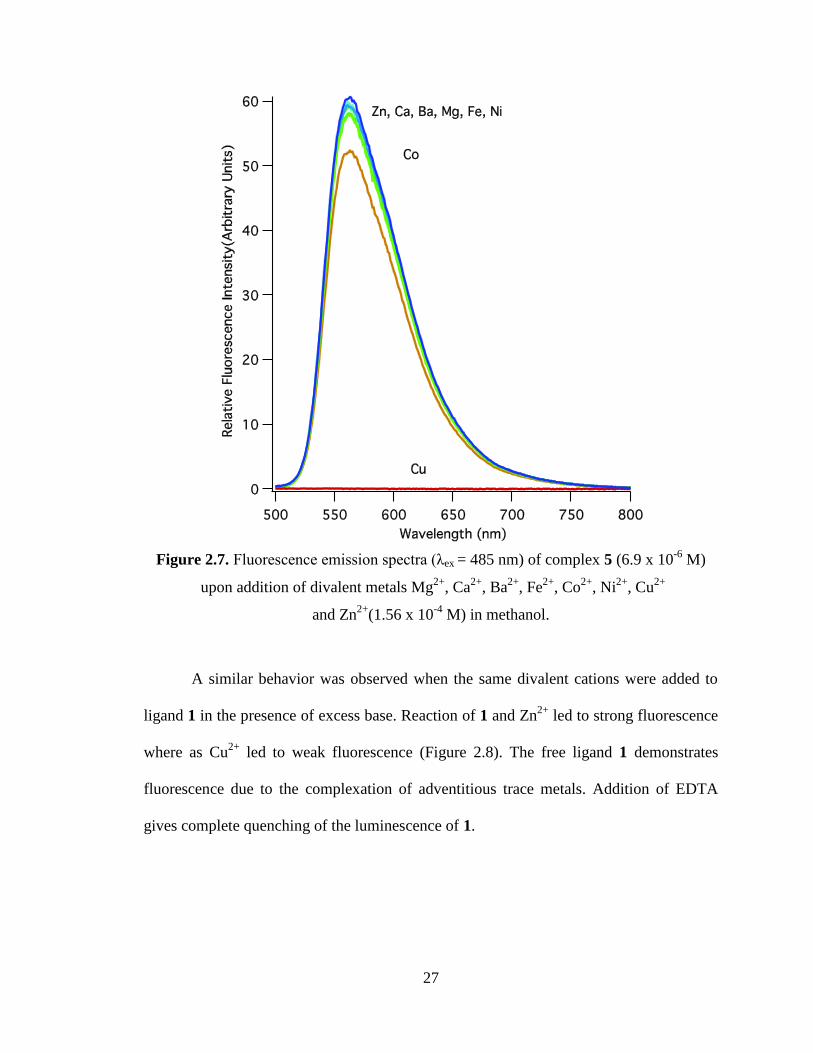
27
Figure 2.7. Fluorescence emission spectra (λex = 485 nm) of complex 5 (6.9 x 10⁻⁶
M)
upon addition of divalent metals Mg2+
, Ca2+
, Ba2+
, Fe2+
, Co2+
, Ni2+
, Cu2+
and Zn2+
(1.56 x 10⁻⁴
M) in methanol.
A similar behavior was observed when the same divalent cations were added to
ligand 1 in the presence of excess base. Reaction of 1 and Zn2+
led to strong fluorescence
where as Cu2+
led to weak fluorescence (Figure 2.8). The free ligand 1 demonstrates
fluorescence due to the complexation of adventitious trace metals. Addition of EDTA
gives complete quenching of the luminescence of 1.
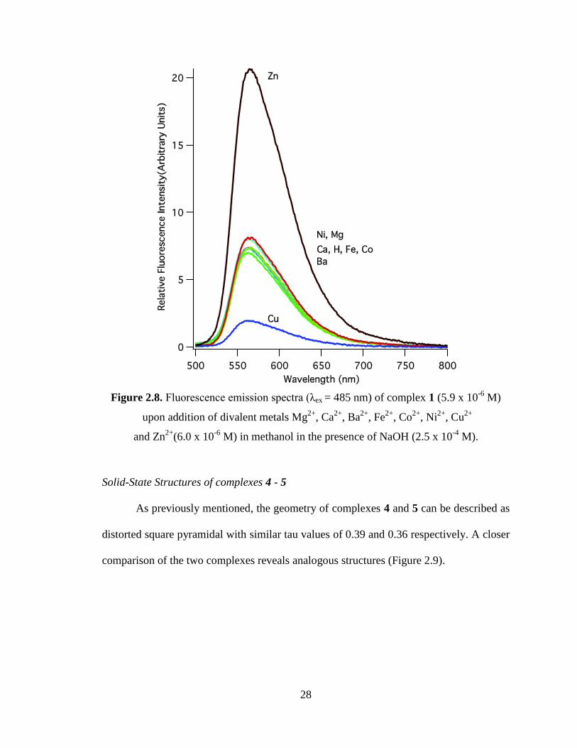
28
Figure 2.8. Fluorescence emission spectra (λex = 485 nm) of complex 1 (5.9 x 10⁻⁶
M)
upon addition of divalent metals Mg2+
, Ca2+
, Ba2+
, Fe2+
, Co2+
, Ni2+
, Cu2+
and Zn2+
(6.0 x 10⁻⁶
M) in methanol in the presence of NaOH (2.5 x 10⁻⁴
M).
Solid-State Structures of complexes 4 - 5
As previously mentioned, the geometry of complexes 4 and 5 can be described as
distorted square pyramidal with similar tau values of 0.39 and 0.36 respectively. A closer
comparison of the two complexes reveals analogous structures (Figure 2.9).
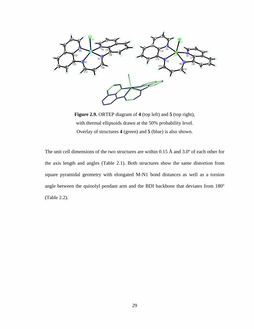
29
Figure 2.9. ORTEP diagram of 4 (top left) and 5 (top right),
with thermal ellipsoids drawn at the 50% probability level.
Overlay of structures 4 (green) and 5 (blue) is also shown.
The unit cell dimensions of the two structures are within 0.15 Å and 3.0º of each other for
the axis length and angles (Table 2.1). Both structures show the same distortion from
square pyramidal geometry with elongated M-N1 bond distances as well as a torsion
angle between the quinolyl pendant arm and the BDI backbone that deviates from 180º
(Table 2.2).
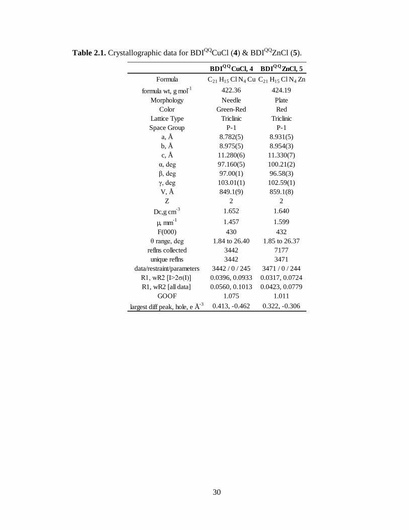
30
Table 2.1. Crystallographic data for BDIQQ
CuCl (4) & BDIQQ
ZnCl (5).
BDIQ Q
CuCl, 4 BDIQ Q
ZnCl, 5
Formula C21 H15 Cl N4 Cu C21 H15 Cl N4 Zn
formula wt, g mol-1 422.36 424.19
Morphology Needle Plate
Color Green-Red Red
Lattice Type Triclinic Triclinic
Space Group P-1 P-1
a, Å 8.782(5) 8.931(5)
b, Å 8.975(5) 8.954(3)
c, Å 11.280(6) 11.330(7)
α, deg 97.160(5) 100.21(2)
β, deg 97.00(1) 96.58(3)
γ, deg 103.01(1) 102.59(1)
V, Å 849.1(9) 859.1(8)
Z 2 2
Dc,g cm-3 1.652 1.640
µ, mm-1 1.457 1.599
F(000) 430 432
θ range, deg 1.84 to 26.40 1.85 to 26.37
reflns collected 3442 7177
unique reflns 3442 3471
data/restraint/parameters 3442 / 0 / 245 3471 / 0 / 244
R1, wR2 [I>2ζ(I)] 0.0396, 0.0933 0.0317, 0.0724
R1, wR2 [all data] 0.0560, 0.1013 0.0423, 0.0779
GOOF 1.075 1.011
largest diff peak, hole, e Å-3 0.413, -0.462 0.322, -0.306
31
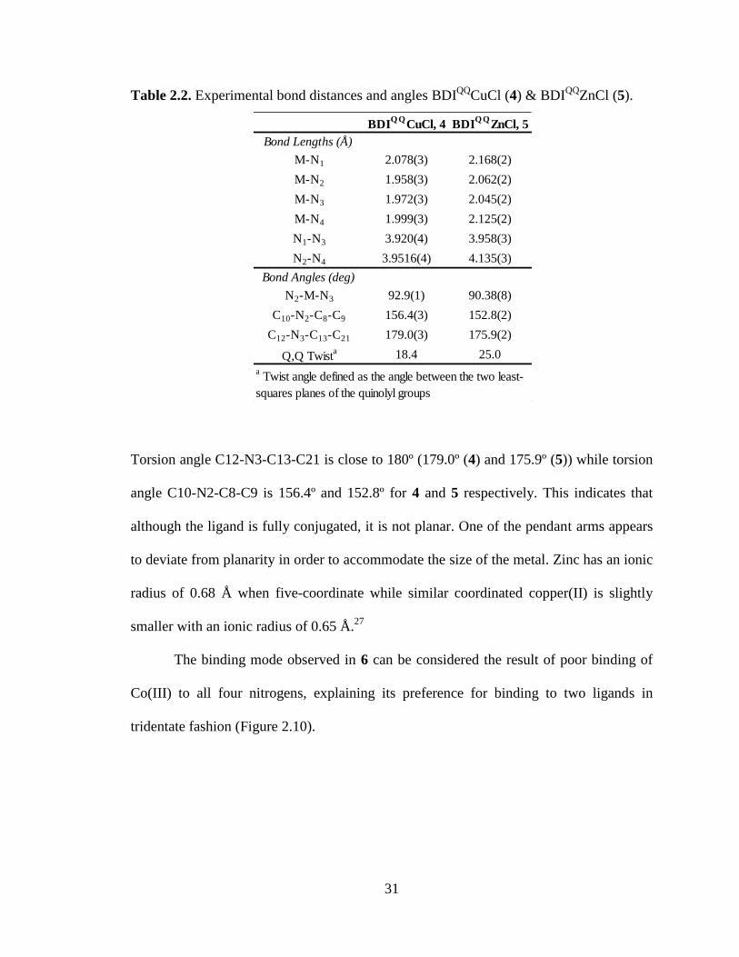
Table 2.2. Experimental bond distances and angles BDIQQ
CuCl (4) & BDIQQ
ZnCl (5).
Torsion angle C12-N3-C13-C21 is close to 180º (179.0º (4) and 175.9º (5)) while torsion
angle C10-N2-C8-C9 is 156.4º and 152.8º for 4 and 5 respectively. This indicates that
although the ligand is fully conjugated, it is not planar. One of the pendant arms appears
to deviate from planarity in order to accommodate the size of the metal. Zinc has an ionic
radius of 0.68 Å when five-coordinate while similar coordinated copper(II) is slightly
smaller with an ionic radius of 0.65 Å.27
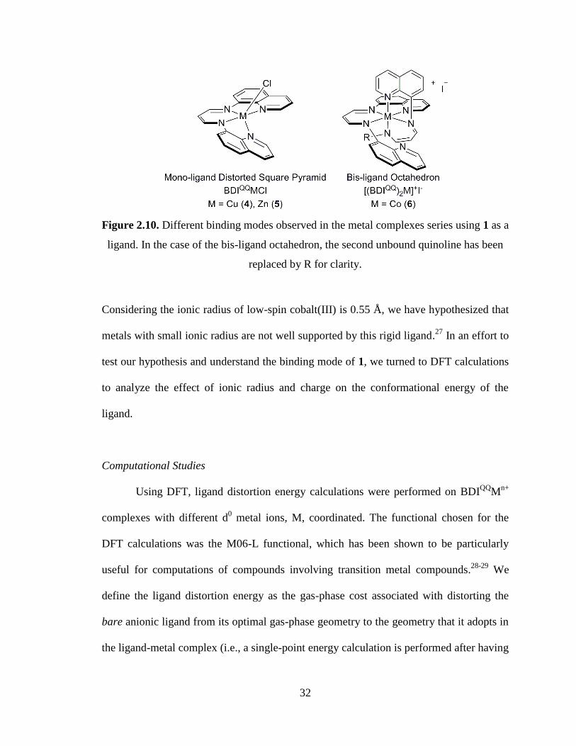
The binding mode observed in 6 can be considered the result of poor binding of
Co(III) to all four nitrogens, explaining its preference for binding to two ligands in
tridentate fashion (Figure 2.10).
BDIQ Q
CuCl, 4 BDIQ Q
ZnCl, 5
Bond Lengths (Å)
M-N1 2.078(3) 2.168(2)
M-N2 1.958(3) 2.062(2)
M-N3 1.972(3) 2.045(2)
M-N4 1.999(3) 2.125(2)
N1-N3 3.920(4) 3.958(3)
N2-N4 3.9516(4) 4.135(3)
Bond Angles (deg)
N2-M-N3 92.9(1) 90.38(8)
C10-N2-C8-C9 156.4(3) 152.8(2)
C12-N3-C13-C21 179.0(3) 175.9(2)
Q,Q Twista 18.4 25.0
a Twist angle defined as the angle between the two least-
squares planes of the quinolyl groups
32
Figure 2.10. Different binding modes observed in the metal complexes series using 1 as a
ligand. In the case of the bis-ligand octahedron, the second unbound quinoline has been
replaced by R for clarity.
Considering the ionic radius of low-spin cobalt(III) is 0.55 Å, we have hypothesized that
metals with small ionic radius are not well supported by this rigid ligand.27
In an effort to
test our hypothesis and understand the binding mode of 1, we turned to DFT calculations
to analyze the effect of ionic radius and charge on the conformational energy of the
ligand.
Computational Studies
Using DFT, ligand distortion energy calculations were performed on BDIQQ
Mn+
complexes with different d0 metal ions, M, coordinated. The functional chosen for the
DFT calculations was the M06-L functional, which has been shown to be particularly
useful for computations of compounds involving transition metal compounds.28-29
We
define the ligand distortion energy as the gas-phase cost associated with distorting the
bare anionic ligand from its optimal gas-phase geometry to the geometry that it adopts in
the ligand-metal complex (i.e., a single-point energy calculation is performed after having
33
removed the metal from the optimized geometry of the complex). Ligand distortion
energy calculations were correlated with Shannon effective ionic radii in order to
facilitate quantitative comparison across a broad spectrum of metals not limited to
transition metals.27
Six-coordinate radii were used in all cases for consistent comparisons
as they are available for all ions examined. By selecting metals from four columns of the
periodic table, we anticipate that differences in electrostatics will be minimized beyond
second-order effects coupled to conformational strain associated with changing ionic
radii.
The results for the Group 1 BDIQQ
M, the Group 2 BDIQQ
M+ and BDI
QQMCl
complexes show an identical trend of decreasing BDIQQ
distortion energy with increasing
effective ionic radius (Table 2.3 and Figure 2.11).
Table 2.3. Ligand distortion energy (LDE) values from DFT calculations and their
Shannon ionic radii for Group 1, 2, 3 and 4 metals. The ionic radii values used are those
reported by Shannon for hexavalent complexes.27
r(M) LDE(BDIQQ
Mn+
) LDE(BDIQQ
MCl)
[Å] [kcal/mol] [kcal/mol]
Li 0.76 8.2
Na 1.02 5.6
K 1.38 3.2
Rb 1.52 2.5
Cs 1.67 1.9
Be 0.45 31.3 19.7
Mg 0.72 11.5 10.6
Ca 1.00 7.8 5.8
Sr 1.18 6.5 4.1
Ba 1.35 5.3 3.7
Group 3 - n = 2
Sc 0.75 15.5
Group 4 - n = 3
Ti 0.61 25.2
M
Group 1 - n = 0
Group 2 - n = 1
34
Figure 2.11. Ligand distortion energy (kcal/mol) versus Shannon effective 6-coordinate
ionic radii of various d0 metal ions.
27 Ligand distortion energies were calculated by
comparing the gas phase energy of the conformations adopted by the free and
tetradentate-bound ligand. Filled symbols correspond to BDIQQ
Mn+
(0 ≤ n ≤ 3) and open
symbols to BDIQQ
MCl complexes. The ionic radii (low spin for Co and Ni) of the
transition metals examined in this study are denoted with vertical lines.
For Group 1 metals, the 9. 1 Å difference in ionic radii between Li (0.76 Å) and Cs (1.67
Å) corresponds to a 6.3 kcal/mol ligand distortion energy change. This trend is more
prominent when looking at both cationic BDIQQ
M+ and neutral BDI
QQMCl complexes
formed from Group 2 metals. In the case of Group 2 metals, a distortion energy
difference of 26.0 kcal/mol and 23.4 kcal/mol for the cationic and neutral BDIQQ
complexes can be observed over a similar ionic radii range of 0.90 Å. The increased
35
BDIQQ
distortion energies of the alkali earth series can be largely explained by the
smaller effective ionic radii of Group 2 compared to Group 1, although coordination of
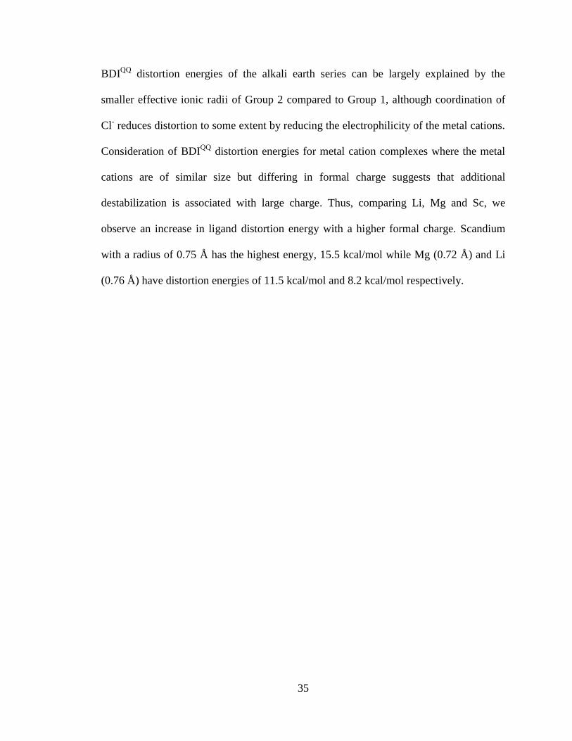
Cl- reduces distortion to some extent by reducing the electrophilicity of the metal cations.
Consideration of BDIQQ
distortion energies for metal cation complexes where the metal
cations are of similar size but differing in formal charge suggests that additional
destabilization is associated with large charge. Thus, comparing Li, Mg and Sc, we
observe an increase in ligand distortion energy with a higher formal charge. Scandium
with a radius of 0.75 Å has the highest energy, 15.5 kcal/mol while Mg (0.72 Å) and Li
(0.76 Å) have distortion energies of 11.5 kcal/mol and 8.2 kcal/mol respectively.
36
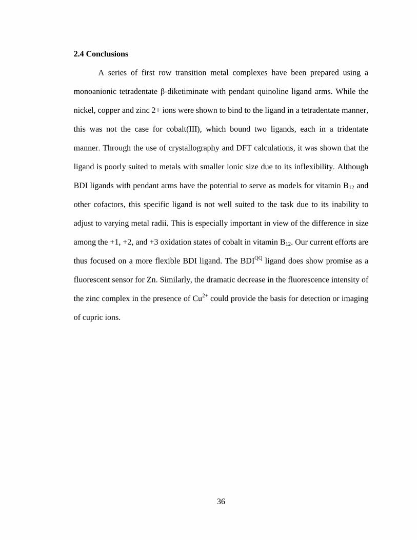
2.4 Conclusions
A series of first row transition metal complexes have been prepared using a
monoanionic tetradentate β-diketiminate with pendant quinoline ligand arms. While the
nickel, copper and zinc 2+ ions were shown to bind to the ligand in a tetradentate manner,
this was not the case for cobalt(III), which bound two ligands, each in a tridentate
manner. Through the use of crystallography and DFT calculations, it was shown that the
ligand is poorly suited to metals with smaller ionic size due to its inflexibility. Although
BDI ligands with pendant arms have the potential to serve as models for vitamin B12 and
other cofactors, this specific ligand is not well suited to the task due to its inability to
adjust to varying metal radii. This is especially important in view of the difference in size
among the +1, +2, and +3 oxidation states of cobalt in vitamin B12. Our current efforts are
thus focused on a more flexible BDI ligand. The BDIQQ
ligand does show promise as a
fluorescent sensor for Zn. Similarly, the dramatic decrease in the fluorescence intensity of
the zinc complex in the presence of Cu2+
could provide the basis for detection or imaging
of cupric ions.
37
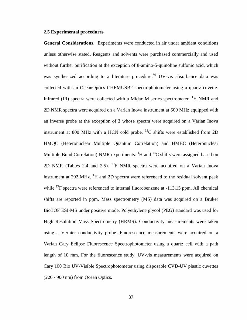
2.5 Experimental procedures
General Considerations. Experiments were conducted in air under ambient conditions
unless otherwise stated. Reagents and solvents were purchased commercially and used
without further purification at the exception of 8-amino-5-quinoline sulfonic acid, which
was synthesized according to a literature procedure.30
UV-vis absorbance data was
collected with an OceanOptics CHEMUSB2 spectrophotometer using a quartz cuvette.
Infrared (IR) spectra were collected with a Midac M series spectrometer. 1H NMR and
2D NMR spectra were acquired on a Varian Inova instrument at 500 MHz equipped with
an inverse probe at the exception of 3 whose spectra were acquired on a Varian Inova
instrument at 800 MHz with a HCN cold probe. 13
C shifts were established from 2D
HMQC (Heteronuclear Multiple Quantum Correlation) and HMBC (Heteronuclear
Multiple Bond Correlation) NMR experiments. 1H and
13C shifts were assigned based on
2D NMR (Tables 2.4 and 2.5). 19
F NMR spectra were acquired on a Varian Inova
instrument at 292 MHz. 1H and 2D spectra were referenced to the residual solvent peak
while 19
F spectra were referenced to internal fluorobenzene at -113.15 ppm. All chemical
shifts are reported in ppm. Mass spectrometry (MS) data was acquired on a Bruker
BioTOF ESI-MS under positive mode. Polyethylene glycol (PEG) standard was used for
High Resolution Mass Spectrometry (HRMS). Conductivity measurements were taken
using a Vernier conductivity probe. Fluorescence measurements were acquired on a
Varian Cary Eclipse Fluorescence Spectrophotometer using a quartz cell with a path
length of 10 mm. For the fluorescence study, UV-vis measurements were acquired on
Cary 100 Bio UV-Visible Spectrophotometer using disposable CVD-UV plastic cuvettes
(220 - 900 nm) from Ocean Optics.
38
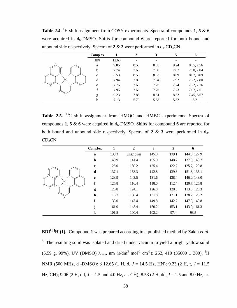
Table 2.4. 1H shift assignment from COSY experiments. Spectra of compounds 1, 5 & 6
were acquired in d6-DMSO. Shifts for compound 6 are reported for both bound and
unbound side respectively. Spectra of 2 & 3 were performed in d3-CD3CN.
Table 2.5. 13
C shift assignment from HMQC and HMBC experiments. Spectra of
compounds 1, 5 & 6 were acquired in d6-DMSO. Shifts for compound 6 are reported for
both bound and unbound side respectively. Spectra of 2 & 3 were performed in d3-
CD3CN.
BDIQQ
H (1). Compound 1 was prepared according to a published method by Zakta et al.
5. The resulting solid was isolated and dried under vacuum to yield a bright yellow solid
(5.59 g, 99%). UV (DMSO) λmax, nm (ε/dm3 mol
-1 cm
-1): 262, 419 (35600 ± 300).
1H
NMR (500 MHz, d6-DMSO): δ 12.65 (1 H, d, J = 14.5 Hz, HN); 9.23 (2 H, t, J = 11.5
Hz, CH); 9.06 (2 H, dd, J = 1.5 and 4.0 Hz, ar. CH); 8.53 (2 H, dd, J = 1.5 and 8.0 Hz, ar.
Complex 1 2 3 5 6
HN 12.65 - - - -
a 9.06 8.58 8.85 9.24 8.35, 7.56
b 7.74 7.68 7.80 7.87 7.50, 7.04
c 8.53 8.58 8.63 8.69 8.07, 8.09
d 7.94 7.89 7.94 7.92 7.22, 7.80
e 7.76 7.68 7.76 7.74 7.22, 7.76
f 7.96 7.68 7.76 7.73 7.07, 7.51
g 9.23 7.85 8.61 8.52 7.45, 6.57
h 7.13 5.70 5.68 5.32 5.21
Complex 1 2 3 5 6
a 138.3 unknown 145.0 139.1 144.0, 127.9
b 149.9 141.4 155.0 148.7 137.9, 148.7
c 123.0 130.2 125.4 122.7 125.7, 120.8
d 137.1 153.3 142.8 139.8 151.3, 135.1
e 128.9 143.5 131.6 138.4 146.0, 143.0
f 125.8 116.4 118.0 112.4 120.7, 125.8
g 126.8 124.1 126.8 128.5 113.5, 125.3
h 116.7 130.4 131.8 121.1 128.2, 125.2
i 135.0 147.4 149.8 142.7 147.8, 149.8
j 161.0 148.4 150.2 153.1 143.9, 161.3
k 101.8 100.4 102.2 97.4 93.5
39
CH); 7.96 (2 H, d, J = 8.0 Hz, ar. CH); 7.94 (2 H, d, J = 8.0 Hz, ar. CH); 7.76 (2 H, dd, J
= 8.0 and 8.0 Hz, ar. CH); 7.74 (2 H, dd, J = 4.0 and 8.5 Hz, ar. CH); 7.13 (1 H, t, J =
11.5 Hz, CH). 13
C NMR (125 MHz, d6-DMSO): δ 161.0, 149.9, 138.3, 137.1, 135.0,
128.9, 126.8, 125.8, 123.0, 116.7, 101.8. ESI-HRMS-TOF m/z: [M + H]+ calc. for
C21H17N4, 325.1448; found, 325.1456.
BDIQ’Q’
H (1a). In a 50 mL reaction flask, 8-amino-5-quinoline sulfonic acid (0.71 g, 3.1
mmol) was dissolved in 20 mL of MeOH, which was heated to 65º C. Once all of the
solid had dissolved completely, 1,1,3,3-tetramethoxypropane (0.26 mL, 1.6 mmol) was
added dropwise via syringe. The solution was stirred for 20 minutes and then acidified
with 10 mL of a 1:1 mixture of 3.5 M HCl and MeOH, added dropwise. Within minutes,
a bright orange solid began to precipitate. The solution was heated to reflux for an
additional hour, then allowed to cool to room temperature. The resulting solid was
collected by filtration, washed with a 1:1 mixture of MeOH and ether, and dried under
vacuum to yield a bright orange solid (0.76 g, 92%). UV (MeOH) λmax, nm (ε/dm3 mol
-1
cm-1
): 250, 424 (29000). 1H NMR (500 MHz, d6-DMSO): δ 12.64 (d, J = 13.0 Hz, 1H);
9.26 (dd, J = 1.5, 8.5 Hz, 2H); 9.08 (t, J = 12.5 Hz, 2H); 9.04 (dd, J = 1.5, 4.0 Hz, 2H);
8.07 (d, J = 8.0 Hz, 2H); 7.83 (d, J = 8.0 Hz, 2H); 7.76 (dd, J = 4.5, 8.5 Hz, 2H); 7.15 (t, J
= 11.5 Hz, 1H). ESI-HRMS-TOF m/z: [M - H]- calc. for C21H15N4O6S2, 483.0438; found,
483.0441.
[BDIQQ
Ni][SO3CF3] (2). Under inert atmosphere, compound 1 (0.133 g, 0.41 mmol)
was dissolved in 30 mL of dry THF. Potassium bis(trimethylsilyl) amide (0.235 g, 1.18
40
mmol) in 10 mL of dry THF was added dropwise to a solution of compound 1. An
immediate color change from yellow to pink was observed. In a separate vial, nickel(II)
triflate (0.149 g , 0.42 mmol) was dissolved in 20 mL of THF. The deprotonated ligand
solution was added dropwise to the metal solution. It was stirred overnight and red solids
precipitated. Under ambient atmosphere, the solution was filtered using a glass fiber
filter. The resulting solids were washed with THF and redissolved in MeOH. The solvent
was evaporated using rotary evaporation and solids were dried under vacuum (0.054 g,
25%). UV (MeOH) λmax, nm (ε/dm3 mol
-1 cm
-1): 212, 234, 287 (10400 ± 500), 343
(14600 ± 200), 524 (15900 ± 500), 631 (2200 ± 200). IR (Nujol Mull) ῦmax (cm-1
): 2922
(s), 2852 (s), 1580 (m), 1493 (m), 1446 (s), 1376 (s), 1353 (w), 1324 (w), 1296 (m), 1267
(s, asym. SO3), 1219 (w, sym. CF3), 1141 (br. m, asym. CF3), 1027 (s, sym. SO3), 827 (s),
771 (s), 633 (s). 1
H NMR (500 MHz, CD3CN): δ 8.58 (4 H, br d, J = 9.0 Hz, ar. CH);
7.89 (2 H, d, J = 7.5 Hz, ar. CH); 7.85 (2 H, d, J = 6.0 Hz, CH); 7.68 (6 H, m, ar. CH);
5.70 (1 H, t, J = 6.5 Hz, CH). 13
C NMR (125 MHz, CD3CN): δ 153.3, 148.4, 147.4,
143.5, 141.4, 130.4, 130.2, 124.1, 116.4, 100.4, one quaternary resonance not observed.
19F NMR (292 MHz, CD3CN): δ -77.58. ESI-HRMS-TOF m/z: [M – SO3CF3]
+ calc. for
C21H15N4Ni, 381.0650; found, 381.0640.
[BDIQQ
NiCl] (3). Under ambient atmosphere, compound 2 (0.040 g, 0.07 mmol) was
dissolved in 60 mL of MeOH. A solution of tetrabutylammonium chloride (0.042 g, 0.15
mmol) in 20 mL of MeOH was added dropwise to compound 2. It was stirred overnight.
Solution was filtered using a glass fiber filter and the solvent from the filtrate was
removed using a rotary evaporator. Solids were washed with THF and dried under
41
vacuum (0.031 g, 98%). UV (MeOH) λmax, nm (ε/dm3 mol
-1 cm
-1): 212, 235, 289 (3800 ±
200), 343 (6300 ± 300), 525 (6400 ± 400). IR (Nujol Mull) ῦmax (cm-1
): 2924 (s), 2853
(s), 1494 (w), 1455 (s), 1376 (s), 1323 (w), 1311 (m), 1274 (w), 825 (w), 757 (w), 722
(w). 1H NMR (800 MHz, CD3CN): δ 8.73 (2 H, d, J = 5.6 Hz, ar. CH); 8.65 (2 H, d, J =
8.0 Hz, ar. CH); 8.21 (2 H, d, J = 4.8 Hz, CH); 7.97 (2 H, dd, J = 1.6 and 7.2 Hz, CH);
7.78 (2 H, dd, J = 5.6 and 8.0 Hz, ar. CH); 7.76 (4 H, m, ar. CH); 5.74 (1 H, t, J = 5.6 Hz,
CH). 13
C NMR (200 MHz, CD3CN): δ 155.0, 150.2, 149.8, 145.0, 142.8, 131.8, 131.6,
126.8, 125.4, 118.0, 102.2. ESI-HRMS-TOF m/z: [M – Cl]+ calc. for C21H15N4Ni,
381.0650; found, 381.0654.
[BDIQQ
CuCl] (4). Under ambient atmosphere, compound 1 (0.032 g, 0.10 mmol) was
dissolved in 30 mL of MeOH. A solution of copper(II) chloride dihydrate (0.016 g, 0.09
mmol) in 10 mL of MeOH was added dropwise to the ligand solution. An immediate
color change to red metal was observed. Three equivalents of 1.0 M sodium hydroxide
(280 μL, 0.28 mmol) were added dropwise to the metal complex solution. The solution
was stirred for another five minutes and was then filtered using a glass fiber filter. The
solvent from the filtrate was removed via a rotary evaporator. The red residue was
washed with THF and redissolved in MeOH. Further purification was obtained by
layering the MeOH solution with diethyl ether (1:2). Solids and green crystals isolated
from purification were dried under vacuum (0.016 g, 40%). UV (MeOH) λmax, nm (ε/dm3
mol-1
cm-1
): 215, 235, 284 (10100 ± 500), 493 (15300 ± 900), 514 (17500 ± 1100). ESI-
HRMS-TOF m/z: [M – Cl]+ calc. for C21H15N4Cu, 386.0593; found, 386.0589. X-ray
42
quality crystals were obtained by layering a saturated MeOH solution of the complex
with diethyl ether (1:2).
[BDIQQ
ZnCl] (5). Under ambient atmosphere, compound 1 (1.871 g, 5.8 mmol) was
dissolved in 80 mL of MeOH. A solution of anhydrous zinc(II) chloride (0.787 g, 5.8
mmol) in 20 mL of MeOH was added dropwise to the ligand solution. Three equivalents
of 1.0 M sodium hydroxide (17.3 mL, 17.3 mmol) were then added dropwise to the
solution. An immediate color change was observed from yellow to red. The solution was
immediately filtered using a glass fiber filter and the solvent removed from the filtrate by
rotary evaporation. The resulting red solid was dried under vacuum (1.80 g, 73%). UV
(DMSO) λmax, nm (ε/dm3 mol
-1 cm
-1): 262, 282, 325, 369 (3700 ± 210), 509 (24900 ±
190), 530 (27600 ± 100). 1H NMR (500 MHz, d6-DMSO): δ 9.24 (2 H, dd, J = 1.0 and
4.5 Hz, ar. CH); 8.69 (2 H, d, J = 8.5 Hz, ar. CH); 8.52 (2 H, d, J = 6.0 Hz, CH); 7.92 (2
H, dd, J = 1.5 and 6.5 Hz, ar. CH); 7.87 (2 H, dd, J = 4.5 and 8.5 Hz, ar. CH); 7.74 (2 H,
dd, J = 8.5 and 8.5 Hz, ar. CH); 7.73 (2 H, dd, J = 7.5 and 14.5 Hz, ar. CH); 5.32 (1 H, t,
J = 6.0 Hz, CH). 13
C NMR (125 MHz, d6-DMSO): δ 153.1, 148.7, 142.7, 139.8, 139.1,
138.4, 128.5, 122.7, 121.1, 112.4, 97.4. ESI-HRMS-TOF m/z: [M – Cl]+ calc. for
C21H15N4Zn, 387.0588; found, 387.0585. X-ray quality crystals were obtained by
layering a saturated MeOH solution of the complex with pure MeOH and diethyl ether.
BDIQ’Q’
ZnCl (5a). Complex 5a was prepared using the same method as complex 5
except using compound 1a (0.050 g, 0.10 mmol) and anhydrous zinc(II) chloride (0.014
g, 0.10 mmol). The resulting solid was dichromatic red and gold (0.067 g, 98%). UV
43
(MeOH) λmax, nm (ε/dm3 mol
-1 cm
-1): 226, 266 (32000), 282 (sh.), 302 (sh.), 349, 382,
538 (29000), 560 (34000). 1H NMR (500 MHz, d6-DMSO): δ 9.44 (dd, J = 1.5, 8.7 Hz,
2H); 9.23 (dd, J = 1.5, 4.5 Hz, 2H); 8.52 (d, J = 6.6 Hz, 2H); 8.04 (d, J = 8.1 Hz, 2H);
7.91 (dd, J = 4.5, 8.7 Hz, 2H); 7.83 (d, J = 8.1 Hz, 2H); 5.35 (t, J = 8.3 Hz, 1H). ESI-
HRMS-TOF m/z: [M – 2H + 2Na - Cl]+ calc. for C21H11N4O6S2Na2Zn, 590.9363; found,
590.9364.
[(BDIQQ
)2Co]I (6). Under ambient atmosphere, compound 6 was prepared by dissolving
compound 1 (0.101 g, 0.31 mmol) into 10 mL of MeOH. Three equivalents of 1.0 M
sodium hydroxide (0.93 mL, 0.93 mmol) were added dropwise to compound 1. A
solution of cobalt(II) iodide (0.097 g , 0.31 mmol) in 10 mL of MeOH was added
dropwise to the solution of compound 1. An immediate color change from yellow orange
to brown was observed. Solution was stirred overnight. Solution was filtered using a
glass fiber filter and the solvent was removed from the filtrate by rotary evaporation. The
resulting brown solids were washed with hexanes, redissolved in MeOH, dried under
vacuum (0.145 g, 83%). 1H NMR (500 MHz, d6-DMSO): δ ppm 8.35 (2 H, d, J = 8.0 Hz,
ar. CH); 8.09 (2 H, dd, J = 1.5 and 8.5 Hz, ar. CH); 8.07 (2 H, dd, J = 0.5 and 5.5 Hz, ar.
CH); 7.80 (2 H, dd, J = 0.5 and 8.0 Hz, ar. CH); 7.76 (2 H, dd, J = 1.0 and 7.0 Hz, ar.
CH); 7.56 (2 H, dd, J = 2.0 and 4.5 Hz, ar. CH); 7.51 (2 H, dd, J = 8.0 and 8.0 Hz, ar.
CH); 7.50 (2 H, d, J = 2.0 and 8.0 Hz, ar. CH); 7.45 (2 H, d, J = 7.0 Hz, CH); 7.22 (4 H,
d, J = 8.0 Hz, ar. CH); 7.07 (2 H, t, J = 8.0 Hz, ar. CH); 7.04 (2 H, dd, J = 4.0 and 8.0 Hz,
ar. CH); 6.57 (2 H, d, J = 6.5 Hz, CH); 5.21 (2 H, t, J = 6.5 Hz, CH). 13
C NMR (125
MHz, d6-DMSO): δ 161.3 (CH), 151.3, 149.8, 148.7, 147.8, 146.0, 144.0, 143.9 (CH),
44
143.0, 137.9, 135.1, 128.2, 127.9, 125.8, 125.7, 125.3, 125.2, 120.8, 120.7, 113.5, 93.5
(CH). ESI-HRMS-TOF m/z: [M – I]+ calc. for C42H30N8Co, 705.1925; found, 705.1920.
Conductivity Study. Solutions of complexes 2, 3 and 4 were prepared in acetonitrile at
a concentration of 5 x 10⁻⁴
M. Tetrabutylammonium chloride was used a standard. All
solutions were measured using a conductivity probe.
Fluorescence Studies. For all fluorescence runs, a UV-vis spectrum was first acquired.
The excitation spectra were scanned between 300 nm and 560 nm with the emission set at
575 nm while the emission spectra, which were excited at 485 nm, were collected
between 495 nm and 800 nm. It should be stated that special care was taken to minimize
the impact of trace metal for these studies.
For the quantum yield determination, stock solution of 5 (5.1 x 10⁻⁵
M) and
fluorescein (3.1 x 10⁻⁵
M with 14 µL of triethylamine) were made with HPLC grade
methanol. Five dilutions of the stock were preparing thus the concentration range for
samples and standard were 1.48 x 10⁻⁶
M - 7.40 x 10⁻⁶
M and 6.12 x 10⁻⁷
M - 3.06 x 10⁻⁶
M respectively.
For the competition studies, methanol stock solutions of the cations (1.3 x 10⁻³
M)
were made prepared from MgCl2·6H2O, CaCl2, BaCl2·2H2O, FeCl2·4H2O, CoCl2·6H2O,
NiCl2·6H2O, CuCl2·2H2O and ZnCl2. The concentration of the stock solution of 5 was
5.52 x 10⁻⁵
M in methanol while for 1, it was 7.83 x 10⁻⁵
M. For the competition study
between 5 and each cation, 250 µL of stock solution of 5, 250 µL of a cation stock and
1.5 mL of methanol were added to a disposable cuvette. For the ligand study, each cation
45
run consisted of 10 µL of cation stock, 150 µL of 1, 1 µL of 0.5 M NaOH and 1.85 mL of
methanol in a disposable cuvette.
Crystallography. Crystal data for 4 and 5 were collected on a Bruker SMART Platform
CCD diffractometer at 173(2) K using Mo Kα radiation and a graphite monochromator.
Structures were solved using SHELXS-97 and refined using SHELXL-97.31
Crystallographic data for 4 and 5 are summarized in Table 1.
The structure of 5 was solved using a Patterson map to place the zinc and chloride
atoms while all other non hydrogen atoms were placed using difference Fourier.
Considering the similarities between the unit cells of 4 and 5, the structure of 4 was
solved using the solution structure of 5 as a model with substitution of the metal.
Complex 4 showed nonmerohedral twinning and the data was corrected for this using
Cell Now program.32
The twin law was found to be 180º rotation of the reciprocal [0,1,0]
axis. The ratio of its twin components was determined to be 56:44.
Computional Methods. All density functional calculations made use of the M06-L
density functional and were carried out in Gaussian03 via the Minnesota Gaussian
Functional Module (MN-GFM).29, 33-34
As M06-L is a local functional, the resolution of
the identity approximation with a fitting basis set was used for greater computational
efficiency in the calculation of Coulomb integrals.35-37
A pruned grid with 99 radial shells
and 590 angular points per shell was used for all calculations. Stuttgart basis sets and
effective core potentials were used for all metals and the 6-31G(d) basis set was used for
all other atoms.38-39
Where appropriate, molecular geometries were optimized in C2 point-
46
group symmetry, and all geometries were verified as local minima by the computation of
analytical vibrational frequencies. Ligand distortion energies (LDE) were calculated by
subtracting the electronic energy of the BDIQQ
anion in its optimized, bare gas phase
geometry from the electronic energies of BDIQQ
anions held at geometries obtained from
optimized geometries of BDIQQ
Mn+
or BDIQQ
MCl complexes.
47
-Chapter Three-
Synthesis and Reactivity of an Isolable Cobalt (I) Complex
Containing a β-diketiminate-based Acyclic Tetradentate
Ligand
In part from:
Marlier, E. E.; Ulrich, B. A.; McNeill, K. Inorg. Chem. Accepted.
48
3.1 Overview
A model for cobalamin was synthesized using a new monoanionic tetradentate
nitrogen donor ligand; 2-(4-tolyl)-1,3-bis(2-isopropylpyridyl)propenediimine (Tol-BDI(2-
pp)2H) (1), which utilizes isopropylpyridines as pendant arms on a β-diketiminate (BDI)
backbone. During the synthesis of 1, the rearrangement product, Tol-BDI(2-pp)(4-pp)
H (2)
was observed. Metallation of 1 with zinc iodide and cobalt chloride yielded the
corresponding Tol-BDI(2-pp)2
ZnI (3) and Tol-BDI(2-pp)2
CoCl (4) complexes. The redox
properties of 4 in comparison to cobalamin were examined though electrochemical
studies. Electrochemical and bulk reduction of complex 4 gave a diamagnetic cobalt(I)
complex, Tol-BDI(2-pp)2
Co (5). Reactivity of 5 towards C-X bonds was investigated using
methyl iodide, 1-iodo-2-(trimethylsilyl)acetylene and trans-1-bromo-1-propene, yielding
Tol-BDI(2-pp)2
Co(CH3)I (6), Tol-BDI(2-pp)2
Co(C2Si(CH3)3)I (7) and Tol-BDI(2-
pp)2Co(CH=CH(CH3))Br (8) respectively. Synthesis and characterization details for these
complexes, including the crystal structure of 3, are reported.
49
3.2 Introduction
Cobalamin is an established catalyst for reductive dehalogenation reactions that
can transform highly chlorinated substrates into less chlorinated species.1-4
In an effort to
understand the scope and limitations of cobalamin’s ability to remediate groundwater
polluted with chlorinated ethylene and benzene compounds, several groups in addition to
our own have examined dimethylglyoxime- and porphyrin-containing model
complexes.5-12
Cobaloxime, (bis(dimethylglyoximato)cobalt), complexes have been used
to gain mechanistic insights and to model proposed intermediates involved in reductive
dehalogenation reactions, due to their similar reactivity towards halogenated substrates.5-
7, 10-12 Cobalt porphyrin complexes also have been successfully used as dechlorination
catalysts; with the discovery of water-soluble cobalt porphyrin complexes having higher
rate constants of dechlorination than vitamin B12.8, 13
While both cobaloxime and
(porphyrin)cobalt complexes have proven useful in modeling the chemistry of cobalamin,
there is room for improvement. In particular, the porphyrin and glyoxime-based systems
mimic the tetraaza coordination environment of the cobalamin corrin, but do not match
its monoanionic charge.
An additional shortcoming of the glyoxime- and porphyrin-based model
compounds is the difficulty in preparing and isolating cobalt(I) complexes in these
systems. While examples of Co(I) have been prepared in these systems, the complexity in
synthesizing these compounds has hindered their use in further studies.14-16
Researchers
have opted instead to generate the Co(I) oxidation state in situ. This is unfortunate from
the standpoint of studying the mechanism of cobalamin-mediated dechlorination
reactions, given that the Co(I) oxidation state is thought to play a central role.4 The ligand
50
choice is clearly important as isolable cobalt(I) complexes supported by a variety of
nitrogen donor ligands such as N2P2, bidentate BDI and iminopyridine pincers have
become more common in the literature in recent years.17-21
With this in mind, we set out
to prepare a ligand system that would allow the isolation of a Co(I) complex that
effectively models cobalamin.
Our approach to preparing a potentially improved structural model for the corrin
has focused on using a β-diketiminate ligand with two pendant donor arms (Chart 1).
This strategy has previously been employed by Roesky and coworkers with pendant
trialkylamine donors and by Zatka et al. with pendant quinoline donors to create a variety
of metal complexes.22-26
Our initial attempts to prepare similar ligands with
methylpyridine pendant arms prompted the discovery of a new route to pyridylpyrroles
due to intramolecular cyclization of the ligand.27
The cyclization is thought to occur
through a nucleophilic attack by the carbon atom α to the pyridine on the neighboring
ketone (or imine). To avoid the cyclization, we followed the lead of Zatka et al., and
chose quinolines as pendant arms for our second generation ligand.28
Subsequent study
revealed that a ligand equipped with quinoline pendant arms was too rigid to
accommodate the different oxidation states of cobalt.29
Considering that all three
oxidation states of cobalt are likely involved in the process of reductive dehalogenation, a
more flexible ligand was devised.4
In this work, we report the synthesis and characterization of a new monoanionic
tetradentate nitrogen donor ligand; 2-(4-tolyl)-1,3-bis(2-isopropylpyridyl)propenediimine
(Tol-BDI(2-pp)2
H) (1), which employs isopropylpyridines as pendant arms on a β-
diketiminate backbone (Chart 3.1).
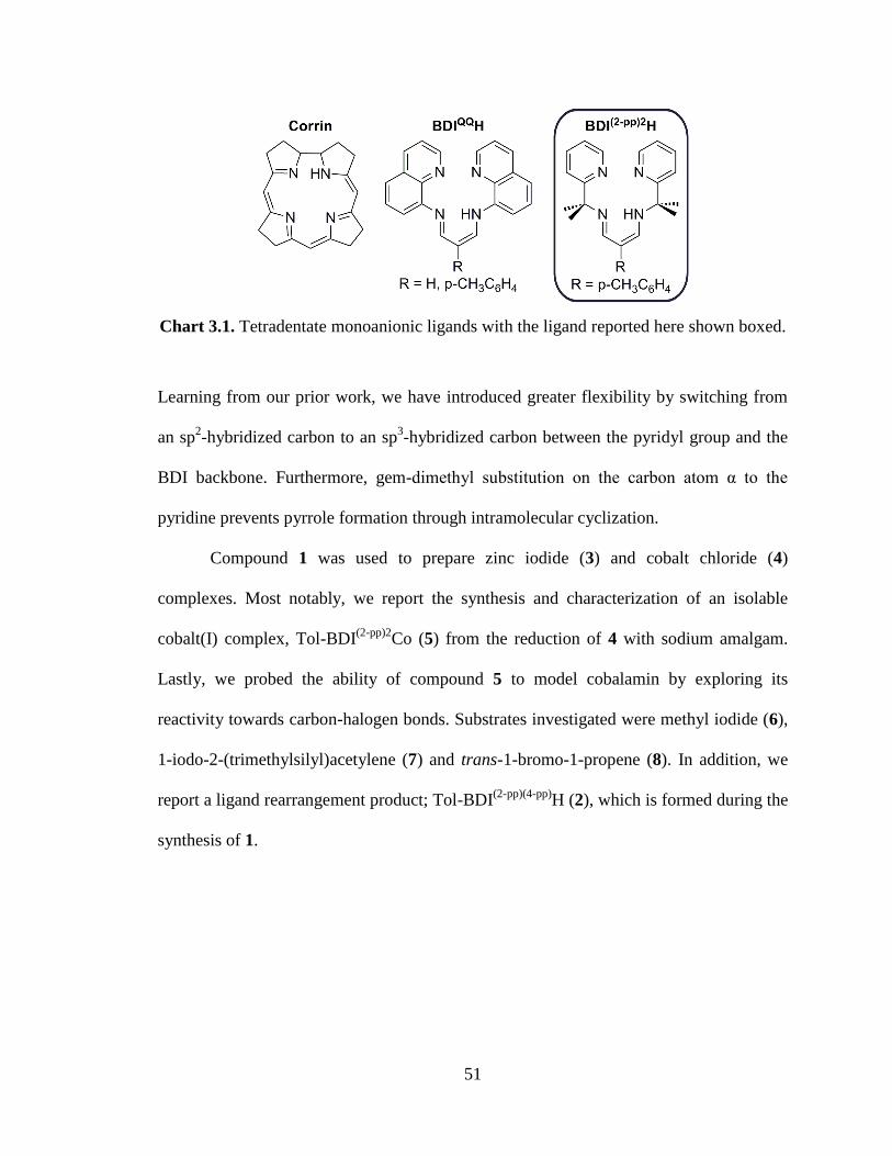
51
Chart 3.1. Tetradentate monoanionic ligands with the ligand reported here shown boxed.
Learning from our prior work, we have introduced greater flexibility by switching from
an sp2-hybridized carbon to an sp
3-hybridized carbon between the pyridyl group and the
BDI backbone. Furthermore, gem-dimethyl substitution on the carbon atom α to the
pyridine prevents pyrrole formation through intramolecular cyclization.
Compound 1 was used to prepare zinc iodide (3) and cobalt chloride (4)
complexes. Most notably, we report the synthesis and characterization of an isolable
cobalt(I) complex, Tol-BDI(2-pp)2
Co (5) from the reduction of 4 with sodium amalgam.
Lastly, we probed the ability of compound 5 to model cobalamin by exploring its
reactivity towards carbon-halogen bonds. Substrates investigated were methyl iodide (6),
1-iodo-2-(trimethylsilyl)acetylene (7) and trans-1-bromo-1-propene (8). In addition, we
report a ligand rearrangement product; Tol-BDI(2-pp)(4-pp)
H (2), which is formed during the
synthesis of 1.
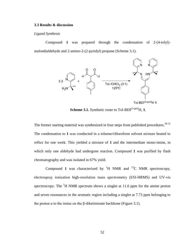
52
3.3 Results & discussion
Ligand Synthesis
Compound 1 was prepared through the condensation of 2-(4-tolyl)-
malondialdehyde and 2-amino-2-(2-pyridyl) propane (Scheme 3.1).
Scheme 3.1. Synthetic route to Tol-BDI(2-pp)2
H, 1.
The former starting material was synthesized in four steps from published procedures.30-31
The condensation to 1 was conducted in a toluene/chloroform solvent mixture heated to
reflux for one week. This yielded a mixture of 1 and the intermediate mono-imine, in
which only one aldehyde had undergone reaction. Compound 1 was purified by flash
chromatography and was isolated in 67% yield.
Compound 1 was characterized by 1H NMR and
13C NMR spectroscopy,
electrospray ionization high-resolution mass spectrometry (ESI-HRMS) and UV-vis
spectroscopy. The 1H NMR spectrum shows a singlet at 11.6 ppm for the amine proton
and seven resonances in the aromatic region including a singlet at 7.73 ppm belonging to
the proton α to the imine on the β-diketiminate backbone (Figure 3.1).
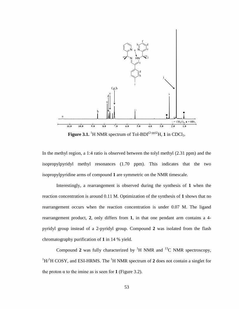
53
Figure 3.1. 1H NMR spectrum of Tol-BDI
(2-pp)2H, 1 in CDCl3.
In the methyl region, a 1:4 ratio is observed between the tolyl methyl (2.31 ppm) and the
isopropylpyridyl methyl resonances (1.70 ppm). This indicates that the two
isopropylpyridine arms of compound 1 are symmetric on the NMR timescale.
Interestingly, a rearrangement is observed during the synthesis of 1 when the
reaction concentration is around 0.11 M. Optimization of the synthesis of 1 shows that no
rearrangement occurs when the reaction concentration is under 0.07 M. The ligand
rearrangement product, 2, only differs from 1, in that one pendant arm contains a 4-
pyridyl group instead of a 2-pyridyl group. Compound 2 was isolated from the flash
chromatography purification of 1 in 14 % yield.
Compound 2 was fully characterized by 1H NMR and
13C NMR spectroscopy,
1H/
1H COSY, and ESI-HRMS. The
1H NMR spectrum of 2 does not contain a singlet for
the proton α to the imine as is seen for 1 (Figure 3.2).

54
Figure 3.2. 1H NMR spectrum of rearranged ligand, 2 (Tol-BDI
(2-pp)(4-pp)H) in CDCl3.
Rather, it has two doublets (7.82 and 7.53 ppm) that share a 2.5 Hz coupling constant,
confirmed by 1H/
1H COSY. Furthermore, the isopropylpyridyl methyls give rise to two
separate resonances (1.70 and 1.63 ppm), each integrating to 6H against the tolyl methyl
proton signal (2.31ppm, 3H). The assignment of a 4-pyridyl group comes from the two
second-order doublets each integrating to 2H at 8.52 and 7.36 ppm.
Metal Complexes Synthesis
Metallation of compound 1 was achieved by deprotonating the ligand with
potassium bis(trimethylsilyl)amide, followed by the addition of the desired metal halide
salt (Scheme 3.2).
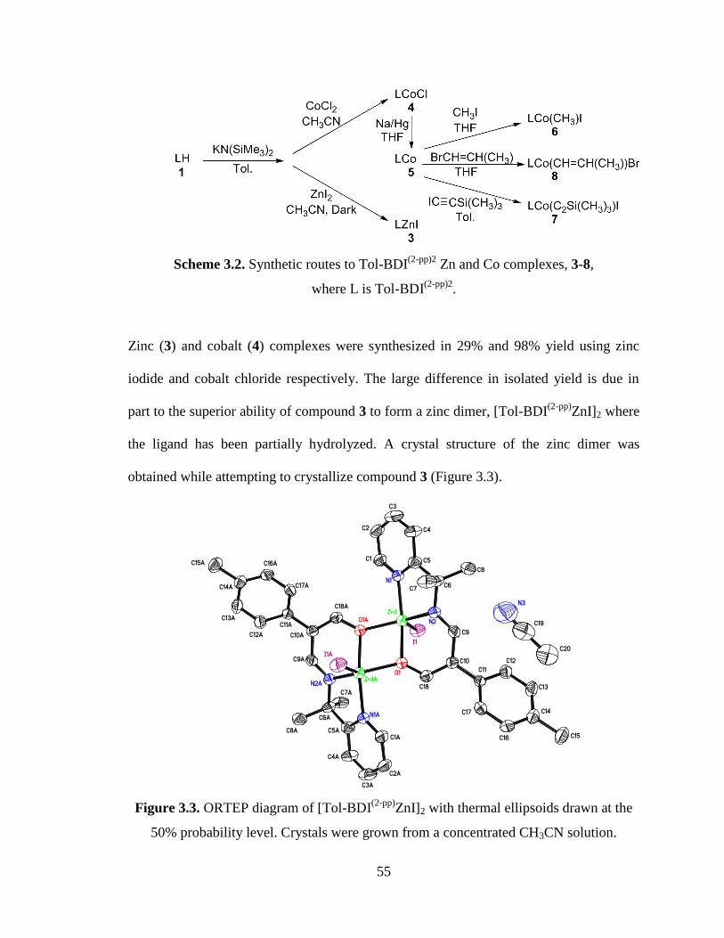
55
Scheme 3.2. Synthetic routes to Tol-BDI(2-pp)2
Zn and Co complexes, 3-8,
where L is Tol-BDI(2-pp)2
.
Zinc (3) and cobalt (4) complexes were synthesized in 29% and 98% yield using zinc
iodide and cobalt chloride respectively. The large difference in isolated yield is due in
part to the superior ability of compound 3 to form a zinc dimer, [Tol-BDI(2-pp)
ZnI]2 where
the ligand has been partially hydrolyzed. A crystal structure of the zinc dimer was
obtained while attempting to crystallize compound 3 (Figure 3.3).
Figure 3.3. ORTEP diagram of [Tol-BDI(2-pp)
ZnI]2 with thermal ellipsoids drawn at the
50% probability level. Crystals were grown from a concentrated CH3CN solution.
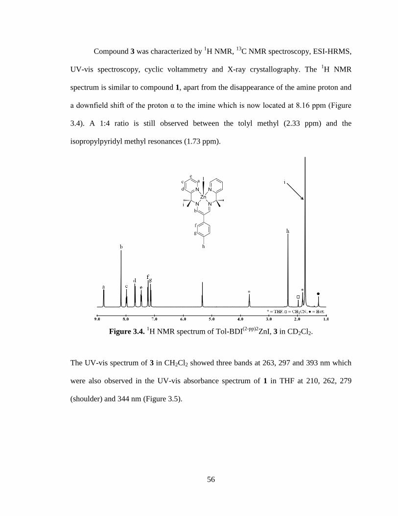
56
Compound 3 was characterized by 1H NMR,
13C NMR spectroscopy, ESI-HRMS,
UV-vis spectroscopy, cyclic voltammetry and X-ray crystallography. The 1H NMR
spectrum is similar to compound 1, apart from the disappearance of the amine proton and
a downfield shift of the proton α to the imine which is now located at 8.16 ppm (Figure
3.4). A 1:4 ratio is still observed between the tolyl methyl (2.33 ppm) and the
isopropylpyridyl methyl resonances (1.73 ppm).
Figure 3.4. 1H NMR spectrum of Tol-BDI
(2-pp)2ZnI, 3 in CD2Cl2.
The UV-vis spectrum of 3 in CH2Cl2 showed three bands at 263, 297 and 393 nm which
were also observed in the UV-vis absorbance spectrum of 1 in THF at 210, 262, 279
(shoulder) and 344 nm (Figure 3.5).
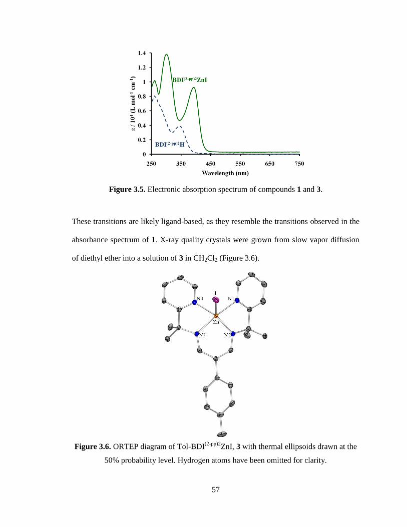
57
Figure 3.5. Electronic absorption spectrum of compounds 1 and 3.
These transitions are likely ligand-based, as they resemble the transitions observed in the
absorbance spectrum of 1. X-ray quality crystals were grown from slow vapor diffusion
of diethyl ether into a solution of 3 in CH2Cl2 (Figure 3.6).
Figure 3.6. ORTEP diagram of Tol-BDI(2-pp)2
ZnI, 3 with thermal ellipsoids drawn at the
50% probability level. Hydrogen atoms have been omitted for clarity.
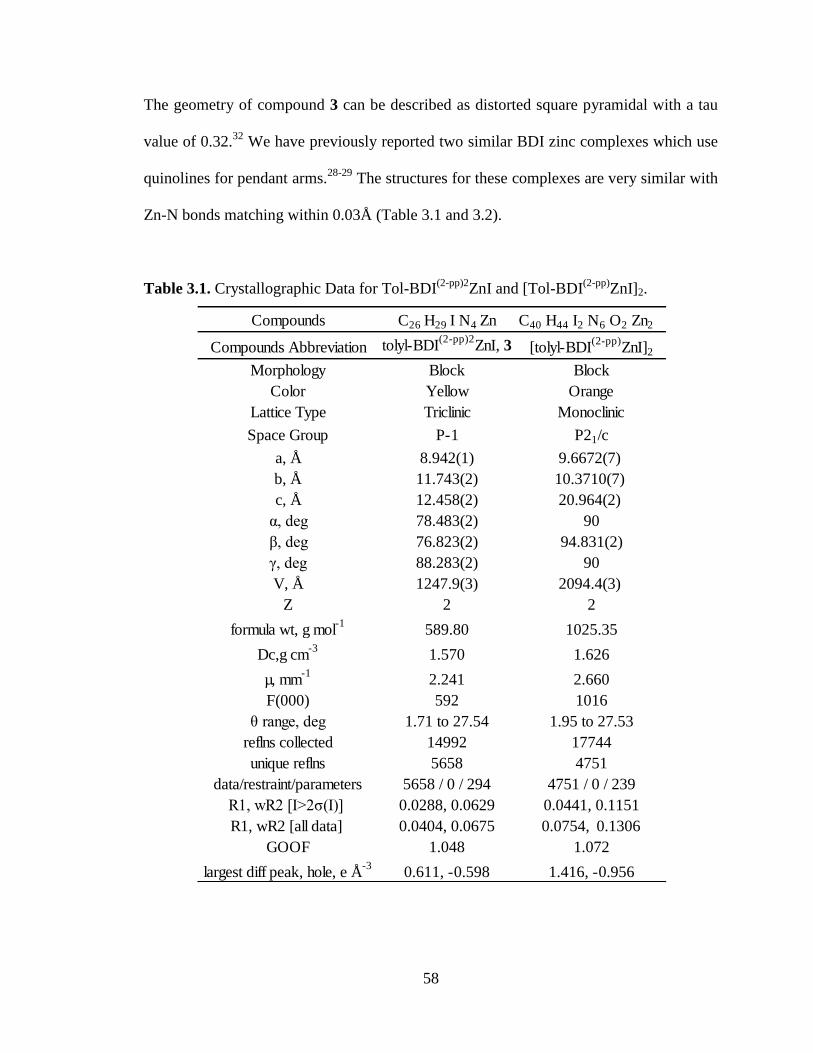
58
The geometry of compound 3 can be described as distorted square pyramidal with a tau
value of 0.32.32
We have previously reported two similar BDI zinc complexes which use
quinolines for pendant arms.28-29
The structures for these complexes are very similar with
Zn-N bonds matching within 0.03Å (Table 3.1 and 3.2).
Table 3.1. Crystallographic Data for Tol-BDI(2-pp)2
ZnI and [Tol-BDI(2-pp)
ZnI]2.
Compounds C26 H29 I N4 Zn C40 H44 I2 N6 O2 Zn2
Compounds Abbreviation tolyl-BDI(2-pp)2
ZnI, 3 [tolyl-BDI(2-pp)
ZnI]2
Morphology Block Block
Color Yellow Orange
Lattice Type Triclinic Monoclinic
Space Group P-1 P21/c
a, Å 8.942(1) 9.6672(7)
b, Å 11.743(2) 10.3710(7)
c, Å 12.458(2) 20.964(2)
α, deg 78.483(2) 90
β, deg 76.823(2) 94.831(2)
γ, deg 88.283(2) 90
V, Å 1247.9(3) 2094.4(3)
Z 2 2
formula wt, g mol-1
589.80 1025.35
Dc,g cm-3
1.570 1.626
µ, mm-1
2.241 2.660
F(000) 592 1016
θ range, deg 1.71 to 27.54 1.95 to 27.53
reflns collected 14992 17744
unique reflns 5658 4751
data/restraint/parameters 5658 / 0 / 294 4751 / 0 / 239
R1, wR2 [I>2ζ(I)] 0.0288, 0.0629 0.0441, 0.1151
R1, wR2 [all data] 0.0404, 0.0675 0.0754, 0.1306
GOOF 1.048 1.072
largest diff peak, hole, e Å-3
0.611, -0.598 1.416, -0.956
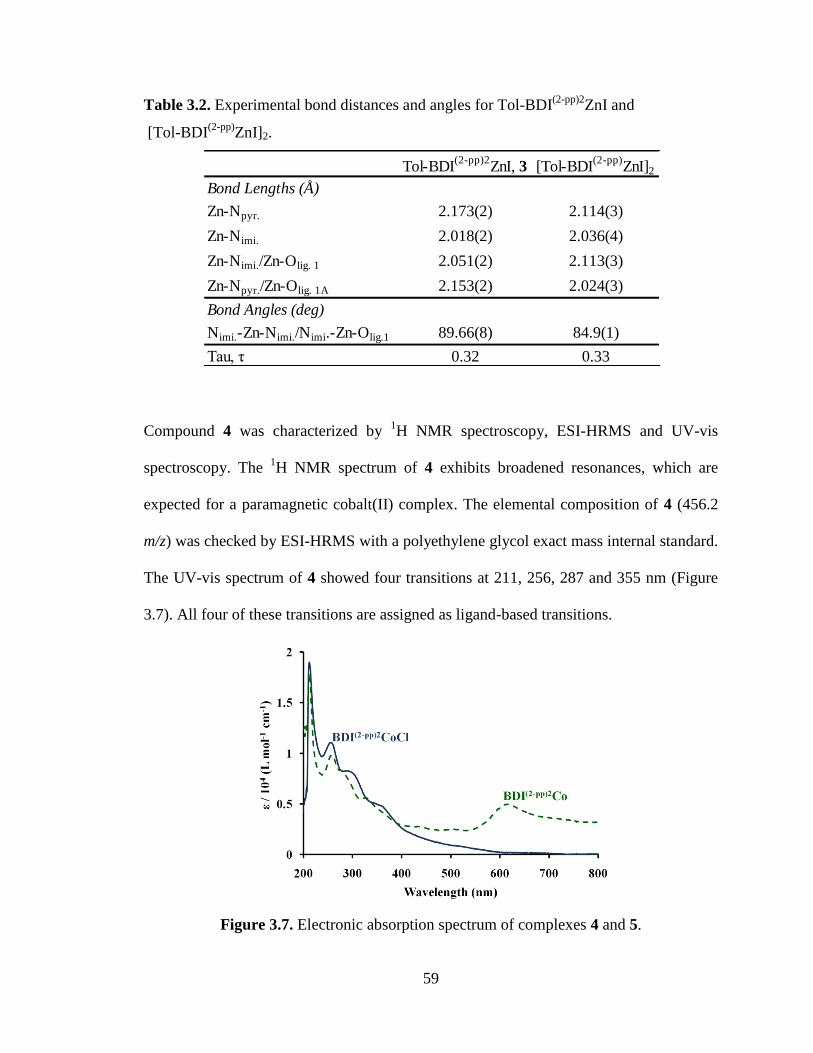
59
Table 3.2. Experimental bond distances and angles for Tol-BDI(2-pp)2
ZnI and
[Tol-BDI(2-pp)
ZnI]2.
Compound 4 was characterized by 1H NMR spectroscopy, ESI-HRMS and UV-vis
spectroscopy. The 1H NMR spectrum of 4 exhibits broadened resonances, which are
expected for a paramagnetic cobalt(II) complex. The elemental composition of 4 (456.2
m/z) was checked by ESI-HRMS with a polyethylene glycol exact mass internal standard.
The UV-vis spectrum of 4 showed four transitions at 211, 256, 287 and 355 nm (Figure
3.7). All four of these transitions are assigned as ligand-based transitions.
Figure 3.7. Electronic absorption spectrum of complexes 4 and 5.
Tol-BDI(2-pp)2
ZnI, 3 [Tol-BDI(2-pp)
ZnI]2
Bond Lengths (Å)
Zn-Npyr. 2.173(2) 2.114(3)
Zn-Nimi. 2.018(2) 2.036(4)
Zn-Nimi./Zn-Olig. 1 2.051(2) 2.113(3)
Zn-Npyr./Zn-Olig. 1A 2.153(2) 2.024(3)
Bond Angles (deg)
Nimi.-Zn-Nimi./Nimi.-Zn-Olig.1 89.66(8) 84.9(1)
Tau, η 0.32 0.33
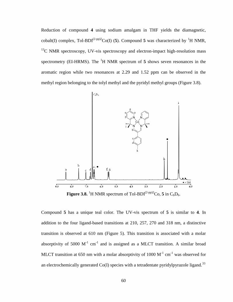
60
Reduction of compound 4 using sodium amalgam in THF yields the diamagnetic,
cobalt(I) complex, Tol-BDI(2-pp)2
Co(I) (5). Compound 5 was characterized by 1H NMR,
13C NMR spectroscopy, UV-vis spectroscopy and electron-impact high-resolution mass
spectrometry (EI-HRMS). The 1H NMR spectrum of 5 shows seven resonances in the
aromatic region while two resonances at 2.29 and 1.52 ppm can be observed in the
methyl region belonging to the tolyl methyl and the pyridyl methyl groups (Figure 3.8).
Figure 3.8. 1H NMR spectrum of Tol-BDI
(2-pp)2Co, 5 in C6D6.
Compound 5 has a unique teal color. The UV-vis spectrum of 5 is similar to 4. In
addition to the four ligand-based transitions at 210, 257, 270 and 318 nm, a distinctive
transition is observed at 610 nm (Figure 5). This transition is associated with a molar
absorptivity of 5000 M-1
cm-1
and is assigned as a MLCT transition. A similar broad
MLCT transition at 650 nm with a molar absorptivity of 1000 M-1
cm-1
was observed for
an electrochemically generated Co(I) species with a tetradentate pyridylpyrazole ligand.33
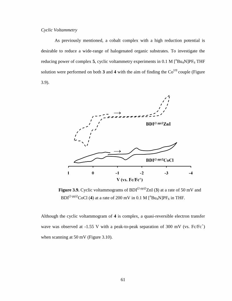
61
Cyclic Voltammetry
As previously mentioned, a cobalt complex with a high reduction potential is
desirable to reduce a wide-range of halogenated organic substrates. To investigate the
reducing power of complex 5, cyclic voltammetry experiments in 0.1 M [nBu4N]PF6 THF
solution were performed on both 3 and 4 with the aim of finding the CoI/II
couple (Figure
3.9).
Figure 3.9. Cyclic voltammograms of BDI(2-pp)2
ZnI (3) at a rate of 50 mV and
BDI(2-pp)2
CoCl (4) at a rate of 200 mV in 0.1 M [nBu4N]PF6 in THF.
Although the cyclic voltammogram of 4 is complex, a quasi-reversible electron transfer
wave was observed at -1.55 V with a peak-to-peak separation of 300 mV (vs. Fc/Fc+)
when scanning at 50 mV (Figure 3.10).
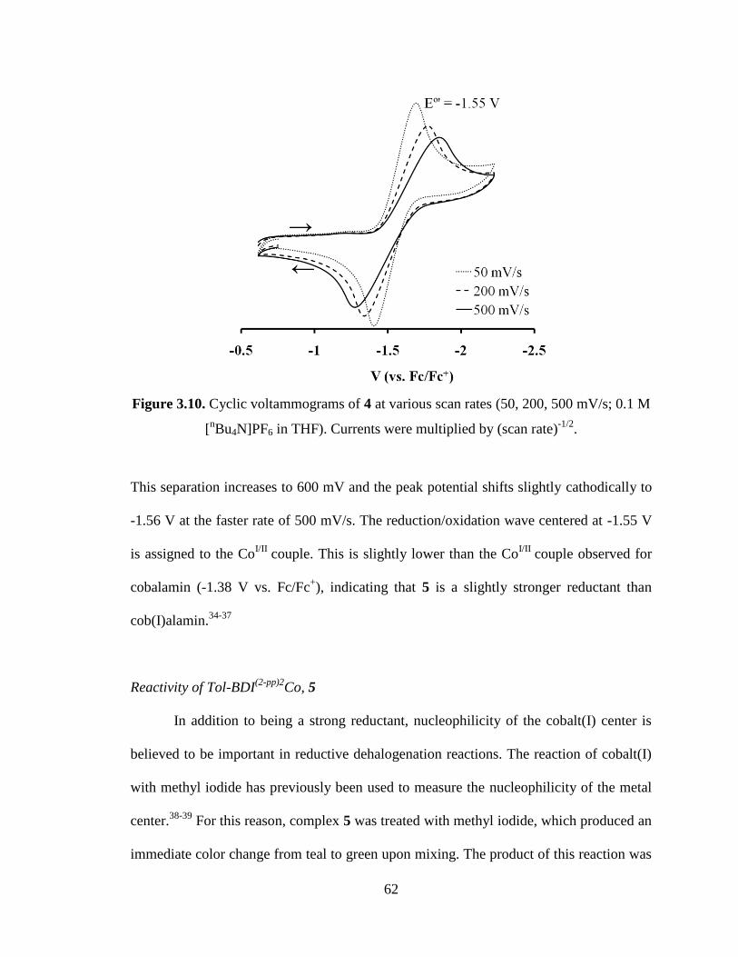
62
Figure 3.10. Cyclic voltammograms of 4 at various scan rates (50, 200, 500 mV/s; 0.1 M
[nBu4N]PF6 in THF). Currents were multiplied by (scan rate)
-1/2.
This separation increases to 600 mV and the peak potential shifts slightly cathodically to
-1.56 V at the faster rate of 500 mV/s. The reduction/oxidation wave centered at -1.55 V
is assigned to the CoI/II
couple. This is slightly lower than the CoI/II
couple observed for
cobalamin (-1.38 V vs. Fc/Fc+), indicating that 5 is a slightly stronger reductant than
cob(I)alamin.34-37
Reactivity of Tol-BDI(2-pp)2
Co, 5
In addition to being a strong reductant, nucleophilicity of the cobalt(I) center is
believed to be important in reductive dehalogenation reactions. The reaction of cobalt(I)
with methyl iodide has previously been used to measure the nucleophilicity of the metal
center.38-39
For this reason, complex 5 was treated with methyl iodide, which produced an
immediate color change from teal to green upon mixing. The product of this reaction was
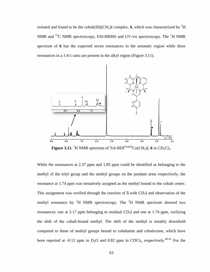
63
isolated and found to be the cobalt(III)(CH3)I complex, 6, which was characterized by 1H
NMR and 13
C NMR spectroscopy, ESI-HRMS and UV-vis spectroscopy. The 1H NMR
spectrum of 6 has the expected seven resonances in the aromatic region while three
resonances in a 1:4:1 ratio are present in the alkyl region (Figure 3.11).
Figure 3.11. 1H NMR spectrum of Tol-BDI
(2-pp)2Co(CH3)I, 6 in CD2Cl2.
While the resonances at 2.37 ppm and 1.85 ppm could be identified as belonging to the
methyl of the tolyl group and the methyl groups on the pendant arms respectively, the
resonance at 1.74 ppm was tentatively assigned as the methyl bound to the cobalt center.
This assignment was verified through the reaction of 5 with CD3I and observation of the
methyl resonance by 2H NMR spectroscopy. The
2H NMR spectrum showed two
resonances: one at 2.17 ppm belonging to residual CD3I and one at 1.74 ppm, verifying
the shift of the cobalt-bound methyl. The shift of the methyl is notably downfield
compared to those of methyl groups bound to cobalamin and cobaloxime, which have
been reported at -0.12 ppm in D2O and 0.82 ppm in CDCl3, respectively.40-41
For the
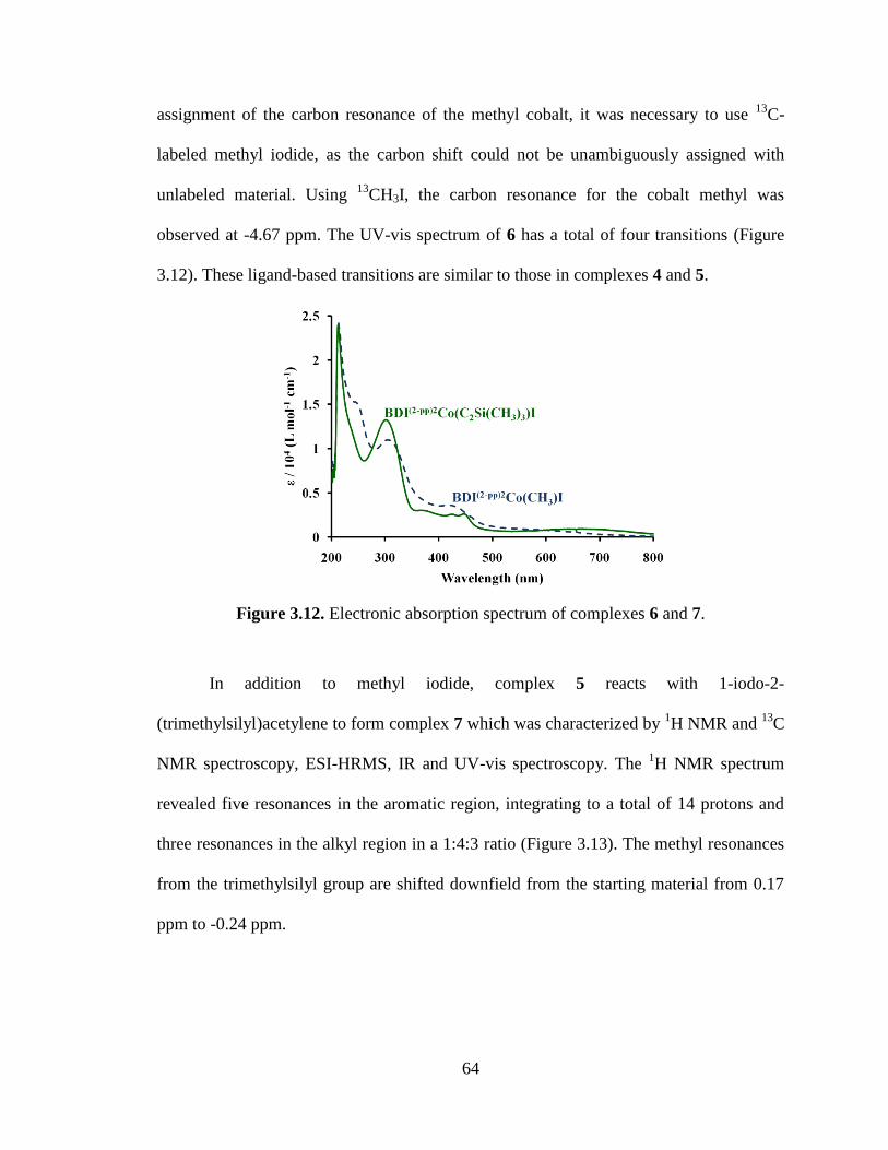
64
assignment of the carbon resonance of the methyl cobalt, it was necessary to use 13
C-
labeled methyl iodide, as the carbon shift could not be unambiguously assigned with
unlabeled material. Using 13
CH3I, the carbon resonance for the cobalt methyl was
observed at -4.67 ppm. The UV-vis spectrum of 6 has a total of four transitions (Figure
3.12). These ligand-based transitions are similar to those in complexes 4 and 5.
Figure 3.12. Electronic absorption spectrum of complexes 6 and 7.
In addition to methyl iodide, complex 5 reacts with 1-iodo-2-
(trimethylsilyl)acetylene to form complex 7 which was characterized by 1H NMR and
13C
NMR spectroscopy, ESI-HRMS, IR and UV-vis spectroscopy. The 1H NMR spectrum
revealed five resonances in the aromatic region, integrating to a total of 14 protons and
three resonances in the alkyl region in a 1:4:3 ratio (Figure 3.13). The methyl resonances
from the trimethylsilyl group are shifted downfield from the starting material from 0.17
ppm to -0.24 ppm.
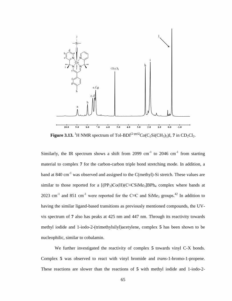
65
Figure 3.13. 1H NMR spectrum of Tol-BDI
(2-pp)2Co(C2Si(CH3)3)I, 7 in CD2Cl2.
Similarly, the IR spectrum shows a shift from 2099 cm-1
to 2046 cm-1
from starting
material to complex 7 for the carbon-carbon triple bond stretching mode. In addition, a
band at 840 cm-1
was observed and assigned to the C(methyl)-Si stretch. These values are
similar to those reported for a [(PP3)Co(H)(C≡CSiMe3]BPh4 complex where bands at
2023 cm-1
and 851 cm-1
were reported for the C≡C and SiMe3 groups.42
In addition to
having the similar ligand-based transitions as previously mentioned compounds, the UV-
vis spectrum of 7 also has peaks at 425 nm and 447 nm. Through its reactivity towards
methyl iodide and 1-iodo-2-(trimethylsilyl)acetylene, complex 5 has been shown to be
nucleophilic, similar to cobalamin.
We further investigated the reactivity of complex 5 towards vinyl C-X bonds.
Complex 5 was observed to react with vinyl bromide and trans-1-bromo-1-propene.
These reactions are slower than the reactions of 5 with methyl iodide and 1-iodo-2-
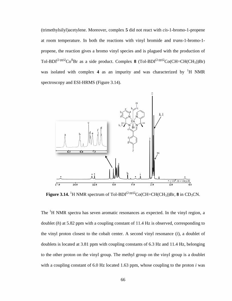
66
(trimethylsilyl)acetylene. Moreover, complex 5 did not react with cis-1-bromo-1-propene
at room temperature. In both the reactions with vinyl bromide and trans-1-bromo-1-
propene, the reaction gives a bromo vinyl species and is plagued with the production of
Tol-BDI(2-pp)2
CoIIBr as a side product. Complex 8 (Tol-BDI
(2-pp)2Co(CH=CH(CH3))Br)
was isolated with complex 4 as an impurity and was characterized by 1H NMR
spectroscopy and ESI-HRMS (Figure 3.14).
Figure 3.14. 1H NMR spectrum of Tol-BDI
(2-pp)2Co(CH=CH(CH3))Br, 8 in CD3CN.
The 1H NMR spectra has seven aromatic resonances as expected. In the vinyl region, a
doublet (h) at 5.82 ppm with a coupling constant of 11.4 Hz is observed, corresponding to
the vinyl proton closest to the cobalt center. A second vinyl resonance (i), a doublet of
doublets is located at 3.81 ppm with coupling constants of 6.3 Hz and 11.4 Hz, belonging
to the other proton on the vinyl group. The methyl group on the vinyl group is a doublet
with a coupling constant of 6.0 Hz located 1.63 ppm, whose coupling to the proton i was
67
confirmed by 1H/
1H COSY. In this complex, the pyridyl methyl groups are split into two
inequivalent sets based on their orientation with respect to the vinyl and bromide group.
Other vinyl substrates were observed to react with 5. With an aim at the
remediation of chlorinated water, complex 5 was revealed to dechlorinate TCE to cis-
DCE, however this reaction appeared to have poor efficiency by GC-MS.
68
3.4 Conclusions
A monoanionic, tetraaza ligand was prepared in an effort to model the
monoanionic corrin ligand in cobalamin. The ligand reported here, Tol-BDI(2-pp)2
H, is a
third-generation ligand that is the product of an iterative design process. The ligand is
composed of a β-diketiminate backbone with isopropylpyridine pendant arms. It is both
flexible enough to support the three important oxidation states of Co (I, II, III) and stable
with respect to decomposition to pyridylpyrrole products. An isolable Co(I) complex of
Tol-BDI(2-pp)2
was prepared and was found to be comparable to cobalamin(I) both in
terms of its Co(I)/Co(II) redox couple and in its reactions as a nucleophile.
69
3.5 Experimental procedures
General Considerations. Experiments were conducted under anhydrous and anaerobic
(dinitrogen) conditions using a glovebox or Schlenk line at the exception of the ligand
synthesis which was conducted in air. Reagents with the exception of 2-amino-2-(2-
pyridyl) propane were purchased commercially and used without further purification. 2-
Amino-2-(2-pyridyl) propane was synthesized in four steps from published procedures.30-
31 Solvents were purchased commercially and dried under standard procedures.
43 UV-vis
absorbance data was collected with an OceanOptics CHEMUSB2 spectrophotometer
using a quartz cuvette. Infrared (IR) spectra were collected with a Midac M series
spectrometer. 1H NMR and 2D NMR spectra were acquired on Varian Inova instruments
at 300 MHz and 500 MHz.13
C shifts were established from 2D HMQC (Heteronuclear
Multiple Quantum Correlation) and HMBC (Heteronuclear Multiple Bond Correlation)
NMR experiments. The assignment of the carbon shifts can be found in the supporting
information (Table 3.3). 13
C NMR spectrum for the 13
CH3I experiment was acquired on
Varian Inova instrument at 75 MHz. 2H NMR spectrum was acquired on a Varian Inova
instrument at 300 MHz. Mass spectrometry (MS) data were acquired on a Bruker
BioTOF ESI-MS under positive mode. Polyethylene glycol (PEG) and polypropylene
glycol (PPG) standards were used for ESI-HRMS. A Finnigan MAT 95 double-focusing
mass spectrometer with BE-geometry was used to collect EI-HRMS with sample
introduction using a solids probe. Perfluorokerosene (PFK) was used as an internal
standard for EI-HRMS.
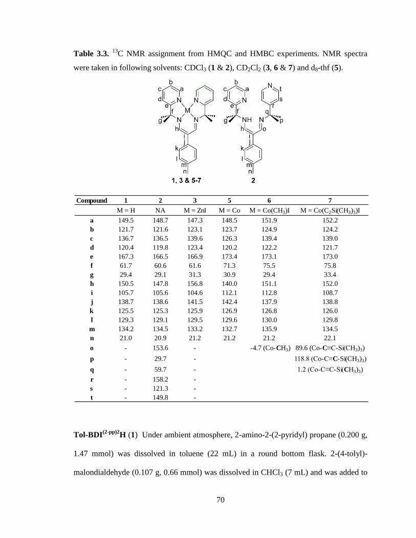
70
Table 3.3. 13
C NMR assignment from HMQC and HMBC experiments. NMR spectra
were taken in following solvents: CDCl3 (1 & 2), CD2Cl2 (3, 6 & 7) and d8-thf (5).
Tol-BDI(2-pp)2
H (1) Under ambient atmosphere, 2-amino-2-(2-pyridyl) propane (0.200 g,
1.47 mmol) was dissolved in toluene (22 mL) in a round bottom flask. 2-(4-tolyl)-
malondialdehyde (0.107 g, 0.66 mmol) was dissolved in CHCl3 (7 mL) and was added to
Compound 1 2 3 5 6 7
M = H NA M = ZnI M = Co M = Co(CH3)I M = Co(C2Si(CH3)3)I
a 149.5 148.7 147.3 148.5 151.9 152.2
b 121.7 121.6 123.1 123.7 124.9 124.2
c 136.7 136.5 139.6 126.3 139.4 139.0
d 120.4 119.8 123.4 120.2 122.2 121.7
e 167.3 166.5 166.9 173.4 173.1 173.0
f 61.7 60.6 61.6 71.3 75.5 75.8
g 29.4 29.1 31.3 30.9 29.4 33.4
h 150.5 147.8 156.8 140.0 151.1 152.0
i 105.7 105.6 104.6 112.1 112.8 108.7
j 138.7 138.6 141.5 142.4 137.9 138.8
k 125.5 125.3 125.9 126.9 126.8 126.0
l 129.3 129.1 129.5 129.6 130.0 129.8
m 134.2 134.5 133.2 132.7 135.9 134.5
n 21.0 20.9 21.2 21.2 21.2 22.1
o - 153.6 - -4.7 (Co-CH3) 89.6 (Co-C≡C-Si(CH3)3)
p - 29.7 - 118.8 (Co-C≡C-Si(CH3)3)
q - 59.7 - 1.2 (Co-C≡C-Si(CH3)3)
r - 158.2 -
s - 121.3 -
t - 149.8 -
71
the solution of 2-amino-2-(2-pyridyl)propane. A concentration of 0.070 M was found to
be optimal to prevent the formation of compound 2. The reaction was heated to reflux
with a bath temperature of 125ºC for one week. The reaction was cooled to room
temperature and solvent was removed using a roto-evaporator. A silica column (99:1
CH2Cl2:Et3N eluent mixture) was used to separate compound 1 from the mono-
condensed intermediate, 2-(4-tolyl)-1-(2-isopropylpyridyl)propeneimine-3-aldehyde. A
brown oil was isolated and dried under vacuum (0.176 g, 67%). UV (THF) λmax, nm (ε, L
mol-1
cm-1
): 210, 262 (9200), 279 sh., 344 (4700). 1H NMR (500 MHz, CDCl3): δ ppm
11.62 (1 H, br. s, HN); 8.57 (2 H, d, J 4.0, pyr. CH); 7.73 (2 H, s, CH); 7.60 (2 H, ddd, J
2.0, 8.0 and 9.0, pyr. CH); 7.49 (2 H, d, J 8.0, pyr. CH); 7.12 (2 H, m, pyr. CH); 7.10 (2
H, m, tolyl CH); 7.10 (2 H, m, tolyl CH); 2.31 (3 H, s, tolyl CH3); 1.70 (12 H, s, CH3).
13C NMR (125 MHz, CDCl3): δ ppm 167.3, 150.3, 149.5, 138.7, 136.7, 134.1, 129.3,
125.5, 121.7, 120.4, 105.7, 61.7, 29.4, 21.0. ESI-TOF HRMS (MeOH) with PEG-400-
600 m/z: [M + H]+ calcd. for C26H31N4, 399.2543; found, 399.2538; 1.25 ppm error.
Tol-BDI(2-pp)(4-pp)
H (2) Compound 2 was synthesized as a side product of the synthesis
of compound 1. Under high reaction concentration (approximately 0.110 M), compound
2 was observed. Under ambient atmosphere, 2-amino-2-(2-pyridyl) propane (0.318 g,
2.33 mmol) was dissolved in toluene (15 mL) in a round bottom flask. 2-(4-tolyl)-
malondialdehyde (0.178 g, 1.10 mmol) was dissolved in CHCl3 (5 mL) and was added to
the solution of 2-amino-2-(2-pyridyl) propane. The reaction was heated to reflux with a
bath temperature of 125ºC for one week. The reaction mixture was cooled to room
temperature and the solvent was removed using a roto-evaporator. Compound 2 was
72
isolated using a silica column (99:1 CH2Cl2:Et3N eluent mixture). A brown oil was
isolated and dried under vacuum (0.050 g, 14%). 1H NMR (500 MHz, CDCl3): δ ppm
11.54 (1 H, br. s, HN); 8.57 (1 H, d, J 3.5, pyr. CH); 8.52 (2 H, d, J 6.0, pyr. CH); 7.82 (1
H, d, J 2.5, CH); 7.61 (1 H, t, J 8.0, pyr. CH); 7.53 (1 H, d, J 2.5, CH); 7.42 (1 H, d, J
8.0, pyr. CH); 7.36 (2 H, d, J 6.0, pyr. CH); 7.15 (1 H, dd, J 5.0 and 7.0, pyr. CH); 7.09
(4 H, br s, tolyl CH); 2.31 (3 H, s, tolyl CH3); 1.70 (6 H, s, CH3); 1.63 (6 H, s, CH3). 13
C
NMR (125 MHz, CDCl3): δ ppm 166.5, 158.2, 153.6, 149.8, 148.7, 147.8, 138.6, 136.5,
134.5, 129.1, 125.3, 121.6, 121.3, 119.8, 105.6, 60.6, 59.7, 29.7, 29.1, 20.9. ESI-TOF
HRMS (MeOH) with PPG-425 m/z: [M + H]+ calcd. for C26H31N4, 399.2543; found,
399.2544; 0.25 ppm error.
Tol-BDI(2-pp)2
ZnI (3) Under inert atmosphere, compound 1 (0.028 g, 0.07 mmol) was
dissolved in toluene (10 mL). A solution of potassium bis(trimethylsilyl)amide was
prepared by dissolving KN(SiMe3)2 (0.016 g , 0.08 mmol) into toluene (5 mL). Upon
addition of KN(SiMe3)2, a slight color change from brown to reddish brown was
observed. After 10 minutes of stirring, a solution of ZnI2 (0.026 g, 0.08 mmol) was
prepared in the dark by dissolving ZnI2 in CH3CN (5 mL) and was added dropwise to the
deprotonated ligand solution. The solution became orange when left stirring overnight.
The solvent was removed under vacuum. The residue was extracted into minimal CH3CN
and was placed in the freezer overnight. The solids precipitated and were dried under
vacuum (0.012 g, 29%). X-ray quality crystals were grown from vapor diffusion of
diethyl ether into a solution of 3 in CH2Cl2. UV (CH2Cl2) λmax, nm (ε, L mol-1
cm-1
): 263,
297, 393 (8500). 1H NMR (500 MHz, CD2Cl2): δ ppm 8.77 (2 H, d, J 5.0, pyr. CH); 8.16
73
(2 H, s, CH); 7.97 (2 H, dd, J 1.5 and 8.5, pyr. CH); 7.67 (2 H, d, J 8.5, pyr. CH); 7.46 (2
H, dd, J 6.0, pyr. CH); 7.22 (2 H, d, J 8.0, tolyl CH); 7.12 (2 H, d, J 8.0, tolyl CH); 2.33
(3 H, s, tolyl CH3); 1.73 (12 H, s, CH3). 13
C NMR (125 MHz, CD2Cl2): δ ppm 166.9,
156.8, 147.3, 141.5, 139.6, 133.2, 129.5, 125.9, 123.4, 123.1, 104.6, 61.6, 31.1, 21.2.
ESI-TOF HRMS (CH3CN) with PEG-400-600 m/z: [M - I]+ calcd. for C26H29N4Zn,
461.1684; found, 461.1680; 0.87 ppm error.
Tol-BDI(2-pp)2
CoCl (4) Under inert atmosphere, compound 1 (0.389 g, 0.98 mmol) was
dissolved in toluene (50 mL). A solution of potassium bis(trimethylsilyl)amide was
prepared by dissolving KN(SiMe3)2 (0.187 g , 0.94 mmol) into toluene (25 mL). Upon
addition of KN(SiMe3)2, a slight color change from brown to reddish brown was
observed. After 10 min. of stirring, a solution of CoCl2 (0.129 g, 0.99 mmol) was
prepared by dissolving it in CH3CN (25 mL) and was added dropwise to the deprotonated
ligand solution. No visible change of color was observed. The reaction was left stirring
overnight. The solvent was removed on a Schlenk line and the residue was extracted
using toluene, THF and CH3CN. The extraction solutions were filtered and evaporated to
dryness. The residues were combined and dried under vacuum (0.473 g, 98%). UV (THF)
λmax, nm (ε, L mol-1
cm-1
): 211 (21000), 256 (12000), 287 (8700), 355 (5300). 1H NMR
(300 MHz, CD3CN): δ ppm 15.67, 12.24, 11.44, 11.12, 7.98, 7.19, 5.44, 2.32, 1.63, -2.87.
ESI-TOF HRMS (CH3CN) with PEG-400-600 m/z: [M - Cl]+ calcd. for C26H29N4Co,
456.1724; found, 456.1715; 1.97 ppm error.
74
Tol-BDI(2-pp)2
Co (5) Under inert atmosphere, compound 4 (0.069 g, 0.14 mmol) was
dissolved in THF (30 mL). Sodium amalgam was created by adding elemental mercury
(1.093 g, 5.45 mmol) to sodium metal (0.013 g, 0.57 mmol). The solution of compound 4
was added to the sodium amalgam and was left stirring overnight. The brown solution
became teal. The THF solution was filtered and filtrate was dried under vacuum. The
residue was extracted using toluene and the extraction solution was dried under vacuum
(0.046 g, 72%). UV (THF) λmax, nm (ε, L mol-1
cm-1
): 210, 257 (10000), 270 sh., 318
(5900), 610 (5000). 1H NMR (500 MHz, d8-THF): δ ppm 9.02 (2 H, d, J 3.5, pyr. CH);
7.77 (2 H, s, CH); 7.56 (2 H, dd, J 7.0, pyr. CH); 7.11 (2 H, d, J 6.5, tolyl CH); 7.02 (2 H,
d, J 6.5, tolyl CH); 6.72 (2 H, d, J 7.0, pyr. CH); 6.57 (2 H, dd, J 8.0, pyr. CH); 2.29 (3 H,
s, tolyl CH3); 1.52 (12 H, s, CH3). 13
C NMR (125 MHz, d8-THF): δ ppm 173.4, 150.5,
149.5, 142.4, 136.7, 132.7, 129.3, 125.5, 121.7, 120.4, 112.1, 71.3, 29.4, 21.0. EI-HRMS
(70 eV) with PFK m/z: [M]+ calcd. for C26H29N4Co, 456.1724; found, 456.1700; 5.26
ppm error.
Tol-BDI(2-pp)2
Co(CH3)I (6) Under inert atmosphere, compound 5 (0.010 g, 0.02 mmol)
was dissolved in THF (5 mL). While stirring, methyl iodide (13 µL, 0.02 mmol) was
added dropwise. The solution changed color immediately to green. The solution was
stirred for 1 hour before being filtered and dried under vacuum (0.012 g, 94%). UV
(THF) λmax, nm (ε, L mol-1
cm-1
): 212, 236, 304 (9800), 418 (3600). 1H NMR (500 MHz,
CD2Cl2): δ ppm 8.73 (2 H, dd, J 1.0 and 5.5, pyr. CH); 8.11 (2 H, dd, J 1.0 and 8.0, pyr.
CH); 7.84 (2 H, s, CH); 7.71 (2 H, dd, J 1.0 and 7.0, pyr. CH); 7.65 (2 H, dd, J 1.0 and
8.0, pyr. CH); 7.29 (2 H, d, J 8.5, tolyl CH); 7.24 (2 H, d, J 8.0, tolyl CH); 2.37 (3 H, s,
75
tolyl CH3); 1.85 (12 H, s, CH3); 1.74 (3 H, s, CoCH3). The 1H NMR shift of the methyl
bound to the cobalt was verified by reacting compound 5 with CD3I. 2H NMR (300 MHz,
CH3CN): δ ppm 1.74 (s, CoCD3). 13
C NMR (125 MHz, CD2Cl2): δ ppm 173.1, 151.9,
151.1, 139.4, 137.9, 135.9, 130.0, 126.8, 124.9, 122.2, 112.8, 75.5, 31.7, 29.4, 21.2. The
resonance for the methyl bound to the cobalt was not observed when using CH3I,
however, the use of 13
CH3I allowed for its observation. 1
H NMR (500 MHz, CD3CN): δ
ppm 1.76 (d, JCH 141, Co13
CH3). 13
C NMR (125 MHz, CD3CN): δ ppm -4.7. ESI-TOF
HRMS (CH3CN) with PPG-425 m/z: [M - I]+ calcd. for C27H32N4Co, 471.1959; found,
471.1957; 0.42 ppm error.
Tol-BDI(2-pp)2
Co(C2Si(CH3)3)I (7) Under inert atmosphere, compound 5 (0.010 g, 0.02
mmol) was dissolved in toluene (7 mL). While stirring, a solution of 1-iodo-2-
(trimethylsilyl)acetylene (10 µL, 0.07 mmol) in toluene (3 mL) was added dropwise to
the solution of compound 5. The solution changed color immediately to red brown. The
solution became green after stirring for an hour. It was filtered and dried under vacuum.
The residue was washed heavily with hexanes to remove starting material and was dried
under vacuum (0.013 g, 86%). UV (THF) λmax, nm (ε, L mol-1
cm-1
): 205 (3900), 301
(13000), 367 (3200), 425 (2800), 447 (2800). IR ºυmax/cm-1
: 2046 (C≡C), 840 (Si-CH3)
(Nujol Mull). 1H NMR (500 MHz, CD2Cl2): δ ppm 8.96 (2 H, br. d, pyr. CH); 7.97 (2 H,
dd, J 7.0, pyr. CH); 7.49 (2 H, dd, J 6.0, pyr. CH); 7.45 (2 H, d, J 7.0, pyr. CH); 7.30 (2
H, s, CH); 7.28 (2 H, d, J 7.5, tolyl CH); 7.19 (2 H, d, J 7.5, tolyl CH); 2.35 (3 H, s, tolyl
CH3); 1.87 (12 H, s, CH3), -0.23 (9 H, s, Si(CH3)3). 13
C NMR (125 MHz, CD2Cl2): δ ppm
173.0, 152.2, 152.0, 139.0, 138.8, 134.5, 129.8, 126.0, 124.2, 121.7, 118.8, 108.7, 89.6,
76
75.8, 33.4, 22.1, 1.2. ESI-TOF HRMS (CH3CN) with PEG-400-600 m/z: [M - I]+ calcd.
for C31H38N4CoSi, 553.2198; found, 553.2205; 1.27 ppm error.
Tol-BDI(2-pp)2
Co(CH=CH(CH3))Br (8) Under inert atmosphere, complex 5 (0.029 g,
0.06 mmol) was dissolved in d8-THF and placed in a sealable NMR tube. Trans-1-bromo-
1-propene (55 μL, 0.06 mmol) was added. Reaction was monitored by NMR over 4 days.
The reaction was filtered and the solvent of the filtrate was removed under reduced
pressure. The resulting solids were washed with cold toluene and dried under vacuum
(0.007 g, 20%, including CoII impurity).
1H NMR (300 MHz, CD3CN): δ ppm 8.60 (2 H,
d. J 3.0, pyr. CH); 8.06 (2 H, t, J 6.9, pyr. CH); 7.71 (2 H, s, CH); 7.65 (2 H, d, J 7.8, pyr.
CH); 7.56 (2 H, d, J 6.0, pyr CH); 7.35 (2 H, d, J 7.2, tolyl CH); 7.21 (2 H, d, J 7.5, tolyl
CH); 5.82 (1 H, d, J 11.4, Co-CH=CH(CH3)); 3.82 (1 H, dd, J 6.3 & 11.4, Co-
CH=CH(CH3)); 2.34 (3 H, s, tolyl CH3); 1.78 (6 H, s, CH3), 1.71 (6 H, s, CH3), 1.63 (3
H, d, J 6.0, Co-CH=CH(CH3)). ESI-TOF HRMS (CH3CN) with PGG-425 m/z: [M - Br]+
calcd. for C29H34N4Co, 497.2115; found, 471.2100; 3.02 ppm error.
Electrochemistry. Cyclic voltammetry experiments were performed inside a glovebox
with a CHInstruments Model 600D potentiostat/galvanostat. A single-chamber cell was
set up with a glassy carbon working electrode (3 mm diameter), a Pt wire as the auxiliary
electrode, and a reference electrode consisting of a silver wire in 10 mM AgNO3 solution
(0.1 M [nBu4N]PF6 in CH3CN). The measurements were taken in a 0.1 M [
nBu4N]PF6
THF solution. The background cyclic voltammograms of the electrolyte solution were
77
taken prior to the addition of the sample. All measurements were calibrated to an internal
ferrocene standard.
X-ray Crystallography. A single crystal of 3 was placed onto the tip of a 0.1 mm
diameter glass capillary and mounted on a Bruker SMART Platform CCD diffractometer
for data collection at 173(2) K. The data collection was carried out using Mo Kα
radiation (graphite monochromator). The data intensity was corrected for absorption and
decay (SADABS). Final cell constants were obtained from least squares fits of all
measured reflections. The structure was solved using SHELXS-97 and refined using
SHELXL-97.44
A direct-methods solution was calculated which provided most non-
hydrogen atoms from the E-map. Full-matrix least squares / difference Fourier cycles
were performed to locate the remaining non-hydrogen atoms. All non-hydrogen atoms
were refined with anisotropic displacement parameters. Hydrogen atoms were placed in
ideal positions and refined as riding atoms with relative isotropic displacement
parameters. Crystallographic data for complex 3 is summarized in Table 3.1 and 3.2.
78
-PART II-
Investigation into the Wide Bite-Angle Diphosphine
iPrDPDBFphos through Preparation of First-Row
Transition Metal Complexes and Catalysis with
(iPr
DPDBFphos)NiCl

79
-Chapter Four-
Introduction
80
4.1 Wide bite-angle diphosphines
Since the development of 1,2-bis(diphenylphosphino)ethane (dppe) in 1959,
diphosphines have been major players in homogeneous catalysis with applications such
as cross-coupling reactions, enantioselective reactions, and hydroformylation.1-5
In the
early 1970’s, diverse diphosphines were developed with the intent of stabilizing square-
planar d8 transition metal catalysts. While both cis- and trans- coordinating diphosphines
were initially targeted, cis-diphosphines became the main focus for catalysis due to their
relevancy to asymmetric hydrogenation.4 Wide bite angle diphosphines were only
revisited in the 1990’s with the development of 2,2’-
bis(diphenylphosphinomethyl)biphenyl (BISBI) and 9,9-dimethyl-4,6-
bis(diphenylphosphino)xanthene (Xantphos). These two diphosphines have a bite angle
of approximately 120°, which is ideal for occupying two equatorial positions in a trigonal
bipyramidal complex. The use of these two diphosphine ligands in rhodium-catalyzed
hydroformylation of α-olefins led to unprecedented selectivity of the linear aldehyde
product over the branched aldehyde product.6-7
This enhanced selectivity motivated the
development of even wider bite angle diphosphine ligands, including exclusively trans-
spanning diphosphines. Trans- coordination requires an optimal distance between the two
phosphorous atoms, which can be obtained through the inclusion of large backbone
between the two phosphines. However, these ligands have an increased flexibility that
can lead to the formation of cis complexes, bimetallic complexes and/or even oligomers.
Additionally, the backbone has been observed in some cases to coordinate the metal to
create a tridentate P-X-P coordination motif.4 Therefore, an exclusively trans-spanning
phosphine remains a challenge. There have been advances towards this goal with the
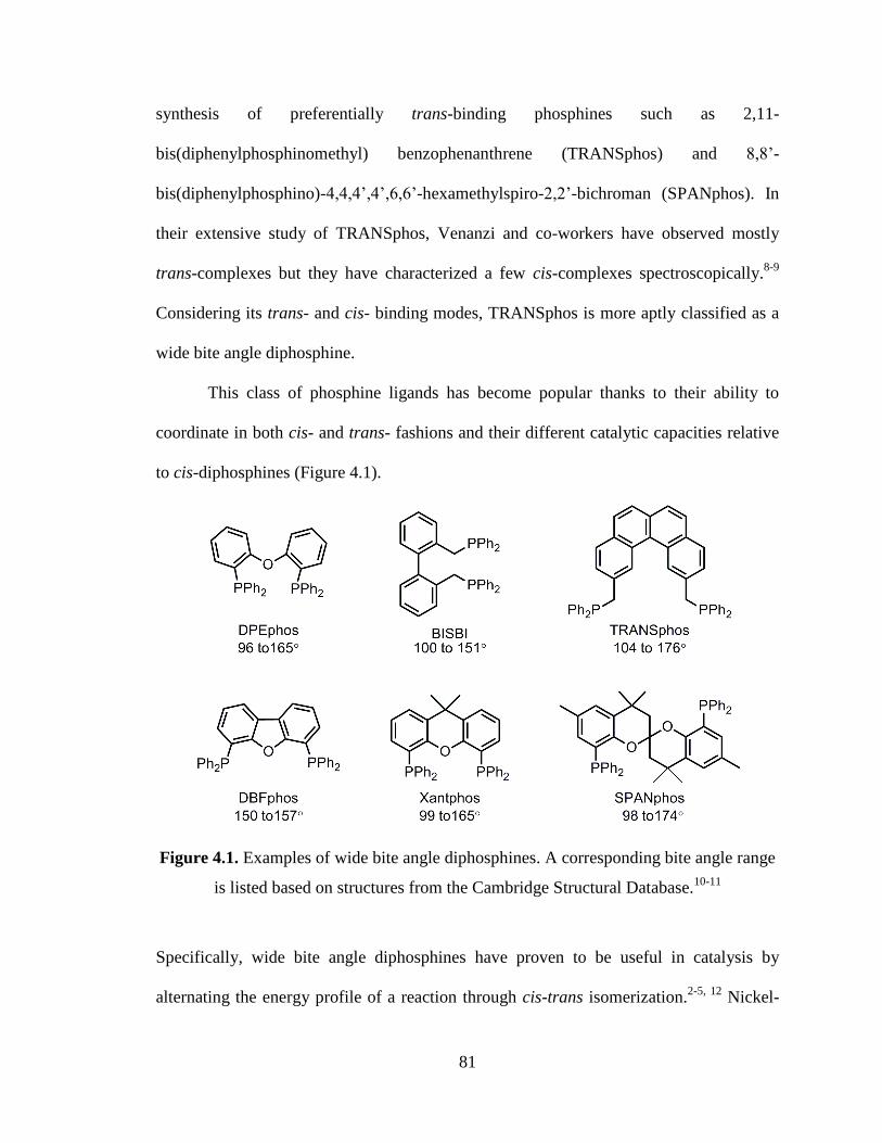
81
synthesis of preferentially trans-binding phosphines such as 2,11-
bis(diphenylphosphinomethyl) benzophenanthrene (TRANSphos) and 8,8’-
bis(diphenylphosphino)-4,4,4’,4’,6,6’-hexamethylspiro-2,2’-bichroman (SPANphos). In
their extensive study of TRANSphos, Venanzi and co-workers have observed mostly
trans-complexes but they have characterized a few cis-complexes spectroscopically.8-9
Considering its trans- and cis- binding modes, TRANSphos is more aptly classified as a
wide bite angle diphosphine.
This class of phosphine ligands has become popular thanks to their ability to
coordinate in both cis- and trans- fashions and their different catalytic capacities relative
to cis-diphosphines (Figure 4.1).
Figure 4.1. Examples of wide bite angle diphosphines. A corresponding bite angle range
is listed based on structures from the Cambridge Structural Database.10-11
Specifically, wide bite angle diphosphines have proven to be useful in catalysis by
alternating the energy profile of a reaction through cis-trans isomerization.2-5, 12
Nickel-
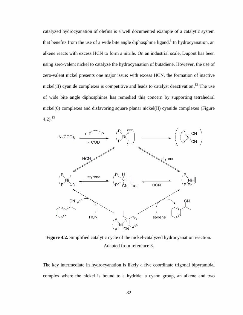
82
catalyzed hydrocyanation of olefins is a well documented example of a catalytic system
that benefits from the use of a wide bite angle diphosphine ligand.3 In hydrocyanation, an
alkene reacts with excess HCN to form a nitrile. On an industrial scale, Dupont has been
using zero-valent nickel to catalyze the hydrocyanation of butadiene. However, the use of
zero-valent nickel presents one major issue: with excess HCN, the formation of inactive
nickel(II) cyanide complexes is competitive and leads to catalyst deactivation.13
The use
of wide bite angle diphosphines has remedied this concern by supporting tetrahedral
nickel(0) complexes and disfavoring square planar nickel(II) cyanide complexes (Figure
4.2).13
Figure 4.2. Simplified catalytic cycle of the nickel-catalyzed hydrocyanation reaction.
Adapted from reference 3.
The key intermediate in hydrocyanation is likely a five coordinate trigonal bipyramidal
complex where the nickel is bound to a hydride, a cyano group, an alkene and two
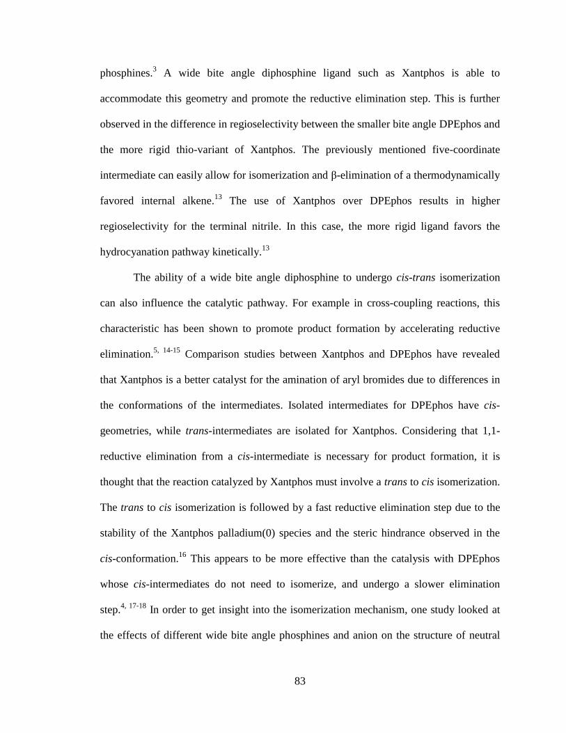
83
phosphines.3 A wide bite angle diphosphine ligand such as Xantphos is able to
accommodate this geometry and promote the reductive elimination step. This is further
observed in the difference in regioselectivity between the smaller bite angle DPEphos and
the more rigid thio-variant of Xantphos. The previously mentioned five-coordinate
intermediate can easily allow for isomerization and β-elimination of a thermodynamically
favored internal alkene.13
The use of Xantphos over DPEphos results in higher
regioselectivity for the terminal nitrile. In this case, the more rigid ligand favors the
hydrocyanation pathway kinetically.13
The ability of a wide bite angle diphosphine to undergo cis-trans isomerization
can also influence the catalytic pathway. For example in cross-coupling reactions, this
characteristic has been shown to promote product formation by accelerating reductive
elimination.5, 14-15
Comparison studies between Xantphos and DPEphos have revealed
that Xantphos is a better catalyst for the amination of aryl bromides due to differences in
the conformations of the intermediates. Isolated intermediates for DPEphos have cis-
geometries, while trans-intermediates are isolated for Xantphos. Considering that 1,1-
reductive elimination from a cis-intermediate is necessary for product formation, it is
thought that the reaction catalyzed by Xantphos must involve a trans to cis isomerization.
The trans to cis isomerization is followed by a fast reductive elimination step due to the
stability of the Xantphos palladium(0) species and the steric hindrance observed in the
cis-conformation.16
This appears to be more effective than the catalysis with DPEphos
whose cis-intermediates do not need to isomerize, and undergo a slower elimination
step.4, 17-18
In order to get insight into the isomerization mechanism, one study looked at
the effects of different wide bite angle phosphines and anion on the structure of neutral
84
and ionic 4-cyanophenylpalladium and methypalladium complexes in solution and in the
solid state.16
From this study, they proposed one mechanism for the cis-trans
isomerization of Xantphos metal complexes focused on the participation of the oxygen on
the backbone (Figure 4.3).16
Figure 4.3. Possible mechanism for the cis-trans isomerization of Xantphos.
Figure adapted from reference 16.
While the participation of the oxygen atom in the backbone of Xantphos is
beneficial in the case of palladium cross-coupling reactions, sp2 and sp
3 C-H bonds of the
ligand backbone of other wide bite angle phosphines can be activated by the metal
center.19
This has been observed in rhodium complexes when a long –CH2 chain is used
to connect the two phosphines.19
One solution to preventing unwanted interaction from
the backbone is the use of an inflexible backbone. For instance, TRANSphos contains a
benzophenanthrene π-system and SPANphos incorporates a fused spirobichroman ring
(Figure 4.1).20-21
Another approach has been lengthening the distance between the donor
atoms and the backbone through the inclusion of phenyl linkers.22-23
Using a similar
approach, our group has synthesized a new wide bite angle diphosphine ligand 4,6-bis(3-
diisopropylphosphinophenyl)dibenzofuran (iPr
DPDBFphos) with two alkylphosphine
donors connected to a dibenzofuran backbone by phenyl linkers (Scheme 4.1).22
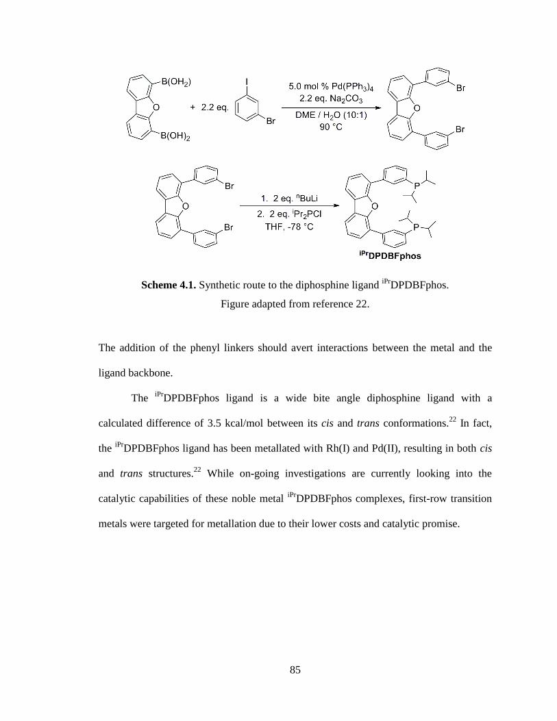
85
Scheme 4.1. Synthetic route to the diphosphine ligand iPr
DPDBFphos.
Figure adapted from reference 22.
The addition of the phenyl linkers should avert interactions between the metal and the
ligand backbone.
The iPr
DPDBFphos ligand is a wide bite angle diphosphine ligand with a
calculated difference of 3.5 kcal/mol between its cis and trans conformations.22
In fact,
the iPr
DPDBFphos ligand has been metallated with Rh(I) and Pd(II), resulting in both cis
and trans structures.22
While on-going investigations are currently looking into the
catalytic capabilities of these noble metal iPr
DPDBFphos complexes, first-row transition
metals were targeted for metallation due to their lower costs and catalytic promise.

86
4.2 Catalysis with nickel diphosphines
Though strides have been made towards the use of a variety of first-row transition
metals in catalysis, nickel-based catalysts have become most prominent in the field of
catalysis employing wide bite-angle diphosphines.2-3, 5, 12
Their use has been shown to
enhance the yield and selectivity of a variety of catalytic reactions. For example,
Ni(COD)2 (COD = cycloocta-1-5-diene) in the presence of wide bite angle diphosphines
can efficiently catalyze the hydrocyanation of alkenes.7, 13
An in-depth look at the
relationship between the bite angle and the efficiency of the catalyst revealed that wider
bite angle diphosphines were favorable and gave improved yields and selectivity over
monophosphines and diphosphines with bite angles smaller than 106°.7, 13, 15
Additionally, the use of wide bite angle diphosphines is believed to stabilize tetrahedral
nickel(0) intermediates, causing faster reductive elimination from nickel(II).7
Cross coupling reactions have also been catalyzed by nickel diphopshine
complexes. Saito and co-workers have reported the coupling of chloroarenes with
arylboronic acids with high yields using NiCl2(dppf) (dppf = 1,1’-
bis(diphenylphosphino)ferrocene) in the presence of n-butyl lithium as a reducing agent
to yield a nickel(0) complex in situ.24
Similarly, NiCl2(dppe) catalyzes for the formation
of styrene from the cross coupling of vinyl chloride and PhMgBr.25
While the key intermediates in these cross coupling reactions have been generally
hypothesized to be nickel(0) species, recent studies of N-heterocyclic carbene nickel(I)
complexes have revived the alternative proposal that nickel(I) species are involved in
both Kumada and Suzuki cross coupling reactions.26-28
Considering the continued
ambiguities in the mechanisms of nickel-mediated cross coupling reactions, the

87
development of well-defined nickel(I) and nickel(II) complexes supported by a wide bite-
angle ligand would be beneficial for gaining insight into the mechanism.
88
4.3 Scope of thesis
The objective of the second part of this dissertation is to synthesize nickel(I) and
nickel(II) complexes supported by the wide bite angle diphosphine iPr
DPDBFphos and to
examine their reactivity and catalytic capability towards cross coupling reactions. In
chapter 5, the synthesis and characterization of a series of first-row transition metal
complexes with iPr
DPDBFphos is described. Within this family of complexes, it is shown
through X-ray crystallography that iPr
DPDBFphos can accommodate a wide range of bite
angles, allowing for a diverse set of geometries. Additionally, chapter 5 presents the
synthesis and characterization of a low-valent three-coordinate nickel(I) complex
(iPr
DPDBFphos)NiCl. Chapter 6 examines the reactivity of (iPr
DPDBFphos)NiCl
including metathesis reactions, small-molecule binding, and C-X bond activation.
Furthermore, the catalytic activity of Ni(II) versus Ni(I) is compared by probing
(iPr
DPDBFphos)NiCl2 and (iPr
DPDBFphos)NiCl, respectively, as potential catalysts in the
Kumada cross coupling of vinyl bromide and phenyl Grignard reagent.
89
-Chapter Five-
First-Row Transition-Metal Complexes of the Wide Bite-
Angle Diphosphine iPrDPDBFphos
In part from:
Marlier, E. E.; Tereniak, S. J.; Ding, K.; Mulliken, J. E.; Lu, C. C. Inorg. Chem. 2011, 50,
9290-9299.
90
5.1 Overview
The diphosphine 4,6-bis(3-diisopropylphosphinophenyl)dibenzofuran
(iPr
DPDBFphos) has been metalated with transition metal dichlorides of zinc, cobalt, and
nickel and iron pentacarbonyl to yield (iPr
DPDBFphos)MCl2 and
(iPr
DPDBFphos)Fe(CO)3 complexes, respectively. Within these compounds, the
diphosphine iPr
DPDBFphos adapts a wide range of bite angles (115 to 180°) as
determined by X-ray crystallography. A three-coordinate planar Ni(I) species was
isolated from the reduction of (iPr
DPDBFphos)NiCl2 with KC8. Low-temperature EPR
measurements of (iPr
DPDBFphos)NiCl allow the determination of g values (2.09, 2.14,
2.37) and hyperfine coupling constants to two 31
P nuclei, Aiso = 46 x 10−4
cm−1
, and one
37Cl/
35Cl nucleus, A = (12, 0.7, 35) x 10
−4 cm
−1. Density functional theory (DFT) studies
reveal the nature of the magnetic orbital to be dxy, which has σ-antibonding and -
antibonding interactions with the phosphorus and chloride atoms, respectively.
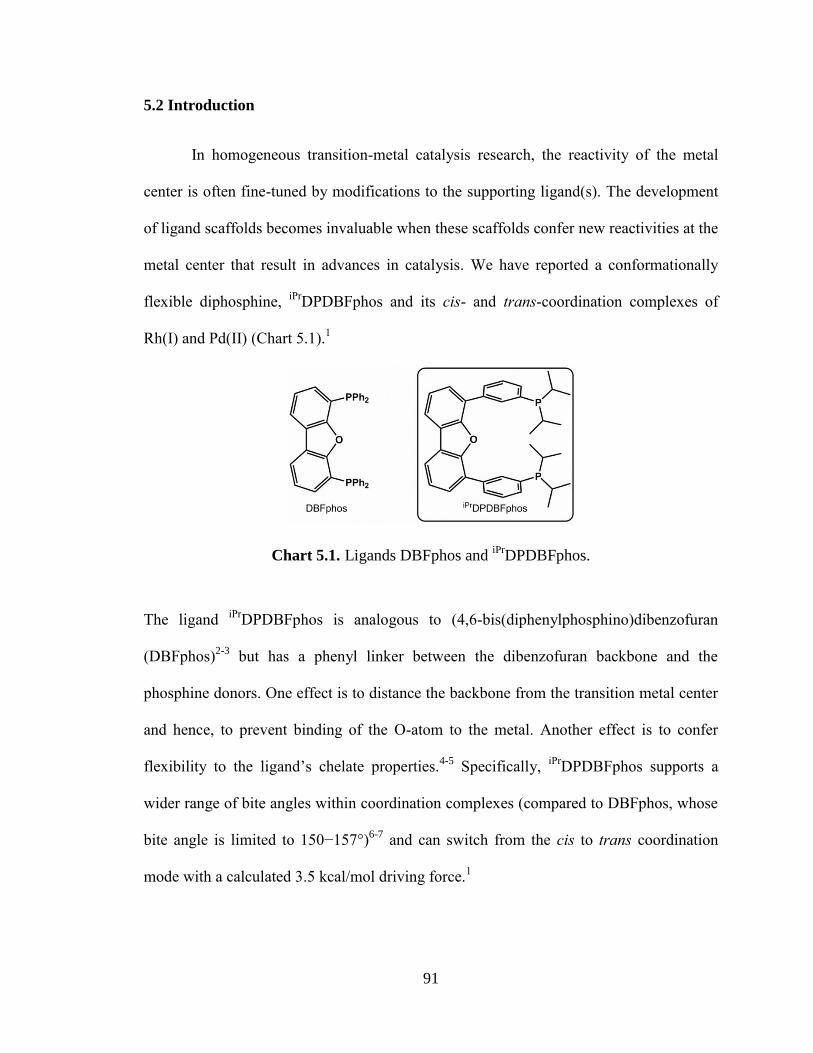
91
5.2 Introduction
In homogeneous transition-metal catalysis research, the reactivity of the metal
center is often fine-tuned by modifications to the supporting ligand(s). The development
of ligand scaffolds becomes invaluable when these scaffolds confer new reactivities at the
metal center that result in advances in catalysis. We have reported a conformationally
flexible diphosphine, iPr
DPDBFphos and its cis- and trans-coordination complexes of
Rh(I) and Pd(II) (Chart 5.1).1
Chart 5.1. Ligands DBFphos and iPr
DPDBFphos.
The ligand iPr
DPDBFphos is analogous to (4,6-bis(diphenylphosphino)dibenzofuran
(DBFphos)2-3
but has a phenyl linker between the dibenzofuran backbone and the
phosphine donors. One effect is to distance the backbone from the transition metal center
and hence, to prevent binding of the O-atom to the metal. Another effect is to confer
flexibility to the ligand’s chelate properties.4-5
Specifically, iPr
DPDBFphos supports a
wider range of bite angles within coordination complexes (compared to DBFphos, whose
bite angle is limited to 150−157°)6-7
and can switch from the cis to trans coordination
mode with a calculated 3.5 kcal/mol driving force.1
92
Herein, we extend the coordination chemistry of iPr
DPDBFphos to the first-row
transition metals zinc, cobalt, and nickel. A long-standing goal in homogeneous catalysis
is to replace precious transition-metal catalysts with their earth-abundant counterparts.8-9
Notable organometallic examples include iron-mediated cross-coupling reactions,10
hydroxylation of aliphatic C−H bonds,11-14
and hydrogenation of alkenes15-17
and
ketones;18-19
cobalt-mediated hydroformylation20
and cross-coupling;21
and nickel-
mediated polymerization and oligomerization of olefins,22-24
cross-coupling,25-26
and
cyclization reactions.27
More relevant to the complexes reported here, P2MX2-type
complexes of cobalt and zinc have been shown to be effective catalysts for the
polymerization of methyl acrylate and CO2/ethylene oxide, respectively.28-29
The (iPr
DPDBFphos)MCl2 and (iPr
DPDBFphos)M(CO)3 complexes are
characterized by UV-Visible (UV-Vis) spectroscopy, heteronuclear NMR spectroscopies,
and single-crystal X-ray diffraction studies. A three-coordinate nickel(I) chloride
complex is presented with additional characterization from electron paramagnetic
resonance (EPR) spectroscopy and density functional theory (DFT) calculations.
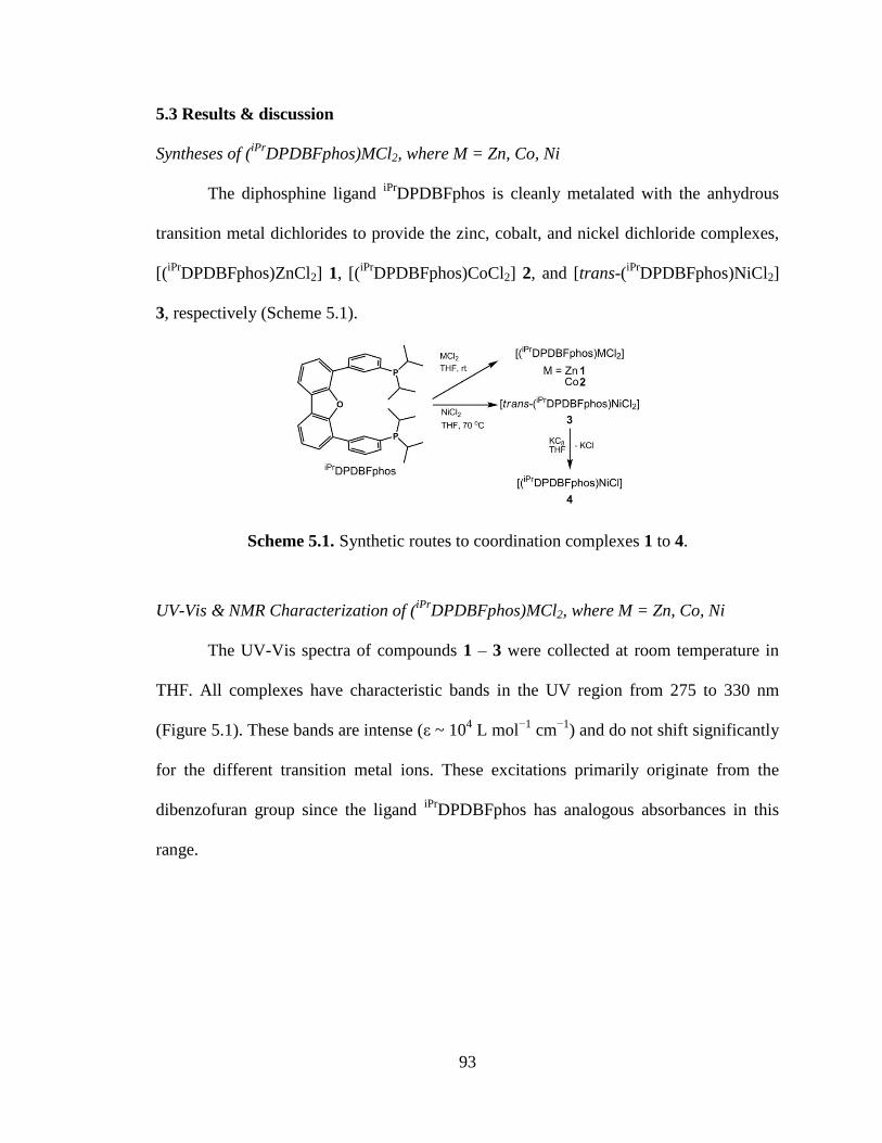
93
5.3 Results & discussion
Syntheses of (iPr
DPDBFphos)MCl2, where M = Zn, Co, Ni
The diphosphine ligand iPr
DPDBFphos is cleanly metalated with the anhydrous
transition metal dichlorides to provide the zinc, cobalt, and nickel dichloride complexes,
[(iPr
DPDBFphos)ZnCl2] 1, [(iPr
DPDBFphos)CoCl2] 2, and [trans-(iPr
DPDBFphos)NiCl2]
3, respectively (Scheme 5.1).
Scheme 5.1. Synthetic routes to coordination complexes 1 to 4.
UV-Vis & NMR Characterization of (iPr
DPDBFphos)MCl2, where M = Zn, Co, Ni
The UV-Vis spectra of compounds 1 – 3 were collected at room temperature in
THF. All complexes have characteristic bands in the UV region from 275 to 330 nm
(Figure 5.1). These bands are intense (ε ~ 104 L mol
−1 cm
−1) and do not shift significantly
for the different transition metal ions. These excitations primarily originate from the
dibenzofuran group since the ligand iPr
DPDBFphos has analogous absorbances in this
range.
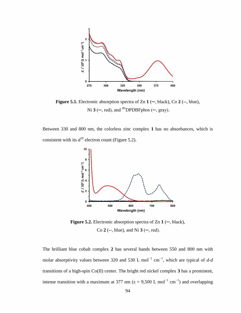
94
Figure 5.1. Electronic absorption spectra of Zn 1 (─, black), Co 2 (--, blue),
Ni 3 (─, red), and iPr
DPDBFphos (─, gray).
Between 330 and 800 nm, the colorless zinc complex 1 has no absorbances, which is
consistent with its d10
electron count (Figure 5.2).
Figure 5.2. Electronic absorption spectra of Zn 1 (─, black),
Co 2 (--, blue), and Ni 3 (─, red).
The brilliant blue cobalt complex 2 has several bands between 550 and 800 nm with
molar absorptivity values between 320 and 530 L mol−1
cm−1
, which are typical of d-d
transitions of a high-spin Co(II) center. The bright red nickel complex 3 has a prominent,
intense transition with a maximum at 377 nm (ε = 9,500 L mol−1
cm−1
) and overlapping
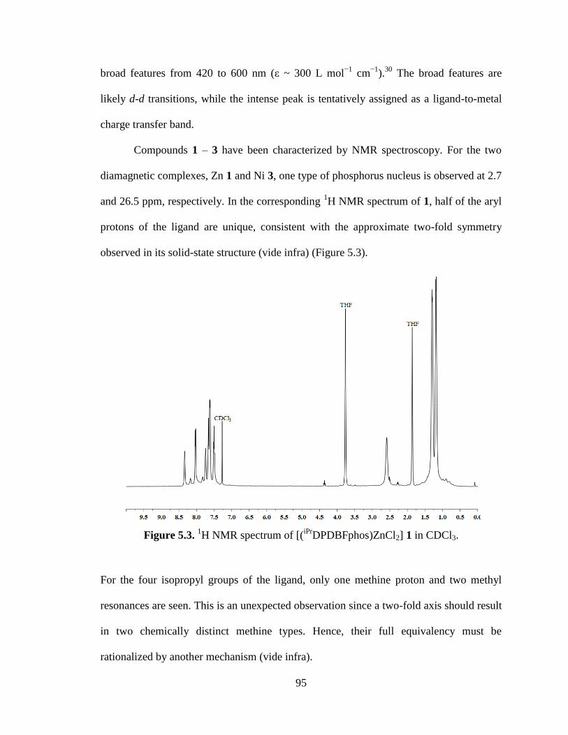
95
broad features from 420 to 600 nm (ε ~ 300 L mol−1
cm−1
).30
The broad features are
likely d-d transitions, while the intense peak is tentatively assigned as a ligand-to-metal
charge transfer band.
Compounds 1 – 3 have been characterized by NMR spectroscopy. For the two
diamagnetic complexes, Zn 1 and Ni 3, one type of phosphorus nucleus is observed at 2.7
and 26.5 ppm, respectively. In the corresponding 1H NMR spectrum of 1, half of the aryl
protons of the ligand are unique, consistent with the approximate two-fold symmetry
observed in its solid-state structure (vide infra) (Figure 5.3).
Figure 5.3. 1H NMR spectrum of [(
iPrDPDBFphos)ZnCl2] 1 in CDCl3.
For the four isopropyl groups of the ligand, only one methine proton and two methyl
resonances are seen. This is an unexpected observation since a two-fold axis should result
in two chemically distinct methine types. Hence, their full equivalency must be
rationalized by another mechanism (vide infra).
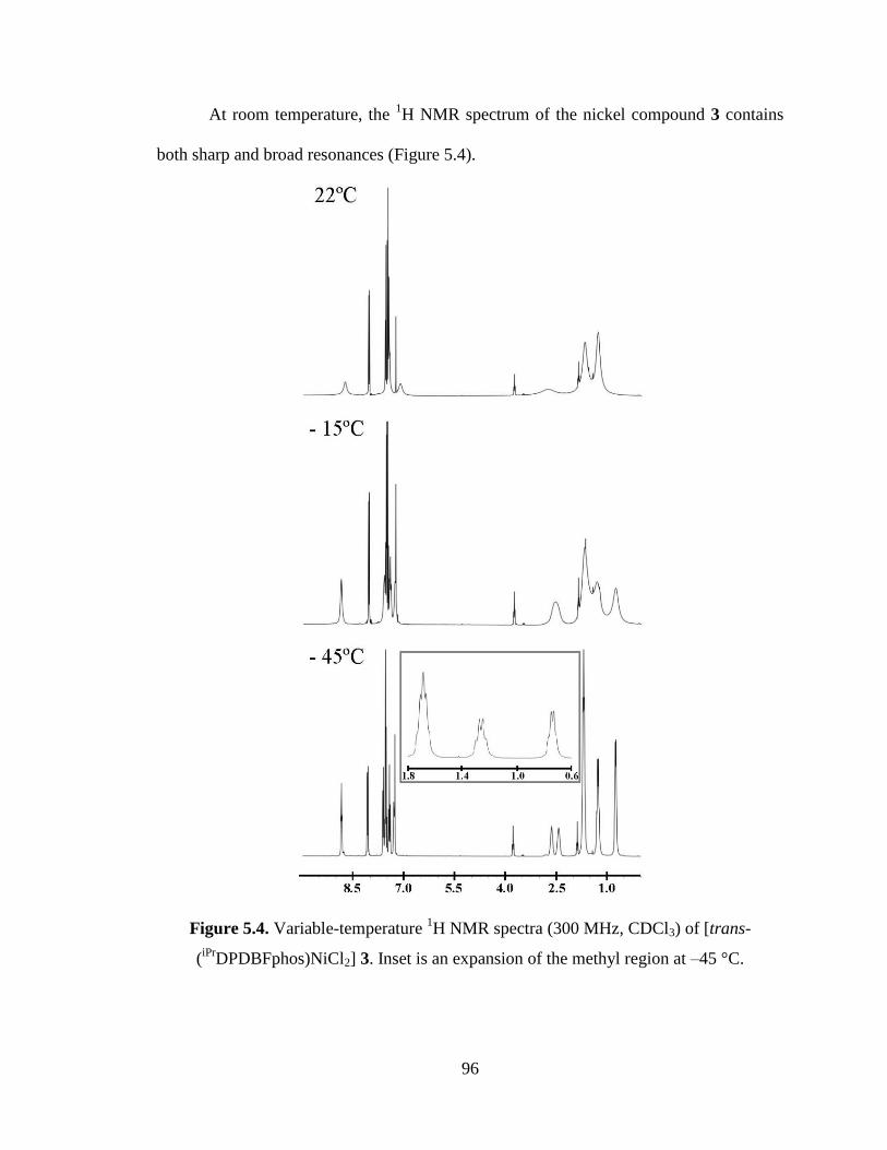
96
At room temperature, the 1H NMR spectrum of the nickel compound 3 contains
both sharp and broad resonances (Figure 5.4).
Figure 5.4. Variable-temperature 1H NMR spectra (300 MHz, CDCl3) of [trans-
(iPr
DPDBFphos)NiCl2] 3. Inset is an expansion of the methyl region at –45 °C.
97
The sharp signals correspond to the dibenzofuran group, while those belonging to the
isopropyl groups and the phenyl linkers are significantly broadened. Upon cooling to −45
°C, the 1H NMR spectrum of 3 sharpens completely, and exactly half of the protons of
the ligand are uniquely observed, consistent with the approximate mirror-plane symmetry
of its molecular structure. At low temperature, the isopropyl groups resolve into two
distinct methine and three methyl resonances. The presence of the mirror plane means
that the two chemically different methine protons occur within the same phosphine
group, ie. P(CHMe2)(C'HMe2). If P−Cmethine bond rotation is fast, then two methyl peaks
would be expected; if slow, four. At −45 °C, only three methyl signals are apparent due
to a coincidental overlap of two of the four possible methyl peaks.31
At room
temperature, the 1H NMR spectrum of 3 shows one methine and two methyl signals, akin
to that of 1. Again, the higher symmetry solution-structure cannot be explained by a
mirror plane alone. Additional fluxional processes will be described in the next section.
The cobalt dichloride compound 2 is characterized by a paramagnetic 1H NMR
spectrum, which is consistent with a Co(II) oxidation state. Its solution magnetic moment
of μeff = 4.01 μB is slightly greater than the spin-only magnetic moment for an S = 3/2
state (μS.O. = 3.88 μB), which leads to the expected assignment of a high-spin Co(II)
center.
Synthesis & Characterization of (iPr
DPDBFphos)Fe(CO)3
When our attempts at metallating iPr
DPDBFphos with iron dichloride did not lead
to a highly purified material, we turned our attention towards metallating with iron
pentacarbonyl. During a 6 hour photolysis using a mercury vapor lamp, iPr
DPDBFphos

98
can be metallated with iron pentacarbonyl to yield [(iPr
DPDBFphos)Fe(CO)3] 5 in nearly
quantitative yield.
Complex 5 was characterized by NMR and IR spectroscopy. The 31
P NMR
spectrum shows one peak, located at 90.4 ppm while the 1H NMR spectrum displays the
same two fold symmetry observed in complexes 1 and 3 (Figure 5.5).
Figure 5.5. 1H NMR spectrum of [(
iPrDPDBFphos)Fe(CO)3] 5 in CDCl3.
A total of three resonances are observed in the aromatic region, integrating to the 14
protons of the benzofuran backbone and phenyl linkers. For the four isopropyl groups of
the ligand, only one methine proton and two methyl resonances are seen.
The IR spectrum of 5 has two different signals; a strong resonance at 1869 cm-1
and a weak resonance at 1959 cm-1
. These are assigned as E´ and A1´ stretching mode
respectively, where the A1´ stretching mode is weak considering it is forbidden. These
stretching resonances are similar to those observed for trans-Fe(CO)3(PR3)2.32
When PR3
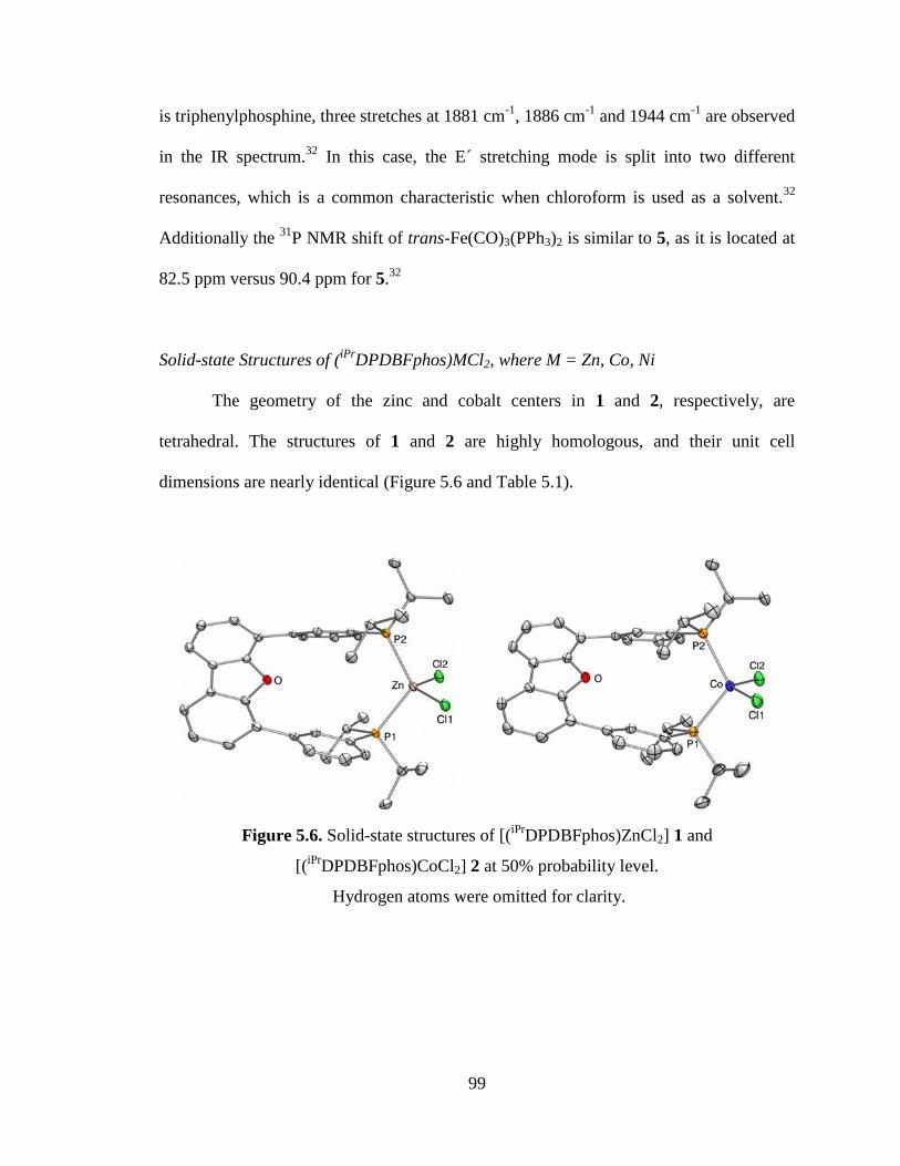
99
is triphenylphosphine, three stretches at 1881 cm-1
, 1886 cm-1
and 1944 cm-1
are observed
in the IR spectrum.32
In this case, the E´ stretching mode is split into two different
resonances, which is a common characteristic when chloroform is used as a solvent.32
Additionally the 31
P NMR shift of trans-Fe(CO)3(PPh3)2 is similar to 5, as it is located at
82.5 ppm versus 90.4 ppm for 5.32
Solid-state Structures of (iPr
DPDBFphos)MCl2, where M = Zn, Co, Ni
The geometry of the zinc and cobalt centers in 1 and 2, respectively, are
tetrahedral. The structures of 1 and 2 are highly homologous, and their unit cell
dimensions are nearly identical (Figure 5.6 and Table 5.1).
Figure 5.6. Solid-state structures of [(iPr
DPDBFphos)ZnCl2] 1 and
[(iPr
DPDBFphos)CoCl2] 2 at 50% probability level.
Hydrogen atoms were omitted for clarity.
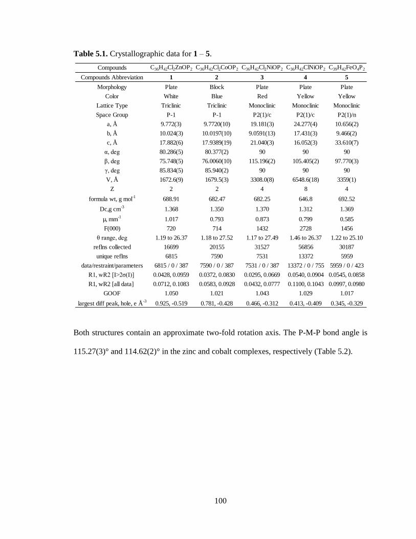
100
Table 5.1. Crystallographic data for 1 – 5.
Both structures contain an approximate two-fold rotation axis. The P-M-P bond angle is
115.27(3)° and 114.62(2)° in the zinc and cobalt complexes, respectively (Table 5.2).
Compounds C36H42Cl2ZnOP2 C36H42Cl2CoOP2 C36H42Cl2NiOP2 C36H42ClNiOP2 C39H42FeO4P2
Compounds Abbreviation 1 2 3 4 5
Morphology Plate Block Plate Plate Plate
Color White Blue Red Yellow Yellow
Lattice Type Triclinic Triclinic Monoclinic Monoclinic Monoclinic
Space Group P-1 P-1 P2(1)/c P2(1)/c P2(1)/n
a, Å 9.772(3) 9.7720(10) 19.181(3) 24.277(4) 10.656(2)
b, Å 10.024(3) 10.0197(10) 9.0591(13) 17.431(3) 9.466(2)
c, Å 17.882(6) 17.9389(19) 21.040(3) 16.052(3) 33.610(7)
α, deg 80.286(5) 80.377(2) 90 90 90
β, deg 75.748(5) 76.0060(10) 115.196(2) 105.405(2) 97.770(3)
γ, deg 85.834(5) 85.940(2) 90 90 90
V, Å 1672.6(9) 1679.5(3) 3308.0(8) 6548.6(18) 3359(1)
Z 2 2 4 8 4
formula wt, g mol-1
688.91 682.47 682.25 646.8 692.52
Dc,g cm-3
1.368 1.350 1.370 1.312 1.369
µ, mm-1
1.017 0.793 0.873 0.799 0.585
F(000) 720 714 1432 2728 1456
θ range, deg 1.19 to 26.37 1.18 to 27.52 1.17 to 27.49 1.46 to 26.37 1.22 to 25.10
reflns collected 16699 20155 31527 56856 30187
unique reflns 6815 7590 7531 13372 5959
data/restraint/parameters 6815 / 0 / 387 7590 / 0 / 387 7531 / 0 / 387 13372 / 0 / 755 5959 / 0 / 423
R1, wR2 [I>2ζ(I)] 0.0428, 0.0959 0.0372, 0.0830 0.0295, 0.0669 0.0540, 0.0904 0.0545, 0.0858
R1, wR2 [all data] 0.0712, 0.1083 0.0583, 0.0928 0.0432, 0.0777 0.1100, 0.1043 0.0997, 0.0980
GOOF 1.050 1.021 1.043 1.029 1.017
largest diff peak, hole, e Å-3
0.925, -0.519 0.781, -0.428 0.466, -0.312 0.413, -0.409 0.345, -0.329
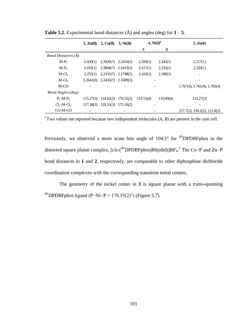
101
Table 5.2. Experimental bond distances (Å) and angles (deg) for 1 – 5.
a Two values are reported because two independent molecules (A, B) are present in the unit cell.
Previously, we observed a more acute bite angle of 104.5° for iPr
DPDBFphos in the
distorted square planar complex, [cis-(iPr
DPDBFphos)Rh(nbd)]BF4.1 The Co−P and Zn−P
bond distances in 1 and 2, respectively, are comparable to other diphosphine dichloride
coordination complexes with the corresponding transition metal centers.
The geometry of the nickel center in 3 is square planar with a trans-spanning
iPrDPDBFphos ligand (P−Ni−P = 176.55(2)°) (Figure 5.7).
1, Zn(II) 2, Co(II) 3, Ni(II) 4, Ni(I)a
5, Fe(0)
A B
Bond Distances (Å)
M-P1 2.420(1) 2.3920(7) 2.2410(5) 2.209(1) 2.243(1) 2.217(1)
M-P2 2.416(1) 2.3844(7) 2.2413(5) 2.217(1) 2.233(1) 2.220(1)
M-Cl1 2.255(1) 2.2331(7) 2.1748(5) 2.163(1) 2.180(1) -
M-Cl2 2.2641(9) 2.2435(7) 2.1609(5) - -
M-CO - - - - 1.767(4), 1.761(4), 1.765(4)
Bond Angles (deg)
P1-M-P2 115.27(3) 114.62(2) 176.55(2) 115.53(4) 119.49(4) 115.27(3)
Cl1-M-Cl2 117.38(3) 118.55(3) 175.10(2) - -
CO-M-CO - - - - 117.7(2), 130.2(2), 112.0(2)
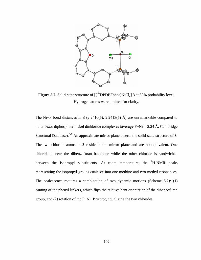
102
Figure 5.7. Solid-state structure of [(iPr
DPDBFphos)NiCl2] 3 at 50% probability level.
Hydrogen atoms were omitted for clarity.
The Ni−P bond distances in 3 (2.2410(5), 2.2413(5) Å) are unremarkable compared to
other trans-diphosphine nickel dichloride complexes (average P−Ni = 2.24 Å, Cambridge
Structural Database).6-7
An approximate mirror plane bisects the solid-state structure of 3.
The two chloride atoms in 3 reside in the mirror plane and are nonequivalent. One
chloride is near the dibenzofuran backbone while the other chloride is sandwiched
between the isopropyl substituents. At room temperature, the 1H-NMR peaks
representing the isopropyl groups coalesce into one methine and two methyl resonances.
The coalescence requires a combination of two dynamic motions (Scheme 5.2): (1)
canting of the phenyl linkers, which flips the relative bent orientation of the dibenzofuran
group, and (2) rotation of the P−Ni−P vector, equalizing the two chlorides.
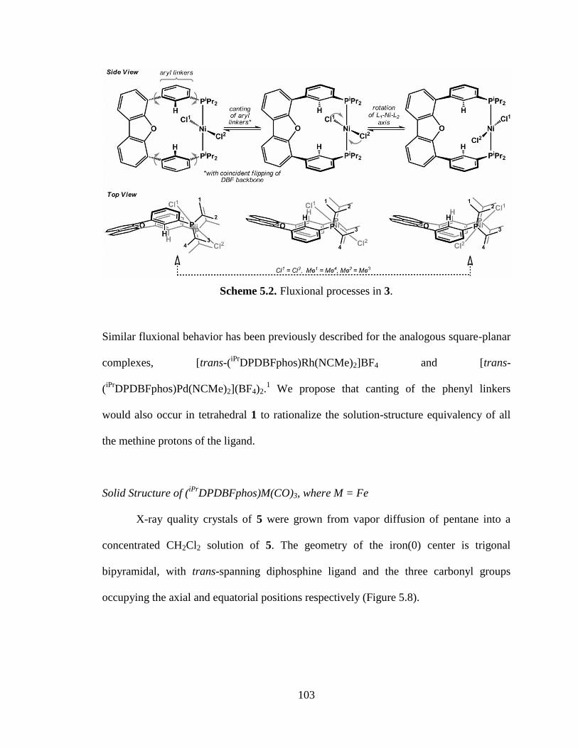
103
Scheme 5.2. Fluxional processes in 3.
Similar fluxional behavior has been previously described for the analogous square-planar
complexes, [trans-(iPr
DPDBFphos)Rh(NCMe)2]BF4 and [trans-
(iPr
DPDBFphos)Pd(NCMe)2](BF4)2.1 We propose that canting of the phenyl linkers
would also occur in tetrahedral 1 to rationalize the solution-structure equivalency of all
the methine protons of the ligand.
Solid Structure of (iPr
DPDBFphos)M(CO)3, where M = Fe
X-ray quality crystals of 5 were grown from vapor diffusion of pentane into a
concentrated CH2Cl2 solution of 5. The geometry of the iron(0) center is trigonal
bipyramidal, with trans-spanning diphosphine ligand and the three carbonyl groups
occupying the axial and equatorial positions respectively (Figure 5.8).
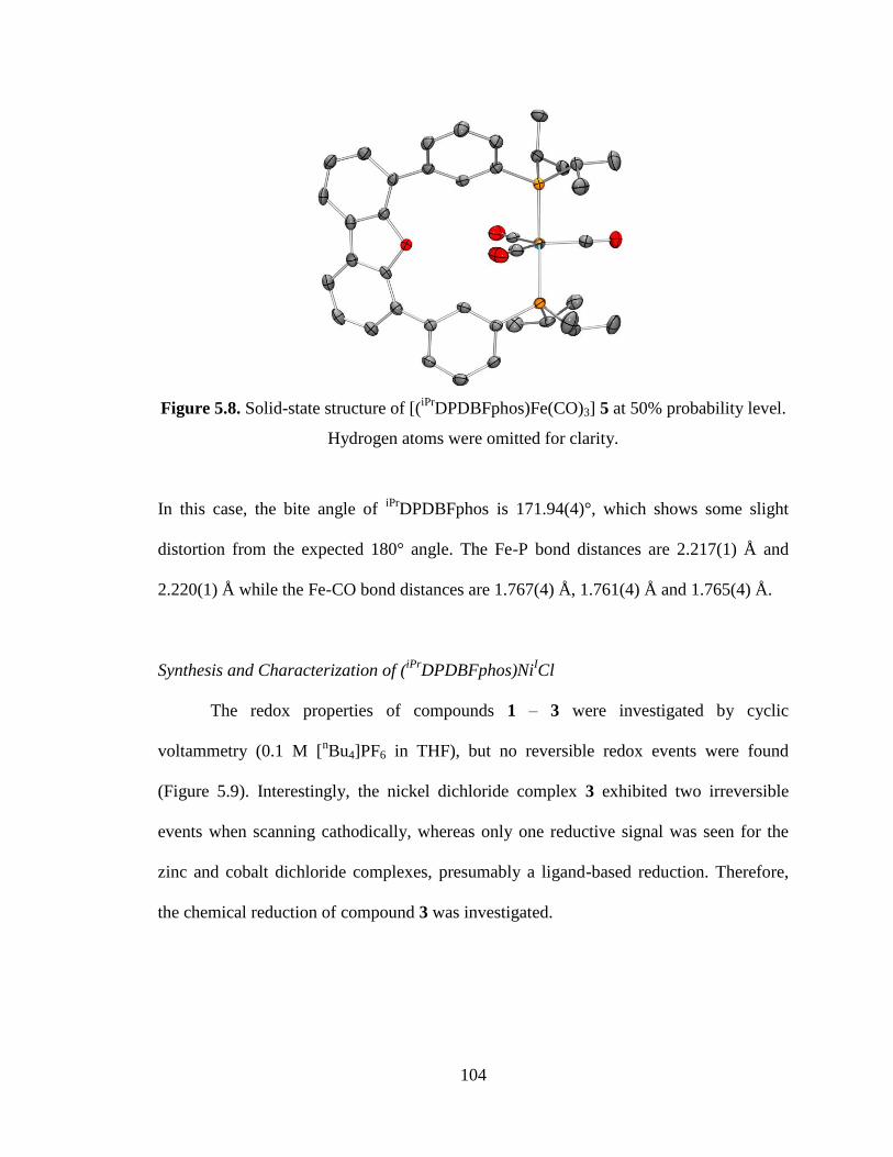
104
Figure 5.8. Solid-state structure of [(iPr
DPDBFphos)Fe(CO)3] 5 at 50% probability level.
Hydrogen atoms were omitted for clarity.
In this case, the bite angle of iPr
DPDBFphos is 171.94(4)°, which shows some slight
distortion from the expected 180° angle. The Fe-P bond distances are 2.217(1) Å and
2.220(1) Å while the Fe-CO bond distances are 1.767(4) Å, 1.761(4) Å and 1.765(4) Å.
Synthesis and Characterization of (iPr
DPDBFphos)NiICl
The redox properties of compounds 1 – 3 were investigated by cyclic
voltammetry (0.1 M [nBu4]PF6 in THF), but no reversible redox events were found
(Figure 5.9). Interestingly, the nickel dichloride complex 3 exhibited two irreversible
events when scanning cathodically, whereas only one reductive signal was seen for the
zinc and cobalt dichloride complexes, presumably a ligand-based reduction. Therefore,
the chemical reduction of compound 3 was investigated.
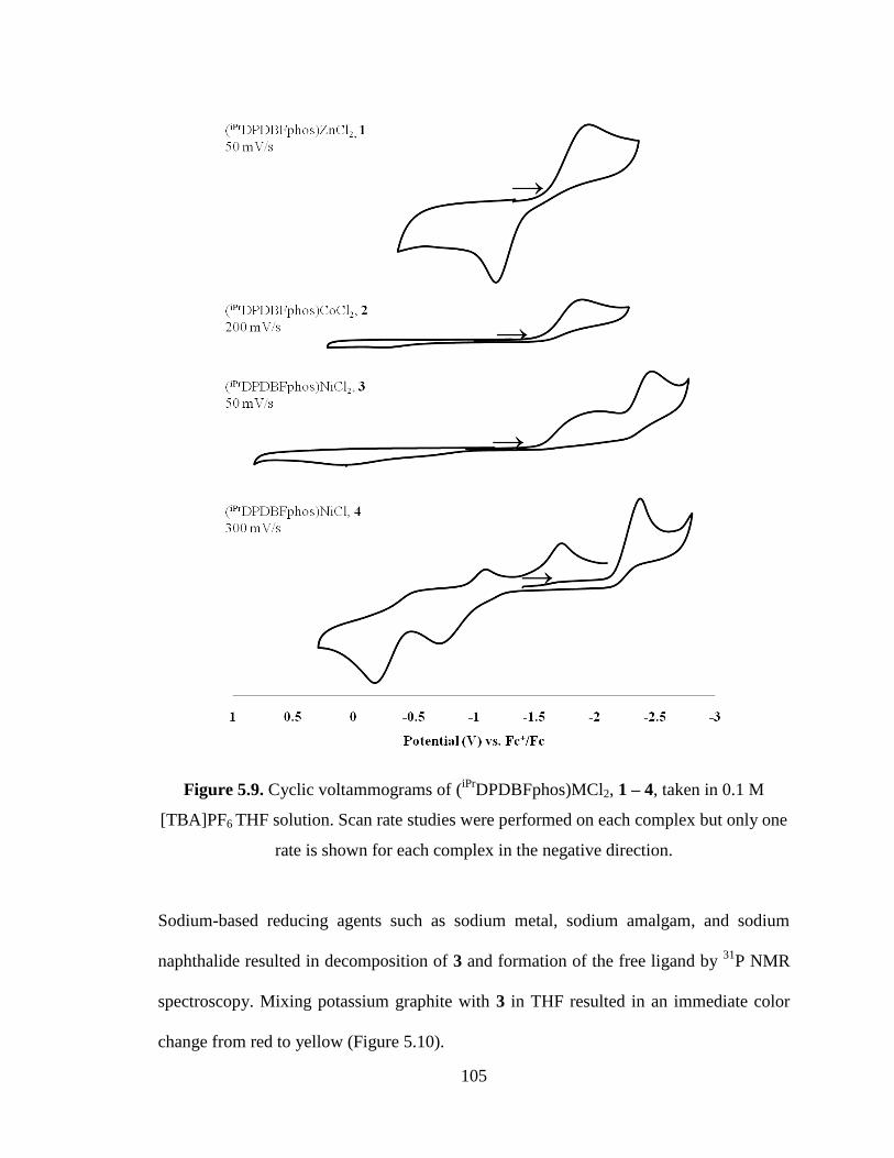
105
Figure 5.9. Cyclic voltammograms of (iPr
DPDBFphos)MCl2, 1 – 4, taken in 0.1 M
[TBA]PF6 THF solution. Scan rate studies were performed on each complex but only one
rate is shown for each complex in the negative direction.
Sodium-based reducing agents such as sodium metal, sodium amalgam, and sodium
naphthalide resulted in decomposition of 3 and formation of the free ligand by 31
P NMR
spectroscopy. Mixing potassium graphite with 3 in THF resulted in an immediate color
change from red to yellow (Figure 5.10).
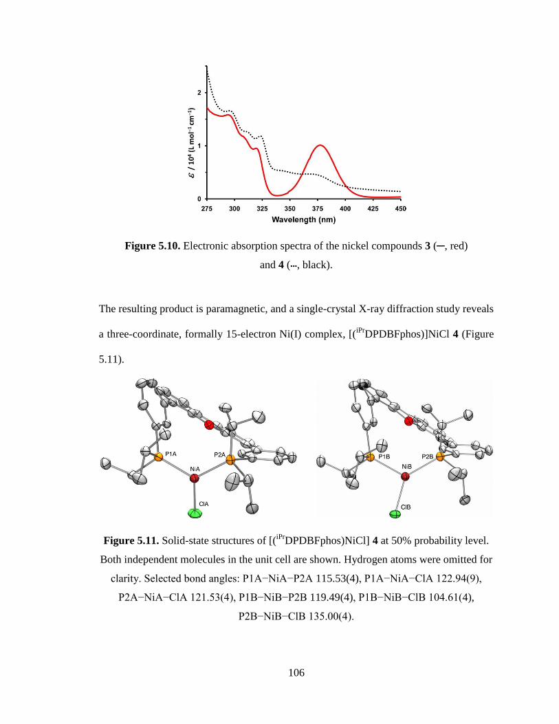
106
Figure 5.10. Electronic absorption spectra of the nickel compounds 3 (─, red)
and 4 (•••, black).
The resulting product is paramagnetic, and a single-crystal X-ray diffraction study reveals
a three-coordinate, formally 15-electron Ni(I) complex, [(iPr
DPDBFphos)]NiCl 4 (Figure
5.11).
Figure 5.11. Solid-state structures of [(iPr
DPDBFphos)NiCl] 4 at 50% probability level.
Both independent molecules in the unit cell are shown. Hydrogen atoms were omitted for
clarity. Selected bond angles: P1A−NiA−P2A 115.53(4), P1A−NiA−ClA 122.94(9),
P2A−NiA−ClA 121.53(4), P1B−NiB−P2B 119.49(4), P1B−NiB−ClB 104.61(4),
P2B−NiB−ClB 135.00(4).
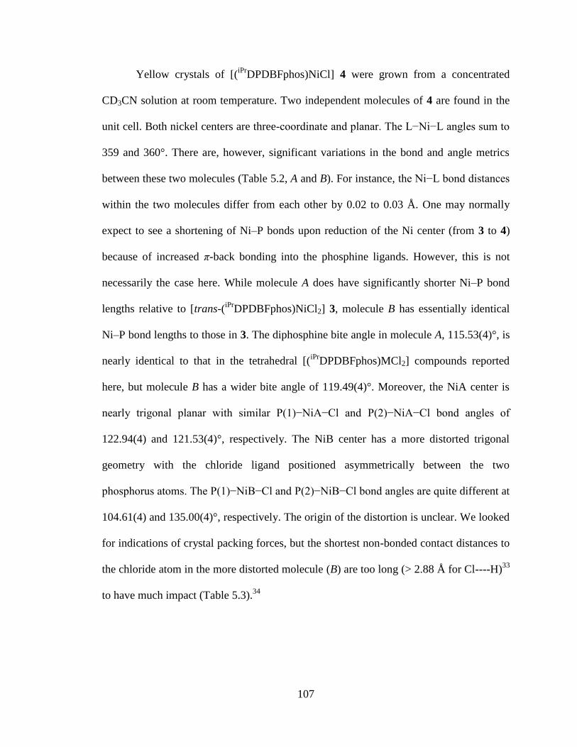
107
Yellow crystals of [(iPr
DPDBFphos)NiCl] 4 were grown from a concentrated
CD3CN solution at room temperature. Two independent molecules of 4 are found in the
unit cell. Both nickel centers are three-coordinate and planar. The L−Ni−L angles sum to
359 and 360°. There are, however, significant variations in the bond and angle metrics
between these two molecules (Table 5.2, A and B). For instance, the Ni−L bond distances
within the two molecules differ from each other by 0.02 to 0.03 Å. One may normally
expect to see a shortening of Ni–P bonds upon reduction of the Ni center (from 3 to 4)
because of increased π-back bonding into the phosphine ligands. However, this is not
necessarily the case here. While molecule A does have significantly shorter Ni–P bond
lengths relative to [trans-(iPr
DPDBFphos)NiCl2] 3, molecule B has essentially identical
Ni–P bond lengths to those in 3. The diphosphine bite angle in molecule A, 115.53(4)°, is
nearly identical to that in the tetrahedral [(iPr
DPDBFphos)MCl2] compounds reported
here, but molecule B has a wider bite angle of 119.49(4)°. Moreover, the NiA center is
nearly trigonal planar with similar P(1)−NiA−Cl and P(2)−NiA−Cl bond angles of
122.94(4) and 121.53(4)°, respectively. The NiB center has a more distorted trigonal
geometry with the chloride ligand positioned asymmetrically between the two
phosphorus atoms. The P(1)−NiB−Cl and P(2)−NiB−Cl bond angles are quite different at
104.61(4) and 135.00(4)°, respectively. The origin of the distortion is unclear. We looked
for indications of crystal packing forces, but the shortest non-bonded contact distances to
the chloride atom in the more distorted molecule (B) are too long (> 2.88 Å for Cl----H)33
to have much impact (Table 5.3).34
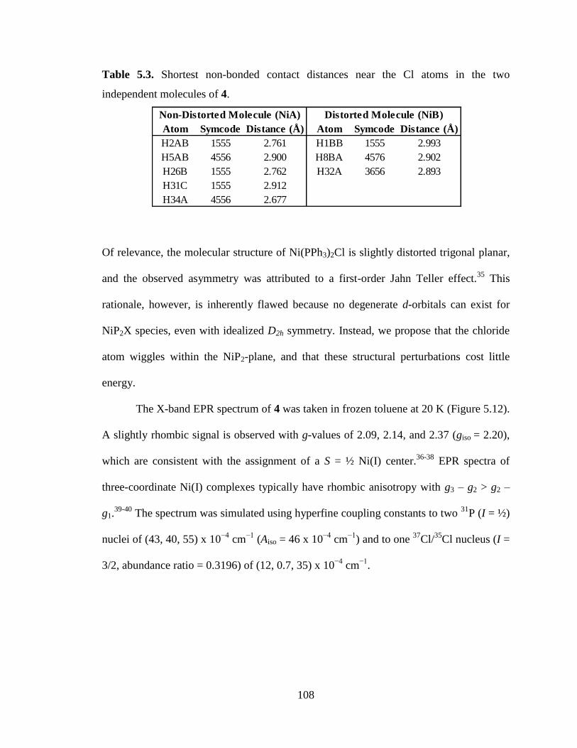
108
Table 5.3. Shortest non-bonded contact distances near the Cl atoms in the two
independent molecules of 4.
Of relevance, the molecular structure of Ni(PPh3)2Cl is slightly distorted trigonal planar,
and the observed asymmetry was attributed to a first-order Jahn Teller effect.35
This
rationale, however, is inherently flawed because no degenerate d-orbitals can exist for
NiP2X species, even with idealized D2h symmetry. Instead, we propose that the chloride
atom wiggles within the NiP2-plane, and that these structural perturbations cost little
energy.
The X-band EPR spectrum of 4 was taken in frozen toluene at 20 K (Figure 5.12).
A slightly rhombic signal is observed with g-values of 2.09, 2.14, and 2.37 (giso = 2.20),
which are consistent with the assignment of a S = ½ Ni(I) center.36-38
EPR spectra of
three-coordinate Ni(I) complexes typically have rhombic anisotropy with g3 – g2 > g2 –
g1.39-40
The spectrum was simulated using hyperfine coupling constants to two 31
P (I = ½)
nuclei of (43, 40, 55) x 10−4
cm−1
(Aiso = 46 x 10−4
cm−1
) and to one 37
Cl/35
Cl nucleus (I =
3/2, abundance ratio = 0.3196) of (12, 0.7, 35) x 10−4
cm−1
.
Atom Symcode Distance (Å) Atom Symcode Distance (Å)
H2AB 1555 2.761 H1BB 1555 2.993
H5AB 4556 2.900 H8BA 4576 2.902
H26B 1555 2.762 H32A 3656 2.893
H31C 1555 2.912
H34A 4556 2.677
Non-Distorted Molecule (NiA) Distorted Molecule (NiB)
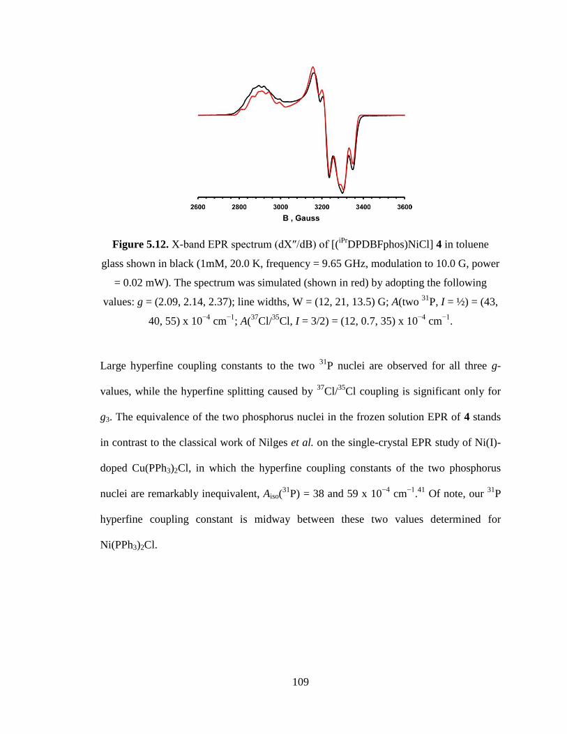
109
Figure 5.12. X-band EPR spectrum (dX″/dB) of [(iPr
DPDBFphos)NiCl] 4 in toluene
glass shown in black (1mM, 20.0 K, frequency = 9.65 GHz, modulation to 10.0 G, power
= 0.02 mW). The spectrum was simulated (shown in red) by adopting the following
values: g = (2.09, 2.14, 2.37); line widths, W = (12, 21, 13.5) G; A(two 31
P, I = ½) = (43,
40, 55) x 10−4
cm−1
; A(37
Cl/35
Cl, I = 3/2) = (12, 0.7, 35) x 10−4
cm−1
.
Large hyperfine coupling constants to the two 31
P nuclei are observed for all three g-
values, while the hyperfine splitting caused by 37
Cl/35
Cl coupling is significant only for
g3. The equivalence of the two phosphorus nuclei in the frozen solution EPR of 4 stands
in contrast to the classical work of Nilges et al. on the single-crystal EPR study of Ni(I)-
doped Cu(PPh3)2Cl, in which the hyperfine coupling constants of the two phosphorus
nuclei are remarkably inequivalent, Aiso(31
P) = 38 and 59 x 10−4
cm−1
.41
Of note, our 31
P
hyperfine coupling constant is midway between these two values determined for
Ni(PPh3)2Cl.
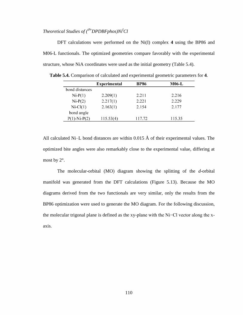
110
Theoretical Studies of (iPr
DPDBFphos)NiICl
DFT calculations were performed on the Ni(I) complex 4 using the BP86 and
M06-L functionals. The optimized geometries compare favorably with the experimental
structure, whose NiA coordinates were used as the initial geometry (Table 5.4).
Table 5.4. Comparison of calculated and experimental geometric parameters for 4.
All calculated Ni–L bond distances are within 0.015 Å of their experimental values. The
optimized bite angles were also remarkably close to the experimental value, differing at
most by 2°.
The molecular-orbital (MO) diagram showing the splitting of the d-orbital
manifold was generated from the DFT calculations (Figure 5.13). Because the MO
diagrams derived from the two functionals are very similar, only the results from the
BP86 optimization were used to generate the MO diagram. For the following discussion,
the molecular trigonal plane is defined as the xy-plane with the Ni−Cl vector along the x-
axis.
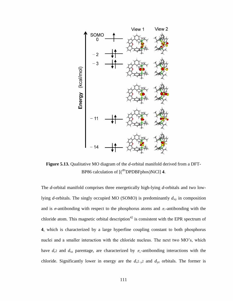
111
Figure 5.13. Qualitative MO diagram of the d-orbital manifold derived from a DFT-
BP86 calculation of [(iPr
DPDBFphos)NiCl] 4.
The d-orbital manifold comprises three energetically high-lying d-orbitals and two low-
lying d-orbitals. The singly occupied MO (SOMO) is predominantly dxy in composition
and is σ-antibonding with respect to the phosphorus atoms and -antibonding with the
chloride atom. This magnetic orbital description42
is consistent with the EPR spectrum of
4, which is characterized by a large hyperfine coupling constant to both phosphorus
nuclei and a smaller interaction with the chloride nucleus. The next two MO’s, which
have dz2 and dxz parentage, are characterized by -antibonding interactions with the
chloride. Significantly lower in energy are the dx2−y2 and dyz orbitals. The former is
112
slightly σ-antibonding with respect to chloride, and the latter is formally non-bonding.
The calculated electronic structure is fully consistent with monovalent nickel. The
LUMO (not shown) is ligand-based and is characterized by delocalized electron density
throughout the -system of the dibenzofuran backbone.
113
5.4 Conclusions
The diphosphine iPr
DPDBFphos was successfully used as a supporting ligand in
tetrahedral Zn(II), tetrahedral Co(II) and trigonal bipyramidal Fe(0) complexes, as well as
in square-planar Ni(II) and distorted trigonal planar Ni(I) compounds. The breadth of
coordination geometries as exemplified by these complexes is enabled by the
conformationally flexible backbone in iPr
DPDBFphos. Compared to cis-chelating
diphosphines, non-rigid trans-spanning diphosphines may be advantageous when a non-
labile ligand platform is required that must also accommodate different coordination
geometries, such as during a catalytic cycle. The reactivity of the (iPr
DPDBFphos)NiCl 4
and its use in catalysis will be discussed in the next chapter.
114
5.5 Experimental procedures
Synthetic Considerations. Unless otherwise stated, all manipulations were performed
under a dinitrogen atmosphere in a MBraun glovebox or using standard Schlenk
techniques. Standard solvents were deoxygenated by sparging with dinitrogen and dried
by passing through activated alumina columns of a SG Water solvent purification system.
Deuterated solvents were purchased from Cambridge Isotope Laboratories, Inc., dried
over CaH2, distilled, and stored over activated 4 Å molecular sieves. Elemental analyses
were performed by Atlantic Microlab. The synthesis of 4,6-bis(3-
diisopropylphosphinophenyl)dibenzofuran, referred to as iPr
DPDBFphos, was previously
reported.1 Other reagents were purchased commercially and used without further
purification.
Synthesis of [(iPr
DPDBFphos)ZnCl2] (1). A solution of zinc (II) chloride (0.026 g, 0.19
mmol) in 10 mL of THF was added dropwise to a solution of iPr
DPDBFphos (0.102 g,
0.18 mmol) in 10 mL of THF. The reaction solution was stirred overnight. After 12
hours, the solvent was removed under reduced pressure, and the resulting solids were
washed with Et2O and then extracted into THF. The THF solution was layered with
hexane and stored at –35 ºC overnight to give a white precipitate, which was collected
and dried under reduced pressure (0.089 g, 70%). Single crystals suitable for X-ray
diffraction analysis were grown from Et2O. UV (THF) λmax, nm (ε, L mol−1
cm−1
): 294 sh
(13,000), 310 sh (6,400). 1H NMR (500 MHz, CDCl3): δ 8.33 (2H, br, CH), 8.03 (2H, d,
J = 7.0 Hz, CH), 7.74 (2H, br. s, CH), 7.66 (2H, d, J = 6.5 Hz, CH), 7.62 (4H, m, J = 6.5
Hz, CH), 7.50 (2H, t, J = 7.5 Hz, CH), 2.59 (4H, m, PCH(CH3)2), 1.29 (12H, dd, JHH =
115
8.0 Hz, JHP = 15 Hz, PCH(CH3)(C′H3)), 1.19 (12H, dd, JHH = 8.0 Hz, JHP = 15 Hz,
PCH(CH3)(C′H3)). 31
P NMR (121 MHz, CDCl3): δ 2.7. ESI-MS-TOF m/z : [M – Cl]+
calc. for C36H42ClZnOP2, 651.1691; found, 651.3.
Synthesis of [(iPr
DPDBFphos)CoCl2] (2). A suspension of cobalt (II) chloride (0.026 g,
0.20 mmol) in 20 mL of THF was added dropwise to a solution of iPr
DPDBFphos (0.109
g, 0.20 mmol) in 20 mL of THF. The reaction solution was stirred overnight. After 12
hours, the solvent was removed under reduced pressure, and the resulting solids were
washed with Et2O and then extracted into THF. The THF solution was filtered and then
evaporated to dryness under reduced pressure to give a blue powder (0.133 g, 99 %).
Single crystals suitable for X-ray diffraction analysis were grown from Et2O. UV (THF)
λmax, nm (ε, L mol−1
cm−1
): 295 sh (16,000), 322 sh (7,600), 618 sh (500), 636 (530), 735
(320). 1H NMR (300 MHz, C6D6): δ 21.36, 13.54, 7.95, 7.64, 6.79, 4.19, 1.83, −14.03.
Evans’ method (CDCl3/CHCl3, 300 MHz): µeff = 4.01 µB. Anal. Calcd. for
C36H42Cl2OP2Co: C, 63.35; H, 6.20; N, 0. Found: C, 62.83; H, 6.05; N, 0.0.
Synthesis of [(iPr
DPDBFphos)NiCl2] (3). A suspension of nickel (II) chloride (0.068 g,
0.52 mmol) in 20 mL of THF was added dropwise to a solution of iPr
DPDBFphos (0.250
g, 0.45 mmol) in 20 mL. The reaction solution was heated at 70 ºC with stirring. After 12
hours, the solvent was removed under reduced pressure, and the resulting solids were
washed with Et2O and then extracted into THF. The THF solution was filtered and then
stored at –35 ºC overnight to give red crystals, which were collected and dried under
reduced pressure (0.202 g, 65%). Single crystals suitable for X-ray diffraction analysis
116
were grown from a CH2Cl2/Et2O (1:3) solution. UV (THF) λmax, nm (ε, L mol−1
cm−1
):
295 sh (18,000), 322 sh (9,300), 377 (9,500), 487 (280). 1H NMR (300 MHz, CDCl3): δ
8.74 (2H, br, CH), 8.06 (2H, d, J = 7.5 Hz, CH), 7.57 (2H, d, J = 7.5 Hz, CH), 7.50 (6H,
m, J = 7.5 Hz, CH), 7.13 (2H, br, CH), 2.75 (4H, br, PCH(CH3)2), 1.67 (12H, br,
PCH(CH3)(C′H3)), 1.27 (12H, br, PCH(CH3)(C′H3)). 31
P NMR (121 MHz, CDCl3): δ
26.5 (br). Anal. Calcd. for C36H42Cl2OP2Ni: C, 63.37; H, 6.20; N, 0. Found: C, 63.16; H,
6.20; N, 0.0.
Synthesis of [(iPr
DPDBFphos)NiCl] (4). A THF solution of 3 (0.095 g, 0.14 mmol) was
added to KC8 (0.020 g, 0.15 mmol) and stirred overnight. An immediate color change
from red to yellow was observed. The reaction solution was filtered, and the filtrate was
evaporated to dryness under reduced pressure. The resulting crude was dissolved in
toluene, filtered, and then stored at –35 ºC. The resulting yellow precipitate was collected
and dried under reduced pressure (0.080 g, 90%). Single crystals suitable for X-ray
diffraction analysis were obtained from a concentrated CD3CN solution. UV (THF) λmax,
nm (ε, L mol−1
cm−1
): 263 (37,000), 298 (16,000), 314 sh (12,000), 324 (11,000), 346 sh
(5,200), 375 (4,500). 1H NMR (300 MHz, d8-THF): δ 13.98, 9.03, 8.62, 7.97, 3.14. Anal.
Calcd. for C36H42ClOP2Ni: C, 66.85; H, 6.54; N, 0. Found: C, 67.00; H, 6.48; N, 0.0.
Synthesis of [(iPr
DPDBFphos)Fe(CO)3] (5). Iron pentacarbonyl (50 μL, 0.37 mmol)
was added to a 10 mL THF solution of iPr
DPDBFphos (0.205 g, 0.37 mmol) in bomb
flask. The flask was evacuated and photolyzed for 6 hours using a mercury vapor lamp.
The THF solution was filtered and then evaporated to dryness under reduced pressure to
117
give a yellow powder (0.235 g, 92 %). Single crystals suitable for X-ray diffraction
analysis were grown from a vapor diffusion of pentane into a concentrate CH2Cl2
solution of 5. IR ºυmax/cm-1
: 1959 (A1´ CO), 1869 (E´ CO) (CDCl3: CH2Cl2). 1H NMR
(500 MHz, CDCl3): δ 8.94 (2H, d, J = 11.1 Hz, CH), 8.00 (2H, dd, J = 1.8 & 7.5 Hz,
CH), 7.75 – 7.43 (10H, m, CH), 2.79 (4H, m, PCH(CH3)2), 1.38 (12H, dd, JHH = 6.9 Hz,
JHP = 15.0 Hz, PCH(CH3)(C′H3)), 1.62 (12H, dd, JHH = 6.6 Hz, JHP = 14.1 Hz,
PCH(CH3)(C′H3)). 31
P NMR (121 MHz, CDCl3): δ 90.4.
Crystallography. A colorless plate of 1, a blue block of 2, a red plate of 3, a yellow
plate of 4, and a yellow plate of 5 were placed onto the tip of a 0.1 mm diameter glass
capillary and mounted on a Bruker or a Siemens SMART Platform CCD diffractometer
for data collection at 123(2) K for complexes 1 – 3 or 173(2) K for complexes 4 and 5.
The data collection was carried out using Mo Kα radiation (graphite monochromator).
The data intensity was corrected for absorption and decay (SADABS). Final cell
constants were obtained from least squares fits of all measured reflections. The structure
was solved using SHELXS-97 and refined using SHELXL-97. For complexes 1 - 4, a
direct-methods solution was calculated which provided most non-hydrogen atoms from
the E-map while complex 5 was solved using a Patterson map. Full-matrix least squares /
difference Fourier cycles were performed to locate the remaining non-hydrogen atoms.
All non-hydrogen atoms were refined with anisotropic displacement parameters.
Hydrogen atoms were placed in ideal positions and refined as riding atoms with relative
isotropic displacement parameters. Crystallographic data are summarized in Table 1.

118
Physical Measurements. 1H and
31P NMR spectra were acquired on Varian Inova 300
and 500 MHz spectrometers at ambient temperature unless otherwise stated. Chemical
shifts were referenced to residual solvent in 1H NMR spectra, while
31P NMR spectra
were referenced to an external reference of 85% H3PO4 set to 0 ppm. Solution magnetic
susceptibilities were determined by the Evans method43-44
and were corrected for
underlying diamagnetism using tabulated Pascal’s constants.45
Electronic spectra of
complexes were recorded on a Cary 300 Bio UV-Visible spectrophotometer. Infrared
(IR) spectra were collected with a Bruker TENSOR 37 spectrometer. Perpendicular-mode
X-band EPR spectra were recorded on a Bruker EPP 300 spectrometer equipped with an
Oxford ESR 910 liquid helium cryostat and an Oxford temperature controller. X-band
EPR spectra were simulated using EPR program (version W95) written by Professor
Frank Neese (University of Bonn, Germany). Cyclic voltammetry experiments were
performed inside a glovebox with a CHInstruments Model 600D potentiostat/galvanostat.
A single-chamber cell was set up with a glassy carbon working electrode (3 mm
diameter), a Pt wire as the auxiliary electrode, and a reference electrode consisting of a
silver wire in 10 mM AgNO3 solution (0.1 M [nBu4N]PF6 in CH3CN). All measurements
were calibrated to an internal ferrocene standard. Mass spectrometry (MS) data were
acquired on a Bruker BioTOF ESI-MS under positive mode.
Theoretical Calculations. DFT calculations (BP86,46-47
M-06L48-49
) were performed
with the Gaussian09 program.50
The all-electron Gaussian basis sets used were those
reported by the Ahlrichs group.51
For nickel, chloride, and phosphorus atoms, the triple-ξ-
quality basis sets with one set of polarization functions was used (def2-TZVP). The
119
carbon, oxygen, and hydrogen atoms were described by smaller polarized split-valence
def2-SV(P) basis sets.
120
-Chapter Six-
Reactivity Studies of the Monovalent Three-coordinate
Nickel Complex iPrDPDBFphosNiCl
In part from:
Marlier, E. E.; Tereniak, S. J.; Ding, K.; Mulliken, J. E.; Lu, C. C. Inorg. Chem. 2011, 50,
9290-9299.
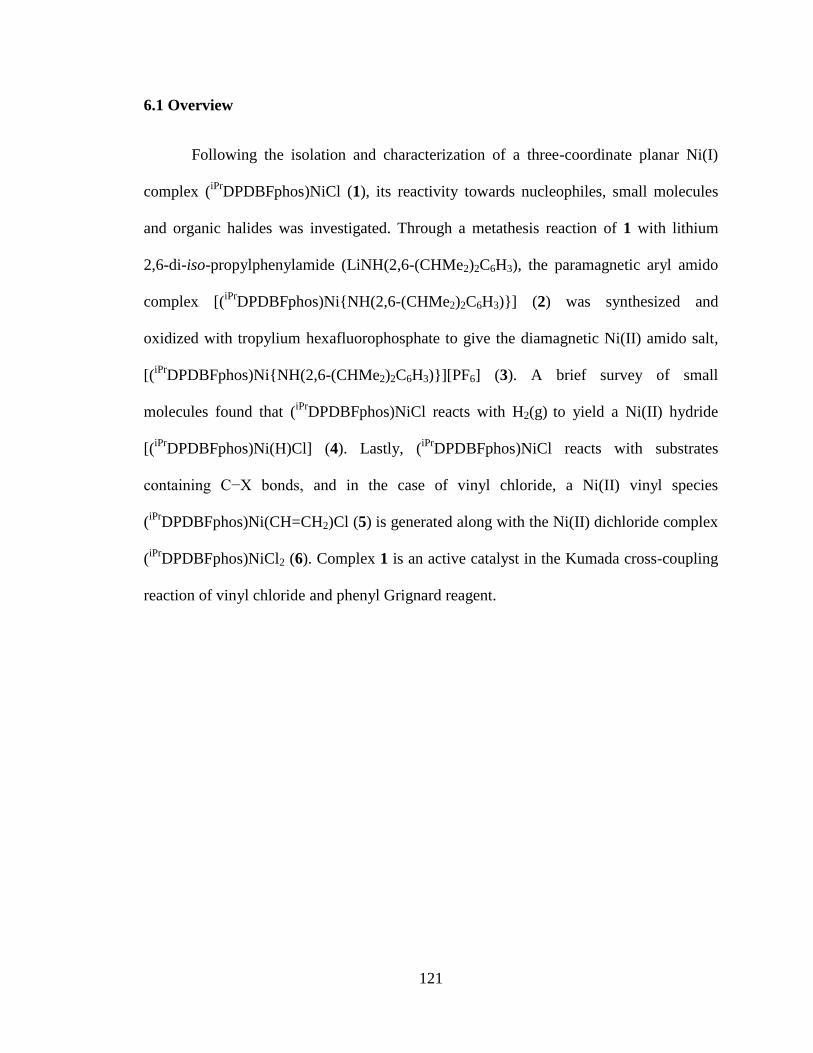
121
6.1 Overview
Following the isolation and characterization of a three-coordinate planar Ni(I)
complex (iPr
DPDBFphos)NiCl (1), its reactivity towards nucleophiles, small molecules
and organic halides was investigated. Through a metathesis reaction of 1 with lithium
2,6-di-iso-propylphenylamide (LiNH(2,6-(CHMe2)2C6H3), the paramagnetic aryl amido
complex [(iPr
DPDBFphos)Ni{NH(2,6-(CHMe2)2C6H3)}] (2) was synthesized and
oxidized with tropylium hexafluorophosphate to give the diamagnetic Ni(II) amido salt,
[(iPr
DPDBFphos)Ni{NH(2,6-(CHMe2)2C6H3)}][PF6] (3). A brief survey of small
molecules found that (iPr
DPDBFphos)NiCl reacts with H2(g) to yield a Ni(II) hydride
[(iPr
DPDBFphos)Ni(H)Cl] (4). Lastly, (iPr
DPDBFphos)NiCl reacts with substrates
containing C−X bonds, and in the case of vinyl chloride, a Ni(II) vinyl species
(iPr
DPDBFphos)Ni(CH=CH2)Cl (5) is generated along with the Ni(II) dichloride complex
(iPr
DPDBFphos)NiCl2 (6). Complex 1 is an active catalyst in the Kumada cross-coupling
reaction of vinyl chloride and phenyl Grignard reagent.
122
6.2 Introduction
Three-coordinate nickel complexes constitute a rich area in coordination
chemistry. Supported by the bulky monophosphine PCy3, zero-valent nickel forms
adducts with small molecules, as observed in the end-on dinitrogen dinickel compound
{(PCy3)2Ni}2(μ-N2)1 and the first structurally characterized carbon dioxide complex,
(PCy3)2Ni(η2-CO2).
2 Hillhouse has pioneered the area of divalent Ni=E complexes
featuring multiply-bonded ligands (E = NR, PR, CR2) using the bulky diphosphine, 1,2-
bis(di-tert-butylphosphino)ethane (dtbpe).3-5
Other notable three-coordinate nickel
complexes have been supported by non-traditional phosphine ligands, including the
Fryzuk-type tridentate bis(phosphine)amides,6 diketiminates,
7-9 N-heterocyclic
carbenes,10-11
and the unusual N-heterocyclic silenes.12
Typically, the oxidation state of
the nickel center is mono-valent for the anionic ligands, and zero-valent for the neutral
carbenes/silenes. The rare Ni(III) oxidation state has also been proposed in three-
coordinate Ni(III) imides, which can be remarkably reactive and decompose via radical
pathways.13,14
A recent report from Hillhouse highlighted the synthesis and
characterization of a Ni(III)-imide complex synthesized from the oxidation of an
arylimido nickel(II) complex.15
Herein, we explore the reactivity of the three-coordinate planar Ni(I) complex
(iPr
DPDBFphos)NiCl (1) through metathesis reactions, small-molecule binding and C-X
bond activation. Considering the reactivity of 1 towards vinyl halides, the cross-coupling
reaction between vinyl chloride and phenyl Grignard reagent was investigated with 1 as a
catalyst. These preliminary results join recent studies in demonstrating the relevance of
monovalent nickel species in cross-coupling catalysis.16, 17
123
6.3 Results and discussion
Metathesis with (iPr
DPDBFphos)NiICl
Inspired by Hillhouse’s work, the synthesis of an arylimido nickel(II) complex
was targeted. Following the procedure established by Mindiola and Hillhouse, complex 1
was reacted with lithium 2,6-di-iso-propylphenylamide (LiNH(2,6-(CHMe2)2C6H3)
(Scheme 6.1).4
Scheme 6.1. Synthetic route for [(iPr
DPDBFphos)Ni{NH(2,6-(CHMe2)2C6H3)}] 2.
Upon addition of LiNH(2,6-(CHMe2)2C6H3), an immediate color change from yellow to
beet purple was observed. X-ray quality crystals revealed this species to be the three
coordinate aryl amido complex [(iPr
DPDBFphos)Ni{NH(2,6-(CHMe2)2C6H3)}] 2, which
was also characterized by 1H NMR spectroscopy (Table 6.1 and Figure 6.1).
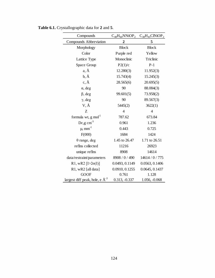
124
Table 6.1. Crystallographic data for 2 and 5.
Compounds C48H60NNiOP2 C38H45ClNiOP2
Compounds Abbreviation 2 5
Morphology Block Block
Color Purple red Yellow
Lattice Type Monoclinic Triclinic
Space Group P2(1)/c P-1
a, Å 12.280(3) 11.952(3)
b, Å 15.743(4) 15.245(3)
c, Å 28.565(6) 20.695(5)
α, deg 90 88.084(3)
β, deg 99.601(5) 73.958(2)
γ, deg 90 89.567(3)
V, Å 5445(2) 3622(1)
Z 4 4
formula wt, g mol-1
787.62 673.84
Dc,g cm-3
0.961 1.236
µ, mm-1
0.443 0.725
F(000) 1684 1424
θ range, deg 1.45 to 26.47 1.71 to 26.51
reflns collected 11216 26923
unique reflns 8908 14614
data/restraint/parameters 8908 / 0 / 490 14614 / 0 / 775
R1, wR2 [I>2σ(I)] 0.0493, 0.1149 0.0563, 0.1406
R1, wR2 [all data] 0.0910, 0.1255 0.0645, 0.1437
GOOF 0.761 1.128
largest diff peak, hole, e Å-3
0.313, -0.337 1.056, -0.068
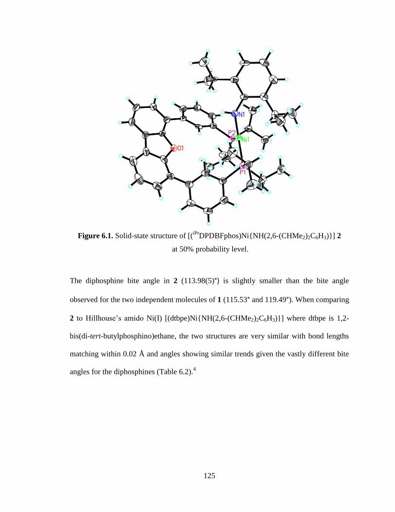
125
Figure 6.1. Solid-state structure of [(iPr
DPDBFphos)Ni{NH(2,6-(CHMe2)2C6H3)}] 2
at 50% probability level.
The diphosphine bite angle in 2 (113.98(5)°) is slightly smaller than the bite angle
observed for the two independent molecules of 1 (115.53° and 119.49°). When comparing
2 to Hillhouse’s amido Ni(I) [(dtbpe)Ni{NH(2,6-(CHMe2)2C6H3)}] where dtbpe is 1,2-
bis(di-tert-butylphosphino)ethane, the two structures are very similar with bond lengths
matching within 0.02 Å and angles showing similar trends given the vastly different bite
angles for the diphosphines (Table 6.2).4
126
Table 6.2. Comparison of selected bond lengths (Å) and angles (°) for 2 and
(dtbpe)NiNHAr, where Ar = 2,6-(CHMe2)2C6H3.
For the following step, the paramagnetic arylamido nickel(I) complex 2 was
oxidized with tropylium hexafluorophosphate to the diamagnetic arylamido nickel(II)
complex 3 (Scheme 6.2).
Scheme 6.2. Synthetic route for [(iPr
DPDBFphos)Ni{NH(2,6-(CHMe2)2C6H3)}][PF6] 3.
Though an immediate color change to blue was observed, the complex appeared sensitive
to temperature and stirring time. Based on 31
P NMR spectroscopy, the oxidation also did
not proceed cleanly with the observation of four different shifts located at 37.8, 32.3, 12.8
and -141.5 ppm. The resonance at 12.8 ppm is identified as free ligand while the peaks at
32.3 and -141.5 ppm belong to complex 3 for the phosphines on the ligand and the anion
2 (dtbpe)NiNHAr*
Bond Distances (Å)
Ni-P1 2.211(1) 2.2012(7)
Ni-P2 2.227(1) 2.2094(8)
Ni-N1 1.878(3) 1.881(2)
N1-C 1.380(5) 1.373(3)
Bond Angles (deg)
P1-Ni-P2 113.98(5) 91.91(3)
P1-M-N1 135.6(1) 140.41(7)
P2-M-N1 109.4(1) 123.18(7)
Ni-N1-C 140.7(3) 134.6(2)
*Values were obtained from reference 4.
127
PF6 respectively. The last peak at 37.8 ppm is hypothesized to be a hydride species
considering a triplet at -23.0 ppm was observed in the crude 1H spectrum. In an attempt to
optimize this reaction, other oxidants such as ferrocenium hexafluorophosphate were
tested, but resulted in a larger amount of free ligand and hydride. Thus, the oxidation was
carried out with tropylium hexafluorophosphate and a toluene wash was found to separate
complex 3 from both the ligand and hydride species.
The last step in the synthesis of an imido nickel(II) complex following
Hillhouse’s route is the deprotonation of the nitrogen bound to the nickel center. For the
deprotonation, Mindiola and Hillhouse report the use of sodium bis(trimethylsilyl)amide
at -35 °C. This step was attempted several times on our system and produced free ligand
each time. The reaction was modified by varying the temperature from -35 °C to -196 °C,
changing the solvent from THF to toluene, surveying a range of bases (e.g. potassium
bis(trimethylsilyl)amide, Hünig’s base (iPr2EtN) and potassium tert-butoxide), and
changing the number of equivalents of base added. Since these modifications led to either
no reaction or free ligand, the synthesis of the nickel(II) imide was abandoned.
Reactivity of (iPr
DPDBFphos)NiICl with small molecules
The reactivity of 1 towards small molecules was surveyed with CO2(g), NO(g)
and H2(g). While 1 did not react with CO2(g), the reaction of 1 with excess NO(g), and
also with excess H2(g) led to the observation of new shifts in the 31
P NMR spectrum. In
the case of NO(g), the reaction became blue upon addition, and two new shifts at 49.04
and 45.93 ppm were detected by 31
P NMR spectroscopy. A similar color change was
observed by Mindiola and Hillhouse upon treatment of their Ni(I) dimer with 2.1
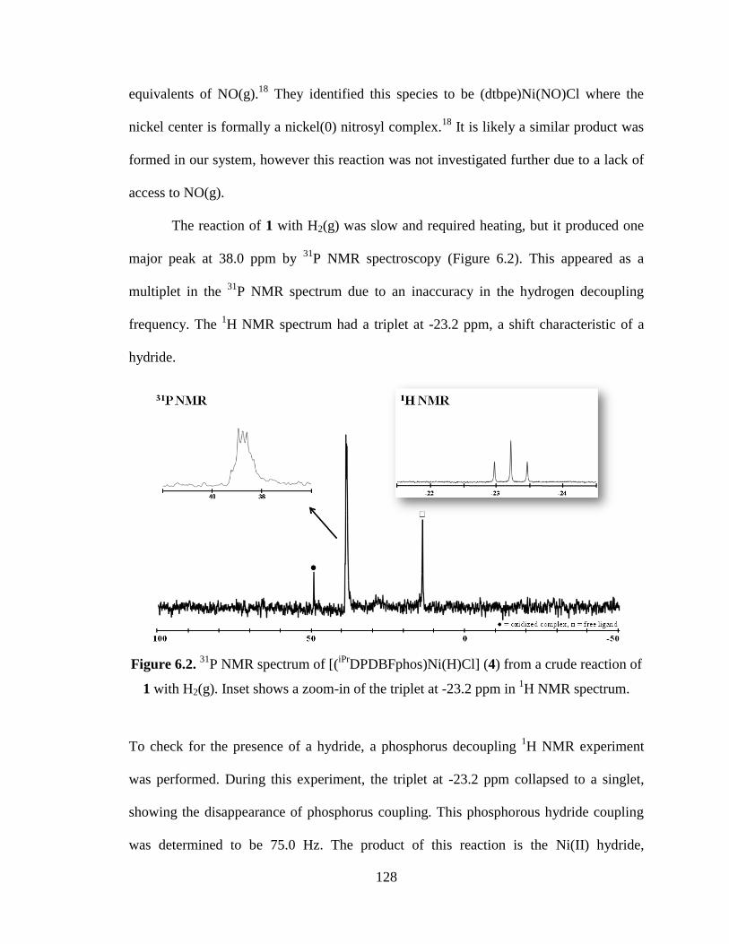
128
equivalents of NO(g).18
They identified this species to be (dtbpe)Ni(NO)Cl where the
nickel center is formally a nickel(0) nitrosyl complex.18
It is likely a similar product was
formed in our system, however this reaction was not investigated further due to a lack of
access to NO(g).
The reaction of 1 with H2(g) was slow and required heating, but it produced one
major peak at 38.0 ppm by 31
P NMR spectroscopy (Figure 6.2). This appeared as a
multiplet in the 31
P NMR spectrum due to an inaccuracy in the hydrogen decoupling
frequency. The 1H NMR spectrum had a triplet at -23.2 ppm, a shift characteristic of a
hydride.
Figure 6.2. 31
P NMR spectrum of [(iPr
DPDBFphos)Ni(H)Cl] (4) from a crude reaction of
1 with H2(g). Inset shows a zoom-in of the triplet at -23.2 ppm in 1H NMR spectrum.
To check for the presence of a hydride, a phosphorus decoupling 1H NMR experiment
was performed. During this experiment, the triplet at -23.2 ppm collapsed to a singlet,
showing the disappearance of phosphorus coupling. This phosphorous hydride coupling
was determined to be 75.0 Hz. The product of this reaction is the Ni(II) hydride,
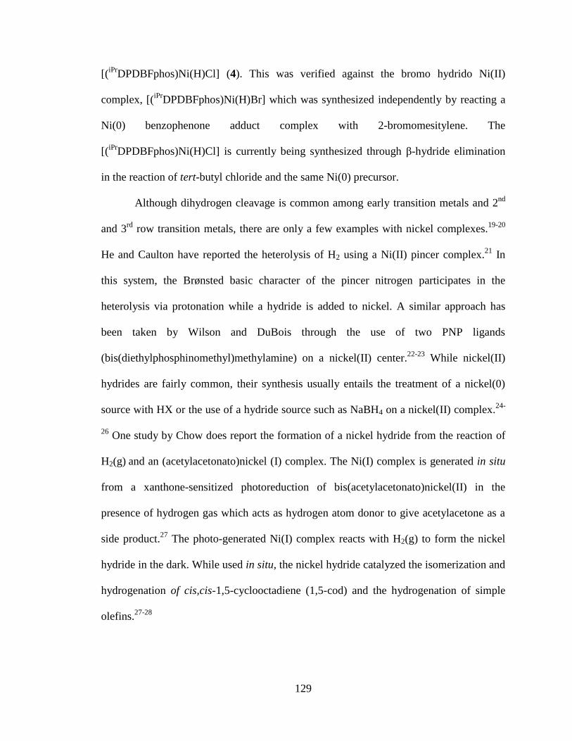
129
[(iPr
DPDBFphos)Ni(H)Cl] (4). This was verified against the bromo hydrido Ni(II)
complex, [(iPr
DPDBFphos)Ni(H)Br] which was synthesized independently by reacting a
Ni(0) benzophenone adduct complex with 2-bromomesitylene. The
[(iPr
DPDBFphos)Ni(H)Cl] is currently being synthesized through β-hydride elimination
in the reaction of tert-butyl chloride and the same Ni(0) precursor.
Although dihydrogen cleavage is common among early transition metals and 2nd
and 3rd
row transition metals, there are only a few examples with nickel complexes.19-20
He and Caulton have reported the heterolysis of H2 using a Ni(II) pincer complex.21
In
this system, the Brønsted basic character of the pincer nitrogen participates in the
heterolysis via protonation while a hydride is added to nickel. A similar approach has
been taken by Wilson and DuBois through the use of two PNP ligands
(bis(diethylphosphinomethyl)methylamine) on a nickel(II) center.22-23
While nickel(II)
hydrides are fairly common, their synthesis usually entails the treatment of a nickel(0)
source with HX or the use of a hydride source such as NaBH4 on a nickel(II) complex.24-
26 One study by Chow does report the formation of a nickel hydride from the reaction of
H2(g) and an (acetylacetonato)nickel (I) complex. The Ni(I) complex is generated in situ
from a xanthone-sensitized photoreduction of bis(acetylacetonato)nickel(II) in the
presence of hydrogen gas which acts as hydrogen atom donor to give acetylacetone as a
side product.27
The photo-generated Ni(I) complex reacts with H2(g) to form the nickel
hydride in the dark. While used in situ, the nickel hydride catalyzed the isomerization and
hydrogenation of cis,cis-1,5-cyclooctadiene (1,5-cod) and the hydrogenation of simple
olefins.27-28
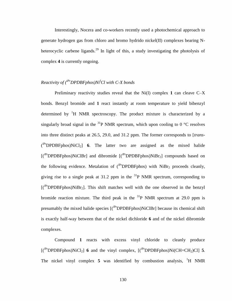
130
Interestingly, Nocera and co-workers recently used a photochemical approach to
generate hydrogen gas from chloro and bromo hydrido nickel(II) complexes bearing N-
heterocyclic carbene ligands.29
In light of this, a study investigating the photolysis of
complex 4 is currently ongoing.
Reactivity of (iPr
DPDBFphos)NiICl with C-X bonds
Preliminary reactivity studies reveal that the Ni(I) complex 1 can cleave C–X
bonds. Benzyl bromide and 1 react instantly at room temperature to yield bibenzyl
determined by 1H NMR spectroscopy. The product mixture is characterized by a
singularly broad signal in the 31
P NMR spectrum, which upon cooling to 0 °C resolves
into three distinct peaks at 26.5, 29.0, and 31.2 ppm. The former corresponds to [trans-
(iPr
DPDBFphos)NiCl2] 6. The latter two are assigned as the mixed halide
[(iPr
DPDBFphos)NiClBr] and dibromide [(iPr
DPDBFphos)NiBr2] compounds based on
the following evidence. Metalation of (iPr
DPDBFphos) with NiBr2 proceeds cleanly,
giving rise to a single peak at 31.2 ppm in the 31
P NMR spectrum, corresponding to
[(iPr
DPDBFphos)NiBr2]. This shift matches well with the one observed in the benzyl
bromide reaction mixture. The third peak in the 31
P NMR spectrum at 29.0 ppm is
presumably the mixed halide species [(iPr
DPDBFphos)NiClBr] because its chemical shift
is exactly half-way between that of the nickel dichloride 6 and of the nickel dibromide
complexes.
Compound 1 reacts with excess vinyl chloride to cleanly produce
[(iPr
DPDBFphos)NiCl2] 6 and the vinyl complex, [(iPr
DPDBFphos)Ni(CH=CH2)Cl] 5.
The nickel vinyl complex 5 was identified by combustion analysis, 1H NMR
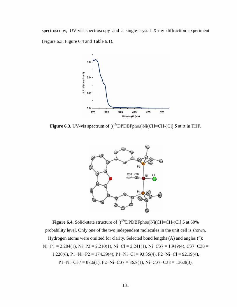
131
spectroscopy, UV-vis spectroscopy and a single-crystal X-ray diffraction experiment
(Figure 6.3, Figure 6.4 and Table 6.1).
Figure 6.3. UV-vis spectrum of [(iPr
DPDBFphos)Ni(CH=CH2)Cl] 5 at rt in THF.
Figure 6.4. Solid-state structure of [(iPr
DPDBFphos)Ni(CH=CH2)Cl] 5 at 50%
probability level. Only one of the two independent molecules in the unit cell is shown.
Hydrogen atoms were omitted for clarity. Selected bond lengths (Å) and angles (°):
Ni−P1 = 2.204(1), Ni−P2 = 2.210(1), Ni−Cl = 2.241(1), Ni−C37 = 1.919(4), C37−C38 =
1.220(6), P1−Ni−P2 = 174.39(4), P1−Ni−Cl = 93.35(4), P2−Ni−Cl = 92.19(4),
P1−Ni−C37 = 87.6(1), P2−Ni−C37 = 86.8(1), Ni−C37−C38 = 136.9(3).
132
The product ratio of 5:6 is 1:1 based on their relative integration in the 1H NMR spectrum
(Figure 6.5).
Figure 6.5. Stacked 1H NMR spectra (300 MHz,d8-THF) of (A)
[(iPr
DPDBFphos)Ni(CH=CH2)Cl], 5 at rt; (B) [(iPr
DPDBFphos)NiCl2], 3 at 0° C; and, (C)
the two-product mixture in the reaction of vinyl chloride and [(iPr
DPDBFphos)NiCl] at rt.
The product distribution of the reaction (C) was determined by the relative integrations of
the two most downfield peaks.
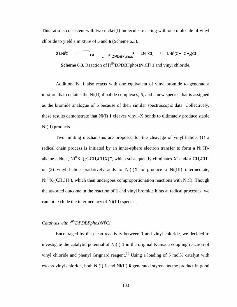
133
This ratio is consistent with two nickel(I) molecules reacting with one molecule of vinyl
chloride to yield a mixture of 5 and 6 (Scheme 6.3).
Scheme 6.3. Reaction of [(iPr
DPDBFphos)NiCl] 1 and vinyl chloride.
Additionally, 1 also reacts with one equivalent of vinyl bromide to generate a
mixture that contains the Ni(II) dihalide complexes, 5, and a new species that is assigned
as the bromide analogue of 5 because of their similar spectroscopic data. Collectively,
these results demonstrate that Ni(I) 1 cleaves vinyl–X bonds to ultimately produce stable
Ni(II) products.
Two limiting mechanisms are proposed for the cleavage of vinyl halide: (1) a
radical chain process is initiated by an inner-sphere electron transfer to form a Ni(II)-
alkene adduct, NiIIX–(η
2-CH2CHX)
•‾, which subsequently eliminates X
• and/or CH2CH
•,
or (2) vinyl halide oxidatively adds to Ni(I)X to produce a Ni(III) intermediate,
NiIII
X2(CHCH2), which then undergoes comproportionation reactions with Ni(I). Though
the assorted outcome in the reaction of 1 and vinyl bromide hints at radical processes, we
cannot exclude the intermediacy of Ni(III) species.
Catalysis with (iPr
DPDBFphos)NiICl
Encouraged by the clean reactivity between 1 and vinyl chloride, we decided to
investigate the catalytic potential of Ni(I) 1 in the original Kumada coupling reaction of
vinyl chloride and phenyl Grignard reagent.30
Using a loading of 5 mol% catalyst with
excess vinyl chloride, both Ni(I) 1 and Ni(II) 6 generated styrene as the product in good
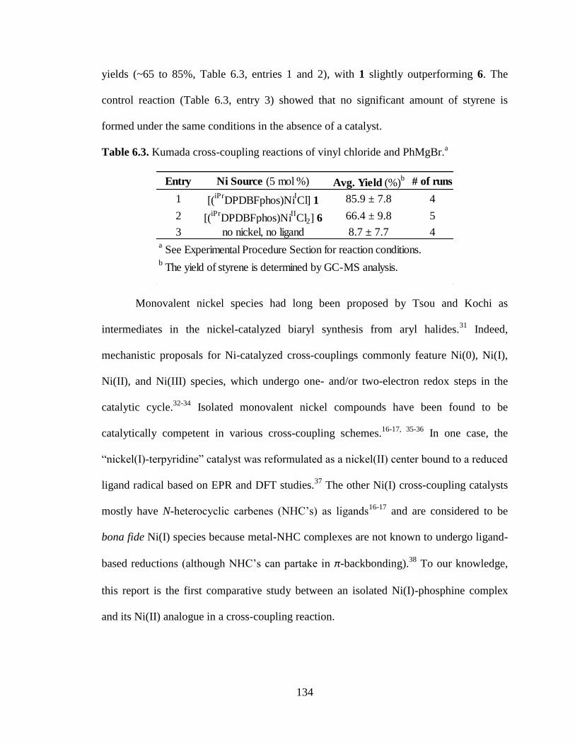
134
yields (~65 to 85%, Table 6.3, entries 1 and 2), with 1 slightly outperforming 6. The
control reaction (Table 6.3, entry 3) showed that no significant amount of styrene is
formed under the same conditions in the absence of a catalyst.
Table 6.3. Kumada cross-coupling reactions of vinyl chloride and PhMgBr.a
Monovalent nickel species had long been proposed by Tsou and Kochi as
intermediates in the nickel-catalyzed biaryl synthesis from aryl halides.31
Indeed,
mechanistic proposals for Ni-catalyzed cross-couplings commonly feature Ni(0), Ni(I),
Ni(II), and Ni(III) species, which undergo one- and/or two-electron redox steps in the
catalytic cycle.32-34
Isolated monovalent nickel compounds have been found to be
catalytically competent in various cross-coupling schemes.16-17, 35-36
In one case, the
“nickel(I)-terpyridine” catalyst was reformulated as a nickel(II) center bound to a reduced
ligand radical based on EPR and DFT studies.37
The other Ni(I) cross-coupling catalysts
mostly have N-heterocyclic carbenes (NHC’s) as ligands16-17
and are considered to be
bona fide Ni(I) species because metal-NHC complexes are not known to undergo ligand-
based reductions (although NHC’s can partake in π-backbonding).38
To our knowledge,
this report is the first comparative study between an isolated Ni(I)-phosphine complex
and its Ni(II) analogue in a cross-coupling reaction.
Entry Ni Source (5 mol %) Avg. Yield (%)b # of runs
1 [(iPr
DPDBFphos)NiICl] 1 85.9 ± 7.8 4
2 [(iPr
DPDBFphos)NiIICl2] 6 66.4 ± 9.8 5
3 no nickel, no ligand 8.7 ± 7.7 4a See Experimental Procedure Section for reaction conditions.
b The yield of styrene is determined by GC-MS analysis.
135
6.4 Conclusions
Using a wide bite-angle diphosphine ligand, a nickel(I) complex participated in
metathesis reactions, small molecule activation and C-X bond cleavage. The metathesis
reactions did not yield the desired imido nickel(II) complex due to an inability to
deprotonate the aniline nitrogen. With regards to small molecules, the nickel(I) complex
was found to cleave H2(g) to form a nickel(II) hydride. A chloro vinyl nickel(II) complex
was isolated from the reaction of nickel(I) with vinyl chloride. Inspired by its reactivity,
the nickel(I) was used a catalyst for the formation of styrene from vinyl chloride and
PhMgBr.
Given the limited evidence for Ni(I) in cross-coupling reactions, our results are
intriguing in that a Ni(I) complex reacts constructively with vinyl–X bonds and is
catalytically competent for the cross coupling of vinyl chloride and phenyl Grignard
reagent. Definitive experimental proof for the direct involvement of monovalent nickel in
cross-coupling catalysis is still lacking. The current results warrant further study of this
Ni(I) system, especially to evaluate its scope in other cross-coupling schemes as well as
to scrutinize the role of Ni(I) through detailed mechanistic investigations.
136
6.5 Experimental Procedures
Synthetic Considerations. Unless otherwise stated, all manipulations were performed
under a dinitrogen atmosphere in a MBraun glovebox or using standard Schlenk
techniques. Standard solvents were deoxygenated by sparging with dinitrogen and dried
by passing through activated alumina columns of a SG Water solvent purification system.
Deuterated solvents were purchased from Cambridge Isotope Laboratories, Inc., dried
over CaH2, distilled, and stored over activated 4 Å molecular sieves. Elemental analyses
were performed by Atlantic Microlab. The synthesis of complexes 1 and 6 was reported
in the chapter 6. Other reagents were purchased commercially and used without further
purification.
Synthesis of [(iPr
DPDBFphos)Ni{NH(2,6-(CHMe2)2C6H3)}] (2). Lithium 2,6-di-iso-
propylphenylamide (0.042 g, 0.23 mmol) was dissolved in 5 mL of Et2O and cooled to -
35°C. Once cold, it was added dropwise to a cold toluene solution of
(iPr
DPDBFphos)NiCl (0.139 g, 0.21 mmol). An immediate color change from yellow to
beet red was observed. Solution was stirred at RT for three hours. The reaction solution
was filtered, and the filtrate was evaporated to dryness under reduced pressure. The
resulting crude was dissolved in toluene, filtered, and the filtrate was evaporated to
dryness under reduced pressure, to give a purple red solid (0.162 g, 96%). X-ray quality
crystals were grown from layering a concentrated toluene solution of 2 with Et2O at -
35°C. 1H NMR (300 MHz, C6D6): δ 13.40, 8.27, 8.13, 7.48, 7.42, 7.07, 7.05, 7.00, 6.90,
4.06, 3.19, 1.37, 1.06, 0.92, 0.30.
137
Synthesis of [(iPr
DPDBFphos)Ni{NH(2,6-(CHMe2)2C6H3)}][PF6] (3). A suspension of
tropylium hexafluorophosphate (0.012 g, 0.05 mmol) in 3 mL of THF was cooled to -
35°C. The cold suspension was added dropwise to a cold THF solution of 2 (0.037 g,
0.05 mmol). An immediate color change from purple red to purple blue was observed.
After stirring for an hour at RT, the solution was filtered and the filtrate was evaporated
to dryness under reduced pressure. The residual solids were washed with cold toluene and
Et2O. 31
P NMR (121 MHz, d8-THF): δ 32.3, -141.8.
Synthesis of [(iPr
DPDBFphos)Ni(H)Cl] (4). A d8-THF solution of 1 (0.013 g, 0.02
mmol) was added to a sealable NMR tube. The tube was frozen, evacuated and refilled
with excess hydrogen gas. The NMR tube was placed in an oil bath at 60°C for three
weeks. Integration of a Ni(I) resonance against the hydride peak gives an approximate
69% conversion. 1H NMR (300 MHz, d8-THF): δ 8.92, 8.10, 7.69, 7.58. 7.52, 7.30, 2.78,
1.32, 1.08, -23.21 (1H, t, JHP = 75.0 Hz, Ni-H). 31
P NMR (121 MHz, d8-THF): δ 38.0.
Synthesis of [(iPr
DPDBFphos)Ni(CH=CH2)Cl] (5). A d8-THF solution of 1 (0.017 g,
0.03 mmol) was added to a sealable NMR tube. The tube was frozen, evacuated and
refilled with excess vinyl chloride. An immediate color change was observed from
yellow to orange red upon warming to room temperature. After 4 days at room
temperature, the NMR spectra showed no starting material and two different products;
complexes 5 and 6, present in a 1:1 ratio. Complex 5 was synthesized independently
through the reaction of vinyl chloride and a nickel(0) complex. Single crystals suitable
for X-ray diffraction analysis were grown from a THF/Pentane (1:2) solution. UV (THF)
138
λmax, nm (ε, L mol−1
cm−1
): 285 (32,000), 308 sh (19,000), 318 sh (15,000), 430 (700). 1H
NMR (500 MHz, d8-THF): δ 8.50 (2H, m, CH), 8.12 (2H, dd, J = 1.5 & 8.0 Hz, CH),
7.62 (2H, d, J = 8.0 Hz, CH), 7.58 (2H, dd, J = 1.5 & 8.0 Hz, CH), 7.50 (2H, t, J = 8.0
Hz, CH), 7.49 (2H, t, J = 8.0 Hz, CH), 7.36 (2H, m, CH), 5.91 (1H, ddt, JHH = 10.2 &
17.5 Hz, JHP = 5.5 Hz, NiCH= CHH′), 4.42 (1H, dt, JHH = 17.5 Hz, JHP = 3.0 Hz, cis-
NiCH=CHH′), 4.14 (1H, dt, JHH = 10.2 Hz, JHP = 4.5 Hz, trans-NiCH=CHH′), 2.96 (2H,
m, PCH(CH3)2), 2.73 (2H, m, PC′H(CH3)2), 1.64 (6H, dd, JHH = 7.0 Hz, JHP = 14.0 Hz,
PC′H(CH3)(C′H3)), 1.62 (6H, dd, JHH = 7.5 Hz, JHP = 15.0 Hz, PC′H(CH3)(C′H3)), 1.36
(6H, dd, JHH = 7.5 Hz, JHP = 15.5 Hz, PCH(CH3)(C′H3)), 0.71 (6H, dd, JHH = 6.0 Hz, JHP
= 12.0 Hz, PCH(CH3)(C′H3)). 31
P NMR (121 MHz, d8-THF): δ 29.1. Anal. Calcd. for
C38H45ClOP2Ni: C, 67.73; H, 6.73; N, 0. Found: C, 67.97; H, 6.89; N, 0.0.
General Procedure for Kumada Coupling Reactions. A THF solution of the Ni
catalyst (0.003 mmol, 2.14 mL) was added to a sealable reaction flask equipped with a
magnetic stir bar, and the solution was frozen with a glovebox LN2 coldwell bath. Once
frozen, a solution of phenylmagnesium bromide (0.06 mmol, 0.02 mL, 3.0 M in Et2O)
diluted with Et2O (0.84 mL) was added on top and frozen as a separate layer. The
reaction flask was then evacuated and charged with excess vinyl chloride. Reactions were
allowed to thaw and stir for 22 hours at room temperature. The reaction was quenched by
adding 2 mL of Et2O and 4 mL of 1.0 M HCl to the solution. The organic layer was
extracted, dried over MgSO4 and filtered. The filtrate was analyzed on the GC-MS.
Yields were calculated using GC-MS peak integrations and a styrene-standard calibration
curve.
139
X-Ray Crystallographic Data Collection and Refinement of the Structures.
A purple block of 2 and a yellow block of 5 were placed onto the tip of a 0.1 mm
diameter glass capillary and mounted on a Bruker SMART Platform CCD diffractometer
for data collection at 123(2) K. The data collection was carried out using Mo Kα
radiation (graphite monochromator). The data intensity was corrected for absorption and
decay (SADABS). Final cell constants were obtained from least squares fits of all
measured reflections. The structure was solved using SHELXS-97 and refined using
SHELXL-97. A direct-methods solution was calculated which provided most non-
hydrogen atoms from the E-map. Full-matrix least squares / difference Fourier cycles
were performed to locate the remaining non-hydrogen atoms. All non-hydrogen atoms
were refined with anisotropic displacement parameters. Hydrogen atoms were placed in
ideal positions and refined as riding atoms with relative isotropic displacement
parameters.
For 2, two highly disordered Et2O molecules could not be modeled accordingly
and were removed using the PLATON program, SQUEEZE function.39
A total of 453
electrons in a volume of 1708 Å3 located at (0, 0, 0) and (0, 0.5, 0.5) in the unit cell were
determined. For 5, a highly disordered THF molecule could not be modeled appropriately
and was removed using the PLATON program, SQUEEZE function.39
A total of 46
electrons in a volume of 353 Å3 located at (0.5, 1, 0) in the unit cell were determined.
Crystallographic data for 1 and 5 are summarized in Table 1.
Physical Measurements. 1H and
31P NMR spectra were acquired on Varian Inova 300
and 500 MHz spectrometers at ambient temperature unless otherwise stated. Chemical
140
shifts were referenced to residual solvent in 1H NMR spectra, while
31P NMR spectra
were referenced to an external reference of 85% H3PO4 set to 0 ppm. For 5, proton
assignments were based on a COSY NMR experiment. Electronic spectra of complexes
were recorded on a Cary 300 Bio UV-Visible spectrophotometer. Mass spectrometry
(MS) data were acquired on a Bruker BioTOF ESI-MS under positive mode.

141
-Bibliography-
142
Chapter One References
(1) Squillace, P. J.; Moran, M. J.; Lapham, W. W.; Price, C. V.; Clawges, R. M.; Zogorski, J.
S. Volatile Organic Compounds in Untreated Ambient Groundwater of the United States,
1985-1995. Environ. Sci. Technol. 1999, 33, 4176-4187.
(2) Squillace, P. J.; Moran, M. J. Factors Associated with Sources, Transport, and Fate of
Volatile Organic Compounds and Their Mixtures in Aquifers of the United States.
Environ. Sci. Technol. 2007, 41, 2123-2130.
(3) Doherty, R. E. A History of the Production and Use of Carbon Tetrachloride,
Tetrachloroethylene, Trichloroethylene and 1,1,1-Trichloroethane in the United States:
Part 1 - Historical Background; Carbon Tetrachloride and Tetrachloroethylene. Environ.
Forensics 2000, 1, 69-81.
(4) Doherty, R. E. A History of the Production and Use of Carbon Tetrachloride,
Tetrachloroethylene, Trichloroethylene and 1,1,1-Trichloroethane in the United States:
Part 2 - Trichloroethylene and 1,1,1-Trichloroethane. Environ. Forensics 2000, 1, 83-93.
(5) USDHHS, Report on Carcinogens; U.S. Department of Health and Human Services:
Washington, DC, 2011.
(6) ATSDR, Toxicological Profile for Tetrachloroethylene; Agency of Toxic Substances &
Disease Registry: Atlanta, GA, 1997.
(7) ATSDR, Toxicological profile for Trichloroethylene; Agency of Toxic Subtances &
Disease Registry: Atlanta, GA, 2003.
(8) Wartenberg, D.; Reyner, D.; Scott, C. S. Trichloroethylene and Cancer: Epidemiologic
Evidence. Environ. Health Perspect. 2000, 108(Suppl. 2), 359-363.
(9) Clewell, H. J.; Gentry, P. R.; Gearhart, J. M.; Allen, B. C.; Andersen, M. E. Comparison
of Cancer Risk Estimates for Vinyl Chloride using Animal and Human Data with a PBPK
Model. The Science of The Total Environment 2001, 274, 37-66.
(10) Holliger, C.; Schraa, G.; Stupperich, E.; Stams, A. J.; Zehnder, A. J. Evidence for the
Involvement of Corrinoids and Factor F430 in the Reductive Dechlorination of 1,2-
Dichloroethane by Methanosarcina Barkeri. J. Bacteriol. 1992, 174, 4427-34.
(11) Krone, U. E.; Thauer, R. K.; Hogenkamp, H. P. C. Reductive Dehalogenation of
Chlorinated C1-Hydrocarbons Mediated by Corrinoids. Biochemistry 1989, 28, 4908-
4914.
143
(12) Gantzer, C. J.; Wackett, L. P. Reductive Dechlorination Catalyzed by Bacterial
Transition-Metal Coenzymes. Environ. Sci. Technol. 1991, 25, 715-22.
(13) Smidt, H.; de Vos, W. M. Anaerobic Microbial Dehalogenation. Annu. Rev. Microbiol.
2004, 58, 43-73.
(14) Burris, D. R.; Delcomyn, C. A.; Deng, B.; Buck, L. E.; Hatfield, K. Kinetics of
Tetrachloroethylene-Reductive Dechlorination Catalyzed by Vitamin B12. Environ.
Toxicol. Chem. 1998, 17, 1681-1688.
(15) Burris, D. R.; Delcomyn, C. A.; Smith, M. H.; Roberts, A. L. Reductive Dechlorination
of Tetrachloroethylene and Trichloroethylene Catalyzed by Vitamin B12 in Homogeneous
and Heterogeneous Systems. Environ. Sci. Technol. 1996, 30, 3047-3052.
(16) Glod, G.; Angst, W.; Holliger, C.; Schwarzenbach, R. P. Corrinoid-Mediated Reduction
of Tetrachloroethene, Trichloroethene, and Trichlorofluoroethene in Homogeneous
Aqueous Solution: Reaction Kinetics and Reaction Mechanisms. Environ. Sci. Technol.
1997, 31, 253-260.
(17) Glod, G.; Brodmann, U.; Angst, W.; Holliger, C.; Schwarzenbach, R. P. Cobalamin-
Mediated Reduction of cis- and trans-Dichloroethene, 1,1-Dichloroethene, and Vinyl
Chloride in Homogeneous Aqueous Solution: Reaction Kinetics and Mechanistic
Considerations. Environ. Sci. Technol. 1997, 31, 3154-3160.
(18) Kliegman, S.; McNeill, K. Dechlorination of Chloroethylenes by Cob(I)alamin and
Cobalamin Model Complexes. Dalton Trans. 2008, 4191-4201.
(19) Lesage, S.; Brown, S.; Millar, K. A Different Mechanism for the Reductive
Dechlorination of Chlorinated Ethenes: Kinetic and Spectroscopic Evidence. Environ.
Sci. Technol. 1998, 32, 2264-2272.
(20) Semadeni, M.; Chiu, P.-C.; Reinhard, M. Reductive Transformation of Trichloroethene
by Cobalamin: Reactivities of the Intermediates Acetylene, Chloroacetylene, and the
DCE Isomers. Environ. Sci. Technol. 1998, 32, 1207-1213.
(21) Follett, A. D.; McNabb, K. A.; Peterson, A. A.; Scanlon, J. D.; Cramer, C. J.; McNeill, K.
Characterization of Co-C Bonding in Dichlorovinylcobaloxime Complexes. Inorg. Chem.
2007, 46, 1645-1654.
(22) Follett, A. D.; McNeill, K. Reduction of Trichloroethylene by Outer-Sphere Electron-
Transfer Agents. J. Am. Chem. Soc. 2005, 127, 844-845.

144
(23) Follett, A. D.; McNeill, K. Evidence for the Formation of a cis-Dichlorovinyl Anion upon
Reduction of cis-1,2-Dichlorovinyl(pyridine)cobaloxime. Inorg. Chem. 2006, 45, 2727-
2732.
(24) McCauley, K. M.; Pratt, D. A.; Wilson, S. R.; Shey, J.; Burkey, T. J.; van der Donk, W.
A. Properties and Reactivity of Chlorovinylcobalamin and Vinylcobalamin and Their
Implications for Vitamin B12-Catalyzed Reductive Dechlorination of Chlorinated
Alkenes. J. Am. Chem. Soc. 2005, 127, 1126-1136.
(25) McCauley, K. M.; Wilson, S. R.; van der Donk, W. A. Dichloroacetylene Is Not the
Precursor to Dichlorinated Vinylcobaloxime and Vinylcobalamin in Cobalt Catalyzed
Dechlorination of Perchloro- and Trichloroethylene. Inorg. Chem. 2002, 41, 5844-5848.
(26) McCauley, K. M.; Wilson, S. R.; van der Donk, W. A. Synthesis and Characterization of
Chlorinated Alkenylcobaloximes To Probe the Mechanism of Vitamin B12-Catalyzed
Dechlorination of Priority Pollutants. Inorg. Chem. 2002, 41, 393-404.
(27) McCauley, K. M.; Wilson, S. R.; Van Der Donk, W. A. Characterization of
Chlorovinylcobalamin, A Putative Intermediate in Reductive Degradation of Chlorinated
Ethylenes. J. Am. Chem. Soc. 2003, 125, 4410-4411.
(28) Nonnenberg, C.; van der Donk, W. A.; Zipse, H. Reductive Dechlorination of
Trichloroethylene: A Computational Study. J. Phys. Chem. 2002, 106, 8708-8715.
(29) Pratt, D. A.; Van der Donk, W. A. Theoretical Investigations into the Intermediacy of
Chlorinated Vinylcobalamins in the Reductive Dehalogenation of Chlorinated Ethylenes.
J. Am. Chem. Soc. 2005, 127, 384-396.
(30) Pratt, D. A.; Van der Donk, W. A. On the Role of Alkylcobalamins in the Vitamin B12-
Catalyzed Reductive Dehalogenation of Perchloroethylene and Trichloroethylene. Chem.
Commun. 2006, 558-560.
(31) Rich, A. E.; DeGreeff, A. D.; McNeill, K. Synthesis of (Chlorovinyl)cobaloxime
Complexes, Model Complexes of Proposed Intermediates in the B12-Catalyzed
Dehalogenation of Chlorinated Ethylenes. Chem. Commun. 2002, 234-235.
(32) Shey, J.; McGinley, C. M.; McCauley, K. M.; Dearth, A. S.; Young, B. T.; van der Donk,
W. A. Mechanistic Investigation of a Novel Vitamin B12-Catalyzed Carbon-Carbon Bond
Forming Reaction, the Reductive Dimerization of Arylalkenes. J. Org. Chem. 2002, 67,
837-846.
145
(33) Shey, J.; van der Donk, W. A. Mechanistic Studies on the Vitamin B12-Catalyzed
Dechlorination of Chlorinated Alkenes. J. Am. Chem. Soc. 2000, 122, 12403-12404.
(34) Kliegman, S.; McNeill, K. Reconciling Disparate Models of the Involvement of Vinyl
Radicals in Cobalamin-Mediated Dechlorination Reactions. Environ. Sci. Technol. 2009,
43, 8961-8967.
(35) Rusling, J. F.; Connors, T. F.; Owlia, A. Electrocatalytic Reduction of Ethylene
Dibromide by Vitamin B12 in a Surfactant-Stabilized Emulsion. Anal. Chem. 1987, 59,
2123-2127.
(36) Connelly, N. G.; Geiger, W. E. Chemical Redox Agents for Organometallic Chemistry.
Chem. Rev. 1996, 96, 877-910.
(37) Lexa, D.; Saveant, J. M. The Electrochemistry of Vitamin B12. Acc. Chem. Res. 1983, 16,
235-43.
(38) Schrauzer, G. N. Organocobalt Chemistry of Vitamin B12 Model Compounds
(Cobaloximes). Acc. Chem. Res. 1968, 1, 97-103.
(39) Liptak, M. D.; Brunold, T. C. Spectroscopic and Computational Studies of
Co1+
Cobalamin: Spectral and Electronic Properties of the "Superreduced" B12 Cofactor. J.
Am. Chem. Soc. 2006, 128, 9144-9156.
(40) Totten, L. A.; Roberts, A. L. Calculated One- and Two-Electron Reduction Potentials and
Related Molecular Descriptors for Reduction of Alkyl and Vinyl Halides in Water. Crit.
Rev. Environ. Sci. Technol. 2001, 31, 175-221.
(41) Schrauzer, G. N.; Deutsch, E. Reactions of Cobalt(I) Supernucleophiles. The Alkylation
of Vitamin B12s, Cobaloximes(I), and Related Compounds. J. Am. Chem. Soc. 1969, 91,
3341-50.
(42) Schrauzer, G. N.; Deutsch, E.; Windgassen, R. J. The Nucleophilicity of Vitamin B12s. J.
Am. Chem. Soc. 1968, 90, 2441-2.
(43) Pearson, R. G.; Sobel, H. R.; Songstad, J. Nucleophilic Reactivity Constants Toward
Methyl Iodide and Trans-Dichlorodi(pyridine)platinum(II). J. Am. Chem. Soc. 1968, 90,
319-26.
146
(44) Dror, I.; Schlautman, M. A. Role of Metalloporphyrin Core Metals in the Mediated
Reductive Dechlorination of Tetrachloroethylene. Environ. Toxicol. Chem. 2003, 22,
525-533.
(45) Fritsch, J. M.; McNeill, K. Aqueous Reductive Dechlorination of Chlorinated Ethylenes
with Tetrakis(4-carboxyphenyl)porphyrin Cobalt. Inorg. Chem. 2005, 44, 4852-4861.
(46) Fritsch, J. M.; Retka, N. D.; McNeill, K. Synthesis, Structure, and Unusual Reactivity of
beta -Halovinyl Cobalt Porphyrin Complexes. Inorg. Chem. 2006, 45, 2288-2295.
(47) Jones, P. G.; Yang, L.; Steinborn, D. (Cis-1,2-Dichlorovinyl)bis(dimethylglyoximato-
N,N')(pyridine-N)cobalt(III) Chloroform Solvate, [Co(dmgH)2(py)(CCl=CHCl)].CHCl3.
Acta Crystallogr., Sect. C: Cryst. Struct. Commun. 1996, C52, 2399-2402.
(48) Thoreson, K. A. Probing Reduced Metal Center-Mediated Dechlorination Mechanisms
Throught the Use of Model Complexes and Isotope Ratio Mass Spectrometry. Ph.D.
Thesis, University of Minnesota, Minneapolis, MN, June 2010.
(49) Lewis, T. A.; Morra, M. J.; Brown, P. D. Comparative Product Analysis of Carbon
Tetrachloride Dehalogenation Catalyzed by Cobalt Corrins in the Presence of Thiol or
Titanium(III) Reducing Agents. Environ. Sci. Technol. 1996, 30, 292-300.
(50) Dror, I.; Schlautman, M. A. Metalloporphyrin Solubility: A Trigger for Catalyzing
Reductive Dechlorination of Tetrachloroethylene. Environ. Toxicol. Chem. 2004, 23,
252-257.
147
Chapter Two References
(1) Bourget-Merle, L.; Lappert, M. F.; Severn, J. R. The Chemistry of -Diketiminatometal
Complexes. Chem. Rev. 2002, 102, 3031-3066.
(2) Dove, A. P.; Gibson, V. C.; Marshall, E. L.; White, A. J. P.; Williams, D. J. Magnesium
and Zinc Complexes of a Potentially Tridentate-Diketiminate Ligand. Dalton Trans.
2004, 570-578.
(3) Wu, J.; Yu, T.-L.; Chen, C.-T.; Lin, C.-C. Recent Developments in Main Group Metal
Complexes Catalyzed/Initiated Polymerization of Lactides and Related Cyclic Esters.
Coord. Chem. Rev. 2006, 250, 602-626.
(4) Xin Xu, X. X., Yaofeng Chen, and Jie Sun Dialkyllanthanide Complexes Containing
New Tridentate Monoanionic Ligands with Nitrogen Donors. Organometallics 2008, 27,
758-763.
(5) Zatka, V.; Holzbecher, J.; Ryan, D. E. Heterocyclic N,N'-disubstituted 1-amino-3-
iminopropenes as Analytical Reagents. Anal. Chim. Acta 1971, 55, 273-8.
(6) Neculai, D.; Roesky, H. W.; Neculai, A. M.; Magull, J.; Schmidt, H.-G.; Noltemeyer, M.
Synthesis and Structure of Monomeric and Solvent-Free LPrX2 Compounds Supported
by a New -Diketiminato Ligand [L=Et2NCH2CH2NC(Me)CHC(Me)NCH2CH2NEt2,
X=Cl, Br, BH4]. J. Organomet. Chem. 2002, 643-644, 47-52.
(7) Neculai, D.; Mirela Neculai, A.; Roesky, H. W.; Herbst-Irmer, R.; Walfort, B.; Stalke, D.
Vanadium Complexes Incorporating the -Diketiminato Ligand L. Syntheses and
Structures of LV(OSO2CF3)2 and LVPPh2. Dalton Trans. 2003, 2831-2834.
(8) Neculai, A. M.; Roesky, H. W.; Neculai, D.; Magull, J. Synthesis of New β-Diketiminato
Complexes of Scandium(III): Unprecedented Formation of a Multicyclic Aggregate.
Organometallics 2001, 20, 5501-5503.
(9) Neculai, D.; Roesky, H. W.; Neculai, A. M.; Magull, J.; Herbst-Irmer, R.; Walfort, B.;
Stalke, D. The First β-Diketiminato Complex of Terbium Containing Two Alkyl Groups:
A Model Compound for LLnR2 (Ln = Lanthanide, R = Alkyl) Systems†.
Organometallics 2003, 22, 2279-2283.
(10) Fritsch, J. M.; Thoreson, K. A.; McNeill, K. Synthesis and Structures of Acyclic
Monoanionic Tetradentate Aza -Diketiminate Complexes of Magnesium, Zinc, and
Cadmium. Dalton Trans. 2006, 4814-4820.
148
(11) Stang, P. J.; Huang, Y. H.; Arif, A. M. Synthesis, Characterization, and Reaction
Chemistry of New Trifluoromethanesulfonato Complexes of Rhodium and Iridium:
Formation of Cationic Rhodium-Platinum and Iridium-Platinum Heterobinuclear
Complexes with Bridging Chloride Ligands. Organometallics 1992, 11, 231-237.
(12) Lawrance, G. A. Coordinated trifluoromethanesulfonate and fluorosulfate. Chem. Rev.
1986, 86, 17-33.
(13) Addison, A. W.; Rao, T. N.; Reedijk, J.; van Rijn, J.; Verschoor, G. C. Synthesis,
Structure, and Spectroscopic Properties of Copper(II) Compounds Containing Nitrogen-
sulphur Donor Ligands; the Crystal and Molecular Structure of Aqua[1,7-bis(N-
methylbenzimidazol-2[prime or minute]-yl)-2,6-dithiaheptane]copper(II) Perchlorate. J.
Chem. Soc., Dalton Trans. 1984, 1349-1356.
(14) Arjunan, V.; Mohan, S.; Balamourougane, P. S.; Ravindran, P. Quantum Chemical and
spectroscopic Investigations of 5-Aminoquinoline. Spectrochim. Acta, Part A 2009, 74,
1215-1223.
(15) Paira, M. K.; Dinda, J.; Lu, T. H.; Paital, A. R.; Sinha, C. Zn(II), Cd(II) and Hg(II)
complexes of 8-aminoquinoline.: Structure, spectra and photoluminescence property.
Polyhedron 2007, 26, 4131-4140.
(16) Jiang, P.; Chen, L.; Lin, J.; Liu, Q.; Ding, J.; Gao, X.; Guo, Z. Novel Zinc Fluorescent
Probe Bearing Dansyl and Aminoquinoline Groups. Chem. Commun. 2002, 1424-1425.
(17) Nolan, E. M.; Jaworski, J.; Okamoto, K.-I.; Hayashi, Y.; Sheng, M.; Lippard, S. J. QZ1
and QZ2: Rapid, Reversible Quinoline-Derivatized Fluoresceins for Sensing Biological
Zn(II). J. Am. Chem. Soc. 2005, 127, 16812-16823.
(18) Rurack, K. Flipping the light switch 'ON' - the design of sensor molecules that show
cation-induced fluorescence enhancement with heavy and transition metal ions.
Spectrochim. Acta, Part A 2001, 57, 2161-2195.
(19) Vallee, B. L.; Falchuk, K. H. The Biochemical Basis of Zinc Physiology. Physiol. Rev.
1993, 73, 79(40).
(20) Williams, R. J. P.; Fraústo da Silva, J. J. R. The Distribution of Elements in Cells. Coord.
Chem. Rev. 2000, 200-202, 247-348.
(21) Berg, J. M.; Shi, Y. The Galvanization of Biology: A Growing Appreciation for the Roles
of Zinc. Science 1996, 271, 1081-1085.
149
(22) Soroka, K.; Vithanage, R. S.; Phillips, D. A.; Walker, B.; Dasgupta, P. K. Fluorescence
Properties of Metal Complexes of 8-Hydroxyquinoline-5-Sulfonic Acid and
Chromatographic Spplications. Anal. Chem. 1987, 59, 629-636.
(23) Jiang, P.; Guo, Z. Fluorescent Detection of zinc in Biological Systems: Recent
Development on the Design of Chemosensors and Biosensors. Coord. Chem. Rev. 2004,
248, 205-229.
(24) Xu, Z.; Yoon, J.; Spring, D. R. Fluorescent Chemosensors for Zn2+
. Chem. Soc. Rev.
2010, 39, 1996-2006.
(25) Zhou, X.; Yu, B.; Guo, Y.; Tang, X.; Zhang, H.; Liu, W. Both Visual and Fluorescent
Sensor for Zn2+
Based on Quinoline Platform. Inorg. Chem. 2010, 49, 4002-4007.
(26) Magde, D.; Wong, R.; Seybold, P. G. Fluorescence Quantum Yields and Their Relation
to Lifetimes of Rhodamine 6G and Fluorescein in Nine Solvents: Improved Absolute
Standards for Quantum Yields. Photochem. Photobiol. 2002, 75, 327-334.
(27) Shannon, R. Revised Effective Ionic Radii and Systematic Studies of Interatomic
Distances in Halides and Chalcogenides. Acta Crystallogr., Sect. A: Found. Crystallogr.
1976, 32, 751-767.
(28) Cramer, C. J.; Truhlar, D. G. Density Functional Theory for Transition Metals and
Transition Metal Chemistry. Phys. Chem. Chem. Phys. 2009, 11, 10757-10816.
(29) Zhao, Y.; Truhlar, D. G. A New Local Density Functional for Main-Group
Thermochemistry, Transition Metal Bonding, Thermochemical Kinetics, and
Noncovalent Interactions. J. Chem. Phys. 2006, 125, 194101/1-194101/18.
(30) Hadjisoteriou, M. S.; James, R. A. Water-Soluble Hexacoordinated Metal Complexes of
Monoazo Dyes for Water-Thinned Jet-Printing Inks. GB 2372750, January 15, 2002.
(31) SHELXTL SHELXTL, V6.14; Bruker AXS: Madison, WI, 2000.
(32) Sheldrick, G. M. CELL NOW, 2; Bruker AXS: Madison, WI, 2008.
150
(33) Frisch, M. J.; Trucks, G. W.; Schlegel, H. B.; Scuseria, G. E.; Robb, M. A.; Cheeseman,
J. R.; Montgomery, J. A.; Vreven, T.; Kudin, K. N.; Burant, J. C.; Millam, J. M.; Iyengar,
S. S.; Tomasi, J.; Barone, V.; Mennucci, B.; Cossi, M.; Scalmani, G.; Rega, N.;
Petersson, G. A.; Nakatsuji, H.; Hada, M.; Ehara, M.; Toyota, K.; Fukuda, R.; Hasegawa,
J.; Ishida, M.; Nakajima, T.; Honda, Y.; Kitao, O.; Nakai, H.; Klene, M.; Li, X.; Knox, J.
E.; Hratchian, H. P.; Cross, J. B.; Adamo, C.; Jaramillo, J.; Gomperts, R.; Stratmann, R.
E.; Yazyev, O.; Austin, A. J.; Cammi, R.; Pomelli, C.; Ochterski, J. W.; Ayala, P. Y.;
Morokuma, K.; Voth, G. A.; Salvador, P.; Dannenberg, J. J.; Zakrzewski, V. G.;
Dapprich, S.; Daniels, A. D.; Strain, M. C.; Farkas, O.; Malick, D. K.; Rabuck, A. D.;
Raghavachari, K.; Foresman, J. B.; Ortiz, J. V.; Cui, Q.; Baboul, A. G.; Clifford, S.;
Cioslowski, J.; Stefanov, B. B.; Liu, G.; Liashenko, A.; Piskorz, P.; Komaromi, I.;
Martin, R. L.; Fox, D. J.; Keith, T.; Al-Laham, M. A.; Peng, C. Y.; Nanayakkara, A.;
Challacombe, M.; Gill, P. M. W.; Johnson, B.; Chen, W.; Wong, M. W.; Gonzalez, C.;
Pople, J. A. Gaussian 03, D.01; Gaussian, Inc.: Wallingford, CT, 2004.
(34) Zhao, Y.; Truhlar, D. G. MN-GFM 4.1; University of Minnesota: Minneapolis, MN,
2008.
(35) Dunlap, B. I.; Connolly, J. W. D.; Sabin, J. R. On First-Row Diatomic Molecules and
Local Density Models. J. Chem. Phys. 1979, 71, 4993.
(36) Vahtras, O.; Almlöf, J.; Feyereisen, M. W. Integral Approximations for LCAO-SCF
Calculations. Chem. Phys. Lett. 1993, 213, 514.
(37) Sierka, M.; Hogekamp, A.; Ahlrichs, R. Fast Evaluation of the Coulomb Potential for
Electron Densities using Multipole Accelerated Resolution of Identity Approximation. J.
Chem. Phys. 2003, 118, 9136.
(38) Dolg, M.; Wedig, U.; Stoll, H.; Preuss, H. Energy-Adjusted Ab Initio Pseudopotentials
for the First Row Transition Elements. J. Chem. Phys. 1987, 86, 866-872.
(39) Hehre, W. J.; Radom, L.; Schleyer, P. V. R.; J. A. Pople, Ab Initio Molecular Orbital
Theory. Wiley: New York, 1986.
151
Chapter Three References
(1) Burris, D. R.; Delcomyn, C. A.; Deng, B.; Buck, L. E.; Hatfield, K. Kinetics of
Tetrachloroethylene-Reductive Dechlorination Catalyzed by Vitamin B12. Environ.
Toxicol. Chem. 1998, 17, 1681-1688.
(2) Burris, D. R.; Delcomyn, C. A.; Smith, M. H.; Roberts, A. L. Reductive Dechlorination
of Tetrachloroethylene and Trichloroethylene Catalyzed by Vitamin B12 in Homogeneous
and Heterogeneous Systems. Environ. Sci. Technol. 1996, 30, 3047-3052.
(3) Gantzer, C. J.; Wackett, L. P. Reductive Dechlorination Catalyzed by Bacterial
Transition-Metal Coenzymes. Environ. Sci. Technol. 1991, 25, 715-22.
(4) Kliegman, S.; McNeill, K. Dechlorination of Chloroethylenes by Cob(I)alamin and
Cobalamin Model Complexes. Dalton Trans. 2008, 4191-4201.
(5) Follett, A. D.; McNabb, K. A.; Peterson, A. A.; Scanlon, J. D.; Cramer, C. J.; McNeill, K.
Characterization of Co-C Bonding in Dichlorovinylcobaloxime Complexes. Inorg. Chem.
2007, 46, 1645-1654.
(6) Follett, A. D.; McNeill, K. Reduction of Trichloroethylene by Outer-Sphere Electron-
Transfer Agents. J. Am. Chem. Soc. 2005, 127, 844-845.
(7) Follett, A. D.; McNeill, K. Evidence for the Formation of a cis-Dichlorovinyl Anion upon
Reduction of cis-1,2-Dichlorovinyl(pyridine)cobaloxime. Inorg. Chem. 2006, 45, 2727-
2732.
(8) Fritsch, J. M.; McNeill, K. Aqueous Reductive Dechlorination of Chlorinated Ethylenes
with Tetrakis(4-carboxyphenyl)porphyrin Cobalt. Inorg. Chem. 2005, 44, 4852-4861.
(9) Fritsch, J. M.; Retka, N. D.; McNeill, K. Synthesis, Structure, and Unusual Reactivity of
beta -Halovinyl Cobalt Porphyrin Complexes. Inorg. Chem. 2006, 45, 2288-2295.
(10) McCauley, K. M.; Wilson, S. R.; van der Donk, W. A. Dichloroacetylene Is Not the
Precursor to Dichlorinated Vinylcobaloxime and Vinylcobalamin in Cobalt Catalyzed
Dechlorination of Perchloro- and Trichloroethylene. Inorg. Chem. 2002, 41, 5844-5848.
(11) McCauley, K. M.; Wilson, S. R.; van der Donk, W. A. Synthesis and Characterization of
Chlorinated Alkenylcobaloximes To Probe the Mechanism of Vitamin B12-Catalyzed
Dechlorination of Priority Pollutants. Inorg. Chem. 2002, 41, 393-404.
152
(12) Rich, A. E.; DeGreeff, A. D.; McNeill, K. Synthesis of (Chlorovinyl)cobaloxime
Complexes, Model Complexes of Proposed Intermediates in the B12-Catalyzed
Dehalogenation of Chlorinated Ethylenes. Chem. Commun. 2002, 234-235.
(13) Dror, I.; Schlautman, M. A. Role of Metalloporphyrin Core Metals in the Mediated
Reductive Dechlorination of Tetrachloroethylene. Environ. Toxicol. Chem. 2003, 22,
525-533.
(14) Ciurli, S.; Gambarotta, S.; Floriani, C.; Chiesi-Villa, A.; Guastini, C. Reduced Cobalt-
meso-Tetraphenylporphyrin Complexes: Synthesis and Structure of [Na(thf)3]2[Co(TPP)].
Angew. Chem. Int. Ed. 1986, 25, 553-554.
(15) Doppelt, P.; Fischer, J.; Weiss, R. Synthesis and Structure of a Cobalt(I) Meso-
tetraphenylporphyrin Complex, [CoI(TPP)][K.cntnd.222]4Cl3.H2O. Inorg. Chem. 1984,
23, 2958-2962.
(16) Shi, S.; Daniels, L. M.; Espenson, J. H. Molecular Structure of a Cobalt(I) Complex
Lacking a Carbonyl Ligand. A Unique Example of Cobalt-Nitrogen Bond Shortening.
Inorg. Chem. 1991, 30, 3407-3410.
(17) Bowman, A. C.; Milsmann, C.; Atienza, C. C. H.; Lobkovsky, E.; Wieghardt, K.; Chirik,
P. J. Synthesis and Molecular and Electronic Structures of Reduced Bis(imino)pyridine
Cobalt Dinitrogen Complexes: Ligand versus Metal Reduction. J. Am. Chem. Soc. 2010,
132, 1676-1684.
(18) Chomitz, W. A.; Arnold, J. Reactivity of a Co(I) [N2P2] Complex with Azides: Evidence
for a Transient Co(III) Imido Species. Chem. Commun. 2008, 3648-3650.
(19) Ding, K.; Pierpont, A. W.; Brennessel, W. W.; Lukat-Rodgers, G.; Rodgers, K. R.;
Cundari, T. R.; Bill, E.; Holland, P. L. Cobalt−Dinitrogen Complexes with Weakened
N−N Bonds. J. Am. Chem. Soc. 2009, 131, 9471-9472.
(20) Knijnenburg, Q.; Hetterscheid, D.; Kooistra, T. M.; Budzelaar, Peter H. M. The
Electronic Structure of (Diiminopyridine)cobalt(I) Complexes. Eur. J. Inorg. Chem.
2004, 2004, 1204-1211.
(21) Dugan, T. R.; Sun, X.; Rybak-Akimova, E. V.; Olatunji-Ojo, O.; Cundari, T. R.; Holland,
P. L. A Masked Two-Coordinate Cobalt(I) Complex That Activates C–F Bonds. J. Am.
Chem. Soc. 2011, 133, 12418–12421.
153
(22) Neculai, A. M.; Roesky, H. W.; Neculai, D.; Magull, J. Synthesis of New β-Diketiminato
Complexes of Scandium(III): Unprecedented Formation of a Multicyclic Aggregate.
Organometallics 2001, 20, 5501-5503.
(23) Neculai, D.; Mirela Neculai, A.; Roesky, H. W.; Herbst-Irmer, R.; Walfort, B.; Stalke, D.
Vanadium Complexes Incorporating the -Diketiminato Ligand L. Syntheses and
Structures of LV(OSO2CF3)2 and LVPPh2. Dalton Trans. 2003, 2831-2834.
(24) Neculai, D.; Roesky, H. W.; Neculai, A. M.; Magull, J.; Herbst-Irmer, R.; Walfort, B.;
Stalke, D. The First β-Diketiminato Complex of Terbium Containing Two Alkyl Groups:
A Model Compound for LLnR2 (Ln = Lanthanide, R = Alkyl) Systems†.
Organometallics 2003, 22, 2279-2283.
(25) Neculai, D.; Roesky, H. W.; Neculai, A. M.; Magull, J.; Schmidt, H.-G.; Noltemeyer, M.
Synthesis and Structure of Monomeric and Solvent-Free LPrX2 Compounds Supported
by a New -Diketiminato Ligand [L=Et2NCH2CH2NC(Me)CHC(Me)NCH2CH2NEt2,
X=Cl, Br, BH4]. J. Organomet. Chem. 2002, 643-644, 47-52.
(26) Zatka, V.; Holzbecher, J.; Ryan, D. E. Heterocyclic N,N'-disubstituted 1-amino-3-
iminopropenes as Analytical Reagents. Anal. Chim. Acta 1971, 55, 273-8.
(27) Klappa, J. J.; Rich, A. E.; McNeill, K. One-Step Synthesis of 3,5-Disubstituted-2-
pyridylpyrroles from the Condensation of 1,3-Diones and 2-(Aminomethyl)pyridine.
Org. Lett. 2002, 4, 435-437.
(28) Fritsch, J. M.; Thoreson, K. A.; McNeill, K. Synthesis and Structures of Acyclic
Monoanionic Tetradentate Aza -Diketiminate Complexes of Magnesium, Zinc, and
Cadmium. Dalton Trans. 2006, 4814-4820.
(29) Marlier, E. E.; Sadowsky, D.; Cramer, C. J.; McNeill, K. Metal Ion Size and
Coordination Mode in Complexes of a -Diketiminate Ligand with Pendant Quinoline
Arms. Inorg. Chim. Acta 2011, 369, 173-179.
(30) Hadzovic, A.; Song, D.; MacLaughlin, C. M.; Morris, R. H. A Mechanism Displaying
Autocatalysis: The Hydrogenation of Acetophenone Catalyzed by RuH(S-binap)(app)
Where app Is the Amido Ligand Derived from 2-Amino-2-(2-pyridyl)propane.
Organometallics 2007, 26, 5987-5999.
(31) Song, D.; Morris, R. H. Cyclometalated Tridentate C-N-N Ligands with an Amine or
Amido Donor in Platinum(II) and Palladium(II) Complexes and a Novel Potassium
Alkoxide Aggregate. Organometallics 2004, 23, 4406-4413.
154
(32) Addison, A. W.; Rao, T. N.; Reedijk, J.; van Rijn, J.; Verschoor, G. C. Synthesis,
Structure, and Spectroscopic Properties of Copper(II) Compounds Containing Nitrogen-
sulphur Donor Ligands; the Crystal and Molecular Structure of Aqua[1,7-bis(N-
methylbenzimidazol-2[prime or minute]-yl)-2,6-dithiaheptane]copper(II) Perchlorate. J.
Chem. Soc., Dalton Trans. 1984, 1349-1356.
(33) Mishra, V.; Mishra, H.; Mukherjee, R. Generation and Properties of CoI/NiI Species
Stabilized by a Tetradentate Pyridylpyrazole Ligand: Crystal Structures of Dialkyl-CoIII
Complexes. Eur. J. Inorg. Chem. 2009, 2009, 2973-2980.
(34) Rusling, J. F.; Connors, T. F.; Owlia, A. Electrocatalytic Reduction of Ethylene
Dibromide by Vitamin B12 in a Surfactant-Stabilized Emulsion. Anal. Chem. 1987, 59,
2123-2127.
(35) Connelly, N. G.; Geiger, W. E. Chemical Redox Agents for Organometallic Chemistry.
Chem. Rev. 1996, 96, 877-910.
(36) Lexa, D.; Saveant, J. M. The Electrochemistry of Vitamin B12. Acc. Chem. Res. 1983, 16,
235-43.
(37) Schrauzer, G. N. Organocobalt Chemistry of Vitamin B12 Model Compounds
(Cobaloximes). Acc. Chem. Res. 1968, 1, 97-103.
(38) Schrauzer, G. N.; Deutsch, E. Reactions of Cobalt(I) Supernucleophiles. The Alkylation
of Vitamin B12s, Cobaloximes(I), and Related Compounds. J. Am. Chem. Soc. 1969, 91,
3341-50.
(39) Schrauzer, G. N.; Deutsch, E.; Windgassen, R. J. The Nucleophilicity of Vitamin B12s. J.
Am. Chem. Soc. 1968, 90, 2441-2.
(40) Anderson, R. J.; Dixon, R. M.; Golding, B. T. Alkylcobalamins: Formation by
enantioselective alkylation of cob(I)alamin, 1H NMR spectra, and conformational
analysis of the alkyl group. J. Organomet. Chem. 1992, 437, 227-237.
(41) Siega, P.; Randaccio, L.; Marzilli, P. A.; Marzilli, L. G. Metal Coordination by Sterically
Hindered Heterocyclic Ligands, Including 2-Vinylpyridine, Assessed by Investigation of
Cobaloximes. Inorg. Chem. 2006, 45, 3359-3368.
(42) Bianchini, C.; Peruzzini, M.; Vacca, A.; Zanobini, F. Metal-Hydride Alkynyl → Metal-
Vinylidene Rearrangements Occurring in both Solid State and Solution. Role of the 1-
Alkyne Substituent in Determining the Relative Stability of -Alkyne, Hydride Alkynyl,
and Vinylidene Forms at Cobalt Organometallics 1991, 10, 3697-3707.
155
(43) Armarego, W. L. F.; Perrin, D. D., Purification of Laboratory Chemicals. 4th ed.;
Butterworth Heinemann: Oxford, 1996.
(44) SHELXTL SHELXTL, V6.14; Bruker AXS: Madison, WI, 2000.
156
Chapter Four References
(1) Issleib, K.; Muller, D. W. Alkali-Phosphorus Compounds and their Reactivity. III.
Preparation of Ditertiary Phosphines R2P(CH2)nPR2. Chem. Ber. 1959, 92, 3175-
82.
(2) Birkholz, M.-N.; Freixa, Z.; van Leeuwen, P. W. N. M. Bite Angle Effects of
Diphosphines in C–C and C–X Bond Forming Cross Coupling Reactions. Chem.
Soc. Rev. 2009, 38, 1099.
(3) Dierkes, P.; van Leeuwen, P. W. N. M. The Bite Angle Makes the Difference: a
Practical Ligand Parameter for Diphosphine Ligands. J. Chem. Soc., Dalton
Trans. 1999, 1519-1530.
(4) Freixa, Z.; Vanleeuwen, P. Trans-Chelating Diphosphines, the Elusive Ligands!
Coord. Chem. Rev. 2008, 252, 1755-1786.
(5) Kramer, P. C. J.; van Leeuwen, P. W. N. M.; Reek, J. N. H. Wide Bite Angle
Diphosphines: Xantphos Ligands in Transition Metal Complexes and Catalysis.
Acc. Chem. Res. 2001, 34, 895-904.
(6) Casey, C. P.; Whiteker, G. T.; Melville, M. G.; Petrovich, L. M.; Gavney, J. A.;
Powell, D. R. Diphosphines with Natural Bite Angles near 120° Increase
Selectivity for n-Aldehyde Formation in Rhodium-Catalyzed Hydroformylation.
J. Am. Chem. Soc. 1992, 114, 5535-5543.
(7) Kranenburg, M.; Kamer, P. C. J.; van Leeuwen, P. W. N. M.; Vogt, D.; Keim, W.
Effect of the Bite Angle of Diphosphine Ligands on Activity and Selectivity in
the Nickel-catalysed Hydrocyanation of Styrene. J. Chem. Soc., Chem. Comm.
1995, 2177-2178.
(8) Boron-Rettore, P.; Grove, D. M.; Venazi, L. M. Transition Metal Complexes with
Bidentate Ligands Spanning Trans-Positions. Part XIV. Some Complexes of 2,11-
bis(diphenylphosphinomethyl)benzo[c]phenanthrene with Platinum (0) and their
Reactivities. Helv. Chim. Acta 1984, 67, 65-72.
(9) Bracher, G.; Grove, D. M.; Venanzi, L. M.; Bachechi, F.; Mura, P.; Zambonelli,
L. Transition Metal Complexes with Bidentate Ligand 2, 11-Bis
(diphenylphosphinomethyl)benzo[c]phenanthrene (1). X. Preparation and
Spectroscopic Properties of cis-[PtCl2 (1)] trans- and cis-[PtH (PPh3) (1)] [BF4]
and Crystal and Molecular Structure of cis-[PtCl2 (1)] · CHCl3. Helv. Chim. Acta
1980, 63, 2519-2530.
(10) Allen, F. The Cambridge Structural Database: a quarter of a million crystal
structures and rising. Acta Cryst., Sect. B 2002, 58, 380-388.
157
(11) Orpen, A. G. Applications of the Cambridge Structural Database to Molecular
Inorganic Chemistry. Acta Cryst., Sect. B 2002, 58, 398-406.
(12) van Leeuwen, P. W. N. M.; Kamer, P. C. J.; Reek, J. N. H.; Dierkes, P. Ligand
Bite Angle Effects in Metal-catalyzed C−C Bond Formation. Chem. Rev. 2000,
100, 2741-2770.
(13) Goertz, W.; C. J. Kamer, P.; W. N. M. van Leeuwen, P.; Vogt, D. Application of
Chelating Diphosphine Ligands in the Nickel-Catalysed Hydrocyanation of Alk-l-
enes and -unsaturated Fatty Acid Esters. Chem. Commun. 1997, 1521-1522.
(14) Brown, J. M.; Guiry, P. J. Bite Angle Dependence of the Rate of Reductive
Elimination from Diphosphine Palladium Complexes. Inorg. Chim. Acta 1994,
220, 249-259.
(15) Marcone, J. E.; Moloy, K. G. Kinetic Study of Reductive Elimination from the
Complexes (Diphosphine)Pd(R)(CN). J. Am. Chem. Soc. 1998, 120, 8527-8528.
(16) Zuideveld, M. A.; Swennenhuis, B. H. G.; Boele, M. D. K.; Guari, Y.; van
Strijdonck, G. P. F.; Reek, J. N. H.; Kramer, P. C. J.; Goubitz, K.; Fraanje, J.;
Lutz, M.; Spek, A. L.; van Leeuwen, P. W. N. M. The Coordination Behaviour of
Large Natural Bite Angle Diphosphine Ligands Towards Methyl and 4-
Cyanophenylpalladium(II) Complexes. J. Chem. Soc., Dalton Trans. 2002, 2308-
2317.
(17) Gillie, A.; Stille, J. K. Mechanisms of 1,l -Reductive Elimination from Palladium.
J. Am. Chem. Soc. 1980, 102, 4933-4941.
(18) Smith, R. C.; Bodner, C. R.; Earl, M. J.; Sears, N. C.; Hill, N. E.; Bishop, L. M.;
Sizemore, N.; Hehemann, D. T.; Bohn, J. J.; Protasiewicz, J. D. Suzuki and Heck
coupling reactions mediated by palladium complexes bearing trans-spanning
diphosphines. J. Organomet. Chem. 2005, 690, 477-481.
(19) Crocker, C.; Errington, R. J.; Markham, R.; Moulton, C. J.; Odell, K. J.; Shaw, B.
L. Large-Ring and Cyclometalated Rhodium Complexes from Some Medium-
Chain -Diphosphines. J. Am. Chem. Soc. 1980, 102, 4373-4379.
(20) Destefano, N. J.; Johnson, D. K.; Venanzi, L. M. Transition-Metal Complexes
with Bidentate Ligands Spanning trans-Positions. II. Preparation and Properties
of Complexes trans-[MX2(1)] (M = Ni, Pd and Pt; X = Cl, Br and I; 1 = 2, 11-
Bis(diphenylphosphinomethyl)benzo[c]phenanthrene). Helv. Chim. Acta 1976,
59, 2683-2690.

158
(21) Freixa, Z.; Beentjes, M. S.; Batema, G. D.; Dieleman, C. B.; van Strijdonck, G. P.
F.; Reek, J. N. H.; Kamer, P. C. J.; Fraanje, J.; Goubitz, K.; van Leeuwen, P. W.
N. M. SPANphos: A C2-Symmetric Trans-Coordinating Diphosphane Ligand.
Angew. Chem. Int. Ed. 2003, 42, 1284-1287.
(22) Ding, K.; Miller, D. L.; Young, V. G.; Lu, C. C. Study of the Conformationally
Flexible, Wide Bite-Angle Diphosphine 4,6-Bis(3-
diisopropylphosphinophenyl)dibenzofuran in Rhodium(I) and Palladium(II)
Coordination Complexes. Inorg. Chem. 2011, 50, 2545-2552.
(23) Kaganovsky, L.; Cho, K.-B.; Gelman, D. New Trans-Chelating Ligands and Their
Complexes and Catalytic Properties in the Mizoroki−Heck Arylation of
Cyclohexene. Organometallics 2008, 27, 5139-5145.
(24) Saito, S.; Oh-tani, S.; Miyaura, N. Synthesis of Biaryls via a Nickel(0)-Catalyzed
Cross-Coupling Reaction of Chloroarenes with Arylboronic Acids. J. Org. Chem.
1997, 62, 8024-8030.
(25) Tamao, K.; Sumitani, K.; Kumada, M. Selective Carbon-Carbon Bond Formation
by Cross-Coupling of Grignard Reagents with Organic Halides. Catalysis by
Nickel-Phosphine Complexes. J. Am. Chem. Soc. 1972, 94, 4374-4376.
(26) Zhang, K.; Conda-Sheridan, M.; R. Cooke, S.; Louie, J. N-Heterocyclic Carbene
Bound Nickel(I) Complexes and Their Roles in Catalysis. Organometallics 2011,
30, 2546-2552.
(27) Amatore, C.; Jutand, A. Rates and Mechanism of Biphenyl Synthesis Catalyzed
by Electrogenerated Coordinatively Unsaturated Nickel Complexes.
Organometallics 1988, 7, 2203-2214.
(28) Miyazaki, S.; Koga, Y.; Matsumoto, T.; Matsubara, K. A New Aspect of Nickel-
Catalyzed Grignard Cross-Coupling Reactions: Selective Synthesis, Structure,
and Catalytic Behavior of a T-shape Three-Coordinate Nickel(I) Chloride Bearing
a Bulky NHC Ligand. Chem. Commun. 2010, 46, 1932-1934.
159
Chapter Five References
(1) Ding, K.; Miller, D. L.; Young, V. G.; Lu, C. C. Study of the Conformationally Flexible,
Wide Bite-Angle Diphosphine 4,6-Bis(3-diisopropylphosphinophenyl)dibenzofuran in
Rhodium(I) and Palladium(II) Coordination Complexes. Inorg. Chem. 2011, 50, 2545-
2552.
(2) Haenel, M. W.; Jakubik, D.; Rothenberger, E.; Schroth, G. Bidentate Phosphines of
Heteroarenes: 4,6-Bis(diphenylphosphino)dibenzofuran and 4,6-
Bis(diphenylphosphino)dibenzothiophene. Chem. Ber. 1991, 124, 1705-1710.
(3) Kranenburg, M.; van der Burgt, Y. E. M.; Kamer, P. C. J.; van Leeuwen, P. W. N. M.;
Goubitz, K.; Fraanje, J. New Diphosphine Ligands Based on Heterocyclic Aromatics
Inducing Very High Regioselectivity in Rhodium-Catalyzed Hydroformylation: Effect of
the Bite Angle. Organometallics 1995, 14, 3081-3089.
(4) Jimenez-Rodriguez, C.; Roca, F. X.; Bo, C.; Benet-Buchholz, J.; Escudero-Adan, E. C.;
Freixa, Z.; van Leeuwen, P. W. N. M. SPANphos: Trans-Spanning Diphosphines as cis
Chelating Ligands! Dalton Trans. 2006, 268-278.
(5) Fairlamb, I. J. S.; Tommasi, S.; Moulton, B. E.; Zheng, W.; Lin, Z.; Whitwood, A. C.
Conformational Flexibility in a Carbobicyclic Diphosphinite Ligand. Eur. J. Inorg.
Chem. 2007, 2007, 3173-3178.
(6) Allen, F. The Cambridge Structural Database: a quarter of a million crystal structures and
rising. Acta Cryst., Sect. B 2002, 58, 380-388.
(7) Orpen, A. G. Applications of the Cambridge Structural Database to Molecular Inorganic
Chemistry. Acta Cryst., Sect. B 2002, 58, 398-406.
(8) Kulkarni, A. A.; Daugulis, O. Direct Conversion of Carbon-Hydrogen into Carbon-
Carbon Bonds by First-Row Transition-Metal Catalysis. Synthesis 2009, 24, 4087-4109.
(9) Bullock, R. M., Catalysis Without Precious Metals. Wiley-VCH: Weinheim, Germany,
2010.
(10) Fürstner, A.; Leitner, A.; Méndez, M.; Krause, H. Iron-Catalyzed Cross-Coupling
Reactions. J. Am. Chem. Soc. 2002, 124, 13856-13863.
(11) Traylor, T. G.; Hill, K. W.; Fann, W. P.; Tsuchiya, S.; Dunlap, B. E. Aliphatic
Hydroxylation Catalyzed by Iron(III) Porphyrins. J. Am. Chem. Soc. 1992, 114, 1308-
1312.

160
(12) Costas, M.; Que, L. Ligand Topology Tuning of Iron-Catalyzed Hydrocarbon Oxidations.
Angew. Chem. Int. Ed. 2002, 41, 2179-2181.
(13) Britovsek, G. J. P.; England, J.; White, A. J. P. Iron(II), Manganese(II) and Cobalt(II)
Complexes containing Tetradentate Biphenyl-Bridged Ligands and their Application in
Alkane Oxidation Catalysis. Dalton Trans. 2006, 1399-1408.
(14) Chen, M. S.; White, M. C. A Predictably Selective Aliphatic C–H Oxidation Reaction for
Complex Molecule Synthesis. Science 2007, 318, 783-787.
(15) Bianchini, C.; Meli, A.; Peruzzini, M.; Frediani, P.; Bohanna, C.; Esteruelas, M. A.; Oro,
L. A. Selective Hydrogenation of 1-Alkynes to Alkenes Catalyzed by an Iron(II) cis-
Hydride 2-Dihydrogen Complex. A Case of Intramolecular Reaction between
2-H2 and
-Vinyl Ligands. Organometallics 1992, 11, 138-145.
(16) Bart, S. C.; Lobkovsky, E.; Chirik, P. J. Preparation and Molecular and Electronic
Structures of Iron(0) Dinitrogen and Silane Complexes and their Application to Catalytic
Hydrogenation and Hydrosilation. J. Am. Chem. Soc. 2004, 126, 13794-13807.
(17) Daida, E. J.; Peters, J. C. Considering FeII/IV
Redox Processes as Mechanistically
Relevant to the Catalytic Hydrogenation of Olefins by [PhBPiPr3]Fe−Hx Species. Inorg.
Chem. 2004, 43, 7474-7485.
(18) Morris, R. H. Asymmetric Hydrogenation, Transfer Hydrogenation and Hydrosilylation
of Ketones Catalyzed by Iron Complexes. Chem. Soc. Rev. 2009, 38, 2282-2291.
(19) Bullock, R. M. An Iron Catalyst for Ketone Hydrogenations Under Mild Conditions.
Angew. Chem. Int. Ed. 2007, 46, 7360-7363.
(20) Hebrard, F.; Kalck, P. Cobalt-Catalyzed Hydroformylation of Alkenes: Generation and
Recycling of the Carbonyl Species, and Catalytic Cycle. Chem. Rev. 2009, 109, 4272-
4282.
(21) Gosmini, C.; Moncomble, A. Cobalt-Catalyzed Cross-Coupling Reactions of Aryl
Halides. Isr. J. Chem. 2010, 50, 568-576.
(22) Keim, W. Nickel: An Element with Wide Application in Industrial Homogeneous
Catalysis. Angew. Chem. Int. Ed. Engl. 1990, 29, 235-244.
161
(23) Younkin, T. R.; Connor, E. F.; Henderson, J. I.; Friedrich, S. K.; Grubbs, R. H.;
Bansleben, D. A. Neutral, Single-Component Nickel (II) Polyolefin Catalysts That
Tolerate Heteroatoms. Science 2000, 287, 460-462.
(24) Ittel, S. D.; Johnson, L. K.; Brookhart, M. Late-Metal Catalysts for Ethylene Homo- and
Copolymerization. Chem. Rev. 2000, 100, 1169-1204.
(25) Rosen, B. M.; Quasdorf, K. W.; Wilson, D. A.; Zhang, N.; Resmerita, A.-M.; Garg, N.
K.; Percec, V. Nickel-Catalyzed Cross-Couplings Involving Carbon−Oxygen Bonds.
Chem. Rev. 2010, 111, 1346-1416.
(26) Negishi, E.-i. Transition Metal-Catalyzed Organometallic Reactions that Have
Revolutionized Organic Synthesis. Bull. Chem. Soc. Jpn. 2007, 80, 233-257.
(27) Montgomery, J. Nickel-Catalyzed Reductive Cyclizations and Couplings. Angew. Chem.
Int. Ed. 2004, 43, 3890-3908.
(28) Castro, P. M.; Lankinen, M. P.; Leskelä, M.; Repo, T. Polymerisation of Acrylates
Catalysed by Methylaluminoxane Activated Ditertiary Phosphine Complexes of Iron and
Cobalt Dichlorides. Macromol. Chem. Phys. 2005, 206, 1090-1097.
(29) Kim, H. S.; Bae, J. Y.; Lee, J. S.; Kwon, O. S.; Jelliarko, P.; Lee, S. D.; Lee, S.-H.
Phosphine-Bound Zinc Halide Complexes for the Coupling Reaction of Ethylene Oxide
and Carbon Dioxide. J. Catal. 2005, 232, 80-84.
(30) Chadwell, S. J.; Coles, S. J.; Edwards, P. G.; Hursthouse, M. B.; Imran, A. Synthesis and
Characterization of Group 10 Metal Complexes with a New Trifunctional Ether
Phosphine. The X-ray Crystal Structures of bis[bis(2-
ethoxyethyl)benzylphosphine]dichloronickel(II) and bis[bis(2-
ethoxyethyl)benzylphosphine]chlorophenylnickel(II). Polyhedron 1995, 14, 1057-1065.
(31) Ding, K.; Pierpont, A. W.; Brennessel, W. W.; Lukat-Rodgers, G.; Rodgers, K. R.;
Cundari, T. R.; Bill, E.; Holland, P. L. Cobalt−Dinitrogen Complexes with Weakened
N−N Bonds. J. Am. Chem. Soc. 2009, 131, 9471-9472.
(32) Keiter, R. L.; Keiter, E. A.; Hecker, K. H.; Boecker, C. A. A facile, high-yield synthesis
of trans-Fe(CO)3(PR3)2 from Fe(CO)5, Fe(CO)4CHO-, HFe(CO)4-, or HFe(CO)3PR3.
Organometallics 1988, 7, 2466-2469.
(33) Rowland, R. S.; Taylor, R. Intermolecular Nonbonded Contact Distances in Organic
Crystal Structures: Comparison with Distances Expected from van der Waals Radii. J.
Phys. Chem. 1996, 100, 7384-7391.
162
(34) Ciurli, S.; Gambarotta, S.; Floriani, C.; Chiesi-Villa, A.; Guastini, C. Reduced Cobalt-
meso-Tetraphenylporphyrin Complexes: Synthesis and Structure of [Na(thf)3]2[Co(TPP)].
Angew. Chem. Int. Ed. 1986, 25, 553-554.
(35) Norman, N. C.; Orpen, A. G.; Quayle, M. J.; Whittell, G. R.
Chlorobis(triphenylphosphine)nickel(I). Acta Cryst., Sect. C 2002, 58, m160-m161.
(36) Langer, J.; Fischer, R.; Görls, H.; Theyssen, N.; Walther, D. Nickel(I)-Komplexe mit
1,1′-Bis(phosphino)ferrocenen als Liganden. Z. Anorg. Allg. Chem. 2007, 633, 557-562.
(37) Bai, G.; Wei, P.; Stephan, D. W. Α β-Diketiminato−Nickel(II) Synthon for Nickel(I)
Complexes. Organometallics 2005, 24, 5901-5908.
(38) Eckert, N. A.; Dinescu, A.; Cundari, T. R.; Holland, P. L. A T-Shaped Three-Coordinate
Nickel(I) Carbonyl Complex and the Geometric Preferences of Three-Coordinate d9
Complexes. Inorg. Chem. 2005, 44, 7702-7704.
(39) Saraev, V. V.; Kraikivskii, P. B.; Svoboda, I.; Kuzakov, A. S.; Jordan, R. F. Synthesis,
Molecular Structure, and EPR Analysis of the Three-Coordinate Ni(I) Complex
[Ni(PPh3)3][BF4]. J. Phys. Chem. A 2008, 112, 12449-12455.
(40) Kogut, E.; Wiencko, H. L.; Zhang, L.; Cordeau, D. E.; Warren, T. H. A Terminal
Ni(III)−Imide with Diverse Reactivity Pathways. J. Am. Chem. Soc. 2005, 127, 11248-
11249.
(41) Nilges, M. J.; Barefield, E. K.; Belford, R. L.; Davis, P. H. Electronic Structure of Three-
Coordinate Nickel(I): Electron Paramagnetic Resonance of Nickel-doped
Halobis(triphenylphosphine)copper(I). J. Am. Chem. Soc. 1977, 99, 755-760.
(42) Kahn, O.; Prins, R.; Reedijk, J.; Thompson, J. S. Orbital Symmetries and Magnetic
Interaction between Copper(II) Ions and the O-semiquinone Radical. Magnetic Studies of
(di-2-pyridylamine)(3,5-di-tert-butyl-o-semiquinonato)copper(II) perchlorate and
bis(bis(3,5-di-tert-butyl-o-semiquinonato)copper(II)). Inorg. Chem. 1987, 26, 3557-3561.
(43) Schubert, E. M. Utilizing the Evans Method with a Superconducting NMR Spectrometer
in the Undergraduate Laboratory. J. Chem. Educ. 1992, 69, 62.
(44) Evans, D. F.; Fazakerley, G. V.; Phillips, R. F. Organometallic Compounds of Bivalent
Europium, Ytterbium, and Samarium. J. Chem. Soc. A 1971, 1931-1934.
163
(45) Bain, G. A.; Berry, J. F. Diamagnetic Corrections and Pascal's Constants. Journal of
Chemical Education 2008, 85, 532-null.
(46) Becke, A. D. Density-Functional Exchange-Energy Approximation with Correct
Asymptotic Behavior. Phys. Rev. A 1988, 38, 3098.
(47) Perdew, J. P. Density-Functional Approximation for the Correlation Energy of the
Inhomogeneous Electron Gas. Physical Review B 1986, 33, 8822.
(48) Zhao, Y.; Truhlar, D. G. Density Functionals with Broad Applicability in Chemistry. Acc.
Chem. Res. 2008, 41, 157-167.
(49) Zhao, Y.; Truhlar, D. G. A New Local Density Functional for Main-Group
Thermochemistry, Transition Metal Bonding, Thermochemical Kinetics, and
Noncovalent Interactions. J. Chem. Phys. 2006, 125, 194101-18.
(50) Frisch, M. J.; Trucks, G. W.; Schlegel, H. B.; Scuseria, G. E.; Robb, M. A.; Cheeseman,
J. R.; Scalmani, G.; Barone, V.; Mennucci, B.; Petersson, G. A.; Nakatsuji, H.; Caricato,
M.; Li, X.; Hratchian, H. P.; Izmaylov, A. F.; Bloino, J.; Zheng, G.; Sonnenberg, J. L.;
Hada, M.; Ehara, M.; Toyota, K.; Fukuda, R.; Hasegawa, J.; Ishida, M.; Nakajima, T.;
Honda, Y.; Kitao, O.; Nakai, H.; Vreven, T.; J. A. Montgomery, J.; Peralta, J. E.; Ogliaro,
F.; Bearpark, M.; Heyd, J. J.; Brothers, E.; Kudin, K. N.; N.Staroverov, V.; Kobayashi,
R.; Normand, J.; Raghavachari, K.; Rendell, A.; Burant, J. C.; Iyengar, S. S.; Tomasi, J.;
Cossi, M.; Rega, N.; Millam, J. M.; Klene, M.; Knox, J. E.; Cross, J. B.; Bakken, V.;
Adamo, C.; Jaramillo, J.; Gomperts, R.; Stratmann, R. E.; Yazyev, O.; Austin, A. J.;
Cammi, R.; Pomelli, C.; Ochterski, J. W.; Martin, R. L.; Morokuma, K.; Zakrzewski, V.
G.; Voth, G. A.; Salvador, P.; Dannenberg, J. J.; Dapprich, S.; Daniels, A. D.; Farkas, O.;
Foresman, J. B.; Ortiz, J. V.; Cioslowski, J.; Fox, D. J. Gaussian 09, Revision A.02,
Gaussian, Inc.: Wallingford, CT, 2009.
(51) Weigend, F.; Ahlrichs, R. Balanced Basis Sets of Split Valence, Triple Zeta Valence and
Quadruple Zeta Valence Quality for H to Rn: Design and Assessment of Accuracy. Phys.
Chem. Chem. Phys. 2005, 7, 3297-3305.
164
Chapter Six References
(1) Jolly, P. W.; Jonas, K.; Krüger, C.; Tsay, Y. H. The Preparation, Reactions and Structure
of bis[bis(tricyclohexylphosphine)nickel] dinitrogen, {[(C6H11)3P]2Ni}2N2. J. Organomet.
Chem. 1971, 33, 109-122.
(2) Aresta, M.; Nobile, C. F.; Albano, V. G.; Forni, E.; Manassero, M. New Nickel-Carbon
Dioxide Complex: Synthesis, Properties, and Crystallographic Characterization of
(carbon dioxide)-bis(tricyclohexylphosphine)nickel. J. Chem. Soc., Chem. Commun.
1975, 636-637.
(3) Mindiola, D. J.; Hillhouse, G. L. Synthesis, Structure, and Reactions of a Three-
Coordinate Nickel-Carbene Complex, {1,2-Bis(di-tert-butylphosphino)ethane}NiCPh2. J.
Am. Chem. Soc. 2002, 124, 9976-9977.
(4) Mindiola, D. J.; Hillhouse, G. L. Terminal Amido and Imido Complexes of Three-
Coordinate Nickel. J. Am. Chem. Soc. 2001, 123, 4623-4624.
(5) Melenkivitz, R.; Mindiola, D. J.; Hillhouse, G. L. Monomeric Phosphido and
Phosphinidene Complexes of Nickel. J. Am. Chem. Soc. 2002, 124, 3846-3847.
(6) Ingleson, M. J.; Fullmer, B. C.; Buschhorn, D. T.; Fan, H.; Pink, M.; Huffman, J. C.;
Caulton, K. G. Influence of the d-Electron Count on CO Binding by Three-Coordinate
[(tBu2PCH2SiMe2)2N]Fe, -Co, and -Ni. Inorg. Chem. 2007, 47, 407-409.
(7) Bai, G.; Wei, P.; Stephan, D. W. Α β-Diketiminato−Nickel(II) Synthon for Nickel(I)
Complexes. Organometallics 2005, 24, 5901-5908.
(8) Holland, P. L.; Cundari, T. R.; Perez, L. L.; Eckert, N. A.; Lachicotte, R. J. Electronically
Unsaturated Three-Coordinate Chloride and Methyl Complexes of Iron, Cobalt, and
Nickel. J. Am. Chem. Soc. 2002, 124, 14416-14424.
(9) Puiu, S. C.; Warren, T. H. Three-Coordinate β-Diketiminato Nickel Nitrosyl Complexes
from Nickel(I)−Lutidine and Nickel(II)−Alkyl Precursors. Organometallics 2003, 22,
3974-3976.
(10) Hu, X.; Castro-Rodriguez, I.; Meyer, K. Synthesis and Characterization of Electron-Rich
Nickel Tris-Carbene Complexes. Chem. Commun. 2004, 2164-2165.
165
(11) Schaub, T.; Backes, M.; Radius, U. Nickel(0) Complexes of N-Alkyl-Substituted N-
Heterocyclic Carbenes and Their Use in the Catalytic Carbon−Carbon Bond Activation
of Biphenylene. Organometallics 2006, 25, 4196-4206.
(12) Schmedake, T. A.; Haaf, M.; Paradise, B. J.; Powell, D.; West, R. Two Trigonal
Ni(silylene)3 Complexes. Organometallics 2000, 19, 3263-3265.
(13) Kogut, E.; Wiencko, H. L.; Zhang, L.; Cordeau, D. E.; Warren, T. H. A Terminal
Ni(III)−Imide with Diverse Reactivity Pathways. J. Am. Chem. Soc. 2005, 127, 11248-
11249.
(14) Bai, G.; Stephan, D. W. Formation of C-C and C-N Bonds in NiII Ketimide Complexes
via Transient NiIII
Aryl Imides. Angew. Chem. Int. Ed. 2007, 46, 1856-1859.
(15) Iluc, V. M.; Miller, A. J. M.; Anderson, J. S.; Monreal, M. J.; Mehn, M. P.; Hillhouse, G.
L. Synthesis and Characterization of Three-Coordinate Ni(III)-Imide Complexes. J. Am.
Chem. Soc. 2011, 133, 13055–13063.
(16) Miyazaki, S.; Koga, Y.; Matsumoto, T.; Matsubara, K. A New Aspect of Nickel-
Catalyzed Grignard Cross-Coupling Reactions: Selective Synthesis, Structure, and
Catalytic Behavior of a T-shape Three-Coordinate Nickel(I) Chloride Bearing a Bulky
NHC Ligand. Chem. Commun. 2010, 46, 1932-1934.
(17) Zhang, K.; Conda-Sheridan, M.; R. Cooke, S.; Louie, J. N-Heterocyclic Carbene Bound
Nickel(I) Complexes and Their Roles in Catalysis. Organometallics 2011, 30, 2546-
2552.
(18) Mindiola, D. J.; Waterman, R.; Jenkins, D. M.; Hillhouse, G. L. Synthesis of 1,2-bis(di-
tert-butylphosphino)ethane (dtbpe) Complexes of Nickel: Radical Coupling and
Reduction Reactions Promoted by the Nickel(I) Dimer [(dtbpe)NiCl]2. Inorg. Chim. Acta
2003, 345, 299-308.
(19) Hoskin, A. J.; Stephan, D. W. Early Transition Metal Hydride Complexes: Synthesis and
Reactivity. Coord. Chem. Rev. 2002, 233-234, 107-129.
(20) Kubas, G. J. Fundamentals of H2 Binding and Reactivity on Transition Metals
Underlying Hydrogenase Function and H2 Production and Storage. Chem. Rev. 2007,
107, 4152-4205.
(21) He, T.; Tsvetkov, N. P.; Andino, J. G.; Gao, X.; Fullmer, B. C.; Caulton, K. G.
Mechanism of Heterolysis of H2 by an Unsaturated d8 Nickel Center: via Tetravalent
Nickel? J. Am. Chem. Soc. 2010, 132, 910-911.
166
(22) Curtis, C. J.; Miedaner, A.; Ciancanelli, R.; Ellis, W. W.; Noll, B. C.; DuBois, M. R.;
DuBois, D. L. [Ni(Et2PCH2NMeCH2PEt2)2]2+
as a Functional Model for Hydrogenases.
Inorg. Chem. 2003, 42, 216-227.
(23) Wilson, A. D.; Newell, R. H.; McNevin, M. J.; Muckerman, J. T.; DuBois, M. R.;
DuBois, D. L. Hydrogen Oxidation and Production Using Nickel-Based Molecular
Catalysts with Positioned Proton Relays. J. Am. Chem. Soc. 2006, 128, 358-366.
(24) Darensbourg, M. Y.; Ludwig, M.; Riordan, C. G. Spectroscopic and Chemical Studies of
Nickel(II) Hydrides. Inorg. Chem. 1989, 28, 1630-1634.
(25) Green, M. L. H.; Saito, T. A stable nickel hydride complex: trans-
hydridochlorobis(tricyclohexylphosphine)nickel. Journal of the Chemical Society D:
Chemical Communications 1969, 208b.
(26) Jones, G. D. J.; Vicic, D. A. A 1-Norbornene Adduct of a Transition Metal and an
Electrochemical Study of the Bridgehead Olefin Complex. Organometallics 2005, 24,
3821-3823.
(27) Chow, Y. L.; Li, H. Light-Promoted Catalysis of Nickel Hydride Complexes in the
Isomerization and Hydrogenation of cis, cis-1,5-cyclooctadiene: Mechanistic Studies.
Can. J. Chem. 1986, 64, 2229-2231.
(28) Chow, Y. L.; Li, H.; Yang, M. Light-Promoted Catalytic Hydrogenation of Olefins with
Nickel Complexes: the Formation of Nickel Hydrides from the Reaction of Ni(1)
Complexes with Hydrogen. Can. J. Chem. 1988, 66, 2920-2927.
(29) Lee, C. H.; Cook, T. R.; Nocera, D. G. HX Addition and Photochemical H2 Elimination
by Ni NHC Complexes. Inorg. Chem. 2011, 50, 714-716.
(30) Tamao, K.; Sumitani, K.; Kumada, M. Selective Carbon-Carbon Bond Formation by
Cross-Coupling of Grignard Reagents with Organic Halides. Catalysis by Nickel-
Phosphine Complexes. J. Am. Chem. Soc. 1972, 94, 4374-4376.
(31) Tsou, T. T.; Kochi, J. K. Mechanism of Biaryl Synthesis with Nickel Complexes. J. Am.
Chem. Soc. 1979, 101, 7547-7560.
(32) Rosen, B. M.; Quasdorf, K. W.; Wilson, D. A.; Zhang, N.; Resmerita, A.-M.; Garg, N.
K.; Percec, V. Nickel-Catalyzed Cross-Couplings Involving Carbon−Oxygen Bonds.
Chem. Rev. 2010, 111, 1346-1416.
167
(33) Gómez-Benítez, V.; Baldovino-Pantaleón, O.; Herrera-Álvarez, C.; Toscano, R. A.;
Morales-Morales, D. High Yield Thiolation of Iodobenzene Catalyzed by the Phosphinite
Nickel PCP Pincer Complex: [NiCl{C6H3-2,6-(OPPh2)2}]. Tetrahedron Lett. 2006, 47,
5059-5062.
(34) Amatore, C.; Jutand, A. Rates and Mechanism of Biphenyl Synthesis Catalyzed by
Electrogenerated Coordinatively Unsaturated Nickel Complexes. Organometallics 1988,
7, 2203-2214.
(35) Jones, G. D.; McFarland, C.; Anderson, T. J.; Vicic, D. A. Analysis of Key Steps in the
Catalytic Cross-Coupling of Alkyl Electrophiles under Negishi-like Conditions. Chem.
Commun. 2005, 4211-4213.
(36) Phapale, V. B.; Buñuel, E.; García-Iglesias, M.; Cárdenas, D. J. Ni-Catalyzed Cascade
Formation of C(sp3)-C(sp3) Bonds by Cyclization and Cross-Coupling Reactions of
Iodoalkanes with Alkyl Zinc Halides. Angew. Chem. Int. Ed. 2007, 46, 8790-8795.
(37) Jones, G. D.; Martin, J. L.; McFarland, C.; Allen, O. R.; Hall, R. E.; Haley, A. D.;
Brandon, R. J.; Konovalova, T.; Desrochers, P. J.; Pulay, P.; Vicic, D. A. Ligand Redox
Effects in the Synthesis, Electronic Structure, and Reactivity of an Alkyl−Alkyl Cross-
Coupling Catalyst. J. Am. Chem. Soc. 2006, 128, 13175-13183.
(38) Hu, X.; Castro-Rodriguez, I.; Olsen, K.; Meyer, K. Group 11 Metal Complexes of N-
Heterocyclic Carbene Ligands: Nature of the Metal Carbene Bond. Organometallics
2004, 23, 755-764.
(39) Spek, A. L. Acta Crystallogr. 1990, A46, C34.