BSCBO- 104 B. Sc. I YEAR - Uttarakhand Open University
315
BSCBO- 104 B. Sc. I YEAR Laboratory Course-I DEPARTMENT OF BOTANY SCHOOL OF SCIENCES UTTARAKHAND OPEN UNIVERSITY
-
Upload
khangminh22 -
Category
Documents
-
view
26 -
download
0
Transcript of BSCBO- 104 B. Sc. I YEAR - Uttarakhand Open University

BSCBO- 104
B. Sc. I YEAR
Laboratory Course-I
DEPARTMENT OF BOTANY
SCHOOL OF SCIENCES
UTTARAKHAND OPEN UNIVERSITY

LABORATORY COURSE-I BSCBO-104
UTTARAKHAND OPEN UNIVERSITY Page 1
BSCBO-104
LABORATORY COURSE-I
SCHOOL OF SCIENCES
DEPARTMENT OF BOTANY
UTTARAKHAND OPEN UNIVERSITY
Phone No. 05946-261122, 261123
Toll free No. 18001804025
Fax No. 05946-264232, E. mail [email protected]
htpp://uou.ac.in
LABORATORY COURSE-I BSCBO-104
UTTARAKHAND OPEN UNIVERSITY Page 2
Board of Studies
Late Prof. S. C. Tewari Prof. Uma Palni
Department of Botany Department of Botany
HNB Garhwal University, Retired, DSB Campus,
Srinagar Kumoun University, Nainital
Dr. R.S. Rawal Dr. H.C. Joshi
Scientist, GB Pant National Institute of Department of Environmental Science
Himalayan Environment & Sustainable School of Sciences
Development, Almora Uttarakhand Open University,
Haldwani
Dr. Pooja Juyal
Department of Botany
School of Sciences
Uttarakhand Open University, Haldwani
Programme Coordinator
Dr. Pooja Juyal
Department of Botany
School of Sciences
Uttarakhand Open University
Haldwani, Nainital
Unit Written By: Unit No.
1. Dr. Pratibha Baluni 1, 7, 8 & 12
Assistant Professor, Department of Botany,
Govt. PG College Agustyamuni (Rudraprayag)
Uttarakhand
2. Dr. Rajan Kumar Gupta 2, 4, 5, & 6
Associate Professor, Department Of Botany
PDBH Govt. P.G College, Kotdwar
Uttarakhand
3. Dr. Sneha lata Bhandari 03
BFIT, Technical Campus
Sudhowala, Dehradun
Uttarakhand

LABORATORY COURSE-I BSCBO-104
UTTARAKHAND OPEN UNIVERSITY Page 3
4. Dr. Urmila Rana 9, 10 & 11
Assistant Professor, Department of Botany,
Pauri Campus, Garhwal University, Pauri,
Uttatakhand
Course Editor
Dr. Renu Negi
Associate Professor
Head, Department of Botany
PDBH Govt. PG College, Kotdwar
Uttatakhand
Title : Laboratory Course-I
ISBN No. : 978-93-857-40-60-2
Copyright : Uttarakhand Open University
Edition : 2019
Published By: Uttarakhand Open University, Haldwani, Nainital-263139

LABORATORY COURSE-I BSCBO-104
UTTARAKHAND OPEN UNIVERSITY Page 4
CONTENTS
BLOCK- I: Microbiology, Mycology and Plant Pathology Page No.
Unit-1-Study of Fungi 6-44
Unit-2-Study of Morphology and Structure of different types
of Lichens 45-58
Unit-3-Symptoms, Morphology of pathogen and host-parasite
relationship of plant diseases 59-85
Unit-4-Different methods of Cultivation and Isolation of Microbes 86-102
BLOCK-II- Diversity of Algae and Bryophytes Page No. Unit-5-Study of the Algae types -Oscillatoria, Nostoc,
Chlamydomonas, Volvox, Oedogonium, by preparing temporary slides 104-121
Unit-6-Study of the Algae types- Vaucheria, Ectocarpus, Sargassum,
Polysiphonia,Batracospermum by preparing temporary slides. 122-141
Unit-7-Study of external features, internal structure and reproductive
structures of Riccia, Marchantia, Anthoceros, with the help of
permanent and /or temporary preparations. 142-167
Unit-8- Study of external features internal structure and reproductive
structures of Notothylus, Funaria and Polytrichum with the help
of permanent and /or temporary preparations. 168-191
BLOCK-III- Pteridophytes, Gymnosperms and Paleobotany Page No.
Unit-9-Study of the external features and internal structures of
rhizome, leaves, roots, sporangia and strobili of Pterodophytes-
Rhynia, Selaginella, Equisetum 193-228
Unit-10- Study of the external features and internal structures
of rhizome, leaves, roots, sporangia and strobili of Pterodophytes –
Adiantum, Marsilea and Azolla 229-255
Unit-11-Study of the morphology and anatomy of vegetative
and reproductive parts of Gymnosperms -Cycas, Pinus and Ephedra 256-302
Unit-12-Study of fossil specimens: Impressions, Casts and Petrifications 303-314

LABORATORY COURSE-I BSCBO-104
UTTARAKHAND OPEN UNIVERSITY Page 5
BLOCK-1- MICROBIOLOGY, MYCOLOGY AND
PLANT PATHOLOGY

LABORATORY COURSE-I BSCBO-104
UTTARAKHAND OPEN UNIVERSITY Page 6
UNIT-1 A STUDY OF FUNGI –ALBUGO,
PHYTOPHTHORA, PUCCINIA, AGARICUS,
ALTERNARIA, SACCHAROMYCES, ERYSIPHE,
MUCOR
1.1- Objectives
1.2-Introduction
1.3-Study of Fungi
1.3.1- Albugo
1.3.2-Phytophthora
1.3.3-Puccinia
1.3.4-Agaricus
1.3.5-Alternaria
1.3.6-Saccharomyces
1.3.7-Erysiphe
1.3.8-Mucor
1.4- Summary
1.5- Glossary
1.6- Self assessment question
1.7-References
1.8-Suggested Readings
1.9-Terminal Questions

LABORATORY COURSE-I BSCBO-104
UTTARAKHAND OPEN UNIVERSITY Page 7
1.1 OBJECTIVES
After reading this unit student will be able to study the Fungal genus namely Albugo,
Phytophthora, Puccinia, Agaricus, Alternaria, Saccharomyces, Erysiphe, Mucor for the
following objectives:
Symptoms of disease.
Study of external features of plant body and cell structure.
Study of vegetative structures.
Study of reproductive structures.
Identification and systematic position.
1.2 INTRODUCTION
Fungi are achlorophyllous, heterotrophic eukaryotic organism. The fungi consist of a large
and diverse group of plant kingdom. These include yeasts, molds, mildews, smuts, rusts,
mushrooms, morels, puffballs etc. Mycology (Gk. Mykes = fungus; logos = study) stands for
study of science of fungi. These are included in a large group thallophyta.
There are about 50,000 to 100,000 known species of fungi all over the world. The fungi
lack photosynthetic pigments and therefore they cannot synthesize their own food. Their
mode of nutrition is saprophytic, parasitic or symbiotic.
The plant body is simple and consist of network of branched filaments called the hyphae.
The tangled mass of the hyphae is known as mycelium. If the vegetative mycelium is absent
the fungus is called holocarpic (e.g. Synchytrium), but if vegetative mycelium is present, it is
called eucarpic. Some fungi are unicellular e.g. Saccharomyces.
Cell wall of fungi consists of chitin or fungal cellulose along with other substances. The
chief food reserves are glycogen and oils.
The reproduction takes place by means of vegetative, asexual and sexual methods.
Asexual reproduction occurs through several types of spores viz., conidiospores, zoospores,
basidiospores, chlamydospores etc. Sexual reproduction occurs in all grouped fungi except
fungi imperfectii.
Fungi cause various diseases in plants as well as in animals including man. They also play
an important role in nutrition of green plants by helping in decomposition of organic matter.
Fungi also serve as food, and used in preparation of medicines and antibiotics.
Several classifications of fungi have been proposed from time to time. The classification
system proposed by G.C. Ainsworth (1973) has been followed in the following text.
1.3-STUDY OF FUNGI
1.3.1- Albugo (=Cystopus)
Kingdom: Mycota
Division: Eumycota

LABORATORY COURSE-I BSCBO-104
UTTARAKHAND OPEN UNIVERSITY Page 8
Sub division: Mastigomycotina
Class: Oomycetes
Order: Peronosporales
Family: Albuginaceae
Genus: Albugo
Habitat and occurrence: Many species of Albugo occur as obligate parasites on many
plants of brassicae, causing the common disease called ―white rust of crucifers‖
Common name of disease: white rust of crucifers
Symptoms
1. Small, circular, white pustules are present on the leaf (on lower surface) and stem. Roots
remain unaffected. These pustules are conidial stage of the fungus.
2. The epidermis is ruptured by the pressure of sporangia and mass of conidia on coming out
provide the appearance of white powdery mass.
3. In some cases, the leaves and other parts of flower become fleshy thickened, malformed,
discoloured and this phenomenon is known as hypertrophy.
Fig.1.1: White rust disease
Somatic structure of fungus
Cut thin transverse section of the infected host, stain them in cotton blue, mount in
lactophenol and study under microscope.

LABORATORY COURSE-I BSCBO-104
UTTARAKHAND OPEN UNIVERSITY Page 9
1. Mycelium consists of well-developed branched, coenocytic and intercellular hyphae.
2. Protoplasm of hyphae consists of oil globules, glycogen and many nuclei.
3. The intercellular hyphae develop knob-like haustorium which penetrates into the host
cells for the absorption of food.
Fig.1.2: Cystopus: T.S. Infected Leaf
Reproductive Structures:
Asexual: Conidia
Fig.1.3: Cystopus: Conidiophore with conidia
1. Conidia develop on conidiophores or conidio sporangiophore.
2. Mycelium below the epidermis gives off many erect short unbranched and club shaped
hyphae called conidiophores.
3. These conidiophores lie parallel to one another and perpendicular to host surface to form
a palisade –like layer.
4. Four to six spherical multinucleate conidia are arranged in basipetal succession on the
conidiophores i.e. youngest at the base and oldest at the top.
5. In between two conidia is present a disc of gelatinous material called as mucilaginous
disc or disjuncture.
6. The conidia disseminate on the rupture of host epidermis in later stages. These germinate
either directly forming a germ tube or form biflagellate zoospores.
Sexual: Oogamous type

LABORATORY COURSE-I BSCBO-104
UTTARAKHAND OPEN UNIVERSITY Page 10
Fig.1.4: Cystopus: Development of Sex organs
1. The sexual reproduction is ooogamous type.
2. The two sex organs i.e. antheridium and oogonium developin stem near each other but on
different male and female hyphae.
3. Oogonium is globular and multinucleate and contains a large amount of food material. It
bears septum at the base.
4. Antheridium is elongated, club shaped and multinucleate structure having septum at the
base. It develops in close close contact with oogonium at the side.
5. The wall between antheridium and oogonium dissolves at the place of their contact and a
tube known as fertilization tube is formed in antheridium.
6. Prior to fertilization, the granular cytoplasm of the oogonium forms a bubble like
structure known as coenocentrum.
7. The fertilization tube bursts near the coenocentrum releasing the male nucleus.
8. The male nuclues fuse with female nucleus to form diploid oospore. The coenocentrum
disappears after fertilization.
Fig.1.5: Cystopus: T.S. Infected Stem showing oospores

LABORATORY COURSE-I BSCBO-104
UTTARAKHAND OPEN UNIVERSITY Page 11
9. Oospore is globular body and remain surrounded by outer thick and sometimes spiny
exosporium and inner thin endosporium
10. It divides meiotically and then mitotically to form many biflagellate reniform and haploid
zoospores or zoomeiospores.
11. Zoospores germinate to form new mycelium on the host.
Fig.1.6: Cystopus: Oospore
Control
1. Use of resistant variety
2. Crop rotation practices.
3. Field sanitation – through destroying the infected debris.
4. Spray of Bordeux mixture.
Identification and systematic Position:
1. Fungi
(i) Lack of cholorophyll and photosynthetic pigments.
(ii) Cell wall consists of chitin or fungal cellulose.
(iii) Simple thallus
(iv) Food reserves are glycogen and oils.
2. Eumycotina
(i) Unicellular or multicellular filamentous vegetative body.
(ii) Reproduction asexually or sexually by spores.
(iii) Definite cell wall present.
3. Mastigomycotina
(i) Zoospores present.
(ii) Oospores produced as a result of sexual reproduction.
4. Oomycetes
(i) Zoospores biflagellate.
(ii) Posterior flagellum whiplash type and anterior tinsel type.
5. Peronosporales

LABORATORY COURSE-I BSCBO-104
UTTARAKHAND OPEN UNIVERSITY Page 12
(i) Single egg in each oogonium.
(ii) Gametes are non-motile.
6. Albuginaceae
(i) Obligate parasitic fungus.
(ii) Conidiophores are unbranched and bear conidia.
(iii) Oospore is thick-walled.
7. Albugo
(i) White pustules are present.
(ii) Conidia are basipetally arranged.
1.3.2-Phytophthora
Kingdom: Mycota
Division: Eumycota
Sub division: Mastigomycotina
Class: Oomycetes
Order: Peronosporales
Family: Pythiaceae
Genus: Phytophthora
Common name: Late blight of potato is caused by Phytophthora infestans
Habitat and occurrence
(i) The genus Phytophthora includes nearly 75 species found all over the world.
(ii) The genus may be either facultative saprophytes or facultative parasites causing great
damage to plants of great economic importance.
P. infestans= Late blight of potato.
P. parasitica= Seedling blight of castor.
P. megasperma= blight of cauliflower, tomato.
(iii) Potato blight is common in U.P. and other potato- growing hilly regions of India.
Fig. 1.7: Phytophora: Infected Potato twig and Tubers

LABORATORY COURSE-I BSCBO-104
UTTARAKHAND OPEN UNIVERSITY Page 13
Control: Bordeaux mixture and diethane.
Symptoms
1. The symptoms appear both upon the aerial and underground parts.
2. The whole plants become blighted in severe conditions.
3. Dry and wet rots damage the tubers.
4. Small brown patches appear on the leaves.
5. Underside of the infected leaves show cottony growth of mycelium and fruitifications of
fungus.
Somatic structure of fungus:
Fig.1.8: Phytophora: Mycelium in the infected host
1. The mycelium is endophytic, branched, aseptate, coenocytic, hyaline, intercellular and
nodulated.
2. The rounded or branched haustoria are present.
Fig.1.9: Phytophora: Intercellular mycelium
Reproductive Structure
Asexual: Biflagellate zoospores

LABORATORY COURSE-I BSCBO-104
UTTARAKHAND OPEN UNIVERSITY Page 14
Fig.1.10: Phytophora: Sporangiophore and sporangium
1. The zoospores are produced within the sporangia.
2. The sporangia are produced on the branches of sporangiophores.
3. After being detached the sporangia leave swellings at the points of contact on
sporangiophores.
4. The sporangia are rounded or lemon shaped. At the apex of the sporangium a papilla is
present.
5. Each sporangium contains many biflagellate, reniform, uninucleate, single vacuolate
naked zoospores.
6. The mature sporangium bursts at the papilla and zoospores liberate.
Fig.1.11: Phytophora: Zoosporangium and Zoospores
Sexual: Oogamous type
1. The female sex organ is oogonium and the male one, antheridium.
2. Both the sex organs may develop on same hypha (monoclinous-homothallic) or on
different hyphae (diclinous-heterothallic).
3. The antheridium may be paragynous and amphigynous.
4. The oogonium is pear-shaped and remains differentiated into peripheral periplasm and
central ooplasm.
5. The oospore remains loose within the oogonium.
6. The oospore is thick walled and acts as perennating body.

LABORATORY COURSE-I BSCBO-104
UTTARAKHAND OPEN UNIVERSITY Page 15
Amphigynous Paragynous
Fig.1.12: Phytophora: Serxual reproduction and Oospore formation
Identification and systematic Position:
1. Fungi
(i) Lack of cholorophyll and photosynthetic pigments.
(ii) Cell wall consists of chitin or fungal cellolose.
(iii) Simple thallus
(iv) Food reserves are glycogen and oils.
2. Eumycotina
(i) Unicellular or multicellular filamentous vegetative body.
(ii) Reproduction asexually or sexually by spores.
(iii) Definite cell wall present.
3. Mastigomycotina
(i) Zoospores present.
(ii) Perfect stages spores are oospores.
4. Oomycetes
(i) Zoospores biflagellate.
(ii) Posterior flagellum whiplash type and anterior tinsel type.
5. Peronosporales
(i) Single egg in each oogonium.
(ii) Gametes are non-motile.
6. Pythiaceae
(i) Sporangiophores are similar to vegetative hyphae.
7. Phytophthora
(i) Sporangiophores are sympodially branched.
(ii) Hyaline papilla is present at tip of each sporangium.
1.3.3-Puccinia =Rust

LABORATORY COURSE-I BSCBO-104
UTTARAKHAND OPEN UNIVERSITY Page 16
Kingdom: Mycota
Division: Eumycota
Sub division: Basidiomycotina
Class: Teliomycetes
Order: Uredinales
Family: Pucciniaceae
Genus: Puccinia
Common name: Rust of Wheat caused by three species of Puccinia
1. Black rust of Wheat: P. graminis var. tritici
2. Brown rust of Wheat: P. recondita
3. Yellow rust of Wheat: P. striformis
Habitat and occurrence
1. It occurs as an obligate parasite on many cereals, millets etc and cause the rust disease.
Important host are wheat, oats, jowar, bajra etc.
2. Rusts are generally macrocyclic. In the life cycle of rusts following stages are generally
observed viz., Uredospores, Teleutospores, Basidiospores, Pycnidiospores and
Aecidiospores.
3. Heteroecious Rust: Some species of Puccinia complete their life cycle on two different
hosts, and are called heteroecious, e.g. P. gramnis.
Primary host: Wheat (Triticum vulgare)-Uredospores, Teleutospores and
basidiospores.
Secondary host: Berberis vulgaris-Pycnidiospore and aceidiospores.
Basidiospores infect the secondary host.
4. Autoecious rust: Species of rust (P. butleri) which complete all stages of its life cycle on
one and the same host (Launea).
Somatic structure of fungus:
1. The mycelium is dikaryotic in Wheat while monokaryotic in Berberis.
2. The mycelium is well branched, septate and intercellular.
3. The wall of hyphae consists of fungal cellulose.
4. Each cell contains either one or two nuclei and many oil globules and glycogen bodies in
form of reserve food.
5. Sometimes, branched or knob-like haustoria are also developed.
Reproductive Structures
Methodology:
1. For the Uredospores, Teleutospores, Basidiospores etc cut the transverse section of the
infected leaf of wheat. Basidiospares are formed by the germination of Teleutospores
generally on soil.

LABORATORY COURSE-I BSCBO-104
UTTARAKHAND OPEN UNIVERSITY Page 17
Fig.1.13: Puccinia: Infected Wheat plant(Primary host)
2. For the Pycnidiospores and Aecidiospores cut the transverse section of the infected leaf of
leaf of Berberis.
Fig.1.14: Puccinia: Infected Berberis plant(Secondary host)
3. Stain them in cotton blue and mount in lactophenol and study under microscope.
On primary host: Wheat
Uredosorus:
Fig.1.15: A-E. Puccinia graminis : unredospore stage : A. Uredosori on wheat leaf, B. Vertical
section of wheat leaf passing through a uredosorus, C.A uredospore, D, E Germination of
uredospores

LABORATORY COURSE-I BSCBO-104
UTTARAKHAND OPEN UNIVERSITY Page 18
1. The mycelium is intercellular, branched, septate`and binucleate.
2. The mycelium aggregates below the epidermis at certain places and produces many
unicellular stalked oblong uredospores.
3. Due to the presences of the uredospores the epidermis ruptures.
4. The uredospores contain two nuclei and are surrounded by thick spiny exine and an inner
smooth wall called intine.
5. The exine bears 5 or 6 thin areas called germ pores.
6. Uredosorus is group of many uredospores present together giving a rusty appeareance.
7. Uredospores can reinfect fresh plants of wheat by producing new mycelium.
8. The tip of the germ tube, formed by the spore, swells up to form an appresorium.
Teleutosorus:
1. Teleutospores develop from the uredia in the uredospores, in the late growing season.
2. Many teleutosori are present inside the teleutopustules and contain many teleutospores
3. Telutosori appear blackish on the host.
4. The mycelium is intercellular, branched, septate and binucleate.
5. Each teleutospore contains a long stalk and a single-shaped bicelled structure.
6. The wall of bicelled spore consists of smooth,thick black exine and thin intine. And each
cell contains a single germ pore.
7. The teleutospores cannot reinfect the wheat plant.
Fig.1.16: A-C. Puccinia graminis : Teleutospore stage ; A. Teleutosori on wheat leaf,
B.Vertical section of wheat leaf passing through a teleutosorus, C. Teleutospore.
Basidiospores:
1. Each cell of the teleutospore germinates and produces an four celled structure called as
epibasidium or promycelium.
2. Each cell of epibasidium is uninucleate and is formed as a result of meiotic division of the
diploid nucleus of each cell of teleutospore.

LABORATORY COURSE-I BSCBO-104
UTTARAKHAND OPEN UNIVERSITY Page 19
Fig.1.17: Puccinia: Germination of Teleutospores and Basidiospore formation
3. Each cell produces a tube like sterigma, the free tip of which swells and produces a
basidiospore.
4. Each basidiospore is a haploid, uninucleate, unicellular and small structure.
5. Basidiospores then infect the alternate host i.e., Berberis or Thallictrum
On Alternate Host: Berberis leaf
Pycnidial and aecial stages of life cycle are completed on this host.
Pycnidiospores
1. The mycelium is monokaryotic.
2. Below the upper epidermis the mycelium collects and form a flask shaped cavity called
the pycnidial cup or spermogonium.
3. Pycnidium opens outside with an opening or ostiole.
4. Pycniophores arise from the monokaryotic mycelium present at the base of pycnidium.
5. A basal cell is presnt at base of pycniophore while the tip develops many pycniospores.

LABORATORY COURSE-I BSCBO-104
UTTARAKHAND OPEN UNIVERSITY Page 20
6. Each pycniospores is an oval, thin walled, small structure containing one nucleus.
7. Receptive hyphae or flexuous hyphae also project out of the pycnidial cup. These do not
produce the pycnidispores.
8. Pycnidiospores cannot infect any of the hosts.
9. Pycniospores and flexuous hyphae of different strains unite and form the dikaryotic
mycelium, which give rise to the aecidial stage on the lower surface of the leaf.
Fig.1.18 : A.E. Puccinia graminis : uredospore stage ; A. Uredosori on wheat leaf, B.
Vertical section of wheat leaf passing through a uredosorus, C. A uredospore,
D, E. Germination of uredospores.
Fig.1.19: A-C, Puccinia graminis : Teleutospore stage ; A. Teleutosori on wheat leaf, B.
Vertical section of wheat leaf passing through a teleutosorus, C. Teleutospore.
Aecidiospores
1. Aecial cups are also present on the lower surface of berberis leaf.
2. The walls of aecial cups are made of sterile layer called peridium.

LABORATORY COURSE-I BSCBO-104
UTTARAKHAND OPEN UNIVERSITY Page 21
3. Mycelium is dikaryotic.
4. It develops many erect hyphae called aecidophore which cut many aecidiospores arranged
in basipetal order.
5. Each aecidiospore is polyhedral binucleate and thick walled structure.
6. A sterile disc called disjuncture or intercalary disc is found between two aeciospores.
7. The aecidiospores can only infect the wheat plant.
Identification and Systematic Position
1. Fungi
(i) Lack of cholorophyll and photosynthetic pigments.
(ii) Cell wall consists of chitin or fungal cellolose.
(iii) Simple thallus
(iv) Food reserve is glycogen and oils.
2. Eumycotina
(i) Unicellular or multicellular filamentous vegetative body.
(ii) Reproduction asexually or sexually by spores.
(iii) Definite cell wall present.
3. Basidiomycotina
(i) Zoospores or zygospores absent.
(ii) Basidiospores present.
4. Teliomycetes
(i) Teliospores present.
(ii) Parasitic on vascular plants.
(iii) Basidiocarp absent.
5. Uredinales
(i) Obligate parasite giving rusty appearance.
(ii) Heteroecious and polymorphic rust.
(iii) Basidiospores develop on sterigmata.
(iv) Basidium is transversely septate.
6. Pucciniaceae
(i) Four basidiospores are formed laterally.
(ii) Basidium is external.
(iii) Teleutospores are stalked.
7. Puccinia graminis
(i) Bicelled teleutospores.
(ii) Fungus completes Life cycle on Wheat and Berberis.
(iii) Exhibit rusty appearance.

LABORATORY COURSE-I BSCBO-104
UTTARAKHAND OPEN UNIVERSITY Page 22
1.3.4-Agaricus (=Mushroom)
Kingdom: Mycota
Division: Eumycota
Sub division: Basidiomycotina
Class: Hymenomycetes
Order: Agaricales
Family: Agaricaceae
Genus: Agaricus
Habitat and occurrence: It is a saprophytic, edible fungus occurring commonly in rainy
season on humus soil, rotten woods, tree trunks and other organic substances.
Symptoms: To study the vegetative structure, button stage, mature fruiting body and T.S.
through gills.
Somatic structure of fungus:
Fig.1.20: Agaricus: Mycelium
1. The somatic part of fungus is made of vegetative mycelium that grows within the soil.
2. Primary mycelium is septate, haploid, short-lived and each cell contains oil globules,
vacoules and one nucleus.
3. The secondar mycelium is dikaryotic and long lived,
4. The hyphae of secondary mycelium are long, branched and remain twisted to form hyphal
cords, called basidiocarp.
Reproductive structures:
Button stage:
Fig.1.21: Agaricus: Developmental stages

LABORATORY COURSE-I BSCBO-104
UTTARAKHAND OPEN UNIVERSITY Page 23
1. The fruiting bodies arise as small, white, globular, apical swellings on the branches of
subterranean mycelial strands.
2. These small tiny knots represent the common button stage of the fungus.
3. The dome shaped upper portion is known as pileus.
4. The lower hyphae constitute the stalk or stipe.
5. The margins of the pileus are connected with the stipe with the help of membrane called
inner veil or velum.
6. Two gill chamber cavities are present, one on either side of pileus.
7. Button stage is developmental stage of the fruiting body of Agaricus.
Mature fruiting body:
1. The basal underground mycelial portion is known as rhizomorph, from which develops
basidiocarp.
2. The basidiocarp is differentiated into a long stalk-like stipe and upper cap like pileus.
3. Stalk gives support to the pileus.
4. Pileus is umbrella shaped stucture, underside of which is lined by many gills.
Fig.1.22: Agaricus: Mature sporophyte
T.S. through the gills:
1. There are three types of gills known as long gills, half-length gills and quater-length gills.
2. In each gill, three different layers are present namely trama, sub-hymenium and
hymenium.
Fig.1.23: Agaricus: V.S. through Gill region
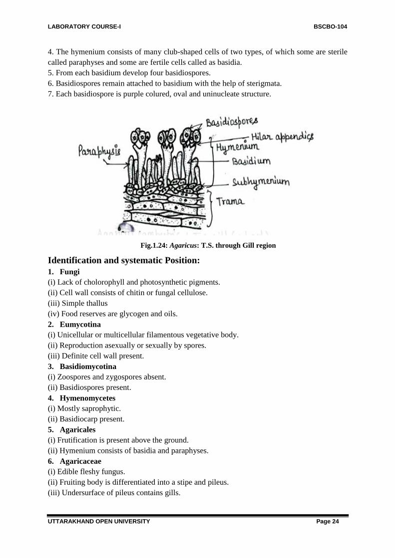
3. The trama is central in position and consit of many anastomosing, interwooven sterile
hyphae.
LABORATORY COURSE-I BSCBO-104
UTTARAKHAND OPEN UNIVERSITY Page 24
4. The hymenium consists of many club-shaped cells of two types, of which some are sterile
called paraphyses and some are fertile cells called as basidia.
5. From each basidium develop four basidiospores.
6. Basidiospores remain attached to basidium with the help of sterigmata.
7. Each basidiospore is purple colured, oval and uninucleate structure.
Fig.1.24: Agaricus: T.S. through Gill region
Identification and systematic Position:
1. Fungi
(i) Lack of cholorophyll and photosynthetic pigments.
(ii) Cell wall consists of chitin or fungal cellulose.
(iii) Simple thallus
(iv) Food reserves are glycogen and oils.
2. Eumycotina
(i) Unicellular or multicellular filamentous vegetative body.
(ii) Reproduction asexually or sexually by spores.
(iii) Definite cell wall present.
3. Basidiomycotina
(i) Zoospores and zygospores absent.
(ii) Basidiospores present.
4. Hymenomycetes
(i) Mostly saprophytic.
(ii) Basidiocarp present.
5. Agaricales
(i) Frutification is present above the ground.
(ii) Hymenium consists of basidia and paraphyses.
6. Agaricaceae
(i) Edible fleshy fungus.
(ii) Fruiting body is differentiated into a stipe and pileus.
(iii) Undersurface of pileus contains gills.

LABORATORY COURSE-I BSCBO-104
UTTARAKHAND OPEN UNIVERSITY Page 25
7. Agaricus
(i) Presence of annulus.
(ii) Gills are of three different sizes.
1.3.5-Alternaria
Kingdom: Mycota
Division: Eumycota
Sub division: Deutromycotina
Class: Hyphomycetes
Order: Monilliales
Family: Dematiaceae
Genus: Alternaria
Habitat and occurrence
1. This is a cosmopolitan genus occurring as a saprophyte as well as a weak parasite.
2. Species of Alternaria are of common occurrence in the atmosphere and the soil.
3. The ―early blight of potato‖ is one of the most commonly occurring diseases caused by
Alternaria solani.
Symptoms
Fig.1.25: Alternaria: Infected Potato twig
1. Presence of yellowish brown spots of varying size on leaves.
2. Spots become rounded to form concentric rings.
3. Colour of infected part later on changes to black.
4. Later lamina of leaf turns black.
5. Inner edible part of the infected tuber turns brown.
Somatic structure of fungus:
Cut thin transverse sections of the host through the infected portion, stain in cotton blue,
mount in lacto phenol and study.
1. Mycelium of fungus is intercellular or intracellular.
2. Light brown coloured hyphae are well branched, septate and each cell is multinucleate.
3. Haustoria absent.

LABORATORY COURSE-I BSCBO-104
UTTARAKHAND OPEN UNIVERSITY Page 26
Fig.1.26: Alternaria: T.S. of Infected Host leaf
Reproductive Structures:
Fungus reproduces only by asexual reproductive bodies called conidia.
1. Conidia are present terminally on conidiophores.
2. Each conidiophore is a multicellular, short or elongated, brown or dark coloured
structure.
3. Each cell of conidiophore is multinucleate.
4. Each conidium is multicellular, obovoid or spindle-shaped structure.
5. Conidia are transversely as well as longitudinally septate.
6. In moist conditions, conidia germinate with the help of 5 to 10 germ tubes.
Fig.1.27: Alternaria: A few Conidia
Identification and systematic Position:
1. Fungi
(i) Lack of cholorophyll and photosynthetic pigments.
(ii) Cell wall consists of chitin or fungal cellulose.
(iii) Simple thallus
(iv) Food reserves are glycogen and oils.
2. Eumycotina
(i) Unicellular or multicellular filamentous vegetative body.
(ii) Reproduce asexuall or sexual by spores.
(iii) Definite cell wall present.
3. Deutromycotina
(i) Reproduction only by asexual means.
4. Hyphomycetes
(i) Pycnidia or acervuli absent.
5. Moniliales
LABORATORY COURSE-I BSCBO-104
UTTARAKHAND OPEN UNIVERSITY Page 27
(i) Conidia develop at the tip of conidiophore.
(ii) Conidia of varying shape.
6. Dematiaceae
(i) Absence of fruiting body.
7. Alternaria
(i) Conidia are macroconide and are transversely as well as longitudinally septate.
(ii) Conidiophores are erect bodies.
1.3.6-Saccharomyces (=Yeast)
Kingdom: Mycota
Division: Eumycota
Sub division: Ascomycotina
Class: Hemiascomycetes
Order: Endomycetales
Family: Saccharomycetaceae
Genus: Saccharomyces
Habitat and occurrence
1. It is a saprophytic fungus found on substratum which is rich in sugars e.g., sugarcane,
milk etc.
2. Their chief characteristic is to ferment the carbohydrates on which they occur profusely.
3. They are very important industrially in bakery and brewery.
4. They bring about alcoholic fermentation of the sugary media in which the resulting
products are alcohol and carbon dioxide.
Somatic structure of fungus
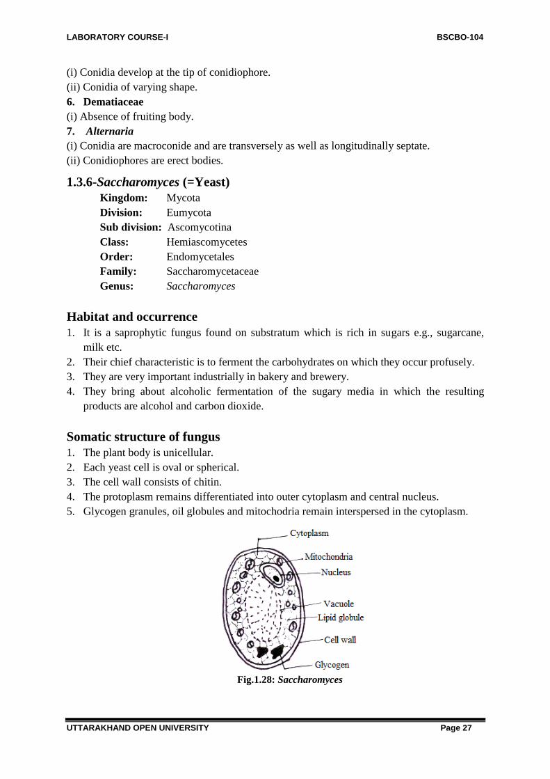
1. The plant body is unicellular.
2. Each yeast cell is oval or spherical.
3. The cell wall consists of chitin.
4. The protoplasm remains differentiated into outer cytoplasm and central nucleus.
5. Glycogen granules, oil globules and mitochodria remain interspersed in the cytoplasm.
Fig.1.28: Saccharomyces

LABORATORY COURSE-I BSCBO-104
UTTARAKHAND OPEN UNIVERSITY Page 28
Reproductive Structures
Asexual:
1. Takes by budding and fission.
2. In budding, each yeast cell gives rise to one or more small outgrowths which gradually
enlarge and detach from the mother cell to form independent individuals.
3. In fission, the cell becomes constricted in centre and divides into two forming two
independent individuals.
4. Sometimes, the yeast cell enlarges in size and is called the ascus.
5. Each such ascus contains four or eight ascospores.
6. Each ascospore germinates to produce new yeast cells.
Fig.1.29: Saccharomyces : Budding in yeast
7. Sometimes the buds do not detach from each other and form Pseudomycelium.
Fig.1.30: Saccharomyces: Pseudomycelium
Sexual Reproduction:
1. Takes place by conjugation.
2. Two individuals come close to each other and develop beak like outgrowths. These
outgrowths fuse with each other. After fusion zygote is formed.
3. The zygote nucleus then divides meiotically forming eight ascospores.
4. Each ascospore develops into a new plant body.
Electron Micrograph of Saccharomyces

LABORATORY COURSE-I BSCBO-104
UTTARAKHAND OPEN UNIVERSITY Page 29
Fig.1.31: Saccharomyces:Electron Micrograph
1. It has a definite cell wall present, followed by a plasma membrane enclosing cytoplasm.
2. It has cell organelles like mitochondria, storage granules, nucleus and nuclear membrane.
3. Central vacuole is also present.
Identification and Systematic Position:
1. Fungi
(i) Lack of cholorophyll and photosynthetic pigments.
(ii) Cell wall consists of chitin or fungal cellulose.
(iii) Simple thallus
(iv) Food reserves are glycogen and oils.
2. Eumycotina
(i) Unicellular or multicellular filamentous vegetative body.
(ii) Reproduction asexually or sexually by spores.
(iii) Definite cell wall present.
3. Ascomycetes
(i) Vegetative body consist of septate mycelium. in some one celled.
(ii) Absence of motile spores or gametes.
(iii) Sexually produce spores, ascospores, formed within ascus.
4. Hemiascomycetidae
(i) Asci naked, not enclosed inside an ascocarp.
5. Endomycetales
(i) Absence of ascogenous cells.
(ii) Asci produced directly from zygote or from diploid somatic cells.
6. Saccharomycetaceae
(i) Multiplication by budding.
(ii) Gametangia absent.
(iii) Copulation somatogamous.
7. Saccharomyces
(i) Unicellular plant body.

LABORATORY COURSE-I BSCBO-104
UTTARAKHAND OPEN UNIVERSITY Page 30
1.3.7-Erysiphe
Kingdom: Mycota
Division: Eumycota
Sub division: Ascomycotina
Class: Pyrenomycetes
Order: Erysiphales
Family: Erysiphaceae
Genus: Erysiphe
Habit and occurrence
1. Erysiphe is a cosmopolitian powdery mildew fungus occurring as an ectoparasite mostly
on the cultivated plants.
2. They are obligate parasites on the leaves, young shoots and inflorescence of the flowering
plants.
3. They form an extramatrical mycellim on the host surface.
Disease and causal organism:
1. Erysiphe graminis var. tritici causes powdery mildew of wheat.
2. Erysiphe graminis var. hordei causes powdery mildew of barley.
3. Erysiphe polygonii occurs on the leaves and stems of a considerable variety of hosts.
Somatic structure:
Fig.1.32: Erysiphe: Infected Wheat plant
1. The mycellium is superficial and mainly found on both the surface of leaves.
2. The hyphae are septate, hemiendophytic and possess uninucleate cells.
3. hypha of limited growth penetrates through the stoma and develops in the substomal
chamber in the intercellular spaces of the adjacent mesophyll cells.
4. The saccate haustoria produced by hyphal branches which then penetrate the adjoining
cells.

LABORATORY COURSE-I BSCBO-104
UTTARAKHAND OPEN UNIVERSITY Page 31
Fig.1.33: Erysiphe: Mycelium and Haustorium within Infected host
Reproductive bodies:
Asexual: Conidia
1. Asexual reproduction occurs by means of the conidiophores and conidia.
2. The conidiophores develop vertically from the superficial mycellium.
3. The conidia are formed singly. They are single celled, clavate , uninucleate , hyline and
thin walled.
4. Conidia are dispersed by wind and each germinates to form a new mycellium.
Fig. 1.34: Erysiphe: Somatic structure and asexual reproduction
Fig.1.35: Powdery mildew of peas (Erysiphe polygoni) ; A ecotophytic mycellium with
conidiophore and conidia : B, Conidia.
LABORATORY COURSE-I BSCBO-104
UTTARAKHAND OPEN UNIVERSITY Page 32
Sexual: Cleistothecia and ascospores:
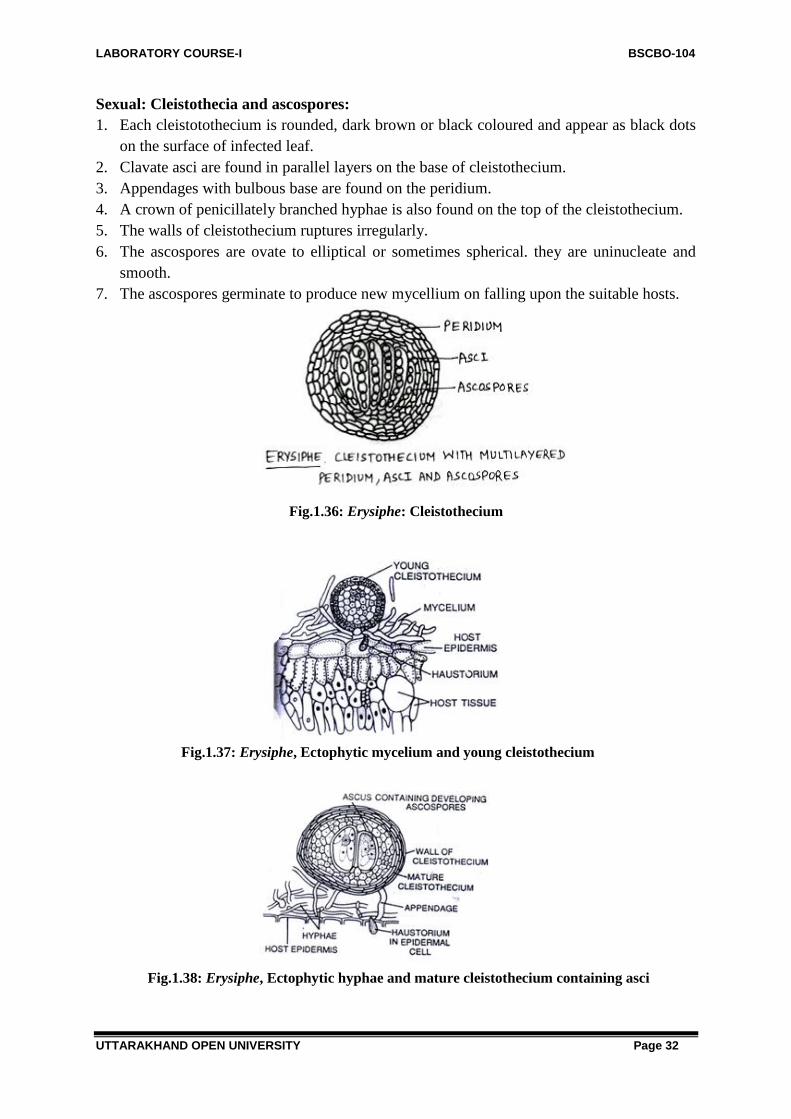
1. Each cleistotothecium is rounded, dark brown or black coloured and appear as black dots
on the surface of infected leaf.
2. Clavate asci are found in parallel layers on the base of cleistothecium.
3. Appendages with bulbous base are found on the peridium.
4. A crown of penicillately branched hyphae is also found on the top of the cleistothecium.
5. The walls of cleistothecium ruptures irregularly.
6. The ascospores are ovate to elliptical or sometimes spherical. they are uninucleate and
smooth.
7. The ascospores germinate to produce new mycellium on falling upon the suitable hosts.
Fig.1.36: Erysiphe: Cleistothecium
Fig.1.37: Erysiphe, Ectophytic mycelium and young cleistothecium
Fig.1.38: Erysiphe, Ectophytic hyphae and mature cleistothecium containing asci

LABORATORY COURSE-I BSCBO-104
UTTARAKHAND OPEN UNIVERSITY Page 33
Fig.1.39: Erysiphe, Single cleistothecium with simple appendages and mycelium
Fig.1.40: Powdery mildew of peas (Erysiphe polygoni), A. Cleistothecium with simple
appendages; B. ascus containing four ascospores; C. Ascospores
Identification and Systematic position:
1. Fungi
(i) Lack of cholorophyll and photosynthetic pigments.
(ii) Cell wall consists of chitin or fungal cellulose.
(iii) Simple thallus
(iv) Food reserves are glycogen and oils.
2. Eumycotina
(i) Unicellular or multicellular filamentous vegetative body.
(ii) Reproduction asexually or sexually by spores.
(iii) Definite cell wall present.
3. Ascomycetes
(i) Vegetative body consist of septate mycellium.
(ii) Absence of motile spores or gametes.
(iii) Sexually produce spores within ascus.
4. Erysiphales
(i) Obligate parasites.
(ii) Mycellium white, cleistothecia with appendages.
5. Erysiphae
(i) Several asci in cleistothecium.

LABORATORY COURSE-I BSCBO-104
UTTARAKHAND OPEN UNIVERSITY Page 34
(ii) Appendages mycellium-like, indefinite.
1.3.8-Mucor
Kingdom: Mycota
Division: Eumycota
Sub division: Zygomycotina
Class: Zygomycetes
Order: Mucorales
Family: Mucoraceae
Genus: Mucor
Habitat and occurrence
1. Mucor is a common saprophytic fungus that grows on the dead organic material.
2. In laboratory this fungus can be cultured by keeping the moist bread under bell jar for two
or three days.
3. The fungus mycelium looks like fine cottony threads on the surface of bread.
Somatic structure of fungus
Pick up small part of hyphae growing on moistened bread stain in cotton blue, mount in
lactophenol and study.
Fig.1.41: (A-C) Mucor: Structure of mucelium. (A) Absorptive hyphae and sporangiophores;
(B) Vegetative mycellum under light microscope; (C) vegetative mycellum under electron
microscope 1. The mycellium is whitish, filamentous, profusely branched hyphae giving a cottony
appearance.
LABORATORY COURSE-I BSCBO-104
UTTARAKHAND OPEN UNIVERSITY Page 35
2. Mycelium is aseptate and multinucleate (coenocytic)
3. Hyphae are surrounded by cell wall made of chitin.
4. Cytoplasm is granular and consists of glycogen and oil as reserve food material.
Reproductive Structures
The fungus may reproduce by vegetative (by fragmentation), sexual or asexual means
Asexual reproduction:
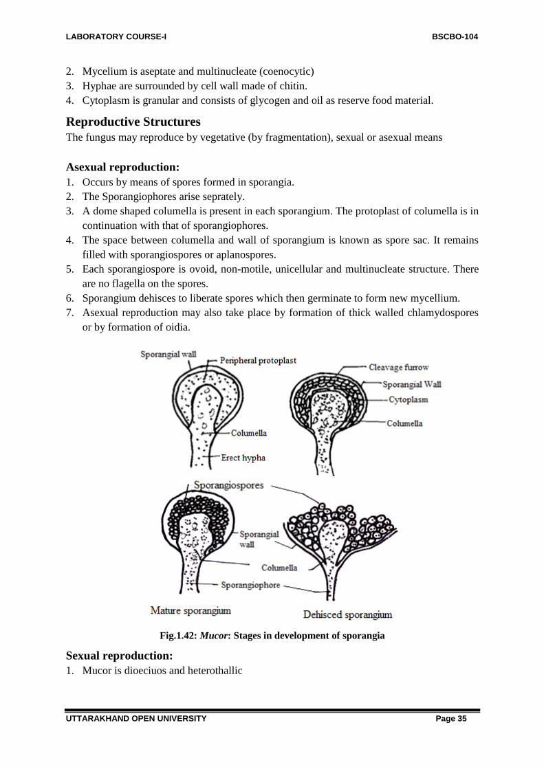
1. Occurs by means of spores formed in sporangia.
2. The Sporangiophores arise seprately.
3. A dome shaped columella is present in each sporangium. The protoplast of columella is in
continuation with that of sporangiophores.
4. The space between columella and wall of sporangium is known as spore sac. It remains
filled with sporangiospores or aplanospores.
5. Each sporangiospore is ovoid, non-motile, unicellular and multinucleate structure. There
are no flagella on the spores.
6. Sporangium dehisces to liberate spores which then germinate to form new mycellium.
7. Asexual reproduction may also take place by formation of thick walled chlamydospores
or by formation of oidia.
Fig.1.42: Mucor: Stages in development of sporangia
Sexual reproduction:
1. Mucor is dioeciuos and heterothallic

LABORATORY COURSE-I BSCBO-104
UTTARAKHAND OPEN UNIVERSITY Page 36
2. The male and female mycellia are morphologically identical but physiologically different
thus represnted by + and – strains.
3. Two hyphae from mycelia of different strains known as progametangia develop towards
each other.their growth results in the adherence of progametangia at their tips. Their tip
swells up and a transverse septum develops in each differentiating gametangium.
4. The remaing part of progametangium is called suspensor.
5. The multinucleate protoplasm of each gametangium is known as coenogamete.
6. The fusion of gametangia takes place to form thick, spiny walled zygospores.
7. Zygospores germinate meiotically by producing a long sporangiophore bearing a
spoangium at the tip.
Fig.1.43: Mucor: Sexual Reproduction
Identification and systematic Position:
1. Fungi
(i) Lack of cholorophyll and photosynthetic pigments.
(ii) Cell wall consists of chitin or fungal cellulose.
(iii) Simple thallus
(iv) Food reserves are glycogen and oils.
2. Eumycotina
(i) Unicellular or multicellular filamentous vegetative body.
(ii) Reproduction asexually or sexually by spores.
(iii) Definite cell wall present.
3. Zygomycotina
(i) Perfect state spore are zygospores.
4. Zygomycetes
(i) Mostly saprobic
5. Mucor
(i) Absence of stolon and rhizoids.
(ii) Absorption takes place by entire mycelial surface.

LABORATORY COURSE-I BSCBO-104
UTTARAKHAND OPEN UNIVERSITY Page 37
(iii) Mycellium white, cleistothecia with appendages.
1.4 SUMMARY
Fungi are acholophyllous, heterotrophic eukaryotic organism. Mycology (Gk. Mykes=fungus;
logos=study) stands for study of science of fungi. The fungi lack photosynthetic pigments and
therefore they cannot synthesize their own food. Their mode of nutrition is saprophytic,
parasitic or symbiotic.
These are normally studied by cutting thin transverse section of the infected host and
then staining them in cotton blue and finally mounted in lactophenol which is then studied
under microscope.
Albugo belongs to family Albuginaceae. Many species of Albugo occur as obligate
parasites on many plants of cruciferae, causing the common disease called ―white rust of
crucifers‖. The genus Phytophthora may be either facultative saprophytes or facultative
parasites causing great damage to plants of great economic importance for eg..P. infestans
causes Late blight of potato, P. parasitica causes seedling blight of castor and P.
megasperma causes blight of cauliflower or tomato.
Genus Puccinia occurs as an obligate parasite on many cereals, millets etc and cause
the rust disease. Important host are wheat, oats, jowar, bajra etc. Puccinia graminis is a
macrocyclic rust and produces 5 types of spores in its life namely uredospore, teleutospores,
basidiospore,pycnidiospore, aecidiospores.
Agaricus is a saprophytic, edible fungus occurring commonly in rainy season on
humus soil, rotten woods, tree trunks and other organic substances.
Species of Alternaria is a cosmopolitan genus occurring as a saprophyte as well as a
weak parasite. The ―early blight of potato‖ is one of the most commonly occurring diseases
caused by Alternaria solani.
Genus Saccharomyces is a saprophytic fungus found on substratum which is rich in
sugars e.g., sugarcane, milk etc. their cheif characteristic is to ferment the carbohydrates on
which they occur profusely.
Genus Erysiphe is a cosmopolitian powdery mildew fungus occurring as an
ectoparasite mostly on the cultivated plants. Erysiphe graminis var. tritici causes powdery
mildew of wheat.
Mucor is a common saprophytic fungus that grows on the dead organic material.
1.5 GLOSSARY
Apothecium: Fruiting body is cup shaped body. It is found in Discomycetes.
Ascogonium: The female reproductive organ of ascomycota
Ascogenus hypha: A dikaryotic hypha that grows out of a fertilized ascogonium.

LABORATORY COURSE-I BSCBO-104
UTTARAKHAND OPEN UNIVERSITY Page 38
Ascus: The reproductive structure of ascomycota in which fusion, meosis, and spore
formation take place.
Basidioma: The fruiting body of basidiomycota in which basidia form.
Basidium: The club-shaped reproductive structure of basidiomycota in which fusion,
meiosis, and spore formation take place.
Binucleate: Having two nuclei.
Budding: Asexual reproductive process in which a small portion of the cell membrane and
cytoplasm receive a nucleus and pinch off from the parent cell.
Cellulose: A major component of plant and algal cell walls. Compare with chitin.
Chitin: A major component of fungal cell walls that is not found in the cell walls of any
other group. Compare with cellulose.
Clamp connection: The structure by which basidiomycota cells divide while retaining their
binucleate dikaryotic condition.
Cleistothecium: Fruiting body closed from all sides with no opening is called cleistothecium.
It is found in plectomycetes.
Conidiophore: Structure in which asexually-produced spores called conidia are formed.
Dikaryotic: Having two genetically different nuclei.
Eucarpic : fungi in which a part of vegetative mycelium forms the reproductive unit and rest
remains vegetative.
Fruiting body: A general term for elaborate structures that contain spore-forming cells.
Gametangia: In zygomycota, the cells which fuse to become the zygote.
Heterothallic fungi: the fungi possessing dioecious mycelia are called heterothallic.
Heterotrophic : it is a mode of nutrition in which organism cannot synthesize its own food
and hence dependent on others.
Holocarpic fungus: fungi in which whole vegetative cell is transformed into reproductive
unit.
Hypha: Individual filaments of fungal cells; compare with mycelium.
Intercellular mycelium: In it the hyphae ramify in the intercellular spaces between the host
cells.
Intracellular mycelium: In it the hyphae penetrate into the host cells.
Karyogamy: The fusion of two nuclei.
Mycelium: The usually underground portion of a fungus that is haploid and sprouts from a
spore.
Mycorrhiza : is close symbitioc association of fungus with the roots of some higher plants
Obligate saprophytes: the plants which can live or survive strictly as saprophytes are called
obligate saprophytes.
Parasitic: takes all their nutrients from the tissues of another organisms.
Perithecium: Fruiting body is flask shaped body having a terminal opening or ostiole. It is
found in pyrenomycetes.
Plasmogamy: Fusion of the plasma membranes of two cells.
Pseudomycelium: Sometimes, the buds formed in process of budding are not detached and
provide appearance of mycelium called pseudomycelium eg. Yeasts.
Rhizoid: The sub-surface hyphae of zygomycota specialized for food absorption

LABORATORY COURSE-I BSCBO-104
UTTARAKHAND OPEN UNIVERSITY Page 39
Sporangiophores: Filamentous stalk on which a sporangium forms.
Sporangium: Spore producing structure of zygomycotena.
Stolon: The hyphae that connect groups of rhizoids and sporangiophores, usually above the
surface.
Symbiont: An organism that lives in close association with another, to the benefit of one or
both organisms.
Trichogyne: Specialized cell on the end of the ascogonium. During mating, the trichogyne
grows to connect the ascogonium to the antheridium.
Zygospore: The heavily encapsulated structure that forms from the zygote of zygomycoina
Zygote: The diploid cell that results from the fusion of two gametes or gametangia during
fertilization.
1.6 SELF ASSESSMENT QUESTIONS
1.6.1 Short Answer Questions:
1. White pustules are formed on the leaf of crucifers by the infection of?
2. What is the mode of nutrition in Cystopus?
3. What type of sexual reproduction is found in Cystopus?
4. Coniodiophores in cystopus are produced in
5. Who coined the term Albugo?
6. Who coined the name Cystopus to white rust causing organisms?
7. Name a fungus that produces knob like haustoria.
8. Name a fungus which produced a chain of sporangia on short sporangiophores.
9. What structures help in the release of sporangia of Albugo?
10. How many nuclei are present in the sporangium of Albugo?
11. What types of sexual reproduction is observed in Albugo?
12. How many functional nuclei are present in the gametangia of Albugo candida?
13. Name the type of life cycle present in Albugo-
14. Name the fungicides used to prevent the spread of white rust of crucifers?
15. Name the organism that causes late blight of potato?
16. In which fungus the haustoria are slender and curled?
17. Name the method of sexual reproduction in Phytophthora?
18. Name the cell wall materials of Phytophthora-
19. Name the resting spore of Phytophthora-
20. Name the type of life cycle present in Phytophthora-
21. To which fungus does the sporangiophores with nodal swellings and sympodial branching
belong?
22. Name one macrocyclic fungus?
23. Name the alternate host of puccinia graminis var. tritici?
24. What are the hosts of Puccinia?
25. By which method basidiospores is discharged from the sterigmata in Puccinia?
26. Name the flask- shaped rod or yellow stuctures produced by puccinia on the upper surface
of barberry leaf.

LABORATORY COURSE-I BSCBO-104
UTTARAKHAND OPEN UNIVERSITY Page 40
27. Name the orange coloured hairs present at the mouth of spermagonium of puccinia-.
28. Name the agent that transfers spermatia from one spermagonium to other spermagonium-
29. Name the dikaryotic spore produced by Puccinia on barberry plant.
30. Name the elongated stucture produced by the germ tube of urediniospore
31. How many germ pores are normally present in the urediospores of Puccinia.
32. Agaricus campestris is commonly known as?
33. Agaricus belongs to class?
34. Button shaped young fruiting bodies belong to
35. Fungus in which is fairy rings are formed.
36. What causes early blight of potato
37. Give three characters of Alternaria conidia.
38. Transverse and longitudinal septa are found in the conidia of
39. The most common method of vegetative reproduction found in saccharomyces is..
40. Powdery mildew disease of wheat is caused by?
41. Columellate sporangia are characteristic feature of?
42. Mycellium is coenocytic in the genus?
43. Hyphal walls in the members of zygomycetes are made up of?
44. Sexual reproduction in the zygomycetes results in the formation of?
45. The most important salient feature of the zygomycetes is the absence of?
1.6.1: Answers to Short Answer Questions:
1. Cystopus
2. Heterotrophic
3. Oogamous type
4. Parallel clusters.
5. Persoon
6. Levellie
7. Albugo
8. Albugo
9. Disjunctors
10. 5 to 8
11. Gametangial contact.
12. One in antheridium and one in oogonium
13. Diplontic
14. Copper fungicides
15. Phtophthora infestans.
16. Phytophthora.
17. Gametangial contact
18. Cellulose, glucan.
19. Oospore.
20. Diplontic.
21. Phytophthora.
22. Puccinia graminis.
23. Barberry.

LABORATORY COURSE-I BSCBO-104
UTTARAKHAND OPEN UNIVERSITY Page 41
24. Wheat and Barberry.
25. Drop excretion method.
26. Spermagonium.
27. Periphyses.
28. Insect.
29. Aeciospores.
30. Appresorium.
31. Four.
32. Field mushroom.
33. Hymenomycetes.
34. Agaricus
35. Agaricus.
36. Alternaria solani
37. Oblevate, muriform and beaked.
38. Alternaria.
39. By budding
40. Erysiphae.
41. Mucor
42. Mucor
43. Chitin.
44. Zygospores
45. Motile cells.
1.6.2: Fill in the blanks:
1. Scientific term for fruiting body of Ascomycetes.........
2. The life cycle of Saccharomyces is.............
3. During unfavourable conditions Saccharomyces form......
4. All fungi are.............
5. Fungi can be stained by..........
6. Fung usually store the reserve food material in the form of..........
7. The fruiting body of Aspergillus is called......
8. Sexual reproduction in Agaricus takes place by..........
9. Heterothallism was discovered by.........
10. Yeasts are an important source of....
11. Name the perfect state of Alternaria
12. Toxin produced by Alternaria.
13. Yeasts are obligate
14. The fungus used for flavouring cheese is –
15. Sexual reproduction in yeast takes place by –
1.6.2: Answers to fill in the blanks:
1. Ascocarp.
2. Haplo-diplobiontic
3. Endospores.

LABORATORY COURSE-I BSCBO-104
UTTARAKHAND OPEN UNIVERSITY Page 42
4. Saprophytes
5. Cotton blue
6. Glycogen
7. Cleistothecium
8. Somatogamy
9. A.F Blakeslee, 1904.
10. Riboflavin.
11. Pleospora infectoria
12. Alternariine
13. Saprophytes.
14. Yeast
15. Union of two cells.
1.7 REFERENCES
Sharma, P.D.2001. The Fungi. Rastogi Co., Meerut.
Bendre, A. and Kumar, A. 1990-91. Practical Botany, Rastogi Publications, Meerut.
Singh, V., Pande, P.C. and Jain, D.K. A Text Book of Botany, Rastogi & Co., Meerut,
2001.
Vashista, B.R. Botany for Degree student Fungi, S. Chand & Co., New Delhi, 2001.
Vashista, B.R. Botany for Degree Students (Algae, Fungi Bryophyta), S. Chand & Co.
Ltd., New Delhi, 2002.
1.8-SUGGESTED READINGS
Alexopoulos, C.J. and Mims. Introductory Mycology, John Wiley and Sons, New York,
2000.
Bilgrami, K.S. and Dube, H.C. A Text Book of Modern Plant Pathology, Vikas Publ.
House, New Delhi, 1976.
Bold, H.C., Alexopoulous, C.J. and Delevoryas, T. Morphology of Plant and Fungi (4th
Ed.) Harper & Foul Co., New York, 1980.
Dube, H.C. 1990. An Introduction to Fungi. Vikas Pub. House Pvt. Ltd. New Delhi.
Dube, H.C. Fungi, Rastogi Publication, Meerut, 1989.
Gilbert, M.S. Cryptogamic Botany, Vol. I & II (2nd Ed.), Tata McGraw Hill, Publishing
Co. Ltd., New Delhi, 1985.
Pandey, S.N. and Trivedi, P.S. A Text Book of Botany 2000 Volume I, Vikas Pub. House
Pvt. Ltd., New Delhi.
Sharma, O.P. Fungi, Today and tomorrow Publication, 2000.
1.9 TERMINAL QUESTIONS
1.9.1: Short Answer Questions-
1. What is rhizomorph?
2. What are toadstools?

LABORATORY COURSE-I BSCBO-104
UTTARAKHAND OPEN UNIVERSITY Page 43
3. What is the shape of sex organs in Cystopus?
4. Who coined the term Phtophthora infestans?
5. What is meant by macrocyclic rust?
6. Name the 5 spores produced by Puccinia in its life cycle?
7. What is a heteroecious rust?
8. Name two important diseases of plants caused by Alternaria?
9. Fungi imperfectii are so named why?
10. What is fairy ring?
11. What are gills in fungus?
12. What is hymenium?
13. What are Paraphyses?
1.9.1: Answers:
1. The secondary mycelium of Agaricus, in the later phase of development produces much
compacted mass of hyphal strands in the soil. These strands are called rhizomorphs and
give rise to fruiting bodies above the ground.
2. Toadstools are non-edible poisonous fruiting bodies of certain basidiomycetes fungi.
They are often called poisonous mushrooms eg. Amanita.
3. Rounded oogonia and Club shaped antheridia.
4. de Bary
5. Rust which produces 5 types of spores in its life cycle.
6. Uredospore, Teleutospores, Basidiospore, Pycnidiospore and Aecidiospores are the five
spores produced by Puccinia.
7. Rust which requires two unrelated hosts to complete its life cycle is called heteroecious
rust.
8. Early blight of potato is caused by-A. solani and Leaf spot crucifers by A. brasicae and A.
brassicola.
9. Because of the absence of sexual reproduction.
10. Fruiting bodies of mushrooms of Agaricus develop in a ring above the ground in lawns
and forest. Such rings or circle of fruiting bodies is called fairy rings.
11. These are thin, vertical plate like structures hanging down from the underside of the
pileus of the Agaricus fruiting body.
12. The fertile region present on both side of the gills is called hymenium
13. The sterile threads present in the hymenium are called paraphyses.
1.9.2: Very short answer Questions-
1. Scientific term for fruiting body of Ascomycetes?
2. The life cycle of Saccharomyces is?
3. During unfavourable conditions Saccharomyces form-
4. Fungi imperfectii are so named because -
5. Name two important diseases of plants caused by Alternaria?
6. What is rhizomorph?
7. What are toadstools?
8. What is the shape of sex organs in Cystopus?

LABORATORY COURSE-I BSCBO-104
UTTARAKHAND OPEN UNIVERSITY Page 44
9. Who coined the term Phtophthora infestans?
10. What is meant by macrocyclic rust ?
11. Name the 5 spores produced by puccinia in its life cycle?
12. What is a heteroecious rust?
1.9.2: Answers:
1. Ascocarp.
2. Haplo-diplobiontic.
3. Endospores.
4. Because of the absence of sexual reproduction.
5. Early blight of potato is caused by-A. solani and Leaf spot crucifers by A. brasicae and A.
brassicola.
6. The secondary mycelium of Agaricus, in the later phase of development produces much
compacted mass of hyphal strands in the soil. These strands are called rhizomorphs and give
rise to fruiting bodies above the ground.
7. Toadstools are non-edible poisonous fruiting bodies of certain basidiomycetes fungi. They
are often called poisonous mushrooms e.g. Amanita.
8. Rounded oogonia and club shaped antheridia.
9. de Bary
10. Rust which produces 5 types of spores in its life cycle.
11. uredospore, teleutospores, basidiospore,pycnidiospore, aecidiospores.
12. Rust which requires two unrelated hosts to complete its life cycle.
LABORATORY COURSE-I BSCBO-104
UTTARAKHAND OPEN UNIVERSITY Page 45
UNIT-2 STUDY OF MORPHOLOGY AND
STRUCTURE OF DIFFERENT TYPES OF LICHENS
2.1 Objectives
2.2 Introduction
2.2.1 Classification of lichens
2.3 Structure of Lichens
2.3.1 Morphology (External)
2.3.2 Structure Internal
2.3.3 Special structure associated with Lichen
2.3.4 Reproductive structure
2.4 Summary
2.5 Glossary
2.6 Self assessment question
2.7 References
2.8 Suggested Readings
2.9 Terminal Questions

LABORATORY COURSE-I BSCBO-104
UTTARAKHAND OPEN UNIVERSITY Page 46
2.1- OBJECTIVES
After reading this section you will know, -
What are Lichens.
Different types of substratum of Lichens.
Different Types of Lichens.
Reproduction in Lichens.
2.2- INTRODUCTION
Lichens are a small group of curious plants. They are made up of algal and fungal
components, livings together in an intimate symbiotic relationship. The algal component is
known as phycobiont (phy kos = alga, bios = life) and the fungal component as mycobiont
(mykes = fungu (bios = life). The plant body of lichens neither resembles algae nor fungi.
Thus, lichen is an association of a fungus and an algal photosynthetic symbionts, resulting in
a stable thallus of specific structure. Phycobionts generally belongs to cyanophyceae or some
times to chlorophy ceae. The alga is unicellular. The phycobiont is generally an ascomycete
but in rare cases it is a basidiomycete.
Lichens were first discovered by Tulasne in 1892. The relationship between the two
partners is a matter of controversy. Some hold it to be a typical case of symbiosis whereas
others consider it to be parasitism. However, it is now considered to ba a case of helotism, a
type of symbiotic association where the fangus has a upper hand. The lichens grow on a
variety of habitats and are common on rocks, bark of trees, etc. Many of them grow under
extreme condition of cold, humidity and drought. They are most conspicuous in the Alpine
and Arctic Tundra where they are dominant form of vegetation. In India lichens are common
in temperate and Alpine regions of Himalaya, hilly region of peninsular India and along the
sea cost.
There are about 400 genera and 1600 species of lichens, widely distributed in most
part of the world. Some common species are: Cladonia aggregata, Graphics duplicata,
Gyrophora cylindrica, Haematomma, puniceum, Phystia aspera, Usnea, aspera and Usnea
dischotoma.
2.2.1 Classification of Lichens
A) On the basis of their general growth, type of thallus and their mode of occurrence.
Lichens are generally four types.
1. Crustose lichens (Encrusting Lichens)
2. Foliose lichens (leafy lichens)
3. Fruticose lichens (Shrubby lichens)
4. Leprose lichen
B) On the basis of the nature of the fungal element the lichens are divided into three
groups.

LABORATORY COURSE-I BSCBO-104
UTTARAKHAND OPEN UNIVERSITY Page 47
1. Ascolichens if the fungal component is a ascomycetous. They are further divided into
two sub groups –
(a) Pyrenocarpeate: Includes those lichens in which the ascocarp is a perithecium e.g.:
Dermatocarpon.
(b) Gymnocarpear: Includes those lichens in which the ascoarp is an apotheciam. e.g.
Parmelia.
2. Basidiolichens: If the fungal component is a basidiomycetous. e.g. cora, Corella,
Dictyonema.
3. Deuterolichens: (Hymenolichens) Fructifications are absent in this group of lichens
or should say that lichens with sterile thalli are constituted by this group. e.g. Lepraria,
Leprocaulo, Crysothrix.
2.3 STRUCTURE
Thalloid lichens are green or bluish – green in colour. Some species may have yellow red,
orange or brown pigments. They are usually dull in appearance because of the translucent
fungal covering over the algal constitutents.
Morphology
On the basis of growth forms, and nature of attachment to the substratum lichens are divided
into following four types.
(1) Crustose lichens (Encrusting Iichens).
1. These lichens occur as thin or thick crust over rocks, soil or tree barks.
2. It is very difficult to separate them from substratum.
3. The thalli may be wholly or partially embedded so that only fruiting bodies are visible
above the surface of the substratum.
4. Common examples are Lecanora, Graphis, Rhizocarpon, Ochrolechia etc. (Fig.2.1).
Fig. 2.1: Lichens: A crustose
(2) Foliose lichens (leafy lichens)
1. These lichens are variously lobed leafy structures.
2. They are attached to the substratum by rhizoid like outgrowth called the rhizines.
3. The thallus is generally greyish or brownish in colour.

LABORATORY COURSE-I BSCBO-104
UTTARAKHAND OPEN UNIVERSITY Page 48
4. Common examples are Xanthoria, Parmelia, Physcia, Anaptychia etc.(Fig.2.2).
Fig. 2.2: Lichens: A foliose lichen
(3) Fruticose lichens (Shrubby lichens)
1. These are the upright or hanging lichens. (pendant forms)
2. These are attached only at the base by a flat disc.
3. These are cylindrical, flat or ribbon like, well branched and resemble with little shrubs
e.g., Cladonia, Usnea, Alectoria etc. (Fig.2.3).
Fig. 2.3: Lichens: A fruiticose Iichen
(4) Leprose lichen:
1. A fourth type of lichen called leprose has also been differentiated.
2. It has some fungal hyphae surrounding one or more algal cells.
3. A distinct fungal layer envelopes the algal cells all over.
4. It appears as a powdery mass over the substratum e.g., Leparia incana
(Fig.2.4)

LABORATORY COURSE-I BSCBO-104
UTTARAKHAND OPEN UNIVERSITY Page 49
Fig. 2.4: A leprose lichen
2.3.1 Internal Structure
Internally the thallus is composed of algal and fungal components. Such type of thallus is
known as consortium. On the basis of internal structure the lichens are divided into two
groups.
(A) Heteromerous lichens
(B) Homoiomerous Iichens
(A) T.S. Heteromerous Lichens
1. A transverse section of the hetermerous (foliose) lichen can be divided into following
distinct zones(Fig 2.5) :
Fig. 2.5: Lichens: Transverse section of heteromerous (foliose) Iichen thallus
(I) Upper cortex: It is the upper- most protective layer made up of compactly interworm
fungal hyphae. The compactly interwoven hyphe produce a tissue like layer
(Plectenchyma and pseudoparenchyma) called the upper cortex. The intercellular
spaces are absent, if present, they are filled with gelatinous substances. In some
species of foliose Iichens this layer is interrupted in different places. These
interruptions or areas are known as breathing pores and serve for aerations. In
addition to these certain other structures are also present for gaseous exchange. These
are called cyphellae.

LABORATORY COURSE-I BSCBO-104
UTTARAKHAND OPEN UNIVERSITY Page 50
(II) Gonidial layer: This layer consists of loosely interwoven hyphae intermingled with
algal cells. This region is the photosynthetic region of the thallus. This layer is also
called gonidial layer because of the earlier concept that these cells are having
reproductive function.
(III) Medulla: It is present just below the algal cells and is made of loosely interwoven
hypnal of fungus. Medulla forms the middle portion of the thallus.
(IV) Lower cortex: Like the upper cortex, it is the lower-most layer. In some lichens the
layer absent e.g., Lobaria pulmonaira. This layer gives rise to bundles of hyphae
(rhizines) which penetrate the substratum to function as anchoring organs.
2. Different types of lichens particulary the foliose and fruticose remain attached to the
substratum by a variety of structures such as itiizinose strand (thick strands e.g. Buellia
pulchella. Hyphal nets (fungal hyphae forming net like structures, e.g. Psora decipiens),
Hypothallus (thick, black, spongy, algal free tissue e.g., Anzia) Holdfast (basal, algae free
region, e.g. Usnea, Letharia). Hapters (short, penetrating branches. e.g. Alectoria) and
medullary hyphae.
3. The above structure of a lichen shows that the algae cells are restricted or confined to
form a distinct layer. Such type of lichens are called heteromerous (Fig. 2.6)
T.S. Homoiomerous Lichens:
1. In some lichens for example, Collema, Leptogium, the thallus shows a simple structure
with little differentiation.
2. The algae cells and fungal hyphae are uniformly distributed.
3. Both algal cells and fungal hyphae are enveloped in a gelatinous matrix.
4. Such type of lichens are called homoiomerous.
Fig. 2.6: Lichens: Transverse section of homoiomerous lichen
2.3.3 Special structures Associated with Lichens
I. Soredia:
1. They are small bud like out growth occuring on the upper surface or margin of the thallus
as greyish powder.
2. The soredia are separable portion of the thallus consisting of one or more algal cells
surrounded by the fungal hyphale.
3. A soredium may develop within definite pustule like compact structure called soralium.
4. Each soredium develops into a new thallus.

LABORATORY COURSE-I BSCBO-104
UTTARAKHAND OPEN UNIVERSITY Page 51
II. Isidia:
1. They also occur on the upper surface of the thalli as coral – like simple or branched
growhts.
2. They consist of an external cortical layer and an internal algal layer.
3. The algal element within the isidia is the same as that of the parent thallus.
III.Cephallodia:
1. They are external or internal gall like out growths, generally of dark colour.
2. They consists of fungal hyphal enclosing algal cells different from those of the thallus.
3. The Cephallodia are either, as flat orbicular discs or as coralloid branches or as irregular
warts and tubers e.g. Lecanora, Lobaria and Peltigera respectively.
IV. Cephellae:
1. They occure on the lower surface of the thallus quite commonly in the genus Stricta, as
small hollow circular, white depressions with its base resting on the medulla.
2. It's margin formed from the ruptured cortex projecting slightly inwards.
Fig.2.7 A-D Lichens: Asexual reproductive structures: A, B, Soredium, C. Cephalodium, D.
Isidium
2.3.4. Reproductive Structures
A) Vegetative and Asexual
I. Fragmentation
1. It commonly occurs by injury.
2. Each fragment is capable to give rise to a new thallus.
II. Soredia
1. After detached from the thallus, each soredium may develop into a new thallus.
2. Examples are Usnea, Parmedia
III. Isidia
1. Each detached isidium may develop into a new thallus.
2. Common example is Peltigera sp.
IV. Oidia

LABORATORY COURSE-I BSCBO-104
UTTARAKHAND OPEN UNIVERSITY Page 52
1. Hyphe of certain lichens break up into oidia.
2. Each oidium germinate into new fungal hyphae and produces a lichen when comes in
contact with suitable alga.
V. Pycniospores
1. Many lichens produce large number of small spore like structures, the pycniospaces.
2. Pycnidiospores are formed within flask-shaped pycnidium, immersed within the thallus.
3. The hyphae lining the cavity of the pycnidium produce many pycniospores that are
discharged through the astiole.
Fig. 2.8: Physcia. V.s. Pycnidium to show pycniospores
B. Sexual Structures
In lichens the process of sexual reproduction is performed only by the fungal component. The
fungal component of most of the lichens belong's to the class Ascomycetes. Hence the sexual
reproductive structures and reproduction is similar to that of ascomycetous fungi.
The female sex organs
1. The female sex organs are known as carpogonium.
2. A carpogonium is differentiated into a basal coiled ascogonium and an elongated
multicelleslar trichogyne.
3. The ascogonium remains embedded within the algal layer of the thallus.
4. The trichogyne projects over the surface of the thallus.
The Male sex organs
1. The male sex organs are flask – shaped spermogonia.
2. They form spermatia which function as male germetes.
3. The Spermogonium usually developes close to carpogonium
4. This enables spermatia to adhere to the projected part of sticky trichogyne.
5. On dissolution of the walls between the spermatium and trichogyne, the nucleus of
spermatium migrates into carpogonium through trichogune.
6. The male nucleus fuses with the female nucleus.

LABORATORY COURSE-I BSCBO-104
UTTARAKHAND OPEN UNIVERSITY Page 53
Fig. 2.9:A-B, Lichen : Reproductive structures ; A. Spermogonium, B. Carpogonium
Apothecia, Perithecia and Ascospores
1. Sexual reproduction results in the formation of apothecia or perithecia.
2. The fruiting bodies are small cuplike or disclike and may be embedded in or raised above
the surface of the thallus by short or long stalks.
3. The structure of the wall of an apothecium is similar to that of the thallus, it consists of an
upper and lower cortical layer with medulla in between.
4. The algal components may or may not be present in the vegetative part of the apothecium.
5. The bottom of the cup or the surface of the disc is the fertile part of apothecium and is
lined by the hymenium.
6. The hymenium consists of asci and paraphyses growing vertically. Paraphyees contain a
reddish oily substance in them and never projects beyond asci.
7. Each ascus contains eight ascospores. The ascospores become two called when they
disseminate.
8. Ascospares when come in contact of suitable alga, produce, the lichen thallus.
Fig. 2.10, A-B – Lichen: Structure of fruiting body; A. L.S. of apthecium B. A part of hymenium
showing asci.

LABORATORY COURSE-I BSCBO-104
UTTARAKHAND OPEN UNIVERSITY Page 54
2.4 SUMMARY
A lichen is a composite organism that arises from algae or cyanobacteria (or both) living
among filaments of a fungus in a symbiotic relationship. The combined life form has
properties that are very different from the properties of its component
organisms. Lichens come in many colors, sizes, and forms. The combined life form has
properties that are very different from the properties of its component organisms. Lichens
come in many colors, sizes, and forms. Lichens may have tiny, leafless branches (fruticose),
flat leaf-like structures (foliose), flakes that lie on the surface like peeling paint (crustose), or
other growth forms. A macrolichen is a lichen that is either bush-like or leafy; Other lichens
are termed microlichens. Here, "macro" and "micro" do not refer to size, but to the growth
form. Lichens do not have roots that absorb water and nutrients as plants do but like plants
they produce their own food by photosynthesis using sunlight energy, from carbon dioxide,
water and minerals in their environment. When they grow on plants, they do not live as
parasites and only use the plants as a substrate.
Some lichens have a portion of their thallus lifted off the substrate to form
'squamules'. They are otherwise similar to crustose lichens in that they possess an upper
cortex but no lower cortex. Foliose Lichens have an upper and lower cortex. They are
generally raised to some extent above the substrate but connected to it by rhizines
(specialised root-like hyphae). They are easier to remove from their substrate when collecting
because of this. Leprose lichens are an odd group of lichens which have never been observed
to produce fruiting bodies. Because knowledge of the form of the fruiting bodies is essential
to the identification of fungi, these lichens have not yet been identified properly, or at least
not yet given full scientific names. These fungi not only lack an inner cortex, but also lack an
outer one, i.e. no cortex, only an algal cell layer and sometimes a weakly defined medulla.
2.5 GLOSSARY
Apothecium (plural apothecia): One type of fruiting structure produced by the fungal
component of the lichen. An apothecium is cup- or disc-shaped (compare with perithecium)
and contains the spores, which allow for sexual reproduction.
Cilia: Linear or thread-like appendages projecting from the thallus or apothecia margins,
Cilia are the black, hair-like appendages pictured here along the margins of powder-edged
ruffle lichen (Parmotrema stuppeum) thallus.
Cortex: The protective outer wall of the thallus, composed entirely of fungal tissue. Lichens
may have two cortices (upper and lower), a single cortex or no cortex at all, depending on
growth form. Below the cortex is the photobiont.
Crustose: A lichen growth form distinguished by the thallus being tightly adhered to the
substrate at all points. Crustose lichens do not have a lower cortex, exposing the hyphae to
the substrate. It is impossible to remove a crustose lichen from its substrate without impacting
the substrate in some way.
Cyphella (plural cyphellae): Small depressions or pits in the thallus cortex that are lined
with cells (compare with pseudocyphella).
LABORATORY COURSE-I BSCBO-104
UTTARAKHAND OPEN UNIVERSITY Page 55
Foliose: A lichen growth form distinguished by a relatively flat, leaf-like thallus. Foliose
lichens have an upper and lower cortex, making it easy to identify an upper and lower thallus
surface.
Fruticose: A lichen growth form distinguished by a tufted, hanging or stalked thallus.
Fruticose lichens have a single, continuous cortex that wraps around the thallus branches,
making it difficult to discern an upper and lower surface.
Hyphae: Fungal filaments loosely distributed below the photobiont on the interior of the
thallus.
Isidium (plural isidia): A structure that projects from the thallus and contains both fungal
and algal components. An isidia can detach from thallus and therefore serves in vegetative
reproduction.
Perithecium (plural perithecia): One type of fruiting structure produced by the fungal
component of the lichen. A perithecium is flask-shaped (compare with apothecium) and often
embedded the thallus, making it somewhat inconspicuous. A small hole at the top of the
perithecium releases spores, which allow for sexual reproduction.
Photobiont: The photosynthetic component of a lichen, either green algae or cyanobacteria,
located beneath the cortex.
Pseudocyphella (plural pseudocyphellae): Small depressions or pits in the thallus
associated with cracks in the cortex. The cracks in the cortex are not lined with cells,
distinguishing these features from cyphellae.
Rhizines: Linear or narrow root-like appendages that protrude from the lower thallus surface
(compare with cilia) and attach to the substrate.
Soredia: A powdery or granular structure released from cracks in the thallus cortex. A
soredia is essentially the photobiont (algal component) wrapped in fungal hyphae and
therefore serves in vegetative, or asexual, reproduction.
Squamulose: A lichen growth form distinguished by small, overlapping thallus units or
scales. Squamulose lichens are not as tightly appressed to the substrate as crustose lichens but
are more appressed than foliose lichens. These lichens have an upper cortex but may or may
not have a lower cortex.
Thallus: The lichen body, which contains both a fungal and algal (photobiont) component.
2.6 SELF ASSESSMENT QUESTIONS
2.6.1 Short answer type questions
1. What are Lichens?
2. What are different types of lichens?
3. Mention different types of reproduction found in lichens.
4. Explain different habitats of lichens
5. What are different sex organs found in Lichens.
2.6.2 Very short answer type questions
Q1. Explain different habitats of lichens
(i) Isidia
(ii) Soredia

LABORATORY COURSE-I BSCBO-104
UTTARAKHAND OPEN UNIVERSITY Page 56
(iii) Soralium
(iv) Rhizines
(v) Cephellae
2.6.3 Multiple choice questions
1. Symbiotic association between algae and fungi is called
(a) VAM (b) Lichen
(c) Mycorrhiza (d) Mutualism
2. Many Scientist consider algal and fungal relationship as ―helotism‖ Helotism is
(a) Kind of symbiotic association (b) A kind of mutualism
(c) A kind of Master – Slave relationship (d) Master – Master relationship
3. Fungal partner in Lichen is called Mycobiont while algal partner is called
(a) Phycobiont (b) Algobiont
(c) Glycobiont (d) Often referred as algal partner
4. The study of Lichen is called
(a) Lichology (b) Lichenology
(c) Mycology (d) Phycology
5. More then 95% of lichens the fungal partner belong to class
(a) Ascomycetes (b) Basidiomycetes
(c) Zygomycetes (d) Mastigomycetes
6. The benefit of algae in this association is
(a) Food (b) Vitamins
(c) Growth substances (d) Protection
7. In Lichens sexual reproduction is carried out by
(a) Algae (b) Fungi
(c) Both algae and fungi. (d) Fungi only
8. The major group of algae involved in formation of Lichens are
(a) Brown algae (b) Red algae
(c) Green algae (d) Blue-green algae
9. Usnea is
(a) Crustose (b) Fruticose
(c) Foliose (d) Filamentous
10. Lichens are the main pollution indicators of
(a) SO2 (b) NO2

LABORATORY COURSE-I BSCBO-104
UTTARAKHAND OPEN UNIVERSITY Page 57
(c) Murcury (d) CO
11. If the fungal partner belong to ascomycetes then the lichen is called
(a) Ascomycetes (b) Lichen
(c) Ascolichen (d) None of above
2.11.3 Answers:
1. (b), 2. (c), 3. (a), 4. (b), 5. (a), 6. (d), 7. (d), 8. (d), 9. (b), 10.(a), 11. (c)
2.7 REFERENCES
Awasthi, D. D., 1957. A new species of Parmelia from Kodaikanal, South India. Curr.
Sci 26:123-124.
Awasthi, D. D., 1988. A key to the macrolichens of India and Nepal; J. Hattori Bot. Lab.
65: 207–302.
Awasthi, D.D., 2000. A Hand Book of Lichens. Bishan Singh Mahendra Pal Singh 23-A,
New Connaught Place Dehradun, India. Pp.19-24.
Awasthi, D. D., 2007. A Compendium of the Macrolichens from India, Nepal, and Sri
Lanka. Bishen Singh Mahendra Pal Singh. Dehra Dun, India. Pp.580.
Culberson, C. F., 1972. Improved conditions and new data for the identification of lichen
products by a standardized thin-layer chromatographic method. J. Chromatog. 72: 113-
125.
Kumar, M. and Stephen Sequiera 1997 a. Lichen flora of Western Ghats : An appraisal.
J.Econ. Tax. Bot., 21(1): 27-39.MacArthur, R. H., 1965. Patterns of species diversity.
Biol. Rev. 40: 510- 533.
Kumar, B., 2008. Lichen species distribution, cover and fall in a Quercus
semiecarpifolia(J .E Smith) forest of Garhwal Himalayas (Ph.D. Thesis), H.N.B Garhwal
university, Srinagar (Garhwal).India.
Kumar, B.,2009. Assessment of lichen species in a temperate region of Garhwal
Himalayas, India. The Journal of American Sciences. 5(4): 107-112.
Kumar, B.,2009. Quantitative analysis of fallen lichens vegetattion in eleven forest of a
Quercus semecarpifolia of Garhwal Himalyas,India.. Nature &Science.7(2):95-100.
Negi, H. R., and Gadgil, M., 1996. Pattern of distribution of macrolichens in western
parts of Nanda Devi Biosphere Reserve , Current science.71: 568-575.
Negi, H. R., 2000 a. Spatial pattern of Biodiversity of Lichens. J. Indian inst. Sci. 80:
571-89.
Negi, H. R. and Gadgil, M., 2002. Cross-taxon surrogace of biodiversity in the Indian
Garhwal Himalaya.105: 143-55.
Rai. H., Upreti, D. K., Gupta, R. K., 2011. Terricolour Lichen as indicator of
anthropogenic distribution in high altitude grassland in Garhwal (Western Himalayas).
India. Botanica Orientalis – Journal of Plant Science. 8: 16-23.
Shukla, V. and Upreti, D. K. 2007. Heavy metal accumulation in Phaeophyscia hispidula
enroute to Badrinath, Uttaranchal, India. Environ. Monit. Assess. 141: 237-243.
LABORATORY COURSE-I BSCBO-104
UTTARAKHAND OPEN UNIVERSITY Page 58
Upreti, D. K., 1995. Loss of diversity in Indian lichen flora. Environmental
Conservation. 22 (4): 361-363.
Upreti, D. K. and Pandev, V.,1994. Heavy metals of Antarctic lichens: 1. Umbilicaria.
Feddes Repert. 105: 197–199.
Upreti, D. K. and Pandev, V.,1994. Heavy metals of Antarctic lichens: 1. Umbilicaria.
Feddes Repert. 105: 197–199.
2.8 SUGGESTED READINGS
Textbook of Microbiology by Prescott.
Textbook of Microbiology by R.C. Dubey.
Textbook of Microbiology by Ananthanarayan and Paniker.
Textbook of Microbiology by Pelczar.
2.9 TERMINAL QUESTIONS
1. What are lichens?
2. What is symbiosis?
3. What is the morphology of lichens?
4. What is the ecological significance of lichens?
5. What are Corticolous, Follicolous and Saxicolous lichens?
6. What are the pigments in lichens?
7. What are the components of Lichens?
8. How lichens are classified?
9. What are Ascolichens, Basidiolichens and Deuterolichens?
10. How the thallus is organized in lichens?
11. How reproduction occurs in lichens?
12. What are the economic importance of lichens?

LABORATORY COURSE-I BSCBO-104
UTTARAKHAND OPEN UNIVERSITY Page 59
UNIT-3 SYMPTOMS, MORPHOLOGY OF PATHOGEN
AND HOST-PARASITE RELATIONSHIP OF PLANT
DISEASES
3.1- Objectives
3.2- Introduction
3.3- Plant Diseases
3.3.1- Citrus Canker
3.3.1.1- Symptoms
3.3.1.2- Morphology of Pathogen
3.3.1.3- Host-Parasite Relationship
3.3.2- Wart Disease of Potato
3.3.2.1- Symptoms
3.3.2.2- Morphology of Pathogen
3.3.2.3- Host-Parasite Relationship
3.3.3- Loose Smut of Wheat
3.3.3.1- Symptoms
3.3.3.2- Morphology of Pathogen
3.3.3.3- Host-Parasite Relationship
3.3.4- Black Rust Of Wheat
3.3.4.1- Symptoms
3.3.4.2- Morphology of Pathogen
3.3.4.3- Host-Parasite Relationship
3.3.5- Red Rot of Sugarcane
3.3.5.1- Symptoms
3.3.5.2- Morphology of Pathogen
3.3.5.3- Host-Parasite Relationship
3.3.6- Late Blight of Potato
3.3.6.1- Symptoms
3.3.6.2- Morphology of Pathogen
3.3.6.3- Host-Parasite Relationship
3.3.7- Early Blight of Potato
3.3.7.1- Symptoms
3.3.7.2- Morphology of Pathogen
3.3.7.3- Host-Parasite Relationship
3.4- Summary
3.5- Glossary
3.6- Self Assessment Question
3.7- References
3.8- Suggested Readings
3.9- Terminal Questions

LABORATORY COURSE-I BSCBO-104
UTTARAKHAND OPEN UNIVERSITY Page 60
3.1- OBJECTIVES
After reading this unit student will be able:
To study the main groups of pathogens causing disease in plants.
To study the general symptoms of diseases caused by bacteria and fungi.
To understand the general morphology of pathogen causing disease.
To study the host-parasite relationship of selective plant diseases.
To study the disease management (control measures).
3.2- INTRODUCTION
For studying different plant diseases, we have to make use of a Compound microscope or
Dissecting microscope. The pathogens induce symptoms on the host plant after infection;
these can be identified with naked eyes. But for detailed study of the pathogen and host
infection, microscopic examination is necessary, for this purpose, the infected material is
taken and slides are prepared.
Following procedure is used for section cutting and staining:
Section cutting
To study the plant disease fresh or preserved material is taken. First we study the morphology
with naked eyes or with the help of magnifying glasses or dissecting microscope.
To study the anatomical structure, sections are cut with the help of rajor in various planes i.e.,
Transverse section (T.S.), Longitudinal section (L.S.) Radial section (R.L.S) or Tangential
Longitudinal section T.L.S.).
The sections in different planes are shown in the figure (Fig.3.1) below:
Fig.3.1: Different planes of section cutting
The sections are cut with the help of a sharp rajor. First the plant material to be studied is kept
inside the pith. Pith can be made of potato or pumpkin. A slit is cut in the pith and the
material is placed in the cut slit. Very thin sections are made with the help of a sharp rajor
and all the sections are kept in the watch glass containing water. The thin sections float on the
water, while the thick ones are submerged. Select the thin section and proceed for staining.

LABORATORY COURSE-I BSCBO-104
UTTARAKHAND OPEN UNIVERSITY Page 61
STAINING
Staining is of two types:
1. Single staining
2. Double staining
Single staining is applied where there is no tissue differentiation e.g. Algae, fungi and
bryophytes. Double staining is applied to differentiate between different types of tissues
present in Pteridophytes and Gymnosperms.
In fungi, as there is no tissue differentiation, therefore single staining technique is applied.
Fungal material is stained in Cotton blue. After staining mounting is carried out, the fungi are
mounted in Lactophenol
Methods to prepare stains
Cotton blue:
Cotton blue - 1 gm
Lactophenol - 100 cc
Lactophenol:
Phenol - 100 gms
Lactic acid, glycerine
and water - 100 cc each
Mounting and preparation of slides
After staining in Cotton blue for 1-2 minutes, the sections of the material are washed twice in
clean water and then mounted in Lactophenol. After mounting, the section is covered with
coverslip or cover glass. For mounting first of all take a clean slide and keep the material in
the centre, now put a drop of mounting medium in centre. Now put a drop of Lactophenol on
the section with the help of a dropper. Hold the slide and coverslip with edges, now place the
coverslip on the mounting medium with the help of needle as shown in figure. 3.2 below-
Fig.3.2: Method of mounting and slide preparation

LABORATORY COURSE-I BSCBO-104
UTTARAKHAND OPEN UNIVERSITY Page 62
3.3- PLANT DISEASES
As students are already familiar with the term ‗plant disease‘, therefore in this unit, focus
would be on different symptoms of plant disease, types of pathogens, and their morphology
and host-parasite relationship.
As you know, that a plant disease is a physiological disorder or structural abnormality that is
harmful to the plant or to any of its parts or products. Or that reduces their economic value.
The different types of pathogens cause infectious diseases in plants.
A plant is said to be healthy if its organs or parts are functioning properly and are involved in
normal growth and reproductive functions. If there is lack of such activity than the plant is
said to be diseased. Disease therefore involves malfunctioning of various plant metabolic
activities and produce visible changes or poor quality thus leading to lessen its economic
value.
The scientific study of these plant diseases caused by the pathogens and environmental
factors affecting these diseases is known as plant pathology or phytopathology.
The pathogens causing different diseases in plants include fungi, oomycetes, bacteria,
viruses, virus-like organisms, protozoa, nematodes. Plant pathology also includes the study of
identification of pathogen, disease etiology, disease cycles and their economic impact. Plant
disease epidemics can cause famines, destroying a thriving industry or can poison animals
and humans.
As a result of successful infection of host plant by the pathogen, a number of physiological
changes occur in the plant. Different anatomical and morphological changes are brought
about by affecting individual cells and growth of entire plant or of an organ, vegetative as
well as floral. Thus, changes in physiology of plants due to infection result into several
anatomical and morphological changes which are expressed externally in the form of visible
symptoms. Symptoms are characteristic of a disease by which it can be recognized in the
field.
Main group of organisms causing diseases are
1. Bacteria
2. Fungi
3.3.1- Citrus canker
The disease is one of the most destructive citrus diseases affecting all types of important
citrus crops. This disease is commonly found in India, Australia, U.S.A. and South Africa. It
is very common in U.P., Punjab, Haryana, Andhra Pradesh, Tamilnadu etc. India. The causal
organism is a bacterium Xanthomonas citri.
Classification:
Domain- Bacteria
Phylum- Proteobacteria
Class- Gammaproteobacteria
Order- Xanthomonadales
Family- Xanthomonadaceae

LABORATORY COURSE-I BSCBO-104
UTTARAKHAND OPEN UNIVERSITY Page 63
Genus- Xanthomonas
Species- citri
3.3.1.1- Symptoms
1. Leaves, twigs as well as fruits are attacked, developing necrotic brown spots with a coarse
surface (Fig.3.3). 2. In leaves lesions appear as small white specks in the beginning, difficult to be seen with
naked eye. Soon the lesions become discernible and after few weeks, they develop into
brown necrotic spots of 1-2 mm diameter
3. The lesions enlarge and forms spongy eruptions which begin to collapse and now appear
as grayish-white, hard and corky, dead tissue.
4. Gradually spots spread on both surface of leaves, petiole, thorns, stems and fruits.
5. Gradually defoliation of affected leaves begin.
6. Due to infection, cracks appear on the fruits which reduce its market value.
7. Growth of the plant becomes stunted.
--
Fig.3.3: Citrus canker on Lemon (a) fruit, (b) leaf and (c) twig.
3.3.1.2- Morphology of pathogen
1. The disease is induced by the bacterial pathogen Xanthomonas citri (Hasse) Dows.
2. The organism is rod-shaped monotrichous with yellow water-soluble pigment
Xanthomonadin. It is gram negative, straight rod shaped, with a single polar flagellum, in
chains with capsules, non-spore forming. It is an aerobic bacterium.
3.3.1.3- Host-parasite relationship
1. The bacterium is not able to survive saprophytically.
2. There is no evidence for multiplication of this bacterium in natural soil.

LABORATORY COURSE-I BSCBO-104
UTTARAKHAND OPEN UNIVERSITY Page 64
3. It can survive hardly for 2-3 months in lesions of diseased defoliated leaves only under dry
conditions.
4. The bacterium could be detected on the surfaces of some weeds growing in citrus groves.
5. The bacterium survives chiefly in parasitic form in cankers on leaf and twig, as well as in
bark tissues (of trunk, lateral branches) for long periods.
6. The bacterium enters through the stomata and wounds in the host plant. For proper growth
and survival of this pathogen the entry into host is an important aspect.
Control:
Following are some effective control measures-
1. Sanitation: Destruction of all affected trees by burning, pruning of infected parts,
particularly during dry season reduces source of inoculums.
2. Spraying of fungicides: Spraying with Bordeaux mixture and lime-sulphur is often very
effective to protect fruits against infection, it should be done during the first three months
of the development of fruits.
3. Use of disease resistant varieties: There is a possibility that cultivation of disease resistant
varieties of citrus may produce good results.
3.3.2- Wart disease of potato
Wart disease of Potato is a very destructive type of disease which causes loss by reducing the
quantity of tubers and even more their quality by forming large irregular warts or galls. The
pathogen causing this disease in potato and other Solanaceous plants is Synchytrium
endobioticum.
It generally occurs as obligate parasite in the epidermal cells of many angiosperm e.g.,
tomato, potato, etc.
Classification
Kingdom: Mycota
Division: Eumycota
Sub-division: Mstigomycotina
Class: Chytridiomycetes
Order: Chytridiales
Family: Synchytriaceae
Genus: Synchytrium
Species: endobioticum
3.3.2.1- Symptoms
1. Characteristic symptoms of this disease are ‗warts‘ which develop on all underground
parts, except roots and basal part of the stem near ground level (Fig. 4).
2. The warts vary in size from small protuberances to large intricately branched structures.
3. The infection initiates the rapid cell division in host tissue and results in hypertrophy as
well as hyperplasia of infected cells.
4. Galls are produced due to hypertrophy and many galls confluent to form warty outgrowths.

LABORATORY COURSE-I BSCBO-104
UTTARAKHAND OPEN UNIVERSITY Page 65
5. Early in the season, these warts are green or greenish-white in colour. In advanced stage,
the warts enlarge into roughly spherical or irregular, cauliflower-like masses. Older warts
become dark, sometimes even black in colour.
Fig.3.4: Warts on potato tubers
3.3.2.2- Morphology of pathogen
1. The fungi Synchytrium is a holocarpic fungi, with no differentiation of vegetative and
reproductive apparatus.
2. The fungus lacks mycelium and the plant body is unicellular sac like which lies in the
epidermis of the host.
3. The unicellular endophytic thallus is first naked but later on gets surrounded by a wall.
The fungus produces a thick walled structure known as a winter sporangium; it is 25-75
µm in diameter and contains 200-300 spores.
4. Sporangia are clustered into thin-walled soruses.
5. The motile life stage, zoospore is about 0.5 µm in diameter and has one posterior
flagellum.
6. Galls may also be formed on aerial parts. The dark warts on the tubers are galls in which
the host cells have been stimulated to divide by the presence of the fungus.
7. Many of the host cells contain resting sporangia. Resting sporangia are spherical cells of
thick dark brown walls with folded, plate-like extensions, Resting sporangia are released
by the decay of warts and they may remain viable in soil for long duration 5 years or
more.
8. Infection of young tubers occurs by zoospores of the pathogen during spring season.
Zoospores later fuse together to form zygote which penetrates the epidermal cells of the
tuber and absorbs nutrients in the host tissue, enlarges and grows in size. This structure is
now called summer spore or prosorous. At the same time this host-cell also increases in
size abnormally, a condition called as hypertrophy.
9. In addition the host cells adjacent to infected cells are also stimulated to divide more and
more, which results in an increase in the tissue volume. This is called hyperplasia.
10. The dispersal of resting sporangia occurs through movement of diseased seed tubers and
infested soil through the agency of man.

LABORATORY COURSE-I BSCBO-104
UTTARAKHAND OPEN UNIVERSITY Page 66
Fig.3.5: Resting sporangium of Synchytrium on host
3.3.2.3- Host-Parasite Relationship
1. The pathogen Synchytrium endobioticum is an obligate, holocarpic, endobiotic parasite of
Potato plant.
2. This fungus has a narrow host range and infects only potato. Other species of Synchytrium
infect other solanaceous plants like tomato and physalis.
3. S. endobioticum is a primitive fungus characterized by the lack of hyphae. It invades the
meristematic tissue of the host potato where it produces zoospores and develops long-lived
resting sporangia.
4. It stops plant emergence when stolon tips or tuber eyes become infected, resultant disease
processes can render the potato tuber unrecognizable and unfit for human consumption.
5. Resting sporangia released from infected tissue can render soil unsuitable for potato
production for decades.
Control
1. Quarantine measures should be effectively followed to prevent entry of diseased tubers
into areas free from the disease.
2. Field sanitation by eradicating pathogen-bearing plant debris from the field, and
eradication of other solanaceous hosts help reducing this disease.
3. Soil treatment with chemicals like: mercuric chloride, ammonium sulphocyanate, copper
sulphate, formalin etc.
4. The only practical and effective measure is to cultivate immune or highly resistant
varieties of potato like Kufri Jyoti, Kufri Sherpa, Kufri Muthu, Kufri Bahar and Kufri
Kumar.
3.3.3- Loose smut of wheat
It is a very destructive disease of wheat and causes heavy losses as it causes damage by
destroying the kernels of infected plants and by smearing and thus reducing the quality of
grain of non-infected plants upon harvest. In India, losses may be upto 40% in some areas.
Loose smut of wheat is caused by Ustilage tritici. Ustilago are commonly called ‗smut‘
because they form ‗black‘ dusty mass of spores which resemble soot or smut.

LABORATORY COURSE-I BSCBO-104
UTTARAKHAND OPEN UNIVERSITY Page 67
Classification
Kingdom : Mycota
Division : Eumycota
Sub- Division : Basidiomycotina
Class : Teliomycetes
Order : Ustilaginales
Family : Ustilaginaceae
Genus : Ustilago
Species : tritici
3.3.3.1- Symptoms
1. Every head of the attacked plant is converted into a black powdery mass of spores and no
grains are formed.
2. Smuts are classified into: loose smut and covered smut.
3. In loose smut the chlamydospores remain covered by a very thin membranous structure.
This structure bursts at the time o f flowering. Then spores disseminated by wind.
4. In covered smut, the spores remain enclosed in a membranous covering of the grain and
are not directly exposed to air. These spores are liberated usually at the time of threshing
of the ear due to breaking of the wall of grain.
5. Grains of the inflorescence turn black except the central axis and the awns.
6. Size of the infected plant is also reduced.
7. Due to hypertrophy, sometimes galls are also formed on leaves and stem.
8. In Sonalika (RR-21) variety, yellow chlorotic streaks may develop on flag leaves before
emergence of ears.
Fig.3.6: Ustilago tritici on wheat with black smut Fig.3.7: Chlamydospores of Ustillago tritici
3.3.3.2- Morphology of pathogen
1. The thallus is mycelium which is branched and septate. Mycelium is sometimes
intercellular but sometimes intracellular also.
2. Primary mycelium is monokaryotic i.e., it has only one nucleus. But later on becomes
dikaryotic secondary mycelium.
In later stages, mycelium becomes gelatinized and disappears.

LABORATORY COURSE-I BSCBO-104
UTTARAKHAND OPEN UNIVERSITY Page 68
3. Chlamydospores or Teliospores are the reproductive structures which form in the grains
of the host by repeated partition of the mycelium.
Each chlamydospore is black coloured sooty structure, and is unicellular, uninucleate, ,
diploid and rounded.
4. Chlamydospore is surrounded by a two layered cell wall. The outer is thick and spiny
(exospores) while the inner is thin and smooth walled (endospore).
5. It germinates to form four basidiospores which produce monokaryotic primary mycelium.
The mycelium is hyaline during growth in plant but changes to brown near maturity. The
hyphal cells are transformed into teliospores.
6. Teliospores are blown away by wind after the membrane of the spore wall ruptures in the
field. They fall on the feathery stigma of healthy flowers. On germination, each teliospore
produced promycelium, and germ tube arises from promycelium which invades the
stigma and then reaches the ovary of the infected carpel. The ovules are next penetrated
through their integuments.
7. Finally, hyphae reaches to embryo and in the ripe grains the hyphae of the fungus are
dormant, thick walled, oily and irregularly swollen.
8. The pathogen survives as dormant mycelium in the cotyledon (scutellum) of infected
kernels. When these infected grains are sown, the mycelium resumes its activity and
grows with the seedling. The mycelium follows closely the growth of the plant and grows
best just behind the growing point.
9. These plants flower earlier than the normal plants. When the plants form ears, and even
before their emergence, the mycelium invades all the young spikelets.
10. The infected heads become longer than the healthy ones, there is no grain formation in the
infected spikelets. Mycelium in infected spikelets soon transformed into teliospores
which are only contained in the delicate outer membrane of the host.
11. The membrane bursts open in the field soon after the teliospores mature and released
spores are blown off by wind to healthy plants.
12. The disease is systemic and internally seed borne. The dormant mycelium in the infected
grain brings about seedling infection. The fungus grows throughout the plant in systemic
manner; finally it infects the floral parts.
Ovaries are filled with teliospores released in the field and germinate on the stigma of
healthy flowers. They infect the embryo of grain and pathogen survives as dormant
mycelium.
3.3.3.3- Host-parasite relationship
There are 1000 species of smut fungi parasitic on flowering plants. Smut fungi are only
confined to Angiosperms. Ustilago lives parasitically on the members of Poaceae e.g., wheat,
oat, barley, sugarcane, doob grass etc. Smut diseases are caused by several species of
Ustilago e.g., Loose smut of wheat by Ustilago tritici, loose smut of oats by U. avenae,
Loose smut of barley by U. nuda. Different smut fungi infect their respective host plants in
different ways.

LABORATORY COURSE-I BSCBO-104
UTTARAKHAND OPEN UNIVERSITY Page 69
Control
1. Since the disease is seed borne, this can be effectively controlled by seed treatment with
fungicides or physical agents to eradicate the dormant mycelium of the fungus.
Fungicides like Vitavax, Bavistin, have been commonly used in India since long time.
Vitavax can also be used in combination with thiram, maneb unicellular or copper
quinolate. Raxil (Bayer India) is also effective and relatively cheaper.
2. Hot water or solar treatment of seeds is also a common practice for control of this disease.
The seeds are first soaked in water for five hours at 20°C , the water is drained off and
then they are treated with hot water at 49°C for about a minute and finally with hot water
at 52°C for 11 minutes. Immediately after the hot water treatment, the seeds are cooled
off by dipping in cold water and dried. The dormant mycelium inside the seed dies off by
this treatment.
3. Several resistant varieties of wheat have also been developed for cultivation in different
regions of India. Some of these are: NP 710, 718, 761 and 770. Bansipali 808, Bansi 224,
MPO 117, 125, 127 A, 128 A,137 and 142.
3.3.4- Black rust of wheat
The disease black rust of wheat is caused by Puccinia graminis fungus and is present in every
country throughout the world where wheat is grown. This disease is more damaging in
moderately moist with low rainfall. Puccinia sp. are called rusts because the infected parts of
host appear like rusted iron.
Classification
Kingdom : Mycota
Division : Eumycota
Sub-Divison : Basidiomycotina
Class : Teliomycetes
Order : Uredinales
Family : Pucciniaceae
Genus : Puccinia
Species : graminis
3.3.4.1- Symptoms
On wheat (Triticum sps)
1. Large elongated and brown pustules appear on the leaf, leaf sheath and stem. These
pustules are of uredosori.
2. The brown pustules turn into large black couloured pustules. These pustules are of
teleutosori.
3. These pustules give rusty appearance on the infected region.
4. Grains of the infected plants become shrivelled and thus the yield also reduces.
On Barberry (Berberis vulgaris)

LABORATORY COURSE-I BSCBO-104
UTTARAKHAND OPEN UNIVERSITY Page 70
1. Barberry plants are the alternate host found in hilly areas.
2. Normally dorsal surface of the leaves are infected. Symptoms appear as yellow spots
which contains pycnidial cup.
3. On the ventral surface, purple or red cirucular spots appear which contain aecidial cup.
3.3.4.2- Morphology of pathogen
1. The causal organism Puccinia graminis is a polymorphic fungus. It is an obligate parasite
and completes its life cycle on two different hosts (heteroceous). Wheat is the primary
host and Bereberis the secondary host.
2. The mycelium is well developed, branched and septate. It is generally intercellular and
sometimes knob like haustoria also develops.
3. A dikaryotic mycelium is found in wheat while monokaryotic mycelium is found in
barberry. The hyphal wall is made up of fungal cellulose.
4. Puccinia graminis is a heterocious fungi and produces two types of spores, Uredospores
and Teleutospores which germinate on wheat. Teleutospores germinate to produce
Basidiospores that infect the barberry. The later two phases i.e., pycnidiospores and
aecidiospores develop on barberry.
5. Uredospores are produced in uredosori in dikaryotic, septate, intercellular mycelium. The
uredopustules or uredosori appear as red, oval or lemon-shaped lesions on the leaves or
leaf sheaths.
6. The dikaryotic mycelium aggregates below the host epidermis and produces uredospores.
The epidermis is ruptured and uredospores get liberated. Each uredospore is double
layered and may reinfect the wheat plant by producing new mycelium.
7. Teleutospores develop from the same mycelium from which uredospores are produced
earlier. Thus teleutospores are produced in late growing season. The teleutosustules or
teleutosori appear as black oval pustules on leaves, leaf sheaths or stem which later
aggregate to form black patches.
8. Host epidermis ruptures and teleutospores are released. Teleutospores are binucleate and
at maturity both the nuclei fuse to form a diploid nucleus. They form basidiospores which
infect the alternate host barberry plant.
9. The basidiospores are haploid, uninucleate and unicellular structure. They germinate to
form mycelium on the leaf of alternate host. These monokaryotic mycelium form cups of
pycinida below the upper epidermis of the host leaf. Each pycnidiophore cut off a chain
of pycnidiospores or spermatia.pycnidiospores oozes out through the ostiole and fused
with the opposite strains (+ or -). This process is called spermatization and results in
formation of dikaryotic mycelium.
10. Aecidial cups are formed by dikaryotic mycelium on the lower surface of the leaves of
barberry plant. Dikaryotic mycelium develops many elongated hyphae called
sporophores. Sporophores cut off a small and a large cell. The small cell forms sterile
disjucture and large one forms aecidiospores. The aecidiospores are carried out to the
plain area by wind where they infect wheat plant (primary host).

LABORATORY COURSE-I BSCBO-104
UTTARAKHAND OPEN UNIVERSITY Page 71
Fig.3.8: Disease cycle of rust of wheat caused by Puccinia graminis tritici
3.3.4.3- Host-parasite relationship
The parasite Puccinia graminis has a restricted host i.e., wheat (Triticum). The fungus
completes its life cycle on two hosts: wheat and barberry. It effects the growth of the host by
altering its photosynthetic and other metabolic activities. It forms reddish-brown pustules
(uredinia) primarly on stem, followed by leaf-sheaths and leaves. Thus stem is more severely
attacked (stem rust).
It also develops another kind of pustules (telia) later in the same sorus or independently. The
infection brings about a number of changes in the host tissue.
Control
1. Cultivation of resistant varieties like N P 120, Np 52, NP 4, NP 165, Pb C591 showed
good resistant to rusts. Sonaara 64 and Lerema Rojo are highly resistant to black rust.
2. Mixed cropping with barley and wheat in the field gives good crop insurance even if the
main crop fails.
LABORATORY COURSE-I BSCBO-104
UTTARAKHAND OPEN UNIVERSITY Page 72
3. Higher dose of nitrogenous fertilizers makes the crop more susceptible to rusts. The
potassium has the opposite effect. Reduction of nitrogen in the proportion of NPK ratio
helps in reducing the incidence of rust in a susceptible variety.
4. Crop rotation can be used in hills, in this method the cultivation of barley and wheat plant
is replaced by oat.
5. Spraying with sulphur (13-6kg per acre) over young healthy wheat plants checks the rust
infection to a great extent.
3.3.5- Red rot of sugarcane
This is one of the most destructive and earliest recognized diseases of sugarcane and is
prevalent in all the sugarcane-growing tropical and subtropical areas of the world.
In India also the disease attacks standing canes causing heavy losses. Local epidemics occur
almost every year in Northern India. The causal organism is Colletotrichum falcatum Went,
synonym- Glomerella tucumanensis. It reduces the yield drastically and deteriorated the juice
quantity and quality.
Classification
Kingdom Mycota
Division Eumycota
Sub-division Deutromycetes
Class Coelomycetes
Order Melanconiales
Family Melanconiaceae
Genus Colletotrichum
Species falcatum
3.3.5.1- Symptoms
Stalk symptoms
1. Drying up of the 3rd
and 4th
leaf of the crown at margins. Later, the entire crown dries up
and drops down.
2. Brown or reddish brown stipes appears externally at nodal region. On splitting the internal
tissue becomes red with white transverse bands.
3. Tissues emit alcoholic sour smell due to fermentation.
4. Tiny acervuli develop on outer surface of shrinkled upper internodes. Cottony gray fungus
mass develops in the pith region of the stem and sporulates abundantly.
5. The dark reddish areas elongate rapidly and form blood red lesions with dark margins

LABORATORY COURSE-I BSCBO-104
UTTARAKHAND OPEN UNIVERSITY Page 73
Fig.3.9: Colletotrichum falcatum, Red rot of sugarcane; A. infected stem showing black dot-like
acervuli; B. L.S. of infected stem showing red longitudinal stripes and white transverse patches;
C. infected leaf showing black dot-like acervuli on straw coloured mid-rib patches; D. magnified
straw coloured mid-patches with acervuli; E. section of infected leaf through acervulus showing
mycelium, conidiophores, conidia and setae.
Fig.3.10: Life cycle of Colletotrichum falcatum on Sugarcane
Leaf symptoms
1. Tiny reddish lesions appear on the upper surface of the lamina. These lesions are 2 to 3
mm in length and about 0.5 mm in width.

LABORATORY COURSE-I BSCBO-104
UTTARAKHAND OPEN UNIVERSITY Page 74
2. Minute red spots on the upper surface of the midrib in both the directions. Later on
becomes straw coloured in the centre with the development of black acervuli and dark
reddish brown margins.
3.3.5.2- Morphology of pathogen
1. The hyphae are intracellular, thin, hyaline and septate containing oil globules. From the
vegetative mycelium, conidia are produced either in acervuli or directly on the hyphal
tips.
2. An acervuli consists of a mass of short, closely packed conidiophores with numerous dark
brown setae.
The setae are 100-200 µm long, septate and slightly bulbous at the base, tapering towards the
apex. These setae imparts black colour to the acervulus and like conidiophores, are capable of
producing conidia.
3. Conidia are hyaline, unicellular, falcate or sickle-shaped and sometimes fusoid with
usually one end rounded and other slightly pointed with oil globule.
4. Conidia germinate to form new mycelium. The germ tube from conidia or infection
threads from appressoria penetrate the tissue of bud scales, leaves, leaf sheath, root bands
or nodes.
Following infection, small lesions are formed on young bud scales, leaf sheath, stem, root
primordial.
3.3.5.3- Host-parasite relationship
The host –parasite relationship is however, rather uncertain in this disease. According to
Stevenson (1965), the environmental conditions may favour the fungus and be so unfavorable
to the host plant that varieties normally rated were incompatible. Variations were noticed at
lower temperatures.
The pathways of nodal infection of red-rot were invariably through the leaf scar tissue and
less often also by direct penetration of the internodal ring and occasionally through the
epidermis of the growth ring. Dormant infections occurred in leaf scar tissue and bud scales
in many varieties.
According to Srinivasan, it is the polyphenolic oxidase activity which is concerned with the
resistance/susceptibility status of a variety and not total polyphenolic contents.
Control
1. There is yet no effective chemical control measure against this disease.
2. Bavistin has shown promising result but needs confirmation.
3. Rouging, prevention of flow of irrigation water from disease infested fields to healthy
ones and selection of healthy seed cuttings are the only means of control.
4. Red rot resistant varieties like Co L 9, Co 62399 and B 42231 are some of the proved
highly resistant varieties.
3.3.6- Late blight of potato

LABORATORY COURSE-I BSCBO-104
UTTARAKHAND OPEN UNIVERSITY Page 75
Late blight of potato is global in distribution. The disease is much found in potato production
areas with cool, moist climatic conditions during growing season. This disease kills the top of
the plant and also involves the tubers. This is a very destructive disease of potato. The famous
Ireland famine between 1845 and 1847 was due to the failure of potato crop, the major source
of food of the population. This destructive disease is caused by Phytophthora infestans.
Classification
Kingdom Mycota
Division Eumycota
Sub-division Mastigomycotina
Class Oomycetes
Order Pernosporales
Family Pythiaceae
Genus Phytophthora
Species infestans
3.3.6.1- Symptoms
1. Symptoms of late blight can be found on above-as well as underground parts of the potato
plant.
2. Often the disease appears late in the growing season after flowering, but in some areas
such as highland valleys, symptoms can be seen only a few weeks after planting.
3. The disease is generally found first on lower leaves, although under continuous high
moisture, symptoms can initially be seen on the upper parts of the plant.
4. Dark green areas or lesions are developed on the upper surface, most often near the edge
or tip of the leaf and spread inwards.
5. Due to infection the tissue dies, the lesion colour becomes dark brown and brittle.
6. Infected leaves get killed within two to four days in moist weather.
7. In dry weather, the blighted areas of leaves curl and shrivel.
8. On the underside of infected leaves, the edge of the lesions bears a fluffy whitish or
grayish downy growth of the fungus, which are the sporangiophores and sporangia.
9. On stems and floral parts, the appearance of water soaked lesions is less marked than on
leaves. The lesions are often black strips along the length of stem and inflorescence. In
severe infection of stem, there is wilting and death of leaves and floral parts above the
point of infection.
10. Potato tubers also become infected while they are in the field and still attached to plants.
11. The infected surface show small to large, irregularly shaped lesions. Purplish – brown
coloured areas develop on the tuber skin and are slightly sunken and frequent in and
around the eyes.
12. Later the affected parts of tuber die and shrink, forming irregular sunken areas on its
surface. The tubers develop wet rot due to invasion of other bacteria and fungi in the
infected areas.
3.3.6.2- Morphology of pathogen

LABORATORY COURSE-I BSCBO-104
UTTARAKHAND OPEN UNIVERSITY Page 76
1. The mycelium of the pathogen is hyaline, branched, coenocytic intercellular hyphae with
haustoria which are finger-like protuberances surrounded by thickenings of host wall
materials.
2. Asexual reproduction takes place by sporangia or conidia.
3. Certain hyphae become septate and form conidiophores which grow out from the stomata
on the underside of the leaf.
The conidiophores produces ovoid or lemon-shaped, multinucleate papillate conidia.
4. Conidia are initially terminal but become lateral due to sympodial branching of
conidiophores.
5. Each conidium forms many biflagellate zoospores which emerge through papilla. Each
zoospore germinates to form a new mycelium.
6. Sexual reproduction is of Oogamous type. The antheridium arises as a globose swelling
which later on attains a funnel-shaped structure. Antheridium is amphigynous (at the base
of the oogonium).
7. Oogonium is spherical or pear shaped structure. It has a uninucleate oosphere or egg in
the centre.
8. Antheridium has many male nuclei. It forms fertilization tube which enters into the
oogonium. One male nuclei passes into the ooplasm through this tube.
Male and female nuclei unite to produce oospore. Oospore germinates to form a germ
tube after resting period. It either develops into new mycelium or may form
sporangium at the apex.
Fig.3.11: Life cycle of Phytophthora infestans on Potato
3.3.6.3- Host-parasite relationship

LABORATORY COURSE-I BSCBO-104
UTTARAKHAND OPEN UNIVERSITY Page 77
Late blight, the disease caused by Phytophthora infestans can grow wherever potatoes are
grown; it is most destructive in cool weather and wet conditions (Marshall 1995). It can be
considered as necrotrophic parasite- a very host specific one at that. It mainly grows on
potato plant and tomato but can also grow in other organisms in the family Solanaceae.
It is able to actively seek infection sites both above and below ground. It can form resting
spores, spores that swim and spores that spread with wind. Being an organism with the ability
for both sexual and asexual reproduction, it is extremely adaptable to environmental changes
such as host genotype, fungicide etc.
This pathogen prefers Solanaceae members (potato and tomato), the host specificity of P.
infestans is implies that there is a specificity in the recognition by host and pathogen and
that this pathogen needs some specific signals to recognize the plant as a host.
3.3.7- Early blight of potato
The disease is very common and destructive in al potato growing areas of the world,
including India. It appears on young plants, much earlier than late blight and occurs in cool as
well as warm areas, thus being destructive in hills as well as plains of India. The same
pathogen also attacks tomato, chillies, eggplant and related wild hosts. Early blight of potato
is caused by Alternaria solani.
Classification
Kingdom Mycota
Division Eumycota
Sub-division Deuteromycotina
Class Hyphomycetes
Order Moniiales
Family Dematiaceae
Genus Alternaria
Species solani
3.3.7.1- Symptoms
1. Initially small yellowish brown spots appear on the leaves of potato.
2. Gradually spots increases in size and become rounded to form concentric rings. Colour of
the ring changes into black. There is a narrow chlorotic zone around the spots.
3. During severe infection whole lamina and leaf turns into black and black spots on petiole,
stem and even tubers are found
4. Infected tubers turn brown or black showing necrotic lesions on skin.
5. The lesions may cause collapse of the branches or the entire aboveground parts of plant.

LABORATORY COURSE-I BSCBO-104
UTTARAKHAND OPEN UNIVERSITY Page 78
Fig.3.12: Symptoms of Alternaria solani on Potato leaf (A) and Conidia of the Alternaria (B)
3.3.7.2- Morphology of pathogen
1. Mycelium is intercellular and sometimes intracellular.
2. Hyphae are multicellular, branched, septate and yellowish brown in colour. Haustoria are
absent.
3. Only asexual reproduction takes place by conidia formation. Conidia are formed on
conidiophores either singly or in chain.
4. Each conidiophore is a multicellular, multinucleate short or elongated dark coloured
structure.
5. Usually conidia are yellowish brown in colour. They are multicellular, elliptical, ovoid or
spindle-shaped structure.
6. Each conidium has 5-15 transverse and longitudinal septa. In moist or humid condition,
conidia germinate to form new mycelium.
3.3.7.3- Host-parasite relationship
Disease severity is highest when potato plants are injured, under stress or lack of proper
nutrition. High levels of nitrogen, moderate potassium and low phosphorous in the soil can
reduce susceptibility of infection by the pathogen. The parasite thrives best in 28-30o C
temperature.
Control
1. Cultural control methods like, crop rotation, removal and burning of infected plant debris
and eradication of wed hosts helps reduce the infection level.
2. Resistant cultivars with good levels of field resistance are also available.
3. Chemical control is the most common and effective method for the control of this disease.
Fungicides like- quadris, headline, maneb be used for disease control.

LABORATORY COURSE-I BSCBO-104
UTTARAKHAND OPEN UNIVERSITY Page 79
3.4 SUMMARY
A plant disease is a physiological disorder or structural abnormality that is harmful to the
plant or to any of its parts or products. Symptoms are the visible effects of disease causing
organism on host plants. They are the characteristics of a disease by which it can be
recognized in the field.
Main group of organisms causing disease in plants are Bacteria and Fungi.
Plant pathogenic prokaryotic bacteria are the largest group of organisms causing disease in
plants. Xanthomonas citri, a gram negative bacterium causes Citrus canker disease in all
types of important citrus crops. The disease is characterised by brown scabby spots on leaf,
petiole, thorns, stem and fruits.
Wart disease of potato is caused by Synchytrium endobioticum, which causes heavy losses by
reducing the quantity and quality of tubers by forming large, irregular warts or galls on the
tubers. S. endobioticum is an obligate parasite which multiplies through asexual reproduction
in spring season by uniflagellate zoospores which later on forms summer spore or prosorus.
Sexual reproduction takes place in unfavourable period by gamete formation which later on
produces resting winter spore which in spring season give rise to spores that infects the new
host cells.
Loose smut of wheat is caused by Ustilago tritici, which forms black smut on grains and
inflorescence that reduces the yield of this important crop. The fungal mycelium is branched
and septate and Chlamydospores or Teliospores are the reproductive structures.
Black rust of wheat is caused by the pathogen Puccinia graminis which is a polymorphic and
heteroecious fungus completing its life cycle in two hosts. Wheat being the primary host and
berberis is the secondary host. Symptoms of the disease include, large, elongated and brown
pustules on the leaf, leaf sheath and stem. These pustules are of uredosori. The brown
pustules turn into black coloured pustules of teleutosori that produces teliospores. Teliospores
give rise to basidiospores which infects the berberis leaf. Aeciospores are formed in aecial
cups and infect wheat leaf through stomata.
Red rot of sugarcane is a highly destructive disease of sugarcane which is caused by
Colletotrichum falcatum, that generally attacks stem and midrib of leaves. The infection
causes the stem to split and red cloured longitudinal lines are formed on it which may extend
upto internodes. dark red areas develops on midribs. The fungus reproduces asexually
through conidia formation.
Late blight of potato is caused by Phytophthora infestans which forms brown necrotic
patches on the leaves. The infection spreads to the petioles and stems and the entire tips of the
plant fall over in rotten pulp. The fungal mycelium is hyaline, tubular, branched and
coenocytic. The special absorbing organ haustoria develop on the mycelium and penetrate the
host cells. Asexual reproduction takes place through sproangia or conidia which gives rise to
zoospores that germinates to form a new mycelium. Sexual reproduction is oogamous type,
where male and female gametes are produced that fuse to form oospore which develops into
mycelium or may form sporangium at its apex.
Early blight of potato is a very destructive disease which is caused by the pathogen,
Alternaria solani. The pathogen forms small, isolated scattered pale brown spots in bulls-eye

LABORATORY COURSE-I BSCBO-104
UTTARAKHAND OPEN UNIVERSITY Page 80
patternon leaflets which later on changes to characteristic concentric ridges with target-board
effect and causes tissue necrosis in the host. The mycelium is septate, branched, intercellular
later becoming intracellular. Conidiophores are formed singly or in groups which give rise to
conidia that are beaked, muriform. Chlamydospores are also developed singly or in chains or
clusters. They germinate in moist weather to produced mycelium.
3.5 GLOSSARY
Acervulus: (pl. acervuli) erumpent, cushionlike fruiting body bearing conidiophores, conidia,
and sometimes setae
Aeciospore: dikaryotic spore of a rust fungus produced in an aecium; in heteroecious rusts, a
spore stage that infects the alternate host
Aecium: the fruiting body of a rust fungus in which the first dikaryotic spores (aeciospores)
are produced
Alternate host: one of two kinds of plant on which a parasitic fungus (e.g. rust) must
develop to complete its life cycle
Antheridium: (pl.antheridia) male sexual organ (male gametangium) found in some fungi
Asexual-reproduction: any type of reproduction not involving the union of gametes and
meiosis
Autoecious: in reference to rust fungi, producing all spore forms on one species of host plant
(see heteroecious)
Canker: a plant disease characterized (in woody plants) by the death of cambium tissue and
loss and/or malformation of bark, or (in non-woody plants) by the formation of sharply
delineated, dry, necrotic, localized lesions on the stem; "canker" may also be used to refer to
the lesion itself, particularly in woody plants
Chlamydospore: thick-walled or double-walled asexual resting spore formed from hyphal
cells (terminal or intercalary) or by transformation of conidial cells that can function as an
overwintering stage
Chlorotic: leaves turning pale yellow due to insufficient chlorophyll
Coenocytic: having multiple nuclei embedded in cytoplasm without cross walls; nonseptate
Conidiophore simple or branched hypha on which conidia are produced
Conidium: (pl.conidia) an asexual, nonmotile fungal spore that develops externally or is
liberated from the cell that formed it
Dikaryotic: having two distinct or different nuclei per cell
Gall: (syn.tumor) abnormal swelling or localized outgrowth, often roughly spherical,
produced by a plant as a result of attack by a fungus, bacterium, nematode, insect, or other
organism
Gametangia: an organ or cell in which gametes are produced
Haustorium: (pl.haustoria) specialized branch of a parasite formed inside host cells to
absorb nutrients
Hostplant: living plant attacked by or harboring a parasite or pathogen and from which the
invader obtains part or all of its nourishment
Hyaline: transparent or nearly so; translucent; often used in the sense of colorless

LABORATORY COURSE-I BSCBO-104
UTTARAKHAND OPEN UNIVERSITY Page 81
Hyperplasia: abnormal increase in the number of cells, often resulting in the formation of
galls or tumors
Hypertrophy: (adj. hypertrophic) abnormal increase in the size of cells in a tissue or organ,
often resulting in the formation of galls or tumors
Hypha:(pl.hyphae;adj.hyphal) single, tubular filament of a fungal thallus or mycelium; the
basic structural unit of a fungus
Infect: process in which an organism enters, invades, or penetrates and establishes a parasitic
relationship with a host plant
Inoculums: (pl.inocula) pathogen or its parts, capable of causing infection when transferred
to a favorable location
Intercellular: between or among cells
Intracellular: through or within cells
Leaf-sheath: lower, tubular part of a grass leaf that clasps the culm
Lesion: localized diseased area or wound
Monokaryotic: having a single nucleus
Necrosis: (adj.necrotic) death of cells or tissue, usually accompanied by black or brown
darkening
Obligate parasite: (syn.biotroph) organism that can grow only as a parasite in association
with its host plant and cannot be grown in artificial culture media
Oogamous: a form of anisogmy (heterogamy) in which the female gamete (egg) is
significantly larger than the male gamete and is non-motile. The male gametes are typically
highly motile spermatozoa for the fertilization of immotile egg.
Oogonium: (pl.oogonia) female gametangium of oomycetes, containing one or more
gametes
Oospore: thick-walled, sexually-derived resting spore of oomycetes
Paraphyses: filaments like support structures in the reproductive apparatus of fungi, ferns
and mosses
Parasite: (adj.parasitic) organism that lives in intimate association with another organism on
which it depends for its nutrition; not necessarily a pathogen
Pathogen: (adj.pathogenic) a disease-producing organism or agent
Pathogenesis: production and development of disease
Pathotypes: any group of organism (of the same species) that have the pathogenicity on a
specified host
Perennial: something that occurs year after year; plant that survives for several to many
years (see annual, biennial)
Pustules: bumps on the underside of leaves due to bacterial or fungal infection
Pycnidiospore: spore (conidium) produced in a pycnidium
Pycnidium: (pl.pycnidia) asexual, globose or flask-shaped fruiting body of certain imperfect
fungi producing conidia
Quarantine: legislative control of the transport of plants or plant parts to prevent the spread
of pests or pathogens
Sanitation: destruction or removal of infected and infested plants or plant parts;
decontamination of tools, equipment, containers, work space, hands, etc.

LABORATORY COURSE-I BSCBO-104
UTTARAKHAND OPEN UNIVERSITY Page 82
Smut: a disease caused by a smut fungus (Ustilaginales) in the Basidiomycota or the fungus
itself; it is characterized by masses of dark brown or black, dusty to greasy masses of
teliospores that generally accumulate in black, powdery sori
Sorus: a cluster of sporangia(structure producing spores)
Spikelet: spike-like appendage comprised of one or more reduced flowers and associated
bracts; unit of inflorescence in grasses; a small spike
Sporangiophore: sporangium-bearing body of a fungus
Sporangium: (pl.sporangia) saclike fungal structure in which the entire contents are
converted into an indefinite number of asexual spores
Symptom: indication of disease by reaction of the host, e.g. canker, leaf spot, wilt (see sign)
Teliospore: thick-walled resting or overwintering spore produced by the rust fungi
(Uredinales) and smut fungi (Ustilaginales) in which karyogamy occurs; it germinates to
form a promycelium (basidium) in which meiosis occurs
Telium: (pl.telia) fruiting body (sorus) of a rust fungus that produces teliospores
Thallus: vegetative body of a fungus
Uniflagellate: having one flagellum
Urediniospore: (also-urediospore,uredospore) the asexual, dikaryotic, often rusty-colored
spore of a rust fungus, produced in a structure called a uredinium; the "repeating stage" of a
heteroecious rust fungus, i.e. capable of infecting the host plant on which it is produced
Zoospore: fungal spore with flagella, capable of locomotion in water
3.6- SELF ASSESSMENT QUESTION
3.6.1. Objective type questions:
(i) Xanthomonas citri is a-
(a) Gram negative bacteria (b) Gram positive bacteria
(c) Neutral to Gram stain (d) None of the above
(ii) Loose smut of wheat is caused by-
(a) Puccinia graminis (b) Ustilago tritici
(c) Alternaria solani (d) Fusarium sp.
(iii) The pathogen causing Red rot disease of Sugarcane is-
(a) Colletotrichum falcatum (b) Escherichia coli
(c) Puccinia graminis (d) None of the above
(iv) Black rust of wheat is caused by-
(a) Ustilago tritici (b) Phytophthora infestans
(c) Xanthomonas citri (d) Puccinia graminis
(v) A Holocarpic fungi-
(a) Fusarium (b) Phytophthora infestans

LABORATORY COURSE-I BSCBO-104
UTTARAKHAND OPEN UNIVERSITY Page 83
(c) Puccinia graminis (d) Synchytrium endobioticum
(vi) The mycelium of which of the fungi is tubular, hyaline, branched and coenocytic?
(a) Fusarium sp. (b) Ustillago tritci
(c) Phytophthora infestans (d) Colletotrichum falcatum
(vii) Summer sporangium is produced in which of the following fungus?
(a) Synchytrium endobioticum (b) Alternaria solani
(c) Phytophthora infestans (d) None of the above
(viii) Pustules of brown or black colour develop on the leaves are of-
(a) Uredosori (b)Teleutosori
(c) Pycnidiospores (d) Chlamydospores
(ix) Fungus Alternaria belongs to the class-
(a) Deutromycetes (b) Zygomycetes
(c) Myxomycetes (d) None of the above
(x) A polymorphic and heteroceous fungus-
(a) Synchytrium endobioticum (b) Puccinia graminis
(c) Colletotrichum falcatum (d) None of the above
3.6.1 Answer Key: (i) a, (ii) b, (iii) a, (iv) d, (v) d, (vi) c, (vii) a, (viii) b, (ix) a, (x) b
3.7: REFERENCES
Agrios, G.N. (1988). Plant Pathology. Academic Press, New York, pp. 803.
Alexopoulos, C.J., C.W. Mims and M. Blackwell (1996). Introductory Mycology. John
Willey and Sons, Inc., New York, pp. 368.
Basu, A.N. and B.K. Giri (1993). The Essentials of Viruses, Vectors and Plant Diseases.
Willey Eastern Ltd., New Delhi, pp. 242.
D.V. Singh 2007, Introductory Plant Pathology, IARI New Delhi.
Ellis, M.B. (1971). Dematiceous Hyphomycetes. Commonwealth Mycological Institute,
Kew, Surrey, England, pp. 608.
Gareth Jones, D. (1989). Plant Pathology, Principles and Practices. Aditya Books, New
Delhi, pp. 191.
Hawksworth, D.L. and B.C. Sutton and G.C. Ainsworth (1983). Dictionary of Fungi, (7th
Edition), Commonwealth Mycological Institute, Kew, Surrey, England, pp. 445.
Holliday, Paul (1980). Fungus Diseases of Tropical Crops. Cambridge University Press,
Cambridge, U.K. pp. 607.
Laidlaw WMR 1985. A method for the detection of resting sporangia of the potato wart
disease (Synchytrium endobioticum) in the soil of old outbreak sites. Potato Res. 28:
223-232

LABORATORY COURSE-I BSCBO-104
UTTARAKHAND OPEN UNIVERSITY Page 84
Marshall KD, 1995. Biological properties of Phytophthora infestans isolates from
Wisconsin. University of Wisconsin-Madison, Madison , WI
Mehrotra, R.S. and K.R. Aneja (1990). An Introduction to Mycology, Wiley Eastern
Limited, New Delhi, pp. 766.
Power, C.B. and H.F . Daginawala (1986). General Microbiology, Vol. II, Himalaya
Publishing House, Bombay, pp. 680.
Rangaswami, G. (1988). Diseases of Crop Plants in India. Prentice Hall of India, New
Delhi, pp. 498.
Raychaudhuri, S.P., Anthony Johnston and A.K. Sarbhoy (14996). History of Plant
Pathology of South East Asia. APC Publications Pvt. Ltd., New Delhi, pp. 124.
Singh, R.S. (1983). Plant Diseases. Oxford and IBH Publishing Co., New Delhi, pp. 608.
Singh, R.S. (1984). Introduction to Principles of Plant Pathology. Oxford and IBH
Publishing Co., New Delhi, pp. 534.
Singh, R.S. (1987). Plant Pathogens- The Fungi, Oxford and IBH Publishing Co. Pvt. Ltd.
New Delhi, pp. 443.
Snell, W.H. and E.A. Dick (1957). A Glossary of Mycology. Harvard University Press,
Cambridge, Massachusetts, pp. 171.
Swarup, Gopal and D.R. Dasgupta (1986). Plant Parasitic Nematodes of India-Problem
and Progress. Indian Agricultural Research Institute, New Delhi, pp. 497.
Verma, J.P. (1992). The Bacteria (2nd
Edition) Malhotra Publishing House, New Delhi pp.
259.
Volk TJ. 2001. Phytophthora infestans , cause of the late blight of potato and the Irish
potato famine. Univ of Wisconsin-La Crosse.
Walker JC 1983. SYnchytrium endobioticum. CMI descriptions of pathogenic fungi and
bacteria. No 755. CAB Int‘l, Wallingford, England.
Walker, J.C. (1969). Plant Pathology. Mc Graw-Hill, Inc. New York, (Indian Edition), pp.
818.
Webster, J. (1993). Introduction to Fungi. Cambridge University Press, New York, pp.
669.
Wildmark Anna-karin 2000. Interaction between the late blight pathogen and potato.
Ecology and Crop production science.
www.vsisugar.com
Young, J.M., Takikawa, Y., Garden L. and Stead, D.E. (1992). Changing concepts in the
taxonomy of plant pathogenic bacteria. Annu. Rev. Phytopathol. 30:67-105.
3.8: SUGGESTED READINGS
Alexopoulos, C.J., C.W. Mims and M. Blackwell (1996). Introductory Mycology. John
Willey and Sons, Inc., New York.
Bhardwaj MK, Buts AK and Bhardwaj A, 2015. A text book of practical Botany – I,
Kedar Nath Ram Nath, Merut
Mehrotra, R.S. and K.R. Aneja (1990). An Introduction to Mycology, Wiley Eastern
Limited, New Delhi

LABORATORY COURSE-I BSCBO-104
UTTARAKHAND OPEN UNIVERSITY Page 85
Singh, R.S. (1983). Plant Diseases. Oxford and IBH Publishing Co., New Delhi.
Singh, R.S. (1987). Plant Pathogens- The Fungi, Oxford and IBH Publishing Co. Pvt. Ltd.
New Delhi
Walker, J.C. (1969). Plant Pathology. Mc Graw-Hill, Inc. New York, (Indian Edition).
3.9: TERMINAL QUESTIONS
1. Write the causal organism and symptoms of the following:
(a) Early blight of Potato
(b) Stem rust of wheat
2. How will you distinguish between the following?
(a) Rust and Smut
(b) Hypertrophy
(b) Hyperplasia
3. Describe the symptoms and morphology of fungus causing late blight of Potato.
4. What types of conidia are produced by Colletrotrichum?
5. Write short notes on the following-
(a) Citrus canker (b) Chlamydospores
6. Write four main symptoms of Citrus canker.
7. Throw light on the morphological features of Xanthomonas citri.
8. Name few disease resistant varieties of wheat against loose smut disease.
9. Give morphology of Colletrotrichum falcatum.
10. What are the main characteristic symptoms of late blight of potato and name the causal
organism of this disease.
11. Name and classify the causal organism of the disease Citrus canker.
12. Warts or galls are formed in potato due to infection of which pathogen? Give a brief
account of its host-parasite relationship.
13. Famous Ireland famine (1845 &1847) occurred due to infection in potato by which
pathogen?
14. Briefly describe the morphology of Puccinia graminis and throw some light on how it
completes its life cycle on two hosts.

LABORATORY COURSE-I BSCBO-104
UTTARAKHAND OPEN UNIVERSITY Page 86
UNIT-4 DIFFERENT METHODS OF CULTIVATION
AND ISOLATION OF MICROBES
4.1 Objectives
4.2 Introduction
4.3 Different methods of cultivation of microbes
4.3.1 Procedure
4.3.2 Culture media
4.3.3 Sterilization.
4.3.4 Inoculation and isolation of pure culture.
4.4 Different methods of isolation of microbes.
4.4.1 Procedure
4.4.2 Maintenance and preservation n of Culture.
4.5 Summary
4.6 Glossary
4.7 Self assessment question
4.8 References
4.9 Suggested Readings
4.10 Terminal Questions

LABORATORY COURSE-I BSCBO-104
UTTARAKHAND OPEN UNIVERSITY Page 87
4.1 OBJECTIVES
This unit deals with isolation of micro organisms and grow them in the laboratory. After
reading this unit students will be able to-
Know about the cultivation of microorganisms.
About different culture media and their preparation.
Know about the method of isolation of microorganisms.
Maintenance and Preservation of Pure cultures obtained from Microbes.
4.2 INTRODUCTION
Microorganisms are generally found in natural environment (like air, soil, water etc.) Even
the diseased parts of plants and animals contain a great number of microorganisms to study
and to know more about them, we have to separate them from mixed forms and cutlure them
under artificial conditions. The growing of micro-organisms in an artificial medium is known
as cultivation. Cultivation of microbes in the laboratory involves a number of steps. When we
culture, me unually get a large number of microbes grow together (mixed culture), but by
various isolation techniques, we can obtain a culture which contain just one species of
microorganisms (Pure culture).
4.3 DIFFERENT METHODS OF CULTIVATION OF
MICROBES
4.3.1 Culture Technique or Procedure
In order to learn more about the pathogen, and microorganism, it
should be cultured (grown) in laboratory. Only non-obligate
parasite can be cultured in artificial culture media. Methods of
culture, isolation and identification are in general, similar to those
used by bactriologist, but differ from them in details.
A) General Requirements for culture
1. Culture tubes and Flasks – Good quality of culture tubes, are
recommonded. Broadly culture are of two size, the smaller are
without rim and larger with rim. Almost all sizes of flask ranging
from 50 ml to 1000 ml are used for microbial culture.
2. Flugs – Tubes and flasks either containing media or pure
culture are always plugged with cotton wool, so that, any air which
enter inside is filtered from all contaminating micro-organisms. A
plug should project inside the tube about an inch and should have
tuft outside the tube, by which it can be taken out. The plug
should fit accurately and tightly, but not so tightly that it Fig.4.1: Inoculating loop
cannot be extracted when gripped between any two fingures and inoculating niddle
of one hand. The plug should also retain its shape, so that after
withdrawl, it can readily be a reinstered.

LABORATORY COURSE-I BSCBO-104
UTTARAKHAND OPEN UNIVERSITY Page 88
3. Petridishes – The Petridish was designed by a German bacteriologist Petri in 1887. These
are flat, circular, shallow glass tubes with perpendicular sides, provided with the cover of
same shape but stightly of larger diameter, The Peridishes are used to provide a flat surface to
the melted culture medium, when poured into it. The greatest disadvantage of the Petri dish is
that it is easily broken.
4. Inoculating Loops and Inoculating Niddles – Inoculating loop is made up of wire of
Platinum, nichrome or eureka, which is fixed into a metal or glass rod at one end and band or
looped at the other. Inoculating niddle on the other hand is a straight wire without any loop or
bend. Inoculating niddle and loop are used to inoculate the inoculum from liquid or semisolid
media.
B) General instruments
(i) Autoclave – Autoclave is the instrument used to sterilize culture media, glass wares, and
other tools by high pressure steam, which developed inside the sterilizing chamber by heating
water. Steam pressure increases inside the chamber with increasing heating time. The
commonly exercised steam pressure for strilization is 15 pound / inch2 in exess of
atmospheric pressure, which is equivalent to the temperature of 1200C (25
0F) at sea level.
The temperature at 15 lb pressure for 15 minutes is sufficient to kill any organism, and to
achieve complete sterility. (Fig.4.2)
Fig.4.2: Autoclave
(ii) Incubator – Majority of fungi grow reasonably well at room temperature, however, in
order to induce maximum rate of growth and in some cases, to promote the formation of
certain types of spores and fruiting structures higher or lower temperature is essential.
Incubator is the instrument, used for such purpose. It is an electrical instrument with narrow
operating temperature range lies generally between 200C to 50
0C.
(iii) Hot Air Owen – The instrument is used for dry sterilization of the glass wares through
developing hot air, electricity. The owen has a steel or aluminium body, which is so designed
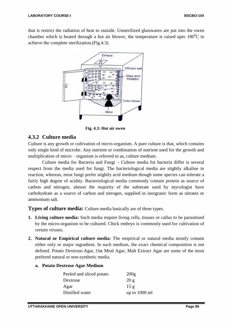
LABORATORY COURSE-I BSCBO-104
UTTARAKHAND OPEN UNIVERSITY Page 89
that is restrict the radiation of heat to outside. Unsterilized glasswares are put into the owen
chamber which is heated through a hot air blower, the temperature is raised upto 1800C to
achieve the complete sterilization.(Fig.4.3)
Fig. 4.3: Hot air owen
4.3.2 Culture media
Culture is any growth or cultivation of micro-organism. A pure culture is that, which contains
only single kind of microbe. Any nutrient or combination of nutrient used for the growth and
multiplication of micro – organism is referred to as, culture medium.
Culture media for Bacteria and Fungi – Culture media for bacteria differ is several
respect from the media used for fungi. The bacteriological media are stightly alkaline in
reaction, whereas, most fungi prefer stightly acid medium though some species can tolerate a
fairly high degree of acidity. Bacteriological media commonly contain protein as source of
carbon and nitrogen, almost the majority of the substrate used by mycologist have
carbohydrate as a source of carbon and nitrogen, supplied in inorgranic form as nitrates or
ammonium salt.
Types of culture media: Culture media basically are of three types.
1. Living culture media: Such media require living cells, tissues or callus to be parasitised
by the micro-organism to be cultured. Chick embryo is commonly used for cultivation of
certain viruses.
2. Natural or Empirical culture media: The empirical or natural media mostly contain
either only or major ingradient. In such medium, the exact chemical composition is not
defined. Potato Dextrose-Agar, Oat Meal Agar, Malt Extract Agar are some of the most
prefered natural or non-synthetic media.
a. Potato Dextrose Agar Medium
Peeled and sliced potato 200g
Dextrose 20 g
Agar 15 g
Distilled water up to 1000 ml

LABORATORY COURSE-I BSCBO-104
UTTARAKHAND OPEN UNIVERSITY Page 90
b. Malt agar Medium
Malt extract 30g
Agar 15g
Distilled water up to 1000 ml
c. Corn Meal Agar
Corn meal (ground maize) 60 gm
Agar 15 g
Distilled water upto 1000 ml
d. Beef Extract Agar
Beef extract 3g
Peptone 5g
Agar 15g
Distilled water upto 1000 ml
3. Synthetic culture medium
Such media are reproducible solution of chemically pure, known inorganic or organic
compounds. The combination is usually based on an exact knowledge of nutritional
requirement of the micro-organism to be cultivated.
(a) Raulins Medium – The first attempt to compound a rational synthetic medium was done
by Raulin in 1869. Who anlaysed the ash of Aspergillus niger and prepared a medium, for the
biochemical studies of the species.
Sugar 70.0 g
Tartaric acid 4.0 g
Ammonium nitrate 4.0 g
Potassium carbonate 0.6 g
Ammonium phosphate 0.6 g
Magnesium carbonate 0.4 g
Ammonium sulphate 0.25 g
Zinc sulphate (crystals) 0.07 g
Ferrous sulphate (crystals) 0.07 g
Potassium silicate 0.07 g
Distilled water up to 1500 ml.
(b) Czapeck-Dox Medium – Many workers considered that Raulin's solution and its various
modificatins are unnecessarily complicated. Czapeck attampted to supply all the elements
necessary for growth with minimum of duplication. The Czapeck's solution was further
modified by Dox in 1910 and Thom and Church in 1926. It is most preferred medium for
physiological study.
Sucrose 30.0 g
Sodium nitrate 2.0 g
Potassium chloride 0.5 g
Magnesium sulphate (MgSO4 7H2O) 0.5 g

LABORATORY COURSE-I BSCBO-104
UTTARAKHAND OPEN UNIVERSITY Page 91
Ferrous sulphate (FeSO4 7H2O) 0.01 g
Dipotassium phosphate 1.0 g
Agar 15 g
Distilled water to 1000 ml
(c) Raper and Thom Medium –
Yeast extract 2.0 g
Peptone 3.0 g
Dextrose 2.0 g
Sucrose 30.0 g
Corn steep solid 5.0 g
NaNO3 2.0 g
K2HPO4 3H2O 1.0 g
MgSO4 7H2O 0.5 g
KCl 0.2 g
FeSO4 7H2O 0.01 g
Agar 20.0 g
Water to 1000 ml
Antibacterial media
To check the unnecessary growth of bacteria in the culture medium for fungi certain chemical
like rose bengal, streptomycin and aureomycin in used by various microbiologist, Such media
are particularly important for the study of fungi.
(a) Smith and Dawson's Medium –
Glucose 10.0 g
NaNO2 1.0 g
K2PO4 1.0 g
Agar 15.0 g
Rose bengal 0.067 g
Soil extrect 1000 ml
The soil extract is prepared by autoclaving 500 g of loam in 1200 ml water for one hour. The
extract is then filtered through paper.
(b) Cooke's Medium –
Dextrose 10.0 g
Peptone 5.0 g
KH2PO4 1.0 g
MgSO4 7H2O 0.5 g
Agar 20 g
Rose bengal 0.035 g
Aureomycin 35 g
Water to 1000 ml
The auromycin is added to sterilized and cooled medium just before pouring.

LABORATORY COURSE-I BSCBO-104
UTTARAKHAND OPEN UNIVERSITY Page 92
4.3.3. Sterilization
Sterilization is the complete destruction of all forms of micro-organism
A. Physical methods of sterilization
1. Heating
(a) Flaming – The small tools like scalpels, inoculating niddle and loops are generally
sterilized by taking over to the flame of gas lamp or burner.
Fig.4.4: Sterilization of inoculating niddle by flaming
(b) Hot air Sterilization – The glasswares and other tools are generally sterilized by
keeping them into an electric or gas woven. For complete sterilization a temperature of 1600C
for at least two hour is recommeded.
(c) Wet Heating – Culture media, aqueous solution cloths etc. are sterilized in autoclave,
Usually, a clear pressure of 15 lb for 15-20 minutes is sufficient for sterilization.
2. Radiation – Certain radiation like ultraviolet light, X-rays, Gamma rays are used
variously for sterilization. Ultravoilet light is very effective for culture room sterilization.
Ionizing radiation is also recommended for heat liable materials.
3. Filtration – Filtration may be economical method for the partial sterilization of gas or
liquid. Cotton wool is generally used for filteration of gas. For liquid filtration, a variety
of filters like Seitz filter (asbestos filter), chamber land-pastus filter (porcelain filter) Berk
feld filter (Diatomaceous earth filter) are used.
Fig.4.5: Chamberland- Pasteur filter

LABORATORY COURSE-I BSCBO-104
UTTARAKHAND OPEN UNIVERSITY Page 93
B. Chemical methods of sterilization
There are a number of chemicals known for their disinfectant (germ killing), antiseptic
(microbial growth stopping), sanitizer (reducing the microbial population to a sap limit)
properties. Ethyl and isopropyl alcohols are used as skin antiseptic chemicals. Lysol cresol,
etc. are the phenolic compounds, which are widely used a germicidal agents, Silver nitrate,
mercuric chloride and some other organic forms of mercury are used as surface sterilization
of certain test materials.
4.3.1 Inoculation and isolation of pure culture
inoculation
The artificial transfer of a micro-organism into a culture medium or healthy tissues is referred
to as inoculation.
Preparation of Agar Slants – Liquefied agar medium is poured into culture tubes, the
latter is plugged with cotton wool and sterilized in autoclave. The sterilized tubes are taken
out and placed in a slanting position and then allowed to cool. The slopy surface provides
maximum area of the agar medium in the culture tube for the growth of the fungus.
Fig.4.6: Seitz filter
Transfer of the Inoculum – The inoculation is done in inoculation chamber, completely
sterilized with ultraviolet light, the hand should also be cleaned with soap and then sterilized
with rectified spirit. For culture tube inoculation, the tube containing inoculum and tube with
sterilized agar medium slant are held in one hand and inoculating niddle in the other. The
cotton plugs of the tubes are taken out with the help of fingers in front of the flame of spirit
lamp. The inoculum is picked out with the help of niddle and then inserted within the agar
surface of the tube. Plugging of the tube is immediately done to avoid contamination. For
petridish inoculation, the lid is removed to a minimu, and inoculation is done in the centre of
the dish. The inoculated tube or dishes are finally incubated at disired temperature.
Isolation of the pure culture
The disease part of the plant contains a great number of micro-organisms beside the chief
pathogen. Isolation is separation of the pathogen from the host tissue, or its inoculation in
culture media. A culture that contains single kind of micro-organism, regardless of number of
individual in a medium is referred to as pure culture or auxenic culture. Such culture is grown

LABORATORY COURSE-I BSCBO-104
UTTARAKHAND OPEN UNIVERSITY Page 94
usually from one or few cells of the species. Following are some common methods for
obtaining the pure culture.
4.4-DIFFERENT METHODS OF MICROBIAL ISOLATION AND
CULTIVATION
4.4.1 Procedure
Pure culture of microorganisms that form discrete colonies on solid media, e.g., yeasts, most
bacteria, many other microfungi, and unicellular microalgae, may be most commonly
obtained by plating methods such as streak plate method, pour plate method and spread plate
method.
But, the microbes that have not yet been successfully cultivated on solid media and are
cultivable only in liquid media are generally isolated by serial dilution method.
1. Streak Plate Method
This method is used most commonly to isolate pure cultures of bacteria. A small amount of
mixed culture is placed on the tip of an inoculation loop/needle and is streaked across the
surface of the agar medium. The successive streaks "thin out" the inoculums sufficiently and
the microorganisms are separated from each other. It is usually advisable to streak out a
second plate by the same loop/needle without reinoculation. These plates are incubated to
allow the growth of colonies. The key principle of this method is that, by streaking, a dilution
gradient is established across the face of the Petri plate as bacterial cells are deposited on the
agar surface. Because of this dilution gradient, confluent growth does not take place on that
part of the medium where few bacterial cells are deposited presumably; each colony is the
progeny of a single microbial cell
Fig.4.7: Various methods of streaking
thus representing a clone of pure culture. Such isolated colonies are picked up separately
using sterile inoculating loop/ needle and restreaked onto fresh media to ensure purity.
2. Pour Plate Method
This method involves plating of diluted samples mixed with melted agar medium. The main
principle is to dilute the inoculum in successive tubes containing liquefied agar medium so as
to permit a thorough distribution of bacterial cells within the medium. Here, the mixed

LABORATORY COURSE-I BSCBO-104
UTTARAKHAND OPEN UNIVERSITY Page 95
culture of bacteria is diluted directly in tubes containing melted agar medium maintained in
the liquid state at a temperature of 42-45°C (agar solidifies below 42°C).
The bacteria and the melted medium are mixed well. The contents of each tube are poured
into separate Petri plates, allowed to solidify, and then incubated. When bacterial colonies
develop, one finds that isolated colonies develop both within the agar medium (subsurface
colonies) and on the medium (surface colonies). These isolated colonies are then picked up
by inoculation loop and streaked onto another Petri plate to insure purity.
Pour plate method has certain disadvantages as follows: (i) the picking up of subsurface
colonies needs digging them out of the agar medium thus interfering with other colonies, and
(ii the microbes being isolated must be able to withstand temporary exposure to the 42-45°
temperature of the liquid agar medium; therefore this technique proves unsuitable for the
isolation of psychrophilic microorganisms.
However, the pour plate method, in addition to its use in isolating pure cultures, is also used
for determining the number of viable bacterial cells present in a culture.
Fig.4.8: Pour plate method
3. Spread Plate Method
In this method the mixed culture of microorganisms is not diluted in the melted agar medium
(unlike the pour plate method); it is rather diluted in a series of tubes containing sterile liquid,
usually, water or physiological saline. A drop of so diluted liquid from each tube is placed on
the centre of an agar plate and spread evenly over the surface by means of a sterilized bent-
glass-rod.

LABORATORY COURSE-I BSCBO-104
UTTARAKHAND OPEN UNIVERSITY Page 96
The medium is now incubated. When the colonies develop on the agar medium plates, it is
found that there are some plates in which well-isolated colonies grow. This happens as a
result of separation of individual microorganisms by spreading over the drop of diluted liquid
on the medium of the plate.
Fig.4.9: Spread plate method
4. Serial Dilution Method
As stated earlier, this method is commonly used to obtain pure cultures of those
Fig.4.10: Serial dilution method
microorganisms that have not yet been successfully cultivated on solid media and grow only
in liquid media. A microorganism that predominates in a mixed culture can be isolated in
pure form by a series of dilutions.
If we take out 1 ml of this medium and mix it with 9 ml of fresh sterile liquid medium, we
would then have 100 microorganisms in 10 ml or 10 microorganisms/ ml. If we add 1 ml of
this suspension to another 9 ml. of fresh sterile liquid medium, each ml would now contain a

LABORATORY COURSE-I BSCBO-104
UTTARAKHAND OPEN UNIVERSITY Page 97
single microorganism. If this tube shows any microbial growth, there is a very high
probability that this growth has resulted from the introduction of a single microorganism in
the medium and represents the pure culture of that microorganism.
Special Methods of Isolation of Pure Culture
A. Single Cell Isolation methods
An individual cell of the required kind is picked out by this method from the mixed culture
and is permitted to grow. The following two methods are in use.
(i) Capillary pipette method
Several small drops of a suitably diluted culture medium are put on a sterile glass-coverslip
by a sterile pipette drawn to a capillary. One then examines each drop under the microscope
until one finds such a drop, which contains only one microorganism. This drop is removed
with a sterile capillary pipette to fresh medium. The individual microorganism present in the
drop starts multiplying to yield a pure culture.
(ii) Micromanipulator method
Micromanipulators have been built, which permit one to pick out a single cell from a mixed
culture. This instrument is used in conjunction with a microscope to pick a single cell
(particularly bacterial cell) from a hanging drop preparation. The micro-manipulator has
micrometer adjustments by means of which its micropipette can be moved right and left,
forward, and backward, and up and down. A series of hanging drops of a diluted culture are
placed on a special sterile coverslip by a micropipette.
Now a hanging drop is searched, which contains only a single microorganism cell.
This cell is drawn into the micropipette by gentle suction and then transferred to a large drop
of sterile medium on another sterile coverslip. When the number of cells increases in that
drop as a result of multiplication, the drop is transferred to a culture tube having suitable
medium. This yields a pure culture of the required microorganism.
The advantages of this method are that one can be reasonably sure that the cultures
come from a single cell and one can obtain strains within the species. The disadvantages are
that the equipment is expensive, its manipulation is very tedious, and it requires a skilled
operator. This is the reason why this method is reserved for use in highly specialized studies.
B. Enrichment Culture Method
Generally, it is used to isolate those microorganisms, which are present in relatively small
numbers or that have slow growth rates compared to the other species present in the mixed
culture. The enrichment culture strategy provides a specially designed cultural environment
by incorporating a specific nutrient in the medium and by modifying the physical conditions
of the incubation. The medium of known composition and specific condition of incubation
favors the growth of desired microorganisms but, is unsuitable for the growth of other types
of microorganisms.
4.4.2 Maintenance and Preservation of Cultures of Microbes

LABORATORY COURSE-I BSCBO-104
UTTARAKHAND OPEN UNIVERSITY Page 98
Once a pure culture is obtained then methods are to be devised for their maintenance and
preservation so that all the characteristics can be conserved. Some of the simple methods of
culture maintenance and their preservation are described below.
1. Use of refrigerator or cooling apparatus:
Live pure cultures can be successfully stored in their respective culture media in refrigerators
or such cooling conditions at about 4°C. Generally, the metabolic activity of the organisms
slows down and they become nearly inert at this temperature.
However, the metabolism does not completely cease and hence, the organisms cannot be
maintained for an indefinite period of time. At regular intervals, say 2-4 weeks, the culture
may be taken out from the refrigerator and inoculated to fresh media, a process known as
sub- culturing.
2. Transfer to fresh media
Periodic transfer to fresh, sterile media tubes can maintain microbial cultures. The frequency
of transfer, however, varies with the organism. A culture the bacterium, Escherichia coli, for
example, needs transfer at monthly intervals. After growth for 24 hours at 37°C, the slants
can be stored at low temperature for 20-30 days to keep the culture viable. It is necessary to
use the appropriate growth medium and proper storage temperature.
3. Overlying with mineral oil or liquid paraffin
Covering the fresh growth in agar slants with sterile mineral oil or liquid paraffin can
preserve many bacteria and fungi. The oil must be above the tip of the slanted surface. The
cell viability in this method is very high as compared to frequent transfer and storage at low
temperature.
4. Freeze drying or lyophilization
Freeze drying (lyophilization) is a rapid dehydration of organisms while they are in frozen
state. In this process, the cell suspension is placed in small vials, which are, frozen by
immersing in a mixture of dry ice and acetone liquid nitrogen. The vials are evacuated and
dried under vacuum, sealed and stored at low temperature. Under such conditions, microbes
can be stored for very long durations without upsetting their characteristics.
5. Storage at sub-zero temperature
In this method, the cultures are frozen in the presence of a protective agent such as glycerol
or dimethylsulphoxide in liquid nitrogen (-196° C). This method is successful in many
organisms particularly those which cannot be preserved under lyophilization.
6. Storage in silica gel
Both bacteria and yeasts can be stored in silica gel at low temperature for 1 to 2 years. In this
method, finely powdered, heat sterilized and cooled silica powder is mixed with a thick
suspension of cells and stored at a low temperature. The quick desiccation at low temperature
allows the cells to remain viable for a long period.

LABORATORY COURSE-I BSCBO-104
UTTARAKHAND OPEN UNIVERSITY Page 99
4.5- SUMMARY
The survival of microorganisms in the laboratory, as well as in nature, depends on their
ability to grow under certain chemical and physical conditions. An understanding of these
conditions enables us to characterize isolates and differentiate between different types of
bacteria. Such knowledge can also be applied to control the growth of microorganisms in
practical situations.
For their normal growth, bacteria must be supplied with moisture, protein, carbohydrate and
inorganic elements such as Iron, sodium, calcium, etc. Therefore, if such a mixture is
prepared which contains these nutritive substances, the bacteria can be grown easily. Such a
mixture is known as culture medium.
It may be in solid form or liquid or semi solid medium. The liquid medium is called broth.
Solid medium is prepared by adding agar agar to the broth. For the isolation bacteria, semi
solid medium is important.
Killing of Microorganisms (viruses, bacteria and fungi) of an article usually by means of heat
is called sterilization. It is required for culture media, suspended fluids, reagents, equipments,
instruments and containers used in microbiology. Drying kills many bacteria & fungi, several
pathogens (e.g. Smallpox virus, tubercle bacilli). Freezing also kills most of bacteria & fungi.
Heat is the most commonly used physical agent for sterilization. It is of two types viz.
Sterilization by dry heat & Sterilization of moist heat.
4.6- GLOSSARY
Anaerobe: an organism that grows in the absence of free oxygen.
Amotation: The process of determining the location of specific genes in a genome map after
it has been produced by nucleic acid sequencing.
Complex medium: Culture medium that contains some ingredients of unknown chemical
composition.
Bacteria: All prokaryotes that are not members of the domain Archaea.
Bactericidal: Term used to describe a drug that kills microorganisms.
Basal medium: A medium which allows the growth of many types of microorganisms
which do not require any special nutrient supplements, e.g. nutrient broth. o
C: Celsius/Centigrade.
Capsul: A colorless, transparent, mucopolysaccharide sheath on the wall of a cell.
Saccharolytic: Capable of breaking down sugars.
Selective medium: A medium which allows the growth of certain types of microorganisms in
preference to others. For example, an antibiotic-containing medium allows the growth of only
those microorganisms which are resistant to this antibiotic.
Slant: See definition of "butt." The slant is the upper surface of the medium in the tube
described. It is exposed to air in the tube.
Spore: Propagule that develops by sexual reproduction (ascospore, basidiospore, zygospore)
or by asexual means within a sporangium (sporangiospore). Those most commonly seen in

LABORATORY COURSE-I BSCBO-104
UTTARAKHAND OPEN UNIVERSITY Page 100
the clinical laboratory are usually enclosed in a sac-like structure (as opposed to conidia
which are free, not enclosed).
Sterilization: Treatment resulting in death of all living organisms and viruses in a material.
4.7 SELF ASSESSMENT QUESTIONS
4.7.1: Multiple choice questions
1. The medium used in membrane filter technique was
(a) EMB agar (b) EMR-Vp medium
(c) Lactose broth (d) Endo agar
2. Lysol is a
(a) Sterilent (b) Disinfectant
(c) Antiseptic (d) Antifungal agent
3. Peptone water is an example for
(a) Synthetic medium (b) Semisynthetic medium
(c) Differential medium (d) None of these
4. The method in which cells are frozen dehydrated is called
(a) Pasteurization (b) Dessication
(c) Disinfection (d) Lypophilization
5. The technique used to avoid all microorganisms is accomplished by
(a) Sterlization (b) Disinfection
(c) Surgical sterilization (d) isinfection Sterilization
6. Separation of a single bacterial colony is called
(a) Isolation (b) Separation
(c) Pure culturing (d) All of these
7. Which of the following method of sterilization has no effect on spores?
(a) Drying (b) Hot air oven
(c) Autoclave (d) None of these
8. The condition required autoclave
(a) 121oC temp.and 15 lbs. pressure for 20 min.
(b) 120oC temp.and 20 lbs. pressure for 30 min
(c) 150oC temp.for 1 hr.
(d) 130oC temp for 2 hr.
9. Agar is obtained form
(a) Brown algae (b) Red algae

LABORATORY COURSE-I BSCBO-104
UTTARAKHAND OPEN UNIVERSITY Page 101
(c) Green algae (d) Blue-green algae
10. Best method for getting pure culture is
(a) Streak-plate (b) Agar slant
(c) Both a & b (d) None of these
11. Separation of a single colony is
(a) Pure-culturing (b) Isolation
(c) Separation (d) Both a and b
12. Growth period of culture is
(a) Inoculation (b) Incubation
(c) Incineration (d) Isolation
4.7.1 Answers: 1. (b), 2. (b), 3. (b), 4. (d), 5.(a), 6. (a), 7. (a), 8. (c), 9. (b), 10. (c), 11. (b), 12. (b)
4.8 REFERENCES
Healthwise, Incorporated (2010-06-28).Throat Culture. WebMD. Retrieved 2013-03-10.
Old, D.C.; Duguid, J.P. (1970). "Selective Outgrowth of Fimbriate Bacteria in Static
Liquid Medium". Journal of Bacteriology (American Society for Microbiology) 103 (2):
447–456.PMID 248102.
Madigan, Michael T. (2012). Brock biology of microorganisms (13th ed.). San Francisco:
Benjamin Cummings. ISBN 9780321649638.
Uruburu, F. (2003). "History and services of culture collections". International
Microbiology 6 (2): 101–103. doi:10.1007/s10123-003-0115-2. PMID 12811589.
4.9 SUGGESTED READINGS
Textbook of microbiology by Prescott.
Textbook of microbiology by R.C. Dubey.
Textbook of microbiology by Ananthanarayan and Paniker.
Textbook of microbiology by Pelczar.
4.10 TERMINAL QUESTIONS
4.10.1 Long answer type questions:
Q1. What are the different media for isolation and cultivation of microorganisms?
Q2. What are different methods of microbial isolation and cultivation?
Q3. What is Serial Dilution Method, why it is important?
Q4. Explain Maintenance and Preservation of Pure Cultures OF Microbes
Q5. What is sterilization?
Q6. Explain pour plate method.
Q7. Describe serial dilution method with the help of diagram.

LABORATORY COURSE-I BSCBO-104
UTTARAKHAND OPEN UNIVERSITY Page 102
Q8.Explain spread plate method.
Q9.Explain general direction for media preparation.
4.10.2 Short answer type questions:
Q1. Explain
(a) Streak Plate Method
(b) Pour Plate method
(c) Spread Plate Method
Q2. Write short notes on following:
(a) Capillary pipette method
(b) Micromanipulator method
(c) Enrichment Culture Method

LABORATORY COURSE-I BSCBO-104
UTTARAKHAND OPEN UNIVERSITY Page 103
BLOCK-2-DIVERSITY OF ALGAE AND
BRYOPHYTES

LABORATORY COURSE-I BSCBO-104
UTTARAKHAND OPEN UNIVERSITY Page 104
UNIT-5 STUDY OF THE TYPES OF ALGAE-I
5.1-Objectives
5.2-Introduction
5.3-Study of the types
5.3.1-Oscillatoria
5.3.2-Nostoc
5.3.3-Chlamydomonas
5.3.4-Volvox
5.3.5-Oedogonium
5.4-Method of preparing of temporary slides
5.5- Summary
5.6- Glossary
5.7- Self assessment question
5.8-References
5.9-Suggested Readings
5.10-Terminal Questions

LABORATORY COURSE-I BSCBO-104
UTTARAKHAND OPEN UNIVERSITY Page 105
5.1- OBJECTIVES
After reading this section students will know -
Study of different types of algae.
Morphology, vegetative and reproductive structures of different algae.
Different algal forms – Oscillatoria, Nostoc, Chlamydomonas, Volvox, Oedogonium.
5.2-INTRODUCTION
Algae is a very large and diverse group of eukaryotic organisms, ranging from unicellular
genera such as Chlorella and the diatoms to multicellular forms such as the giant kelp, a
large brown alga that may grow up to 50 meters in length. Most are autotrophic and lack
many of the distinct cell and tissue types found in land plants such as stomata,
xylem and phloem. The largest and most complex marine algae are called seaweeds, while
the most complex freshwater forms are the Charophyta, a division of algae that
includes Spirogyra and the stoneworts.
There is no generally accepted definition of algae. One definition is that algae
"have chlorophyll as their primary photosynthetic pigment and lack a sterile covering of cells
around their reproductive cells". Other authors exclude all prokaryotes and thus do not
consider cyanobacteria (blue-green algae) as algae.
Algae constitute a polyphyletic group since they do not include a common ancestor, and
although their plastids seem to have a single origin, from cyanobacteria, they were acquired
in different ways. Green algae are examples of algae that have primary chloroplasts derived
from endosymbiotic cyanobacteria. Diatoms are examples of algae with secondary
chloroplasts derived from an endosymbiotic red alga.
Algae exhibit a wide range of reproductive strategies, from simple asexual cell division to
complex forms of sexual reproduction.
Fig. 5.1: Algal growth in pond water

LABORATORY COURSE-I BSCBO-104
UTTARAKHAND OPEN UNIVERSITY Page 106
Algae lack the various structures that characterize land plants, such as the phyllids (leaf-like
structures) of bryophytes, rhizoids in nonvascular plants, and the roots, leaves, and
other organs that are found in tracheophytes (vascular plants). Most are phototrophic,
although some groups contain members that are mixotrophic deriving energy both from
photosynthesis and uptake of organic carbon either by osmotrophy, myzotrophy,
or phagotrophy. Some unicellular species of green algae, many golden
algae, euglenoids, dinoflagellates and other algae have become heterotrophs (also called
colorless or apochlorotic algae), sometimes parasitic, relying entirely on external energy
sources and have limited or no photosynthetic apparatus. Some other heterotrophic
organisms, like the apicomplexans, are also derived from cells whose ancestors possessed
plastids, but are not traditionally considered as algae. Algae have photosynthetic machinery
ultimately derived from cyanobacteria that produce oxygenas a by-product of photosynthesis,
unlike other photosynthetic bacteria such as purple and green ulphur bacteria. Fossilized
filamentous algae from the Vindhya basin have been dated back to 1.6 to 1.7 billion years
ago.
5.3- STUDY OF TYPES OF ALGAE
Oscillatoria
Division- Cyanophyta
Class - Cyanophyceae
Order - Nostocales
Family - Oscillatoriaceae
Genus - Oscillatoria
The genus Oscillatoria is very common. It grows abundantly in dirty stagnant and polluted
water channels forming a blackish blue-green mass. Besides, it also occurs on moist rocks,
temporary water pools, ditches, drains, streams, sewers and muddy banks of rivers.
Fig. 5.2: Oscillatoria - Habit of filaments, single filament in enlarged view and a single filament
showing Oscillating movement

LABORATORY COURSE-I BSCBO-104
UTTARAKHAND OPEN UNIVERSITY Page 107
Vegetative structure
The thallus:
1. The plant body is filamentous. The filamentous occurs singly or large numbers of them
are interwoven to form extensive flat stratum or spongy sheets.
2. The filaments are unbranched.
3. They are long or short, usually straight.
4. Usually, sheath around the trichomes is absent. However, an inconspicuous delicate
sheath is present in some species so that they are slippery in touch.
5. All the cells alike except the terminal one which may be conical, convex, rounded,
pointed, bent or coiled.
6. In most of the species the cells are usually shorter than the breadth of the trichome.
7. Freshly mounted filaments (in water) show a characteristic oscillating movement, like the
movement of pendulum in a clock.
8. The cell structure is typically similar to myxophycean cells. The cellular protoplasm is
differentiated into outer colored chromoplasm and central hyaline centroplasm.
9. The cells are prokaryotic.
Reproductive structure:
1. The reproduction in Oscillatoria takes place by the formation of hormogones.
2. The hormogones are small piece of trichomes which separate from parent filament and
grow separately into new thalli.
3. They are formed as a result of the death of intercalary cell which becomes empty and acts
as biconcave separation disc.
Identification
Sub-division – Algae. (1) Thallus simple, (2) Presence of chlorophyll, (3) Cell wall of
cellulose.
Class – Myxophyceae. (1) Chromatophore not organised, pigments diffused, blue-green, (2)
Photosynthetic reserve cyanophycean starch, (3) True nucleus absent, (4) Sexual
reproduction absent.
Order – Nostocales. (1) Thallus with trichomes, unbranched, or with false branching,
(2) Hormogones, heterocysts, exospores and endospores generally present.
Family – Oscillatoriaceae. (1) Trichomes uniseriate, sometimes tapering at the ends,
(2) Heterocysts and spores absent, (3) Sheath absent or diffluent.
Genus – Oscillatoria. (1) Trichomes not in bundles, (2) Trichomes without a sheath
(3) Trichomes straight and cylindrical
Nostoc
Division- Cyanophyta
Class - Cyanophyceae
Order - Nostocales
Family - Nostocaceae
Genus - Nostoc

LABORATORY COURSE-I BSCBO-104
UTTARAKHAND OPEN UNIVERSITY Page 108
The genus Nostoc is colonial and grows in the form of mucilaginous balls. It occurs both in
terrestrial and aquatic habitats. It grows commonly in fresh water ponds, pools, puddles and
ditches. Some species grow in the paddy fields, moist soils and rocks. Few species are
endophytic and in the tissue of higher plants.
Fig. 5.3: Nostoc – Colony growing as balls, an enlarged view of colony and a single filament in
enlarged view
Vegetative structures:
Nostoc balls:
1. The alga is colonial and a large number of filaments are embedded within a mucilaginous
envelope forming a ball shaped colony. These are called Nostoc balls.
2. The balls are olive green or blue green in color.
3. The size and the shape of the ball change with the age. They measure from a pin head to
the size of a hen‘s egg.
4. The shape of the ball may be spherical, oblong, ellipsoidal or in some cases irregular.
5. Young colonies are solid but at maturity they may becomes hollow ro even break open
into flat or lobed expanses.
The thallus:
1. Nostoc filaments are uniseriate and unbranched. They remain embedded in a common
mucilaginous envelope forming a colony.
2. Each filament consists of a large number of spherical cells which give it a moniliform
beaded appearance.
3. The filaments are curved, contorted and interwined.
4. A filament may possess its own sheath which may be hyaline or colored.
5. The filaments possess terminal or intercalary heterocysts. The heterocysts show one or
two polar nodules towards the side of attachment with vegetative cell or cells.
6. The structure of vegetative cells is typically similar myxophycean cells. The protoplasm
is differentiated into outer colored chromoplasm and central hyaline centroplasm.
7. The cells are prokaryotic.

LABORATORY COURSE-I BSCBO-104
UTTARAKHAND OPEN UNIVERSITY Page 109
Reproductive structures
Akinetes
1. The akinetes are thick walled and larger cells which store large cyanophycian granules.
2. They may occurs singly or in chains and germinate to produce young filaments.
Identification
Sub-division – Algae. (1) Thallus simple, (2) Presence of chlorophyll, (3) Cell wall of
cellulose.
Class – Myxophyceae. (1) Chromatophore not organised, pigments diffused, blue-green, (2)
Photosynthetic reserve of cyanophycean starch, (3) True nucleus absent.
Order – Nostocales. (1) Thallus with trichomes, unbranched, or branching false,
(2) Hormogones, heterocysts, exospores and endospores generally present.
Family – Nostocaceae. (1) Trichomes simple, unbranched, uniseriate and approximately of
the same diameter throughout (2) Heterocysts and akinetes present, (3) Trichomes not
differentiated.
Genus – Nostoc. (1) Trichomes much twisted into a mass of difinite form with a firm
colonial envelope, (2) Heterocysts intercalary and single.
Chlaymydomonas
Division- Chlorophyta
Class - Chlorophyceae
Order - Volvocales
Family - Chlamydomonadaceae
Genus - Chlaymydomonas
The alga occurs as free swimming in fresh water ponds, lakes and ditches. A few species
grow on moist damp soil. It forms green surface layer on water.
Fig. 5.4: Chlamydomonas – A single cell in enlarged view and Palmella stage of life cycle

LABORATORY COURSE-I BSCBO-104
UTTARAKHAND OPEN UNIVERSITY Page 110
The thallus:
1. The plant body is unicellular, motile and the cells occur singly.
2. The shape of cell may be oval, spherical or oblong. Size is approximately 20 µ in length.
3. The anterior end of the cell usually shows papillate projection to which two whiplash type
of flagella are attached.
4. The cell possesses a firm, two layered cell wall which encloses protoplasm.
5. Each cell possesses single, large, cup-shaped chloroplast. Single large nucleus is situated
in the cavity of chloroplast. The cup-shape of chloroplast can be seen only in side view.
6. Each cell is characterized by the presence of single pyrenoid on the broad portion of
chloroplast.
7. The pyrenoid body shows central protein surrounded by starch grains.
Palmella stage:
1. Stage asexual reproduction of chlamydomonas that resembles a genus palmella is called
palmella stage.
2. This stage results under unfavorable conditions.
3. The group of daughter cells (two, four or eight) remains embedded in a common
mucilaginous envelope of parent cell.
4. The cells are non-motile but as soon as they get moisture they develop flagella and escape
from envelop.
Identification
Sub-division – Algae. (1) Presence of a simple thallus, (2) Chlorophyll present, (3) Cell wall
made of cellulose.
Class – Chlorophyceae. (1) Presence of a difinite nucleus, (2) Chloroplast present, grass
green colour, (3) presence of starch (4) Reproductive structure motile and flagella equal in
length.
Order – Volvocales. (1) Thallus motie, (2) Protoplast with contractile vocuoles.
Family – Chlamydomonadaceae.
Genus – Chlamydomonas. (1) Oval or Pyriform shape of the thallus which in unicellular, (2)
Cup-shaped chloroplast, (3) Presence of an eye spot, (4) Fornation of Palmella stage.
Volvox
Division- Chlorophyta
Class - Chlorophyceae
Order - Volocales
Family - Volvocaceae
Genus - Volvox
The genus Volvox includes about 20 species. All are aquatic and free floating. They occur in
fresh water ponds, pools, ditches and occasionally in lakes. The alga is plank-tonic and
occurs as green rolling balls of pin head size imparting green color to the surface of water.
Vegetative structure:

LABORATORY COURSE-I BSCBO-104
UTTARAKHAND OPEN UNIVERSITY Page 111
The thallus (Coenobium):
1. The plant body is multi-cellular, motile coenobium.
2. The coenobia are spherical or oval in shape. The size of mature coenobium is
approximately 0.5mm in diameter.
3. A large number of cells are arranged in single layer towards the periphery of the
coenobium.
4. The coenobium is hollow in the centre. The hollow cavity is filled with mucilage.
5. The coenobium shows polarity anterior and posterior sides. It rolls over the surface of
water by the joint action of flagella.
The cell:
1. All the cells, in a coenobium are similar in size, shape and structure.
2. Each cell is elliptical in shape with its narrow anterior end pointed towards periphery and
broad posterior end towards hollow centre of coenobium.
3. The cells are biflagellate and resemble, in structure, with Chlamydomonas. The two
flagella are equal and whiplash type. The posterior part of the cell is occupied by a large
cup-shaped chloroplast with single pyrenoid. The cavity of cup has single nucleus.
4. Each cell is enveloped by its own gelatinous sheath. The cells are joint with each other by
cytoplasmic strands called plasmodesmeta.
Fig. 5.5: Volvox – A colony showing daughter cells, vegetative coenobium, coenobium showing
reproductive structures.

LABORATORY COURSE-I BSCBO-104
UTTARAKHAND OPEN UNIVERSITY Page 112
Reproductive structures:
(a) Coenobium with daughter colonies:
1. The asexual reproduction in Volvox occurs by the formation of daughter colonies.
2. More than one daughter colony can be seen in mother coenobium. Sometimes
granddaughter colonies are also form.
3. The daughter colonies are formed from special cells called gonidia towards the posterior
side of coenobium.
4. The numbers of cells in a daughter colony are same as found in parent colony but their
size remains very small.
5. The mature daughter colonies escape by the repture of parent coenobial wall.
(b) Coenobium with androgonidia showing antherozoids:
1. The male coenobium shows androgonidia mostly towards posterior side.
2. Each andrognidium forms about 64-128 biflagellated antherozoids.
3. The antherozoids are arranged in a hollow sphere. Each of them is biflagellate, conical in
shape and possesses single, pale chloroplast.
(c) Female coenobium with gynogonidia showing oogonia and zygotes:
1. Each gynogonidium is large, enclosed within a flash shaped firm membrane and
possesses single non-flagellated, dark colored ovum.
2. The zygote is large, thick walled and orange colored structure. The wall is madeup of
three layers.
3. The female coenobium shows a large number of gynogonidia which after fertilization
show oospores (zygotes).
Identification
Sub-division – Algae. (1) Thallus simple, (2) Presence of chlorophyll, (3) Cell wall of
cellulose.
Class – Chlorophyceae. (1) Chrooplasts green, (2) Photosynthetic product starch, (3) motile
cells flagellated (4) Flagella equal in length.
Order – Volvocales. (1) Vegetative cells flagellated (2) Thallus motile.
Family – Volvocaceae. (1) Thallus colonial, (2) Division of cells in longitudinal plane.
Genus – Volvox. (1) Colony spherical or sub-spherical, (2) Number of cell at least 500.
Oedogonium
Division- Chlorophyta
Class - Chlorophyceae
Oeder - Oedogoniales
Family - Oedogoniaceae
Genus - Oedogonium
The genus Oedogonium includes more than 285 species. All are aquatic and grow in fresh
water ponds, pools, swallow tanks, lakes or even in slow streams. The young filaments are
generally attached to submerged stones, rocks or on woods where as the mature filaments
may be free floating. It also occurs epiphytically on submerged aquatic plants.

LABORATORY COURSE-I BSCBO-104
UTTARAKHAND OPEN UNIVERSITY Page 113
Vegetative structures:
The thallus:
1. The plant body is multicellular, filamentous, long and unbranched.
2. The filaments are attached to the substrartum by means of long, hyaline, basal hold-fast.
3. The hold fast is the basal cell of the filament. The lower part of hold fast may be simple
or multi-lobed disc shaped where as its upper part is bulbous.
4. The apical cell of a filament is generally rounded at its free surface.
5. All the other cells hold fast and apical cell are elongated, cylindrical and arranged end to
end. This intercalary (vegetative cell) are slightly swollen at their upper ends.
6. The few intercalary cells show ring like striations towards their distal ends. These annular
rings are called caps or scars and the cells which possess caps are called cap cells.
7. The cells possess three layered walls. The cell wall is thick and rigid. It is three layered.
8. Each cell is uninucleate. The nucleus usually lies in the peripheral cytoplasm.
9. Each cell possesses, single, large, reticulate chloroplast which is parietal in position and
encloses. A big central vacuole.
10. A large number of pyrenoids are present in the cells. They usually occur irregularly or at
the intersections of the reticulate chloroplast.
Fig. 5.6: Oedogonium – Macrandrous and Nannandrous stage of life cycle

LABORATORY COURSE-I BSCBO-104
UTTARAKHAND OPEN UNIVERSITY Page 114
(a) Asexual:
1. Asexual reproduction takes place by the formation of thick walled, reddish brown, more
or less rounded structures called akinetes. Single akinete is formed from single cell.
2. A sexual reproduction also occurs by the formation of zoospores. Single zoospore is
formed from single cell (except apical and basal cell).
3. Each zoospore is spherical or pear shaped. It has a ring of short flagella at its colorless,
beak like anterior end. The zoospore is deep green in colour and possesses reticulate
chloroplast.
(b) Sexual: The sexual reproduction is oogamous. Female sex organ is oogonium and male
sex organ is antheridium.
On the basis of sex organs in the filaments there may be two types of species---
Macrandrous and Nannandrous. They may be monoecious or dioecious.
Macrandrous species showing oogonia:
1. Any cap cell may differentiate into oogonium.
2. The oogoniom is swollen, rounded or oval structure. It possesses one or more caps at its
upper ends.
3. The oogonium encloses single egg or oosphere. The egg stores reserve food material. It is
green due to presence of chloroplast. The egg is uninucleate ( nucleus centrally located )
and non-motile.
4. There is a small pore near the anterior end of oogonium which lies just above the
receptive spot.
5. Usually there is always a small supporting or suffultory cell lies just below the oogonium.
6. The oogonia and antheridia are formed in the same filament in monoecious species but in
dioecious species they develop on separate filaments.

LABORATORY COURSE-I BSCBO-104
UTTARAKHAND OPEN UNIVERSITY Page 115
Fig. 5.7: Oedogonium – Vegetative structures, Hold fast and Akinetes.
Macrandrous species showing anthridia:
1. The anthridia develop in the same filament where oogoina are formed or separately in
male filament.
2. They are terminal or intercalary in position.
3. The antheridia are formed in row or series in variable numbers.
4. Each anthridium is short, cylindrical and disc like cell which encloses to sperms.
5. Each sperm is small, spherical body with a ring of short flagella attached to its colourless,
beak like anterior end.
6. They are pale or yellow in colour.
Nannandrous species showing Oogonia and Nannandria:
1. Nannandrous species are always dioecious and exhidit dimorthism (i.e. male and female
filaments are morphologically distinct).
2. The structure of female reproductive organ (oogonium) is similar as in macrandrous
species.

LABORATORY COURSE-I BSCBO-104
UTTARAKHAND OPEN UNIVERSITY Page 116
3. The antheridia are borne on a special small filament called nannandrium or dwarf male,
which is produced by the germination of androspore.
4. The androspores are small, flagellated spores produced singly in androsporangia. The
androsporangia may develop in oogonial filament or separate filament.
5. The andospore germinates either on wall of oogonium or on suffultory cell and produces
a small male filament caleed nannandrium or dwarf male.
6. Each nannandrium has a basal long, sterile stalk cell which has a disc like or finger like
hold fast to get itself attached to the wall of oogonium or suffultory cell.
7. There are two three small cells at the apex of basal stalk cell in a nannandrium. The tip
cell produces one or more antheridia.
8. The antheridia are small, flat and disc like. Each antheridium produces single sperm
multi-flagellated sperms.
Identification
Sub-division – Algae. (1) Thallus filamentous, (2) chlorophyll present, (3) Cell wall of
cellulose.
Class – Chlorophyceae. (1) Chloroplasts grass-green, (2) Photosynthetic reserve-starch,
(3) Motile structures flagellated, (4) Flagella equal in length.
Order – Oedogoniales. (1) Cells uninucleate, filaments branched or unbrahched, (2) Cell
division forming ‗caps‘. (3) Chloroplast reticulate, (4) Zoospores and antherozoids bear a
whorl of flagella, (5) Production of dwarf males.
Family – Oedogoniaceae. (A single family)
Genus – Oedogonium. (1) Filaments unbranched, (2) Cells cylindrical, (3) Holdfast well
developed.
Method of permanent slide preparation
Select thin section
Stain with safranin
Wash properly with water
Dehydrate the stained section with 15% alcohol
30% alcohol
50% alcohol
70% alcohol
90% alcohol
Stain with Fast green

LABORATORY COURSE-I BSCBO-104
UTTARAKHAND OPEN UNIVERSITY Page 117
Destain with 90% alcohol.
Absolute alcohol
Pass the section through xylene series
20% xylol.
50% xylol.
70% xylol.
90% xylol.
100% xylol.
Mounting in Canada Balsam or DPX.
Fig: 5.8- A flow diagram showing different to make the permanent slide, the section of plant
material is passed through alcoholic as well as xylene series.
5.4 SUMMARY
The algal genus Oscillatoria is very common. It grows abundantly in dirty stagnant and
polluted water channels forming a blackish blue-green mass. Besides, it also occurs on moist
rocks, temporary water pools, ditches, drains, streams, sewers and muddy banks of rivers.
The genus Nostoc is colonial and grows in the form of mucilaginous balls. It occurs both in
terrestrial and aquatic habitats. It grows commonly in fresh water ponds, pools, puddles and
ditches. Some species grow in the paddy fields, moist soils and rocks. Few species are
endophytic and in the tissue of higher plants.
The alga Chlamydomonas occurs as free swimming in fresh water ponds, lakes and ditches. A
few species grow on moist damp soil. It forms green surface layer on water.
The genus Volvox includes about 20 species. All are aquatic and free floating. They occur in
fresh water ponds, pools, ditches and occasionally in lakes. The alga is plank-tonic and
occurs as green rolling balls of pin head size imparting green color to the surface of water.
The genus Oedogonium includes more than 285 species. All are aquatic and grow in fresh
water ponds, pulls, swallow tanks, lakes or even in slow streams. The young filaments are
generally attached to submerge stones, rocks or on woods where as the mature filaments may
be free floating. It also occurs epiphytically on submerge aquatic plants.

LABORATORY COURSE-I BSCBO-104
UTTARAKHAND OPEN UNIVERSITY Page 118
5.6 GLOSSARY
Aquatic Algae: Aquatic algae may be fresh water (when salinity is as low-as 10 ppm) or
marine (when salinity is 33-40%). Again, certain algae grow in brackish water which is
unpalatable for drinking, but less salty than sea water. The fresh water algae usually grow in
ponds, lakes, tanks, ditches etc.
Terrestrial Algae: Some algae are found to grow in terrestrial habitats like soils,‘ rocks, logs
etc. The algae that grow on the surface of the soil are known as saprophytes. Many blue-
greens, on the other hand, grow under the surface of the soil, and are called cryptophytes.
Algae of Remarkable Habitats: Some algae also occur in uncommon habitats and
termed as:
1. Halophytic Algae (or Eurhaline): They grow in the highly concentrated salt lakes, and
include Chlamydomonas ehrenbergli, Dunaliella and Stephanoptera sp.
2. Symbiotic Algae: They grow in association with fungi, bryophytes, gymnosperms or
angiosperms. The best examples of symbiotic algae found in association with fungi are
Nostoc, Gloeocapsa, Rivularia; the members of Cyanophyceae and Chlorella, Cytococcus,
Pleurococcus; the members of Chlorophyceae.
This symbiotic association consisting of algae and fungi is called lichen. Nostoc may also
associate with Anthoceros and Anabaena associates with the roots of Cycas to form coralloid
roots.
3. Cryophytic Algae: This group of algae growing on ice or snow provides attractive colours
to snow-covered mountains. The alpine and arctic mountains become red due to the growth
of the Haemotococcous nivalis; green snow in Europe is due to the growth of
Chlamydomonas yellow stonensis.
Scotiella nivalis and Raphidonema brevirostri cause black colouration of snow, whereas
Ancyclonema nordenskioldii is responsible for brownish purple colouration.
4. Thermophytes or Thermal Algae: This group of algae occurs in hot water springs (50-
70°C) where normal life is not possible. Many blue-greens (e.g., Oscillatoria brevis,
Synechococcus elongates, Heterohormogonium sp.) are grown in such hot springs.
5. Lithophytes: They grow on the moist surface of stones and rocks, e.g., Nostoc,.
Gloeocapsa, Enteromofpha, Batrachospermum etc.
6. Epiphytic Algae: They grow on other plants including other algal members.
These are:
a. Algae on Algae: Ptilota plumosa and Rhodymenia pseudopalmatta on Laminaria
hyperborean, ii. Diatoms on Oedogonium, Spirogyra etc.
b. Algae on Bryophytes: Blue-green algae like Nostoc, Oscillatoria, diatoms like
Achnanthes etc. grow on different bryophytes.
c. Algae on Angiosperms: Algae like Cocconis, Achnanthes etc. grow epiphytically on
Lemna, an aquatic angiosperm. Alga like Trentepohlia grows on the barks of different
angiospermic plants, and is very common in Darjeeling (India).
7. Epizoic Algae: The algae growing on animals like fish, snail etc. are called as epizoic,
e.g., Stigeoclonium are found in the gills of fishes.

LABORATORY COURSE-I BSCBO-104
UTTARAKHAND OPEN UNIVERSITY Page 119
8. Endozoic Algae: They grow in the tissues of animals, e.g., Zoochlorella sp. is found in
Hydra viridis.
9. Parasitic Algae: Some algae grow parasitically on different plants and animals.
These are: a. Cephaleuros (Chlorophyceae) is parasitic and grows on the leaves of various
angiosperms, such as tea (Camellia sinensis), coffee (Coffea arabica), Rhododendron,
Magnolia and pepper (Piper nigrum). The most important one is Cephaleuros virescens,
which causes Red rust of tea.
b. Rhodochytrium (Chlorophyceae) grows on ragweed (Ambrosia) leaves.
c. Phyllosiphon (Chlorophyceae) grows on the leaves of Arisarum vulgare.
d. Ceratocolax (Rhodophyceae) grows in Phyllophora thallus.
10. Psammon: The algae which grow in sandy beaches are called psammon, e.g., Vaucheria,
Phormidium etc.
5.7 SELF ASSESSMENT QUESTIONS
5.7.1 Short answer type questions:
1. Name some algae used for food.
2. Name the algae which yield agar agar.
3. Name the algae which help in Nitrogen fixation.
4. Name the class of algae which is placed in procaryota together with bacteria.
5. What are heterocysts and where are they found.
6. Write short notes on:
(i) False branching
(ii) Cap cells
(iii) Polar nodules
(iv) Water net
(v) Pond algae
(vi) Palmella stage
(vii) Zoospores
5.7.2 Multiple choice type questions:
1. Which of the following algal divisions is characterized by possession of chlorophylls A
and B, starch as the energy storage material, cellulosic cell walls and live in freshwater and
marine habitats?
(a) Chlorophyta (b) Pyrrophyta
(c) Chrysophyta (d) Phaeophyta
2. Which algal division is divided up into three main groups consisting of the golden-brown
algae, the yellow-green algae and the diatoms?
(a) Chlorophyta (b) Pyrrophyta
(c) Chrysophyta (d) Phaeophyta
3. All algae possess

LABORATORY COURSE-I BSCBO-104
UTTARAKHAND OPEN UNIVERSITY Page 120
(a) Chloroplast (b) Nuclei
(c) None of a and b (d) Both a & b
4. Chlamydomonas and Volvox both are similar because
(a) They both are motile (b) They are member of chlorophyta
(c) None of these (d) Both a and b
5. The algae Chlamydomonas demonstrates a complex life cycle that switches between
haploid and diploid forms. This life cycle is called
(a) The sexual asexual exchange (b) Transposition cycle
(c) An alternation of generation (d) Algal transformation
6. Trichome splits near to heterocyst and helps in fragmentation by formation of
(a) Trichome division (b) Metagonia
(c) Harmogonia (d) Archegonia
7. Thick walled, enlarged vegetative cells which accumulate food are called as
(a) Cytokinesis (b) Akinetes
(c) Endokinetes (d) Exokinetes
8. Algae which serves as a "complete whole food" and contain all essential amino acids in
perfect balance, is known as super
(a) Storing algae (b) Blue algae
(c) Green algae (d) Blue green algae
5.7.2 Answers:
1. a 5. c
2. c 6. c
3. d 7. b
4. d 8. d
5.8 REFERENCE
Desikachary, T.V. 1959. Cyanophyta. New York Academic Press, New York. 686 pp.
Dillard, G.E. 1999. Common Freshwater Algae of the United States. J. Cramer, Berlin,
Germany. 173 pp.
Gomont, M. 1962. Monographie des Oscillariees. Wheldon and Wesley, Ltd., New York.
264 pp.
Patrick, R. and C.W. Reimer. 1966. The Diatoms of the United States, (Volume 1).
Academy of Natural Sciences of Philadelphia, PA. 688 pp.

LABORATORY COURSE-I BSCBO-104
UTTARAKHAND OPEN UNIVERSITY Page 121
Patrick, R. and C.W. Reimer. 1975. The Diatoms of the United States, (Volume 2, Part 1).
Academy of Natural Sciences of Philadelphia, PA. 213 pp.
Prescott, G.W. 1978. How to Know the Freshwater Algae. W.C. Brown Co., Dubuque,
Iowa. 293 pp.
Smith, G.M. 1950. Freshwater Algae of the United States, 2nd Ed. McGraw-Hill, New
York. 719 pp
5.9 SUGGESTED READINGS
Algae by Linda E Graham, James M. Graham and Lee Warren Wilcox Published by
Benjamin Cummings 2009. University of California
An Introduction to Algae by H.D. Kumar
Structure & Reproduction of Algae by F.E. Fritsch
A Textbook of Algae by O.P. Sharma, Tata McGraw-Hill Education
5.10 TERMINAL QUESTIONS
1. Write down the classification, vegetative structure and reproductive structures of
Oscillatoria with the help of illustrated diagrams
2. Write down the classification, vegetative structure and reproductive structures of Nostoc
with the help of illustrated diagrams
3. Write down the classification, vegetative structure and reproductive structures of
Chlamydomonas with the help of illustrated diagrams
4. Write down the classification, vegetative structure and reproductive structures of Volvox
with the help of illustrated diagrams
5. Write down the classification, vegetative structure and reproductive structures of
Oscillatoria with the help of illustrated diagrams
6. Write down the classification, vegetative structure and reproductive structures of
Oedogonium with the help of illustrated diagrams
7. Write down the method of permanent slide preparation.

LABORATORY COURSE-I BSCBO-104
UTTARAKHAND OPEN UNIVERSITY Page 122
UNIT-6 STUDY OF THE TYPES OF ALGAE-II
6.1-Objectives
6.2-Introduction
6.3-Study of the types
6.3.1-Vaucheria
6.3.2- Sargassum
6.3.3- Ectocarpus
6.3.4-Polysiphonia
6.3.5-Batracospermum
6.4- Summary
6.5- Glossary
6.6- Self assessment question
6.7-References
6.8-Suggested Readings
6.9-Terminal Questions

LABORATORY COURSE-I BSCBO-104
UTTARAKHAND OPEN UNIVERSITY Page 123
6.1- OBJECTIVES
After reading this section student will know about-
Study of different types of algae.
Morphology, vegetative and reproductive structures of different algae.
Different algal forms – Vaucheria, Ectocarpus, Sargassum. Polysiphonia,
Batrachospermum
6.2 INTRODUCTION
We have already discussed about algae in previous chapter, i.e. Algae is a very large and
diverse group of eukaryotic organisms, ranging from unicellular genera such as Chlorella and
the diatoms to multicellular forms such as the giant kelp, a large brown alga that may grow
up to 50 meters in length. Most are autotrophic and lack many of the distinct cell and tissue
types found in land plants such as stomata, xylem and phloem. The largest and most complex
marine algae are called seaweeds, while the most complex freshwater forms are
the Charophyta, a division of algae that includes Spirogyra and the stoneworts.
6.3- STUDY OF TYPES OF ALGAE
6.3.1 Vaucheria
Division Xanthophyta
Class Xanthophyeae
Order Heterosiphonales
Family Vaucheriaceae
Genus Vaucheria
The genus Vaucheria has about 40 species, out of which about 9 are reported form India. The
most common species are V. sessilis and V. geminata, which occur during winters.
The alga is aquatic as well as terristrial. Most of the species grow in damp garden soil, moist
wall, in stagnant ponds, ditches and slow moving streams. Some species are marine.
The thallus:
1. The plant body is filamentous, branched multinucleated, acellular and coenocytic.
2. The filaments are extensively branched. Branching is lateral but looks dichotomous.
3. The filaments are cylinderical and aseptate. They appear like siphons.
4. The terristrial species are attached to the substratum by means of small tufts of colorless,
lobed hapteron (rhizoids).
5. The cell wall is thin made up of two layers.
6. There is a big central vacuole which runs throughout the plant body. The vacuole is
surrounded by thin layer of cytoplasm.
7. A large number of small, disc-shaped, yellow green chromatophores are scattered in the
cytoplasm.

LABORATORY COURSE-I BSCBO-104
UTTARAKHAND OPEN UNIVERSITY Page 124
8. The pyrenoids are completely absent.
Reproductive structure:
Asexual
(a) Zoospores:
1. The asexual reproduction occurs by the formation of zoospores. They are formed in
aquatic species.
2. Single zoospore is formed inside the terminal zoosporangium.
3. The zoosporangium is cut off from rest of filament by transverse septum.
4. Each zoospore is large, oval shaped, yellow-green in color and bears many flagella.
5. The zoospore is regarded as compound zoospore. It is called synzoospore.
6. It has a big central vacuole surrounded by many chromatophores. It is multinucleated.
Fig.6.1: Vaucheria- Habit of the thallus and different Reproductive stages
(b) Aplanospores:
1. The terrestrial species develop thin walled, non-motile, rounded or elongated spores
called aplanospores.

LABORATORY COURSE-I BSCBO-104
UTTARAKHAND OPEN UNIVERSITY Page 125
2. They serve as means of asexual reproduction.
(c) Gongrosira stage:
1. Some aquatic or terrestrial species, under adverse conditions, develop a row of short,
thick walled, gelatinous segments called akinetes.
2. This stage is called Gongrisira stage because the plant body looks like another alga
Gongrisira.
Sexual:
1. The plants are mostly monoecious but a few species are dieocious.
2. The sexual reproduction is oogamous. The male sex organs are anthredia and female are
oogonuia.
3. Each antheridium is borne on a short stalk. It is cylindrical, curved and hook like.
4. The antheredium produces numerous, biflagellate male gametes (antherozoids) which
librate through a small apical pore.
5. The oogonia are oval in shape, sessile or sub-sessile, sepaerated from the filament by a
transverse septum.
6. Each oogonium has a lateral beak, a receptive spot and a large ovum. The ovum bears
single large egg nucleus and many chromatophores.
Position of Sex-organs:
According the position of sex-organs, the monoecious species may be of two types.
1. Position of sex-organs in V. sessilis:
The sex-organ is directly formed on the main filament. The antheridia and oogonia are
formed close to each other, but they are sessile.
2. Position of sex-organs in V. geminate:
The sex-organs are borne on certain special branches. These branches are short and bear
terminal antheridium and lateral group of oogonia.
Identification
Sub- division – Algae. (1) Filamentous thallus, (2) Presence of chlorophyll, (3) Cell wall of
cellulose.
Class – Xanthophyceae. (1) Chromatophores yellow-green, (2) Photosynthetic reserve-oil
droplets, (3) Motile cells with unequal flagella.
Order – Heterosiphonales. (1) Thallus multinucleate, unicellular and siphonaceous.
Family – Vaucheriaceae. (1) Thallus branched, filamentces tabular and coenocytic, (2)
Zoospores multiflagellate (3) Sexual reproduction oogamous.
Genus – Vaucheria. (1) Branching irregular or lateral, (2) Sex organs without constriction at
the basal septum.
6.3.2 Sargassum
Division Phaeophyta
Class Phaeophyceae
Subclass Cyclosporae
Order Fucales

LABORATORY COURSE-I BSCBO-104
UTTARAKHAND OPEN UNIVERSITY Page 126
Family Sargassaceae
Genus Sargassum
It is marine alga grow abundantly in tropical seas of southern hemisphere. Many species of
this grow in India, in southern and western coast at Okha, Dwarka and other places in India.
The thallus
External features:
1. Plant body is diploid, perennial, erect and bushy.
2. Thallus consists of main axis which is attached to the substratum by a hold-fast.
3. Main axis may be short or long, cylindrical and branched.
4. Branch is monopodial, main axis bears the primary lateral which give rise to secondary
lateral. The secondary lateral may be further branched.
5. A few secondary laterals become flat, leaf like. They are photosynthetic and posses
prominent midrib, and entire and smooth or serrate margine.
6. The leaves show minute pores on the surface cryptostomata.
7. The leaves are sometimes replaced by golden brown colored air bladder. They are
swollen, berry like filled with air and help in buoyancy.
8. Other lateral may become converted into receptacles bearing both fertile and sterile
conceptacle.
Internal structures:
(a) T.S. of main axis:
1. The outline of section is circular.
2. The outermost single layer is epidermis which is covered by mucilage. The cells are
photosynthetic and contain chromatophores.
3. The cortex is broad, multilayered consists of thin walled parenchymatous cells. A few
outer layers of cortex contain chromatophores
4. The center is occupied by narrow, elongated, thick-walled cells of medulla.
5. This zone serves the function of conduction.
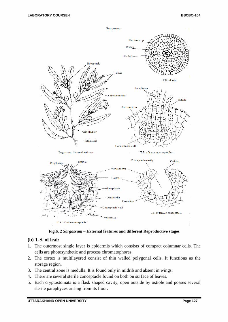
LABORATORY COURSE-I BSCBO-104
UTTARAKHAND OPEN UNIVERSITY Page 127
Fig.6. 2 Sargassum – External features and different Reproductive stages
(b) T.S. of leaf:
1. The outermost single layer is epidermis which consists of compact columnar cells. The
cells are photosynthetic and process chromatophores.
2. The cortex is multilayered consist of thin walled polygonal cells. It functions as the
storage region.
3. The central zone is medulla. It is found only in midrib and absent in wings.
4. There are several sterile conceptacle found on both on surface of leaves.
5. Each cryptostomata is a flask shaped cavity, open outside by ostiole and posses several
sterile paraphyces arising from its floor.

LABORATORY COURSE-I BSCBO-104
UTTARAKHAND OPEN UNIVERSITY Page 128
Fig. 6.3: Sargassum. T.S. leaf (diagrammatic) Fig.6.4: Sargassum. T.S. through air bladder
Reproductive structures
Sexual:
1. The sexual reproduction is oogamous.
2. The sex organs (i.e. antheridia and oogonia) are born inside the conceptacles produced in
recepticles.
3. The recepticles repeatedly branched fertile branches. The conceptacles are always
unisexual.
T.S. of receptacle passing through male conceptacles:
1. The receptacles show internal structure similar to that of leaf. It bears conceptacles.
2. Each conceptacle is flask shaped cavity with an opening called ostiole.
3. The antheridia are born on branched paraphyces which are intermixed with sterile
paraphyces.
4. Each antheridium is small, oval body which contains many anthrozoids.
T.S. of receptacle passing through female conceptacles:
1. The receptacle shows internal structure similar to that of leaf. It bears many conceptacles.
2. Each conceptacle is flask shaped cavity with an opening called ostiole.
3. The oogonia are unicellular, ovoid and sessile.
4. The mature oogonium contanes single uninucleate egg.
5. At maturity the oogonium is discharged but remains still attached to the wall of
conceptacle by means of long, mucilaginous stalk.
Identification
Sub-division – Algae (1) Simple thallus, (2) Chlorophyll present (3) cell wall of cellulose.
Class - Phaeophyceae. (1) Chromatophores yellowish – brown, (2) Photosynthetic reserves –
laminarin adn mannital, (3) Motile reproductive cells – pyriform and flagellated.
Order – Fucales (i) Plants parenchymatous, morphologically and anatomically
differentiaged (2) Asexual reproduction absent (4) Sex organs in conceptacles.
Family – Sargassaceae (i) Axes beaving distinct foliar organs. (2) Vericles usually present
(3) Branching of the thallus radial to the central axis.

LABORATORY COURSE-I BSCBO-104
UTTARAKHAND OPEN UNIVERSITY Page 129
Genus – Sargassum (i) Foliar organs narrow, branches, leaf like with a distinct midrib (2)
vesicles generally lateral (3) Fertile branches (receptacles) latral or terminal panciles.
6.3.3 Ectocarpus
Division: Phaeophyta
Class: Phaeophyceae
Order: Ectocarpales
Family: Ectocarpaceae
Genus: Ectocarpus
It is a marine algae, grows abundantly in tropical seas of western coast. Many species of this
grow in India. They grow on other body surface.
Vegetative structure:
1. Plant body is macroscopic, multicellular, filamentous and branched.
2. The plant shows heterotrichous habit.
3. The prostrate portion in many species is filamentous, irregularly branched and firmly
attached to the substratum with the help of rhizoids.
4. The erect portion arises from prostrate portion in the form of crowded tuft of branches
arising from a main axis.
5. The main axis shows the lateral branching that end in a taper form.
6. Filamentous are uniseriate and possess basal or intercalary meristem.
7. Cells are small cylindrical and possess double layered wall.
8. Each cell is uninucleate. It possess one many chromatophores that may be disc or ribbon
shaped.
9. They are naked and represent pyranoid in chromatophores.
Fig.6.5: Ectocarpus Habit Fig.6.6: Ectocarpus, A single cell
Reproductive structures:
1. Asexual reproduction takes place by the formation of biflagellate zoospores produced in
sporangia.

LABORATORY COURSE-I BSCBO-104
UTTARAKHAND OPEN UNIVERSITY Page 130
2. Sexual reproduction takes place by flagellated planogametes produced in plurilocular.
Gametes are isogamous but show anisogamy.
Filaments with Unilocular sporangia:
1. The unilocular sporangia are always found in a sporophytic plant.
2. They may be stalked or sessile.
3. It is uninulcleate in the beginning but later on becomes multi nucleate.
4. Mature unilocular sporangium produces many pear shaped biflagellate zoospores. They
germinate directly and serve as the asexual reproduction.
Filaments with Plurilocular sporangia:
1. These occur on both haploid and diploid plants.
2. Plurilocular sporangia are elongated in shape. They borne on lateral branches.
3. They may be stalked or sessile.
4. Each small cubical cell differentiates into single uninucleate, avoid zoogoes.
5. These sporangia develop both on sporophytic as well as gametophytic plant body.
6. When they are produced on a haploid plant, they behave as gametes and if borne on
diploid plant these act as haploid zoospores.
Fig.6.7: Reproductive Structures
Identification
Sub-division– Algae. (1) Simple thallus, (2) Presence of chlorophyll, (3) Cell wall of
cellulose.
Class – Phaeophyceae. (1) Yellowish-brown chromatophores, (2) Photosynthetic reserve –
laminarin and mannitol, (3) Motile reproductive cell – pyriform and flagellated , (4)
Flagella laterally inserted and unequal.
Order – Ectocarpales. (1) Thallus filamentous, (2) Growth trichothallic, (3) Reproductive
organs – unilocular and plurilocular sporangia (4) Isomorphic alternation of
generations.

LABORATORY COURSE-I BSCBO-104
UTTARAKHAND OPEN UNIVERSITY Page 131
Family–Ectocarpaceae. (1) Thallus monoaxial, branched, branches uniseriate, (2) Growth
trichothallic, (3) Sporophytes with uni- or plurilocular sporangia, terminal or
intercalary.
Genus – Ectocarpus. (1) Chromatophores discoid or band –shaped. (2) Pyrenoids absent, (3)
Reproductive parts terminal, stalked.
Genus: Polysiphonia
The genus Polysiphonia includes about 50 species. All are marine and occurs commonly on
the sea shores. A few species grow in western coast of India. Some of the common Indian
species are P. platycarpa, P. urceolata and P. variegate.
The species of Polysiphonia grow attached to the rocks or as epiphytes on rock weeds. They
look red or purple in color.
Vegetative structures:
External Features:
1. The plant body is multicellular, filamentous, branched and hetero-trichous.
2. The thallus is characteristically polysiphonous.
3. The prostrate system creeps over the substratum.
4. The erect system is much branched and exhibits a feathery appearance.
5. It consists of main axis and lateral branches.
6. There are two types of branches one of long and other of short.
7. The trichoblast are usually vegetative but in some species they bear sex organs.
8. The main axis and the branches of unlimited growth are identical. They terminate into an
apical cell followed by few flat cells. Later on they become polysiphonous.
Internal features:
1. The thallus consists of a large central siphon surrounded by 4-20 pericentral siphons.
2. Pericentral siphons are usually broader than the central siphons.
3. The cells of the siphons are usually connected with each other by pit connections.
4. The cells are uninucleate and bear central vacuole and show discoid chromatophores in
each cell.

LABORATORY COURSE-I BSCBO-104
UTTARAKHAND OPEN UNIVERSITY Page 132
Fig.6.8: Polysiphonia – Habit and external structures
Reproductive structures: Sexual:
1. The genus is dioecious and show sexual reproduction which is oogamous.
2. There are three types of plants i.e. male, female, and tetrasporophyte. All three are
morphologically indentical.
3. Male plants produced antheridium spermatangium while female plant represents
carpogonia.
4. Male gametophyte, female gametophyte and sporophytic and sporophytic plant bodies are
morphologically identical.
A. Male :
1. The antheridia are borne upon short branches in clusters near the apical portion of thallus.
2. Each antheridial branch consists of a central trichoblast filament which produces many
lateral pericentral cells.
3. The antheridial mother cells develop on pericentral cells. The antheri dial mother cells are
called the spermatangia.

LABORATORY COURSE-I BSCBO-104
UTTARAKHAND OPEN UNIVERSITY Page 133
4. A single spermatangium is liberated from each spermatangium.
5. The spermatia are non-motile and uninucleated.
Fig.6.9: Polysiphonia. T.S. thallus
B. Female :
1. The carpogonia are present on the female plants inside the procarp.
2. Procarp is urn-shaped body. The wall is called pericarp that has an opening known as
ostiole.
3. A long, tubular, receptive organ caleld trichogyne protrudes out of the ostiole.
4. At the base of trichogyne lies a swollen part, called carpogonium with a single female
nucleus.
5. Cystocarp is a post – fertilization product. The thallus bearing this structure forms a phase
called carposporophyte.
6. The oval or urn-shaped structure is attacjhed to a lateral branch.
7. Cystocarp opens to the exterior by an opening called ostiole.
8. Wall of the cystocarp is called pericarp and is composed of a single layer of cells.
Fig.6.10: Polysiphonia. A-B, A. Cluster of spermatangia, B. Spermatangium

LABORATORY COURSE-I BSCBO-104
UTTARAKHAND OPEN UNIVERSITY Page 134
Fig. 6.11: Polysiphonia. A part of thallus with cystocarp
9. Carpospores are produced from the base of the cystocarp. These are arranged in single
spehrical layer.
10. Each carpospore is oval, uninucleate and diploid.
C. Tetrasporophyte
1. Tetrasporophytes are morphologically similar to male and the female gametophytes.
2. The thallus is polysiphonous being made of a central siphon surrounded by pericentral
siphons.
3. A cell shows a nucleus, discoi chromatorphores and pit connections.
4. The plant is diploid and bears tetrasporangia in longitudinal series, produced mostly by
pericentral cells.
5. Tetrasporangia are small and spherical bodies borne on short one-celled stalk.
Fig.6.12: Polysiphonia. A part of thallus with tetrasporngia
6. Each tetrasporangium possesses four tetrahedrally arranged uninucleate and haploid
tetrasproes.

LABORATORY COURSE-I BSCBO-104
UTTARAKHAND OPEN UNIVERSITY Page 135
Identification
Sub- division – Algae. (1) Thallus simple, (2) Chlorophyll present, (3) Cell walls of cellulose.
Class – Rhodophyceae. (1) Chromatophores pure red to dark – purple (2) Photosynthetic
reserve floridoside, (3) Male gametes non-motile, (4) Female reproductive organ with
a receptive structure – trichogyen, (5) Post – fertilizaiton product – cystocarp.
Sub-class–Florideae. (1) Thallus basically filamentous, (2) Pit connections between sister
cells, (3) Cells with more than one chromatophore, (4) Carpogonium highly
specialized.
Order – Ceramiales. (1) Thalli uni-multiaxial or filamentous, (2) Filaments corticated,
Polysiphonous, (3) Spermatangia in clusters, (4) Presence of trichoblasts.
Famil – Rhodomelaceae. (1) Axes polysiphonous, (2) Axes naked, corticated or covered
with branches, (3) Main axis surrounded with pericentrals, (4) Plants bushed,
sparingly branched, branches delicate.
Genus–Polysiphonia. (1) Ultimate branches uncoritcated, (2) Tetrasporangia borne singly.
6.3.5 Batrachospermum
Division Rhodophyta
Class Rhodophyceae
Sub-class Florideae
Order Nemalionales
Family Batrachosermaceae
Genus Batrachospermum
It is fresh water alga. It usually grows attached to stones, rocks, sticks or even shells of
milluscs, in slow flowing streams, margins of akes and pools. It grows in well aerated, cool
and clean water. The alga most commonly occurs in the streams of Dehradun. The deep water
forms appear violet or reddish in color but the shallow water forms are olive blue-green.
The thallus:
1. The plant body is mucilaginous, multicellular, filamentous, branched and heterotrichous.
2. The branched thallus appears as chain of beaded filaments.
3. The main axis consists of uniseraite row of long, cylindrical axial cells. It may reach upto
20 cm or more in length. It is divided into nodes and internodes.
4. The lateral branches arise monopod ally from 4-6 lateral basal cells near the septa.
5. The branches are of two types (1) branches of unlimited growth and (2) branches of
limited growth.
6. The branches of unlimited growth grow continuously, from nodes and inter nodes,
develop cortication and resemble the main axis.
7. The branches of limited growth arise in whorls from each node. They further branch
repeatedly but stop their growth at certain limit in slender hairs. These whorls are known
as glomerules.
8. The branches of limited growth comprise of small, ellipsoidal or moniliform cells.

LABORATORY COURSE-I BSCBO-104
UTTARAKHAND OPEN UNIVERSITY Page 136
9. A number of cortical filaments also arise from each node which creep over the long axial
cells and form an envelope of cortex around axis.
Sexual reproductive structures:
1. The sexual reproduction is oogamous.
External features A portion of thallus showing wharl
(Habit) of laterials (glomerules)
Fig. 6.13, Batrachospermum – External features and a portion of thallus showing
branches of limited and unlimited growth.
Fig. 6.14, Batrachospermum. A part of fertile branch with glomerule and carposporangia
2. The species are monocecious and male and female sex organs occur near the apex.
3. Male sex organs are antheridia. These are present in clusters on short branches of lateral
filaments.

LABORATORY COURSE-I BSCBO-104
UTTARAKHAND OPEN UNIVERSITY Page 137
4. Antheridia are oblong or spherical and unicellular.
5. Each antheridium produces a single, spherical, colourless, naked, uninucleate and non-
motile spermatium.
6. Female sex organs are carpogonia situated at the apex of 3-4 celled lateral carpogonial
branch.
7. Carpogonium is made of a basal swollen portion with a terminal, elongated, tubular
process called trichogyne.
8. As a result of fertilization cystocarp is formed. This appears as a cluster of carpospores in
glomerules.
9. Cystocarp remains covered by sterile branches.
10. Inside the cystocarp lie many branched gonimoblast filaments.
11. The terminal swollen cells of these filaments are carposporangia. Each carposporangium
produces a single carpospore.
Identification
Sub-division – Algae. (1) Thallus simple, (2) Presence of chlorophyll, (3) Cell walls of
cellulose.
Class – Rhodophyceae. (1) Chromatophores pure red to dark purple, (2) Photosynthetic
reserve – Floridean starch and floridoside, (3) Male gametes non-motile, (4) Female
reproductive organ with trichogyne – a receptive structure, (5) Post-ferilization product a
cystocarp.
Sub-class – Florideae. (1) Thallus basically filamentous, (2) Pit connections between sister
cells, (3) Cells with more than one chromatophore, (4) Carpogonium highly specialised.
Order – Nemalionales. (1) Plants filamentous, corticated, uni or multiaxial, (2) Cells
unincleate, chromatophores axial or lateral (3) Cystocarps superficial or deeply embedded in
the thallus (4) Life cycle without free-living tetrasporophyte.
Family – Batrachospermaceae. (1) Inhabit freshwater, (2) Thallus uniaxial, (3) Life cycle
haplobiotic.
Genus – Batrachospermum, (1) Main axis and branches free from one another, (2) Branching
appears beaded, (3) Threads embedded in large amount of mucilage.
6.4 SUMMARY
The genus Vaucheria has about 40 species, out of which about nine are reported form India.
The most common species are V. sessilis and V. geminata, which occur during winters. The
alga is aquatic as well as terrestrial. Most of the species grow in damp gardent soil, moist
wall, in stagnant ponds, ditches and slow moving streams. Some species are marine.
Ectocarpus is a marine algae, grows abundantly in tropical seas of western coast. Many
species of this grow in India. They grow on other body surface.
Sargassum is marine alga grow abundantly in tropical seas of southern hemisphere. Many
species of this grow in India, in southern and western coast at Okha, Dwarka and other places
in India.

LABORATORY COURSE-I BSCBO-104
UTTARAKHAND OPEN UNIVERSITY Page 138
The genus Polysiphonia includes about 50 species. All the genus are marine and occurs
commonly on the sea shores. A few species grow in western coast of India. Some of the
common Indian species are P. platycarpa, P. urceolata and P. variegate. The species of
Ploysiphonia grow attached to the rocks or as epiphytes on rock weeds. It look red or purple
in color.
Batrachospermum is fresh water alga. It usually grows attached to stones, rocks, sticks or
even shells of molluscs, in slow flowing streams, margins of lakes and pools. It grows in well
aerated, cool and clean water. The alga most commonly occurs in the streams of Dehradun in
India. The deep water forms appear violet or reddish in color but the shallow water forms are
olive blue-green.
6.5 GLOSSARY
Aggregation - a grouping of algal cells but not the organization of a colony, often held
together by mucilage
Biflagellate - having two flagella
Chlorophyll - pigment found in photosynthetic organisms, all algae have chlorophyll a;
chlorophylls b, c, and d are found in one or more groups of algae
Coenobium - a colony where the number of cells is fixed at the time of reproduction
Coenocyte / Coenocytic - a multinucleate cell that does not have cellular cross walls
Colony - a group of cells that function on one, organized unit such as Hydrodictyon
Cyst - a general term for thick-walled vegetative cell
Desmokont - dinoflagellate with two flagella at the anterior end
Dichotomous - split into two parts
Dinokont - a dinoflagellate with an flagellum circling the middle and a flagella on the
posterior end
Epicone - anterior part of a dinoflagellate cell
Epiphyte - an organism that spends part or all of its life cycle growing on a plant
Eyespot - swelled area attached to a flagella that contains pigment. The pigment proteins
respond to the presence of light and signal the flagella to move toward it.
False branching - found in Cyanophyta (blue-greens); appearance of branched cells but
when cells are simply adjacent to each other and connected by only mucilage
Flagella - a cellular appendage that enables cells to have motility
Fusiform - narrow shaped cell with a sharp tapering at both end
Heterocysts - specialized cell in Cyanophyta that is able to fix nitrogen
Heterotrichy - a differentiated growing pattern in which some filaments grow appressed to
the anchoring surface and others are erect usually in a branch-like pattern.
Intercalary - located within the algal filament or thallus
Lorica - a cell wall covering that has space between the cell wall and the cell membrane,
often in Euglenophyta
Mucilage - a carbohydrate based material found on the outside of some algal cells (see
Lyngbya sp.)
Palmelloid formation - non-flagellated cells in a common mucilage

LABORATORY COURSE-I BSCBO-104
UTTARAKHAND OPEN UNIVERSITY Page 139
Paramylom - found in Euglenophyta; carbohydrate source that is long chain of glucose
molecules
Parietal - arranged along the cell walls
Pellicle - found in Euglenophyta; a series of strip-like plates underneath the cell membrane
arranged in a spiral, pellicle may be rigid or plates may be able to slide as the cell expands
and contracts during movement.
Pyrenoid - protein region inside chloroplast that accumulates carbohydrates
Sheath - thin mucilaginous covering over a filamentous algae
Stellate - star-shaped
Theca - cellulose plates that are "armorlike" in appearance found in Pyrrhophyta
(dinoflagellates)
Terminal - located at the end algal filament or thallus
Trichome - in Cyanophyta; the cells making up a filament
True Branching - found in Cyanophyta (blue-greens); trichome branches (compare to false
branching)
6.6 SELF ASSESSMENT QUESTIONS
6.6.1 Very short answer type questions
Q1. Name the character of algae which distinguish it from fungi.
Q2. Name some planktonic algae
Q3. Name some marine algae.
Q4. Name the algae responsible for the red colour of the ―Red Sea‖.
Q5. Name the algae which causes red snow ball in alpine region.
Q6. Name the alga which is used in space research.
Q7. Name some algae used for food.
Q8. Name the algae which yield agar agar.
Q9. Name the algae which help in Nitrogen fixation.
Q10. Name the class of algae which is placed in prokaryota together with bacteria.
Q11. What are heterocysts and where are they found.
6.6.2 Multiple choice type questions
(i) Laminarin is an energy storage material which is a characteristic of
(a) Phaeophyta (b) Chlorophyta
(c) Crysophyta (d) Pyrrophyta
(ii) Which alagal group never produces motile flagellated cells among any of its members?
(a) Chlorophyceae (b) Crysophyta
(c) Phaeophyta (d) Rhodophyta
(iii) Starch is an energy storage material characteristic of
(a) Rhodophyta (b) Chlorophyceae

LABORATORY COURSE-I BSCBO-104
UTTARAKHAND OPEN UNIVERSITY Page 140
(c) Crysophyta (d) Phaeophyta
(iv) The kelps are algae found in
(a) Rhodophyta (b) Chlorophyceae
(c) Crysophyta (d) Phaeophyta
(v) The kelps are algae found in
(a) Rhodophyta (b) Chlorophyceae
(c) Crysophyta (d) Phaeophyta
(vi) Chemical in kelp that is used in foods is called?
(a) Agar (b) Alganin
(c) Gametes (d) Starch
6.6.2 Answer Key:
(i) a (iv) d
(ii) d (v) d
(iii) a (vi) b
6.7 REFERENCE
Desikachary, T.V. 1959. Cyanophyta. New York Academic Press, New York. 686 pp.
Dillard, G.E. 1999. Common Freshwater Algae of the United States. J. Cramer, Berlin,
Germany. 173 pp.
Gomont, M. 1962. Monographie des Oscillariees. Wheldon and Wesley, Ltd., New York.
264 pp.
Patrick, R. and C.W. Reimer. 1966. The Diatoms of the United States, (Volume 1).
Academy of Natural Sciences of Philadelphia, PA. 688 pp.
Patrick, R. and C.W. Reimer. 1975. The Diatoms of the United States, (Volume 2, Part 1).
Academy of Natural Sciences of Philadelphia, PA. 213 pp.
Prescott, G.W. 1978. How to Know the Freshwater Algae. W.C. Brown Co., Dubuque,
Iowa. 293 pp.
Smith, G.M. 1950. Freshwater Algae of the United States, 2nd Ed. McGraw-Hill, New
York. 719 pp
6.8 SUGGESTED READINGS
Algae by Linda E Graham, James M. Graham and Lee Warren Wilcox Published by
Benjamin Cummings 2009. University of California

LABORATORY COURSE-I BSCBO-104
UTTARAKHAND OPEN UNIVERSITY Page 141
An Introduction to Algae by H.D. Kumar
Structure & Reproduction of Algae by F.E. Fritsch
A Textbook of Algae by O.P. Sharma, Tata McGraw-Hill Education
6.9 TERMINAL QUESTIONS
1. Write down the classification, vegetative structure and reproductive structures of
Vaucheria with the help of illustrated diagrams
2. Write down the classification, vegetative structure and reproductive structures of
Ectocarpus with the help of illustrated diagrams
3. Write down the classification, vegetative structure and reproductive structures of
Sargassum with the help of illustrated diagrams
4. Write down the classification, vegetative structure and reproductive structures of
Polysiphonia with the help of illustrated diagrams
5. Write down the classification, vegetative structure and reproductive structures of
Batrachospermum with the help of illustrated diagrams

LABORATORY COURSE-I BSCBO-104
UTTARAKHAND OPEN UNIVERSITY Page 142
UNIT-7-STUDY OF EXTERNAL FEATURES,
INTERNAL STRUCTURE AND REPRODUCTIVE
STRUCTURES OF BRYOPHYTES - RICCIA,
MARCHANTIA, ANTHOCEROS, WITH THE HELP OF
PERMANENT AND /OR TEMPORARY
PREPARATIONS
7.1-Objectives
7.2-Introduction
7.3-Study of external features, internal structure and reproductive structures of-
7.3.1-Riccia
7.3.2-Marchantia
7.3.3-Anthoceros
7.4-Method of preparation of permanent preparations
7.5-Method of preparation of temporary preparations
7.6- Summary
7.7- Glossary
7.8- Self assessment question
7.9- References
7.10-Suggested Readings
7.11-Terminal Questions

LABORATORY COURSE-I BSCBO-104
UTTARAKHAND OPEN UNIVERSITY Page 143
7.1 OBJECTIVES
After reading this unit students will be able:
To study the external features of Riccia, Marchantia, Anthoceros
To understand Internal structure of Riccia, Marchantia, Anthoceros
To study Reproductive structures of Riccia, Marchantia, Anthoceros with the help of
temperory and permanent slides.
7.2 INTRODUCTION
Bryophytes are called as the ―amphibians of the plant kingdom‖ as they are mostly of
terrestrial origin but require water for the act of fertilization. They are found in humid and
shady place. They occupy a central position in between algae on one hand and the
pteridophytes on the other.
They are quite small and inconspicuous organisms. The gametophyte is highly
developed and differentiated than that of a complex alga. The plant body is gametophytic,
thalloid (e.g., Riccia, Marchantia) or an erect plant body (e.g., Funaria, Polytrichum). The
true roots are absent. Instead unicellular or multicellular hair-like rhizoids develop from the
thalli which absorb the nutrients from the moist soil. They lack vascular tissues.
Vegetative reproduction occurs by means of progressive death and decay of older
parts of plants, or by adventitious branches, gemmae, tubers, bulbils and protonema etc.
The sexual reproduction is of oogamous type, i.e., it take place by means of gametes.
The male gametes are motile and known as antherozoids; the female gametes are non-motile
and known as eggs (oospheres).
The gametes are produced within the sex organs known as antheridium (male) and
archegonium (Female). Antheridia are club shaped structure and produce uninucleate
bicilleate antherozoids.
The archegonia are flask-shaped. Each archegonium consists of venter and a neck.
The venter is basal swollen portion and the neck is elongated. Within the neck and the venter
there is an axial row of cells surrounded by sterile jacket. This axial row consists of a few
neck canal cells, a ventral canal cell and an egg.
Water is essential for the act of fertilization. The motile ciliated antherozoids swim in
the film of water and reach to the neck of an archegonium. The antherozoid enters the neck
and ultimately approaches the eggs. The antherozoid penetrates the egg and fertilization is
affected. With the result of fertilization the zygote is formed.
The zygote begins to develop into a multicellular embryo just after the act of
fertilization without going under any resting period. The embryo remains within the venter of
the archegonium and not liberated as in the case of algae. The venter wall enlarges along with
the developing embryo to form the protective envelope known as calyptra.

LABORATORY COURSE-I BSCBO-104
UTTARAKHAND OPEN UNIVERSITY Page 144
Zygote represents first cell of sporophyte. Sporophyte in bryophytes is dependent on
the gametophyte partially or wholly. It is generally differentiated into foot, seta and capsule.
The foot of the sporophyte remains embedded in the tissues of gametophyte and act as
haustorium. Seta help in elongation of capsule and the function of capsule is spore
production.
The spore mother cells are produced within the sporogonium. The spore mother cells
are diploid (2n) and they represent the last stage of the sporophyte generation. After reduction
division in spore mother cell four haploid spores are formed, which remain arranged
tetrahedrally.
All the spores are homosporous i.e., all the spores of a species are morphologically
similar in size and form. They germinate into gametophytic plant directly or germinate into a
filamentous or thalloid green protonema which later on gives rise to the thallus
(gametophyte). The spores represent the beginning of the gametophytic generation.
Division Bryophyta is divided into three classes.
1. Hepaticopsida.
2. Anthocerotopsida.
3. Bryopsida.
7.3-STUDY OF EXTERNAL FEATURES, INTERNAL
STRUCTURE AND REPRODUCTIVE STRUCTURES
7.3.1. Riccia
Division: Bryophyta
Class: Hepaticopsida
Order: Marchantiales
Family: Ricciaceae
Genus: Riccia
Habit and occurrence:
1. There are about 130 species. About 18 species have been reported from India by various
workers. Prof. S.R Kashyap recorded a number of species from the Punjab. Kashmir and
other regions of Western Himalayas.
2. Mostly species are terrestrial and grow on damp, soil of moist rocks and have been
recorded from the hills as well as from the plains.
3. R. himalayensis has been recorded even from the altitude of 9000 feet in the Himalayas.
4. Riccia fluitans and an allied species Ricciocarpos natans are aquatic, and found in free
floating condition.
Proposed Laboratory Work:
1. Study of external structure of thallus (gametophyte)
2. Study of anatomy of thallus.
3. Study of reproductive structures.
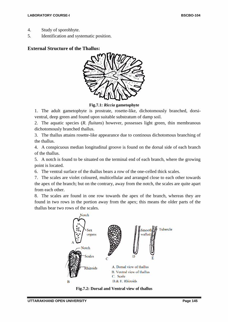
LABORATORY COURSE-I BSCBO-104
UTTARAKHAND OPEN UNIVERSITY Page 145
4. Study of sporohhyte.
5. Identification and systematic position.
External Structure of the Thallus:
Fig.7.1: Riccia gametophyte
1. The adult gametophyte is prostrate, rosette-like, dichotomously branched, dorsi-
ventral, deep green and found upon suitable substratum of damp soil.
2. The aquatic species (R. fluitans) however, possesses light green, thin membranous
dichotomously branched thallus.
3. The thallus attains rosette-like appearance due to continous dichotomous branching of
the thallus.
4. A conspicuous median longitudinal groove is found on the dorsal side of each branch
of the thallus.
5. A notch is found to be situated on the terminal end of each branch, where the growing
point is located.
6. The ventral surface of the thallus bears a row of the one-celled thick scales.
7. The scales are violet coloured, multicellular and arranged close to each other towards
the apex of the branch; but on the contrary, away from the notch, the scales are quite apart
from each other.
8. The scales are found in one row towards the apex of the branch, whereas they are
found in two rows in the portion away from the apex; this means the older parts of the
thallus bear two rows of the scales.
Fig.7.2: Dorsal and Ventral view of thallus

LABORATORY COURSE-I BSCBO-104
UTTARAKHAND OPEN UNIVERSITY Page 146
Rhizoids:
1. The rhizoids are also found on the ventral surface of the thallus.
2. The rhizoids are unicellular.
3. Rhizoids are of two types; i.e. smooth-walled and tuberculate type.
4. The tuberculate rhizoids possess the peg like infolding peeping into the lumen to the
rhizoids.
5. The simple or smooth walled rhizoids have no such infoldings.
Internal Structure (Anatomy) of the Thallus:
Thin vertical sections of thallus are cut to study the anatomy of the adult thallus of Riccia.
1. Thallus is differentiated into two regions dorsal and ventral (storage region).
Fig.7.3: Diagrammatic View of Riccia Thallus
2. The dorsal region (Photosynthetic region) of the thallus consists of layer of epidermis,
many air pores and loose green chlorophyllous cells with discoid chloroplasts in them.
3. There are regular air canals in between each two verticals row of the chlorophyllous
cells.
4. The epidermis of the dorsal surface of the thallus is discontinuous and opened outside at
several places by the openings of the air canal; the epidermis is single layered.
5. The uppermost cells of the assimilatory filaments are somewhat large and colourless.
6. On the two flanks of the boat-shaped section, violet coloured scales are present.
7. The ventral region of the thallus lacks intercellular spaces, is colourless and made up of
simple parenchyma.
8. Cells of this region make storage tissue and filled with starch grains.
9. From the single-layered epidermis of the ventral surface several unicellular rhizoids
(smooth walled and tuberculate) and multicellular scales are given out.
Fig.7.4: T.S of thallus showing internal structure

LABORATORY COURSE-I BSCBO-104
UTTARAKHAND OPEN UNIVERSITY Page 147
Riccia fluitans: aquatic species
1. Thallus is long ribbon-like and with dichotomous branches.
2. Lack rhizoids and scales.
3. Internally upper epidermis is continuous layer.
4. Large air spaces are formed by irregularly arranged lamellae.
5. Cells of upper epidermis and lamellae contain chloroplast.
Fig.7.5: Riccia fluitans: External habit and V.T.S of thallus
Reproductive Structures: Reproduction in Riccia takes place by means of
(1) Vegetative and (2) Sexual methods
Vegetative Reproduction: This type of reproduction takes place by several ways.
By the Death and Decay of the Older Parts of the Thallus: Sometimes a part of the
dichotomously branched thallus decays from the posterior end in such a way that the
terminal ends of the branches remain unaffected, which may grow separately into new
thalli. Sometimes in the drought period all the parts of the thalli become dead, leaving the
apices, which on the approach of favourable conditions develop into new thalli.
By Adventitious Branches: Several species of Riccia develop the adventitious branches
on the ventral surface from the thalli which develop into new gametophytes.
By Cell Divison or Gemma Formation: The Riccia may also reproduce vegetatively by
cell divison of the young rhiziods which develop into gemma like structure of the cell.
These structures give rise to new plants.
By Tubers: Species, such as R.discolor: R. vesicata: R bulbifera and R. perennis develop
the vegetative structure called the tubers at the apices of the branches of the thallus to face
adverse conditions.
By Thick Apices: In certain species like R. himalayensis the apex of the thallus grows
downward into the soil and becomes thick and develops into a new plant on the approach
of favourable condition.
Sexual Reproduction:
1. The species may be homothallic (e.g., R. himalayensis, R. bishoffi).
2. The sex organs are antheidia (male) and archegonia (female)
The Antheridium:
1. The mature antheridium remains embedded in the antheridial chamber, which opens by
an ostiole on the dorsal side of the thallus.

LABORATORY COURSE-I BSCBO-104
UTTARAKHAND OPEN UNIVERSITY Page 148
2. The mature antheridium consists of a few-celled stalk and rounded or somewhat pointed
antheridial proper at its apical end.
3. Antheridium is encircled by a sterile single layered jacket layer which protects it.
4. The mature antheridium contains androcytes within the jackets layer; each androcyte
metamorphose into an biflagellate antherozoid.
Fig.7.6: V.T.S. of thallus and mature antheridium
The Archegonium:
Fig.7.7: V.T.S. of thallus and mature archegonium
1. The archegonium is a flask-shaped body attached to the thallus by a short stalk.
2. It consists of an elongated neck and a bulbous venter
3. The neck is 6-9 cells in height, and consists of six vertical rows of the cells.
4. There are neck-canal cells within the neck.
5. There are four cover cells at the top of neck canal.
6. The venter encloses a venter canal cell and a large egg.
7. It has a single layered wall around it which is of 12-20 cells in perimeter.
8. The venter canal cell disintegrates on the maturity of the archegonium and only the large-
egg remains there.
The Sporophyte:

LABORATORY COURSE-I BSCBO-104
UTTARAKHAND OPEN UNIVERSITY Page 149
Fig.7.8: V.T.S. of thallus through mature sporophyte
1. The mature sporophyte is simple and consists of spore sac or capsule only.
2. The capsule is enclosed within two layered calyptra.
3. The foot and seta are absent.
4. The capsule is embedded in the gametophyte and lacks chlorophyll hence dependent on
gametophyte for nutrition.
5. The tetrads of the spores are found within the sporophyte.
6. Elaters are absent.
7. The mature spore is three layered :
a) Exosporium: outermost cutinized layer.
b) Mesosporium: middle layer, which is thick-walled and consists of three concentric zones.
c) Endosporium: innermost layer is endosoporim; the complete spore wall that surrounds
the spore is thick and irregular in thickness.
Fig.7.9: Riccia: Stages in the germination of spore
Identification and Systematic position
Bryophyta:
(i) Gametophytic plant body
(ii) Absence of true roots.
(iii) Absence of vascular strands.
(iv) They are homosporous.
Hepaticopsida:
(i) Thalloid Dichoto mously branched plant body.
(ii) Chloroplasts are devoid of pyrenoids.
(iii) Rhizoids without septa.
(iv) Capsule lacks columella

LABORATORY COURSE-I BSCBO-104
UTTARAKHAND OPEN UNIVERSITY Page 150
Marchantiales:
(i) Ventral portion of thallus parenchymatous.
(ii) Scales and rhizoids present on the ventral side of thallus.
(iii) Air chamber opens out by air pores.
Ricciaceae:
(i) Rosette-like, dichotomously branched thallus.
(ii) Sex organs in the mid-dorsal groove.
(iii) Simple air pores present.
Riccia:
(i) Thallus is usually rosette-like.
(ii) Scales present on the margins.
(iii) Sporogonium represented by simple capsule only.
7.3.2. Marchantia
Division: Bryophyta
Class: Hepaticopsida
Order: Marchantiales
Family: Marchantiaceae
Genus: Marchantia
Habit and Occurrence:
1. They are cosmopolitan. There are about 65 species. There are about 11 India species.
They are mostly confined in the Himalayas. Only one or two species have been recorded
from the plants.
2. Marchantia polymorpha the best known species, is widely distributed. They commonly
thrive upon moist soil found on the rocks in shady places, in open woodland or near the
banks of stream. This genus grows best in the burnt soil.
Proposed Laboratory work:
Study of external structure of thallus (gametophyte).
Study of internal structure of thallus.
Study of reproductive structure.
Study of sporophyte.
Identification and systematic position.
External Structure of the Thallus:
Fig.7.10: Marchantia thallus

LABORATORY COURSE-I BSCBO-104
UTTARAKHAND OPEN UNIVERSITY Page 151
1. The thallus is prostrate, dichotomously branched and dorsiventral; it reaches to the length
of 2-10 cms.
2. The dorsal surface is deeper green than ventral one.
3. The rhomboidal or polygonal small areas are found on the dorsal surface of the thallus;
each such small area is provided with a small pore in its centre.
4. The apex of each branch of the thallus bears a notch in which the growing point is
situated.
5. On maturity the margin of the thallus is somewhat wavy.
6. The thallus bears a conspicuous midrib, which is well recognizable on the dorsal surface
by the presence of a groove and on the ventral surface by presence of a ridge.
Scales:
Fig.7.11: Marchantia: Ventral View of thallus
7. On the ventral surface of the thallus, on either side of the midrib two or more rows of the
pinkish, multicellular scales are present.
Fig.7.12: Marchantia: scales
Rhizoids:
1. The simple and tuberculate unicellular rhizoids are also found on the ventral surface,
which are absorptive in nature.
2. The thallus remains attached to the substratum by means of rhizoids.
Fig.7.13: Marchantia: rhizoids

LABORATORY COURSE-I BSCBO-104
UTTARAKHAND OPEN UNIVERSITY Page 152
Gemma cups-gemmae
1. On the dorsal surface of the plant gemetophytes, the gemmae cups are found usually
along the mid ribs; the margins of the gemma cup are cut.
2. The gemma cup contains the vegetative bodies, the gemmae.
Antheridiophores and Achegoniophores
Fig.7.14: Marchantia: Antheridiophore and Archegoniophore.
1. Some thalli produce special, erect, stalked upright branches at their growing apices.
2. They bear the male and female sex organs and are called the antheridiophores and
archegoniophores respectively; they are produced on different thalli.
Internal Structure (Anatomy) of the Thallus:
To study the internal stucture of the thallus thin vertical section of the thallus are cut.
1. Thallus is differentiated internally into upper photosynthetic zone and lower storage zone.
Fig.7.15: Marchantia: Internal Structure of Thallus
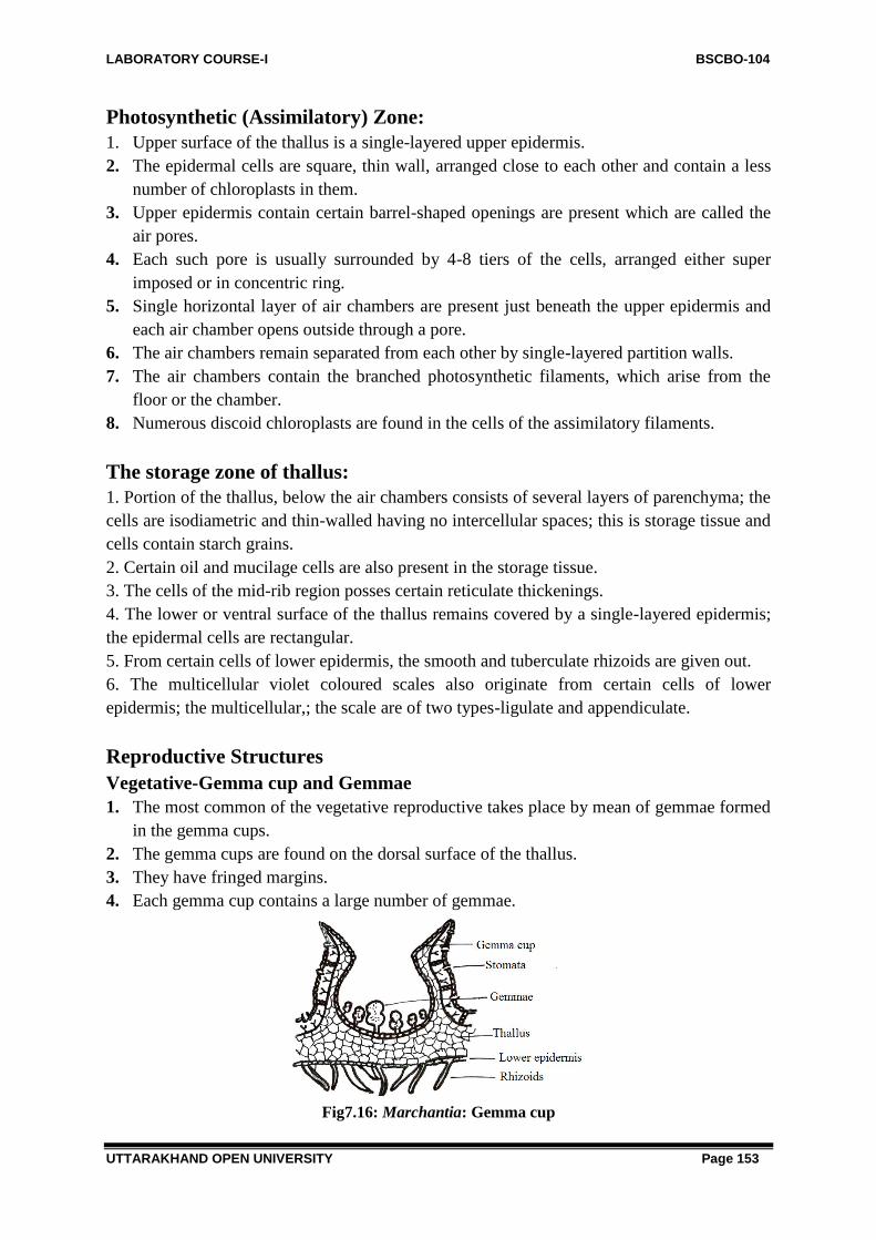
LABORATORY COURSE-I BSCBO-104
UTTARAKHAND OPEN UNIVERSITY Page 153
Photosynthetic (Assimilatory) Zone:
1. Upper surface of the thallus is a single-layered upper epidermis.
2. The epidermal cells are square, thin wall, arranged close to each other and contain a less
number of chloroplasts in them.
3. Upper epidermis contain certain barrel-shaped openings are present which are called the
air pores.
4. Each such pore is usually surrounded by 4-8 tiers of the cells, arranged either super
imposed or in concentric ring.
5. Single horizontal layer of air chambers are present just beneath the upper epidermis and
each air chamber opens outside through a pore.
6. The air chambers remain separated from each other by single-layered partition walls.
7. The air chambers contain the branched photosynthetic filaments, which arise from the
floor or the chamber.
8. Numerous discoid chloroplasts are found in the cells of the assimilatory filaments.
The storage zone of thallus:
1. Portion of the thallus, below the air chambers consists of several layers of parenchyma; the
cells are isodiametric and thin-walled having no intercellular spaces; this is storage tissue and
cells contain starch grains.
2. Certain oil and mucilage cells are also present in the storage tissue.
3. The cells of the mid-rib region posses certain reticulate thickenings.
4. The lower or ventral surface of the thallus remains covered by a single-layered epidermis;
the epidermal cells are rectangular.
5. From certain cells of lower epidermis, the smooth and tuberculate rhizoids are given out.
6. The multicellular violet coloured scales also originate from certain cells of lower
epidermis; the multicellular,; the scale are of two types-ligulate and appendiculate.
Reproductive Structures
Vegetative-Gemma cup and Gemmae
1. The most common of the vegetative reproductive takes place by mean of gemmae formed
in the gemma cups.
2. The gemma cups are found on the dorsal surface of the thallus.
3. They have fringed margins.
4. Each gemma cup contains a large number of gemmae.
Fig7.16: Marchantia: Gemma cup

LABORATORY COURSE-I BSCBO-104
UTTARAKHAND OPEN UNIVERSITY Page 154
Gemma
1. The mature gemma is found to be attached by a single celled stalk to the floor of the
gemma cup.
2. It is disciform and few celled thick in is centre, and quite thin at its margins; it is
somewhat dumb-bell like.
3. There are two notches situated on the internal margins opposite to each other.
4. About all the cells of a gemma contain chloroplasts.
5. A few rhiziodal cells are colourless and larger in size.
6. There are certain isolated cells, which contain oil and known as oil cells.
Fig.7.17: Marchantia: Mature Gemma
Sexual reproduction:
The genus is strictly heterothallic (dioecious) i.e., male (antheridiophores) and female
(archegoniophores) reproductive branches develop on different thalli.
The antheridiophore:
1. The antheridiophore consists of a two to three centimetres long stalk, and a flattened
usually eight lobed disc at its apical end.
2. The rhizoids and scales are present in the two grooves situated on the ventral surface of
antheridiophore.
3. The disc of antheridiophore is somewhat convex at its upper face; the upper epidermis is
interrupted by several usual barrel shaped air pores; these pores open in the air chambers,
assimilatory filaments in them.
4. The flask-like cavities are also found among and in the neighbourhood of these air
chambers.
5. Each cavity contains a stalked antheridium, which partially or wholly fills up the cavity.
6. The antheridial cavities open outside through small openings, the ostioles.

LABORATORY COURSE-I BSCBO-104
UTTARAKHAND OPEN UNIVERSITY Page 155
Fig.7.18: Marchantia: V. S. of Anthridiophore
The antheridium:
1. The mature antheridium consists of a short stalk and a rounded structure above it.
2. The antheridium proper remains surrounded by a single layered jacket.
3. Inside the jacket layer a large number of androcytes are filled up.
Fig.7.19: Marchantia: Mature Antheridium
The archegoniophore:
1. The archegoniophore is a modified branch of the prostrate thallus.
2. The archegoniophore are developed on dorsal side of young rosette-like disc
acrogenously.
3. As soon as the fertilization is over, the marginal portion of the disc becomes inverted and
the archegonia become upside down, i.e., their necks become downward.
Fig.7.20: Marchantia: Stages in Inversion of Archegonia after fertilization.
4. Now the oldest archegonia are situated towards the periphery of the disc and youngest
towards the stalk.
5. Usually, the disc possesses nine or eight rays.

LABORATORY COURSE-I BSCBO-104
UTTARAKHAND OPEN UNIVERSITY Page 156
6. Each group of 12-15 archegonial is enclosed in a single-layered fringed perichaetium.
7. In many cases the archegonial lobes elongate further in the green cylindrical rays.
8. The disc looks stellate in appearance from the upper side, because of the presence of these
stout cylindrical rays.
Fig.7.21: Marchantia: V. S. of Archegoniophore
The archegonium:
Fig.7.22: Marchantia: Archegonia
1. The flask-like archegonium is found to be attached to the lobe of the disc by a small stalk.
2. The archegonium consists of an elongated neck and a bulbous venter.
3. The neck consists of the jackets cells arranged in six vertical rows.
4. Four or more neck canal cells are found in the neck canal.
5. The venter in surrounded by a single layered jacket.
6. The venter contains a large egg (oosphere) and a venter canal cell.
7. At the top of the neck there are four or five cover cells.
Sporophyte-the sporogonium:
1. The mature sporogonium is differentiated into three regions,viz., the foot, seta and
capsule.
2. Towards the base of the archegonium, there is a bulbous, absorptive structure, the foot.
3. The seta is somewhat elongated and connects the foot and the capsule.
4. The seta pushes the capsule out of the three layers, the calyptra, perigynium and
perichaetium.
5. The capsule is somewhat oval-shaped and remains covered by a single layered jacket
layer.

LABORATORY COURSE-I BSCBO-104
UTTARAKHAND OPEN UNIVERSITY Page 157
6. Within the jacket layer, there are spores and sterile, hygroscopic, spindle-like elaters.
Spores:
1. The spores are small and spherical; they range from 12µ to 13µ in diameter.
2. Each spore is covered by a thin coat, differentiated into two layers.
3. The outer layer is thick exospore and the inner one endospore is thin and smooth.
4. Each spore contains a small amount of granular cytoplasm and a nucleus in it.
Fig.7.23: Marchantia: Sporophyte, Elaters and Spores
Identification and Systematic position
Bryophyta:
(i) Gametophytic plant body
(ii) Absence of true roots.
(iii) Absence of vascular strands.
(iv) They are homosporous.
Hepaticopsida:
(i) Thalloid plant body.
(ii) Chloroplasts devoid of pyrenoids.
(iii) Rhizoids without septa.
(iv) Capsule lacks columella
Marchantiales:
(i) Ventral portion of thallus parenchymatous.
(ii) Scales and rhizoids present on the ventral side of thallus.
(iii) Air chamber opens out by air pores.
Marchantiaceae:
(i) Barrel-shaped air pores present.
(ii) Sex Organs are borne on specialized branches.
(iii) Air chambers have photosynthetic filaments.
Marchantia:
(i) Foot and seta present.
(ii) Gemma cups are present.

LABORATORY COURSE-I BSCBO-104
UTTARAKHAND OPEN UNIVERSITY Page 158
7.3.3. Anthoceros
Division: Bryophyta
Class: Anthocerotopsida
Order: Anthocerotales
Family: Anthocerotaceae
Genus: Anthoceros
Habit and Occurrence:
1. There are about 200 species of this genus. About 25 species have been recorded from
India.
2. The three common Himalayan species are : A. himalayensis, A.erectus and A.chambensis.
3. These species are commonly found in the hilly regions of Kumaon, Mussoorie and
Chamba valley, 5,000 to 8,000 feet elevation above sea level.
4. All species are found in the hollows of moist rock in the dense patches.
Proposed Laboratory Work:
1. Study of external structure of thallus.
2. Study of internal structure (anatomy).
3. Study of sporophyte.
4. Study of reproductive structures.
5. Identification and systematic position.
External Structure:
Fig.7.24: Anthoceros thallus
1. The thallus is small, prostrate, dark green thin and dorsiventrally differentiated.
2. The thallus is lobed and the lobes are somewhat divided.
3. The midrib is absent.
4. The dorsal surface of thallus: Smooth (A .leavis); velvet likes (in A.crispus ) or with
spines and ridges (in A.fusiformis).
5. Smooth-walled, simple rhizoids are found.
6. The ventral scales and tuberculate rhizoids are altogether absent.

LABORATORY COURSE-I BSCBO-104
UTTARAKHAND OPEN UNIVERSITY Page 159
Fig.7.25: Anthoceros thallus: Ventral view, Rhizoids and tubers.
Internal Structure (Anatomy):
1. The anatomy of the thallus is quite simple and not differentiated.
2. Air chambers and air pores are absent.
3. Internal to the upper and lower epidermis there are simple, parenchymatous cells.
4. The cells of parenchyma are isodiametric and uniform.
5. Each cell contains a big chloroplast which possesses a single pyrenoid in its centre.
6. The chloroplasts are lens-shaped. The nucleus lies in the close vicinity of the chloroplasts,
near the pyrenoid.
7. On the ventral side of the thallus several intercellular mucilage cavities are found which
open by small openings, the slime pores on the ventral surface of the thallus.
8. The colonies of blue green alga Nostoc are found in the mucilage cavities.
9. The thalli are dark green, because of the presence of the Nostoc colonies, which may
easily be seen with the help of lens from the underside of the thallus.
Fig.7.25: Anthoceros: Internal structure of thallus.
Reproductive Structure
Vegetative:
1. Tubers. The thallus becomes thickened at several places on the margins; these marginal
thickenings are called, the tubers; each tuber may develop into a new thallus on return of
favourable conditions.
2. Gemmae. The gemmae have been recorded from the species, A. glandulosus, A.
formosae.
3. Apices. The apices develop into new thalli.

LABORATORY COURSE-I BSCBO-104
UTTARAKHAND OPEN UNIVERSITY Page 160
Sexual reproduction:
1. The species may be homothallic (monoecious) or heterothallic (dioecious).
2. Homothallic species are: A.fusiformis, A.punctatus.
3. The heterothallic species are: A.pearsoni, A.halli, A.erectus.
4. The sex organs: antheridia (male) and archegonia (female) are found embedded in the
tissues of the dorsal side of the thallus.
The Antheridium:
Fig.7.26: Anthoceros: Developing Antheridium and Mature antheridium
1. The antheridia are produced singly or in group in the antheridium chambers.
2. Each mature antheridium is stalked and club-shaped.
3. The stalk of antheridium consists of the mass of cell, e.g., in A.laevis of the four rows of
the cells as in A.erectus and A.punctatus.
4. The antheridium proper is covered by a single layered jacket.
5. Inside the jacket there are numerous androcytes which metamorphose into antherozoids.
6. Each antherozoid is spindle-like and biciliate; the cillia are attached to the anterior end of
the body.
The Archegonium:
1. The archegonium remains embedded in the thallus and only the cover cells project out of
the thallus.
2. The nearly mature archegonium is composed of a neck and venter, the neck contains 4-6
neck canal cells; the venter contains a ventral canal and an egg.
3. On the maturity of the archegonium, the ventral canal cell and canal cells are gelatinized.
4. A mature archegonium is flask shaped, without neck canal cells and with an egg
(oosphere) in its venter.
5. At the top of the neck of archegonium there are four cover cells.

LABORATORY COURSE-I BSCBO-104
UTTARAKHAND OPEN UNIVERSITY Page 161
Fig.7.27: Anthoceros: Mature archegoium
The Sporophyte:
Fig.7.28: Anthoceros: Young sporophyte, Spore tetrad and pseudoelaters
Fig.7.29: Anthoceros: V.S. through Mature sporophyte
1. The mature sporogonium consists of a bulbous foot and a projecting slender and erect
capsule.
2. Capsules arise from the thalli in the form of small horny structures; usually they are 2 to 3
cm long, but some species they range even from 5-15 cm.

LABORATORY COURSE-I BSCBO-104
UTTARAKHAND OPEN UNIVERSITY Page 162
3. The bulbous foot consists of parenchyma; it remains penetrated in the thallus and acts as
haustorium.
4. The cells developing from the meristem become differentiated into jacket layer, columella
and archesporium.
5. The main capsule consists of many important parts.
6. In the region just above the foot the archesporium is single layered and too young.
7. The tip region of sporogonium possesses mature spores and pseudoelaters.
8. The wall of the capsule consists of the four to six layers of the parenchymatous cells.
9. The outermost layer is epidermis which is interrupted by stomata at several places; the
epidermal cells are cutinized; the stomata open in the intercellular spaces of the
chlorophyllous cells; usually each cell possesses two chloroplasts.
10. On maturation, the tip of the sporogonium becomes black or brown in colour.
11. The dehiscence begins from the tip region of capsule.
12. The pseudoelaters are 2-3 celled structure. They lack the characteristic spiral thickenings
of true elaters, and therefore, are known as pseudoelaters.
13. Each spore is somewhat spherical and possesses two wall layers.
14. The outer wall layer is exine and the inner wall layer is intine; the intine is smooth and
thin while the exine is thick and ornamental.
15. Each spore possesses a single nucleus, a colourless plastid, few oil droplets and food
material within it.
Identification and Systematic Position
Bryophyta:
(i) Gametophytic plant body
(ii) Absence of true roots.
(iii) Absence of vascular strands.
(iv) They are homosporous.
Anthocerotopsida (Anthocerotae):
(i) Gametophytic plant body thalloid
(ii) Rhizoids simple and smooth walled;
(iii) Tuberculate rhizoids and scales absent.
(iv) Thallus homogenous not differentiated; air chambers and air pores not present;
(v) Each cell of thallus possesses a large chloroplast and pyrenoid within it.
(vi) Capsule with central sterile columella.
Anthocerotales: Single order
Anthocerotaceae: Single family
Anthoceros:
(i) Nostoc colonies present within thallus.
(ii) Involucre present at the base of cylindrical sporogonium.
(iii) Capsule wall ventilated.
LABORATORY COURSE-I BSCBO-104
UTTARAKHAND OPEN UNIVERSITY Page 163
7.4 METHOD OF PREPARATION OF PERMANENT
PREPARATIONS
To make permanent slides, the section of plant material is passed through alcholic as well as
xylene series.
1. Select a thin sections as per requirement
2. Stain with aqueous safranin (for 2 to 4 min)
3. Wash thoroughly with water
4. Dehydrate with alcohol in following order (15% followed by 30%, 50%, 70%, and
then 90%)
5. stain with fast green (few sec to 1 min)
6. Destain with 90% alcohol
7. Dealchololize with xylol in following order (25% followed by 50%, 70%, and then
90%)
8. Mount in glycerine (10%) or Canada balsam.
9. Cover with the help of cover glass carefully
7.5 METHOD OF PREPARATION OF TEMPORARY
PREPARATIONS
Single staining is applied in those cases where there is no tissue differentiation e.g. Algae,
fungi and bryophytes. Single staining in bryophytes is done with safranin.
1. Select a thin sections as per requirement
2. Stain with aqueous safranin (for 2 to 4 min)
3. Wash thoroughly with water
4. Then stain with fast green
5. Again wash out extra stain
6. Study the slide prepared under microscope
7.6 SUMMARY
Bryophytes are the amphibians of plant kindom possessing gametophytic thalloidor foliose
plant body. These show an alternation of generation. The gametophytic generation is
independent and concerned with the production of gametes in Antheridia and Archegonia.
The sexual reproduction is of oogamous type. Zygote represents the first cell of sporohytic
generation.
Sporophyte is usually differentiated into foot seta and capsule. It is partially or wholly
dependent upon the gametophytic plant body for its nourishment. The spore mother cell
inside the capsule undergoes reduction division to form tetrad of haploid spores. these spores
on germination produce the gametophytic plant body directly or produce a thalloisd or folise
protonema which later develops into the gametophytic plant body.Bryophyta division is
classified into three classes namely Hepaticopsida, Anthocerotopsida and Bryopsida.

LABORATORY COURSE-I BSCBO-104
UTTARAKHAND OPEN UNIVERSITY Page 164
Genus Riccia belongs to family Ricciaceae of class Hepaticopsida. It grows on
damp, soil of moist rocks and has been recorded from the hills as well as from the plains.
The adult gametophyte is prostrate, rosette-like, dichotomously branched and dorsi-ventral.
Thallus is differentiated into two regions dorsal (Photosynthetic region) and ventral (storage
region). The dorsal region of the thallus consists of layer of epidermis, many air pores and
loose green chlorophyllous cells with discoid chloroplasts in them. The ventral regions of the
thallus lacks intercellular spaces, is colourless and made up of simple parenchyma and filled
with starch grains. Several unicellular rhizoids (smooth walled and tuberculate) and
multicellular scales emerge from the lower epidermis. Reproduction in Riccia takes place by
means of (1) vegetative and (2) sexual methods. The sex organs are antheridia (male) and
archegonia (female). The mature sporophyte is simple and consists of spore sac or capsule
only. The tetrads of the spores are found within the sporophyte. Elaters are absent.
Genus: Marchantia belongs to family Marchantiaceae of class Hepaticopsida. They
commonly thrive upon moist soil found on the rocks in shady places, in open woodland or
near the banks of stream. The thallus is prostrate, dichotomously branched and dorsiventral; it
reaches to the length of 2-10 cms. On the ventral surface of the thallus, on either side of the
midrib two or more rows of the pinkish, multicellular scales are present. The simple and
tuberculate unicellular rhizoids are also found on the ventral surface, which are absorptive in
nature. Thallus is differentiated internally into upper photosynthetic zone and lower storage
zone. Upper epidermis contain certain barrel-shaped openings are present which are called
the air pores. The air chambers contain the branched photosynthetic filaments, which arise
from the floor or the chamber. Numerous discoid chloroplasts are found in the cells of the
assimilatory filaments. The most common of the vegetative reproductive takes place by mean
of gemmae formed in the gemma cups found on the dorsal surface. The genus is strictly
heterothallic (dioecious) i.e., male (antheridiophores) and female (archegoniophores)
reproductive branches develop on different thalli. The mature sporogonium is differentiated
into three regions,viz., the foot, set and capsule.
Genus Anthoceros belongs to family Anthocerotaceae of class Anthocerotopsida. It is
found in the hollows of moist rock in the dense patches. The thallus is small, prostrate, dark
green thin and dorsiventrally differentiated. The midrib is absent. The ventral scales and
tuberculate rhizoids are also absent. The anatomy of the thallus is quite simple and not
differentiated. Each cell contains a big chloroplast which possesses a single pyrenoid in its
centre. On the ventral side of the thallus certain intercellular mucilage cavities containing
nostoc colonies are found which open by small openings, the slime pores on the ventral
surface of the thallus. The sex organs: antheridia (male) and archegonia (female) are found
embedded in the tissues of the dorsal side of the thallus. The mature sporogonium consists of
a bulbous foot and a projecting slender and erect capsule. The tip region of sporogonium
possesses mature spores and pseudoelaters.
7.7 GLOSSARY
Spore is haploid, unicellular and uninucleate, first cell of gametophytic generation.

LABORATORY COURSE-I BSCBO-104
UTTARAKHAND OPEN UNIVERSITY Page 165
Venter: It is the enlarged basal portion of sterile cells found in an archegonium in which an
egg is present.
Elaters: gk. =drive. These are elongated, spindle shaped sterile, hygroscopic cells with spiral
thickenings in the sporogonium of some bryophytes. These help in spore dispersal.
Pseudoelaters: These are found in Anthoceros. These are also the sterile cells that help in
spore dispersal. These lack spiral thickenings characteristics of elaters.
Elaterophore: coherent mass of sterile tissue that helps in spore dispersal. The elaterophore
occupy an axial position in the capsule. It can be at apical end in Riccardia; or at basal end in
Pellia.
Perigynium: is a collar like envelope at the base of archegonium. It is found in Marchantia.
Perichaetium: is an envelope surrounding many archegonia.
Liverworts: primitive bryophytes whose thallus resemble liver of an animal. The members of
class Hepaticopsida are referred as liverworts.
Hornworts: Refer to members of Anthoceotopsida. Sporophyte of Anthoceros appears like a
horn.
Rhizoid: nonvascular filamentous structure that helps in anchorage and absorption like roots.
Tuberculate rhizoids: The inner walls of these rhizoids develop into peg like projections.
Scales: are multicellular, one celled thick membranous structures found on the ventral surface
of thallus. These are violet colured due to presence of the anthocyanin pigments.
Alternation of generation: Bryophytes exhibit two successive generations, gametophytes
(concerned with production of gametes) and sporophytes (concerned with production of
diploid spores). The alternation of these two phases is termed as alternation of generation
Appendiculate scale: Characterised by the presence of an a pical sub-retund appendage.
relatively small and do not have any appendage.
Ligulate scale:
Gemma: are special reproductive bodies produced in large numbers in small gemmae cups.
Each gemmae is small stalked discoid body with two notches on the lateral sides. Most of
gemmae contain chloroplast, oil cells and rhizoidal cells.
7.8 SELF ASSESSMENT QUESTION
7.8.1 Short answer questions:
1. Which type of branching pattern is found in Riccia?
2. Two members belonging to Hepaticopsida?
3. Types of scales found in Riccia?
4. Name an aquatic bryophyte?
5. Spores in the Riccia develop from...........
6. Sporophyte in Riccia is represented by.........
7. Elaters in Marchantia originate from…………. .
8. Elaters are.................
9. Name any dioecious species of Marchantia.
10. In Anthoceros spores in the Marchantia develop from what?
11. Sporophyte in Marchantia is represented by?

LABORATORY COURSE-I BSCBO-104
UTTARAKHAND OPEN UNIVERSITY Page 166
12. Specialized branches that bear sex organs in Marchantia are named as?
13. What types of scales are found in Marchantia?
14. Name any dioecious species of Marchantia?
15. Mention the Shape of chloroplast in Marchantia thallus?
16. How many layers of protective coverings envelope the sporophyte of Marchantia?
17. What kinds of rhizoids are found in Anthoceros?
18. Name two bryophytes having columella?
19. From which part of sporogonium, does the sporogenous tissues originate in Anthoceros?
20. Father of Indian bryology...........
21. Bryological flora of India has been written by...
7.8.1 Answer to Self Assessment Questions:
1. Dichotomous branching
2. Marchantia and Riccia.
3. Smooth Walled Rhizoids.
4. Riccia fluitans.
5. Endothecium.
6. Capsule only.
7. Sporogenous cells.
8. Diploid
9. M .polymorpha.
10. Endothecium.
11. Foot, Seta and Capsule.
12. Gametophores (Antheridiophore and Archegoniophores).
13. Two types of scales are found in Marchantia are ligulate scales and appendiculate scale.
14. Marchantia nepalensis.
15. Discoid.
16. Three-Calyptra, Perigynium and Perichaetium.
17. Only smooth walled.
18. Anthoceros and Funaria.
19. From amphithecium.
20. Prof. Shiv Ram Kashyap.
21. W. Milten (1859)
7.9 REFERENCES
Bendre, A. and Kumar, A. 1990-91. Practical Botany, Rastogi Publications, Meerut.
7.10 SUGGESTED READINGS
Gilbert, M.S. Cryptogamic Botany, Vol. I & II (2nd Ed.), Tata McGraw Hill, Publishing
Co. Ltd., New Delhi, 1985.

LABORATORY COURSE-I BSCBO-104
UTTARAKHAND OPEN UNIVERSITY Page 167
Pandey, S.N. and Trivedi, P.S. A Text Book of Botany 2000 Volume I, Vikas Pub. House
Pvt. Ltd., New Delhi.
Puri, P. Bryophytes, Atmaram & Sons, Delhi, Lucknow, 1985.
Rashid, A. 1998. An Introduction to Bryophyta. Vikas Pub. House Pvt. Ltd. New Delhi.
Singh, S.K. 2008. Bryophyta, Campus Book, International, New Delhi.
Singh, V., Pande, P.C. and Jain, D.K. A Text Book of Botany, Rastogi & Co., Meerut,
2001.
Vashista, B.R. Botany for Degree Students (Algae, Fungi Bryophyta), S. Chand & Co.
Ltd., New Delhi, 2002.
Vasishta, P.C. 1996. Bryophyta. S. Chand & Co. Ltd., New Delhi.
7.11 TERMINAL QUESTIONS
1. Where is the growing point present in Riccia?
2. Who is the Father of Indian bryology?
3. Name three Indian species of Riccia, Marchantia and Anthoceros ?
4. Capsule of which bryophyte contain stomata?
5. Specialized branches containing male and female sex organs in Marchantia are known
as?
6. What are gemmae?
7. Discuss the spore dispersal mechanism in Anthoceros?
8. What is the difference between elaters and pseudoelaters?

LABORATORY COURSE-I BSCBO-104
UTTARAKHAND OPEN UNIVERSITY Page 168
UNIT-8- STUDY OF EXTERNAL FEATURES INTERNAL
STRUCTURE AND REPRODUCTIVE STRUCTURES OF
BRYOPHYTES –NOTOTHYLUS, FUNARIA AND
POLYTRICUM WITH THE HELP OF PERMANENT AND /OR
TEMPORARY PREPARATIONS
8.1-Objectives
8.2-Introduction
8.3-Study of external features, internal structure and reproductive structures of-
8.3.1-Notothylus
8.3.2-Funaria
8.3.3-Polytricum
8.4-Method of permanent preparations
8.5-Method of temporary preparations
8.6- Summary
8.7- Glossary
8.8- Self assessment question
8.9- References
8.10-Suggested Readings
8.11-Terminal Questions

LABORATORY COURSE-I BSCBO-104
UTTARAKHAND OPEN UNIVERSITY Page 169
8.1 OBJECTIVES
After reading this unit students will be able:
To study the external features of Notothylus, Funaria and Polytricum
To understand Internal structure of Notothylus, Funaria and Polytricum
To study Reproductive structures of Riccia, Marchantia, Anthoceros with the help of
temperory and permanent slides.
8.2 INTRODUCTION
Genus Notothylus belongs to class Anthocerotopsida and is found to grow on shady moist soil
or rock in tropical and subtropical region. Genus: Funaria belongs to class bryopsida. They
grow luxuriantly in human soil and on soils burnts by fire. Genus: Polytricum belongs to
class bryopsida. They grow luxuriantly in bogs and marshes, on soil of firm or loose texture,
on rocks and cliffs and as epiphytes on trunks of trees.
8.3 STUDY THE EXTERNAL FEATURES, INTERNAL
STRUCTURE AND REPRODUCTIVE STRUCTURES OF-
NOTOTHYLUS, FUNARIA AND POLYTRICUM
8.3.1-Notothylus Division: Bryophyta
Class: Anthocerotopsida
Order: Anthocerotales
Family: Anthocerotaceae
Genus: Notothylus
Indian species: N. indica, N. levieri, N. chaudhurii
Habitat: These grow on shady moist soil or rock in tropical and subtropical region.
External Features:
1. The gametophytic thallus is yellow green forming an orbicular or sub-orbicular rosette.
2. The thallus has a characteristic pleated appearance.
Fig.8.1: Notothylus thallus

LABORATORY COURSE-I BSCBO-104
UTTARAKHAND OPEN UNIVERSITY Page 170
Anatomy:
1. The thallus tissue shows no internal differentiation.
2. Thallus is composed of uniform thin walled parenchymatous cells, which is 6 to 8 cells
deep in the middle and 1 to3 cells thick in the edges.
3. There are no air chambers or air pores on the thallus tissue
4. Each cell of thallus shows a single oval choloroplast with a central pyrenoid.
Asexual reproduction:
Thallus reproduces vegetatively:
1. By the progressive death and decay of the older parts.
2. By tuber formation.
Sexual reproduction: 1. Notothylas may be dioecious but all the Indian species are monoecious and protandrous.
2. The antheridia and archegonia generally resemble those of Anthoceros.
3. Both the antheridia and archegonia are embedded in the dorsal surface of thallus.
4. The sex organs are initiated just behind the growing points where they develop in
continuous rows.
Antheridia: 1. The antheridium develops from a hypodermal cell.
2. A superficial cell usually divides by periclinal wall.
3. The upper daughter cell becomes the root intial which divides and redivides forming a
multicelled roof over the antheridia.
4. The lower cell is the antheridial intial which divides to form antheridium.
5. There may antheridia of different ages within a single antheridial chamber.
Fig.8.2: Notothylas Antheridia
6. The mature antheridium shows more or less slender stalk (4 or more rowed) bearing a
club shaped antheridium with multilayered jacket.
7. The jacket cells develop chlorophyll and become green as antheridium matures.
8. Each androcyte forms a biflagellate antherozoid.
Archegonia:

LABORATORY COURSE-I BSCBO-104
UTTARAKHAND OPEN UNIVERSITY Page 171
Fig.8.3: Notothylas: Archegonium
1. The archegonium develops singly and is completely sunken in the gametophyte in direct
contact with the surrounding vegetative cells.
2. The archegonia are produced acropetally from superficial dorsal cells close to the
growing point.
3. In monoecious species, the development of archegonium follows antheridial development
in the same thallus.
4. The structure of archegoium is similar to that of Anthocerosonly the neck canal cells are
wider in Notothylas.
5. The archegoium has a 4 to 6 neck canal cells, venter cell and the egg and rosette of four
cover cell.
6. There is no stalk.
Fertilization: 1. Takes place by the chemotactic act and is facilitated by presence of water.
2. The antheridia absorb water and rupture by apical apertures.
3. The antherozoids get liberated at this stage
4. The mature archegonia also brusts.
5. The neck canal cell and venter cell disintegrate and are extruded into mucilage mound.
6. The eggs are directly exposed.
Sporophyte:
Fig.8.4: Notothylas Sporophyte (Diagrammatic and L.S of sporophyte)
1. The sporophyte differs in different species.

LABORATORY COURSE-I BSCBO-104
UTTARAKHAND OPEN UNIVERSITY Page 172
2. In N. indica the endothecium forms the columella and amphithecium forms the
archesporium on the inside and jacket on outside.
3. In N. levieri and N. chaudhurii, there is no columella, and the entire endothecium forms
the archesporium.
4. The mature sporophyte is 2 to 3 mm long; tapering at both ends and lies flat on the
thallus.
5. The sporophyte is enseathed by the thin, membranous involucres.
6. The foot is much smaller although the haustorial outgrowths are well developed.
7. The intermediate meristematic zone is also less developed and as a result the capsule does
not grow much.
8. The pseudoelaters are unicellular, of irregular form and have thickenings on the walls.
9. The capsule dehisces along one suture like a follicle.
10. Spores are often liberated by the capsule wall.
11. Hygroscopic movements of pseudoelaters release the mature spores at the top.
12. Spores are mostly opaque, dark brown and consist of minute granules on the surface.
Fig.8.5: Notothylas Spore
13. The spores germinate on the suitable substratum to form a new sporophyte.
Fig.8.6: Notothylas Germinating spore
Identification and Systematic Position
Bryophyta:
(i) Gametophytic plant body
(ii) Absence of true roots.
(iii) Absence of vascular strands.
(iv) They are homosporous.
Anthocerotopsida (Anthocerotae):
(i) Gametophytic plant body thalloid
(ii) Rhizoids simple and smooth walled;
(iii) Tuberculate rhizoids and scales absent.

LABORATORY COURSE-I BSCBO-104
UTTARAKHAND OPEN UNIVERSITY Page 173
(iv) Thallus homogenous not differentiated; air chambers and air pores not present;
(v) Each cell of thallus possesses a large chloroplast and pyrenoid within it.
(vi) Capsule with central sterile columella.
Anthocerotales: Single order.
Notothylaceae: Single family.
Notothylas:
(i) Nostoc colonies present within thallus.
(ii) Involucre present at the base of cylindrical sporogonium.
(iii) Capsule wall ventilated.
8.3.2-Funaria
Division: Bryophyta
Class: Anthocerotopsida
Order: Anthocerotales
Family: Anthocerotaceae
Genus: Funaria
Habit and Occurrence:
1. There are about 117 species in this genus. About 15 species have been recoded from
India.
2. The species Funaria hygrometrica is best known among the mosses. They grow
luxuriantly in human soil and on soils burnts by fire.
3. Some of them occur on the rocks and damp walls in the form of velvety mat.
4. Some are epiphytic and grow upon the tree trunk.
Proposed Laboratory Work:
1. Study of external structure of plant body.
2. Study of internal structure of axis and leaf.
3. Study of reproductive structure.
4. Study of sporophyte (sporogonium).
5. Identification and systematic position.
External Structure:
Fig.8.7: Funaria: Gametophyte

LABORATORY COURSE-I BSCBO-104
UTTARAKHAND OPEN UNIVERSITY Page 174
1. The small erect, green gametophytic plant arises from a prostrate alga-like filament the
protonema.
2. The gametophytic plant body is differentiated into rhizoids, leaves and stem.
3. The rhizoids are present at the base of gametophytic plant; they are branched,
multicellular obliquely septate and thread like, they usually contain oil, but if exposed to
light they develop chorophyll.
4. The leaves are sessile, small, ovate, bright green and spirally arranged on the stem; each
leaf possesses a distinct mid rib;
5. The upper leaves are somewhat larger in size and crowded at the apex of the plant
whereas the lower leaves are smaller and scattered on the stem
6. The stem in upright erect. green and monopodial branched; sometimes this may or may
not be branched.
Internal Structure (Anatomy)
Anatomy of leaf: V.T.S of leaf:
Fig.8.8: Funaria: T. S. of Leaf
1. To study the anatomy of leaf one has to cut the leaf in vertical transverse sections.
2. The leaf possesses a distinct mid-rib and wings.
3. The mid rib is several-celled in thickness whereas the wing on either strand in the centre
of the midrib.
4. There is a central stand in the centre of the midrib.
5. The cells of the wings contain chloroplasts.
Anatomy of Stem: T.S of Stem:
1. The internal structure of stem consists of three parts—
(a) the epidermis, (b) the cortex, and (c) the central cylinder.
2. The epidermis is a single-layered, devoid of cuticle and stomata.
Fig.8.9: Funaria: T.S. of Stem

LABORATORY COURSE-I BSCBO-104
UTTARAKHAND OPEN UNIVERSITY Page 175
3. It consists of thin-walled compactly arranged cells with chloroplast
4. The cortex is multilayered and consists of thin walled parenchymatous cells.
5. The central cylinder of the stem consists of somewhat thick-walled, compactly arranged
cells. The central cylinder acts as conducting tissue.
6. The cells of this region are vertically elongated and of smaller diameter than those of
cortical cells.
Reproductive Structures
Vegetative-Gemmae:
1. Many mosses produce small, multicellular gemmae, in groups at the apices of the leaves;
sometimes solitary gemmae may be produced on the rhizoids.
2. Subterranean gemmae, produced on the rhizoids are called the bulbils.
3. Each such gemmae or bulbils develop into a new moss plant.
Sexual reproduction: 1. Funaria hygrometrica is strictly monoecious and autoecious species.
2. The term autoecious means that the male (antheridia) and female (archegonia) sex organs
develop on the two separate branches of the same plant.
3. The main shoot of the gametophyte bears the male the sex organs whole the lateral branch
bears the female sex organs.
4. On maturity the male branches become brownish in colour whereas the female branches
remain green.
The Antheridium: 1. The antheridium consists of two parts --- a short massive stalk and the main body; it is
clavate in outline;
2. The main body remains surrounded by a single layered outer jacket; the jacket cells
contain chloroplasts, they turn red or brown on maturity.
Fig.8.10: Funaria: Antheridia on the Antheridial head\
3. The jacket layer surrounds the central dense mass of androcytes.
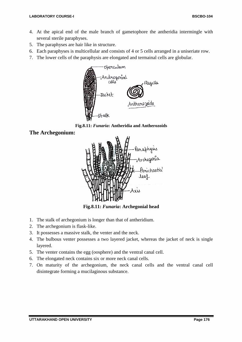
LABORATORY COURSE-I BSCBO-104
UTTARAKHAND OPEN UNIVERSITY Page 176
4. At the apical end of the male branch of gametophore the antheridia intermingle with
several sterile paraphyses.
5. The paraphyses are hair like in structure.
6. Each paraphyses is multicellular and consists of 4 or 5 cells arranged in a uniseriate row.
7. The lower cells of the paraphysis are elongated and termainal cells are globular.
Fig.8.11: Funaria: Antheridia and Antherozoids
The Archegonium:
Fig.8.11: Funaria: Archegonial head
1. The stalk of archegonium is longer than that of antheridium.
2. The archegonium is flask-like.
3. It possesses a massive stalk, the venter and the neck.
4. The bulbous venter possesses a two layered jacket, whereas the jacket of neck is single
layered.
5. The venter contains the egg (oosphere) and the ventral canal cell.
6. The elongated neck contains six or more neck canal cells.
7. On maturity of the archegonium, the neck canal cells and the ventral canal cell
disintegrate forming a mucilaginous substance.

LABORATORY COURSE-I BSCBO-104
UTTARAKHAND OPEN UNIVERSITY Page 177
Fig.8.12: Funaria: Archegoina
The Sporophyte (Sporogonium)
External Structure:
Fig.8.13: Funaria: Gametophyte bearing sporophyte
1. The sporophyte consists of a foot, set and capsule.
2. The foot is a small, conical structure embedded in the apical portion of the female branch
of gametophore.
3. The seta is an elongated slender and thread-like structure which bears the capsule at its
apical end.
4. The outline of a mature capsule is pear shaped in the beginning an irregular calyptra
is found as a cup-like structure at the apical end of the capsule, but very soon this is
blown off.
I.S. of Capsule: L.S. of capsule shows following regions namely apophysis, theca and upper region.

LABORATORY COURSE-I BSCBO-104
UTTARAKHAND OPEN UNIVERSITY Page 178
Fig.8.14: Funaria: L.S. of Capsule, Persistome and Spore dispersal
The Apophysis: 1. The elongated seta expands at its apical end to form the apophysis.
2. This is assimilatory region.
3. The stomata may be seem here and there in the epidermis.
4. The cells with chloroplasts possess intercellular spaces among them.
5. The central conducting strand consists of colourless cells.
6. This strand is conducting with the conducting strand of seta.
Theca: 1. The central fertile region of the capsule is known as the theca proper.
2. This region consists of many layers of cells.
3. The outermost layer is the epidermis, which lies in continuation of the epidermis, of the
apophysis of the capsule.
4. Just beneath the epidermis, there is one to two layered hypodermis; the cells of
hypodermal regions are colourless.

LABORATORY COURSE-I BSCBO-104
UTTARAKHAND OPEN UNIVERSITY Page 179
5. Next to the hypodermis there is one or two layered chlorenchyma tissue; the cells of this
layer contain choroplasts and act as assimilatory tissue.
6. Just beneath the chlorenchmatous layer there is a big air space, traversed by several
trabeculae; each trabeculae consists of 3 or 4 green cells; these filiform structures act as
connections between chlorenchymatous layer and the outer spore sac of the capsule.
7. The central region of the theca is occupied by a pith-like solid cylinder of parenchyma,
called the columella; the columella is narrow at its base and broad at its apical end; it is
connected to the conducting strand of the apophysis of the capusle at its base.
8. The columella remains surrounded by the spore sac.
9. The spore sac is barrel shaped; the spore sac has an inner single layered wall called the
inner spore sac; the outer wall of the spore sac is three of four layered and known as the
outer spore sac.
10. In between these inner and outer spore sacs there are spore mother cells; each spore
mother cell develops into a spore tetrad of four spores after meiosis.
The Upper Region: 1. The upper region of the capsule consists of operculum and peristome.
2. This part is differentiated from the theca proper by a well marked constriction.
3. The lid of the capsule is somewhat obliquely placed upon it.
4. The lid or operculum is 4 or 5 layers of cells in thickness.
5. The outermost layer is epidermis; the epidermis consists of the cells of greatly thickened
walls.
6. The next 3 or 4 layers of the cells consist of thin walled parenchymatous cells.
7. Just beneath the constriction there is a 2 or 3 layered rim of radially elongated cells.
Fig.8.15: Funaria: Persistomial teeth, Spores
1. The rim stretches inward from the epidermis and connects the peristome to the epidermis
of the capsule.
2. The peristome lies just beneath the operculum; it consists of two sets of incurved teeth
known as inner and outer peristome.
3. Each set of peristomial teeth consists of sixteen incurved triangular teeth; the teeth are
spirally twisted towards left.

LABORATORY COURSE-I BSCBO-104
UTTARAKHAND OPEN UNIVERSITY Page 180
4. The teeth of outer peristome are red and of inner peristome and colourless; the inner teeth
are shorter than the outer ones.
5. The annulus is found just above the rim; it consists of 5 or 6 layers of epidermal cells.
6. The mouth of the capsule remains closed by means of lid or operculum.
7. The spores are more less spherical; each spore possesses a covering wall of two layers;
the outer layer is somewhat coloured, smooth and known as exosporium; the inner layer
is colourless; smooth and known as endosporium; the spore contains within it, a nucleus,
oil globules and chloroplasts.
8. On germination the spore gives rise to a multi-cellular filament called the primary
protonema.
Fig.8.16: Funaria: Stage of spore germination
9. The primary protonema further differentiates into prostrate chloronemal branches and the
obliquely septate rhizoidal branches penetrating the substratum.
Fig.8.17: Funaria: Primary and Secondary Protonema
Identification and Systematic Position
Bryophyta:
(i) The true roots are always absent.
(ii) The lack typical vascular tissues.
(iii) They are homosporus.
Bryopsida (Musci): -
(i) Two stages of gametophyte-
(ii) (a) prostrate, thalloid, branched protonema, and (b) erect leafy gametophore.
(iii) Rhizoids multicellular, branched and obliquely septate.

LABORATORY COURSE-I BSCBO-104
UTTARAKHAND OPEN UNIVERSITY Page 181
(iv) Tissues of stem differentiated into outer cortex, and central conducting tissue.
(v) The sporogonium is differentiated into foot, seta and capsule;
(vi) capsule wall interrupted by stomata.
(vii) Elaters absent.
Eubrya: -
(i) Leaves are more than one cell in thickness; mid-rib present.
(ii) Protonema filamentous.
(iii) Well differentiated elongated stem present.
(iv) Partitioned air spaces present in between spore sac and columella.
(v) Capsule – a complex structure.
(vi) Capsule opens by operculum; spore dispersal regulated by peristome.
Funariales: -
(i) Terrestrial plants.
(ii) Leaves with midribs arranged in rosettes at the apex of gametophyte.
(iii) Capsules wide; provided with unbeaked operculum.
(iv) Peristome double.
Funariaceae: -
(i) Leaves one cell in thickness except at midrib region.
(ii) Small mosses.
(iii) Calyptra soon detached from operculum; calyptra with long beaks.
(iv) Capsules pyriform.
(v) Seta long.
Funaria: -
(i) Leaves spirally arranged.
(ii) The internal structure stem consists of three pars –
(a) epidermis (b) cortex and (c) central conducting stand.
(iii)Rosette of leaves present at the apex of the branch of gametophore.
8.3.3-Polytricum
Division: Bryophyta
Class: Anthocerotopsida
Order: Anthocerotales
Family: Anthocerotaceae
Genus: Polytricum
Habit and Occurrence:
1. There are about 92 species reported in this genus.
2. It is found in cool and shady places. They may be found growing in bogs and marshes, on
soil of firm or loose texture, on rocks and cliffs and as epiphytes on trunks of trees.
3. In India, they are commonly found in hills.
4. Species recorded from our country-Polytrichum densifolium, P.juniperunum and
P.xanthopitum.

LABORATORY COURSE-I BSCBO-104
UTTARAKHAND OPEN UNIVERSITY Page 182
Fig.8.18: Polytrichum: Gametophytic plant
Proposed Laboratory Work:
1. Study of external structure of gametophyte.
2. Study of internal structure (anatomy) of gametophyte.
3. Anatomy of rhizome.
4. Anatomy of leaf.
5. Study of reproductive structure.
6. Study of sporophytes.
7. Identification and systematic position.
External Structure:
Fig.8.19: Polytrichum: Female Gametophytic plant
Fig.8.20: Polytrichum: Male Gametophytic plant

LABORATORY COURSE-I BSCBO-104
UTTARAKHAND OPEN UNIVERSITY Page 183
1. The gametophyte remains differentiated into rhizoids, the underground rhizome, the erect
stem and leaves.
2. Numerous rhizoids are found on the rhizome of gametophyte; the rhizoids are long and
thick walled; they possess oblique septa in them.
3. The rhizoids form a soft or twisted mass which looks like a string; besides giving
mechanical support to the plant the rhizoids also help in vegetative reproduction.
4. The young leaves at the apex of a stem are arranged in a definite manner; the young
leaves being spirally arranged in three vertical rows.
5. Each leaf possesses a broad colourless membranous sheath at its base.
6. The leaf becomes narrow upwards.
7. The upper portion of the leaf is expanded which is green or brownish in colour.
8. Each leaf possesses a broad midrib.
9. The midrib remains covered on its upper surface by means of longitudinal cell plates
which contain chlorophyll and are known as lamellae.
Internal Structure (Anatomy):
T.S. of Rhizome:
Fig.8.21: Polytrichum: T.S. of Rhizome
1. The outline of rhizome is broadly triangular.
2. The cortex is divided by three radial strands; these strands penetrate into the central
cylinder, giving it a three lobed outline.
3. The cortex consists of 3 to 4 layers of cells of which outermost layer are strongly
suberized; the innermost layer of the cortex consists of very large cells with thin
suberized walls, known as the endodermis.
4. Two or three layered, primitive pericycle is found just beneath the endodermis; this
surrounds the central cylinder but does not form a continuous band.
5. The greater part of the central cylinder is made up of the thick-walled, somewhat
elongated cells, which form the ‗sterome‘ among which there are scattered groups of
empty, elongated cells the ‗hydroids‘.
6. The three radial strands start from groups of thick-walled cortical cells, while their inward
ends which remains embedded in the central cylinder, consists of thin walled ‗leptoids‘
corresponding to the ‗leptoids‘ in the aerial stem.
7. In the pericycle there is a ‗lepton mantle‘ similar to the phloem of higher plants.

LABORATORY COURSE-I BSCBO-104
UTTARAKHAND OPEN UNIVERSITY Page 184
8. Internal to the lepton mantle there is ‗hydrom sheath‘which is also known as ‗amlylom
layer‘; it consists of one of two layers of cells; the cell walls are suberized and the
consists starch.
9. Inside the amylom layer there is the ‗hydrom mantle‘; it consists of thin walled cells; in
the centre of the stem there is ‗hydrom‘ which is composed of thick walled cells; this
tissue is equivalent to xylem of higher plants.
V.T.S. of the leaf:
Fig.8.22: Polytrichum: T.S. of Leaf
1. The broad midrib is several cells thick in the centre and gradually merges into the so
called lamina at the margins.
2. On the lower surface there is well marked epidermis composed of large cells whose outer
walls are thickened.
3. Inside the epidermis there are one or two layers of very small cells with very thick walls.
4. The central tissue of the leaf is composed of thin-walled parenchymatous cells; among
which are scattered small groups of small thick-walled cells.
5. The upper (adaxial) surface is composed of a layer cells high, containing chloroplasts;
these plates are knows as lamellae.
Reproductive Structure: Usually the plants are diocecious and the antherida (male) and archegonia (female) are borne
in terminal clusters at the apex of separate gametophores.
Sexual reproduction: The Antheridia:
Fig.8.23: Polytrichum: Male Gametophore and Antheridia

LABORATORY COURSE-I BSCBO-104
UTTARAKHAND OPEN UNIVERSITY Page 185
1. The antheridia arise at the top of the leafy stems within an involucre of leaves; these
leaves are bright red or orange in colour.
2. The involucral leaves on the male head are arranged spirally from the vegetative apex
outwards.
3. The antheridia are produced in groups in the axils of these perichaetial leaves so that the
whole head becomes compound and contains a variable number of these closely set
antheridial groups.
4. Each mature antheridium of a stalk and a clavate body.
5. The club-shaped body remain surrounded by a single-layered jacket.
6. Inside the jacket there are androcytes within the antheridum.
7. The antheridia are intermingled with paraphyes; some paraphyses are simple and filament
like while the others are broadened at their tips.
The Archegonia:
Fig.8.24: Polytrichum: L.S of Female Gametophore and Archegonia
1. The archegonia arise at the top of the leafy stems within an involucre of leaves.
2. Usually three archegonia are found in an archegonial head.
3. Each stalked archegonium consists of a venter and a long neck.
4. The neck contanis 6-9 neck canal cells.
5. The venter contains a ventral canal cell and an egg.
The Sporophyte (Sporogonium):
1. The mature sporogonium consists of a foot, seta and a capusle.
2. The foot remains embedded in the tissue of an archegonium.
3. Just above the foot and continuous with it there is a long slender seta which sypports the
capsule at its terminal end; the seta may reach a length of several inches; the seta and
apophysis are considerably larger in size.
4. In T.S. the capsule is angular as well as square in outline.
5. The wall of capsule consists of several layers of cholorophyllous cells; the outermost
layer represents the epidermis.

LABORATORY COURSE-I BSCBO-104
UTTARAKHAND OPEN UNIVERSITY Page 186
Fig.8.25: Polytrichum: Sporophyte, Capsule and L.S. of capsule
1. In the mature capsule the sporogenous tissue forms a tube around columelle and is
sperarated from it by air spaces which are traversed by filaments of assimilatory cells.
2. A similar assimilatory tissue is also developed between the spore mass and the wall of the
capsule.
3. All cells of sporogeonus tissue give rise to spores.
4. At the apex of the capusle, the calyptra remains attached for a long time.
5. The calyptra develops a brown colour, and grows after its separation from the basal part
of the archegonium, forming a shaggy, hairy cup which covers the whole capsule and
because of this feature it is known as ‗hair moss.
6. At the top of the capsule there is an operculum which appears as a lid.
7. The operculum is conical with a long beak or rostrum, there is no well marked annulus,
though the thickened diaphragm (rim) is present.
8. At the base of operculum there is thin membranous tissue which constitutes the
epiphragm.
9. The peristome teeth are short and stout, formed of a group of sclerotic cells.
10. At maturity the peristome consists of 32 or 64 teeth.
11. The openings between the teeth form a ring of pores and the spores are dispersed through
these pores by the force of the wind shaking the sporogonium.

LABORATORY COURSE-I BSCBO-104
UTTARAKHAND OPEN UNIVERSITY Page 187
Spores and Protonema: 1. The spor are uninucleate and two walled; the outer wall is exine while the inner one is
intine.
2. On germination, the spore gives rise to septate branched filament, the protonema; some
filaments grows upwards and turn green while some grow downwards to the substratum
and remain colourless.
3. The buds develop near the base of upright filaments.
4. The older filaments of the protonema sometimes become twisted together into cable like
strands.
Identification and systematic Position:
Bryophyta:
(i) The true roots are always absent.
(ii) They lack typical vascular tissues.
(iii)They are homosporous.
Bropsida (musci):
(i) Two stages of gametophyte—(a) prostrate, thalloid branched protonema, and (b) erect
leafy gametophore.
(ii) Rhizoids multicellular, branched and obliquely septate.
(iii)Tissue of stem differentiated into (a) outer thin walled cortex, and (b) central thicl walled
conducting tissue.
(iv) The sporogonium is differentiated into food, seta and capsule; capsule wall interruped by
stomata.
(v) Elaters absent.
Eubray:
(i) Leaves more than one cell in thickness; midrib present.
(ii) Protonema filamentous.
(iii) Well differentiated elongated seta present.
(iv) Partitioned air spaces present in between spore sac and coumella.
(v) Capsule a complex structure.
(vi) Capsule opens by operculum; spore dispersal regulated by persitome.
Polytrichales:
(i) Gametophyte perennial and tall.
(ii) Leaves quite narrow and possess longtiudinal lamellae on upper surface of midrib.
(iii) Capsule terminal.
(iv) 32-64 pyramidal teeth in peristome.
(v) Epiphragm covers the mouth of capsule.
Polytrichaceae: Single family; characters as of order.
Polytrichum: (i) Capsule angular.
(ii) Rhizoids form string like structure, the rhizoids strands.

LABORATORY COURSE-I BSCBO-104
UTTARAKHAND OPEN UNIVERSITY Page 188
8.4 METHOD OF PERMANENT PREPARATIONS
To make permanent slides, the section of plant material is passed through alcoholic as well as
xylene series.
1. Select a thin sections as per requirement
2. Stain with aqueous safranin (for 2 to 4 min)
3. Wash thoroughly with water
4. Dehydrate with alcohol in following order (15% followed by 30%, 50%, 70%, and then
90%)
5. Stain with fast green (few sec to 1 min)
6. Destain with 90% alcohol
7. Dealchololize with xylol in following order (25% followed by 50%, 70%, and then 90%)
8. Mount in glycerine (10%) or canada balsam.
9. Cover with the help of coverglass carefully
8.5 METHOD OF TEMPORARY PREPARATIONS
Single staining is applied in those cases where there is no tissue differentiation e.g. Algae,
fungi and bryophytes. Single staining in bryophytes is done with safranin.
1. Select a thin sections as per requirement
2. Stain with aqueous safranin (for 2 to 4 min)
3. Wash thoroughly with water
4. Then stain with fast green
5. Again wash out extra stain
6. Study the slide prepared under microscope
8.6 SUMMARY
Genus Notothylus belongs to class Anthocerotopsida and is found to grow on shady moist
soil or rock in tropical and subtropical region. The gametophytic thallus is yellow green
forming an orbicular or sub-orbicular rosette and shows no internal differentiation. Each cell
of thallus shows a single oval choloroplast with a central pyrenoid. Indian species are
monoecious and protandrous. Both the antheridia and archegonia are embedded in the dorsal
surface of thallus. The sporophyte is enseathed by the thin, membranous involucres. The foot
is much smaller although the haustorial outgrowths are well developed. The intermediate
meristematic zone is also less developed and as a result the capsule does not grow much.
Hygroscopic movements of pseudoelaters release the mature spores at the top.
Genus: Funaria belongs to class bryopsida. They grow luxuriantly in human soil and
on soils burnts by fire. The small erect, green gametophytic plant arises from a prostrate alga-
like filament the protonema. The gametophytic plant body is differentiated into rhizoids,
leaves and stem. The rhizoids are branched, multicellular obliquely septate and thread like,

LABORATORY COURSE-I BSCBO-104
UTTARAKHAND OPEN UNIVERSITY Page 189
stem is upright erect. green and monopodial branched; leaves are sessile, small, ovate, bright
green and spirally arranged on the stem; Funaria hygrometrica is strictly monoecious and
autoecious species. The main shoot of the gametophyte bears the male the sex organs whole
the lateral branch bears the female sex organs. The sporophyte consists of a foot, set and
capsule. The foot is a small, conical structure embedded in the apical portion of the female
branch of gametophore. The seta is an elongated slender and thread-like structure which bears
the pear shaped capsule at its apical end. The upper region of the capsule consists of
operculum and peristome. Rculum; it consists of two sets of incurved teeth known as inner
and outer peristome; each set of teeth consists of sixteen incurved triangular teeth; the teeth
are spirally twisted towards left; the teeth of outer peristome are red and of inner peristome
and colourless; the inner teeth are shorter than the outer ones. he spores are more less
spherical; each spore possesses a covering wall of two layers; the outer layer is somewhat
coloured, smooth and known as exosporium ; the inner layer is colourless; smooth and known
as endosporium; the spore contains within it, a nucleus, oil globules and chloroplasts. On
germination the spore gives rise to a multi-cellular filament called the primary protonema.
Genus: Polytricum belongs to class bryopsida. They grow luxuriantly in bogs and
marshes, on soil of firm or loose texture, on rocks and cliffs and as epiphytes on trunks of
trees. In India, they are commonly found in hills. The gametophyte remains differentiated
into rhizoids, the underground rhizome, the erect stem and leaves. The plants are diocecious
and the antherida (male) and archegonia (female) are borne in terminal clusters at the apex of
separate gametophores. The mature sporogonium consists of a foot, seta and a capusle. The
foot remains embedded in the tissue of an archegonium. There is a long slender seta which
supports the capsule at its terminal end; the seta may reach a length of several inches; the seta
and apophysis are considerably larger in size. In the mature capsule the sporogenous tissue
forms a tube around columelle and is sperarated from it by air spaces which are traversed by
filaments of assimilatory cells. At the apex of the capusle, the calyptra remains attached for a
long time. All cells of sporogeonus tissue give rise to spores.
8.7 GLOSSARY
Peristome: Peristome is a fringe of teeth around the mouth of capsule in mosses.
Operculum: It is the cap of capsule in mosses.
Apospory: The production of gametophyte directly from the unspecialized cells of
sporophytes without the formation of spores is termed as apospory. e.g., anthoceros. The new
gametophytic plants are diploid because these are formed directly from the diplod cells of
sporophyte without undergoing meiosis.
Protonema: is a filamentous stage in the development of gametophyte of a moss from the
spore.
Secondary protonema: Protonema which is formed by means other by the germination of
spores is called secondary protonema.

LABORATORY COURSE-I BSCBO-104
UTTARAKHAND OPEN UNIVERSITY Page 190
Columella: Central column of sterile cells found inside the capsule of some bryophtes. It is
surrounded by sporogenous tissue which consists of spores and elaters.
8.9 SELF ASSESSMENT QUESTIONS
8.9.1: Short answer type questions:
1. The Sporophyte of Funaria lives as..............
2. Anatomically thallus of Notothylus shows differentiation- True/False?
3. Funaria is also known as..............
4. Which is the cheif assimilatory part of Funaria capsule?
5. At what region, the cells of Funaria capsule break to liberate operculum?
6. What is mode of nutrition in sporophyte of Funaria?
7. Name the early filamentous structure produced by spores cheifly in Moss?
8. Moss rhizoids are characterized by having?
9. In Polytrichum leaves are arranged in how many rows?
10. How many pyramidal teeth are present in peristome of Polytrichum?
11. Which organ in Polytrichum sporogonium controls the spore liberation?
12. Name one saprophytic member of bryophyte?
13. Who coined the term bryophyte?
14. Who is known as father of Indian Bryology?
15. Alternation of generation in bryophytes is known as........
16. Which type of cells conduct water in Mosses?
17. Moss sporophyte is.............dependent on sporophyte?
18. Columella in the moss sporophte is situated in.......
19. .............is mother cell of sporophytic generation.
20. Gemmae are responsible for..........mode of reproduction.
8.9.1 Answer to Self assessment Questions: 1. Partial parasite on gametophyte.
2. False.
3. Cord Moss.
4. Apophysis.
5. Annulus.
6. Autotrophic.
7. Protonema.
8. Oblique septa.
9. 3 rows.
10. 32.
11. Peristome.
12. Buxbaumia.
13. Braun (1864)
14. Prof Shiv Ram Kashyap.

LABORATORY COURSE-I BSCBO-104
UTTARAKHAND OPEN UNIVERSITY Page 191
15. Heteromorphic.
16. Hyaline cells.
17. Partially.
18. Theca
19. Zygote.
20. Vegetative.
8.9 REFERENCES Bendre, A. and Kumar, A. 1990-91. Practical Botany, Rastogi Publications, Meerut.
Gilbert, M.S. Cryptogamic Botany, Vol. I & II (2nd Ed.), Tata McGraw Hill, Publishing
Co. Ltd., New Delhi, 1985.
Pandey, S.N. and Trivedi, P.S. A Text Book of Botany 2000 Volume I, Vikas Pub. House
Pvt. Ltd., New Delhi.
8.10 SUGGESTED READINGS Puri, P. Bryophytes, Atmaram & Sons, Delhi, Lucknow, 1985.
Rashid, A. 1998. An Introduction to Bryophyta. Vikas Pub. House Pvt. Ltd. New Delhi.
Singh, S.K. 2008. Bryophyta, Campus Book, International, New Delhi.
Singh, V., Pande, P.C. and Jain, D.K. A Text Book of Botany, Rastogi & Co., Meerut,
2001.
Vashista, B.R. Botany for Degree Students (Algae, Fungi Bryophyta), S. Chand & Co.
Ltd., New Delhi, 2002.
Vasishta, P.C. 1996. Bryophyta. S. Chand & Co. Ltd., New Delhi.
8.11 TERMINAL QUESTIONS
8.11.1 Long Answer Type Questions: 1. Write the systematic position of Polytrichum?
2. Which generation is well developed in Moss plant?
3. Name the two growth stages of the gametophyte in life-cycle of Funaria?
4. What term is used for mosses bearing antheridia and archegonia on different branches of
same plant?
5. How can we distinguish the protonema of moss from filamentous green alga?

LABORATORY COURSE-I BSCBO-104
UTTARAKHAND OPEN UNIVERSITY Page 192
BLOCK – 3 PTERIDOPHYTES, GYMNOSPERMS
& PALAEOBOTANY

LABORATORY COURSE-I BSCBO-104
UTTARAKHAND OPEN UNIVERSITY Page 193
UNIT 9- STUDY OF THE EXTERNAL FEATURES AND
INTERNAL STRUCTURES OF RHIZOME, LEAVES,
ROOTS, SPORANGIA AND STROBILI OF
PTERIDOPHYES- RHYNIA, SELAGINELLA,
EQUISETUM
9.1-Objectives
9.2-Introduction
9.3-Study of external features, internal structures of Rhynia
9.4- Study of external features, internal structures of Selaginella
9.5- Study of external features, internal structures of Equisetum
9.6- Summary
9.7- Glossary
9.8- Self assessment question
9.9- References
9.10-Suggested Readings
9.11-Terminal Questions

LABORATORY COURSE-I BSCBO-104
UTTARAKHAND OPEN UNIVERSITY Page 194
9.1 OBJECTIVES
After reading this unit student will be able:
To understand the general characters of various groups of Pteridophytes.
To study the general outlines of classification of different groups of Pteridophyta
To study the morphology and anatomy of vegetative and reproductive parts of Rhynia,
Selaginella and Equisetum.
To understand about the economic and other significance of different Pteridophytes.
9.2 INTRODUCTION
The Pteridophyta include a group of land plants with well - developed vascular tissue- xylem
and phloem. Therefore, this group is also known as Vascular Cryptogams. Carolus Linnaeus
(1754) classified these plants underclass Cryptogamia. The word also derived from Greek
words Kruptons means hidden and gamos means wedded, plants with concealed flowers.
Members of this group are most primitive vascular plants and reproduced by present
day living generas such as, Selaginella, Equisetum etc. and fossil vascular plants – such as
Rhynia, Homeophyton, Aseroxylon etc. Plant body is sporophytic and differentiated into stem,
leaves and roots. In some plants an intermediate stage between root and stem i.e. rhizophore
is also present (Selaginella).
Fig.9.1 Selaginella: Habit sketch Fig.9.2 Rhynia: Habit sketch
Hence these plants are an assemblage of flowerless, seedless, spore bearing primitive land
terrestrial plants with well- developed vascular system.Pteridophyta are reported by more
than about 10,000 species.
The members of Pteridophytes vary greatly in form and size, some
plants are either annuals with small size or perennials with longer
size, the plants are annuals e.g. Azolla or perennials e.g. Osmunda,
Cyathea and Alsophila are Tree Ferns Alsophila is the tallest fern.
1. Unlike Bryophytes, the Pteridophytes are found in great
majority of habitats, ranging from auatic to xerophytic.
2. Generally they are land inhabiting or terrestrial in nature but
plants like Azolla, Marsilea and Salvinia grow in aquatic habitat.
Fig.9.3 Equisetum

LABORATORY COURSE-I BSCBO-104
UTTARAKHAND OPEN UNIVERSITY Page 195
3. They prefer cool, moist and shady paces. While some are xerophytic i.e. Selaginella
rupestris, Selaginella lepidophyllum. While Lycopodium phlegmaria is an epiphytic.
4. Generally Pteridophytes are herbaceous but few are woody e.g. Cythea spinulosa.Mostly
the plants have dorsiventral or radial symmetry with dichotomous or monopodial branching.
5. The leaves may be small/microphyllous e.g. Lycopodiun, Equisetum or very large/
Macrophyllous e.g. Pteris, Pteridium and Dryopteris and other ferns.
6. Stomata are present on both surfaces of leaf. Trichomes may be present on stem and
leaves. The anatomy of these plants is to some extent dependent upon the type of leaf they
bear.
7. A similar but generally simpler structure is found in roots. Leaves may be simple, small
and sessile as in microphyllous types i.e. Selaginella, Lycopodium very large with petiole and
megaphyllous type i.e. members of Filicinae.
Fig.9.4: Marsilea: Habit sketch Fig.9.5 Pteris: Habit sketch
8. Due to lack of cambium generally secondary growth is also absent in Pteridophytes.
However, stem of some living Pteridophytes show secondary growth e.g. Isoetes,
Botrychium etc.
9. Vessels are generally absent but in some plants primitive type of vessels has been reported
e.g. Selaginella, Equisetum.
10. After maturation the pothallus develops two sex organs, antheridia (male) and archegonia
(female) on the prothallus.
Classification of Pteridophytes:
As we know from the above studies that Pteridophytes are the most primitive group of
terrestrial plants known as Vascular Cryptogames being possessing vascular cylinder. Thus to
classify these plants is somewhat complicated. However, various workers suggested various
methods of classification but none is satisfactory and final. Some of them may be discussed
here in brief.

LABORATORY COURSE-I BSCBO-104
UTTARAKHAND OPEN UNIVERSITY Page 196
Fig.9.6 Azolla Fig.9.7 Lycopodium
According to records the first attempt to classify the Pteridophytes was made by Prantle in
1879, He classifies them into three classes namely- Filicinae, Equisetinatae and Lycopodinae.
Again Engler and Prantle (1890-1900) elaborate the previous classification. Jeffery
(1910), proposed new classification on the basis of anatomical details. In 1935, Sinnott
classify pteridophytes on the basis of origin and development of vascular system and
introduced new term ―Tracheophyta‖ and allied to Thallophyta and Bryophyta. There are
some other workers too, worked on classification, e.g. Eames (1936), Zimmermann (1930),
Tippo (1942), Reimers (1956) etc. However, Doyle (1972), dividedPteridophyta into four
classes- Psilopsida, Lycopsida, Sphenopsida and Pteropsida.
Economic Importance of Pteridophytes:
Humans have been dependent upon plants as an important source of food, shelter and
medicines. The group Pteridophyta has very less economic importance on records yet they
are playing great role in the soil formation during different series of succession. The valuable
plant species Violascrpens and Aiantum has proved their economic importance since long
time. Adiantum raddiantum is an important horticultural plant. Adiantum and Pteris plants
have a great medicinal value as it has been reported in the literature. Since ancient times, A,
cappilus- veneris L. is used as remedies in traditional therapy in Iran. Indo- Nepal border
areas also used them for medicinal uses. After forest fire, ferns are the first plants to appear as
their underground rhizome grows. Azolla is known as water fern, and used as biofertilizer.
9.3 STUDY OF EXTERNAL FEATURES AND INTERNAL
STRUCTURES OF RHYNIA
Systematic Position-
Division- Pteridophyta
Sub division- Psilophytopsida
Order- Psilophytales
Family- Rhyniaceae
Genus- Rhynia
Fig. 9.8 Rhynia major

LABORATORY COURSE-I BSCBO-104
UTTARAKHAND OPEN UNIVERSITY Page 197
Introduction
Rhynia is a fossil genus. It was discovered by Kidson and Lang in 1971 in district
Aberdenshire of Scotland. It was present in Middle Devonian period (about 380 million years
ago). Rhynia possess two species Rhynia Gwynne-vaughani and Rhynia major. Rhynia is not
found in present age. These fossils were extinct and found in perrified fossils. These fossils
were embedded and impregnated with silica and after aligning different parts or sections
prepared from rocks, construction of both the species of Rhynia were done carefully.
On the basis of evidences it was suggested or hypothesized that the plant were growing in
swampy marshes near volcanos. It prefers sulphrus vapours and soil, saturated with acid
water from hot springs. The specimens are so preserved that they give detailed information
about the form and structure of this primitive vascular plant. It was the most primitive
Pteridophyta. Main plant body is sporophyte. The sporophyte is differentiated into a rhizoid
with subterranean rhizome and an aerial position known as aerial shoot. It also bears
adventitious roots. The aerial shoot or portion also bears branching system.
The branches also bear sporangia on its tips. Mercker, (1959)
suggested that the rhizomatous parts of plant represents
gametophyte and upper aerial part is sporophyte.
Fig.9.9 Rhynia gwynne-vaughani
Object- Study of External Features of Rhynia
1. As we studied that the two species of Rhynia were identified in fossil form e.g. Rhynia
major and R. gwynne-vaughani.
2. Both the species were herbaceous plants. R. major was larger in size than R. gwynne-
vaghani.
3. R. major was attained a height of 50 cm with a diameter between 1.5 to 6.0 mm., while R.
Gwynne- vaghani had a height of 20 cm and a diameter of 1 to 3 mm.
Fig. 9.10 Rhynia sporophyte

LABORATORY COURSE-I BSCBO-104
UTTARAKHAND OPEN UNIVERSITY Page 198
4. The plant body of both the reported species was dichotomously branching and had a
rhizome.
5. The aerial stem was erect; from rhizome developed many dichotomously branched erect
axial shoots towards the upper side while many rhizoids towards the lower side.
6. The aerial branch was leafless shoots, cylindrical, forked and tapering at their apices.
7. The aerial shoots of R. major were smooth and devoid of leaf or any other outgrowth,
while many adventitious branches were reported on the aerial shoots of R.gwynne- vaughani
8. The aerial shoots were either ending into simple vegetative branches or having terminal
sporangia.
9. Stomata were present all over the surface of aerial branch.
Object-Study of Internal Structure of Rhynia:
1. Based on studied of internal structure of rhizome and aerial shoots, the plant were
internally was not show any major difference.
2. Transverse section of stem show a well- developed epidermis interrupted by stomata and
covered by thick cuticle.
3. Internally the rhizome and aerial shoot was quite similar and differentiated into epidermis,
cortex and stele.
4. The epidermis is made up of one cell thick having conspicuous cuticle on outer side.
5. The epidermal cells were broadly fusiform from the surface view.
6. The aerial shoot shown stomata on the epidermis along with pairs of guard cells.
7. The rhizoids did not possess any stomata but show unicellular rhizoids on its surface.
8. Cortex was differentiated into two zones, the outer cortex or hypodermis situated beneath
Fig.9.11 (A) Rhynia: T.S. Rhizome, (B) L. S. Sporoangium
epidermis and made up of 1 -4 layers of parenchymatous cells without any intercellular
spaces except below the stomata.
9. The inner cortex was broader zone and made up of small rounded parenchymatous cells
with large intercellular spaces between them, which communicated to the stomata through the
gaps in hypodermis.
10. The inner endodermis and pericycle layers were altogether absent.

LABORATORY COURSE-I BSCBO-104
UTTARAKHAND OPEN UNIVERSITY Page 199
11. The inner cortex formed to photosynthetic region of plant and was about 10 times more in
thickness than stele.
Fig.9.12 Rhynia: L.S. stem with stomata
12. Most part of cortex was known as stele which was of a simple protostele type
(haplostele), while the central column of xylem was surrounded by phloem or five cells in
thickness.
13. The xylem consists of tracheids only and had annular and spiral thickening at their walls.
14. The cells surrounding the central xylem were thin walled and represented phloem.
15. The phloem cells were elongated with oblique end walls. Some minute sieve like areas
were also present on lateral walls of the cells.
16. The growing points ware composed of numerous dividing cells but the apical cells was
not reported.
Object-Studies of external features and internal structure of Sporangia-
1. At the tip of some aerial branches sporangia was present and each apex bore a single
sporangia.
2. The sporangia were oval or slightly cylindrical in shape. They were 4.0.mm long and
1.4mm broad.
3. They possessed the greater diameter than the tips of the branches on which they were
found.
4. The jacket layer or wall of sporangium was many layered consisted of three layers of
cells.
5. The outermost cells of jacket layer of sporangium were thick walled and cutinized at their
outer face and an epidermis beneath this layer.
6. The middle layer of sporangium composed of thin walled cells and innermost layer
consisted of thin walled rounded cells which functioned as tapetum.
7. The sporangium had no central columella within it.
8. The sporangial cavity contained many spores of same size represented the homosporous
condition and measured 40u in diameter.

LABORATORY COURSE-I BSCBO-104
UTTARAKHAND OPEN UNIVERSITY Page 200
Fig.9.13 Rhynia: A- rhizome, B- aerialshoot C&D sporangiaum
9. In some cases, spore tetrad or spores were united in tetrad, Rhynia stomata exhibited
the reduction division phase.
10. The sporangia were indehiscent and after decay and disintegration of sporangial wall,
spores were released means having no spatial dehiscence process for spore.
11. The spores were spherical and cutinized.
Identification Points
Division- Pteridophyta
i) Plant body is differentiated into stem, roots and leaves, ii) Sporophyte possesswell
developed vascular strands- xylem, phloem and mechanical elements, iii) True roots
generally present, vi) Fossil plant body
Sub- Division- Psilophytopsidai) The main plant or sporophyte differentiated into two parts
e.g. abranched aerial portion and a rhizoid bearing subterranean rhizome, ii) True roots
absent, iii) Plant homosporous.
Order- Psilotales
i) Sporangia borne at the tips of the erect branches either singly or in pairs, ii) Sporophyte
dichotomously branched, iii) Stele simple and protostelic type.
Family- Rhyniaceae
i) Aerial branches were leafless, ii) Rhizome and aerial shoots were dichotomously branched,
iii) Sporangia were present at terminal position, iv) Rhizoids bore unicellular rhizoids.
Genus- Rhynia
i) Plant was homosporous, ii) Sporangia without columella, iii) Rhizoids were unicellular,
iv) Plant had a multilayered thick jacket.

LABORATORY COURSE-I BSCBO-104
UTTARAKHAND OPEN UNIVERSITY Page 201
UNIT 9.4 STUDY OF EXTERNAL FEATURES AND INTERNAL
STRUCTURE OF SELAGINELLA (SPIKE MOSS)
Systematic Position
Divison: Pteridophyta
Sub Divison: Lycopsida
Order: Selaginellale
Family: Selaginellaleaceae
Genus: Selaginella
Fig.9.14 Selaginella: Habit sketch
Introduction- This genus Selaginella is generally known as small spike mosses comprises
about 700 species. In India about 58 species have been recorded from different parts of the
country. Most of the species are reported from tropical and also grow in rain forest. However,
some species are xerophytic and grows upon barren rocks and dry soils, yet some species
grow on moist and shady places on the hilly areas. Some species Eastern and Western
Himalayas of Selaginella, are commonly found in. S. lepidophylla and S. rupestris.
The xerophytic species species grow in arid and most exposed situations on granite
rocks and sandy soil. While S.oregana is an epiphytic species and grow on moss covered tree
trunks. Some species of Selaginella are cultivated as agricultural plant due to their feathery
fronds and delicacy. It is worldwide in distribution and occurs commonly in tropical and
temperate regions. Some common species of Selaginella are S. rependa, S. cilliaris, S.
nepalensis, S. wigtritii, S. haltei, S. biformis, S. mousporea etc.Hieronymus in 1900 divided
this genus Selaginella into two subgenera-i) Homoeophyllum and ii) Hetrophytlum. Based on
the general structure of the stem the plants are divided into two groups or sub general
Homophyllic and Hetrophyllic. The Sub genera Hetrophyllum includes majority of species.
External features external features of the plant Selaginella
1. Main plant body is sporophytic and sprophyte is evergreen and perennial.
2. Many species are prostrate, creeping on the ground e.g. S. kraussiana, others are sub-erect
e.g. S. trachuphylla, some others are erect e.g. S. erythrosa. A few are climbers also and
climb with the help of rhizophores e.g. S. alligans.
Fig.9.15 Selaginella: External features

LABORATORY COURSE-I BSCBO-104
UTTARAKHAND OPEN UNIVERSITY Page 202
3. In each case of the stem is covered with few rows of leaves ranges from few centimeters to
several feet in different sizes.
4. The plant body is divided into stem, leaves, Rhizophores and roots.
5. The primary root is short lived while other roots are adventitious.
6. The sub-genus namely Heterophyllum and Homoeophyllum have been recognized in the
genus Selaginella based on the characters of stem and leaves.
7. Sub-genus Heterophyllum possess prostrate stem, short erect branches and the dimorphic,
different sizes of leaves (small and large).
8. The above group have lateral branching also.
9. Sub genus Homoeophyllum is characterized by erect stem showing dichotomous branching
and all leaves are of one type.
10. Beside this, in Homoeophyllum species, the leaves are although dimorphic as
reportedearlier and are borne in pairs on dorsiventral stem.
The two leaves are markedly different insize e.g. one is larger and other is smaller.
11. In this, sub- genera, (Homoeophyllum) the stem is dorsiventral, and leaves are arranged in
pairs each pair consists of two different sizes leaves.
Fig. 9.16 Selaginella: A part of stem showing arrangement of leaves
12. The large leaf of each pair is inserted on the ventral side of axis.
13. The leaves are arranged on the stem in four longitudinal rows, in which the two rows of
smaller while remaining two rows of larger leaves on theventral surface.
14. The pairs of leaves are arranged in such a way that smaller leaf alternatesthe larger leaf.
15. The successive pairs of leaves are so arranged that large leaf alternates with the large leaf
and small leaf with the small leaf
16. S. kraussiana belongs to this sub genus and possess all the typical characters of the sub
genus Heterophyllum.
17. The leaves are quite small in size, somewhat triangular or lanceolate.
18. Each leaf is sessile, generally ovate with acute apex with a distinct midrib.
19. It is also reported that with the habitat change the morphology of leaf also changed as the
leaves may be thin in moist loving species and thick in xerophytic species.

LABORATORY COURSE-I BSCBO-104
UTTARAKHAND OPEN UNIVERSITY Page 203
20. At the base of upper or adaxial of each young leaf is present a thin membranous finger or
tongue like outgrowth called ligule and develop early during ontogeny of leaf.
21. The mature ligule is thin membranous tongue like or fan shaped.
22. It is differentiated in to basal sheath, glossopodium and the body of the ligule. The basal
sheath is cup shaped at the base of the ligule.
23. Just adjacent to the basal sheath there is a hemispherical mass of thin walled cellsknown
as glossopodium.
24. The cells of the sheath are tubular in shape and are dead, while those of the glossopodium
are vertically elongated.
25. The body of the ligule is made up of parenchymatous cells with dense protoplasm.
26. At the place of branching in stem a cylindrical leafless organ is seen growing towards the
lower side or down wards, known as rhizophore.
27. The rhizophore is an elongate, colourless cylindrical structure, arise from the axis
ramification and growing downwardly in the prostrate axis.
28. Rhizophore becomes branching at its tip and forms many adventitious roots.
29. Some vertical branches from the stem are reproductive in nature and bear stobili.
Internal structure
A.Stem
Work-procedure- Cut a T. S. of the stem, leaf, root and rhizophore by inserting the material
in pith, stain them separately in safranin- fast green combination, mount in glycerine and
observe under microscope.
Fig.9.17 Selaginella: T.S. Stem
1. The outline of the section appear slightly wavy and circular.
2. Anatomically the stem is clearly divided into three zones- an outer layer of epidermis,
middle layer of cortex and centrally located stele.

LABORATORY COURSE-I BSCBO-104
UTTARAKHAND OPEN UNIVERSITY Page 204
3. The epidermis is the outermost superficial layer of stem consisting parenchymatous
cutinized cells with no stomata.
4. Just beneath the epidermis there is cortical region is found, in some species the cortex is
composed of thin walled parenchymatous cells lacking intercellular spaces, e.g. S.kraussiana.
5. Majority of species possess rigid stems have compact sclerenchyma in the
hypodermalregion beneath the epidermis.
6. In most of the species the cortex is differentiated into a few outer layers of thick walled
sclerenchymatous hypoderms and many inner layers of thin walled parenchymatous cells.
7. In the most of the part of the cortex is found to sclerenchymatous in xerophytic
speciessuch as S.lepidophylla and S.rupestris.
8. Hypodermis occurs close to epidermis, which develops fromthe cells of outer cortex while
becomes thick walled. In xerophytic species S.lepidopylla hypodermis hypodermis more
thickened.
9. Centrally located stele or steles is connected with the cortex with the help of many long,
radially elongated cells called trabeculae. These are stretched endodermal cells having
intercellular spaces among them.
10. The trabeculae are the endodermal structure as shown by the presence of many band like
casparian strips in them.
11. In some species trabeculae are absent e. g. S.adunca and S.rupestris etc.
12. Behind the endodermis, there is pericycle is a single layer surrounding the xylem and
phloem. Pericycle possess single parenchymatos cells.
13. The xerophytic species such as S.lepidophyllum
and S.rupestris lack trabeculae.
14. The organization of the stele ranges from a simple
protostele to a complex polycyclic siphonostele.
15. Single stele, when present is generally diarch and
exarch.
16. The number of stele is variable fom 1 to 16 as
under-
S. spinulosa –monostelic (One stele)
S. kraussiana- distelic (two stele)
S. laevigata- polystelic (upto sixteen stele).
17. In S. kraussiana, the commonest species there are
two steles, each with a single exarch mass of
protoxylem.
18. Each stele consists of pericycle, xylem and
phloem. Fig.9.18 Selaginella: Types of steles
19. Each stele possesses a single mass of exarch protoxylem in it, composed of annular and
spiral tracheids.
20. The metaxylem in each bundle is placed centrally in the stele and lies centripetal to the
protoxylem. The metaxylem is composed of scalariform tracheids.
21. Phloem consists of sieve cells and phloem parenchyma and lacking companion cells.
22. In the stele, xylem is located in the centre and surrounded by phloem.

LABORATORY COURSE-I BSCBO-104
UTTARAKHAND OPEN UNIVERSITY Page 205
23. In Selaginella stem xylem is exarch with one or two protoxylem group.
ROOT
In case of Selaginella, two types of roots may be observed.The primary roots and later arising
adventitious roots. Primary root is short lived and disappearsor die after some time.
Work procedure- Cut a T.S. of root, stain it in double stain e.g. safranin- fast green
combination. Mount in glycerine and study under microscope.
1. Visually the section is circular in shape and outline.
2. The tissue is differentiated into three layers, epidermis, cortex and central stele.
3. The outermost superficial layer is known as epidermis.
This layer is single layered, thin walled, constituted by parenchymatous cells and called as
piliferous layer because it bears many unicellular root hairs on its surface.
4. From some epidermal cells arise root hairs.
5. The cells of are tangentially elongated.
6. Below the epidermis are few layers of parenchymatouscortex devoid of intercellular
spaces, which is situated inner to the piliferous layer.
7. In some species e.g. S.willdenovii there is three to five layered hypodermis constituted of
sclerotic cells, and then it contains thin walled parenchymatous cells.
8. It is also reported that cortex may be made of thick walled sclerenchymatous hypodermal
cells.
9. The inner most layer of cortex is endodermis. Generally endodermis is one layered and
also indistinct but in certain species e.g. S.willdenovii and S. rubella, endodermis is well
defined.
10. Just beneath the endodermis, there is three layered parenchymatous pericycle. The cells
of pericycle are small in size comparatively to endodermis.
11. The central part of root is known as stele. In Selaginella, stele is monarch and exarch. The
structure of xylem and phloem resemble to that of stem.
12. The stele is simple protostelic type.
13. Xylem is composed of spiral and annular tracheids.
14. The metaxylem is situated in the centre and is constituted of scalariform tracheids.
15. Phloem surrounds the centrally located xylem and is composed of sieve tubes.
4. Object- Study of anatomy T.S. of rhizophore
Fig.9.19 Selagiella: T.S. rhizophore

LABORATORY COURSE-I BSCBO-104
UTTARAKHAND OPEN UNIVERSITY Page 206
Work Procedure-
Cut a T.S. of rhizophore, stain in safranin- fast green combination, mount in glycerin and
study anatomically. It is reported that the structure of rhizophore is similar to that the
structure of root. There are some differences in its structure due to environment.
Rhizophores arises from the axis ramification. These are elongate, colourless, leafless and
cylindrical growing downwardly. The swollen tips gives rise a tuft of adventitious roots.
1. The outline of the section is almost circular.
2. The outermost layer is thick walled epidermis.
3. The section shows epidermis, hypodermis, cortex, endodermis and stele.
4. The epidermis is cuticularized.
5. Root hairs are absent.
6. Just beneath the epidermis, hypodermis present, which is 2 to 3 layered thick.
7. Next to hypodermis is cortex, constituted thin walled parenchymatous cells of 3-4 layered
thick. It occupies most of the part of the section.
8. Between the cortex and stele, endodermis present. It is followed by a single layered
pareechymatous pericycle layer.
9. The stele of rhizophore is monarch and exarch like in root. It is also protostelictype. In
somespecies e.g. S. atroviridis the metaxylem is lunar shaped and many protoxylem groups
are also situated on the concave adaxial side.
Leaf:
Work procedure- Cut a T.S. of Leaf, stain in safranin- fast green combination, mount in
glycerin and study under microscope.
Fig. 9.20 Selaginella: Leaf structure
1. The section of leaf shows a slightly bulged midrib in the centre and in wings.
2. It is bounded by upper and lower epidermis in both sides of leaf.The epidermis is single
layered and composed ofthin walled cells.
3. The epidermal layer of both e.g. upper and lower surfaces contains discoid chloroplasts in
them. Cells of epidermal layer contain chloroplasts.
4. The stomata are confined to the abaxial epidermis and sometimes also reported on the
adaxial epidermis, or on both the surfaces.

LABORATORY COURSE-I BSCBO-104
UTTARAKHAND OPEN UNIVERSITY Page 207
Fig.9.21 : Selaginella Leaf; A. Branch of S.kraussiana showing leaf arrangement, B. A leaf with
ligule, C. Vertical section of ligule
5. The mesophyll cells is usually not differentiated into palisade and spongy parenchyma
andshows intercellular spaces.
6. Mesophyll cell contain chloroplasts and all the cells are alike each of which has several
pyrenoids.
Fig. 9.22 Selaginella: T.S. Leaf (A part cellular)
7. In the centre is present a single vascular bundle surrounded by a bundle sheath.
8. The vascular bundle is concentric.
9. The xylem remains surrounded by phloem.
10. Phloem is composed of parenchymatous cells with one to two sieve tubes.
Strobilus
Work Procedure- To study he external and internal features of the strobilus, cut a L.S. of the
structure, stain in safranin fast green and study.
The spore producing orgams commonly known as sporangiaor strobilus are foundon adaxial
surface of sporophyll. Each sporangia bears short stalk and may be foliar (leaf like) or cauline
in nature. Selaginella has two types of sporangia which differ in shape, size and colour.
Megasporangium Selaginella (representing female spore) usually contains four large
megaspores and are found on megasporophyll. However, the microsporangia contain large
number of microspore (representing male spores) and arepresent in microsporophylls. In most
of the species strobili bears both, microsporophyll and megasporophyll. But sometimes, some

LABORATORY COURSE-I BSCBO-104
UTTARAKHAND OPEN UNIVERSITY Page 208
species, the strobili may consists only one type of sporophyll either megasporophyll or
microsporophylls. It differs in different species.
Fig. 9.23 Selaginella: L.S. Strobilus
1. The spore producing organs are present on the apical part of
main axis, aggregated in strobili and are known as
sporophyll.
2. The strobilus (singular) is a compact structure of sporophyll.
The strobili are present in the axil of the leaf like structure,
the sporophylls.
3. In some cases e.g. S. patula, the axis may grow beyond the
strobilus, terminating into a vegetative shoot.
4. Each sporophyll bears a ligule at its base like in each
ordinary leaf and stalked sporangium on the adaxial surface.
5. Sporangium is present in between the main axis and the
ligule of the sporophyll.
6. Selaginella bears two distinct types sporophylls. i. e.
micrpsporangium (larger) and microsporangia (smaller) attached either strictly to the
axils or to the axis just above.
7. Selaginella is the best example of hetrospory as it bears two different types sporangia in
their respective sporangia, the megasporangia (female part) and microsporangia (male
part).

LABORATORY COURSE-I BSCBO-104
UTTARAKHAND OPEN UNIVERSITY Page 209
Fig. 9.24, L.S. of Micro and Megasporangium
8. In some cases the plant bears both megasporamgium and microsporangium in the
same strobilus or in different strobilus e. g. S. gracilis, there is only one sporangia, either
mega or microsporangia.
Fig.9.25 Selaginella, L.S.Strobilus showing different positions in which megasporangia and
microsporangia occur
9. These sporophylls are arranged loosely and spirally usually in four rows on the main
stem.
10. In most of the species both types of sporophylls micro and megasporophylls are brone
either in the same spike e.g. S. kraussiana, or the strobilis bear one type of sporophtll
e.g. S. gracilis, S. atroviridis. Megasporophylls are found in the lower portion of the
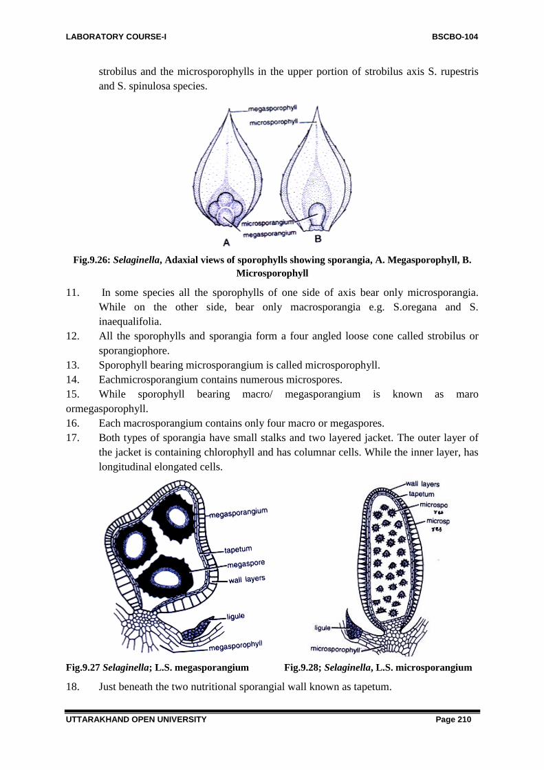
LABORATORY COURSE-I BSCBO-104
UTTARAKHAND OPEN UNIVERSITY Page 210
strobilus and the microsporophylls in the upper portion of strobilus axis S. rupestris
and S. spinulosa species.
Fig.9.26: Selaginella, Adaxial views of sporophylls showing sporangia, A. Megasporophyll, B.
Microsporophyll
11. In some species all the sporophylls of one side of axis bear only microsporangia.
While on the other side, bear only macrosporangia e.g. S.oregana and S.
inaequalifolia.
12. All the sporophylls and sporangia form a four angled loose cone called strobilus or
sporangiophore.
13. Sporophyll bearing microsporangium is called microsporophyll.
14. Eachmicrosporangium contains numerous microspores.
15. While sporophyll bearing macro/ megasporangium is known as maro
ormegasporophyll.
16. Each macrosporangium contains only four macro or megaspores.
17. Both types of sporangia have small stalks and two layered jacket. The outer layer of
the jacket is containing chlorophyll and has columnar cells. While the inner layer, has
longitudinal elongated cells.
Fig.9.27 Selaginella; L.S. megasporangium Fig.9.28; Selaginella, L.S. microsporangium
18. Just beneath the two nutritional sporangial wall known as tapetum.

LABORATORY COURSE-I BSCBO-104
UTTARAKHAND OPEN UNIVERSITY Page 211
19. Both types of sporangia when ripe, differ in their size, structure, colour and form.
20. Each microsporangium is smooth, ovoid and generally has brown or red coloured
body and contains many microspores.
21. Contrary to this each macrosporangium is four lobed and greenish-white, orange or
pale yellow in coloured.
22. The macrospores are large in size and possess a triadiate ridge at its apex and two
layered wall, the outer exineis sculptured and the inner, intine is thick and uniform.
23. The microspores are smaller in size and with uniform outline, dark red in colour and
have many small spores.
Fig.9.29, Selaginella: Prothallus and microspore
24. The microspores lack chlorophyll butmay contains oil droplets. The diameter of
microspores ranges from 0.015 to 0.0.mm.
25. However, the megaspores are quite big in size and ranges from 1.5 to 5.0 mm in
diameter.
26. Both types of spores develop into male gametophytes. While a microspore
developsinto male gametophyte.
Points of identification
Divison Pteridophyte-
i). Plant body is differentiated into stem, root and leaves, ii) Presence of true vascular
system, iii). Presence of independent gametophyte and sporophyte. iv). Gametophyte is small.
Sub Divison- Lycophyte-
i). Sporophyte differentiated into stem root and leaves, ii). Leaves microphyllus or small in
size with a single vein, iii). Sporangia develop in the axils of sporophyll,iv). Sporophylls
borne in strobili
Order- Selaginellales
i). Plant body is herbaceous, ii). Rhizophores present on the stem and roots arises at the tip of
rhizophore ,iii). Plant is hetrospores, iv) Leaves bear ligule.
Family- Selaginellaceae
i) Stem herbaceous, horizontal, creeping or erect, ii). Gametophyte extremely reduced,
iii). Absence of secondary growth in the stem, iv) Sporophylls are originated in strobili.
vi). Two types of spores e.g. macro and microspores are present.

LABORATORY COURSE-I BSCBO-104
UTTARAKHAND OPEN UNIVERSITY Page 212
Genus- Selaginella
i) Plant is living, ii). Roots arise from rhizophore, iii). Generally more than one stele in stem
either protostele or sometimes siphonostele, iv). Each macrosporangium contains four
macrospores.vi) Trabeculae present.
9.5 STUDY OF EXTERNAL FEATURES AND INTERNAL
STRUCTURES OF EQUISETUM
Systematic Position-
Divison: Pteridophyta
Sub Divison: Sphenopsida
Order: Equisetales
Family: Equisetaceae
Genus: Equisetum
Fig. 9.30 Equisetum: Habit sketch
Introduction
Equisetum is commonly known as ―Horse Tail‖ derived from Latin word –equs means horse
and seato means bristle. As its appearance resemble with the horse tail, thus named as such.
Equisetum is the only living genus of family Equisetaceae. The genus is represented by 25
species distributed in all parts of world except Australia and New Zealand. The common
Indian species are E. debile and E. ramosissimum. However, most of the species are reported
from North Temperate Zone but some species are also growing in tropical Zone too. Mostly
reported from West Indies, South America, Chile, Rene in arctic and alpine zones. Its various
species grows in rarity of habitats. Certain species grows in ponds and marshes, others prefer
damp shady places, some others growsin exposed and relatively dry habitats.
Equisetum debile is commonly found in Indian habitat, and found along the banks of the
rivers, canals, and pools in the Indian plains. E. palustre grows in marshes or along streams
banks known as meadow horse tail. Equisetum shows maximum power of habitat
acclimatization, can grow in hydrophilic, mesophilic and even in xerophytic habitats. It is
also reported from hill ares of Uttarakhand Himalaya, India. E. debile, E. patustris. E. arrens,
E. pretense. E. ramosissimum are some common Indian species are perennials, long lived,
herbaceous plants with creeping habit, possess branched rhizomes from which erect aerial
branches arises. Due to presence of nodes and
Study of external structure of sporophyte
The plant body of Equisetum is sporophyte, (diploid, 2n), which is differentiated into stem,
leaves, and root. All the species of Equisetum are herbaceous, perennials possessing a widely
creeping branched rhizome.
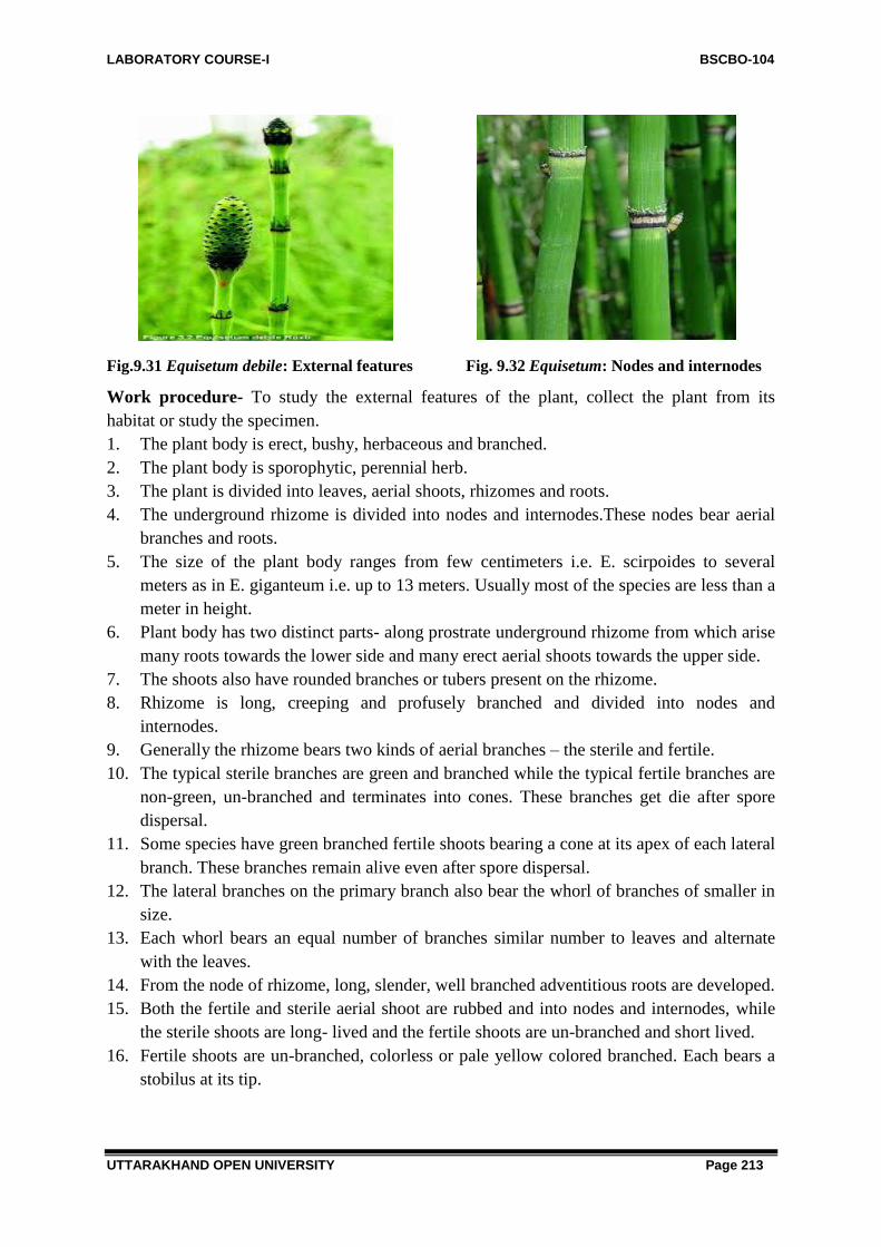
LABORATORY COURSE-I BSCBO-104
UTTARAKHAND OPEN UNIVERSITY Page 213
Fig.9.31 Equisetum debile: External features Fig. 9.32 Equisetum: Nodes and internodes
Work procedure- To study the external features of the plant, collect the plant from its
habitat or study the specimen.
1. The plant body is erect, bushy, herbaceous and branched.
2. The plant body is sporophytic, perennial herb.
3. The plant is divided into leaves, aerial shoots, rhizomes and roots.
4. The underground rhizome is divided into nodes and internodes.These nodes bear aerial
branches and roots.
5. The size of the plant body ranges from few centimeters i.e. E. scirpoides to several
meters as in E. giganteum i.e. up to 13 meters. Usually most of the species are less than a
meter in height.
6. Plant body has two distinct parts- along prostrate underground rhizome from which arise
many roots towards the lower side and many erect aerial shoots towards the upper side.
7. The shoots also have rounded branches or tubers present on the rhizome.
8. Rhizome is long, creeping and profusely branched and divided into nodes and
internodes.
9. Generally the rhizome bears two kinds of aerial branches – the sterile and fertile.
10. The typical sterile branches are green and branched while the typical fertile branches are
non-green, un-branched and terminates into cones. These branches get die after spore
dispersal.
11. Some species have green branched fertile shoots bearing a cone at its apex of each lateral
branch. These branches remain alive even after spore dispersal.
12. The lateral branches on the primary branch also bear the whorl of branches of smaller in
size.
13. Each whorl bears an equal number of branches similar number to leaves and alternate
with the leaves.
14. From the node of rhizome, long, slender, well branched adventitious roots are developed.
15. Both the fertile and sterile aerial shoot are rubbed and into nodes and internodes, while
the sterile shoots are long- lived and the fertile shoots are un-branched and short lived.
16. Fertile shoots are un-branched, colorless or pale yellow colored branched. Each bears a
stobilus at its tip.
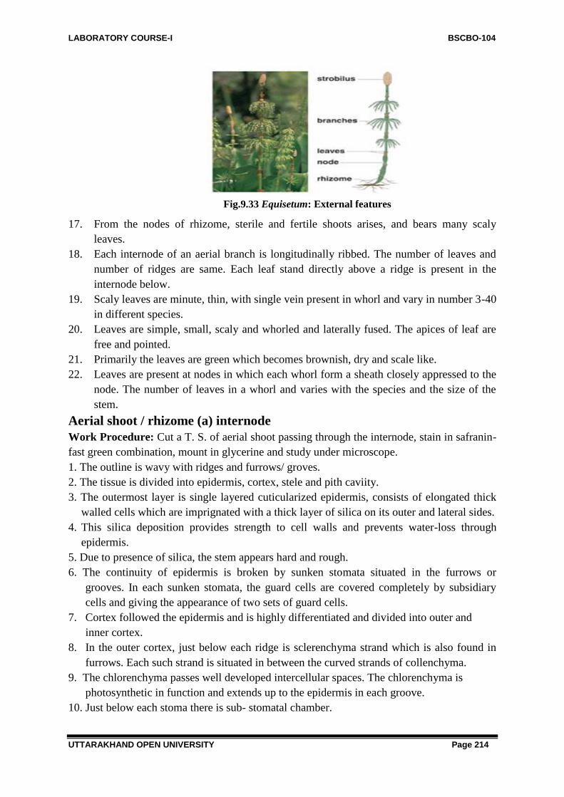
LABORATORY COURSE-I BSCBO-104
UTTARAKHAND OPEN UNIVERSITY Page 214
Fig.9.33 Equisetum: External features
17. From the nodes of rhizome, sterile and fertile shoots arises, and bears many scaly
leaves.
18. Each internode of an aerial branch is longitudinally ribbed. The number of leaves and
number of ridges are same. Each leaf stand directly above a ridge is present in the
internode below.
19. Scaly leaves are minute, thin, with single vein present in whorl and vary in number 3-40
in different species.
20. Leaves are simple, small, scaly and whorled and laterally fused. The apices of leaf are
free and pointed.
21. Primarily the leaves are green which becomes brownish, dry and scale like.
22. Leaves are present at nodes in which each whorl form a sheath closely appressed to the
node. The number of leaves in a whorl and varies with the species and the size of the
stem.
Aerial shoot / rhizome (a) internode
Work Procedure: Cut a T. S. of aerial shoot passing through the internode, stain in safranin-
fast green combination, mount in glycerine and study under microscope.
1. The outline is wavy with ridges and furrows/ groves.
2. The tissue is divided into epidermis, cortex, stele and pith caviity.
3. The outermost layer is single layered cuticularized epidermis, consists of elongated thick
walled cells which are imprignated with a thick layer of silica on its outer and lateral sides.
4. This silica deposition provides strength to cell walls and prevents water-loss through
epidermis.
5. Due to presence of silica, the stem appears hard and rough.
6. The continuity of epidermis is broken by sunken stomata situated in the furrows or
grooves. In each sunken stomata, the guard cells are covered completely by subsidiary
cells and giving the appearance of two sets of guard cells.
7. Cortex followed the epidermis and is highly differentiated and divided into outer and
inner cortex.
8. In the outer cortex, just below each ridge is sclerenchyma strand which is also found in
furrows. Each such strand is situated in between the curved strands of collenchyma.
9. The chlorenchyma passes well developed intercellular spaces. The chlorenchyma is
photosynthetic in function and extends up to the epidermis in each groove.
10. Just below each stoma there is sub- stomatal chamber.

LABORATORY COURSE-I BSCBO-104
UTTARAKHAND OPEN UNIVERSITY Page 215
11. The inner cortex is composed of large and thin walled parenchymatous cells.
12. Vellecular canal are present in the cortex.
Fig.9. 34 Equisetum: sporophyte
These are situated below the groove. They are alternate to the vascular bundles.
Fig. 9.35 Equisetum: T.S. aerial shoot

LABORATORY COURSE-I BSCBO-104
UTTARAKHAND OPEN UNIVERSITY Page 216
13. Innermost layer of cortex is known as endodermis. It contains casparian strips. While in
some species i.e. E. sylveticum contains a layer of inner endodermis is also present, and in E.
litorale, each vascular bundle contains its individual endodermis.
14. Below the endodermis is present single layered endodermis.
15. The vascular bundles are conjoint closed, endarch and arranged in a ring. Each bundle lies
below each ridge and consists of xylem and phloem.
16. The number of vascular bundles and vallecular -canals are equal to the number of ridges
and grooves respectively.
17. Each vascular bundle contains both metaxylem and protoxylem.
18. The protoxylem elements lie on the ridges of a protoxylem lacuna. In each vascular
bundle is present a water containing cavity of canal called carinal canal.
19. The two metaxylem groups lie on the lateral sides of carinal canal. It consists of annular
and spiral tracheids.
Fig.9.36, Equisetum: T. S. aerial shoot: Internode (A part cellular)

LABORATORY COURSE-I BSCBO-104
UTTARAKHAND OPEN UNIVERSITY Page 217
Fig. 9.37 Equisetum: T.S. stem
20. The mataxylem elements are found in two groups. These two groups are arranged on the
margin of carinal canal towards outside. The metaxylem elements are composed of reticulate
scalariform or pitted tracheids.
21. The two groups of metaxylem lies phloem, the phloem is composed of sieve tubes and
phloem parenchyma while companion cells areabsent.
22. The central region of stem is occupied by hollow pith.
Procedure B- Node-
Cut a T. S. of aerial shoot passing through the node, stain in safranin fast green combination,
mount in glycerine and study under microscope.
1. The aerial shoot shows distinct ridges and furrows.
2. The section shows distinct epidermis, cortex, stele and nodal diaphragm instead of hollow
pith cavity in internode.
3. The outermost layer is thick cuticularized epidermis.
4. It is also resemble the internode of sterile shoot beside some differences.
Fig.9.38, Equisetum; A part os stem showing alternation of ridges and grooves and scale leaves

LABORATORY COURSE-I BSCBO-104
UTTARAKHAND OPEN UNIVERSITY Page 218
Fig.9.39, A-B Equisetum: Structure of stomata, A. Stoma in vertical section, B. Thickening band
band in stomata
5. Likewise aerial sterile internode middle and outer cortex.
6. The inner cortex occupies maximum part of the cortex and made up of parenchymatous
tissues
7. While the outer cortex is sclerenchymatous and followed by chlorenchymatous or palisad
tissues middle cortex.
8. The vascular bundles are arranged in a continuous ring with outer ring of phloem enclosing
a ring of xylem.
9. The bundles at thenode do not have carinal canals.
10. Absence of all three types of canals i.e. vallecular canal, cainal canal and cetral canal.
Fig.9.40 Equisetum: T.S. aerial shoot showing different conditions of endodermis
11. But sometimes the protoxylem elements are intact and completely occupy the lacuna or
carinal canal.
12. Instead of a central pith cavity, a nodal diaphragm is present.

LABORATORY COURSE-I BSCBO-104
UTTARAKHAND OPEN UNIVERSITY Page 219
Leaves
1. Leaves are simple, small scaly whorled and fused laterally and possess longer or shorter
free tips.
2. The species with narrow stens usually have few leaves and those with thick stems have
many leaves (up to 40) at each node.
3. Leaves are non-photosynthetic and their main function is to protect the branch buds at the
node.
4. Each leaf contains a single vascular bundle.
5. The vascular bundles of the leaf sheath are simple.
6. The outer tissues of the leaf sheath are composed of narrow sclerenchymatous bands.
7. The bands of sclerenchyma pass up the leaf ridge and alternate with the strips of
chlorophyllous tissue associateted with stomata.
RHIZOME-Work Procedure-Cut a T. S. of rhizome, stain in double staining- safranin
fastgreen, mount in glycerine and study under microscope.
1. The outline is wavy and possess ridges and grooves.
2. The outermost layer is epidermis, thickly cuticularized and presence of stomata.
Fig. 9.41: EquisetumT.S. Rhizome
3. The anatomy of rhizome is quite similar to that of aerial shoot.
4. The cortex is made up of a few layers of sclerenchyma just below the epidermis. The
assimilatory tissue is not found in the rhizome.
5. A large zone of parenchyma spread up to the vascular bundle ring.
6. The mechanical tissue is less developed completely to that of aerial shoot.
7. Presence of large vallecular canals in the parenchymatous cortex below the grooves is
reported.
8. The ring of vascular bundles is encloses by single layered endodermis
9. The vascular bundle is conjoint, collateral, endarch and located below the ridge.
10. In the centre a large pith cavity is present.In E. arvense the pith is solid while in some
other species the pith and the vascular canals are very much reduced.
Study of anatomy of root

LABORATORY COURSE-I BSCBO-104
UTTARAKHAND OPEN UNIVERSITY Page 220
Work Pocedure: Cut a T. S. of root, stain in safranin- fast green combination, mount in
glycerine and observe under microscope.
1. The section appears almost circular in outline.
2. The outermost layer is epidermis made up of proliferous layer and bears unicellular hairs
upon it known as root hairs.
3. The epidermis is single layered.
4. Just beneath the epidermis there is a wide cortex, which is thick and multilayered.
5. The cortex is often divided into an outer cortex and an inner cortex.
6. The cortex consists of thin walled parenchyma with well- developed intercellular spaces.
7. The endodermis is two layered thicki.e. outer and inner endodermis, while the cells of
inner endodermis acts as pericycle. Here a distinct pericycle is absent.
8. The vascular bundle shows a single, large metaxylem in the centre with 3,4 or six
protoxylem.
9. Stele is protostele, which is triarch, tetrarch or some times hexarch .
10. With the increase in diameter of root, number o protoxylem groups also increases.
11. In the centre is present a large metaxylem traicheds having many protoxylem groups
towards the periphery.
12. Phloem is present in between the angles of protoxylem.
13. Each protoxylem point consists of a single tracheids which is spirally thickened.
14. The phloem is composed of phloem parenchyma and sieve tubes.
Fig.9.42 Equisetum, T.S. root (a part cellular)
Sporangia spore producing organs
Work Procedure-To study the external and internal structure of spore producing organs or
sporangia, borne in cones. Generally terminating the main axis and sometimes the lateral
branches. To study this cut the T.S. and L. S. of cone, stain it in safranin fast green
combination mount in glycerine and study.
1. Fertile, aerial, unbranched shoot bear at their apices the spore producing organs known as
strobili or cones.
2. The strobilus cosists of a thick central axis known as strobilus axis, coneaxis or
sporangiophore.
3. This axis, bearing several whorls of densely crowded sporangia on theunderside of peltate
disc situated on the apex of sporangiophore.

LABORATORY COURSE-I BSCBO-104
UTTARAKHAND OPEN UNIVERSITY Page 221
4. Each sporangiophore consists a stalk and a peltate disc. Stalk keeps the disc attached to
cone axis.
5. Just below the sporangiophore, the central axis of the strobilus bears a small ring like
outgrowth known as annulus. Sporangiophore is one of the units of which cone is made
of.
6. Each sporangiophore is composed of a slender stalk, the free end of which becomes
flattened to form a peltate disc.
7. The peltate disc is generally hexagonal in outline in its surface view. Each sporangium is
elongated and pendant and contains a rounded apex
9. The stalk is attached to the cone axis on one side and to peltate disc on the otherside.
10. The apex opens by longitudinal silts on maturity and helps in dispersal of spores
13. The sporamgium is surrounded by jacket on outside and tapetum layer on inside, which
enclosesspores. All the spores are alike, as Equisetum is homosporous.
Fig. 9.43 Equisetum: T.S showing Sporangium
Fig. 9.44 Equiseum: sporangiophore from ventral side

LABORATORY COURSE-I BSCBO-104
UTTARAKHAND OPEN UNIVERSITY Page 222
Fig.9.45 Equiseum: Sporangium
Strobilus
Work Procedure-The sporangia is the spore producing organs compact aggregation and
sporangia is called distrobiusor cone which generally originated at the apex of main axis and
sometimes the lateral branches. For detailed study the L.S. of cone is best for structural
details.
Fig.9.46 Equisetum: T.S. Cone Fig.9.47 Equisetum: L.S. Cone
1. L.S. of cone shows cone axis, attached with sporangiophore.
2. In the centre cone axis is located.
3. It shows sporangiophores in whorls which alternates each other yet not in regular manner.
4. A calyx like whorl or annulus is lies at the base of cone.
5. The stalk holds a polygonal peltate disc at right angles to it. The sporangiophores are
attached to the cone axis at right angles.
Spores and elaters:
Work Procedure-

LABORATORY COURSE-I BSCBO-104
UTTARAKHAND OPEN UNIVERSITY Page 223
With the help of needle the sporangia tease smoothly, spores and elaters collected in water or
stained in safranin and studied.
1. Each spore is uni-nucleate, spherical or globular in shape and bears many chloroplasts.
2. Each spore consists of four layers.
3. As usual each spore has two usual layers along with third cuticular layer or the middle
layer and a fourth, thick outermost layer, the perisporium is reported.
4. The outermost layer is known as exosporium and the innermost endosporium.
5. The third layer is made up of the cuticle and known as middle layer and lies outside the
exosporium.
6. The middle layer and the outermost layer of exosporium are the derivatives of few tapetal
cells and spore mother cells.
7. The episporium which lies on the extreme outside of the spore which later on, develops
into four strips and get separated from each other on maturity from rest of the wall but
remain attached at a known point. The strips after matutration develop as elaters.
Fig.9.48 Equisetum: Spore with elaters Fig.9.49. Equisetum: Spore showing different
stages of elaterexpantion
8. The ends of these elaters are spoon like or broad however, these elaters are different from
the elaters of Bryophytes.
9. The elaters are hygroscopic in nature and with the changes in atmospheric humidity they
coil and uncoil.
10. These elaters help in dehiscence of sporangium and dispersal of spores similarly to
Bryophytes.
11. Each spore contains many chloroplast and a nucleus located in the centre..
Prothallus:
Work Procedure- Prepare a slide of prothallus and study.
1. The prothallus of Equisetum is monoceious and bearing both
male (antheridia) and female (archegonia) sex organs in a single
prothallus.
2. The yonger and small prothalli show only antheridia as the
antheridia develops first while in the older, archegonia are present.
3. The archegonia show two parts i.e. a sunken base and a projecting
neck. Fig.9.50 Equisetum: Prothallus

LABORATORY COURSE-I BSCBO-104
UTTARAKHAND OPEN UNIVERSITY Page 224
4. The archegonial neck consists of four vertical rows of cells. Each row is two or four cells
in height.
5. The upper cells of archegonial neck are quite long and becomes separated and curved
outwards on maturity.
6. Archegonia remain embedded in the prothallus tissue.
7. The archegonium consists of 3 or 4 cells, the egg, the ventre c. Each androcyte is
metamorphoses into multiflagellated spirally coiled anthrozoids and cells and one or two
neck cells.
8. The antheridia consists single layered jacket, which encloses androcytes inside this.
Identification-
Divison- Pteridophyta-
i) Plant body is differentiated into stem, root and leaves,ii) Sporophyte possess well
developed vascular strand. iii) True vascular strand present.iv) Gametophyte is small and
independent.
Sub divison- Sphenopsida-
i) Plant body articutated and branched,ii) Stem with distinct nodes and internodes.iii) Ridges
and furrows presentiv) Leaves small, scaly and microphyllous and in whorls at nodes.
Order- Equisetales-
i) Plant herbaceous and not tree like,ii) Stem branched, branches originated in transeverse
whorls,iii) Secondary thickeness absent,iv) Vasculat cylinder siphonostele, endarch.
Family- Equisetaceae-
i) Secondary growth is absent, ii)Plant is homosporous,iii) Sporangia borne on
sporangiophores, Which forms a compact cone.
Genus- Equisetum-
i) Presence of colorless scaly leaves.ii) Sunken stomata in grooves, iii) Presence of palisade
tissue in the stem,iv) Presence of vellecular, carinal and central canals,.
Significant Points-
The anatomy of Equisetum shows Xerophytic and Hydrophytic characters, which also helps
in its identification.
Xerophytic Characters-
1. Thick cuticle present over epidermis.
2. Presence of ridges and furrows.
3. Presence of stomata in grooves.
4. Presence of palisade.
5. Presence of well- developed sclerenchyma below the ridges.
Hydrophytic Characters-
1. Presence of three distinct canals i.e. carinal, vellecular and central canals.

LABORATORY COURSE-I BSCBO-104
UTTARAKHAND OPEN UNIVERSITY Page 225
9.6 SUMMARY
Rhynia
Rhynia is a species of Devonian vascular plant. Rhynia Gwynne-vaghani was the sporophyte
generation of vascular, axils, free- sporing, diplohaplontic embryophytic plants of the lower
Devonian that had anatomical features more advanced than those of the bryophytes. Rhynia
gwynne- vaghani was amember of sister group to all other eutrachephytes including modern
vascular plants. Rhynia is a fossil plant and had two species, Rhynia major and R. Gwynne-
vaughani.
Selaginella
Genus Selaginella is the sole genus of vascular plants in the family Selaginellaceae. The
spike moss or lesser known club- mosses. This family is placed in class Isoetopsida,
distinguished from the sister group Lycopodiopsida having scale leaves bearing a ligule and
by having spores of two types and hence representing the best example of heterospory.
Beside this, the main significant point of their identification is presence of trabeculae in its
stellar structure.
Equisetum
Horsetail plant or Equisetum is a member of the Equisetaceae family and is closely related to
ferns. Horsetail is also known by a number of other names, including mare‖ tail, horse pipes,
snake etc. Horse tail (Equisetum arvense) is considered as “living fossil” and is one of the
oldest plants on the planet, primarily because it is one of the toughest to eradicate. It belongs
to a class of Pteridophyta (ferns). They are one of the most important plants grown in
palaeozoic era. Equisetum is the only living genus in Equisetaceae, a family of vascular
plants that reproduce by spores rather than seeds. The Horsetail (Equisetum arvense) belongs
to a family of plants some 400 million years old. Some of which grew over 300 feet.
Horsetail is a perennial plant that found in or near watery areas, such as marshes, streams or
rivers. Horsetail is a versatile plant that will grow in pretty much in any water logged area. It
makes good filler for areas in the yard with poor drainage or as a pond grass. Horsetail grows
in temperate northern hemisphere areas. The stem has a characteristic jointed appearance.
They are seedless vascular plants. They have true vascular system.
9.6 GLOSSARY
Eusporangiate- Describing the sporangium that is derived from a number of sporangial
initials.
Fossile- Remains of the organismfrom past geological ages preserved mostly in sedemantary
rocks as actual structures or as impressions, caste or moulds of structures that are no more
living.
Heterosporous- Describing conditions where different types of spores are produced by a
species. Usually the species produces large megaspores containing reserve food and produce
female gametophytes and the smaller microspores that produce male gametophyte.

LABORATORY COURSE-I BSCBO-104
UTTARAKHAND OPEN UNIVERSITY Page 226
Homosporous- Describing production of only one type of spores by a species used in certain
non- seed producing vascular plants.
Internode- A part of stem that lies between two adjacent nodes.
Ligule- Describing the structure of a thin membranous finger like structure present at the
base of upper or adaxial surface of each leaf is called ligule, especially in case of Selaginella.
Leptosporangate- Describing the type of sporangium that is derived from one initial cell
completely.
Megasporophyll- The leaf typical of seed plants and fernsthat is relatively large and usually
with leaf gaps associated with leaf traces.
Microsporophyll- A leaf typical of lower plants means Lycopodes that is relatively smaller
and is not associated with leaf gaps inthe stele.
Node- A point on the stem, from where one or more leaves arise. In mature stem, nodes are
usually well separated due to activity of intercalary meristem pattern of vascular connections
between stem and leaf.
Prothalii- Free living gametophytes of certain lower vascular plants e.g. species that is
usually poorly differentiated and often resemble thallose liverworts.
Pteridophytes- These plants are also known as vascular cryptogams. These are most
primitive living and fossil plants. They shows heterospory means forms two types seeds in
their life- cycle e,g, small (microspores) and bigger (megaspores).
Rhizophore- At the base of branching in stem, arises a long, unbranched, leafless structure
towards the lower side. This is known as rhizophore. It becomes branced and forms many
adventitious roots, usually in Selaginella.
Sori or Sorus- A group or cluster of sporangia borne abaxially on a frond.
Spore- The unicellular reproductive unite that may be formed by sporophytic generation
following meiosis and therefore, have half the number of chromosomes.
Sporangium- The structure in which spore are produced it may be simple and unicellular as
in algae and fungi or multicellular as in bryophytes and vascular plants.
Sporophyte- Individual usually of deploidbphase of life cycle, that is formedfrom the fusion
product of two gametes.
Tapetum- The layers of cells surroundingspore mother cells in the sporangium on a frond.
Trabeculae- Bar like structure extended across a lumen or lacuna, e.g. certain endodermal
cells elongate radially to form trabeculae in endodermis of stem of Selaginella.
Whorl- Describing the form of leaves in which leaf arrangement three or more leaves arises
at each node.
Xerophytic- Describing condition of habitat by deficiency of water representing either
physical or physiological dryness, high temperature and strong sunlight as in deserts.
9.7 SELF ASSESSMENT QUESTIONS
9.7.1 True/ False
(i) Plant body of Rhynia consisted stem, roots and leaves-------------------
(ii) Peteridophytes was the first vascular plants-------------------------------

LABORATORY COURSE-I BSCBO-104
UTTARAKHAND OPEN UNIVERSITY Page 227
9.7.2 Fill in the blanks:
1. Fossil of Rhynia discovered by----------------------------------------
2. Name any homosporous fossil pteridophyte-------------------------------
3. Seed habit is observed in?---------------------------------------------
4. Common name of Selaginella is--------------------------
5. The development of sporangia in Selaginella is-------------------
6. Protostele is found in -------------------------------------------
7. Rhizophore and trabeculae are found in -------------------------------
8. Presence of both, megaspores and microsporse is called-------------
9. Vellecular canal, carinal canal and central canals are present in---------------
10. The common name of Equisetum is ----------------------------------------
11. Elaters are present in which Pteridophyta ----------------------------------
12. Mention the name of reproductive organ of Equisetum-----------------------
13. The substance deposited on outer walls of epidermis of Equisetum is------------
14. The condition of xylem in the stem of Equisetum is -------------------
15. How many elaters are present in the spore of Equisetum--------------------
16. Name the cavities present in the cortex of Equisetum -----------
17. Mention the type of stele found in Equisetum -----------------
18. Sporangia are present on the underside of which part of sporangiophore in Equisetum---
19. Development of sporangium in Equisetum is of what type--------
20. Equisetum is---------------------as all the spores are similar
9.7.1: Answers Key
(i) False, (ii) True
9.7.2: Answers Key
1. Kidston and Lang, 2. Rhynia, 3. Selaginella, 4. spike moss, 5. Eusporangiate, 6.
Selaginella, 7. Selaginella, 8. Heterospory, 9. Equisetum, 10. Horse tail, 11. Equisetum, 12.
Srobilus, 13. Silica, 14. Endarch, 15. Two, 16. Vellecular canal, carinal canal and central
canals, 17. Sphenostele, 18. Peltate disc, 19. Eusporangiate, 20. Homosporous
9.8 REFERENCES
K. Sharma and Rajeshwari Sharma (2011) Algae Bryophytes and Pteridophytes.
Jagdamba Publishing Co. 423011, Ground Floor, Ansari, Daryaganj, New Delhi, 110002.
Ashok Bendre (2011) A Text Bookof Practical Botany I. Rastogi Publications. Gangotri
Shivaji Road. Meerut.
P. Pandey (2012) Practical Botany Vol. I, S. Chand and Company, Pvt.Ltd. Ramnagar,
New Delhi- 110055.
O. P. Sharma (2010) Pragati Practical Botany Vol. I, Pragati Prakashan, Meerut.
S. K. Gupta and D. K. Gupta (2013) New Pattern Botany Guide Vol. III, Published by
Kedarnath Ramnath, 132 R. G. Collage Road, Meerut (U.P.).
Wikipedia the Free Encykopedia.

LABORATORY COURSE-I BSCBO-104
UTTARAKHAND OPEN UNIVERSITY Page 228
Encyclopedia Britanica.
https:// wwww.google.co.in Wikipedia
9.9 SUGGESTED READINGS
P. Pandey (2012) Practical Botany Vol. I, S. Chand and Company, Pvt. Ltd. Ramnagar,
New Delhi- 110055.
O. P. Sharma (2010) Pragati Practical Botany Vol. I, Pragati Prakashan, Meerut.
S. K. Gupta and D. K. Gupta (2013) New Pattern Botany Guide Vol. III, Published by
Kedarnath Ramnath, 132 R. G. Collage Road, Meerut (U.P.).
K. Sharma and Rajeshwari Sharma (2011) Algae Bryophytes and Pteridophytes.
Jagdamba Publishing Co. 423011, Ground Floor, Ansari, Daryaganj, New Delhi, 110002.
9.10 TERMINAL QUESTIONS
1. With suitable sketches compare the sporophyte of R. major and R. gwaynne- vaughanii?
2. Describe the internal structure of Rhynia stem with the help of diagramme?
3. Draw the sketch of Rhynia sporangia?
4. Describe the nature of sporangia of Rhynia with the help of suitable sketch?
5. With the help of suitable diagram explain the external features of Rhynia?
6. Draw T.S. of aerial branch of Rhynia and explain it?
7. Explain the internal structures of reproductive parts of Rhynia withsuitable diagram?
8. Draw a well labeled diagram of sporophyte of Rhynia?
9. Explain the morphology of Selaginella with the help of illustrated diagram?
10. Descrie briefly the anatomical characters of stem and leaf of Selaginella?
11. Describe briefly the systematic position, morphological structures of Selaginella?
12. With labeled diagrams describe the reproductive organs of Selaginella?
13. Draw a well labeled sketch of gametophyte of Equisetum with the help of labeled
sketches explains the anatomy of sterile internode of Equisetum?
14. Describe the anatomy of root of Equisetum?
15. Draw a labeled diagram of sporangia of Equisetum/
16. With the help of suitable sketchexplain the structure of spore producing organs of
Equisetum?
17. Draw a labeled diagram of rhizophore of Equisetum?
18. Explain in detail the structures of spore and elaters of Equisetum diagrammatically?
19. Draw T.S. of Selaginella stem i) showing monostelic condition ii) polystelic condition?
20. Explain the anatomical differences between T.S. root and T.S. rhizophore of Selaginella
with the help of diagrams?
21. With the help of diagram describe the internal structure of Selaginella leaf?
22. Draw well labeled diagram of megasporophyll and microsporophyll of Selaginella?
23. Draw the diagrams of megasporophyll and microsporophyll of Selaginella, also explain
the heterospory in Selaginella?

LABORATORY COURSE-I BSCBO-104
UTTARAKHAND OPEN UNIVERSITY Page 229
UNIT-10 STUDY OF EXTERNAL FEATURES AND
INTERNAL STRUCTURES OF RHIZOME, LEAVES,
ROOTS, SPORANGIA AND STROBILII OF
PTERIDOPHYTES - ADIANTUM, AZOLLA AND
MARSILEA
10.1-Objectives
10.2-Introduction
10.3-Study of external features, internal structures of Adiantum
10.3.1-Root
10.3.2-Rhizeme
10.3.3-Roots
10.3.4-Leaves and Rachis
10.3.5-prothallus
10.4- Study of external features, internal structures of Azolla
10.4.1-Stem
10.4.2-Leaves
10.4.3-Roots
10.4.4-Sporocarp
10.5- Study of external features, internal structures of Marsilea
10.5.1-Rhizome
10.5.2-Leaves
10.5.3-Roots
10.5.4-Sporocarp
10.6- Summary
10.7- Glossary
10.8- Self assessment question
10.9- References
10.10-Suggested Readings
10.11-Terminal Questions

LABORATORY COURSE-I BSCBO-104
UTTARAKHAND OPEN UNIVERSITY Page 230
10.1 OBJECTIVES
After reading this unit student will be able:
To study the general characters of mentioned Pteridophytes.
To study of morphology and anatomy of vegetative and reproductive parts of
Pteridophytes- Adiantum, Marsilea and Azolla.
10.2 INTRODUCTION
The Pteridophyta include a group of land plants with well - developed vascular tissue- xylem
and phloem. Therefore, this group is also known as Vascular Cryptogams. Carolus Linnaeus
(1754) classified these plants underclass Cryptogamia. The word also derived from Greek
words Kruptons means hidden and gamos means wedded, plants with concealed flowers. In
this unit we will discuss about the species- Adiantum, Marsilea and Azolla.
10.3 STUDY OF THE EXTRNAL FEATURES AND INTERNAL
STRUCTURES OF ADIANTUM
Systematic position
Divison- Pteridophyta
Sub Divison- Pteropsida
Class- Leptosporangiate
Order- Filicales
Family- Adiantaceae
Genus- Adiantum
Fig.10.1 Adiantum: Habit sketch
Adiantum the maidenhair fern is a genus of about 200 species of ferns in the world. However,
of Adiantum raddianum and possibly a few other species, are removed the remaining plant.
The genus Adiantum is cosmopolitan in its distribution. It is distributed throughout the whole
world including temperate South America and Arctic zone. It is distributed throughout India
evenly in plains and on mountains at an elevation of 2000 to 8000 feet. It is -
Adiantum: External features - a terrestrial fern and prefers moist and shady places in all
warmer parts of the world. The name Adiantum is derived from the Greek word its common
name, is Some species of Adiantum are also used medicinal purposes. It grows abundantly in
tropical and sub- tropical zones of world.
The common species reportedfrom India are Adiantum incism, A. caudatum, A.
capillus-veneris, A. lanulatum, A. caudatum etc. Nayar (1961) reported 24 species from India.
Study of external feature of adiantum-(morphology)
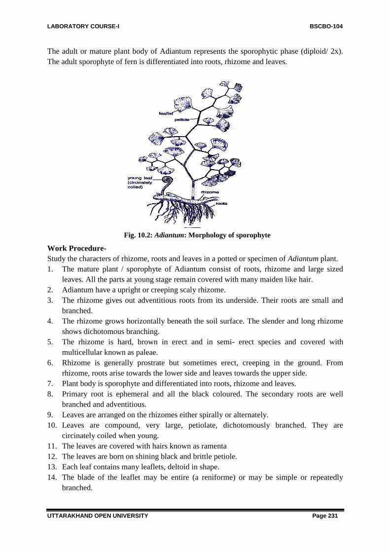
LABORATORY COURSE-I BSCBO-104
UTTARAKHAND OPEN UNIVERSITY Page 231
The adult or mature plant body of Adiantum represents the sporophytic phase (diploid/ 2x).
The adult sporophyte of fern is differentiated into roots, rhizome and leaves.
Work Procedure-
Study the characters of rhizome, roots and leaves in a potted or specimen of Adiantum plant.
1. The mature plant / sporophyte of Adiantum consist of roots, rhizome and large sized
leaves. All the parts at young stage remain covered with many maiden like hair.
2. Adiantum have a upright or creeping scaly rhizome.
3. The rhizome gives out adventitious roots from its underside. Their roots are small and
branched.
4. The rhizome grows horizontally beneath the soil surface. The slender and long rhizome
shows dichotomous branching.
5. The rhizome is hard, brown in erect and in semi- erect species and covered with
multicellular known as paleae.
6. Rhizome is generally prostrate but sometimes erect, creeping in the ground. From
rhizome, roots arise towards the lower side and leaves towards the upper side.
7. Plant body is sporophyte and differentiated into roots, rhizome and leaves.
8. Primary root is ephemeral and all the black coloured. The secondary roots are well
branched and adventitious.
9. Leaves are arranged on the rhizomes either spirally or alternately.
10. Leaves are compound, very large, petiolate, dichotomously branched. They are
circinately coiled when young.
11. The leaves are covered with hairs known as ramenta
12. The leaves are born on shining black and brittle petiole.
13. Each leaf contains many leaflets, deltoid in shape.
14. The blade of the leaflet may be entire (a reniforme) or may be simple or repeatedly
branched.
Fig. 10.2: Adiantum: Morphology of sporophyte

LABORATORY COURSE-I BSCBO-104
UTTARAKHAND OPEN UNIVERSITY Page 232
15. The leaves when fertile, then leaflet margins remain folded towards the lower side
forming a false inducium which encloses many sori.
16. The fertile portion of each leaflet contains many sporangia filled with spores.
17. The venation is dichotomous type, Vein spread in a fan like manner in the lamina.
Usually the veins are free but in some species they are forming the reticulum.
Root
Object- Study of root morphology
The roots are adventitious and arise in clusters from under side of the rhizome. The roots are
creeping generally but in erect form, they arise from base of rhizome. They are black and
branched.
Study of anatomy of root
1. The root is almost circular in outline.
2. The outermost layer of the T.S. of root is epidermis, which is piliferous. It is single
layered and bears root hairs.
3. The outermost layer or epidermis is thin walled consisting cortex and stele in centre.
4. The cortex is inner to epidermis, It is multilayered and parenchymatous in nature.
5. The stele remains surrounded by a single layered endodermis. Casparian strips on radial
walls of the cells of endodermis.
6. The pericycle is the outermost layer of stele which is one or two layered thick.
7. The stele is protostele type present in the center. It is diarch or exarch.
8. There is a central core of xylem surrounded by phloem.
9. Outside the xylem there are phloem elements, the phloem is arranged laterally to the xylem
in the form of phloem plates.
Fig. 10.3 Adiantum; T.S. root (a part cellular)
10. Xylem elements occur in the centre. Protoxylem groups are situated on the two opposite
sides of the metaxylem.
11. Phloem surrounds the xylem on all the sides.
Rhizome object-study of morphology of rhizome
In Adiantum the rhizome grow beneath the surface of the soil in horizontal manner. It is
slender and long, shows dichotomous branching with indefinite growth power. From the

LABORATORY COURSE-I BSCBO-104
UTTARAKHAND OPEN UNIVERSITY Page 233
above surface of rhizome from nodes arise roots on lower surface while leaves arise on upper
surface.
Study of anatomy of rhizome (T.S. of rhizome)
Work Procedure- Cut a T. S. of rhizome, stain in safranin- fast green combination, mount
in glycerine and study.
1. T.S. rhizome is mostly circular or gutter shaped.
2. Internally the rhizome is differentiated into outermost layer of epidermis bearing
multicellular hairs.
3. Just beneath the epidermis there is multilayered sclerenchymatous hypodermis.
4. Ground tissue occupies major part of rhizome contains parenchymatous cells and many
layered deep, without any intercellular space.
5. The species of Adiantun having an elongated rhizome, thus different types of steles are
reported which are variable in nature differing from region to region but a few shows
actual solenostele e.g. A. pedatum, A. nobile.
6. In A. rubellum the stele is amphiphloic siphonostele, but in A.caudatum it is dictylostele.
7. Commonly in Adiantum the stele is gutter shape due to great elongation of leaf gaps in
these rhizome possessing short internodes.
8. Due to presence of many leaf gaps the stele is in the form of many separate strands called
meristeles.
9. Meristeles vary in number but more often 5-7 lie arranged in ground tissue in a gutter
shaped ring and then exhibiting dictyostelic condition.
10. The spaces between neighbouring meristele are leaf gaps.
11. Each meristele has the general structure of a protostele e.g. xylem surrounds by ploem
and distinct layers of pericycle and endodermis. There is no pith.
Fig. 10.4 Adiantum. T.S. rthizome Fig.10.5: Adiantum. T.S. rhizome (part
shown by (outlines: diagrammatic)dotted
lines in Fig. 4: cellular)
12. Xylem elements occupy the central part of ground tissue. Metaxylem and protoxylem are
arranged in mesarch condition.
Study of external structure of leaf (rachis and leaflet)
The leaves are normally pinnately compound and are originated on the upper side of the
rhizome in alternate manner, and are separated from one another by long internodes. The

LABORATORY COURSE-I BSCBO-104
UTTARAKHAND OPEN UNIVERSITY Page 234
young stage the leaves are circinately coiled and are covered with hairy outgrowth called
“ramenta‖.
The leaves may be uni, bi or multipinnate. The pinnae are stalked. The rachis of leaf either
terminates in a pinna or may beelongated bearing a vegetative bud at tip under suitable
conditions. This bud develops into a daughter plant after touching the ground. The venation is
free, dichotomous and vein spread in fan like manner in the lamina.
Fig. 10.6 Adiantum: morphology of leaf Fig. 10.7 Adiantum: circinate leaf
Fig.10. 8 Adiantum: T.S. rachis Fig.10. 9 Adiantum: T.S. rachis (a part cellular)
(Outlines: diagrammatic)
Object- Study of anatomy of rachis (T.S. of rachis)
Work Procedure: Cut a T. S. of rachis, stain with safranin- fast green combination, mount in
glycerine and study.
1. The tissue is differentiated anatomically into an outermost layer of epidermis which is
cuticularized and without epidermal hairs.
2. Tissues of the rachis are differentiated into epidermis, hypodermis and central stele.
3. The epidermis is followed by 3-4 layered parenchymatous hypodermis. The cells of which
are sclerenchymatous.
4. The hypodermis is followed by multilayered parenchymatous cortex.
5. The cells of cortex may contain chloroplast.
6. The stele is of protostele type. Endodermis is single layered and is followed by a pericycle.

LABORATORY COURSE-I BSCBO-104
UTTARAKHAND OPEN UNIVERSITY Page 235
8. The phloem which surrounds the Y shaped xylem, protoxylem is present on all the three
ends of Y.
Study of anatomy of leaflet (T.S. of leaflet)
Work Procedure: Cut a T. S. of leaflet by keeping a leaf in a pith, stain in safranin- fast
green combination, mount in glycerine and study.
1. Leaflet is bounded on both sides by a layer of one cell thick epidermis. The lower
epidermis bears stomata.
Fig. 10.10 Adiantum: T.S. Leaflet
2. The whole leaflet is differentiated into upper and lower epidermis, mesophyll,
sclerenchyma and vascular bundle.
3. In between both epidermis, mesophyll is present which consists of only spongy
parenchyma.
4. Just above the lower epidermis lies single layer of compactly arranged cells containing
numerous chloroplasts.
5. In the centre is present a single vascular bundle surrounded by a thin sclerenchymatous
sheath.
6. Each vascular bundle is surrounded by a single layered endodermis and unilayered
pericycle.
7. Centrally located xylem has protoxylem group facing towards the adaxial surface of the
leaf.
8. Phloem surrounds xylem.
Sporangia
Study of anatomy of Sorus (T.S. of leaf with sorus)
Work Procedure: The sori are present on the lower side of the leaf so observe the lower
side of the leaflet, cut section through this portion, study the arrangement of sori and structure
of sporangia. Stain in safranin- fast green combination, mount in glycerine and study.
1. Sporangia are the spore producing organs. These sporangia are grouped in sori. Each

LABORATORY COURSE-I BSCBO-104
UTTARAKHAND OPEN UNIVERSITY Page 236
sporangia shows sporangia of different ages.
2. Adiantum is characterized by the presence of apparently marginal sori (on the leaflet tips),
these are superficial in origin and covered by a sharply reflaxed leaf margin which blocks
like an inducium.
3. The sporangia are inserted upon the distal region of the veins traversing the fertile lobe.
4. Many sori are present on the margin of the under surface of the leaf. These sori are covered
by reflaxed margins of leaf, the brown membranous, false inducium. True inducium is
absent.
5. The mature leaf bearing sori is called sporophyll.
6. Each sorus bears many sporangia in different developmental stages.
7. Each sporangium is composed of multicellular stalk and a capsule, with the shape of
biconvex lens.
8. Along both the sides of each vein are many sporangia attached by their long slender stalk.
The group of sporangia is known as sorus and such sori are situated along each of the vien.
9. The wall of sporangium is made up of thin walled cells.
10. The wall of the capsule is composed of a single layered thin walled, a row of specially
thickened cells, the annulus.
11. On one side of annulus are a few either 2-3, thin walled cells forming a stromium, from
where, dehiscence of sporangium takes place.
12. On maturity, the sporangium contains about 32-64 dark rugger and brown coloured
spores.
Fig.10.11 Adiantum: Leaf with sporophyll Fig.10.12 Adiantum: Section through sorus
13. Each spore is uninucleate structure and remain surrounded by a thin, smooth intine
and thick, rugged exine
Fig.10.13 Adiantum: Leaf showing sporangia Fig.10.14 Adiantum; Dehiscence of sporangium

LABORATORY COURSE-I BSCBO-104
UTTARAKHAND OPEN UNIVERSITY Page 237
10.3.5. Study of morphology of prothallus
Work Procedure- Take a young prothallus, stain in fast green only, mount in glycerine
and study.
1. After germination of spore, prothallus formed, which
shows the gametophytic stage.
2. The prothallus is gree, thin and heart shaped.
3. It is only one cell thick except the region posterior to the
apical notch. This region is called cushion.
4. Many unicellular colourless rhizoids are originated from the
ventral side.
Fig. 10.15 Adiantum: Prothallus
5. The cells of the prothallus are thin walled. Each cell bears
discoid chloroplasts and a nucleus in it.
6. On the lower surface of the prothallus are present the sex organs. Usually the archegonia
developed around the notch upon the cushion and the antheridia in the basal central region
of the prothallus among the rhizoids.
7. After fertilization, the diploid zygote develops to sporophyte.
Identiification points
Divison- Pteridophyta
i) Plant body is differentiated into stem, leaves and roots, ii). True vascular strand present,
iii) Plant body issporophyte, iv). Gametophyte small and independent,
Sub- Divison- Pteropsida
i) Vascular cylinder siphonostele with leaf- gaps, ii). Plant macrophyllus,
iii) Leaves compound with rachis, iv), Spoangia groupedin leavesin the form of sori,
v) Ridges and grooves absent in stem.
Class- Leptosporangiate
i) Sporangia with a jacket layer one cell in thickness, ii) Leptosporangiate type of
sporangium develops, iii) Definite number of spores,
Order- Filicales
i) Sori are mixed, ii) Plant homosporous.
Family- Adiantaceae
i) Sori are mixed.ii) Sporocarp absent, iii) Margin bearing sori are sharply reflexed,
iv) Inducium oblong or linear, formed of the frond, opening inwardly. v) Annulus not
vertical.
Genus- Adiantum
i) Leaves are large and dichotomously branched. ii) Sori apparently marginal but superficial
in origin, open dichotomous venation in the leaflets. iii) Presence of false inducium, iv)
Inducium globose to linear usually many and distinct, v) Xylem Y shaped in rachis.

LABORATORY COURSE-I BSCBO-104
UTTARAKHAND OPEN UNIVERSITY Page 238
10.4 STUDY OF EXTERNAL FEATURES AND INTERNAL
STRUCTURES OF AZOLLA
Systematic Position
Division- Pteridophyta
Sub Division- Pteropsida
Class- Leptosporangiatae
Order- Salviniales
Family Salviniaceae
Genus Azolla
Fig. 10.16 Azolla
Introduction- Azolla is an aquatic fern and generally known as ‗water fern‖. It is found
free floating in ditches and ponds. Azolla forms red coloured bloom in ditches and pond
water. The common Indian species is Azolla pinnata, others are A. filiculoides, is also known
to occur frequently. A. imbricate is found mostly in Eastern Himalaya. It is a water fern as
reported earlier and being used as biofertilizer since it contains the colonies of blue- green
algae ―Anabeana azollae‖ in its leaf chambers/cavities and thus helps to fix the atmospheric
nitrogen.
Study of external features and internal structures of Azolla (Morphology)
Work Procedure- To study the morphology of the plant, collect a fresh specimen or study
a preserved plant.
1. Plant body is sporophyte and divided into stem, leaves and roots.
2. It is aquatic in nature and grows free floating in ditches and ponds.
3. The stem floating horizontally in the water and possess pinnately branching. The branches
are extra axillary in nature.
4. The roots always remain submerged in water and are originated from the lower side of the
stem.
5. Leaves are small and present in two alternate row overlappes each other. These also cover
the stem and its branches.
6. Each leaf is divided into approximately in equal size lobes (looks like cotyledons).
7. The upper lobe or aerial lobe, remains slightly above or oblique in the water surface and
thick and green in colour. Its only one edge touches the water and not submerged.
8. The lower or submerged lobe is thin comparatively the previous one and is nearly
colourless. It is believed that the absorption of water may take place through this lobe‘

LABORATORY COURSE-I BSCBO-104
UTTARAKHAND OPEN UNIVERSITY Page 239
Fig.10.17 A-C Azolla: External morphology; A- A. microphylla, B- A. filiculodies, C- A leaf.
Object-Study of anatomy of Stem
Work Procedure: Cut a T.S. of the stem, stain in safranin fast green combination, mount in
glycerine and observe the details.
1. The section of the stem of Azolla is almost circular in outlines.
2. It is divided into outer epidermis, middle cortex and innerside into endodermis.
3. The outermost layer of the section is single layered epidermis.
4. Just beneath the epidermis is lies cortex. It occupies the most of the area of the section and
made up of thin walled parenchymatous cells.
5. The cortex is five to six cells in thickness without any intercellular spaces.
6. In the cortex lies stele, surrounded by single layered endodermis which is followed by a
single layered pericycle.
7. The vascular cylinder is simple and is greatly reduced in response to aquatic habit.
8. The vascular strand lies in the centre and is of protostelic nature.
9. The vascular tissue are represented by about six xylem elements and about double the
number as many elements in the phloem in the stele.
10. Leaf trace and leaf gaps are also present in the section.
Study of anatomy of Leaf-
Work Procedure- To study the anatomy of leaf, cut a vertical transverse section (V,T.S.) of
the upper lobe with the help of pith. Stain in safranin fast green combination, mount in
glycerine and observe the details.
1. The upper lobe of leaf shows two layers of epidermis as it bounds on both the sides by
upper and lower epidermis.
2. Both the epidermal layers possess stomata,
3. Many unicellular or two celled/ bicelled hairs are also present in upper epidermis.
4. The middle portion of the leaf is constituted by palisade tissue works like photosynthetic
cells possess intercellular spaces also.
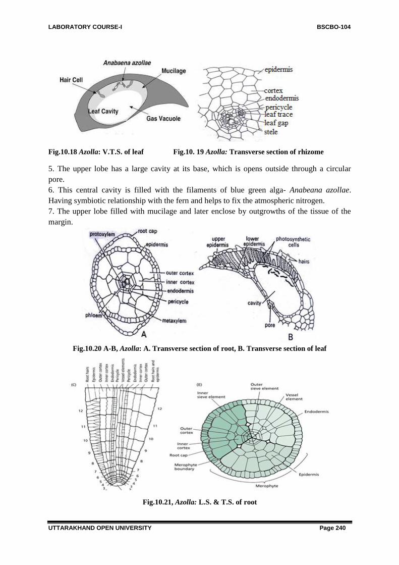
LABORATORY COURSE-I BSCBO-104
UTTARAKHAND OPEN UNIVERSITY Page 240
Fig.10.18 Azolla: V.T.S. of leaf Fig.10. 19 Azolla: Transverse section of rhizome
5. The upper lobe has a large cavity at its base, which is opens outside through a circular
pore.
6. This central cavity is filled with the filaments of blue green alga- Anabeana azollae.
Having symbiotic relationship with the fern and helps to fix the atmospheric nitrogen.
7. The upper lobe filled with mucilage and later enclose by outgrowths of the tissue of the
margin.
Fig.10.20 A-B, Azolla: A. Transverse section of root, B. Transverse section of leaf
Fig.10.21, Azolla: L.S. & T.S. of root

LABORATORY COURSE-I BSCBO-104
UTTARAKHAND OPEN UNIVERSITY Page 241
Study of root
Work Procedure- To study the root anatomy, cut a T. S. of root, stain in safranin fast green
combinations, mount in glycerine, and observe details.
1. The outline of the section is almost circular.
2. The whole section is divided into epidermis, cortex, endodermis and pericycle alongwith a
central vascular cylinder. Pith is not distinct.
3. The outermost layer is epidermis made up of single layered parenchymatous cells.
4. Just beneath the epidermis is cortex. This zone is divided into two sub zones e.g. the outer
cortex and the inner cortex.
5. Cortex contains 2- 8 layered thick parenchymatous cells.
6. Endodermis lies inner to cortex, made up of a single layer, consisting of 6 cells thickness.
7. Endodermis is followed by pericycle layer is also single layer and consisting only 6 big
size cells. The size of the cells is bigger than the previous layer.
8. Vascular strand consisting xylem and phloem in the centre of the section.
9. Xylem lies in the centre and is represented by two centrally placed metaxylem, surrounded
by four small outer group of protoxylem.
10. Xylem constitute only trecheids.
11. Phloem are placed on either side of the metaxylem and consist only few elements.
Object-Study of sporocarp-
Work Procedure- To study the sporocarp and its structural details first to know that these
are situated on the lower side of the plant (leaf). To examine the microsporocarp and
megasporocarp, tease them and stain with safranin only. Study the structural details under
microscope.
1. Sporocarps are originated only on the lowermost (mature) leaf of the lateral branch at the
end of annual season
2. Only the submerged lobe of the leaf bears 2- 4 sporocarps.
3. A hood like covering around the sporocarp is fored by the upper lobe of the fertile lobe.
4. The sporocarp possess microsporocarps and megasporocarps and hence shows dimorphic
habit.
5. Microsporocarps are larger in size than the megasporocaprs.
6. Each sporocarp is a sorus covered by indusium.
7. Microsporocarp bears a central raised cushion on which sporangia develop
basipetally.Asingle microzporengium has one layered jacket followed by tapetum and a
single microsporangium encloses about 64 microspores.
8. Microsporangium possess a multi-nucleate periplasmodium originated as a result of
breakdown of tatetum layer. Periplsmodium forms four or more quadrately arranged
massulae, in which spores remain embedded at periphery.
9. The surface of massulae bears many anchor- shaped hairy structures called glochidia,which
helps in the attachment of massulae to the microspore
10. Megaspore shows a single large megasporangium, remain surrounded by a flask shaped
indusium. It covers the sporangium completely except for a narrow slit at the apex.
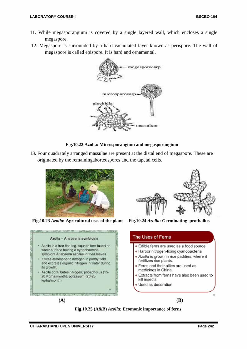
LABORATORY COURSE-I BSCBO-104
UTTARAKHAND OPEN UNIVERSITY Page 242
11. While megasporangium is covered by a single layered wall, which encloses a single
megaspore.
12. Megaspore is surrounded by a hard vacuolated layer known as perispore. The wall of
megaspore is called epispore. It is hard and ornamental.
Fig.10.22 Azolla: Microsporangium and megasporangium
13. Four quadrately arranged massulae are present at the distal end of megaspore. These are
originated by the remainingabortedspores and the tapetal cells.
Fig.10.23 Azolla: Agricultural uses of the plant Fig.10.24 Azolla: Germinating prothallus
(A) (B)
Fig.10.25 (A&B) Azolla: Ecomonic importance of ferns

LABORATORY COURSE-I BSCBO-104
UTTARAKHAND OPEN UNIVERSITY Page 243
Identification Points-
Division- Pteridophyta
i) Plant body is sporophyte and divided into stem, leaves and roots,ii). Definite vascular
system is present.
Sub Division- Pteropsida
i) Vascular cylinderis siphonostelic or dictyostele,ii) . Plant is macrophyllus with large leaf
gaps. iii) Leaves bear sporangia in sori, iv). Gametophyte small, green and free living,
Class- Leptosporangiatae
i) Sporangial wall is one cell thick, ii). Number of spores per sporangiumnis definite,
Order- Salviniales
i) Sporocarp is a single sorus, enclosing either micro or megasporangia,ii) Wall of sporocarp
is formed by the inducisium,
Family- Salviniaceae
i) It represents single family and the species represents this family are - Salvinia and Azolla.
Genus- Azolla
i) Presence of endophytic blue green algea in the leaves .ii) Each leaflet is divided in two
lobes- upper and lower ones,iii) Megasporocarp possess only one megasporangium.
10.5 STUDY OF EXTERNAL FEATURES AND INTERNAL
STRUCTURE OF MARSILEA
Systematic Position
Division - Pteridophyta
Sub- Division- Pteropsidar
Class- Leptosporangiate
Family- Marsileales
Genus- Marsilea
Fig.10.26 Marsilea: Habit sketch
Introduction-Marsilea is commonly known as “Water -fern‖ or “Peppermint wort‖. It is
worldwide in distribution but mainly occurs in warmer parts of the world e.g. tropical Africa
and Australia where many species are reported. Commonly it is reported from marshy places,
but most of the species occurs in aquatic or amphibian conditions. There are 65 species of
this genus have been reported from the world. Among them, 10 species have been reported
from India. The common one among them are Marsilea brachypus, M. minuta, M. aegyptica,
M. quadrifolia etc. Some species are commonly occurs in the shallow water of ditches, in
moist banks of ponds and nearby places in Uttar Pradesh, Punjab, Bihar, Delhi and other
places
Most of the species are aquatic or amphibians and grow either partially or fully out of
water. These are submerged in water with their roots or in shallow pools. Some other species

LABORATORY COURSE-I BSCBO-104
UTTARAKHAND OPEN UNIVERSITY Page 244
show hydrophytic habit and either grow submerged or partially out of water e.g. M. minuta,
M. quadrifolia, their sporocarp are usually produced under water.
M. hirsute is a xerophytic species grown on dry lands, Some species are amphibious and
produced sporocarp in dry terrestrial condition e.g. M. vestita, M. aegyptiaca. However, all
the species belongs to any category of habit, produce root either in soil or in water
Study of external features of sporophyte-
Work Procedure- To study the external features or morphology of a plant, and observe
various features of root, rhizome and leaves. Note down the circinate venation of young
leaves and the chatacteristic venation of leaf.
1. Plant body is sporophyte and possess a rhizome and having many roots arising towards
lower side and leaves towards upper side.
2. Rhizome is long, slender, well branched and grows either on or below the soil surface as
per habitat, and many species attain a length of several meters in some species.
Fig.10. 27 Marsilea: External features of plant
3. It bears nodes and internods. The leaves and roots originated in acropetal manner e. g. The
youngest towards the apex of rhizome. Roots may also arise occasionally from the
internodes.
4. The rhizome is dichotomously branched and is capable of indefinite growth in all
directions.
5. Roots are adventitious, branched or un-branched and arise from nodes.
6. Leaves are long compound and originated from the nodes and occur in two rows, one on
either side of the mid line of the rhizome.
7. Each leaf consists of a long petiole, bearing at its top generally four leaflets or pinnate
apparently arising from one common point. However, in M. quadrifolia, a common Indian
species, six leaflets are found. These leaflets are called as pinnules, but in another case one
leaflet bearing four pinnules is also reported by some workers.
8. The number of leaflets may vary from species to species.
9. The young leaves are circinate coiled, remain covered with numerous multicellular hairs
and all the leaves may develop in acropetal succession (manner) on the rhizome.

LABORATORY COURSE-I BSCBO-104
UTTARAKHAND OPEN UNIVERSITY Page 245
10. The leaves of submerged plants bear long flexible petiole and leaf lamina floats on the
water surface.
11. Plants grows on mud, the leaves possess short, erect and stout petioles which spread
leaves in the air.
12. The leaves are compound and lamina of each leaf is divided into four leaflets or pinnae
arising from the apex of the petiole.
13. The veins of each lamina of each leaf are dichotomously branched with numerous cross
connections resulting in a close reticulum of veins.
14. The leaflet or pinnae are ovate or elliptical in shape with smooth or toothed margins.
Sometimes the margins also varies from entire to cercinate e.g. M. minuta or from crenate to
lobed (M. aegyptiaca) or from ovate to obcuneate.
15. Leaflets folds in the night or early morning, thus showing sleeping movement.
16. Out of four leaflets the two lower ones are arranged in pair while the upper two leaflets
are arranged alternately.
17. Many dichotomously branched veinlets are present in each leaflets. Veinlets are
connected with cross connections.
18. Veinlets are attached to one margin with the marginal loops.
19. Many bean shaped or oval reproductive bodies known as sporocarp are attached on the
petiole with the help of their peduncle.
20. These sporocarps are borne laterally near the base on the petiole, but sometimes higher
up. M. minuta and M. qudrifolia show variation in the number of
sporocarp from one to four.
21. From each node or on the underside of the rhizome one or more adventitious roots are
borne.
22. Some species e.g.- M. aegyptica the adventitious roots may develop laterally.
Study of internal structure of rhizome
Fig. 10.28 Marsilea: T.S. rhizome

LABORATORY COURSE-I BSCBO-104
UTTARAKHAND OPEN UNIVERSITY Page 246
Work Procedure- Cut thin transverse sections of different parts of the plant e.g. rachis,
leaflet, rhizome and root.
Stain them in safranin- fast green combination, mount in glycerine and study under
microscope.
1. The outline of the section appears almost circular
2. The outermost layer is single layered epidermis which is continuous and without any
alternate
3. The epidermis of aquatic plants takes cuticle but that of terrestrial plants has a distinct
layer of cuticle.
4. The cortex is differentiated into three parts e.g. inner, middle and outer cortex.
5. The outermost region of cortex is made up of one to many celled thick parenchymatous
tissue. Some tannin cells are also present in this region. Italsohas well- developed air
spaces, represented by radially arranged parenchymatous cells. The cell of outermost
region contains chloroplasts.
6. Just beneath this region contains large lacunae are separated from each other by one
layered parenchymatous septa. This region is made up of sclerenchymatous cells and few
celled thick.
7. The innermost region of cortex consists of many celled thick compact parenchymatous
tissue. In this region again some tannin cells are
8. The vascular cylinder is amphiploic siphonostele, i.e. xylem is surrounded on both the
sides by phloem and in the centre there is present the pith.9. The siphonostele , i. e. xylem
is surrounded on both the sides by phloem and in the centre pith is present.
10. The pith consists either parenchymatous cells in aquatic plants or sclerenchymatous in
terrestrial nature depending upon Environmental conditions.
11. Protoxylem groups may or may not be distinct. They are generally exarch but in some
cases it may be mesarch.
Fig.10. 29 Marsilea: A – T.S. Leaflet, B – T.S. Petiole

LABORATORY COURSE-I BSCBO-104
UTTARAKHAND OPEN UNIVERSITY Page 247
Leaves study of anatomy of petiole
Work Procedure- To study the anatomy of petiole cut a section of petiole, stain in safranin
fast green combinations, mount in glycerine, and study.
1. The outline of the section is circular. The outermost layer is the single layered epidermis,
which consists some rectangular cells.
2. Hypodermis is sometimes present below the epidermis,It is one or two layered thick. The
hypodermis is followed by cortex.
3. Cortex is divided into inner and outer cortex, The inner cortex is composed of compact
parenchymatous cells consisting of tannin cells here and there.
4. The outer cortex consisting large air spaces or lacunae (aerenchyma) separated from each
other by septa.
5. The stele is protostele and triangular in shape, present in the centre.
6. The stele is bounded by a layer of endodermis and with one layered thick pericycle.
7. Xylem is V shaped and the arms of V contain a metaxylem in the centre and protoxylem
towards the ends, The xylem is surrounded by phloem.
Study of leaflet (anatomy)
Work Procedure- To study the leafletcut a T. S. of the leaflet, stain in safranin fast - green
combination, mount in glycerine and study the structures under microscope.
1. The section shows an upper and a lower epidermis, mesophyll and vascular bundle.
2. Stomata are generally present on the upper epidermis e.g. in floating leaves but they may
be present on both the layer e.g. in terrestrial species.
3. Mesophyll is differentiated into palisade and spongy parenchyma cells.
4. The vascular bundled are concentric. The xylem is surrounded by phloem and a layer of
endodermis.
Study of anatomy of root (T. S. root)
1. The outline of the section appears almost circular.
2. The outermost layer is epidermis which is a continuous layer.
3. The epidermis is single layered with tangentially elongated cells
4. The cortex consists of two zones, outer cortex and inner cortex. The outer zone consists of
large air spaces or lacunae separated by septa from each other.
5. The inner cortex is parenchymatous but if the roots are present in dry situations it is
sclerenchymatous.
6. Endodermis is single layered and followed by single layered pericycle.
7. The xylem is diarch and exarch, situated in the centre. The protoxylem elements are
situated opposite to one another.
8. The phloem is present in the form of bands.

LABORATORY COURSE-I BSCBO-104
UTTARAKHAND OPEN UNIVERSITY Page 248
Fig. 10.30 Marsilea: (A) T.S. root, (B) a portion detailed structure
Study of external features of sporocarp (morphology)
Work Procedure- To study the external structure or morphology of sporocarp or the spore
producing organs.
Sporocarp- It has two types of spores e.g. micro- and megaspores are present in a special
body is known as sporocarp.
1. Each sporocarp is stalked, and generally bean shaped, ovoid or capsule like structure.
2. These spore bearing organs or sporocarps are borne laterally on the adaxial side of the
petiole.
3. Sporocarps are situated at the base or slightly higher on the petioe and attached to it with
the help of a long stalk of petiole and attached to it with the help of a long stalk called
peduncle.
4. The place of attachment of peduncle with the body of the sporocarp is known as raphe.
5. Slightly above the raphe, on the body of sporocarp there present two protuberances
(outgrowths) or teeth like structures called tubercles. While in M. polycarpa, tubercles on the
sporocarp is absent. One teeth is lower than the other.
Fig.10.31, Marsilea: A single sporocarp
Fig.10. 32 Marsilea: Wall of soporocarp
6. The lower teeth are usually stouter and more prominent while the upper teeth are more
slender and delicate.
7. Sporocarp has two sides, the apical and basal ones. In which the side the raphe is present

LABORATORY COURSE-I BSCBO-104
UTTARAKHAND OPEN UNIVERSITY Page 249
known as basal side and opposite to it is the apical side. The side on which the tubercles
are present is represents the dorsal side and opposite to this, is ventral side.
8. Marsilea is heterosporous.
9. The number of sporocarps may vary from one, two or more than two in different species.
(A) V.T.S. of sporocarp
Work Procedure- To study the anatomy of sporocarp cut a section (V.T.S.) of sporocarp in
aplane almost parallel to the stalk, stain the section in safranin onky, mount in glycerine and
observe details.
Fig. 10.33 Marsilea: V.T.S. of sporocarp
1. The section shows wall of sporocarp which encloses sori
2. The sporocarp is surrounded by a wall known as sporocarp wall consisting two layers of
epidermis, interrupted by stomata and also consisting two layers of hypodermis.
3. The epidermis consists of thick walled cells.
4. The cells of inner hypodermal layer are more elongated and thin walled comparatively to
the outer layer.
5. All the cells of both the layers have their own nuclei arranged in one row.
6. The gelatinous sheath is observed in the form of two gelatinous masses on both sides. The
gelatinous mass present on the dorsal side is more prominent and clear than that of the
ventral side.
7. Only two sori covered by their individual inducium are present in the centre of the
sporocarp.
8. The receptacles of the sorus bears microsporangia at the centre and megasporangia all
along the receptacle ridge.
9. The dorsal bundle, lateral bumdles placental bundles along with placental branches are
present and constitute the vascular supply of the sporocarp in V.T. S,

LABORATORY COURSE-I BSCBO-104
UTTARAKHAND OPEN UNIVERSITY Page 250
Fig.10.34, Marsilea: V.L.S. of sporocarp A- through megasporangia, B- through
microsporangia, C&D- H.L.S, of sporocarp
(B) H.L.S. of sporocarp
Work Procedure- To study the H.L.S. of sporocarp hold the stalk of Marsilea sporocarp
between the thumb and the index finger. Cut a section by passing a blade at right angle to the
stalk axis, stain in safranin, mount in glycerine and study.
The section shows transversely cut section of sporocarp wall and two rows of sori, the
gelatinous ring and in two rows on ventral and dorsal side each is similar to that of observed
in the vertical transverse section (V.T.S.).
1. The section shows transversely cut stalk, sporocarp wall and two rows of sori, the
gelatinous ring.
2. The wall of sporocarp is made up of epidermis having stomata along with double layered
hypodermis.
3. The stalk bundle is present in stalk showing transverse cut.
4. There are two rows of sori, alternating to one another.
5. Each sorus is covered by its own indusium.
6. Two microsporangia on one side and megasporangium at the apex are present in each
sorus.
7. A single sorus consists of a receptacle. Megasporangium is present at the apex of
receptacle, while microsporangiums are present on the sides.
8. Dorsal bundle, central bundle, placental bundle and stalk bundle constitute the vesicular
supply of the sporocarp in H.L.S.also.
(C) V.L.S. of sporocarp

LABORATORY COURSE-I BSCBO-104
UTTARAKHAND OPEN UNIVERSITY Page 251
Work Procedure- To study the V.L.S. of sporocarp, hold the sporocarp with tubercles
point of upwards. Split/cut the sporocarp by sharp blade in two halves. Study the section
under dissecting microscope.
1. The section shows the wall of sporocarp enclosing sori which are embedded in a gelatinous
wall
2. In the centre of the section all the sori are showing its own inducium.
Fig. 10.35 Marsilea: Vertical longitudinal section of sporocarp
3. The outermost wall, the epidermis of sporocarp consists stomata and two layered
hypodermis.
4. If the section passess through median line then megasporangia are visible in all the sori
since the megasporangia are present at the apex of the receptacle so recetacle not visible.
5. The microsporangia are seen attached on either side, if the section is not perfectly cut in
the medianlateral bundles and a single stalk bundle constitute the vascular supply in V.L.S.
Fig. 10.36, Marsilea: A- extraction of getatinous ring and sori, B- a sorus surrounded by
inducium, C- vascular supply of the sporocarp in lateral veiw

LABORATORY COURSE-I BSCBO-104
UTTARAKHAND OPEN UNIVERSITY Page 252
Identification Points-
Division- Pteridophyta
i) Plant body differentiated into stem, leaf and roots ii) A definite vascular strand present iii)
Plant body is sporophyte.
Sub- Division- Pteropsida
i) Leaves large, megaphyllous and compound extending a few exceptions, iii) Leaves
differentiated into a petiole and dissected blade, iv0 The sporangia borne on abaxial surface
of leaf.
Class-Leptosporangiate
i) Leptosporangiate type of sporangial development, ii) Jacket one cell in thickness, iii)
Spores in definite number within a sporangium.
Order- Marsileales
i) Heterosporous aquatic fern sporangia formed within sporocarp, ii) Each sporocarp possess
many sori bearing microsporangia and megasporangia, iii) The sori are gradate, iv)
Youngleaves circinate coiled .
Family- Marsileaceae
i) Plants are aquatic, ii) Leaves, petiolate, large, containing four pinnae at their apex, iv)
Rhizome contains mostly amphephloic siphonostele.
Genus- Marsilea
i) V- shaped xylem in petiole, ii) Sori presentin sporocarp, iii) Aerenchyma present in stem
and petiole.
10.6 SUMMARY
Adiantum-The genus name of the maiden hair fern, Adiantum is derived from the Greek
word, giving this plant its alternative common name, the Venus maiden hair. Adiantum, the
maiden hair fern is a genus of about 200 species of ferns in the world. However, A. radianum
and possibly a few other species are removed the remaining plants. A. capillus- veneris is
known as Southern maiden hair fern. This plant is osed medicinally by Native Americans.
These plants, in the world will sprawl from wet lime stone rocks. Culture grows in
consistently moist, neutral to alkaline soils in part shade to full shade. A. sinicumis fern
species in the Vittariodeae sub family of pteridaceae, also known as Northern maiden hair
fern, particularly if good soil moisture is not maintained and/ or plants are germinate into
much sun.
Azolla-The group is commonly known as the pepperwort family or water- clover family
because the leaves of the genus are boat shaped. Marsilea at the base of the petiole many
bean shaped or oval and stalked sporocarps develope Marsileaceae. This order includes a
single family Marsileaceae. This family includes the living genera. Marsilea pilularis and
Regnellidium. They are heterosporous and aquatic. Marsilea drumondii is a common and
widespread fern of the wetland areas of the world. The species of Marsilea possess a rhizome
which creeps on or beneath the surfaces The Pteridophyta usually grows in mud.

LABORATORY COURSE-I BSCBO-104
UTTARAKHAND OPEN UNIVERSITY Page 253
Marsilea- Azolla plant or commonly known as Mosquito fern is a genus of seven species of
aquatic ferns in the family Salviniaceae. They are extremely reduced and specialized, looking
nothing like other typical ferns but more resembling duckweed or some mosses. Azolla is a
free- floating fern which grows very well in the right conditions. It is little fern with massive-
green potential. Azolla plants died and became part of the rudiment, they took atmospheric
carbon down with them the globalatmospheric level of CO2 fell significantly.
The Azolla plant (A. filicoloides) is an free= floating fern, it can cover large areas like
duckweed. Azolla grows naturally in North, Central ans South America. Azolla is easy to
cultivate and can be used and an ideal feed for cattle, fish, pgs and poultry and also of value
as bio- fertilizer is therefore, compensated feed , resulting in increasing costs in meat and
milk.
10.7 GLOSSARY
Abaxial sporocarp- Sporocarps originated at the upper surface of the plant that is away from
the substratum.
Adventitious roots- Describe the condition of organs that are arise in places where they are
not usually found e.g. roots arising from leaves.
Aquatic- Describing the nature of habitat which is dominated by excessive water.
Deploid- Describing a nucleus, cell or organism having double the number of chromosome in
its nucleus or nuclei of its somatic cells respectively, usually represented by symbol 2n.
Filicales- The sporophyte is divided into stem, roots and leaves. Leaves are macrophyllous
and leaf bears many sporangia on either the margin or the abaxial face of the lamina.
Haploid-A nucleus, cell or organism having only one genome, condition represented by the
symbol n is derived by meiosis in adiploid nucleus.
Inducium- Flap like covering of tissue that partially or completely covers the sporangia in a
sorus in certain ferns.
Sporophyll- The mature leaf of Pteridophytes especially in Adiantum (fern) bearing the sori,
is called sporophyll.
Terminal- Describing the top position of any plant/ plant part.
10.8 SELF- ASSESSMENT QUESTIONS
10.8.1 Fill in the Blanks:
1. The young leaves of ferns exhibit--------------------venation?
2. In ferns the stem is generally modified into-----------------?
3. The plant body of fern is----------------------------------------?
4. Which of the fern is commonly called as walking fern?
5. Name the type of stele found in Marsilea-----------------(Amphephloic, siphonostel)
6. What is the common name of Marsilea-------------------------
7. What type of common leaf are seen in Marsilea------------
8. Where does the sporocarp develop in Marsilea-------------------
9. Name the spore producing organs of Marsilea-------------------------

LABORATORY COURSE-I BSCBO-104
UTTARAKHAND OPEN UNIVERSITY Page 254
10. The common name of Azolla is--------------------------------
11. Name any water fern having definite number of spores in its sporocarp-----
10.8.1 Answer keys:
1-Circinate Venation, 2. Rhizome, 3. Sporophyte, 4. Adiantum, 5. Amphephloic, siphonostel,
6. Water fern, 7. Quadrifoliate compound leaves, 8. At the base of the petiole, 9. Sporocarp,
10. Water fern, 11.Azolla
10.8.2 True/ False:
1. The leaflet of fern leaf show open furcate venation.
2. Large photosynthetic leaves of fern are called as fronds.
3. Stele in fern rhizome is dictyostele.
4. Gametophyte in ferns is heart shaped, independent and autotrophic.
5. Inducium in fern is true.
6. The plant of Azolla is an aquatic fern.
7. The leaf of Azolla is bilobed, in which the terminal upper one remain slightly above the
water.
8. The lower lobe of Azolla leaf remain submerged in water.
9. The specific features massulae and glochidia are present in Azolla fern.
10. Young leaf of Marsilea show circinate venation.
11. Raphe is the part of Marsilea sporocarp.
12. Marsilea is heterosporous.
13. False indusium is developed in sporocarp of Marsilea.
14. Marsilea is marshy plant.
15. The plant of Azolla is an aquatic fern.
16. Azolla plant is found in ditches/ ponds.
17. The water fern, Azolla is remain submerged in water.
10.8.2 Answer keys:
1. True, 2. True, 3. True, 4. True, 5. False, 6. True, 7.True, 8.True, 9.True, 10. True, 11. True,
12. True, 13. False, 14. False, 15. True, 16. True. 17. True
10.9 REFERENCES
A. K. Sharma and Rajeshwari Sharma (2011).Algae, Bryophytes and
Pteridophytea.Jagdamba Publishing Co. 423011. Ground Floor, Ansari, Daryaganj, New
Delhi, 11ooo2.
Ashok Bendre (2011). A Text Book of Practical Botany I. Rastogi Publications. Gangotri
Shivaji Road, Meerut.
B. P. Pandey (2012). Practical Botany Vol.I, S. Chand and Company, Pvt. Ltd.
Ramnagar, New Delhi-110055.
O. P. Sharma (2010). Pragati Practical Botany Vol.I, Pragati Prakashan, Meerut.
S. K. Gupta and D. K. Gupta (2013). New Pattern Botany Guide Vol. III, Published by
Kedarnath Ramnath, 132 R. G. Road College Road, Meerut (U.P.)

LABORATORY COURSE-I BSCBO-104
UTTARAKHAND OPEN UNIVERSITY Page 255
Wikipedia, the Free Encylopedia.
10.10 SUGGESTED READINGS
Ashok Bendre (2011). A Text Book of Practical Botany I. Rastogi Publications. Gangotri
Shivaji Road, Meerut.
B. P. Pandey (2012). Practical Botany Vol.I, S. Chand and Company, Pvt. Ltd.
Ramnagar, New Delhi-110055.
O. P. Sharma (2010). Pragati Practical Botany Vol.I, Pragati Prakashan, Meerut.
10.11 TERMINAL QUESTIONS
1. Draw a labeled sketch of Adiantum T.S. leaflet?
2. Explain with diagram the internal structure offern T.S. root?
3. Draw the diagram of prothallus of Adiantum?
4. Draw a well labeled diagram of Adiantum sporophyte?
5. Draw a labeled sketch of fern petiole and describe its anatomy?
6. Explain the structure of sporophyll of Adiantum with the help of suitable diagram?
7. Draw a well labeled sketch of Azolla?
8. Explain with suitable diagram the anatomy of stem of Azolla?
9. With the help of suitable diagrams describe the characteristic of Azolla leaf?
10. Define the external structure or morphology of any water fern?
11. Draw a well labeled diagram of sporocarp of family Salviniaceae?
12. Draw a labeled diagram of sporocarp of Marsilea ?
13. Draw the internal structure of the sporocarp of Marsilea and indicate its morphological
nature with the help of suitable sketches?
14. Draw H.L.S. and V.L.S. of sporocarp of Marsilea?
15. Draw a Transverse section of petiole of Marsilea?
16. Compare the anatomy of stem and root of Marsilea only through diagrams?
17. Draw T. S. of rhizome of Marsilea?

LABORATORY COURSE-I BSCBO-104
UTTARAKHAND OPEN UNIVERSITY Page 256
UNIT-11 STUDY OF THE MORPHOLOGY AND
ANATOMY OF VEGETATIVE AND REPRODUCTIVE
PARTS OF GYMNOSPERMS- CYCAS, PINUS AND
EPHEDRA
11.1-Objectives
11.2-Introduction
11.3-Study of morphology and anatomy of vegetative and reproductive parts of Cycas
11.4-Study of morphology and anatomy of vegetative and reproductive parts of Pinus
11.5- Study of morphology and anatomy of vegetative and reproductive parts of Ephedra
11.6- Summary
11.7- Glossary
11.8- Self assessment question
11.9- References
11.10-Suggested Readings
11.11-Terminal Questions

LABORATORY COURSE-I BSCBO-104
UTTARAKHAND OPEN UNIVERSITY Page 257
11.1 OBJECTIVES
After reading this unit student will be able:
To study the general characters of Gymnosperms.
To study the classification of Gymnosperms.
To study the morphology and anatomy of vegetative and reproductive parts of
Gymnosperms- Cycas, Pinus and Ephedra.
To study the economic and environmental significance of Gymnosperms.
11.2 INTRODUCTION
Gymnosperm literally means the plants bearing naked seeds. Gymnosperms form a large
group of evergreen, ancient plants, in which true seeds are born and this group differs to
other seed bearing plants or the angiosperns in many points e.g. i) It possess naked ovules.
ii) The pollen grains lodging directly on the micropyle and iii) True vessels and companion
cells are absent.
Due to possessing of naked ovules, this group resemble the members of Pteropsida
(Pteridophyta) in which the megasporangia or ovules are borne in an exposed position on the
sporophyll or equivalent structure and thus form a bridge between the Pteridophyta on one
hand and the angiosperms on the other ( bearing ovules or seeds). This group is more ancient
than angiosperms, and shows its presence fossil as well as living members.
The gymnosperms vary in size and ranges from small plant to giant plans. Sequoia
sempervirens is upto 150 meters in attained maximum height (California). Taxadium
maxicanum has about 17 meters diameter. Zamia pygmia is the smallest gymnosperm bearing
underground tuberous stem. Most of the plants of gymnosperms prefer dry and poor soil.
Gymnosperm is a large group which includes fossil as well as living species, so to
classify them is quite complicated but since so many workers have classified gymnosperms
differently. Among them the important ones are as below- Bentham and Hooker (1866-68)
for the first time try to classify gymnosperms into three groups as follows- i) Cycadaceae ii)
Gnetaceae iii) Coniferae. In nineteenth century Eangler (1936) after detailed studies of
gymnosperm group into seven groups such as- i) Cycadofilicales ii) Cycadales iii)
Bennettiales iv) Cordiales v) Gingoales vi) Coniferales and vii) Gnetales.
Contrary to this, Chamberlain in 1934 divided gymnosperms into two major classes i)
Cycadophyta ii) Coniferophyta. Sahni in 1940, after discovering of the new group ―
Pentoxyleae‖ a unique Jurrassic gymnosperm, revised the old classification and then Raizada
and Sahni (1958-61) give new classification of gymnosperms- Division Gymnosperms
divided into three classes- i) Cycadophytes ii) Pentoxylae and iii) Coniferophytes. Beside
this Pant (1957), Andrews (1961), Sporne, K. R. (1965) and Tayler (1981) are some other
worker also classified gymnosperms differently.

LABORATORY COURSE-I BSCBO-104
UTTARAKHAND OPEN UNIVERSITY Page 258
11.3 STUDY OF MORPHOLOGY AND ANATOMY OF
VEGETATIVE AND REPRODUCTIVE PARTS OF CYCAS
Systematic Position
Division: Gymnosperm
Class: Cycadopsida
Order: Cycadales
Family: Cycadaceae
Genus: Cycas
Fig. 11.1 Cycas: Habit sketch
Introduction- The genus Cycas exhibits worldwide or cosmopolitan distribution. Till date
about 20 species have been reported from all over the world, mostly occurring in Australia
and extending only four species of Cycas grow naturally in India. These are Cycas circinalis
L., widely distributed in the dry deciduous forest of Southern India. C. beddomei, Dyer is
found in Cuddapah district of Andhra Pradesh. C. pectinata Griffits found in several parts of
Eastern India and grows up to 3.5 meters tall, C. rumphii is found in Andaman and Nicobar
Islands, Ceylon, Cocos Islands etc. While Cycas siamensis Mig. It is reported from Burma,
Cochin- China and Siam.
Habit- The plants are short or medium sized trees and resembles the Palm tree. Trunk is
covered with persistent leaf bases. Axis is monopodial and grows under xerophytic
conditions in exposed slopes of hills and under other sunny places where eater is scarce.
1. Object- Study of external features of the plant:
Work Procedure- Study the external features of the plant, observe the armor of leaf traces
on the stem, branching system, crown of leaves,
types of roots etc.
1. The plant is sporophyte and is differentiated
into stem, root and leaves.
2. The young stem is stout, tuberous and
subterranean and at top bearing crown of fern
like leaves, but when grow old it becomes,
aerial, thick columnar, normally un-branched
trunk. The trunk is covered with armour of
persistant leaves.
3. The stem bears a terminal group of leaves.
These are of two kinds, arranged in close spiral
succession alternating with each other as- (i)
Green assimilatory fronds or the foliage leaves, Fig. 11.2 Cycas: External features

LABORATORY COURSE-I BSCBO-104
UTTARAKHAND OPEN UNIVERSITY Page 259
the foliage leaves are long, compound and pinnately divided into many leaflets.(ii) The scale
leaves, these are brown in color and are in larger number than the foliage leaves. They are
persistant and protective in function as covers the stem apex and protect the young foliage.
4. Young foliage leaves are circinately coiled and are covered with ramenta of hairs.
5. Mature leaves are arranged spirally and pinnately compound in nature. Each contain about
80-100 pairs of pinnae that are arranged closely opposite to one another on the rachis with a
decurrent base or sessile sometimes.
6. Some lower pinnae may be rather tough and leathery in texture exhibiting their xerophytic
nature. The pinnae are linear in outline and entire with a definite midrib but lacking lateral
veins.
7. Cycas possess two type roots i) primary or normal roots and ii) secondary or corolloid
roots. The roots, are positively geotropic and thus growing deep into the soil, sparsely
branched and sometimes grows as thick as aerial stem.
8. Beside this another negatively geotropic roots, projecting above the soil surface, branched
repeatedly forming dichotomy and forms coral- like masses. Hence are called corolloid roots
or corallorhiza. They became inhabited by a green alga called Anabaena cycadacearum. The
surface of these roots bears lenticels- like structures suggest that, they are respiratory in
function.
9. Cycas is deciduous in nature and terminally bears either male cone or female sporophylls,
the reproductive organs.
10. The male cone borne terminally at the top of the stem and the further growth of stem
continues by axillary or side buds, which developed at the base of the cone and thus pushes
the male cone on one side. Hence, the branching of cycas is referred as monopodial.
11. The female reproductive structures are referred as sporophylls in place of foliage leaves.
However, the vegetative apex grows continuously as normal.
2. Object- Study of anatomy of normal young stem- (T. S. of young stem)
Fig.11.3. Diagrammatic representation of Cycas: T.S. young stem
Work Procedure- Sections are not cut as the stem is very thick, un-branched and slow
growing hence a slice of stem cut transversely and can be preserved as specimen for further
studies. The important anatomical characters it shows as follows-

LABORATORY COURSE-I BSCBO-104
UTTARAKHAND OPEN UNIVERSITY Page 260
1. Anatomically the stem is simple and shows some primitive features. A cross section of a
young stem is irregular in outline due to presence of persistant leaf bases.
2. The whole structure is divided in to epidermis, cortex, vascular cylinder and pith.
3. The outer protective layer or the epidermis is not regular, it is discontinuous and ruptured
owing to the presence of the persistant leaf bases which completely ensheath the stem.
4. Cortex is the largest part of the stem, made of starch filled simple parenchymatous cells,
possessing many cut, girdle shaped leaf traces supplying the leaves.
5. Numerous mucilage canals are found in this margins connected directly with mucilage
canals of the pith by medullary rays.
6. In case of Cycas, leaf is supplied by two large girdle traces, two direct traces and
numerous smaller radial traces.
7. The two girdles traces originate from side of the stele and opposite to leaf. These girdles
are first unite than bifurcate and take a circular route through the cortex before entering
the leaf.
8. While the radial traces originate from other points of vascular ring but contrary to girdle
traces they adopt a straight radial direction in the cortex. They again bifurcate and
produced anastomosing branches which get attached to the girdle traces.
9. In a transverse section of cycas stem presence of large number of girdling leaf traces is
one of the most significance features of the stem anatomy.
10. Endodermis and pericycle are present. Single layered endodermis surrounds the stele
which underlying pericycle is few celled thick.
11. Stele is ectophloic siphonostele.
12. Vascular cylinder is composed of many vascular bundle, arranged in a ring which is
situated near the center and is very small comparatively to massive cortex.
13. Vascular bundles are conjoint, collateral, endarch and open.
14. The xylem consists only of tracheids and xylem parenchyma, wood vessels are absent.
15. Phloem consists of sieve tubes, phloem parenchyma and phloem fiber.
16. Young stem is monoxylic or consists only one vascular bundle.
17. In the centre there is parenchymatous pith having scattered mucilage canals.
3. Object- Study of anatomy of the old stem (T. S. of old stem)
Work Procedure- For the suitability of studies thick slice of an old stem is generally
preserved as specimen and only prominent and significant features can be studied or
analyzed.
1. It resembles almost all the structures similar to the young one, except those formed after
secondary growth.
2. A thick walled periderm is present at the outer side of the section.
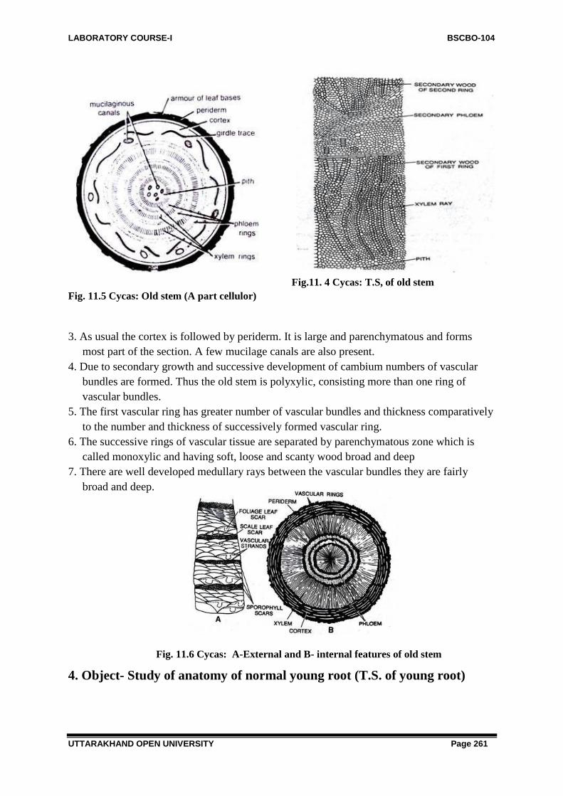
LABORATORY COURSE-I BSCBO-104
UTTARAKHAND OPEN UNIVERSITY Page 261
Fig.11. 4 Cycas: T.S, of old stem
Fig. 11.5 Cycas: Old stem (A part cellulor)
3. As usual the cortex is followed by periderm. It is large and parenchymatous and forms
most part of the section. A few mucilage canals are also present.
4. Due to secondary growth and successive development of cambium numbers of vascular
bundles are formed. Thus the old stem is polyxylic, consisting more than one ring of
vascular bundles.
5. The first vascular ring has greater number of vascular bundles and thickness comparatively
to the number and thickness of successively formed vascular ring.
6. The successive rings of vascular tissue are separated by parenchymatous zone which is
called monoxylic and having soft, loose and scanty wood broad and deep
7. There are well developed medullary rays between the vascular bundles they are fairly
broad and deep.
Fig. 11.6 Cycas: A-External and B- internal features of old stem
4. Object- Study of anatomy of normal young root (T.S. of young root)

LABORATORY COURSE-I BSCBO-104
UTTARAKHAND OPEN UNIVERSITY Page 262
Work Procedure: Cut a T.S. of the young part of primary root, stain in safranin fast green
combination, mount in glycerine and
study.
1. The section is generally circular in
outline. It is identical to that of
dicotyledonous root.
2. There is a single layered epiblema.
Some of these cells bear unicellular root
hairs also.
3. The epiblema or exodermis
surrounding a large thin walled
parenchymatous cortex. This is
multilayered with a few tannin filed
cells are also scattered in this region.
Fig. 11.7 Cycas: T.S. normal root
4. The cortex is made up of by single longitudinally elongated cells.
5. Endodermis is followed by many layered pericycle and forms the outer layer of the stele.
6. Stele is exarch with radial vascular bundles, containing two protoxylem groups and shows
diarch condition.
5. Object- Study of anatomy of older part of normal root (T.S. of older
root)
Work Procedure- Cut a T.S. of older part of normal/ primary root. Stain in safranin- fast
green combinations, mount in glycerine and observe the details.
1. The older stem show secondary growth. The outermost layer
is epiblema which is ruptured due to the thick walled cork cells
below it. The cork cells arranged in brick like fashion and are
few layered deep.
2. Below this layer cortex is large. It is parenchymatous and
multilayered. It is present below the cork, some tannin filled
tannin cells occurs in the cortex.
3. Endodermis is single layered, followed by multilayered
pericycle.
4. Due to secondary growth phloem and xylem is divided into two layers, the outer phloem is
formed the outermost layer near the pericycle and is in crushed form due to secondary
growth. This layer is followed by secondary phloem the cells of this layer are intact (not
crushed).
5. Primary and secondary xylem is situated towards pit.
6. Cambium arcs are formed in the vascular region along the inner edges of phloem.
7. In the centre a parenchymatous pith is present.
8. Medullary rays are also reported.
Fig.11.8 Cycas: A bunch of
coralloid roots

LABORATORY COURSE-I BSCBO-104
UTTARAKHAND OPEN UNIVERSITY Page 263
6. Object- Study of coralloid roots (T.S. of coralloid root)
Work Procedure- Cut a T.S. of the root, stain in safranin- fast green combination, mount in
glycerine, and observe the details.
1. The structure is similar to the primary root. The outermost layer is epiblema. It is single
layered.
2. The cortex is the middle region and occupied the maximum part of the section. It is divided
into three regions- Outer, middle and inner cortex. These are similar in size and are made up
of simple parenchymatous cells.
Fig. 11.9 Cycas revoluta: T.S. coralloid root
3. The middle cortex zone is also known as ‗algal zone‘. It lies midway between the vascular
bundle and the epiblema. The cells of this region are radially elongated and blue green alga-
Anabaena cycadocearum occurs, endophytically in these cells. It is believed that it is
symbiotic in nature and helps in nitrogen fixation.
4. The next layer is endodermis which seperates cortex and vascular bundles. It is single
layered and followed by multilayered pericycle.
5. Vascular bundles are radially arranged and xylem is triarch and exarch.
6. Secondary growth is generally absent; if present it is very less.
7. Object- Study the anatomy of rachis- (T.S. of rachis)
Work Procedure- Cut a T.S. of rachis from its middle portion, stain in safranin-fast green
combination, mount in glycerine and observe the details.
1. The leaf of cycas is large and compound. The leaflets are tough, thin and leathery.
2. The outline of the rachis section is cylindrical and shows insertion of pinnae on the upper
side.
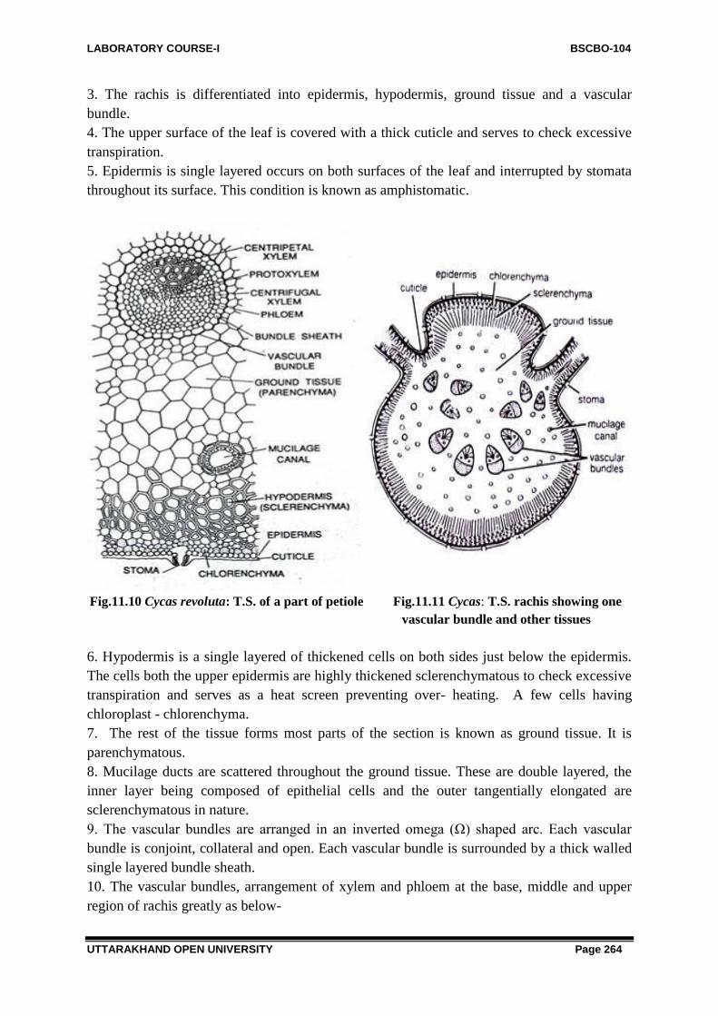
LABORATORY COURSE-I BSCBO-104
UTTARAKHAND OPEN UNIVERSITY Page 264
3. The rachis is differentiated into epidermis, hypodermis, ground tissue and a vascular
bundle.
4. The upper surface of the leaf is covered with a thick cuticle and serves to check excessive
transpiration.
5. Epidermis is single layered occurs on both surfaces of the leaf and interrupted by stomata
throughout its surface. This condition is known as amphistomatic.
Fig.11.10 Cycas revoluta: T.S. of a part of petiole Fig.11.11 Cycas: T.S. rachis showing one
vascular bundle and other tissues
6. Hypodermis is a single layered of thickened cells on both sides just below the epidermis.
The cells both the upper epidermis are highly thickened sclerenchymatous to check excessive
transpiration and serves as a heat screen preventing over- heating. A few cells having
chloroplast - chlorenchyma.
7. The rest of the tissue forms most parts of the section is known as ground tissue. It is
parenchymatous.
8. Mucilage ducts are scattered throughout the ground tissue. These are double layered, the
inner layer being composed of epithelial cells and the outer tangentially elongated are
sclerenchymatous in nature.
9. The vascular bundles are arranged in an inverted omega (Ω) shaped arc. Each vascular
bundle is conjoint, collateral and open. Each vascular bundle is surrounded by a thick walled
single layered bundle sheath.
10. The vascular bundles, arrangement of xylem and phloem at the base, middle and upper
region of rachis greatly as below-

LABORATORY COURSE-I BSCBO-104
UTTARAKHAND OPEN UNIVERSITY Page 265
(i) For most part of the rachis and higher up region, vascular bundles are diploxylic means
two types of xylem elements are present- centripetal and centrifugal xylem. The centrifugal
xylem occurs in two small groups and present on both sides of centrally located triangular
centripetal xylem, while the phloem is situated on the lower side of the rachis.
ii) At the very base of the rachis only centrifugal endarch xylem is present in the vascular
bundle, occurs on the abaxial side of the rachis.
Fig. 11.12 Cycas: A single foliage leaf
Features of Special interest:
i) Presence of chlorenchyma is scattered among the thick walled sclerenchymatous
hypodermis
ii) Presence of sunken stomata all over the leaf surface symbolizes xerophytic characters.
iii) Specific arrangement of vascular bundles e.g. arranged in invested omega (Ω) shaped arc.
iv) Diploxylic nature of vascular bundles.
v) Presence of scattered mucilage ducts.
8. Object- Study of anatomy of leaflet (Pinna)
Work Procedure- Cut a T.S. of leaflet, stain in safranin fast green combination, mount in
glycerine and observe the details.
1. The leaflet has a distinct midrib and the wings.
2. The midrib portion is swollen while the wings on the lateral sides are narrower and
flattened.
3. On the upper side, the upper epidermis is present. It is thick and contains cuticle layer. It
differs in different species.
4. Below the epidermis, hypodermis is present. It is sclerenchymatous. It again differs in
different species.

LABORATORY COURSE-I BSCBO-104
UTTARAKHAND OPEN UNIVERSITY Page 266
Fig. 11.13 Cycas revoluta: T.S. leaflet
5. Mesophyll lies below hypodermis and is well developed and differentiated into upper
palisade layer and lower of spongy parenchyma. The palisade layer functions as the main
photosynthetic tissue. It has elongated columnar cell chloroplasts.
6. Spongy parenchyma lies immediately above the lower epidermis with many intercellular
spaces.
7. On either side of the centripetal metaxylem of midrib bundle and somewhat connected
with that, are present two transfusion tissue which are tracheid like cells.
8. On the either side of the midrib between the palisade and spongy layers is a three celled
thick colourless cells, run parallel to the leaf surface from the midrib to the margin. It
constitutes the accessory transfusion tissue. It is connected with the xylem of the vascular
bundle of midrib through the transfusion tissue
9. The lower side of leaflet is bounded by lower epidermis. It is cuticularized, single layered
and in the midrib region. The lower epidermis possesses sunken stomata.
10. The middle swollen portion of rachis is represented by midrib. It lies a single vascular
bundle surrounded by parenchymatous tissue generally with calcium oxalates. Each vascular
bundle has a definite and thick parenchymatous bundle sheath.
11. The vascular bundle is conjoint, collateral, open and diploxylic similar to upper region of
the rachis.
12. A large, triangular, centripetal xylem and two small group of centripetal xylem are
present.
13. Phloem is lies towards the abaxial or lower side Cambium is present between xylem and
phloem.
Features of special interest
i) Thickly cuticularized upper and lower epidermis.
ii) Lateral views are absent.
iii) In the lower epidermis presence of sunken stomata.

LABORATORY COURSE-I BSCBO-104
UTTARAKHAND OPEN UNIVERSITY Page 267
iv) Transfusion tissue is present.
v) Diploxylic nature of vascular bundle.
Fig.11.14 Cycas revoluta: Leaflet anatomy (cellular)
9. Object- Study of external features of male cone
Work Procedure- Study of male cone attached to the plant.
1. The male or staminate cones are borne singly and
terminating on the male stem.
2. It is shortly stalked, compact, large and oval or conical in
shape and consists of a central cone axis, which is surrounded
by numerous spirally arranged microsporophylls.
3. The apical growth of the stem is continued by the formation
of the axillary buds at the base of the cone. It becomes the new
stem apex. As it grows the male cone is displaced to one side.
Therefore, the stem of cycas male plant is a sympodial.
Fig. 11.15 Cycas: Male cone 4. The outer covering of the male cone is formed by closely set
sterile ends of the microsporophylls usually possessing upcurved apices or apophysis.
10. Object- Study of L.S. of the male cone-
Work Procedure- The male cone is about 60-80 cms. in height and can be split into two
parts thus specimen is studied.
1. The L.S. of cycas shows stalk and cone.
2. Male cone is attached at the apex of the plant by a broad and stout stalk.
3. The cone has a central axis with many microsporophylls.
4. Each microsporophyll is a woody flattened wedge shaped structure consisting of a narrow
lower portion broadened into a flat sterile disc above.
5. Each microsporophyll is attached to the cone axis. The distal part of microsporophyll
which is away from the axis and upcurved is known as apophysis.
6. The microsporophylls at the apex, and at the base of the cone may remain sterile.
7. The upper surface of the microsporophyll is sterile while the lower surface of the
microsporophyll is fertile and bears many microsporangia in groups.
8. The microsporangia are arranged in clusters of three to six.

LABORATORY COURSE-I BSCBO-104
UTTARAKHAND OPEN UNIVERSITY Page 268
9. In the middle part of the cone bears largest microsporophylls while it get gradually smaller
towards the apex and base.
Fig. 11.16 Cycas L.S. male cone
11. Object- Study of microsporophyll and microsporangia
Work Procedure- To study microsporophyll and microsporangia, separate the
microsporophyll from male cone, Study both surfaces- upper and lower ones. Observe the
microsporangia on the lower surface with the help of a magnifying lens.
1. A singe microsporangia is typical in shape. It is
flattened and triangular structure and more or less horizontal and woody, attached by a short
stalk at one end.
2. Internally it is differentiated into two parts i.e. a fertile and a sterile part. The lower fertile
part is somewhat wedge shaped while the sterile part of the microsporophyll is a distal part
which tapers into an upcurved portion or apophysis.
3. The lower or fertile part at its lower or abaxial surface of the microsporophyll bears
microsporangia (pollen sacs) in groups of 3-4 forming definite sori.
Fig.11.17 Cycas: (A) Microsporophyll (B) Microsporangia, (C) dehisced microsporangia
4. The microsporangia in a sorus arise from a common central papilla or protuberance. These
are arranged in sori around central papilla
5. Each sporangia show radial lines of dehiscence and also possess many hair on its surface,
mixed with sporangia.

LABORATORY COURSE-I BSCBO-104
UTTARAKHAND OPEN UNIVERSITY Page 269
12. Object- Study of T.S. of microsporophyll
Work Procedure- Study the slide of T.S. of microsporophyll and observe the details.
Fig.11.18 Cycas: T.S. Misporophyll
1. The section shows microsporangia attached to the micrisporophyll by their short stalk to its
lower surface.
2. A mature microsporangium shows three layered structure. The outer most layer is thick
and cutinized, while the inner layers are thin and collectively known as endothecium. It also
encloses a third innermost layer or tapetum.
3. Inside a single microsporangium (pollen sac) there are numerous microspores remain
enclosed.
4. Many mucilage ducts are also present in the microsporophyll, scattered regularly among
the rounded mesophyll like cells and forming the tissue of the sporophyll.
13. Object- Study of megasporophyll
Work Procedure- Since in Cycas there is no true and compact or properly organized female
cone. Megasporophylls forms a crown at the apex like foliage leaves, so only megasporophyll
can be studied as a specimen.
1. Female reproductive body or megasporophyll arising spirally in acropetal succession at
the apex of the stem.
2. Megasporophylls appear as a crown or rosette and does not affected the apical meristem to
grow further. In each year a crown of megasporophyll is formed. So the number of
megasporo phylls exceeds than the number of leaves.
3. The megasporophylls leave their persistant bases on the stem every year.
4. Each megasporophyll resembles with leaf in its structure and appearance but it is densely
covered with brown hairs and varies in size from species to species.
5. Each megasporophyll is divided into an upper broad leafy portion and a lower stalk
portion.
6. The broad leafy portion is lobed rather pinnate tapering to a point and show a marked
resemblance with the foliage leaves but are much reduced.
7. Each megasporophyll is divided into a lower (proximal) petiole, a middle ovule bearing
position and a upper (distal) pinnately dissected sterile part, while the nature of upper sterile
part varies with species to species.
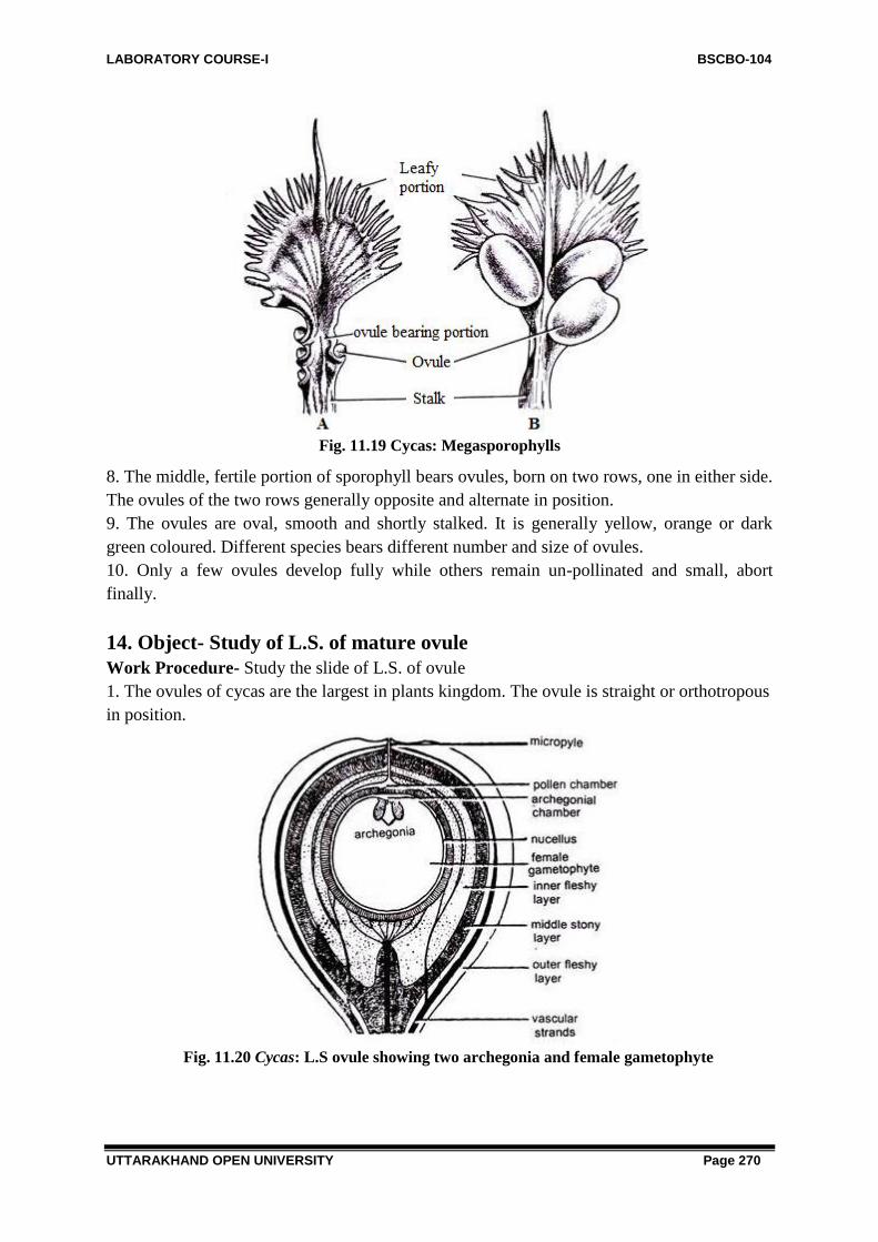
LABORATORY COURSE-I BSCBO-104
UTTARAKHAND OPEN UNIVERSITY Page 270
Fig. 11.19 Cycas: Megasporophylls
8. The middle, fertile portion of sporophyll bears ovules, born on two rows, one in either side.
The ovules of the two rows generally opposite and alternate in position.
9. The ovules are oval, smooth and shortly stalked. It is generally yellow, orange or dark
green coloured. Different species bears different number and size of ovules.
10. Only a few ovules develop fully while others remain un-pollinated and small, abort
finally.
14. Object- Study of L.S. of mature ovule
Work Procedure- Study the slide of L.S. of ovule
1. The ovules of cycas are the largest in plants kingdom. The ovule is straight or orthotropous
in position.
Fig. 11.20 Cycas: L.S ovule showing two archegonia and female gametophyte

LABORATORY COURSE-I BSCBO-104
UTTARAKHAND OPEN UNIVERSITY Page 271
2. Each ovule of the cycas consists of a single massive integument or unitegmic. The
integument is very thick and remain fused with the body of nucellus except nucellar beak and
narmicropyle.
3. The integument in a well- developed ovule consists of three easily recognized layers, an
outer and an inner fleshy layer of rather simple structures and a very complex stone layer
between them.
4. The outer and inner fleshy layers are supplied with vascular strand while the middle stony
layer receives no vascular supply.
5. A few cells of nucellus beak after dissolving themselves from a chamber known as pollen
chamber that lies in the central region of the beak.
6. The innermost region of the ovule is occupied by the tissue of female gametophyte,
consisting two archegonia, situated opposite the pollen chamber.
7. Just above the archegonia is situated the archegonial chamber.
8. The fleshy ovules are oval in shape, orange coloured and each shows a small point at the
distal end which represents the ramnant of the micropyle.
15. Object- Study of L.S. of seed
Work Procedure- Take a L.S. of seed and stain it in safranin fast green combination and
observe the details.
1. Cycas seed shows an outer layer or seed coat, nucellus , embryo and a developing female
gametophyte.
2. Seed coat consists three layers i) outer fleshy layer, ii) middle stony layer, iii) the inner
fleshy layer of the integument.
3. Inside the seed coat nucellus is situated in papery form.
4. The innermost part of the seed is known as endosperm.
5. A single straight embryo remains embedded in the endosperm. It has two unequal
cotyledons.
Fig. 11.21 Cycas: L.S. seed
Identification Points-
Division- Gymnosperms-
i) Absence of vessels, ii) ovules naked, iii) scales generally form a cone, iv) seeds attached
with woody scales.
Class- Cycadopsida
i) Large frond like leaves, ii) seeds with radial symmetry iii) wood monoxylic.

LABORATORY COURSE-I BSCBO-104
UTTARAKHAND OPEN UNIVERSITY Page 272
Order- Cycadales i) Plants woody unbranched stem, ii) wood monoxylic, iii) Plants
dioecious, iv) Mucilage canals present, v) ovules straight or orthotropous.
Family- Cycadaceae
i) Presence of coralloid root and presence of blue green algae, ii) leaves with circinate
venation, iii) megaphylls foliar.
Genus- Cycas
i) Presence of two leaves, ii) Two types of roots iii) foliage leaves pinnately compound,
young leaves circinately coiled, iv) presence of transfusion tissue and diploxylic vascular
bundle in leaf, v) stem possess monoxylic secondary xylem, vi) vascular bundles arranged in
an inverted omega shape in the rachis, vii) male cone is large and single.
11.4- STUDY OF MORPHOLOGY AND ANATOMY OF
VEGETATIVE AND REPRODUCTIVE PARTS OF PINUS
Systematic Position-
Division: Gymnosperms
Class: Coniferopsida
Order: Coniferales
Family: Pinaceae
Genus: Pinus
Fig.11.22 Pinus: Habit sketch
Habitat- The order Coniferales is very large order of division gymnosperms consisting of
nearly 52 living genera and over 500 species distributed throughout the north and South
Temperate Zone and only few representatives in the tropics. In India Himalayas is the chief
centre of conifers. This group includes many economically important species i.e. Pinus
(Pine), Picea (spruce), Abies (fur), Cedrus (cedar), Juniperus (juniper), Taxus (yew), Seqoia
(red wood) etc.
Pinus is the most important genus of the family Pinaceae and is represented by over
two hundred species in the world. Usually they grow on the sloppy and exposed sunny areas.
In India Pinus is represented by five species which grow wild in North -East and North- West
Himalayas. P. roxburghii or P. longifolia, P. gerardiana (chilgoza in Hindi), P. wallichiana
or P. excelsa, P. insularis or P. khasya and P. armandi are common. They are also cultivated
artificially for ornamental value in the plane areas
1. Object- Study of external morphology of plant
Work Procedure- In natural habitat observe type of stem, its size, pattern of branching,
types of branches, types of leaves, male and female cones etc.
1. The adult plant of Pinus represents the sporophytic phase and is well differentiated into
leaf, stem and root.
2. The plant is a tall and the arrangement of branches gives the tree a graceful tapering or
cone like appearance and therefore, this tree is commonly grouped, under group Coniferales.

LABORATORY COURSE-I BSCBO-104
UTTARAKHAND OPEN UNIVERSITY Page 273
3. The upper aerial branch system consists of cylindrical rough and branched stem being
covered with scaly bark.
4. Pinus shows monopodial branching and branches are arranged in whorls.
5. The branches are dimorphic i.e. two type i) branches of unlimited growth or long
shoots and ii) branches of limited growth or dwarf or spur.
6. The long shoots or branches of unlimited growth are present on the main trunk and are
produced in regular intervals.
7. While branches of limited growth or dwarf shoots are borne on the main stem and on
long shoots in the axils of scale leaves. These shoots also possess many scale leaves and
bear foliage leaves at its apex.
8. The leaves are also two types or diamorphic- scale leaves and foliage leaves.
9. Scale leaves are brown, membranous and small. They are present on both the types of
branches- long and dwarf shoots.
10. However, the foliage leaves are acicular and needle like and are borne only on the
dwarf shoots and are usually called as needles. These leaves are simple, have smooth
surface, persistant and evergreen.
Fig. 11.23 Pinus: External morphology of adult plant Fig. 11.24 Pinus: Long & dwarf shoots
showing needle-like leaves and scales
11.Besides the long shoots, there are very numerous branches of limited growth on
ordinary branches in the axils of scale leaves, and often called the dwarf shoots or shoots of
limited growth.
12. A dwarf shoot with a group of needle like foliage leaves is known as foliage spur. The
number of needles in a particular group varies from species to species e.g. in P. monophylla
has a single leaf spur and hence known as monofoliar, while in P. sylvestris, two leaves are
present and so the spur is called bifoliar. In P. longifolia and P.gerardiana, the number is
three, thus the spur being called as trifoliar. In P. quadrifolia, number of spur leaves is four
and pentafoliar, the number of spur is five in P. excelsa and P. walichiana
13. The shape of the needle varies according to their number in a spur e.g. P. sylvestris with
bifoliar spur, the shape of single needle is semi- circular in T.S. while in P. longifolia with

LABORATORY COURSE-I BSCBO-104
UTTARAKHAND OPEN UNIVERSITY Page 274
trifoliar spur a single needle is almost triangular in shape. While P.monophylla with
monofoliar spur the shape of the needle in T.S. is almost circular.
14. The underground root sysyem is formed by tap roots, which disappears after sometime
and later only lateral roots are present.
Fig.11.25 Pinus: Different types of spur
15. The sporophytic plant of Pinus is mostly monoceious i.e. male and female cones are
borne in the same plant.
16. The male cones are borne on lateral branches of unlimited growth. They are produced in
clusters and replaced the dwarf shoots. They are formed earlier than the female cone earlier in
the season.
17. The female cones are developed laterally in the axils of scale leaves. They are usually
produced in clusters in place of shoots of unlimited growth and on different branches from
the male cones. They appear after every three years.
18. Generally male and female cones are formed on different branches.
2. Object- Study of anatomy of young root
Work Procedure- Study the safranin- fast green combination or double stained slide of T.S.
of young part of root and observe the details.
Fig. 11.26 Pinus: T.S. young root
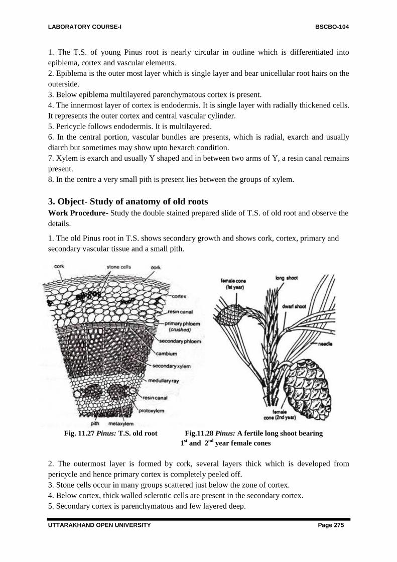
LABORATORY COURSE-I BSCBO-104
UTTARAKHAND OPEN UNIVERSITY Page 275
1. The T.S. of young Pinus root is nearly circular in outline which is differentiated into
epiblema, cortex and vascular elements.
2. Epiblema is the outer most layer which is single layer and bear unicellular root hairs on the
outerside.
3. Below epiblema multilayered parenchymatous cortex is present.
4. The innermost layer of cortex is endodermis. It is single layer with radially thickened cells.
It represents the outer cortex and central vascular cylinder.
5. Pericycle follows endodermis. It is multilayered.
6. In the central portion, vascular bundles are presents, which is radial, exarch and usually
diarch but sometimes may show upto hexarch condition.
7. Xylem is exarch and usually Y shaped and in between two arms of Y, a resin canal remains
present.
8. In the centre a very small pith is present lies between the groups of xylem.
3. Object- Study of anatomy of old roots
Work Procedure- Study the double stained prepared slide of T.S. of old root and observe the
details.
1. The old Pinus root in T.S. shows secondary growth and shows cork, cortex, primary and
secondary vascular tissue and a small pith.
Fig. 11.27 Pinus: T.S. old root Fig.11.28 Pinus: A fertile long shoot bearing
1st and 2
nd year female cones
2. The outermost layer is formed by cork, several layers thick which is developed from
pericycle and hence primary cortex is completely peeled off.
3. Stone cells occur in many groups scattered just below the zone of cortex.
4. Below cortex, thick walled sclerotic cells are present in the secondary cortex.
5. Secondary cortex is parenchymatous and few layered deep.

LABORATORY COURSE-I BSCBO-104
UTTARAKHAND OPEN UNIVERSITY Page 276
6. In the secondary cortex many resin canals are present.
7. Primary phloem is mostly crushed and obliterated and occurs in two groups.
8. Secondary phloem is a few layered deep rings. It consists of sieve tubes, sieve plate,
phloem parenchyma and albuminous cells also.
9. The two circles of phloem- secondary phloem and secondary xylem are separated of a
cambium.
10. Secondary xylem arranged in regular rows and is composed of traicheds. It is traversed by
uniseriate medullary rays.
11. Each primary xylem group is Y shaped. The divided arms always faces the outer side or
away from the pith.
12. In the centre a small pith is present. In which two groups of primary xylem are situated on
opposite radii.
13. Between the divided arms of Y and close to each primary protoxylem group, presence of
large resin canals, the characteristic of Pinus root are present.
4. Object- Study of anatomy of young shoot
Work Procedure- Cut a T.S. of younger part of the long shoot towards the apex, stain in
safranin fast green combination, mount in glycerine, study and observe the details.
1. The transverse section of young Pinus stem resemblances of a great extent that of
dicotyledonous Angiospermic stem.
2. The section displays a wavy outlines and shows the arrangement of tissues as epidermis,
cortex, and stele.
3. Epidermis is single layered, with thick cuticle. The outline of epidermis is irregular due to
presence of bases of dwarf shoot.
4. Cortex is multilayered and situated below the epidermis. The outer few layers forming
which is sclerenchymatous, while the inner layers are thick walled and parenchymatous. A
number of large resin canal ducts are also present in this region. Each resin canal duct is
surrounded by a layer of the epithelial cells. The resin canals are the characteristic feature of
Pinus stem.
5. Endodermis is the innermost layer of the parenchymatous cortex but not marked off.
6. Pericycle is parenchymatous and inconspicuous.
7. The stele is ectophloic siphonstelic.
8. Vascular cylinder is composed of 5-8 vascular bundles, separated by medullary rays. These
bundles are arranged in ring.
9. Each vascular bundle is conjoint, collateral, endarch and open. Each vascular bundle
consists of xylem on the inner side and phloem on the outside with a strip of cambium
between xylem and phloem.
10. Xylem is composed of tracheids and xylem parenchyma only, Vessels are absent, resin
ducts are found in xylem. It is endarch.
11. The phloem is made up of sieve tubes, sieve plate and phloem parenchyma. Albuminous
cells are also present.

LABORATORY COURSE-I BSCBO-104
UTTARAKHAND OPEN UNIVERSITY Page 277
12. In the centre there is well developed pith consisting a mass of parenchymatous cells and
few resin ducts also present in this region. It is also connected with the cortex, but narrow
medullary rays seperates the vascular bundles.
Fig. 11.29 Pinus roxburghii: T.S. long shoot (young)
5. Object- Study the anatomy of the old stem
Work Procedure- Cut a T.S. of old part of the stem, stain with safranin fast green
combination, mount in glycerine and observe the details.
1. The T.S. of old stem of Pinus shows cork, cortex primary and secondary vascular tissues
and pith.
2. The outermost region is formed by the successive layers of cork or periderm, consists of
thick and suberized cells.
3. Next to cork is cork cambium layer. It is made up of few layers of regularly arranged stone
cells occur in many groups scattered below the zone of cork.
4. Below cork cambium or phellogen layer is lies the cortex. It is multilayered and divided
into two zones- the outer cortex and inner cortex.
5. Below the parenchymatous layer secondary cortex is present. Primary cortex is
parenchymatous and multilayered. The resin canals occur irregularly distributed in the region.
6. Inner to the primary cortex is lies primary phloem in small patches of crushed tissue.
7. While secondary phloem occurs in a well distinguished ring form.
8. Secondary growth resulted due to the presence of cambium, results in the formation of
secondary xylem and secondary phloem. Cambium separates the secondary phloem on its
outer side and secondary xylem on its inner side.
9. Secondary xylem shows distinct and sharp annual rings. The ring of spring wood consist
thin walled and large xylem elements. However, the ring of autumn wood consist thick
walled and small elements. The wood is pycnoxylic (compact).
10. The autumn and spring wood alternates one another and together form annual ring and
known as rings of secondary xylem.

LABORATORY COURSE-I BSCBO-104
UTTARAKHAND OPEN UNIVERSITY Page 278
Fig. 11.30 Pinus: Anatomy of stem,
T.S. of stem showing secondary
growth
Fig.11.31 Pinus: Anatomy of Stem. (A) T.S. young stem, (B) T.S. of three year old stem
11. The wood or secondary xylem is composed of tracheids and xylem parenchyma. Vessels
are completely absent. Hence it is also called non- porous wood, (characteristic feature of
Pinus wood).
12. Primary xylem groups are endarch and lies just near the pith.
13. Xylem and phloem is traverse by medullary rays, which runs from primary xylem to
secondary phloem.
14. In between primary and secondary xylem, few resin canals are scattered as in cortex.
15. In the centre, there is a few small parenchymatous pith and many tannin filled cells also
present in the pith region.
6. Object- Study of R.L.S. of the wood-

LABORATORY COURSE-I BSCBO-104
UTTARAKHAND OPEN UNIVERSITY Page 279
Work Procedure- The study of R.L.S. of the Pinus wood, cut a thin section of wood, along
any one of the radii, stain in safranin fast green combination, mount in glycerine and observe
the details.
1. The stem of Pinus is composed of very complex tissue consisting primary and secondary
xylem, the medullary rays etc. To study the detail structures of these tissues is only possible
by R.L.S. and T.L.S. of the stem, because transverse section can only observe simple
structutres of above mentioned tissues.
2. The radial longitudinal section or R.L.S. shows the xylem, cambium, phloem, cortex and
pith.
3. It shows presence of secondary xylem, ray- tracheids and medullary rays.
4. Xylem is composed of tracheids with bordered pits on their radial walls. In this section the
bordered pit are seen in surface view.
Fig. 11.32 Pinus: R.L.S. Wood
5. Borderd pits are rounded or circular areas surrounded by special cellulose thicknings
known as crassulae or Bars of Sano. If pits are close to one another or the bars are fused and
form Rims of Sanio.
6. In radial longitudinal section the medullary rays are cut length- wise and so their length
and height can be noticed easily.. They run horizontally and are uniseriate.
7. Each medullary ray is made up of ray cells, ray- tracheids and paremchyma.
8. On both the sides of the medullary ray cells, ray tracheids are present only in the region of
xylem. These cells are thick, narrow and long, and show bordered pits.
9. In the region of phloem, medullary- rays replaces, ray tracheids with albuminous cells.
They are small and contents are dense. The ray parenchyma is associated with these cells are
filled with large amount of starch.
7. Object- Study of T.L.S. of wood

LABORATORY COURSE-I BSCBO-104
UTTARAKHAND OPEN UNIVERSITY Page 280
Work Procedure- Cut a thin section of wood along the tangent in the outer region, stain in
safranin fast green combinations, mount in glycerine and observe the details.
1. Medullary rays and tracheids are cut transversely in this place.
2. The bordered pits forming a dome- like structure and cuts in a manner that show
overarching borders. It encloses in the cortex a small disc, called Torus.
3. Medullary rays cut transeversly since they appers uniseriate and their height and bredth can
thus be determined.
4. Each medullary ray appears to be a short row of more or less rounded cells, three or four
cells high.
5. Composition of medullary rays reveals centrally placed thin walled and living cells or
albuminous cells, in the phloem region and the ray cells in the xylem region respectively.
6. These are surrounded on the both, lower and upper sides by thick walled and dead cells
known as tracheids.
Fig. 11.33 Tracheids with bordered pits of pine stem in T.L.S.
8. Object-Study of T.S. of dwarf shoot at the base (before secondary
growth)
Work Procedure- Take out a spur (dwarf shoot), insert it with needle downwards the base
is now the uppermost, cut T.S. at the base, mount in glycerine and observe the details.
1. The T.S. of dwarf shoot almost resembles with that of the stem.
2. Presence of scaly leaves shows the outline in wood.
3. The T.S. is differentiated into epidermis, cortex and stele.
4. The outermost layer is single layered
5. Cortex follows epidermis which is divided into two parts- the outer few layers, close to the
epidermis are thick walled, while the inner layers are thin - walled and parenchymatous in
nature.
6. Just below the cortex, is single layered endodermis is followed by pericycle. Both the
layers are indistinguishable.
7. Stele is an ectophloic siphonostele.
8. Vascular bundles vary in number with sepecies to species. Generally they are six. Each one
is conjoint, collateral, endarch and open.
9. In the centre, resin canals are present. These are six in number. Some irregularly scattered
tannin cells are also present in this region.
LABORATORY COURSE-I BSCBO-104
UTTARAKHAND OPEN UNIVERSITY Page 281
10. In the centre, small pith is present. The cells are thick walled.
11. Medullary rays play the role of connecting tissue, between cortex and pith and seperates
vascular bundles from one another.
Fig. 11.34 Pinus: T.S. dwarf shoot Fig.11.35 Pinus : T.S. old dwarf shoot
a part enlarged
9. Object- Study of T.S. of dwarf shoot at base
Work Procedure- Cut a T.S. of dwarf shoot above the base, stain in safranin fast green
combination, mount in glycerine, study and observe the details.
1. The section of T.S. of dwarf shoot shows a little amount of secondary growth.
2. The section shows, single layered thick - walled epidermis, remarks of scale leaves are also
present outside the epidermis. It also shows few layers of cork cells and tannin filled cells.
3. In the centre xylem and phloem tissues shows division due to secondary growth and thus
primary phloem is crushed and form patches while secondary phloem underlies it and form a
complete ring.
4. Secondary xylem is small and separated by a thin ring of cambium from the phloem
region. Medullary rays traversed the secondary xylem.
5. Protoxylem is endarch lies just near the pith. It is small and consists of thick walled cells,
while, few tannin filled cells are also present.
10. Object- Study of T.S. of needle (leaf)
Work Procedure- Cut a thin T.S. of a needle, stain with safranin fast green combination,
mount in glycerine and observe the details.

LABORATORY COURSE-I BSCBO-104
UTTARAKHAND OPEN UNIVERSITY Page 282
Fig. 11.36 Pinus: T.S. upper part of dwarf shoot showing the formation of three needles in a
trifoliar spur (Diagramatic)
1. The anatomy of leaf that it is adopted to endense severe environmental conditions. It is not
flat but is about as thick as broad.
2. In a cross section it is shaped like the tri- sector of a circle with the curved surface facing
outwards and vertex inwards.
3. The outline of the section varies according to the species. Triangular if spur is trifoliar,
semi-circular if spur is bifoliar and compeletely circular if spur is monofoliar etc.
4. The needle of Pinus is differentiated into epidermis, mesophyll and stele.
5. Epidermis forming the outer boundary of the leaf is a single layered with tangentially
elongated and thickly cuticularized cells.
6. The deeply sunken stomata are present on sides of the leaf. The leaf (needle) is thus said to
the amphistomatic. They are developed in longitudinal rows.
7. Epidermis is followed by hypodermis. It is few layered thick at the corners and 1-2 layered
in other parts, cells are sclerenchymatous. sub- stomata chambers occurs in this region.
8. Mesophyll lies below the hypodermis. It is compact and shows no differentiation into
palisade and spongy tissues. It is made up of polygonal parenchymatous cells, densely filled
with the chloroplasts. Numerous plate- like or peg- like infoldings, project into the cell lumen
or cavity, from the walls of the mesophyll cells.
9. The mesophyll contains a number of resin ducts immediately under the hypodermis.
10. The central tissue of the leaf is enclosed by a conspicuous single layered endodermis
which delimits the mesophyll. Cells are barrel shaped and tangentially thickened. It is
followed by multilayered parenchymatous pericycle.
11. Generally two vascular bundles remain surrounded by this tissue but in P. strobus there is
only one vascular bundle.
12. T- shaped thick walled transfusion tissue seperates the vascular bundles from one-
another.
13. Each vascular bundle is conjoint, collateral and open. Protoxylem is faces, adaxial side
while phloem is located on the abaxial side.
14. Xylem and phloem groups are separated from one another by cambium at the base of the
needle and in the upper region by parenchymatous cells.

LABORATORY COURSE-I BSCBO-104
UTTARAKHAND OPEN UNIVERSITY Page 283
15. Secondary growth is observed very little during this the medullary rays run between
xylem and phloem.
Fig. 11.37 Pinus: T.S. needle (cellular)
Features of Special Interest-
i) Leaf is acicular of or narrow and needle like.
ii) Presence of thick cuticle in the upper region.
iii) Amphistomatic nature.
iv) Sunken stomata present.
v) Hypodermis is thick and sclerenchymatous.
vi) Presence of transfusion tissue.
vii) Simple vascular system.
viii) Infolded peg- like structures in the mesophyll.
11. Object- Study of male cone, microsporophylls and microsporangia
Work Procedure- For this dissect the male cone,
separate the microsporophylls, study and observe the
shape, size and microspores.
1. Pinus is monoecious as it bears both types of cones
on the same tree on separate branches. The main
shoot, on which they are produced continues to grow
further.
2. The male cone occurs laterally in clusters, each in
the axils of a scale leaf at the base of a terminal
vegetative bud which they surrounded.
3. Male cones are grouped in clusters on the shoots
of the same year only. Fig. 11.38 Pinus: male cone

LABORATORY COURSE-I BSCBO-104
UTTARAKHAND OPEN UNIVERSITY Page 284
4. A single male cone has single, centrally located cone axis around. Which many scaly
microsporophylls arranged spirally.
5. Each microsporophyll has an expanded triangular central part and stalk like base in which
the terminal part projects into a tip.
6. Most of the upper sporophylls are fertile but a few lowermost sporophylls are sterile and
do not bear any male reproductive structures.
7. On the abaxial side each microsporophyll bears two ovoid microsporangia or pollen sacs
on its lateral sides.
8. Each microsporangium has its own wall, encloses many microspores inside it.
9. The young microspore is globular or spherical in shape and is uninucleate.
10. A mature pollen grain or microspore shows two wall layers- exine and intine, 2 prothelial
cells and one antheridial cell.
11. Each pollen grain has a thick expanded exine in the form of wings on the lateral sides,
followed by smooth intine.
12. Object- Study of L.S. of male cone
Work Procedure- Study a slide of L.S. of male
cone and observe the details.
1. Each cone is simple, ovoid; structure usually
the male cone shows a cone axis is located
centrally.
2. The male cone is shortly stalked and consists of
an elongated central axis, bearing a number of
dwarfs spirally arranged and closely fitted scale
like microsporophylls. These are triangular and
expanded.
Fig.11. 39 Pinus: L.S. male cone
3. The outer expanded part is sterile and is known as apophysis.
4. Microsporamgia are present on the lower or abaxialsurface.
5. A single microsporangium has a wall layers and tapetum.
6. The sporangium wall consists of epidermiswall- layers and tepetum.
7. The sporangia are filled with numerous winged microspores in various stages of
development.
Fig. 11.40 Pinus: Pollen grains

LABORATORY COURSE-I BSCBO-104
UTTARAKHAND OPEN UNIVERSITY Page 285
13. Object- Study the morphology of the female cone
Work Procedure- To study the external features of 1st, 2
nd and 3
rd year female cones.
Observe the position, arrangement and structure of sporophyll.
1. The female cones are fewer in number and arise single
or in a small cluster or two or four in number.
2. When young it is small, erect, and reddish to pinkish
structure, which stands erect on a short stalk covered with
scales.
3. Female cones are larger than the male cones. They are
borne at the apices of the young elongated shoots,
replacing the shoot of unlimited growth or long shoots.
4. In the 2nd
year cones are compact and sporophylls are
closely arranged and large in size and woody in nature.
Fig. 11.41 Pinus: Female cone
5. In the 3rd
or final year cones becomes loose. Sporophylls separate from one another due
to elongated of cone axis as the female cones takes three years for maturation.
6. Each female cone consists of many sporophylls, all are arranged spirally around the axis.
I Year II Year III Year
Fig.11.42 Pinus: Different stages of female cone, (A, B & C)
14. Object- Study of L.S. of female cone
Work Procedure: Observe the slide of L.S. of female
cone.
1. Each female cone consists of a central axis to which are
attached the megasporophylls.
2. Each megasporophyll has an outer small bract scale and
a large ovuliferous.
3. Many small and thin bract scales are arranged spirally
around the cones. They are directly borne on the cone
axis. Each of these are present on the lower side of the
ovuliferous scale. Fig.11.43 Pinus: L.S. female cone

LABORATORY COURSE-I BSCBO-104
UTTARAKHAND OPEN UNIVERSITY Page 286
4. A thick, large woody and triangular ovulifeous scale is present on the upper side of the
bract scale.
5. The ovuliferous scales thicken and become enlarged.Their broad sterile ends fit tightly
over one another so as to form rhomboidal areas on the outer surface.
6. The bract scales remain small and concealed.
7. The structure of ovuliferous scale is typical, which is largest in the middle part of the cone
and get gradually smaller towards its base and apex.
Fig. 11.44 Pinus, Different types branches with male and female cones.
8. Both the scales - Ovuliferous and bract scales are fused for a little distance near the cone
axis while free a distance away from it.
9. The ovuliferous scale is shortly stalked and rest of the part is expanded.
10. At the base of this expanded, triangular part two nacked and sessile ovules are present,
situated on the upper or adaxial surface of the ovuliferous scale at its base, bearing their
micropylar, directed towards cone axis.
11. The terminal part of the ovuliferous scale is known as apophysis, which is broad and
sterile comparatively to the lower narrow fertile part of the scale.
15. Object- Study of L.S. of ovule
Work Procedure- Study a preprepared slide of L.S. of Ovule, observe its different parts i.e.
integuments, nucellus, female gametophyte and archegonia.
1. Pinus ovule is elongated in size.
2. It is unitegmic in nature and the integument is three layered. The outermost layer is thin.
The middle layer is stony and prominent while the third or innermost layer is fleshy and well
developed.
3. Nucellus is fused with inner fleshy layer of the integument all its length except at its tip
where it forms an elongated and slender micropyle, directed towards the cone axis or tip of
the seed.
4. A pollen chamber, in the form of a small cavity is lies in the nucellus region just opposite
the micropyle.

LABORATORY COURSE-I BSCBO-104
UTTARAKHAND OPEN UNIVERSITY Page 287
5. The whole above structure forms the endosperm or female gametophyte, which is
differentiated from nucellus. About 2-5 archegonia are situated in this region of the
archegonial chamber at the micropylar end near the base.
Fig. 11.45 Pinus, L.S. of mature ovule
16. Object- Study of seed
Work Procedure- Study the position and arrangement of seed on ovuliferous scale along
with the prepared slide of L.S. of seed and observe the details.
1. After fertilization the ovules get transformed into seed, situated on the upper/ adaxial side
of the ovuliferous scale at its base near the cone axis.
Fig.11.46 Pinus: Seed with wings
Fig.11.47 Pinus: Ovuliferous scale
bearing two winged seeds
2. It is rather complicated structures and is formed when the cone is running into its third
year.
3. There is the outer hard shell or seed coat called the testa. It protects the inner delicate parts
of the seed from drying and mechanical injury. It is red and brown in colour.
4. Seeds are small, elongated and winged. The wings are the thin layer of tissue which splits
off from the upper face of the ovuliferous scale.
5. Within the testa there is inner fleshy layer of the integument which is still persists. It is
thin, papery and membranous and termed as tagment.
6. Towards the pointed end of the endosperm or seed, nucellus cap is present, which
represents the remains of nucellus in the form of a thin layer at the micropylar end.
7. The larger part of the seed consists of oily endosperm.

LABORATORY COURSE-I BSCBO-104
UTTARAKHAND OPEN UNIVERSITY Page 288
8. Embryo is differentiated into radicle, plumule and cotyledons. The tip of the radicle is
attached to a dry, coiled, thread like structure, the dried- up suspensor.
9. A well- developed hypocotyle is present in between radicle and plumule.
17. Object- Study of seedling
Work Procedure-Study a newly germinated seed and observe its various parts.
1. Newly germinated seed or seedling shows three clear parts-
i) root, ii) hypocotyl and iii0 leaves.
2. Roots arise from the radicle and are well branched.
3. Hypocotyl develops into un -branched, slender and primary shoots.
4. Leaves are green and needle- like which are borne on the primary shoot. These are known
as cotyledonary leaves.
5. The first spur shoots /dwarf shoots or the primary leaves arise in the axils of some of these.
Juvenile or cotyledonary leaves and are borne in spiral series on the primary shoot.
Fig.11.48 Pinus: Seedling showing growth
Points of Identification
Division- Gymnosperms
i) Absence of vessels ii) Seeds attached iii) Naked ovules iv) Scales generally form a cone.
Class- Coniferopsida
i) Needle shaped leaves ii) Wood compact or pycnoxylic iii) Presence of resin canals iv)
Compact male and female cones v) Male gamete non- flagellate vi) Seeds bilaterally
symmetrical.
Order- Coniferales
Family- Pineaceae
i) Wood is resinous ii) Plants are monoecious iii) Spirally arranged sporophylls- micro and
megasporophylls iv) Microsporophyllsbears two microsporangia v) Winged pollen grains
vi) Female cone is woody vii) Polyembryony present and viii) Seeds are dry and winged.
Genus- Pinus
i) Main plant is sporophytic and monoecious,ii) Male and female reproductive organs are in
the form of cones.iii) Dimorphic branching present,iv) The dwarf shoots shows little
secondary growth while long shoots shows secondary growth and secondary xylem, annual
rings are present, v) Wood pycnoxylic and resinous,vi) Leaves are of two types i.e. scale
leaves and foliage leaves.vii) Scale leaves are brown and membranous while the foliage

LABORATORY COURSE-I BSCBO-104
UTTARAKHAND OPEN UNIVERSITY Page 289
leaves are acicular, greon, xerophytic, mesophyll cells with peg- like ingrowths, resin canals
and T- shaped transfusion tissue.viii) Female cones are borne single and at terminal point,
while male cones borne laterally in clusters and each microsporophyll bears two
microsporangia at its lower side.ix) Bract scales are ovuliferous scales are arranged
spirally.x) Two nacked ovules present on the adaxial or upper side of the ovuliferous scale.xi)
Seeds dry, winged and oily.
11.5- STUDY OF MORPHOLOGY AND ANATOMY OF
VEGETATIVE AND REPRODUCTIVE PARTS OF EPHEDRA
Systematic Position
Division: Gymnosperms
Class: Gnetopsida
Order: Gnetales
Family: Ephedraceae
Genus: Ephedra
Fig.11.49 Ephedra: Habit sketch
Distribution and Habitat-The genus Ephedra is widely distributed in both of Eastern as well
as Western hemisphere. It grows in Southern part of North America, Central part of South
America, North Africa, Spain< Germany, Portugal, France, Italy, China, Baluchistan,
Afghanistan etc.
The genus Ephedra includes nearly 42 species of which about six occur in India. E.
foliata variety grows widely as a scandent shrub climbing over small trees in the southern
parts of the Punjab and Rajasthan. It is also cultivated in gardens. E. foliata, E. intermedia,
E. gerardiana, E. sextilis, E.religiana and E.nebrodensis reported from different parts of
India.
1. Object- Study of external features
Work Procedure- To study the external features of the plant, observe the structure of plant,
its joined nature, scale leaves and underground tap root, and the positions of male and female
strobilii.
1. Ephedra plant is small, woody, much branched shrubs. It is bushy, trailing or climbing
shrubs attaining height of less than 2 meters.
2. The plant body consists of a profused branching stem. The stem is green, hard, ribbed,
glabrous and possess only minute leaves in pairs at the nodes.
3. The plant is differentiated into short nodes and long internodes, therefore, resembles
superficially with the Psilotum and Equisetum.
4. It is differentiated into stem, leaves and underground roots.
5. The roots are long, bearing a tp deep tap root and many adventitious roots.
5. Two to three branches arranged in whorls arise from the nodes in the axils of leaves. The
branches are shed off during dry season.

LABORATORY COURSE-I BSCBO-104
UTTARAKHAND OPEN UNIVERSITY Page 290
6. The older part of the stem shows secondary growth and may bear many branches. Due to
secondary growth it becomes hard and woody.
7. Leaves arises at each node in a whorl of 2-4.
8. Leaves are usually arranged in opposite and decassate manner and are jointed connate to
each other at the base to form a small sheath around the node.
9. Each leaf or scale is inervated by two un- branched veins run parallel to each other. They
bear a bud each in their axils.
10. Foliage leaves are completely absent.
11. The reproductive organs- male and female organs are borne in small strobili.
12. The plants are mostly dioecious and bear only one type of reproductive organs.
Sometimes they may be monoecious too when they bear both of strobili.
2. Object- Study of T.S. of young stem
Work Procedure- Cut a T.S. of young part of the stem, stain in safranin fast green
combination, mount in glycerine and observe the details
1. The outline of the section is circular
with ridges and furrows.
2. The internal tissues are differentiated
into epidermis, hypodermis, cortex and
stele.
3. The outermost layer is epidermis,
made up of single layered
parenchymatous cells. It is very thick
and heavily cuticularized.
4. The continuity of epidermis is
interrupted by the sunken- stomata,
situated in circular pits and are located
only in the furrows.
Fig. 11.50 Ephedra: T.S. young stem (Diagramatic)
5. Hypodermis is sclerenchymatous and occurs in small patches below the ridges.
6. Cortex is distinguishable in to two zones. The outer chlorenchymatous cortex consists of
elongated palisade like cells full of chloroplasts. These are arranged loosely and large
intercellular spaces are present between these cells. It is called the photosynthetic zone.
7. Next to it is the parenchymatous cortex, made up of thin walled also possessing
chloroplast.
A few patches of sclerenchyma dispersed in the cortex, mostly in young stem rendering
hardness and resistance.
8. Endodermis is the last or innermost layer of cortex and is quite distinct in young stem
followed by pericycle which is not clearly defined.
9. Stele is ectophloic siphonostele, composed of many vascular bundles, their number being
variable.

LABORATORY COURSE-I BSCBO-104
UTTARAKHAND OPEN UNIVERSITY Page 291
10. A few vascular bundles are arranged in a ring. Each is conjoint, collateral, endarch and
open.
11. Both the phloem groups i.e. external and internal ones are separated by a narrow strip of
cambium.
Fig. 11.51 Ephedra: T.S. Young stem, a part cellular
12. In the centre parenchymatous pith is present.
13. Presence of nodal diaphragm is the characteristic anatomical feature of Ephedra, means
the presence of diaphragm like plate of cells at the base of each internode or at the node is
known as nodal diaphragm. This is the intercalary meristem whose cells becomes thick-
walled and helps in abscission or shed off the branches.
3. Object- Study of T.S. of stem with secondary growth (Old stem)
Work Procedure- Cut a T.S. of the older part of the stem, stain in safranin fast green
combination, mount in glycerine and observe the details.
1. The section is differentiated into epidermis, cortex, and primary and secondary vascular
tissues.
2. It is affected by means of vascular cambium present in between the primary phloem and
primary xylem.
3. The epidermis and cortex remain un-changed. However, after 3-4 years the growth is just
outside the phloem and outer tissue- e.g. epidermis, cortex etc. cork develops and therefore,
these tissues cut off.
4. Just above the zone of secondary tissue stone cells or sclerotic cells develop.
5. Due to cambial activity phloem is divided into primary phloem and secondary phloem.
Primary phloem occurs as obliterated patches.
6. Secondary phloem forms a zone below. Phloem is composed of sieve tubes and phloem
parenchyma.

LABORATORY COURSE-I BSCBO-104
UTTARAKHAND OPEN UNIVERSITY Page 292
Fig.11.52 Ephedra: T. S. old stem showing
secondary growth (Diagrammatic)
Fig.11.53 Ephedra: T.S. old stem, a part Cellular
7. The secondary xylem consists of vessels, tracheids and scanty amount of xylem
parenchyma. Xylem fibres are absent.
8. Annual rings are distinct comprising autumn and spring wood each. These are formed in
secondary xylem or wood.
9. Autumn wood is made up of smaller cells, while those of spring wood are bigger in size.
10. The secondary xylem (wood) shows a thin walled spring wood and thick walled autumn
wood, which are formed in alternating zones.
11. The tracheidial cells of the secondary xylem or wood is associated with broad vessels.
Although absence of vessels is the characteristics of Gymnosperms, but Ephedra itself is an
exception.
12. Abundant number of vessels presents in the spring wood whole a few or none are
reported in the autumn wood. Spring wood is often ring porous.
13. The vessels and tracheids have bordered pits that may have uniseriate arrangement or
may be irregularly scattered. Protoxylem elements of primary xylem show spiral, annular or
reticulate tracheids.
14. Medullary rays are uniseriate in the young stem but very broad and long (multiseriate) in
the old stem.
15. Medullary rays traversed in the wood. The primary medullary rays runs, from primary
xylem to primary phloem while secondary medullary rays run from secondary xylem to
secondary phloem.
16. Primary xylem groups are endarch and are present at the end of the secondary wood near
the pith.
17. In the centre lies large parenchymatous pith.

LABORATORY COURSE-I BSCBO-104
UTTARAKHAND OPEN UNIVERSITY Page 293
Features of Special Interest-
Ephedra stem shows the following xerophytic characters
i) Epidermis is thickly cuticularized. ii) Stomata are sunken iii) Presence of palisade and
spongy parenchyma in the cortex iv) Presence of patches of sclerenchyma v) Presence of
nodal diaphragm vi) Shedding of branches vii) Presence of vessels in the secondary wood or
xylem.
4. Object- Study of R.L.S. of wood
Work Procedure- Cut a thin section of wood along any one of the radii, stain in safranin
fast green combination, mount in glycerine and observe the details
1. Due to secondary growth it shows the presence of secondary xylem and medullary rays.
2. Secondary xylem consists of tracheids and bears bordered pits on their radial walls.
3. The bordered pits of xylem mostly circular or slightly elliptical. Sometimes due to the
dissolution of walls of cavities of pits there may form reticulations. Such specific
performation, being characteristic of Ephedra and are known as Ephedroid perforations.
These pits are either scattered or arranged in 2or 3 tight rows.
4. Below the pit special cellulose thicknings are also present and known as Bars of Sanio.
5. In the secondary xylem few vessels also present which shows bordered pits and are either
scattered or may remain arranged in 2 or 3 rows.
Fig.11. 54 Ephedra: R.L.S. of wood
6. The aperture of the border pits are commonly horizontal oriented. There end walls are also
perforated.
7. Medullary rays are uniseriate or multiseriate, and run horizontally. In R.L.S. plane
medullary rays are cut lengthwise and so their length and height can be observed easily.
8. The height of medullary rays may range upto 40-50 cells heigh.
9. In the secondary xylem region the medullary ray is composed of ray cells and ray-
tracheids regularly.

LABORATORY COURSE-I BSCBO-104
UTTARAKHAND OPEN UNIVERSITY Page 294
10. The ray cells are thick as well as thin walled and occur in the same medullary ray. Their
tangential walls possess bordered pits or slit- like openings.
11. Ray tracheids has thick walls and their radial and tangential walls bears pits, pits being
bordered.
5. Object- Study of T.L.S. of wood
Work Procedure- Cut a transverse longitudinal
section (T.L.S.) of wood by the sharp rajor along
any one of the tangent, stain in safranin fast
green combination, mount in glycerene and
observe the details
1. In T.L.S. plane tracheids, vessels and
medullary rays are cut transversely.
2. Bordered pits are visible in surface view.
Fig.11.55 Ephedra: T.L.S. of wood
3. The bordered pits show usual over- arching dome- shaped structure and a small disc- torus.
4. Medullary rays are cut transversely and their height and breadth can be determined in this
tissue.
5. Rays are elongate in shape and many tangential walls show simple slit-like pits.
6. Object- Study of male cone
Work Procedure- Study the position, structure and male strobilus by observations of
external morphology of the strobilus and a slide of longitudinal section of the same.
1. The plant of Ephedra is sporophytic. Male and female reproductive organs are borne on
small strobili.
2. The male or microsporangiate strobili develop on axils of leaves.
3. The origin and position of strobili is different in different species as follows-
i) The strobilus may termed as monosporangiate when male and female strobili mat be borne
in two different plant, then the species is termed as diocious.
ii) In another case both the strobili may arise in a single plant then the species is known as
monoecious.
iii) While a few plants, sometimes bear both the reproductive organs in one strobilus only
such species are known as biosporangiate, e.g. E. foliata and E. intermedia, in such cases
male flower which occurs at the higher level in the same strobilus.
4. The strobilus resembles inflorescence or spike.
5. The microsporangiate strobilus of Ephedra is regarded as a compound structure. These
strobili arise in clusters from the nodes of branches.
6. Each strobilus arises in the axils of a scale leaf. Each strobilus consists of an axis which
bears decussately arranged sterile spikes and stamens. The number of pairs varies fom 2-12.
7. Each spike is generally round in shape but may be ovoid or spherical.
8. In the axils of each bract arises a single male flower.

LABORATORY COURSE-I BSCBO-104
UTTARAKHAND OPEN UNIVERSITY Page 295
Fig. 11.56 Ephedra: a part of male plant Fig.11. 57 Ephedra: A-Compound male
strobilus, B- L.S. male strobilus
7. Object- Study of male flower
Work Procedure- To study the structure of male
flower study it under dissecting microscope,
separate the sterile scales to isolate the single male
flower, stain in safranin only, mount in glycerine,
observe the details and also compare with the slide
of L.S. of a stamen.
Fig. 11.58 Ephedra: A single male flower
1. Male flower has a perianth of bract scales which encloses the stamen.
2. A single stamen has two parts, the lower one is a long stalk, column or antheridiophore and
the upper broader one is the microsporangia or anther, bears 2-5 anthers at its tip.
3. A single microsporangium bears two chambers or is bilocular structure. It has two outer
wall layers and an inner prominent tapetal layer which encloses pollen grains or microspores.
4. Each microspore is elliptical and consist an outer thick and ribbed exine and a thin intine.
5. A microsporangium opens by apical part and hence shows apical dehiscence.
8. Object- Study of female strobilus-
Work Procedure- To study the position, structure and organization of the female strobilus
study and observe the external features and a slide of its longitudinal section and note the
details.

LABORATORY COURSE-I BSCBO-104
UTTARAKHAND OPEN UNIVERSITY Page 296
1. The strobilus looks like a spike inflorescence.
2. Each female flower arises in the axils of a
fertile bract or scale leaves. It is also sessile and
not so richly branched as the male flower as it
possess short stalk.
3. Each female strobilus is an elongated structure
pointed at the apex and consists a central axis with
2, 4 or more pairs or opposite decussate bract.
4. The apex ovulate strobilus is mostly acute.
Fig.11.59 Ephedra: branch with female strobilus
Fig.11. 60 Ephedra: Female strobilus Fig.11.61 Ephedra trifurca, L.S.Ovule
5. Lowermost 1 or 2 pairs are sterile while terminal pairs bear short, stalked ovules. The
bracts are generally dry, winged and variously coloured.
6. Each bract mostly encloses two ovules among which one may be aborted.
9. Object- Study of L.S. of Ovule
Work Procedure- Study a prepared slide of L.S. of ovule
and observe the details
1. The megasporangium or ovule has two integuments
(layers).
2. The outer integument consists of 4 basally coherent
bracts, sometime collectively referred to as perianth. It is a
cup- like structure, attached at the base of the ovule and
remains free above.
Fig.11.62 Ephedra bracts

LABORATORY COURSE-I BSCBO-104
UTTARAKHAND OPEN UNIVERSITY Page 297
3. The inner integument consists of two bracts also fused at base. It is delicate, composed of
two segments and prolongs into a tubular process
and comes out beyond the bracts and involucre at
the time of pollination.
4. In the upper region of the ovule and in between
the integument, there is an opening known as
micropyle.
5. Below the integuments, nucellus is lies. A small
pollen chamber is present just below the micropyle
in the tissue of nucellus.
6. Below the pollen chamber, the gametophyte
tissue is situated. In the female gametophyte just
below the pollen chamber, two archegonia are
present.
7. Haustorial region lies opposite the micropylar
end. It is occupied by tissue filled with stored food
material. It is also gives out haustorial processes
for the absorption of food and is also known as
haustorial region.
10. Object- Study of L.S. of seed
Work Procedure- For this study the double stained prepared slide of L.S. of seed studied
and observe the details
1. The seed is encloses within an outer fleshy envelop formed by the two subtending bracts of
the megasporangiate strobilus.
2. Outer integument encloses entire seed. It is hick walled.
3. The inner integument persists at the micropylar end only.
4. Nucellus is located inside the inner integument.
5. Female gametophyte or endosperm surrounds a big embryo with two large cotyledons.
6. Bracts situated adjacent to strobilus are fleshy and thick in a completely mature seed.
These forms an additional envelop.
Identification Points-
Division- Gymnosperms i) Plants are sporophytic in natue and divided into root, stem and
leaves. ii) Ovules are nacked,iii) Seed attachd to a scale, iv) Scales form a strobilus.
Class- Gnetopsida i) Wood with vessels, ii) Flowers in compound strobili or inflorescence,
unisexual usually dioecious, iii) Ovules surrounded by several envelop.
Order- Gnetales i) Leaves opposite or whorled and simple, ii) Plants are woody, trees, shrubs
or lianas.
Family- Ephederaceae i) Plants are either shrubs or woody climbers. Ii) Leaves scaly, foliage
leaves are absent, iii) Nodal diaphragm is present, iv) Stamens enclosed by bract, v) Seeds
covered with fleshy bracts.
Fig. 11.63 Ephedra foliata: L.S.
mature seed

LABORATORY COURSE-I BSCBO-104
UTTARAKHAND OPEN UNIVERSITY Page 298
Economic Importance of Gymnosperms-
The Gymnosperms are very important from economic point of vew. Canada balsam, a chief
mounting medium generally used in biological laboratories is the resin of Abies balsamea.
While some conifers such as Cedrus deodara (vern, deodara) form valuable timber.
Turpentine oil, chiefly used in medicine is extracted from a conifer tree i.e. Pinus species.
Paper is also prepared from the pulp wood of many pine species. Many species of Ephedra
are also used for medicinal purposes, especially in China in their Traditional Chinese
Medicine or TCM. The world famous Epherine drug is obtained from this valuable plant.
Ephedra gerardian. Beside this the Gymnosperms are also valuable for their food value viz.
Sago palm or Sabudana in Hindi is also obtained from a gymnosperms- Cycas revoluta.
11.6 SUMMARY
Cycas- Cycas is the genus and the only genus recognized in the family Cycadaceae. About
113 species are accepted. Cycas are native to Japan and southern China and specially to Asia
Pacific. Cycas revolute, sago palm, (also known as king palm, sago cycad, Japenese sago
palm,) is a species of Gymnospers in the family Cycadaceae. Cycas circinalis, a species
endemic to India. The best known Cycas species is Cycas revoluta. This species is most
primitive living seed plant and are also very useful and popular ornamentals with rugged
trunk topped with whorled feathery leaves and thus is only Cycas plant from its genus which
is sold in garden stores.
Pinus- Conifers is a major group within the gymnosperm plants. Pinus is the largest genus
with about 120 species, Pinus is the sole genus in the sub family Pineaceae. Pinus
roxburghaii, known as chir pine, it is a native of the Himalaya. P. contorta used for ecological
restoration of western forests. Pinus trees are major source of timber wood and is used for a
variety of commercial purposes. Pine wood is widely used in high value carpentry items such
as furniture, window frames, paneling, floors and the resin of some species is an important
source of turpentine.
Ephedra- Ephedra is a genus of gymnosperm shrubs, the only genus in its family
Epheraceae, and order Ephedrales. The various species of Ephedra are widespread. Ephedra
is used for medicinal preparation especially the plant Ephedra sinica. Several additional
species belonging to this genus, Ephedra have traditionally being used. The stem of most
Ephedra species contain the alkaloid ephedrine which is very useful. Ephedra (Ephedra
sinica) also called Ma Huang, is an herb that has been used in Traditional Chinese Medicine
(TMC) for more than 5,000 years, primarily to treat Asthama, Bronchitis, and Hay fever.
Ephedra is also prescribed for symptoms of cold and flu, including Nasal congestion, Cough,
Fever and Chills. E. monosperma- a small herb is distributed from China to Silberia and
found growing in rocky slopes.
11.7 GLOSSARY
Amphiphloic siphonostele- If phloem is present on both sides of xylem, external and
internal to xylem than it is termed amphiphloic siphonostele.

LABORATORY COURSE-I BSCBO-104
UTTARAKHAND OPEN UNIVERSITY Page 299
Cambium- Lateral meristem present in vascular plants exhibiting secondary growth, present
in stem mostly between xylem and phloem tissue formed at other position also -, in secondary
growth.
Companion cells- A specialized cellparenchymatous in nature, characterized by dense
cytoplasm and conspicuous nucleusassociated adjacently to a sieve tube element in the
phloem of angiosperms.
Cone- Compact aggregation of sporophylls is known as strobilus or cone.
Cryptogams- Describing plants which are flowerless and seedless.
Dorsal side- i) In thallose plants, the upper surface that is away from the substratum.
Dioecious- Describing the plants with male and female reproductive organs situated on two
different individuals. This makes cross fertilization obligatory in sexual reproduction.
Ectophloic siphonostele- When Phloem is present external, towards periphery to xylem it is
termed ectophloic siphonostele.
Exine- The outer portion of the wall of pollen grain that is external to intine layer, composed
of caretenoid which is highly resistsnt to decay
Gamete- A cell or nucleus that can take a sexual reproduction when it fuses with another
gamete to form a fusion product called zygote.
Gymnospems- Gymnosperms form a large group of evergreen, slow- growing plants.The
word Gymno means nacked and sperms means seed. Thus the plants bear nacked seeds,
without any covering or seed coat.
.Hahitat- The natural area where an organism or group of organisms/ population, is
commonly found. It relates to all the physical factores and biotic factors of the particular area.
Inducium- A flap like covering of tissue covers spores.
Intine- Inner most layer of pollen wall,beneath the exine, composed of mainly cellulose and
hence not preserved in pollen fossil
Leaf gap/ lacuna- The parenchymatous area in the stele of many vascular plants associated
with and positioned immediately above a leaf trace. These are characteristic of angiosperms
but are also found in gymnosperms and ferns.
Megaspore- It is also known as macrospore, the larger of the two typesof spore produced
after meiosis in heterosporous species that is immobile and contains food reserve for the
female gametophyte that develops from it.
Parenchyma- Relatively un- specified tissue made up of more or less isodiametric,
polyhedral thin walled living cells which are non- lignified and was made up of cellulose.
Protostele- Stele consisting of a cylinder of phloem and pericycle surrounding a central core
of xylem and lacking a central pith. Such stele is supposed to be most primitive both
ontogenetically and phylogenetically.
Secondarygrowth- Describing the increase in diameter of a plant organ due to formation of
tissue by the activity of cambium.
Siphonostele- A type of stele, in which a ring of xylem surrounda a central pith which may
be extrastellar in origin or formed in xylem itself. In a single siphonostele, leaf traces leave
no gaps in vascular cylinder.
Suspensor- Row of cells differentiating from proembryo by mitosis that serves to anchor
embryo in the parental tissue and also to conduct nutrients to embryo.

LABORATORY COURSE-I BSCBO-104
UTTARAKHAND OPEN UNIVERSITY Page 300
Terrestrial- Describing the nature of habitat which is dominated by land (soil) and devoid of
water.
Tracheids- The relatively primitive trachery cellsin the xylem of many plants having
lignified secondary wall and usually without protoplas at maturity.
Vascular plants- Describing plants contains vascular conducting tissue of xylem and
phloem. These include pteridophytes, gymnosperms and angiosperms.
Ventral side- i) In thallose plants that are flattened like a leaf, the lower surface is towards
the ground orsubstrat, ii) In lateral organs, the adaxial side.
Vessels- A continuous longitudinally elongated tube made up of advanced tracery cells
(vessel elements) Joined end to end. These are found in xylem of some pteridophytes.
Worldwide Distribution- Distribution representing the distribution of any species in every
part of globe or world.
11.8 SELF ASSESSMENT QUESTIONS
11.8.1 Fill in the blanks-
1. Which is commonly known as Sago Palm-------------------------------
2. Which alga is present in the coralloid roots of Cycas---------------------
3. In rachis of Cycas the vascular bundles in which fashion----------------
4. The seeds of Pinus gerardiana called as---------------------------------------
5. Winged pollen grains are characteristics of? ------------------------------
6. Mycorrhiza is present in the roots of? ----------------------------------
7. Name the gymnosperm having picnoxylic wood / ----------------
8. Bars of Sanio is found in ?----------------------------------
9. Ephedrine drug is obtained from----------------------------------
10. In which gymnosperm vessels are found----------------------------
11. The jointed stem is found in which gymnosperm-----------------
12. Fleshy bracts are attached the seeds of which plant-----------
13. Deepest situated ovule is foundin--------------------------
14. Cycas is known as----------------- because its all related genus are fossil?
15. Cycas has------------------Types of wood?
16. The ovule of Cycas is-------------------------------
17. Monoxylic wood is the characteristic of-----------
18. Centripetal and centrifugal xylem is present in----------
19. Pinus is---------------------------
20. The wood of Pinus is-----------------
21. Several cotyledons are present in-------------------
22. Leaves of Pinus are commonly known as----------------
23. Leaves of Ephedra are------------------------------------
24. In Ephedra photosynthesis is carried out by-----------------
25. Fleshy bract is found over the seed of---------------------
26. The haustorial region is found in the ovule of-------------
27. Ephedra is small woody much branched --------------------

LABORATORY COURSE-I BSCBO-104
UTTARAKHAND OPEN UNIVERSITY Page 301
11.8.1 Answer key: 1-Cycas, 2. Anabaena, 3. Inverted, 4. Chilgoza, 5. Pinus, 6. Pinus, 7.
Pinus, 8. Pinus, 9. Ephedra, 10. Ephedra, 11. Ephedra, 12. Ephedra, 13. Ephedra, 14. Living
fossil, 15. Two, 16. Orthotrpous, 17. Cycas, 18. Cycas, 19. Monoecious, 20. Pycnoxylic, 21.
Pinus, 22. Needle, 23. Scaly, 24. Stem, 25. Ephedra, 26. Ephedra, 27. Shrub.
11.9 REFERENCES
K., Sharma and Rajeshwari Sharma (2011). Algae, Bryophytes and Pteridophyta.
Jagdamba Publishing Co. 42311, Ground Floor, Ansari, Darya ganj, New Delhi, 11ooo2.
Ashok Bendre (2011). A Text Book of Practical Botany Vol. Ist. Rastogi Publications.
Gangotri Shivaji Road, Meerut.
P., Pandey (2012). Practical Botany Vol. Ist. S. Chand and Company, Pvt. Ltd.
Ramnagar, New Delhi- 110055.
O. P., Sharma (2010). Pragati Practical Botany. Vol. Ist Pragati Prakashan, Meerut.
S. K., Jain (2013). New Pattern Botany Guide Vol. IIIrd. Published by Kedarnath
Ramnath, 132 R. G. College Road, Meerut (U. P.).
Plant Biology, Evolution of Land Plants from the Ordovian Period through the Middle
Devonian.
Wikipedia the Free Encyclopedia.
Encyclopedia Britanica
https:// www.google.co.in Wikipedia.
11.10 SUGGESTED READINGS
1. Ashok Bendre (2011). A Text Book of Practical Botany Vol. Ist. Rastogi Publications.
Gangotri Shivaji Road, Meerut.
2. B. P., Pandey (2012). Practical Botany Vol. Ist. S. Chand and Company, Pvt. Ltd.
Ramnagar, New Delhi- 110055.
3. O. P., Sharma (2010). Pragati Practical Botany. Vol. Ist Pragati Prakashan, Meerut.
11.11 TERMINAL QUESTIONS
1. Draw a well labelled diagram of Cycas plant?
2. With the help of labelled sketches show thedetails of young stem of Cycas?
3. Draw the diagram of T.S. of Cycas old stem showing secondary growth?
4. Draw the transverse section of Cycas normal root and compare it with the coralloid root?
5. Draw well labelled diagams of -
i) T.S. of Cycas rachis
ii) T.S. of Cycas leaflet
6. Give an illustrated account of the anatomy of Pinus needle (leaf) and also mention its
xerophytic featurs?
7. Draw a labelled diagram of T.S. of Pinus needle?
8. Draw diagrams ofR.L.S. and T.L.S. of Pinus stem?

LABORATORY COURSE-I BSCBO-104
UTTARAKHAND OPEN UNIVERSITY Page 302
9. Draw the labelled sketch of Pinus root?
10. Draw only the labelled diagram of male gametophyte of Pinus?
11. Draw the diagrams of Pinus-
i) L.S. of female cone, ii) L.S. of male cone, iii) T.S.of young root iv) Old root?
12. Draw a well labelled diagram of T.S. of young stem of Ephedra?
13. Draw T.S. of old stem of Ephedra?
14. Draw labelled sketch of L.S. of male strobilus or spike of Ephedra?
15. Draw diagram of L.S. female flower of Ephedra?
16. Draw well labelled sketches of Ephedra.
i) L.S. of ovule, ii) L. S. of seed?

LABORATORY COURSE-I BSCBO-104
UTTARAKHAND OPEN UNIVERSITY Page 303
UNIT-12-STUDY OF FOSSIL SPECIMENS:
IMPRESSIONS, CASTS AND PETRIFACTIONS,
COMPRESSION, COAL BALLS, AMBER,
INCRUSTATION, COMPACTIONS AND PETROLEUM
OIL
12.1-Objectives
12.2-Introduction
12.3-Study of fossil specimens
12.3.1-Impressions
12.3.2-Casts
12.3.3-Petrifactions
12.3.4 – Compression.
12.3.5 – Coal balls
12.3.6 – Amber
12.3.7- Incrustation
12.3.8- Compactions
12.3.9- Petroleum oil
12.4- Summary
12.5- Glossary
12.6- Self assessment question
12.7- References
12.8-Suggested Readings
12.9-Terminal Questions

LABORATORY COURSE-I BSCBO-104
UTTARAKHAND OPEN UNIVERSITY Page 304
12.1 OBJECTIVES
After reading this unit student will be able:
To study of fossil specimens: impressions, casts and petrifactions.
Discuss some other types also.
12.2-INTRODUCTION
The discipline of botany in which we study the plants of past, with the help of fossils, is
called paleobotany. Paleobotany, also spelled as palaeobotany (from the Greek words paleon
= old and ―botany‖ study of plants), is the branch of paleontology or paleobiology dealing
with the recovery and identification of plant remains from geological contexts, and their use
for the biological reconstruction of past environments, and the evolution of both the plant
kingdom and life in general.
Paleobotany includes the study of plant life of geologic past. It tells about preserved vestiges
of the plant life of the past. It is defined as the branch of botany which deals with the study of
such plants which were living in the past but are vestigial now.
Paleobotany includes the study of terrestrial plant fossils, as well as the study of prehistoric
marine photoautotrophs, such as photosynthetic algae, seaweeds or kelp. A closely-related
field is fossil palynology, which is the study of fossilized and extant spores and pollen.
Fossils (latin word, fodere = ―to dig‖):
Fossils are the remains of animals and plants of prehistoric time, preserved in various strata
of earth crust. Literal meaning of fossil is anything dugout of earth.
Sir Charles Lyell defined fossils as ―anybody or trace of body, animal or plant burried and
preserved by natural causes‖. Fossils are the dead remains of plant and animals of past which
have been preserved by natural processes in the rocks of past. Study of fossils provides
important clues to the evolution and history of living organisms. Plant fossils are normally
prescribed in the rocks composed sediments deposited in waters. These stratified rocks are
superimposed upon one another in series and accumulate at the bottom of seas, lakes,
swamps, beaches etc.
The Fossils are infact the markers of geological time. The study of botany as well as geology
is essential for the study of fossil plants.
Aims of paleobotanical studies:
Calculation of the age of rocks
Geographical distribution of coal and water.
To provide useful information in the exploration of fossil fuel like coal and petroleum.
To correlate the paleo-climate with the distribution of plants.
To study external and internal structure of fossil plants and their distribution.

LABORATORY COURSE-I BSCBO-104
UTTARAKHAND OPEN UNIVERSITY Page 305
Importance of Palaeobotany:
The study of fossil plants and facts derived from them are of great importance as:
Provide clues about phylogeny and Evolution of plants
Fossil plants give a historical approach to plant kingdom.
Fossils are helpful in classification of plants.
Fossil plants can be used in the field of descriptive and comparative anatomy.
Fossils as climatic indicators.
Fossils and Paleogeography
Geological Time scale
The geological history of earth has been divided into five eras: - Archaeozoic, Proterozoic,
Palaeozoic, Mesozoic, and Cenozoic. Each Era is further divided into periods which are
further divided into Epochs.
1. Precambrian: Archaeozoic and Proterozoic era together are known as Precambrian era.
Life originated in this era.
(a) Archaeozoic: (3800 million years ago)
(b) Proterozoic: (1500 million years ago)
2. Palaeozoic: (550 to 240 million years ago)
3. Mesozoic : (200 to 110 million years ago):
4. Coenozoic.: (80 million years to recent)
Fossilization:
Formation of fossil is called fossilization. When a plant or animal dies, its remains are
destroyed by weather or eaten by animals or decayed by the bacterial action. Occasionally, an
animal or plant may fall into water and sink into the soft mud present on the bottom of Pond
Lake or sea. The remains get buried and protected under right conditions. With time, the
addition of more and more silt layers result in formation of rocks and the animal or plant
remains trapped in, change into fossil. The chemicals within the surrounding waters aid in the
preservation of both soft tissues and skeleton. Therefore, fossilization is dependent on rapid
burial of organism in a protective medium and preservation of its decay. Bacterial action is
prevented by the burial of living organism in sand, soft mud or under volcanic ash or in ice.
Very high temperature and very dry conditions such as deserts results in mummified fossils.
The various methods of their fossilization depend on medium of preservation, nature of parts
or structure of plants and animals.
Factors affecting the process of fossilization
The preservation of the organic remains of plant and animal material mainy depend on the
environment and other conditions. Perfect preservation rarely occurs in nature. The main
factors governing the degree of tissue preservation in fossil plants are as follows:
Nature of plant material.
Environmental conditions before and during fossilization.
Intensity and Duration of destructive forces.

LABORATORY COURSE-I BSCBO-104
UTTARAKHAND OPEN UNIVERSITY Page 306
Low amount of available oxygen.
Prevention of auto-catalytic or microbial decomposition.
Modes of fossilization:
1. Infiltration theory:
Infiltration of Mineral substances takes place at first which is then followed by precipitation
due to interaction between soluble mineral salts present in the surrounding water and certain
compounds released during partial disintegration of plant cell walls.
In this process, free carbon is released which reduces the sulphides present in water.
2. Molecule by Molecule Theory:
According to this theory, decomposed material of plant body is simply replaced by molecules
of mineral substances like calcium carbonate, iron pyrite or silica. This theory is not widely
accepted.
Techniques used for the study of fossils
The age of fossil is determined by analyzing the radioactive substances present rocks.
Radioactive carbon dating method is used to determine age of fossils. Various other methods
for study of fossils are:
1. Microtomy Technique
This technique is useful for fossil woods which retain much of the cell wall structure intact.
In it the specimen is first treated with hydrofluoric acid and potassium chlorate to soften it.
Then, it is treated with a mixture of equal parts of phenol and 95 % alcohol. Thereafter, it is
embedded in paraffin and sectioned with the help of microtome
2. Ground thin Section Technique
This technique is suitable for the study of petrifications. In it the specimen is cut to
convenient size and the surface meant for study is smoothed with carborundum. The smooth
surface is then mounted on a glass slide by a special resin. Thereafter, the fastened specimen
is sectioned and mounted. The major limitation of this technique is that there is lot of wastage
of material.
3. Film or peel Technique
This technique is most suitable for the study of well preserved petrifications. In this the
specimen is ground and smoothened and then it is etched with hydrofluoric acid and
hydrochloric acid. The etched surface is gently washed in running water as to remove the
acid, air-dried and then covered with a thin film of nitrocellulose. After drying in air for about
6 hours, the film is carefully peeled and mounted on a slide by Canada balsam. The peel is
then permanently sealed with cover glass.
4. X- ray radiography Technique
This technique is useful for the study of calcified seeds obtained from coal balls. The
specimen is photographed by x rays and difference in density are recorded by noting
corresponding differences in the shadow effects.

LABORATORY COURSE-I BSCBO-104
UTTARAKHAND OPEN UNIVERSITY Page 307
then by dilute solution of sodium hydroxide. In this process, breakdown of matrix takes place
and plant fragments are washed properly with water. These fragments are mounted on glass
slide.
1. Excavation Technique: This technique is suitable for the study of minute leaves and
sporangiophores. The specimen is removed with the help of small hammer and steel
needles. This process is tedious but is quite effective.
2. Transfer Technique: This technique is suitable for the study of compressions. Structural
features, such as venation pattern and cuticular structures can be easily observed by this
technique. In it the face of specimen adjoining the rock surface is coated with two coats
of peel solution. When the film is dried the rock matrix is removed by cutting. The
specimen is then immersed in 25 % hydrofluoric acid to dissolve away the rock matrix.
Finally the specimen is washed, dehydrated and mounted.
Some Important Categories of fossils:
1. Compression
2. Impression
3. Petrifications
4. Mold
5. Incrustations
6. Casts
7. Coal balls
8. Petroleum
12.3 STUDY OF FOSSIL SPECIMENS
12.3.1-Impressions (Fig.12.1):
Fig.12.1: Impression
1. These fossils are just impressions of plants or plant parts on sediments. Hence, they do
not contain organic materials as in compressions.
2. Impressions formed in fine and soft materials show better details.
3. They occur abundantly in clay deposits of Puryear in Western Tennessee.
4. The plant material disorganizes and leaves impression, which gives a superficial
resemblance of the plant preserved when clay gets converted into a rock.

LABORATORY COURSE-I BSCBO-104
UTTARAKHAND OPEN UNIVERSITY Page 308
5. The Impressions are often darker in colour than the rock surface when they contain some
organic materials.
6. They show clear details of venation patterns and in some cases cuticular details can also
be seen.
7. These fossils are extremely useful in the study of external features of various plant parts
like the stem, leaf and flower.
8. In this type of plant fossil- the roots, stems, leaves, fruits and seeds are preserved in such
a fashion that they seem to the actual direct specimens laid on stone.
9. This type of fossil shows true replica of original plant material.
10. Organic matter of the plant material is not present in this type of fossil. Details of cellular
organization are not visible in the specimens.
11. eg: Impression of Zamites
12.3.2-Casts (Fig.12.2):
Fig.12.2: Cast of Stigmaria
1. In this type of plant fossil the external form of plant are preserved as cast.
2. When the crystalline substances such as iron, pyrite, chalerite etc., are deposited in to the
plant parts; it results in formation of casts.
3. The cast result from the filling of cavity formed by the decay of tissues of plant part.
4. Here the internal structures are destroyed and carbonaceous substance of the plant has
totally gone e.g. Stem leaf scars, large seeds.
5. Eternal structures of original parts of plants are shown by casts.
6. Cellular details of plant parts are absent in casts.
7. Casts of Stigmaria spp.
12.3.3-Petrifactions (Fig.12.3):
1. Those fossils which preserve both external form and internal structure are called
petrifications. Petrifications are rare but are most suitable for the study of structural
details. Besides, substances like carbon, carboxylic acid, humic acid and hydrogen
sulphide etc., formed by disintegration of plant tissues are helpful in precipitation of
insoluble petrifying agents such as pyrites.
2. In this type of plant fossil the original cell of the plant tissue is retained by means of some
minerals like CaCO3, silica etc that has infiltrated the tissues.
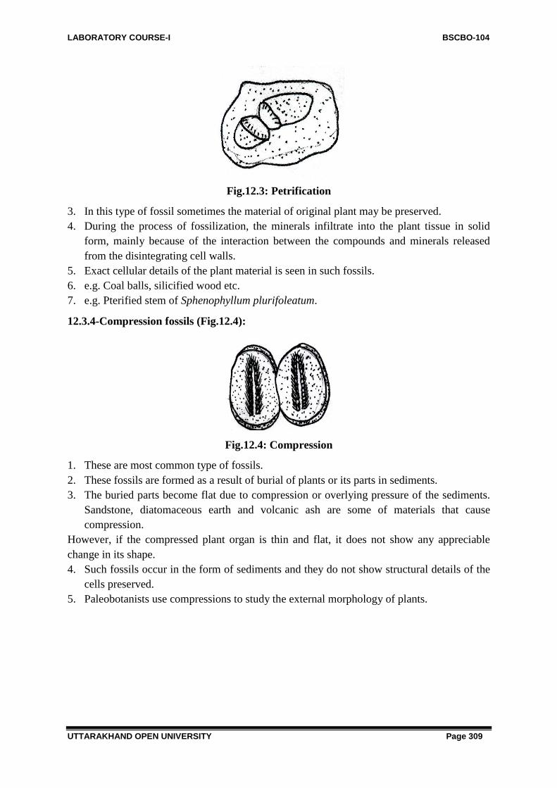
LABORATORY COURSE-I BSCBO-104
UTTARAKHAND OPEN UNIVERSITY Page 309
Fig.12.3: Petrification
3. In this type of fossil sometimes the material of original plant may be preserved.
4. During the process of fossilization, the minerals infiltrate into the plant tissue in solid
form, mainly because of the interaction between the compounds and minerals released
from the disintegrating cell walls.
5. Exact cellular details of the plant material is seen in such fossils.
6. e.g. Coal balls, silicified wood etc.
7. e.g. Pterified stem of Sphenophyllum plurifoleatum.
12.3.4-Compression fossils (Fig.12.4):
Fig.12.4: Compression
1. These are most common type of fossils.
2. These fossils are formed as a result of burial of plants or its parts in sediments.
3. The buried parts become flat due to compression or overlying pressure of the sediments.
Sandstone, diatomaceous earth and volcanic ash are some of materials that cause
compression.
However, if the compressed plant organ is thin and flat, it does not show any appreciable
change in its shape.
4. Such fossils occur in the form of sediments and they do not show structural details of the
cells preserved.
5. Paleobotanists use compressions to study the external morphology of plants.

LABORATORY COURSE-I BSCBO-104
UTTARAKHAND OPEN UNIVERSITY Page 310
12.3.5-Coal balls (Fig.12.5):
Fig.12.5: Coal balls
1. Petrified plant organs of roughly spherical shape are known as coal balls.
2. Coal balls are formed by infiltration of CaCo3, MgCo3, FeS2 etc in the buried plant parts.
3. The size of the coal balls ranges from a few centimeter to several meter.
4. Coal balls are found in some seams of coal.
5. The original plant tissue has been more fully decomposed or converted into carbon.
12.3.6-Amber (Fig.12.6):
Fig.12.6: Amber fossil of spider
1. These are resinous excretion of certain fossils.
2. Carboniferous trees which clot due to injuries caused by boring insects or decaying
branches.
3. Besides the insects and small animals these enclose plant fragments of which are well
preserved and ranked as fossils.
4. Pollens, spores etc are mostly preserved in this form.
12.3.7-Incrustations (Fig.12.7):
1. In it, external form of plant is preserved as cast but internal structures are destroyed.
2. These are formed by precipitation of mineral like carbonates on plant surface.

LABORATORY COURSE-I BSCBO-104
UTTARAKHAND OPEN UNIVERSITY Page 311
Fig.12.7: Incrustations
3. Fossils do not contain original organic materials and devoid of cellular details.
4. Suitable for the study of morphology of plants.
12.3.8-Compactions/ Mummified plants:
Fig.12.8: Mummified walnut
1. These are plants or plant parts compressed by vertical pressure.
2. Usually found in desert caves or buried in the desert sand.
3. Organism does not rot as bacteria need moisture to work.
4. Aggregation of plant fragments without interwining matrix such as found in peat, lignite,
coal are large scale compactions.
5. Tissue of leathery leaves or hard fruits are retained in mummified condition.
12.3.9-Petroleum (oil):
1. It is an oily, dark-coloured, inflammable liquid.
2. It is a mixture of various hydrocarbons occurring naturally in the upper strata of earth in
various parts of earth.

LABORATORY COURSE-I BSCBO-104
UTTARAKHAND OPEN UNIVERSITY Page 312
Fig.12.9: Petroleum formation
3. It is believed that petroleum had an organic origin.
4. It is formed under pressure from the minute floating plants and animal life.
5. It is commonly obtained by drilling.
Fossil Parks:
Fossil Park is a park where large number of fossils occurs. e.g:
1. RajMahal Hills in Bihar.
2. Old Coal forming forests in Orissa.
3. Mandla district in Madhya Pradesh.
Birbal Sahani Institute of Palaeobotany, Lucknow is a primier institute engaged in study of
fossils.
12.4 SUMMARY
Paleobotany is discipline of botany which involves the study of plant life of geological past,
with the help of fossils. Paleobotany includes the study of terrestrial plant fossils, as well as
the study of prehistoric marine photo-autotrophs, such as photosynthetic algae, seaweeds or
kelp. Fossils are the dead remains of plant and animals of past which have been preserved by
natural processes in the rocks of past. Study of fossils provides important clues to the
evolution and history of living organisms. Plant fossils are normally prescribed in the rocks
composed sediments deposited in waters. These stratified rocks are superimposed upon one
another in series and accumulate at the bottom of seas, lakes, swamps, beaches etc. The main
factors governing the degree of tissue preservation in fossil plants are Nature of plant
material, Environmental conditions before and during fossilization and the intensity and
Duration of destructive forces etc. Some Important Categories of fossils are Compression,
Impression, Petrifications, Mold, Incrustations, Casts, Coal balls and Petroleum.
12.5 GLOSSARY
Fossils: Fossils are the remains of plant and animals of past age preserved in rocks.

LABORATORY COURSE-I BSCBO-104
UTTARAKHAND OPEN UNIVERSITY Page 313
Petrifications: A petrification is formed when the encased specimen is impregnated with
silicates, carbonates, sulphates or phosphates present in water.
Mold: Mold is an impression of an organism or its hard part which is formed when the
organism is pressed down into sediment.
Coal balls: Coal balls are irregular rounded masses that show petrified remains of many plant
fragments.
Palaeontology: Branch of science dealing with study of fossils is Palaeontology.
12.6 SELF ASSESSMENT QUESTION
1. Who is known as father of Indian Palaeobotany?
2. What do you mean by the geological time?
3. How old is earth?
4. Who devised Carbon dating method for the estimation of age of fossils?
5. What is meant by fossilization?
6. What is the basis for the classification of fossils?
7. How are Petrifications formed?
8. Give few examples of Petrified fossils?
Answer to Self Assessment Question:
1. Prof. Birbal Sahni
2. Geological time is the total span of earth from the time of origin.
3. Nearly Five billion of years
4. Libby (1956)
5. The process of formation of fossils is called fossilization.
6. Method of preservation and composition of the fossil material form the basis of
classification of fossils.
7. A petrification is formed when the encased specimen is impregnated with silicates,
carbonates, sulphates or phosphates present in water.
8. Azolla intertrappea, Callixylon, Lepidodendron
12.7 REFERENCES
Sharma, O.P. 2014. Practical Botany. Fifth edition. Pragati Prakashan.
Arnold, C.A. (1947) : An Introduction to Paleobotany McGraw Hill Book Co., N.Y.
12.8 SUGGESTED READINGS
Delavoryas, T. (1962) : Morphology and Evolution of Fossil Plants Holt, Rinehart &
Winston, N.Y. Chicago, San Francisco, Toronto, London.
Scott, D.H. (1962) : Studies in Fossil Botany (Vol.I & Vol.II) Hafner Publishing Co.,
N.Y.
Seward, A.C. (1959) : Plant Life Through the Ages Hafner Publishing Co.,N.Y.

LABORATORY COURSE-I BSCBO-104
UTTARAKHAND OPEN UNIVERSITY Page 314
Shukla, A.C. & Misra, S.P. (1975) : Essentials of Paleobotany Vikas Publishing House
(P)Ltd., Delhi,Bombay, Kanpur.
Stewart, W.N. (1983) : Paleobotany & the Evolution of Plants Cambridge University
Press, Cambridge, London, N.Y., New Rochelle, Melbourne, Sydney.
Venkatachala, B.S., Shukla , M.& Sharma, M. (1992) : Plant Fossils-a Link with the Past
(A Birbal Sahni Birth Centenary Tribute) Birbal Sahni Institute of Paleobotany,
Lucknow, India.
12.9 TERMINAL QUESTIONS
1. What are fossils? What are the major types of fossils?
2. What is form genus? Describe briefly the nomenclature of fossils.
3. Write short note on the factors affecting fossilization?
4. Write short note on the techniques used for the study of fossils.
5. Mention the five eras of geological time scale?
a) Archaeozoic,
b) Proterozoic,
c) Palaeozoic,
d) Mesozoic and
e) Coenozoic
6. Name the era which is commonly called
a) Age of thallophytes
b) Age of Dinosours
c) Age of Pteridophytes
d) Age of Gymnosperms