Unfamiliar faces are not faces: Evidence from a matching task
Brief Report: Visual Processing of Faces in Individuals withFragile X Syndrome: An Eye Tracking Study
Faraz Farzin1,2, Susan M. Rivera1,2,3, and David Hessl3,4
1Department of Psychology, University of California, Davis, Davis, CA
2Center for Mind and Brain, University of California, Davis, Davis, CA
3M.I.N.D. Institute, University of California, Davis, Medical Center, Sacramento, CA
4Department of Psychiatry and Behavioral Sciences, University of California, Davis, Medical Center,Sacramento, CA
AbstractGaze avoidance is a hallmark behavioral feature of fragile X syndrome (FXS), but little is knownabout whether abnormalities in the visual processing of faces, including disrupted autonomicreactivity, may underlie this behavior. Eye tracking was used to record fixations and pupil diameterwhile adolescents and young adults with FXS and sex- and age-matched typically developing controlspassively viewed photographs of faces containing either a calm, happy, or fearful expression,preceded by a scrambled face matched on luminance. Results provide quantitative evidence forsignificant differences in gaze patterns and increased pupillary reactivity when individuals with FXSpassively view static faces. Such abnormalities have significant implications in terms ofunderstanding causes of gaze avoidance observed in individuals with FXS.
Keywordsface processing; fragile X syndrome; FMR1 gene; eye tracking; pupil reactivity
Fragile X syndrome (FXS) is the most common cause of inherited mental impairment(Crawford, 2001) and, to date, the most common known genetic cause of autism (D. Cohen etal., 2005). Although it is widely reported that gaze avoidance is a hallmark behavioral featureof FXS (Bregman, Leckman, & Ort, 1988; I. L. Cohen, Vietze, Sudhalter, Jenkins, & Brown,1991; Garrett, Menon, MacKenzie, & Reiss, 2004), it remains unknown whether abnormalitiesin the visual processing of faces, including disrupted autonomic reactivity, may contribute tothis observed social deficit.
Difficulty for individuals with FXS to establish and maintain eye-gaze during socialinteractions has been physiologically associated with markedly increased levels of cortisol orautonomic reactivity, suggesting that gaze aversion may be a coping mechanism that serves toreduce negative arousal (Belser & Sudhalter, 1995; Hall, DeBernardis, & Reiss, 2006; Hesslet al., 2002; Hessl, Glaser, Dyer-Friedman, & Reiss, 2006; Wisbeck et al., 2000). Secretion ofcortisol, by means of a cascade of hormones along the hypothalamic-pituitary-adrenal (HPA)axis, involves feedback between several limbic structures, among which the amygdala plays a
Correspondence concerning this article should be addressed to: Fraz Farzin, University of California, Davis, Center for Mind and Brain,202 Cousteau Place, Suite 250, Davis, California 95618, Phone: (530) 747-3808, Fax: (530) 297-4603.Data from this study were presented at the 11th International Fragile X Conference, St. Louis, MO, in July, 2008.
NIH Public AccessAuthor ManuscriptJ Autism Dev Disord. Author manuscript; available in PMC 2009 June 1.
Published in final edited form as:J Autism Dev Disord. 2009 June ; 39(6): 946–952. doi:10.1007/s10803-009-0744-1.
NIH
-PA Author Manuscript
NIH
-PA Author Manuscript
NIH
-PA Author Manuscript

significant role, and which have been implicated in FXS (Binstock, 1995; Hessl, Rivera, &Reiss, 2004; Paradee et al., 1999).
Studies have shown that pupillary responses are regulated by the autonomic nervous systemin part in response to level of emotional arousal (Bradley, Miccoli, Escrig, & Lang, 2008;Ekman, Levenson, & Friesen, 1983; Hess & Polt, 1960; Steinhauer, Siegle, Condray, & Pless,2004). Pupil dilation has been linked with direct electrical stimulation of the amygdala (Gloor,1997) and neuroimaging findings have shown that amygdala activity is associated with anobserver’s pupil size as well as is sensitive to the pupil size of others (Demos, Kelley, Ryan,Davis, & Whalen, 2008; Harrison, Singer, Rotshtein, Dolan, & Critchley, 2006).
The goals of the current study were to examine number of fixations and gaze duration whenindividuals with FXS viewed photographs of faces with different emotional content, and tomeasure face-specific changes in pupil diameter as an index of autonomic arousal.
MethodsParticipants
Participants included sixteen individuals with FXS (13 males) and sixteen typically developing(TD) controls (13 males). Individuals with FXS were confirmed to carry the full FMR1mutation as previously described (Saluto et al., 2005; Tassone et al., 2004). TD participantswith no current or past psychiatric diagnoses were recruited from the community. Groups wereindividually matched on sex and chronological age (t1, 30 = -0.085, p > .05). At the time oftesting, 10 individuals with FXS (8 males) were being treated with at least one class ofmedication; stimulant (n = 3), antipsychotic (n = 4), and anti-depressant (n = 5). One femalecontrol participant was being treated with an anti-hypertensive medication.
The Social Communication Questionnaire (SCQ; (Rutter, Bailey, & Lord, 2003) was used toevaluate symptoms of autism spectrum disorder in participants with FXS. Individuals with anSCQ score at or above 15 (n = 11, 9 males), or who raised clinical concerns for ASD (1 male),were referred for a more complete evaluation using the Autism Diagnostic ObservationSchedule-Generic (ADOS-G; (Lord, Rutter, DiLavore, & Risi, 2002). Of these twelve, 8 werediagnosed with Autistic Disorder and 2 were diagnosed with Pervasive DevelopmentalDisorder-Not Otherwise Specified (PDD-NOS). Group characteristics are given in Table 1.
Apparatus and StimuliAll stimuli were presented on a Tobii 1750 binocular eye tracker monitor. This eye trackingsystem consists of a high-resolution camera embedded in a 17-inch monitor (1280 × 1024pixels resolution, 50 Hz sampling rate, average precision of 0.5 degrees of visual angle). Thereare several benefits of the Tobii 1750 system that make it conducive to testing individuals withdevelopmental disorders, including approximately 20 centimeters of tolerance to head-motionin any direction without requiring any head restraints. Stimuli consisted of sixty coloredphotographs of adult human faces (equal numbers of males and females, different races andethnicities) from the NimStim Face Stimulus Set (Tottenham, 2002), each showing a calm,happy, or fearful expression, and sixty scrambled versions of the face images. Since it wascritical that pupil responses following the onset of the face stimulus be independent of apupillary light reflex, each face and its scramble were matched on mean luminance, andequivalence was confirmed using a photometer (Minolta, LS-100, Osaka, Japan). Face imagessubtended a 12.12 degree by 17.19 degree region (the size of an actual human face) whenviewed from a distance of 60 cm, and were presented on a standard 50% grey background.
Farzin et al. Page 2
J Autism Dev Disord. Author manuscript; available in PMC 2009 June 1.
NIH
-PA Author Manuscript
NIH
-PA Author Manuscript
NIH
-PA Author Manuscript

ProcedureThe experimental protocol was approved by the Institutional Review Board at the Universityof California, Davis, and informed consent was obtained from all participants or their parents.Following a nine-point calibration, participants were told to view the pictures shown. Eachtrial consisted of a scrambled face for 1 second followed immediately by its matched face for3 seconds. An inter-trial interval containing a uniform grey screen was shown for 0.5, 1, or 2seconds. The order of face presentation was randomized.
AnalysesFour area-of-interest (AOI) regions were defined for each face: eyes (including the eyebrows),nose, mouth, and other. Scrambled faces included a single AOI around the ellipse. Measuresincluded number of fixations (where a fixation was defined as any data point within a 30 pixelradius for a minimum duration of 100 milliseconds (ms)) and proportion of looking time toeach AOI region.
Each participant’s pupil data, averaged from both eyes, were first filtered to remove any outliervalues corresponding to blinks, loss of tracking data, or large changes in head position. Meanpupil diameter was calculated for 250 ms intervals across the trial duration, time-locked to theonset of the face stimulus. In order to determine the presence of a face-specific pupil response,relative change in pupil size was calculated by subtracting the mean pupil size during thescrambled face from the mean pupil size during each interval of the face presentation, and then“standardized” by dividing by the mean pupil size during the scrambled face. A similar pupilchange was calculated for the intervals of the scrambled face, relative to the mean pupil sizeduring the inter-trial interval.
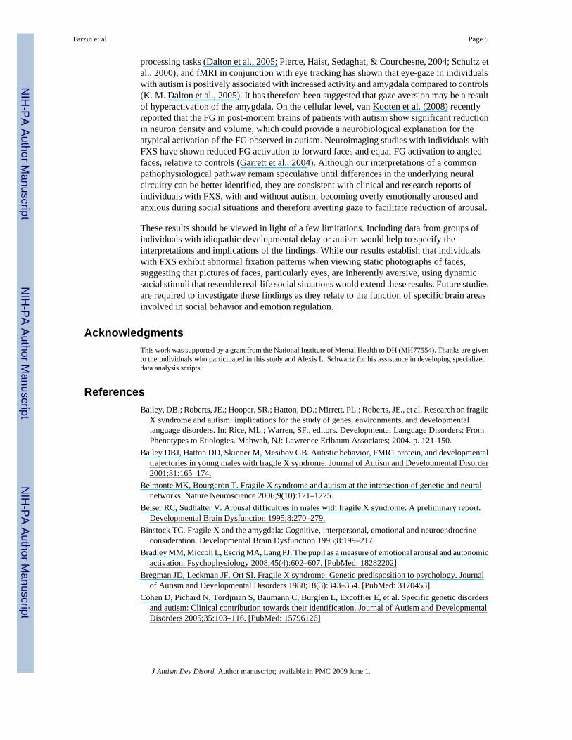
ResultsIndividuals with FXS made significantly fewer fixations to the eye region of all faces (Figure1). A repeated-measures analysis of variance (RMANOVA) with AOI region, emotion, andgroup as independent variables and number of fixations as the dependent variable revealed amain effect of AOI region [F(3, 28) = 16.74, p = 0.001, η2 = 0.642]. This effect was qualifiedby a significant interaction between AOI region and group [F(3, 28) = 5.12, p = 0.006, η2 =0.354], such that individuals with FXS made fewer fixations to the eye region of faces comparedto controls (Figure 2). Individuals with FXS made fewer fixations to the face overall [F(1, 31)= 8.926, p <0.01), but no group difference was found for number of fixations made to thescrambled faces [F(1, 31) = 3.154, p > 0.05], suggesting that the fixation differences observedwere specific to faces rather than generalizable to all images.
Individuals with FXS spent significantly less time looking at the eye region of all faces (Figure3). A RMANOVA with AOI region, emotion, and group as independent variables andproportion of gaze duration as the dependent variable revealed a main effect of AOI region[F(3,28) = 29.94, p = 0.001, η2 = 0.762] and a significant interaction between AOI region andgroup [F(3,28) = 3.56, p = 0.028, η2 = 0.274]. Independent t-tests revealed that, across emotionconditions, individuals with FXS spent a smaller proportion of time looking at the eyes [t(1,30) = 3.28, p = 0.003] and greater proportion of time looking at the nose [t(1, 30) = -2.58, p =0.015], compared to controls. No group difference was found for gaze duration to the scrambledfaces [F(1, 31) = 0.062, p > 0.05].
Individuals with FXS demonstrated significantly greater pupillary reactivity in response toemotional faces, relative to controls (Figure 4). A RMANOVA with interval (12), emotion,and group as independent variables and relative pupil change as the dependent variable resultedin a main effect of interval [F(11, 60) = 4.66, p = 0.001, η2 = 0.134], such that, for all faces
Farzin et al. Page 3
J Autism Dev Disord. Author manuscript; available in PMC 2009 June 1.
NIH
-PA Author Manuscript
NIH
-PA Author Manuscript
NIH
-PA Author Manuscript

combined, there was an increase in pupil size (dilation) across time for both groups. Asignificant interaction between interval and emotion also was found [F (22, 9) = 3.35, p =0.033, η2 = 0.891], driven by pupil dilation to happy and fearful faces for both groups. Asignificant interaction between interval and group [F (11, 60) = 4.72, p = 0.001, η2 = 0.136]showed that individuals with FXS demonstrated greater pupil dilation compared to TDcontrols. No group difference was found for change in pupil size to scrambled faces [F (1, 31)= 0.722, p > 0.05].
An exploratory analysis revealed that, in individuals with FXS, but not controls, pupil dilationin response to the fearful faces was inversely correlated with number of fixations to the eyesof calm [r = -0.550, p = 0.027], happy [r = -0.612, p = 0.012], and fearful [r = -0.641, p =0.007] faces, as well as proportion of gaze duration to the eyes of calm faces [r = -0.579, p =0.019]. There were no significant sex differences within either group on any of the above eyetracking measures. However, a lack of difference may also reflect limitations in statisticalpower as there were only three girls in each group.
Finally, correlational analyses revealed no significant associations between symptoms ofautism in individuals with FXS as measured by the SCQ and ADOS and number of fixations,gaze duration, and changes in pupil diameter in response to the faces. This finding is consistentwith studies of gaze aversion in individuals with autism (K.M. Dalton, Nacewicz, Alexander,& Davidson, 2007; Klin, Jones, Schultz, Volkmar, & Cohen, 2002; Speer, Cook, McMahon,& Clark, 2007).
DiscussionThese findings reveal both qualitative and quantitative differences in the way that individualswith FXS visually process static human faces, and to our knowledge, are the first to utilizepupillometry as a measure of autonomic reactivity in individuals with FXS. Individuals withFXS demonstrated fewer fixations and less gaze time to the eye region of faces, and increasedpupil reactivity to emotional faces, compared to controls. Importantly, pupil dilation inresponse to the fearful faces was inversely correlated with number of fixations to the eyes ofall faces. This pattern was observed in both males and females with FXS, and was notsignificantly associated with severity of autism symptomology. Documentation of faceprocessing differences is critical for developing treatment approaches aimed at alleviating thesymptoms of social anxiety and has significant potential for use in assessing specificphenotypic outcomes following treatment in clinical research studies (Guastella, Mitchell, &Dadds, 2008).
Research has firmly established that FXS and autism are closely associated. Two to six percentof children with autism have the FMR1 mutation (Reddy, 2005; Wassink, Piven, & Patil,2001) and approximately 30% of children with FXS are diagnosed with autism and anadditional 20-30% have PDDNOS (D. B. J. Bailey, Hatton, Skinner, & Mesibov, 2001; Harriset al., in press; Kaufmann et al., 2004; Rogers, Wehner, & Hagerman, 2001). Researchers havehypothesized that the presence of autism symptoms in FXS exists on a spectrum, such thatsome individuals with FXS who are also diagnosed with ASD may not be qualitatively differentfrom those without ASD (D. B. Bailey et al., 2004; Lewis et al., 2006).
Our results are in line with research demonstrating that individuals with ASD spend a smallerpercentage of time fixating the eyes (Klin et al., 2002; Merin, Young, Ozonoff, & Rogers,2007; Pelphrey et al., 2002; Spezio, Adolphs, Hurley, & Piven, 2007), and thus support theargument that gaze aversion observed in idiopathic autism and in FXS may reflect similaritiesin underlying network dysfunctions (Belmonte & Bourgeron, 2006). Several fMRI studiesreport reduced activity in the fusiform gyrus (FG) when individuals with autism perform face
Farzin et al. Page 4
J Autism Dev Disord. Author manuscript; available in PMC 2009 June 1.
NIH
-PA Author Manuscript
NIH
-PA Author Manuscript
NIH
-PA Author Manuscript
processing tasks (Dalton et al., 2005; Pierce, Haist, Sedaghat, & Courchesne, 2004; Schultz etal., 2000), and fMRI in conjunction with eye tracking has shown that eye-gaze in individualswith autism is positively associated with increased activity and amygdala compared to controls(K. M. Dalton et al., 2005). It has therefore been suggested that gaze aversion may be a resultof hyperactivation of the amygdala. On the cellular level, van Kooten et al. (2008) recentlyreported that the FG in post-mortem brains of patients with autism show significant reductionin neuron density and volume, which could provide a neurobiological explanation for theatypical activation of the FG observed in autism. Neuroimaging studies with individuals withFXS have shown reduced FG activation to forward faces and equal FG activation to angledfaces, relative to controls (Garrett et al., 2004). Although our interpretations of a commonpathophysiological pathway remain speculative until differences in the underlying neuralcircuitry can be better identified, they are consistent with clinical and research reports ofindividuals with FXS, with and without autism, becoming overly emotionally aroused andanxious during social situations and therefore averting gaze to facilitate reduction of arousal.
These results should be viewed in light of a few limitations. Including data from groups ofindividuals with idiopathic developmental delay or autism would help to specify theinterpretations and implications of the findings. While our results establish that individualswith FXS exhibit abnormal fixation patterns when viewing static photographs of faces,suggesting that pictures of faces, particularly eyes, are inherently aversive, using dynamicsocial stimuli that resemble real-life social situations would extend these results. Future studiesare required to investigate these findings as they relate to the function of specific brain areasinvolved in social behavior and emotion regulation.
AcknowledgmentsThis work was supported by a grant from the National Institute of Mental Health to DH (MH77554). Thanks are givento the individuals who participated in this study and Alexis L. Schwartz for his assistance in developing specializeddata analysis scripts.
ReferencesBailey, DB.; Roberts, JE.; Hooper, SR.; Hatton, DD.; Mirrett, PL.; Roberts, JE., et al. Research on fragile
X syndrome and autism: implications for the study of genes, environments, and developmentallanguage disorders. In: Rice, ML.; Warren, SF., editors. Developmental Language Disorders: FromPhenotypes to Etiologies. Mahwah, NJ: Lawrence Erlbaum Associates; 2004. p. 121-150.
Bailey DBJ, Hatton DD, Skinner M, Mesibov GB. Autistic behavior, FMR1 protein, and developmentaltrajectories in young males with fragile X syndrome. Journal of Autism and Developmental Disorder2001;31:165–174.
Belmonte MK, Bourgeron T. Fragile X syndrome and autism at the intersection of genetic and neuralnetworks. Nature Neuroscience 2006;9(10):121–1225.
Belser RC, Sudhalter V. Arousal difficulties in males with fragile X syndrome: A preliminary report.Developmental Brain Dysfunction 1995;8:270–279.
Binstock TC. Fragile X and the amygdala: Cognitive, interpersonal, emotional and neuroendrocrineconsideration. Developmental Brain Dysfunction 1995;8:199–217.
Bradley MM, Miccoli L, Escrig MA, Lang PJ. The pupil as a measure of emotional arousal and autonomicactivation. Psychophysiology 2008;45(4):602–607. [PubMed: 18282202]
Bregman JD, Leckman JF, Ort SI. Fragile X syndrome: Genetic predisposition to psychology. Journalof Autism and Developmental Disorders 1988;18(3):343–354. [PubMed: 3170453]
Cohen D, Pichard N, Tordjman S, Baumann C, Burglen L, Excoffier E, et al. Specific genetic disordersand autism: Clinical contribution towards their identification. Journal of Autism and DevelopmentalDisorders 2005;35:103–116. [PubMed: 15796126]
Farzin et al. Page 5
J Autism Dev Disord. Author manuscript; available in PMC 2009 June 1.
NIH
-PA Author Manuscript
NIH
-PA Author Manuscript
NIH
-PA Author Manuscript

Cohen IL, Vietze PM, Sudhalter V, Jenkins EC, Brown WT. Effects of age and communication level oneye contact in fragile X males and non-fragile X autistic males. American Journal of Medical Genetics1991;38(23):498–502. [PubMed: 2018093]
Crawford D, Acuna JM, Sherman SL. FMR1 and the fragile X syndrome: human genome epidemiologyreview. Genetics in Medicine 2001;3(5):359–371. [PubMed: 11545690]
Dalton KM, Nacewicz BM, Alexander AL, Davidson RJ. Gaze-fixation, brain activation, and amygdalavolume in unaffected siblings of individuals with autism. Biological Psychiatry 2007;61(4):512–520.[PubMed: 17069771]
Dalton KM, Nacewicz BM, Johnstone T, Schaefer HS, Gernsbacher MA, Goldsmith HH, et al. Gazefixation and the neural circuitry of face processing in autism. Nature Neuroscience 2005;8:519–526.
Demos KE, Kelley WM, Ryan SL, Davis FC, Whalen PJ. Human amygdala sensitivity to the pupil sizeof others. Cerebral Cortex 2008:bhn034.
Ekman P, Levenson RW, Friesen WV. Autonomic nervous system activity distinguishes amongemotions. Science 1983;221:1208–1210. [PubMed: 6612338]
Garrett AS, Menon V, MacKenzie K, Reiss AL. Here’s looking at you, kid: neural systems underlyingface and gaze processing in fragile X syndrome. Archives of General Psychiatry 2004;61(3):281–288. [PubMed: 14993116]
Gloor, P. The Temporal Lobe and Limbic System. New York: Oxford University Press; 1997.Guastella AJ, Mitchell PB, Dadds MR. Oxytocin increases gaze to the eye region of human faces.
Biological Psychiatry 2008;63(1):3–5. [PubMed: 17888410]Hall S, DeBernardis M, Reiss A. Social escape behaviors in children with fragile X syndrome. Journal
of Autism and Developmental Disorders 2006;36:935–947. [PubMed: 16897394]Harris SW, Goodlin-Jones B, Nowicki ST, Hessl D, Tassone F, Barabato I, et al. Autism Profiles of
Young Males with Fragile X Syndrome. American Journal of Medical Genetics Part A. in pressHarrison NA, Singer T, Rotshtein P, Dolan RJ, Critchley HD. Pupillary contagion: central mechanisms
engaged in sadness processing. Social Cognition and Affective Neuroscience 2006;1:5–17.Hess EH, Polt JM. Pupil size as related to interest value of visual stimuli. Science 1960;132:349–350.
[PubMed: 14401489]Hessl D, Glaser B, Dyer-Friedman J, Blasey C, Hastie T, Gunnar M, et al. Cortisol and behavior in fragile
X syndrome. Psychoneuroendocrinology 2002;27(7):855–872. [PubMed: 12183220]Hessl D, Glaser B, Dyer-Friedman J, Reiss AL. Social behavior and cortisol reactivity in children with
fragile X syndrome. Journal of Child Psychology and Psychiatry 2006;47(6):602–610. [PubMed:16712637]
Hessl D, Rivera SM, Reiss AL. The neuroanatomy and neuroendocrinology of fragile X syndrome.Mental Retardation and Developmental Disabilities Research Review 2004;10:17–24.
Kaufmann WE, Cortell R, Kau AS, Bukelis I, Tierney E, Gray RM, et al. Autism spectrum disorder infragile X syndrome: Communication, social interaction, and specific behaviors. American Journalof Medical Genetics Part A 2004;129A:225–234. [PubMed: 15326621]
Klin A, Jones W, Schultz R, Volkmar F, Cohen D. Visual fixation patterns during viewing of naturalisticsocial situations as predictors of social competence in individuals with autism. Archives of GeneralPsychiatry 2002;59(9):809–816. [PubMed: 12215080]
Lewis P, Abbeduto L, Murphy M, Richmond E, Giles N, Bruno L, et al. Cognitive, language, and social-cognitive skills of individuals with fragile X syndrome with and without autism. Journal ofIntellectual Disability Research 2006;50(7):532–545. [PubMed: 16774638]
Lord, C.; Rutter, M.; DiLavore, PC.; Risi, S. Autism Diagnostic Observation Schedule- Manua. LosAngeles: Western Psychological Services; 2002.
Merin N, Young GS, Ozonoff S, Rogers SJ. Visual Fixation Patterns during Reciprocal Social InteractionDistinguish a Subgroup of 6-Month-Old Infants At-Risk for Autism from Comparison Infants.Journal of Autism and Developmental Disorder 2007;37(1):108–121.
Paradee W, Melikian HE, Rasmussen DL, Kenneson A, Conn PJ, Warren ST. Fragile X mouse: straineffects of knockout phenotype and evidence suggesting deficient amygdala function. Neuroscience1999;94(1):185–192. [PubMed: 10613508]
Farzin et al. Page 6
J Autism Dev Disord. Author manuscript; available in PMC 2009 June 1.
NIH
-PA Author Manuscript
NIH
-PA Author Manuscript
NIH
-PA Author Manuscript

Pelphrey KA, Sasson NJ, Reznick JS, Paul G, Goldman BD, Piven J. Neural basis of eye gaze processingdeficits in autism. Brain 2002;128:1038–1048. [PubMed: 15758039]
Pierce K, Haist F, Sedaghat F, Courchesne E. The brain response to personally familiar faces in autism:Findings of fusiform activity and beyond. Brain 2004;127:2703–2716. [PubMed: 15319275]
Reddy KS. Cytogenetic abnormalities and fragile-X syndrome in Autism Spectrum Disorder. BMCMedical Genetics 2005;6:3. [PubMed: 15655077]
Rogers SJ, Wehner EA, Hagerman RJ. The behavioral phenotype in Fragile X: Symptoms of autism invery young children with Fragile X syndrome, idiopathic autism, and other developmental disorders.Journal of Developmental and Behavioral Pediatrics 2001;22:409–417. [PubMed: 11773805]
Rutter, M.; Bailey, A.; Lord, C. Social Communication Questionnaire (SCQ). Los Angeles: WesternPsychological Services; 2003.
Saluto A, Brussino A, Tassone F, Arduino C, Cagnoli C, Pappi P, et al. An enhanced polymerase chainreaction assay to detect pre- and full mutation alleles of the fragile X mental retardation 1 gene.Journal of Molecular Diagnostics 2005;7(5):605–612. [PubMed: 16258159]
Schultz RT, Gauthier I, Klin A, Fulbright RK, Anderson AW, Volkmar F, et al. Abnormal ventraltemporal cortical activity during face discrimination among individuals with autism and Aspergersyndrome. Archives of General Psychiatry 2000;57:331–340. [PubMed: 10768694]
Speer LL, Cook AE, McMahon WM, Clark E. Face processing in children with autism:effects of stimuluscontents and type. Autism 2007;11(3):265–277. [PubMed: 17478579]
Spezio ML, Adolphs R, Hurley RS, Piven J. Abnormal use of facial information in high-functioningautism. Journal of Autism and Developmental Disorder 2007;37(5):929–939.
Steinhauer SR, Siegle GJ, Condray R, Pless M. Sympathetic and parasympathetic innervation of pupillarydilation during sustained processing. International Journal of Psychophysiology 2004;52:77–86.[PubMed: 15003374]
Tassone F, Hagerman RJ, Garcia AD, Khandijian EW, Greco CM, Hagerman PJ. Intranuclear inclusionsin neural cells with premutation alleles in fragile X associated tremor/ataxia syndrome. Journal ofMedical Genetics 2004;41:1–3. [PubMed: 14729816]
Tottenham N. MacArthur Foundation Research Network. 2002van Kooten IAJ, Palmen SJMC, von Cappeln P, Steinbusch HWM, Korr H, Heinsen H, et al. Neurons
in the fusiform gyrus are fewer and smaller in autism. Brain 2008;131:987–999. [PubMed: 18332073]Wassink TH, Piven J, Patil SR. Chromosomal abnormalities in a clinic sample of individuals with autistic
disorder. Psychiatric Genetics 2001;11:57–63. [PubMed: 11525418]Wechsler, D. Wechsler Intelligence Scale for Children. Vol. Fourth Edition. San Antonio: Harcourt
Assessment, Inc.; 2003.Wisbeck JM, Huffman LC, Freund L, Gunnar MR, Davis EP, Reiss AL. Cortisol and social stressors in
children with fragile X: A pilot study. Journal of Developmental and Behavioral Pediatrics2000;21:278–282. [PubMed: 10972251]
Farzin et al. Page 7
J Autism Dev Disord. Author manuscript; available in PMC 2009 June 1.
NIH
-PA Author Manuscript
NIH
-PA Author Manuscript
NIH
-PA Author Manuscript
Figure 1.Mean number of fixations to each AOI region by group for (a) calm, (b) happy, and (c) fearfulfaces. Double asterisk (**) indicates significant difference between pairwise comparisons atthe p < .01 level. Error bars represent SEM.
Farzin et al. Page 8
J Autism Dev Disord. Author manuscript; available in PMC 2009 June 1.
NIH
-PA Author Manuscript
NIH
-PA Author Manuscript
NIH
-PA Author Manuscript

Figure 2.Example fixation plots from one subject in each group (TD on left, FXS on right) for a single(a) calm, (b) happy, (c) fearful, and (d) scrambled face trial. The plot displays a static frameof fixation data for each image. Each fixation is illustrated with a blue circle where the radiusrepresents the length of the fixation.
Farzin et al. Page 9
J Autism Dev Disord. Author manuscript; available in PMC 2009 June 1.
NIH
-PA Author Manuscript
NIH
-PA Author Manuscript
NIH
-PA Author Manuscript
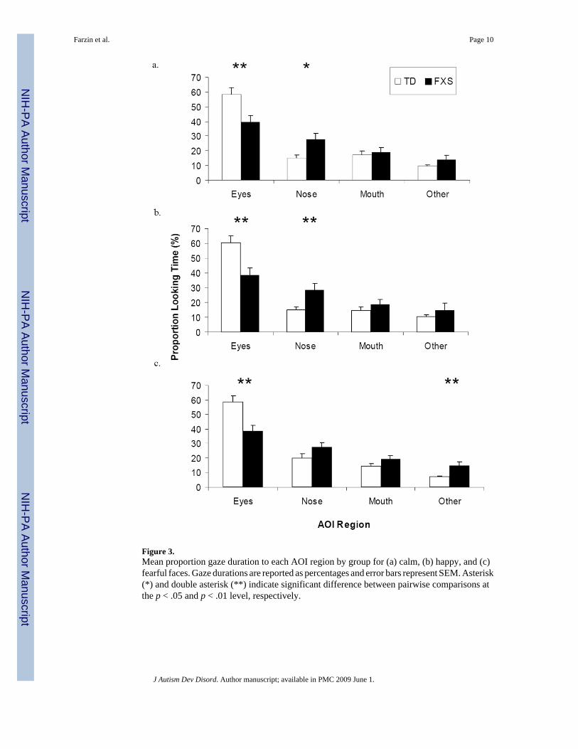
Figure 3.Mean proportion gaze duration to each AOI region by group for (a) calm, (b) happy, and (c)fearful faces. Gaze durations are reported as percentages and error bars represent SEM. Asterisk(*) and double asterisk (**) indicate significant difference between pairwise comparisons atthe p < .05 and p < .01 level, respectively.
Farzin et al. Page 10
J Autism Dev Disord. Author manuscript; available in PMC 2009 June 1.
NIH
-PA Author Manuscript
NIH
-PA Author Manuscript
NIH
-PA Author Manuscript

Figure 4.Relative change in pupil diameter (mm) between scrambled face slide to (a) calm, (b) happy,and (c) fearful faces, across 250 ms intervals, by group. Curves are time-locked to the onset ofthe face stimulus. Gray shaded region with asterisk (*) or double asterisk (**) indicatessignificant pairwise difference at the p < .05 and p < .01 level, respectively.
Farzin et al. Page 11
J Autism Dev Disord. Author manuscript; available in PMC 2009 June 1.
NIH
-PA Author Manuscript
NIH
-PA Author Manuscript
NIH
-PA Author Manuscript

NIH
-PA Author Manuscript
NIH
-PA Author Manuscript
NIH
-PA Author Manuscript
Farzin et al. Page 12
Table 1Descriptive characteristics of the fragile X syndrome (FXS) and control groups. (Mean ± SEM)
FXS (n = 16) TD (n = 16)
Gender (M:F) 13:3 13:3
Chronological Age (years) 16.99 ± 6.8 17.14 ± 6.3
Full Scale IQa 58.36 ± 9.8
SCQ 15.45 ± 7.51
ADOS Totalb 10.46 ± 4.07
FXS, fragile X Syndrome; TD, Typically Developing; SCQ, Social Communication Questionnaire-Lifetime Version; ADOS, Autism DiagnosticObservation Schedule
aIntellectual level was measured using the Weschler Intelligence Scales (Wechsler, 2003), n = 14 (2 participants were not testable due to extremely low
functioning).
bn = 13
J Autism Dev Disord. Author manuscript; available in PMC 2009 June 1.
Copyright © 2022 FDOKUMEN