Brain Tumors: Convection-Enhanced Delivery of Drugs (Method)
10
Chapter 21 Brain Tumors: Convection-Enhanced Delivery of Drugs (Method) Anne-Laure Laine, Emilie Allard, Philippe Menei, and Catherine Passirani Abstract Delivery of therapeutic agents into the brain has been an ongoing challenge for many years. The poor prognosis for patient with primary malig- nant brain tumors treated with conventional techniques (surgery, radiotherapy and chemotherapy) has moti- vated the development of new strategies to deliver drugs into the brain. Local intracranial delivery of anti- neoplastic agents has appeared to be the most effec- tive drug delivery technique into the central nervous system by circumventing the limitations imposed by the blood brain barrier (BBB). Convection-enhanced delivery (CED) is an alternative strategy to directly infuse drugs into brain tissue. This approach is based on continuous injection of the therapeutic agent under positive pressure via a catheter implanted into the brain. Convective transport driven by pressure gra- dient allows a widespread distribution of small and large drugs within the brain. In vivo experiments in rodents, cats and primates proved the efficacy of CED to deliver drugs into a targeted zone. However, clini- cal trials have reported frequent leakage phenomenon leading to mixed results for this delivery technique. A better optimization of operational parameters includ- ing infusion rate, catheter design, catheter place- ment and drug pharmacological formulation should allow achieving accurate and efficient delivery. In conjunction with CED, the use of nanocarriers to enhance drug pharmacokinetic behavior may help to achieve higher therapeutic index against tumor cells C. Passirani () INSERM, U646, Universite d’Angers, Angers F-491000, France e-mail: [email protected] over healthy tissues. Additionally, the development of computer simulation to predict drug distribution and the real-time imaging for immediate assessment of convection efficiency may contribute to the CED improvement. Keywords Therapeutic agents · Convection-enhanced delivery · Catheter · Infusion rate · Infusate · CNS Introduction Despite considerable advances of research in the area of Central Nervous System (CNS) disorders, the prog- nosis for patients with brain tumors remains poor. Median survival for patients with the most severe form treated with surgical resection, radiation and the addi- tion of systemic chemotherapy is 12–15 months (Stupp et al., 2005). Finding new therapeutic strategies that will provide efficient treatment against CNS tumors remains a major challenge. Conventional methods for the treatment of brain tumors usually involve delivery of drugs via the systemic circulation. Failure of those therapies is attributed to the presence of the blood brain barrier (Pardridge, 2007). The BBB is a physical barrier that separates circulating blood from cerebrospinal fluid in the CNS. This separation protects the brain from the penetration of toxic substances throughout the CNS. The restricted entry into the brain is imposed by a layer of endothelial cells joined by tight junctions. This structure confers a limited diffusion through the BBB that prevents the entry of most pharmaceuticals into the CNS. 207 M.A. Hayat (ed.), Tumors of the Central Nervous System, Volume 3, DOI 10.1007/978-94-007-1399-4_21, © Springer Science+Business Media B.V. 2011
Transcript of Brain Tumors: Convection-Enhanced Delivery of Drugs (Method)

Chapter 21
Brain Tumors: Convection-Enhanced Delivery of Drugs(Method)
Anne-Laure Laine, Emilie Allard, Philippe Menei, and Catherine Passirani
Abstract Delivery of therapeutic agents into thebrain has been an ongoing challenge for many years.The poor prognosis for patient with primary malig-nant brain tumors treated with conventional techniques(surgery, radiotherapy and chemotherapy) has moti-vated the development of new strategies to deliverdrugs into the brain. Local intracranial delivery of anti-neoplastic agents has appeared to be the most effec-tive drug delivery technique into the central nervoussystem by circumventing the limitations imposed bythe blood brain barrier (BBB). Convection-enhanceddelivery (CED) is an alternative strategy to directlyinfuse drugs into brain tissue. This approach is basedon continuous injection of the therapeutic agent underpositive pressure via a catheter implanted into thebrain. Convective transport driven by pressure gra-dient allows a widespread distribution of small andlarge drugs within the brain. In vivo experiments inrodents, cats and primates proved the efficacy of CEDto deliver drugs into a targeted zone. However, clini-cal trials have reported frequent leakage phenomenonleading to mixed results for this delivery technique. Abetter optimization of operational parameters includ-ing infusion rate, catheter design, catheter place-ment and drug pharmacological formulation shouldallow achieving accurate and efficient delivery. Inconjunction with CED, the use of nanocarriers toenhance drug pharmacokinetic behavior may help toachieve higher therapeutic index against tumor cells
C. Passirani (�)INSERM, U646, Universite d’Angers, Angers F-491000,Francee-mail: [email protected]
over healthy tissues. Additionally, the developmentof computer simulation to predict drug distributionand the real-time imaging for immediate assessmentof convection efficiency may contribute to the CEDimprovement.
Keywords Therapeutic agents · Convection-enhanceddelivery · Catheter · Infusion rate · Infusate · CNS
Introduction
Despite considerable advances of research in the areaof Central Nervous System (CNS) disorders, the prog-nosis for patients with brain tumors remains poor.Median survival for patients with the most severe formtreated with surgical resection, radiation and the addi-tion of systemic chemotherapy is 12–15 months (Stuppet al., 2005). Finding new therapeutic strategies thatwill provide efficient treatment against CNS tumorsremains a major challenge.
Conventional methods for the treatment of braintumors usually involve delivery of drugs via thesystemic circulation. Failure of those therapies isattributed to the presence of the blood brain barrier(Pardridge, 2007). The BBB is a physical barrier thatseparates circulating blood from cerebrospinal fluid inthe CNS. This separation protects the brain from thepenetration of toxic substances throughout the CNS.The restricted entry into the brain is imposed by alayer of endothelial cells joined by tight junctions. Thisstructure confers a limited diffusion through the BBBthat prevents the entry of most pharmaceuticals into theCNS.
207M.A. Hayat (ed.), Tumors of the Central Nervous System, Volume 3,DOI 10.1007/978-94-007-1399-4_21, © Springer Science+Business Media B.V. 2011

208 A.-L. Laine et al.
Despite a variety of approaches tested for enhancingBBB permeability of drugs, no significant improve-ment was showed for the treatment of CNS tumors(Misra et al., 2003). The poor results obtained by allconventional therapies and the unchanged prognosesfor patient with GBM have motivated more directapproaches of drug delivery. Intracranial drug deliv-ery has appeared to be the most adequate method ofovercoming the BBB (Sawyer et al., 2006).
In theory, regional drug approach constitutes away to deliver high concentration of drug directlyinto the tumor. Such delivery can also reduce sys-temic exposure to drug-induced toxicity. A local deliv-ery technique was achieved implanting a degradableor non-degradable polymer delivery system into thebrain (Raza et al., 2005). This approach presentssome advantages such as sustained controlled drugrelease. However, polymer-controlled release as intrac-erebral bolus injection suffers from restricted drugdistribution due to limited drug diffusion within thebrain (Fung et al., 1998). An alternative local deliv-ery based on convective transport has been proposedto overcome the limitation imposed by the diffusionobserved with polymer-delivery system. This tech-nique called Convection-Enhanced Delivery (CED)was introduced by researchers from the US NationalInstitutes of Health (NIH) by the early 1990s (Boboet al., 1994). In CED method, the infusate is admin-istered via a small catheter connected to a pump.The continuous injection of a convective flow directlyin the brain results in a widespread distribution ofsmall as well as large molecules into it (Debinskiand Tatter, 2009). By bypassing the BBB, this tech-nique allows the local delivery of a variety ofagents, ranging from conventional therapeutics agents,to monoclonal antibodies, targeted toxins, nanocar-riers (Allard et al., 2009) and viruses (Bidros andVogelbaum, 2009).
However, several parameters can affect therapeu-tic efficacy of CED, including infusion rate, cannulashape, infused tissue structure and physico-chemicalproperties of the infusate. Those parameters have to becarefully investigated and set up for an optimal deliv-ery. This chapter aims at giving an overview of theCED approach. The following paragraphs will describethe technical aspects and critical parameters of thispromising local delivery technique. Optimal setting forefficient delivery will be proposed according to theliterature.
Convection Enhanced Delivery
Convection-enhanced delivery is a local deliverymethod in which drugs are infused directly into braintissue. The therapeutic agents are administered viaone to several catheters implanted into the brain(Fig. 21.1a). An external pump produces a posi-tive pressure which pushes the infusate through thecatheter. With this delivery system, the drug is directlyinfused into the target tissue at a predetermined con-centration, rate and duration.
The use of a catheter allows a stereotaxic placementinto a CNS targeted zone. Stereotaxy uses a three-dimensional coordinates system to localize structuresand targets within the brain. Thanks to this tech-nique, the catheter can be accurately implanted withinthe tumor mass or around the tumor or the resectioncavity. The ability to target the delivery placement lim-its structural damage to the infused tissue, except atthe site of the catheter, and allows avoiding systemictoxicity.
Most of local delivery techniques including polymersystems and bolus injection rely on diffusive trans-port to distribute a therapeutic agent within the CNS.Diffusion corresponds to the spread of molecules fromregions of higher concentration to regions of lowerconcentration. The diffusion of a compound dependsmainly on the concentration gradient and its diffu-sivity properties. Many therapeutic agents such asneurotrophic factors, antibodies, growth factors andenzymes are not able to diffuse over large distancesinto the brain due to high molecular weight. This poordiffusion severely limits drug distribution and penetra-tion into the brain tissue. For example, an antibody IgGrequires 3 days to diffuse 1 mm from its delivery sitein tumor (Jain, 1989). That is why most antineoplasticsubstances show limited effects after direct admin-istration into brain via diffused-based techniques. Incontrast to those delivery techniques, CED relies onconvection, a molecular weight independent transport,to overcome the limitation imposed by diffusion. Theterm convection refers to the mass transfer by bulkmotion of the fluid. In CED, the convective flow, alsocalled bulk flow, is driven by pressure gradient inducedby the difference between the interstitial pressure andthe pressure at the tip of the catheter. Drugs are car-ried along with the fluid flow and distributed throughthe brain tissue by convective and diffusive transport.

21 Convection-Enhanced Delivery of Drugs (Method) 209
A
B
Fig. 21.1 a Convection-enhanced delivery in a human brain.The drug is infused via a catheter implanted into the brain.An external pump connected to the catheter provides positivepressure pushing the drug through the interstitial space of the
central nervous system. b Brain section with identification of thegray matter (GM) and white matter (WM) location in humansand rats
The superposition of transports by random motion ofthe molecules (diffusion) and by the bulk motion ofthe fluid (convection) offers a greater volume of dis-tribution than simple diffusion. Morrison et al. (1994)showed that CED can provide distribution volumes inbrain that are 2- to 10-fold larger than those obtainedafter direct interstitial injection (Morrison et al., 1994).Additionally, data from CED studies showed enhanceddistribution of various therapeutic agents in rat, cat andnon human primate brains with an order of magnitudegreater than simple diffusion-based delivery (Boboet al., 1994; Yokosawa et al., 2010).
However, despite proven efficacy, CED shows somelimitations. Most common issues with infusions of
drug into the brain are reflux along the catheter trackand leakage outside the targeted area. It has beenreported that leakage phenomenon affects 20% of CEDexperiments (Varenika et al., 2008). While preventingcontinued pressure in the interstitial space, reflux andleakage can also increase the risk of neurotoxicity toadjacent tissue and induce adverse effects. The occur-rence of leakage and reflux may explain the mixedresults obtained in clinical trials (Kunwar, 2003, 2007).Indeed, for optimal CED efficiency, it is essential tocover the entire target zone while avoiding exposure ofhealthy tissue. The extent of drug distribution achievedvia CED depends on the type of tissue infused (that is,tumor, gray matter or white matter), infusion volume

210 A.-L. Laine et al.
and rate, catheter design but also on drug eliminationmechanism including metabolism, binding, efflux andleakage. These different parameters are discussed inthe following paragraphs.
Key Parameters
Catheter Placement
During CED, the distribution volume for a given agentis highly dependent on the type of tissue being infusedand its structural properties, such as hydraulic con-ductivity, vascular volume fraction, and extracellularfluid fraction. Clinical trials outcomes have highlightedthe importance of accurate catheter placement for opti-mal drug distribution (Kunwar et al., 2007). Two mainareas can be distinguished in the CNS; the gray matterand the white matter (Fig. 21.1b). The gray matter ismainly composed of cell bodies and glial cells and thetransport within this structure is isotropic. In contrast,the white matter consists mostly of axons leading to theperipheral nervous system. This structure composedof fiber pathways makes the transport anisotropic. Itis worth noting that gliomas invasion preferentiallyoccurs along these white bundles. It has been reportedthat a significant natural bulk flow with an interstitialvelocity of 10 μm/mL may exist in porous white matternear the ventricular surface (Rosenberg et al., 1980).
Bobo et al. (1994) demonstrated that an infusionvia CED into the corona radiate (white matter) ofcats allowed an effective and large distribution of thedrug within the brain tissue. Similar observations werereported by Lieberman et al. (1995) regarding a druginfusion via CED into the gray matter. Both grayand white matters have the capability to distribute theinfused drug within the CNS. However, the distribu-tion profile differs between both structures and thedifference is even more significant compared to braintumor.
When the infusion takes place into a homogeneousbrain structure (e.g., gray matter) the infusion is ini-tially driven by pressure gradient from the source ofinjection toward surrounding tissue producing spher-ical volume (Fig. 21.2a). The concentration profilein this volume is relatively uniform and proportionalto the delivered concentration. At the flow front, thepressure gradient reaches zero and the distribution
is no longer driven by convection but by diffusion.The concentration declines precipitately and exponen-tially within adjacent tissues in accordance with themathematics of diffusion (Morrison et al., 1994).
If the infused drug encounters a structure containingfiber pathways (eg. white matter), the convective flowcan be modified by the natural bulk flow within thatpathway. It has been reported that interstitial veloci-ties into the white matter may be used to increase therate of the infusate (Rosenberg et al., 1980). However,this natural movement in white matter can also lead tounpredictable flow and leakage outside the target zoneif the direction of the natural bulk flow is opposed tothat of the infusate flow (Krauze et al., 2005).
It is apparent that inhomogeneities in the tissue maycause unpredictable distribution. The presence of oede-mas, often observed in brain cancer and also inducedby infusion, reduces volume of treated tissue and candisturb the flow of the infused drug. Moreover, thedistribution profile into brain tumors differs markedlyfrom the normal brain. This variation may be due tothe difference of interstitial fluid pressure. In normalbrain tissue the interstitial pressure is relatively low,1–2 mmHg, whereas in brain tumor tissue it can bemore than 25 times greater. The increased interstitialpressure in peritumoral tissue and within the tumorproduces a counterproductive pressure gradient thatcan drive infusate to follow the path of lowest intersti-tial pressure, out of high pressure areas such as tumortissue. The complex anatomy of brain tumor and itshigh intracranial pressure can lead to drug distributionin undesired area and leakage into the subarachnoidspace.
Infusion Volume and Rate
The flow of injection is a critical parameter forforming an efficient convection and is related to theresistance of the infused tissue (gray and white mat-ter). Experimental results have demonstrated that thehigher the infusion rate, the greater the reflux induced(Morrison et al., 1999). It has been shown that infu-sion rate below 0.5 μL/min in normal brain limits thespread of the drug but results in homogeneous distri-bution whereas high flow (above 5 μL/min) facilitatesbackflow (Bobo et al., 1994; Morrison et al., 1999).In most cases, the optimal infusion rate is that whichallows the delivery of the therapeutic volume over the
21 Convection-Enhanced Delivery of Drugs (Method) 211
A
B
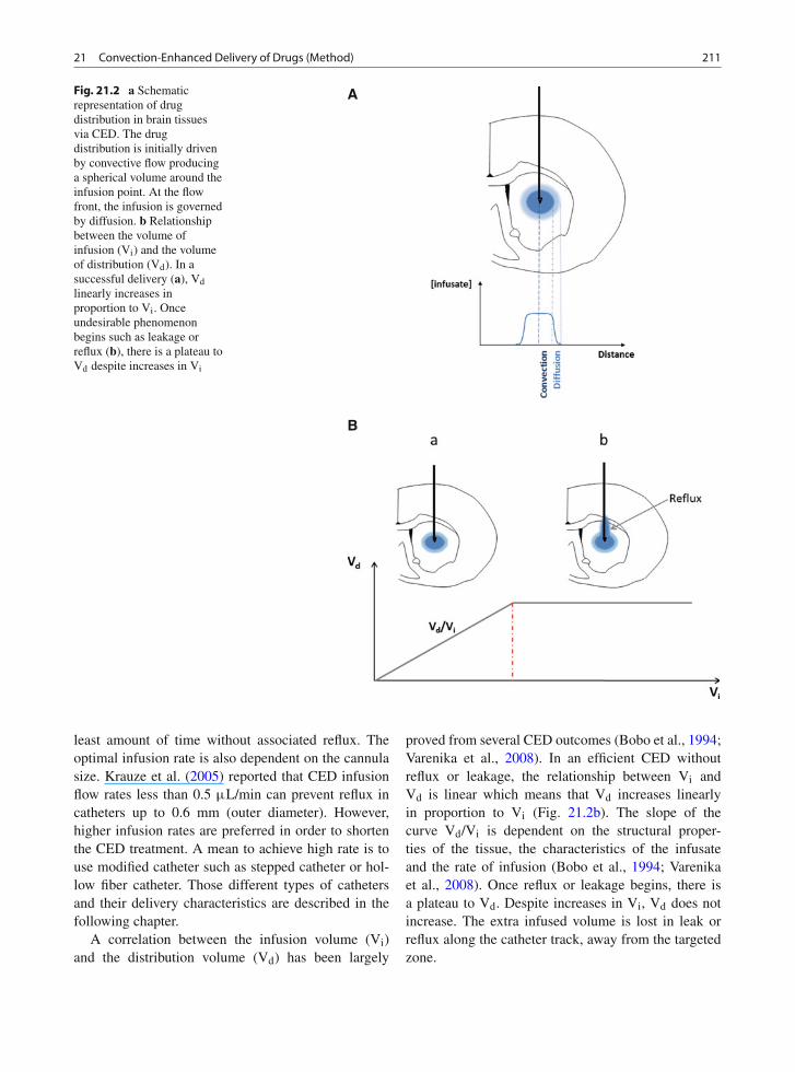
Fig. 21.2 a Schematicrepresentation of drugdistribution in brain tissuesvia CED. The drugdistribution is initially drivenby convective flow producinga spherical volume around theinfusion point. At the flowfront, the infusion is governedby diffusion. b Relationshipbetween the volume ofinfusion (Vi) and the volumeof distribution (Vd). In asuccessful delivery (a), Vdlinearly increases inproportion to Vi. Onceundesirable phenomenonbegins such as leakage orreflux (b), there is a plateau toVd despite increases in Vi
least amount of time without associated reflux. Theoptimal infusion rate is also dependent on the cannulasize. Krauze et al. (2005) reported that CED infusionflow rates less than 0.5 μL/min can prevent reflux incatheters up to 0.6 mm (outer diameter). However,higher infusion rates are preferred in order to shortenthe CED treatment. A mean to achieve high rate is touse modified catheter such as stepped catheter or hol-low fiber catheter. Those different types of cathetersand their delivery characteristics are described in thefollowing chapter.
A correlation between the infusion volume (Vi)and the distribution volume (Vd) has been largely
proved from several CED outcomes (Bobo et al., 1994;Varenika et al., 2008). In an efficient CED withoutreflux or leakage, the relationship between Vi andVd is linear which means that Vd increases linearlyin proportion to Vi (Fig. 21.2b). The slope of thecurve Vd/Vi is dependent on the structural proper-ties of the tissue, the characteristics of the infusateand the rate of infusion (Bobo et al., 1994; Varenikaet al., 2008). Once reflux or leakage begins, there isa plateau to Vd. Despite increases in Vi, Vd does notincrease. The extra infused volume is lost in leak orreflux along the catheter track, away from the targetedzone.

212 A.-L. Laine et al.
Catheter Design
The first infusion tool for CED was a needle implantedinto the brain of experimental animals (Bobo et al.,1994). The technique has been improved replacingthe needle by a catheter which can be stereotacti-cally placed in the CNS. The material used for thecatheter fabrication has to be biocompatible to allowa long implantation time. Such material can be plas-tic, teflon or fused silica. Standard catheters have onlyone port through that the drug is released. It hasbeen recognized that the larger the catheter diameter,the more easily reflux occurs (Morrison et al., 1999).Additionally, data from experiments with agarosegelatin have shown that small diameter catheters canproduce repeatable and uniform spherical volume ofdistribution (Bauman et al., 2004).
New types of CED catheters (Fig. 21.3) are cur-rently under development to increase the efficiency andthe reliability of drug delivery. Fiandaca et al. (2008)developed a stepped cannula that allows increasingthe infusion rate up to 5 μL/min without any reflux.This reflux-preventing catheter consists of a needleextended with a narrower tip at its end, the differ-ing diameter making the step. This design preventsthe reflux along the catheter track by restricting initialbackflow of fluid flow beyond the step. The steppedcannula was recently compared with a stepless can-nula by Yin et al. (2010). They showed that reflux
occurred at only 0.5 μL/min in agarose gel for thestepless cannula leading to a small infusion volumeof 4 μL. In contrast, with the 1-mm step cannulano reflux occurred even at high infusion rate of 3.0μL/min allowing a wide distribution volume of 79 μL.Moreover, the infusion of a recombinant growth factor,GDNF via the step design cannula allowed achieving agreat distribution volume of 97.2% in rat striatum withlittle evidence of reflux. The distribution volume per-centage reached via the cannula without step was only70.7% with significant reflux and leakage in the corpuscallosum and the white matter (Yin et al., 2010).
Catheters with a single opening for the drug releasehave shown their limitation for the delivery into braintumor. The pressure gradient resulting from high inter-stitial pressure in brain tumor drives the drug flow intolow-pressure area. With one port catheter, the infusateflow will follow the path of lowest interstitial pres-sure which will restrict the distribution and may leadto leakage. Thus, multiple-hole catheters have beendesigned to provide infusate flow in all the direc-tions around the catheter overcoming the limitation ofthe one port catheter. The design consists of multi-ple openings of a few millimeters along the distal endof the catheter. However, a previous study performedin agarose gel showed that the drug release is effec-tive only via the proximal port, rending the other portsuseless (Raghavan et al., 2006). This issue can be over-come by reducing the hole size. A porous hollow fiber
Fig. 21.3 Novel catheterdesigns for CED: steppedcannula, multiple-holecatheter, balloon-tippedcatheter, hollow fibercatheter

21 Convection-Enhanced Delivery of Drugs (Method) 213
catheter composed of millions of nano-openings (0.45μm) along its surface has been developed to providemultiple pathways for the drug to infuse within thetissue. Seunguk et al. (2009) compared the distribu-tion of Evans blue dye into the mouse brain throughCED via a hollow fiber catheter versus a conven-tional 28-gauge needle. Experimental results showed athreefold increase in the dye distribution volume withthe hollow fiber catheter relative to the single-lumenneedle.
Since recently, a new type of catheter namedballoon-tipped catheter has been proposed. Thiscatheter is a one-port-opening with a balloon proxi-mal to its tip which can be inflated. The inflated formhas been designed to fill a resection cavity and pre-vent reflux phenomenon. A CED experiment usingballoon-tipped catheter into canine model showed anefficient drug penetration within the brain parenchymaall around the balloon (Olson et al., 2008).
Infusate Formulation
In a typical CED application, the therapeutic agentis dissolved in saline solution and delivered by infu-sion under positive pressure (Yang et al., 2011). It hasbeen observed that infusion of some drugs achievespoorer efficiency than others due to their low viscos-ity. Mardor et al. (2005) have shown that low-viscosityinfusates tend to backflow along the catheter and intothe ventricles whereas high-viscosity infusates limitbackflow event and tend to form efficient convection.The infusate viscosity can be increased by addingsucrose, human serum albumin or polyethylene glycolin the aqueous phase of the infusate. A linear correla-tion between infusate viscosity and distribution volumehas been established showing the impact of the vis-cosity on the CED efficacy. Additionally, infusion ofhigh-viscosity infusate via CED allows covering largervolumes of distribution in less time, thus leading toshorter treatment. Highly viscous formulation can beapplied to a wide range of infusate including genetherapy, proteins, and nanoparticles (Perlstein et al.,2008).
It has also been shown that the use of co-infusate(e.g., heparin, basic fibroblast growth factor or manni-tol) expedites the spread of the drug within the CNSby reducing the affinity of therapeutic agent to the
brain environment. For example, the tissue penetrationof GDNF and GDNF-homologous trophic factors wassignificantly enhanced with heparin co-infusion, prob-ably by binding and blocking heparin-binding sites inthe extracellular matrix (Hamilton et al., 2001).
Despite these progresses, CED of free drug showssevere limitations. First of all, the drug concentrationthat can be delivered is limited by the solubility ofthe drug. Additionally, efficiency of free drug CED isrestricted by rapid clearance of the therapeutic agentfrom the tumor interstitium (Kunwar et al., 2007) andno selective accumulation in targeted tissue. Otherissues inherent to the physico-chemical properties ofthe drug can undermine CED efficiency. Althoughbeing a promising therapeutic agent, CED of free syn-thetic retinoid Am80, did not show any improvementon the mean survival days of rats bearing intracra-nial glioblastoma xenografts (Yokosawa et al., 2010).The failure was attributed to the Am80 hydrophobicitywhich increases the retention within the brain tissueresulting in a poor distribution volume.
A solution to overcome solubility issue and to pro-tect therapeutic agents against elimination and in vivodegradation is to formulate them into nanocarriers.Different drug carrier systems have recently emergedand been tested in CED technique including lipo-somes (Saito et al., 2006), polymer micelles (Inoueet al., 2009), lipid nanocapsules (Allard et al., 2009). . .Inoue et al. (2009) compared the therapeutic effi-cacy of free doxorubicin (DOX) and polymer micellarDOX infused by CED against 9L brain tumors inrat model. They showed that polymer micellar dox-orubicin (DOX) achieved much wider distribution inbrain tissue than free DOX. Accordingly, the mediansurvival obtained with micellar DOX was 36 dayscompared to 19.6 days obtained from the free DOXtreatment.
Nanocarriers are constructed systems in nanometersize that can carry drugs (Yokosawa et al., 2010) andimaging agents (Fiandaca et al., 2008). Carrier sys-tems include mainly polymer or lipid-based carrierssuch as liposomes, nanoparticles, micelles and den-drimers. These differ in structure composition, drug-loading capacity, carrier stability, targeting possibilityand ability to encapsulate hydrophobic or hydrophilicmolecule. Nanocarrier systems are of great interestas they are inert until the drug is released from theconfine of the carriers, thus minimizing side effectsand toxicity to normal brain tissue. Additionally, they

214 A.-L. Laine et al.
offer several advantages, including enhancement ofdrug pharmacokinetic behavior and convection prop-erties. Once a drug is encapsulated, the pharmacoki-netic properties are no longer related to the drug butare determined by the nanocarrier. Furthermore, thenanocarrier architecture offers the possibility to adjustdrug release rate. Improving sustained drug releaseallows the prolongation of drug’s half-life leading toincreased exposure of brain tumor tissue to the thera-peutic agent. It also limits the high peak concentrationof free drug potentially associated with toxicity andprovides an effective inhibition of drug absorption bycells.
Surface properties have a considerable impact onthe spread of the infusate distribution. The poorer braindistribution observed with hydrophobic or cationicinfusate can be circumvented by adding polyethyleneglycol or dextran onto the nanocarrier. Indeed, PEGor dextran coating make the infusate more hydrophilicleading to less tissue affinity and increased distribu-tion. Saito et al. (2006) have reported that athymicnude rats bearing U87MG human glioma cells sur-vived significantly longer after treatment with topote-can encapsulated into PEGylated liposome given byCED compared with free topotecan. PEG encapsu-lation also provides steric stabilization and reducessurface charge (Yokosawa et al., 2010).
As the pore size of the extracellular matrix (ECM)is estimated as about 50 nm, nanocarrier size shouldbe in the range of 20–50 nm for an effective transitthrough the interstitial space (MacKay et al., 2005). Ithas been showed that the infusion of a hyperosmolarsolution of mannitol, either before the CED treatmentor in co-infusion with the drug, may increase the size ofchannels of the interstial space enhancing the nanopar-ticle transport trough the ECM (Neeves et al., 2007).For instance, CED of 40 nm-liposomes co-infused with25% mannitol showed an improvement of the Vd from52.5 ± 2.1 to 78.5 ± 5.5 units immediately after CED(Mamot et al., 2004).
Ced Patient-Specific Simulationand Real-Time Imaging
Imaging of brain tumor can be realized via CED. Theco-infusion of tracers with the drug allows monitoringits distribution as it is being delivered (Fiandaca et al.,2008). The most common technique currently usedto track real-time distribution of therapeutics is the
Magnetic Resonance Imaging (MRI). Varenika et al.(2008) used gadoteridol-loaded liposomes infused byCED to visualize the distribution in various anatomi-cal structures of the CNS of nonhuman primates andcanines with MR imaging. This technique evidencedthe unexpected high frequency of leakage phenomenon(20% of infusions) into cerebral ventricles.
Monitoring of CED is of crucial importance toensure optimal delivery of therapeutic agents into tar-get sites while minimizing exposure of healthy tissue.Adjustment and alteration to the infusion plan can becarried out in case of undesirable events during the pro-cess. In the event of reflux, reducing the infusion ratecan allow CED process to continue while backflow islimited. More drastic changes would include alteringthe cannula placement or inserting a second cannula atthe onset of leakage. Those adjustments could allowovercoming leakage issues and covering the targetedarea without stopping the process.
Additionally, direct visualization can help to deter-mine whether a treatment failed because of the drug’slack of therapeutic efficacy or because of inadequatedelivery. Monitoring information allows optimizingthe process including catheter placement in order toachieve robust and reliable delivery of therapeutics.
As for any invasive procedure, it would be ofgreat interest to base decision on how to plan CEDwith patient-specific simulation. The entry of CEDin clinical trial has motivated the development ofcomputational methods to aid in planning CED treat-ments. Such software developed by BrainLAB AGand approved by FDA is now commercially avail-able for neuro-oncology. This program simulates drugdistribution into the human brain based on the indi-vidual patient’s tractography MRI data and brain tis-sue anatomy. It is worth noting that tractographyallows visualizing the white matter bundles which isof great interest for the simulation of drug distribution.Treatment of specific tumor can be visualized in 3Dincluding the number and position of catheters. Thedistribution concordance between the actual distribu-tion of ILI3-PE38QQR co–infused with 123I-albuminby CED and the distribution prediction has provedthe software specificity and reliability in treatmentplanning (Sampson et al., 2007). Additionally, thissimulation allows observing infusate leakage into thesubarachnoid cerebrospinal fluid space when catheterscross deep sulci (Sampson et al., 2007). Computationalsimulation provides therefore a useful tool to optimizeCED catheter placement.

21 Convection-Enhanced Delivery of Drugs (Method) 215
Conclusion
Convection enhanced-delivery has been developed asa local drug delivery strategy. Using convective trans-port, it offers the advantage of better drug distributionthan the regional delivery technology based on diffu-sion. CED represents a potential powerful methodol-ogy for targeted therapy in the field of neuro-oncology.Bypassing the blood-brain barrier, this approach allowstumor and other brain tissue to be exposed to highdrug concentration that could not be achieved via sys-temic application. The potential of this technique hasbeen clearly demonstrated in preclinical studies (Yanget al., 2011; Yokosawa et al., 2010). However, adverseevents including drug backflow along the catheter trackand leakage in undesired zone can occur during infu-sion. Such events undermine CED efficacy and canimply side effects including neurotoxicity and adjacentstructure damages.
Some improvements are under development formaking CED an effective way of delivering antineo-plastic drugs in human brain. The use of nanocarriertechnology to deliver drugs via CED seems to be apromising combination to improve the efficacy of thetreatment against brain tumor. Further development oftargeting nanocarriers would allow the accumulation inthe desired area and limit extensive distribution into theCNS. It mays also help to penetrate the tumor whichshows relative inaccessibility. However, there is a needto better understand the drug release mechanism outof the carrier system. It would be also of great inter-est to pay special attention to others parameters whichmay impact the distribution such as the manipulationof physiological parameters including blood pressureand heart rate. Indeed, it was shown that larger Vd
could be obtained by increasing blood pressure/heartrate induced by epinephrine (Hadaczek et al., 2006).
There is also a need for a better understanding ofthe drug delivery into tumor brain, markedly differentfrom normal brain. Clinical trials have also reportedthat target tissue anatomy and patient-specific anatomyplay a major role in drug distribution using CED.Gaining more information about the brain’s anatomyand fluid dynamics that govern delivery would allowpredicting the time, direction and eventually, the effi-ciency of therapeutic molecule distribution. Finally,the transposition from rodent or non human primatebrain to human brain remains an ongoing challenge
due to significant differences between brain structures(Fig. 21.1b) and CED intra-operational parametershave to be adjusted by taking precisely anatomicalcriteria into account.
References
Allard E, Huynh NT, Vessières A, Pigeon P, Jaouen G, BenoitJP, Passirani C (2009) Dose effect activity of ferrocifen-loaded lipid nanocapsules on a 9L-glioma model. Int J Pharm379(2):317–323
Allard E, Passirani C, Benoit JP (2009) Convection-enhanceddelivery of nanocarriers for the treatment of brain tumors.Biomaterials 30:2302–2318
Bauman MA, Gillies GT, Raghavan R, Brady ML, Pedain C(2004) Physical characterization of neurocatheter perfor-mance in a brain phantom gelatin with nanoscale porosity:steady-state and oscillatory flows. Nanotechnology 15:92–97
Bidros DS, Vogelbaum MA (2009) Novel drug delivery strate-gies in neuro-oncology. J Am Soc Exp Neuroth 6:539–546
Bobo RH, Laske DW, Akbasak A, Morrison PF, DedrickRL, Oldfield EH (1994) Convection-enhanced delivery ofmacromolecules in the brain. Proc Natl Acad Sci USA 91:2076–2080
Debinski W, Tatter ST (2009) Convection-enhanced deliveryfor the treatment of brain tumors. Expert Rev Neurother9:15719–1527
Fiandaca MS, Forsayeth JR, Dickinson PJ, Bankiewicz KS(2008) Image-guided convection-enhanced delivery platformon the treatment of neurological diseases. Neurotherapeutics5:123–127
Fung LK, Ewend MG, Sills A, Sipos EP, Thompson R,Watts M, Colvin OM, Brem H, Saltzman WM (1998)Pharmacokinetics of interstitial delivery of carmustine,4-hydroperoxycyclophosphamide, and paclitaxel from abiodegradable polymer implant in the monkey brain. CancerRes 58:672–684
Hadaczek P, Mirek H, Tamas L, Bohn MC, Noble C, Park JW,Bankiewicz K (2006) The “perivascular pump” driven byarterial pulsation is a powerful mechanism for the distribu-tion of therapeutic molecules within the brain. Mol Therapy14:69–71
Hamilton JF, Morrision PF, Chen MY, Harvey-White J, PernauteRS, Phillips H, Oldfield E, Bankiewicz KS (2001) Heparincoinfusion during convection-enhanced delivery (CED)increases the distribution of the glial-derived neurotrophicfactor (GDNF) ligand family in rat striatum and enhancesthe pharmacological activity of neurturin. Exp Neurol 168:155–161
Inoue T, Yamashita Y, Nishihara M, Sugiyama S, Sonoda Y,Kumabe T, Yokoyama M, Tominaga T (2009) Therapeuticefficacy of a polymeric micellar doxorubicin infused byconvection-enhanced delivery against intracranial 9L braintumor models. Neuro-Oncology 11:151–157
Jain RK (1989) Delivery of novel therapeutic agent in tumors:physiological barriers and strategies. J Natl Cancer Inst81:570–576
216 A.-L. Laine et al.
Krauze MT, Saito R, Noble C, Bringas J, Forsayeth J, McknightTR, Park J, Bankiewicz KS (2005) Effects of the perivascu-lar space on convection-enhanced delivery of liposomes inprimate putamen. Exp Neurol 196:104–111
Kunwar S (2003) Convection enhanced delivery of IL13-PE38QQR for treatment of recurrent malignant glioma: pre-sentation of interim findings from ongoing phase 1 studies.Acta Neurochir Suppl 88:105–111.
Kunwar S, Prados MD, Chang SM, Berger MS, Lang FF,Piepmeier JM, Sampson J, Ram Z, Gutin PH, Gibbons RD,Aldape KD, Croteau DJ, Sherman JW, Puri RK (2007)Direct intracerebral delivery of cintredekin besudotox (IL13-PE38QQR) in recurrent malignant glioma: a report by theCintredekin Besutodox Intraparenchymal Study Group. JClin Oncol 25:837–844
Lieberman DM, Laske DW, Morrison PF, Bankiewicz KS,Oldfield EH (1995) Convection-enhanced distribution oflarge molecules in gray matter during interstitial drug infu-sion. J Neurosurg 82:1021–1029
MacKay JA, Deen DF, Szoka C Jr (2005) Distribution in brainof liposomes after convection enhanced delivery; modulationby particle charge, particle diameter, and presence of stericcoating. Brain Res 1035:139–153
Mamot C, Nguyen JB, Pourdehnad M, Hadaczek P, Saito R,Bringas JR, Drummond DC, Hong K, Kirpotin DB,McKnight T, Berger MS, Park JW, Bankiewicz KS(2004) Extensive distribution of liposomes in rodent brainsand brain tumors following convection-enhanced delivery.J Neurooncol 68:1–9
Mardor Y, Rahav O, Zauberman Y, Lidar Z, OcherashvilliA, Daniels D, Roth Y, Maier SR, Orenstein A, Ram Z(2005) Convection-enhanced drug delivery: increased effi-cacy and Magnetic Resonance Image Monitoring. CancerRes 65:6858–6863
Misra A, Ganesh s, Shahiwala A, Shah SP (2003) Drug Deliveryto the central nervous system: a review. J Pharm Pharm Sci6:252–273
Morrison PF, Chen MY, Chadwick RS, Lonser RR, Oldfield EH(1999) Focal delivery during direct infusion to brain: roleof flow rate, catheter diameter, and tissue mechanics. Am JPhysiol 277:1218–1229
Morrison PF, Laske DW, Bobo H, Oldfield EH, Dedrick RL(1994) High-flow microinfusion: tissue penetration and phar-macodynamics. Am J Physiol 266:292–305
Neeves KB, Sawyer AJ, Foley CP, Saltzman WM, Olbricht WL(2007) Dilation and degradation of the brain extracellularmatrix enhances penetration of infused polymer nanoparti-cles. Bain Res 1180:121–132
Olson JJ, Zhang Z, Dillehay D, Stubbs J (2008) Assessmentof a balloon-tipped catheter modified for intracerebralconvection-enhanced delivery. J Neurooncol 89:159–168
Pardridge WM (2007) Blood-brain barrier delivery. Drug DiscovToday 12:54–61
Perlstein B, Ram Z, Daniels D, Ocherashvilli A, Roth Y,Margel S, Mardor Y (2008) Convection-enhanced deliveryof maghemite nanoparticles: increased efficacy and MRImonitoring. Neuro-Oncology 10(2):153–161
Raghavan R, Brady ML, Rodríguez-Ponce MI, Hartlep A,Pedain C, Sampson JH (2006) Convection-enhanced deliveryof therapeutics for brain disease, and its optimization.Neurosurg Focus 20:E12
Raza SM, Pradilla G, Legnani FG, Thai QA, Olivi A, WeingartJD, Brem H (2005) Local delivery of antineoplastic agents bycontrolled-release polymers for the treatment of malignantbrain tumours. Expert Opin Biol Ther 5:477–494
Rosenberg GA, Kyner WT, Estrada E (1980) Bulk flow of braininterstitial fluid under normal und hyperosmolar conditions.Am J Physiol 238:42–49
Saito R, Krauze MT, Noble CO, Drummond DC, Kirpotin DB,Berger MS, Park JW, Bankiewicz KS (2006) Convection-enhanced delivery of Ls-TPT enables an effective, con-tinuous, low-dose chemotherapy against malignant gliomaxenograft model. Neuro-Oncology 8:205–214
Sampson JH, Raghavan R, Brady ML, Provenzale JM, HerndonJE II, Croteau D, Friedman AH, Reardon DA, Coleman RE,Wong T, Bigner DD, Pastan I, Rodríguez-Ponce MI, TannerP, Puri R, Pedain C (2007) Clinical utility of a patient-specificalgorithm for simulating intracerebral drug infusions.Neuro-Oncology 9:343–353
Sawyer AJ, Piepmeier JM, Saltzman WM (2006) New methodsfor direct delivery of chemotherapy for treating brain tumors.Yale J Biol Med 79:141–152
Seunguk O, Odland R, Wilson SR, Kroeger KM, Liu C,Lowenstein PR, Castro MG, Hall WA, Ohlfest JR (2009)Improved distribution of small molecules and viral vectorsin the murine brain using a hollow fiber catheter. Neuro-Oncology 9:343–353
Stupp R, Mason WP, van den Bent MJ, Weller M, Fisher B,Taphroorn MJB, Belanger K, Brandes AA, Marosi C,Bogdahn U, Curschmann J, Janzer RC, Ludwin SK, GorliaT, Allgeier A, Lacombe D, Cairncross JG, Eisenhauer E,Mirimanoff RO (2005) Radiotherapy plus concomitant andadjuvant temozolomide for glioblastoma. N Engl J Med352:987–996
Varenika V, Dickinson P, Bringas J, LeCouteur R, Higgins R,Park J, Fiandaca M, Berger M, Sampson J, Bankiewicz K(2008) Detection of infusate leakage in the brain using real-time imaging of convection-enhanced delivery. J Neurosurg109:874–880
Yang W, Huo T, Barth RF, Gupta N, Weldon M, Grecula JC,Ross BD, Hoff BA, Chou TC, Rousseau J, Elleaume H(2011) Convection enhanced delivery of carboplatin in com-bination with radiotherapy for the treatment of brain tumors.J Neurooncol 101:379–390
Yin D, Forsayeth J, Bankiewicz KS (2010) Optimized cannuladesign and placement for convection-enhanced delivery in ratstriatum. J Neurosci Meth 187:46–51
Yokosawa M, Sonoda Y, Sugiyama S, Saito R, Yamashita Y,Nishihara M, Satoh T, Kumabe T, Yokoyama M, Tominaga T(2010) Convection-enhanced delivery of a synthetic retinoidAm80, loaded into polymeric micelles, prolongs the survivalof rats bearing intracranial Glioblastoma xenografts. TohokuJ Exp Med 221:257–264