Brain regions with mirror properties: A meta-analysis of 125 human fMRI studies
9
Neuroscience and Biobehavioral Reviews 36 (2012) 341–349 Contents lists available at ScienceDirect Neuroscience and Biobehavioral Reviews jou rnal h omepa ge: www.elsevier.com/locate/neubiorev Review Brain regions with mirror properties: A meta-analysis of 125 human fMRI studies Pascal Molenberghs ∗ , Ross Cunnington, Jason B. Mattingley The University of Queensland, Queensland Brain Institute & School of Psychology, Queensland 4072, Australia a r t i c l e i n f o Article history: Received 31 March 2011 Received in revised form 23 June 2011 Accepted 6 July 2011 Keywords: Action observation Activation likelihood estimation fMRI Inferior frontal cortex Inferior parietal cortex Mirror neurons a b s t r a c t Mirror neurons in macaque area F5 fire when an animal performs an action, such as a mouth or limb movement, and also when the animal passively observes an identical or similar action performed by another individual. Brain-imaging studies in humans conducted over the last 20 years have repeatedly attempted to reveal analogous brain regions with mirror properties in humans, with broad and often speculative claims about their functional significance across a range of cognitive domains, from language to social cognition. Despite such concerted efforts, the likely neural substrates of these mirror regions have remained controversial, and indeed the very existence of a distinct subcategory of human neurons with mirroring properties has been questioned. Here we used activation likelihood estimation (ALE), to provide a quantitative index of the consistency of patterns of fMRI activity measured in human studies of action observation and action execution. From an initial sample of more than 300 published works, data from 125 papers met our strict inclusion and exclusion criteria. The analysis revealed 14 separate clusters in which activation has been consistently attributed to brain regions with mirror properties, encompassing 9 different Brodmann areas. These clusters were located in areas purported to show mirroring proper- ties in the macaque, such as the inferior parietal lobule, inferior frontal gyrus and the adjacent ventral premotor cortex, but surprisingly also in regions such as the primary visual cortex, cerebellum and parts of the limbic system. Our findings suggest a core network of human brain regions that possess mirror properties associated with action observation and execution, with additional areas recruited during tasks that engage non-motor functions, such as auditory, somatosensory and affective components. Crown Copyright © 2011 Published by Elsevier Ltd. All rights reserved. Contents 1. Introduction . . . . . . . . . . . . . . . . . . . . . . . . . . . . . . . . . . . . . . . . . . . . . . . . . . . . . . . . . . . . . . . . . . . . . . . . . . . . . . . . . . . . . . . . . . . . . . . . . . . . . . . . . . . . . . . . . . . . . . . . . . . . . . . . . . . . . . . . . 341 2. Materials and methods . . . . . . . . . . . . . . . . . . . . . . . . . . . . . . . . . . . . . . . . . . . . . . . . . . . . . . . . . . . . . . . . . . . . . . . . . . . . . . . . . . . . . . . . . . . . . . . . . . . . . . . . . . . . . . . . . . . . . . . . . . . . . . 342 2.1. Literature selection and exclusion criteria . . . . . . . . . . . . . . . . . . . . . . . . . . . . . . . . . . . . . . . . . . . . . . . . . . . . . . . . . . . . . . . . . . . . . . . . . . . . . . . . . . . . . . . . . . . . . . . . . . 342 2.2. Selection of activated voxels . . . . . . . . . . . . . . . . . . . . . . . . . . . . . . . . . . . . . . . . . . . . . . . . . . . . . . . . . . . . . . . . . . . . . . . . . . . . . . . . . . . . . . . . . . . . . . . . . . . . . . . . . . . . . . . . 342 2.3. Activation likelihood estimation (ALE) . . . . . . . . . . . . . . . . . . . . . . . . . . . . . . . . . . . . . . . . . . . . . . . . . . . . . . . . . . . . . . . . . . . . . . . . . . . . . . . . . . . . . . . . . . . . . . . . . . . . . . 343 2.4. Label-based review of Brodmann areas . . . . . . . . . . . . . . . . . . . . . . . . . . . . . . . . . . . . . . . . . . . . . . . . . . . . . . . . . . . . . . . . . . . . . . . . . . . . . . . . . . . . . . . . . . . . . . . . . . . . . 343 3. Results . . . . . . . . . . . . . . . . . . . . . . . . . . . . . . . . . . . . . . . . . . . . . . . . . . . . . . . . . . . . . . . . . . . . . . . . . . . . . . . . . . . . . . . . . . . . . . . . . . . . . . . . . . . . . . . . . . . . . . . . . . . . . . . . . . . . . . . . . . . . . . . 343 3.1. Meta-analysis across all included studies .... . . . . . . . . . . . . . . . . . . . . . . . . . . . . . . . . . . . . . . . . . . . . . . . . . . . . . . . . . . . . . . . . . . . . . . . . . . . . . . . . . . . . . . . . . . . . . . . 343 3.2. Meta-analyses of motor versus non-motor studies . . . . . . . . . . . . . . . . . . . . . . . . . . . . . . . . . . . . . . . . . . . . . . . . . . . . . . . . . . . . . . . . . . . . . . . . . . . . . . . . . . . . . . . . . 343 4. Discussion .. . . . . . . . . . . . . . . . . . . . . . . . . . . . . . . . . . . . . . . . . . . . . . . . . . . . . . . . . . . . . . . . . . . . . . . . . . . . . . . . . . . . . . . . . . . . . . . . . . . . . . . . . . . . . . . . . . . . . . . . . . . . . . . . . . . . . . . . . . 344 Acknowledgements . . . . . . . . . . . . . . . . . . . . . . . . . . . . . . . . . . . . . . . . . . . . . . . . . . . . . . . . . . . . . . . . . . . . . . . . . . . . . . . . . . . . . . . . . . . . . . . . . . . . . . . . . . . . . . . . . . . . . . . . . . . . . . . . . 348 Appendix A. Supplementary data .. . . . . . . . . . . . . . . . . . . . . . . . . . . . . . . . . . . . . . . . . . . . . . . . . . . . . . . . . . . . . . . . . . . . . . . . . . . . . . . . . . . . . . . . . . . . . . . . . . . . . . . . . . . . . . . 348 References . . . . . . . . . . . . . . . . . . . . . . . . . . . . . . . . . . . . . . . . . . . . . . . . . . . . . . . . . . . . . . . . . . . . . . . . . . . . . . . . . . . . . . . . . . . . . . . . . . . . . . . . . . . . . . . . . . . . . . . . . . . . . . . . . . . . . . . . . . . 348 ∗ Corresponding author at: School of Psychology, McElwain Building, The Univer- sity of Queensland, St Lucia, QLD 4072, Australia. Tel.: +61 7 3365 6257; fax: +61 7 3365 4466. E-mail address: [email protected] (P. Molenberghs). 1. Introduction Mirror neurons were originally described as visuomotor neu- rons that fire both when an action is performed, and when a similar or identical action is passively observed (Rizzolatti and Craighero, 2004). These neurons were first discovered using single-cell 0149-7634/$ – see front matter. Crown Copyright © 2011 Published by Elsevier Ltd. All rights reserved. doi:10.1016/j.neubiorev.2011.07.004
Transcript of Brain regions with mirror properties: A meta-analysis of 125 human fMRI studies
R
B
PT
a
ARRA
KAAfIIM
C
sf
0d
Neuroscience and Biobehavioral Reviews 36 (2012) 341–349
Contents lists available at ScienceDirect
Neuroscience and Biobehavioral Reviews
jou rna l h omepa ge: www.elsev ier .com/ locate /neubiorev
eview
rain regions with mirror properties: A meta-analysis of 125 human fMRI studies
ascal Molenberghs ∗, Ross Cunnington, Jason B. Mattingleyhe University of Queensland, Queensland Brain Institute & School of Psychology, Queensland 4072, Australia
r t i c l e i n f o
rticle history:eceived 31 March 2011eceived in revised form 23 June 2011ccepted 6 July 2011
eywords:ction observationctivation likelihood estimation
MRInferior frontal cortexnferior parietal cortex
irror neurons
a b s t r a c t
Mirror neurons in macaque area F5 fire when an animal performs an action, such as a mouth or limbmovement, and also when the animal passively observes an identical or similar action performed byanother individual. Brain-imaging studies in humans conducted over the last 20 years have repeatedlyattempted to reveal analogous brain regions with mirror properties in humans, with broad and oftenspeculative claims about their functional significance across a range of cognitive domains, from languageto social cognition. Despite such concerted efforts, the likely neural substrates of these mirror regions haveremained controversial, and indeed the very existence of a distinct subcategory of human neurons withmirroring properties has been questioned. Here we used activation likelihood estimation (ALE), to providea quantitative index of the consistency of patterns of fMRI activity measured in human studies of actionobservation and action execution. From an initial sample of more than 300 published works, data from125 papers met our strict inclusion and exclusion criteria. The analysis revealed 14 separate clusters inwhich activation has been consistently attributed to brain regions with mirror properties, encompassing
9 different Brodmann areas. These clusters were located in areas purported to show mirroring proper-ties in the macaque, such as the inferior parietal lobule, inferior frontal gyrus and the adjacent ventralpremotor cortex, but surprisingly also in regions such as the primary visual cortex, cerebellum and partsof the limbic system. Our findings suggest a core network of human brain regions that possess mirrorproperties associated with action observation and execution, with additional areas recruited during tasksthat engage non-motor functions, such as auditory, somatosensory and affective components.Crown Copyright © 2011 Published by Elsevier Ltd. All rights reserved.
ontents
1. Introduction . . . . . . . . . . . . . . . . . . . . . . . . . . . . . . . . . . . . . . . . . . . . . . . . . . . . . . . . . . . . . . . . . . . . . . . . . . . . . . . . . . . . . . . . . . . . . . . . . . . . . . . . . . . . . . . . . . . . . . . . . . . . . . . . . . . . . . . . . 3412. Materials and methods . . . . . . . . . . . . . . . . . . . . . . . . . . . . . . . . . . . . . . . . . . . . . . . . . . . . . . . . . . . . . . . . . . . . . . . . . . . . . . . . . . . . . . . . . . . . . . . . . . . . . . . . . . . . . . . . . . . . . . . . . . . . . . 342
2.1. Literature selection and exclusion criteria . . . . . . . . . . . . . . . . . . . . . . . . . . . . . . . . . . . . . . . . . . . . . . . . . . . . . . . . . . . . . . . . . . . . . . . . . . . . . . . . . . . . . . . . . . . . . . . . . . 3422.2. Selection of activated voxels . . . . . . . . . . . . . . . . . . . . . . . . . . . . . . . . . . . . . . . . . . . . . . . . . . . . . . . . . . . . . . . . . . . . . . . . . . . . . . . . . . . . . . . . . . . . . . . . . . . . . . . . . . . . . . . . 3422.3. Activation likelihood estimation (ALE) . . . . . . . . . . . . . . . . . . . . . . . . . . . . . . . . . . . . . . . . . . . . . . . . . . . . . . . . . . . . . . . . . . . . . . . . . . . . . . . . . . . . . . . . . . . . . . . . . . . . . . 3432.4. Label-based review of Brodmann areas . . . . . . . . . . . . . . . . . . . . . . . . . . . . . . . . . . . . . . . . . . . . . . . . . . . . . . . . . . . . . . . . . . . . . . . . . . . . . . . . . . . . . . . . . . . . . . . . . . . . . 343
3. Results . . . . . . . . . . . . . . . . . . . . . . . . . . . . . . . . . . . . . . . . . . . . . . . . . . . . . . . . . . . . . . . . . . . . . . . . . . . . . . . . . . . . . . . . . . . . . . . . . . . . . . . . . . . . . . . . . . . . . . . . . . . . . . . . . . . . . . . . . . . . . . . 3433.1. Meta-analysis across all included studies . . . . . . . . . . . . . . . . . . . . . . . . . . . . . . . . . . . . . . . . . . . . . . . . . . . . . . . . . . . . . . . . . . . . . . . . . . . . . . . . . . . . . . . . . . . . . . . . . . . 3433.2. Meta-analyses of motor versus non-motor studies . . . . . . . . . . . . . . . . . . . . . . . . . . . . . . . . . . . . . . . . . . . . . . . . . . . . . . . . . . . . . . . . . . . . . . . . . . . . . . . . . . . . . . . . . 343
4. Discussion . . . . . . . . . . . . . . . . . . . . . . . . . . . . . . . . . . . . . . . . . . . . . . . . . . . . . . . . . . . . . . . . . . . . . . . . . . . . . . . . . . . . . . . . . . . . . . . . . . . . . . . . . . . . . . . . . . . . . . . . . . . . . . . . . . . . . . . . . . . 344Acknowledgements . . . . . . . . . . . . . . . . . . . . . . . . . . . . . . . . . . . . . . . . . . . . . . . . . . . . . . . . . . . . . . . . . . . . . . . . . . . . . . . . . . . . . . . . . . . . . . . . . . . . . . . . . . . . . . . . . . . . . . . . . . . . . . . . . 348Appendix A. Supplementary data . . . . . . . . . . . . . . . . . . . . . . . . . . . . . . . . . . . . . . . . . . . . . . . . . . . . . . . . . . . . . . . . . . . . . . . . . . . . . . . . . . . . . . . . . . . . . . . . . . . . . . . . . . . . . . . . 348References . . . . . . . . . . . . . . . . . . . . . . . . . . . . . . . . . . . . . . . . . . . . . . . . . . . . . . . . . . . . . . . . . . . . . . . . . . . . . . . . . . . . . . . . . . . . . . . . . . . . . . . . . . . . . . . . . . . . . . . . . . . . . . . . . . . . . . . . . . . 348
∗ Corresponding author at: School of Psychology, McElwain Building, The Univer-ity of Queensland, St Lucia, QLD 4072, Australia. Tel.: +61 7 3365 6257;ax: +61 7 3365 4466.
E-mail address: [email protected] (P. Molenberghs).
149-7634/$ – see front matter. Crown Copyright © 2011 Published by Elsevier Ltd. All rioi:10.1016/j.neubiorev.2011.07.004
1. Introduction
Mirror neurons were originally described as visuomotor neu-rons that fire both when an action is performed, and when a similaror identical action is passively observed (Rizzolatti and Craighero,2004). These neurons were first discovered using single-cell
ghts reserved.
3 d Biob
rewonpfc2Rii((2OoJhf2
tmia1tmaaRu(nerDFRTiaCMues(atob
irrmehfrOi2r
42 P. Molenberghs et al. / Neuroscience an
ecordings in macaque area F5 (di Pellegrino et al., 1992; Galleset al., 1996; Rizzolatti et al., 1996a) and later in the PF/PFG complexithin the inferior parietal cortex (Gallese et al., 2002). Since these
riginal studies there has been an explosion of interest in mirroreurons, both in the scientific literature and the popular media, inart because of their purported role in a diverse range of cognitiveunctions, from imitation and action understanding to socialognition (Iacoboni, 2005, 2009; Iacoboni et al., 2005; Fogassi et al.,005; Keysers et al., 2010; Rizzolatti and Fabbri-Destro, 2008;izzolatti and Sinigaglia, 2010). Mirror neurons have also been
mplicated in a range of neurological and psychiatric disorders,ncluding multiple sclerosis (Rocca et al., 2008), schizophreniaArbib and Mundhenk, 2005), autism spectrum disorder (ASD)Cattaneo et al., 2007; Dapretto et al., 2006; Iacoboni and Dapretto,006; Williams, 2008) and alexithymia (Moriguchi et al., 2009).ther investigators have argued that evidence for the existencef human mirror neurons is lacking (Dinstein et al., 2008a,b;onas et al., 2007; Lingnau et al., 2009; Turella et al., 2009), orave challenged claims for the role of mirror neurons in language
unction (Johnson-Frey, 2003), action understanding (Hickok,009) and imitation (Makuuchi, 2005; Molenberghs et al., 2009).
Immediately following the initial reports of mirror neurons inhe macaque brain, investigators sought evidence for an analogous
echanism in humans. Based on early human brain-imaging stud-es that compared neural activity during perceived and executedctions (Rizzolatti et al., 1996b; Decety et al., 1997; Iacoboni et al.,999), it was widely assumed that the ventral premotor cortex andhe pars opercularis of the posterior inferior frontal gyrus (Brod-
ann area 44) are human homologues of macaque mirror area F5;nd that the rostral inferior parietal lobule (IPL) is the human equiv-lent of mirror area PF/PFG (Rizzolatti et al., 2001; Rizzolatti, 2005;izzolatti and Craighero, 2004). Subsequent investigations havesed behavioural approaches, transcranial magnetic stimulationTMS), electroencephalography (EEG), functional magnetic reso-ance imaging (fMRI) and human single-cell recordings (Mukamelt al., 2010) to provide further evidence for fronto-parietal mir-or neuron regions in humans (for recent reviews see Iacoboni andapretto, 2006; Fabbri-Destro and Rizzolatti, 2008; Keysers andadiga, 2008; Keysers et al., 2010; Cattaneo and Rizzolatti, 2009;izzolatti and Fabbri-Destro, 2010; Rizzolatti and Sinigaglia, 2010).hese studies have used a variety of tasks to uncover “mirror activ-ty”. Some have employed action observation and execution tasks,nalogous to those used in the original monkey investigations (e.g.,hong et al., 2008; Gazzola and Keysers, 2009; Kilner et al., 2009;olenberghs et al., 2010). Others have used tasks involving stim-
li across a range of modalities, including audition (e.g., Gazzolat al., 2006; Lewis et al., 2005; Tettamanti et al., 2005), somatosen-ation (e.g., Keysers et al., 2004; Schaefer et al., 2009), vision onlye.g., Molnar-Szakacs et al., 2006; Newman-Norlund et al., 2010);s well as tasks employing stimuli with emotional (affective) con-ent (e.g., Carr et al., 2003; Leslie et al., 2004). This wide varietyf approaches in humans has led to an ever-expanding number ofrain regions being implicated in mirror mechanism functioning.
The aim of the current investigation was to draw together imag-ng results from all relevant fMRI studies of the human mirroregions, with the goal of determining the range and extent of brainegions implicated. Based upon the original single-cell findings inonkeys (di Pellegrino et al., 1992; Gallese et al., 1996; Rizzolatti
t al., 1996a; Gallese et al., 2002), it might be predicted that theuman homologues of macaque areas F5 and PF/PFG – the inferior
rontal gyrus and inferior parietal lobule, respectively – should beeliably engaged by tasks designed to elicit mirror neuron activity.
n the other hand, it has recently been proposed that mirror activ-ty is widespread in the human brain (e.g., Keysers and Gazzola,009; Heyes, 2010). If this is true, it might be predicted that brainegions outside the classically defined mirror network would be
ehavioral Reviews 36 (2012) 341–349
engaged, depending on task demands. To address these predictions,we performed a meta-analysis of all human fMRI studies in whichthe authors attributed their findings to mirror neuron functioning.We used a quantitative meta-analysis technique, known as activa-tion likelihood estimation (ALE; Eickhoff et al., 2009), to investigatewhich brain regions are most reliably associated with human mir-ror neuron functions. Contrary to previous ALE studies that focusedexclusively on action observation (Caspers et al., 2010), imitation(Caspers et al., 2010; Molenberghs et al., 2009), and the role of themirror system in action understanding (Van Overwalle and Baetens,2009), our meta-analysis included all fMRI studies in which signif-icant activations were attributed to the mirror system, regardlessof task requirements. We also performed a label-based review todetermine the Brodmann areas most consistently associated withmirror neuron regions. In follow-up analyses, we separated studiesbased on whether they targeted the “classical” (motor) mirror neu-rons, or instead examined activity during observation of auditory,somatosensory or emotional (affective) stimuli.
2. Materials and methods
2.1. Literature selection and exclusion criteria
We searched the Web of Science database (http://apps.isiknowledge.com) using the keywords ‘fMRI’ and ‘mirror system’.As of January 2011, this search revealed 438 published, peer-reviewed papers. The inclusion criteria for our analyses were asfollows:
1. Studies that explicitly mentioned the mirror system wereincluded, whereas those that did not were excluded (e.g., thesearch also uncovered studies about “mirrored” hand move-ments). Three hundred and thirty (330) of the 438 papers metthis criterion.
2. Studies that used fMRI were included, whereas those thatemployed other techniques (positron emission tomography(PET), single-photon emission tomography (SPECT), magnetoen-cephalography (MEG), TMS, behavioural measures and reviewarticles) were excluded. We restricted our study to fMRI databecause we wanted to have approximately comparable spatialand temporal resolution for the ALE analyses. One hundred andninety (190) of the remaining 330 papers met this criterion.
3. Studies that failed to reveal significant “mirror” activation wereexcluded (e.g., Lingnau et al. (2009) failed to find cross-modaladaptation for observed and executed motor acts). One hun-dred and eighty one (181) of the remaining 190 papers met thiscriterion.
4. Studies in which the authors did not attribute their fMRI resultsdirectly to the mirror system were excluded (e.g., Cross et al.(2009) talk about an “action observation network” (AON) ratherthan a “mirror neuron system”). One hundred and forty (140) ofthe remaining 181 papers met this criterion.
5. Studies in which the authors interpreted their results as reflect-ing activity within the mirror system, but did not report thecoordinates of the activation clusters, were excluded from theanalysis. One hundred and twenty five (125) of the remaining140 papers met this criterion.
2.2. Selection of activated voxels
From the 125 studies that passed the Exclusion Criteria listed
above, we included all voxels that the authors explicitly inter-preted as reflecting significant mirror mechanism activity. If thevoxels reported in the original study were reported in MNI space wetransformed them to Talairach space using the icbm2tal algorithm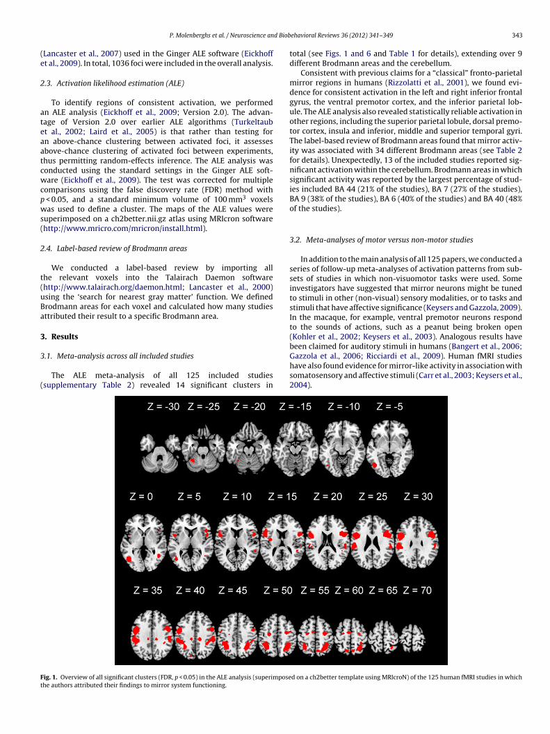
d Biob
(e
2
ateaatcwcpws(
2
t(uBa
3
3
(
Ft
P. Molenberghs et al. / Neuroscience an
Lancaster et al., 2007) used in the Ginger ALE software (Eickhofft al., 2009). In total, 1036 foci were included in the overall analysis.
.3. Activation likelihood estimation (ALE)
To identify regions of consistent activation, we performedn ALE analysis (Eickhoff et al., 2009; Version 2.0). The advan-age of Version 2.0 over earlier ALE algorithms (Turkeltaubt al., 2002; Laird et al., 2005) is that rather than testing forn above-chance clustering between activated foci, it assessesbove-chance clustering of activated foci between experiments,hus permitting random-effects inference. The ALE analysis wasonducted using the standard settings in the Ginger ALE soft-are (Eickhoff et al., 2009). The test was corrected for multiple
omparisons using the false discovery rate (FDR) method with < 0.05, and a standard minimum volume of 100 mm3 voxelsas used to define a cluster. The maps of the ALE values were
uperimposed on a ch2better.nii.gz atlas using MRIcron softwarehttp://www.mricro.com/mricron/install.html).
.4. Label-based review of Brodmann areas
We conducted a label-based review by importing allhe relevant voxels into the Talairach Daemon softwarehttp://www.talairach.org/daemon.html; Lancaster et al., 2000)sing the ‘search for nearest gray matter’ function. We definedrodmann areas for each voxel and calculated how many studiesttributed their result to a specific Brodmann area.
. Results
.1. Meta-analysis across all included studies
The ALE meta-analysis of all 125 included studiessupplementary Table 2) revealed 14 significant clusters in
ig. 1. Overview of all significant clusters (FDR, p < 0.05) in the ALE analysis (superimposehe authors attributed their findings to mirror system functioning.
ehavioral Reviews 36 (2012) 341–349 343
total (see Figs. 1 and 6 and Table 1 for details), extending over 9different Brodmann areas and the cerebellum.
Consistent with previous claims for a “classical” fronto-parietalmirror regions in humans (Rizzolatti et al., 2001), we found evi-dence for consistent activation in the left and right inferior frontalgyrus, the ventral premotor cortex, and the inferior parietal lob-ule. The ALE analysis also revealed statistically reliable activation inother regions, including the superior parietal lobule, dorsal premo-tor cortex, insula and inferior, middle and superior temporal gyri.The label-based review of Brodmann areas found that mirror activ-ity was associated with 34 different Brodmann areas (see Table 2for details). Unexpectedly, 13 of the included studies reported sig-nificant activation within the cerebellum. Brodmann areas in whichsignificant activity was reported by the largest percentage of stud-ies included BA 44 (21% of the studies), BA 7 (27% of the studies),BA 9 (38% of the studies), BA 6 (40% of the studies) and BA 40 (48%of the studies).
3.2. Meta-analyses of motor versus non-motor studies
In addition to the main analysis of all 125 papers, we conducted aseries of follow-up meta-analyses of activation patterns from sub-sets of studies in which non-visuomotor tasks were used. Someinvestigators have suggested that mirror neurons might be tunedto stimuli in other (non-visual) sensory modalities, or to tasks andstimuli that have affective significance (Keysers and Gazzola, 2009).In the macaque, for example, ventral premotor neurons respondto the sounds of actions, such as a peanut being broken open(Kohler et al., 2002; Keysers et al., 2003). Analogous results havebeen claimed for auditory stimuli in humans (Bangert et al., 2006;
Gazzola et al., 2006; Ricciardi et al., 2009). Human fMRI studieshave also found evidence for mirror-like activity in association withsomatosensory and affective stimuli (Carr et al., 2003; Keysers et al.,2004).d on a ch2better template using MRIcroN) of the 125 human fMRI studies in which
344 P. Molenberghs et al. / Neuroscience and Biobehavioral Reviews 36 (2012) 341–349
Table 1Significant clusters (FDR, p < 0.05) revealed by the ALE analysis of 125 human fMRI studies in which the authors attributed significant brain activations to mirror systemfunctioning.
Cluster Cluster size (mm3) Peak Talairach coordinates (x, y, z) Anatomical region Brodmann area
1 29,896 −48, 6, 28 Left inferior frontal gyrus 9−34, −50, 50 Left superior parietal lobule 7−48, −32, 38 Left inferior parietal lobule 40−50, 22, 14 Left inferior frontal gyrus 45−48, −30, 22 Left inferior parietal lobule 40−50, −40, 20 Left superior temporal gyrus 13
2 18,824 30, −50, 50 Right precuneus 740, −30, 40 Right inferior parietal lobule 4052, −20, 38 Right postcentral gyrus 356, −34, 16 Right insula 1354, −32, 38 Right inferior parietal lobule 406, −64, 56 Right superior parietal lobule 754, −20, 4 Right insula 1346, −40, 16 Right insula 13
3 13,704 44, 10, 28 Right inferior frontal gyrus 928, −8, 54 Right middle frontal gyrus 6
4 3472 −46, −66, −2 Left inferior temporal gyrus 375 2400 −26, −8, 50 Left middle frontal gyrus 66 1628 −34, −52, −24 Left cerebellum
7 1056 44, −56, 2 Right middle temporal gyrus 3750, −54, −6 Right inferior temporal gyrus 37
8 800 14, −28, 40 Right cingulate gyrus 319 752 −36, −4, 14 Left insula 1310 496 −14, −62, 60 Left superior parietal lobule 711 456 48, 22, 4 Right inferior frontal gyrus 4512 424 36, −50, −20 Right cerebellum13 288 −12, −28, 38 Left cingulate gyrus 3114 240 −8, −6, 60 Left medial frontal gyrus 6
Table 2Number of studies (from a total of 125) in which significant mirror-related activity was attributed to designated Brodmann areas (BA). N = number of studies.
BA N BA N BA N BA N BA N BA/Region N
1 1 7 34 18 5 28 1 38 1 44 262 12 8 2 19 10 31 4 39 7 45 233 11 9 48 20 2 32 5 40 60 46 154 13 10 4 21 9 34 1 41 4 47 7
36
37
etltftawrvtp
plcrTptc
(
5 4 13 29 22 19
6 59 17 3 24 4
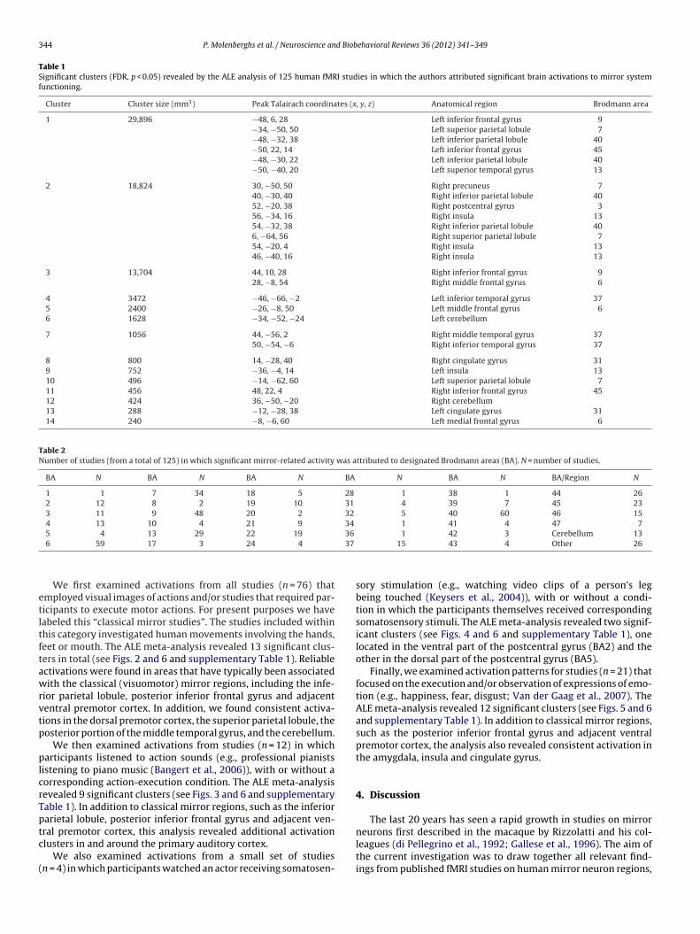
We first examined activations from all studies (n = 76) thatmployed visual images of actions and/or studies that required par-icipants to execute motor actions. For present purposes we haveabeled this “classical mirror studies”. The studies included withinhis category investigated human movements involving the hands,eet or mouth. The ALE meta-analysis revealed 13 significant clus-ers in total (see Figs. 2 and 6 and supplementary Table 1). Reliablectivations were found in areas that have typically been associatedith the classical (visuomotor) mirror regions, including the infe-
ior parietal lobule, posterior inferior frontal gyrus and adjacententral premotor cortex. In addition, we found consistent activa-ions in the dorsal premotor cortex, the superior parietal lobule, theosterior portion of the middle temporal gyrus, and the cerebellum.
We then examined activations from studies (n = 12) in whicharticipants listened to action sounds (e.g., professional pianists
istening to piano music (Bangert et al., 2006)), with or without aorresponding action-execution condition. The ALE meta-analysisevealed 9 significant clusters (see Figs. 3 and 6 and supplementaryable 1). In addition to classical mirror regions, such as the inferiorarietal lobule, posterior inferior frontal gyrus and adjacent ven-
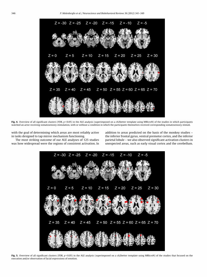
ral premotor cortex, this analysis revealed additional activationlusters in and around the primary auditory cortex.We also examined activations from a small set of studiesn = 4) in which participants watched an actor receiving somatosen-
1 42 3 Cerebellum 1315 43 4 Other 26
sory stimulation (e.g., watching video clips of a person’s legbeing touched (Keysers et al., 2004)), with or without a condi-tion in which the participants themselves received correspondingsomatosensory stimuli. The ALE meta-analysis revealed two signif-icant clusters (see Figs. 4 and 6 and supplementary Table 1), onelocated in the ventral part of the postcentral gyrus (BA2) and theother in the dorsal part of the postcentral gyrus (BA5).
Finally, we examined activation patterns for studies (n = 21) thatfocused on the execution and/or observation of expressions of emo-tion (e.g., happiness, fear, disgust; Van der Gaag et al., 2007). TheALE meta-analysis revealed 12 significant clusters (see Figs. 5 and 6and supplementary Table 1). In addition to classical mirror regions,such as the posterior inferior frontal gyrus and adjacent ventralpremotor cortex, the analysis also revealed consistent activation inthe amygdala, insula and cingulate gyrus.
4. Discussion
The last 20 years has seen a rapid growth in studies on mirror
neurons first described in the macaque by Rizzolatti and his col-leagues (di Pellegrino et al., 1992; Gallese et al., 1996). The aim ofthe current investigation was to draw together all relevant find-ings from published fMRI studies on human mirror neuron regions,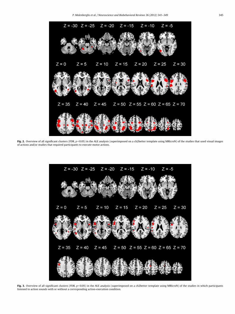
P. Molenberghs et al. / Neuroscience and Biobehavioral Reviews 36 (2012) 341–349 345
Fig. 2. Overview of all significant clusters (FDR, p < 0.05) in the ALE analysis (superimposed on a ch2better template using MRIcroN) of the studies that used visual imagesof actions and/or studies that required participants to execute motor actions.
Fig. 3. Overview of all significant clusters (FDR, p < 0.05) in the ALE analysis (superimposed on a ch2better template using MRIcroN) of the studies in which participantslistened to action sounds with or without a corresponding action-execution condition.
346 P. Molenberghs et al. / Neuroscience and Biobehavioral Reviews 36 (2012) 341–349
F rimpow in w
wi
w
Fe
ig. 4. Overview of all significant clusters (FDR, p < 0.05) in the ALE analysis (supeatched an actor receiving somatosensory stimulation, with or without a condition
ith the goal of determining which areas are most reliably activen tasks designed to tap mirror mechanism functioning.
The most striking outcome of our ALE analyses of 125 studiesas how widespread were the regions of consistent activation. In
ig. 5. Overview of all significant clusters (FDR, p < 0.05) in the ALE analysis (superimpxecution and/or observation of facial expressions of emotion.
sed on a ch2better template using MRIcroN) of the studies in which participantshich the participants themselves received corresponding somatosensory stimuli.
addition to areas predicted on the basis of the monkey studies –the inferior frontal gyrus, ventral premotor cortex, and the inferiorparietal lobule – we also observed significant activation clusters inunexpected areas, such as early visual cortex and the cerebellum.
osed on a ch2better template using MRIcroN) of the studies that focused on the
P. Molenberghs et al. / Neuroscience and Biobehavioral Reviews 36 (2012) 341–349 347
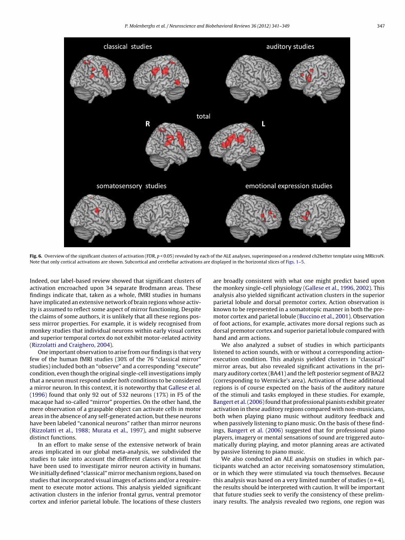
F ach ofN are d
Iafihitsma(
fscta(mmah(d
ashWsmac
ig. 6. Overview of the significant clusters of activation (FDR, p < 0.05) revealed by eote that only cortical activations are shown. Subcortical and cerebellar activations
ndeed, our label-based review showed that significant clusters ofctivation encroached upon 34 separate Brodmann areas. Thesendings indicate that, taken as a whole, fMRI studies in humansave implicated an extensive network of brain regions whose activ-
ty is assumed to reflect some aspect of mirror functioning. Despitehe claims of some authors, it is unlikely that all these regions pos-ess mirror properties. For example, it is widely recognised fromonkey studies that individual neurons within early visual cortex
nd superior temporal cortex do not exhibit motor-related activityRizzolatti and Craighero, 2004).
One important observation to arise from our findings is that veryew of the human fMRI studies (30% of the 76 “classical mirror”tudies) included both an “observe” and a corresponding “execute”ondition, even though the original single-cell investigations implyhat a neuron must respond under both conditions to be considered
mirror neuron. In this context, it is noteworthy that Gallese et al.1996) found that only 92 out of 532 neurons (17%) in F5 of the
acaque had so-called “mirror” properties. On the other hand, theere observation of a graspable object can activate cells in motor
reas in the absence of any self-generated action, but these neuronsave been labeled “canonical neurons” rather than mirror neuronsRizzolatti et al., 1988; Murata et al., 1997), and might subserveistinct functions.
In an effort to make sense of the extensive network of brainreas implicated in our global meta-analysis, we subdivided thetudies to take into account the different classes of stimuli thatave been used to investigate mirror neuron activity in humans.e initially defined “classical” mirror mechanism regions, based on
tudies that incorporated visual images of actions and/or a require-ent to execute motor actions. This analysis yielded significant
ctivation clusters in the inferior frontal gyrus, ventral premotorortex and inferior parietal lobule. The locations of these clusters
the ALE analyses, superimposed on a rendered ch2better template using MRIcroN.isplayed in the horizontal slices of Figs. 1–5.
are broadly consistent with what one might predict based uponthe monkey single-cell physiology (Gallese et al., 1996, 2002). Thisanalysis also yielded significant activation clusters in the superiorparietal lobule and dorsal premotor cortex. Action observation isknown to be represented in a somatotopic manner in both the pre-motor cortex and parietal lobule (Buccino et al., 2001). Observationof foot actions, for example, activates more dorsal regions such asdorsal premotor cortex and superior parietal lobule compared withhand and arm actions.
We also analyzed a subset of studies in which participantslistened to action sounds, with or without a corresponding action-execution condition. This analysis yielded clusters in “classical”mirror areas, but also revealed significant activations in the pri-mary auditory cortex (BA41) and the left posterior segment of BA22(corresponding to Wernicke’s area). Activation of these additionalregions is of course expected on the basis of the auditory natureof the stimuli and tasks employed in these studies. For example,Bangert et al. (2006) found that professional pianists exhibit greateractivation in these auditory regions compared with non-musicians,both when playing piano music without auditory feedback andwhen passively listening to piano music. On the basis of these find-ings, Bangert et al. (2006) suggested that for professional pianoplayers, imagery or mental sensations of sound are triggered auto-matically during playing, and motor planning areas are activatedby passive listening to piano music.
We also conducted an ALE analysis on studies in which par-ticipants watched an actor receiving somatosensory stimulation,or in which they were stimulated via touch themselves. Because
this analysis was based on a very limited number of studies (n = 4),the results should be interpreted with caution. It will be importantthat future studies seek to verify the consistency of these prelim-inary results. The analysis revealed two regions, one region was
3 d Biob
leat(aabttetoc
cuvwvrifa2t(er(r
ufadissdgiattTtsbaw
A
Hat
A
t
48 P. Molenberghs et al. / Neuroscience an
ocated in the ventral part of the postcentral gyrus (BA2). Keyserst al. (2004) found that the secondary somatosensory cortex wasctivated both when participants observed another person beingouched, and when they were touched themselves. Ebisch et al.2008) found that observing another person being touched can alsoctivate the primary somatosensory cortex, especially when thection is intentionally made by a person rather than accidentallyy an object (Ebisch et al., 2008). The other region was located inhe dorsal part of the postcentral gyrus (BA5). This region is knowno be involved in higher order somatosensory processing (Sakatat al., 1973). Although no motor activation is involved in observingouch, it seems that as with auditory stimuli, the mere observationf someone being touched is sufficient to activate somatosensoryortex vicariously (Keysers and Gazzola, 2009; Keysers et al., 2010).
Finally, we analyzed a subset of studies that focused on the exe-ution and/or observation of expressions of emotion. Most studiessed facial expressions and mouth actions are represented moreentrally than foot and arm/hand actions. This probably explainshy the premotor regions in this analysis were located more
entrally. Additionally, this analysis revealed vicarious activity inegions known to be involved in emotion processing, including thensula, amygdala and cingulate gyrus. The insula connects regionsor action representation with the limbic system (Carr et al., 2003),nd is believed to be involved in mirroring disgust (Wicker et al.,003). The amygdala, on the other hand, has been implicated inhe processing of threat-related emotions, including anger and fearPhan et al., 2002; though see also Van der Gaag et al., 2007; Sergeriet al., 2008). Different subregions of the cingulate gyrus seem to rep-esent different emotions, with the subgenual subregion of the ACCsACC) involved in negatively valenced events, and the pregenualegion (pACC) engaged in positively valenced events (Vogt, 2005).
To summarize, based on a meta-analysis of 125 studies, we havencovered a core network of brain areas, including the inferiorrontal gyrus, dorsal and ventral premotor cortex, and the inferiornd superior parietal lobule, which in humans is reliably activateduring tasks examining the classic mirror mechanism, typically
nvolving the visual observation and execution of actions. Ourubanalyses showed that additional areas involved in somatosen-ory, auditory and emotional processing complement these areasepending on the sensory modalities involved. These results sug-est that brain regions with mirror properties extend beyond thosedentified as being part of the mirror network in previous meta-nalyses (Caspers et al., 2010; Molenberghs et al., 2009). It seemshat in human participants, overlapping brain regions are activatedhrough simulation when observing or executing certain actions.he precise regions that are activated depend on the modality ofhe task (e.g., visual, auditory, somatosensory). Our results are con-istent with the view that vicarious brain activity made possibley mirror neurons (Keysers and Gazzola, 2009) extends beyondctions to include sharing of emotions and sensations of others asell.
cknowledgements
This work was supported by a Project Grant from the Nationalealth and Medical Research Council of Australia (511148),warded to RC and JBM and a UQ Postdoctoral Fellowship awardedo PM.
ppendix A. Supplementary data
Supplementary data associated with this article can be found, inhe online version, at doi:10.1016/j.neubiorev.2011.07.004.
ehavioral Reviews 36 (2012) 341–349
References
Arbib, M.A., Mundhenk, T.N., 2005. Schizophrenia and the mirror system: an essay.Neuropsychologia 43, 268–280.
Bangert, M., Peschel, T., Schlaug, G., Rotte, M., Drescher, D., Hinrichs, H., Heinze,H.J., Altenmuller, E., 2006. Shared networks for auditory and motor processingin professional pianists: evidence from fMRI conjunction. Neuroimage 30 (3),917–926.
Buccino, G., Binkofski, F., Fink, G.R., Fadiga, L., Fogassi, L., Gallese, V., Seitz, R.J., Zilles,K., Rizzolatti, G., Freund, H.J., 2001. Action observation activates premotor andparietal areas in a somatotopic manner: an fMRI study. Eur. J. Neurosc. 13,400–404.
Carr, L., Iacoboni, M., Dubeau, M.C., Mazziotta, J.C., Lenzi, G.L., 2003. Neural mecha-nisms of empathy in humans: a relay from neural systems for imitation to limbicareas. Proc. Natl. Acad. Sci. U.S.A. 100, 5497–5502.
Caspers, S., Zilles, K., Laird, A.R., Eickhoff, S.B., 2010. ALE meta-analysis of actionobservation and imitation in the human brain. Neuroimage 50, 1148–1167.
Cattaneo, L., Fabbri-Destro, M., Boria, S., Pieraccini, C., Monti, A., Cossu, G., Rizzolatti,G., 2007. Impairment of actions chains in autism and its possible role in intentionunderstanding. Proc. Natl. Acad. Sci. U.S.A. 104, 17825–17830.
Cattaneo, L., Rizzolatti, G., 2009. The mirror neuron system. Arch. Neurol. 66 (5),557–560.
Chong, T.T.-J., Cunnington, R., Williams, M.A., Kanwisher, N., Mattingley, J.B., 2008.fMRI adaptation reveals mirror neurons in human inferior parietal cortex. Curr.Biol. 18, 1576–1580.
Cross, E.S., Kraemer, D.J.M., Hamilton, A.F., Kelley, W.M., Grafton, S.T., 2009. Sensi-tivity of the action observation network to physical and observational learning.Cereb. Cortex 19, 315–326.
Dapretto, M., Davies, M.S., Pfeifer, J.H., Scott, A.A., Sigman, M., Bookheimer, S.Y.,Iacoboni, M., 2006. Understanding emotions in others: mirror neuron dysfunc-tion in children with autism spectrum disorders. Nat. Neurosci. 9, 28–30.
Decety, J., Grèzes, J., Coster, N., Perani, D., Jeannerod, M., Procyk, E., Grassi, F., Fazio,F., 1997. Brain activity during observation of actions. Influence of action contentand subject’s strategy. Brain 120, 1763–1777.
di Pellegrino, G., Fadiga, L., Fogassi, L., Gallese, V., Rizzolatti, G., 1992. Under-standing motor events: a neurophysiological study. Exp. Brain Res. 91 (1),176–180.
Dinstein, I., Thomas, C., Behrmann, M., Heeger, D.J., 2008a. A mirror op to nature.Curr. Biol. 18 (1), R13–R18.
Dinstein, I., Gardner, J.L., Jazayeri, M., Heeger, D.J., 2008b. Executed and observedmovements have different distributed representations in human aIPS. J. Neu-rosci. 28 (44), 11231–11239.
Ebisch, S.J.H., Perruci, M.G., Ferretti, A., Del Gratta, C., Romani, G.L., Gallese, V., 2008.The sense of touch: embodied simulation in a visuotactile mirroring mechanismfor observed animate or inanimate touch. J. Cogn. Neurosci. 20, 1–13.
Eickhoff, S.B., Laird, A.R., Grefkes, C., Wang, L.E., Zilles, K., Fox, P.T., 2009. Coordinate-based activation likelihood estimation meta-analysis of neuroimaging data: arandom-effects approach based on empirical estimates of spatial uncertainty.Hum. Brain Mapp. 30, 2907–2926.
Fabbri-Destro, M., Rizzolatti, G., 2008. Mirror neurons and mirror systems in mon-keys and humans. Physiology 23, 171–179.
Fogassi, L., Ferrari, P.F., Gesierich, B., Rozzi, S., Chersi, F., Rizzolatti, G., 2005. Pari-etal lobe: from action organization to intention understanding. Science 308,662–667.
Gallese, V., Fadiga, L., Fogassi, L., Rizzolatti, G., 1996. Action recognition in the pre-motor cortex. Brain 119 (2), 593–609.
Gallese, V., Fogassi, L., Fadiga, L., Rizzolatti, G., 2002. Action representation and theinferior parietal lobule. In: Prinz, W., Hommel, B. (Eds.), Common Mechanisms inPerception and Action: Attention and Performance, vol. XIX. Oxford UniversityPress, New York, pp. 247–266.
Gazzola, V., Aziz-Zadeh, L., Keysers, C., 2006. Empathy and the somatotopic auditorymirror system in humans. Curr. Biol. 16, 1824–1829.
Gazzola, V., Keysers, C., 2009. The observation and execution of actions share motorand somatosensory voxels in all tested subjects: single-subject analyses ofunsmoothed fMRI data. Cereb. Cortex 19, 1239–1255.
Heyes, C., 2010. Where do mirror neurons come from? Neurosci. Biobehav. Rev. 34,575–583.
Hickok, G., 2009. Eight problems for the mirror neuron theory of action understand-ing in monkeys and humans. J. Cogn. Neurosci. 21 (7), 1229–1243.
Iacoboni, M., Woods, R.P., Brass, M., Bekkering, H., Mazziotta, J.C., Rizzolatti, G., 1999.Cortical mechanisms of human imitation. Science 286, 2526–2528.
Iacoboni, M., 2005. Neural mechanisms of imitation. Curr. Opin. Neurobiol. 15,632–637.
Iacoboni, M., Molnar-Szakacs, I., Gallese, V., Buccino, G., Mazziotta, J.C., Rizzolatti, G.,2005. Grasping the intentions of others with one’s own mirror neuron system.PLoS Biol. 3, e79.
Iacoboni, M., Dapretto, M., 2006. The mirror neuron system and the consequencesof its dysfunction. Nat. Rev. Neurosci. 7 (12), 942–951.
Iacoboni, M., 2009. Imitation, empathy, and mirror neurons. Annu. Rev. Psychol. 60,653–670.
Johnson-Frey, S.H., 2003. Mirror neurons, Broca’s area and language: reflecting on
the evidence. Behav. Brain Sci. 26, 226–227.Jonas, M., Siebner, H.R., Biermann-Ruben, K., Kessler, K., Bäumer, T., Büchel, C.,Schnitzler, A., Münchaub, A., 2007. Do simple intransitive finger movementsconsistently activate frontoparietal mirror neuron areas in humans? Neuroim-age 36, T44–T53.
d Biob
K
K
K
K
K
K
K
L
L
L
L
L
L
MM
M
M
M
M
M
N
P. Molenberghs et al. / Neuroscience an
eysers, C., Kohler, E., Umiltà, M.A., Nanetti, L., Fogassi, L., Gallese, V., 2003. Audio-visual mirror neurons and action recognition. Exp. Brain Res. 153, 628–636.
eysers, C., Wicker, B., Gazzola, V., Anton, J.L., Fogassi, L., Gallese, V., 2004. A touchingsight: SII/PV activation during the observation and experience of touch. Neuron42, 335–346.
eysers, C., Fadiga, L., 2008. The mirror neuron system: new frontiers. Soc. Neurosci.3, 193–198.
eysers, C., Gazzola, V., 2009. Expanding the mirror: vicarious activity for actions,emotions, and sensations. Curr. Opin. Neurobiol. 19, 666–671.
eysers, C., Kaas, J.H., Gazzola, V., 2010. Somatosensation in social perception. Nat.Rev. Neurosci. 11, 417–428.
ilner, J.M., Neal, A., Weiskopf, N., Friston, K.J., Frith, C.D., 2009. Evidence of mirrorneurons in human inferior frontal gyrus. J. Neurosci. 29, 10153–10159.
ohler, E., Keysers, C., Umiltà, M.A., Fogassi, L., Gallese, V., Rizzolatti, G., 2002. Hearingsounds, understanding actions: action representation in mirror neurons. Science297, 846–848.
aird, A.R., Fox, M., Price, C.J., Glahn, D.C., Uecker, A.M., Lancaster, J.L., Turkeltaub,P.E., Kochunov, P., Fox, P.T., 2005. ALE meta-analysis: controlling the falsediscovery rate and performing statistical contrasts. Hum. Brain Mapp. 25,155–164.
ancaster, J.L., Woldorff, M.G., Parsons, L.M., Liotti, M., Freitas, C.S., Rainey, L.,Kochunov, P.V., Nickerson, D., Mikiten, S.A., Fox, P.T., 2000. Automated TalairachAtlas labels for functional brain mapping. Hum. Brain Mapp. 10, 120–131.
ancaster, J.L., Tordesillas-Gutierrez, D., Martinez, M., Salinas, F., Evans, A.,Zilles, K., Mazziotta, J.C., Fox, P.T., 2007. Bias between MNI and Talairachcoordinates analyzed using the ICBM-152 template. Hum. Brain Mapp. 28,1194–1205.
eslie, K.R., Johnson-Frey, S.H., Grafton, S.T., 2004. Functional imaging of face andhand imitation: towards a motor theory of empathy. Neuroimage 21, 601–607.
ewis, J.W., Brefczynski, J.A., Phinney, R.E., Janik, J.J., DeYoe, E.A., 2005. Distinctcortical pathways for processing tool versus animal sounds. J. Neurosci. 25,5148–5158.
ingnau, A., Gesierich, B., Caramazza, A., 2009. Asymmetric fMRI adaptation revealsno evidence for mirror neurons in humans. Proc. Natl. Acad. Sci. U.S.A. 106,9925–9930.
akuuchi, M., 2005. Is Broca’s area crucial for imitation? Cereb. Cortex 15, 563–570.olenberghs, P., Cunnington, R., Mattingley, J.B., 2009. Is the mirror neuron system
involved in imitation? A short review and meta-analysis. Neurosci. Biobehav.Rev. 33, 975–980.
olenberghs, P., Brander, C., Mattingley, J.B., Cunnington, R., 2010. The role of thesuperior temporal sulcus and the mirror neuron system in imitation. Hum. BrainMapp. 31, 1316–1326.
olnar-Szakacs, I., Kaplan, J., Greenfield, P.M., Iacoboni, M., 2006. Observing com-plex action sequences: the role of the fronto-parietal mirror neuron system.Neuroimage 33, 923–935.
origuchi, Y., Ohnishi, T., Decety, J., Hirakata, M., Maeda, M., Matsuda, H., Komaki,G., 2009. The human mirror neuron system in a population with deficient self-awareness: an fMRI study in alexithymia. Hum. Brain Mapp. 30, 2063–2076.
ukamel, R., Ekstrom, A.D., Kaplan, J., Iacoboni, M., Fried, I., 2010. Single-neuronresponses in humans during execution and observation of actions. Curr. Biol.20, 750–756.
urata, A., Fadiga, L., Fogassi, L., Gallese, V., Raos, V., Rizzolatti, G., 1997. Object
representation in the ventral premotor cortex (area F5) of the monkey. J. Neu-rophysiol. 78, 2226–2230.ewman-Norlund, R., van Schie, H.T., van Hoek, M.E.C., Cuijers, R.H., Bekkering, H.,2010. The role of inferior frontal and parietal areas in differentiating meaningfuland meaningless object-directed actions. Brain Res. 1315, 63–74.
ehavioral Reviews 36 (2012) 341–349 349
Phan, K.L., Wager, T., Taylor, S.F., Liberzon, I., 2002. Functional neuroanatomy of emo-tion: a meta-analysis of emotion activation studies in PET and fMRI. Neuroimage16, 331–348.
Ricciardi, E., Bonino, D., Sani, L., Vecchi, T., Guazzelli, M., Haxby, J.V., Fadiga, L.,Pietrini, P., 2009. Do we really need vision? How blind people “see” the actionsof others. J. Neurosci. 29, 9719–9724.
Rizzolatti, G., Camarda, R., Fogassi, L., Gentilucci, M., Luppino, G., Matelli, M., 1988.Functional organization of inferior area 6 in the macaque monkey. II. Area F5and the control of distal movement. Exp. Brain Res. 71, 491–507.
Rizzolatti, G., Fadiga, L., Gallese, V., Fogassi, L., 1996a. Premotor cortex and the recog-nition of motor actions. Cogn. Brain Res. 3, 131–141.
Rizzolatti, G., Fadiga, L., Matelli, M., Bettinardi, V., Paulesu, E., Perani, D., Fazio, F.,1996b. Localization of grasp representations in humans by PET: 1. Observationversus execution. Exp. Brain Res. 111, 246–252.
Rizzolatti, G., Fogassi, L., Gallese, V., 2001. Neurophysiological mechanisms under-lying the understanding and imitation of action. Nat. Rev. Neurosci. 2, 661–670.
Rizzolatti, G., Craighero, L., 2004. The mirror-neuron system. Annu. Rev. Neurosci.27, 169–192.
Rizzolatti, G., 2005. The mirror neuron system and its function in humans. Anat.Embryol. 210, 419–421.
Rizzolatti, G., Fabbri-Destro, M., 2008. The mirror system and its role in social cog-nition. Curr. Opin. Neurobiol. 18, 179–184.
Rizzolatti, G., Fabbri-Destro, M., 2010. Mirror neurons: from discovery to autism.Exp. Brain Res. 200, 223–237.
Rizzolatti, G., Sinigaglia, C., 2010. The functional role of the parieto-frontal mirrorcircuit: interpretations and misinterpretations. Nat. Rev. Neurosci. 11, 264–274.
Rocca, M.A., Tortorella, P., Ceccarelli, A., Falini, A., Tango, D., Scotti, G., Comi, G., Filippi,M., 2008. The “mirror-neuron system” in MS: A 3 Tesla fMRI study. Neurology70 (4), 255–262.
Schaefer, M., Xu, B., Flor, H., Cohen, L.G., 2009. Effects of different viewing perspec-tives on somatosensory activations during observation of touch. Hum. BrainMapp. 30, 2722–2730.
Sergerie, K., Chochol, C., Armony, J.L., 2008. The role of the amygdala in emotionalprocessing: a quantitative meta-analysis of functional neuroimaging studies.Neurosci, Biobehav. Rev. 32, 811–830.
Sakata, H., Takaoka, Y., Atsushi Kawarasaki, A., Shibutani, H., 1973. Somatosensoryproperties of neurons in the superior parietal cortex (area 5) of the rhesus mon-key. Brain Res. 64, 85–102.
Tettamanti, M., Buccino, G., Saccuman, M.C., Gallese, V., Danna, M., Scifo, P., Fazio, F.,Rizzolatti, G., Cappa, S.F., Perani, D., 2005. Listening to action-related sentencesactivates fronto-parietal motor circuits. J. Cogn. Neurosci. 17, 273–281.
Turella, L., Pierno, A.C., Tubaldi, F., Casteillo, U., 2009. Mirror neurons in humans:consisting or confounding evidence? Brain Lang. 108, 10–21.
Turkeltaub, P.E., Eden, G.F., Jones, K.M., Zeffiro, T.A., 2002. Meta-analysis of thefunctional neuroanatomy of single-word reading: method and validation. Neu-roimage 16 (3), 765–780.
Van der Gaag, C., Minderaa, R.B., Keysers, C., 2007. Facial expressions: what themirror neuron system can and cannot tell us. Soc. Neurosci. 2, 179–222.
Van Overwalle, F., Baetens, K., 2009. Understanding others’ actions and goals bymirror and mentalizing systems: a meta-analysis. Neuroimage 48, 564–584.
Vogt, B.A., 2005. Pain and emotion interactions in subregions of the cingulate gyrus.Nat. Rev. Neurosci. 6 (7), 533–544.
Wicker, B., Keysers, C., Plailly, J., Royet, J.P., Gallese, V., Rizzolatti, G., 2003. Both of usdisgusted in my insula: the common neural basis of seeing and feeling disgust.Neuron 40, 655–664.
Williams, J.H., 2008. Self-other relations in social development and autism: multipleroles for mirror neurons and other brain bases. Autism Res. 1, 73–90.