Both of Us Disgusted in My Insula
10
Neuron, Vol. 40, 655–664, October 30, 2003, Copyright 2003 by Cell Press Both of Us Disgusted in My Insula: The Common Neural Basis of Seeing and Feeling Disgust leads to a propositional representation of the inferred state of disgust. This representation then determines our decision not to eat the food. Another interpretation of the phenomenon may be based on a “sensory motor resonance” hypothesis. Observing the facial expression Bruno Wicker, 1 Christian Keysers, 2,3 Jane Plailly, 4 Jean-Pierre Royet, 4 Vittorio Gallese, 2 and Giacomo Rizzolatti 2, * 1 Institut de Neurosciences Physiologiques et Cognitives CNRS of another person evokes a similar facial motor repre- sentation in the observer (see Hess et al., 1999, for a Chemin Joseph Aiguier 13402 Marseille cedex 20 review). This motor representation (Lipps, 1907) and its associated somatosensory consequences (Adolphs et France 2 Physiology section al., 2000; Adolphs, 2001, 2002) might be sufficient to understand the meaning of the other’s facial expression. Department for Neuroscience University of Parma Neither of these hypotheses predicts that the observer shares the emotion of disgust with the observed individ- Via Volturno 39 43100 Parma ual, in that they both—although in different ways— assign a causal role to mechanisms not directly involved Italy 3 BCN Neuroimaging Center in the experience of emotions. We will refer to them jointly as “cold hypotheses.” A third possibility is that, University Groningen Antonius Deusinglaan 2 in order to understand the facial expression of disgust displayed by others, a feeling of disgust must occur also 9713 AV Groningen The Netherlands in the observer. This hypothesis predicts that brain areas responsible for experiencing this emotion will become 4 Laboratoire de Neurosciences et Syste ` mes Sensoriels active during the observation of that emotion in others. We will refer to this hypothesis as the “hot hypothesis.” UMR CNRS 5020 Universite Claude-Bernard LYON 1 Gerland So far, there is only indirect evidence to support the latter hypothesis. A number of investigations show that, 50, Avenue Tony Garnier 69007 Lyon cedex 07 among other structures, the insula and the amygdala are activated when subjects are exposed to disgusting France odors or tastes (Royet et al., 2003; Small et al., 2003; Zald and Pardo, 2000; Zald et al., 1998a). Independently, a number of functional imaging studies (Phillips et al., Summary 1997, 1998; Sprengelmeyer et al., 1998; Schienle et al., 2002) and electrophysiological investigations (Krolak- What neural mechanism underlies the capacity to un- derstand the emotions of others? Does this mecha- Salmon et al., 2003) have suggested that the insula is activated during the observation of disgusted facial ex- nism involve brain areas normally involved in experi- encing the same emotion? We performed an fMRI pressions. The aim of the present study will be to directly determine whether the same locations in the insula are study in which participants inhaled odorants produc- ing a strong feeling of disgust. The same participants activated during the experience of disgust and the ob- servation of the facial expression of disgust in others. observed video clips showing the emotional facial ex- pression of disgust. Observing such faces and feeling To this purpose, we performed an fMRI study com- posed of four functional runs. In the first and second disgust activated the same sites in the anterior insula and to a lesser extent in the anterior cingulate cortex. (“visual runs”), participants passively viewed movies of individuals smelling the contents of a glass (disgusting, Thus, as observing hand actions activates the observ- er’s motor representation of that action, observing an pleasant, or neutral) and expressing the facial expres- sions of the respective emotions. In the third and fourth emotion activates the neural representation of that emotion. This finding provides a unifying mechanism (“olfactory runs”), the same participants inhaled dis- gusting or pleasant odorants through a mask placed on for understanding the behaviors of others. their nose and mouth. Our core finding is that the anterior insula is activated both during the observation of dis- Introduction gusted facial expressions and during the emotion of disgust evoked by unpleasant odorants. This result indi- In a natural environment, food poisoning is a substantial threat. When an individual sees a conspecific looking cates that, for disgust, there is a common substrate for feeling an emotion and perceiving the same emotion disgusted after tasting some food, he or she automati- cally infers that the food is bad and should not be eaten. in others. What happens in the observer’s brain to keep him or her from eating the potentially damaging food? Ac- Results cording to a cognitive account, the processing of the facial expression occurring in the visual cortical areas The experiment was carried out on 14 healthy right- handed male subjects. As mentioned in the Introduction, all subjects took part in two visual and two olfactory *Correspondence: [email protected]
-
Upload
independent -
Category
Documents
-
view
1 -
download
0
Transcript of Both of Us Disgusted in My Insula
Neuron, Vol. 40, 655–664, October 30, 2003, Copyright 2003 by Cell Press
Both of Us Disgusted in My Insula:The Common Neural Basis ofSeeing and Feeling Disgust
leads to a propositional representation of the inferredstate of disgust. This representation then determinesour decision not to eat the food. Another interpretationof the phenomenon may be based on a “sensory motorresonance” hypothesis. Observing the facial expression
Bruno Wicker,1 Christian Keysers,2,3
Jane Plailly,4 Jean-Pierre Royet,4
Vittorio Gallese,2 and Giacomo Rizzolatti2,*1Institut de Neurosciences Physiologiques
et CognitivesCNRS of another person evokes a similar facial motor repre-
sentation in the observer (see Hess et al., 1999, for aChemin Joseph Aiguier13402 Marseille cedex 20 review). This motor representation (Lipps, 1907) and its
associated somatosensory consequences (Adolphs etFrance2 Physiology section al., 2000; Adolphs, 2001, 2002) might be sufficient to
understand the meaning of the other’s facial expression.Department for NeuroscienceUniversity of Parma Neither of these hypotheses predicts that the observer
shares the emotion of disgust with the observed individ-Via Volturno 3943100 Parma ual, in that they both—although in different ways—
assign a causal role to mechanisms not directly involvedItaly3 BCN Neuroimaging Center in the experience of emotions. We will refer to them
jointly as “cold hypotheses.” A third possibility is that,University GroningenAntonius Deusinglaan 2 in order to understand the facial expression of disgust
displayed by others, a feeling of disgust must occur also9713 AV GroningenThe Netherlands in the observer. This hypothesis predicts that brain areas
responsible for experiencing this emotion will become4 Laboratoire de Neurosciences etSystemes Sensoriels active during the observation of that emotion in others.
We will refer to this hypothesis as the “hot hypothesis.”UMR CNRS 5020Universite Claude-Bernard LYON 1 Gerland So far, there is only indirect evidence to support the
latter hypothesis. A number of investigations show that,50, Avenue Tony Garnier69007 Lyon cedex 07 among other structures, the insula and the amygdala
are activated when subjects are exposed to disgustingFranceodors or tastes (Royet et al., 2003; Small et al., 2003;Zald and Pardo, 2000; Zald et al., 1998a). Independently,a number of functional imaging studies (Phillips et al.,Summary1997, 1998; Sprengelmeyer et al., 1998; Schienle et al.,2002) and electrophysiological investigations (Krolak-What neural mechanism underlies the capacity to un-
derstand the emotions of others? Does this mecha- Salmon et al., 2003) have suggested that the insula isactivated during the observation of disgusted facial ex-nism involve brain areas normally involved in experi-
encing the same emotion? We performed an fMRI pressions. The aim of the present study will be to directlydetermine whether the same locations in the insula arestudy in which participants inhaled odorants produc-
ing a strong feeling of disgust. The same participants activated during the experience of disgust and the ob-servation of the facial expression of disgust in others.observed video clips showing the emotional facial ex-
pression of disgust. Observing such faces and feeling To this purpose, we performed an fMRI study com-posed of four functional runs. In the first and seconddisgust activated the same sites in the anterior insula
and to a lesser extent in the anterior cingulate cortex. (“visual runs”), participants passively viewed movies ofindividuals smelling the contents of a glass (disgusting,Thus, as observing hand actions activates the observ-
er’s motor representation of that action, observing an pleasant, or neutral) and expressing the facial expres-sions of the respective emotions. In the third and fourthemotion activates the neural representation of that
emotion. This finding provides a unifying mechanism (“olfactory runs”), the same participants inhaled dis-gusting or pleasant odorants through a mask placed onfor understanding the behaviors of others.their nose and mouth. Our core finding is that the anteriorinsula is activated both during the observation of dis-Introductiongusted facial expressions and during the emotion ofdisgust evoked by unpleasant odorants. This result indi-In a natural environment, food poisoning is a substantial
threat. When an individual sees a conspecific looking cates that, for disgust, there is a common substrate forfeeling an emotion and perceiving the same emotiondisgusted after tasting some food, he or she automati-
cally infers that the food is bad and should not be eaten. in others.What happens in the observer’s brain to keep him or
her from eating the potentially damaging food? Ac- Resultscording to a cognitive account, the processing of thefacial expression occurring in the visual cortical areas The experiment was carried out on 14 healthy right-
handed male subjects. As mentioned in the Introduction,all subjects took part in two visual and two olfactory*Correspondence: [email protected]

Neuron656
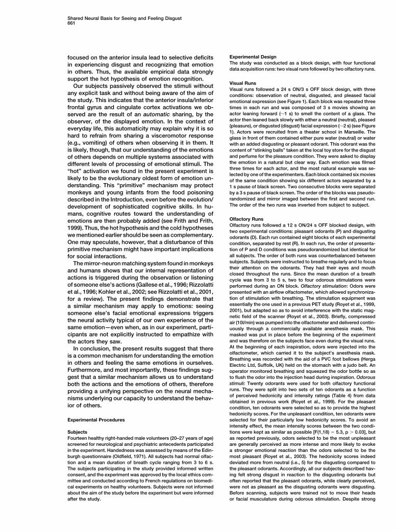
the activated structures, two are of particular interestfor the present study: the amygdala and the insula (Fig-ure 2). Amygdala activations were present with bothdisgusting and pleasant odorants, with a clear overlapbetween the two types of activations (Figure 2A). Thefact that the amygdala is activated by both pleasant andunpleasant odorants is in accord with previous findings(Gottfried et al., 2002; Hudry et al., 2001; Anderson et al.,2003; Zald, 2003). In contrast, disgusting and pleasantodorants produced clearly separated activation foci inthe insula. Disgusting odorants activated the anteriorsector of the insula bilaterally, whereas pleasant odor-ants activated a more posterior site of only the rightinsula (Figure 2B).
Visual StimulationThe visual runs were analyzed using two contrasts: ob-servation of disgust – neutral and observation of plea-
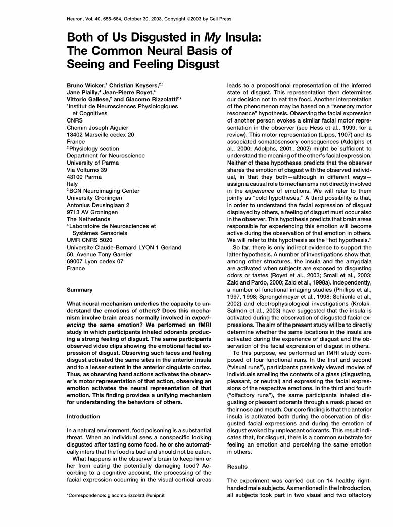
Figure 1. Frames from Movies Used in the Visual Runssure – neutral. Both contrasts revealed significant BOLD
The demonstrators leaned forward to sniff at the content of a glasssignal changes in various locations (see Table 2). The(top two rows) and then retracted the torso and expressed a facialinsula in particular was only activated in the observationexpression of disgust (left) pleasure (center) or neutral (right column).of disgust – neutral contrast. Most importantly, clustersEach movie lasted 3 s. Six different demonstrators (three are shown
here) expressed the three types of facial expressions, leading to six of overlap were found between the observation of dis-variants of each expression. A vision-of-disgust block, for instance, gust – neutral and the disgusting odorant – rest contrastswas then composed of the six variants of the disgusted emotion (Figure 3 and Table 3). These clusters were located inseparated by 1 s pauses.
the left anterior insula and in the transition zone betweenthe insula and inferior frontal gyrus. A smaller overlapwas also observed in the anterior right cingulate cortex.
functional acquisition sessions. Visual runs containedThe amygdalae were not activated by the observation
three experimental conditions: “observation of disgust,”of disgusted facial expressions. This lack of amygdalar
“observation of pleasure,” and “neutral.” Each conditionactivation is in agreement with previous studies sug-
was composed of blocks of six movies showing individu-gesting a dissociation between the neural basis of the
als leaning forward to smell the content of a glass. De-recognition of fear, in which the amygdala is strongly
pending on the condition, the glass contained an unpleas-involved, and that of disgust, in which the amygdala
ant, pleasant, or neutral odorant, and the individuals in thedoes not appear to play a crucial role (Calder et al., 2001).
movie reacted accordingly with a disgusted, pleased, orTo test if the BOLD signal increases in these zones of
neutral facial expression (see Figure 1). Movies wereoverlap were selective for disgust, we performed direct
used instead of static facial expressions for three rea-comparisons between the disgusting odorants and
sons. First, under ecological conditions, facial expres-pleasant odorants conditions and between the observa-
sions are intrinsically dynamic stimuli. Second, emotionstion of disgust and the observation of pleasure condi-
are recognized better from movies compared to statictions within these regions of interest (see Table 3, last
displays (Wehrle et al., 2000). Third, in a recent neuro-two columns). In all cases, responses were stronger to
imaging study, Kilts et al. (2003) compared the brainthe disgust compared to the pleasure stimuli, be they
activity during the recognition of emotions from staticolfactory or visual (i.e., all t values were positive). For
and dynamic displays of facial expressions and con-two of the insular clusters, both the visual and olfactory
cluded that the encoding of facial expressions of emo-responses were significantly larger for the disgust stim-
tion by static or dynamic displays is associated withuli (p � 0.05). The third cluster of the insula responded
different neural correlates for their decoding.significantly more to the observation of disgust com-
Olfactory runs were composed of two experimentalpared to the observation of pleasure but did not signifi-
conditions separated by periods of rest. Conditionscantly discriminate between the two types of odorants.
were composed of blocks of olfactory stimulation duringFinally, the cluster located in the anterior cingulate cor-
which subjects were exposed to different disgusting ortex showed significantly stronger activations for the ob-
pleasant odorants (“disgusting odorant” and “pleasantservation of disgust versus observation of pleasure con-
odorant” conditions, respectively). Full details of theditions but only showed a nonsignificant trend for
procedures are provided in the Experimental Proce-disgusting odorants versus pleasant odorants.
dures section. The data obtained during the olfactoryTo confirm the presence of the overlaps observed
and visual runs were analyzed separately using random-between the observation of disgust – neutral and the
effect analyses (n � 14 subjects, p � 0.005 uncorrected,disgusting odorant – rest contrast t maps, we performed
and k � 20).a modified conjunction analysis (p � 0.005, k � 20)between these two contrasts (see Experimental Proce-dures). The results confirmed the presence of overlapsOlfactory Stimulation
The results of the disgusting odorant – rest and pleasant between these two conditions, showing a cluster [33voxels with a peak at x � �38, y � 26, z � �6, and aodorant – rest contrasts are reported in Table 1. Among

Shared Neural Basis for Seeing and Feeling Disgust657
Table 1. Olfactory Activations
MNI TALSize
Anatomical Description Hem. x y z x y z t Value (Voxels)
A. Disgusting odorant � rest, random effect,p � 0.005, k � 20, n � 14 s
Amygdala/uncus L �24 �2 �30 �24 �3 �25 6.57 64Amygdala R 20 �4 �16 20 �5 �13 5.82 200Anterior insula/inferior frontal gyrus R 36 28 �2 36 27 �3 4.89 155Anterior insula R 36 10 4 36 10 3 5.78 64Middle frontal gyrus L �44 52 8 44 51 5 4.71 38Anterior insula L �36 22 8 �36 22 6 6.28 149Inferior frontal gyrus R 50 26 14 50 26 12 4.82 32Inferior frontal gyrus R 46 16 18 46 16 16 6.63 66Middle frontal gyrus R 44 36 22 44 36 18 4.59 31Anterior cingulate R � L 4 26 26 4 26 23 5.14 69Precentral sulcus L �34 0 36 �34 2 33 7.43 54Superior parietal lobule R 36 �70 56 36 �65 55 4.53 25
B. Pleasant odorant – rest, random effect,p � 0.005, k � 20, n � 14 s
Cerebellum L �20 �54 �30 �20 �54 �23 5.23 20Amygdala/uncus L �18 �2 �28 �18 �3 �23 6.51 40Brain stem R � L 2 �20 �24 2 �20 �19 5.35 43Amygdala R 26 0 �22 26 �1 �18 8.37 327Inferior frontal gyrus pars orbitalis R 34 34 �12 34 32 �12 4.84 114Cerebellum (culmen) L �2 �42 �6 �2 �41 �3 5.36 64Anterior insula R 38 �2 2 38 �2 2 4.5 48Anterior tip of caudate R 26 28 10 26 28 8 9.31 34Middle frontal gyrus R 48 42 24 48 42 20 5 80Middle frontal gyrus L �46 34 24 �46 34 20 6.52 183Precentral sulcus R 46 8 28 46 9 25 4.77 41Middle frontal gyrus R 40 30 30 40 30 26 5.13 43Rostral inf. parietal lobule R 44 �62 48 44 �58 47 4.48 58Superior parietal lobule R 34 �70 54 34 �65 53 4.83 23
Location in MNI and Talaraich (TAL) coordinates (x, y, z), anatomical description, maximum t value, and number of voxels for all clusters foundto be significantly activated during the olfactory contrasts. Activations are shown in ventrodorsal order. The voxel size was 2 � 2 � 2 mm3.
t(13) � 5.41, p � 0.001] corresponding to the first cluster that are selectively activated during the feeling of dis-gust. This suggests that the understanding of the facialof Table 3. All the three remaining clusters of Table 3
are significant in this conjunction analysis if k is lowered expressions of disgust as displayed by others involvesthe activation of neural substrates normally activatedto the cluster size of these remaining clusters.
Applying the same modified conjunction analysis to during the experience of the same emotion. Theseshared neural substrates are the left anterior insula andthe observation of pleasure – neutral and pleasant odor-
ant – rest conditions revealed no significant clusters of the right anterior cingulate cortex.overlap. Nor did the conjunction analysis between thenonmatching contrasts, i.e., observation of pleasure – The Insulaneutral with disgusting odorants – rest or observation Cytoarchitectonically, the monkey’s insula can be di-of disgust – neutral with pleasant odorants – rest. The vided into three zones (agranular, dysgranular, and gran-lack of overlap between the observation of pleased fa- ular; Mesulam and Mufson, 1982a). Functionally, how-cial expressions and the olfaction of pleasant odorants ever, the insula is formed by two major functionalis probably due to the fact that, in contrast to the emotion sectors: an anterior sector comprising the agranular andof disgust, which is tightly linked to bad odorants/tastes, the anterior dysgranular insula and a posterior sectorthe emotion of pleasure can be triggered by many stim- comprising the posterior dysgranular and the granularuli, only few of which are olfactory or gustatory. There insula (Mesulam and Mufson, 1982b; Mufson and Mesu-is therefore a strong link between bad tastes/smells, the lam, 1982). The anterior sector is an olfactory and gusta-emotion of disgust, and the facial expression of disgust, tory center that appears to control visceral sensationswhile there is a much weaker link between pleasant and the related autonomic responses. Additionally, itodors/smells, the emotion of pleasure, and pleased fa- receives visual information from the anterior sectors ofcial expressions. the ventral bank of the superior temporal cortex, where
cells have been found in the monkey to respond to thesight of faces (Bruce et al., 1981; Perrett et al., 1982,Discussion1984, 1985; Keysers et al., 2001). In contrast, the poste-rior sector of the insula is characterized by connectionsThe main finding of the present study is that the observa-
tion of disgust automatically activates neural substrates with auditory, somatosensory, and premotor areas and

Neuron658
hemisphere (Zald and Pardo, 1997, 2000; Zald et al.,1998b; Royet et al., 2000, 2001, 2003; Gottfried et al.,2002; Anderson et al., 2003; Zald, 2003). Investigationsusing gustatory stimuli confirm this finding, showing thatthe left anterior insula/opercular region responded pref-erentially to unpleasant compared to pleasant tastes(Zald et al., 1998a; Small et al., 2003).
While unpleasant tastes and smells are often per-ceived as more intense than their pleasant counterparts,recently, Small et al. (2003) showed that the left anteriorinsula preference for unpleasant tastes is maintainedeven if these unpleasant tastes are perceived as lessintense than the pleasant tastes they are comparedagainst. The cluster showing this property included thecoordinates at which we found the overlap between theobservation of disgust and the disgusting odorants.
Finally, Yaxley et al. (1990) and Scott et al. (1991)report the existence of single neurones in the macaqueanterior insula and opercular frontal cortex respondingselectively to particular gustatory stimuli (see also Au-gustine, 1996, and Dolan, 2002, for reviews). None ofFigure 2. Results of the Olfactory Stimulationthese studies, however, addressed the issue of whetherResults of the olfactory stimulation superimposed on the anatomical
image of a standard MNI brain using neurological conventions (right the same area was also activated during the observationis right). (A) Coronal sections focusing on the amygdalae. Note the of facial expressions of disgust. Taken together the ana-large degree of overlap (orange) between the activations determined tomical and functional data indicate that the left anteriorby disgusting (red) and pleasant odorants (green) in the right amyg- insula and neighboring opercular frontal cortex aredala and left parahippocampal cortex. (B) Axial slice showing the
structures strongly involved in the sensation of dis-response to odorants in the insula. The activity is bilateral and ante-gusting stimuli.rior for the disgusting odorants and is confined to a more posterior
location of the right insula for the pleasant odorants. There is no The insula, however, is not only a center for elaborat-overlap in the insula between the activations determined by the two ing olfactory and gustatory stimuli. Electrical stimulationodorants. The color coding is indicated on the bottom right. of the anterior sector of the insula conducted during
neurosurgery (Penfield and Faulk, 1955) evoked nauseaor the sensation of being sick (“Feeling as if she wereis not related to the olfactory or gustatory modalities.going to be sick,” Penfield and Faulk [1955], p. 451). ItA direct comparison between the macaque monkey’salso evoked visceromotor activity (“My stomach wentinsula and the human one showed that, although theup and down like when you vomit,” ibidem, p. 451). More
human insula is substantially larger than the macaque’srecently, Krolak-Salmon and colleagues (2003) showed
counterpart, the general architectonic organization isthat electrically stimulating the anterior insula through
strikingly similar in the two species and shows the sameimplanted depth electrodes produced sensations in the
subdivisions (Mesulam and Mufson, 1982a). throat and mouth that were “difficult to stand.” TakenThe activations observed during the disgusting odor- together, these findings demonstrate a role for the ante-
ant condition of our experiment fall within the anterior rior insula in transforming unpleasant sensory input intohalf of both insulae, most likely corresponding to the visceromotor reactions and the accompanying feelinganterior sectors of Mesulam and Mufson (1982a, 1982b). of disgust.No activations were found in the posterior sectors. An Here we show that the same visceromotor region,activation of the same anterior sector, but restricted to related to such an evolutionary ancient basic emotionthe left insula, was found during the observation of the as disgust, can be directly activated by the observationdisgusted facial expression, a finding in agreement with of the facial expression of disgust displayed by others.the higher-order visual information reaching the insula This finding is in agreement with previous experimentsfrom the superior temporal sulcus. Most interestingly, showing that the vision of disgusted static facial expres-there was a clear overlap between both activations. This sions leads to activations in the anterior insula (Phillipsis, to our knowledge, the first direct neuroimaging dem- et al., 1997, 1998; Sprengelmeyer et al., 1998; Schienleonstration that the same sites in the insula mediate both et al., 2002; Krolak-Salmon et al., 2003). None of thesethe observation and the feeling of disgust. studies, though, evoked the sensation of disgust in the
The activation of the anterior insula during disgusting participants to investigate if the activated locations areolfactory stimulation found in the present experiment is common to both the experience of disgust and the per-in accord with previous neuroimaging findings showing ception of the same emotion in others.its activation during olfactory stimulation. These studies Carr et al. (2003) showed an activation of the anteriorindicate that the transition zone between the anterior insula/inferior frontal gyrus during both the observationinsula and the frontal operculum located in the left hemi- and imitation of facial expressions. In their block design,sphere was preferentially activated for unpleasant com- each block contained examples of all six basic emotionspared to pleasant odors (Zald and Pardo, 2000; Royet in random order: happy, sad, angry, surprised, afraid,et al., 2003). Indeed, emotional responses to disgusting and disgusted. It is important to stress that imitation
usually does not require experiencing the imitated emo-stimuli are generally reported to be stronger in the left

Shared Neural Basis for Seeing and Feeling Disgust659
Table 2. Visual Activations
MNI TAL
Anatomical Description Hem. x y z x y z t Value Size (Voxels)
A. Observation of disgust – neutral, randomeffect, p � 0.005, k � 20, n � 14 s
Fusiform gyrus/declive L �30 �74 �20 �30 �73 �13 5.37 82Middle occipital R 44 �70 �14 44 �68 �8 5.23 107Brain stem L �4 �26 �10 �4 �26 �7 5.77 46Inferior frontal gyrus L �38 26 �6 �38 25 �6 5.41 103Pulvinar/lentiform nucleus R 22 12 0 22 12 �1 5.2 39Anterior insula/inferior frontal gyrus L �24 30 4 �24 29 2 4.03 67Superior temporal sulcus R 62 �44 8 61 �42 9 4.48 43Anterior insula L �24 8 12 �24 8 11 4.59 30Precentral gyrus R 64 12 14 63 12 12 5.11 29Dorsal bank of the silvian fisure L �50 �14 18 �50 �13 17 6.13 60Middle frontal gyrus L �36 56 24 �36 55 19 5.33 23Supramarginal gyrus R 40 �50 28 40 �47 28 4.71 30Cingulate gyrus R 4 24 30 4 25 26 5.59 20Middle frontal R 54 8 40 53 10 36 5.07 51Cingulate/medial frontal gyrus L �4 12 48 �4 14 44 4.22 36Postcentral gyrus L �52 �20 52 �51 �17 49 6.63 33Superior/medial frontal gyrus R 8 12 52 8 14 47 6.12 101
B. Observation of pleasure – neutral, randomeffect, p � 0.005, k � 20, n � 14 s
Cerebellum (declive) L �2 �64 �24 �2 �63 �17 5.90 26Parahippocampal gyrus L �16 �10 �22 �16 �11 �18 4.10 38Fusiform gyrus R 40 �74 �20 40 �73 �13 3.73 34Precentral gyrus L �50 �12 10 �50 �11 10 4.68 28Inferior frontal gyrus R 48 24 18 48 24 15 4.81 46Precuneus R 6 �70 44 6 �66 44 4.40 34
Location in MNI and Talaraich (TAL) coordinates (x, y, z), anatomical description, maximum t value, and number of voxels for all clusters foundto be significantly activates during the visual contrasts. Activations are shown in ventrodorsal order. The voxel size was 2 � 2 � 2 mm3
tion. Their data, therefore, indicate that the insula is we show the anterior insula to respond to disgusted butnot to happy dynamic facial expressions.involved in imitation but not that it is directly involved
in the experience of emotions. However, in the light of The fact that the feeling of disgust and the perceptionof that emotion in others share a common neural sub-our findings, it is possible that, during imitation, some
of their participants felt the imitated emotion—as actors strate confirms previous neuropsychological studies(Calder et al., 2000, and Adolphs et al., 2003). Afterdo when using the “Stanislavsky” method of emotion
induction (Stanislavsky, 1936). The relatively low statisti- lesions affecting the insulae and neighboring structures,two patients were selectively impaired in recognizingcal significance of the activation in the insula reported
by Carr et al. (2003) during the observation of emotions the facial expression of disgust as compared to otherfacial expressions and reported having reduced sensa-(t � 3.02) is probably a consequence of their experimen-
tal design: they used blocks of mixed emotions, while tions of disgust themselves. In those patients, the le-
Table 3. Overlap between Observing and Feeling Disgust
MNI TAL t Value Direct ComparisonsSize
Anatomical Description Hem x y z x y z Vis. Olf. (vox.) Disg. – pleas. odorants Observ. of disgust – pleasure
Anterior insula/inferior L �38 26 �6 �38 25 �6 5.41 4.07 25 t(13) � 2.44, p � 0.01 t(13) � 2, p � 0.03frontal gyrus
Anterior insula/inferior L �34 28 6 �34 27 4 3.92 4.00 12 t(13) � 0.02, p � 0.49 t(13) � 1.64, p � 0.06frontal gyrus
Anterior insula L �34 10 16 �34 10 14 3.55 4.22 2 t(13) � 2, p � 0.03 t(13) � 2.52, p � 0.01Anterior cingulate R 4 24 30 4 25 26 5.59 4.43 6 t(13) � 1.29, p � 0.11 t(13) � 3.63, p � 0.002
cortex
Location in MNI and Talairach space and size of the clusters common to both the disgusting odorants – rest and the observation of disgust– neutral contrasts together with the anatomical description of their location. The maximal t score observed in the clusters of overlap is shownseparately for the visual and olfactory contrasts. The last two columns show the result of a direct comparison between the BOLD signalevoked by disgusting versus pleasant odorants and between the observation of disgusted versus pleased faces. Results that are significantat p � 0.05 are shown in bold. The probability of finding five or more significant t tests with a p � 0.05 criterion is less than 2 � 10�5 accordingto a binomial distribution.

Neuron660
Figure 3. Illustration of the Overlap
Illustration of the overlap (white) between the brain activation during the observation (blue) and the feeling (red) of disgust. The olfactory andvisual analysis were performed separately as random-effect analysis. The results are superimposed on parasagittal slices of a standardMNI brain.
sions were not restricted to the insula, but our data sence of further imaging studies demonstrating the acti-vation of the anterior cingulate during the observationsuggest that, among the affected structures, the insula
was probably responsible for the symptomatology. of the facial expressions of others, conclusions aboutthe overlapping activation found in our study can onlybe tentative. In the light of the study of Hutchison et al.The Cingulate Cortex
Anatomically, the cingulate cortex is a very heteroge- (1999), our data nevertheless suggest that the anteriorcingulate might be implicated both in the experienceneous structure formed by a large number of cytoarchi-
tectonic areas. It can be divided along the rostrocaudal and the observation of aversive stimuli, be they painfulor disgusting.axis into a posterior granular and an anterior agranular
sector (Brodmann, 1909). Furthermore, it can be dividedalong the dorsoventral dimension into an old periallocor- Understanding Others by Matching Felt
and Observed Emotionstical area, adjacent to the corpus callosum (Brodmannareas, BA 33), a proisocortical region (BA 24, 25), and The idea that we perceive emotions in others by activat-
ing the same emotion in ourselves is not new. It hasa paralimbic region on the upper bank of the cingulatesulcus and in the paracingulate gyrus (BA 32). Our acti- been the explicit content of many theoretical papers and
the tentative conclusion of many experimental studiesvation is located in the anterior sector of the cingulatecortex and is relatively ventral, thus, most likely falling (Phillips et al., 1997; Adolphs, 2002; Goldman and Gal-
lese, 2000; Gallese, 2003; Calder et al., 2000; Carr et al.,within the paracingulate gyrus.The anterior cingulate cortex is considered to be im- 2003). In the present study, by using disgusting olfactory
stimulation, we evoked what is called “core disgust”portant for the processing of painful stimuli. Single neu-ron studies in monkeys (Koyama et al., 1998) and hu- (Rozin et al., 2000)—the most primitive and intimate feel-
ing of disgust. The neural substrate of this core disgustmans (Lozano et al., 1995, and Hutchison et al., 1999)show neurons in the anterior cingulate cortex re- overlapped considerably with the neural activation ob-
tained during the passive viewing of another’s facialsponding to painful stimulation. This finding was con-firmed by neuroimaging studies in humans (Talbot et expression of disgust. This finding is in accord with the
above-mentioned data of Krolak-Salmon et al. (2003)al., 1991; Casey et al., 1996; Vogt et al., 1996; Davis etal., 1997; Peyron et al., 2000, for a review). The anterior showing that that the anterior ventral insula is activated
by the observation of disgusted facial expressions andcingulate has also been shown to participate in the pro-cessing of aversive olfactory and gustative stimuli (Zald that electrical stimulation of the same location causes
unpleasant sensations in the throat and mouth. Takenet al., 1998b; Royet et al., 2000).On the other hand, evidence for the activation of the together, these findings demonstrate that observing
someone else’s facial expression of disgust automati-same structure during the observation of aversive stimulioccurring to others is still very scarce. Only Hutchison cally retrieves a neural representation of disgust. The
fact that the anterior insula is necessary for our abilityet al. (1999) report that a single neuron in the anteriorcingulate cortex of a patient responded both when the to feel disgust and recognize the same emotion in others
is supported by neuropsychological studies (Calder etfinger of the patient was pinpricked and when the patientobserved the surgeon pinpricking himself. In the ab- al., 2000, and Adolphs et al., 2003) showing that lesions
Shared Neural Basis for Seeing and Feeling Disgust661
Experimental Designfocused on the anterior insula lead to selective deficitsThe study was conducted as a block design, with four functionalin experiencing disgust and recognizing that emotiondata acquisition runs: two visual runs followed by two olfactory runs.in others. Thus, the available empirical data strongly
support the hot hypothesis of emotion recognition.Visual RunsOur subjects passively observed the stimuli withoutVisual runs followed a 24 s ON/3 s OFF block design, with three
any explicit task and without being aware of the aim of conditions: observation of neutral, disgusted, and pleased facialthe study. This indicates that the anterior insula/inferior emotional expression (see Figure 1). Each block was repeated three
times in each run and was composed of 3 s movies showing anfrontal gyrus and cingulate cortex activations we ob-actor leaning forward (�1 s) to smell the content of a glass. Theserved are the result of an automatic sharing, by theactor then leaned back slowly with either a neutral (neutral), pleasedobserver, of the displayed emotion. In the context of(pleasure), or disgusted (disgust) facial expression (�2 s) (see Figureeveryday life, this automaticity may explain why it is so1). Actors were recruited from a theater school in Marseille. The
hard to refrain from sharing a visceromotor response glass in front of them contained either pure water (neutral) or water(e.g., vomiting) of others when observing it in them. It with an added disgusting or pleasant odorant. This odorant was the
content of “stinking balls” taken at the local toy store for the disgustis likely, though, that our understanding of the emotionsand perfume for the pleasure condition. They were asked to displayof others depends on multiple systems associated withthe emotion in a natural but clear way. Each emotion was filmeddifferent levels of processing of emotional stimuli. Thethree times for each actor, and the most natural example was se-“hot” activation we found in the present experiment islected by one of the experimenters. Each block contained six movies
likely to be the evolutionary oldest form of emotion un- of the same condition showing six different actors separated by aderstanding. This “primitive” mechanism may protect 1 s pause of black screen. Two consecutive blocks were separated
by a 3 s pause of black screen. The order of the blocks was pseudo-monkeys and young infants from the food poisoningrandomized and mirror imaged between the first and second run.described in the Introduction, even before the evolution/The order of the two runs was inverted from subject to subject.development of sophisticated cognitive skills. In hu-
mans, cognitive routes toward the understanding ofOlfactory Runsemotions are then probably added (see Frith and Frith,Olfactory runs followed a 12 s ON/24 s OFF blocked design, with1999). Thus, the hot hypothesis and the cold hypothesestwo experimental conditions: pleasant odorants (P) and disgusting
we mentioned earlier should be seen as complementary. odorants (D). Each run contained eight blocks of each experimentalOne may speculate, however, that a disturbance of this condition, separated by rest (R). In each run, the order of presenta-
tion of P and D conditions was pseudorandomized but identical forprimitive mechanism might have important implicationsall subjects. The order of both runs was counterbalanced betweenfor social interactions.subjects. Subjects were instructed to breathe regularly and to focusThe mirror-neuron matching system found in monkeystheir attention on the odorants. They had their eyes and mouthand humans shows that our internal representation ofclosed throughout the runs. Since the mean duration of a breath
actions is triggered during the observation or listening cycle was from 3 to 5 s, two to four odorous stimulations wereof someone else’s actions (Gallese et al., 1996; Rizzolatti performed during an ON block. Olfactory stimulation: Odors were
presented with an airflow olfactometer, which allowed synchroniza-et al., 1996; Kohler et al., 2002; see Rizzolatti et al., 2001,tion of stimulation with breathing. The stimulation equipment wasfor a review). The present findings demonstrate thatessentially the one used in a previous PET study (Royet et al., 1999,a similar mechanism may apply to emotions: seeing2001), but adapted so as to avoid interference with the static mag-someone else’s facial emotional expressions triggersnetic field of the scanner (Royet et al., 2003). Briefly, compressed
the neural activity typical of our own experience of the air (10l/min) was pumped into the olfactometer and delivered contin-same emotion—even when, as in our experiment, parti- uously through a commercially available anesthesia mask. This
masked was put in place before the beginning of the experimentcipants are not explicitly instructed to empathize withand was therefore on the subjects face even during the visual runs.the actors they saw.At the beginning of each inspiration, odors were injected into theIn conclusion, the present results suggest that thereolfactometer, which carried it to the subject’s anesthesia mask.is a common mechanism for understanding the emotionBreathing was recorded with the aid of a PVC foot bellows (Herga
in others and feeling the same emotions in ourselves. Electric Ltd, Suffolk, UK) held on the stomach with a judo belt. AnFurthermore, and most importantly, these findings sug- operator monitored breathing and squeezed the odor bottle so as
to flush the odor into the injection head during inspiration. Odorousgest that a similar mechanism allows us to understandstimuli: Twenty odorants were used for both olfactory functionalboth the actions and the emotions of others, thereforeruns. They were split into two sets of ten odorants as a functionproviding a unifying perspective on the neural mecha-of perceived hedonicity and intensity ratings (Table 4) from datanisms underlying our capacity to understand the behav-obtained in previous work (Royet et al., 1999). For the pleasant
ior of others. condition, ten odorants were selected so as to provide the highesthedonicity scores. For the unpleasant condition, ten odorants wereselected for their particularly low hedonicity scores. To avoid anExperimental Proceduresintensity effect, the mean intensity scores between the two condi-tions were kept as similar as possible [F(1,18) � 5.3, p � 0.03], butSubjects
Fourteen healthy right-handed male volunteers (20–27 years of age) as reported previously, odors selected to be the most unpleasantare generally perceived as more intense and more likely to evokescreened for neurological and psychiatric antecedents participated
in the experiment. Handedness was assessed by means of the Edin- a stronger emotional reaction than the odors selected to be themost pleasant (Royet et al., 2003). The hedonicity scores indeedburgh questionnaire (Oldfield, 1971). All subjects had normal olfac-
tion and a mean duration of breath cycle ranging from 3 to 6 s. deviated more from neutral (i.e., 5) for the disgusting compared tothe pleasant odorants. Accordingly, all our subjects described hav-The subjects participating in the study provided informed written
consent, and the experiment was approved by the local ethics com- ing felt strong disgust in reaction to the disgusting odorants butoften reported that the pleasant odorants, while clearly perceived,mittee and conducted according to French regulations on biomedi-
cal experiments on healthy volunteers. Subjects were not informed were not as pleasant as the disgusting odorants were disgusting.Before scanning, subjects were trained not to move their headsabout the aim of the study before the experiment but were informed
after the study. or facial musculature during odorous stimulation. Despite strong

Neuron662
contrast significantly differed from zero. Clusters were consideredTable 4. List of Odorants Selected for Pleasant and Disgustingsignificant only if they were composed of at least 20 contiguousConditions during Olfactory Runsvoxels, each of which having a p � 0.005 (uncorrected). Overlaps
Pleasant Disgusting between different contrasts were obtained by transforming the fil-tered statistic three-dimensional maps into true-false maps of signif-1 passion fruit valeraldehydeicant and nonsignificant voxels. Voxels were considered to be part2 lavender ethyl-mercaptana
of an “overlap” when they had a “true” value in both contrasts.3 apricot hexaneSince we were considering results from a random-effect analysis4 anise butyric acid(second-order analysis) a reliable estimate of the type I error of5 pear tetrahydrothiophenea
finding voxels of overlap is not currently available.6 caramel ethyl-diglycol7 coconut isovaleric acida
Direct Comparisons8 wild strawberry furfuryl mercaptanOnce we determined clusters activated both by disgusting odorants9 mint onionand by the observation of disgusted facial expressions, we tested10 banana iso amylphenyl acetatewithin these clusters if the activation caused by disgust stimuli (be
Hedonicity they odorants or facial expressions) was significantly larger thanthat determined by pleasure stimuli. Using the toolbox MarsBarMean score (SD) 6.39 (0.55) 1.16 (0.45)(http://marsbar.sourceforge.net; M. Brett, J.-L. Anton, R. Valabregue,Score range 5.58–7.24 0.55–1.93and J.-B. Poline, 2002, Region of interest analysis using an SPM
Intensity toolbox, abstract), for each of the four clusters of Table 3 and foreach subject, we evaluated the GLM used for the group analysisMean score (SD) 5.91 (0.68) 6.88 (1.13)but considered the mean BOLD signal of the voxels composingScore range 4.69–6.62 4.95–8.25each cluster instead of the voxel-by-voxel values used in the groupanalysis. This method yielded a single time series and a single seta Odorant with high potency and of which the concentration wasof GLM parameters for each cluster and subject. We then calculatedlimited to 1%.the contrast values for disgusting odorants – pleasant odorants andfor observation of disgust – observation of pleasure for each subjectseparately. Finally, we tested if these contrast values had a mean
unpleasant and possible trigeminal sensations, the results from the larger than zero using a one-sided t test with df � 13. This analysisrealignment procedures confirm that the subjects did not move their was a random-effect analysis for ROI. The last two columns of Tableheads in reaction to the odorants. Odorants were presented in white 3 show the results.polyethylene squeeze bottles (100 ml) provided with a dropper (Osi,France). They were diluted in mineral oil so that 5 ml of odorous Modified Conjunction Analysessolution (10%) were prepared and adsorbed by compressed fila- Conjunction analyses between two contrasts A and B have beenments of polypropylene. The concentration of the products with described as a method to test if both contrasts are different fromvery high potency was limited to 1%. zero in a particular voxel (Price and Friston, 1997). Due to the imple-
mentation of the conjunction analysis in SPM99 (Price and Friston,fMRI Acquisition 1997), the probability reported by such a conjunction analysis canImages were acquired using a 3T whole-body imager MEDSPEC 30/ pass a certain statistical threshold despite the fact that one of the80 AVANCE (Brucker, Ettlingen, Germany) equipped with a circular contrasts would not be significant if tested alone. To exclude thispolarized head coil. For each participant, we first acquired a high- possibility, we masked the results of the conjunction analysis be-resolution structural T1-weighted anatomical image (inversion- tween A and B (p � 0.005 and k � 20) with the results of the individualrecovery sequence, 1 � 0.75 � 1.22 mm) parallel to the bicommis- t test for the two contrasts A and B at p � 0.01. All these analysessural plane, covering the whole brain. For functional imaging, we were performed at the second level, i.e., on the contrast imagesused a T2*-weighted echo-planar sequence at 30 interleaved 3.5 obtained from the single subject analyses, and were therefore ran-mm thick axial slices with 1 mm gap (TR � 3000 ms, TE � 35 ms, dom-effect analyses.flip angle � 80�, FOV � 19.2 � 19.2 cm, 64 � 64 matrix of 3 � 3mm voxels). Acknowledgments
fMRI Data Preprocessing The research was financed by the Fondation de France, the Fonda-Data were preprocessed and analyzed using Statistical Parametrical tion Lejeune, the Italian MIURST, and the Neuroscience and SensoryMapping (SPM 99, Wellcome Department of Cognitive Neurology, System laboratory of the CNRS. C.K. held a European Union Marie-London, UK; http://www.fil.ion.ucl.ac.uk; Friston et al., 1995a). All Curie fellowship. We wish to thank M. Roth, B. Nazarian, and J.-L.functional volumes for each subject were realigned to the first vol- Anton for their expert help with the fMRI scanning. The Neuroscienceume acquired. Images were then spatially normalized (Friston et al., and Sensory System laboratory belongs to the Institut Federatif des1995b) to the Montreal Neurological Institute (MNI) standard brain Neurosciences de Lyon.and resampled to obtain images with a voxel size of 2 � 2 � 2mm. Allvolumes were then smoothed with a 6 mm full-width half-maximum Received: July 18, 2003isotropic Gaussian kernel. This smoothing is necessary to fulfill the Revised: September 16, 2003statistical assumptions of the random field analysis. Accepted: October 10, 2003
Published: October 29, 2003Random-Effect Statistical Data AnalysisPreprocessed data were analyzed subject-by-subject using the Referencesstandard General Linear Model (GLM) approach of SPM99 with box-car predictors. Four t contrast maps were calculated: disgusting Adolphs, R. (2001). The neurobiology of social cognition. Curr. Opin.odorants – rest, pleasant odorants – rest, observation of disgust – Neurobiol. 11, 231–239.neutral, and observation of pleasure – neutral, where “–neutral”
Adolphs, R. (2002). Neural systems for recognizing emotion. Curr.refers to the observation of neutral facial expressions. Random-Opin. Neurobiol. 12, 169–177.effects analyses were applied to extrapolate statistical inferences
into the healthy population. This two-stage analysis (second-order Adolphs, R., Damasio, H., Tranel, D., Cooper, G., and Damasio, A.R.(2000). A role for somatosensory cortices in the visual recognitionanalysis) accounted first for intrasubject (scan-to-scan) variance
and second for between-subject variance. At the group level, a of emotion as revealed by three-dimensional lesion mapping. J.Neurosci. 20, 2683–2690.voxel-by-voxel single sample t test was then performed to test if the

Shared Neural Basis for Seeing and Feeling Disgust663
Adolphs, R., Tranel, D., and Damasio, A.R. (2003). Dissociable neural Kohler, E., Keysers, C., Umilta, M.A., Fogassi, L., Gallese, V., andRizzolatti, G. (2002). Hearing sounds, understanding actions: actionsystems for recognizing emotions. Brain Cogn. 52, 61–69.representation in mirror neurons. Science 297, 846–848.Anderson, A.K., Christoff, K., Stappen, I., Panitz, D., Ghahremani,
D.G., Glover, G., Gabrieli, J.D., and Sobel, N. (2003). Dissociated Koyama, T., Tanaka, Y.Z., and Mikami, A. (1998). Nociceptive neu-neural representations of intensity and valence in human olfaction. rons in the macaque anterior cingulate activate during anticipationNat. Neurosci. 6, 196–202. of pain. Neuroreport 9, 2663–2667.
Augustine, J.R. (1996). Circuitry and functional aspects of the insular Krolak-Salmon, P., Henaff, M.A., Isnard, J., Tallon-Baudry, C.,lobe in primates including humans. Brain Res. Brain Res. Rev. 22, Guenot, M., Vighetto, A., Bertrand, O., and Mauguiere, F. (2003). An229–244. attention modulated response to disgust in human ventral anterior
insula. Ann. Neurol. 53, 446–453.Brodmann, K. (1909). Vergleichende lokalisationslehre der Gross-hirnrinde in ihren Prinzipien dargestellt auf Grund des Zellenbaues Lipps, T. (1907). Das Wissen von fremden Ichen. In Psychologische(Leipzig, Germany: Barth). Untersuchungen (Band 1), T. Lipps, ed. (Engelmann, Leipzig), pp.
694–722.Bruce, C., Desimone, R., and Gross, C.G. (1981). Visual propertiesof neurons in a polysensory area in superior temporal sulcus of the Lozano, A.M., Hutchison, W.D., and Dostrovsky, J.O. (1995). Micro-macaque. J. Neurophysiol. 46, 369–384. electrode monitoring of cortical and subcortical structures during
stereotactic surgery. Acta Neurochir. Suppl. (Wien). 64, 30–34.Calder, A.J., Keane, J., Manes, F., Antoun, N., and Young, A.W.(2000). Impaired recognition and experience of disgust following Mesulam, M.M., and Mufson, E.J. (1982a). Insula of the old worldbrain injury. Nat. Neurosci. 3, 1077–1088. monkey. I. Architectonics in the insulo-orbito-temporal component
of the paralimbic brain. J. Comp. Neurol. 212, 1–22.Calder, A.J., Lawrence, A.D., and Young, A.W. (2001). Neuropsychol-ogy of fear and loathing. Nat. Rev. Neurosci. 2, 352–363. Mesulam, M.M., and Mufson, E.J. (1982b). Insula of the old world
monkey. III: Efferent cortical output and comments on function. J.Carr, L., Iacoboni, M., Dubeau, M.C., Mazziotta, J.C., and Lenzi, G.L.Comp. Neurol. 212, 38–52.(2003). Neural mechanisms of empathy in humans: a relay from
neural systems for imitation to limbic areas. Proc. Natl. Acad. Sci. Mufson, E.J., and Mesulam, M.M. (1982). Insula of the old worldUSA 100, 5497–5502. monkey. II: Afferent cortical input and comments on the claustrum.
J. Comp. Neurol. 212, 23–37.Casey, K.L., Minoshima, S., Morrow, T.J., and Koeppe, R.A. (1996).Comparison of human cerebral activation pattern during cutaneous Oldfield, R.C. (1971). The assessment and analysis of handedness:warmth, heat pain, and deep cold pain. J. Neurophysiol. 76, 571–581. the Edinburgh inventory. Neuropsychologia 9, 97–113.
Davis, K.D., Taylor, S.J., Crawley, A.P., Wood, M.L., and Mikulis, Penfield, W., and Faulk, M.E. (1955). The insula: further observationsD.J. (1997). Functional MRI of pain- and attention-related activations on its function. Brain 78, 445–470.in the human cingulate cortex. J. Neurophysiol. 77, 3370–3380. Perrett, D.I., Rolls, E.T., and Caan, W. (1982). Visual neurones re-Dolan, R.J. (2002). Emotion, cognition and behavior. Science 298, sponsive to faces in the monkey temporal cortex. Exp. Brain Res.1191–1194. 47, 329–342.
Friston, K.J., Ashburner, J., Frith, C.D., Poline, J.B., Heather, J.D., Perrett, D.I., Smith, P.A., Potter, D.D., Mistlin, A.J., Head, A.S., Milner,and Frackowiak, R.J.S. (1995a). Spatial registration and normaliza- A.D., and Jeeves, M.A. (1984). Neurones responsive to faces in thetion of images. Hum. Brain Mapp. 2, 165–189. temporal cortex: studies of functional organization, sensitivity to
identity and relation to perception. Hum. Neurobiol. 3, 197–208.Friston, K.J., Holmes, A.P., Worsley, K.J., Poline, J.B., Frith, C.D.,and Frackowiak, R.J.S. (1995b). Statistical parametric maps in func- Perrett, D.I., Smith, P.A., Potter, D.D., Mistlin, A.J., Head, A.S., Milner,tional imaging: a general linear approach. Hum. Brain Mapp. 3, A.D., and Jeeves, M.A. (1985). Visual cells in the temporal cortex189–210. sensitive to face view and gaze direction. Proc. R. Soc. Lond. B.
Biol. Sci. 223, 293–317.Frith, C.D., and Frith, U. (1999). Interacting minds--a biological basis.Science 286, 1692–1695. Peyron, R., Laurent, B., and Garcia-Larrea, L. (2000). Functional
imaging of brain responses to pain. A review and meta-analysis.Gallese, V. (2003). The manifold nature of interpersonal relations:Clin. Neurophysiol. 30, 263–288.the quest for a common mechanism. Philos. Trans. R. Soc. Lond.
B Biol. Sci. 358, 517–528. Phillips, M.L., Young, A.W., Senior, C., Brammer, M., Andrew, C.,Calder, A.J., Bullmore, E.T., Perrett, D.I., Rowland, D., Williams, S.C.,Gallese, V., Fadiga, L., Fogassi, L., and Rizzolatti, G. (1996). Actionet al. (1997). A specific neural substrate for perceiving facial expres-recognition in the premotor cortex. Brain 119, 593–609.sions of disgust. Nature 389, 495–498.Goldman, A., and Gallese, V. (2000). Reply to Schulkin. Trends Cogn.Phillips, M.L., Young, A.W., Scott, S.K., Calder, A.J., Andrew, C.,Sci. 4, 255–256.Giampietro, V., Williams, S.C., Bullmore, E.T., Brammer, M., andGottfried, J.A., Deichmann, R., Winston, J.S., and Dolan, R.J. (2002).Gray, J.A. (1998). Neural responses to facial and vocal expressionsFunctional heterogeneity in human olfactory cortex: an event relatedof fear and disgust. Proc. R. Soc. Lond. B. Biol. Sci. 265, 1809–1817.functional magnetic resonance imaging study. J. Neurosci. 22,Price, C.J., and Friston, K.J. (1997). Cognitive conjunction: a new10819–10828.approach to brain activation experiments. Neuroimage 5, 261–270.Hess, U., Blairy, S., and Philippot, P. (1999). Facial mimicry. In TheRizzolatti, G., Fadiga, L., Gallese, V., and Fogassi, L. (1996). PremotorSocial Context of Nonverbal Behavior, P. Philippot, R. Feldman,cortex and the recognition of motor actions. Brain Res. Cogn. Brainand E. Coats, eds. (Cambridge: Cambridge University Press), pp.Res. 3, 131–141.213–241.
Rizzolatti, G., Fogassi, L., and Gallese, V. (2001). NeurophysiologicalHudry, J., Ryvlin, P., Royet, J.P., and Mauguiere, F. (2001). Odorantsmechanisms underlying the understanding and imitation of action.elicit evoked potentials in the human amygdala. Cereb. Cortex 11,Nat. Rev. Neurosci. 2, 661–670.619–627.
Royet, J.P., Hudry, J., Zald, D.H., Godinot, D., Gregoire, M.C., La-Hutchison, W.D., Davis, K.D., Lozano, A.M., Tasker, R.R., and Dos-venne, F., Costes, N., and Holley, A. (2001). Functional neuroanat-trovsky, J.O. (1999). Pain-related neurons in the human cingulateomy of different olfactory judgments. Neuroimage 13, 506–519.cortex. Nat. Neurosci. 2, 403–405.
Royet, J.P., Plailly, J., Delon-Martin, C., Kareken, D.A., and Sege-Keysers, C., Xiao, D.K., Foldiak, P., and Perrett, D.I. (2001). Thebarth, C. (2003). FMRI of emotional responses to odors: Influencespeed of sight. J. Cogn. Neurosci. 13, 90–101.of hedonic valence and judgment, handedness, and gender. Neuroi-Kilts, C.D., Egan, G., Gideon, D.A., Ely, T.D., and Hoffman, J.M.mage, in press.(2003). Dissociable neural pathways are involved in the recognition
of emotion in static and dynamic facial expressions. Neuroimage Royet, J.P., Koenig, O., Gregoire, M.C., Cinotti, L., Lavenne, F., LeBars, D., Costes, N., Vigouroux, M., Farget, V., Sicard, G., et al.18, 156–168.

Neuron664
(1999). Functional anatomy of perceptual and semantic processingfor odors. J. Cogn. Neurosci. 11, 94–109.
Royet, J.P., Zald, D., Versace, R., Costes, N., Lavenne, F., Koenig,O., and Gervais, R. (2000). Emotional responses to pleasant andunpleasant olfactory, visual, and auditory stimuli: A positron emis-sion tomography study. J. Neurosci. 20, 7752–7759.
Rozin, R., Haidt, J., and McCauley, C.R. (2000). Disgust. In Handbookof Emotions, 2nd Edition, M. Lewis and J.M. Haviland-Jones, eds.(New York: Guilford Press), pp. 637–653.
Schienle, A., Stark, R., Walter, B., Blecker, C., Ott, U., Kirsch, P.,Sammer, G., and Vaitl, D. (2002). The insula is not specifically in-volved in disgust processing: an fMRI study. Neuroreport 13, 2023–2026.
Scott, T.R., Plata-Salaman, C.R., Smith, V.L., and Giza, B.K. (1991).Gustatory neural coding in the monkey cortex: stimulus intensity.J. Neurophysiol. 65, 76–86.
Small, D.M., Gregory, M.D., Mak, Y.E., Gitelman, D., Mesulam, M.M.,and Parrish, T. (2003). Dissociation of neural representation of inten-sity and affective valuation in human gustation. Neuron 39, 701–711.
Sprengelmeyer, R., Rausch, M., Eysel, U.T., and Przuntek, H. (1998).Neural structures associated with recognition of facial expressionsof basic emotions. Proc. R. Soc. Lond. B. Biol. Sci. 265, 1927–1931.
Stanislavsky, C. (1936). An Actor Prepares (New York: TheaterArts/Routledge).
Talbot, J.D., Marrett, S., Evans, A.C., Meyer, E., Bushnell, M.C.,and Duncan, G.H. (1991). Multiple representations of pain in humancerebral cortex. Science 251, 1355–1358.
Vogt, B.A., Derbyshire, S., and Jones, A.K. (1996). Pain processingin four regions of human cingulate cortex localized with co-regis-tered PET and MR imaging. Eur. J. Neurosci. 8, 1461–1473.
Wehrle, T., Kaiser, S., Schmidt, S., and Scherer, K.R. (2000). Studyingthe dynamics of emotional expression using synthesized facial mus-cle movements. J. Pers. Soc. Psychol. 78, 105–119.
Yaxley, S., Rolls, E.T., and Sienkiewicz, Z.J. (1990). Gustatory re-sponses of single neurons in the insula of the macaque monkey. J.Neurophysiol. 63, 689–700.
Zald, D.H. (2003). The human amygdala and the emotional evaluationof sensory stimuli. Brain Res. Brain Res. Rev. 41, 88–123.
Zald, D.H., and Pardo, J.V. (1997). Emotion, olfaction, and the humanamygdala: Amygdala activation during aversive olfactory stimula-tion. Proc. Natl. Acad. Sci. USA 94, 4119–4124.
Zald, D.H., and Pardo, J.V. (2000). Functional neuroimaging of theolfactory system in humans. Int. J. Psychophysiol. 36, 165–181.
Zald, D.H., Donndelinger, M.J., and Pardo, J.V. (1998a). Elucidatingdynamic brain interactions with across-subjects correlational analy-ses of positron emission tomographic data: The functional connec-tivity of the amygdala and orbitofrontal cortex during olfactory tasks.J. Cereb. Blood Flow Metab. 18, 896–905.
Zald, D.H., Lee, J.T., Fluegel, K.W., and Pardo, J.V. (1998b). Aversivegustatory stimulation activates limbic circuits in humans. Brain121, 1143–1154.