Location of value added activities in hi-tech industries. The case of pharma-biotech firms in Italy
Upload
khangminh22Category
view
0download
0
BOOK CLINIQUEDENTAIRE

2
A. EFFETS PRINCIPAUX DU LLLT
SOMMAIRE
1. EFFET ANTI -INFLAMMATOIRE
1.1. TGF-β1 inactif activé par thérapie au laser à faible intensité. Tristan Hunt, Eason Hahm, Praveen Arany – 2012
1.2. Effet de la thérapie LLLT de faible niveau sur la pulpe dentaire pendant le mouvement orthodontique. Domínguez A, Ballesteros RE, Viáfara JH, Tamayo OM – 2013
1.3. Les effets de l’irradiation LLLT de faible niveau sur l’inflammation gingivale. Pejcic A, Kojovic D, Kesic L, Obradovic R – 2011
1.4. Une étude pilote comparative de LLLT de faible intensité en fonction de corticostéroïdes topiques dans le traitement du lichen plan oral d’érosion-atrophique. Jajarm HH, Falaki F, Mahdavi O – 2011
1.5. Effects of low-level laser therapy as an adjunct to standard therapy in acute pericoronitis, and its impact on oral health-related quality of life.Sezer U, Eltas A, Ustun K, Senyurt SZ, Erciyas K, Aras MH – 2012
2. EFFET ANTALGIQUE
2.1. Effect of low-power laser on treatment orocafial pain.Hamid Reza Khalighi, Fahimeh Anbari, Jamiteh Beygom Taheri, Sedigheh Bakhatiari, Zahara Namazi, Firoz Pouralibaba – 2010
2.2. Effet de la thérapie LLLT dans la reduction de l’hypersensibilité dentaire et de la douleur après la chirurgie parodontale.Doshi S, Jain S, Hegde R – 2014
2.3. Un essai clinique contrôlé randomise sur l’efficacité des LLLT pour réduire la douleur induite par post-ajustement de l’arc orthodontique.Domínguez A, Velásquez SA – 2013
2.4. Une étude clinique avec ou sans thérapie photonique LLLT de faible intensité dans le niveau maxillaire supérieur chez l’homme.Singh N, Uppoor A, Naik D – 2015

3
2.5 Effet de l’application clinique des LLLT (810nm) dans le traitement de l’hypersensibilité dentaire.
2.6 Antalgique dans la pathologie de la douleur orthodontique.Kim WT, Bayome M, Park JB, Park JH, Baek SH, Kook YA – 2013
2.7 Douleur post-opératoire.Marković AB, Todorović L – 2006
2.8 Antalgique dans la pathologie d’arthrose secondaire associée à la polyarthite rhumatoïde.Starodubtseva IA, Vasil’eva LV – 2015
2.9 Low-level laser therapy as a treatment for chronic pain. J. Derek Kinglsey, Timothy Demchak, Reed Mathis – 2014
2.10 The effect of low level laser therapy on pain reduction after third molar surgery.Saber K, Chiniforush N, Shahabi S – 2012
2.11 A randomized clinical trial of the effect of low-level laser therapy before composite placement on postoperative sensitivity restorations.Moosavi H, Maleknejad F, Sharifi M, Ahari F – 2014
2.12 Laser therapy and the pain-related behavior after injury of the inferior alveolar nerve: involvement of neutrophins.Martins DD, Santos FM, Oliveira ME, Britto LR, Lemos JB, Chacur M – 2012
3. CICATRISATION
3.1 L’effet d’une longueur d’onde de 670nm de faible intensité photonique sur l’herpès simplex de type 1. Muñoz Sanchez PJ, Capote Femenías JL, Díaz Tejeda A, Tunér J – 2012
3.2 Effet de la thérapie LLLT sur la régénération de l’os maxillaire après une expansion.2012
3.3 Cicatrisation dans la pathologie d’un complément à un traitement parodontal non-chirurgical.Aykol G, Basser U, Maden I, Kazak Z, Onan U, Tanrikulu-Kucuk S, Ademoglu E, Issever H, Yalcin F – 2011
3.4 Pathologie dans le gonflement et le contrôle de la douleur après l’extraction des troisièmes molaires inférieures impactées.Merigo E, Vescovi P, Margalit M, Ricotti E, Stea S, Meleti M, Manfredi M, Fornani C – 2015

4
3.5 La prevention de la cicatrice en utilisant la thérapie LLLT en chirurgie plastique.Capon A, Iarmarcovai G, Gonnelli D, Degardin N, Magalon G, Mordon S – 2010
3.6 Amélioration de la cicatrisation par la thérapie LLLT des fibroblastes gingivaux.Basso FG, Pansani TN, Turrioni AP, Bagnato VS, Hebling J, de Souza Costa CA – 2012
3.7 La thérapie (LED-LLLT améliore la cicatrisation des plaies : une étude préliminaire.Mink PK, Goo BL – 2013
3.8 Une étude histologique du processus et de la thérapie laser au niveau de la guérison dans la parodontite superficielle.Mârţu S, Amălinei C, Tatarciuc M, Rotaru M, Porârnichie O, Liliac L, Căruntu ID – 2012
A. Exemple d’action à partir de ces 3 effets principaux du LLLT :1) Mucites buccales (MUCITES ORALES)
a. Low level laser therapy (LLLT): A new paradigm in the management of cancer therapy-induced mucositis?René-Jean Bensadoun –2006
b. Low-energy He/Ne laser in the prevention of radiation-induced mucositis.R.-J. Bensadoun, J.C. Franquin, G. Clais, V. Darcourt, M.M. Schubert, M. Viot, J. Dejou, C. Tardieu, K. Benezery, T.F. Nguyen, Y. Laudoyer, O. Dassonville, G. Poissonnet, J. Vallicioni, A. Thyss, M. Hamdi, P. Chauvel, F. Demard – 1999
c. Mucite radio-induite des voies aérodigestives : prévention et prise en charge. Recommandations du groupe Mucites MASCC/ISOO.R.-J. Bensadoun, F. Le Page, V. Darcourt, F. Bensadoun, G. Ciais, Y. A Rostom, G. Poissonnet, O. Dassonville, F. Demard – 2006
d. A systematic review of low level laser therapy (LLLT) in cancer-therapy-induced oral mucositis.Jan Magnus Bjordal, René-Jean Bensadoun, Rodrigo Álvaro Brandão Lopes-Martins, Jan Turner, Antonio Pinheiro, Anne Elisabeth Ljunggren – 1997
e. The effect of low-level laser irradiation (IN-Ga-Al-AsP-660nm) on melanoma in vitro and in vivo. Frigo L, Luppo FS, Favero GM, Maria DA, Penna SC, Bjordal JM, Bensadoun R-J, Lopes-Martins RA – 2009
f. Low level laser therapy (LLLT) in the prevention and treatment of cancer therapy-induced mucositis.R-J Bensadoun, G.G. Nair – 2012
g. Low level laser therapy (LLLT): A real hope in the management of chemo-induced and radiation-induced mucositis?R.-J. Bensadoun – 2001
h. Improving the quality of research in low level laser therapy in clinical conditions.Roberta T. Chow, Jan Marcus Bjordal, René-Jean Bensadoun, Pekka Pontinen

5
i. Management of oral and gastrointestinal mucositis: ESMO Clinical Recommendations.D.E. Peterson, R-J Bensadoun, F. Roila
j. Low level laser therapy (LLLT): clearly a new paradigm in the management of cancer therapy-induced mucositis.René-Jean Bensadoun
k. Research Digest: Low level laser therapy (LLLT) and phtobiomodulation for oral mucositis (THOR Photomedecine).James D Caroll
l. Chemotherapy and radiotherapy – induced mucositis in head cancer patients: new trends in pathophysiology, prevention and treatment.René-Jean Bensadoun, Nicolas Magné, Pierre-Yves Marcy, François Demard – 2001
m. A phase III randomized double-blind placebo-controlled clinical trial to determine the efficacy of LLLT for the prevention of oral mucositis in patients undergoing hematopoietic cell transplantation.Mark M. Schubert, Fernanda P. Eduardo, Katherine A. Guthrie, Jean-Claude Franquin, René-Jean Bensadoun, Cesar A. Migliorati, C. Michele E. Lloid, Carlos P. Eduardo, Niccoli-Fihlo Walter, Marcia M. Marques, Mohd Hamdi – 2006
n. Photobiomodulation therapy: management of mucosal necrosis of the oropharynx in previously treated head and neck cancer patientsJoel B. Epstein, Paul Y. Song, Allen S. Ho,Babak Larian, Arash Asher, René-Jean Bensadoun - 2016
2) Regeneration Osseuse
a. Histologique et analyse de la guérison du péri-implantaire osseux par fréquence de résonnance après la thérapie LLLT : une étude d’In Vivo.Mayer L, Gomes FV, Carisson L, Gerhardt-Oliveira M – 2015
b. Evaluation de l’effet adjuvant de la thérapie LLLT, dans le facteur de croissance dérivé des plaquettes (PGDF) – assistée par ostéogénèse denteo-alvéolaire.Chang PC, Wang CY, Sheng-Chueh T – 2014
c. Evaluation de la thérapie LLLT en biomodulation pour la réparation osseuse dans les cavités faites dans le fémur de rats.Blaya DS, Guimarães MB, Pozza DH, Weber JB, de Oliveira MG – 2008
d. Evaluation de la douleur post-opératoire immédiate, la cicatrisation des plaies et les résultats cliniques après l’application d’une matrice tuberculine (EMD) seule ou en association avec une thérapie LLLT pour le traitement des défauts profonds intra osseux.Ozcelik O, Cenk Haytac M, Seydaoglu G – 2008
e. Evaluation, grâce à la spectroscopie Raman proche infrarouge (NIRS), l’incorporation d’hydroxyapatite de calcium (CHA ; environ 960 cm) sur la cicatrisation osseuse autour des implants dentaires soumis ou non à l’athérapie LLLT 830 nm.Lopes CB, Pinheiro AL, Sathaiah S, Duarte J, Cristinamartins M – 2005

6
f. Efficacité thérapeutique de la thérapie LLLT et des Bio-Oss, les deux et séparément, sur le post traumatique de la régénération du tissu osseux chez les rats en utilisant la spectroscopie infrarouge comme une méthode de mesure informative et précise.Rochkind S, Kogan G, Luger EG, Salame K, Karp E, Grafi M, Weiss J – 2004g. Evaluer sur le plan histologique l’effet de la thérapie LLLT 830nm sur la réparation des défauts osseux du fémur des rats Wistar alibinus greffé avec des bovins inorganiques et associés (ou pas) avec la membrane de l’os cortical bovine décalcifiées.Pinheiro AL, Limeira Júnior Fde A, Gerbi ME, Ramalho LM, Marzola C, Ponzi EA, Soares AO, De Carvalho LC, Lima HC, Gonçalves To – 2003
h. Effect of low-level laser on bone defects treated with bovine or autogenous bone grafts: in vivo study in rat calcaria.Mércia J.S Cunha, Luis A. Esper, Michyele C. Sbrana, Paula G.F.P. de Oliveira, Accácio L. do Valle, Ana Lúcia P.F. de Almeida – 2014
i. Bone healing after low-level laser application in extraction sockets grafter with allograft material and covered with a resorbable collagen dressing: a pilot histological evolution.Adriana Monea, Gabriela Beresecu, Mezei Tibor, Sorin Pospor, Dragos Mihai Antonescu – 2015
j. Effect of low-level laser therapy irradiation and Bio-Oss material on the osteogenesis process in rabbit calcarium defects: a double blind experimental study. Amir Alireza Rasouli Ghahroudi, Amir Reza Rokn, Katayoun A.M. Kalhori, Afshin Khorsand, Alireza Pournabi, A.L.B. Pinheiro, Reza Fekrazad – 2013
k. Influence of low-level laser treatment on bone regeneration and osseointegration of dental implants following sinus augmentation. An experimental study on sheep.Norbert Jakse, Michael Payer, Stefan Tangl, Andrea Berghold, Robert Kirmeier, Martin Lorenzoni – 2007
l. Recherche bibliographique n°9 bis – LLLT et ROG
B. Autres effets1) Anti-infactieux, anti-viral
1.1. ANTI-BACTERIENSynergic antibactérial effect between visible light and hydrogen peroxide on Streptococcus mutans.Osnat Feuerstein, Daniel Moreinos, Doron Steinberg – 2006
1.2. HERPES
a. The effects of 830nm light-emitting diode therapy on acute herpes. Zoster Ophtalmicus: A Pilot Study.Park KY, Han TY, Kim IS, Yeo IK, Kim BJ, Kin MN – 2013
b. Traitement de l’herpès simplex labial recurrent en dentisterie pédiatrique par la thérapie LLLT.Stona P, da Silva Viana E, Dos Santos Pires L, Blessmann Weber JB, Floriani Kramer P – 2014

7
c. La thérapie LLLT sur l’herpès simplex de type 1.Muñoz Sanchez PJ, Capote Femenías JL, Díaz Tejeda A, Tunér J – 2012
d. L’effet de la thérapie LLLT (670nm) sur l’herpès simplex de type 1.Muñoz Sanchez PJ, Capote Femenías JL, Díaz Tejeda A, Tunér J – 2012
C. En chirurgie dentaire, orthodontie, omplantologie, parodontologie:
1. PULPOTOMIE
1.1 Laser-assisted pulpotomy in primary teeth: a systematic review.Peter De Coster, Sivaprakash Rajasekharan, Luc Martens – 2014
2. TRAUMATOLOGIE DENTAIRE
2.1. Lasers en traumatologie dentaire. Claudia Caprioglio – 2012
3. LICHEN PLANUS
3.1. Clinical evaluation of the efficiency of low-level laser therapy for oral lichen planus: a prospective case series.Cafaro A, Arduino PG, Massolini G, Romagnoli E, Broccoletti R – 2013
3.2. Use of low-level laser therapy for oral lichen planus: report of two cases.Mahdavi O, Boostani N, Jajarm HH, Falaki F, Tabesh A – 2013
3.3. Evaluation de l’effet de la thérapie au LLLT sur le trismus postopératoire et un œdème molaires après l’extraction chirurgicale d’une troisième molaire mandibulaire. Agha-Hosseini F, Moslemi E, Mirzaii-Dizgah I – 2012
3.4. Une étude pilote comparative de la thérapie LLLT en fonction de corticostéroïdes topiques dans le traitement du lichen plan oral d’érosion-atrophique.Jajarm HH, Falaki F, Mahdavi O – 2011
3.5. Différentes applications de la thérapie LLLT de lumière monochromatique 308 nm dans les maladies de la peau.Nisticò SP, Saraceno R, Schipani C, Costanzo A, Chimenti S – 2009
4. PARODONTITE
4.1. La thérapie LLLT pour gérer la maladie parodontale: un concept valable ? Low SB, Mott A – 2014
4.2. Effet de la thérapie LLLT dans la réduction de l’hypersensibilité dentaire et de la douleur après la chirurgie parodontale.Doshi S, Jain S, Hegde R – 2014
4.3. Une étude histologique du processus et de la thérapie LLLT au niveau de la guérison

8
dans la parodontite superficielle. Mârţu S, Amălinei C, Tatarciuc M, Rotaru M, Porârnichie O, Liliac L, Căruntu ID – 2012
4.4. L’effet de la thérapie LLLT comme un complément à un traitement parodontal non chirurgical.Aykol G, Baser U, Maden I, Kazak Z, Onan U, Tanrikulu-Kucuk S, Ademoglu E, Issever H, Yalcin F – 2011
4.5. Etude de la combinaison de la thérapie LLLT avec le cisplatine et l’acide zolédronique comme photo sensibilisant potentiel in vitro.Heymann PG, Mandic R, Kämmerer PW, Kretschmer F, Saydali A, Neff A, Draenert FG – 2014
5. GINGIVITES
5.1. Etude Clinique sur la guérison de la gencive après une gingivectomie et une thérapie LLLT. Amorim JC, de Sousa GR, de Barros Silveira L, Prates RA, Pinotti M, Ribeiro MS – 2006
5.2. La thérapie LLLT testée comme adjuvant dans le traitement parodontal chez les patients atteints de diabète sucré.Obradović R, Kesić L, Mihailović D, Jovanović G, Antić S, Brkić Z – 2012
5.3. Une évaluation histologique d’une thérapie LLLT en tant que complément à la thérapie parodontale chez les patients atteints de diabète sucré. Obradović R, Kesić L, Mihailović D, Jovanović G, Petrović A, Peševska S – 2013
5.4. Gingivite chronique : la prévalence de pathogènes parodontaux et l’efficacité de la thérapie LLLT. Igić M, Kesić L, Lekovic V, Apostolović M, Mikailović D, Kostadinovic L, Milasin J – 2012
5.5. Enquête Cytomorphometric et clinique de la gencive avant et après la thérapie au LLLT de la gingivite chez les enfants.Igić M, Kesić L, Lekovic V, Apostolović M, Mikailović D, Kostadinovic L, Janjic OT – 2012
5.6. Les effets de la thérapie LLLT sur l’inflammation gingivale.Pejcic A, Kojovic D, Kesic L, Obradovic R
5.7 L’efficacité de la thérapie LLLT dans le traitement de la gingivite chronique chez les enfants.Igić M, Kesić L, Lekovic V, Apostolović M, Kostadinović L – 2008
5.8. Une étude clinique avec ou sans thérapie photonique LLLT dans le traitement de cratérisation multiple des plaies gingivales au niveau du maxillaire supérieur chez l’homme. Singh N, al J Esthet Restor Dent. – 2015
5.9. Effet de l’application Clinique de la thérapie LLLT (810nm) dans le traitement de l’hypersensibilité dentaire.Hashim NT, Gasmalla BG, Sabahelkheir AH, Awooda AM – 2014

9
5.10. Les effets de l’irradiation de la thérapie LLLT sur l’inflammation gingivale.Pejcic A, Kojovic D, Kesic L, Obradovic R – 2009
6. TRISMUS
6.1. Evaluation de l’effet de la thérapie au LLLT sur le trismus postopératoire et un œdème molaires après l’extraction chirurgicale d’une troisième molaire mandibulaire.Aras MH, Güngörmüş M – 2009
7. PERI IMPLANTITE
7.1 Effets de la thérapie LLLT sur la répartition des structures dentaires après préparation de la cavité. Une étude ultrastructurale.Godoy BM, Arana-Chavez VE, Nuñez SC, Ribeiro MS – 2007
7.2. Etude comparative de l’efficacité de la thérapie LLLT et la dexaméthasone après l’ablation chirurgicale des troisièmes molaires inférieures sous anesthésie locale (lidocaïne 2% / épinéphrine).Markovic A, Todorovic Lj – 2007
8. EXTRACTION
8.1 Influence of superpulsed laser therapy on healing processes following tooth extraction.Mozzati M, Martinasso G, Cocero N, Pol R, Maggiora M, Muzio G, Canuto RA – 2011
8.2 Efficacité de la thérapie LLLT sur le gonflement et le contrôle de la douleur après l’extraction des troisièmes molaires inférieures.Merigo E, Vescovi P, Margalit M, Ricotti E, Stea S, Meleti M, Manfredi M, Fornaini C – 2015
8.3 Evaluation des effets de laser de faible niveau sur la douleur postopératoire des patients qui ont eu à subir une chirurgie de la troisième molaire. Saber K, Chiniforush N, Shahabi S – 2012
8.4 Effect of low-level laser therapy after extraction of impacted lower third molars.Ferrante M, Petrini M, Trentini P, Perfetti G, Spoto G – 2012
9. OEDEME
9.1 Placebo-controlled randomized clinical trial of the effect of two different low-level laser therapies (LLLT) – intraoral and extraoral—on trismus and facial swelling following surgical extraction of the lower third molar.Aras MH, Güngörmus M – 2009

10
10. ORTHODONTIE
1. Effet Anti-Inflammatoire
1.1 Effet de la thérapie LLLT de faible niveau sur la pulpe dentaire pendant le mouvement orthodontique. Domínguez A, Ballesteros RE, Viáfara JH, Tamayo OM – 2013
1.2 Les effets de l’irradiation LLLT de faible niveau sur l’inflammation gingivale. Pejcic A, Kojovic D, Kesic L, Obradovic R – 2011
2. Effet antalgique
2.1 Effect of low-power laser on treatment of orofafial pain.Hamid Reza Khalighi, Fahimeh Anbari, Jamiteh Beygom Taheri, Sedigheh Bakhatiari, Zahara Namazi, Firoz Pouralibaba – 2010
2.2 Un essai clinique contrôlé randomise sur l’efficacité des LLLT pour réduire la douleur induite par post-ajustement de l’arc orthodontique.Domínguez A, Velásquez SA – 2013
2.3 Une étude clinique avec ou sans thérapie photonique LLLT de faible intensité dans le niveau maxillaire supérieur chez l’homme.Singh N, Uppoor A, Naik D – 2015
2.4 Antalgique dans la pathologie de la douleur orthodontique.Kim WT, Bayome M, Park JB, Park JH, Baek SH, Kook YA – 2013
2.5 Low-level laser therapy as a treatment for chronic pain. J. Derek Kinglsey, Timothy Demchak, Reed Mathis – 2014
3. Cicatrisation
3.1 Effet de la thérapie LLLT sur la régénération de l’os maxillaire après une expansion.2012
3.2 Cicatrisation dans la pathologie d’un complément à un traitement parodontal non-chirurgical.Aykol G, Basser U, Maden I, Kazak Z, Onan U, Tanrikulu-Kucuk S, Ademoglu E, Issever H, Yalcin F – 2011
3.3 Amélioration de la cicatrisation par la thérapie LLLT des fibroblastes gingivaux.Basso FG, Pansani TN, Turrioni AP, Bagnato VS, Hebling J, de Souza Costa CA – 2012

11
3.4 Une étude histologique du processus et de la thérapie laser au niveau de la guérison dans la parodontite superficielle.Mârţu S, Amălinei C, Tatarciuc M, Rotaru M, Porârnichie O, Liliac L, Căruntu ID – 2012
4. Traumatologie Dentaire
4.1. Lasers en traumatologie dentaire. Claudia Caprioglio – 2012
5. Orthodontie
5.1 Effect of frequent laser irradiation on orthodontic pain.Kim WT, Bayome M, Park JB, Park JH, Baek SH, Kook YA – 2012
5.2 Effect of low-level light technology on pain following activation on the orthodonticfinal activation of the orthodontic final archwires a randomized controlled clinical trial.Dominguez A, Velasquez SA – 2013
5.3 Low-level laser therapy for treatment of pain associated with orthodontic elastomeric separator placement: A placebo-controlled randomized double blind clinical trial. Nobrega C, da Silva EM, de Macedo CR – 2012
5.4 Analgesic effect of a low-level laser therapy (830nm) in early orthodontic treatment.Artes-Ribas M, Arnabat-Dominguez J, Puigdollers A – 2012
5.5 Efficiency of low-level laser therapy in reducing pain induced by orthodontic forces.Bicakci AA, Kocoglu-Atlan B, Toker H, Mutaf H, Sumer Z – 2012
5.6 Effects of low-intensity laser therapy on the rate of orthodontic tooth movement: A clinical trialAnkur Kansal, Nandan Kittur, Vinayak Kumbhojkar, Kanhoba Mahabaleshwar Keluskar, Parveen Dahiya – 2012
5.7 Accelerating orthodontic tooth movement using surgical and non-surgical approachesBarts and The London School of Medicine and Dentistry, Institute of Dentistry, Queen Mary University of London, UK.
5.8 Acceleration of tooth movement during orthodontic treatment - a frontier in OrthodonticsGhada Nimeri, Chung H Kau*, Nadia S Abou-Kheir and Rachel Corona
5.9 Biomechanical effect of one session of low-level laser on the bone-titanium implant interface.Boldrini C1, de Almeida JM, Fernandes LA, Ribeiro FS, Garcia VG, Theodoro LH, Pontes AE.
5.10 Caries inhibition in vital teeth using 9.6-μm CO2-laser irradiationPeter Rechmann,a Daniel Fried,a Charles Q. Le,a Gerald Nelson,b Marcia Rapozo-Hilo,a Beate M. T. Rechmann,aand John D. B. Featherstonea
5.11 Combined effect of photobiomodulation with a matrix metalloproteinase inhibitor on the

12
rate of relapse in ratsSang-Hyun Leea*; Kyung-A Kimb*; Stephanie Andersonc; Yoon-Goo Kangd; Su-Jung Kime
5.12 Connective tissue graft associated or not with low laser therapy to treat gingival reces-sion: randomized clinical trialFernandes-Dias SB1, de Marco AC, Santamaria M Jr, Kerbauy WD, Jardini MA, Santamaria MP.
5.13 Contemporary approach to diagnosis and treatment of impacted teethGasymova ZV.
5.14 Current indications for low level laser treatment in maxillofacial surgery: a review.Doeuk C1, Hersant B2, Bosc R1, Lange F1, SidAhmed-Mezi M1, Bouhassira J1, Meningaud JP1.
5.15 Current indications for low level laser treatment in maxillofacial surgery: a review.Doeuk C1, Hersant B2, Bosc R1, Lange F1, SidAhmed-Mezi M1, Bouhassira J1, Meningaud JP1.
5.16 Diode lasers: a magical wand to an orthodontic practiceSrivastava VK1, Mahajan S.
5.17 Does low level laser therapy relieve the pain caused by the placement of the orthodon-tic separators? — A meta-analysisQuan Shi, Shuo Yang, Fangfang Jia and Juan Xu*
5.18 Does ultra-pulse CO(2) laser reduce the risk of enamel damage during debonding of ceramic brackets?Ahrari F1, Heravi F, Fekrazad R, Farzanegan F, Nakhaei S.
5.19 Effect of 940 nm low-level laser therapy on osteogenesis in vitroJawad MM1, Husein A2, Azlina A3, Alam MK4, Hassan R4, Shaari R5.
5.20 Effect of a low-level laser on bone regeneration after rapid maxillary expansionCepera F1, Torres FC, Scanavini MA, Paranhos LR, Capelozza Filho L, Cardoso MA, Siqueira DC, Siqueira DF.
5.21 Effect of a single dose of low-level laser therapy on spontaneous and chewing pain caused by elastomeric separatorsQamruddin I1, Alam MK2, Fida M3, Khan AG4.
5.22 Effect of frequent application of low-level laser therapy on corticotomized tooth move-ment in dogs: a pilot studyHan KH1, Park JH2, Bayome M3, Jeon IS4, Lee W5, Kook YA6.
5.23 Effect of frequent laser irradiation on orthodontic pain A single-blind randomized clini-cal trialWon Tae Kima; Mohamed Bayomeb; Jun-Beom Parkc; Jae Hyun Parkd; Seung-Hak Baeke;Yoon-Ah Kookf
5.24 Effect of laser phototherapy on the hyalinization following orthodontic tooth movement in rats
Habib FA1, Gama SK, Ramalho LM, Cangussú MC, dos Santos Neto FP, Lacerda JA, de Araújo TM, Pinheiro AL.

13
5.25 Effect of LED-mediated photobiomodulation therapy on orthodontic tooth movement and root resorption in ratsEkizer A1, Uysal T, Güray E, Akkuş D.
5.26 Effect of low-level laser irradiation on proliferation of human dental mesenchymal stem cells; a systemic review.Borzabadi-Farahani A1
5.27 Effect of low-level laser therapy (LLLT) on orthodontic tooth movementGenc G1, Kocadereli I, Tasar F, Kilinc K, El S, Sarkarati B.
5.28 Effect of low-level laser therapy after rapid maxillary expansion: a clinical investigation.Garcia VJ1, Arnabat J2, Comesaña R3, Kasem K4, Ustrell JM4, Pasetto S5, Segura OP5, ManzanaresCéspedes MC6, Carvalho-Lobato P6.
5.29 Effect of low-level laser therapy on Candida albicans growth in patients with denture stomatitisMaver-Biscanin M1, Mravak-Stipetic M, Jerolimov V.
5.30 Effect of low-level laser therapy on orthodontic tooth movement into bone-grafted alveolar defectsKim KA1, Choi EK2, Ohe JY3, Ahn HW4, Kim SJ5.
5.31 Effect of low-level laser therapy on pain following activation of orthodontic final archwires: a randomized controlled clinical trialDomínguez A1, Velásquez SA.
5.32 Effect of single-dose low-level helium-neon laser irradiation on orthodontic pain: a split-mouth single-blind placebo-controlled randomized clinical trialFarhad Sobouti1, Maziar Khatami2, Nasim Chiniforush3, Vahid Rakhshan4,5 and Mahsa Shariati6*
5.33 Effect of the laser and light-emitting diode (LED) phototherapy on midpalatal suture bone formation after rapid maxilla expansion: a Raman spectroscopy analysisRosa CB1, Habib FA, de Araújo TM, Aragão JS, Gomes RS, Barbosa AF, Silveira L Jr, Pinheiro AL.
5.34 Effectiveness of Er:YAG laser-aided fiberotomy and low-level laser therapy in allevia-ting relapse of rotated incisorsJahanbin A1, Ramazanzadeh B2, Ahrari F3, Forouzanfar A4, Beidokhti M5.
5.35 Effectiveness of non-conventional methods for accelerated orthodontic tooth move-ment: a systematic review and meta-analysisGkantidis N1, Mistakidis I2, Kouskoura T3, Pandis N3.
5.36 Effects of a Low Level Laser on Periodontal Tissue in Hypofunctional TeethHidetaka Hayashi1*, Akiko Terao2, Ryo Kunimatsu3, Toshitsugu Kawata4
5.37 Effects of low-level laser therapy after Corticision on tooth movement and paradental remodelingKim SJ1, Moon SU, Kang SG, Park YG.
5.38 Effects of low-level laser therapy and epidermal growth factor on the activities of gingi-val fibroblasts obtained from young or elderly individuals

14
Pansani TN1, Basso FG2, Turrioni AP3, Soares DG1, Hebling J2, de Souza Costa CA4.
5.39 Effects of low-level laser therapy on orthodontics: rate of tooth movement, pain, and release of RANKL and OPG in GCFDomínguez A1, Gómez C, Palma JC.
5.40 Effects of low-intensity laser therapy on periodontal tissue remodeling during relapse and retention of orthodontically moved teethKim SJ1, Kang YG, Park JH, Kim EC, Park YG.
5.41 Effects of low-intensity laser therapy over mini-implants success rate in pigsGarcez AS1, Suzuki SS, Martinez EF, Iemini MG, Suzuki H.
5.42 Effects of low-level laser irradiation on the pathogenicity of Candida albicans: in vitro and in vivo study.Seyedmousavi S1, Hashemi SJ, Rezaie S, Fateh M, Djavid GE, Zibafar E, Morsali F, Zand N, Alinaghizadeh M, Ataie-Fashtami L.
5.43 Effects of Low-Level Laser Therapy and Orthodontic Tooth Movement on Dental Pulps in RatsLuciana Baptista Pereira Abi-Ramiaa; Andrea Sasso Stuanib; Adriana Sasso Stuanic; Maria Bernadete Sasso Stuanid; Alvaro de Moraes Mendese
5.44 Effects of Neodymium-Doped Yttrium Aluminium Garnet (Nd:YAG) Laser Irradiation on Bone Metabolism During Tooth Movement.Tsuka Y1, Fujita T1, Shirakura M1, Kunimatsu R1, Su SC1, Fujii E1, Tanimoto K1.
5.45 Effects of neuromuscular electrical stimulation, laser therapy and LED therapy on the masticatory system and the impact on sleep variables in cerebral palsy patients: a rando-mized, five arms clinical trialLilian Chrystiane Giannasi1,2*†, Miriam Yumi Matsui1†, Sandra Regina de Freitas Batista1, Camila Teixeira Hardt1†, Carla Paes Gomes1†, José Benedito Oliveira Amorim1†, Isabella de Carvalho Aguiar2, Luanda Collange2†, Israel dos Reis dos Santos2, Ismael Souza Dias2†, Cláudia Santos de Oliveira2, Luis Vicente Franco de Oliveira2 and Mônica Fernandes Gomes1
5.46 Effects of the pulse frequency of low-level laser therapy on the tooth movement speed of rat molarsDuan J1, Na Y, Liu Y, Zhang Y.
5.47 Effects of two low-intensity laser therapy protocols on experimental tooth movementMarquezan M1, Bolognese AM, Araújo MT.
5.48 Effects of two types of low-level laser wave lengths (850 and 630 nm) on the orthodon-tic tooth movements in rabbitsSeifi M1, Shafeei HA, Daneshdoost S, Mir M.
5.49 Efficacy of low-level laser therapy for accelerating tooth movement during orthodontic treatment: a systematic review and meta-analysisGe MK1, He WL, Chen J, Wen C, Yin X, Hu ZA, Liu ZP, Zou SJ.
5.50 Efficacy of low-intensity laser therapy in reducing treatment time and orthodontic pain:

15
a clinical investigationDoshi-Mehta G1, Bhad-Patil WA.
5.51 Efficacy of low-level laser therapy in accelerating tooth movement, preventing relapseand managing acute pain during orthodontic treatment in humans: a systematic reviewMikael Sonesson1*, Emelie De Geer2, Jaqueline Subraian3 and Sofia Petrén1
5.52 Efficacy of surgical and non-surgical interventions on accelerating orthodontic tooth movement: a systematic reviewKalemaj Z, DebernardI CL, Buti J.
5.53 Evaluation of the use of low-level laser therapy in pain control in orthodontic patients: A randomized split-mouth clinical trialRodrigo Duarte Fariasa; Luciane Quadrado Clossb; Sergio Augusto Quevedo Miguens Jrc
5.54 Evaluation of two protocols for low-level laser application in patients submitted to orthodontic treatmentMarquezan M1, Bolognese AM, Araújo MT.
5.55 Implantable Self-Powered Low-Level Laser Cure System for Mouse Embryonic Osteoblasts’ Proliferation and DifferentiationTang W1, Tian J1, Zheng Q1, Yan L2, Wang J2, Li Z1, Wang ZL1,3
5.56 Influence of low-intensity laser therapy on the stability of orthodontic mini-implants: a study in rabbitsMardônio Rodrigues Pinto,a Rogério Lacerda dos Santos,b Matheus Melo Pithon,c Mônica Tirre de Souza Araújo,d João Paulo Viana Braga,d and Lincoln Issamu Nojima,d Rio de Janeiro, Paraiba, and Bahia, Brazil
5.57 Influence of low-level laser on bone remodeling during induced tooth movement in ratsEliziane Cossetina; Guilherme Jansonb; Maria Goretti F. de Carvalhoc; Rejane A. de Carvalhod;Jose´ Fernando Castanha Henriquese; Daniela Garibf
5.58 Influence of low-level laser on the speed of orthodontic movementSousa MV1, Scanavini MA, Sannomiya EK, Velasco LG, Angelieri F.
5.59 Influence of low-level laser therapy on the rate of orthodontic movement: a literature reviewTorri S1, Weber JB.
5.60 Infrared laser therapy after surgically assisted rapid palatal expansion to diminish pain and accelerate bone healingAbreu ME1, Viegas VN, Pagnoncelli RM, de Lima EM, Farret AM, Kulczynski FZ, Farret MM.
5.61 Interventions for pain during fixed orthodontic appliance therapyLi Xiaotinga; Tang Yinb; Chen Yangxic
5.62 Laser applications in oral surgery and implant dentistry.Deppe H1, Horch HH.
5.63 Laser-activated transforming growth factor-β1 induces human β-defensin 2: implica-tions for laser therapies for periodontitis and peri-implantitis.

16
Tang E1, Khan I1, Andreana S2, Arany PR1,3.
5.64 Laser-Aided Circumferential Supracrestal Fiberotomy and Low-Level Laser Therapy Effects on Relapse of Rotated Teeth in BeaglesSu-Jung Kima; Joo-Hee Paekb; Ki-Ho Parkc; Seung-Goo Kangd; Young-Guk Parke
5.65 Low-level laser therapy enhances the stability of orthodontic mini-implants via bone formation related to BMP-2 expression in a rat modelOmasa S1, Motoyoshi M, Arai Y, Ejima K, Shimizu N.
5.66 Low-level laser therapy stimulates mineralization via increased Runx2 expression and ERK phosphorylation in osteoblastsKiyosaki T1, Mitsui N, Suzuki N, Shimizu N.
5.67 Low-level laser therapy stimulates mineralization via increased Runx2 expression and ERK phosphorylation in osteoblastsKiyosaki T1, Mitsui N, Suzuki N, Shimizu N.
5.68 Low Level Laser Therapy: A Panacea for oral maladies.Kathuria V1, Dhillon JK2, Kalra G3.
5.69 Low-level laser effects on simulated orthodontic tension side periodontal ligament cellsHuang TH1, Liu SL, Chen CL, Shie MY, Kao CT.
5.70 Low-level laser therapy and invisible removal aligners.Caccianiga G1, Crestale C2, Cozzani M2, Piras A2, Mutinelli S2, Lo Giudice A3, Cordasco G3.
5.71 Low-level laser therapy effects in traumatized permanent teeth with extrusive luxation in an orthodontic patientIlker Go¨ ru¨ ra; Kaan Orhanb; Deniz C. Can-Karabulutc; Ayse Isıl Orhand; Adnan O¨ ztu¨ rke
5.72 Low-level laser therapy effects on pain perception related to the use of orthodontic elastomeric separatorsRachel D’Aurea Furquim1, Renata Correa Pascotto2, José Rino Neto3, Jefferson Rosa Cardoso4, Adilson Luiz Ra-mos5
5.73 Low-level laser therapy for pain caused by placement of the first orthodontic archwire: a randomized clinical trialTortamano A1, Lenzi DC, Haddad AC, Bottino MC, Dominguez GC, Vigorito JW.
5.74 Low-level laser therapy for treatment of pain associated with orthodontic elastomeric separator placement: a placebo-controlled randomized double-blind clinical trialNóbrega C1, da Silva EM, de Macedo CR.
5.75 Low-level laser therapy stimulates bone metabolism and inhibits root resorption during tooth movement in a rodent model.Sayuri Suzuki S1, Silva Garcez A2, Suzuki H3, Ervolino E4, Moon W5, Simões Ribeiro M6.
5.76 Low-level laser therapy supported teeth extractions of two patients receiving IV zolen-dronate.Kan B1, Altay MA, Taşar F, Akova M.

17
5.77 Low-level laser use in dentistry.Parker S1.
5.78 Mechanical evaluation of the influence of low-level laser therapy in secondary stability of implants in mice shinbonesMaluf AP1, Maluf RP, Brito Cda R, França FM, de Brito RB Jr.
5.78 Metrical and histological investigation of the effects of low-level laser therapy on ortho-dontic tooth movementAltan BA1, Sokucu O, Ozkut MM, Inan S.
5.79 Minimally Invasive Techniques to Accelerate the Orthodontic Tooth Movement: A Sys-tematic Review of Animal StudiesIrfan Qamruddin,1 Mohammad Khursheed Alam,2 Mohd Fadhli Khamis,3 and Adam Husein4
5.80 Nonsurgical Methods for the Acceleration of the Orthodontic Tooth MovementAlmpani K, Kantarci A.
5.81 Overview of non-invasive factors (low level laser and low intensity pulsed ultrasound) accelerating tooth movement during orthodontic treatmentJawad MM1, Husein A, Alam MK, Hassan R, Shaari R
5.82 Overview of non-invasive factors (low level laser and low intensity pulsed ultrasound) accelerating tooth movement during orthodontic treatmentJawad MM1, Husein A, Alam MK, Hassan R, Shaari R
5.83 Pain reduced by low-level laser therapy during use of orthodontic separators in early mixed dentitionStein S1, Korbmacher-Steiner H1, Popovic N2, Braun A3.
5.84 Pain relief by single low-level laser irradiation in orthodontic patients undergoing fixed appliance therapyTurhani D1, Scheriau M, Kapral D, Benesch T, Jonke E, Bantleon HP.
5.85 Photobiomodulation and implants: implications for dentistry.Tang E1, Arany P1.
5.86 Photobiomodulation and LasersChiari S.
5.87 Short-and Medium-Term Effects of Low Level Laser Therapy on Periodontal Status in Lingual Orthodontic Patients.Abellán R1, Gómez C2, Oteo MD1, Scuzzo G1, Palma JC1.
5.88 Systematic literature review: influence of low-level laser on orthodontic movement and pain control in humansSousa MV1, Pinzan A, Consolaro A, Henriques JF, de Freitas MR.
5.89 The current status of low level laser therapy in dentistry. Part 1. Soft tissue applica-tions.Walsh LJ1

18
5.90 The current status of low level laser therapy in dentistry. Part 2. Hard tissue applica-tionsWalsh LJ1.
5.91 The effect of 810-nm low-level laser therapy on pain caused by orthodontic elastomeric separatorsEslamian L1, Borzabadi-Farahani A, Hassanzadeh-Azhiri A, Badiee MR, Fekrazad R.
5.92 The effect of diode superpulsed low-level laser therapy on experimental orthodontic pain caused by elastomeric separators: a randomized controlled clinical trialMarini I1, Bartolucci ML, Bortolotti F, Innocenti G, Gatto MR, Alessandri Bonetti G.
5.93 The effect of light-emitting diode and laser on mandibular growth in ratsTarek El-Bialya; Adel Alhadlaqb; Nayef Felembanc; Jasper Yeungd; Amal Ebrahime; Ali H. Hassanf
5.94 The effect of low level laser on condylar growth during mandibular advancement in rabbitsMostafa Abtahi1, Maryam Poosti2*, Nasrollah Saghravanian3, Kamran Sadeghi1 and Hooman Shafaee1
5.95 The effect of low level laser therapy on the rate of tooth movement and pain perception during canine retractionHeravi F1, Moradi A, Ahrari F.
5.96 The effect of low-level therapy during orthodontic movement : a preliminary study Youssef M1, Ashkar S, Hamade E, Gutknecht N, Lampert F, Mir M.
5.97 The effect of photobiomodulation on root resorption during orthodontic treatmentGhada Nimeri, Chung H Kau, Rachel Corona, Jeffery Shelly
5.98 The effect of two phototherapy protocols on pain control in orthodontic procedure--a preliminary clinical studyEsper MA1, Nicolau RA, Arisawa EA.
5.99 The effectiveness of low-level diode laser therapy on orthodontic pain management: a systematic review and meta-analysisChong Ren1 & Colman McGrath1 & Yanqi Yang1
5.100 The effectiveness of low-level laser therapy in accelerating orthodontic tooth move-ment: a meta-analysisLong H1, Zhou Y, Xue J, Liao L, Ye N, Jian F, Wang Y, Lai W.
5.101 The effects of CO2 laser with or without nanohydroxyapatite paste in the occlusion of dentinal tubules.Al-Maliky MA1, Mahmood AS2, Al-Karadaghi TS2, Kurzmann C1, Laky M3, Franz A4, Moritz A5
5.102 The effects of low-level laser therapy on orthodontically induced root resorptionAltan AB1, Bicakci AA2, Mutaf HI3, Ozkut M4, Inan VS4.
5.103 The influence of low-level laser on orthodontic relapse in ratsTanya J. Franzen*,‡, Sherif E. Zahra**,‡, Abbadi El-Kadi** and Vaska Vandevska-Radunovic*

19
5.104 The Role of Low-Level Laser in Periodontal SurgeriesFarhad Sobouti1, Maziar Khatami2, Mohaddase Heydari2*, Maryam Barati3
5.105 Tooth extractions in high-risk patients under bisphosphonate therapy and previously affected with osteonecrosis of the jaws: surgical protocol supported by low-level laser the-rapy.Vescovi P1, Giovannacci I, Merigo E, Meleti M, Manfredi M, Fornaini C, Nammour S.
5.106 Tooth movement after infrared laser phototherapy: clinical study in rodentsGama SK1, Habib FA, Monteiro JS, Paraguassú GM, Araújo TM, Cangussú MC, Pinheiro AL.
5.107 Tooth Movement Alterations by Different Low Level Laser Protocols: A Literature ReviewMassoud Seifi1, Elahe Vahid-Dastjerdi2*
5.108 Tooth movement in orthodontic treatment with low-level laser therapy: a systematic review of human and animal studiesCarvalho-Lobato P1, Garcia VJ, Kasem K, Ustrell-Torrent JM, Tallón-Walton V, Manzanares-Céspedes MC.
5.109 Use of laser in orthodontics: applications and perspectivesFornaini C 1,2, Merigo E 1, Vescovi P 1, Lagori G 1 and Rocca JP 2
5.110 Use of laser technology in orthodontics: hard and soft tissue laser treatmentsGenovese MD, Olivi G.
5.111 Does low-level therapy decrease swelling and pain resulting from orthognathic surgery? Gasperini G, Rodrigues de Siqueira IC, Rezende Costa L – 2014
5.112 Brevet Biolux – 2007
5.113 Dossier clinique et scientifique OrhtoPulse (Biolux) – 2015
11. DOULEUR ARTICULATION TEMPORO-MANDIBULAIRE MAXILLOFACIALE
11.1 Efficacy of red and infrared lasers in treatment of temporomandibular disorders – a double-blind, randomized, parallel clinical trial. Pereira TS, Flecha OD, Guimaraes RC, de Oliveira D, Botelho AM, Ramos Gloria JC, Aguiar Tavano KT – 2014
11.2 Evaluation of pain, jaw movements, and psychosocial factors in elderly individuals with temporomandibular disorders under laser phototherapy.Rodrigues JH, Marques MM, Biasotto-Gonzalez DA, Moreira MS, Bussadori SK, Mesquita-Ferrari RA – 2013
11.3 Evaluation of anti-nociceptive and anti-inflammatory activity of low-level laser therapy on temporomandibular joint inflammation in rodents.

20
Barretto SR, de Melo GC, Dos Santos JC, de Oliveira MG, Pereira-Filho RN, Alves AV, Ribeiro MA, Lima-Verde IB, Quintans Junior LJ, de Albuquerque-Junior RL, Bonjardim LR – 2013
11.4 Comparative clinical study of light analgesic effect on temporomandibular disorder (TMD) using red and infrared led therapy.Panhoca VH, de Fatima Zanirato Lizarelli R, Nunez SC, Pizzo RC, Grecco C, Paolillo FR, Bagnato VS – 2013
11.5 The efficacy of low-level laser therapy for the treatment of myogenous temporomandibular joint disorder.Ahrari F, Mafani AS, Ghafouri ZS, Turner J – 2013
11.6 Low level laser therapy as an adjunctive technique in the management of temporomandibular disorders.da Silva MA, Botelho AL, Turim CV, da Silva AM – 2012
11.7 Evaluation of low-level laser therapy effectiveness on the pain and masticatory performance of patients with myofascial pain. de Moraes Maia ML, Ribeiro MA, Maia LG, Stuginski-Barbosa J, Costa YM, Porporatti AL, Conti PC, Bonjardim LR – 2012
11.8 Effectiveness of physiotherapy and GaAIAs laser in the management of temporomandibular joint disorders.Dostalova T, Hlinkova P, Kasparova M, Rehacek A, Vavrickova L, Navratil L – 2012
11.9 Evaluation of low-level laser therapy in patients with acute and chronic temporomandibular disorders.Salmos-Brito JA, de Menezes RF, Teixeira CE, Gonzaga RK, Rodrigues BH, Braz R, Bessa-Nogueira RV, de Martinez Gerbi ME – 2012
11.10 Efficacy evaluation of low-level laser therapy on temporomandibular disorder.Wang X, Yang Z, Zhang W, Yi X, Liang C, Li X – 2011
11.11 Management of myofascial pain: low-level laser therapy versus occlusal splints.Oz S, Gokcen-Rohling B, Saruhanoglu A, Tuncer EB – 2010
11.12 Measurements of jaw movements and TMJ pain intensity in patients treated with GaAIAs laser.Mazzetto MO, Hotta TH, Pizzo RC – 2010
11.13 Effects of superpulsed low-level laser therapy on temporomandibular joint pain.Marini I, Gatto MR, Bonetti GA – 2010
11.14 Lasertherapy efficacy in temporomandibular disorders: control study.Santos T de S, Piva MR, Ribeiro MH, Antunes AA, Melo AR, Silva ED - 2010
11.15 Wavelength effect in temporomandibular joint pain: a cinical experience.Carvalho CM, de Lacerda JA, Dos Santos Neto FP, Cangussu MC, Marques AM, Pinheiro AL – 2009
11.16 Low intensity laser therapy in temporomandibular disorder: a phase II double-blind

21
study.Carrasco TG, Mazzetto MO, Mazzetto RG, Mestriner W Jr – 2008
11.17 Effectiveness of low-level laser therapy in temporomandibular joint disorders: a placebo-controlled study.Fikackova H, Dostalova T, Navratil L, Klaschka J – 2007
11.18 Low intensity laser application in temporomandibular disorders: a phase I double-blind study.Mazzeto MO, Carrasco TG, Bodinelo EF, de Andrade Pizzo RC, Mazzetto RG – 2007
11.19 Evaluation of low-level laser therapy in the treatment of temporomandibular disorders. Cetiner S, Kahraman SA, Yucetas S – 2006
11.20 Arthralgia of the temporomandibular joint and low-level laser therapy.Fikackova H, Dostalova T, Vosicka R, Peterova V, Navratil L, Lesak J – 2006
11.21 Management of mouth opening in patients with temporomandibular disorders through low-level laser therapy and transcutaneous electrical neural stimulation.Nunez SC, Garcez AS, Suzuki SS, Ribeiro MS – 2006
11.22 Laser application effects on the bite strength of the masseter muscle, as an orofacial paintreatment.de Medeiros JS, Vieira GF, Nishimura PY – 2005
11.23 A systematic review of low level laser therapy with location- specific doses.Bjordal JM, Couppe C, Chow RT, Tuner J, Ljunggren EA – 2003
11.24 Low-level laser therapy is an important tool to treat disorders of the maxillofacial region.Pinheiro AL, Cavalcanti ET, Pinheiro TI, Alves MJ, Miranda ER, De Quevedo AS, Manzi CT, Vieira AL – 1998
11.25 Treatment of persistent idiopathic facial pain (PIFP) with a low-level energy diode laser.Yang HW, Huang YF – 2011
11.26 Can low reactive-level laser therapy be used in the treatment of neurogenic facial pain? A double-blind, placebo controlled investigation of patients with terminal neuralgia.Arne Eckerdal, H Lehmann Bastian – 1996
11.27 Laser therapy for pain of trigeminal neuralgia.J.B. Walker
12. DENSITE OSSEUSE-PREIMPLANTAIRE
12.1 Combined technologies to improve dental implant success – quantitative ultrasound evaluation of NIR-LED photobiomodulation.Jerry Bouquot, Peter Brawn – 2008
12.2 A histologic comparison of light emitting diode phototherapy-treated hydroxyapaitegrafted extraction sockets.Brawn P, Kwong-Hing A – 2007

22
13. IMPLANTOLOGIE
13.1 Accelerated implant stability after LED photobiomodulation.P. Brawn, A. Kwong-Hing, S. Boeriu, CM Clokie – 2008
13.2 Low-level laser therapy for implants without initial stability.Campanha BP, Galina C. Geremia T, Drumond Loro RC, Valiati R, Hubler R, Gerhardt de Oliveira M - 2009
13.3 Combined technologies to improve dental implant success – quantitative ultrasound evaluation of NIR-LED photobiomodulation.Jerry Bouquot, Peter Brawn – 2008
13.4 Determining optimal dose of laser therapy for attachment and proliferation of human oral fibroblasts cultured on titanium implant material.Maawan Khadra, Ståle P. Lyngstadaas, Hans R. Haanæq, Kamal Mustafa – 2004
13.5 OsseoPulse dossier clinique et scientifique – 2011
13.6 OsseoPulse Science Technology Presentation
14. PULPE VITALE DES DENTS PRIMAIRES
14.1 Clinical and radiographic outcomes of the use of low-level laser therapy in vital pulp of primary teeth.Ana Paula Fernandes, Natalino Lourenço Neto, Nádia Carolina Teixeira Marques, Ana Beatriz Silveira Moretti, Vivien Thiemy Sakai, Thiago Cruvinel Silva, Maria AP Aparecida Andrea De Moreira Machado,Thais Marchini Oliveira – 2015
15. REPARATION DE LA STRUCTURE DENTAIRE
15.1 Effets de la thérapie LLLT sur l’ultrastructure de l’interface de la pâte dentaire après la préparation des cavités de classe I.Godoy BM, Arana-Chavez VE, Núñez SC, Ribeiro MS – 2007
D. Recherche sur les mécanismes d’action du LLLT (in vitro et in vivo)
1) ETUDE IN VITRO
1.1 Low level laser irradiation precondition to create friendly milieu of infracted myocardium and enhance early survival of transplanted bone marrow cells.Zhang H, Hou JF, Wang W, Wei YJ, Hu S – 2009
1.2 Effect of low-level laser therapy on typical oral microbial biofilms.Fernanda G. Basso, Camila F. Oliveira, Amanda Fontana, Cristina Kurachi, Vanderlei S. Bagnato, Denise M.P. Spolidório, Josimeri Hebling, Carlos A. De Souza Costa – 2011
1.3 The effects of low-level laser irradiation on osteoblastic cells.Coombe AR, Ho CT, Darendeliler MA, Hunter N, Philips JR, Chapple CC, Yum LW – 2001

23
1.4 Phototherapy promotes attachement an dsubsequent proliferation of human osteoblast-like cells.M. Khadran N. Kasem, P. Brawn
2. Etude In Vivo
2.1 Resonance frequency analysis of orthodontics miniscrews subjected to light-emitting diode photobiomodulation.Tancan Uysal, Abdullah Ekizer, Huseyin Akcay, Osman Etoz, Enis Guray – 2010
3. Etudes Raphaël
3.1 Recherche bibliographique n°9 – Bénéfices de l’ATP38
4. Osteoblastes Humaines
4.1 Phototherapy promotes attachement and subsequent proliferation of human osteoblast-like cells M. Khadra, N. Kasemn, P. Brawn
5. Cicatrisation Osseuse
5.1 Increase of bone volume by a nanosecond pulsed laser irradiation is caused by a decreased osteoclast number and an activated osteoblasts.Tadashi Ninomiya, Akihiro Hosoya, Hiroaki Nakamura, Kazuo Sano, Tsuyoshi Nishisaka, Hidehiro Ozawa – 2007
5.2 Effect of low-level laser therapy on proliferation and differentiation of the cells contribuying in bone regeneration. Reza Amid, Mahdi Kadkhodazadeh, Mitra Ghazizadeh Ahsaie, Arian Hakakzadeh – 2014
5.3 Enhacement of bone formation in rat calcarial bone defects using low-level laser therapy.Maawan Khadra, Nesrin Kasem, Hans R. Haanæs, Jan Ellingsen, Ståle P. Lyngstadaas – 2004
5.4 Une série de cas de 589 extractions dentaires chez les patients sous traitement de bisphosphonates. Proposition d’un protocole clinique soutenu par la thérapie LLLT Vescovi P, Meleti M, Merigo E, Manfredi M, Fornaini C, Guidotti R, Nanmour S – 2013
5.5 Etude sur le contrôle de la douleur chez les patients attaints d’ostéonécrose induite par bisphosphonate en utilisant la thérapie LLLT : résultats préliminaires.Romeo U, Galanakis A, Marias C, Vecchio AD, Tenore G, Palaia G, Vescovi P, Polimeni A – 2010
5.6 Effet de la thérapie LLLT sur l’ostéonécrose des mâchoires induite par bisphosphonate : résultats préliminaires d’une étude prospective.Scoletta M, Arduino PG, Reggio L, Dalmasso P, Mozzati M – 2010

24
5.7 Evaluation grâce à la spectoscopie Raman proche infrarouge (NIRS), l’incorporation d’hydroxyapatite de calcium (CHA ; environ 960 cm) sur la cicatrisation osseuse autour des implants dentaires soumis ou non à la thérapie LLLT.Lopes CB, Pinheiro AL, Sathaiah S, Duarte J, Cristinamartins M – 2005
5.8 La consolidation osseuse après l’application de la thérapie LLLT dans les alveolus d’extraction greffés avec un matériau d’allogreffe et couverts avec un collagène résorbable : une évaluation histologique pilote.Monea A, Beresescu G, Tibor M, Pospor S, Antonescu DM – 2015
5.9 L’influence de la thérapie LLLT sur la cicatrisation osseuse.T. Ebrahimi, N. Moslemi, A.R. Rokn, M. Heidari, N. Nokhbatolfoghahaie, R. Fekrazad – 2012
5.10 Efficacy of laser therapy in the management of bisphosphonate related osteonecrosis of the jaw (BRONJ) ! a systematic reviewJB. Weber, RS. Camilotti, ME. Ponte

25
A. Effets principaux du LLLT :1. Effet anti-inflammatoire
1.1. TGF-β1 inactif activé par thérapie au laser à faible intensité.Tristan Hunt, Eason Hahm, Praveen Arany – 2012
1.2. Effet de la thérapie LLLT de faible niveau sur la pulpe dentaire pendant le mouvement orthodontique.Domínguez A, Ballesteros RE, Viáfara JH, Tamayo OM – 2013
ButValider le protocole dans les essais cliniques futurs liés à l’effet de la thérapie au laser sur la pulpe dentaire.
MéthodeHistologiquement huit échantillons traités de prémolaires saines d’humains obtenus à partir de la racine du milieu ont été distribués en quatre groupes : le groupe 1 (G1) de contrôle absolu; le groupe 2 (G2) seulement d’irradiation par LLLT; groupe 3 (G3) exposée seulement à l’orthodontie; et le groupe 4 (G4) traités par orthodontie et LLLT. Le traitement au laser a été effectuée avec une lon-gueur d’onde de 830nm, 100 mW (énergie de 80 J / cm (2), 2,2 J), pour 22 s à la surface vestibulaire et 22 s dans la face palatine, 1 mm de la muqueuse de la racine dentaire. Trois méthodes de colora-tion ont été réalisées: l’hématoxyline-éosine (HE), la méthode trichrome de Masson et la méthode de Gomori.
RésultatsLes paramètres histologiques de pâte ont été évaluées et les résultats classés en 3 parties : une réponse inflammatoire, la réponse des tissus mous (de la pulpe dentaire) et la réponse des tissus durs (de dentine et prédentine). Il n’y avait aucune inflammation (chronique ou aiguë) dans l’un des groupes évalués. Les zones de nécrose pulpaire ont été trouvés dans une prémolaire de G3 et G4 dans l’un des; dans les groupes G2 et G4 il y avait une angiogenèse plus élevée que dans les deux autres groupes. Le groupe G4 a présenté le plus haut niveau de la vascularisation. Une densité nerveuse réduite a été observée chez G3. Un spécimen de G2 a montré une densité accrue du nerf. Un taux de calcification élevée a été observé dans le G1 par rapport à G2. Denticules, réels ou faux, ont été observés dans G1, G2 et G3. Sclérose de la dentine et la dentine focale perte a été observée chez tous les groupes. Dentine secondaire était présente dans un échantillon dans G1 et G2. Une

26
zone de nécrose a été trouvée dans un échantillon de G3 et G4. Aucune différence entre les groupes n’a été observée dans la couche d’odontoblaste irrégularité mais la couche est plus grande dans le groupe traité avec le LLLT seul. Une différence notable a été détecté dans la réduction de la couche libre-cellule entre les groupes G1 et G4. Les conclusions de tissu pulpaire favorisent sa réponse adaptative contre le mouvement dentaire induite par l’orthodontie. Aucune conclusion définitive ne peut être obtenu par cette étude pilote.
ConclusionLe protocole décrit ici a été montré pour être une méthode efficace pour évaluer les changements dans la pulpe dentaire soumis au LLLT de faible niveau dans le mouvement orthodontique des dents.
EtudeWorld J Methodol. 2013 Jun 26; 3(2):19-26. doi: 10.5662/wjm.v3.i2.19. eCollection 2013.Effect of low level laser therapy on dental pulp during orthodontic movement.Domínguez A1, Ballesteros RE1, Viáfara JH1, Tamayo OM1.
Rom J Morphol Embryol. 2012; 53(1):111-6.
1.3 Les effets de l’irradiation LLLT de faible niveau sur l’inflammation gingivale.Pejcic A, Kojovic D, Kesic L, Obradovic R – 2011
ButLe but de cette étude était d’analyser les effets des LLLT en traitement d’irradiation et un traitement conservateur sur l’inflammation gingivale.
MéthodeIl est largement admis aujourd’hui que le facteur étiologique primaire de l’apparition de la parodontite est la plaque dentaire, bien que le mécanisme exact de dommages demeure inconnu. L’inflammation est une réponse de base de tissus parodontaux à des dommages et sert rapidement de première ligne de défense contre les dommages et les infections. Le traitement de la gingivite et la parodontite est passé par différents stades : des plus simples, les méthodes de traitement classiques, grâce à des interventions radicales améliorées, à une nouvelle ère marquée par la technologie LLLT. L’irradia-tion LLLT de faible niveau a un effet anti-inflammatoire, à la fois général et local. La recherche a été effectuée sur des patients qui ont eu une maladie parodontale chronique (parodontite légère) avec des symptômes cliniques exprimées d’inflammation gingivale. Tous les patients de l’étude ont subi un traitement conservateur. Après le traitement conservateur, les patients du groupe expérimental ont été soumis à 10 séances de traitement LLLT de faible niveau. Les deux groupes ont subi des visites régulières de suivi 1, 3 et 6 mois après le traitement, ce qui impliquait que l’examen clinique utilisait l’indice de plaque (PI), gingival index (GI), et le saignement au sondage index (indice de la balance des paiements).
RésultatsUne diminution considérable dans les trois indices après l’application des deux traitements a été re-marquée. Les visites de suivi ont révélé la différence dans les valeurs de l’indice. Avec la thérapie au LLLT, les valeurs des indices ont diminué de façon constante, alors qu’avec un traitement conserva-teur, elles ont augmenté jusqu’à un certain point, mais n’ont pas atteinte les valeurs en pré-thérapie.
ConclusionUne conclusion générale peut être tirée que l’irradiation de faible niveau LLLT (semi-conducteur, 670 nm) pouvant être utilisé en tant que méthode d’adjuvant physique en succès du traitement, qui,

27
conjointement avec la thérapie parodontale traditionnelle, conduit à de meilleurs et plus durables résultats thérapeutiques.
EtudePhotomed Laser Surg. 2010 Feb; 28(1):69-74. doi: 10.1089/pho.2008.2301.The effects of low level laser irradiation on gingival inflammation.Pejcic A1, Kojovic D, Kesic L, Obradovic R.
1.4 Une étude pilote comparative de LLLT de faible intensité en fonction de corticosté-roïdes topiques dans le traitement du lichen plan oral d’érosion-atrophique.Jajarm HH, Falaki F, Mahdavi O – 2011
ButLe traitement du lichen plan oral (OLP) reste un grand défi pour les cliniciens. Le but de notre étude était de comparer l’effet de la thérapie LLLT de faible intensité avec des corticostéroïdes topiques dans le traitement de l’érosion orale et le lichen plan atrophique.MéthodeTrente patients présentant une érosion-atrophique OLP ont été répartis au hasard en deux groupes. Le groupe expérimental était composé de patients traités avec le 630 nm LLLT. Le groupe témoin était constitué de patients qui ont utilisé Dexamethason en lavage de la bouche. Le taux de réponse a été défini en fonction des changements dans le score de l’apparence et du score de douleur (échelle visuelle analogique) des lésions avant et après chaque traitement.
RésultatsLe score de la douleur, et la gravité des lésions ont été réduits dans les deux groupes. Aucune dif-férence significative n’a été observée entre les groupes de traitement en ce qui concerne le taux de réponse et de rechute.
ConclusionNotre étude a démontré que le traitement LLLT était aussi efficace que la thérapie corticostéroïde topique sans effets secondaires et il peut être considéré comme un traitement alternatif pour éro-sive-atrophique OLP à l’avenir.
EtudePhotomed Laser Surg. 2011 Jun; 29(6):421-5. doi: 10.1089/pho.2010.2876. Epub 2011 Jan 8.A comparative pilot study of low intensity laser versus topical corticosteroids in the treatment of ero-sive-atrophic oral lichen planus.Jajarm HH1, Falaki F, Mahdavi O.
1.5 Effects of low-level laser therapy as an adjunct to standard therapy in acute pericoro-nitis, and its impact on oral health-related quality of life.Sezer U, Eltas A, Ustun K, Senyurt SZ, Erciyas K, Aras MH – 2012
2. EFFET ANTALGIQUE
2.1 Effect of Low-power Laser on Treatment of Orofacial PainHamid Reza Khalighi, Fahimeh Anbari, Jamiteh Beygom Taheri, Sedigheh Bakhatiari, Zahara Namazi, Firoz Pourali-baba – 2010
AbstractLow-power lasers are a group of lasers with a power less than 250 mW and unlike high-power

28
lasers they have no effect on tissue temperature; they produce light-dependent chemical reactions in tissues. These lasers have analgesic features with their ability to trigger reactions that reduce pain and inflammatory mediators. Low-power lasers can also be used instead of needles in acupunc-ture to decrease pain. Due to these features they have been used in the treatment of orofacial pain, including tooth hypersensitivity, post-operative flare-ups, mucositis, facial myalgia, temporomandi-bular joint disorders and neuralgia. In this article we review the effects of low-power lasers and their success rate in different studies. As the name implies (LASER: Light Amplification by the Stimulated Emission of Radiation), laser amplifies light by stimulated and ex-cited radiation; in other words, it is amplification of excited light emission. Such radiation usually has some characteristic features, inclu-ding mono-chromaticity, coherency, high intensity and polarity. There are various classifications for lasers based on their active material (solid, fluid and gas), wavelength, emission type and power.
Key words Laser, low-power laser, orofacial pain
Introductionased on power, lasers can be classified into the following three categories:
I. High-power lasers (hard, hot)
These lasers increase tissue kinetic energy and produce heat. As a result, they leave their therapeutic effects through thermal interactions. These effects include necrosis, carbonization, vaporization, coa-gulation and denaturation. These lasers usually have an output power of more than 500 mW. [1,2]
II. Intermediate-power lasers
These lasers leave their therapeutic effects without producing significant heat. To shorten treatment pe-riod length and to accelerate the therapeutic effect in some cases, low-power lasers are replaced by inter-mediate lasers with output powers ranging from 250-500 mW. [1,2]
III. Low-power lasers (soft, cold)
These lasers have no thermal effect on tissues and produce a reaction in cells through light, called photobiostimulation or photobiochemical reaction. Output power of these lasers is less than 250m.
The critical point that differentiates low-power lasers from high-power ones is photochemical reac-tions with or without heat. The most important factor to achieve this feature in lasers is not their power but the power density per cm2. If the density is lower than 670 mW/cm2, it can mimic stimu-latory effect of low-power lasers without any thermal effects. [1,2]
Analgesic effects of laserStimulation of any point of the body creates neural impulses that are transmitted to upper nervous centers by neurons that have different features. These impulses finally reach the CNS. Low-power lasers can leave their effects in different parts of the body. Currently the following analge-sic effects are recognized:
1. Low-power lasers inhibit the release of media-tors from injured tissues. In other words, they de-crease concentration of chemical agents such as histamine, acetylcholine, serotonin, H+ and K+, all of which are pain mediators.
2. Low-power lasers inhibit concentration of acetylcholine, a pain mediator, through increased acetyl-choline esterase activity.
3. They cause vasodilatation and increase blood flow to tissues, accelerating excretion of secreted factors. On the other hand, better circulation leads to a decrease in tissue swelling.
4. They decrease tissue edema by increasing lymph drainage. They also remove the pressure on nerve endings, resulting in stimulation decrease.

29
5. These lasers decrease sensitivity of pain receptors as well as transmission of impulses.
6. They decrease cell membrane permeability for Na+ and K+ and cause neuronal hyperpolarization, resulting in increased pain threshold.
7. Injured tissue metabolism is increased by electromagnetic energy of laser. This is induced by ATP production and cell membrane repolarization.
8. Low-power lasers increase descending analgesic impulses at dorsal spinal horn and inhibit pain feeling at cortex level.
9. They balance the activity of adrenalin and noradrenalin system (autonomous system) as a res-ponse to pain.
10. Low-power lasers increase the urinary excretion of serotonin and glucocorticoids, increasing the production of β-endorphin.
Reflexotherapy or laser acupunctureAt present acupuncture is generally accepted as an adjunctive treatment, with documented analge-sic effects on different kinds of pain. In this method specific points of the body are selected and stimulated with needles that are inserted into various depths, which resultant analgesia. Low-power lasers can be used for stimulation instead of needles. Access to different depths is possible by applying low-level lasers with different wavelengths and changing the output power. This can have the same effect as acupuncture. Furthermore, there will be no pain, dis-comfort, inflammation and cross-contamination compared to needle use. [3]
Effect of low-level laser on maxillofacial painMaxillofacial pain has different origins such as teeth, mucosa, muscles, nerves and vessels. Since most of these tissues are within reach, low-level lasers can be used to initiate most of its previously mentioned effects.
1. Effect of low-level laser on toothache
A. Toothache of dentinal origin
In addition to caries, other lesions such as erosion, abrasion, inappropriate restorations and gingival re-cession, which expose the root, may induce tooth-ache of dentinal origin. There are different ways to reduce dentin hypersensitivity, including fluoridated varnish, meticulous hygiene, desensitizing agents, restoration of exposed areas with restorative materials and covering the tooth with crowns. [4,5] Brugnera et al6 used He-Ne low-power laser to treat 300 patients with dentin hypersensitivity in 1995-1997. The success rate was reported to be 92%. Compared to the control group there was a significant difference between patients’ complaints after application of low-level laser on apical and cervical segments of teeth for one minute and this difference was greater after the second and third laser applica-tions.7 Corona et al8 showed that Ga-Al-As low-level laser has the same effect as fluoridated varnish.
B. Effect of low-level laser on preventing or eliminating pain after surgical removal of third molars
Although studies in 1990s indicated that low-level lasers have no effect on pain after third molar sur-gery,9,10 Marković & Todorović11 showed that patients who received 100 mg of diclofenac sodium before surgery and were also exposed to laser after surgery had less pain compared to those who only received 100 mg of diclofenac sodium. Bjordal et al12 studied the effect of different doses of low-power laser on pain after third molar surge-ry in 658 patients and concluded that 0.37-0.96 J ⁄cm2
C. Effect of low-level laser on post-operative pain in endodontics Previous studies have shown that exposure of the gingiva over periapical area to low-level laser with 809-nm wavelength can reduce post-operative endodontic pain compared to control groups. Howe-

30
ver, differences in the severity of pain between the two groups a few days after treatment is more notice-able. [13]
D. Effect of low-level laser on reducing post-orthodontic pain
Earlier studies have not reported any significant differences between the patients who received laser after placement of brackets and those who were ex-posed to placebo,14 but Turhani et al15 re-ported that exposure to 670-nm wavelength laser resulted in significant pain relief during the first 6 hours after placement of brackets compared to the control group. This trend remained the same for 30 hours after treatment, but there were no significant differences between the two groups after 54 hours.
2. Effect of low-level laser on mucositis pain
Maiya & Fernande16 showed that in patients who had oral mucositis because of radiotherapy of neck and head region, exposure to 632.6-nm wavelength de-creased pain more than that in those who received oral analgesics or topical anesthesia. Mucositis pain following chemotherapy can also be reduced by low-level laser with a wavelength of 650 nm.17 In addition, it has been shown that low-level lasers have prophylactic effect on mucositis following chemo-therapy. [18,19]
3. Effect of low-level laser on myofacial pain
Several studies have shown that use of 830-nm wavelength laser in several appointments can reduce or eliminate myofacial pain.20,21 Altofini et al22 re-ported no pain in their patients up to 3 months. Furthermore, effectiveness of laser acupuncture has been confirmed in decreasing myofacial pain. [23]
4. Effect of low-level laser on temporomandibular joint disorder pain
JODDD, Vol. 4, No. 3 Summer 2010 low-power Laser Effect on Orofacial Pain 77 laser had no effect on eliminating symptoms but 6-7 laser reduced pain to a greater degree. Therefore, there is a need for more research on low-level lasers in the treatment of pain to reach the optimal dose.
Kulokciglu et al24 showed decrease in pain related to temporomandibular joint disorders in 35 patients. In another study pain decreased significantly in patients suffering from temporomandibular joint disorders, and exposed to 785-nm laser compared to the placebo group. They also had no pain during the 6-month follow-up period. [25]
5. Effect of Low-level laser on trigeminal neuralgic pain
According to Eckerdal & Bastin26 low-level laser of 830-nm wavelength was efficient in the treatment of 81% of patients, with 42% of them having no pain after a year. In contrast, there was an impro-vement in 50% of patients who had been treated with injection of alcohol and only 20% remained pain-free after a year. It has also been shown that compared to placebo, low-level laser is signifi-cantly effective in pain relief.27 The effectiveness of low-level laser in the prevention and treatment of post-herpetic neuralgia has also been confirmed in several studies. [28,29]
ConclusionAs mentioned before, low-level lasers cause photo-biochemical reactions that result in pain relief. Considering the effect of neurotransmitters on nerves, lasers are expected to be effective in elimina-ting all kinds of pain that result from nerve irritation and nociceptor excitation (neuropathic pain). If location of inflammation is within reach, lasers can reduce pain of inflammatory origin through their anti-inflammatory properties. If irritated and inflamed sites are not accessible, laser acupuncture can be used. Although low-level lasers have been shown to be effective in improving oral and maxillofa-cial pain, they are not used widely. The need for several appointments and the novelty of the proce-dure limit the widespread use of lasers.

31
References1. Mokameli S. Principles of Low Power Laser Therapy, 1st ed. Tehran: Boshra; 2004: 7-8.
2. Eslami Faresani R, Ashtiyani Araghi B, Kamrava K, Rezvan F. Laser Therapy: Basis, Principles & Application of Low Power Laser, 1st ed. Tehran: Boshra; 2005:25-8.
3. Wong W, Xiao S, Guo X. Effect of laser acupuncture for treating pain. SPIE 2001; 4606:104-13.
4. Melzac R, Wall P. Pain Management, 1st ed. London: Chur-chill Livingstone; 2003:179-97.
5. Bonica J. The Management of Pain, 2nd ed. Philadelphia: Lea and Febiger; 1990:687-792.
6. Brugnera A, Zanin FA, Pecora JD, Cruz FM. Clinical results evaluation of dentinary hypersensitivity patients treated with laser therapy. SPIE 1999; 21:66-8.
7. Gerchman JA, Ruben J, Gebant N, Eaglemont J. Low level laser therapy for dentinal tooth hy-persensitivity. Aust Dent J 1994; 39:353-7.
8. Corona SA, Nascimento TN, Catrise AB, Lizarelli RF, Di-nelli W, Palma Dib RG. Clinical evaluation of low-level laser therapy and fluoride varnish for treating cervical dentinal hypersensitivity. J Oral Rehabil 2003; 30: 1183-9.
9. Fernando S, Hill CM, Walker R. A randomised double blind comparative study of low level laser therapy following surgical extraction of lower third molar teeth. Br J Oral Maxil-lofac Surg 1993; 31:170-2.
10. Brams JW, Stegnova B, Raghoebar Gm, Roodenburg JL. Treatment with soft laser. The effect on complaints after the removal of wisdom teeth in the mandible. Ned Tijdschr Tandheelkd 1994; 101:100-3.
11. Marković AB, Todorović L. P Postoperative analgesia after lower third molar surgery: contribution of the use of long-acting local anesthetics, low-power laser, and diclofenac. Oral Surg Oral Med Oral Pathol Oral Radio 2006;102:e4-8.
12. Bjordal JM, Turner J, Frigo L, Jerde K, et al. A systemic review of postoperative pain relief by low level laser therapy after 3rd molar extraction. Br J Oral Maxillofacial Surg 2007; 54:253-6.
13. Kreisler MB, Haj HA, Norozi N, Willershauson B. Efficacy of low level laser therapy in reducing postoperative pain, blind clinical study. Int J Oral Maxilofacial Surg 2004; 33:38-41.
14. Lim HM, Lew KK, Tay DK. A clinical investigation of the efficacy of low level laser therapy in or-thodontic post ad-justment pain. Am J Ortho Dentofacial Orthop 1995; 108:614-22.
15. Turhani D, Scheriau M, Kapral D, Benesch T, Jonke E. Pain relief by single low level laser irra-diation in orthodontic pa-tients undergoing fixed appliance therapy. Am J Ortho Den-tofacial Orthop 2006; 130:371-7.
16. Arun Maiya G, Fernande D. Effect of low level lithium-neon laser therapy in the prevention and treatment of radiation in-duced mucositis in head and neck cancer patients. Indian J Med Res 2006; 124:399-402.
17. Schubert MM, Eduardo FP, Guthrie KA, Franquin JC, Ben-sadoun RJ, Migliorati CA, et al. A phase III randomized double-blind placebo-controlled clinical trial to determine the efficacy of low level laser therapy for the prevention of oral mucositis in patients undergoing hematopoietic cell transplantation. Support Care Cancer 2007; 15:1145-54.
18. Wong SF, Wilder Smith P. Pilot study of laser effect on oral mucositis in patients receiving che-motherapy. Cancer J 2002; 8:247-54.
19. Antunes HS, de Azevedo AM, da Silva Bouzas LF, Adão CA, Pinheiro CT, Mayhe R, et al. Low-

32
power laser in the prevention of induced oral mucositis in bone marrow trans-plantation patients: a randomized trial. Blood 2007; 109:2250-5.
20. Dundar U, Ericke D, Samli F, Kavuncu V. The effect of Gal-lium laser therapy in the management of myofacial pain syn-drome. Clin Rheumathol 2006; 212:90-4.
21. Bradly P, Heller G. The effect of 830 nm laser on chronic myofacial pain. Pain 2006; 124:201-10.
22. Altafini, Catro G, Ambrosio F. Diode laser in myofacial pain. Clin J Pain 1998; 5:301-4.
23. Whittaker P. Laser acupuncture: past, present and future. Laser in Med 2004; 19:69-80.
24. Kulokciglu S, Sivriglu K, Ozcan O, Parlok M. Effectiveness of low level laser therapy in temporo-mandibular disorder. Scand J Rheumatol 2003; 32:114-8.
25. Ribeiro Ms. Clinical evaluation of the low intensity laser anti algic action of Ga Al As in the treat-ment of the temporo-mandibular disorder. Laser Med Surg 2002; 18:205-7.
26. Eckerdal A, Bastin L. Can low reactive-level laser therapy be used in treatment of neurogenic fa-cial pain? A double blind placebo controlled investigation of patients with trigeminal neuralgia. Laser Therapy 1996; 247-52.
27. Eckerdal A, Bastin L. A double blind placebo controlled in-vestigation of patients with trigeminal neuralgia. Laser Ther-apy 2003; 12:112-20.
28. Mel’nichenko EM, Orda EM, Mostovnikov VA, Khokhlov IV, Nechaev SV, Orda VN. [The clini-co-experimental vali-dation of the use of low-intensity laser radiation for the treatment of exacerbated recurrent herpetic stomatitis in children]. Stomatologiia (Mosk) 1992;(2):76-8.
29. Ohshiro T, Moone K. Double blind cross over trial of low level laser therapy (LLLT) in the treat-ment of post herpetic neuralgia. Laser Therapy 1998; 1:7
2.2 Effet de la thérapie LLLT dans la reduction de l’hypersensibilité dentaire et de la dou-leur après la chirurgie parodontale.Doshi S, Jain S, Hegde R – 2014
ButCette étude randomisée en double aveugle contrôlée a cherché à comparer les niveaux de l’hy-persensibilité dentaire (DH) et la douleur après l’irradiation LLLT 660 nm dans les sites de test et de contrôle suivant la chirurgie parodontale. L’hypersensibilité dentaire et la douleur sont les deux principales causes de l’inconfort après la chirurgie parodontale. La propriété analgésique et la désen-sibilisation aux LLLT peut être utilisé pour réduire les complications postopératoires de la chirurgie au lambeau parodontal.
MéthodeTrente patients ont été inclus dans cette étude. La chirurgie parodontale a été réalisée sur 60 sites. Le site d’essai a été déterminé de manière aléatoire pour l’irradiation LLLT, et irradié par un mouve-ment de balayage LLLT de 660 nm (25 mW, J 4,5) pendant 3 min, pendant 3 jours consécutifs. Le site de contrôle a servi de placebo. Bien que le LLLT a été utilisé dans un mouvement similaire dans les sites de contrôle, il n’a pas été activé après l’opération. Une échelle visuelle analogique (VAS) et une note verbale échelle (VRS) pour la douleur et DH ont été enregistrées pour les deux sites sur chaque patient, les 1er, 3e, 5e, et 7e jours après la chirurgie.
RésultatIls étaient statistiquement significatif, à la fois dans la diminution de l’hyper sensibilité dentaire DH et de la douleur sur le site irradié le 7ème jour après la chirurgie parodontale, par rapport au site de contrôle (p <0,05).

33
ConclusionL’hyper sensibilité dentaire DH postopératoire et la douleur après la chirurgie parodontale peuvent être réduits en utilisant la thérapie LLLT à faible niveau.
2.3 Un essai clinique contrôlé randomise sur l’efficacité des LLLT pour réduire la douleur induite par post-ajustement de l’arc orthodontique.Domínguez A, Velásquez SA – 2013
ButLe but de cette étude était d’évaluer l’efficacité des LLLT pour réduire la douleur induite par post-ajustement de l’arc orthodontique, par rapport à un groupe contrôle placebo, et aussi d’évaluer s’il y a des différences de gradient auto-douleur lorsque les supports classiques de ligature sont utili-sés pour le traitement orthodontique. Les rapports antérieurs indiquent que la thérapie LLLT est une alternative sûre et efficace pour soulager la douleur causée dans les premières phases du traitement, mais il n’y a pas d’études sur son efficacité au cours des dernières étapes du traitement orthodon-tique.
MéthodeL’échantillon initial était de 60 patients orthodontiques d’une pratique privée, traité par la technique de fil droit, 30 d’entre eux avec des mini-consoles Equilibrium (®) (Dentaurum, Ispringen, Allemagne) et 30 avec auto-ligature In-Ovation C (®) (GAC / Dentsply, Tokyo, Japon) fente 0.022 parenthèses pouces. Les arcs utilisés dans la phase finale du traitement orthodontique étaient en acier inoxydable 0,019 × 0,025 pouce, fente 0,022 pouce dans les deux groupes. Dans une conception de la bouche divisée, les arcades dentaires ont été répartis au hasard pour recevoir une irradiation de l’arcade dentaire avec 830 nm 100mW LLLT thérapeutique (Photon Lase II), 22 sec (2.2 J, 80 J / cm (2)) le long de la face vestibulaire et 22 sec (2,2 J, 80 J / cm (2)) le long de la surface palatine de la racine de l’arc sélectionné de façon aléatoire. L’arcade dentaire opposée a reçu un traitement placebo, avec l’arrêt de la lumière LLLT. La douleur a été évaluée en utilisant une échelle visuelle analogique (VAS) après 2, 6 et 24 h, et 2, 3 et 7 jours après l’application.
RésultatLe cours du temps de la douleur a montré la même tendance dans les deux groupes, atteignant un sommet de 24 h après l’activation de l’arc. L’application de la thérapie au LLLT réduit la douleur pen-dant une période de temps allant jusqu’à 7 jours (p <0,00001) et pour tout type de support.
ConclusionLe LLLT de faible intensité réduit la douleur induite par des arcs utilisés lors de la phase finale de traitement orthodontique, sans aucune ingérence concernant le type de support, tel que rapporté par les patients.
2.4 Une étude clinique avec ou sans thérapie photonique LLLT de faible intensité dans le niveau maxillaire supérieur chez l’homme.Singh N, Uppoor A, Naik D – 2015
But(SCAF) et ses modifications ou additifs ont été proposés dans la littérature pour la couverture de la racine. La thérapie de faible intensité (LLLT) a été démontrée pour améliorer la cicatrisation. Le but de cette étude contrôlée randomisée en essai clinique était d’évaluer les effets de l’application de LLLT qui concerne la couverture de la racine après la procédure SCAF pour le traitement des maxillaires lors de multiples caractérisations des plaies gingivales.

34
MéthodeDix sujets avec de multiples défauts bilatéraux de cratérisation des plaies gingivales au niveau du maxillaire supérieur (Miller I et II) ont été inclus dans cette étude (20 dans le test, 20 dans le groupe témoin). Une diode LLLT (810 nm) à 0,3 W a été appliquée à tester des sites, pendant 1 semaine après la chirurgie avec une durée de 10 secondes. Les comparaisons des sites chirurgicaux ont été faites avec des mesures cliniques.
RésultatDes différences statistiquement significatives ont été observées entre les sites d’essai et de contrôle dans le changement en profondeur et en largeur de la cratérisation gingivale, le niveau d’attache cli-nique, et la largeur des mesures de tissus kératinisés après 6 mois (p = 0,003, p = 0,001, p = 0,006, et p = <0,001, respectivement). Le groupe de test présente une couverture beaucoup plus grande au niveau de la racine (N = 18/20, 90%) par rapport au groupe témoin (N = 6/20, 30%) à 6 mois post-opératoire.
ConclusionDans les limites de cette étude, les résultats représentent que l’application de LLLT peut améliorer la prévisibilité de la procédure SCAF. D’autres études à long terme avec plusieurs tailles d’échantillon sont nécessaires pour une base de données plus solide. Les cratérisations gingivales sont cou-ramment rencontrés dans la dentisterie et posent une préoccupation esthétique. Les cratérisations gingivales minimales peuvent être traités par le SCAF, mais la prévisibilité et la stabilité des résultats sont discutable. Dans le présent rapport, l’application LLLT en complément au SCAF représente une amélioration significative de la prévisibilité et de la stabilité des résultats de couverture de la racine (pour une période de six mois) par rapport à ceux atteint par le SCAF seul. De ce rapport, on peut affirmer que la thérapie photonique LLLT peut être utilisée efficacement dans une journée pour la pratique quotidienne pour améliorer les résultats en matière de couverture de la racine du SCAF.
2.5 Effet de l’application clinique des LLLT (810nm) dans le traitement de l’hypersensibi-lité dentaire.
ButL’hypersensibilité dentaire est une constatation clinique commune avec une grande variation dans les valeurs de prévalence. Le but de cette étude était d’évaluer l’utilisation du LLLT (810 nm) dans le traitement de l’hypersensibilité dentaire.
MéthodeCinq patients, avec au moins deux dents sensibles ont été sélectionnés. Un total de 14 dents ont été inclus dans cet essai. En utilisant l’échelle visuelle analogue de la douleur de l’hyper sensibilité den-taire et les lectures de prétraitement étaient enregistrées. Procédé diode LLLT (810 nm), a été irradié sur le mode (sans contact) sur les échantillons de région. Les cas ont été divisés en deux groupes selon la durée de l’exposition : Pour le groupe 1 la durée de l’exposition était de 30 secondes et pour le groupe 2 la durée d’exposition était d’une minute. Le procédé du traitement a été évalué en deux d’examens : 15 minutes après la première application et 7 jours après la première application, le degré de sensibilité a été déterminé en utilisant l’échelle analogue visuelle.
RésultatLes résultats montrent une réduction significative de la douleur après 15 minutes d’application du LLLT dans le groupe avec 30 secondes de durée d’exposition (P = 0,001), et la douleur a complè-tement dispararue après une semaine dans le même groupe, tandis que dans le groupe avec une exposition de 1 minute la durée de la douleur a complètement disparu (échelle visuelle analogue = (0)) après 15 minutes et une semaine d’application LLLT (P = 0,001).

35
ConclusionL’étude a conclu que l’application du LLLT (810 nm) a été efficace pour la réduction de l’hypersensi-bilité dentinaire.
2.6 Antalgique dans la pathologie de la douleur orthodontique.Kim WT, Bayome M, Park JB, Park JH, Baek SH, Kook YA – 2013
ButPour analyser l’effet de la thérapie de faible niveau (LLLT) sur la perception de la douleur après le placement de séparation et de le comparer avec les perceptions des groupes témoins et placebo en utilisant un protocole d’irradiation fréquente.
MéthodeQuatre-vingt-huit patients ont été assignés au hasard à un groupe de LLLT, une diode (LED) groupe placebo d’émission de lumière, ou un groupe de contrôle. Des séparateurs en élastomère sont pla-cés sur les premières molaires. Dans le groupe LLLT et les groupes de LED, les premières molaires ont été irradiées pendant 30 secondes toutes les 12 heures pour 1 semaine en utilisant un dispositif portable. La douleur a été marquée sur une échelle visuelle analogique à des intervalles prédétermi-nés. Des analyses répétées de mesure de la variance a été effectuée pour l’analyse statistique.
RésultatLes scores de douleur du groupe LLLT étaient nettement inférieurs à ceux du groupe de contrôle jusqu’à 1 jour. Les scores de douleur dans le groupe LED ne sont pas significativement différentes de celles du groupe de LLLT pendant les 6 premières heures. Après ce point, les scores de douleur du groupe LED ne sont pas significativement différentes de celles de la commande.
ConclusionLe traitement fréquent des LLLT a diminué la perception de la douleur au long de la semaine après le placement de séparation, par rapport à la perception de la douleur dans les groupes placebo et de contrôle. Par conséquent, le traitement LLLT pourrait être une méthode efficace de réduction de la douleur orthodontique.
Angle Orthod. 2013 Jul;83(4):611-6. doi: 10.2319/082012-665.1. Epub 2012 Dec 14.
2.7 Douleur post-opératoire.Marković AB, Todorović L – 2006
2.8 Antalgique dans la pathologie d’arthrose secondaire associée à la polyarthite rhuma-toïde.Starodubtseva IA, Vasil’eva LV – 2015
ButPour évaluer les indicateurs de la modification oxydative des protéines (OMP) pour les patients présentant une arthrose secondaire associée à la polyarthrite rhumatoïde (PR) et pour déterminer leur réaction sous l’effet du traitement combiné avec l’utilisation de l’irradiation photonique de faible intensité (LLLT).
MéthodeUn total de 50 patients atteints de PR associées à l’arthrose secondaire et 25 sujets sains ont été inclus dans cette étude. Les patients du sous-groupe étude une (n = 25) ont reçu une thérapie combinée avec l’utilisation de LLLT, ceux du deuxième sous-groupe (n = 25) ont reçu seulement un

36
traitement médicamenteux. Nous avons utilisé les échelles VAS et DAS 28 pour estimer l’intensité de la douleur et des OMP sérique par rapport aux patients et aux sujets sains.
RésultatLes analyses des données obtenues ont montré l’OMP accrue chez les patients atteints de PR par rapport aux sujets sains. Les patients du sous-groupe 1 ont connu une diminution significative des paramètres cliniques de la douleur sur la base des 28 échelles VAS DAS et accompagnés de la réduction marquée de l’OMP. Dans le sous-groupe 2, les patients présentaient également l’intensité statistiquement significative de ces indicateurs, mais elle était moins prononcée que dans le sous-groupe 1.
ConclusionLes patients présentant une polyarthrite rhumatoïde sont caractérisés par une modification du taux élevé de protéine oxydative, un marqueur de stress oxydatif. La thérapie LLLT introduit dans le traite-ment combiné des patients atteints de PR, non seulement augmente les effets anti-inflammatoires et analgésiques, mais a également des propriétés anti-oxydantes.
Vopr Kurortol Fizioter Lech Fiz Kult. 2015 Jan-Feb;92(1):19-22. [The analysis of dynamics of oxida-tive modification of proteinsin the blood sera of the patients presenting with secondary osteoarthrosis associated with rheumatoid arthritis and treated by laser therapy] - Starodubtseva IA, Vasil’eva LV.
2.9 Low-level laser therapy as a treatment for chronic pain.J. Derek Kinglsey, Timothy Demchak, Reed Mathis – 2014
Chronic pain is defined as pain that persists for greater than 12 weeks (Task-Force, 1994) and cur-rently affects roughly 30% of the population in the United States (Johannes et al., 2010). The most common method for managing chronic pain has traditionally been pharmacological (Nalamachu, 2013). These treatments often include non-steroidal anti-inflammatory drugs (NSAIDS), opioids, acetaminophen, and anticonvulsants (Nalamachu, 2013). Alternative medicine is now also being used more frequently to treat chronic pain and may consist of acupuncture (McKee et al., 2013), Tai Chi (Wang et al., 2010; Wang, 2012), and low-level laser therapy (LLLT) (Enwemeka et al., 2004; Ay et al., 2010). The focus of this manuscript is to highlight the physiological aspects of LLLT, and to dis-cuss its application for those suffering from chronic pain, alone and in combination with exercise. It will also provide justification for the use of LLLT using specific data and case studies from the existing literature which have resulted in positive outcomes for those suffering from chronic pain.
The physiological mechanisms of LLLT are not well-understood and the mechanisms tend to be very broad (Yamamoto et al., 1988; Kudoh et al., 1989; Campana et al., 1993; Sakurai et al., 2000; Chow et al., 2007; Moriyama et al., 2009; Cidral-Filho et al., 2014). One hypothesis is that there may be an increase in nociceptive threshold after LLLT resulting in neural blockade, specifically an inhibition of A and C neural fibers (Kudoh et al., 1989; Chow et al., 2007). This inhibition may be mediated by altering the axonal flow (Chow et al., 2007) or by inhibiting neural enzymes (Kudoh et al., 1989). In addition, data suggests an increase in endorphin production (Yamamoto et al., 1988) and opioid-re-ceptor binding via opioid-containing leukocytes with LLLT (Cidral-Filho et al., 2014). LLLT may also mimic the effects of anti-inflammatory drugs by attenuating levels of prostaglandin-2 (PGE2) (Cam-pana et al., 1993) and inhibiting cyclooxygenase-2 (COX-2) (Sakurai et al., 2000). In addition, data have suggested that LLLT may augment levels of nitric oxide, a powerful vasodilator, which would in turn act to increase blood flow and assist with healing (Samoilova et al., 2008; Moriyama et al., 2009; Cidral-Filho et al., 2014; Mitchell and Mack, 2013). While the mechanisms have not been completely explained, it is clear that LLLT may have an analgesic effect.

37
Studies have demonstrated that LLLT may have positive effects on symptomology associated with chronic pain (Fulop et al., 2010; Hsieh and Lee, 2013); however this finding is not universal (Ay et al., 2010). A meta-analysis utilizing 52 effect sizes from 22 articles on LLLT and pain from Fulop et al. (2010) demonstrated an overall effect size of 0.84. This would be classified as a large effect size and suggests a strong inclination for the use of LLLT to reduce chronic pain. Twenty-two studies were utilized with doses ranging from 1 to 30 J/cm2. On the other hand, a meta-analysis from Gam et al. (1993) demonstrated no effect of LLLT on musculoskeletal pain but this study was published over 20 years ago when LLLT was just emerging. More recently data from Ay et al. (2010) have reported no difference in chronic pain compared to placebo using twice weekly treatment 5 days a week for 3 weeks. Treatment consisted of a total energy of 40 J/cm2 (850 nm, 100 mV, a treatment spot area of 0.07 cm2, 4 min over each of the four different points). Taken together, it is hard to assess whether LLLT is an effective modality. However, it is clear that LLLT may be effective in treating chronic pain in many individuals and should not be overlooked as a treatment modality.
A systematic review and meta-analysis from 16 randomized control studies on LLLT and neck pain (Chow et al., 2009) interpreted the analysis that LLLT caused an immediate decrease in pain for acute neck pain and up to 22 weeks post in chronic neck pain patients. Recently, in a double blinded placebo control study Leal et al. (2014) reported a decrease pain and increase in function in patients with knee pain.
One issue with these meta-analyses is that participants were grouped together, under the heading of chronic pain. However, chronic pain has different manifestations which inhibit the ability to make general observations. Separate subheadings of chronic pain may include but are not limited to chronic neck pain and lower back pain, myofascial pain syndrome, and fibromyalgia. A meta-analysis by Gross et al. (2013) worked to separate out the effect of LLLT on a variety of different conditions. Based on their review, the effect of LLLT on chronic neck pain has a moderate level of evidence for effectiveness when using 830 or 940 nm but not 632.8 nm. However, it was mentioned that the trials investigating chronic neck pain and LLLT failed to blind participants which may limit the application of the data. The authors also included the effect of LLLT on myofascial pain syndrome and reported that the data are mixed and evidence is lacking. In addition, LLLT treatments have been reported to be effective for decreasing pain and increasing function in other chronic pain pathologies including fibromyalgia syndrome (Gur et al., 2002a,b; Armagan et al., 2006; Moore and Demchak, 2012).
Studies that examine the use of LLLT combined with exercise seem to have merit, as exercise is a staple of rehabilitation. Interestingly, Djavid et al. (2007) and Gur et al. (2003) both combined LLLT with exercise and each reported no additional effect of exercise in patients with chronic lower back pain. Djavid et al. utilized 27 J/cm2 of total energy (810 nm, 50 mW with an aperture of 0.2211 cm2, 8 points total) while Gur et al. utilized 1 J/cm2 (10 W with an aperature of 10.1 cm2, 4 min per point) for each of the 8 points. Matsutani et al. (2007) combined stretching exercise with LLLT (830 nm, 30 mW with an intensity of 3 J/cm2 over 18 tender points) in 20 women with fibromyalgia. There was no additive effect of combining stretching with LLLT in this study. Both groups reported reductions in pain scores and fatigue. Ultimately, the data are scarce and more are needed to truly understand the implications of LLLT when combined with exercise.
What tends to plague research using LLLT as a treatment modality is that there is no standard of care. Studies differ in overall dosage and wavelength which limits the ability to accurately draw conclusions. Currently, there are also no long-term studies that have evaluated LLLT. Pain is a very complex condition that manifests itself in a variety of different forms. Perhaps there is no set standard of care that will encompass everyone’s needs. However, it is clear that LLLT may be beneficial for many individuals suffering from pain, regardless of the condition that is causing it.

38
Conflict of interest statementThe authors declare that the research was conducted in the absence of any commercial or financial relationships that could be construed as a potential conflict of interest.
References1. Armagan O., Tascioglu F., Ekim A., Oner C. (2006). Long-term efficacy of low level laser therapy in women with fibromyalgia: a placebo-controlled study. J. Back Musculoskelet. Rehabil. 19, 135–140
2. Ay S., Dogan S. K., Evcik D. (2010). Is low-level laser therapy effective in acute or chronic low back pain? Clin. Rheumatol. 29, 905–910 10.1007/s10067-010-1460-0 [PubMed] [Cross Ref]
3. Campana V., Catsel A., Vidal A. E., Juri H., Palma J. A. (1993). Prostaglandin E2 in experimental arthritis of rats irradiated with He-Ne- laser. J. Clin. Laser Med. Surg. 11, 79–81
4. Chow R. T., David M. A., Armati P. J. (2007). 830 nm laser irradiation induces varicosity formation, reduces mitochondrial membrane potential and blocks fast axonal flow in small and medium diame-ter rat dorsal root ganglion neurons: implications for the analgesic effects of 830 nm laser. J. Peri-pher. Nerv. Syst. 12, 28–39 10.1111/j.1529-8027.2007.00114.x [PubMed] [Cross Ref]
5. Chow R. T., Johnson M. I., Lopes-Martins R. A., Bjordal J. M. (2009). Efficacy of low-level laser therapy in the management of neck pain: a systematic review and meta-analysis of ran-domised placebo or active-treatment controlled trials. Lancet 374, 1897–1908 10.1016/S0140-6736(09)61522-1 [PubMed] [Cross Ref]
6. Cidral-Filho F. J., Mazzardo-Martins L., Martins D. F., Santos A. R. S. (2014). Light-emitting diode therapy induces analgesia in a mouse model of postoperative pain through activation of peripheral opioid receptors and the L-arginine/nitric oxide pathway. Lasers Med. Sci. 29, 695–702 10.1007/s10103-013-1385-3 [PubMed] [Cross Ref]
7. Djavid G. E., Mehrdad R., Ghasemi M., Hasan-Zadeh H., Sotoodeh-Manesh A., Pouryaghoub G. (2007). In chronic low back pain, low level laser therapy combined with exercise is more beneficial than exercise alone in the long term: a randomised trial. Aust. J. Physiother. 53, 155–160 10.1016/S0004-9514(07)70022-3 [PubMed] [Cross Ref]
8. Enwemeka C. S., Parker J. C., Dowdy D. S., Harkness E. E., Sanford L. E., Woodruff L. D. (2004). The efficacy of low-power lasers in tissue repair and pain control: a meta-analysis study. Photomed. Laser Surg. 22, 323–329 10.1089/pho.2004.22.323 [PubMed] [Cross Ref]
9. Fulop A. M., Dhimmer S., Deluca J. R., Johanson D. D., Lenz R. V., Patel K. B., et al. (2010). A meta-analysis of the efficacy of laser phototherapy on pain relief. Clin. J. Pain 26, 729–736 10.1097/AJP.0b013e3181f09713 [PubMed] [Cross Ref]
10. Gam A. N., Thorsen H., Lonnberg F. (1993). The effect of low-level laser therapy on musculoske-letal pain: a meta-analysis. Pain 52, 63–66 10.1016/0304-3959(93)90114-5 [PubMed] [Cross Ref]
11. Gross A. R., Dziengo S., Boers O., Goldsmith C. H., Graham N., Lilge L., et al. (2013). Low Level Laser Therapy (LLLT) for neck pain: a systematic review and meta-regression. Open Orthop. J. 7, 396–419 10.2174/1874325001307010396 [PMC free article] [PubMed] [Cross Ref]
12. Gur A., Karakoc M., Cevik R., Nas K., Sarac A. J., Karakoc M. (2003). Efficacy of low power laser therapy and exercise on pain and functions in chronic low back pain. Lasers Surg. Med. 32, 233–238 10.1002/lsm.10134 [PubMed] [Cross Ref]
13. Gur A., Karakoc M., Nas K., Cevik R., Sarac J., Ataoglu S. (2002a). Effects of low power laser and low dose amitriptyline therapy on clinical symptoms and quality of life in fibromyalgia: a single-blind, placebo-controlled trial. Rheumatol. Int. 22, 188–193 10.1007/s00296-002-0221-z [PubMed]

39
[Cross Ref]
14. Gur A., Karakoc M., Nas K., Cevik R., Sarac J., Demir E. (2002b). Efficacy of low power laser therapy in fibromyalgia: a single-blind, placebo-controlled trial. Lasers Med. Sci. 17, 57–61 10.1007/s10103-002-8267-4 [PubMed] [Cross Ref]
15. Hsieh R., Lee W. (2013). Short-term therapeutic effects of 890-nanometer light therapy for chronic low back pain: a double-blind randomized placebo-controlled study. Lasers Med. Sci. 29, 671–679 10.1007/s10103-13-1378-2 [PubMed] [Cross Ref]
16. Johannes C. B., Le T. K., Zhou X., Johnston J. A., Dworkin R. H. (2010). The prevalence of chronic pain in United States adults: results of an Internet-based survey. J. Pain 11, 1230–1239 10.1016/j.jpain.2010.07.002 [PubMed] [Cross Ref]
17. Kudoh C., Inomata K., Okajima K., Motegi M., Ohshiro T. (1989). Effects of 830nm gallium alu-minum arsenide diode laser radition on rat saphenous nerve sodium-potassium-adenosine triphos-phate activity: a possible pain attenuation mechanism explained. Laser Surg. 1, 63–67
18. Leal E. C., Johnson D. S., Saltmache A., Demchak T. (2014). Adjunctive use of combination of super-pulsed laser and light-emitting diodes phototherapy on nonspecific knee pain: double-blinded randomized placebo-controlled trial. Lasers Med. Sci. [Epub ahead of print]. 10.1007/s10103-014-1592-6 [PubMed] [Cross Ref]
19. Matsutani L. A., Marques A. P., Ferreira E. A., Assumpcao A., Lage L. V., Casarotto R. A., et al. (2007). Effectiveness of muscle stretching exercises with and without laser therapy at tender points for patients with fibromyalgia. Clin. Exp. Rheumatol. 25, 410–415 [PubMed]
20. McKee M. D., Kligler B., Fletcher J., Biryukov F., Casalaina W., Anderson B., et al. (2013). Out-comes of acupuncture for chronic pain in urban primary care. J. Am. Board Fam. Med. 26, 692–700 10.3122/jabfm.2013.06.130003 [PMC free article] [PubMed] [Cross Ref]
21. Mitchell U. H., Mack G. L. (2013). Low-level laser treatment with near-infrared light increases ve-nous nitric oxide levels acutely: a single-blind, randomized clinical trial of efficacy. Am. J. Phys. Med. Rehabil. 92, 151–156 10.1097/PHM.0b013e318269d70a [PubMed] [Cross Ref]
22. Moore J., Demchak T. J. (2012). Treatment of fibromyalgia syndrome with low level laser therapy. Int. J. Athl. Ther. Train. 17, 28–34
23. Moriyama Y., Nguyen J., Akens M., Moriyama E. H., Lilge L. (2009). In vivo effects of low-level laser therapy on inducible nitric oxide synthase. Laser Surg. Med. 41, 227–231 10.1002/lsm.20745 [PubMed] [Cross Ref]
24. Nalamachu S. (2013). An overview of pain management: the clinical efficacy and value of treat-ment. Am. J. Manag. Care 19, s261–s266 [PubMed]
25. Sakurai Y., Yamaguchi M., Abiko Y. (2000). Inhibitory effect of low-level laser irradiation on LPS-stimulated prostaglandin E2 production and cyclooxygenase-2 in human gingival fibroblasts. Eur. J. Oral Sci. 108, 29–34 10.1034/j.1600-0722.2000.00783.x [PubMed] [Cross Ref]
26. Samoilova K. A., Zhevago N. A., Petrishchev N. N., Zimin A. A. (2008). Role of nitric oxide in the visible light-induced rapid increase of human skin microcirculation at the local and systemic levels: II. healthy volunteers. Photomed. Laser Surg. 26, 443–449 10.1089/pho.2007.2205 [PubMed] [Cross Ref]
27. Task-Force. (1994). Part III: pain terms, a current list with definitions and notes on usage, in Clas-sification of Chronic Pain, 2nd Edn. (Seattle, WA: IASP Press; ), 209–214
28. Wang C. (2012). Role of Tai Chi in the treatment of rheumatologic diseases. Curr. Rheumatol. Rep. 14, 598–603 10.1007/s11926-012-0294-y [PubMed] [Cross Ref]

40
29. Wang C., Schmid C. H., Rones R., Kalish R., Yinh J., Goldenberg D. L., et al. (2010). A ran-domized trial of tai chi for fibromyalgia. N. Engl. J. Med. 363, 743–754 10.1056/NEJMoa0912611 [PMC free article] [PubMed] [Cross Ref]
Yamamoto H., Ozaki A., Iguchi N., Kinoshita S. (1988). Antinociceptive effect of laser irradiaion on Hoku points in rats. Pain Clin. 8, 43–48
2.10 The effect of low level laser therapy on pain reduction after third molar surgery.Saber K, Chiniforush N, Shahabi S – 2012
Laser Research Center of Dentistry, School of Dentistry, Tehran University of Medical Sciences, Tehran,Iran.
ButThe aim of this study was to evaluate the effects of low level laser on the postoperative pain of pa-tients who had to undergo third molar surgery.
MéthodesIn a randomized clinical setting, 100 patients were assigned to two groups of 50 in each. Every patient underwent surgical removal of one mandibular third molar (with osteotomy). After suturing the flap, the soft laser was applied to every patient. In group I laser radiation was applied by the dental assistant with output power of 100 mW, in continuous mode with sweeping motion, in group II, the laser hand piece was only brought into position without releasing energy, so that no patient knew which group he belonged to. The patient was given a pain evaluation form where they could deter-mine their individual pain level and duration.
RésultatsThe statistical tests showed significant difference in pain level between laser and control group (P<0.001) but no significant difference found in pain duration in two groups (P=0.019).
ConclusionThe result of this study verifies the positive effect of the soft-laser therapy in the postoperative com-plication after third molar extraction.
Minerva Stomatol 2012 Jul-Aug 61(7-8) 319-22http://www.ncbi.nlm.nih.gov/entrez/query.fcgi?cmd=Retrieve&db=PubMed&dopt=Citation&list_uids=22976514
2.11 A randomized clinical trial of the effect of low-level laser therapy before composite placement on postoperative sensitivity restorations.Moosavi H, Maleknejad F, Sharifi M, Ahari F – 2014
Author information1Dental Materials Research Center, School of Dentistry, Mashhad University of Medical Sciences, Vakilabad Boulevard, Mashhad, Iran.
AbstractThis study aimed to investigate the efficacy of low-level laser irradiation when applied just before placement of resin composite on reducing postoperative sensitivity of class V lesions. In this rando-mized clinical trial, 31 patients with 62 class V cavities were included (two teeth in each participant). The teeth were randomly assigned into laser and placebo groups. After cavity preparation, the teeth in the experimental group were subjected to irradiation from a low-power red laser (630 nm, 28 mW,

41
continuous wave, 60 s, 1.68 J), which was applied for 1 min on the axial wall of the cavity. In the control group, the same procedure was performed but with laser simulation. Then, a self-etch adhe-sive was applied and the cavities were restored with a microhybrid resin composite. Before treatment and on days 1, 14, and 30 after treatment, tooth sensitivity to a cold stimulus was recorded using a visual analogue scale. Data were analyzed by Friedman and Wilcoxon signed-rank tests (p < 0.05). Pain scores after restorative procedures were significantly lower in the laser group compared to the placebo application (p < 0.05). Although both groups experienced a significant improvement in pain and discomfort throughout the follow-up periods (p < 0.001), the changes in visual analogue scale (VAS) scores between baseline and each follow-up examination were significantly greater in the laser than the placebo group (p < 0.05). Low-level laser therapy (LLLT) before placement of resin composite could be suggested as a suitable approach to reduce postoperative sensitivity in class V restorations.
2.13 Laser therapy and the pain-related behavior after injury of the inferior alveolar nerve: involvement of neutrophins.Martins DD, Santos FM, Oliveira ME, Britto LR, Lemos JB, Chacur M – 2012
Nerve-related complications have been frequently reported in dental procedures, and a very frequent type of occurrence involves the inferior alveolar nerve (IAN). The nerve injury in humans often results in persistent or chronic neuropathic pain characterized by spontaneous burning pain accompa-nied by allodynia and hyperalgesia. In this investigation we used an experimental IAN injury in rats to which we associated laser therapy to assess how laser stimulates nerve repair in experimental animals. We also studied the nociceptive behavior (allodynia von Frey test) before and after the injury and the behavioral effects of treatment with laser therapy. Since neurotrophins are essential for the process of nerve regeneration, we used immunoblotting techniques to approach the effects of laser therapy upon the expression of nerve growth factor (NGF) and brain-derived neurotrophic factor (BDNF). The injured animals treated with laser had an improved nociceptive behavior. In irradiated animals there was an enhanced expression of NGF (53%) and a decrease of BDNF expression (40%) after laser therapy. These results indicate that BDNF plays a locally crucial role in pain-related behavior development after IAN injury, increasing after lesions (in parallel to the installation of pain behavior) and decreasing with laser therapy (in parallel to the improvement of pain behavior), whereas NGF probably contributes for the repair of nerve tissue and acts by improving the pain-related beha-vior, thus increasing after laser therapy.
J Neurotrauma 2012 Nov 29http://www.ncbi.nlm.nih.gov/entrez/query.fcgi?cmd=Retrieve&db=PubMed&dopt=Citation&list_uids=23190308
2.12 Laser therapy and the pain-related raumatol after injury of the inferior alveolar nerve: involvement of neutrophins.Martins DD, Santos FM, Oliveira ME, Britto LR, Lemos JB, Chacur M – 2012
Nerve-related complications have been frequently reported in dental procedures, and a very frequent type of occurrence involves the inferior alveolar nerve (IAN). The nerve injury in humans often results in persistent or chronic neuropathic pain characterized by spontaneous burning pain accompa-nied by allodynia and hyperalgesia. In this investigation we used an experimental IAN injury in rats to which we associated laser therapy to assess how laser stimulates nerve repair in experimental animals. We also studied the nociceptive behavior (allodynia von Frey test) before and after the injury and the behavioral effects of treatment with laser therapy. Since neurotrophins are essential for the process of nerve regeneration, we used immunoblotting techniques to approach the effects of laser therapy upon the expression of nerve growth factor (NGF) and brain-derived neurotrophic factor

42
(BDNF). The injured animals treated with laser had an improved nociceptive behavior. In irradiated animals there was an enhanced expression of NGF (53%) and a decrease of BDNF expression (40%) after laser therapy. These results indicate that BDNF plays a locally crucial role in pain-related behavior development after IAN injury, increasing after lesions (in parallel to the installation of pain behavior) and decreasing with laser therapy (in parallel to the improvement of pain behavior), whereas NGF probably contributes for the repair of nerve tissue and acts by improving the pain-related beha-vior, thus increasing after laser therapy.
J Neurotrauma 2012 Nov 29http://www.ncbi.nlm.nih.gov/entrez/query.fcgi?Cmd=Retrieve&db=PubMed&dopt=Citation&list_uids=23190308
3. CICATRISATION
3.1 L’effet d’une longueur d’onde de 670nm de faible intensité photonique sur l’herpès simplex de type 1. Muñoz Sanchez PJ, Capote Femenías JL, Díaz Tejeda A, Tunér J – 2012
ButLe but de ce travail était d’étudier l’effet de la thérapie photonique de faible intensité (LLLT) sur des intervalles de guérison et de rechute chez les patients atteints d’herpès labial à infections récurrentes simplex. Plusieurs produits pharmaceutiques sont disponibles pour réduire les symptômes et la guérison de l’herpès labial, mais seulement la thérapie photonique LLLT a été signalé à influencer de manière significative la durée de la période de récurrence.
MéthodeDans une première étude, 232 patients atteints d’herpès simplex de type 1 avec symptômes de virus ont été sélectionnés pour application consécutive, soit de LLLT ou de la thérapie convention-nelle, y compris la crème de l’acyclovir ou de comprimés. Un des dentistes était responsable pour le diagnostic, un second dentiste pour le traitement, et un troisième pour l’évaluation, afin de per-mettre une procédure de semi-aveugle. Les patients dans le groupe de LLLT ont reçu une irradiation photonique de 670 nm, 40 mW, 1,6 J, 2,04 J / cm (2), 51 mW / cm (2) par blister dans le stade prodromique et 4,8 J au stade de la croûte secondairement infectées, majoré de 1,2 J aux vertèbres C2-C3. Les patients ont été suivis quotidiennement pendant la première semaine pour contrôler la guérison, et mensuellement, pour 1 an, pour vérifier la récidive. Dans une étude consécutive, 322 patients recevant la thérapie LLLT ont été suivis pendant 5 ans pour observer la période d’occur-rence.
RésultatsUn effet évident de la thérapie LLLT a été constatée tant pour la cicatrisation initiale que pour la du-rée des périodes de récurrence.
ConclusionL’application de la thérapie LLLT pour le traitement de l’herpès viral de type 1 (HSV-1) semble être une modalité de traitement efficace, sans effets secondaires observés.
Photomed Laser Surg. 2012 Jan;30(1):37-40. doi: 10.1089/pho.2011.3076. Epub 2011 Nov 2.

43
3.2 Effet de la thérapie LLLT sur la régénération de l’os maxillaire après une expansion.2012
ButDans cette étude, nous avons évalué les effets de la thérapie LLLT sur la régénération osseuse dans les procédures d’expansion maxillaire rapide.
MéthodeVingt-sept enfants, âgés de 8 à 12 ans, ont participé à l’expérience, avec un âge moyen de 10,2 ans, divisés en 2 groupes : le groupe LLLT (n = 14), dans lequel l’expansion palatine rapide a été réalisée en collaboration avec le LLLT, et le groupe de non LLLT (n = 13), avec l’expansion palatine rapide seulement. Le protocole d’activation de la vis d’expansion était de 1 tour complet le premier jour et un demi-tour tous les jours jusqu’à la réalisation de surcorrection. Protocole suivant : 780 nm de longueur d’onde, la puissance de 40 MW, et 10 J / cm (2) la densité à 10 points situés autour de la palatine suture. Les étapes d’application étaient 1 (1-5 jours d’activation), 2 (à vis de blocage, sur 3 jours consécutifs), 3, 4 et 5 (7, 14, et 21 jours après l’étape 2). Les radiographies occlusales du maxillaire ont été prises à l’aide d’une règle-échelle d’aluminium comme une référence de densito-métrie à des moments différents : T1 (initial), T2 (jour de fermeture), T3 (3-5 jours après T2), T4 (30 jours après T3), et T5 (60 jours après T4). Les radiographies ont été numérisées et présentées au logiciel d’imagerie (Image Tool; UTHSCSA, San Antonio, Texas) pour mesurer la densité optique des zones précédemment sélectionnés. Pour effectuer le test statistique, une analyse de covariance a été utilisée. Dans tous les essais, un niveau de signification de 5% (P <0,05) a été adopté.
RésultatsDe l’évaluation de la densité osseuse, les résultats ont montré que le LLLT a amélioré l’ouverture de la suture palatine et accéléré le processus de régénération de l’os.
ConclusionLe LLLT, associé à l’expansion palatine rapide, à condition d’une ouverture efficace de la suture palatine a influencé le processus de régénération de l’os de la suture, et a contribué à l’accélération de la cicatrisation.
Am J Orthod Dentofacial Orthop. 2012 Apr;141(4):444-50. doi: 10.1016/j.ajodo.2011.10.023.
3.3 Cicatrisation dans la pathologie d’un complément à un traitement parodontal non-chirurgical.Aykol G, Basser U, Maden I, Kazak Z, Onan U, Tanrikulu-Kucuk S, Ademoglu E, Issever H, Yalcin F – 2011
ButLe but de cette étude est d’évaluer l’effet de la thérapie LLLT comme un complément à la thérapie parodontale non-chirurgicale de patients fumeurs et de patients non-fumeurs avec une parodontite chronique avancée.
MéthodeTous les 36 patients sains qui ont été inclus dans l’étude ont reçu initialement un traitement paro-dontal non-chirurgical. La LLLT groupe (n = 18) a reçu la thérapie au LLLT GaAlAs à diode comme traitement d’appoint à la thérapie parodontale non chirurgicale. Un LLLT à diode avec une longueur d’onde de 808 nm a été utilisé pour la LLLT. La densité d’énergie de 4 J / cm (2) a été appliqué à la surface après le traitement parodontal gingival sur les premièrs, deuxième et septième jours. Chacun

44
des groupes de LLLT et de contrôle a été divisé en deux groupes, patients fumeurs et non-fumeurs pour étudier l’effet du tabagisme sur le traitement. Des échantillons de liquide gingival ont été recueil-lis chez tous les patients et les paramètres cliniques ont été enregistrés sur la ligne de base, les pre-miers, troisième et sixième mois après le traitement. La matrice de niveaux de facteur de croissance basique des fibroblastes métalloprotéinase-1, l’inhibiteur tissulaire de métalloprotéinase de matrice-1, facteur de croissance transformant β1-et dans le fluide gingival ont été recueillies et ont été mesurés.
RésultatsLa variable de résultat principal de cette étude était le changement du saignement gingival et de l’inflammation. À tous les points de temps, le groupe de LLLT a montré beaucoup plus d’améliora-tion de l’indice sillon de saignement (SBI), niveau d’attache clinique, et la profondeur de sondage (PD) niveaux par rapport au groupe de contrôle (P <0,001). Il y avait des améliorations cliniquement significatives de la PD du LLLT appliqué sur les fumeurs et les niveaux SBI par rapport aux fumeurs à qui un le LLLT n’a pas été appliqué, entre la base et tous les points de temps (p <0,001) (SBI score: groupe témoin 1.12, groupe LLLT 1,49 ; PD: groupe de contrôle de 1,21 mm, un groupe de LLLT 1,46 mm, entre le début et 6 mois). Transformer les niveaux du facteur de croissance ß1 et le rapport de la métalloprotéinase matricielle-1 de la matrice de tissu de métalloprotéinase inhibiteur-1 diminué de manière significative dans les deux groupes à 1, 3 et 6 mois après la thérapie parodontale (P <0,001). Les niveaux de facteur de croissance de base-fibroblastes ont considérablement diminué dans les deux groupes dans le premier mois après le traitement, puis augmenté dans les troisième et sixième mois (p <0,005). Aucun changement au niveau du marqueur a montré des différences significatives entre les groupes (p <0,05).
ConclusionLe LLLT comme traitement adjuvant à un traitement parodontal non-chirurgical améliore la cicatrisa-tion parodontale.
J Periodontol. 2011 Mar;82(3):481-8. doi: 10.1902/jop.2010.100195. Epub 2010 Oct 8.
3.4 Pathologie dans le gonflement et le contrôle de la douleur après l’extraction des troi-sièmes molaires inférieures impactées.Merigo E, Vescovi P, Margalit M, Ricotti E, Stea S, Meleti M, Manfredi M, Fornani C – 2015
ButLe traitement LLLT peut faciliter la cicatrisation des plaies en stimulant une résolution plus rapide et un démarrage plus précoce de la phase de prolifération. Le but de cette étude est d’évaluer les effets de la LLLT sur la douleur et l’œdème post-opératoire après l’ablation des troisièmes molaires inférieures.
MéthodeCinquante-neuf patients qui devaient subir une ablation chirurgicale de leurs troisièmes molaires inférieures, ont été étudiés. Les patients ont été répartis au hasard en trois groupes : 17 patients LLLT + traitement avec médicament traditionnel et 17 patients avec un traitement de médicaments traditionnels comme groupe contrôle et un groupe de 25 patients traités avec LLLT sur un seul côté + traitement traditionnel du médicament. Les longueurs d’ondes étaient l’infrarouge de 910 nano-mètres (source pulsée et superpulsé), et dans le visible (source continue) à la longueur d’onde de 650 nanomètres (rouge). Le traitement LLLT a été réalisée juste après l’intervention et environ 12 heures après la chirurgie délivrant 240 J en 15 minutes avec les valeurs de fluence théoriques de 480 J / cm (2) et pour chaque minute d’irradiation 31 J / cm (2). Nous avons examiné et contrôlé avec un label de repères constants sur les deux côtés du visage de chaque patient; des mesures ont été prises : avant la chirurgie, après la chirurgie à droite, après le 1er traitement au LLLT, après environ 24 heures, après le 2ème traitement LLLT.

45
RésultatNous avons recueilli toutes les valeurs des mesures de l’œdème et les rapports de l’EVA et effectué une analyse statistique par analyse unidirectionnelle de variance (Anova) : pour les valeurs évaluées (X, Y, Z) une différence très significative a été trouvée avec des valeurs de 0,003 pour Y dans la pre-mière évaluation (pré-12 heures) et à moins de 0,001 pour les autres évaluations. Un résultat signifi-catif a été obtenu pour VAS enregistré à la sortie de l’hôpital (p <0,0001).
ConclusionCette étude démontre que le traitement LLLT est efficace sur la douleur et l’œdème postopératoire pour accélérer le temps de guérison et réduire le stress des patients.
Laser Ther. 2015 Mar 31;24(1):39-46. doi: 10.5978/islsm.15-OR-05.Efficacy of LLLT in swelling and pain control after the extraction of lower impacted third molars.
3.5 La prevention de la cicatrice en utilisant la thérapie LLLT en chirurgie plastique.Capon A, Iarmarcovai G, Gonnelli D, Degardin N, Magalon G, Mordon S – 2010
Author information1Service de Chirurgie Plastique et Réparatrice, CHRU, Lille, France.
AbstractBackgroundThe use of lasers has been proposed for scar revision. A recent pilot clinical study demonstrated that lasers could also be used immediately after surgery to reduce the appearance of scars. The LASH (Laser-Assisted Skin Healing) technique induces a temperature elevation in the skin which modifies the wound-healing process. We report a prospective comparative clinical trial aimed at evaluating an 810-nm diode-laser system to accelerate and improve the healing process in surgical scars imme-diately after skin closure.
MethodsTwenty-nine women and 1 man (mean age = 41.4 years; Fitzpatrick skin types I-IV) were included to evaluate the safety and performance of the laser system. The laser dose (or fluence in J/cm(2)) was selected as a function of phototype and skin thickness. Each surgical incision (e.g., abdomino-plasty) was divided into two parts. An 8-cm segment was treated with the laser immediately after skin closure. A separate 8-cm segment was left untreated as a control. Clinical evaluations (overall appearance ratings, comparative scar scale) of all scars were conducted at 10 days, 3 months, and 12 months by both surgeon and patients. Profilometry analysis from silicone replicas of the skin was done at 12 months. Wilcoxon signed-rank test analyses were performed.
ResultsTwenty-two patients were treated using a high dose (80-130 J/cm(2)) and 8 patients with a low dose (<80 J/cm(2)). At 12 months in the high-dose group, both surgeon and patients reported an impro-vement rate of the laser-treated segment over the control area of 72.73 and 59.10%, respectively. For these patients, profilometry results showed a decrease in scar height of 38.1% (p = 0.027) at 12 months for the laser-treated segment versus control. Three patients treated with higher doses (>115 J/cm(2)) experienced superficial burns on the laser-treated segment, which resolved in about 5-7 days. For the eight patients treated at low dosage (<80 J/cm(2)), there was no significant difference in the treated segment versus the control segment. No side effects were observed.

46
ConclusionThis prospective comparative trial demonstrates that an 810-nm diode laser treatment, performed immediately after surgery, can improve the appearance of a surgical scar. The dose plays a great role in scar improvement and must be well controlled. There is interest in LASH for hypertrophic scar revision. LASH can be used to prevent and reduce scars in plastic surgery.
3.6 Amélioration de la cicatrisation par la thérapie LLLT des fibroblastes gingivaux.Basso FG, Pansani TN, Turrioni AP, Bagnato VS, Hebling J, de Souza Costa CA – 2012
AbstractThe aim of this study was to determine adequate energy doses using specific parameters of LLLT to produce biostimulatory effects on human gingival fibroblast culture. Cells (3 × 10⁴ cells/cm2) were seeded on 24-well acrylic plates using plain DMEM supplemented with 10% fetal bovine serum. After 48-hour incubation with 5% CO2 at 37°C, cells were irradiated with a InGaAsP diode laser prototype (LASERTable; 780 ± 3 nm; 40 mW) with energy doses of 0.5, 1.5, 3, 5, and 7 J/cm2. Cells were irra-diated every 24 h totalizing 3 applications. Twenty-four hours after the last irradiation, cell metabolism was evaluated by the MTT assay and the two most effective doses (0.5 and 3 J/cm2) were selected to evaluate the cell number (trypan blue assay) and the cell migration capacity (wound healing assay; transwell migration assay). Data were analyzed by the Kruskal-Wallis and Mann-Whitney nonpa-rametric tests with statistical significance of 5%. Irradiation of the fibroblasts with 0.5 and 3 J/cm2 resulted in significant increase in cell metabolism compared with the nonrradiated group (P < 0.05). Both energy doses promoted significant increase in the cell number as well as in cell migration (P < 0.05). These results demonstrate that, under the tested conditions, LLLT promoted biostimulation of fibroblasts in vitro.
IntroductionTissue healing involves an intense activity of diverse cell types, such as epithelial and endothelial cells, as well as fibroblasts which play a key role in this process [1]. Fibroblasts secrete multiple growth factors during wound reepitelialization and participate actively in the formation of granulation tissue and the synthesis of a complex extracellular matrix after reepitelialization [1]. All these pro-cesses directly involve the proliferation and migration capacity to these cells [1]. The use of low-level laser therapy (LLLT) has been proposed to promote biostimulation of fibroblasts and accelerate the healing process [2].
Previous studies have evaluated the effect of LLLT on the proliferation and migration of human gingi-val fibroblasts as well as other cellular effects and responses, such as protein production and growth factor expression [2–6]. Nevertheless, there is a shortage of studies investigating irradiation parame-ters capable of promoting biostimulatory effects on fibroblasts in order to establish an ideal irradiation protocol for these cells [7]. Therefore, the aim of this study was to determine the most adequate energy doses using specific parameters of LLLT to produce biostimulatory effects on human gingival fibroblast cultures in an in vitro wound healing model.
Material and MethodsGingival Fibroblast Cell CultureAll experiments were performed using human gingival fibroblast cell culture (continuous cell line; Ethics Committee 64/99-Piracicaba Dental School, UNICAMP, Brazil). The fibroblast cells were cultured in Dulbecco’s Modified Eagle’s Medium (DMEM; Sigma-Aldrich, St. Louis, MO, USA) supple-mented with 10% fetal bovine serum (FBS; Gibco, Grand Island, NY, USA), with 100 IU/mL penicillin, 100 μg/mL streptomycin, and 2 mmol/L glutamine (Gibco, Grand Island, NY, USA) in an humidified incubator with 5% CO2 and 95% air at 37°C (Isotemp; Fisher Scientific, Pittsburgh, PA, USA) [8]. The cells were subcultured every 2 days in the incubator under the conditions described above until an

47
adequate number of cells were obtained for the study. The cells (3×10⁴ cells/cm²) were then seeded on sterile 24-well acrylic plates using plain DMEM supplemented with 10% FBS for 48 h.
LLLT on Fibroblast CultureThe LLLT device used in this study was a near infrared indium gallium arsenide phosphide (InGaAsP) diode laser prototype (LASERTable; 780 ± 3 nm wavelength, 0.04 W maximum power output), which was specifically designed to provide a uniform irradiation of each well (2 cm²) in which cultured cells are seeded [8, 9]. The power loss through the acrylic plate was calculated using a potentiometer (Coherent LM-2 VIS High-Sensitivity Optical Sensor, USA), which was placed inside the culture plate. After this measure, the power loss of the plate was determined as 5%. After that, the power of all diodes was checked and standardized. Therefore, a final power of 0.025 W reached the cultured cells. This standardization was performed as previously described in the literature [8, 9]. For the eva-luation of cell metabolism, the radiation originated from the LASERTable was delivered on the base of each 24-well plate with energy doses of 0.5, 1.5, 3, 5, and 7 J/cm², and irradiation times of 40, 120, 240, 400, and 560 s, respectively. The laser light reached the cells on the bottom of each well with a final power of 0.025 W because of the loss of optical power in each well due to the interposition of the acrylic plate. The cells were irradiated every 24 h totalizing 3 applications during 3 consecutive days. The cells assigned to control groups received the same treatment as that of the experimen-tal groups. The 24-well plates containing the control cells were maintained at the LASERTable for the same irradiation times used in the respective irradiated groups, though without activating the laser source (sham irradiation) [8, 9]. Twenty-four hours after the last irradiation (active or sham), the metabolic activity of the cells was evaluated using the MTT assay (described below). Based on cell metabolism results, the two most effective irradiation doses were selected to evaluate the cell num-ber (trypan blue assay), cell migration capacity by using the wound healing assay (qualitative analysis) and the transwell migration assay (quantitative analysis), as described below.
Analysis of Cell Metabolism (MTT Assay)Cell metabolism was evaluated using the methyltetrazolium (MTT) assay [8–10]. This method deter-mines the activity of succinic dehydrogenase (SDH) enzyme, which is a measure of cellular (mito-chondrial) respiration and can be considered as the metabolic rate of cells.
Each well with the fibroblasts received 900 μL of DMEM plus 100 μL of MTT solution (5 mg/mL sterile PBS). The cells were incubated at 37°C for 4 h. Thereafter, the culture medium (DMEM; Sigma Chemical Co., St. Louis, MO, USA) with the MTT solution were aspirated and replaced by 700 μL of acidified isopropanol solution (0.04 N HCl) in each well to dissolve the violet formazan crystals resul-ting from the cleavage of the MTT salt ring by the SDH enzyme present in the mitochondria of viable cells, producing a homogenous bluish solution. Three 100 μL aliquots of each well were transferred to a 96-well plate (Costar Corp., Cambridge, MA, USA). Cell metabolism was evaluated by spectro-photometry as being proportional to the absorbance measured at 570 nm wavelength with an ELISA plate reader (Thermo Plate, Nanshan District, Shenzhen, China) [8, 9]. The values obtained from the three aliquots were averaged to provide a single value. The absorbance was expressed in numerical values, which were subjected to statistical analysis to determine the effect of LLLT on the mitochon-drial activity of the cells.
Viable Cell Counting (Trypan Blue Assay)Trypan blue assay was used to evaluate the number of cells in the culture after LLLT application. This test provides a direct assessment of the total number of viable cells in the samples as the trypan blue dye can penetrate only porous, permeable membranes of lethally damaged (dead) cells, which is clearly detectable under optical microscopy [11]. The LLLT protocol was undertaken as previously described using energy doses of 0.5 and 3 J/cm². Cell counting was performed in the experimen-tal and control groups 24 h after the last irradiation (active or sham). The DMEM in contact with the

48
cells was aspirated and replaced by 0.12% trypsin (Invitrogen, Carlsbad, CA, USA), which remained in contact with the cells for 10 min to promote their detachment from the acrylic substrate. Then, 50 μL aliquots of this cell suspension were added to 50 μL of 0.04% trypan blue dye (Sigma Aldrich Corp., St. Louis, MO, USA), and the resulting solution was maintained at room temperature for 2 min so that the trypan blue dye could pass through the cytoplasmic membrane of the nonviable cells, changing their color into blue. Ten microliters of the solution were taken to a hemocytometer and examined with an inverted light microscope (Nikon Eclipse TS 100, Nikon Corporation, Tokyo, Japan) to determine the number of total cells and nonviable cells. The number of viable cells was calculated by deducting the number of nonviable cells from the number of total cells [8]. The number of cells obtained in the counting corresponded to n × 104 cells per milliliter of suspension.
Cell MigrationWound Healing AssayThe wound healing assay was used because it is a classic method of evaluation in vitro tissue hea-ling assays [12, 13]. After 48 h of cell culture, a sterile 5 mL pipette tip was used to make a straight scratch on the monolayer of cells attached to the acrylic substrate, simulating a wound. Formation of the in vitro wound was confirmed under an inverted microscope (TS 100, Nikon, Tokyo, Japan). The LLLT protocol was undertaken as previously described using energy doses of 0.5 and 3 J/cm². Twenty-four hours after the last irradiation, the cells were fixed in 1.5% glutaraldehyde for 1 h, stained with 0.1% violet crystal for 15 min, and washed twice with distilled water. Wound repopulation was assessed with a light microscope (Olympus BX51, Miami, FL, USA) equipped with a digital camera (Olympus C5060, Miami, FL, USA).
Transwell Migration AssayThe capacity of human gingival fibroblasts to migrate through a cell permeable membrane was assessed using 6.5 mm-diameter transwell chambers (Corning Costar, Cambridge, MA, USA) with polycarbonate membrane inserts (8 μm pore size) [14]. The chambers were placed in 24-well plates containing 1 mL of plain DMEM per well. The cells were seeded onto the upper compartment of the chamber (1.5 × 10⁴ cells/cm²) and incubated at 37°C for 48 h. After this period, the LLLT protocol was undertaken as previously described using energy doses of 0.5 and 3 J/cm². Twenty-four hours after the last irradiation (active or sham), the cells that had migrated through the membrane to the lower compartment of the chamber were fixed in 1.5% glutaraldehyde for 1 h, incubated with 0.1% violet crystal dye for 15 min, and washed twice with distilled water. After the last wash, the stained cells were viewed under a light microscope (Olympus BX51, Miami, FL, USA) equipped with a digital camera (Olympus C5060, Miami, FL, USA) and photomicrographs from three randomly chosen fields were taken at ×10 magnification for counting the number of migrated cells using the image-analysis J 1.45S software (Wayne Rasband, National Institutes of Health, Bethesda, MD, USA). Two samples of each group were evaluated and the experiment was performed in triplicate.
Analysis of Migrated Cells by Scanning Electron Microscopy (SEM)Part of the specimens used in the transwell migration assay was also used for the analysis of the cells by SEM. Twenty-four hours after the last irradiation (active or sham), the culture medium was aspirated and the transwell inserts were fixed in 1 mL of 2.5% glutaraldehyde in PBS for 2 h. Then, the glutaraldehyde solution was aspirated and the cells adhered to the transwell inserts were washed with PBS and distilled water two consecutive times (5 min each) and then dehydrated in a series of increasing ethanol concentrations (30, 50 and 70%, one time for 30 min each; 95 and 100%, two times for 60 min each) and covered 3 times with 200 μL of 1,1,1,3,3,3-hexamethyldisilazane (HMDS; Sigma Aldrich Corp., St. Louis, USA) [8]. The transwell inserts were stored in a desiccator for 24 h, sputter-coated with gold, and the morphology of the surface-adhered cells was examined with a scanning electron microscope (JMS-T33A scanning microscope, JEOL, Tokyo, Japan).

49
Statistical AnalysisData from MTT, Trypan blue and Transwell assay had a nonnormal distribution (Kolmogorov-Smirnov, P < 0.05) and were analyzed by the Kruskal-Wallis and Mann-Whitney nonparametric tests. A signifi-cance level of 5% was set for all analyses.
ResultsAnalysis of Cell Metabolism (MTT Assay)Data from SDH production by human gingival fibroblast cultures (MTT assay) after LLLT, according to the energy dose are presented in Table 1.
Table 1
Succinate dehydrogenase enzyme (SDH) production by human gingival fibroblasts detected by the MTT assay according to the energy dose used in the low-level laser therapy.Regarding the energy dose of 5 J/cm² no statistically significant difference between the irradiated group and the nonirradiated control group was observed (P > 0.05). Conversely, irradiation of the fibroblast cultures with doses of 0.5 J/cm² and 3 J/cm² resulted in 11% and 17% increases in cell metabolism, respectively, differing significantly from the control group (P < 0.05). The cells irradiated with 1.5 J/cm² and 7 J/cm² presented the lowest metabolic rate compared with the nonirradiated control group (6% and 8% decrease, resp., P < 0.05).
Viable Cell Counting (Trypan Blue Assay)The number of viable cells (%) after LLLT application, according to the energy dose, is presented in
Table 2
Number of viable cells (%) detected by the trypan blue assay, according to the energy doses used in the low-level laser therapy.Comparison among the energy doses revealed that irradiation of the human gingival fibroblast cultures with 0.5 J/cm² and 3 J/cm² increased the number of viable cells by 31% and 66%, respec-tively, differing significantly from the control (P < 0.05), but without statistically significant difference between each other (P > 0.05).
Fibroblast Migration
Wound Healing AssayThe analysis of the monolayer of human gingival fibroblasts after irradiation of the “in vitro wound” showed more intense cell migration, with consequent better coverage of the substrate (wound repo-pulation) (Figure 1).
Figure 1
Photomicrographs showing human gingival fibroblast cultures seeded in 24-well plates after LLLT. The control group exhibits a large cell-free area on acrylic surface. The group irradiated with 0.5 J/cm² exhibits cell proliferation and ...3.3.2. Transwell AssayData from the transwell assay after LLLT, according to the energy dose are, presented in Table 3.

50
Table 3
Cell migration (%) by the transwell assay, according to the energy dose used in the low-level laser therapy.Comparison among the energy doses revealed that irradiation of the human gingival fibroblast cultures with 0.5 J/cm² and 3 J/cm² increased cell migration by 16% and 18%, respectively, differing significantly from the control (P < 0.05), but without statistically significant difference between each other (P > 0.05).
Analysis of Migrated Cells by Scanning Electron Microscopy (SEM)The SEM analysis of the transwell inserts, which complemented the viable cell counting by the trypan blue assay, revealed that the fibroblasts were capable of migrating through the transwell membrane. The cells obtained from human gengiva did not change their morphology after been submitted to LLLT (Figure 2).
Figure 2
SEM micrograph showing cells with normal morphology that migrated through the transwell membrane. SEM ×500.Go to:4. DiscussionDifferent LLLT modalities have been used for diverse treatments in the health fields. In Dentistry, LLLT has been widely investigated and indicated for accelerating the healing process, especially in the treatment of ulcerative oral mucosa lesions [15, 16].
Several in vitro studies have evaluated the effect of LLLT on healing [7, 17]. Nevertheless, current re-search involving irradiation of cell cultures has not yet established the irradiation patterns specific for the different cell lines. Establishing the ideal irradiation parameters and techniques is mandatory for the development of sequential studies that can determine the potential biostimulatory effect of LLLT on oral mucosa cells, such as keratinocytes and fibroblasts, which are directly involved in the local healing process.
In the present study, the metabolic activity of human gingival fibroblast cultures after LLLT with different energy doses was evaluated to determine the adequate doses to produce biostimulatory effects on these cells in vitro. The results for SDH production showed that the 0.5 and 3 J/cm² doses increased cell metabolism. Therefore, these two most effective irradiation doses were selected to evaluate the number of viable cells as well as the cell migration capacity. The increase of SDH pro-duction after irradiation of gingival fibroblasts has also been observed by Damante et al. [18], using a similar laser prototype to the one used in the present study. In the same way as in the present study, the SDH production results also served as guide for subsequent experiments that evaluated the expression of growth factors by cultured fibroblasts.
In the present study, a significant increase in the number of viable cells that presented normal mor-phological characteristics (SEM analysis) was observed after LLLT using doses of 0.5 and 3 J/cm2. These results confirm those of previous laboratory investigations in which LLLT with the same wave-length as that of the present study (780 nm) increased the proliferation of gingival fibroblasts [19, 20]. Kreisler et al. [2] also reported increase of fibroblast cell culture in vitro after direct and consecutive low level laser irradiations. The mechanism by which LLLT can promote biostimulation and induce proliferation of different cell types remains a controversial subject [20, 21]. Some authors [21, 22] claim that this mechanism is derived from light absorption by the enzyme cytochrome c oxidase in the cells, which participates in the cascade of oxidative respiration. Eells et al. [23] demonstrated the increase in the production of this enzyme after different LLLT application of cell cultures. It has also

51
been suggested that the mechanism of cell proliferation induced by LLLT might be derived from the activation of singling pathways, such as the MAPK and PI3K/Akt pathways, which control both cell proliferation and regulation of gene expression [21, 24].
Fibroblast cell migration and proliferation are essential events for tissue healing and are directly related with its success [1, 3]. In the present study, the effect of LLLT on the capacity of gingival fibroblast migration, using two energy doses capable of increasing cell metabolism (0.5 and 3 J/cm²), was evaluated qualitatively, by the wound healing assay, and quantitatively, by the transwell migration assay. Both methodologies demonstrated that LLLT was able to increase the migration capacity of fibroblasts and the quantitative analysis of the results revealed no significant difference between the energy doses. These results are in accordance with those of previous investigations [7, 17], but stu-dies using the transwell migration method to evaluate the LLLT on cell cultures are still scarce. This methodology is relevant because it measures the number of cells that can pass through the transwell membrane inserts, demonstrating their migration capacity after stimulation by LLLT.
Diverse mechanisms are involved in cell migration during tissue healing, including expression and secretion of growth factors [1]. Previous studies demonstrated that LLLT may cause positive effects on cells by increasing growth factor expression, which could be a form of action of specific laser pa-rameters on cell migration [2, 25]. A recent study of our research group demonstrated that LLLT had a biostimulatory effect on epithelial cells in vitro by increasing their metabolic activity, number of viable cells and expression of growth factors [8]. In the present paper, the biostimulation of human gingival fibroblast cultures by LLLT with consequent increase in the number of viable cells and cell migration capacity demonstrates the efficacy of specific laser parameters and irradiation technique on the healing process. In addition, the obtained results are supportive to those of previous in vivo studies in which acceleration of the healing process was observed after LLLT [15, 16, 26], but the limitations of an in vitro experiment should be considered.
In conclusion, the findings of the present study demonstrated that the preset laser parameters in combination with the sequential irradiation technique caused biostimulation, proliferation, and mi-gration of human gingival fibroblast cultures. These encouraging laboratory outcomes should guide forthcoming studies involving tissue irradiation with laser and its effects on in vivo tissue healing.
AcknowledgmentsThe authors acknowledge the Fundação de Amparo à Pesquisa do Estado de São Paulo-FAPESP (Grants: 2009/54722-1 and BP.DR: 2009/52326-1) and the Conselho Nacional de Desenvolvimento Científico e Tecnológico—CNPq (Grant: 301029/2010-1) for the financial support.
References1. Häkkinen L, Uitto VJ, Larjava H. Cell biology of gingival wound healing. Periodontology 2000. 2000;24(1):127–152. [PubMed]
2. Kreisler M, Christoffers AB, Al-Haj H, Willershausen B, D’Hoedt B. Low level 809-nm diode laser-induced in vitro stimulation of the proliferation of human gingival fibroblasts. Lasers in Surgery and Medicine. 2002;30(5):365–369. [PubMed]
3. Posten W, Wrone DA, Dover JS, Arndt KA, Silapunt S, Alam M. Low-level laser therapy for wound healing: mechanism and efficacy. Dermatologic Surgery. 2005;31(3):334–340. [PubMed]
4. Saygun I, Karacay S, Serdar M, Ural AU, Sencimen M, Kurtis B. Effects of laser irradiation on the release of basic fibroblast growth factor (bFGF), insulin like growth factor-1 (IGF-1), and receptor of IGF-1 (IGFBP3) from gingival fibroblasts. Lasers in Medical Science. 2008;23(2):211–215. [PubMed]
5. Skopin MD, Molitor SC. Effects of near-infrared laser exposure in a cellular model of wound hea-

52
ling. Photodermatology Photoimmunology and Photomedicine. 2009;25(2):75–80. [PubMed]
6. Hakki SS, Bozkurt SB. Effects of different setting of diode laser on the mRNA expression of growth factors and type I collagen of human gingival fibroblasts. Lasers in Medical Science. 2012;27(2):325–331. [PubMed]
7. Peplow PV, Chung TY, Baxter GD. Laser photobiomodulation of proliferation of cells in culture: a review of human and animal studies. Photomedicine and Laser Surgery. 2010;28(supplement 1):S3–S40. [PubMed]
8. Basso FG, Oliveira CF, Kurachi C, Hebling J, Costa CA. Biostimulatory effect of low-level laser therapy on keratinocytes in vitro. Lasers in Medical Science. In press. [PubMed]
9. Oliveira CF, Basso FG, Lins EC, et al. In vitro effect of low-level laser on odontoblast-like cells. Laser Physics Letters. 2011;8(2):155–163.
10. Mosmann T. Rapid colorimetric assay for cellular growth and survival: application to proliferation and cytotoxicity assays. Journal of Immunological Methods. 1983;65(1-2):55–63. [PubMed]
11. Wiegand C, Hipler U. Methods for the measurement of cell and tissue compatibility including tissue regeneration process. GMS Krankenhaushygiene Interdisziplinär. 2008;3(1):1863–5245. [PMC free article] [PubMed]
12. Hoang AM, Oates TW, Cochran DL. In vitro wound healing responses to enamel matrix deriva-tive. Journal of Periodontology. 2000;71(8):1270–1277. [PubMed]
13. Liang CC, Park AY, Guan JL. In vitro scratch assay: a convenient and inexpensive method for analysis of cell migration in vitro. Nature Protocols. 2007;2(2):329–333. [PubMed]
14. Cáceres M, Romero A, Copaja M, Díaz-Araya G, Martínez J, Smith PC. Simvastatin alters fibroblastic cell responses involved in tissue repair. Journal of Periodontal Research. 2011;46(4):456–463. [PubMed]
15. Chor A, de Azevedo AM, Maiolino A, Nucci M. Successful treatment of oral lesions of chronic lichenoid graft-vs.-host disease by the addition of low-level laser therapy to systemic immunosup-pression. European Journal of Haematology. 2004;72(3):222–224. [PubMed]
16. Abramoff MMF, Lopes NNF, Lopes LA, et al. Low-level laser therapy in the prevention and treat-ment of chemotherapy-induced oral mucositis in young patients. Photomedicine and Laser Surgery. 2008;26(4):393–400. [PubMed]
17. Woodruff LD, Bounkeo JM, Brannon WM, et al. The efficacy of laser therapy in wound repair: a meta-analysis of the literature. Photomedicine and Laser Surgery. 2004;22(3):241–247. [PubMed]
18. Damante CA, De Micheli G, Miyagi SPH, Feist IS, Marques MM. Effect of laser phototherapy on the release of fibroblast growth factors by human gingival fibroblasts. Lasers in Medical Science. 2009;24(6):885–891. [PubMed]
19. Almeida-Lopes L, Rigau J, Zângaro RA, Guidugli-Neto J, Jaeger MMM. Comparison of the low level laser therapy effects on cultured human gingival fibroblasts proliferation using different irradiance and same fluence. Lasers in Surgery and Medicine. 2001;29(2):179–184. [PubMed]
20. AlGhamdi KM, Kumar A, Moussa NA. Low-level laser therapy: a useful technique for enhancing the proliferation of various cultured cells. Lasers in Medical Science. 2011;27(1):237–249. [PubMed]
21. Gao X, Xing D. Molecular mechanisms of cell proliferation induced by low power laser irradiation. Journal of Biomedical Science. 2009;16, article 4 [PMC free article] [PubMed]

53
22. Karu TI, Pyatibrat LV, Kolyakov SF, Afanasyeva NI. Absorption measurements of a cell monolayer relevant to phototherapy: reduction of cytochrome c oxidase under near IR radiation. Journal of Pho-tochemistry and Photobiology B. 2005;81(2):98–106. [PubMed]
23. Eells JT, Henry MM, Summerfelt P, et al. Therapeutic photobiomodulation for methanol-induced retinal toxicity. Proceedings of the National Academy of Sciences of the United States of America. 2003;100(6):3439–3444. [PMC free article] [PubMed]
24. Zhang L, Xing D, Gao X, Wu S. Low-power laser irradiation promotes cell proliferation by activa-ting PI3K/Akt pathway. Journal of Cellular Physiology. 2009;219(3):553–562. [PubMed]
25. Azevedo LH, De Paula Eduardo F, Moreira MS, De Paula Eduardo C, Marques MM. Influence of different power densities of LILT on cultured human fibroblast growth: a pilot study. Lasers in Medical Science. 2006;21(2):86–89. [PubMed]
26. Lagan KM, Clements BA, McDonough S, Baxter GA. Low intensity laser therapy (830 nm) in the management of minor postsurgical wounds: a controlled clinical study. Lasers in Surgery and Medi-cine. 2001;28(1):27–32. [PubMed]
3.7 La thérapie (LED-LLLT améliore la cicatrisation des plaies : une étude préliminaire.Mink PK, Goo BL – 2013
3.8 Une étude histologique du processus et de la thérapie laser au niveau de la guérison dans la parodontite superficielle.Mârţu S, Amălinei C, Tatarciuc M, Rotaru M, Porârnichie O, Liliac L, Căruntu ID – 2012
ButPour évaluer l’efficacité de la thérapie LLLT dans le processus de guérison, de régénération et de réparation situés dans le parodonte superficiel après les procédures de gingivectomie.
MéthodeLe groupe d’étude comprenait 38 patients sans maladies systémiques présentant une hypertrophie gingivale développé exclusivement dans le contexte clinique de la gingivite et / ou de la parodon-tite. Tous les patients ont été inclus dans l’étude sur la base de leur consentement éclairé. Tous les patients ont nécessité plusieurs interventions chirurgicales au niveau du parodonte superficiel. Le sous-groupe 1 (17 patients) a été traité uniquement par des procédures de gingivectomie. Pour le sous-groupe 2 (21 patients), la gingivectomie a été associée à la thérapie LLLT, appliqué tous les jours pendant sept jours. Des fragments de muqueuses gingivales ont été pris le jour 1 (gingivecto-mie curative) et le jour 21 (le contrôle clinique et gingivectomie corrective), et traités en routine pour l’examen microscopique, en utilisant l’hématoxyline-éosine et les colorations spéciales (trichrome Szekely et Schiff périodique acide).
RésultatsLa comparaison entre les images morphologiques qui caractérisent le processus de guérison asso-cié ou non à la thérapie au LLLT, a permis l’identification de certaines fonctionnalités soutenant les avantages de la thérapie au LLLT. Nous croyons que la diminution de l’infiltrat inflammatoire situé dans la lamina propria est le trait morphologique critique pour la commande d’un processus de guérison plus près à la restitutio ad integrum que possible. Le nombre de lymphocytes et de macro-phages ont implicitement diminués ainsi qu’une baisse de la production de médiateurs chimiques qui interfèrent avec les séquences du processus de guérison.
ConclusionLes différences morphologiques identifiées au niveau de l’épithélium gingival et sous-jacente de la

54
lamina propria soutiennent la valeur de la thérapie au LLLT pour stimuler une guérison des tissus endommagés.
EtudeRom J Morphol Embryol. 2012;53(1):111-6.Healing process and laser therapy in the superficial periodontium: a histological study.Mârţu S1, Amălinei C, Tatarciuc M, Rotaru M, Potârnichie O, Liliac L, Căruntu ID.Photomed Laser Surg. 2010 Feb; 28(1):69-74. doi: 10.1089/pho.2008.2301.
A. Exemples d’actions à partir de ces 3 effets principaux du LLLT1) Mucites buccales (MUCITES ORALES)
a. Low level laser therapy (LLLT): A new paradigm in the management of cancer the-rapy-induced mucositis?René-Jean Bensadoun –2006
b. Low-energy He/Ne laser in the prevention of radiation-induced mucositis.R.-J. Bensadoun, J.C. Franquin, G. Clais, V. Darcourt, M.M. Schubert, M. Viot, J. Dejou, C. Tardieu, K. Benezery, T.F. Nguyen, Y. Laudoyer, O. Dassonville, G. Poissonnet, J. Vallicioni, A. Thyss, M. Hamdi, P. Chauvel, F. Demard – 1999

55
c. Mucite radio-induite des voies aérodigestives : prévention et prise en charge. Re-commandations du groupe Mucites MASCC/ISOO.R.-J. Bensadoun, F. Le Page, V. Darcourt, F. Bensadoun, G. Ciais, Y. A Rostom, G. Poissonnet, O. Dassonville, F. Demard – 2006
d. A systematic review of low level laser therapy (LLLT) in cancer-therapy-induced oral mucositis.Jan Magnus Bjordal, René-Jean Bensadoun, Rodrigo Álvaro Brandão Lopes-Martins, Jan Turner, Antonio Pinheiro, Anne Elisabeth Ljunggren – 1997
A systematic review of low level laser therapy (LLLT) in cancer therapy-induced oral mucositisJan Magnus Bjordal1, 2, Rene Jean Bensadoun3, Rodrigo Álvaro Brandão Lopes - Martins4, Jan Tuner 5, Antonio Pinheiro 6, Anne Elisabeth Ljunggren 2
1 - Bergen University College, Institute for Physical Therapy, Bergen - Norway
2 - Section for Physiotherapy Science, Department of Public Health and Primary Health Care, University of Bergen, Bergen - Norway.
3 – University Hospital, Regional Cancer Institute, Department of Radiation Oncology, Poitiers – France.
4 - Laboratory of Pharmacology and Experimental Therapeutics, Department of Pharmacology, Institute of Biomedical Sciences, University of São Paulo (USP), São Paulo, SP - Brazil
5 - Grangesberg dental clinic, Sweden
6. Federal University of Bahia, Brazil
* Author for Correspondence
Professor Jan Magnus Bjordal, PT, PhDInstitute of Physiotherapy,Bergen University College - HiB,Moellendalsvn. 65009 Bergen - Norway.Email: [email protected]
IntroductionOral Mucositis (OM) is a serious and disabling acute side effect for patients undergoing cancer therapy. The frequency of its appearance varies from 12% in patients receiving adjuvant chemotherapy to 100% in patients submitted to radiotherapy of the oral cavity when the total dose

56
exceeds 50 Gy (1) .
The lesions of the oral cavity, and the functional problems which they generate, are grouped under the general term of «oral mucositis». They are induced by the conjunction of different complementary factors, linked either to the type of therapy, or to patient susceptibilities (2). Direct toxicity of chemotherapy or radiotherapy is the most important biological factor, but complications of local traumatism, and local or systemic infections can modify the aspect and evolution of mucositis (3, 4). Mucositis may induce severe and debilitating pain which can significantly increase the morbidity of cancer therapy and be sufficiently intense to necessitate the administration of high-dose opioid analgesics, and/or require enteral or parenteral nutrition (5). And finally, severe mucositis can lead to modifications of treatment planning, suspension of therapy, with an impact on patient’s survival (6). It is frequently associated with nausea, vomiting, diarrhea, pain, and considerably reduces comfort and the sensation of well-being of patients who sleep poorly, become anorexic and lose weight. The impact of oral mucositis on the cost of treatment can certainly increase the duration of hospitalization and the need for special care (7). Pathologic evaluation of mucositis reveals mucosal thinning leading to a shallow ulcer thought to be caused by inflammation and depletion of the epithelial basal layer with subsequent denudation and bacterial infection. (8). The wound healing response to this injury is characterized by inflammatory cell infiltration, interstitial exudate, fibrin and cell debris producing a «pseudo membrane» analogous to the eschar of a superficial skin wound (9)The evaluation and scoring of mucositis and pain is a key point in this type of studies. Criteria for evaluation are the standard WHO scale for mucositis in the oral vavity and the oropharynx (subjective assessment), the NCI-CBC scoring system also for mucositis (objective assessment), and a segmented visual analogic scale for pain (patient self evaluation). Management of oral mucositis is currently directed at palliation of the symptoms and prevention of infections (9). Numerous agents and methods have been tested in attempt to prevent or modulate cancer therapy-induced mucositis. Investigated strategies of mucositis prophylaxis include : 1) administration of direct cytoprotectants such as amifostine (10) , prostaglandin E2, silver nitrate and beta-carotene ; 2) pharmacologic manipulation of cytotoxic drug metabolism such as modulation of 5-FU metabolism with allopurinol, or TGF-B3 (11); modulation of 5FU by a pharmacokinetically based adaptation of dose (12) ; 4) infection prophylaxis with topical antimicrobials like chlorhexidine (13) or benzydamine ; 5) and non pharmacologic methods including oral cryotherapy (14). Clinical trials with these modalities have yielded unconsistent results, thus none of them has become a standard adjunct with proven efficacy in modern cancer therapy. Some 12 different interventions have yielded partly positive results in controlled trials with varying degrees of scientific support, but no single intervention has emerged as the gold standard in cancer therapy-induced OM (15). Irradiation by Low Level Laser Therapy (LLLT) is performed with local application of a monochromatic, narrow-band, coherent light source. LLLT effects have been investigated by numerous in vitro studies (16), and they can be influenced by cell type, tissue types, laser wavelength, and energy dose. Early reports indicated that LLLT may have a beneficial effect on woundhealing in humans (17). But LLLT has remained controversial for this indication for decades. Several reviewers have questioned if LLLT can induce any beneficial effects in cell cultures and animals, and if LLLT has any positive effect on the healing of human skin wounds (18) (19). Indeed, the literature with trials of skin wound healing is diverse with seemingly contradictory findings for LLLT of both positive (20) and non-significant (21) valour in humans. There may be several reasons for this, such as causal differences in wound pathology, presence of different bacteria, differences in laser irradiation procedures and the laser parameters used. Due to the limited number of randomized placebo-controlled trials, nobody has so far been able to identify dose-response patterns for LLLT in the healing of wounds in human skin. However, recent advances in other areas have identified fairly distinct dose-response patterns for LLLT in clinical studies of osteoarthritis (22) and tendinopathies (23). This development has partly origined from a better understanding of underlying mechanisms of LLLT actions. From increasing number laboratory trials, therapeutic windows have

57
been determined for an anti-inflammatory LLLT effect (24) and dose-dependent, biostimulating LLLT effects on oral mucosal cells in terms of increased collagen production (25) and fibroblast cell proliferation (26). There are some indications that healing of lesions inside the oral cavity may respond better to LLLT than skin wounds. Early animal studies indicated that LLLT may be beneficial on intra-oral woundhealing (27). Results from clinical LLLT studies in minor oral surgery (28) (29) (30) also suggest that LLLT has a beneficial, but a dose-dependent, effect on pain and tissue healing. These mechanisms do not seem to be wavelength-specific in the red and near infra-red spectre. But optimal doses seem to deviate slightly between these wavelengths. LLLT was initially reported effective in reducing the severity of oral mucositis lesions in a non-randomized trial initiated in Nice (France) (31). During recent years there has been considerable interest into performing clinical LLLT trials in cancer therapy-induced oral mucositis. Guidelines for cancer management mention that LLLT is a possible option, but they also point out that expensive equipment and specialized training is needed because of variations in procedures, doses and laser devices (32).In this perspective, there seems to be a need for a systematic review of the clinical evidence with meta-analyses to identify possible success factors and optimal doses and procedures. This approach has successfully been applied for LLLT in other areas (33) (34) (35). The development of LLLT guidelines by World Association for Laser Therapy (WALT), has been based on this approach. Recently, trial compliance with WALT guidelines for tendinopathies where shown to predict a positive treatment outcome in 92% of the available studies (36).
ResultsLiterature search and exclusion procedureThe literature search revealed 33 potentially relevant papers. Of these, nine studies were reviews and six studies were case studies, while another three were animal studies. Three controlled studies were excluded for lack of randomization, while one study lacked a placebo-control group (37). The exclusion/ inclusion procedure is described according to the (38) Quorum standard in figure 1.
Figure 1 The final sample consisted of 11 randomized placebo-controlled trials published from 1997 until 2009 with a total of 415 patients (39-48).
Methodological qualityMethodological assessments were made independently according to the Jadad 5 point scale by JMB and RABLM. The assessors gave similar gradings for all the included studies, and a consensus meeting was not needed. Methodological quality was high for the included studies with a mean score of 4.10 (SD +/- 0.74). The individual method scores are given in table 1.
Table 1
Relative risk for the development of oral mucositis after LLLTEight studies presented categorical data for the risk of developing oral mucositis during or after cancer therapy. There was a significant effect in favour of LLLT with a relative risk at 2.45 (95% CI: 1.85 to 3.18) for avoiding oral mucositis to occur in conjunction with cancer therapy. However, the analysis revealed significant heterogeneity (I2=54%, p=0.03) between trials. An analysis of irradiation parameters revealed that one study deviated from the others by giving a considerably lower dose (0.18 Joules) and shorter irradition time (3 s) than the other studies. After subgrouping this trial in a separate category, heterogeneity was no longer present (I2=16%, p=0.31) and the relative risk improved to 2.86 with a narrow confidence interval (95%CI: 2.15 to 3.82). The results for each study

58
and the combined effect are presented in figure 2. Figure 2
Subgroup analysis of LLLT wavelength effects on the relative risk for developing OMThe subgroup analysis revealed no heterogeneity between trials for the red (630-670nm) and the infrared (780-830nm) subgroups respectively (p> 0.21 and I2<32%), and there were no significant wavelength differences in relative risks between red at 2.72 (95% CI: 1.98 to 3.74) and infrared at 3.48 (95%CI: 1.79 to 6.75). The results are summarized in figure 3.
Figure 3
Effect of LLLT on the duration of OM grade 2 or higher during cancer therapySix studies presented data for this outcome, and LLLT reduced significatly the number of days with OM grade 2 or worse with 4.38 (95%CI: 3.35 to 5.40). The results for each individual study and the combined results are summarized in figure 4.
Figure 4
Effects of LLLT on mucositis severitySix trials presented seven different comparisons of continuous data for mucositis severity. As the trials used different mucositis index scales, the combined results were only calculated as the standardized mean difference (SMD). The combined SMD effect size was 1.33 (95% CI: 0.68 to 1.98) and corresponding to a very good effect. The results for each trial and the combined effect size are presented in figure 5.
Figure 5
However, heterogeneity was present and the reasons for heterogeneity were explored in a separate subgroup analysis of wavelengths without resolving the heterogeneity. The results are shown in figure 6
Figure 6
However a further analysis of wavelength-specific doses, revealed that a dose of 2 Joules with an infrared wavelength was ineffective SMD 0.38 (95%CI: -0.19 to 0.96) in reducing mucositis severity, whereas a dose of 6 joules was highly effective with an SMD at 2.17 (95%CI: 1.48 to 2.86) and without signs of heterogeneity between trials (I2= 0% and p=0.89). This dose analysis is presented in figure 7.
Figure 7
Pain-relieving effect of LLLT in oral mucositisFour trials reported continuous data on pain intensity from different scales. The combined analysis revealed a significant effect of LLLT with an SMD at 1.22 (95% CI: 0.19 to 2.25), but also significant heterogeneity caused by one trial (Maiya 2006) (see Figure 8).

59
Figure 8
This trial differed clinically from the other trials by a considerably longer treatment period of 6 weeks, while the other studies lasted 2-3 weeks. Removal of this study restored homogeneity (I2= 0% and p=0.58), and reduced the effect size to 0.61 but now with narrow confidence intervals (95%CI: 0.29 to 0.94).
Side-effects of LLLTAll the studies reported possible side-effects, but none found side-effects or adverse effects beyond those reported for placebo LLLT. On the contrary, several trials reported that LLLT was well tolerated among patients.
Discussion This systematic review has revealed moderate to strong evidence for the efficacy of LLLT in cancer therapy-induced oral mucositis. In the guidelines from the American Cancer Society, the evidence behind LLLT is characterized as promising, but conflicting evidence with large operator variability and cost variability (?) (32). Our analysis show that fairly inexpensive diode lasers (from $2500) with low optical outputs (10-100mW) can be used with similar success as the more expensive gas lasers which were used in the early trials. However, diode laser have longer coherence lengths, which seems to require slightly higher doses for optimal effects in other oral inflammatory disorders like gingivitis (49). After exploring the apparent discrepancies of the material, our subgroup analyses revealed plausible causes for the few conflicting results. The misunderstandings caused by reporting LLLT doses in J/cm2 with small laser spot sizes led to under-dosing in one trial (45). Our analyses showed that there is scientific evidence with meta-analyses and narrow confidence intervals from high quality randomized placebo-controlled trials. From this evidence we gather that a fairly simple treatment procedure can be synthesized. LLLT should be performed with diode laser outputs of 10-100mW in a stationary manner (not scanning) with a minimum irradiation time of 30 seconds per point and a dose of 2-3 Joules for red wavelengths or 6 Joules for infrared wavelengths. Between 6 and 20 points should be covered depending on the severity and distribution of mucositis in the oral cavity. Lesions and inflammatory areas should be specifically targeted for the irradiation, and treatment can be applied daily or every other day. Our findings relate well to the emerging LLLT evidence in other inflammatory conditions such as rheumatoid arthritis (50) and acute postoperative pain (24). The optimal clinical doses found in the current review, are in the same range as those previously found for rheumatoid arthritis and postoperative pain. It is also interesting to note that the variety of different cancer therapies involved in the included trials did not seem to seriously interfere with the beneficial effects of LLLT. How LLLT efficacy compares to the efficacy of various pharmacological agents in controlling oral mucositis, is outside the scope for this review. In terms of side-effects, LLLT was very well tolerated with hardly any withdrawals due to adverse events, and no serious incidents were reported.
ConclusionWe conclude that there is moderate to strong evidence in favour of clinically relevant effects when LLLT is applied with optimal doses in cancer therapy-induced oral mucositis. Based on our dose-finding subgroup analyses, LLLT procedures can be made easy and inexpensively with the use of diode laser technology.
References1) Dreizen S. Oral complications of cancer therapies. Description and incidence of oral complications. NCI Monogr. 1990; 11-15.

60
2) Sonis S, Clark J. Prevention and management of oral mucositis induced by antineoplastic therapy. Oncology (Williston Park). 1991; 5:11-8; discussion 18-22.
3) Raber-Durlacher JE, Abraham-Inpijn L, van Leeuwen EF, Lustig KH, van Winkelhoff AJ. The prevention of oral complications in bone-marrow transplantations by means of oral hygiene and dental intervention. Neth J Med. 1989;34:98-108.
4) SPIJKERVET FK, VAN SAENE HK, PANDERS AK J Oral Pathol 1989; 18 : 167 – 71.
5) Arfons LM, Lazarus HM. Total parenteral nutrition and hematopoietic stem cell transplantation: an expensive placebo? Bone Marrow Transplant. 2005;36:281-288.
6) Elting LS, Keefe DM, Sonis ST, Garden AS, Spijkervet FK, Barasch A, Tishler RB, Canty TP, Kudrimoti MK, Vera-Llonch M. Patient-reported measurements of oral mucositis in head and neck cancer patients treated with radiotherapy with or without chemotherapy: demonstration of increased frequency, severity, resistance to palliation, and impact on quality of life. Cancer. 2008;113:2704-2713.
7) Elting LS, Cooksley C, Chambers M, Cantor SB, Manzullo E, Rubenstein EB. The burdens of cancer therapy. Clinical and economic outcomes of chemotherapy-induced mucositis. Cancer. 2003; 98:1531-1539.
8) Treister N, Sonis S. Mucositis: biology and management. Curr Opin Otolaryngol Head Neck Surg. 2007;15:123-129.
9) Epstein JB, Schubert MM. Oropharyngeal mucositis in cancer therapy. Review of pathogenesis, diagnosis, and management. Oncology (Williston Park). 2003;17:1767-79; discussion 1779-82, 1791-2.
10) Hwang WY, Koh LP, Ng HJ, Tan PH, Chuah CT, Fook SC, Chow H, Tan KW, Wong C, Tan CH, Goh YT. A randomized trial of amifostine as a cytoprotectant for patients receiving myeloablative therapy for allogeneic hematopoietic stem cell transplantation. Bone Marrow Transplant. 2004;34:51-56.
11) SONIS ST, VAN VUGT AG, BRIEN JP, oral Onco 1997; 33: 47 – 54
12) Thyss A, Milano G, Renée N, Vallicioni J, Schneider M, Demard F.Clinical pharmacokinetic study of 5-FU in continuous 5-day infusions for head and neck cancer. Cancer Chemother Pharmacol. 1986;16(1):64-6.
13) DODD MJ, LARSON PJ, DIBBLE SL, Oncol Nurs Forum 1996 ; 23 : 921-7
14) Lilleby K, Garcia P, Gooley T, McDonnnell P, Taber R, Holmberg L, Maloney DG, Press OW, Bensinger W. A prospective, randomized study of cryotherapy during administration of high-dose melphalan to decrease the severity and duration of oral mucositis in patients with multiple myeloma undergoing autologous peripheral blood stem cell transplantation. Bone Marrow Transplant. 2006;37:1031-1035.
15) Worthington HV, Clarkson JE, Eden OB. Interventions for preventing oral mucositis for patients with cancer receiving treatment. Cochrane Database Syst Rev. 2007;CD000978.
16) Basford JR. Low intensity laser therapy: still not an established clinical tool. Lasers Surg Med. 1995;16:331-342.
17) Mester AF, Mester A. Clinical data of laser biostimulation in wound healing. Lasers Surg Med. 1987;7:78.
18) Lucas C, Criens-Poublon LJ, Cockrell CT, de Haan RJ. Wound healing in cell studies and animal model experiments by Low Level Laser Therapy; were clinical studies justified? A systematic review.

61
Lasers Med Sci. 2002;17:110-134.
19) Posten W, Wrone DA, Dover JS, Arndt KA, Silapunt S, Alam M. Low-level laser therapy for wound healing: mechanism and efficacy. Dermatol Surg. 2005;31:334-340.
20) Hopkins JT, McLoda TA, Seegmiller JG, David Baxter G. Low-Level Laser Therapy Facilitates Superficial Wound Healing in Humans: A Triple-Blind, Sham-Controlled Study. J Athl Train. 2004;39:223-229.
21) Kopera D, Kokol R, Berger C, Haas J. Does the use of low-level laser influence wound healing in chronic venous leg ulcers? J Wound Care. 2005;14:391-394.
22) Bjordal JM, Couppe C, Chow RT, Tuner J, Ljunggren EA. A systematic review of low level laser therapy with location-specific doses for pain from chronic joint disorders. Aust J Physiother. 2003;49:107-116.
23) Bjordal JM, Couppe C, Ljunggreen AE. Low level laser therapy for tendinopathy. Evidence of a dose-response pattern. Physical Therapy Reviews. 2001;6:91-99.
24) Bjordal JM, Johnson MI, Iversen V, Aimbire F, Lopes-Martins RA. Photoradiation in acute pain: a systematic review of possible mechanisms of action and clinical effects in randomized placebo-controlled trials. Photomed Laser Surg. 2006;24:158-168.
25) Loevschall H, Arenholt-Bindslev D. Effect of low level diode laser irradiation of human oral mucosa fibroblasts in vitro. Lasers Surg Med. 1994;14:347-354.
26) Kreisler M, Christoffers AB, Willershausen B, D’Hoedt B. Effect of low-level GaAlAs laser irradiation on the proliferation rate of human periodontal ligament fibroblasts: an in vitro study. J Clin Periodontol. 2003;30:353-358.
27) Neiburger EJ. The effect of low-power lasers on intraoral wound healing. N Y State Dent J. 1995;61:40-43.
28) Kreisler MB, Haj HA, Noroozi N, Willershausen B. Efficacy of low level laser therapy in reducing postoperative pain after endodontic surgery-- a randomized double blind clinical study. Int J Oral Maxillofac Surg. 2004;33:38-41.
29) Markovic A, Todorovic L. Effectiveness of dexamethasone and low-power laser in minimizing oedema after third molar surgery: a clinical trial. Int J Oral Maxillofac Surg. 2007;36:226-229.
30) Neckel C, Kukiz Å. Biostimulation: A comparative study into the postoperative outcome of patients after third molar extraction. J Oral Laser Appl. 2001;1:215-219.
31) Ciais G, Namer M, Schneider M, Demard F, Pourreau-Schneider N, Martin PM, Soudry M, Franquin JC, Zattara H. [Laser therapy in the prevention and treatment of mucositis caused by anticancer chemotherapy]. Bull Cancer. 1992;79:183-191.
32) Keefe DM, Schubert MM, Elting LS, Sonis ST, Epstein JB, Raber-Durlacher JE, Migliorati CA, McGuire DB, Hutchins RD, Peterson DE. Updated clinical practice guidelines for the prevention and treatment of mucositis. Cancer. 2007;109:820-831.
33) Bjordal JM, Johnson MI, Lopes-Martins RA, Bogen B, Chow R, Ljunggren AE. Short-term efficacy of physical interventions in osteoarthritic knee pain. A systematic review and meta-analysis of randomised placebo-controlled trials. BMC Musculoskelet Disord. 2007;8:51.
34) Bjordal JM, Lopes-Martins RA, Joensen J, Ljunggren AE, Couppe C, Stergioulas A, Johnson MI. A systematic review with procedural assessments and meta-analysis of Low Level Laser Therapy in lateral elbow tendinopathy (tennis elbow). BMC Musculoskelet Disord. 2008;9:75.
35) Chow RT, Johnson MI, Lopes-Martins RAB, Bjordal JM. Effifacy of low-level laser therapy in

62
the management of neck pain: a systematic review and meta-analysis of randomised, plavebo, and active-treatment controlled trials. The Lancet. 2009;In press
36) Tumilty S, Munn J, McDonough S, Hurley DA, Basford JR, Baxter GD. Low Level Laser Treatment of Tendinopathy: A Systematic Review with Meta-analysis. Photomed Laser Surg. 2009
37) Simoes A, Eduardo FP, Luiz AC, Campos L, Sa PH, Cristofaro M, Marques MM, Eduardo CP. Laser phototherapy as topical prophylaxis against head and neck cancer radiotherapy-induced oral mucositis: comparison between low and high/low power lasers. Lasers Surg Med. 2009; 41:264-270.
38) Shea B, Moher D, Graham I, Pham B, Tugwell P. A comparison of the quality of Cochrane reviews and systematic reviews published in paper-based journals. Eval Health Prof. 2002;25:116-129.
39) Antunes HS, Ferreira EM, de Matos VD, Pinheiro CT, Ferreira CG. The Impact of low power laser in the treatment of conditioning-induced oral mucositis: a report of 11 clinical cases and their review. Med Oral Patol Oral Cir Bucal. 2008;13:E189-92.
40) Arun Maiya G, Sagar MS, Fernandes D. Effect of low level helium-neon (He-Ne) laser therapy in the prevention & treatment of radiation induced mucositis in head & neck cancer patients. Indian J Med Res. 2006;124:399-402.
41) Abramoff MM, Lopes NN, Lopes LA, Dib LL, Guilherme A, Caran EM, Barreto AD, Lee ML, Petrilli AS. Low-level laser therapy in the prevention and treatment of chemotherapy-induced oral mucositis in young patients. Photomed Laser Surg. 2008; 26:393-400.
42) Bensadoun RJ, Franquin JC, Ciais G, Darcourt V, Schubert MM, Viot M, Dejou J, Tardieu C, Benezery K, Nguyen TD, Laudoyer Y, Dassonville O, Poissonnet G, Vallicioni J, Thyss A et al. Low-energy He/Ne laser in the prevention of radiation-induced mucositis. A multicenter phase III randomized study in patients with head and neck cancer. Support Care Cancer. 1999;7:244-252.
43) Cowen D, Tardieu C, Schubert M, Peterson D, Resbeut M, Faucher C, Franquin JC. Low energy Helium-Neon laser in the prevention of oral mucositis in patients undergoing bone marrow transplant: results of a double blind randomized trial. Int J Radiat Oncol Biol Phys. 1997;38:697-703.
44) Genot-Klastersky MT, Klastersky J, Awada F, Awada A, Crombez P, Martinez MD, Jaivenois MF, Delmelle M, Vogt G, Meuleman N, Paesmans M. The use of low-energy laser (LEL) for the prevention of chemotherapy- and/or radiotherapy-induced oral mucositis in cancer patients: results from two prospective studies. Support Care Cancer. 2008;16:1381-1387.
45) Cruz LB, Ribeiro AS, Rech A, Rosa LG, Castro CGJ, Brunetto AL. Influence of low-energy laser in the prevention of oral mucositis in children with cancer receiving chemotherapy. Pediatr Blood Cancer. 2007;48:435-440.
46) Kuhn A, Porto FA, Miraglia P, Brunetto AL. Low-level infrared laser therapy in chemotherapy-induced oral mucositis: a randomized placebo-controlled trial in children. J Pediatr Hematol Oncol. 2009;31:33-37.
47) Schubert MM, Eduardo FP, Guthrie KA, Franquin JC, Bensadoun RJ, Migliorati CA, Lloid CM, Eduardo CP, Walter NF, Marques MM, Hamdi M. A phase III randomized double-blind placebo-controlled clinical trial to determine the efficacy of low level laser therapy for the prevention of oral mucositis in patients undergoing hematopoietic cell transplantation. Support Care Cancer. 200748) Chor A, Torres SR, Maiolino A, Nucci M. Low-power laser to prevent oral mucositis in autologous hematopoietic stem cell transplantation. Eur J Haematol. 2009
49) Qadri T, Miranda L, Tuner J, Gustafsson A. The short-term effects of low-level lasers as adjunct therapy in the treatment of periodontal inflammation. J Clin Periodontol. 2005;32:714-719.

63
50) Brosseau L, Robinson V, Wells G, Debie R, Gam A, Harman K, Morin M, Shea B, Tugwell P, Brosseau L. Low level laser therapy (Classes I, II and III) for treating rheumatoid arthritis. Cochrane Database Syst Rev. 2005; CD002049.
e. The effect of low-level laser irradiation (IN-Ga-Al-AsP-660nm) on melanoma in vitro and in vivo. Frigo L, Luppo FS, Favero GM, Maria DA, Penna SC, Bjordal JM, Bensadoun R-J, Lopes-Martins RA – 2009
BMC Cancer. 2009 Nov 20; 9:404.Frigo L, Luppi JS, Favero GM, Maria DA, Penna SC, Bjordal JM, Bensadoun RJ, Lopes-Martins RA.Laboratory of Pharmacology and Phototherapy of Inflammation, Department of Pharmacology, Institute of Biomedical Sciences, University of São Paulo - São Paulo 05508-900 SP - Brasil. [email protected]
Background It has been speculated that the biostimulatory effect of Low Level Laser Therapy could cause undesirable enhancement of tumor growth in neoplastic diseases. The aim of the present study is to analyze the behavior of melanoma cells (B16F10) in vitro and the in vivo development of melanoma in mice after laser irradiation.
Methods We performed a controlled in vitro study on B16F10 melanoma cells to investigate cell viability and cell cycle changes by the Tripan Blue, MTT and cell quest histogram tests at 24, 48 and 72 h post irradiation. The in vivo mouse model (male Balb C, n = 21) of melanoma was used to analyze tumor volume and histological characteristics. Laser irradiation was performed three times (once a day for three consecutive days) with a 660 nm 50 mW CW laser, beam spot size 2 mm(2), irradiance 2.5 W/cm(2) and irradiation times of 60s (dose 150 J/cm(2)) and 420s (dose 1050 J/cm(2)) respectively.
Results There were no statistically significant differences between the in vitro groups, except for an increase in the hypodiploid melanoma cells (8.48 +/- 1.40% and 4.26 +/- 0.60%) at 72 h post-irradiation. This cancer-protective effect was not reproduced in the in vivo experiment where outcome measures for the 150 J/cm (2) dose group were not significantly different from controls. For the 1050 J/cm (2) dose group, there were significant increases in tumor volume, blood vessels and cell abnormalities compared to the other groups.
Conclusion LLLT Irradiation should be avoided over melanomas as the combination of high irradiance (2.5 W/cm (2)) and high dose (1050 J/cm (2)) significantly increases melanoma tumor growth in vivo.
f. Low level laser therapy (LLLT) in the prevention and treatment of cancer therapy-induced mucositis.R-J Bensadoun, G.G. Nair – 2012
2012 State of the Art based on literature review and meta-analysis*Correspondence to:1Professor and ChairmanRadiation Oncology DepartmentCHU de PoitiersBP 577, 86021 - Poitiers Cedex FranceTel : +33 5 49 44 4493Fax: +33 5 49 44 3863

64
Email : [email protected]
² Discipline Head in Oral Medicine, Oral Pathology and Human DiseasesCentre for Medicine and Oral Health, Griffith University andOral Medicine Consultant, Haematology and Oncology, Queensland Health, Gold Coast Hospital, AustraliaTel: +61 7 567 807 53Email: [email protected]
AbstractPurpose of reviewTo discuss the promising state of the art low level laser therapy (LLLT) for preventative and therapeutic usage in oral mucositis due to cancer therapy.Recent findings: Photomedicine using LLLT is very effective with intra-oral and extra-oral devices in the management of OM, based on several reports including randomised control studies. A systematic review identified 33 relevant articles which were subjected to meta-analysis based on which laser parameters in routine practice are being defined. Meta-analysis showed that LLLT reduced risk of OM with relative risk (RR) 2.45 (CI 1.85-3.18), reduced duration, severity of OM and reduced number of days with OM (4.38 days, p=0.0009). Relative risk was similar between the red (630-670 nm) and infrared (780-830 nm) LLLT. Pain-relieving effect based on the Cohen scale was at 1.22 (CI 0.19-2,25).
Summary No adverse side effects of LLLT were reported hence we recommend red or infrared LLLT with diode output between 10-100mW; dose of 2-3 Joules/cm2 /cm2 for prophylaxis and 4 Joules/cm2 (maximum limit) for therapeutic effect; application on single spot rather than scanning motion. Lesions must be evaluated by a trained clinician and therapy should be repeated daily or every other day or a minimum of three times per week until resolution. There is moderate to strong evidence in favour of LLLT at optimal doses as a safe, relatively inexpensive intervention for cancer therapy-induced OM. It is envisaged that, LLLT will soon become part of routine oral supportive care in cancer.
Key-words Low Level Laser Therapy (LLLT), Oral Mucositis, Radiation therapy, Chemotherapy, HSCT.
IntroductionConsiderable orofacial toxicity such as oral mucositis (OM) exist in patients treated for cancer by radiotherapy (RT) and/or chemotherapy and haematopoietic stem cell transplantation (HSCT) leading to discouraged patients with a compromised quality of life [1, 2, 3]. In addition, such toxicity often necessitates alterations of treatment planning, with grave consequences in terms of tumour response and even survival (concept of dose-intensity) such as in 5-fluorouracil (5FU) and head and neck radiotherapy, for example (Fig. 1a, 1b and 2). To date, there is no clinically appropriate prophylaxis or efficacious antidote for OM while the management primarily aimed at palliation of symptoms and prevention of infections. The frequency of OM varies from 12% in patients receiving adjuvant chemotherapy, 80% to 100% in patients submitted to chemotherapy/HSCT and RT of the orofacial region when the total dose exceeds 50 Gray (Gy), respectively [4].
The clinical evaluation and scoring of OM and oral pain is critical to clinical care and research. The commonly used professional tools for oral assessment are from World Health Organization (WHO), National Cancer Institute Common Toxicity Criteria (NCI-CTC), Radiation Therapy Oncology Group (RTOG), Western Consortium for Cancer Nursing Research (WCCNR) and a segmented visual analogous scale (VAS) for pain [5]. Numerous pharmaceutical agents and methods have been tested in an attempt to prevent or modulate cancer therapy-induced mucositis. Investigated strategies

65
of mucositis prophylaxis include: i) administration of direct cytoprotectants such as amifostine, prostaglandin E2, silver nitrate and beta-carotene; ii) pharmacologic manipulation of cytotoxic drug metabolism such as modulation of 5-FU metabolism with allopurinol, or TGF-β3; iii) modulation of 5FU by a pharmacokinetically based adaptation of dose; iv) prophylaxis with topical antimicrobials such as chlorhexidine or benzydamine; and v) non-pharmacologic methods including oral cryotherapy [6-10]. Clinical trials on these modalities have yielded inconsistent results, thus none of them has become a standard adjunct with proven efficacy. Some 12 different interventions have yielded partly positive results in controlled trials with varying degrees of scientific support, but no single intervention has emerged as the gold standard in cancer therapy-induced OM [10]. This update focuses on the novel clinical application of low level laser therapy (LLLT) in OM induced as a consequence of different cancer treatment.
Low Level Laser TherapyThe term ‘laser’ is often misleading to the patient and the professional to some degree due to the wider use as a surgical tool than the non-invasive therapeutic device in ‘photomedicine’, a term used in medicine for LLLT. The phrase ‘laser’ stands for Light Amplification by Stimulated Emission of Radiation. Low level laser therapy or «Low Energy Laser” or “Light Emitting Diode” (LED), output power ranging from 5 to 200 mW with helium/neon (He/Ne laser of wavelength 632.8 nm) or diode lasers of various wavelengths (630-680 nm, 700-830 nm, 900 nm) has been reported to be a an efficacious, simple and atraumatic technique in the treatment of OM with no known toxicity in clinical setting [11, 12, 13] (Fig. 3 and 4). In this review we will only be using the term LLLT for descriptive purpose.
Low level laser therapy corresponds to a local application of a high photon density monochromatic light source. Effects of LLLT on various tissues have been confirmed by numerous in vitro and in vivo studies and are influenced by cell type, laser wavelength, and energy dose [14-22]. Three main effects are known for this type of radiation with adequate energy rate or fluence on the target such as i) analgesic effect ii) anti-inflammatory effect and iii) wound healing property. Both analgesic and anti-inflammatory effects comes within same wavelengths of = 630-650 nm and = 780-900 nm and these two effects play a major role in the prophylactic or preventative effect on toxicity such as OM while wound healing effect are in ranges of = 632.8 nm and β = 780-805 nm from physical, biological, and experimental studies [14]. The mechanism of action of the healing effect at a molecular and enzymatic level consists mainly of the activation of energy production in mitochondria (ATP). During treatment by chemotherapy/RT, detoxification of free radicals and/or reduction of free radicals formation are possible as complementary effects and are being studied. The preventative effect of LLLT is promising, raises a lot of interest, and needs more experimental data with large cohorts to be confirmed.
Analgesia and InflammationRecent advances in research through clinical studies have identified fairly distinct dose-response patterns for LLLT in osteoarthritis [23, 24, 25] and tendinopathies [26, 27]. This development has partly originated from a better understanding of underlying mechanism of action of LLLT. There exist several reports on therapeutic effects such as an analgesic [28-31], an anti-inflammatory [32-35] and a dose-dependent, bio-stimulating effect on oral mucosal/epithelial cells in terms of increased collagen production [36, 37] and fibroblast cell proliferation [36, 17]. There are some indications that healing of lesions inside the oral cavity may respond better to LLLT than skin wounds bearing in mind the rich blood supply of the former region. Results from clinical LLLT studies in minor oral surgery procedures also suggest that LLLT has a beneficial, but a dose-dependent effect on pain and tissue healing [18, 38, 39]. These mechanisms do not seem to be wavelength-specific in the red and near infra-red spectrum however optimal doses seem to deviate slightly between these wavelengths.

66
Wound HealingEarly reports indicated that LLLT may have a beneficial effect on wound healing in humans even though remained controversial for this indication for decades [19]. Several reviewers have questioned if LLLT can induce any beneficial effects in cell cultures and animals [14, 20], and if LLLT has any positive effect on the healing of human skin wounds [21, 22]. Indeed, the literature with trials of skin wound healing is diverse with seemingly contradictory findings for LLLT of both positive [21] and non-significant [22] in humans. There may be several reasons for this such as causal differences in wound pathology, presence of different bacteria, differences in laser irradiation procedures, laser parameters used and obviously limited number of randomized placebo-controlled trials resulting in the identification of dose-response patterns for LLLT in the healing of wounds in humans.
Oral Mucositis Low level laser therapy was reported effective in reducing the severity of OM lesions in a non-randomized trial initiated in Nice, France in 1992 [11, 40]. In the recent decade there has been considerable interest into performing clinical LLLT trials in cancer therapy-induced OM, both pilot studies [41-44], and randomized controlled studies [45-55]. All these studies but one confirmed the efficacy of LLLT in the prevention of cancer therapy-induced OM, especially a reduction in high grade OM, duration and delayed onset of OM and associated oral pain.
Current Recommendations on LLLT for OMMultinational Association of Supportive Care in Cancer/International Society of Oral Oncology (MASCC/ISOO) guidelines of 2004 on cancer supportive care and management reported LLLT as a “possible option” with a mention on the expensive nature of the commercially available devices requiring specialized training due to variations in laser products, procedures and doses [47, 56]. In 2007, MASCC-ISOO ‘evidence-based’ mucositis guidelines have upgraded LLLT as a «recommended» method for the prevention of OM during bone-marrow transplantation or HSCT [56]. An international authority in this field, World Association for Laser Therapy (WALT) has existing guidelines in the treatment doses for LLLT for inflammatory conditions and diseases but not specific to OM. American Cancer Society mentioning the evidence behind LLLT as ‘promising’, but with conflicting evidence on large operator and cost variability.
Outcome of our literature review and meta-analysisIn this perspective, we initiated a systematic review of the clinical evidence with meta-analyses of the use of LLLT to prevent (prophylactic) and treat (therapeutic) OM in cancer patients, to identify possible factors affecting, optimal doses, devices and procedures [57]. Systematic reviews of this nature have been extremely important in reaching consensus and useful in proposing guidelines as it is evident from WALT (23, 25-28]. For example, the trial compliance with WALT guidelines for tendinopathies predicting a positive treatment outcome in 92% from the reported studies [26, 27, 58].
There were 33 potentially relevant papers on LLLT out of which nine were reviews, six case studies and three were animal studies. Three controlled studies were excluded for lack of randomization, while one study lacked a placebo-control group [57]. The final sample consisted of 11 randomized placebo-controlled trials published from 1997 until 2009 with a total of 415 patients [45-55]. Methodological assessments were made independently according to the Jadad 5 point scale by JMB and RABLM. The assessors gave similar grading for all the included studies, and a consensus meeting was not needed. Methodological quality was high for the included studies with a mean score of 4.10 (SD +/- 0.74). The following is a concise summary of the literature search which was published in detail elsewhere [57]. The insight acquired from the results that would clearly have direct relevance on a day to day application of LLLT in OM and future of LLLT in medical therapeutics at large.

67
6.5 LLLT in the prevention, duration and severity of OM during cancer therapyIt is well known that, OM is inevitable in certain cancer therapy and the underlying pathophysiology being hypothesized mostly based on primate studies. The sole purpose of LLLT is topical, to eliminate the local effects of either RT or chemotherapy in the orofacial region. Eight studies presented categorical data for the risk of developing OM during or after cancer therapy. There was a significant effect in favor of LLLT with a relative risk at 2.45 (95% CI: 1.85 to 3.18) for prevention of OM in conjunction with cancer therapy. However, the analysis revealed significant heterogeneity (I2=54%, p=0.03) between trials. An analysis of irradiation parameters revealed that one study deviated from the others by giving a considerably lower dose (0.18 Joules/cm2) and shorter irradiation time (3s) than the other studies [19]. After sub-grouping this trial in a separate category, heterogeneity was no longer present (I2=16%, p=0.31) and the relative risk improved to 2.86 with a narrow confidence interval (95%CI: 2.15 to 3.82). Duration of OM, especially in higher grades is critical since it influence the treatment, duration of hospital stay and to a certain extend predict success of treatment and complications such as graft versus host disease (GVHD) in HSCT. Six studies looked at the effect of duration of OM and LLLT reduced significantly the number of days with OM grade 2 or above with 4.38 days (p = 0.0004, 95%CI: 3.35 to 5.40). Six trials presented seven different comparisons of continuous data for mucositis severity. As the trials used different mucositis index scales, the combined results were only calculated as the standardized mean difference (SMD). The combined SMD effect size was 1.33 (95% CI: 0.68 to 1.98) and corresponding to a very good effect. However, heterogeneity was present and the reasons for heterogeneity were explored in a separate subgroup analysis of wavelengths without resolving the heterogeneity. A further analysis of wavelength-specific doses revealed that a dose of 2 Joules/cm2 with an infrared wavelength was ineffective (SMD 0.38; 95%CI: -0.19 to 0.96) in reducing mucositis severity, whereas a dose of 6 Joules/cm2 was highly effective with an SMD at 2.17 (95%CI: 1.48 to 2.86) and without signs of heterogeneity between trials (I2= 0% and p=0.89).
6.6 Wavelength of LLLT and OM It is critical to find an optimal wavelength of LLLT since the oral mucosa in jeopardy is fragile both physiologically and literally and, an insult of any kind will have devastating consequences such as compromised oral function resulting in deterioration of OM due to functional trauma and subsequent portal of entry of pathogens leading to possible systemic infection. Compromised oral epithelial thickening and increased oral mitotic index are detrimental to the damage that is anticipated while either undergoing a local obliteration of normal cells as in RT of head and neck and also through local infiltration of chemotherapeutic agents giving rise to secondary damage. The subgroup analysis revealed no heterogeneity between trials for the red (630-670nm) and the infrared (780-830nm) subgroups respectively (p> 0.21 and I2<32%), and there were no significant wavelength differences in relative risks between red at 2.72 (95% CI: 1.98 to 3.74) and infrared at 3.48 (95%CI: 1.79 to 6.75).
3.7 Pain-relieving effect of LLLT in OMPain is a common complaint in the rather sensitive orofacial region. Severity of pain in OM may depend upon parameters such as pain threshold of individuals, existing oral health, type of treatment, underlying disease and of course the extent and severity of OM. Four trials reported continuous data on pain intensity from different scales. The combined analysis revealed a significant effect of LLLT with an SMD at 1.22 (95% CI: 0.19 to 2.25), but also significant heterogeneity caused by one trial [54]. This trial differed clinically from the other trials by a considerably longer treatment period of 6 weeks, while the other studies lasted 2-3 weeks. Removal of this study restored homogeneity (I2= 0% and p=0.58), and reduced the effect size to 0.61 but with narrow confidence intervals (95%CI: 0.29 to 0.94).
6.8 Complications of LLLT in OM

68
An already compromised patient both systemically and locally in the orofacial region, it is crucial to make sure that any type of therapy would have minimal, if not nil late complications in the form of adverse side effects. All the studies reported possible side-effects, but none found side-effects or adverse effects other than those reported for placebo. On the contrary, vast majority of published trials reported that LLLT was very well tolerated among patients.
Discussion ‘Photomedicine’ or the art and science of LLLT will soon be a common term in the preventative and therapeutic regimen of OM in cancer patients. It is apparent from the systematic review that a moderate to strong evidence for the efficacy of LLLT in cancer therapy-induced OM. It is comprehensible that, fairly inexpensive diode lasers (from USD2500.00) with low optical outputs (10-100mW) can be used with similar success compared to the expensive gas lasers which were used in the early trials. Conversely, diode laser have longer coherence lengths, which seems to require slightly higher doses for optimal effects in other oral inflammatory disorders like gingivitis, bearing in mind that gingivitis has an infectious aetiology. After exploring the apparent discrepancies, subgroup analysis revealed plausible causes for the few conflicting results such as the misunderstanding caused by reporting LLLT doses in J/cm2 with small laser spot sizes leading to under-dosing in one trial [50].
Our analyses showed that there is scientific evidence indicating narrow confidence intervals from high quality randomized placebo-controlled trials. From this evidence we gather that a fairly simple regimen on prophylaxis and therapeutics can be proposed by strictly following the parameters such as the i) output, ii) dosage, mode of application by site, duration of application per target, duration of total therapy and also most importantly targeting the lesion per sae and finding the right commercially available product for your specific needs (Table 1).
Our observations from this review relate well to the emerging LLLT evidence in other inflammatory conditions such as rheumatoid arthritis and acute post-operative pain [30]. The optimal clinical doses found in the current review are within the same range as those previously found for rheumatoid arthritis and post-operative pain. It is also interesting to note that the variety of different cancer therapies involved in the included trials did not seem to seriously interfere with the beneficial effects of LLLT. Comparison of LLLT efficacy to other pharmacological agents indicated in controlling OM is outside the scope for this review. In terms of adverse side-effects, LLLT was very well tolerated with hardly any withdrawals due to adverse events, and no serious incidents were reported. With regard to the different types of LLLT applicators for the head and neck region, we have the option of commercially available extra-oral devices and intra-oral devices (Figure 4); targeting structures such as cutaneous and oral mucosal surfaces, respectively. Also we must remember the fact that, while using an extra-oral device for the application of LLLT, to a certain extent (with wavelengths around 830 nm, not with 630-660 nm), we may be able to indirectly reach intra-oral surfaces such as the buccal mucosae, vestibule and inner epithelial surfaces of the lips in a dentate subject. This proves that, a combination of the above two devices must be considered while managing the head and neck RT-induced effects but not necessary in chemotherapy induced intra-oral effects.
Bearing in mind the positive effects, we must follow good practice rules such as therapeutic optimization of a new commercially available device by calibrating according to the need by following the above recommendations. It is acknowledged with gratitude on the recent reviews and guidelines on LLLT which has tremendously helped come up with conclusions based on solid research data. One must consider the underlying cause of OM such as RT of the head and neck or total body irradiation (TBI) or chemotherapy alone or a combination of both before optimizing their laser devices. Finally, we believe that, the following parameters should become mandatory while considering LLLT in OM. The parameters to be considered include, wavelength (nm), power (mW), Joules/cm2 per

69
point (or “dose”), energy density, spot size, power density (mW/cm²), and laser machine calibration. Treatment characteristics should include the total number of Joules/cm2 in any single laser session, the total number of sessions, the frequency of sessions (treatment protraction), the site(s) of treatment, and some precision regarding laser administration (contact pressure treatment, application over single area at one time than scanning motion, preparation of the mucosal or cutaneous surface) and most importantly a well-trained individual such as oral medicine specialist who could assess the orofacial region, lesions and grade them accordingly.
ConclusionThere is moderate to strong evidence in favor of clinically relevant effects when LLLT is applied with optimal doses in cancer therapy-induced OM. Based on our analyses as described in this article, LLLT procedures can be made easy and inexpensively with the use of diode laser technology.In the year 2009, a national French cooperative study involving 6 cancer centers has commenced, for the prevention of mucositis in patients treated with chemo-radiation for head and neck cancer. This large study is anticipated to confirm preliminary results, allowing an increase of the level of evidence for the efficacy of LLLT for cancer patients for both prophylactic and therapeutic control of OM.It is now imperative to include photomedicine using LLLT as a possible mode of prophylactic and therapeutic intervention in the management protocol of OM in cancer patients. We envisage a joint consensus on this from the wider community of clinicians such as radiation oncologists, medical oncologists, haematologists, oral medicine specialists, nurses and other allied health care workers who are involved in cancer care.
SummaryThere exists no consensus on any agent for prophylactic or therapeutic use in oral mucositisLow level laser therapy (LLLT) in the right dose and duration is a promising prophylactic and therapeutic agent in oral mucositis managementThere is an urgent need for an international consensus on the use of photomedicine and LLLT through collaborative efforts amongst clinicians and researchers in the field of cancer treatment
References* Recent papers of high interest
1) Bensadoun RJ, Magné N, Marcy PY, Demard F (2001). Chemotherapy- and Radiotherapy- induced Mucositis in head and neck cancer patients: new trends in pathophysiology, prevention and treatment. Eur Arch Otorhinolaryngol 258 (9): 481-487.
2) Elting LS, Cooksley C, Chambers M, Cantor SB, Manzullo E, Rubenstein EB. The burdens of cancer therapy. Clinical and economic outcomes of chemotherapy-induced mucositis. Cancer. 2003;98:1531-1539.
3) Sonis ST, Elting LS, Keefe D, Peterson DE, Schubert M, Hauer-Jensen M, Bekele N, Raber-Durlacher, Donnellly JP, Rubenstein EB. Perspectives on Cancer Therapy-induced mucosal injury: Pathogenesis, measurement, epidemiology, and consequences for patients. Cancer 100 (supp 9):1995-2025, 2004.
4) Dreizen S. Oral complications of cancer therapies. Description and incidence of oral complications. NCI Monogr. 1990; 11-15.
5) Bensadoun RJ, Magné N, Marcy PY, Demard F (2001). Chemotherapy- and Radiotherapy- induced Mucositis in head and neck cancer patients: new trends in pathophysiology, prevention and treatment. Eur Arch Otorhinolaryngol 258 (9): 481-487.
6) Hwang WY, Koh LP, Ng HJ, Tan PH, Chuah CT, Fook SC, Chow H, Tan KW, Wong C, Tan CH,

70
Goh YT. A randomized trial of amifostine as a cytoprotectant for patients receiving myeloablative therapy for allogeneic hematopoietic stem cell transplantation. Bone Marrow Transplant. 2004; 34:51-56.
7) Sonis ST, Van Vugt AG, Brien JP. Transforming growth factor-beta 3 mediated modulation of cell cycling and attenuation of 5-fluorouracil induced oral mucositis. Oral Onco 1997; 33: 47–54.
8) Thyss A, Milano G, Renée N, Vallicioni J, Schneider M, Demard F. Clinical pharmacokinetic study of 5-FU in continuous 5-day infusions for head and neck cancer. Cancer Chemother Pharmacol. 1986; 16(1):64-6.
9) Lilleby K, Garcia P, Gooley T, McDonnnell P, Taber R, Holmberg L, Maloney DG, Press OW, Bensinger W. A prospective, randomized study of cryotherapy during administration of high-dose melphalan to decrease the severity and duration of oral mucositis in patients with multiple myeloma undergoing autologous peripheral blood stem cell transplantation. Bone Marrow Transplant. 2006; 37:1031-1035.
10) Treister N, Sonis ST. Mucositis: biology and management. Curr Opin Otolaryngol Head Neck Surg. 2007; 15:123-129.
11) Ciais G, Namer M, Schneider M, Demard F, Pourreau-Schneider N, Martin PM, Soudry M, Franquin JC, Zattara H (1992). La laserthérapie dans la prévention et le traitement des mucites liées à la chimiothérapie anticancéreuse (French). Bull Cancer 79: 183-191.
12) Migliorati, C.A.; Massumoto, C.; Eduardo, F.P.; Muller, K.P.; Carrieri, T.; Haypek, P.; Eduardo, C.P. Low-energy laser therapy in oral mucositis. J. Oral Laser Aplications, v. 1, p. 97-101, 2001.
13) Pourreau-Schneider N, Soudry M, Franquin JC, Zattara H, Martin PM, Ciais G, Namer M, Schneider M, Chauvel P, Demard F. Soft-laser therapy for iatrogenic mucositis in cancer patients receiving high-dose fluorouracil: a preliminary report. J Natl Cancer Inst. 1992 Mar 4; 84(5):358-9.
14) Braverman B, McCarthy, R: Effect of HeNe and infrared laser irradiation on wound healing in rabbits. Laser Surg Med, 9:50-8, 1989.
15) Kreisler M, Christoffers AB, Willershausen B, D’Hoedt B. Effect of low-level GaAlAs laser irradiation on the proliferation rate of human periodontal ligament fibroblasts: an in vitro study. J Clin Periodontol. 2003;30:353-358.
16) Pereira AN, Eduardo CP, Matson E, Marques MM. Effect of low-power laser irradiation on cell growth and procollagen synthesis of cultured fibroblasts. Lasers Surg Med, 31:263-267, 2002.
17) Pourreau-Schneider N, Ahmed A, Soudry M, Jacquemier J, Kopp F, Franquin JC, Martin PM.Helium-neon laser treatment transforms fibroblasts into myofibroblasts. Am J Pathol. 1990 Jul; 137(1):171-8.
18) Qadri T, Miranda L, Tuner J, Gustafsson A. The short-term effects of low-level lasers as adjunct therapy in the treatment of periodontal inflammation. J Clin Periodontol. 2005; 32:714-719.
19) Mester AF, Mester A. Clinical data of laser biostimulation in wound healing. Lasers Surg Med. 1987; 7:78.20) Lucas C, Criens-Poublon LJ, Cockrell CT, de Haan RJ. Wound healing in cell studies and animal model experiments by Low Level Laser Therapy; were clinical studies justified? A systematic review. Lasers Med Sci. 2002; 17:110-134.
21) Hopkins JT, McLoda TA, Seegmiller JG, David Baxter G. Low-Level Laser Therapy Facilitates Superficial Wound Healing in Humans: A Triple-Blind, Sham-Controlled Study. J Athl Train. 2004; 39:223-229.
22) Kopera D, Kokol R, Berger C, Haas J. Does the use of low-level laser influence wound healing in

71
chronic venous leg ulcers? J Wound Care. 2005; 14:391-394.
23) Bjordal JM, Couppé C, Chow RT, Tuner J, Ljunggren EA. A systematic review of low level laser therapy with location-specific doses for pain from chronic joint disorders. Aust J Physiother. 2003; 49:107-116.
24) Brosseau L, Robinson V, Wells G, Debie R, Gam A, Harman K, Morin M, Shea B, Tugwell P, Brosseau L. Low level laser therapy (Classes I, II and III) for treating rheumatoid arthritis. Cochrane Database Syst Rev. 2005; CD002049.
25) *Chow RT, Johnson MI, Lopes-Martins RAB, Bjordal JM. Efficacy of low-level laser therapy in the management of neck pain: a systematic review and meta-analysis of randomised, placebo or active-treatment controlled trials. Lancet. 2009 Dec 5; 374(9705):1897-908. *° Important meta-analysis published in Lancet journal in 2009 (high impct on International Guidelines regarding neck pain)
26) Bjordal JM, Couppé C, Ljunggreen AE. Low level laser therapy for tendinopathy. Evidence of a dose-response pattern. Physical Therapy Reviews. 2001; 6:91-99.
27) Tumilty S, Munn J, McDonough S, Hurley DA, Basford JR, Baxter GD. Low Level Laser Treatment of Tendinopathy: A Systematic Review with Meta-analysis. Photomed Laser Surg. 2009
28) Bjordal JM, Johnson MI, Iversen V, Aimbire F, Lopes-Martins RA. Photoradiation in acute pain: a systematic review of possible mechanisms of action and clinical effects in randomized placebo-controlled trials. Photomed Laser Surg. 2006; 24:158-168.
29) Gam, A.N.; Thorsen, H.; Lonnberg, F. The effects of low level laser therapy on musculoskeletal pain: a meta-analysis. Pain, v. 52, p. 63-66, 1993.
30) Kreisler MB, Haj HA, Noroozi N, Willershausen B. Efficacy of low level laser therapy in reducing postoperative pain after endodontic surgery-- a randomized double blind clinical study. Int J Oral Maxillofac Surg. 2004; 33:38-41.
31) Nes AG, Posso MB. Patients with moderate chemotherapy-induced mucositis: pain therapy using low intensity lasers. Int Nurs Rev. 52:68-72, 2005.
32) Arora H, Pai KM, Maiya A, Vidyasagar MS, Rajeev A. Efficacy of He-Ne Laser in the prevention and treatment of radiotherapy-induced oral mucositis in oral cancer patients. Oral Surg Oral Med Oral Pathol Oral Radiol Endod. 2008 Feb; 105(2):180-6.
33) Basford JR. Low intensity laser therapy: still not an established clinical tool. Lasers Surg Med. 1995; 16:331-342.
34) Bensadoun, RJ, Ciais, G. Radiation and chemotherapy-induced mucositis in oncology: results of multicenter phase III studies testing Low Energy Laser. J Oral Laser Applications, v. 2, p. 115-120, 2002.
35) Genot MT, Klastersky J. Low-level laser for prevention and therapy of oral mucositis induced by chemotherapy or radiotherapy. Curr Opinion Oncol, 17:236-240, 2005.
36) Kreisler M, Christoffers AB, Willershausen B, D’Hoedt B. Effect of low-level GaAlAs laser irradiation on the proliferation rate of human periodontal ligament fibroblasts: an in vitro study. J Clin Periodontol. 2003;30:353-358.
37) Pereira AN, Eduardo CP, Matson E, Marques MM. Effect of low-power laser irradiation on cell growth and procollagen synthesis of cultured fibroblasts. Lasers Surg Med, 31:263-267, 2002.
38) Markovic A, Todorovic L. Effectiveness of dexamethasone and low-power laser in minimizing oedema after third molar surgery: a clinical trial. Int J Oral Maxillofac Surg. 2007; 36:226-229.

72
39) Posten W, Wrone DA, Dover JS, Arndt KA, Silapunt S, Alam M. Low-level laser therapy for wound healing: mechanism and efficacy. Dermatol Surg. 2005;31:334-340.
40) Pourreau-Schneider N, Soudry M, Franquin JC, Zattara H, Martin PM, Ciais G, Namer M, Schneider M, Chauvel P, Demard F. Soft-laser therapy for iatrogenic mucositis in cancer patients receiving high-dose fluorouracil: a preliminary report. J Natl Cancer Inst. 1992 Mar 4; 84(5):358-9.
41) Schubert MM, Franquin JC, Niccoli-Filho F, Marcial F, Lloid M, Kelly J (1994). Effects of low-energy laser on oral mucositis: a phase I/II pilot study. Cancer Researcher Weekly 7: 14.
42) Simoes A, Eduardo FP, Luiz AC, Campos L, Sa PH, Cristofaro M, Marques MM, Eduardo CP. Laser phototherapy as topical prophylaxis against head and neck cancer radiotherapy-induced oral mucositis: comparison between low and high/low power lasers. Lasers Surg Med. 2009; 41:264-270.
43) Whelan HT, Connelly JF, Hodgson BD, Barbeau L, Post AC, Bullard G, Buchmann EV, Kane M, Whelan NT, Warwick A, Margolis D. NASA light-emitting diodes for the prevention of oral mucositis in pediatric bone marrow transplant patients. J Clin Laser Med Surg, 20:319-24, 2002.
44) Wong SF, Wilder-Smith P. Pilot study of laser effects on oral mucositis in patients receiving chemotherapy. Cancer J. 8(3):247-54, 2002.
45) Abramoff MM, Lopes NN, Lopes LA, Dib LL, Guilherme A, Caran EM, Barreto AD, Lee ML, Petrilli AS. Low-level laser therapy in the prevention and treatment of chemotherapy-induced oral mucositis in young patients. Photomed Laser Surg. 2008; 26:393-400.
46) Antunes HS, Ferreira EM, de Matos VD, Pinheiro CT, Ferreira CG. The Impact of low power laser in the treatment of conditioning-induced oral mucositis: a report of 11 clinical cases and their review. Med Oral Patol Oral Cir Bucal. 2008;13:E189-92.
47) Bensadoun RJ, Franquin JC, Ciais G, Darcourt V, Schubert MM, Viot M, Dejou J, Tardieu C, Benezery K, Nguyen TD, Laudoyer Y, Dassonville O, Poissonnet G, Vallicioni J, Thyss A, Hamdi M, Chauvel P, Demard F (1999). Low-energy He/Ne laser in the prevention of radiation-induced mucositis. A multicenter phase III randomized study in patients with head and neck cancer. Support Care Cancer 7: 244-252.
48) Chor A, Torres SR, Maiolino A, Nucci M. Low-power laser to prevent oral mucositis in autologous hematopoietic stem cell transplantation. Eur J Haematol 2010 Feb 1;84(2):178-9.
49) Cowen D, Tardieu C, Schubert MM, Peterson D, Resbeut M, Faucher C, Franquin JC (1997). Low energy helium-neon laser in the prevention of oral mucositis in patients undergoing bone marrow transplant: results of a double blind randomized trial. Int. J. Radiat Oncol Biol Phys 38 (4): 697-703.
50) Cruz LB, Ribeiro AS, Rech A, Rosa LG, Castro CGJ, Brunetto AL. Influence of low-energy laser in the prevention of oral mucositis in children with cancer receiving chemotherapy. Pediatr Blood Cancer. 2007; 48:435-440.
51) Genot-Klastersky MT, Klastersky J, Awada F, Awada A, Crombez P, Martinez MD, Jaivenois MF, Delmelle M, Vogt G, Meuleman N, Paesmans M. The use of low-energy laser (LEL) for the prevention of chemotherapy- and/or radiotherapy-induced oral mucositis in cancer patients: results from two prospective studies. Support Care Cancer. 2008;16:1381-1387.
52) Kuhn A, Vacaro G, Almeida D, Machado A, Braghini PB, Shilling MA, Guerra L, Brunett AL. Low-level infrared laser therapy for chemo- or radiation-induced oral mucositis: A randomized placebo-controlled study. J Oral Laser Applications 2007;7:175-181.
53) Kuhn A, Porto FA, Miraglia P, Brunetto AL. Low-level infrared laser therapy in chemotherapy-induced oral mucositis: a randomized placebo-controlled trial in children. J Pediatr Hematol Oncol.

73
2009; 31:33-37.
54) Maiya Arun G, Sagar MS, Fernandes D. Effect of low level helium-neon (He-Ne) laser therapy in the prevention & treatment of radiation induced mucositis in head & neck cancer patients. Indian J Med Res. 2006; 124:399-402.
55) Schubert MM, Eduardo FP, Guthrie KA, Franquin JC, Bensadoun RJ, Migliorati CA, Lloid CM, Eduardo CP, Walter NF, Marques MM, Hamdi M. A phase III randomized double-blind placebo-controlled clinical trial to determine the efficacy of low level laser therapy for the prevention of oral mucositis in patients undergoing hematopoietic cell transplantation. Support Care Cancer. 2007 Oct; 15(10):1145-54.
56) Keefe DM, Schubert MM, Elting LS, Sonis ST, Epstein JB, Raber-Durlacher JE, Migliorati CA, McGuire DB, Hutchins RD, Peterson DE. Updated clinical practice guidelines for the prevention and treatment of mucositis. Cancer. 2007; 109:820-831.
57) *Bjordal JM, Bensadoun RJ, Tunèr J, Frigo L, Gjerde K, Lopes-Martins RA. A systematic review with meta-analysis of the effect of Low-Level Laser Therapy (LLLT) in cancer therapy-induced oral mucositis. Support Care Cancer. 2011 Aug; 19(8):1069-77.*° Recently published meta-analysis which put the bases for the definition of “good” LLLT parameters in the prevention and treatment of cancer therapy induced mucositis.
58) Shea B, Moher D, Graham I, Pham B, Tugwell P. A comparison of the quality of Cochrane reviews and systematic reviews published in paper-based journals. Eval Health Prof. 2002; 25:116-129.
Figures legendsFig. 1a: Radiation-induced intra-oral mucositis affecting the buccal mucosa and lateral border of the tongue
Fig. 1b: Radiation-induced peri-oral cutaneous effects and mucositis affecting the labial mucosa and tongue
Fig. 3: WHO Grade 3 Chemotherapy-induced oral mucositis
Fig. 4: LLLT Intra-oral device and application.
Fig. 5: LLLT Extra-oral device and application Legend to the table:
Table 1: Recommendations for LLLT in oral mucositis prophylaxis and therapeutics
Fig. 1a: Radiation-induced intra-oral mucositis affecting the buccal mucosa and lateral border of the tongue

74
Fig. 1b: Radiation-induced peri-oral cutaneous effects and mucositis affecting the labial mucosa and tongue
Fig. 2: WHO Grade 3 Chemotherapy-induced oral mucositis
Fig. 3: LLLT Intra-oral device and application

75
Fig. 4: LLLT Extra-oral device and application

76
Table 1: Recommendations for LLLT in oral mucositis prophylaxis and therapeutics(Intra-oral laser applicators only)*
Parameters to be considered
Description Recommendation
WavelengthHistorically:
He/Ne: 632.8 nm Diodes: 630 to 950 nm
Red Wavelength 633-685 nmInfrared Wavelength 780-830 nm
OutputDepending upon the commercially
available productDiode laser outputs of 10-150mW
DoseDepending upon the type
of light source
Total dose per application (all over the treated surface):Not less than 2 Joules/cm2 red wavelengths and 3
Joules/cm2 for infrared for prophylactic useNot less than 4 Joules/cm2 red and infrared wavelengths
for therapeutic effect.
Mode of application How to applyApplication should be in a stationary manner, per a small
area not more than 1 cm².Application should be made moving from point to point.
Duration of application Minimum required irradiation time
An average of 6-20 points may be covered per applica-tion, depending on the surface area of the lesions in the
oral cavity.Time of treatment per point is done by the formula:
t (s) = D (Joules/cm2 ) x Surface (cm²) / Power (W)
For example, with a 100 mW device, t will be 20 seconds per point (1 cm²) for prophylactic effect (D = 2 J), and 40
seconds per point for therapeutic effect (D= 4 J).
Targeting the lesion Where to applyLesions must be identified first by trained clinicians before
commencing therapy.
Duration of therapyHow often should
therapy be followed
Therapy should be repeated daily during RT or every other day depending upon the clinical staging or grading
and severity of oral lesions and/or OM(minimum of 3 times a week).
Until lesion(s) resolution.
* For extra-oral laser treatments, modalities are still under investigation.
g. Low level laser therapy (LLLT): A real hope in the management of chemo-induced and radiation-induced mucositis?R.-J. Bensadoun – 2001
In this issue of the Cancer Journal, S-F Wong & P Wilder-Smith report the University of California experience with Low Level Laser Therapy (LLLT) in the prevention of chemo-induced mucositis, using a 830 nm low-power diode laser (infra-red spectra).This pilot study included 15 patients treated with 5-fluorouracil (5-FU) continuous infusion (4 conse-cutive weeks) who had experienced a grade 3 or 4 mucositis during a previous cycle of 5-FU. LLLT was administered the day before the new cycle of 5-FU, and then weekly until the end of the 4-week cycle. The dose of 5-FU was not reduced in spite of previous grade 3 or 4 mucositis. A 70 mW laser device was used (45 – 50 mW at the fiberoptic tip), with an energy density of 0.7 - 0.8 J/cm², and

77
transplant. The efficacy of this method in the prevention of chemotherapy induced oral mucositis has been subsequently confirmed by Cowen et al. in a prospective, double-blind randomized trial, in patients undergoing bone marrow transplant (4). In this study, He/Ne laser was administered to the treatment group during conditioning (5 days), prior to the day of transplant. It showed a 33% reduc-tion of grades 3 and 4 mucositis in laser treated patients. High incidence of radiation-induced mucositis prompted a phase III randomized multicenter trial in France in 1994 to evaluate LLLT (60 mW 632.8 nm He/Ne laser) for the prevention of acute radia-tion-induced oropharyngeal mucosal lesions (5). Patients were assigned to either laser treatment (L+) or sham-treatment (L-) by computer blocked randomization. Analgesics were authorized, but not du-ring the 2 days preceding each week evaluation. Laser was delivered to the tissues by a straight op-tical fiber with a 1.2 mm spot size. The 9 treatment areas, each one being a 1 cm² surface, included: posterior third of oral mucosa, soft palate, and tonsils (anterior pillars). Laser illumination consisted of a continuous beam calibrated at the end of the optical fiber every day. The treatment time (t) for each application point was given by the equation: t (sec) = energy (J/cm²) x surface (cm²)/ Power (W). The average energy density delivered to the treatment areas was 2 J/cm². All laser illuminations were per-formed by the same individual in each center. This operator was the only person to know whether or not the patient was sham-treated, and did not participate in the evaluation and scoring of mucositis. The whole irradiation field, the oral cavity and the visible oropharynx were inspected weekly during seven weeks by one specific physician blinded to the result of randomization. Criteria for evaluation were the standard WHO scale for mucositis in the oropharynx, and a segmented visual analogic scale for pain (patient self evaluation). In this “radiotherapy” study also, that we had the opportunity to coordinate, laser applications delayed the time of onset, attenuated the peak severity and shor-tened the duration of oral mucositis. Regarding the degree of objective mucositis, the difference between laser treated and non laser treated patients was statistically significant from week 4 to week 7. Results on decrease in pain intensity were also quite convincing. Laser applications reduced the incidence and duration of morphine administration. Ability to swallow was improved. Hence, LLLT leaded to optimization of treatment in those different clinical situations, with a tremen-dous potential interest for combined treatment of advanced head and neck cancer with concomitant chemotherapy and radiotherapy. Ongoing complementary phase III randomized multicenter double blind studies, conducted in Europe and USA for the same type of patients, should confirm these results, both for the decrease of muco-sitis severity and for pain relief, using 632.8 nm laser devices (He/Ne or diodes).We should highly recommend SF Wong et P Wilder-Smith to conduct such a randomized trial with 830 nm diode lasers, considering the fact that 830 nm diode laser should be easier, much cheaper, and quicker to use than 632.8 nm He/Ne laser. The Multinational Association for Supportive Care in Cancer (MASCC) and the International Society of Oral Oncology (ISOO) are organizing during 2002 a “Mucositis Consensus Conference”, with com-plete literature review, to draw guidelines for evidence-based management of oral mucositis during chemotherapy and radiotherapy. In addition to basic oral care, bland oral rinses, mucosal coating agents, topical anesthetics, analgesics, anti-inflammatory agents, antibiotics, antivirals, antifungals, growth factors, cytokines, amifostine, cryotherapy, and enteral nutritional support, the place of Low Level Laser Therapy should be addressed, regarding its current encouraging results.
Références1) BENSADOUN RJ, MAGNE N, MARCY PY, DEMARD F (2001). Chemotherapy- and Radiotherapy- in-duced Mucositis in head and neck cancer patients: new trends in pathophysiology, preven-tion and treatment. Eur Arch Otorhinolaryngol 258 (9) : 481-487.
2) CIAIS G, NAMER M, SCHNEIDER M, DEMARD F, POURREAU-SCHNEIDER N, MARTIN PM, SOUDRY M, FRANQUIN JC, ZATTARA H (1992). La laserthérapie dans la prévention et le traitement des mucites liées à la chimiothérapie anticancéreuse (French). Bull Cancer 79: 183-191.

78
3) SCHUBERT MM, FRANQUIN JC, NICCOLI-FILHO F, MARCIAL F, LLOID M, KELLY J (1994). Effects of low-energy laser on oral mucositis: a phase I/II pilot study. Cancer Researcher Weekly 7: 14.
4) COWEN D, TARDIEU C, SCHUBERT M, PETERSON D, RESBEUT M, FAUCHER C, FRANQUIN JC (1997). Low energy helium-neon laser in the prevention of oral mucositis in patients undergoing bone marrow transplant: results of a double blind randomized trial. Int. J. Radiat Oncol Biol Phys 38 (4): 697-703.
5) BENSADOUN RJ, FRANQUIN JC, CIAIS G, DARCOURT V, SCHUBERT MM, VIOT M, DEJOU J, TARDIEU C, BENEZERY K, NGUYEN TD, LAUDOYER Y, DASSONVILLE O, POISSONNET G, VALLICIONI J, THYSS A, HAMDI M, CHAUVEL P, DEMARD F (1999). Low-energy He/Ne laser in the prevention of radiation-induced mucositis. A multicenter phase III randomized study in patients with head and neck cancer. Support Care Cancer 7 : 244-252.
h. Improving the quality of research in low level laser therapy in clinical conditions.Roberta T. Chow, Jan Marcus Bjordal, René-Jean Bensadoun, Pekka Pontinen
Introduction
Over the last 20 years the use of Low Level Laser Therapy has emerged as being of potential benefit in a wide range of medical conditions. Especially well known are the studies in wound healing (Mes-ter A. 1985, Dyson & Young 1985), pain management in a variety of conditions (Moore et al 1988, Soriano et al 1996, Simunovic Z 1996) and as a mode of stimulating acupuncture points (Baxter G D 1989). Less well known, even among those who use lasers, are the areas of nerve regeneration (Rochkind S 1987), mucositis prevention in oncology (Cowen et al. 1997, Bensadoun et al. 1999), management of tinnitus (Wilden & Dindinger 1996), stimulation of bone growth (Trelles 1987), lym-phoedema (Piller et al 1998), effects on the lymphatic system (Lievens 1986) and the biostimulative effects of laser on the immune system, through intravenous as well as percutaneous stimulation, of the blood. This is likely to be the tip of the iceberg of conditions potentially able to be treated and it may be said that this is the newly emerging speciality of Laser Medicine. Basic scientific research involving cellular effects and potential mechanisms for these are now well established and there is little doubt, at the in vitro level and at the animal level, that the phenomenon of a photobiological response of tissues exists (Karu T 1987). What has been very frustrating for those who use low level laser therapy in the clinical setting is the continuing lack of a substantial evi-dence base for the clinical effects of LLLT (Brosseau al 2000, de Bie et al 1998). As Evidence Based Medicine is being applied in most countries to assess all modalities of medical treatment it is appro-priate that LLLT should also be subject to these same standards of evaluation.
There is a large variety of wavelengths, power, frequency modulation, doses etc which are used in the studies. The combinations and permutations of these parameters are infinite. Added to this are the patient variables, not just in terms of the clinical condition, but also their skin pigmentation and tissue absorption. It is apparent that conducting well designed clinical trials in LLLT present a very different problem from that of a trial of a pharmaceutical drug. The importance of the correct “dose” of laser has been emphasised by a number of authors, however the need for a much broader approach in describing a laser protocol becomes apparent when the current literature is examined. What constitutes an appropriate dose is not just dependent on the wavelength or power, or whether the laser is pulsed or not. It also depends on what tissue sites are being treated, what is the under-lying pathology being treated, what the total dose of treatment is etc…

79
Criteria for assessing the LLLT trials can be summarised as follows
MethodologyAttention must be paid to the blinding of the observer and the patients. Laser technology makes this relatively easy to achieve by adjustment of the laser device. Randomisation must occur in such a way that a patient has an equal chance of being in the placebo group or the control group. Care must be taken that there is true randomisation, rather than pseudo-randomisation. Another element of impor-tance is the description of the “drop-out” rate within the groups. This is an element often missing in description of trials and makes interpretation of the paper difficult if it is omitted. Correct methodolo-gy should be easily achievable.
This is only one element, though essential, of a high quality laser trial. It is not uncommon to find a trial which is methodologically sound but it is not reproducible on the basis of the information sup-plied. Worse still, the dose of laser used in certain trials is very inappropriate in one way or another or applied in a way that is unlikely to lead to a positive outcome. In order to avoid these errors which are technological rather than methodological, as well as to make a trial reproducible, it is necessary to describe in considerable detail parameters and mode of application of the laser as outlined below.
Laser parametersMany of these parameters have been described previously and are well known to workers in the field. Some are less well described, however, and these elements are essential to understanding how a laser is used in a particular trial.
1) Wavelength (nanometres)The wavelength of the laser is one of the essential elements to be described and should be des-cribed in nanometres. It is not sufficient to describe the laser as “visible’ or “infra-red” though this can be added. The depth of penetration of the laser into tissues appears to be largely dependent on wavelength and it is critical that the correct wavelength is selected for the correct condition such as tissue healing or pain management. It is likely that certain wavelengths are more appropriate for certain conditions and the lack of a response may be a function of that variable.
2) Power (milliwatts)The power of the laser is measured in milliwatts or Watts and may range from 1 mW up to 500 mW in the low a medium power range in the continuous wave mode or several Watts for the pulsed lasers. When using pulsed lasers the average power and the peak power will need to be described in this situation as well as the frequency of pulsation. The upper limit of the spectrum of low level la-sers is more dependent on the tissue response than the power of the laser per se. If there is no more than a temperature rise of one degree during treatment then it can be said to be a low power laser. The power of the laser is used in the calculation of the number of joules, energy density and power density of laser.
3) Pulsation of the laserThe pulsation of laser is another variable in the description of laser parameters. The frequency of pulsation must be stated as well as the peak power and average power.
4) Joules per point When laser is applied in contact with the skin and preferably with some degree of pressure, over the treatment area it is possible to calculate the number of joules per treated point.
Energy, measured in Joules, is a function of the power of the laser and the time the laser is applied. J

80
= Watts x seconds (of laser application), for a continuous wave laser. For a pulsed laser this becomes J = average output (Watts) x seconds.
The energy density is a measure of the density of photons of the laser applied to the tissue surface or interface. It is a function of both the energy in joules and the time of application as well as the area under the probe tip or the area scanned. It is described in J/sq.cm. (Or J/cm²)
ED = Watts x seconds/sq.cm.
It is necessary to have the energy density as well as the number of joules applied per point or over an area, as the same number of joules delivered over a different area is likely to have a very different tis-sue response. For example one joule of energy delivered over an area of 1 square cm by a 100mW laser for 10 seconds will give an energy density of 1J/sq.cm but 1 joule delivered by a 100mW laser for ten seconds over 0.1sq.cm, as at the end of a fine laser probe tip, would give an energy density of 10J/sq. cm which is likely to have a very different tissue effect.
Diagramatically this may appear as below:
If this is one square centimetre (1 cm²) and 1 joule of energy is applied over the total area, then the energy density is 1J/sq.cm. (100mW /1sq.cm = 1J/sq.cm)
If the black area is the area of a fine laser probe applied in contact with the skin and has an area of 0.1J/sq.cm. Then the energy density is 10J/sq.cm if 1 joule of energy is applied. (100mW/0.1sq.cm. = 10J/ sq.cm) 6) Spot sizeThe spot size is the area being treated. When the laser probe is in contact with the skin the area under the probe tip is the spot size. When a scanning technique is used, then the area being treated will be the “spot” size.
7) Power Density (mW/sq.cm) (mW/cm²)The power density of the machine is a function of the power of the laser and the spot size of the area being treated. When in contact with the skin, the spot size will be the area under the probe tip. It is a measure of the potential thermal effects of the laser beam.
Power Density = milli-watts/spot size
8) Laser Machine CalibrationIt is recognised that as a laser diode heats up with use, its power tends to fall off unless the machine has an appropriate cooling device. The power of the laser should be measured, preferably by an independent source, before the beginning of the trial as well as at appropriate intervals within the trial and on completion of the trial to be able to determine that the laser power has remained constant throughout.
Treatment characteristics1) Total number of joules in any single treatment sessionThe total number of joules per treatment session should also be stated. If 10 contact points are treated at a rate of 1 Joule/point then the total dose will be 10 joules per session. This may have relevance in terms of the condition treated as well as potential side-effects. This may not need to be explicitly stated if it can be calculated from the number of points treated and the number of joules/point.

81
2) Site(s) of treatmentThe anatomical entity which is treated should be explicitly described. This will differ depending on the condition or site being treated as well as the site of the pathology. A schematic diagram would be desirable.
1 Acupuncture points should be described using the WHO nomenclature for acupuncture points. It is also appropriate to describe the rationale for the point selection.Trigger points should be described according to the muscle in which they are located, a diagram used where possible and the rationale for their selection.Anatomical sites should be described as well as sites of muscle insertions or ligaments. For example in the treatment of lateral epicondylitis, the lateral epicondyle itself would be treated, tender/trigger points in the extensor muscles of the forearm and their insertions may be treated as well as tender points in the neck relating to the myotome of the affected muscles may all be treated.Where possible a quantitative estimate of the depth of a site being treated by should be performed, by ultrasound, CT scan or MRI, to help assess whether or not an appropriate dose has been applied to that area.
3) Contact pressure treatment or scanning applicationA description of whether the laser is used in a scanning fashion or in contact with the surface of the skin should be given. Doses will vary according to which technique is used. Manufacturers should be able to indicate the appropriate distance from the skin surface used to achieve a particular do-sage, as calculation of the dose is dependent on the particular characteristics of the machine. 4) Appropriate treatment dose It can be very difficult to make decisions about what is an optimal dose for a particular pathology when the primary evidence is equivocal. In the absence of clearly defined protocols in the current literature, decisions about dosage need to come from clinical experience, case series and reports and from secondary sources of information such as books and manufacturer’s manuals. The often quoted figure of 4J/sq.cm has been derived from wound healing studies and from studies on the biostimulative effects of laser on cell cultures (Mester A1989). The latter are very far removed from the clinical situation and while 4J/sq.cm is likely to be effective in superficial wound healing any target tissues deeper in the body are unlikely to have an adequate “dose” of laser if laser is applied at the surface with a energy density of 4J/sq.cm. Given that the computer modelling of penetration of laser into the tissues demonstrates that energy density of laser falls off exponentially in the tissues, much higher doses of laser are needed to achieve this dose at sites such as entheses, or deep muscle trigger points or where thick skin is present eg plantar fasciitis etc.
5) Total numbers of treatmentsThe total number of treatments given in the course of treatment needs to be stated. Three or four treatments given for laser acupuncture may be totally inadequate when a course of ten is regarded as usual.
6) Frequency of treatmentsThe intervals between treatments should be stated. Treatment once a week may be inappropriate for an acute condition but may be quite appropriate for a chronic condition.The pathology will determine the frequency of treatment.
7) Preparation of the skinIdeally the skin should be cleaned to remove sebum to allow penetration of the laser.

82
Patient factors1) Homogenity of the groupIn selecting the patient population to be used within the trial appropriate inclusion and exclusion fac-tors should be used to obtain as homogeneous a group as possible.
2) Skin pigmentation A patient with more melanin than another may have different absorption characteristics of laser or be more likely to respond to one wavelength than another.
3) Drug treatmentDrugs which patients are taking may alter their response to laser. There is a theoretical considera-tion that calcium channel blockers may interfere with response to laser as they may block one of the postulated modes of action of laser, i.e. modulation of calcium channels in the cell membrane and/or mitochondria (Lubart R1992). Other drugs may alter a patient’s response to laser such as anti-inflam-matory agents.
4) PathologyThe particular pathology of the patient being treated should conform to standard diagnostic criteria such as that of the International Society for Pain or other appropriate international bodies. Acute and Chronic conditions need to be differentiated. The dose of laser may need to be adjusted according to the type of pathology
Outcome measures Appropriate outcome measures for pain conditions need to be determined for the pathology being studied. In pain studies the following outcome measures have been used though it is beyond the scope of this article to describe their use in detail and appropriate references would need to be consulted:
Visual Analogue Scale (VAS) or Numerical Rating Scale (NRS)Specific pain scales such as the Oswestry Disability Questionnaire back pain scale or Northwick neck pain questionnaire (Leak et al1994). Drug intake diary Quality of Life Questionnaire – of which there are several standardised which examine factors such as sleep, levels of activity, libido etcRange of movement of the affected joint (where appropriate) – or other measures of function.Humoral markers (where appropriate) such as urinary 5HIAA. (Laasko et al 1994)Algometry (Pontinen & Airaksinen 1995)Return to work, resumption of activity
Similar outcomes for wound healing, nerve regeneration, and dental conditions should be followed, too. ConclusionProgress in LLLT will only occur with good quality research. The aim of this paper is to identify, in a clear and explicit manner, those factors which are critical in the conduct of good quality clinical research in Low Level Laser Therapy. It is to be hoped that by improving the design as well as the reporting of all the variables used in Low Level Laser Therapy trials, Laser Medicine will become as well recognised an entity as Laser Surgery.
List of references available with the author

83
i. Management of oral and gastrointestinal mucositis: ESMO Clinical Recommenda-tions.D.E. Peterson, R-J Bensadoun, F. Roila
j. Low level laser therapy (LLLT): clearly a new paradigm in the management of cancer therapy-induced mucositis.René-Jean Bensadoun

84
k. Research Digest: Low level laser therapy (LLLT) and photobiomodulation for oral mucositis (THOR Photomedecine).James D Caroll
l. Chemotherapy and radiotherapy – induced mucositis in head cancer patients: new trends in pathophysiology, prevention and treatment.René-Jean Bensadoun, Nicolas Magné, Pierre-Yves Marcy, François Demard – 2001

85
m. A phase III randomized double-blind placebo-controlled clinical trial to determine the efficacy of LLLT for the prevention of oral mucositis in patients undergoing hematopoietic cell transplantation.Mark M. Schubert, Fernanda P. Eduardo, Katherine A. Guthrie, Jean-Claude Franquin, René-Jean Bensadoun, Cesar A. Migliorati, C. Michele E. Lloid, Carlos P. Eduardo, Niccoli-Fihlo Walter, Marcia M. Marques, Mohd Hamdi – 2006
n. Photobiomodulation therapy: management of mucosal necrosis of the oropharynx in previously treated head and neck cancer patientsJoel B. Epstein, Paul Y. Song, Allen S. Ho,Babak Larian, Arash Asher, René-Jean Bensadoun - 2016
2. REGENERATION OSSEUSE
a. Histologique et analyse de la guérison du péri-implantaire osseux par fréquence de résonnance après la thérapie LLLT : une étude d’In Vivo.Mayer L, Gomes FV, Carisson L, Gerhardt-Oliveira M – 2015

86
ConclusionLa thérapie LLLT avec une dose de 20 J par séance de traitement, basé sur le protocole d’irradiation utili-sé dans cette étude, a été en mesure d’accroître sensiblement les valeurs QSI et BIC après le placement de l’implant, ce qui indique que l’irradiation LLLT a contribué à une amélioration de la guérison osseuse péri-implantaire.
RéférenceInt J Oral Maxillofac Implants. 2015 Sep-Oct;30(5):1028-35. doi: 10.11607/jomi.3382.Histologic and Resonance Frequency Analysis of Peri-Implant Bone Healing After Low-Level Laser The-rapy: An In Vivo Study.Mayer L, Gomes FV, Carlsson L, Gerhardt-Oliveira M
b. Evaluation de l’effet adjuvant de la thérapie LLLT, dans le facteur de croissance déri-vé des plaquettes (PGDF) – assistée par ostéogénèse denteo-alvéolaire.Chang PC, Wang CY, Sheng-Chueh T – 2014
ConclusionSous l’irradiation LLLT, une ostéogenèse était significativement. Une plus grande densité de la moelle osseuse a été relevée chez les spécimens irradiés par LLLT, surtout dans les défauts de PDGF-traités aux deux périodes. La thérapie LLLT pourrait être un appoint afin de promouvoir le début assistée par PDGF dento-alvéolaire ostéogenèse en facilitant l’accouplement de l’ostéoblaste-ostéoclastes.
RéférenceJ Clin Periodontol. 2014 Oct;41(10):999-1006. doi: 10.1111/jcpe.12301. Epub 2014 Sep 15.Combination of LED light and platelet-derived growth factor to accelerate dentoalveolar osteogenesis.Chang PC1, Wang CY, Sheng-Chueh T.
Author information1Graduate Institute of Clinical Dentistry, School of Dentistry, National Taiwan University, Taipei, Taiwan; Department of Dentistry, National Taiwan University Hospital, Taipei, Taiwan.
c. Evaluation de la thérapie LLLT en biomodulation pour la réparation osseuse dans les cavités faites dans le fémur de rats.Blaya DS, Guimarães MB, Pozza DH, Weber JB, de Oliveira MG – 2008
ConclusionLa thérapie LLLT dans le protocole de cette étude était efficace pour la réparation osseuse. L’utilisation de la technologie LLLT a été utilisée pour améliorer les résultats cliniques de la chirurgie osseuse et promou-voir une période postopératoire plus efficace et une guérison plus rapide.
RéférenceJ Contemp Dent Pract. 2008 Sep 1; 9(6):41-8.Histologic study of the effect of laser therapy on bone repair.Blaya DS1, Guimarães MB, Pozza DH, Weber JB, de Oliveira MG.
Author information1Centro Universitário Franciscano, Santa Maria, RS, Brazil.

87
d. Evaluation de la douleur post-opératoire immédiate, la cicatrisation des plaies et les résultats cliniques après l’application d’une matrice tuberculine (EMD) seule ou en associa-tion avec une thérapie LLLT pour le traitement des défauts profonds intra osseux.Ozcelik O, Cenk Haytac M, Seydaoglu G – 2008
ConclusionLes résultats ont montré que le traitement des défauts intra-osseux avec EMD seul ou EMD + LLLT entraînent la réduction de la profondeur et un gain en matière niveau des joints. En outre, EMD + LLLT ont entraîné moins de récession gingivale (p < 0,05), moins de gonflement (p < 0,001) et des scores de dou-leurs inférieurs VAS (p < 0,02) par rapport aux EMD seuls. Cette étude montre que l’EMD est un biomaté-riau efficace, sûr et prévisible pour la régénération parodontale et la thérapie LLLT peut améliorer les effets de l’EMD en réduisant les complications postopératoires.
Référence :J Clin Periodontol. 2008 Feb;35(2):147-56. Epub 2007 Dec 13.Enamel matrix derivative and low-level laser therapy in the treatment of intra-bony defects: a randomized placebo-controlled clinical trial.Ozcelik O1, Cenk Haytac M, Seydaoglu G.
Author information1Department of Periodontology, Faculty of Dentistry, Cukurova University, Adana, Turkey. [email protected]
e. Evaluation, grâce à la spectroscopie Raman proche infrarouge (NIRS), l’incorporation d’hydroxyapatite de calcium (CHA ; environ 960 cm) sur la cicatrisation osseuse autour des implants dentaires soumis ou non à l’athérapie LLLT 830 nm.Lopes CB, Pinheiro AL, Sathaiah S, Duarte J, Cristinamartins M – 2005
ConclusionLes résultats ont montré des différences significatives dans la concentration de CHA sur le groupe irradié à 30 à 45 jours après la chirurgie (p < 0,001). En conclusion, la thérapie LLLT améliore-t-il la guérison osseuse, et cela peut être évalué en toute sécurité par spectroscopie Raman.
RéférencePhotomed Laser Surg. 2005 Feb; 23(1):27-31.Infrared laser light reduces loading time of dental implants: a Raman spectroscopic study.Lopes CB1, Pinheiro AL, Sathaiah S, Duarte J, Cristinamartins M.
Author information1IP&D and Department of Dentistry, FCS, UNIVAP, S. J. Campos, São Paulo, Brazil.
f. Efficacité thérapeutique de la thérapie LLLT et des Bio-Oss, les deux et séparément, sur le post traumatique de la régénération du tissu osseux chez les rats en utilisant la spec-troscopie infrarouge comme une méthode de mesure informative et précise.Rochkind S, Kogan G, Luger EG, Salame K, Karp E, Grafi M, Weiss J – 2004
ConclusionLes résultats suggèrent que l’irradiation LLLT, seuls ou en combinaison avec le Bio-Oss améliorent la guérison osseuse et augmente la réparation osseuse.

88
RéférencePhotomed Laser Surg. 2004 Jun; 22(3):249-53.Molecular structure of the bony tissue after experimental trauma to the mandibular region followed by laser therapy.Rochkind S1, Kogan G, Luger EG, Salame K, Karp E, Graif M, Weiss J.
Author information1Department of Neurosurgery, Division of Peripheral Nerve Reconstruction, Tel Aviv Sourasky Medical Center, Tel Aviv University, Israel. [email protected]
g. Evaluer sur le plan histologique l’effet de la thérapie LLLT 830nm sur la réparation des défauts osseux du fémur des rats Wistar alibinus greffé avec des bovins inorganiques et associés (ou pas) avec la membrane de l’os cortical bovine décalcifiées.Pinheiro AL, Limeira Júnior Fde A, Gerbi ME, Ramalho LM, Marzola C, Ponzi EA, Soares AO, De Carvalho LC, Lima HC, Gonçalves To - 2003
ConclusionLes résultats ont montré une réparation plus avancée des groupes irradiés par rapport à ceux non irradiées. La réparation du groupe irradié a été caractérisée par la formation de deux cavités osseuses accrues et une quantité de fibres de collagène autour de la prothèse dans la cavité, dès le 15e jour après la chirurgie, considérant la capacité ostéoconductrice du Gen-buffle et l’incrément de la réparation corti-cale chez les spécimens avec une membrane de Gen-derm. En conclusion, la thérapie LLLT a eu un effet positif sur la réparation osseuse par greffe associée ou non et de l’utilisation d’une membrane biologique.
RéférenceJ Clin Laser Med Surg. 2003 Oct; 21(5):301-6.Effect of 830-nm laser light on the repair of bone defects grafted with inorganic bovine bone and decalcified cortical osseus membrane.Pinheiro AL1, Limeira Júnior Fde A, Gerbi ME, Ramalho LM, Marzola C, Ponzi EA, Soares AO, De Carvalho LC, Lima HC, Gonçalves TO.
Author information1Laser Center, School of Dentistry, Federal University of Bahia, Canela, Salvador, Brazil. [email protected]
h. Effect of low-level laser on bone defects treated with bovine or autogenous bone grafts: in vivo study in rat calcaria.Mércia J.S Cunha, Luis A. Esper, Michyele C. Sbrana, Paula G.F.P. de Oliveira, Accácio L. do Valle, Ana Lúcia P.F. de Almeida – 2014
AbstractObjectiveThe purpose of this study was to histologically evaluate the effect of low-level laser (LLL) on the healing of critical size defects (CSD) in rat calvaria, filled with autogenous or inorganic bovine bone grafts.
MethodsSixty rats were divided into 6 groups (n = 10): C (control-filled with blood clot), LLL (low-level laser-GaAlAs, λ 780 nm, 100 mW, 210 J/cm(2), Φ 0.05 cm(2); 6 J/point), AB (autogenous bone), ABL (autogenous bone + low-level laser), OB (inorganic bovine bone), and OBL (inorganic bovine bone + LLL).

89
Material And MethodsThe animals were killed after 30 days. Histological and histometric analyses were performed by light mi-croscopy. Results. The groups irradiated with laser, LLL (47.67% ± 8.66%), ABL (39.15% ± 16.72%), and OBL (48.57% ± 28.22%), presented greater area of new bone formation than groups C (9.96% ± 4.50%), AB (30.98% ± 16.59%), and OB (11.36% ± 7.89%), which were not irradiated. Moreover, they were signi-ficantly better than group C (Kruskal-Wallis test followed by Dunn test, P < 0.05).
ConclusionThe laser accelerated the healing of bone defects and the resorption of particles of the graft material.
i. Bone healing after low-level laser application in extraction sockets grafter with allo-graft material and covered with a resorbable collagen dressing: a pilot histological evolution.Adriana Monea, Gabriela Beresecu, Mezei Tibor, Sorin Pospor, Dragos Mihai Antonescu – 2015
AbstractBackgroundOur aim was to determine whether low level laser therapy (LLLT) can decrease the time between extrac-tion/socket graft and implant placement, by evaluating histological changes in sockets grafted with a particulate allograft material and treated with LLLT.
MethodsThirty patients had a socket grafted with a particulate allograft material (MinerOss) covered with a resor-bable collagen wound dressing. The patients were then randomly divided into two equal groups (n = 15): test group receiving postoperative LLLT treatment, and control group without postoperative laser treat-ment. The assessment of bone formation was carried out in both groups at well-determined time intervals after surgery by histostomorphometric analysis.
ResultsThe histological results of the site treated with LLLT for 21 days, harvested at 60 days after grafting showed abundant new bone formation without any sign of inflammation. The same results were obtained in the control group not before 120 days post-surgery.
ConclusionsIt can be concluded that LLLT photobiomodulation can reduce the healing time after grafting the extrac-tion socket. Histological evidence suggests that new bone formation in the sockets appeared within 60 days after LLLT treatment compared to a minimum of 120 days in the control group.
Keywords Low level laser therapy Bone regeneration Socket graftBackground
Low level laser treatment (LLLT) has increased in popularity and is more frequently used as an adjuvant in the treatment in a various conditions in dentistry.
The process of bone regeneration, which includes proliferation and differentiation of the osteoblasts, ma-trix formation and calcification, is influenced by a series of factors - biomechanical, biochemical, cellular, hormonal and pathological [1]. It has been argued that LLLT may be supportive in the healing process by influencing various tissue responses such as blood flow, inflammation, cellular proliferation and cellular differentiation [1].
At low doses, LLLT has been shown to enhance cell proliferation in vitro in several types of cells:

90
fibroblasts [1, 2], keratinocytes [3], endothelial cells [4], osteoblasts [5], lymphocytes [6, 7]. LLLT stimulate lymphocytes, activate mast cells and proliferation of various cell types therefore acting as anti-inflamma-tory [7]. Stein and collaborators showed that LLLT (He-Ne laser irradiation) promotes proliferation and maturation of humans osteoblasts in vitro [5].
The successful placement and integration of the dental implants in the previously grafted extraction sockets require adequate time for the healing and sufficient regeneration of the bone. A number of different studies showed that the healing time of an extraction socket grafted with a particulate allograft material can range from 4 to 6 months depending on the site of the defect [8, 9, 11]. A decrease in the time interval between the extraction/grafting time and the implant placement would be very beneficial to the patients. Experimental research has shown different methods to enhance bone regeneration such as mechanical stimulation [10, 11], low intensity ultrasound [12, 13], biological growth factors [14] and low level laser therapy [15].
The aim of this study was to determine whether LLLT can decrease the time between extraction/socket graft and implant placement, by evaluating histological changes in sockets grafted with a particulate allo-graft material and treated with LLLT.
Methods
Thirty-five patients were included in the our study. Inclusion criteria were as follows: age over 20, non-smoker, systemically healthy, no chronic treatment for any systemic disease, no active infection pre-sent at the time of extraction. The study protocol had been approved by Ethical Committee of University of Medicine and Pharmacy Tirgu Mures, Romania (No 16/29.05.2014). All the patients recruited for the study signed an informed consent.
All the patients were received an a-traumatic extraction, following the protocol described by Wang et al. [16]. The following tooth sites were considered as long as the remaining socket was intact: single-rooted (anterior teeth, posterior teeth or teeth with fused roots). The most common reasons for tooth extraction were: coronar fracture, profound decay, tooth mobility which do not damage the wall socket after extrac-tion. Teeth with periapical lesions were excluded. In order to decrease the variability in the results only 5 wall extraction/wall defects were considered for this study. Two patients with a missing wall caused by infection or surgical trauma were removed from the study. Only areas with primary or secondary closure were included in the study. Each patient had a socket grafted with a particulate allograft material (Mine-rOss, Biohorizons, Canada) covered with a resorbable collagen wound dressing (CollaPlug, Zimmer Dent - for smaller extraction sites and MemLok, Biohorizons - for larger extraction sites), either in the maxilla or in the mandible (Fig. 1). Three patients with immediate complications after grafting such as loose membrane, loose bone graft material, etc. were excluded from the study.
Fig. 1 a Preoperative view failing root canal therapy on number 1.1, mobility 2+; (b) A-traumatic extraction following the surgical protocol bleeding in the socket was obtained with ½ round bur and copious irriga-tion; (c) Socket grafting using wetted particulate allograft material; (d) Extraction socket grafted with parti-culate allograft covered with a collagen dressing material Colla-Plug; (e) Cross mattress suture to stabilize the graft material; (f) Acrylic flipper used to protect the wound and for aesthetic purposes; (g) Collection of tissue biopsy after healing period; (h, i) Correct implant placement in the grafted socket after the tissue biopsy was collected from mid socket

91
All patients were pre-medicated with 800 mg Ibuprophen, 2000 mg Amoxicillin (or 600 mg Clindamycin in case of allergy to Amoxicillin) and 8 mg Dexomethasone 1 h before the extraction.
Postoperative instructions were given to the patients and included rinsing twice with warm salt water for the first 2 weeks before switching to with chlorhexadine gluconate 0.12 %, twice daily, for the next 2 weeks. Postoperative Ibuprophen 600 mg or Tylenol was recommended to control pain. Patients also received Dexomethasone 6 mg in day 1, 4 mg in day 2 and 2 mg in day 3 post-extraction. All patients were reappointed for suture removal 10–14 days post-extraction and grafting. 2–3 weeks postoperative all sockets showed uneventful healing with most of the surface of the soft tissue covered. The healing process was monitored periodically.
The patients were randomly divided in two equal groups (n = 15) using block randomization method: test group receiving postoperative treatment with the OsseoPulse phototherapy, delivery by operators, at an intensity of 20 mW/cm2 for 20 min per day for 21 consecutive days, and control group without postope-rative laser treatment.
The assessment of bone formation was carried out in both groups at various time intervals after surgery by the means of a trephine, biopsy of tissue sampled at midpoint, followed by a histological analysis. All patients were scheduled for biopsies. The harvesting of the samples were possible in all patients in both groups. The biopsy in the control group were harvested at day 120, and in the test group were harvested at day 60. The biopsy time were determined radiologically.
The treatment performed was in the best interest of the patients. No biopsies were taken without im-mediate placement of a dental implant. If a site could be biopsied without compromising the long term success of the dental implant, the biopsy was carried out as described above. If the situation dictated otherwise (not proper healing time), the site was not biopsied until a later date. The welfare of the patient was the main criteria for the biopsy timing.
The harvested samples were immediately placed in 10 % formaldehyde fixative, decalcified in ethylene diaminetetracetic acid, dehydrated in increasing concentrations of ethanol, embedded in paraffin and cut sagittally. The sections were stained with Hematoxylin-Eosin and examined microscopically (Leitz DM - RBE Microscope, Leica Wetzlar Germany) at different magnifications (X6.3, X10, X25) by a trained, calibrated and blind to the groups evaluator.

92
ResultsFrom the 35 patients included in the study, five were drop-out, two due to missing wall caused by surgical trauma and three with immediate complications after grafting.
In the control group, not receiving LLLT biopsies were harvested after 120 days, and a complete turnover of the grafted material into woven bone was noticed on radiographic evaluation (Fig. 2).
Fig. 2 Radiographic evaluation. a at the extration date - preoperative view; (b) defect after the extraction; (c) grafted area with MinerOss and Mem-Lock membrane at 120 days post-operative
The diagnosis of the biopsied site was interpreted to be vital woven bone. Histological examination revealed that the graft turnover - resorption and replacement by new bone -occurred rapidly with Mine-rOss cancelous and cortical bone chips. The new bone was not uniformly distributed throughout the core however most of it was histologically mature and the graft particles were integrated so that it was impos-sible to distinguish them from the new bone. High power photomicrograph showed that a lamellar pattern of mature bone had formed on the surface and surrounded the particles of MinerOss (Fig. 3a, b, c).
Fig. 3 Biposy taken from a 43 years old subject in control group (non-treated with LLLT). a H& E stain, original magnification X6.3; (b) H& E stain, original magnification X10; (c) H& E stain, original magnification X25
Radiographic evaluation showed rapid bone regeneration in the test group (Fig. 4).
Fig. 4 Radiographic imagines. a preoperative view, (b) 60 days post grafting; (c) final implant placement

93
In the test group biopsies were harvested much sooner, that is 60 days after placement. The samples consisted of fragments of vascular fibrous connective tissue containing numerous bony trabeculae. The bony fragments were irregular in shape, some of which were interpreted to represent reactive bone formation showing numerous osteoblasts and osteocytes within the woven bone. Several fragments of vital laminar bone were also present. Occasional fragments of non vital laminar bone were present. No evidence of graft material was present. No significant differences in terms of vascularity of the regenerating bone between the groups was observed. The diagnosis of the biopsied site was interpreted to be reactive bone formation (Fig. 5a, b, c).
Fig. 5 Biopsy taken from a 45 years old subject in study group (treated with LLLT). a H& E stain, original magnification X6.3; (b) H& E stain, original magnification X10; (c) H& E stain, original magnification X25
DiscussionIn our clinical study, the histological results of the sites treated with the LLLT for 21 days, harvested at 60 days after the grafting showed abundant new bone formation without any sign of inflammation. Osteoblasts and osteocytes were present in the woven bone. A vascular fibrous connective tissue was also present surrounding the numerous bony trabeculae. The presence of high amounts of collagen fibers in the test group may represent an early effect of the LLLT on bone repair [17]. Since the collagen fibers represent an important part of the extracellular matrix of the bone, the increase in amount can be an indicator of the positive effect of LLLT on bone regeneration. It can be considered that the large amount of the collagen can represent an increase in the bone formation after mineralization of the matrix.
Frozanfar [18] demonstrates that low level laser therapy stimulates human gingival fibroblast (HGF3-PI 53) proliferation and collagen type I gene expression in vitro which is in agreement with the results reported on the stimulatory effect of low laser irradiation on gingival fibroblast proliferation in vitro [19].
Graft matures into lamellar bone within a certain amount of time for healing depending on parameters such as: patient’s age, healing capacity, residual infection in the graft and the size of the defect. Generally, the healing period is considered 4–12 months. A previous study [20] suggested a healing period of over 4 months in order for the graft material (MinerOss, Biohorizons) to be resorbe and replace with a mature bone of the host. Enhancing and accelerating bone regeneration in the grafted extraction sockets would enable implant placement at a shorter time interval and therefore decrease the overall time of the treat-ment.
Tissue healing is a complex process that involves local and systemic organic activity, and fibroblasts are some of the cells directly involved in this mechanism. The action of lasers in healing is widely used therapeutic by inducing local and systemic regenerative, anti-inflammatory and analgesic effects [21, 22]. These effects have been demonstrated in vitro and in vivo particularly in studies that focus on the increase of local microcirculation, activity of the lymphatic system, proliferation of the epithelial cells and osteoblasts and increased collagen synthesis by osteoblasts [23, 24]. Pinheiro et al. [24] has suggested that although the benefits of laser in soft tissue healing have been demonstrated, the effects of laser on bone were

94
controversial and the studies are conflicting.
LLLT has been applied in cell cultures and animal experiments on bone formation and have shown a positive effect on osteoblast proliferation and differentiation [25, 26]. A vitro study, performed by Stein et al. [5], showed that He-Ne Laser irradiation promotes proliferation and maturation of human osteoblasts. A number of studies also show a positive influence of the laser irradiation on wound healing [27] and collagen synthesis [28]. In addition, LLLT has been shown to moderate inflammation, stimulate HeLa cells proliferation [29] and angiogenesis [30].
A number of animal studies have shown the positive effect of the LLLT on bone repair and regeneration. Pinheiro et al. [17] assessed the effect of LLLT (wave length 830 nm) on repair of standardized bone defects on the femur of Wistar Albinus rats which were grafted with inorganic bovine bone Gen-ox. The results showed evidence of a more advanced repair in the irradiated group when compared to the non-ir-radiate group. The repair of the irradiated group was characterized by both increased bone formation and amount of collagen fibers around the graft within 15 days post-surgery. As the collagen is an important part of the extracellular matrix of bone the increased amounts of collagen in some specimens indicates a positive effect of the LLLT, even though the amount of new bone was the same in control and treated groups. The author concluded that LLLT had a positive effect on the repair of bone defects implanted with inorganic bovine bone.
The first human study was done by Brawn et al. [29] when he studied the effect of a red and near infrared (NIR) laser phototherapy on bone regeneration. Brawn, in these case report bilateral extraction sites were grafted with the synthetic hydroxyapatite (HA) particulate - OsteografLD300 (Dentsply Friadent CeraMed LAkeWOOD CO), one phototherapy treated and one untreated. The histological evaluation of the two sites showed an increased bone formation and faster particle resorption associated with the phototherapy treated site compared to the non-treated site. In a different clinical case study, Brawn et al. [30] studied the effect of a LED phototherapy on a sinus grafted with a particulate bovine bone material xenograft. A course of 20 mW/cm2 620 nm Light Emitting Diode (LED) phototherapy was performed for a period of 10 min two times per day for 2 weeks. After 4 weeks a biopsy was analysed histologically and it demons-trated a robust healing in response to the LED phototherapy.
Our results are in agreement with those of others authors [31], but further research need to be performed in order to identify the exact mechanisms of LLLT action on bone regeneration.
Limitation of the study are that we did not perfome histomorphometric analyses yet, the present study beeing a preliminary one, and the reduced number of patients. Further studies are necessary to sustain our results.
ConclusionsLLLT has the ability to reduce healing time after grafting in the extraction sockets. Histological evidence suggests that in about 60 days there is new bone formation in the test group sockets compared to a minimum of 120 days in the control group. The LLLT has a positive biomodulatory effect on bone repair grafted with particulate allograft.
For the future, we propose to include a control group who will receive only LLLT with no socket grafting, since LLLT enhances “de novo” bone healing [29].
LLLT can be considered are useful method for reducing the oberall treatment time between extraction-im-plant placement. Although the patients need to visit the clinic for 21 consecutive days after surgery, with additional treatment costs, they consider that the benefit of LLLT treatment are higher in comparison with their efforts. Our histological results sustain the efficiency of LLLT for this purpose.

95
However, further studies are necessary to demonstrate the exact mechanism through which LLLT stimu-lates new bone formation.
NotesAdriana Monea and Gabriela Beresescu contributed equally to this work.
AbbreviationsLLLT: Low level laser treatment NIR: Near-infrared laser phototherapy HA: HydroxyapatiteLED: Light emitting diode
AcknowledgementsThis paper was published under the frame of European Social Found, Human Resources Development Operational Program 2007–2013, project no. POSDRU/159/1.5/S/136893.
The study was also conducted under the framework of the research grant no.1064/26.01.2015 financed by SC COSAMEXT SRL Tirgu-Mures and developed by the University of Medicine and Pharmacy Tg.Mureş.
Competing interestsThe authors declare that they have no competing interests.
Authors’ contributionsAM, SP, GB participated in designing the study, clinically procedures, in the data analysis and drafting of the manuscript. MT performed the histologically analyses. DMA performed the corrections for the final version of the manuscript. All authors have read and approved the final manuscript.
ReferencesLubart R, Wollman Y, Friedmann H, Rochkind S, Laulicht I. Effects of visible and near-infrared lasers on cell cultures. J Photochem Photobiol B. 1992;12:305–10.View ArticlePubMedGoogle ScholarYu W, Naim JO, Lanzafame RJ. The effect of laser irradiation on the release of bFGF from 3 T3 fibroblasts. Photochem Photobiol. 1994;59:167–70.View ArticlePubMedGoogle ScholarGrossman N, Schneid N, Reuveni H, Halevy S, Lubart R. 780 nm low power diode laser irradiation sti-mulates proliferation of keratinocyte cultures: involvement of reactive oxygen species. Lasers Surg Med. 1998;22:212–8.View ArticlePubMedGoogle ScholarMoore P, Ridgway TD, Higbee RG, Howard EW, Lucroy MD. Effect of wavelength on low-intensity laser irradiation-stimulated cell proliferation in vitro. Lasers Surg Med. 2005;36:8–12.View ArticlePubMedGoogle ScholarStein A, Benayahu D, Maltz L. Low Level Irradiation promotes proliferation and differentationon human osteoblasts in vitro. Photomed Laser Surg. 2005;23:161–6.View ArticlePubMedGoogle ScholarAgaiby AD, Ghali LR, Wilson R, Dyson M. Laser modulation of angiogenic factor produc- tion by T-lym-phocytes. Lasers Surg Med. 2000;26:357–63.View ArticlePubMedGoogle ScholarStadler I, Evans R, Kolb B, Naim JO, Narayan V, Buehner N, et al. In vitro effects of low-level laser irradia-

96
tion at 660 nm on peripheral blood lymphocytes. Lasers Surg Med. 2000;27:255–61.View ArticlePubMedGoogle ScholarSchropp L, Wenzel A, Kostopoulos L, Karring T Bone healing and soft tissue contour changes fol-lowing single tooth extraction a clinical and radiographic 12 months prospective study. Int J Periodontol. 2003;23:313–23.Google ScholarNevins M, Mellonig JT. Enhancement of the damaged edentulous ridge to receive dental implants: a combination of allograft and the Gore-Tex membranes. Int J Periodontics Rest Dent. 1992;12:96–111.Google ScholarTurner CH, Forwood MR, Rho JY, Yoshikawa T. Mechanical loading thresholds for lamellar and woven bone formation. J Bone Miner Res. 1994;9:87–97.View ArticlePubMedGoogle ScholarRubin C, Lanyon LE. Regulation of bone mass by mechanical strain magnitude. Calcif Tissue Int. 1985;37:411–7.View ArticlePubMedGoogle ScholarDuarte LT. The stimulation of bone growth by ultrasound. Arch Othop Trauma. 1983;101:153–9.View ArticleGoogle ScholarGebauer D, Mayer E, Orthner E, Ryaby JP. Low intensity pulsed ultrasound effects on nonunions. Ultra-sound Med Boil. 2005;10:1931–402.Google ScholarEl-Graham A. Bone reconstruction from bioceramics to tissue engineering. Expert Rev Med Dievice. 2005;2:87–101.View ArticleGoogle ScholarGaravello-Reitas I, Baranauskas V, Joazeiro PP, Padovani CR, Dal Pai-Silva, Cruz-Hofling MA. Low power laser irradiation improves histomorphometric parameters and bone matrix organization during tibi wound healing in rats. J Photocem Phobiolog Biol. 2003;70:81–90.View ArticleGoogle ScholarWang HL, Kiayobu K, Neiva RF. Socket augmentation rationale and technique. Implant Dent. 2004;13:286–96.PubMedGoogle ScholarPinheiro AB, Limeira F, Gerbi MEM, Pedreira Ramalho LM, Marzola C, Carneiro Ponzi EA. Effects of low level laser therphy on the repair of bone defects grafted with inorganic bovine. Braz Surg. 2003;14:177–81.Google ScholarPikos MA. Esthetic zone implant therapy: a sequential protocol for soft-and hard- tissue regeneration of single tooth extraction sites. Inside Dent. 2009;28–30:32–5.Google ScholarPosten W, Wrone DA, Dover JS, Arndt KA, Silapunt S, Alam M. Low-level lase therapy for wound healing mechanism and efficacy. Dermatol Surg. 2005;31(3):334–40.View ArticlePubMedGoogle ScholarPinheiro AB, Oliveira MG, Martins PPM, Pedreira Ramalho ZM, de Oliveira MAM, Novaes A, et al. Biomo-dulatory effects of LLLT on bone regeneration. Laser Ther. 2001;13:73–9.View ArticleGoogle ScholarNicola RA, Jorgeti V, Rigau J, Pacheco MTT, Reis LM, Zangaro RA. Effect of low power GaAIAs laser on bone structure and cell activity and experimental study. Laser Med Sci. 2003;18(2):89–94.View ArticleGoogle ScholarPinheiro AB, Gerbi M. Photoengineering of bone repair process. Photomed Laser Surg. 2006;24:169-78.View ArticlePubMedGoogle ScholarPizzarelli RFZ, La Mano-Carvalho. Histological evaluation of the healing of the dental alveolus in rats after irradiation with a low power GaAIAs laser. SPIF. 1999;3593:49–55.Google Scholar

97
Khadra M, Stale P, Lyngstadaas SP, Haanaes HR, Mustafa K. Effect of laser therapy on attachment pro-liferation and differentiation of human osteoblast like cells cultured on titanium implant material. Biomate-rials. 2005;26:3504–9.View ArticleGoogle ScholarMedrado ARAP, Pugliese LS, Reis SRA, Andrade ZA. Influence of low level laser therapy on wound healing and its biological action upon myofibroblasts. Lasers Surg Med. 2003;32:239–44. doi:10.1002/lsm.10126.View ArticlePubMedGoogle ScholarHopkins JT, McLoda TA, Seegmiller JG, David Baxter G. Low-level laser therapy facilitates superficial wound healing in humans: a triple-blind, sham-controlled study. J Athl Train. 2004;39(3):223–9.PubMed CentralPubMedGoogle ScholarKaru TI, Kolyakov SF. Exact action spectra for cellular responses relevant to phototherapy. Photomed Laser Surg. 2005;23:355–61.View ArticlePubMedGoogle ScholarGaravello I, Baranauskas V, da Cruz-Höfling MA. The effects of low level laser irradiation angiogenesis in injured rat tibia. Histol Histopathol. 2004;19:43–8.PubMedGoogle ScholarBrawn PR, Kwong-Hing A, MScBrawn P, Kwong-Hong A. Histologic comparison of light emitting diode phototherapy-treated hydroxyapatite-grafted extraction sockets: a same-mouth case study. Implant Dent. 2007;16(2):204–10.View ArticlePubMedGoogle ScholarBrawn P, Kwonh-Hong A. Phototherapy enhances bone regeneration in direct sinus lifts. In: BIOS Biome-dical Optics Symposium and exhibition SPIe Photonics west. 2007. Scientific poster.Google ScholarAlGhamdi KM, Kumar A, Moussa NA. Low-level laser therapy: a useful technique for enhancing the proli-feration of various cultured cells. Lasers Med Sci. 2012;27(1):237–49.
j. Effect of low-level laser therapy irradiation and Bio-Oss material on the osteogenesis process in rabbit calcarium defects: a double blind experimental study. Amir Alireza Rasouli Ghahroudi, Amir Reza Rokn, Katayoun A.M. Kalhori, Afshin Khorsand, Alireza Pournabi, A.L.B. Pinheiro, Reza Fekrazad – 2013
AbstractThis study aims to assess the effect of low-level laser therapy (LLLT) irradiation and Bio-Oss graft material on the osteogenesis process in the rabbit calvarium defects. Twelve white male New Zealand rabbits were included in this study. Four 8-mm diameter identical defects were prepared on each rabbit’s calvarium. One site was left as an untreated control (C), the second site was filled with Bio-Oss (B), the third site was treated with laser irradiation (L), and the fourth site treated with Bio-Oss and laser irradiation (B + L). In the laser group, a diode laser (wavelength 810 nm, output power 300 mW, irradiation mode CW, energy density 4 J/cm2) was applied immediately after surgery and then one other day for the next 20 days. After 4 and 8 weeks, the animals were sacrificed and histological and histomorphometric examinations were performed and the data were subjected to Friedman and repeated measurements ANOVA tests. Signi-ficant differences were not found regarding inflammation severity, foreign body reactions, and vitality of newly formed bone on 4th and 8th week after operation. The mean amount of new bone was 15.83 and 18.5 % in the controls on the 4th and 8th week; 27.66 and 25.16 % in the laser-irradiated group; 35.0 and 41.83 % in Bio-Oss and 41.83 and 47.0 % in the laser + Bio-Oss treated specimens with significant statistical differences (p <0.05). Application of LLLT in combination with Bio-Oss® can promote bone healing. Therefore, LLLT may be clinically beneficial in promoting bone formation in skeletal defects.
KeywordsLow-level laser therapyOsteogenesisBone graftInorganic bovine bone mineralAnimal study

98
References1. Kauschke E, Rumpel E, Fanghanel J, Bayerlein T, Gedrange T, Proff P (2006) The in vitro viability and growth of fibroblasts cultured in the presence of different bone grafting materials (NanoBone and Strau-mann Bone Ceramic). Folia Morphol (Warsz) 65(1):37–42
2. Habibovic P, Kruyt MC, Juhl MV, Clyens S, Martinetti R, Dolcini L, Theilgaard N, van Blitterswijk CA (2008) Comparative in vivo study of six hydroxyapatite-based bone graft substitutes. J Orthop Res 26(10):1363–1370PubMedCrossRef
3. Fellah BH, Gauthier O, Weiss P, Chappard D, Layrolle P (2008) Osteogenicity of biphasic calcium phosphate ceramics and bone autograft in a goat model. Biomaterials 9:1177–1188CrossRef
4. Newman MG, Takei HH, Carranza FA (2006) Carranza’s clinical periodontology, 10th edn. WB Saun-ders Co, America
5. Brown KLB, Cruess RL (1982) Bone and cartilage transplantation surgery. J Bone Jt Surg Am 64:270–279
6. Stauropoulos A, Kostopoulos L, Nyengaard JR, Karring T (2003) Deproteinized bovine bone (Bio-Oss) and bio active glass (Biogram) arrest bone formation when used as an adjust to guided tissue regenera-tion (GTR): an experimental study in the rat. J Clin Periodontol 7:636–643CrossRef
7. Hammerle CH, Chiantella GC, Karring T, Lang NP (1998) The effect of a deproteinized bovine bone mineral on bone regeneration around titanium dental implants. Clin Oral Impl Res 3:151–162CrossRef
8. Iezzi G, Scarano A, Mangano C, Cirotti B, Piattelli A (2008) Histologic results from a human implant retrieved due to fracture 5 years after insertion in a sinus augmented with an organic bovine bone. J Pe-riodontol 79(1):192–198PubMedCrossRef
9. Scarano A, Pecora G, Piattelli M, Piattelli A (2004) Osseointegration in a sinus augmented with bovine porous bone mineral: histological results in an implant retrieved 4 years after insertion. A case report. J Periodontol 75(8):1161–1166PubMedCrossRef
10. Zucchelli G, Amor C, Montebugnoli L (2003) Enamel matrix proteins and bovine porous bone mineral in the treatment of intra bony defects: a comparative controlled clinical trial. J Periodontal 74:1725–1735CrossRef
11. Luger EJ, Rochkind S, Wollman Y, Kogan G, Dekel S (1998) Effect of low-power laser irradiation on the mechanical properties of bone fracture healing in rats. Lasers Surg Med 22:97–102PubMedCrossRef
12. Lam TS, Abergel RP, Meeker CA, Castel JC, Dwyer RM, Uitto J (1982) Laser stimulation of collagen synthesis in human skin fibroblast cultures. Lasers Life Sci 1:61–77
13. Anders JJ, Borke RC, Woolery SK, Merwe WP (1993) Low power laser irradiation alters the rate of regeneration of the rat facial nerve. Lasers Surg Med 13:72–82PubMedCrossRef
14. Cruz DR, Kohara EK, Ribeiro MS, Wetter NU (2004) Effects of low-intensity of laser therapy on the orthodontic movement velocity of human teeth: a preliminary study. Lasers Surg Med 35(2):117–120PubMedCrossRef

99
15. Trelles MA, Mayayo E (1987) Bone fracture consolidates faster with low level laser. Lasers Surg Med 7:36–45PubMedCrossRef
16. Barushka O, Yakoobi T, Oron U (1995) Effect of low-energy laser (He–Ne) irradiation on the process of bone repair in the rat tibia. Bone 16:47–55PubMed
17. Yakoobi T, Maltz L, Oron U (1996) Promotion of bone repair of the tibia in rats by low energy laser (He–Ne) irradiation. Calcif Tiss Int 59:297–300CrossRef
18. Dortbudak O, Haas R, Maillath-Pokomy G (2000) Biostimulation with a diode soft laser. Clin Oral Impl Res 2:540–545CrossRef
19. OzawaY SN, Kariya G, Abiko Y (1998) Low energy laser irradiation stimulates bone nodule formation at early stages of cell culture in rat calvarial cell. Bone 22:347–354CrossRef
20. Guzzardella GA, Guzzardella P, Torricelli N, Nicoli-Aldini N, Giardino R (2003) Osseointegration of en-dosseous ceramic implants after postoperative low power laser stimulation: an in vivo comparative study. Clin Oral Impl Res 14:226–232CrossRef
21. Kahdra M, Kasem N, Ellingsen JE, Haanes HR, Lyngstadaas SP (1997) Enhancement of bone formation in rat calvarial bon defects using low level laser therapy. Oral Surg Oral Med Oral Pathol Oral Radiol Endod 97:693–700CrossRef
22. Basford JR (1995) Low intensity laser therapy: still not an established clinical tool. Lasers Surg Med 16:331–342PubMedCrossRef
23. Walsh LJ (1997) The current status of low level laser therapy in dentistry. Part I. Soft tissue applica-tions. Aust Dent J 42:247–254PubMedCrossRef
24. Gross AJ, Jelkmann W (1990) Helium-neon laser irradiation inhibits the growth of kidney epithelial cells in culture. Lasers Surg Med 10:40–41PubMedCrossRef
25. Sattayut S, Hughes F, Bradly P (1999) 820 nm gallium aluminium arsenide laser modulation of pros-taglandin E2 production in interleukin-1 stimulated myoblasts. Laser Therapy 11:88–95CrossRef
26. Rokn AR, Khodadoostan MA, Reza Rasouli Ghahroudi AA, Motahhary P, Kharrazi Fard MJ, Bruyn HD, Afzalifar R, Soolar E, Soolari A (2011) Bone formation with two types of grafting materials: a histologic and histomorphometric study. Open Dent J 5:96–104PubMedCentralPubMedCrossRef
27. Lindhe J, Berglundh T (1997) Healing around implant placed in bone defects treated with Bio-Oss. An experimental study in the dog. Clin Oral Implants Res 8(2):117–124PubMedCrossRef
28. Valentini P, Abensur DJ (2003) Maxillary sinus grafting with an organic bovine bone: a clinical report of long- term results. Int Oral Maxillofac Impl 18(4):556–560
29. Richardson CR, Melloning JT, Brunsvold MA, Donnel MC, Cochran HTDL (1999) Clinical evaluation

100
of Bio-Oss: a bovine-derived xenograft for the treatment of periodontal osseous defect in humans. J Clin Periodontol 26(7):421–428PubMedCrossRef
30. Hammerle CH, Olah AJ, Shmid J, Fluckiger L, Gogolewski S, Winkler JR, Lang NP (1997) The biological effect of natural bone mineral on bone new formation on the rabbit skull. Clin Oral Implants Res 8(3):198–207PubMedCrossRef
31. Nomura T, Katz JL, Powers MP, Satio C (2005) Evaluation of the micromechanical elastic properties of potential bone-grafting materials. J Biomed Mater Res B Appl Bio Mater 73:29–34CrossRef
32. Sculean A, Stavropoulos A, Widisch P, Keglevich T, Karring T, Gera I (2004) Healing of human intra-bony defects following regenerative periodontal therapy with a bovine-derived xenograft and guided tissue regeneration. Clin Oral Investing 8:70–74
33. Stavropoulos A, Kostopoulos L, Nyengaard JR, Karring T (2004) Deproteinized bovine bone (Bio-Oss) and bio-active glass (Biogran) arrestion (GTR): an experimental study in the rat. J Clin Periodontal 30:636–643CrossRef
34. Fang TD, Salim A, Xia W, Nacamuli RP, Guccione S, Song HM, Carano RA, Filvaroff EH, Bednarski MD, Giaccia AJ, Longaker MT (2005) Angiogenesis is required for successful bone induction during dis-traction osteogenesis. J Bone Miner Res 20(7):1114–1124PubMedCrossRef
35. Jakse N, Payer M, Tangl S, Berghold A, Kirmeier R, Lorenzoni M (2007) Influence of low-level laser treatment on bone regeneration and osseointegration of dental implants following sinus augmentation. An experimental study on sheep. Clin Oral Implants Res 18(4):517–524PubMedCrossRef
36. Payer M, Jakse N, Pertl C, Truschnegg A, Lechner L, Eskici A (2005) The clinical effect of LLLT in endodontic Surgery: a prospective study on 72 cases. Oral Surg Oral Med Oral Pathol Oral Radiol Endod 100:375–379PubMedCrossRef
37. Lopes CB, Pacheco MT, Silveira L Jr, Duarte J, Cangussú MC, Pinheiro AL (2007) The effect of the association of NIR laser therapy BMPs, and guided bone regeneration on tibial fractures treated with wire osteosynthesis: Raman spectroscopy study. J Photochem Photobiol 89(2–3):125–130CrossRef
38. Oron U, Yaakobi T, Oron A, Hayam G, Gepstein L, Rubin O (2001) Attenuation of infarct size in rats and dogs after myocardial infarction by low energy laser irradiation. Lasers Surg Med 28:204–211PubMedCrossRef
39. Yaakobi T, Maltz L, Uoron U (1996) Promotion of bone repair in the cortical bone of the tibia in rats by low energy laser (He–Ne) irradiation. Calcif Tissue Int 59:297–300PubMedCrossRef
40. Morimoto Y, Arai T, Kikuchi M, Nakajima S, Nakamura H (1994) Effects of low intensity Argon laser irradiation on mitochondria respiration. Lasers Surg Med 15:191–199PubMedCrossRef
41. Yu W, McGowan M, Ippolito K, Lanzafame RJ (1997) Photomodulation of oxidative metabolism and electron chain enzymes in rat liver mitochondria. Photochem Photobiol 66:866–871PubMedCrossRef

101
42. Chen JW, Zhou YC (1989) Effect of low level carbon dioxide laser radiation on biochemical metabo-lism of rabbit mandibular bone callus. Laser Therapy 2:83–87
43. Pinheiro ALB, Gerbi ME (2006) Photo-engineering of bone repair processes. Photomed Laser Surg 24:169–178PubMedCrossRef
44. Gerbi ME, Pinheiro ALB, Marzola C, Limeira Júnior FA, Ramalho LMP, Ponzi EAC, Soares AO, Car-valho LCB, Lima AV, Goncalves TO (2005) Assessment of bone repair associated with the use of organic bovin bone and membrane irradiated at 830- nm. Photomed Laser Surg 23:382–388PubMedCrossRef45. Lopes CB, Pinheiro ALB, Sathaiah S, Duarte J, Martins MC (2005) Infrared laser light reduce loading time of dental implants: a Raman spectroscopy study. Photomed Laser Surg 23:27–31PubMedCrossRef
46. Tay JYY, Bay BH, Ye OJF, Harris M, Meghji S, Dheen ST (2004) Identification of RANKL in osteolytic lesions of the facial skeleton. J Dent Res 83:347–353
47. Kanzaki H, Chiba M, Shimizu Y, Mitani H (2001) Dual regulation of osteoclost differentiation by perio-dontal ligament cells through RANKL stimulation and OPG inhibition. J Dent Res 80:887–891PubMedCrossRef
48. Lacey DL, Timms E, Tan HL, Kelly MJ, Dunstan CR, Burgess T (1998) Osteoprotegrin ligand is a cytokine that regulates osteoclast differentiation and activation. Cell 93:165–176PubMedCrossRef
49. Kim YD, Kim SS, Hwang DS, Kim SG, Kwon YH, Shin SH, Kim UK, Kim JR, Chung IK (2007) Effect of low-level laser treatment after installation of dental titanium implant-immunohistochemical study of RANKL, RANK, OPG: an experimental study in rats. Lasers Surg Med 39(5):441–450PubMedCrossRef
50. Udagawa N (2000) Osteoprotegrin produced by osteoblasts is an important regulator in osteoclast development and function. Ensocrinology 141:3478–3484
51. Kralauer T (2008) Nuclear factor kappa B: Fine-tuning a central integrator of diverse biologic stimuli. Int Rev Immunol 27(5):286–292CrossRef
52. Artzi Z, Tal H, Dayan D (2000) Porous bovine bone mineral in healing of human extraction sockets. Histomorphologic evaluation of 9 months. J Periodontol 71(6):1015–1023PubMedCrossRef
53. Torres J, Tamimi FM, Tresquerres IF, Alkhraisat MH, Khraisat A (2008) Effect of solely applied platted rich plasma on osseous regeneration compared to Bio-Oss: a morphometric and densitometric study of rabbit calvarium. Clin Implant Dent Rel Res 10(2):106–112CrossRef
54. Kohli SS, Kohli VS (2011) Role of RANKL-RANK/osteoprotegerin molecular complex in bone remo-deling and its immunopathologic implications. Indian J Endocrinol Metab 15(3):175–181PubMedCentralPubMedCrossRef
55. Tapety FL, Amizukin UK, Nonura S, Maeda T (2004) A histological evaluation of the involvement of Bio-Oss osteoblastic differentiation and matrix synthesis. Clin Oral Implants Res 15(3):315–324PubMedCrossRef
56. Avera SP, Stampley WA, Callister BS (1997) Histologic and clinical observation of resorbable and non-resorbable barrier membranes used in maxillary sinus graft containment. Int J Oral Maxillofac Im-plants 12:88–94

102
PubMed
57. Schliephake H, Dord M, Planck H, Hierlemann H, Jakob A (2000) Guided bon regeneration around endosseus implants using a resorbable membrane vs aPTFE membrane. Clin Oral Implants Res 11:230–241PubMedCrossRef
58. Piatelli M, Favero GA, Scarano A, Orsini G, Piatelli A (1999) Bone reaction to an organic bovine bone (Bio-Oss) used in sinus augmentation procedures: a histologic long-term. Int J Oral Maxillofac Implants 14:835–840
59. Martinasso G, Mozzati M, Pol R, Canuto RA, Muzio G (2007) Effect of super pulsed laser irradiation on bone formation in a human osteoblast-like cell line. Minerva Stomatol 56(1-2):27–30
k. Influence of low-level laser treatment on bone regeneration and osseointegration of dental implants following sinus augmentation. An experimental study on sheep.Norbert Jakse, Michael Payer, Stefan Tangl, Andrea Berghold, Robert Kirmeier, Martin Lorenzoni – 2007
Key words biostimulation, bone regeneration, low-level laser treatment, osseointegration, sinus augmen-tation
AbstractObjectives The aim of this experimental study was to evaluate if low-level laser treatment (LLLT) en-hances bone regeneration and osseointegration of dental implants in a sinus graft model.Material and methods: Twelve sheep underwent a bilateral sinus floor elevation procedure with cancellous bone from the iliac crest. Implant insertion followed 4 weeks (six sheep) and 12 weeks (six sheep) later. Sixteen weeks after second-stage surgery, animals were sacrificed.Unilaterally, the grafted sinus and during the second-stage surgery the implant sites were irradiated intraoperatively and three times during the first postoperative week with a diode laser (75mW, 680 nm). The overall energy density per irradiation was 3–4 J/cm2. Biopsies of the augmented area were obtained during implant insertion and after scarification.
Results Bone regeneration within the grafted sinus histomorphometric analysis hardly differed between control and test side both 4 and 12 weeks after sinus grafting.Osseointegration measurements resulted in a significantly higher bone/implant contact (BIC) on the test side (P¼0.045). Further evaluation of peri-implant bone tends to amount in significant higher percentage on the laser side (P¼0.053).
Conclusion The presented experimental study on sheep did not confirm a positive LLLT effect on bone regeneration within a cancellous sinus graft. Nevertheless, LLLT possibly has a positive effect on os-seointegration of dental implants inserted after sinus augmentation.Low-level laser treatment (LLLT) has become a well-accepted adjuvant medical tool to enhance wound healing processes in general and to treat functional disorders. Although the exact biochemical mech nisms are not quite clear so far, numerous experimental and clinical studies in the literature indicate laser-induced stimulation of especially soft tissue healing (Mester et al. 1971; Rochkind et al. 1989, 2001; Miloro et al. 2002). In particular, low-level laser irradiation seems to promote healing of ulcers (Mester et al. 1985), postoperative wound dehiscences (Liao et al. 2004), burns (Rochkind et al. 2001) and nerve injuries (Rochkind et al. 1989; Miloro et al. 2002). A recent metaanalysis of the literature concerning thepotential of low-energy laser treatment revealed a highly significant positive effect on wound healing in ge-neral (d¼ þ2.22) and a significant shortening of healing time (d¼ þ3.24) (Woodruff et al. 2004). A possible biochemical explanation of LLLTs stimulating effect is an increase of ATP synthesis in low-energy laser irradiated cells cultivated in vitro (Karu et al. 1995).More recently the influence of low-level laser irradiation on hard tissue regeneration has become a focus

103
of scientific interest. Numerous authors report in clinical and experimental studies about potential enhan-cement of bone regeneration by laser irradiation (Trelles & Mayayo 1987; Barushka et al. 1995; Yaakobi et al. 1996; Do¨ rtbudak et al. 2000, 2002; Guzzardella 2001; Torricelli et al. 2001; Ueda & Shimizu 2001; Silva et al. 2002; Rochkind et al. 2004; Kahdra et al. 2004a, 2005a; Stein et al. 2005). Experimental in vivo studies indicate accelerated bone repair in standardized bone defects and enhanced healing of artificial fractures (Trelles & Mayayo 1987; Barushka et al. 1995; Yaakobi et al. 1996). In vitro low-energy laser irradiation results in significantly stimulated bone matrix formation in osteoblast cultures (Do¨ rtbudak et al. 2000). In particular, in vitro laser irradiation of osteoblast cultures enhances both cellular proliferation, es-pecially proliferation of nodule-forming cells of the osteoblast lineage, and cellular differentiation, resulting in an increase the number of more differentiated osteoblastic cells and finally an increase bone matrixformation (Ozawa et al. 1998). A recent in vitro study of Stein et al. 2005 confirms that low-energy laser irradiation promotes proliferation and maturation of human osteoblasts as well. Low-level laser irradiation even seems to enhance osseointegration of dental implants. In their experimental study on baboons Do¨ rtbudak et al. revealed a significantly higher osteocyte viability in the peri-implant bone, when the implant site was intraoperatively irradiated with a 100mW low-energy laser (690 nm) for 1min (overall energy 6 joule). They concluded that this may have positive effects on the integration of dental implants (Do¨ rtbudak et al. 2002). Histomorphometric results revealed through further experimental studies on rabbits confirm LLLTs potential to increase the bone to implant interface and to enhance the osseointegrationof implants (Guzzardella et al. 2003; Kahdra et al. 2004b). A recently published study of Lopes et al. 2005 reports of accelerated maturation of the peri-implant bone. With respect to the mentioned encouragingresults in the literature the presented experimental study on sheep focused on the potential of intra- and postoperative low-level laser therapy in the course of a staged sinus augmentation procedure with a ca cellous iliac bone graft. In the first phase it was the aim of the study to evaluate the influence of LLLT on bone regeneration within an augmented sinus. The second phase particularly investigated LLLTs effect on osseointegration of dental implants inserted in a staged procedure.
Material and methodsThe animal study protocol was approved by the Austrian Federal Ministry of Education, Science and Culture. Basically, the overall protocol corresponds to a former published study concerning the potentialof platelet-rich plasma (Jakse et al. 2003).
Study protocolA total of 12 adult female sheep underwent a standardized bilateral two-stage sinus floor elevation proce-dure. According to a split-mouth design, the entire treatment was performed identically on both sides ofeach sheep except of a strict unilateral lowenergy laser irradiation in the course of first- (sinus floor eleva-tion) and secondstage (implant insertion) surgery.
Phase ITwenty-four sinus floor elevation procedures with cancellous bone from the iliac crest were carried out on 12 sheep. Unilaterally, the sinus graft was intraoperatively irradiated with a low-energy laser. Irradiation has been repeated on the first, the third and the seventh postoperative day.
Phase IIRe-entry followed 4 (six sheep) and 12 weeks (six sheep) after sinus grafting. Two biopsies were obtained from each grafted site to evaluate bone regeneration within the first 4 and 12 weeks, respectively. One dental implant was inserted into each grafted site. Again, strictly unilateral, the implant site was intraope-ratively irradiated with a low-energy laser and irradiation has been repeated on the first, the third and the seventh postoperative day.All animals were sacrificed 16 weeks after the second-stage surgery and the entire specimens including the augmented sinus wall and the inserted implants were taken to evaluate bone regeneration andosseointegration of the inserted implants.

104
Animal managementBoth housing and feeding of the animals, and perioperative medication and anaesthesia were performed according to standard animal care protocol in detail described in the above-mentioned study (Jakse et al.2003). All surgical interventions were performed under general anaesthesia, intravenously induced and maintained according to effect. At the time of anaesthesia, all animals received oxygen (5 l/min) througha transnasal tube. The general anaesthesia was supplemented by local anaesthesia in order to reduce haemorrhage in the surgical field. A standard monitoring of general anaesthesia was performed during the entire course of anaesthesia.
Surgical treatment and LLLT protocolFirst-stage surgery (sinus floor elevation): Identically, on both maxillary sides, the sinus floor elevation procedure was carried out with cancellous bone graft from the iliac crest. A transbuccal access below the lower eye lid were used to expose the facial antral wall. According to the external sinus floor ele-vation technique, a bone window of 10mmdiameter was created with a burr. Subsequently, the sinus membrane was carefully elevated from the bony sinus wall using variably bent dissectors (Frios Sinus Sets, Dentsply-Friadent,Mannheim, Germany). The created extrasinusoidal space was then tightly packed up with the previously harvested cancellous bone graft.Immediately after the grafting procedure, the augmented area was unilaterally irradiated for 1 min with a 75mW diode laser (680 nm) (Minilaser 2075Fs, Helbo, Medizintechnik GmbH, Austria) (Fig. 1a, b).The overall energy density per irradiation was 3–4 J/cm2. The following surgical steps were then again carried out identically on both sides. Two titanium nails (Frios Membrane Nailss, Dentsply Friadent, Mann-heim, Germany) were inserted to mark the augmented sinus wall for the second-stage surgery and the grafted site was covered by a resorbable collagenous membrane (BioGides, Geistlich, Wolhusen,Switzerland). Second-stage surgery (biopsies and insertion of implants): The re-entry surgery was perfor-med 4 (six sheep) respectively, 12
Fig. 1. (a) Bilateral sinus floor elevation with cancellous bone from the iliac crest. Two titanium nails mark the augmented area. (b) On the test side the sinus graft was irradiated intraoperatively with a low-energy laser.
Fig. 2. (a) Re-entry followed 4 (six sheep) and 12 weeks (six sheep) after sinus grafting. Two biopsies were obtained from each grafted site. (b) One dental implant was inserted into each grafted site

105
weeks (six sheep) after augmentation. Again bilaterally, the same transbuccal access was used to expose the lateral sinus wall and the titanium nails, which marked the augmented area, were removed. Subse-quently, two bone biopsies were obtained from each augmented site using a trephine burr of 3.1mm diameter. After that, one implant (Frialit IIs, Densply-Friadent, Mannheim, Germany) was inserted into each augmented facial sinus wall, again identically on both sides, except unilateral low-energy laser irradiation of the prepared implant site (Fig. 2a, b). Unilateral laser irradiation has been repeated on the first, third and seventh postoperative day. The applied overall energy density was again 3–4 J/cm2 per irradiation.
Scarification All animalswere sacrificed 16 weeks after second-stage surgery and specimens including the augmented sinus and the inserted implants were removed.
Specimen preparationThe bone biopsies, that were obtained with a trephine burr during second-stage surgery and the entire specimens including the implants, which were taken after sacrification, were immediately stored in 10%formalin solution. The preparation of the histological sections were performed according to the published technique of Donath (1988). After dehydration, specimens were embedded in light-curing resin (Technovit7200 VLCþBPO, Kulzer & Co, Wehrheim, Germany). Sections were produced with the help of saws and grinding machines (Exakt Cutting and Grinding Equipement, Exakt Apparatebau, Norderstedt, Germany). Each section was reduced to a thickness between 10 and 20 mm followed by a Levai–Lazcko staining.
Histomorphometric analysisThe investigator who performed histomorphometric analysis was blinded to animals treatment status.The undecalcified sections of trephine burr biopsies of phase I were photographed and digitised with a Kodak Professional DCS 420 digital camera (Eastman Kodak Company, Rochester, USA) mounted on a Nikon Microphot-FXA microscope (Nikon Corporation, Tokyo, Japan) resulting in pictures where 1mm measures 872 pixel (1 pixel equals 1.15 mm). Many digital photographs were taken and assembled inan overlapping manner with the Adobe Photoshop program as were necessary to depict the complete section. Sections of implants of phase II were photographed in a similar manner at a resolution of 1 pixelequalling 3.65 mm (1mm being equivalent to 274 pixel).
Histomorphometric analysis of phase IFor histomorphometric analysis of the biopsies obtained from the grafted sinus during second-stage surgery, an interactive colouring of pristine cortical bone, grafted cancellous bone in squaremillimetre particles and newly formed bone was carried out. Areas of newly formed bone in square millimetre within a 2mmwide zone of the augmented region directly adjacent to pristine bone and the contact length in millimetre between the graft and newly formed bone and the length of uncovered graft surface were determined by the morphometry program Lucia G 4.51 (Laboratory Imaging Ltd. Praha, Czech Republic). From these direct measurements the percentage of newly formed bone and the percentage of the graft

106
surface covered by newly formed bone were calculated. Additionally, the number of osteocyte lacunae containing stainable osteocytes were counted using the light microscope at magnification 500. Emptyosteocyte lacunae were also recorded. From these numbers the percentage of lacunae containing stai-nable osteocytes was determined (Fig. 3a, b).
Histomorphometric analysis of phase IIFor histomorphometric analysis of the entire specimens including the inserted implants, which were removed after sacrification, an interactive colouring of the implant per se and peri-implant bone tissue was performed (Fig. 4a–c). Area of bone tissue within 1mm distance to the implant contour in squaremillimetre and contact length between the implant and the peri-implant bone in millimetre were deter-mined with the morphometry program Lucia G 4.51 (Laboratory Imaging Ltd.). Contact length was measured from the implant shoulder to the apical tip on both sides of the implant, thereby evaluating the whole circumference that was either situated within the pristine bone or within the augmented sinus area. From these direct measurements again the percentage of peri-implant bone tissue within a defined distance to the implant (0–1mm around the implant) and the percentage of bone to implant contact (BIC) were calculated.
Fig. 3. (a) Histological section of the biopsy from phase I of the study – HE staining. (b) Interactive colou-ring of pristine cortical bone (blue), grafted (yellow) and newly formed bone (red) of the biopsy section.
Fig. 4. (a) Histological section of entire specimens including the grafted sinus and the osseointegratedimplant from phase II of the study. (b) Section of the implant and the peri-implant bone tissue – HE staining. (c) Interactive colouring of the implant (green) and the peri-implant bone tissue (blue) – areas of peri-implant bone tissue and bone to implant contact length were evaluated.

107
Statistical analysisThe results of histomorphometric analysis were subjected to ANOVA procedures for a split-plot design using the General Linear Models procedure of SPSS Version 12.0. The model included time (i.e. implantinsertion 4 and 12 weeks after grafting), sheep (time), treatment (i.e. LLLT and control) and time treatment interaction as possible sources of variation. A P-value ofo0.05 was regarded as being statisticallysignificant. Data are presented as mean and standard deviations.
ResultsClinical and macroscopic findings All animals survived and tolerated the surgical procedures and were healthy during the entire observation period. In each case the first-stage surgery was performed without any complication. Twenty-four sinus floor elevation procedures were carried out on 12 sheep. Particularly,no perforation of the sinus membrane occurred and wound healing in all cases was uneventful as well.However, more or less in all cases the re-entry surgery revealed evident resorption of the grafted bone. In nine cases this caused perforations to the sinus when taking the biopsy with the trephine burr, orwhen preparing the implant site. Four perforations occurred during second-stage surgery 4 weeks after sinus grafting (two in the laser group and two in the control group) and five occurred 12 weeks after the first-stage surgery (three in the laser group and two in the control group): there was no obvious difference between the laser and the control group. Focusing on time interval resorption of the bone graft tends to be more advanced in the 12-week group.Nevertheless, all implants could be insertedwith a primary stability of more then 30Ncm. Again the pos-toperative wound healing was uneventful in all cases. In particular, none of the inserted implants got lost over the entire observation period until sacrification.
Histological findingsDuring second-stage surgery 4 weeks (six sheep) respectively, 12 weeks (6 sheep) after sinus grafting, two biopsies were taken from each augmented site, which finally resulted in 48 biopsies of the firstphase of the study. Subsequent to sacrification 16 weeks after second-stage surgery, entire specimensincluding both the augmented sinus wall and the inserted implant were removed from each maxilla, which finally resulted in 24 specimens of the second phase of the study.
Fig. 5. Percentage of newly formed bone after 4 and 12 weeks after surgery.

108
The first phase of the study especially evaluated bone regeneration within the sinus graft, whereas the second phase focused on osseointegration of the inserted implants.Forty-eight histological sections obtained from phase I All sections confirmed the macroscopi impression of evident resorption of the cancellous sinus bone graft. Sections of the 12-week group tended to present more advanced signs of resorption. In particular, in the 12-week group osteoclasts were found in resorptive lacunae (Howship lacunae) of the grafted bone particles. However, there was no obvious difference between the laser and the control sides. In contrast to process of bone resorption near the pristine sinus wall and around the grafted bone particles all different stages of new bone formation were observed. Again the 4-week group differed from the 12-week group. In the 4-week group the newly formed hard tissue was without exception of immature woven structure,whereas after 12 weeks parallel-fibred lamellar bonewas found. But again there was no evident diffe-rence between the laser and the control group.Twenty-four histological sections obtained from phase II All sections showed a similar impression of advanced resorption of the augmented sinus wall, respectively, progressive repneumatisationof the sinus. Each section included an osseointegrated implant. Apically most of the implants were covered by the sinus membrane alone, and few were integrated just in a thin layer of peri-implant bone. Nevertheless, no perforations to the sinus and no signs of inflammation in the sinus have been observed. Peri-implant bone tissue was without exception of parallel-fibred lamellar structure.Histomorphometric and statistical analysis The histomorphometric and the statistical analysis eva-luated both bone regeneration within the grafted sinus (phase I) and osseointegrationof the inserted implants (phase II). Bone regeneration study on forty-eight sections obtained from phase I Percentage of newly formed bone within the augmented extrasinusoidal space: In the 4-week group the mean percentage of newly formed bone on the control side was 30.2%, with a range from 15.5% to 43.1% (SD 9.47). On the test side the mean percentage of newly formed bone was 27.8%, with a range from 16.4% to 42.2% (SD 11.75). In the 12-week group the mean percen-tage of newly formed bone on the control side was 68.1%, with a range from 50.9% to 80.3% (SD 11.06). On the test side the mean percentage of newly formed bonewas 54%, with a range from 19.7% to 73% (SD 17.92). No laser effect has been detected (P¼0.160). A significant influence hasbeen observed (Po0.001) exclusively regarding time interval. After 12 weeks of bone regeneration histomorphometric analysis revealed significant more newly formed bone (Fig. 5). Percentage of the length of contact between the graft and the newly formed bone within the augmented extrasinusoidalspace: In the 4-week group 80.95% (range from 73.24% to 84.55%, SD 4.52) of the grafted bone was covered with newly formed bone on the control side, whereas on the test side 83.5% (range from 66.7% to 98.85%, SD 13.2) of the grafted bone was in direct contact with newly formedbone. In the 12-week group 95.8% (range from 86.84% to 100%, SD 6.22) of the grafted bone was in direct contact to the newly formed bone on the control side, whereas on the test side 95.4% (range from 92.28% to 98.99%, SD 3.47) of the graft was covered by newly formed bone.

109
Again statistical analysis revealed no significant laser effect (P¼0.341). Nevertheless, a significant effect has been observed concerning time interval (P¼0.005) (Fig. 6). Percentage of lacunae contai-ning stainable osteocytes in the pristine sinus wall: In the 4-week group 41.8% (range from 27.48% to 63.24%, SD 14.31) of stainable osteocytes were counted in the pristine cortical sinus wall on the control side, whereas on the test side 48.6% (range from 32.54% to 69.24%, SD 13.26) were found.In the 12-week group 54.6% (range from 22.15% to 69.67%, SD 17.22) of stainable osteocytes were counted on the control side, whereas the percentage amounted to 57.9% (range from 45.95% to 70.91%, SD 9.28) on the test side. Statistical analysis revealed no significantlaser effect (P¼0.226). There was no significant time interval effect observed as well (P¼0.141) (Fig. 3a and b). Osseointegration study on twenty-four sections obtained from phase II Percentage of pe-ri-implant bone tissue 16 weeks after implantation (0–1mm around the implant):When implants were inserted 4 weeks after sinus grafting the histomorphometric analysis revealed a percentage of 31.3% (range from 22.17% to 40.89%, SD 6.22) of peri-implant bone tissue on the control side, whereas the percentage amounted to 36.5% (range from 31.71% to 45.58%, SD 5.64) on the test side.Implants that were inserted 12 weeks after sinus grafting presented 16 weeks after implantation 27.9% (range from 18.95% to 34.18%, SD 6.27) of peri-implant bone tissue on the control side,whereas on the test side a percentage of 32.4% (range from 25.41% to 38.87%, SD 4.5) was calcu-lated. Statistical analysis revealed a tendency to have a significant laser effect (P¼0.053). No signi-ficant time interval effect (P¼0.153) has been observed (Fig. 8). Percentage of BIC 16 weeks after implantation: When implants were inserted 4 weeks after sinus grafting histomorphometricanalysis revealed a BIC of 9.3% (range from 6.79% to 18.27%, SD 6.79) on the control side, whe-reas on the test side the percentage amounted to 15.2% (range from 7.97% to 36.05%, SD 10.5).Implants that were inserted 12 weeks after sinus grafting showed a BIC of 17.5% (range from 11.45% to 29.3%, SD 7.21) on the control side, whereas on the test side a percentage of 25.2% (range from 18.16% to 39.33%, SD 8.33) was calculated. Statistical analysis revealed both a signi-ficant time interval (P¼0.035) and laser effect (P¼0.045), whichmeans that both a longer period of peri-implantological bone regeneration and laser irradiation resulted in an increased percentage of bone/implant contact (Fig. 9).
Fig. 6. Percentage of contact length between grafted and newly formed bone 4 and 12 weeks after sinus augmentation.
Fig. 7. Percentage of viable osteocyts within the recipient cortical bone after 4 and 12 weeks, res-pectively

110
Fig. 8. Percentage of periimplant bone tissue (0–1mm around the implant).
Fig. 9. Percentage of bone/implant contact.
DiscussionThe presented experimental study on sheep evaluated the potential of intraand postoperative low-le-vel laser therapy in the course of a staged sinus augmentation procedure. Concerning sinus floor elevation itself, no remarkable LLLT effect on bone regeneration within the cancellous sinus graft has been detected. Nevertheless, LLLT obviously promoted osseointegration of dental implants inserted 4, respectively, 12 weeks after sinus grafting. Focusing on the first phase of treatment (sinus floor elevation), the study confirmed clinical experiences and data from similar experimental studies (Haas et al. 1998; Jakse et al. 2003) that a tendency to extensiv resorption during healing is an unfavou-rable characteristic of pure cancellous sinus grafts. In particular, a longer time interval of healing led to advanced resorption of the grafted cancellous bone. Concerning LLLT neither macroscopic andhistological findings nor the histomorphometric analysis did detect any potential to reduce these resorptive processes in cancellous sinus grafts. Nevertheless, apart from graft resorption the first phase of the study also demonstrated new bone formation within the augmented sinus. Regarding

111
bone formation a prolonged period of healing obviously resulted in significantly advanced boneformation and maturation. However, in contrast to a significant time effect again, no beneficial impact of low-energy laser irradiation with respect to bone formation within a cancellous sinus graft could be detected. The applied energy density of 3– 4 J/cm2 per irradiation may be a possibleexplanation. A recent clinical study by Payer et al. also showed no significant clinical benefit of an intra- and postoperative LLLT compared with a control group when using a laser energy density of 3–4 J/ cm2 (Payer et al. 2005). Compared with previously presented studies, this energy density is rather low (Do¨rtbudak et al. 2002; Guzzardella et al. 2003). In addition to the laser’s output power, a potential further explanation may consequently be the cortical sinus wall, which absorbs a majo-rity of penetrating laser radiation, resulting in a decreased energy density within the sinus graft itself. Indeed, this hypothesis was confirmed by histomorphometric analysis, which indicated a slight po-sitive impact of LLLT on the percentage of stainable osteocytes within the irradiated pristine cortical sinus wall, although no remarkable laser effect has been detected behind the sinus wall within the grafted extrasinusoidal space. In this respect our study revealed results comparable with those of whoinvestigated LLLT’s effect on osteocyte viability and bone regeneration in superficial peri-implant bone tissue. Their histomorphometric analysis resulted in a significantly higher percentage of stai-nable peri-implant osteocytes in samples that were subjected to laser irradiation immediately= after implant site drilling and implant insertion in comparison with control sites. As they understood this higher percentage of stainable osteocytes as a sign of increased osteocytet viability, they concluded that LLLT might have a positive effect on osseointegration of implants. In their study a 100mW diode laser with a wave length of 690 nm was used. The applied overall energy density was 6 J/ cm2 (Do¨ rtbudak et al. 2002). Indeed, Do¨ rtbudak’s findings consequently led to further experimental studies, which evaluated LLLT’s impact on osseointegration of dental implants. Guzzardella and co-workersinserted ceramic implants in distal femurs of 12 rabbits. They used a Ga–Al– As laser with a wave length of 780 nm and the overall applied energy density on the test site was 300 J/cm2. The histo-morphometric analysis indicated a significantly (Po0.01) higher bone microhardness due to osteocyte viability in the LLLT group compared with the control group. Furthermore, they revealed a significantly higher affinity index (Po0.0005) at a HA–bone interface of inserted ceramic implants in the low-energy laser-irradiated group.As a consequence, they suggested that postoperative low-level laser irradiation enhances the bone–implant interface (Guzzardella et al. 2003). Khadra et al. also investigated the effect of LLLT on the bone–implant interaction. They inserted coin-shaped titanium implants into the cortical bone of proximal tibiae in rabbits. A Ga–Al–As diode laser with a wavelength of 830nm and an output power of 150mW was used. LLLT was performed immediately after surgery. The applied energy density was 23 J/cm2. Histomorphometric analysis of the irradiated group showed significantly (Po0.037) more BIC than the controls. The results were concluded as a favourable effect of LLLT on healing and attachment of titanium implants (Khadra et al. 2004a, 2004b).The second phase of our study confirmed results and conclusions of these previously published experimental studies. Histomorphometric and statistical analysis showed a tendency to have a signi-ficantly higher percentage of peri-implant bone (Po0.053) and further revealed a significantly higher percentage of BIC (Po0.045). From our own results and those of other groups (Guzzardella 2001; Kahdra et al. 2004a, 2005b), it is concluded in vivo and in vitro (Kahdra et al. 2005b) that LLLT has got the potential of beneficial effects on the initial establishment of the implant– bone interface. Fur-ther investigations will be necessary to define standardised guidelines such as type of laser, optimum wavelength, irradiation time and dosage of LLLT in implantology and for augmentation procedures, in order to achieve the maximum benefit of this adjuvant therapy.
ConclusionThe presented experimental sinus graft study on sheep confirms the results of Do¨ rtbudak et al. (2002), Guzzardella et al. (2003) and Khadra et al. (2005a, 2005b), stating that low-power laser irra-diation could have positive impact on osseointegration of dental implants.

112
Acknowledgements The authors wish to thank the Department of Surgical Research (Chairman: Prof. Dr. Selman Uranu¨ s), the Department of Anaesthesiology (Chairman: Prof. Dr. Werner List) and the Department of Biomedical Research (Chairman: Prof. Dr. Heinz Juan). The study was supported by Dentsply- Friadent (Mannheim/Germany).
ReferencesBarushka, O., Yaakobi, T. & Oron, U. (1995) Effect of low-energy laser (He–Ne) irradiation on the process of bone repair in the rat tibia. Bone 16: 47–55.
Donath, K. (1988) Die Trenn-Du¨nnschliff-technik zur Herstellung histologischer Pra¨parate von nicht schneidbaren Geweben und Materialien. Der Pra¨- parator 34: 197–206.Do¨ rtbudak, O., Haas, R. & Maillath-Pokorny, G. (2000) Biostimulation with a diode soft laser.Clinical Oral Implants Research 11: 540– 545.
Do¨ rtbudak, O., Haas, R. & Maillath-Pokorny, G. (2002) Effect of low power laser irradiation on bony implant sites. Clinical Oral Implants Research 13: 288–292.
Guzzardella, G.A., Guzzardella, P., Torricelli, N., Nicoli-Aldini, N. & Giardino, R. (2003) Osseointe-gration of endosseous ceramic implants after postoperative low-power laser stimulation: an in vivo comparative study. Clinical Oral Implants Research 14: 226–232.
Guzzardella, G.A., Tigani, D., Torricelli, P., Fini, M., Martini, L., Morrone, G. & Giardino, R. (2001) Low-power diode laser stimulation of surgical osteochondral defects: results after 24 weeks. Artificial Cells, Blood Substitutes and Immobilization Biotechnology 29: 235–244.
Haas, R., Donath, K., Fo¨dinger, M. & Watzek, G. (1998) Bovine hydroxylapatite for maxillary sinus grafting: comparative histomorphometric findings in sheeps. Clinical Oral Implants Research 9:107–116.
Jakse, N., Tangl, S., Gilli, R., Berghold, A., Lorenzoni, M., Eskici, A., Haas, R. & Pertl, C. (2003) In-fluence of PRP on autogenous sinus grafts. An experimental study on sheep. Clinical Oral Implantsresearch 14: 578–583.
Kahdra, M. (2005a) The effect of low level laser irradiation on implant-tissue interaction. In vivoand in vitro studies. Swedish Dental Journal Supplement 172: 1–63.
Kahdra, M., Kasem, N., Ellingsen, J.E., Haanes, H.R. & Lyngstadaas, S.P. (2004a) Enhancement of bone formation in rat calvarial bone defects using low level laser therapy. Oral Surgery, Oral Medicine, Oral Pathology, Oral Radiology and Endodontics 97: 693–700.
Kahdra, M., Ronold, H.J., Lyngstadaas, S.P., Ellingsen, J.E. & Haanaes, H.R. (2004b) Low level lasertherapy stimulates bone-implant interaction: an experimental study in rabbits. Clinical Oral ImplantsResearch 15: 325–332.
Kahdra, M., Stale, P., Lyngstadaas, S.P., Haanaes, H.R. & Mustafa, K. (2005b) Effect of laser therapy on attachment, proliferation and differentiation of human osteoblast-like cells cultured on titanium implant material. Biomaterials 26: 3503–3509.
Liao, H.F., Chen, Q.J., Yi, J.L., Feng, Z., Zhang, X.R. & Nie, P.P. (2004) Semiconductor low level laser irradiation for exposure of hydroxyapatite orbital implants. Zhonghua Zheng Xing Wai KeZa Zhi 20: 177–179.
Lopes, C.B., Pinheiro, A.L., Sathaiah, S., Duarte, J. & Cristinamartins, M. (2005) Infrared laser lightreduces loading time of dental implants: a Raman spectroscopic study. Photomedicine and LaserSurgery 23: 27–31. Mester, E., Mester, A.F. & Mester, A. (1985) The biomedical effects of laser appli-cation. Lasers in Surgery and Medicine 5: 31–39.
Mester, E., Szende, B., Spiry, F. & Sacher, A. (1971) Effects of the laser in wound healing. Lyon Chirurgical 67: 416–419.

113
Miloro, M., Halkias, L.E., Mallery, S., Travers, S. & Rashid, R.G. (2002) Low level laser effect on neural regeneration in Gore-Tex tubes. Oral Surgery, Oral Medicine, Oral Pathology, Oral Radiology and Endodontology 93: 27–34.
Ozawa, Y., Shimizu, N., Kariya, G. & Abiko, Y. (1998) Low energy laser irradiation stimulates bone nodule formation at early stages of cell culture in rat calvarial cells. Bone 22: 347– 354.
Payer, M., Jakse, N., Pertl, C., Truschnegg, A., Lechner, L. & Eskici, A. (2005) The clinical effect of LLLT in endodontic Surgery: a prospective study on 72 cases. Oral Surgery, Oral Medicine, Oral Pathology, Oral Radiology and Endodontology 100: 375–379.
Rochkind, S., Kogan, G., Luger, E.G., Salame, K., Karp, E., Graif, M. & Weiss, J. (2004) Molecular structure of the bony tissue after experimental trauma to the mandibular region followed by laser therapy. Photomedicine and Laser Surgery 22: 249–253.
Rochkind, S., Nissan, M., Alon, M. & Shamir, M. (2001) Effects of laser irradiation on the spinal cord for the regeneration of crushed peripheral nerve in rats. Lasers in surgery and medicine 28: 216–219.
Rochkind, S., Rousso, M., Nissan, M., Villarreal, M., Barr-Nea, L. & Rees, D.G. (1989) Systemic ef-fects of low-power laser irradiation on the peripheral and central nervous system, cutaneous wounds and burns. Lasers in surgery and medicine 9: 174–182.
Silva, A.N. Jr., Pinheiro, M.G., Oliveira, R., Weismann, R., Ramalho, L.M.P. & Nicolau, R.A. (2002) Computerized morphometric assessment of the effect of low-level laser therapy on bone repair an experimental study. Journal of Clinical Laser Medicine & Surgery 20: 83–87.
Stein, A., Benayahu, D., Maltz, L. & Oron, U. (2005) Low level laser irradiation promotes proliferation and differentiation of human osteoblasts in vitro. Photomedicine and Laser Surgery 23: 161–166.
Torricelli, P., Giavaresi, G., Fini, G.A., Guzzardella, G., Morrone, G., Carpi, A. & Giardino, R. (2001) La-ser biostimulation of cartilage in vitro evaluation. Biomedical Pharmacotherapy 55: 117–120. Trelles, M.A. & Mayayo, E. (1987) Bone fracture consolidates faster with low power laser. Lasers in Surgery and Medicine 7: 36–45.
Ueda, Y. & Shimizu, N. (2001) Pulse irradiation of low-power laser stimulates bone nodule formation. Journal of Oral Science 43: 555– 560.
Woodruff, L.D., Bounkeo, J.M., Brannon, W.M., Dawes, K.S., Barham, C.D., Waddell, D.L. & En-wemeka, C.S. (2004) The efficacy of laser therapy in wound repair: a meta-analysis of the literature. Photomedicine and Laser Surgery 22: 241–247.
Yaakobi, T., Maltz, L. & Oron, U. (1996) Promotion of bone repair of the tibia in rats by low energy laser (He–Ne) irradiation. Calcified Tissue International 59: 297–300.
l. Recherche bibliographique n°9 bis – LLLT et ROG
ContexteAsseoir la validité d’un traitement par ATP38
Sujets d’intérêtEtudes et domaines d’efficacité de la technique “low level taser therapy” (LLLT)
Mots clefs low level laser therapy, LLLT, tooth, bone level, implant

114
Résumé- 1 seule étude clinique en relation avec réduction temps cicatrisation :Doc 2 : Diminution du temps de cicatrisation par 2 (quantité d’os suffisante pour pose implant)
- 1 seule étude clinique en relation avec réduction temps pour mise en fonction (déjà mentionnépar bibliographie document vente ATP38)
- Nombreuses études (hausse activité des ostéoblastes, ostéocalcine, réduction réaction inflammatoire via LLLT en faveur réduction du temps de cicatrisation) (cf résumé doc9)
Source Description Intérêt
Doc 1 Influence LLLT sur greffeosseuses
Cicatrisation osseuse optimale via LLLT (surface d’os au contact de la greffe plus
importante par mesure histologique).
Doc 2
Influence LLLT sur cicatrisation osseuse post extraction avec comblement via allogreffe (Miner Oss) et membrane
collagène sur patient
Diminution du temps de cicatrisation (Quantité d’os suffisante à 60jours pour groupe LLLT
contre 120 pour contrôle)
Doc 3Influence LLLT sur
regénération osseuse via Bioss (crâne de rat)
Quantité moyenne d’os avec Bioss+LLLTsignificativement plus importante que Biooss seul (à 4 et 8 semaines 41.8 et 47% contre 35
et 41.8%) groupe contrôle 15 et 18%)
Doc 4 Influence de LLLT sur procédé de regénération osseuse post implant
Pas d’influence LLLT sur quantité/qualité d’os via carottes d’os implant
Doc 5 Revue influence LLLT surregénération osseuse
Hausse de prolifération des ostéoblastes prouvée par de nombreux articles (notammant
[Khadra et al 2004]
Doc 6Doc 7
Brawn P KHA. Acceleratingimplant stability after LED
photobiomodulation treatment.Europian Assoc Osseointegration, 2007
Diminution des temps de temporisation avant mise en charge d’implant suite à l’implantation
B. Autres effets1) Anti-infactieux, anti-viral
1.1. ANTI-BACTERIEN
Synergic antibactérial effect between visible light and hydrogen peroxide on Streptococcus mutans.Osnat Feuerstein, Daniel Moreinos, Doron Steinberg – 2006
ObjectivesTo evaluate the possibility of enhancing the phototoxic effectonStreptococcus mutans using a poten-tially antibacterial synergic effectbetweenblue lightandhydrogenperoxide (H2O2),andto investigate the antibacterial mechanism involved.
Methods

115
Growth of S. mutans sampleswasdetermined after exposure to light in the presence and absence of H2O2. The effect of such light on H2O2 degradation, on reactive oxygen species (ROS) generation and on the exposed-medium temperature was examined.
ResultsThe combination of light exposure for 20 s (_23 J/cm2) and a concentration of 0.3mMH2O2 yielded 96% growth inhibition, whereas, when applied separately, light exposure decreased bacterial growth by 3% and H2O2 by 30% compared with the control. The results showed no direct effect of the light on H2O2 degradation, a partial protective effect of ROS scavengers on S. mutans and a non-lethal increase in the medium temperature after light exposure.
ConclusionsAnantibacterial synergic effectbetweenblue lightandH2O2wasobserved.Themechanism of the pho-totoxic effect on S. mutans was basically a photochemical process, in which ROS were involved. Application of such light in combination withH2O2 to an infected tooth could be an alternative to or serve as an additional minimally invasive antibacterial treatment.
Keywords light exposure, phototoxic effect, reactive oxygen species
IntroductionThere is no dispute that topical antibacterial agents commonly used in dentistry have a potential bactericidal effect on oral bacteria. However, most agents have undesired side effects, which can be minimized by reducing their concentration. The synergic effect of certain antibacterial agents may enable their concentration to be reduced without affecting their biological activity.1–3 Conventional synergy is achieved by a combination of two chemical antibacterial agents. The use of a chemical photosensitizer agent in conjunction with lethal light photosensitization has been shown to be effec-tive against bacteria.4–9 However, photosensitizers have the disadvantages of possibly colouring the surrounding tissues and of low availability. Hydrogen peroxide (H2O2) and near-ultraviolet (UV) radiation is another combination of chemical agent and light that may enhance the damaging effect on microorganisms.10 This effect may be explained by OH· production, from homolytic fission of the H2O2 caused by UV light. This phenomenon has not yet been investigated using visible light. Blue non-coherent light sources, such as the plasma-arc curing (PAC) light, the halogen lamp and the light emitting diode, are often used in dentistry for photocuring resin composites. Previous studies have shown that visible light at wavelengths of 400– 500 nm (blue light) induced an oxygen-dependent phototoxic effect on the periopathogenic bacteria Porphyromonas gingivalis11–13 and Fusobacte-rium nucleatum, in which reactive oxygen species (ROS) such as hydroxyl radicals (OH·) were invol-ved.12 These ROS have been shown to cause damage to proteins, lipids and nucleic acids.14,15 In-deed, although nonionizing, visible light (wavelengths 408–750 nm) causes mutagenic and metabolic damage to Escherichia coli cells.16 In a recent study we found that the phototoxic effect of blue light on Streptococcus mutans, which is associated with dental caries, was lower than that on P. gingivalis and F. nucleatum.11 This is probably related to the fact that S. mutans is protected by antioxidant defence enzymes such as superoxide dismutase (SOD).17
The aim of the present study was to evaluate the possibility of enhancing the relatively low phototoxic effect on S. mutans by making use of a potentially antibacterial synergic effect between blue light and H2O2, and to investigate the mechanism involved. Materials and methods Bacteria S. mutans (ATCC 27351) was used in these experiments. The bacteria were grown in brain heart infusion (BHI) broth (Acumedia Manufacturers, Baltimore, MD, USA) and incubated at 37_C in 5% CO2. All bacteria were subcultured at least twice before exposure to light. The bacteria were then suspended in PBS (Sig-ma, Steinheim, Germany), and a 50 mL suspension was placed in the wells of a 96- well microplate. Hydrogen peroxide (H2O2) Before exposure to light, 50 mL of H2O2 was added to each well, at the

116
following final concentrations: 30 mM, 3 mM and 0.3 mM. Control bacterial samples, in the absence of H2O2, were prepared with the addition of 50 mL of PBS. The H2O2 concentrations used were significantly lower than the MIC.
Light sourceA xenon lamp with a combined filter for transmission of blue light (450–490 nm) (MSq, Caesarea, Israel), the dental PAC light, was applied. The distance between the light source tip and the ex-posed sample was fixed to obtain a constant power density. An average light power of 440 mW was measured using a power meter (Ophir, Jerusalem, Israel) over a spot of 0.7 cm diameter. To calculate power density, the average power was divided by the area of the light spot.
Effect of light exposure in combination with H2O2 on bacterial growthThe bacterial samples (100 mL) in the presence and absence of H2O2 were exposed to blue light with a power density of 1144 mW/cm2 for 20, 30 and 40 s and 10 min, equivalent to 23, 34, 46 and 686 J/cm2. Following light exposure, 100 mL of BHI at twice the normal concentration was added to each well. The experiment was conducted at room temperature under aerobic conditions, and the samples were then immediately incubated for 24 h at 37_C in 5% CO2. Bacterial growth was determined by measuring the optical density at OD650 of each sample using a microplate reader (VERSAmax, Molecular Devices, Sunnyvale, CA, USA). All experiments were conducted in triplicate and repeated four times (n = 12). To determine the synergic, additive or antagonist effect between H2O2 and the light source, the minimal inhibitory dose (MID, i.e. the minimum level of light expo-sure required to inhibit 90% of bacterial growth) and the MIC of H2O2 were determined. The MIC of H2O2, when applied separately, was established using a broth dilutionmethod similar to that descri-bed by Shani et al.18 Then, the fractional inhibitory concentration index (FICI) was calculated, based on the formula described by Giertsen et al.,19 as follows: FICI = H2O2 (MIC) (in combination with light exposure)/H2O2 (MIC) + Light exposure (MID) (in combination with H2O2)/Light exposure (MID) An index value lower than 1.0 indicates that a synergic effect has taken place. An index value equal to 1.0 indicates an additive effect. An index value higher than 1.0 indicates an antagonistic effect between H2O2 and the light exposure.
Direct effect of blue light on H2O2 degradationThe following experiment was performed to determine whether blue light affects the homolytic fission of H2O2, which results in the formation of ROS. The degradation of H2O2 is enhanced in vivo in the presence of trace amounts of transition metals. Samples (100 mL) containing H2O2 to which a cock-tail of three transition metals (cupric chloride, ammonium ferrous sulphate and manganese chloride at final concentrations of 10 mM each), PBS or double distilled water was added were placed in a 96-well microplate. Experimental samples were exposed to blue light for 60 s, whereas control samples were not exposed. The concentration of H2O2 in each sample was measured using a modification of the ferrithiocyanate method described by Thurman et al.20 Briefly, after exposure to the light, 10 mL of 10 mM ferrous ammonium sulphate and subsequently 5 mL of 2.5 M potassium thiocyanate were added to each well. The absorption of the red ferrithiocyanate complex formed in the presence of H2O2 was measured at 480 nm using a microplate reader (VERSAmax, Molecular Devices, Sunny-vale, CA, USA).
Effect of light on bacterial growth in the presenceof scavengersThis experiment was performed to determine whether generation of ROS is involved in the photo-toxic effect of blue light in the absence of H2O2 on S. mutans. Before exposing bacterial suspen-sions to light, a cocktail containing the following ROS scavangers was added (final concentration): 20 U/mL catalase from bovine liver (Sigma, Steinheim, Germany), 100 mM dimethylthiourea (DMTU) (Sigma), 30 U/mL SOD from Escherichia coli (Sigma) and 30 mM ascorbic acid (Sigma). Samples (100 mL) were placed in a 96-well microplate and exposed to blue light at 686 J/cm2 (1144 mW/

117
cm2 for 10 min) under aerobic conditions. Then, 100 mL of sterile broth was added to the samples and the microplate was incubated at 37_C in 5% CO2 for 24 h. Bacterial growth was determined as described above. All experiments were carried out in triplicate and repeated four times (n = 12).
Temperature change following exposure to lightAn increase in temperature during exposure to light could affect bacterial growth. The temperature was measured in triplicate using thermocouple electrodes (Almemo, Holzkirchen, Germany) placed in 100 mL of medium (PBS) in a 96-well microplate, before and immediately after exposure to light for 20 s and 1, 2, 3, 4 and 10 min.
Statistical methodsTo assess the effect of different combinations of H2O2 and light exposure on bacterial growth, two-way ANOVA was applied. The influence of scavengers on the effect of the light source on bacterial growth was assessed using one-way ANOVA test. The effect of exposure to the light source on the degradation of hydrogen peroxide was assessed by comparing red ferrithiocyanate complex forma-tion between exposed and non-exposed H2O2 samples, using the t-test as well as the non-parame-tric Mann–Whitney test. All the applied tests were two-tailed, and a P value of £0.05 was considered statistically significant.
ResultsEffect of blue light in combination with H2O2 on bacterial growth Bacterial growth was assessed following light exposure in combination with different concentrations of H2O2. Growth of the non-ex-posed (control) bacterial samples, and exposed samples in the absence and presence of H2O2, was expressed as the percentage OD650 of the control non-exposed bacterial samples in the absence of H2O2 (100%) (Figure 1). Exposure of bacterial samples to blue light in the absence of H2O2 showed no effect upon exposure for 20, 30, 40, 60 and 180 s. Only an exposure time of 10 min (_686 J/cm2) caused a reduction in bacterial growth. H2O2 at a concentration of 0.3 mM decreased bac-terial growth by 30% compared with the control. An exposure time of 20 s (_23 J/cm2) decreased bacterial growth by 3% compared with the control. The combination of light exposure for 20 s and a concentration of 0.3 mM H2O2 yielded 96% growth inhibition compared with the control. Statisti-cal analysis showed that H2O2 treatment, exposure to light and their interaction are responsible for 95.9% of the variability in bacterial growth (coefficient of determination R2 = 0.959). The FICI value of this combination was 0.0501, suggesting that a synergic effect had taken place. Direct effect of blue light on the degradation of H2O2 The concentration of H2O2 was determined in the non-exposed samples and in the 60 s light-exposed H2O2 samples. H2O2 concentration was essentially the same in the exposed H2O2 samples and in the control (data not shown). Effect of light on bacterial growth in the presence of scavengers Figure 2 shows the growth of the control non-exposed bacterial samples and of the light-exposed bacterial samples in the presence and absence of ROS scaven-gers. Bacterial growth was expressed as the percentage OD650 of the control nonexposed bacterial samples in the absence of ROS scavengers (100%). Bacterial growth after exposure to light in the presence of ROS scavengers was significantly higher than in their absence. On the other hand, a comparison between samples exposed to blue light with and without ROS scavengers showed that the presence of scavengers did not completely eliminate the bactericidal effect of the blue light (P < 0.001 one-way ANOVA).
Temperature change following exposure to light and its effect on bacterial growthThe bacterial medium temperature was measured before and immediately after exposure to blue light for up to 10 min. Increases in temperature of 1, 3.6, 4.6, 5.7 and 13.9_C after exposures of 20, 60, 120, 180 and 600 s, respectively, were measured when compared with the control at 25_C. There was no difference in bacterial growth between samples incubated at 40_C for 10 min and the control samples (data not shown).

118
DiscussionThe results of the present study show a synergic antibacterial effect between blue light and H2O2. The combination of light exposure for 20 s (_23 J/cm2) and a concentration of 0.3 mM H2O2 yielded 96% growth inhibition, whereas, when they were applied separately, bacterial growth was decreased by 3% when exposed to light and by 30% in the presence of H2O2 as compared with the control. The results do not support the assumption that most of the damage to the bacterial cells was the result of the fission of H2O2, caused by the visible light, similar to the mechanism of action of UV light.10 However, the synergy between blue light and H2O2 might be the result of the following mechanisms: (i) Highly reactive OH· could be generated when H2O2 encounters ‘free Fe(II)’, via the Fenton reaction.10 Therefore, conditions under which bound Fe(II) is liberated, such as pho-tooxidation, are extremely dangerous to metabolically active Fe-containing cells, not only because of the generation of OH· but also because the loss of Fe from iron-dependent enzymes leads to failure of the biochemical pathways in which they participate.21 (ii) OH·, being a potent oxidant, can react readily with macromolecules such as DNA or lipids in the cell membrane,22 a principal site of photo-oxidative damage.23 (iii) H2O2 could increase the plasma membrane permeability24 of the cells sublethally injured by exposure to light. This might also lead to a higher penetration of H2O2, resulting in damage to the intracellular organelles. Overall, these results are in agreement with Khaen-graeng and Reed,25 who suggested that the sublethal damage to bacterial cells caused by light leads to an ROS-sensitive state, since it imposes an additional stress on these bacteria. Indeed, our results showed a partial protective effect of ROS scavengers on bacteria exposed to blue light alone, indicating that the mechanism of the phototoxic effect on S. mutans was mainly a photochemical process, in which ROS were involved. Those results regarding S. mutans are similar to the results demonstrating the effect of blue light on P. gingivalis and F. nucleatum.12 In both studies, the lack of complete protection by the scavengers could be due to their partially inefficient access to the ROS generated within the cells and their inability to scavenge the highly reactive radicals.12,26 Involve-ment of a photothermal process in the mechanism of the phototoxic effect on bacteria27 can be ruled out, since the increase in medium temperature following light exposure was not lethal. Howe-ver, the contribution of this minimal temperature elevation to the photochemical toxic effect cannot be excluded. The study showed that only a minute amount of H2O2, which is most likely present in saliva and tissues, was required to induce the synergic antibacterial effect between light exposure and H2O2. Application of such light in combination with H2O2 to infected tooth tissue could be an alternative to or serve as an additional minimally invasive antibacterial treatment of dental caries or of root canal infection. Planktonic bacteria may exhibit properties that are different from those exhibited by biofilm bacteria.28 Therefore, testing this effect in biofilm conditions of monoculture or mixed bac-terial culture is of interest as bacteria in the oral cavity are also present in biofilms attached to tooth surfaces. The safety of applications of blue light with or without the addition of H2O2, as an antibac-terial treatment, should also be further investigated on various tissues and under different physiolo-gical conditions. In conclusion, this study shows a synergic antibacterial effect between exposure to blue light and H2O2, based on a photochemical mechanism in which ROS are involved. Future studies exploring the molecular level at which the bacterial cells are affected may help to elucidate this synergic mechanism.
AcknowledgementsThis study is part of the PhD dissertation of Daniel Moreinos. This research was funded in part by The Israel Health Ministry and was performed in the Ronald E Goldstein Center for Esthetic Dentistry and Dental Materials Research, Hebrew University-Hadassah School of Dental Medicine. Transparen-cy declarations None to declare.

119
References
1. Ginsburg I, Kohen R. Synergistic effects among oxidants, membrane-damaging agents, fatty acids, proteinases, and xenobiotics: killing of epithelial cells and release of arachidonic acid. Inflam-mation 1995; 19: 101–18.020406080100*120No exposure 10 min exposure No exposure 10 min exposure Bacterial growth (%)ScavengersNo scavengersBacterial growth of the control non-exposed samples and of the blue-light-exposed samples (1144 mW/cm2, 10 min) in the presence (black) and absence (white) ofROSscavengers. Bacterial growth is expressed as percentageOD650 of the control non-exposed bacterial samples in the absence ofROSscavengers (100%). *Significant difference between the group of samples exposed to light in the presence of scavengers and all the other groups (P < 0.001).Antibacterial synergy of H2O2 and visible light 875Downloaded from http://jac.oxfordjournals.org/ by guest on July 13, 2012
2. Drake DR, Grigsby W, Cardenzana A et al. Synergistic, growthinhibitory effects of chlorhexidine and copper combinations on Streptococcus mutans, Actinomyces viscosus, and Actinomyces naes-lundii. J Dent Res 1993; 72: 524–8.
3. Steinberg D, Heling I, Daniel I et al. Antibacterial synergistic effect of chlorhexidine and hydrogen peroxide against Streptococcus sobrinus, Streptococcus faecalis and Staphylococcus aureus. J Oral Rehabil 1999; 26: 151–6.
4. Malik Z, Hanania J, Nitzan Y. Bactericidal effects of photoactivated porphyrins—an alternative approach to antimicrobial drugs. J Photochem Photobiol B 1990; 5: 281–93.
5. Okamoto H, Iwase T, Morioka T. Dye-mediated bactericidal effect of He-Ne laser irradiation on oral microorganisms. Lasers Surg Med 1992; 12: 450–8.
6. Wilson M. Photolysis of oral bacteria and its potential use in the treatment of caries and periodon-tal disease: a review. J Appl Bacteriol 1993; 75: 299–306.
7. Wood S, Nattress B, Kirkham J et al. An in vitro study of the use of photodynamic therapy for the treatment of natural oral plaque biofilms formed in vivo. J Photochem Photobiol B 1999; 50: 1–7.
8. O’Neill JF,HopeCK,WilsonM.Oral bacteria in multispecies biofilms can be killed by red light in the presence of toluidine blue. Lasers Surg Med 2002; 31: 86–90.
9. Soukos NS, Ximenez-Fyvie LA, HamblinMRet al. Targeted antimicrobial photochemotherapy. Anti-microb Agents Chemother 1998; 42: 2595–601.
10. Halliwell B, Gutteridge JMC. Free Radicals in Biology and Medicine. New York: Clarendon Press, 1989.
11. Feuerstein O, Persman N, Weiss EI. Phototoxic effect of visible light on Porphyromonas gingivalis and Fusobacterium nucleatum: an in vitro study. Photochem Photobiol 2004; 80: 412–5.
12. Feuerstein O, Ginsburg I, Dayan E et al. Mechanism of visible light phototoxicity on Porphyromo-nas gingivalis and Fusobacterium nucleatum. Photochem Photobiol 2005; 81: 1186–9.
13. Soukos NS, Som S, Abernethy AD et al. Phototargeting oral blackpigmented bacteria. Antimi-crob Agents Chemother 2005; 49: 1391–6.
14. Storz G, Tartaglia, LA, Farr SB et al. Bacterial defenses against oxidative stress. Trends Genet 1990; 6: 363–8.

120
15. Farr SB, Kogoma K. Oxidative stress responses in Escherichia coli and Salmonella typhimurium. Microbiol Rev 1991; 55: 561–85.
16. Webb RB, Malina MM. Mutagenesis in Escherichia coli by visible light. Science 1967; 156: 1104–5.
17. Nakayama K. Nucleotide sequence of Streptococcus mutans superoxide dismutase gene and isolation of insertion mutants. J Bacteriol 1992; 174: 4928–34.
18. Shani S, FriedmanM,Steinberg D. In vitro assessmentof the antimicrobial activity of a local sus-tained release device containing amine fluoride for the treatment of oral infectious diseases. Diagn Microbiol Infect Dis 1998; 30: 93–7.
19. Giertsen E, Scheie AA, Rolla G. Inhibition of plaque formation and plaque acidogenicity by zinc and chlorhexidine combinations. Scand J Dent Res 1988; 96: 541–50.
20. ThurmanRG,LeyHG,Scholz R. Hepaticmicrosomal ethanol oxidation. Hydrogen peroxide forma-tion and the role of catalase. Eur J Biochem 1972; 25: 420–30.
21. Ghosal D, Omelchenko MV, Daly MJ et al. How radiation kills cells: survival of Deinococcus ra-diodurans and Shewanella oneidensis under oxidative stress. FEMS Microbiol Rev 2005; 29: 361–75.
22. Block BB. Peroxygen compounds. In: Block SS, ed. Disinfection, Sterilization, and Preservation. Philadelphia: Lea & Febiger, 1991; 167.
23. Gourmelon M, Cillard J, Pommepuy M. Visible light damage to Escherichia coli in seawater: oxidative stress hypothesis. J Appl Bacteriol 1994; 77: 105–12.
24. Branco MR, Marinho HS, Cyrne L et al. Decrease of H2O2 plasma membrane permeability du-ring adaptation to H2O2 in Saccharomyces cerevisiae. J Biol Chem 2004; 279: 6501–6.
25. Khaengraeng R, Reed RH. Oxygen and photoinactivation of Escherichia coli in UVA and sunlight: implications for solar water treatment. J Appl Microbiol 2005; 99: 39–50.
26. Hassan HM, Fridovich I. Paraquat and Escherichia coli.Mechanism of production of extracellular superoxide radical. J Biol Chem 1979; 254: 10846–52.
27. Izzo AD, Walsh JT. Light-induced modulation of Porphyromonas gingivalis growth. J Photochem Photobiol B 2004; 77: 63–9.
28. Black C, Allan I, Ford SK et al. Biofilm-specific surface properties and protein expression in oral Streptococcus sanguis. Arch Oral Biol 2004; 49: 295–304.
1.2 HERPES
A. The effects of 830nm light-emitting diode therapy on acute herpes. Zoster Ophtalmi-cus: A Pilot Study.Park KY, Han TY, Kim IS, Yeo IK, Kim BJ, Kin MN – 2013
Department of Dermatology, Chung-Ang University College of Medicine, Seoul, Korea.
Background Skin lesions and pain are the most distinctive features of herpes zoster. Light-emitting diode (LED) therapy is an effective treatment known for its wound-healing effects. OBJECTIVE: To determine whether the LED treatment affects wound healing and acute pain in acute herpes zoster ophthalmi-cus.
Methods

121
We recruited 28 consecutive Korean patients with acute herpes zoster ophthalmicus for the study. In the control group (group A), 14 subjects received oral famcyclovir. In the experimental group (group B), 14 subjects received oral famcyclovir and 830 nm LED phototherapy on days 0, 4, 7, and 10. In order to estimate the time for wound healing, we measured the duration from the vesicle formation to when the lesion crust fell off. The visual analogue scale (VAS) was used for the estimation of pain on days 4, 7, 10, and 14.
ResultsThe mean time required for wound healing was 13.14+/-2.34 days in group B and 15.92+/-2.55 days in group A (p=0.006). From day 4, the mean VAS score showed a greater improvement in group B, compared with group A. A marginal but not statistically significant difference in the VAS scores was observed between the two groups (p=0.095).
ConclusionLED treatment for acute herpes zoster ophthalmicus leads to faster wound healing and a lower pain score.
Ann Dermatol 2013 May 25(2) 163-7http://www.ncbi.nlm.nih.gov/entrez/query.fcgi?cmd=Retrieve&db=PubMed&dopt=Citation&list_uids=23717006
b. Traitement de l’herpès simplex labial recurrent en dentisterie pédiatrique par la théra-pie LLLT.Stona P, da Silva Viana E, Dos Santos Pires L, Blessmann Weber JB, Floriani Kramer P – 2014
ConclusionLa thérapie LLLT est une alternative importante pour le traitement de l’herpès labial simplex récurrent en dentisterie pédiatrique. Il permet de soulager les symptômes douloureux et d’accélérer le proces-sus de guérison.
RéférenceInt J Clin Pediatr Dent. 2014 May; 7(2):140-3. doi: 10.5005/jp-journals-10005-1252. Epub 2014 Aug 29.Recurrent Labial Herpes Simplex in Pediatric Dentistry: Low-level Laser Therapy as a Treatment Option.Stona P1, da Silva Viana E1, Dos Santos Pires L1, Blessmann Weber JB2, Floriani Kramer P1.
Author information1Managing Editor, Department of Pediatric Dentistry, Universidade Luterana do Brasil, RS, Brazil.2Managing Editor, Department of Pediatric Dentistry, Pontificia Universidade Católica do Rio Grande do Sul, RS, Brazil.
c. La thérapie LLLT sur l’herpès simplex de type 1.Muñoz Sanchez PJ, Capote Femenías JL, Díaz Tejeda A, Tunér J – 2012
ConclusionUn effet évident de la thérapie LLLT a été constatée tant pour la cicatrisation initiale que pour la durée des périodes de récurrence. La thérapie LLLT de l’herpès virus de type 1 (HSV-1) semble être une modalité de traitement efficace, sans effets secondaires observés.

122
RéférencePhotomed Laser Surg. 2012 Jan; 30(1):37-40. doi: 10.1089/pho.2011.3076. Epub 2011 Nov 2. The effect of 670-nm low laser therapy on herpes simplex type 1. Muñoz Sanchez PJ1, Capote Femenías JL, Díaz Tejeda A, Tunér J.
Author information1Leonardo Fernández Sánchez Dental Clinic, Cienfuegos, Cuba.
d. L’effet de la thérapie LLLT (670nm) sur l’herpès simplex de type 1.Muñoz Sanchez PJ, Capote Femenías JL, Díaz Tejeda A, Tunér J – 2012
ConclusionL’application de la thérapie LLLT pour le traitement de l’herpès viral de type 1 (HSV-1) semble être une modalité de traitement efficace, sans effets secondaires observés.
RéférencePhotomed Laser Surg. 2012 Jan; 30(1):37-40. doi: 10.1089/pho.2011.3076. Epub 2011 Nov 2.The effect of 670-nm low laser therapy on herpes simplex type 1.Muñoz Sanchez PJ1, Capote Femenías JL, Díaz Tejeda A, Tunér J.
C. En chirurgie dentaire, orthodontie, omplantologie, parodontologie
1. PULPOTOMIE
Laser-assisted pulpotomy in primary teeth: a systematic review.Peter De Coster, Sivaprakash Rajasekharan, Luc Martens – 2014
ObjectiveThe purpose of this systematic review was to identify high-quality articles comparing laser with conventional pulpotomy procedures, and to assess whether laser treatment may offer an appreciable benefit over conventional approaches.
MethodsA systematic search was implemented for MEDLINE, WEB of SCIENCE and Cochrane’s CENTRAL databases (1980–2012) to identify eligible studies. Two reviewers independently assessed the me-thodological quality of the articles (Κ = 0.89) using specific study design-related quality assessment forms (Dutch Cochrane Collaboration).
ResultsSeven articles met the inclusion criteria, of which five randomized control trials (RCT) and two case series (CS), involving Nd:YAG, Er:YAG, CO2 and 632/980 nm diode lasers. Although heterogeneity between pulpotomy studies was high, odds ratios (OR) were generally <1, indicating that laser is less successful than conventional pulpotomy techniques.
ConclusionGiven the paucity and high heterogeneity of high-quality articles, general recommendations for the clinical use of laser in pulpotomy in primary teeth can yet not be formulated.
2. TRAUMATOLOGIE DENTAIRE
Lasers en traumatologie dentaire.Claudia Caprioglio – 2012

123
ConclusionsLes lasers sont très efficaces non seulement en médecine dentaire pédiatrique, mais aussi pour soi-gner les traumatismes dentaires. Ils permettent des interventions optimalement préventives, intercep-tives et minimalement invasives pour les procédures touchant aussi bien aux tissus durs que mous. Il est important pour le professionnel de comprendre les caractéristiques physiques des différentes longueurs d’ondes laser et leur interaction avec les tissus biologiques pour s’assurer qu’ils sont utili-sés de façon sécurisée, afin d’offrir les avantages de cette technologie. Par conséquent, une période d’éducation et de formation est fortement recommandée avant l’application de cette technologie, surtout pour les patients pédiatriques.
3. LICHEN PLANUS
3.1 Clinical evaluation of the efficiency of low-level laser therapy for oral lichen planus: a prospective case series.Cafaro A, Arduino PG, Massolini G, Romagnoli E, Broccoletti R – 2013
Oral lichen planus (OLP) is an inflammatory disease that can be painful, mainly in the atrophic and erosive forms. Numerous drugs have been used with dissimilar results, but most treatments are em-pirical. However, to date, the most commonly employed and useful agents for the treatment of OLP are topical corticosteroids. The study objective was to detail the clinical effectiveness of low-level laser therapy (LLLT) for the management of OLP unresponsive to standard topical therapy. The au-thors studied a prospective cohort of 30 patients affected by OLP, who received biostimulation with a 980-nm galliumaluminum- arsenide (GaAIAs) diode laser (DM980, distributed by DMT S.r.l., Via Nobel 33, 20035, Lissone, Italy). Outcome variables, statistically evaluated, were: the size of lesions; visual analogue score of pain and stability of the therapeutic results in the follow-up period. Eighty-two lesions were treated. We reported significant reduction in clinical scores of the treated lesions and in reported pain. No detailed complications or therapy side effects were observed during the study. As previously reported by our group with a preliminary report, this study suggests that LLLT could be a possible treatment choice for patients with unresponsive symptomatic OLP, also reducing the possible invasiveness correlated with other therapies.Lasers Med Sci 2013 Apr 3 http://www.ncbi.nlm.nih.gov/entrez/query.fcgi? cmd=Retrieve&db=Pub-Med&dopt=Citation&list_uids=23549680
3.2 Use of low-level laser therapy for oral lichen planus: report of two cases.Mahdavi O, Boostani N, Jajarm HH, Falaki F, Tabesh A – 2013
Dept. of Oral and Maxillo Facial Medicine, School of Dentistry, Shahid Sadoughi University of Medi-cal Sciences, Yazd, Iran. Resident, Dept. of Anesthesiology, School of Medicine, Shahid Sadoughi University of Medical Sciences, Yazd, Iran. Dept. of Oral Medicine, School of dentistry, Mashhad University of Medical Sciences, Mashhad, Iran. Dept. of Oral Medicine, School of dentistry, Mashhad University of Medical Sciences, Mashhad, Iran. Resident, Dept. of Oral and Maxillo Facial Medicine, School of dentistry, Shahid Sadoughi University of Medical Sciences, Yazd, Iran. Oral Lichen Planus is a chronic inflammatory disease of unknown etiology. Erosive/ ulcerative oral lichen planus is often a painful condition that tends to become malignant, urging appropriate therapy. Laser therapy has recently been suggested as a new treatment option without significant side effects. This article pre-sents two cases of erosive/ ulcerative oral lichen planus, who had not received any treatment before, treated with 630 nm low level laser. Lesion type and pain was recorded before and after treatment. Severity of lesions and pain were reduced after treatment. Low Level Laser Therapy was an effective treatment with no side effects and it may be considered as an alternative therapy for erosive/ulcera-

124
tive oral lichen planus.J Dent (Shiraz) 2013 Dec 14(4) 201-4 http://www.ncbi.nlm.nih.gov/entrez/query.fcgi? cmd=Retrie-ve&db=PubMed&dopt=Citation&list_uids=24724146
3.3 Evaluation de l’effet de la thérapie au LLLT sur le trismus postopératoire et un œdème molaires après l’extraction chirurgicale d’une troisième molaire mandibulaire. Agha-Hosseini F, Moslemi E, Mirzaii-Dizgah I – 2012
3.4 Une étude pilote comparative de la thérapie LLLT en fonction de corticostéroïdes topiques dans le traitement du lichen plan oral d’érosion-atrophique.Jajarm HH, Falaki F, Mahdavi O – 2011
3.5 Différentes applications de la thérapie LLLT de lumière monochromatique 308 nm dans les maladies de la peau.Nisticò SP, Saraceno R, Schipani C, Costanzo A, Chimenti S – 2009
Author information1Department of Dermatology, University of Rome Tor Vergata, Rome, Italy. [email protected]
AbstractBackgroundUltraviolet radiation has been used for curative purposes in dermatologic conditions, especially in the last 30 years.
ObjectivesWe analyzed the efficacy of monochromatic excimer light in psoriasis, palmoplantar pustulosis, vitili-go, mycosis fungoides and alopecia areata, and to examine potential new indications.
MethodsTwo hundred seventy-nine patients with common and persistent skin diseases were enrolled in an open prospective study: 152 patients with stable and localized plaque psoriasis, 47 with palmoplan-tar psoriasis, 7 with palmoplantar pustulosis, 32 with vitiligo, 11 with prurigo nodularis, 9 with myco-sis fungoides stage Ia, 8 with alopecia, 5 with localized scleroderma, 5 with genital lichen sclerosus, and 3 with granuloma annulare. The 308 nm excimer light was used at a power density of 48 mW/cm(2). An average of 12 sessions (range, 6-18), one session per week, was performed and yielded a total dose range of 4-12.5 J/cm(2). Clinical response was assessed using photos, biopsies, and specific clinical scores. Patients were monitorized for 6 and 12 months for psoriasis, 12 months for mycosis fungoides, and 4 months for the remaining conditions.
ResultsWe observed complete remission in more than 50% of patients with plaque psoriasis and palmoplan-tar dermatoses, respectively, complete remission in all patients affected by mycosis fungoides, excellent repigmentation in one third of vitiligo patients, hair regrowth in three patients with alopecia areata, an overall improvement in prurigo nodularis, a partial remission in patients affected by loca-lized scleroderma, and a complete remission in most of the patients with genital lichen sclerosus and granuloma annulare.
ConclusionsOur study confirms the use of monochromatic excimer light as a valid choice for the treatment of psoriasis, vitiligo, and mycosis fungoides; we also observed and report for the first time that mono-

125
chromatic excimer light produces a therapeutic response in prurigo nodularis, localized scleroderma, genital lichen sclerosus, and granuloma annulare.
4. PARODONTITE
4.1 La thérapie LLLT pour gérer la maladie parodontale un concept valable ?Low SB, Mott A – 2014
4.2 Effet de la thérapie LLLT dans la réduction de l’hypersensibilité dentaire et de la dou-leur après la chirurgie parodontale.Doshi S, Jain S, Hegde R – 2014
4.3 Une étude histologique du processus et de la thérapie LLLT au niveau de la guérison dans la parodontite superficielle. Mârţu S, Amălinei C, Tatarciuc M, Rotaru M, Porârnichie O, Liliac L, Căruntu ID – 2012
1Department of Periodontology, Faculty of Dental Medicine, Grigore T. Popa University of Medicine and Pharmacy, Iassy, Romania.
AbstractAIM:To evaluate the efficiency of laser therapy in healing, regeneration and repair processes located in the superficial periodontium after gingivectomy procedures.
Materials And MethodsThe study group consisted of 38 patients without any systemic diseases presenting with gingival hypertrophy developed exclusively within the clinical context of gingivitis and/or periodontitis. All pa-tients were included in the study based on their informed consent. All patients required several sur-gical interventions at the level of the superficial periodontium. Subgroup 1 (17 patients) was treated only through gingivectomy procedures. For subgroup 2 (21 patients), the gingivectomy was asso-ciated with laser therapy, applied every day for seven days. Gingival mucosa fragments were taken on day 1 (curative gingivectomy) and on day 21 (clinical control and corrective gingivectomy), and routinely processed for the microscopic exam, using Hematoxylin-Eosin and special stains (trichrome Szekely and Periodic Acid-Schiff).
Results And DiscussionThe comparison between the morphological pictures characterizing the healing process associated or not with laser therapy, allowed the identification of some features supporting the benefits of laser therapy. We believe that the decrease in the inflammatory infiltrate located in the lamina propria is the critical morphological trait for the control of a healing process as near to restitutio ad integrum as possible. The diminished number of lymphocytes and macrophages will implicitly determine a lower production of chemical mediators interfering with the sequences of the healing process.
ConclusionsThe morphological differences identified at the gingival epithelium level and subjacent lamina propria support the value of laser therapy, stimulating an improved healing of the damaged tissues.
4.4 L’effet de la thérapie LLLT comme un complément à un traitement parodontal non chirurgical.Aykol G, Baser U, Maden I, Kazak Z, Onan U, Tanrikulu-Kucuk S, Ademoglu E, Issever H, Yalcin F – 2011

126
Author information1Department of Periodontology, Faculty of Dentistry, Istanbul University, Istanbul, Turkey.
AbstractBackgroundThe aim of this study is to evaluate the effect of low-level laser therapy (LLLT) as an adjunct to non-surgical periodontal therapy of smoking and non-smoking patients with moderate to advanced chronic periodontitis.
MethodsAll 36 systemically healthy patients who were included in the study initially received non-surgical periodontal therapy. The LLLT group (n = 18) received GaAlAs diode laser therapy as an adjunct to non-surgical periodontal therapy. A diode laser with a wavelength of 808 nm was used for the LLLT. Energy density of 4 J/cm(2) was applied to the gingival surface after periodontal treatment on the first, second, and seventh days. Each of the LLLT and control groups was divided into two groups as smoking and non-smoking patients to investigate the effect of smoking on treatment. Gingival crevicular fluid samples were collected from all patients and clinical parameters were recorded on baseline, the first, third, and sixth months after treatment. Matrix metalloproteinase-1, tissue inhibitor matrix metalloproteinase-1, transforming growth factor-β1, and basic-fibroblast growth factor levels in the collected gingival crevicular fluid were measured.
ResultsThe primary outcome variable in this study was change in gingival bleeding and inflammation. At all time points, the LLLT group showed significantly more improvement in sulcus bleeding index (SBI), clinical attachment level, and probing depth (PD) levels compared to the control group (P <0.001). There were clinically significant improvements in the laser-applied smokers’ PD and SBI levels compared to smokers to whom a laser was not applied, between the baseline and all time points (P <0.001) (SBI score: control group 1.12, LLLT group 1.49; PD: control group 1.21 mm, LLLT group 1.46 mm, between baseline and 6 months). Transforming growth factor-β1 levels and the ratio of ma-trix metalloproteinase-1 to tissue inhibitor matrix metalloproteinase-1 decreased significantly in both groups at 1, 3, and 6 months after periodontal therapy (P <0.001). Basic-fibroblast growth factor le-vels significantly decreased in both groups in the first month after the treatment, then increased in the third and sixth months (P <0.005). No marker level change showed significant differences between the groups (P <0.05).
ConclusionLLLT as an adjunctive therapy to non-surgical periodontal treatment improves periodontal healing.
4.5 Etude de la combinaison de la thérapie LLLT avec le cisplatine et l’acide zolédro-nique comme photo sensibilisant potentiel in vitro.Heymann PG, Mandic R, Kämmerer PW, Kretschmer F, Saydali A, Neff A, Draenert FG – 2014
5. GINGIVITES
5.1. Etude Clinique sur la guérison de la gencive après une gingivectomie et une thérapie LLLT.Amorim JC, de Sousa GR, de Barros Silveira L, Prates RA, Pinotti M, Ribeiro MS – 2006
Author information1Department of Mechanical Engineering, Pampulha, Belo Horizonte, Brazil. joseclaudioamorim@

127
yahoo.com.br
AbstractObjectiveThe purpose of this study was to investigate gingival healing after gingivectomy and adjunctive use of low-level laser therapy (LLLT).
Background DataLLLT has been used in animal experiments to examine the influence of laser radiation on the wound healing process since the 1960s. However, clinical trials in dentistry are scarce, and most of them re-fer to treatment after extraction of the third molars, with only a few reports in the area of periodontics.
MethodsTwenty patients with periodontal disease were selected, and treatment was planned for gingivectomy to bilateral maxillary and mandibular premolar teeth. After surgery, one side was submitted to LLLT using a 685-nm wavelength, output power of 50 mW, and energy density of 4 J/cm(2). The other side was used as the control and did not receive laser irradiation. Healing was evaluated, clinically and biometrically, immediately post-surgery and at days 3, 7, 14, 21, 28, and 35. Results were sub-mitted to statistical analysis.
ResultsBiometrical evaluation indicated a significant improvement in healing for the laser group at 21 and 28 days. Clinical evaluation showed better repair for the laser group, mainly after the third day.
ConclusionLLLT was an effective adjunctive treatment that appeared to promote healing following gingivectomy.
5.2 La thérapie LLLT testée comme adjuvant dans le traitement parodontal chez les pa-tients atteints de diabète sucré.Obradović R, Kesić L, Mihailović D, Jovanović G, Antić S, Brkić Z – 2012
5.3 Une évaluation histologique d’une thérapie LLLT en tant que complément à la théra-pie parodontale chez les patients atteints de diabète sucré. Obradović R, Kesić L, Mihailović D, Jovanović G, Petrović A, Peševska S – 2013
5.4 Gingivite chronique : la prévalence de pathogènes parodontaux et l’efficacité de la thérapie LLLT. Igić M, Kesić L, Lekovic V, Apostolović M, Mikailović D, Kostadinovic L, Milasin J – 2012
5.5 Enquête Cytomorphometric et clinique de la gencive avant et après la thérapie au LLLT de la gingivite chez les enfants.Igić M, Kesić L, Lekovic V, Apostolović M, Mikailović D, Kostadinovic L, Janjic OT – 2012
5.6 Les effets de la thérapie LLLT sur l’inflammation gingivale.Pejcic A, Kojovic D, Kesic L, Obradovic R
Author information1Department of Periodontology and Oral Medicine, Medical Faculty, University of Nis, Nis, Serbia. [email protected]
Abstract

128
ObjectiveThe goal of this study was to analyze the effects of low level laser irradiation treatment and conserva-tive treatment on gingival inflammation.
BackgroundIt is widely accepted today that the primary etiological factor for the onset of periodontitis is dental plaque, although the exact mechanism of damage remains unknown. Inflammation is a basic res-ponse of periodontal tissue to damage and serves as a fast first line of defense against damage and infections. The treatment of gingivitis and periodontitis has gone through various stages: from the simplest, classical treatment methods, through improved radical interventions, to a new era marked by laser technology. Low level laser irradiation has an anti-inflammatory effect, both general and local.
Materials And MethodsThe research was done on patients who had chronic periodontal disease (mild periodontitis) with ex-pressed clinical symptoms of gingival inflammation. All patients in the study underwent conservative treatment. After conservative therapy, the patients from the experimental group were subjected to 10 low level laser treatment sessions. Both groups underwent regular follow-up visits 1, 3, and 6 months after treatment, which involved only clinical examination using plaque index (PI), gingival index (GI), and bleeding on probing index (BOP index).
ResultsA considerable decrease in all three indexes after the application of both therapies was noticed. The follow-up visits revealed the difference in index values. With laser therapy, the values of indexes de-creased steadily, whereas with conservative therapy they increased up to a certain point, but did not reach the pre-therapy values.
ConclusionsA general conclusion can be drawn that low level laser irradiation (semiconductor, 670 nm) can be used as a successful physical adjuvant method of treatment, which, together with traditional perio-dontal therapy, leads to better and longer-lasting therapeutic results.
5.7 L’efficacité de la thérapie LLLT dans le traitement de la gingivite chronique chez les enfants.Igić M, Kesić L, Lekovic V, Apostolović M, Kostadinović L – 2008
5.8 Une étude clinique avec ou sans thérapie photonique LLLT dans le traitement de cratérisation multiple des plaies gingivales au niveau du maxillaire supérieur chez l’homme.Singh N, al J Esthet Restor Dent. – 2015
5.9 Effet de l’application Clinique de la thérapie LLLT (810nm) dans le traitement de l’hy-persensibilité dentaire.Hashim NT, Gasmalla BG, Sabahelkheir AH, Awooda AM – 2014
5.10 Les effets de l’irradiation de la thérapie LLLT sur l’inflammation gingivale.Pejcic A, Kojovic D, Kesic L, Obradovic R – 2009
6. TRISMUS
6.1 Evaluation de l’effet de la thérapie au LLLT sur le trismus postopératoire et un œdème molaires après l’extraction chirurgicale d’une troisième molaire mandibulaire.

129
Aras MH, Güngörmüş M – 2009
7. PERI IMPLANTITE
7.1 Effets de la thérapie LLLT sur la répartition des structures dentaires après prépara-tion de la cavité. Une étude ultrastructurale.Godoy BM, Arana-Chavez VE, Nuñez SC, Ribeiro MS – 2007
7.2 Etude comparative de l’efficacité de la thérapie LLLT et la dexaméthasone après l’ablation chirurgicale des troisièmes molaires inférieures sous anesthésie locale (lidocaïne 2% / épinéphrine).Markovic A, Todorovic Lj – 2007
8. EXTRACTION
8.1 Influence of superpulsed laser therapy on healing processes following tooth extrac-tion.Mozzati M, Martinasso G, Cocero N, Pol R, Maggiora M, Muzio G, Canuto RA – 2011
AbstractObjective This research studied the effects of laser therapy on healing processes following toothextraction in healthy human subjects, evaluating some inflammation, osteogenesis, and clinicalparameters.
Background data Alveolar healing following tooth extraction is a complex repair processinvolving different types of tissues, including epithelium and bone. Therefore, it can be advantageousto use techniques able to influence the healing of all tissues.
Patients and methods Ten healthy human subjects with indications for bilateral tooth extractionentered the split-mouth study. The subject/patient becomes his/her own control, thereby eliminatingall individual differences in response to laser treatment. This consisted of: 904-nm laser, 33 W peakpower, 30 KHz, 200 ns, average power 200 mW, illuminated area 1 cm(2), 200 mW/cm(2), 15 min,180 J, 180 J/cm(2). In each patient, one post-extraction site was treated with laser radiation,whereas the other was left untreated as a control. Soft-tissue specimens were removed from theextraction site before tooth extraction (T0) and 7 days after from extraction (T7); expression ofinflammatory and osteogenesis parameters was evaluated on these specimens. The clinicalparameter «pain» was evaluated for each subject.
Results Superpulsed laser irradiation prevented the increase of interleukin (IL)-1β, IL-6, IL-10, andcyclooxygenase-2 (COX-2), and induced an insignificant increase in collagen at 7 days afterextraction, versus levels on day of extraction; no changes were found in the other parametersexamined. Patients reported less pain at the site treated with superpulsed laser irradiation than at thecontrol site.
Conclusions This study suggests that superpulsed laser irradiation may be a treatment of choicefor patients scheduled for tooth extraction, as it provides clinical efficacy, is safe and well tolerated,and is able to prevent inflammation.
8.2 Efficacité de la thérapie LLLT sur le gonflement et le contrôle de la douleur après l’ex-

130
traction des troisièmes molaires inférieures.Merigo E, Vescovi P, Margalit M, Ricotti E, Stea S, Meleti M, Manfredi M, Fornaini C – 2015
Author information1Unit of Oral Pathology, Medicine and Laser Surgery, Department of Biomedical, Biotechnological and Translational Sciences (S.Bi.Bi.T), University of Parma, Italy.2Maria Cecilia Hospital, Cotignola, Italy.
AbstractIntroduction And AimLow Level Laser Therapy (LLLT) can facilitate wound healing stimulating a more rapid resolution and an earlier start for the proliferation phase. The purpose of this study is to evaluate the effects of LLLT on postoperative pain and oedema following the removal of impacted lower third molars.
Materials And MethodsFifty-nine patients, who were to undergo surgical removal of their lower third molars, were studied. Patients were randomly allocated to one of three groups: 17 patients LLLT + traditional drug treat-ment17 patients traditional drug treatment as control group25 patients treated with LLLT only on one side+traditional drug treatment. The laser we have used for this study is a diode laser, GaAs, which delivers both in the infrared band at the wavelength of 910 nanometers (pulsed and superpulsed source), and in the visible (continuous source) at the wavelength of 650 nanometers (red). LLLT was performed just after the intervention and approximately 12 hours after surgery delivering 240 J in 15 minutes with theoretical fluence values of 480 J/cm(2) and 31 J/cm(2) for every minute of irradiation. We considered and signed with a label constant landmarks on both sides of the face of each patient; measurements were taken: before the surgery, after the surgery right after the 1st laser treatment, after approximately 24 hours after the 2(nd) laser treatment.
ResultsWe collected all the values of the oedema measurements and the VAS reports and performed a statistical analysis by means One-way Analysis of Variance (ANOVA) test: for the evaluated values (X, Y, Z) an extremely significant difference was found with p values of 0.003 for Y at the first evaluation (pre-12 hours) and less than 0.001 for the other evaluations. A significant result was obtained for VAS recorded at hospital discharge (p<0.0001).
ConclusionsThis study demonstrates that LLLT is effective on postoperative pain and oedema accelerating hea-ling time and reducing patients distress.
KeywordsLLLT; Pain; Low Level Laser Therapy; Lower third molars; Quality of life; extraction
8.3 Evaluation des effets de laser de faible niveau sur la douleur postopératoire des pa-tients qui ont eu à subir une chirurgie de la troisième molaire. Saber K, Chiniforush N, Shahabi S – 2012
Author information1Laser Research Center of Dentistry, School of Dentistry, Tehran University of Medical Sciences, Tehran, Iran.
AbstractAimThe aim of this study was to evaluate the effects of low level laser on the postoperative pain of pa-tients who had to undergo third molar surgery.

131
MethodsIn a randomized clinical setting, 100 patients were assigned to two groups of 50 in each. Every patient underwent surgical removal of one mandibular third molar (with osteotomy). After suturing the flap, the soft laser was applied to every patient. In group I laser radiation was applied by the dental assistant with output power of 100 mW, in continuous mode with sweeping motion, in group II, the laser hand piece was only brought into position without releasing energy, so that no patient knew which group he belonged to. The patient was given a pain evaluation form where they could deter-mine their individual pain level and duration.
ResultsThe statistical tests showed significant difference in pain level between laser and control group (P<0.001) but no significant difference found in pain duration in two groups (P=0.019).
ConclusionThe result of this study verifies the positive effect of the soft-laser therapy in the postoperative com-plication after third molar extraction.
8.4 Effect of low-level laser therapy on typical oral microbial biofilms.Fernanda G. Basso, Camila F. Oliveira, Amanda Fontana, Cristina Kurachi, Vanderlei S. Bagnato, Denise M.P. Spo-lidório, Josimeri Hebling, Carlos A. De Souza Costa – 2011
Dental Materials, Department of Oral Dental Diseases, University of Chieti-Italy, Via Vestini 31, 66013,Chieti, Italy, [email protected] aim of this study is to evaluate the effectiveness of the low-level laser therapy (LLLT) in the control of pain, swelling, and trismus associated with surgical removal of impacted lower third molars. Thirty patients were randomized into two treatment groups, each with 15 patients-group test (LLLT) and a group control (no-LLLT)-and were told to avoid any analgesics 12 h before the procedure. In group test, the 980-nm diode-laser (G-Laser 25 Galbiati, Italy) was applied, using a 600-mum handpiece, intraorally (lingual and vestibular) at 1 cm from the involved area and extraoral at the insertion point of the masseter muscle immediately after surgery and at 24 h. The group control received only routine management. Parameters used for LLLT were: continuous mode, at 300 mW (0.3 W) for a total of 180 s (60 s x 3) (0.3 W x 180 s = 54 J). Group test showed improvement in the interincisal opening and remarkable reduction of trismus, swelling and intensity of pain on the first and the seventh postoperative days. Although LLLT has been reported to prevent swelling and trismus following the removal of impacted third molars, some of these studies reported a positive laser effect while others did not. All references to the use of laser

132
therapy in the postoperative management of third molar surgery employ different methodologies and, in some, explanations as to selection of their respective radiation parameters are not given. This study has demonstrated that LLLT, with these parameters, is useful for the reduction of postoperative discomfort after third-molar surgery.
Lasers Med Sci 2012 Jul 28
9. Oedeme
9.1 Placebo-controlled randomized clinical trial of the effect of two different low-level laser therapies (LLLT) – intraoral and extraoral—on trismus and facial swelling following surgical extraction of the lower third molar. Aras MH, Güngörmüş M, Lasers Med Sci. 2009 May 31.
Abstract The purpose of this study is to compare the effects of extraoral and intraoral lowlevel laser therapies (LLLT) on postoperative trismus and oedema following the removal of mandibular third molars. Forty-eight patients who were to undergo surgical removal of their lower third molars were studied. Patients were randomly allocated to one of three groups: extraoral LLLT, intraoral LLLT, or placebo. In the study, a Ga-Al-As diode laser device with a continuous wavelength of 808 nm was used, and the laser therapy was applied by using a 1 x 3-cm handpiece. The flat-top laser beam profile was used in this therapy. For both of the LLLT groups, laser energy was applied at 100 mW (0.1 W) for a total of 120 s (0.1 W x 120 s = 12 J).Patients in the extraoral-LLLT group (n = 16) received 12-J (4 J/cm(2)) low-level laser irradiation, and the laser was applied at the insertion point of the masseter muscle immediately after the operation. Patients in the intraoral-LLLT group (n = 16) received 12-J (4 J/cm(2)) lowlevel laser irradiation intraorally at the operation site 1 cm from the target tissue. In the placebo group (n = 16), the handpiece was inserted intraorally at the operation site and then was touched extraorally to the masseter muscle for 1 min at each site (120 s total), but the laser was not activated. The size of the interincisal opening and facial swelling were evaluated on the second and seventh postoperative days. At the second postoperative day, trismus (29.0 +/- 7.6 mm [p = 0.010]) and swelling (105.3 +/- 5.0 mm [p = 0.047]) in the extraoral-LLLT group were significantly less than in the placebo group (trismus: 21.1 +/- 7.6 mm, swelling: 109.1 +/- 4.4 mm). Trismus (39.6 +/- 9.0 mm [p = 0.002]) in the extraoral-LLLT group at the seventh postoperative day was also significantly less than in the placebo group (29.0 +/- 6.2 mm).However, at the seventh postoperative day in the intraoral-LLLT group, only trismus (35.6 +/-8.5 [p = 0.002]) was significantly less than in the placebo group (29.0 +/- 6.2 mm). This study demonstrates that extraoral LLLT is more effective than intraoral LLLT forthe reduction of postoperative trismus and swelling after extraction of the lower third mola

133
10. ORTHODONTIE
A. Effets principaux du LLLT :
1. Effet anti-inflammatoire
1.1 Effet de la thérapie LLLT de faible niveau sur la pulpe dentaire pendant le mouvement orthodontique.Domínguez A, Ballesteros RE, Viáfara JH, Tamayo OM – 2013
ButValider le protocole dans les essais cliniques futurs liés à l’effet de la thérapie au laser sur la pulpe dentaire.
MéthodeHistologiquement huit échantillons traités de prémolaires saines d’humains obtenus à partir de la ra-cine du milieu ont été distribués en quatre groupes : le groupe 1 (G1) de contrôle absolu; le groupe 2 (G2) seulement d’irradiation par LLLT; groupe 3 (G3) exposée seulement à l’orthodontie; et le groupe 4 (G4) traités par orthodontie et LLLT. Le traitement au laser a été effectuée avec une longueur d’onde de 830nm, 100 mW (énergie de 80 J / cm (2), 2,2 J), pour 22 s à la surface vestibulaire et 22 s dans la face palatine, 1 mm de la muqueuse de la racine dentaire. Trois méthodes de coloration ont été réalisées: l’hématoxyline-éosine (HE), la méthode trichrome de Masson et la méthode de Gomori.
RésultatsLes paramètres histologiques de pâte ont été évaluées et les résultats classés en 3 parties : une réponse inflammatoire, la réponse des tissus mous (de la pulpe dentaire) et la réponse des tissus durs (de dentine et prédentine). Il n’y avait aucune inflammation (chronique ou aiguë) dans l’un des groupes évalués. Les zones de nécrose pulpaire ont été trouvés dans une prémolaire de G3 et G4 dans l’un des; dans les groupes G2 et G4 il y avait une angiogenèse plus élevée que dans les deux autres groupes. Le groupe G4 a présenté le plus haut niveau de la vascularisation. Une densité nerveuse réduite a été observée chez G3. Un spécimen de G2 a montré une densité accrue du nerf. Un taux de calcification élevée a été observé dans le G1 par rapport à G2. Denticules, réels ou faux, ont été observés dans G1, G2 et G3. Sclérose de la dentine et la dentine focale perte a été observée chez tous les groupes. Dentine secondaire était présente dans un échantillon dans G1 et G2. Une zone de nécrose a été trouvée dans un échantillon de G3 et G4. Aucune différence entre les groupes n’a été observée dans la couche d’odontoblaste irrégularité mais la couche est plus grande dans le groupe traité avec le LLLT seul. Une différence notable a été détecté dans la réduction de la couche libre-cellule entre les groupes G1 et G4. Les conclusions de tissu pulpaire favorisent sa réponse adaptative contre le mouvement dentaire induite par l’orthodontie. Aucune conclusion définitive ne peut être obtenu par cette étude pilote.
ConclusionLe protocole décrit ici a été montré pour être une méthode efficace pour évaluer les changements dans la pulpe dentaire soumis au LLLT de faible niveau dans le mouvement orthodontique des dents.
EtudeWorld J Methodol. 2013 Jun 26; 3(2):19-26. doi: 10.5662/wjm.v3.i2.19. eCollection 2013.Effect of low level laser therapy on dental pulp during orthodontic movement.Domínguez A1, Ballesteros RE1, Viáfara JH1, Tamayo OM1.

134
Rom J Morphol Embryol. 2012; 53(1):111-6.
1.2 Les effets de l’irradiation LLLT de faible niveau sur l’inflammation gingivale.Pejcic A, Kojovic D, Kesic L, Obradovic R – 2011
ButLe but de cette étude était d’analyser les effets des LLLT en traitement d’irradiation et un traitement conservateur sur l’inflammation gingivale.
MéthodeIl est largement admis aujourd’hui que le facteur étiologique primaire de l’apparition de la parodontite est la plaque dentaire, bien que le mécanisme exact de dommages demeure inconnu. L’inflammation est une réponse de base de tissus parodontaux à des dommages et sert rapidement de première ligne de défense contre les dommages et les infections. Le traitement de la gingivite et la parodontite est passé par différents stades : des plus simples, les méthodes de traitement classiques, grâce à des in-terventions radicales améliorées, à une nouvelle ère marquée par la technologie LLLT. L’irradiation LLLT de faible niveau a un effet anti-inflammatoire, à la fois général et local. La recherche a été effectuée sur des patients qui ont eu une maladie parodontale chronique (parodontite légère) avec des symp-tômes cliniques exprimées d’inflammation gingivale. Tous les patients de l’étude ont subi un traitement conservateur. Après le traitement conservateur, les patients du groupe expérimental ont été soumis à 10 séances de traitement LLLT de faible niveau. Les deux groupes ont subi des visites régulières de suivi 1, 3 et 6 mois après le traitement, ce qui impliquait que l’examen clinique utilisait l’indice de plaque (PI), gingival index (GI), et le saignement au sondage index (indice de la balance des paiements).
RésultatsUne diminution considérable dans les trois indices après l’application des deux traitements a été remar-quée. Les visites de suivi ont révélé la différence dans les valeurs de l’indice. Avec la thérapie au LLLT, les valeurs des indices ont diminué de façon constante, alors qu’avec un traitement conservateur, elles ont augmenté jusqu’à un certain point, mais n’ont pas atteinte les valeurs en pré-thérapie.
ConclusionUne conclusion générale peut être tirée que l’irradiation de faible niveau LLLT (semi-conducteur, 670 nm) pouvant être utilisé en tant que méthode d’adjuvant physique en succès du traitement, qui, conjointement avec la thérapie parodontale traditionnelle, conduit à de meilleurs et plus durables résul-tats thérapeutiques.
EtudePhotomed Laser Surg. 2010 Feb; 28(1):69-74. doi: 10.1089/pho.2008.2301.The effects of low level laser irradiation on gingival inflammation.Pejcic A1, Kojovic D, Kesic L, Obradovic R.
2. Effet antalgique
2.1 Effect of Low-power Laser on Treatment of Orofacial PainHamid Reza Khalighi, Fahimeh Anbari, Jamiteh Beygom Taheri, Sedigheh Bakhatiari, Zahara Namazi, Firoz Pouralibaba – 2010
AbstractLow-power lasers are a group of lasers with a power less than 250 mW and unlike high-power lasers

135
they have no effect on tissue temperature; they produce light-dependent chemical reactions in tissues. These lasers have analgesic features with their ability to trigger reactions that reduce pain and inflam-matory mediators. Low-power lasers can also be used instead of needles in acupuncture to decrease pain. Due to these features they have been used in the treatment of orofacial pain, including tooth hypersensitivity, post-operative flare-ups, mucositis, facial myalgia, temporomandibular joint disorders and neuralgia. In this article we review the effects of low-power lasers and their success rate in different studies. As the name implies (LASER: Light Amplification by the Stimulated Emission of Radiation), laser amplifies light by stimulated and ex-cited radiation; in other words, it is amplification of excited light emission. Such radiation usually has some characteristic features, including mono-chromaticity, coherency, high intensity and polarity. There are various classifications for lasers based on their active material (solid, fluid and gas), wavelength, emission type and power.
Key words Laser, low-power laser, orofacial pain
Introductionased on power, lasers can be classified into the following three categories:
I. High-power lasers (hard, hot)
These lasers increase tissue kinetic energy and produce heat. As a result, they leave their therapeutic effects through thermal interactions. These effects include necrosis, carbonization, vaporization, coagu-lation and denaturation. These lasers usually have an output power of more than 500 mW. [1,2]
II. Intermediate-power lasers
These lasers leave their therapeutic effects without producing significant heat. To shorten treatment pe-riod length and to accelerate the therapeutic effect in some cases, low-power lasers are replaced by inter-mediate lasers with output powers ranging from 250-500 mW. [1,2]
III. Low-power lasers (soft, cold)
These lasers have no thermal effect on tissues and produce a reaction in cells through light, called pho-tobiostimulation or photobiochemical reaction. Output power of these lasers is less than 250m.
The critical point that differentiates low-power lasers from high-power ones is photochemical reactions with or without heat. The most important factor to achieve this feature in lasers is not their power but the power density per cm2. If the density is lower than 670 mW/cm2, it can mimic stimulatory effect of low-power lasers without any thermal effects. [1,2]
Analgesic effects of laserStimulation of any point of the body creates neural impulses that are transmitted to upper nervous cen-ters by neurons that have different features. These impulses finally reach the CNS. Low-power lasers can leave their effects in different parts of the body. Currently the following analgesic effects are recognized:
1. Low-power lasers inhibit the release of media-tors from injured tissues. In other words, they de-crease concentration of chemical agents such as histamine, acetylcholine, serotonin, H+ and K+, all of which are pain mediators.
2. Low-power lasers inhibit concentration of acetylcholine, a pain mediator, through increased acetyl-choline esterase activity.
3. They cause vasodilatation and increase blood flow to tissues, accelerating excretion of secreted factors. On the other hand, better circulation leads to a decrease in tissue swelling.
4. They decrease tissue edema by increasing lymph drainage. They also remove the pressure on nerve

136
endings, resulting in stimulation decrease.
5. These lasers decrease sensitivity of pain receptors as well as transmission of impulses.
6. They decrease cell membrane permeability for Na+ and K+ and cause neuronal hyperpolarization, resulting in increased pain threshold.
7. Injured tissue metabolism is increased by electromagnetic energy of laser. This is induced by ATP production and cell membrane repolarization.
8. Low-power lasers increase descending analgesic impulses at dorsal spinal horn and inhibit pain feeling at cortex level.
9. They balance the activity of adrenalin and noradrenalin system (autonomous system) as a response to pain.
10. Low-power lasers increase the urinary excretion of serotonin and glucocorticoids, increasing the production of β-endorphin.
Reflexotherapy or laser acupunctureAt present acupuncture is generally accepted as an adjunctive treatment, with documented analgesic effects on different kinds of pain. In this method specific points of the body are selected and stimulated with needles that are inserted into various depths, which resultant analgesia. Low-power lasers can be used for stimulation instead of needles. Access to different depths is possible by applying low-level lasers with different wavelengths and changing the output power. This can have the same effect as acupuncture. Furthermore, there will be no pain, dis-comfort, inflammation and cross-contamination compared to needle use. [3]
Effect of low-level laser on maxillofacial painMaxillofacial pain has different origins such as teeth, mucosa, muscles, nerves and vessels. Since most of these tissues are within reach, low-level lasers can be used to initiate most of its previously men-tioned effects.
1. Effect of low-level laser on toothache
A. Toothache of dentinal origin
In addition to caries, other lesions such as erosion, abrasion, inappropriate restorations and gingi-val re-cession, which expose the root, may induce tooth-ache of dentinal origin. There are different ways to reduce dentin hypersensitivity, including fluoridated varnish, meticulous hygiene, desensitizing agents, restoration of exposed areas with restorative materials and covering the tooth with crowns. [4,5] Brugnera et al6 used He-Ne low-power laser to treat 300 patients with dentin hypersensitivity in 1995-1997. The success rate was reported to be 92%. Compared to the control group there was a signifi-cant difference between patients’ complaints after application of low-level laser on apical and cervical segments of teeth for one minute and this difference was greater after the second and third laser applica-tions.7 Corona et al8 showed that Ga-Al-As low-level laser has the same effect as fluoridated varnish.
B. Effect of low-level laser on preventing or eliminating pain after surgical removal of third molars
Although studies in 1990s indicated that low-level lasers have no effect on pain after third molar sur-gery,9,10 Marković & Todorović11 showed that patients who received 100 mg of diclofenac sodium before surgery and were also exposed to laser after surgery had less pain compared to those who only received 100 mg of diclofenac sodium. Bjordal et al12 studied the effect of different doses of low-power laser on pain after third molar surgery in 658 patients and concluded that 0.37-0.96 J ⁄cm2
C. Effect of low-level laser on post-operative pain in endodontics Previous studies have shown that exposure of the gingiva over periapical area to low-level laser with

137
809-nm wavelength can reduce post-operative endodontic pain compared to control groups. However, differences in the severity of pain between the two groups a few days after treatment is more no-tice-able. [13]
D. Effect of low-level laser on reducing post-orthodontic pain
Earlier studies have not reported any significant differences between the patients who received laser after placement of brackets and those who were ex-posed to placebo,14 but Turhani et al15 reported that exposure to 670-nm wavelength laser resulted in significant pain relief during the first 6 hours after placement of brackets compared to the control group. This trend remained the same for 30 hours after treatment, but there were no significant differences between the two groups after 54 hours.
2. Effect of low-level laser on mucositis pain
Maiya & Fernande16 showed that in patients who had oral mucositis because of radiotherapy of neck and head region, exposure to 632.6-nm wavelength de-creased pain more than that in those who received oral analgesics or topical anesthesia. Mucositis pain following chemotherapy can also be reduced by low-level laser with a wavelength of 650 nm.17 In addition, it has been shown that low-level lasers have prophylactic effect on mucositis following chemo-therapy. [18,19]
3. Effect of low-level laser on myofacial pain
Several studies have shown that use of 830-nm wavelength laser in several appointments can reduce or eliminate myofacial pain.20,21 Altofini et al22 re-ported no pain in their patients up to 3 months. Fur-thermore, effectiveness of laser acupuncture has been confirmed in decreasing myofacial pain. [23]
4. Effect of low-level laser on temporomandibular joint disorder pain
JODDD, Vol. 4, No. 3 Summer 2010 low-power Laser Effect on Orofacial Pain 77 laser had no effect on eliminating symptoms but 6-7 laser reduced pain to a greater degree. Therefore, there is a need for more research on low-level lasers in the treatment of pain to reach the optimal dose.
Kulokciglu et al24 showed decrease in pain related to temporomandibular joint disorders in 35 patients. In another study pain decreased significantly in patients suffering from temporomandibular joint disor-ders, and exposed to 785-nm laser compared to the placebo group. They also had no pain during the 6-month follow-up period. [25]
5. Effect of Low-level laser on trigeminal neuralgic pain
According to Eckerdal & Bastin26 low-level laser of 830-nm wavelength was efficient in the treatment of 81% of patients, with 42% of them having no pain after a year. In contrast, there was an improvement in 50% of patients who had been treated with injection of alcohol and only 20% remained pain-free after a year. It has also been shown that compared to placebo, low-level laser is significantly effective in pain relief.27 The effectiveness of low-level laser in the prevention and treatment of post-herpetic neuralgia has also been confirmed in several studies. [28,29]
ConclusionAs mentioned before, low-level lasers cause photo-biochemical reactions that result in pain relief. Considering the effect of neurotransmitters on nerves, lasers are expected to be effective in eliminating all kinds of pain that result from nerve irritation and nociceptor excitation (neuropathic pain). If location of inflammation is within reach, lasers can reduce pain of inflammatory origin through their anti-inflammato-ry properties. If irritated and inflamed sites are not accessible, laser acupuncture can be used. Although low-level lasers have been shown to be effective in improving oral and maxillofacial pain, they are not used widely. The need for several appointments and the novelty of the procedure limit the widespread use of lasers.
References

138
1. Mokameli S. Principles of Low Power Laser Therapy, 1st ed. Tehran: Boshra; 2004: 7-8.
2. Eslami Faresani R, Ashtiyani Araghi B, Kamrava K, Rezvan F. Laser Therapy: Basis, Principles & Ap-plication of Low Power Laser, 1st ed. Tehran: Boshra; 2005:25-8.
3. Wong W, Xiao S, Guo X. Effect of laser acupuncture for treating pain. SPIE 2001; 4606:104-13.
4. Melzac R, Wall P. Pain Management, 1st ed. London: Chur-chill Livingstone; 2003:179-97.
5. Bonica J. The Management of Pain, 2nd ed. Philadelphia: Lea and Febiger; 1990:687-792.
6. Brugnera A, Zanin FA, Pecora JD, Cruz FM. Clinical results evaluation of dentinary hypersensitivity patients treated with laser therapy. SPIE 1999; 21:66-8.
7. Gerchman JA, Ruben J, Gebant N, Eaglemont J. Low level laser therapy for dentinal tooth hypersen-sitivity. Aust Dent J 1994; 39:353-7.
8. Corona SA, Nascimento TN, Catrise AB, Lizarelli RF, Di-nelli W, Palma Dib RG. Clinical evaluation of low-level laser therapy and fluoride varnish for treating cervical dentinal hypersensitivity. J Oral Rehabil 2003; 30: 1183-9.
9. Fernando S, Hill CM, Walker R. A randomised double blind comparative study of low level laser the-rapy following surgical extraction of lower third molar teeth. Br J Oral Maxil-lofac Surg 1993; 31:170-2.
10. Brams JW, Stegnova B, Raghoebar Gm, Roodenburg JL. Treatment with soft laser. The effect on complaints after the removal of wisdom teeth in the mandible. Ned Tijdschr Tandheelkd 1994; 101:100-3.
11. Marković AB, Todorović L. P Postoperative analgesia after lower third molar surgery: contribution of the use of long-acting local anesthetics, low-power laser, and diclofenac. Oral Surg Oral Med Oral Pathol Oral Radio 2006;102:e4-8.
12. Bjordal JM, Turner J, Frigo L, Jerde K, et al. A systemic review of postoperative pain relief by low level laser therapy after 3rd molar extraction. Br J Oral Maxillofacial Surg 2007; 54:253-6.
13. Kreisler MB, Haj HA, Norozi N, Willershauson B. Efficacy of low level laser therapy in reducing pos-toperative pain, blind clinical study. Int J Oral Maxilofacial Surg 2004; 33:38-41.
14. Lim HM, Lew KK, Tay DK. A clinical investigation of the efficacy of low level laser therapy in ortho-dontic post ad-justment pain. Am J Ortho Dentofacial Orthop 1995; 108:614-22.
15. Turhani D, Scheriau M, Kapral D, Benesch T, Jonke E. Pain relief by single low level laser irradiation in orthodontic pa-tients undergoing fixed appliance therapy. Am J Ortho Den-tofacial Orthop 2006; 130:371-7.
16. Arun Maiya G, Fernande D. Effect of low level lithium-neon laser therapy in the prevention and treatment of radiation in-duced mucositis in head and neck cancer patients. Indian J Med Res 2006; 124:399-402.
17. Schubert MM, Eduardo FP, Guthrie KA, Franquin JC, Ben-sadoun RJ, Migliorati CA, et al. A phase III randomized double-blind placebo-controlled clinical trial to determine the efficacy of low level laser therapy for the prevention of oral mucositis in patients undergoing hematopoietic cell transplantation. Support Care Cancer 2007; 15:1145-54.
18. Wong SF, Wilder Smith P. Pilot study of laser effect on oral mucositis in patients receiving chemo-therapy. Cancer J 2002; 8:247-54.
19. Antunes HS, de Azevedo AM, da Silva Bouzas LF, Adão CA, Pinheiro CT, Mayhe R, et al. Low-power laser in the prevention of induced oral mucositis in bone marrow trans-plantation patients: a randomized trial. Blood 2007; 109:2250-5.

139
20. Dundar U, Ericke D, Samli F, Kavuncu V. The effect of Gal-lium laser therapy in the management of myofacial pain syn-drome. Clin Rheumathol 2006; 212:90-4.
21. Bradly P, Heller G. The effect of 830 nm laser on chronic myofacial pain. Pain 2006; 124:201-10.
22. Altafini, Catro G, Ambrosio F. Diode laser in myofacial pain. Clin J Pain 1998; 5:301-4.
23. Whittaker P. Laser acupuncture: past, present and future. Laser in Med 2004; 19:69-80.
24. Kulokciglu S, Sivriglu K, Ozcan O, Parlok M. Effectiveness of low level laser therapy in temporo-mandibular disorder. Scand J Rheumatol 2003; 32:114-8.
25. Ribeiro Ms. Clinical evaluation of the low intensity laser anti algic action of Ga Al As in the treatment of the temporo-mandibular disorder. Laser Med Surg 2002; 18:205-7.
26. Eckerdal A, Bastin L. Can low reactive-level laser therapy be used in treatment of neurogenic facial pain? A double blind placebo controlled investigation of patients with trigeminal neuralgia. Laser The-rapy 1996; 247-52.
27. Eckerdal A, Bastin L. A double blind placebo controlled in-vestigation of patients with trigeminal neuralgia. Laser Ther-apy 2003; 12:112-20.
28. Mel’nichenko EM, Orda EM, Mostovnikov VA, Khokhlov IV, Nechaev SV, Orda VN. [The clinico-ex-perimental vali-dation of the use of low-intensity laser radiation for the treatment of exacerbated recur-rent herpetic stomatitis in children]. Stomatologiia (Mosk) 1992;(2):76-8.
29. Ohshiro T, Moone K. Double blind cross over trial of low level laser therapy (LLLT) in the treatment of post herpetic neuralgia. Laser Therapy 1998; 1:7
2.2 Un essai clinique contrôlé randomise sur l’efficacité des LLLT pour réduire la douleur induite par post-ajustement de l’arc orthodontique.Domínguez A, Velásquez SA – 2013
ButLe but de cette étude était d’évaluer l’efficacité des LLLT pour réduire la douleur induite par post-ajus-tement de l’arc orthodontique, par rapport à un groupe contrôle placebo, et aussi d’évaluer s’il y a des différences de gradient auto-douleur lorsque les supports classiques de ligature sont utilisés pour le traitement orthodontique. Les rapports antérieurs indiquent que la thérapie LLLT est une alternative sûre et efficace pour soulager la douleur causée dans les premières phases du traitement, mais il n’y a pas d’études sur son efficacité au cours des dernières étapes du traitement orthodontique.
MéthodeL’échantillon initial était de 60 patients orthodontiques d’une pratique privée, traité par la technique de fil droit, 30 d’entre eux avec des mini-consoles Equilibrium (®) (Dentaurum, Ispringen, Allemagne) et 30 avec auto-ligature In-Ovation C (®) (GAC / Dentsply, Tokyo, Japon) fente 0.022 parenthèses pouces. Les arcs utilisés dans la phase finale du traitement orthodontique étaient en acier inoxydable 0,019 × 0,025 pouce, fente 0,022 pouce dans les deux groupes. Dans une conception de la bouche divisée, les arcades dentaires ont été répartis au hasard pour recevoir une irradiation de l’arcade dentaire avec 830 nm 100mW LLLT thérapeutique (Photon Lase II), 22 sec (2.2 J, 80 J / cm (2)) le long de la face vesti-bulaire et 22 sec (2,2 J, 80 J / cm (2)) le long de la surface palatine de la racine de l’arc sélectionné de façon aléatoire. L’arcade dentaire opposée a reçu un traitement placebo, avec l’arrêt de la lumière LLLT. La douleur a été évaluée en utilisant une échelle visuelle analogique (VAS) après 2, 6 et 24 h, et 2, 3 et 7 jours après l’application.
Résultat

140
Le cours du temps de la douleur a montré la même tendance dans les deux groupes, atteignant un sommet de 24 h après l’activation de l’arc. L’application de la thérapie au LLLT réduit la douleur pen-dant une période de temps allant jusqu’à 7 jours (p <0,00001) et pour tout type de support.
ConclusionLe LLLT de faible intensité réduit la douleur induite par des arcs utilisés lors de la phase finale de trai-tement orthodontique, sans aucune ingérence concernant le type de support, tel que rapporté par les patients.
2.3 Une étude clinique avec ou sans thérapie photonique LLLT de faible intensité dans le niveau maxillaire supérieur chez l’homme.Singh N, Uppoor A, Naik D – 2015
But(SCAF) et ses modifications ou additifs ont été proposés dans la littérature pour la couverture de la racine. La thérapie de faible intensité (LLLT) a été démontrée pour améliorer la cicatrisation. Le but de cette étude contrôlée randomisée en essai clinique était d’évaluer les effets de l’application de LLLT qui concerne la couverture de la racine après la procédure SCAF pour le traitement des maxillaires lors de multiples caractérisations des plaies gingivales.MéthodeDix sujets avec de multiples défauts bilatéraux de cratérisation des plaies gingivales au niveau du maxil-laire supérieur (Miller I et II) ont été inclus dans cette étude (20 dans le test, 20 dans le groupe témoin). Une diode LLLT (810 nm) à 0,3 W a été appliquée à tester des sites, pendant 1 semaine après la chirur-gie avec une durée de 10 secondes. Les comparaisons des sites chirurgicaux ont été faites avec des mesures cliniques.
RésultatDes différences statistiquement significatives ont été observées entre les sites d’essai et de contrôle dans le changement en profondeur et en largeur de la cratérisation gingivale, le niveau d’attache cli-nique, et la largeur des mesures de tissus kératinisés après 6 mois (p = 0,003, p = 0,001, p = 0,006, et p = <0,001, respectivement). Le groupe de test présente une couverture beaucoup plus grande au niveau de la racine (N = 18/20, 90%) par rapport au groupe témoin (N = 6/20, 30%) à 6 mois post-opé-ratoire.
ConclusionDans les limites de cette étude, les résultats représentent que l’application de LLLT peut améliorer la prévisibilité de la procédure SCAF. D’autres études à long terme avec plusieurs tailles d’échantillon sont nécessaires pour une base de données plus solide. Les cratérisations gingivales sont couramment rencontrés dans la dentisterie et posent une préoccupation esthétique. Les cratérisations gingivales minimales peuvent être traités par le SCAF, mais la prévisibilité et la stabilité des résultats sont discu-table. Dans le présent rapport, l’application LLLT en complément au SCAF représente une amélioration significative de la prévisibilité et de la stabilité des résultats de couverture de la racine (pour une période de six mois) par rapport à ceux atteint par le SCAF seul. De ce rapport, on peut affirmer que la thérapie photonique LLLT peut être utilisée efficacement dans une journée pour la pratique quotidienne pour améliorer les résultats en matière de couverture de la racine du SCAF.
2.4 Antalgique dans la pathologie de la douleur orthodontique.Kim WT, Bayome M, Park JB, Park JH, Baek SH, Kook YA – 2013

141
ButPour analyser l’effet de la thérapie de faible niveau (LLLT) sur la perception de la douleur après le place-ment de séparation et de le comparer avec les perceptions des groupes témoins et placebo en utilisant un protocole d’irradiation fréquente.
MéthodeQuatre-vingt-huit patients ont été assignés au hasard à un groupe de LLLT, une diode (LED) groupe placebo d’émission de lumière, ou un groupe de contrôle. Des séparateurs en élastomère sont placés sur les premières molaires. Dans le groupe LLLT et les groupes de LED, les premières molaires ont été irradiées pendant 30 secondes toutes les 12 heures pour 1 semaine en utilisant un dispositif portable. La douleur a été marquée sur une échelle visuelle analogique à des intervalles prédéterminés. Des ana-lyses répétées de mesure de la variance a été effectuée pour l’analyse statistique.
RésultatLes scores de douleur du groupe LLLT étaient nettement inférieurs à ceux du groupe de contrôle jusqu’à 1 jour. Les scores de douleur dans le groupe LED ne sont pas significativement différentes de celles du groupe de LLLT pendant les 6 premières heures. Après ce point, les scores de douleur du groupe LED ne sont pas significativement différentes de celles de la commande.
ConclusionLe traitement fréquent des LLLT a diminué la perception de la douleur au long de la semaine après le placement de séparation, par rapport à la perception de la douleur dans les groupes placebo et de contrôle. Par conséquent, le traitement LLLT pourrait être une méthode efficace de réduction de la douleur orthodontique.
Angle Orthod. 2013 Jul;83(4):611-6. doi: 10.2319/082012-665.1. Epub 2012 Dec 14.
2.5 Low-level laser therapy as a treatment for chronic pain.J. Derek Kinglsey, Timothy Demchak, Reed Mathis – 2014
Chronic pain is defined as pain that persists for greater than 12 weeks (Task-Force, 1994) and currently affects roughly 30% of the population in the United States (Johannes et al., 2010). The most common method for managing chronic pain has traditionally been pharmacological (Nalamachu, 2013). These treatments often include non-steroidal anti-inflammatory drugs (NSAIDS), opioids, acetaminophen, and anticonvulsants (Nalamachu, 2013). Alternative medicine is now also being used more frequently to treat chronic pain and may consist of acupuncture (McKee et al., 2013), Tai Chi (Wang et al., 2010; Wang, 2012), and low-level laser therapy (LLLT) (Enwemeka et al., 2004; Ay et al., 2010). The focus of this manuscript is to highlight the physiological aspects of LLLT, and to discuss its application for those suffering from chronic pain, alone and in combination with exercise. It will also provide justification for the use of LLLT using specific data and case studies from the existing literature which have resulted in positive outcomes for those suffering from chronic pain.
The physiological mechanisms of LLLT are not well-understood and the mechanisms tend to be very broad (Yamamoto et al., 1988; Kudoh et al., 1989; Campana et al., 1993; Sakurai et al., 2000; Chow et al., 2007; Moriyama et al., 2009; Cidral-Filho et al., 2014). One hypothesis is that there may be an increase in nociceptive threshold after LLLT resulting in neural blockade, specifically an inhibition of A and C neural fibers (Kudoh et al., 1989; Chow et al., 2007). This inhibition may be mediated by altering the axonal flow (Chow et al., 2007) or by inhibiting neural enzymes (Kudoh et al., 1989). In addition, data suggests an increase in endorphin production (Yamamoto et al., 1988) and opioid-receptor binding via opioid-containing leukocytes with LLLT (Cidral-Filho et al., 2014). LLLT may also mimic the effects

142
of anti-inflammatory drugs by attenuating levels of prostaglandin-2 (PGE2) (Campana et al., 1993) and inhibiting cyclooxygenase-2 (COX-2) (Sakurai et al., 2000). In addition, data have suggested that LLLT may augment levels of nitric oxide, a powerful vasodilator, which would in turn act to increase blood flow and assist with healing (Samoilova et al., 2008; Moriyama et al., 2009; Cidral-Filho et al., 2014; Mit-chell and Mack, 2013). While the mechanisms have not been completely explained, it is clear that LLLT may have an analgesic effect.
Studies have demonstrated that LLLT may have positive effects on symptomology associated with chronic pain (Fulop et al., 2010; Hsieh and Lee, 2013); however this finding is not universal (Ay et al., 2010). A meta-analysis utilizing 52 effect sizes from 22 articles on LLLT and pain from Fulop et al. (2010) demonstrated an overall effect size of 0.84. This would be classified as a large effect size and suggests a strong inclination for the use of LLLT to reduce chronic pain. Twenty-two studies were utilized with doses ranging from 1 to 30 J/cm2. On the other hand, a meta-analysis from Gam et al. (1993) de-monstrated no effect of LLLT on musculoskeletal pain but this study was published over 20 years ago when LLLT was just emerging. More recently data from Ay et al. (2010) have reported no difference in chronic pain compared to placebo using twice weekly treatment 5 days a week for 3 weeks. Treatment consisted of a total energy of 40 J/cm2 (850 nm, 100 mV, a treatment spot area of 0.07 cm2, 4 min over each of the four different points). Taken together, it is hard to assess whether LLLT is an effective modality. However, it is clear that LLLT may be effective in treating chronic pain in many individuals and should not be overlooked as a treatment modality.
A systematic review and meta-analysis from 16 randomized control studies on LLLT and neck pain (Chow et al., 2009) interpreted the analysis that LLLT caused an immediate decrease in pain for acute neck pain and up to 22 weeks post in chronic neck pain patients. Recently, in a double blinded placebo control study Leal et al. (2014) reported a decrease pain and increase in function in patients with knee pain.
One issue with these meta-analyses is that participants were grouped together, under the heading of chronic pain. However, chronic pain has different manifestations which inhibit the ability to make general observations. Separate subheadings of chronic pain may include but are not limited to chronic neck pain and lower back pain, myofascial pain syndrome, and fibromyalgia. A meta-analysis by Gross et al. (2013) worked to separate out the effect of LLLT on a variety of different conditions. Based on their review, the effect of LLLT on chronic neck pain has a moderate level of evidence for effectiveness when using 830 or 940 nm but not 632.8 nm. However, it was mentioned that the trials investigating chronic neck pain and LLLT failed to blind participants which may limit the application of the data. The authors also included the effect of LLLT on myofascial pain syndrome and reported that the data are mixed and evidence is lacking. In addition, LLLT treatments have been reported to be effective for decreasing pain and increasing function in other chronic pain pathologies including fibromyalgia syndrome (Gur et al., 2002a,b; Armagan et al., 2006; Moore and Demchak, 2012).
Studies that examine the use of LLLT combined with exercise seem to have merit, as exercise is a staple of rehabilitation. Interestingly, Djavid et al. (2007) and Gur et al. (2003) both combined LLLT with exercise and each reported no additional effect of exercise in patients with chronic lower back pain. Djavid et al. utilized 27 J/cm2 of total energy (810 nm, 50 mW with an aperture of 0.2211 cm2, 8 points total) while Gur et al. utilized 1 J/cm2 (10 W with an aperature of 10.1 cm2, 4 min per point) for each of the 8 points. Matsutani et al. (2007) combined stretching exercise with LLLT (830 nm, 30 mW with an intensity of 3 J/cm2 over 18 tender points) in 20 women with fibromyalgia. There was no additive effect of combining stretching with LLLT in this study. Both groups reported reductions in pain scores and fa-tigue. Ultimately, the data are scarce and more are needed to truly understand the implications of LLLT when combined with exercise.

143
What tends to plague research using LLLT as a treatment modality is that there is no standard of care. Studies differ in overall dosage and wavelength which limits the ability to accurately draw conclusions. Currently, there are also no long-term studies that have evaluated LLLT. Pain is a very complex condi-tion that manifests itself in a variety of different forms. Perhaps there is no set standard of care that will encompass everyone’s needs. However, it is clear that LLLT may be beneficial for many individuals suffering from pain, regardless of the condition that is causing it.
Conflict of interest statementThe authors declare that the research was conducted in the absence of any commercial or financial relationships that could be construed as a potential conflict of interest.
References1. Armagan O., Tascioglu F., Ekim A., Oner C. (2006). Long-term efficacy of low level laser therapy in women with fibromyalgia: a placebo-controlled study. J. Back Musculoskelet. Rehabil. 19, 135–140
2. Ay S., Dogan S. K., Evcik D. (2010). Is low-level laser therapy effective in acute or chronic low back pain? Clin. Rheumatol. 29, 905–910 10.1007/s10067-010-1460-0 [PubMed] [Cross Ref]
3. Campana V., Catsel A., Vidal A. E., Juri H., Palma J. A. (1993). Prostaglandin E2 in experimental arthritis of rats irradiated with He-Ne- laser. J. Clin. Laser Med. Surg. 11, 79–81
4. Chow R. T., David M. A., Armati P. J. (2007). 830 nm laser irradiation induces varicosity formation, reduces mitochondrial membrane potential and blocks fast axonal flow in small and medium diameter rat dorsal root ganglion neurons: implications for the analgesic effects of 830 nm laser. J. Peripher. Nerv. Syst. 12, 28–39 10.1111/j.1529-8027.2007.00114.x [PubMed] [Cross Ref]
5. Chow R. T., Johnson M. I., Lopes-Martins R. A., Bjordal J. M. (2009). Efficacy of low-level laser the-rapy in the management of neck pain: a systematic review and meta-analysis of randomised placebo or active-treatment controlled trials. Lancet 374, 1897–1908 10.1016/S0140-6736(09)61522-1 [PubMed] [Cross Ref]
6. Cidral-Filho F. J., Mazzardo-Martins L., Martins D. F., Santos A. R. S. (2014). Light-emitting diode therapy induces analgesia in a mouse model of postoperative pain through activation of peripheral opioid receptors and the L-arginine/nitric oxide pathway. Lasers Med. Sci. 29, 695–702 10.1007/s10103-013-1385-3 [PubMed] [Cross Ref]
7. Djavid G. E., Mehrdad R., Ghasemi M., Hasan-Zadeh H., Sotoodeh-Manesh A., Pouryaghoub G. (2007). In chronic low back pain, low level laser therapy combined with exercise is more beneficial than exercise alone in the long term: a randomised trial. Aust. J. Physiother. 53, 155–160 10.1016/S0004-9514(07)70022-3 [PubMed] [Cross Ref]
8. Enwemeka C. S., Parker J. C., Dowdy D. S., Harkness E. E., Sanford L. E., Woodruff L. D. (2004). The efficacy of low-power lasers in tissue repair and pain control: a meta-analysis study. Photomed. La-ser Surg. 22, 323–329 10.1089/pho.2004.22.323 [PubMed] [Cross Ref]
9. Fulop A. M., Dhimmer S., Deluca J. R., Johanson D. D., Lenz R. V., Patel K. B., et al. (2010). A meta-analysis of the efficacy of laser phototherapy on pain relief. Clin. J. Pain 26, 729–736 10.1097/AJP.0b013e3181f09713 [PubMed] [Cross Ref]
10. Gam A. N., Thorsen H., Lonnberg F. (1993). The effect of low-level laser therapy on musculoskele-tal pain: a meta-analysis. Pain 52, 63–66 10.1016/0304-3959(93)90114-5 [PubMed] [Cross Ref]
11. Gross A. R., Dziengo S., Boers O., Goldsmith C. H., Graham N., Lilge L., et al. (2013). Low Level Laser Therapy (LLLT) for neck pain: a systematic review and meta-regression. Open Orthop. J. 7,

144
396–419 10.2174/1874325001307010396 [PMC free article] [PubMed] [Cross Ref]
12. Gur A., Karakoc M., Cevik R., Nas K., Sarac A. J., Karakoc M. (2003). Efficacy of low power laser therapy and exercise on pain and functions in chronic low back pain. Lasers Surg. Med. 32, 233–238 10.1002/lsm.10134 [PubMed] [Cross Ref]
13. Gur A., Karakoc M., Nas K., Cevik R., Sarac J., Ataoglu S. (2002a). Effects of low power laser and low dose amitriptyline therapy on clinical symptoms and quality of life in fibromyalgia: a single-blind, placebo-controlled trial. Rheumatol. Int. 22, 188–193 10.1007/s00296-002-0221-z [PubMed] [Cross Ref]
14. Gur A., Karakoc M., Nas K., Cevik R., Sarac J., Demir E. (2002b). Efficacy of low power laser therapy in fibromyalgia: a single-blind, placebo-controlled trial. Lasers Med. Sci. 17, 57–61 10.1007/s10103-002-8267-4 [PubMed] [Cross Ref]
15. Hsieh R., Lee W. (2013). Short-term therapeutic effects of 890-nanometer light therapy for chronic low back pain: a double-blind randomized placebo-controlled study. Lasers Med. Sci. 29, 671–679 10.1007/s10103-13-1378-2 [PubMed] [Cross Ref]
16. Johannes C. B., Le T. K., Zhou X., Johnston J. A., Dworkin R. H. (2010). The prevalence of chro-nic pain in United States adults: results of an Internet-based survey. J. Pain 11, 1230–1239 10.1016/j.jpain.2010.07.002 [PubMed] [Cross Ref]
17. Kudoh C., Inomata K., Okajima K., Motegi M., Ohshiro T. (1989). Effects of 830nm gallium alumi-num arsenide diode laser radition on rat saphenous nerve sodium-potassium-adenosine triphosphate activity: a possible pain attenuation mechanism explained. Laser Surg. 1, 63–67
18. Leal E. C., Johnson D. S., Saltmache A., Demchak T. (2014). Adjunctive use of combination of super-pulsed laser and light-emitting diodes phototherapy on nonspecific knee pain: double-blinded randomized placebo-controlled trial. Lasers Med. Sci. [Epub ahead of print]. 10.1007/s10103-014-1592-6 [PubMed] [Cross Ref]
19. Matsutani L. A., Marques A. P., Ferreira E. A., Assumpcao A., Lage L. V., Casarotto R. A., et al. (2007). Effectiveness of muscle stretching exercises with and without laser therapy at tender points for patients with fibromyalgia. Clin. Exp. Rheumatol. 25, 410–415 [PubMed]
20. McKee M. D., Kligler B., Fletcher J., Biryukov F., Casalaina W., Anderson B., et al. (2013). Out-comes of acupuncture for chronic pain in urban primary care. J. Am. Board Fam. Med. 26, 692–700 10.3122/jabfm.2013.06.130003 [PMC free article] [PubMed] [Cross Ref]
21. Mitchell U. H., Mack G. L. (2013). Low-level laser treatment with near-infrared light increases venous nitric oxide levels acutely: a single-blind, randomized clinical trial of efficacy. Am. J. Phys. Med. Rehabil. 92, 151–156 10.1097/PHM.0b013e318269d70a [PubMed] [Cross Ref]
22. Moore J., Demchak T. J. (2012). Treatment of fibromyalgia syndrome with low level laser therapy. Int. J. Athl. Ther. Train. 17, 28–34
23. Moriyama Y., Nguyen J., Akens M., Moriyama E. H., Lilge L. (2009). In vivo effects of low-level laser therapy on inducible nitric oxide synthase. Laser Surg. Med. 41, 227–231 10.1002/lsm.20745 [Pub-Med] [Cross Ref]
24. Nalamachu S. (2013). An overview of pain management: the clinical efficacy and value of treatment. Am. J. Manag. Care 19, s261–s266 [PubMed]
25. Sakurai Y., Yamaguchi M., Abiko Y. (2000). Inhibitory effect of low-level laser irradiation on LPS-sti-mulated prostaglandin E2 production and cyclooxygenase-2 in human gingival fibroblasts. Eur. J. Oral Sci. 108, 29–34 10.1034/j.1600-0722.2000.00783.x [PubMed] [Cross Ref]

145
26. Samoilova K. A., Zhevago N. A., Petrishchev N. N., Zimin A. A. (2008). Role of nitric oxide in the visible light-induced rapid increase of human skin microcirculation at the local and systemic levels: II. healthy volunteers. Photomed. Laser Surg. 26, 443–449 10.1089/pho.2007.2205 [PubMed] [Cross Ref]
27. Task-Force. (1994). Part III: pain terms, a current list with definitions and notes on usage, in Classi-fication of Chronic Pain, 2nd Edn. (Seattle, WA: IASP Press; ), 209–214
28. Wang C. (2012). Role of Tai Chi in the treatment of rheumatologic diseases. Curr. Rheumatol. Rep. 14, 598–603 10.1007/s11926-012-0294-y [PubMed] [Cross Ref]29. Wang C., Schmid C. H., Rones R., Kalish R., Yinh J., Goldenberg D. L., et al. (2010). A rando-mized trial of tai chi for fibromyalgia. N. Engl. J. Med. 363, 743–754 10.1056/NEJMoa0912611 [PMC free article] [PubMed] [Cross Ref]
Yamamoto H., Ozaki A., Iguchi N., Kinoshita S. (1988). Antinociceptive effect of laser irradiaion on Hoku points in rats. Pain Clin. 8, 43–48
3. Cicatrisation
3.1 Effet de la thérapie LLLT sur la régénération de l’os maxillaire après une expansion.2012
ButDans cette étude, nous avons évalué les effets de la thérapie LLLT sur la régénération osseuse dans les procédures d’expansion maxillaire rapide.
MéthodeVingt-sept enfants, âgés de 8 à 12 ans, ont participé à l’expérience, avec un âge moyen de 10,2 ans, divisés en 2 groupes : le groupe LLLT (n = 14), dans lequel l’expansion palatine rapide a été réalisée en collaboration avec le LLLT, et le groupe de non LLLT (n = 13), avec l’expansion palatine rapide seulement. Le protocole d’activation de la vis d’expansion était de 1 tour complet le premier jour et un demi-tour tous les jours jusqu’à la réalisation de surcorrection. Protocole suivant : 780 nm de longueur d’onde, la puissance de 40 MW, et 10 J / cm (2) la densité à 10 points situés autour de la palatine su-ture. Les étapes d’application étaient 1 (1-5 jours d’activation), 2 (à vis de blocage, sur 3 jours consé-cutifs), 3, 4 et 5 (7, 14, et 21 jours après l’étape 2). Les radiographies occlusales du maxillaire ont été prises à l’aide d’une règle-échelle d’aluminium comme une référence de densitométrie à des moments différents : T1 (initial), T2 (jour de fermeture), T3 (3-5 jours après T2), T4 (30 jours après T3), et T5 (60 jours après T4). Les radiographies ont été numérisées et présentées au logiciel d’imagerie (Image Tool; UTHSCSA, San Antonio, Texas) pour mesurer la densité optique des zones précédemment sélection-nés. Pour effectuer le test statistique, une analyse de covariance a été utilisée. Dans tous les essais, un niveau de signification de 5% (P <0,05) a été adopté.
RésultatsDe l’évaluation de la densité osseuse, les résultats ont montré que le LLLT a amélioré l’ouverture de la suture palatine et accéléré le processus de régénération de l’os.
ConclusionLe LLLT, associé à l’expansion palatine rapide, à condition d’une ouverture efficace de la suture pala-tine a influencé le processus de régénération de l’os de la suture, et a contribué à l’accélération de la cicatrisation.

146
Am J Orthod Dentofacial Orthop. 2012 Apr;141(4):444-50. doi: 10.1016/j.ajodo.2011.10.023.
3.2 Cicatrisation dans la pathologie d’un complément à un traitement parodontal non-chirurgical.Aykol G, Basser U, Maden I, Kazak Z, Onan U, Tanrikulu-Kucuk S, Ademoglu E, Issever H, Yalcin F – 2011
ButLe but de cette étude est d’évaluer l’effet de la thérapie LLLT comme un complément à la thérapie parodontale non-chirurgicale de patients fumeurs et de patients non-fumeurs avec une parodontite chronique avancée.
MéthodeTous les 36 patients sains qui ont été inclus dans l’étude ont reçu initialement un traitement parodontal non-chirurgical. La LLLT groupe (n = 18) a reçu la thérapie au LLLT GaAlAs à diode comme traitement d’appoint à la thérapie parodontale non chirurgicale. Un LLLT à diode avec une longueur d’onde de 808 nm a été utilisé pour la LLLT. La densité d’énergie de 4 J / cm (2) a été appliqué à la surface après le traitement parodontal gingival sur les premièrs, deuxième et septième jours. Chacun des groupes de LLLT et de contrôle a été divisé en deux groupes, patients fumeurs et non-fumeurs pour étudier l’effet du tabagisme sur le traitement. Des échantillons de liquide gingival ont été recueillis chez tous les patients et les paramètres cliniques ont été enregistrés sur la ligne de base, les premiers, troisième et sixième mois après le traitement. La matrice de niveaux de facteur de croissance basique des fibroblastes métalloprotéinase-1, l’inhibiteur tissulaire de métalloprotéinase de matrice-1, facteur de croissance transformant β1-et dans le fluide gingival ont été recueillies et ont été mesurés.
RésultatsLa variable de résultat principal de cette étude était le changement du saignement gingival et de l’in-flammation. À tous les points de temps, le groupe de LLLT a montré beaucoup plus d’amélioration de l’indice sillon de saignement (SBI), niveau d’attache clinique, et la profondeur de sondage (PD) niveaux par rapport au groupe de contrôle (P <0,001). Il y avait des améliorations cliniquement significatives de la PD du LLLT appliqué sur les fumeurs et les niveaux SBI par rapport aux fumeurs à qui un le LLLT n’a pas été appliqué, entre la base et tous les points de temps (p <0,001) (SBI score: groupe témoin 1.12, groupe LLLT 1,49 ; PD: groupe de contrôle de 1,21 mm, un groupe de LLLT 1,46 mm, entre le début et 6 mois). Transformer les niveaux du facteur de croissance ß1 et le rapport de la métalloprotéinase ma-tricielle-1 de la matrice de tissu de métalloprotéinase inhibiteur-1 diminué de manière significative dans les deux groupes à 1, 3 et 6 mois après la thérapie parodontale (P <0,001). Les niveaux de facteur de croissance de base-fibroblastes ont considérablement diminué dans les deux groupes dans le premier mois après le traitement, puis augmenté dans les troisième et sixième mois (p <0,005). Aucun change-ment au niveau du marqueur a montré des différences significatives entre les groupes (p <0,05).
ConclusionLe LLLT comme traitement adjuvant à un traitement parodontal non-chirurgical améliore la cicatrisation parodontale.
J Periodontol. 2011 Mar;82(3):481-8. doi: 10.1902/jop.2010.100195. Epub 2010 Oct 8.
3.3 Amélioration de la cicatrisation par la thérapie LLLT des fibroblastes gingivaux.Basso FG, Pansani TN, Turrioni AP, Bagnato VS, Hebling J, de Souza Costa CA – 2012
Abstract

147
The aim of this study was to determine adequate energy doses using specific parameters of LLLT to produce biostimulatory effects on human gingival fibroblast culture. Cells (3 × 10⁴ cells/cm2) were seeded on 24-well acrylic plates using plain DMEM supplemented with 10% fetal bovine serum. After 48-hour incubation with 5% CO2 at 37°C, cells were irradiated with a InGaAsP diode laser prototype (LASERTable; 780 ± 3 nm; 40 mW) with energy doses of 0.5, 1.5, 3, 5, and 7 J/cm2. Cells were irra-diated every 24 h totalizing 3 applications. Twenty-four hours after the last irradiation, cell metabolism was evaluated by the MTT assay and the two most effective doses (0.5 and 3 J/cm2) were selected to evaluate the cell number (trypan blue assay) and the cell migration capacity (wound healing assay; transwell migration assay). Data were analyzed by the Kruskal-Wallis and Mann-Whitney nonparametric tests with statistical significance of 5%. Irradiation of the fibroblasts with 0.5 and 3 J/cm2 resulted in significant increase in cell metabolism compared with the nonrradiated group (P < 0.05). Both energy doses promoted significant increase in the cell number as well as in cell migration (P < 0.05). These results demonstrate that, under the tested conditions, LLLT promoted biostimulation of fibroblasts in vitro.
IntroductionTissue healing involves an intense activity of diverse cell types, such as epithelial and endothelial cells, as well as fibroblasts which play a key role in this process [1]. Fibroblasts secrete multiple growth factors during wound reepitelialization and participate actively in the formation of granulation tissue and the synthesis of a complex extracellular matrix after reepitelialization [1]. All these processes directly in-volve the proliferation and migration capacity to these cells [1]. The use of low-level laser therapy (LLLT) has been proposed to promote biostimulation of fibroblasts and accelerate the healing process [2].
Previous studies have evaluated the effect of LLLT on the proliferation and migration of human gingi-val fibroblasts as well as other cellular effects and responses, such as protein production and growth factor expression [2–6]. Nevertheless, there is a shortage of studies investigating irradiation parame-ters capable of promoting biostimulatory effects on fibroblasts in order to establish an ideal irradiation protocol for these cells [7]. Therefore, the aim of this study was to determine the most adequate energy doses using specific parameters of LLLT to produce biostimulatory effects on human gingival fibroblast cultures in an in vitro wound healing model.
Material and MethodsGingival Fibroblast Cell CultureAll experiments were performed using human gingival fibroblast cell culture (continuous cell line; Ethics Committee 64/99-Piracicaba Dental School, UNICAMP, Brazil). The fibroblast cells were cultured in Dulbecco’s Modified Eagle’s Medium (DMEM; Sigma-Aldrich, St. Louis, MO, USA) supplemented with 10% fetal bovine serum (FBS; Gibco, Grand Island, NY, USA), with 100 IU/mL penicillin, 100 μg/mL streptomycin, and 2 mmol/L glutamine (Gibco, Grand Island, NY, USA) in an humidified incubator with 5% CO2 and 95% air at 37°C (Isotemp; Fisher Scientific, Pittsburgh, PA, USA) [8]. The cells were sub-cultured every 2 days in the incubator under the conditions described above until an adequate number of cells were obtained for the study. The cells (3×10⁴ cells/cm²) were then seeded on sterile 24-well acrylic plates using plain DMEM supplemented with 10% FBS for 48 h.
LLLT on Fibroblast CultureThe LLLT device used in this study was a near infrared indium gallium arsenide phosphide (InGaAsP) diode laser prototype (LASERTable; 780 ± 3 nm wavelength, 0.04 W maximum power output), which was specifically designed to provide a uniform irradiation of each well (2 cm²) in which cultured cells are seeded [8, 9]. The power loss through the acrylic plate was calculated using a potentiometer (Coherent LM-2 VIS High-Sensitivity Optical Sensor, USA), which was placed inside the culture plate. After this measure, the power loss of the plate was determined as 5%. After that, the power of all diodes was checked and standardized. Therefore, a final power of 0.025 W reached the cultured cells. This stan-

148
dardization was performed as previously described in the literature [8, 9]. For the evaluation of cell me-tabolism, the radiation originated from the LASERTable was delivered on the base of each 24-well plate with energy doses of 0.5, 1.5, 3, 5, and 7 J/cm², and irradiation times of 40, 120, 240, 400, and 560 s, respectively. The laser light reached the cells on the bottom of each well with a final power of 0.025 W because of the loss of optical power in each well due to the interposition of the acrylic plate. The cells were irradiated every 24 h totalizing 3 applications during 3 consecutive days. The cells assigned to control groups received the same treatment as that of the experimental groups. The 24-well plates containing the control cells were maintained at the LASERTable for the same irradiation times used in the respective irradiated groups, though without activating the laser source (sham irradiation) [8, 9]. Twenty-four hours after the last irradiation (active or sham), the metabolic activity of the cells was eva-luated using the MTT assay (described below). Based on cell metabolism results, the two most effective irradiation doses were selected to evaluate the cell number (trypan blue assay), cell migration capacity by using the wound healing assay (qualitative analysis) and the transwell migration assay (quantitative analysis), as described below.
Analysis of Cell Metabolism (MTT Assay)Cell metabolism was evaluated using the methyltetrazolium (MTT) assay [8–10]. This method deter-mines the activity of succinic dehydrogenase (SDH) enzyme, which is a measure of cellular (mitochon-drial) respiration and can be considered as the metabolic rate of cells.
Each well with the fibroblasts received 900 μL of DMEM plus 100 μL of MTT solution (5 mg/mL sterile PBS). The cells were incubated at 37°C for 4 h. Thereafter, the culture medium (DMEM; Sigma Chemi-cal Co., St. Louis, MO, USA) with the MTT solution were aspirated and replaced by 700 μL of acidified isopropanol solution (0.04 N HCl) in each well to dissolve the violet formazan crystals resulting from the cleavage of the MTT salt ring by the SDH enzyme present in the mitochondria of viable cells, producing a homogenous bluish solution. Three 100 μL aliquots of each well were transferred to a 96-well plate (Costar Corp., Cambridge, MA, USA). Cell metabolism was evaluated by spectrophotometry as being proportional to the absorbance measured at 570 nm wavelength with an ELISA plate reader (Thermo Plate, Nanshan District, Shenzhen, China) [8, 9]. The values obtained from the three aliquots were averaged to provide a single value. The absorbance was expressed in numerical values, which were subjected to statistical analysis to determine the effect of LLLT on the mitochondrial activity of the cells.
Viable Cell Counting (Trypan Blue Assay)Trypan blue assay was used to evaluate the number of cells in the culture after LLLT application. This test provides a direct assessment of the total number of viable cells in the samples as the trypan blue dye can penetrate only porous, permeable membranes of lethally damaged (dead) cells, which is clearly detectable under optical microscopy [11]. The LLLT protocol was undertaken as previously described using energy doses of 0.5 and 3 J/cm². Cell counting was performed in the experimental and control groups 24 h after the last irradiation (active or sham). The DMEM in contact with the cells was aspi-rated and replaced by 0.12% trypsin (Invitrogen, Carlsbad, CA, USA), which remained in contact with the cells for 10 min to promote their detachment from the acrylic substrate. Then, 50 μL aliquots of this cell suspension were added to 50 μL of 0.04% trypan blue dye (Sigma Aldrich Corp., St. Louis, MO, USA), and the resulting solution was maintained at room temperature for 2 min so that the trypan blue dye could pass through the cytoplasmic membrane of the nonviable cells, changing their color into blue. Ten microliters of the solution were taken to a hemocytometer and examined with an inverted light microscope (Nikon Eclipse TS 100, Nikon Corporation, Tokyo, Japan) to determine the number of total cells and nonviable cells. The number of viable cells was calculated by deducting the number of nonviable cells from the number of total cells [8]. The number of cells obtained in the counting corres-ponded to n × 104 cells per milliliter of suspension.
Cell Migration

149
Wound Healing AssayThe wound healing assay was used because it is a classic method of evaluation in vitro tissue healing assays [12, 13]. After 48 h of cell culture, a sterile 5 mL pipette tip was used to make a straight scratch on the monolayer of cells attached to the acrylic substrate, simulating a wound. Formation of the in vitro wound was confirmed under an inverted microscope (TS 100, Nikon, Tokyo, Japan). The LLLT protocol was undertaken as previously described using energy doses of 0.5 and 3 J/cm². Twenty-four hours after the last irradiation, the cells were fixed in 1.5% glutaraldehyde for 1 h, stained with 0.1% violet crystal for 15 min, and washed twice with distilled water. Wound repopulation was assessed with a light microscope (Olympus BX51, Miami, FL, USA) equipped with a digital camera (Olympus C5060, Miami, FL, USA).
Transwell Migration AssayThe capacity of human gingival fibroblasts to migrate through a cell permeable membrane was as-sessed using 6.5 mm-diameter transwell chambers (Corning Costar, Cambridge, MA, USA) with polycarbonate membrane inserts (8 μm pore size) [14]. The chambers were placed in 24-well plates containing 1 mL of plain DMEM per well. The cells were seeded onto the upper compartment of the chamber (1.5 × 10⁴ cells/cm²) and incubated at 37°C for 48 h. After this period, the LLLT protocol was undertaken as previously described using energy doses of 0.5 and 3 J/cm². Twenty-four hours after the last irradiation (active or sham), the cells that had migrated through the membrane to the lower com-partment of the chamber were fixed in 1.5% glutaraldehyde for 1 h, incubated with 0.1% violet crystal dye for 15 min, and washed twice with distilled water. After the last wash, the stained cells were viewed under a light microscope (Olympus BX51, Miami, FL, USA) equipped with a digital camera (Olympus C5060, Miami, FL, USA) and photomicrographs from three randomly chosen fields were taken at ×10 magnification for counting the number of migrated cells using the image-analysis J 1.45S software (Wayne Rasband, National Institutes of Health, Bethesda, MD, USA). Two samples of each group were evaluated and the experiment was performed in triplicate.
Analysis of Migrated Cells by Scanning Electron Microscopy (SEM)Part of the specimens used in the transwell migration assay was also used for the analysis of the cells by SEM. Twenty-four hours after the last irradiation (active or sham), the culture medium was aspirated and the transwell inserts were fixed in 1 mL of 2.5% glutaraldehyde in PBS for 2 h. Then, the gluta-raldehyde solution was aspirated and the cells adhered to the transwell inserts were washed with PBS and distilled water two consecutive times (5 min each) and then dehydrated in a series of increasing ethanol concentrations (30, 50 and 70%, one time for 30 min each; 95 and 100%, two times for 60 min each) and covered 3 times with 200 μL of 1,1,1,3,3,3-hexamethyldisilazane (HMDS; Sigma Aldrich Corp., St. Louis, USA) [8]. The transwell inserts were stored in a desiccator for 24 h, sputter-coated with gold, and the morphology of the surface-adhered cells was examined with a scanning electron microscope (JMS-T33A scanning microscope, JEOL, Tokyo, Japan).Statistical AnalysisData from MTT, Trypan blue and Transwell assay had a nonnormal distribution (Kolmogorov-Smirnov, P < 0.05) and were analyzed by the Kruskal-Wallis and Mann-Whitney nonparametric tests. A significance level of 5% was set for all analyses.
ResultsAnalysis of Cell Metabolism (MTT Assay)Data from SDH production by human gingival fibroblast cultures (MTT assay) after LLLT, according to the energy dose are presented in .
Succinate dehydrogenase enzyme (SDH) production by human gingival fibroblasts detected by the MTT assay according to the energy dose used in the low-level laser therapy.Regarding the energy dose of 5 J/cm² no statistically significant difference between the irradiated group and the nonirradiated control group was observed (P > 0.05). Conversely, irradiation of the fibroblast cultures with doses of 0.5 J/cm² and 3 J/cm² resulted in 11% and 17% increases in cell metabolism,

150
respectively, differing significantly from the control group (P < 0.05). The cells irradiated with 1.5 J/cm² and 7 J/cm² presented the lowest metabolic rate compared with the nonirradiated control group (6% and 8% decrease, resp., P < 0.05).
Viable Cell Counting (Trypan Blue Assay)The number of viable cells (%) after LLLT application, according to the energy dose, is presented in
Table 2
Number of viable cells (%) detected by the trypan blue assay, according to the energy doses used in the low-level laser therapy.Comparison among the energy doses revealed that irradiation of the human gingival fibroblast cultures with 0.5 J/cm² and 3 J/cm² increased the number of viable cells by 31% and 66%, respectively, differing significantly from the control (P < 0.05), but without statistically significant difference between each other (P > 0.05).
Fibroblast Migration
Wound Healing AssayThe analysis of the monolayer of human gingival fibroblasts after irradiation of the “in vitro wound” showed more intense cell migration, with consequent better coverage of the substrate (wound repopu-lation) (Figure 1).
Figure 1
Photomicrographs showing human gingival fibroblast cultures seeded in 24-well plates after LLLT. The control group exhibits a large cell-free area on acrylic surface. The group irradiated with 0.5 J/cm² exhi-bits cell proliferation and ...3.3.2. Transwell AssayData from the transwell assay after LLLT, according to the energy dose are, presented in Table 3.
Table 3
Cell migration (%) by the transwell assay, according to the energy dose used in the low-level laser the-rapy.Comparison among the energy doses revealed that irradiation of the human gingival fibroblast cultures with 0.5 J/cm² and 3 J/cm² increased cell migration by 16% and 18%, respectively, differing significantly from the control (P < 0.05), but without statistically significant difference between each other (P > 0.05).
Analysis of Migrated Cells by Scanning Electron Microscopy (SEM)The SEM analysis of the transwell inserts, which complemented the viable cell counting by the trypan blue assay, revealed that the fibroblasts were capable of migrating through the transwell membrane. The cells obtained from human gengiva did not change their morphology after been submitted to LLLT (Figure 2).
Figure 2
SEM micrograph showing cells with normal morphology that migrated through the transwell membrane. SEM ×500.Go to:

151
4. DiscussionDifferent LLLT modalities have been used for diverse treatments in the health fields. In Dentistry, LLLT has been widely investigated and indicated for accelerating the healing process, especially in the treat-ment of ulcerative oral mucosa lesions [15, 16].
Several in vitro studies have evaluated the effect of LLLT on healing [7, 17]. Nevertheless, current research involving irradiation of cell cultures has not yet established the irradiation patterns specific for the different cell lines. Establishing the ideal irradiation parameters and techniques is mandatory for the development of sequential studies that can determine the potential biostimulatory effect of LLLT on oral mucosa cells, such as keratinocytes and fibroblasts, which are directly involved in the local healing process.
In the present study, the metabolic activity of human gingival fibroblast cultures after LLLT with different energy doses was evaluated to determine the adequate doses to produce biostimulatory effects on these cells in vitro. The results for SDH production showed that the 0.5 and 3 J/cm² doses increased cell metabolism. Therefore, these two most effective irradiation doses were selected to evaluate the number of viable cells as well as the cell migration capacity. The increase of SDH production after irradiation of gingival fibroblasts has also been observed by Damante et al. [18], using a similar laser prototype to the one used in the present study. In the same way as in the present study, the SDH production results also served as guide for subsequent experiments that evaluated the expression of growth factors by cultured fibroblasts.
In the present study, a significant increase in the number of viable cells that presented normal morpho-logical characteristics (SEM analysis) was observed after LLLT using doses of 0.5 and 3 J/cm2. These results confirm those of previous laboratory investigations in which LLLT with the same wavelength as that of the present study (780 nm) increased the proliferation of gingival fibroblasts [19, 20]. Kreisler et al. [2] also reported increase of fibroblast cell culture in vitro after direct and consecutive low level laser irradiations. The mechanism by which LLLT can promote biostimulation and induce proliferation of different cell types remains a controversial subject [20, 21]. Some authors [21, 22] claim that this mechanism is derived from light absorption by the enzyme cytochrome c oxidase in the cells, which participates in the cascade of oxidative respiration. Eells et al. [23] demonstrated the ncrease in the production of this enzyme after different LLLT application of cell cultures. It has also been suggested that the mechanism of cell proliferation induced by LLLT might be derived from the activation of singling pathways, such as the MAPK and PI3K/Akt pathways, which control both cell proliferation and regula-tion of gene expression [21, 24].
Fibroblast cell migration and proliferation are essential events for tissue healing and are directly related with its success [1, 3]. In the present study, the effect of LLLT on the capacity of gingival fibroblast mi-gration, using two energy doses capable of increasing cell metabolism (0.5 and 3 J/cm²), was evaluated qualitatively, by the wound healing assay, and quantitatively, by the transwell migration assay. Both me-thodologies demonstrated that LLLT was able to increase the migration capacity of fibroblasts and the quantitative analysis of the results revealed no significant difference between the energy doses. These results are in accordance with those of previous investigations [7, 17], but studies using the transwell migration method to evaluate the LLLT on cell cultures are still scarce. This methodology is relevant be-cause it measures the number of cells that can pass through the transwell membrane inserts, demons-trating their migration capacity after stimulation by LLLT.
Diverse mechanisms are involved in cell migration during tissue healing, including expression and secretion of growth factors [1]. Previous studies demonstrated that LLLT may cause positive effects on cells by increasing growth factor expression, which could be a form of action of specific laser pa-rameters on cell migration [2, 25]. A recent study of our research group demonstrated that LLLT had

152
a biostimulatory effect on epithelial cells in vitro by increasing their metabolic activity, number of viable cells and expression of growth factors [8]. In the present paper, the biostimulation of human gingival fibroblast cultures by LLLT with consequent increase in the number of viable cells and cell migration capacity demonstrates the efficacy of specific laser parameters and irradiation technique on the healing process. In addition, the obtained results are supportive to those of previous in vivo studies in which ac-celeration of the healing process was observed after LLLT [15, 16, 26], but the limitations of an in vitro experiment should be considered.
In conclusion, the findings of the present study demonstrated that the preset laser parameters in com-bination with the sequential irradiation technique caused biostimulation, proliferation, and migration of human gingival fibroblast cultures. These encouraging laboratory outcomes should guide forthcoming studies involving tissue irradiation with laser and its effects on in vivo tissue healing.
AcknowledgmentsThe authors acknowledge the Fundação de Amparo à Pesquisa do Estado de São Paulo-FAPESP (Grants: 2009/54722-1 and BP.DR: 2009/52326-1) and the Conselho Nacional de Desenvolvimento Científico e Tecnológico—CNPq (Grant: 301029/2010-1) for the financial support.
References1. Häkkinen L, Uitto VJ, Larjava H. Cell biology of gingival wound healing. Periodontology 2000. 2000;24(1):127–152. [PubMed]
2. Kreisler M, Christoffers AB, Al-Haj H, Willershausen B, D’Hoedt B. Low level 809-nm diode laser-in-duced in vitro stimulation of the proliferation of human gingival fibroblasts. Lasers in Surgery and Medi-cine. 2002;30(5):365–369. [PubMed]
3. Posten W, Wrone DA, Dover JS, Arndt KA, Silapunt S, Alam M. Low-level laser therapy for wound healing: mechanism and efficacy. Dermatologic Surgery. 2005;31(3):334–340. [PubMed]
4. Saygun I, Karacay S, Serdar M, Ural AU, Sencimen M, Kurtis B. Effects of laser irradiation on the re-lease of basic fibroblast growth factor (bFGF), insulin like growth factor-1 (IGF-1), and receptor of IGF-1 (IGFBP3) from gingival fibroblasts. Lasers in Medical Science. 2008;23(2):211–215. [PubMed]
5. Skopin MD, Molitor SC. Effects of near-infrared laser exposure in a cellular model of wound healing. Photodermatology Photoimmunology and Photomedicine. 2009;25(2):75–80. [PubMed]
6. Hakki SS, Bozkurt SB. Effects of different setting of diode laser on the mRNA expression of growth factors and type I collagen of human gingival fibroblasts. Lasers in Medical Science. 2012;27(2):325–331. [PubMed]
7. Peplow PV, Chung TY, Baxter GD. Laser photobiomodulation of proliferation of cells in culture: a re-view of human and animal studies. Photomedicine and Laser Surgery. 2010;28(supplement 1):S3–S40. [PubMed]
8. Basso FG, Oliveira CF, Kurachi C, Hebling J, Costa CA. Biostimulatory effect of low-level laser the-rapy on keratinocytes in vitro. Lasers in Medical Science. In press. [PubMed]
9. Oliveira CF, Basso FG, Lins EC, et al. In vitro effect of low-level laser on odontoblast-like cells. Laser Physics Letters. 2011;8(2):155–163.
10. Mosmann T. Rapid colorimetric assay for cellular growth and survival: application to proliferation and cytotoxicity assays. Journal of Immunological Methods. 1983;65(1-2):55–63. [PubMed]
11. Wiegand C, Hipler U. Methods for the measurement of cell and tissue compatibility including tissue regeneration process. GMS Krankenhaushygiene Interdisziplinär. 2008;3(1):1863–5245. [PMC free article] [PubMed]

153
12. Hoang AM, Oates TW, Cochran DL. In vitro wound healing responses to enamel matrix derivative. Journal of Periodontology. 2000;71(8):1270–1277. [PubMed]
13. Liang CC, Park AY, Guan JL. In vitro scratch assay: a convenient and inexpensive method for ana-lysis of cell migration in vitro. Nature Protocols. 2007;2(2):329–333. [PubMed]
14. Cáceres M, Romero A, Copaja M, Díaz-Araya G, Martínez J, Smith PC. Simvastatin alters fibroblas-tic cell responses involved in tissue repair. Journal of Periodontal Research. 2011;46(4):456–463. [PubMed]
15. Chor A, de Azevedo AM, Maiolino A, Nucci M. Successful treatment of oral lesions of chronic liche-noid graft-vs.-host disease by the addition of low-level laser therapy to systemic immunosuppression. European Journal of Haematology. 2004;72(3):222–224. [PubMed]
16. Abramoff MMF, Lopes NNF, Lopes LA, et al. Low-level laser therapy in the prevention and treat-ment of chemotherapy-induced oral mucositis in young patients. Photomedicine and Laser Surgery. 2008;26(4):393–400. [PubMed]
17. Woodruff LD, Bounkeo JM, Brannon WM, et al. The efficacy of laser therapy in wound repair: a meta-analysis of the literature. Photomedicine and Laser Surgery. 2004;22(3):241–247. [PubMed]
18. Damante CA, De Micheli G, Miyagi SPH, Feist IS, Marques MM. Effect of laser phototherapy on the release of fibroblast growth factors by human gingival fibroblasts. Lasers in Medical Science. 2009;24(6):885–891. [PubMed]
19. Almeida-Lopes L, Rigau J, Zângaro RA, Guidugli-Neto J, Jaeger MMM. Comparison of the low level laser therapy effects on cultured human gingival fibroblasts proliferation using different irradiance and same fluence. Lasers in Surgery and Medicine. 2001;29(2):179–184. [PubMed]
20. AlGhamdi KM, Kumar A, Moussa NA. Low-level laser therapy: a useful technique for enhancing the proliferation of various cultured cells. Lasers in Medical Science. 2011;27(1):237–249. [PubMed]
21. Gao X, Xing D. Molecular mechanisms of cell proliferation induced by low power laser irradiation. Journal of Biomedical Science. 2009;16, article 4 [PMC free article] [PubMed]
22. Karu TI, Pyatibrat LV, Kolyakov SF, Afanasyeva NI. Absorption measurements of a cell monolayer relevant to phototherapy: reduction of cytochrome c oxidase under near IR radiation. Journal of Photo-chemistry and Photobiology B. 2005;81(2):98–106. [PubMed]
23. Eells JT, Henry MM, Summerfelt P, et al. Therapeutic photobiomodulation for methanol-induced retinal toxicity. Proceedings of the National Academy of Sciences of the United States of America. 2003;100(6):3439–3444. [PMC free article] [PubMed]
24. Zhang L, Xing D, Gao X, Wu S. Low-power laser irradiation promotes cell proliferation by activating PI3K/Akt pathway. Journal of Cellular Physiology. 2009;219(3):553–562. [PubMed]
25. Azevedo LH, De Paula Eduardo F, Moreira MS, De Paula Eduardo C, Marques MM. Influence of different power densities of LILT on cultured human fibroblast growth: a pilot study. Lasers in Medical Science. 2006;21(2):86–89. [PubMed]
26. Lagan KM, Clements BA, McDonough S, Baxter GA. Low intensity laser therapy (830 nm) in the management of minor postsurgical wounds: a controlled clinical study. Lasers in Surgery and Medicine. 2001;28(1):27–32. [PubMed]
3.4 Une étude histologique du processus et de la thérapie laser au niveau de la guérison dans la parodontite superficielle.Mârţu S, Amălinei C, Tatarciuc M, Rotaru M, Porârnichie O, Liliac L, Căruntu ID – 2012

154
ButPour évaluer l’efficacité de la thérapie LLLT dans le processus de guérison, de régénération et de répa-ration situés dans le parodonte superficiel après les procédures de gingivectomie.
MéthodeLe groupe d’étude comprenait 38 patients sans maladies systémiques présentant une hypertrophie gingivale développé exclusivement dans le contexte clinique de la gingivite et / ou de la parodontite. Tous les patients ont été inclus dans l’étude sur la base de leur consentement éclairé. Tous les patients ont nécessité plusieurs interventions chirurgicales au niveau du parodonte superficiel. Le sous-groupe 1 (17 patients) a été traité uniquement par des procédures de gingivectomie. Pour le sous-groupe 2 (21 patients), la gingivectomie a été associée à la thérapie LLLT, appliqué tous les jours pendant sept jours. Des fragments de muqueuses gingivales ont été pris le jour 1 (gingivectomie curative) et le jour 21 (le contrôle clinique et gingivectomie corrective), et traités en routine pour l’examen microscopique, en utilisant l’hématoxyline-éosine et les colorations spéciales (trichrome Szekely et Schiff périodique acide).
RésultatsLa comparaison entre les images morphologiques qui caractérisent le processus de guérison associé ou non à la thérapie au LLLT, a permis l’identification de certaines fonctionnalités soutenant les avan-tages de la thérapie au LLLT. Nous croyons que la diminution de l’infiltrat inflammatoire situé dans la lamina propria est le trait morphologique critique pour la commande d’un processus de guérison plus près à la restitutio ad integrum que possible. Le nombre de lymphocytes et de macrophages ont impli-citement diminués ainsi qu’une baisse de la production de médiateurs chimiques qui interfèrent avec les séquences du processus de guérison.
ConclusionLes différences morphologiques identifiées au niveau de l’épithélium gingival et sous-jacente de la lami-na propria soutiennent la valeur de la thérapie au LLLT pour stimuler une guérison des tissus endom-magés.
EtudeRom J Morphol Embryol. 2012;53(1):111-6.Healing process and laser therapy in the superficial periodontium: a histological study.Mârţu S1, Amălinei C, Tatarciuc M, Rotaru M, Potârnichie O, Liliac L, Căruntu ID.Photomed Laser Surg. 2010 Feb; 28(1):69-74. doi: 10.1089/pho.2008.2301.
4. Traumatologie Dentaire
4.1 Lasers en traumatologie dentaire.Claudia Caprioglio – 2012
ConclusionsLes lasers sont très efficaces non seulement en médecine dentaire pédiatrique, mais aussi pour soigner les traumatismes dentaires. Ils permettent des interventions optimalement préventives, interceptives et minimalement invasives pour les procédures touchant aussi bien aux tissus durs que mous. Il est important pour le professionnel de comprendre les caractéristiques physiques des différentes longueurs d’ondes laser et leur interaction avec les tissus biologiques pour s’assurer qu’ils sont utilisés de façon sécurisée, afin d’offrir les avantages de cette technologie. Par conséquent, une période d’éducation et de formation est fortement recommandée avant l’application de cette technologie, surtout pour les patients pédiatriques.

155
5. Orthodontie
5.1 Effect of frequent laser irradiation on orthodontic pain.Kim WT, Bayome M, Park JB, Park JH, Baek SH, Kook YA – 2012
A Former graduate student, Graduate School of Clinical Dental Science, The Catholic University of Korea, Seoul, Korea.
Abstract Objective To analyze the effect of low-level laser therapy (LLLT) on perception of pain after separator placement and compare it with perceptions of control and placebo groups using a frequent irradiation protocol. Materials and Methods: Eighty-eight patients were randomly allocated to a laser group, a light-emitting diode (LED) placebo group, or a control group. Elastomeric separators were placed on the first molars. In the laser and LED groups, first molars were irradiated for 30 seconds every 12 hours for 1 week using a portable device. Pain was marked on a visual analog scale at predetermined intervals.Repeated measure analysis of variance was performed for statistical analysis. Results: The pain scores of the laser group were significantly lower than those of the control group up to 1 day. The pain scores in the LED group were not significantly different from those of the laser group during the first 6 hours. After that point, the pain scores of the LED group were not significantly different from those of the control.
Conclusions Frequent LLLT decreased the perception of pain to a nonsignificant level throughout the week after separator placement, compared with pain perception in the placebo and control groups.Therefore, LLLT might be an effective method of reducing orthodontic pain.Angle Orthod 2012 Dec 14
5.2 Effect of low-level light technology on pain following activation on the orthodontic final activation of the orthodontic final archwires a randomized controlled clinical trial.Dominguez A, Velasquez SA – 2013
Department of Orthodontics, Universidad del Valle, Cali, Colombia. [email protected]
Objective The purpose of this study was to evaluate the efficacy of GaAlAs laser light to reduce pain induced by post-adjustment orthodontic final archwire, compared with a placebo control group, and also to evaluate if there are differences in pain gradient when conventional brackets or self-ligating brackets are used for orthodontic treatment.
Background Data Previous reports indicate that laser therapy is a safe and efficient alternative to alleviate pain caused in the initial stages of treatment, but there are no studies about its efficacy during the last stages of orthodontic treatment.
Methods The initial sample was 60 orthodontic patients from a private practice, treated by straight wire technique, 30 of them with mini brackets Equilibrium((R)) (Dentaurum, Ispringen, Germany) and 30 with self-ligation In-Ovation C ((R)) (GAC/Dentsply, Tokyo, Japan) slot 0.022 inch brackets. The archwires used in the final stage of orthodontic treatment were stainless steel 0.019x0.025 inch, slot 0.022 inch in both groups. In a design of divided mouth, the dental arches were randomly assigned to receive one dental arch irradiation with 830 nm 100mW therapeutic laser (Photon Lase II), for 22 sec (2.2 J, 80 J/cm(2)) along the vestibular surface and 22 sec (2.2 J, 80 J/cm(2)) along the palatal surface

156
of the root in the randomly selected arch.The opposite dental arch received placebo treatment, with the laser light off. Pain was evaluated using a visual analog scale (VAS) after 2, 6, and 24 h, and 2, 3, and 7 days of application.
Results The time course of pain showed the same tendency in both groups, reaching a peak 24 h after the archwire activation. The application of laser therapy reduced pain for any period of time up to 7 days (p<0.00001) and for any kind of bracket.
Conclusions Low intensity laser application reduces pain induced by archwires used during the final stage of orthodontic treatment, without any interference regarding the kind of bracket, as reported by patients.
Photomed Laser Surg 2013 Jan 31(1) 36-40
5.3 Low-level laser therapy for treatment of pain associated with orthodontic elastomeric separator placement: A placebo-controlled randomized double blind clinical trial.Nobrega C, da Silva EM, de Macedo CR – 2012
1 Department of Internal Medicine, School of Medicine, Federal University of Sao Paulo , UNIFESP, Brazil.
Abstract Objective The objective of this study was to evaluate the effectiveness of the use of irradiation with a low-level laser therapy (LLLT), wavelength 830 nm, for treating pain inherent to tooth movement caused by orthodontic devices, simulated by positioning interdental elastomeric separators.
Methods Sixty orthodontic patients were randomly assigned to two groups: GA (ages 12-25 years; mean 17.1 years) was the control, and GB (ages 12-26 years; mean 17.9 years) the intervention group. All patients received elastomeric separators on the mesial and distal surfaces of one of the lower first molars, and immediately after insertion of the separators received irradiation as randomly indicated. The intervention group (GB) received irradiation with LLLT (aluminum gallium arsenide diode), by a single spot in the region of the radicular apex at a dose of 2 J/cm(2) and application along the radicular axis of the buccal surface with three spots of 1 J/cm(2) (wavelength 830 nm; infrared). Control group (GA) received irradiation with a placebo light in the same way. This was a double-blind study. All the patients received a questionnaire to be filled out at home describing their levels of pain 2, 6, and 24 h and 3 and 5 days after orthodontic separator placement, in situations of relaxed and occluded mouth.
Results The patients in the intervention group (LLLT) had lower mean pain scores in all the measures. The incidence of complete absence of pain (score=0) was significantly higher the intervention group. Conclusions: Based on this study, authors concluded that single irradiation with LLLT of wavelength 830 nm efficiently controlled the pain originating from positioning interdental elastomeric separators, to reproduce the painful sensation experienced by patients when fixed orthodontic devices are used.
Photomed Laser Surg 2012 Nov 15
5.4 Analgesic effect of a low-level laser therapy (830nm) in early orthodontic treatment.Artes-Ribas M, Arnabat-Dominguez J, Puigdollers A – 2012
Dental School, International University of Catalunya, Campus Sant Cugat, Josep Trueta s/n, 08195-St. Cugat del Valles, Barcelona, Spain.The aim of this study was to evaluate the pain sensation that orthodontic patients experience when

157
elastic separators are placed between molars and premolars and to determine the degree of analgesic efficacy of low-level laser therapy (LLLT) compared to a placebo treatment. The study was conducted with 20 volunteers who were fitted with elastic separators between the maxillary molars and premolars. One quadrant was randomly chosen to be irradiated with an 830-nm laser, 100 mW, beam diameter of 7 mm, 250 mW/cm(2) applied for 20 s per point (5 J/cm(2)). Three points were irradiated in the buccal face and three were irradiated in the palate. The same procedure was applied in the contralateral quadrant with a placebo light. A visual analogue scale was used to assess pain 5 min, 6 h, 24 h, 48 h, and 72 h after placement of the separators. Maximum pain occurred 6-24 h after placement of the elastic separators.Pain intensity was significantly lower in the laser-treated quadrant (mean, 7.7 mm) than in the placebotreated quadrant (mean, 14.14 mm; p = 0.0001). LLLT at these parameters can reduce pain in patients following placement of orthodontic rubber separators.
Lasers Med Sci 2012 Jul 21
5.5 Efficiency of low-level laser therapy in reducing pain induced by orthodontic forces.Bicakci AA, Kocoglu-Atlan B, Toker H, Mutaf H, Sumer Z – 2012
1 Department of Orthodontics, Faculty of Dentistry, Cumhuriyet University , Sivas, Turkey .
Abstract Objective The aim of this study was to investigate the effect of low-level laser therapy (LLLT) on reducing post-adjustment orthodontic pain via evaluation of gingival crevicular fluid (GCF) composition changes at the level of prostaglandin-E(2) (PGE(2)) and visual analogue scale (VAS).
Background data LLLT has been found to be effective in pain relief. PGE(2) has the greatest impact on the process of pain signals and can be detected in GCF in order to investigate the response of dental and periodontal tissues in a biochemical manner.
Materials and methods Nineteen patients (11 females and 8 males; mean age 13.9 years) were included in this study. Maxillary first molars were banded and then a randomly selected first molar at one side was irradiated (lambda820 nm; continuous wave; output power: 50 mW; focal spot: 0.0314 cm(2); exposure duration: 5 sec; power density: 1.59 W/cm(2); energy dose: 0.25 J; energy density: 7.96 J/cm(2) for each shot), while the molar at the other side was served as placebo control. The GCF was collected from the gingival crevice of each molar to evaluate PGE(2) levels, before band placement, 1 and 24 h after laser irradiation. Pain intensity was analyzed at 5 min, 1 h, and 24 h after band placement by using VAS.
Results Although no difference was found in pain perception at 5 min and 1 h, significant reduction was observed with laser treatment 24 h after application (p<0.05). The mean PGE(2) levels were significantly elevated in control group, whereas a gradual decrease occurred in laser group. The difference in PGE(2) levels at both 1 and 24 h were statistically significant between two groups (p<0.05).
Conclusions The significant reductions in both pain intensity and PGE(2) levels revealed that LLLT was efficient in reducing orthodontic post-adjustment pain.
5.6 Effects of low-intensity laser therapy on the rate of orthodontic tooth movement: A clinical trialAnkur Kansal, Nandan Kittur, Vinayak Kumbhojkar, Kanhoba Mahabaleshwar Keluskar, Parveen Dahiya – 2012

158
Department of Orthodontics and Dentofacial Orthopaedics, Himachal Institute of Dental Sciences, Paonta Sahib, Sirmour, Himachal Pradesh, India
Department of Orthodontics and Dentofacial Orthopaedics, KLE V.K. Institute of Dental Sciences, KLE University, Belgaum, Karnataka, India
Department of Periodontics, KLE V.K. Institute of Dental Sciences, KLE University, Belgaum, Karnataka, India
Department of Periodontics, Himachal Institute of Dental Sciences, Paonta Sahib, Sirmour, Himachal Pradesh, India
Address for correspondenceDr. Ankur Kansal, Department of Orthodontics and Dentofacial Orthopaedics, Himachal Institute of Dental Sciences,
Paonta Sahib, Sirmour - 173 025, Himachal Pradesh, India. E-mail: [email protected] 2012 Oct; Accepted 2013 Sep.Copyright : © Dental Research Journal
This is an open-access article distributed under the terms of the Creative Commons Attribution-Noncommercial-Share Alike 3.0 Unported, which permits unrestricted use, distribution, and reproduction in any medium, provided the original work is properly cited.
AbstractBackgroundLow-intensity laser therapy (LILT) can be utilized for different treatments in the field of orthodontics and dentofacial orthopedics. The aim of the present study was to evaluate the efficacy of LILT on (1) the rate of canine movement during canine retraction phase and (2) evaluate the radiographic changes occurring during LILT around the irradiated area.
Materials and MethodsA total of 10 patients of both genders were included for this study. One quadrant of the upper arch was considered control group (CG) and received mechanical activation of the canine teeth with 150 g. The opposite quadrant received the same mechanical activation and was also irradiated with a diode emitting light (gallium-arsenide laser) at 904 nm, for 10 s at 12 mW, at 4.2 J/cm . Laser application was done on 1 day, 3 , 7 , 14 , 21 , 28 , 35 , 42 , 49 , 56 day respectively during the canine retraction phase. Distance was measured on 1 day, 35 day and 63 day and appliance activation was done on 1 and 35 day. Results were analyzed using t-test with the significance level set at P < 0.01.
ResultsMean value obtained from 1 to 63 day was 3.30 ± 2.36 mm for CG and 3.53 ± 2.30 mm for laser group (LG).
Conclusion:There was no statistically significant difference in the rate of tooth movement during canine retraction between the LG and the CG. There was no evidence of any pathologic changes in the radiograph following LILT.
5.7 Accelerating orthodontic tooth movement using surgical and non-surgical approachesBarts and The London School of Medicine and Dentistry, Institute of Dentistry, Queen Mary University of London, UK.
Data sources

159
Pubmed, Embase, Google scholar beta and the Cochrane Databases.
Study selectionRandomised controlled trials (RCTs) and controlled clinical trials (CCTs) reporting on results or treatment parameters related to accelerated orthodontic tooth movement were considered.
Data extraction and synthesisData abstraction and quality assessment using the Cochrane risk of bias tool were carried out independently by two reviewers. A meta-analysis and narrative synthesis was presented.
ResultsEighteen studies (342 patients ) were included. Eight involved low intensity laser, seven corticotomy, and interseptal bone reduction, pulsed electromagnetic fields and photobiomodulation were each investigated by a single trial. Twelve RCTs and six CCTs were included. Two RCTs were considered to be at low risk of bias, five at unclear risk and five at high risk of bias. Three CCTS were at high risk of bias and three at unclear risk. Two studies on corticotomy and two on low intensity laser were combined in a random effects model. Higher canine retraction rate was evident with corticotomy during the first month of therapy (WMD=0.73; 95% CI: 0.28, 1.19, p<0.01) and with low intensity laser (WMD=0.42mm/month; 95% CI: 0.26, 0.57, p<0.001) in a period longer than three months. The quality of evidence supporting the interventions is moderate for laser therapy and low for corticotomy intervention.
ConclusionsThere is some evidence that low intensity laser therapy and corticotomy are effective, whereas the evidence is weak for interseptal bone reduction and very weak for photobiomodulation and pulsed electromagnetic fields. Overall, the results should be interpreted with caution given the small number of studies, allied to limited quality and heterogeneity of the included studies. Further research is required in this field with additional attention to application protocols, adverse effects and cost-benefit analysis.
https://www.ncbi.nlm.nih.gov/pubmed/25522944
5.8 Acceleration of tooth movement during orthodontic treatment - a frontier in OrthodonticsGhada Nimeri, Chung H Kau*, Nadia S Abou-Kheir and Rachel Corona
AbstractNowadays, there is an increased tendency for researches to focus on accelerating methods for tooth movement due to the huge demand for adults for a shorter orthodontic treatment time. Unfortunately, long orthodontic treatment time poses several disadvantages like higher predisposition to caries, gingival recession, and root resorption. This increases the demand to find the best method to increase tooth movement with the least possible disadvantages. The purpose of this study is to view the successful approaches in tooth movement and to highlight the newest technique in tooth movement. A total of 74 articles were reviewed in tooth movement and related discipline from 1959 to 2013. There is a high amount of researches done on the biological method for tooth movement; unfortunately, the majority of them were done on animals. Cytokine, PTH, vitamin D, and RANKL/RANK/ OPG show promising results; on the other hand, relaxin does not accelerate tooth movement, but increases thetooth mobility. Low-level laser therapy has shown positive outcome, but further investigation should be done for the best energy and duration to achieve the highest success rate. Surgical approach has the most predictable outcomes but with limited application due to its aggressiveness. Piezocision technique is considered one of the best surgical approaches because it poses good periodontal tissue response and excellent aesthetic outcome. Due to the advantages and disadvantages of each approach, further

160
investigations should be done to determine the best method to accelerate tooth movement.
KeywordsAccelerating tooth movement; Biology; Photobiomodulation
ReviewIntroductionOrthodontics has been developing greatly in achieving the desired results both clinically and technically. This is especially so by using new technologies, like stimulation software that can assist in treatment planning and translational products. In addition, continuous modification of wires and brackets as a result of the biomechanical efficiencies in orthodontics has greatly improved. However, these biomechanical systems may have reached their limit and there is a need to develop new methodsto accelerate teeth movement. Today, it is still very challenging to reduce the duration of orthodontic treatments. It is one of the common deterents that faces orthodontist and causes irritation among adults plus increasing risks of caries, gingival recession, and root resorption. A number of attempts have been made to create different approaches both preclinically and clinically in order to achieve quicker results, but still there are a lot of uncertainties and unanswered questions towards most of these techniques. Most attempts can broadly be categorized into biological, physical, biomechanical, and surgical approaches. Before going into details of these attempts, we need to understand the basics of orthodontic tooth movements and the factors that initiate inhibition and delayed tooth movement.Orthodontic tooth movement occurs in the presence of a mechanical stimuli sequenced by remodeling of the alveolar bone and periodontal ligament (PDL). Bone remodeling is a process of both bone resorption on the pressure site and bone formation on the tension site [1].Orthodontic tooth movement can be controlled by the size of the applied force and the biological responses from the PDL [2]. The force applied on the teeth will cause changes in the microenvironment around the PDL due to alterations of blood flow, leading to the secretion inhibited orthodontic tooth movements [28]. In another study it was found that juvenile teeth move faster than adults, which is due to the lower amount of RANKL/ OPG ratio in the gingival crevicular fluid (GCF) in adultpatients measured by the enzyme-linked immunosorbent assay method. Also a correlation was found among RANK, OPG, and root resorption during orthodontic teeth movement, and patients with root resorption produced a large amount of RANKL in the compressed site [15,29]. Prostaglandin effect on tooth movement Prostaglandins (PGs) are inflammatory mediator and a paracrine hormone that acts on nearby cells; it stimulates bone resorption by increasing directly the number of osteoclasts.In vivo and in vitro experiments were conducted to show clearly the relation between PGs, applied forces, and the acceleration of tooth movement. Yamasaki [10,11] was among the first to investigate the effect of local administration of prostaglandin on rats and monkeys.In addition, experiments done in [7] have shown that injections of exogenous PGE2 over an extendedperiod of time caused acceleration of tooth movements in rats. Furthermore, the acceleration rate was not affected by single or multiple injections or between different concentrations of the injected PGE2. However, root resorption was very clearly related to the different concentrations and number of injections given. It has also been shown that the administration of PGE2 in the presence of calcium stabilizes root resorption while accelerating tooth movement [13].Furthermore, chemically produced PGE2 has been studied in human trials with split-mouth experiments in the first premolar extraction cases. In these experiments the rate of distal retraction of canines was 1.6-fold faster than the control side [12].

161
Effect of Vitamin D3 on tooth movement Vitamin D3 has also attracted the attention of some scientist to its role in the acceleration of tooth movement; 1,25 dihydroxycholecalciferol is a hormonal form of vitamin D and plays an important role in calcium homeostasis with calcitonin and parathyroid hormone (PTH). Another set of investigators [16] has made an experiment where they have injected vitamin D metabolite on the PDL of cats for several weeks; it was found that vitamin D had accelerated tooth movement at 60% more than the control group due to the increasement of osteoclasts on the pressure site as detected histologically. A comparison between local injection of vitamin D and PGEs on two different groups of rats was also investigated. It was found that there is no significant differencein acceleration between the two groups. However, the number of osteoblasts on the pressure side which was injected by vitamin D was greater than on the PGE2 side. This indicates that vitamin D may be more effective in bone turnover [17]. PTH effect on tooth movement PTH has been shown to accelerate orthodontic tooth movement on rats, which was studied by continuous infusion of PTH (1 to10 μg/100 g of body weight/day) implantation in the dorsocervical region, and the molars were moved 2- to 3-fold faster mesially by orthodontic coil spring [18]. Some studies have shown that locally injected PTH induces local bone resorption, and it is more advantageous to give PTH locally rather than systemically [30]. The development of a slow-release application that keeps the local concentration of PTH for a long time was very efficient as shown later in [19] where the daily injection of PTH dissolved in gel medium allowed a slow release which caused 1.6-fold faster acceleration of teeth compared to daily injection of PTH dissolved in saline solution which did not cause any acceleration.Relaxin effect on tooth movement Relaxin effect has also been investigated. Relaxin is a hormone that helps during childbirth by widening of the pubic ligaments in females and is suggested to be present in cranial suture and PDL [31]. The role of relaxin is known in the remodeling of soft tissue rather than remodeling of bone. It has been shown that it increases collagen in the tension site and decreases it in compression site during orthodontic movement [32,33]. Also, the administration of human relaxin may accelerate the early stages of orthodontic tooth movement in rat experiments [20]. However, another study showed that human relaxin does not accelerate orthodontic tooth movement in rats, but can reduce the level of PDL organization and mechanical strength of PDL and increase tooth mobility [21].In these experiments in vitro studies were also performed to test the PDL mechanical strength and
162
tooth mobility using tissue from additional 20 rats that had previously received the same relaxin treatment for several days [21]. The remodeling of PDL by relaxin might reduce the rate of relapse after orthodontic treatment as suggested by others [34]. Recently, randomized clinical trials on humans were done by weekly injections of 50 μg of relaxin or placebo control for 8 weeks. Tooth movement was measured weekly on polyvinyl siloxane impressions which were scanned digitally. There was no significant difference between the relaxin and the placebo control group regarding the acceleration and relapse [22]. However, the mechanism of how relaxin accelerates tooth movement is not yet fully understood.
Device-assisted treatment Another approach in accelerating tooth movement is by using device-assisted therapy (Table 2). This technique includes direct electric currents, pulsed electromagneticfield, static magnetic field, resonance vibration, and lowlevel laser which was mostly investigated and gave the most promising results. The concept of using physical approaches came from the idea that applying orthodontic forces causes bone bending (bone bending theory) and bioelectrical potential develops. The concave site will be negatively charged attracting osteoblasts and the convex site will bepositively charged attracting osteoclasts as detected by Zengo [43] in his measurements on dog alveolar bone. The bioelectrical potential is created when there is application of discontinuous forces, which leads to the idea of trying cyclic forces and vibrations. It has been found that applying vibrations for different duration per day accelerated tooth movements between 15% and 30% in animal experiments [35,44]. Cyclical force device effect on tooth movement We have also used this concept by using the cyclical force device with patients and achieved 2 to 3 mm/month of tooth movement. The vibration rate was 20 to 30 Hz and used for 20 min/day [36]. Further results needed to be investigated to clearly identify the range of Hertz that can be used in these experiments to get the maximumdesired results.Direct electric current effect on tooth movement Another approach is to use direct electric current. Thistechnique was tested only on animals by applying direct current to the anode at the pressure sites and cathode at the tension sites (by 7 V), thus, generating local responses and acceleration of bone remodeling as shown by group of investigators [37]. Their studies were more successful than the previous attempts because electrodes were placed as close as possible to the moving tooth. The bulkiness of the devices and the source of electricity made it difficult to be tested clinically. Several attempts were made to develop biocatalytic fuel cells to generate electricity intraorally by the use of enzymes and glucose as fuel [45,46]. Further development of the direct electric device and the biocatalytic fuel cells is needed to be done so that these can be tested clinically. Low-level laser therapy Photobiomodulation or lowlevel laser therapy (LLLT) is one of the most promising approaches today. Laser has a biostimulatory effect on bone regeneration, which has been shown in the midpalatalsuture during rapid palatal expansion [47], and also stimulates bone regeneration after bone fractures and extraction site [48,49]. It has been found that laser light stimulates the proliferation of osteoclast, osteoblast, and fibroblasts, and thereby affects bone remodeling and accelerates tooth movement. The mechanism involved in the acceleration of tooth movement is by the production of ATP and activation of cytochrome C, as shown in [38,50,51] that low-energy laser irradiation enhanced the velocity of tooth movement via RANK/RANKL and the macrophage colony-stimulating factor and its receptor expression. Animal experiments have shown that low-level laser can accelerate tooth movement. Furthermore, clinical trial attempts were made in which different intensities of laser were used and different results were obtained [40,42]. Low-level laser therapy can be a very useful technique for acceleration of tooth movement since it increases bone remodeling without side effects to the periodontium. Laser wavelength of 800 nm and output power of 0.25 mW have indicated significant stimulation of bone metabolism, rapid ossification [39,49], and also acceleration of tooth movement to 1.5-fold in rat experiments.Lately in a clinical trial study, the laser wavelength they have used in a continuous wave mode at 800nm, with an output of 0.25 mW, and exposure of 10 s was found to accelerate tooth movement at 1.3-foldhigher than the control [42]. In another study done by Kau [41] on 90 subjects (73 test subjects and 17 controls), there was 1.12-mm change per week in the test subjects versus 0.49 mm in the control

163
group. Having said this, there are a lot of contradictory results related to the LLLT. Therefore, more experiments are needed to differentiate the optimum energy, wavelength, and the optimum duration for usage.
Surgical approachThe surgical technique has been documented in many case reports. It is a clinically effective technique used for adult patients, where duration of orthodontic treatment may be critical in selected groups of patients. The PDL and alveolar bone remodeling are the important parameters in tooth movement, and bone turnover is known to increase after bone grafting, fracture, and osteotomy. Several surgical approaches that have been tried in order to accelerate tooth movement were interseptal alveolar surgery, osteotomy, corticotomy, and Piezocision technique (Table 3).
Interseptal alveolar surgery Interseptal alveolar surgery or distraction osteogenesis is divided into distraction of PDL or distraction of the dentoalveolar bone; example of both is the rapid canine distraction. The concept of distraction osteogenesis came from the early studies [66] of limb lengthening. Also from surgical treatments of craniofacial skeletal dysplasia, this concept was later adapted in relation to the rapid tooth movement. In the rapid canine distraction of PDL, the interseptalbone distal to the canine is undermined surgically at the same time of extraction of the first premolars, thus, this will reduce the resistance on the pressure site. In this concept the compact bone is replaced by the woven bone, and tooth movement is easier and quicker due to reduced resistance of the bone [52]. It was found that these rapid movements are during the initial phases of tooth movement especially in the first week as show in [53]. In this technique the interseptal bone is undermined 1 to 1.5 mm in thickness distal to the canine after the extraction of the first premolar, and the socket is deepened by a round bur to the length of the canine. The retraction of the canine is done by the activation of an intraoral device directly after the surgery. It has been shown that it took 3 weeks to achieve 6 to 7 mm of full retraction of the canine to the socket of the extracted first premolars [52].Rapid canine distraction of the dentoalveolar bone is done by the same principle of the distraction of PDL, with the addition of more dissection and osteotomies performed at the vestibule as shown in [54-57,63]. In all the studies done, both techniques accelerated tooth movement with no evidence of significant root resorption, ankylosis, and root fracture. However, there were contradictory results regarding of the electrical vitality test of the retracted canines. Liou [52] reported 9 out of 26 teeth showed positive vitality, while Sukurica [54] reported that 7 out of 20 showed positive vitality after the sixth month of retraction. So there are still some uncertainties regarding this technique.
Corticotomy and osteotomy Osteotomy and corticotomy are also surgical techniques that have been clinically used for many years. Osteotomy is when a segment of the bone is cut into the medullary bone and is separated and then moved as a unit as shown in [58,67]. Corticotomy is one of the surgical procedures that is commonly used in which only the cortical bone is cut and perforated but not the medullary bone, suggesting that this will reduce the resistance of the cortical bone and accelerate tooth

164
movements. It was first tried in orthodontics by Kole [68], where tooth movements wereachieved between 6 and 12 months. The technique was further used by others, for example, Grenerson [69] who used this for open bites treatments, and others in [70-72]. In 2001 Wilcko [59] reported that the acceleration of tooth movement is not due to the bony block movement as postulated by Kole [68]; it was rather a process of bone remodeling at the surgical site, which was called regional acceleratory phenomenon (RAP). He developed patent techniques which were called accelerated osteogenicorthodontics (AOO) and periodontal accelerated osteogenic orthodontics. Also, modification of RAP was done by adding bioabsorbable grafting material over the injured bone to enhance healing.This technique is reported to have postoperative stability and improved retention as shown in [73], but more studies are still needed to be done. The negativity of these surgical techniques is their invasiveness and the acceleration was only in the first 3 to 4 months and it declines with time to the same level of the controls, as shown by others [60-62].
Piezocision technique One of the latest techniques in accelerating tooth movement is the Piezocision technique. Dibart [63] was among the first to apply thePiezocision technique which starts with primary incision placed on the buccal gingiva followed by incisions by Piezo surgical knife to the buccal cortex [74]. Piezocision technique did not cause any periodontal damage as reported by Hassan [64]. Another benefit of this technique is that it can be used with Invisalign, which leads to a better aesthetic appearance and less treatment time as reported by Keser [65]. Piezocision is a promising tooth acceleration technique because of its various advantages on the periodontal, aesthetic, and orthodontic aspects.
Clinical application for the futureThe administration of exogenous biological molecules to accelerate tooth movement during orthodontic treatments has been intensively tested on animal experiments. However, clinical trials on humans are limited since they must be administered occasionally by local injections that can be painful and cause discomfort to the patients avoiding systemic applications, plus their side effect was not tested for long periods of time. However, administration of certain molecules has shown promising results; for example, cytokine, PTH, vitamin D, and RANKL/RANK/OPG system play an important role in bone remodeling and tooth movement. Human relaxin does not accelerate tooth movement in rats, but increasestooth mobility by decreasing the organization and mechanical strength of the PDL. However, a lot ofthese mechanisms are not fully understood and the dose-dependent mechanisms should also be

165
further investigated. In the physical approach, the low level laser therapy is the most promising method; however, contradictory results were shown. This is due to the different energies, duration, and experimental design. Furthermore, most of these experiments were done in only few weeks, which is a very short time to notice any side effects. The surgical approach is the most clinically used and most tested with known predictions and stable results.However, it is invasive, aggressive, and costly, and patients are not open to the ideas involving surgery unless it is the only option that is needed to have a good occlusion.Piezocision technique is one of the newest techniques in accelerating tooth movement, and it has goodclinical outcome and is considered the least invasive in the surgical approach.
ConclusionsIn general, all these techniques had draw backs and uncertainties that made them not commonly used clinically. However, there has been a rapid increase in the interest levels of product companies to enhance the effects of biology in orthodontics. These new approaches have the potential to be the next frontier for orthodontics and its resources.
Competing interestsThe authors declare that they have no competing interests.
Authors’ contributionsGN wrote the biological, physical and surgical parts. CK designed, revised,edited and checked the information on the article. NSA-K wrote thepeizocision technique & and contributed in the formation of the tables.RC contributed in the final lay out of the paper. All authors read andapproved the final manuscriptReceived: 6 June 2013 Accepted: 26 September 2013Published: 29 October 2013
References
1. Davidovitch Z. Tooth movement. Crit Rev Oral Biol Med. 1991; 2(4):411–50.2. Meikle MC. The tissue, cellular, and molecular regulation of orthodontic tooth movement: 100 years after Carl Sandstedt. Eur J Orthod. 2006; 28(3):221–40.3. Davidovitch Z, Nicolay OF, Ngan PW, Shanfeld JL. Neurotransmitters, cytokines, and the control of alveolar bone remodeling i n orthodontics. Dent Clin North Am. 1988; 32(3):411–35.4. Krishnan V, Davidovitch Z. Cellular, molecular, and tissue-level reactions to orthodontic force. Am J Orthod Dentofacial Orthop. 2006; 129(4):469. e461-432.5. Burstone CJ, Tanne K. Biomechanical basis of tooth movement. Nippon Kyosei Shika Gakkai Zasshi. 1986; 45(4):541–51.6. Garlet TP, Coelho U, Silva JS, Garlet GP. Cytokine expression pattern in compression and tension sides of the periodontal ligament during orthodontic tooth movement in humans. Eur J Oral Sci. 2007; 115(5):355–62.7. Leiker BJ, Nanda RS, Currier GF, Howes RI, Sinha PK. The effects of exogenous prostaglandins on orthodontic tooth movement in rats. Am J Orthod Dentofacial Orthop. 1995; 108(4):380–8.8. Krishnan V, Davidovitch Z. The effect of drugs on orthodontic tooth movement. Orthod Craniofac Res. 2006; 9(4):163–71.9. Saito M, Saito S, Ngan PW, Shanfeld J, Davidovitch Z. Interleukin 1 beta and prostaglandin E are involved in the response of periodontal cells to mechanical stress in vivo and in vitro. Am J Orthod Dentofacial Orthop. 1991; 99(3):226–40.10. Yamasaki K, Miura F, Suda T. Prostaglandin as a mediator of bone resorption induced by experimental tooth movement in rats. J Dent Res. 1980; 59(10):1635–42.

166
11. Yamasaki K, Shibata Y, Fukuhara T. The effect of prostaglandins on experimental tooth movement in monkeys (Macaca fuscata). J Dent Res. 1982; 61(12):1444–6.12. Yamasaki K, Shibata Y, Imai S, Tani Y, Shibasaki Y, Fukuhara T. Clinical application of prostaglandin E1 (PGE1) upon orthodontic tooth movement. Am J Orthod. 1984; 85(6):508–18.13. Seifi M, Eslami B, Saffar AS. The effect of prostaglandin E2 and calcium gluconate on orthodontic tooth movement and root resorption in rats. Eur J Orthod. 2003; 25(2):199–204.14. Kanzaki H, Chiba M, Arai K, Takahashi I, Haruyama N, Nishimura M, Mitani H. Local RANKL gene transfer to the periodontal tissue accelerates orthodontic tooth movement. Gene Ther. 2006; 13(8):678–85.15. Nishijima Y, Yamaguchi M, Kojima T, Aihara N, Nakajima R, Kasai K. Levels of RANKL and OPG in gingival crevicular fluid during orthodontic tooth movement and effect of compression force on releases from periodontal ligament cells in vitro. Orthod Craniofac Res. 2006; 9(2):63–70.16. Collins MK, Sinclair PM. The local use of vitamin D to increase the rate of orthodontic tooth movement. Am J Orthod Dentofacial Orthop. 1988; 94(4):278–84.17. Kale S, Kocadereli I, Atilla P, Asan E. Comparison of the effects of 1,25 dihydroxycholecalciferol and prostaglandin E2 on orthodontic tooth movement. Am J Orthod Dentofacial Orthop. 2004; 125(5):607–14.18. Soma S, Iwamoto M, Higuchi Y, Kurisu K. Effects of continuous infusion of PTH on experimental tooth movement in rats. J Bone Miner Res. 1999; 14(4):546–54.19. Soma S, Matsumoto S, Higuchi Y, Takano-Yamamoto T, Yamashita K, Kurisu K, Iwamoto M. Local and chronic application of PTH accelerates tooth movement in rats. J Dent Res. 2000; 79(9):1717–24.20. Liu ZJ, King GJ, Gu GM, Shin JY, Stewart DR. Does human relaxin accelerate orthodontic tooth movement in rats? Ann N Y Acad Sci. 2005; 1041:388–94.21. Madan MS, Liu ZJ, Gu GM, King GJ. Effects of human relaxin on orthodontic tooth movement and periodontal ligaments in rats. Am J Orthod Dentofacial Orthop. 2007; 131(1):8. e1-10.22. McGorray SP, Dolce C, Kramer S, Stewart D, Wheeler TT. A randomized, placebo-controlled clinical trial on the effects of recombinant human relaxin on tooth movement and short-term stability. Am J Orthod Dentofacial Orthop. 2012; 141(2):196–203.23. Udagawa N, Takahashi N, Jimi E, Matsuzaki K, Tsurukai T, Itoh K, Nakagawa N, Yasuda H, Goto M, Tsuda E, Higashio K, Gillespie MT, Martin TJ, Suda T. Osteoblasts/stromal cells stimulate osteoclast activation through expression of osteoclast differentiation factor/RANKL but not macrophage colony-stimulating factor: receptor activator of NF-kappa Bligand. Bone. 1999; 25(5):517–23.24. Drugarin DDM, Negru S, Cioace R. RANKL/RANK/OPG molecular complexcontrol factors in bone remodeling. TMJ. 2003; 53:296–302.25. Kim SJ, Kang YG, Park JH, Kim EC, Park YG. Effects of low-intensity laser therapy on periodontal tissue remodeling during relapse and retention of orthodontically moved teeth. Lasers Med Sci. 2013; 28(1):325–33.26. Simonet WS, Lacey DL, Dunstan CR, Kelley M, Chang MS, Luthy R, Nguyen HQ, Wooden S, Bennett L, Boone T, Shimamoto G, DeRose M, Elliott R, Colombero A, Tan HL, Trail G, Sullivan J, Davy E, Bucay N, Renshaw-Gegg L, Hughes TM, Hill D, Pattison W, Campbell P, Sander S, Van G, Tarpley J,Derby P, Lee R, Boyle WJ. Osteoprotegerin: a novel secreted protein involved in the regulation of bone density. Cell. 1997; 89(2):309–19.27. Oshiro T, Shiotani A, Shibasaki Y, Sasaki T. Osteoclast induction in periodontal tissue during experimental movement of incisors in osteoprotegerin-deficient mice. Anat Rec. 2002; 266(4):218–25.28. Kanzaki H, Chiba M, Takahashi I, Haruyama N, Nishimura M, Mitani H. Local OPG gene transfer to periodontal tissue inhibits orthodontic tooth movement. J Dent Res. 2004; 83(12):920–5.29. Yamaguchi M. RANK/RANKL/OPG during orthodontic tooth movement.Orthod Craniofac Res. 2009; 12(2):113–9.30. Takano-Yamamoto T, Rodan GA. A model for investigating the local action of bone-acting agents in vivo: effects of hPTH(1–34) on the secondary spongiosa in the rat. Calcif Tissue Int. 1990; 47(3):158–63.31. Nicozisis JL, Nah-Cederquist HD, Tuncay OC. Relaxin affects the dentofacial sutural tissues. Clin Orthod Res. 2000; 3(4):192–201.

167
32. Han GL, He H, Hua XM, Wang SZ, Zeng XL. Expression of cathepsin K and IL-6 mRNA in root-resorbing tissue during tooth movement in rats. Zhonghua Kou Qiang Yi Xue Za Zhi. 2004; 39(4):320–3.33. Bumann A, Carvalho RS, Schwarzer CL, Yen EH. Collagen synthesis from human PDL cells following orthodontic tooth movement. Eur J Orthod. 1997; 19(1):29–37.34. Masella RS, Meister M. Current concepts in the biology of orthodontic tooth movement. Am J Orthod Dentofacial Orthop. 2006; 129(4):458–68.35. Nishimura M, Chiba M, Ohashi T, Sato M, Shimizu Y, Igarashi K, Mitani H. Periodontal tissue activation by vibration: intermittent stimulation by resonance vibration accelerates experimental tooth movement in rats. Am J Orthod Dentofacial Orthop. 2008; 133(4):572–83.36. Kau CH. A radiographic analysis of tooth morphology following the use of a novel cyclical force device in orthodontics. Head Face Med. 2011; 7:14.37. Davidovitch Z, Finkelson MD, Steigman S, Shanfeld JL, Montgomery PC, Korostoff E. Electric currents, bone remodeling, and orthodontic tooth movement. II. Increase in rate of tooth movement and periodontal cyclic nucleotide levels by combined force and electric current. Am J Orthod. 1980; 77(1):33–47.38. Fujita S, Yamaguchi M, Utsunomiya T, Yamamoto H, Kasai K. Low-energy laser stimulates tooth movement velocity via expression of RANK and RANKL. Orthod Craniofac Res. 2008; 11(3):143–55.39. Kawasaki K, Shimizu N. Effects of low-energy laser irradiation on bone remodeling during experimental tooth movement in rats. Lasers Surg Med. 2000; 26(3):282–91.40. Limpanichkul W, Godfrey K, Srisuk N, Rattanayatikul C. Effects of lowlevel laser therapy on the rate of orthodontic tooth movement. Orthod Craniofac Res. 2006; 9(1):38–43.41. Kau CH, Kantarci A, Shaughnessy T, Vachiramon A, Santiwong P, da la-Fuente A, et al. Extra-oral photobiomodulation in the alignment phase of orthodontics. Prog Orthod. 2013: . in press article doi and year.42. Doshi-Mehta G, Bhad-Patil WA. Efficacy of low-intensity laser therapy in reducing treatment time and orthodontic pain: a clinical investigation. Am J Orthod Dentofacial Orthop. 2012; 141(3):289–97.43. Zengo AN, Bassett CA, Pawluk RJ, Prountzos G. In vivo bioelectric petentials in the dentoalveolar complex. Am J Orthod. 1974; 66(2):130–9.44. Shimizu Y. Movement of the lateral incisors in Macaca fuscata as loaded by a vibrating force. Nippon Kyosei Shika Gakkai Zasshi. 1986; 45(1):56–72.45. Kakehi N, Yamazaki T, Tsugawa W, Sode K. A novel wireless glucose sensor employing direct electron transfer principle based enzyme fuel cell.Biosens Bioelectron. 2007; 22(9–10):2250–5.46. Kolahi J, Abrishami M, Davidovitch Z. Microfabricated biocatalytic fuel cells: a new approach to accelerating the orthodontic tooth movement. Med Hypotheses. 2009; 73(3):340–1.47. Saito S, Shimizu N. Stimulatory effects of low-power laser irradiation on bone regeneration in midpalatal suture during expansion in the rat. Am J Orthod Dentofacial Orthop. 1997; 111(5):525–32.48. Trelles MA, Mayayo E. Bone fracture consolidates faster with low-powerlaser. Lasers Surg Med. 1987; 7(1):36–45.49. Takeda Y. Irradiation effect of low-energy laser on alveolar bone after tooth extraction. Experimental study in rats. Int J Oral Maxillofac Surg. 1988; 17(6):388–91.50. Karu TI. Mitochondrial signaling in mammalian cells activated by red and near-IR radiation. Photochem Photobiol. 2008; 84(5):1091–9.51. Eells JT, Henry MM, Summerfelt P, Wong-Riley MT, Buchmann EV, Kane M, Whelan NT, Whelan HT. Therapeutic photobiomodulation for methanolinduced retinal toxicity. Proc Natl Acad Sci USA. 2003; 100(6):3439–44.52. Liou EJ, Huang CS. Rapid canine retraction through distraction of the periodontal ligament. Am J Orthod Dentofacial Orthop. 1998; 114(4):372–82.53. Ren A, Lv T, Kang N, Zhao B, Chen Y, Bai D. Rapid orthodontic tooth movement aided by alveolar surgery in beagles. Am J Orthod Dentofacial Orthop. 2007; 131(2):160. e161-110.54. Sukurica Y, Karaman A, Gurel HG, Dolanmaz D. Rapid canine distalization through segmental alveolar distraction osteogenesis. Angle Orthod. 2007; 77(2):226–36.

168
55. Kisnisci RS, Iseri H, Tuz HH, Altug AT. Dentoalveolar distraction osteogenesis for rapid orthodontic canine retraction. J Oral Maxillofac Surg. 2002; 60(4):389–94.56. Iseri H, Kisnisci R, Bzizi N, Tuz H. Rapid canine retraction and orthodontic treatment with dentoalveolar distraction osteogenesis. Am J Orthod Dentofacial Orthop. 2005; 127(5):533–41. quiz 625.57. Sayin S, Bengi AO, Gurton AU, Ortakoglu K. Rapid canine distalization using distraction of the periodontal ligament: a preliminary clinical validation of the original technique. Angle Orthod. 2004; 74(3):304–15.58. Lee W, Karapetyan G, Moats R, Yamashita DD, Moon HB, Ferguson DJ, Yen S. Corticotomy-/osteotomy-assisted tooth movement microCTs differ. J Dent Res. 2008; 87(9):861–7.59. Wilcko WM, Wilcko T, Bouquot JE, Ferguson DJ. Rapid orthodontics with alveolar reshaping: two case reports of decrowding. Int J Periodontics Restorative Dent. 2001; 21(1):9–19.60. Baloul SS, Gerstenfeld LC, Morgan EF, Carvalho RS, Van-Dyke TE, Kantarci A. Mechanism of action and morphologic changes in the alveolar bone in response to selective alveolar decortication-facilitated tooth movement. Am J Orthod Dentofacial Orthop. 2011; 139(4 Suppl):S83–101.61. Aboul-Ela SM, El-Beialy AR, El-Sayed KM, Selim EM, El-Mangoury NH, Mostafa YA. Miniscrew implant-supported maxillary canine retraction with and without corticotomy-facilitated orthodontics. Am J Orthod Dentofacial Orthop. 2011; 139(2):252–9.62. Han XL, Meng Y, Kang N, Lv T, Bai D. Expression of osteocalcin during surgically assisted rapid orthodontic tooth movement in beagle dogs. J Oral Maxillofac Surg. 2008; 66(12):2467–75.63. Dibart S, Surmenian J, Sebaoun JD, Montesani L. Rapid treatment of Class II malocclusion with piezocision: two case reports. Int J Periodontics Restorative Dent. 2010; 30(5):487–93.64. Hassan NHANE, Sa IT. The effect of using piezocision technique in orthodontic tooth movement on the periodontal condition. Egypt Dent J. 2011; 57:3047.65. Keser EI, Dibart S. Piezocision-assisted Invisalign treatment. Compend Contin Educ Dent. 2011; 32(2):46–8. 50–41.66. Ilizarov GA. The possibilities offered by our method for lengthening various segments in upper and lower limbs. Basic Life Sci. 1988; 48:323–4.67. Wang L, Lee W, Lei DL, Liu YP, Yamashita DD, Yen SL. Tisssue responses in corticotomy- and osteotomy-assisted tooth movements in rats: histology and immunostaining. Am J Orthod Dentofacial Orthop. 2009; 136(6):770. e771-711; discussion 770–771.68. Kole H. Surgical operations on the alveolar ridge to correct occlusal abnormalities. Oral Surg Oral Med Oral Pathol. 1959; 12(5):515–29. concl.69. Generson RM, Porter JM, Zell A, Stratigos GT. Combined surgical and orthodontic management of anterior open bite using corticotomy. J Oral Surg. 1978; 36(3):216–9.70. Anholm JM, Crites DA, Hoff R, Rathbun WE. Corticotomy-facilitated orthodontics. CDA J. 1986; 14(12):7–11.71. Gantes B, Rathbun E, Anholm M. Effects on the periodontium following corticotomy-facilitated orthodontics. Case reports. J Periodontol. 1990; 61(4):234–8.72. Suya H. Corticotomy in orthodontics. In: Hosl E, Baldauf A, editors. Mechanical and biological basis in orthodontic therapy. Heidelberg, Germany: Huthig Buch Verlag; 1991: p. 207–26.73. Nazarov AD, Ferguson D, Wilcko WM, Wilcko MT. Improved retention following corticotomy using ABO objective grading system. J Dent Res. 2004; 83:2644.74. Mittal SKS, Singla A. Piezocision assisted orthodontics: a new approach to accelerated orthodontic tooth movement. Innovative Dentistry. 2011; 1:1.
doi:10.1186/2196-1042-14-42Cite this article as: Nimeri et al.: Acceleration of tooth movement duringorthodontic treatment - a frontier in Orthodontics. Progress inOrthodontics 2013 14:42.

169
5.9 Biomechanical effect of one session of low-level laser on the bone-titanium implant interface.Boldrini C1, de Almeida JM, Fernandes LA, Ribeiro FS, Garcia VG, Theodoro LH, Pontes AE.1Educational Foundation of Barretos, UNIFEB, Rua Prof. Roberto Frade Monte 389, Bairro Aeroporto, 14783-226, Barretos, SP, Brazil.
AbstractLow-level laser (LLL) has been used on peri-implant tissues for accelerating bone formation. However, the effect of one session of LLL in the strength of bone-implant interface during early healing process remains unclear. The present study aims to evaluate the removal torque of titanium implants irradiated with LLL during surgical preparation of implant bed, in comparison to non-irradiation. Sixty-four Wistar rats were used. Half of the animals were included in LLL group, while the other half remained as control. All animals had the tibia prepared with a 2 mm drill, and a titanium implant (2.2 × 4 mm) was inserted. Animals from LLL group were irradiated with laser (gallium aluminum arsenide), with a wavelength of 808 nm, a measured power output of 50 mW, to emit radiation in collimated beams (0.4 cm(2)), for 1 min and 23 s, and an energy density of 11 J/cm(2). Two applications (22 J/cm(2)) were performed immediately after bed preparation for implant installation. Flaps were sutured, and animals from both groups were sacrificed 7, 15, 30, and 45 days after implant installation, when load necessary for removing implant from bone was evaluated by using a torquimeter. In both groups, torque values tended to increase overtime; and at 30 and 45 days periods, values were statistically higher for LLL group in comparison to control (ANOVA test, p < 0.0001). Thus, it could be suggested that a single session of irradiation with LLL was beneficial to improve bone-implant interface strength, contributing to the osseointegration process.
https://www.ncbi.nlm.nih.gov/pubmed/22825319
5.10 Caries inhibition in vital teeth using 9.6-μm CO2-laser irradiationPeter Rechmann,a Daniel Fried,a Charles Q. Le,a Gerald Nelson,b Marcia Rapozo-Hilo,a Beate M. T. Rechmann,aand John D. B. FeatherstoneaaUniversity of California at San Francisco, Department of Preventive and Restorative Dental Sciences, School ofDentistry, 707 Parnassus Avenue, San Francisco, California 94143bUniversity of California at San Francisco, Department of Orofacial Sciences, School of Dentistry, San Francisco,California 94143
AbstractThe aim of this study was to test the hypothesis that in a short-term clinical pilot trial short-pulsed 9.6 μm CO2-laser irradiation significantly inhibits demineralization in vivo. Twenty-four subjects scheduled for extraction of bicuspids for orthodontic reasons (age 14.9 ± 2.2 years) were recruited. Orthodontic brackets were placed on bicuspids (Transbond XT, 3M). An area next to the bracket was irradiated with a CO2-laser (Pulse System Inc, Los Alamos, New Mexico), wavelength 9.6 μm, pulse duration 20 μs, pulse repetition rate 20 Hz, beam diameter 1100 μm, average fluence 4.1 ± 0.3J/cm2, 20 laser pulses per spot. An adjacent nonirradiated area served as control. Bicuspids were extracted after four and twelve weeks, respectively, for a quantitative assessment of demineralization by cross-sectional microhardness testing. For the 4-week arm the mean relative mineral loss Z (vol%× μm) for the laser treated enamel was 402 ± 85 (mean ± SE), while the control showed significantly higher mineral loss (Z 738 ± 131; P = 0.04, t-test). The difference was even larger after twelve weeks (laser arm Z 135 ± 98; control 1067 ± 254; P = 0.002). The laser treatment produced 46% demineralization inhibition for the 4-week and a marked 87% inhibition for the 12-week arm. This study shows, for the first time in vivo, that the short-pulsed 9.6 μm CO2-laser irradiation successfully inhibits demineralization of tooth enamel in humans.

170
C 2011 Society of Photo-Optical Instrumentation Engineers (SPIE). [DOI: 10.1117/1.3564908]
KeywordsCO2-laser; microsecond pulsed; in vivo caries prevention; orthodontic bracket model; randomized prospective clinical trial.
Paper 10554SSR received Nov. 11, 2010; revised manuscript received Nov. 26, 2010; accepted for publication Dec. 3, 2010; publishedonline Jul. 1, 2011.
1 IntroductionEnhancing caries resistance of enamel with lasers had been reported soon after the invention of the first laser. Besides CO2- lasers, which had originally been used for this purpose1–10 to reduce the acid dissolution of enamel, other lasers have been investigated in laboratory studies including Nd:YAG,11–14Er:YAG-,15–18 and Er,Cr:YSGG-,19–21 as well as argon ion lasers22–28 with and without additional topical fluoride application.There are also reports from small scale in vivo studies using an argon laser around orthodontic brackets29 or Nd:YAG-laser treatment coupled with initiation dye and acidulated fluoride application in children with the effects assessed by following the development of white spot lesions or fissure caries.30 Featherstone et al. have shown in several studies that enhancementof caries resistance of enamel can be achieved in the laboratory by irradiation with short-pulsed CO2-lasers under well-specified irradiation conditions.10, 31, 32 Nevertheless, a clinical trial to demonstrate that those conditions inhibit dental caries progression in vital teeth in humans has not yet beenreported. In order to investigate the efficiency of specific CO2-laser irradiation, an orthodontic model33 was used in the present study. Orthodontic treatment has been associated with increased enamel demineralization because of increased plaque accumu- Address all correspondence to: Peter Rechmann University of California, School of Dentistry, 707 Parnassus Avenue, San Francisco, CA 94143. Tel: 415 514 3225; Fax: 415 476 0858; E-mail: [email protected] around the brackets34 and the development of a more cariogenic bacterial environment.35, 36 After bracket placement, the most common place for this demineralization to occur in orthodonticpatients is the gingival and middle thirds of the facial surfaces,37 thus shifting the tendency of demineralization from interproximal areas to the facial tooth surface as well as from posterior to anterior regions of the mouth.38, 39 For the purposes of the present study this well-established form of dental caries was used as a model system to determine whether the laser treatment inhibits demineralization and/or enhances remineralization in vital teeth in the oral cavity of humans.33, 40 In other studies, Featherstone et al. have successfully used the orthodontic bracket model on teeth scheduled for extraction, in order to study means of reducing demineralization or enhancing remineralization.33, 41, 42 Each of the studies involved four weeks of wearing those appliances in combination with a variety offluoride delivery systems. In each study, teeth were extracted after four weeks, cross-sectioned and detailed cross-sectional microhardness analyses were done to determine the mineral loss profiles. In the O’Reilly study a measurable demineralization around the brackets was demonstrated even when a 1100-ppm fluoride dentifrice alonewas used daily, illustrating that this high bacterial challenge situation overrides the beneficial effect of this clinically proven dentifrice. When a daily 0.05% sodium fluoride(NaF) mouthrinsewas added demineralizationwas eliminated.41 In another study, Gorton and Featherstone33 compared a control group that used a 1100-ppm fluoride dentifrice daily with a1083-3668/2011/16(7)/071405/6/$25.00 C 2011 SPIE Journal of Biomedical Optics 071405-1 July 2011 Vol. 16(7) Rechmann et al.: Caries inhibition in vital teeth using 9.6-μm CO2-laser irradiationtest group in which the brackets were bonded with a fluoridereleasing glass ionomer cement instead of the conventional composite. The control group demonstrated significantly more demineralizationaround the brackets in just four weeks evenwith the use of the fluoride-containing dentifrice. In contrast, in the test group demineralization was, on average, completely inhibited. Since the orthodontic bracket model had proven to be successful in the O’Reilly as well as in the Gorton study, the identical model

171
was therefore used in the present study with laser treatment being used in place of the F-releasing glass ionomer cement. The present study was a single blind, controlled, prospective clinical trial assessing treatment effects within-person thereby controlling for genetic, nutritional, hygiene, and oral environmentfactors. The hypothesis to be tested was that the use of a microsecond pulsed 9.6 μm CO2-laser will significantly inhibit the formation of carious lesions around orthodontic brackets in vivo in comparison to a nonirradiated control area on the same tooth over a short and midterm observation interval.
2 Materials And Methods2.1 Study Inclusion and Exclusion Criteria
Approval for the study was obtained from the Committee on HumanResearch at UCSF (approval numberH9136–25290-04). Prior to enrollment of each subject into the study, an independent dental examiner, not otherwise involved in the study, conducted a clinical examination to assess caries status and to determine an appropriate orthodontic treatment plan. An intraoral exam, review of intraoral radiographs, medical history, and definitive dental history were also completed. Inclusion criteria to be eligible for the study were a subject age of 12 to 18 years, being in orthodontic treatment, and scheduled for extraction of bicuspids for orthodontic treatment reasons. The teeth had to be noncarious and not restored on the buccal surface. Subjects had to be willing to comply with all study procedures and protocols. They had to be residents of San Francisco or other nearby local communities with waterfluoridation (to eliminate water fluoridation as a potential confounding variable). Subjects had to be healthy and willing to sign the “Authorization for release of personal health information and use of personally unidentified study data for research” form. There were no gender restrictions. Subjects were excluded from the study if they were suffering from systemic diseases, had a significant past or medical history with conditions that may affect oral health (i.e., diabetes, HIV, heart conditions that require antibiotic prophylaxis), were taking medications that may affect the oral flora or salivary flow (e.g.,antibiotic use in the past threemonths, drugs associated with dry, mouth / xerostomia), had in-office fluoride treatment within the last threemonths prior to being enrolled in the study, or were not willing to stop the use of any mouth rinse during the duration of the study. Subjects who met the selection criteria were asked to provide verbal assent/consent and their parent/guardian to providewritten informed consent. Twenty-four subjectswere recruited for the study, comprising 13 females and 11 males with an average age of 14.9 ± 2.2 years. Twelve subjects were randomly selected for the 4-week and twelve for the 12-week study arm. The average age for the 4-week subjects was 14.6 ± 2.3 years and the average for the 12-week groupwas 15.2 ± 2.1. The average age for both groupswas not significantly different (P > 0.5, t-test).
2.2 Study Procedure
After enrollment, brackets were bonded with a conventional light cured composite resin (Transbond XT, 3M Unitek, REF 712–035), as previously described,33 onto the buccal surface of the bicuspid scheduled for extraction. An enamel area directly next to the bracket at the cervical area of the tooth was treated according to the laser treatment protocol (see below). The participants were instructed to brush twice daily with a provided dentifrice containing 1100 ppm fluoride as NaF for one timed minute each brushing. They were asked to fill in a log of their daily tooth-brushing schedule. Free tubes of toothpaste were distributed and weighed before and after the study to crosscheck compliance. Further, the study coordinator called the households twice a week to verify compliance and offer support when necessary.
2.3 Laser Treatment Protocol
The laser used in the study was a CO2-laser, Pulse System, Inc. (PSI) (Model #LPS-500, Los Alamos, NewMexico), wavelength 9.6 μm, pulse duration 20 μs, pulse repetition rate 20 Hz, beam diameter at focus 1100 μm delivered through a straight laser handpiece. The goal was to irradiate each spot of

172
the testing area with 20 laser pulses. The laser fluence per pulse used in this study averaged 4.1 ± 0.3 J/cm2 (range 3.3 to 4.4 J/cm2). The laser treated area was cervical to the bracket on one side of an imaginary line perpendicular through the slot of the bracket, while the opposite site to this line on the same tooth served as the control side. The area of the surface to be irradiated was measured and the number of laser pulses and the irradiation time, respectively, was calculated (Fig. 1). The laser irradiationwas performed using a straight laser handpiece. High volume evacuation was used and a water coolant was not applied. The laser irradiation of the testing area, as described above, occurred only once during the study period.
2.4 Laboratory Microhardness Testing to Evaluate
Z Mineral Loss The bicuspids were carefully extracted four or twelve weeksafter irradiation, respectively. They were cut into halves using a custom-made high-speed microtome. The cut was vertically positioned through the bracket separating the laser treated area from the nontreated control area (Fig. 2). Teeth from all 24 subjectswere sectioned in thisway and embedded in epoxy resin with the cut surface exposed, and serially polished to ensure the tested area was in the laser treated or control, nonirradiated region, respectively. Prior to microhardness testing (Fig. 3), a technician not directlyinvolved in the study coded each half of an extracted tooth to insure blinding of the laboratory investigator. The overall relative mineral loss, Z, for each sample was calculated by creating a hardness profile curve by plotting normalized volume percent mineral against distance from the enamel surface.The area under the curve that represents Z (vol% mineral × μm) was calculated using Simpson’s integration rule.43, 44 Also, Journal of Biomedical Optics 071405-2 July 2011 Vol. 16(7)Rechmann et al.: Caries inhibition in vital teeth using 9.6-μm CO2-laser irradiation
Fig. 1 Orthodontic bracket placed on the study bicuspids using a composite resin (Transbond XT); an area to be irradiated cervical to the bracket is marked. the individual Z values for each lesion in each group were combined to give a mean Z and standard deviation.3 Results
3.1 Mineral Loss Profile for 4- and 12-week
Study ArmsIn Fig. 4 the volume percentage mineral of enamel is plottedversus the depth from the outer surface resulting in a mineral lossprofile for the samples of the 4-week study arm. Each symbolon each curve represents the mean vol% mineral at each depthmeasured for the 12 laser treated areas and the 12 other nonlaser

173
treated controls. The error bars represent standard error. At a depth of 15 μm, the control teeth (square dots) show an average vol% mineral of only 40%, which increases to an average of 82% at a depth of 45 μm. In contrast for the laser treated enamel (triangular symbols), the average vol% mineral at the 15-μm depth is still 62% and increases to the typical vol% mineral content of sound enamel (85% volume mineral) at the depth of 45 μm.45 Figure 5 presents the mineral loss profile for the 12-weekstudy arm and the controls, respectively. The control group had a mean vol% mineral of only 35% at the outer 15-μm depth, increasing to an average of 72% at a depth of 45 μmand reached 85% at the depth from the surface of 75 μm. In contrast, the laser treated enamel in the 12-week arm (triangle symbols) had a mean vol% mineral of 72% at the 15-μm depth and the mineral content was already 85% at a depth of 25 μm.
Fig. 2 Four or twelve weeks after irradiation the bicuspids wereextracted; for quantitative assessment of demineralization by cross-section microhardness testing to evaluate the relative mineral loss Z(vol% × μm), the teeth were cut into halves separating the laser irradiated area (L) from the nonirradiated control area (C).
Fig. 3 Cross-section microhardness testing: The cross-section of a bicuspid is shown, presenting dentin (D), enamel (E), and the com-posite (Transbond XT) (C), which was used to glue the orthodontic bracket (B) onto the enamel surface; the lines of micro-indentations (M) were placed right below the enamel surface following a distinct distribution pattern; they are located directly below the area where the metallic orthodontic bracket (B) was fixed to the tooth with a composite (C); this area is where the microbial plaque challenge is most likely to cause demineralization.
Fig. 4 Depth profile of vol% mineral loss for the controls (square symbols) in comparison to the laser treated areas (triangular symbols) from the bicuspids four weeks after treatment.
3.2 Overall Relative Mineral Loss, Z, 4- and12-week ArmsIn the 4-week arm the mean relative mineral loss, Z (vol% × μm), for the laser treated enamel group for all subjects was 402 ± 85 (SE) while the control area showed a much higher relative mineral loss of 737 ± 131 (SE). The differences were statistically significant at the P = 0.04 value level (unpaired t-test). The laser treatment produced a 46% demineralization inhibition around the orthodontic brackets in comparison to the nonlaser treated control group (Fig. 6). For the 12-

174
week arm (Fig. 7), the mean relative mineral loss was Z 135 ± 98 (SE) while the control group showed a comparatively very high relative mineral loss Z of 1067 ± 254 (SE). This difference was statistically significant at P = 0.002 value level (unpaired t-test). For the 12-week arm, the lasertreatment produced a marked 87% demineralization inhibition.
4 DiscussionIn the past, several laboratory studies have shown that enhancing enamel demineralization resistance can be achieved by irradiation with CO2-lasers emitting laser pulses in the microsecond range.31, 32 The wavelengths absorbed most strongly in dental enamel are the 9.3- and 9.6-μm CO2-laser wavelen-gths.46 The loss of the carbonate phase from the enamel crystals due to the irradiation heat is reported to be responsible for the reduction in acid dissolution of enamel.47, 48 The orthodontic bracket model used in this study has been proven to present a high caries demineralization challenge to the enamel. It has been shown that this demineralization challenge cannot simply be overcome by using 1100-ppm fluoride toothpaste.33 Gorton reported in her study using the orthodontic bracket model that the mean mineral loss value (Z) in the control group was 805 ± 78 (SE) vol% × μm demonstrating considerable measurable demineralization in just four weeks even with the use of a fluoride dentifrice.Comparable to the Gorton Study, in our study the subjects showed a very similar mineral loss in the control regions of the teeth adjacent to the brackets, namely a mean Z of 737 ± 131 (SE) vol% × μm in the 4-week arm and even higher at 1067 ± 254 (SE) vol% × μm in the 12-week arm, respectively. The mean mineral loss for the control groups for both study arms in the present study were not significantly different (P = 0.26 value level, t-test). As in the Gorton study, the twice per day application of the 1100-ppm fluoride toothpaste could not overcome the demineralization challenge alone. However, the appli-cation of the laser irradiation significantly reduced the mineral loss to a mean Z value of 402 ± 85 (vol% × μm) in the 4-week study comparable to, and in, the 12-week arm with Z 135 ± 98 (SE) even slightly lower than Gorton’s test group (glass ionomer fluoride-containing cement) with a mean Z value of 160 ±80 (SE). The difference in mineral loss between the 4- and 12- week laser treated groups showed a tendency to be statistically significant (P = 0.052, t-test). While the mineral loss for the controls for the 12-week group was higher than for the 4-week group, the smaller mineral loss for the treatment group after twelve weeks in comparison to after four weeks might be explained by enhanced remineralization over a longer observation time period.The quantitative assessment of demineralization by crosssectional microhardness testing of laser treated enamel revealed that using a 9.6 μmCO2-laser irradiation (20 μs pulses) significantly inhibits the formation of carious lesions around orthodontic brackets. Our study showed, to the best of our knowledge, that for the first time in vital teeth in human mouths, this irradiation scheme reduces enamel mineral loss by up to 46% over a time period of four weeks. Evaluating the caries resistance enhancing capacity of the CO2-laser treatment over twelve weeks, at which time there was an 87% reduction in mineral loss in comparison to the control surfaces, might in addition be related to an enhancement of remineralization due to the laser irradiation. At the same time, demineralization for the controls, ofcourse continued to become more severe. This study showed that caries inhibition demonstrated innumerous models and experiments in the laboratory9, 49–51 can also be achieved in humans in vital teeth using short-pulsed 9.6-μm CO2-laser irradiation.Moreover, this study demonstrates that the orthodontic bracket model can successfully be used to investigate several agents that can inhibit the caries challenge in living teeth in humans. Using the same laser irradiation conditions in a “pulpal safety study” on teeth in humans, we provided evidence that there is no harm to the pulpal tissue of those irradiated teeth.52 Further clinical studies will verify the efficacy of the CO2-laser irradiation with respect to its long-term capability in caries resistance enhancement in dental enamel. Further studies to ascertain the efficiency of treating fissures to reduce demineralization with the short-pulsed CO2-laser are also needed.

175
Fig. 5 Depth profile of vol% mineral versus depth from the outer surface for the control group (square symbols) in comparison to the laser treated group (triangular sym-bols) from the bicuspids twelve weeks after treatment.
Fig. 6 Mean relative mineral loss Z (vol%× μm) for the laser treated enamel and for the controls (n = 12, SE) four weeks after treatment.
Fig. 7 Mean relative mineral loss Z (vol%× μm) for the laser treated enamel group and for the control group (n = 12, SE) twelve weeks after treatment.
5 ConclusionThis study shows, to the best of our knowledge, for the first time in vivo, that the specific short-pulsed 9.6-μm CO2-laser irradiation can be successfully used for the inhibition of dental caries in enamel in humans.
AcknowledgmentsThis work was supported by NIH/NIDR Grant No.R01DE09958.
References
1. R. H. Stern and R. F. Sognnaes, “Laser inhibition of dental caries suggested by first tests in vivo,” J. Am. Dent. Assoc. 85(5), 1087–1090 (1972).2. R. H. Stern, J. Vahl, and R. F. Sognnaes, “Lased enamel: ultrastructural observations of pulsed car-bon dioxide laser effects,” J. Dent. Res. 51(2), 455–460 (1972).3. P. O. Beeking, C. Herrmann, and R. Zuhrt, “Examination of lasertreated tooth surfaces after exposure to acid,” Dtsch. Stomatol. 40(12), 490–492 (1990).4. S. Nammour, C. Renneboog-Squilbin, D. Coomans, and N. Dourou, “The resistance of the dentine to acid following vaporisation of the caries by CO2 laser,” in The Impact of Lasers in Dentistry, J. Melcer,

176
Ed, p. 96, Innov. Tech. Biol. Med. (1990).5. J. L. Fox, D. Yu,M. Otsuka,W. I. Higuchi, J. Wong, and G. L. Powell, “Combined effects of laser irra-diation and chemical inhibitors on the dissolution of dental enamel,” Caries Res. 26, 333–339 (1992).6. S. Nammour, C. Renneboog-Squilbin, and C. Nyssen-Behets, “Increased resistance to artificial ca-ries-like lesions in dentin treated with CO2 laser,” Caries Res. 26(3), 170–175 (1992).7. J. Hsu, J. L. Fox, Z. Wang, G. L. Powell, M. Otsuka, and W. I. Higuchi, “Combined effects of laser irradiation/solution fluoride ion on enamel demineralization [published erratum appears in J Clin LaserMed Surg 1998 Oct;16(5):294–5],” J. Clin. Laser Med. Surg. 16(2), 93–105 (1998).8. M. Hossain, Y. Nakamura, Y. Kimura, M. Ito, Y. Yamada, and K. Matsumoto, “Acquired acid resis-tance of dental hard tissues by CO2 laser irradiation,” J. Clin. Laser Med. Surg. 17(5), 223–226 (1999).9. J. D. Featherstone, N. A. Barrett-Vespone, D. Fried, Z. Kantorowitz, and W. Seka, “CO2 laser inhibitor of artificial caries-like lesion progression in dental enamel,” J. Dent. Res. 77(6), 1397–1403 (1998).10. Z. Kantorowitz, J. D. Featherstone, and D. Fried, “Caries prevention by CO2 laser treatment: depen-dency on the number of pulses used,” J. Am. Dent. Assoc. 129(5), 585–591 (1998).11. S. Tagomori and T. Morioka, “Combined effects of laser and fluoride on acid resistance of human dental enamel,” Caries Res. 23, 225–231 (1989).12. G. F. Huang, W. H. Lan, M. K. Guo, and C. P. Chiang, “Synergistic effect of Nd:YAG laser combined with fluoride varnish on inhibition of caries formation in dental pits and fissures in vitro,” J. Formos Med.Assoc. 100(3), 181–185 (2001).13. M. Hashimoto, “Effects of Nd:YAG laser irradiation on acid resistance of defective rat enamel,” Shoni Shikagaku Zasshi 28(4), 956–967 (1990).14. M. Hossain, Y. Nakamura, Y. Kimura, Y. Yamada, T. Kawanaka, and K. Matsumoto, “Effect of pulsed Nd:YAG laser irradiation on acid demineralization of enamel and dentin,” J. Clin. Laser Med. Surg. 19(2),105–108 (2001).15. T. Kayano, S. Ochiai, K. Kiyono, H. Yamamoto, S. Nakajima, and T. Mochizuki, “Effects of Er:YAG laser irradiation on human extracted teeth,” Kokubyo Gakkai Zasshi 56(2), 381–392 (1989).16. M. Hossain, Y. Nakamura, Y. Kimura, Y. Yamada, M. Ito, and K. Matsumoto, “Caries-preventive effect of Er:YAG laser irradiation with or without water mist,” J. Clin. Laser Med. Surg. 18(2), 61–65 (2000).17. A. C. Delbem, J. A. Cury, C. K. Nakassima, V. G. Gouveia, and L. H. Theodoro, “Effect of Er:YAG laser on CaF2 formation and its anticariogenic action on human enamel: an in vitro study,” J. Clin. LaserMed. Surg. 21(4), 197–201 (2003).18. F. M. Bevilacqua, D. M. Zezell, R. Magnani, P. A. da Ana, and P. Eduardo Cde, “Fluoride uptake and acid resistance of enamel irradiated with Er:YAG laser,” Lasers Med. Sci. 23(2), 141–147 (2008).19. M. Hossain, Y. Kimura, Y. Nakamura, Y. Yamada, J. I. Kinoshita, and K. Matsumoto, “A study on acquired acid resistance of enamel and dentin irradiated by Er,Cr:YSGG laser,” J. Clin. Laser Med. Surg.19(3), 159–163 (2001).20. M. Moslemi, R. Fekrazad, N. Tadayon, M. Ghorbani, H. Torabzadeh, and M. M. Shadkar, “Effects of ER,Cr:YSGG laser irradiation and fluoride treatment on acid resistance of the enamel,” Pediatr. Dent. 31(5), 409–413 (2009).21. P. M. de Freitas, M. Rapozo-Hilo, P. Eduardo Cde, and J. D. Featherstone, “In vitro evaluation of erbium, chromium:yttrium-scandiumgallium- garnet laser-treated enamel demineralization,” Lasers Med. Sci. 25(2), 165–170 (2010).22. G. H. Westerman, M. J. Hicks, C. M. Flaitz, R. J. Blankenau, G. L. Powell, and J. H. Berg, “Ar-gon laser irradiation in root surface caries: in vitro study examines laser’s effects,” J. Am. Dent. Assoc. 125(4), 401–407 (1994).23. C. M. Flaitz, M. J. Hicks, G. H.Westerman, J. H. Berg, R. J. Blankenau, and G. L. Powell, “Argon laser irradiation and acidulated phosphate fluoride treatment in caries-like lesion formation in enamel: an in vitro study,” Pediatr. Dent. 17, 31–35 (1995).

177
5.11 Combined effect of photobiomodulation with a matrix metalloproteinase inhibitor on the rate of relapse in ratsSang-Hyun Leea*; Kyung-A Kimb*; Stephanie Andersonc; Yoon-Goo Kangd; Su-Jung Kime
ABSTRACTObjective To investigate combined effect of photobiomodulation with a matrix metalloproteinase (MMP) inhibitor on the relapse rate in relation to MMP expression in rats.
Materials and MethodsFifty-two rats were divided into four groups according to the treatment modality: control group, irradia-tion group, doxycycline group, and irradiation with doxycycline group. During a relapse period of 5 days after orthodontic movement, maxillary central incisors were treated by low-level laser therapy (LLLT) as a photobiomodulation and/or doxycycline as a synthetic MMP inhibitor. Relapse rate was evaluated in association with MMP expression at the gene and protein levels.
ResultsRelapse rates were increased by LLLT (1.57-fold) and decreased by doxycycline (0.83-fold) compared with the control, showing positive correlation with the levels of expression for all MMPs in the periodon-tal ligament (PDL). LLLT concomitant with doxycycline administration resulted in no significant diffe-rences of relapse rate and MMP expression from the control.
ConclusionsThe combined effect of photobiomodulation with an MMP inhibitor around the relapsing teeth proved to be antagonistic to PDL remodeling activity during relapse. This study suggests a basis for developing a novel biologic procedure targeting the MMP-dependent PDL remodeling to control the relapse rate. (Angle Orthod. 2016;86:206–213.)
KEY WORDS Orthodontic relapse; Photobiomodulation; Doxycycline
INTRODUCTIONImmediate relapse after orthodontic tooth movement (OTM) occurs through the remodeling of the trans-formed periodontal ligament (PDL) and alveolar bone around the moved teeth.1 During active OTM, accumulated strains on the PDL fiber proliferate and generate an opposite force to that of the previous tooth movement, initiating relapse movement in the absence of secure retention after appliance re-moval; conversely, the PDL fibers move in opposite configurations from compressed to stretched or from stretched to compressed conditions.2 PDL remodeling can progress as a result of degeneration, degradation, and regeneration of extracellular matrix (ECM) components such as type I and III col-lagens. Matrix metalloproteinases (MMPs) are proteolytic enzymes that degrade the ECM of connec-tive tissues1,3; they are produced by periodontal fibroblasts and counteracted by tissue inhibitors of metalloproteinases in the PDL.4 The expression of MMPs was investigated to support the biological mechanism of periodontal remodeling during OTM.5,6 Collagenase subfamilies (MMP-1, MMP-8, and MMP-13) were reported to increase cleavage of type I and III collagens of the PDL, and gelatinase subfamilies (MMP-2 and MMP-9) increased removal of degenerated collagen-like gelatin or hyalinized tissues in the compressed PDL during OTM. As relapse can be considered an active tooth movement, both transcriptional and translational changes in the regulation of* The first two authors contributed equally to this work. a Postgraduate student, Department of Orthodontics, Kyung Hee University Graduate School, Seoul, Korea. b Clinical Instructor, Department of Orthodontics, Kyung Hee University Dental Hospital, Seoul, Korea.c Graduate student, Kyung Hee University Graduate School, Seoul, Korea.

178
d Assistant Professor, Department of Orthodontics, Kyung Hee University School of Dentistry, Seoul, Korea. e Associate Professor, Department of Orthodontics, KyungHee University School of Dentistry, Seoul, Korea.Corresponding author: Dr Su-Jung Kim, Department of Orthodontics, Oral Biology Research Institute, Kyung-Hee University School of Dentistry, 1 Hoegi-Dong, Dongdaemoon- Ku, Seoul 130-701, Korea(e-mail: [email protected])
Accepted: May 2015. Submitted: February 2015.Published Online: July 8, 2015G 2016 by The EH Angle Education and Research Foundation,
MMPs can serve as biologic markers of periodontium remodeling during relapse. As a pharmacolo-gic approach, tetracycline or chemically modified tetracyclines have been applied as synthetic MMP inhibitors to inhibit periodontal remodeling during OTM as well as pathologic periodontal degradation.7 Doxycycline, a more potent MMP inhibitor than other tetracyclines, was reported to decrease the rate of OTM by decreasing boneresorbing activity and root resorption.8–11 Administration of doxycycline might help verify the role of MMPs in PDL remodeling in the initial relapse state, thus providing the basis for further development of biologic retention protocols targeting the control of MMPs andcollagens in the PDL.As a photobiomodulation approach, low-level laser therapy (LLLT) was reported to affect the rate of re-lapse movement by stimulating MMP gene expression in the periodontium in our previous studies.12,13 LLLT on moved teeth without retainers increased the relapse rate while maintaining a balance betweencollagen degradation and synthesis in the activated remodeling state. In contrast, LLLT on teeth fixed with retainers facilitated collagen synthesis exceeding collagen degradation, which could be indicated by the relative expression levels of MMPs and collagen in the PDL. Related studies regarding the effect of LLLT on MMP expression in the connective tissues were performed. In aortic smooth muscle cells, LLLT upregulated MMP-1 and MMP-2 expression concurrently with collagen synthesis stimulation,14 whereas it increased MMP-1, MMP-8, and MMP-13 gene expression with reduced collagen expression in the PDL during OTM.15 Focusing on the postorthodontic relapse state, it was questionable whether the biostimulation induced by LLLT would act on MMP expression in the PDL synergistically or antago-nistically with a pretreated MMP inhibitor.The aim of this study was to investigate the combined effects of photobiomodulation with doxycyclineon the expression levels of MMPs in rat periodontium during the initial relapse after OTM. Differentialexpression levels of five subtypes of MMPs will be elucidated at the protein and gene levels in relation tothe relapse rate.
MATERIALS AND METHODSThe animal experimental protocol used in this study was approved by Kyung Hee Medical Center Insti-tutional Animal Care and Use Committee (Approval No. KHMC IACUC 11-029).A total of 52 male, 8-week-old Sprague-Dawley rats, weighing 200 6 20 g, were enrolled. Animals were randomly divided into four groups as follows: a control group without any treatment after OTM (group C,n 5 12); an irradiation group (group L, n 5 12); a doxycycline group (group D, n 5 12); and anirradiation + doxycycline group (group LD, n 5 12). Animals in each group were euthanized at days 1, 3,and 5 after relapse. Another four rats were used to set up a baseline reference MMP messenger RNA(mRNA) expression level.Under general anesthesia with Zoletil (0.25 mg/kg; Virac Lab, Carros, France), the distal movement of maxillary central incisors was performed by inserting elastomeric rings (Unistick colored ligature, Ameri-can Orthodontics, Sheboygan, Wis) in all groups (Figure 1B). After the completion of OTM for 2 weeks,the distally moved central incisors were temporarily fixed by a resin-wire splint for stabilization for a weekafter elastic removal (Figure 1C, D). Then, the resinwire splint was removed to induce relapse movement(Figure 1E). In groups D and LD, doxycycline (CollaGenex Pharmaceuticals Inc, Newton, Pa), dissolved

179
in phosphate buffered saline (PBS) to the minimum effective dose of 5 mg/kg per day,16 was orally administered every day from day 1 before elastic removal to the day of euthanasia; (PBS) was adminis-tered in the other groups (Figure 1G). In groups L and LD, LLLT was performed once a day from the dayof elastic removal to the day of euthanasia. A galliumaluminum- arsenide diode laser (Laser Hand, MMOptics Ltd, Sa˜o Carlos, Brazil) with a wavelength of 780 nm was used in the biostimulation mode.Continuous waves were delivered on the gingiva over the root areas of each central incisor in a contact mode with the total energy dose of 20J/cm2 per day (Figure 1F).The distance of OTM was measured between the most mesial points of the central incisors at the gingi-val margin level on stone models. Relapse distance was defined as the difference between the distance of OTM and the remaining space at each time. All measurements were repeated twice by an examiner to determine the error of method 2 weeks apart. Relapse rate (%) was calculated as the ratio of relapse distance to the OTM at three observation time points: 1, 3, and 5 days. Expression of MMP-1, MMP-8, MMP-13, MMP-2, and MMP-9 was determined using the TaqMan GeneExpression Assays kit (Applied Biosystems Inc, Carlsbad, Calif). Real-time reverse transcription polyme-rase chain reaction (RT-PCR) was performed using Chromo4 RT-PCR analysis (Bio-Rad Laboratories,Hemel Hempstead, UK). Bio-Rad Laboratories, Hemel Hempstead, UK, was used for the relative quan-tification of gene expression with GAPDH as a housekeeping gene. These data were converted into relative values as the value of the cage control group without any intervention was converted into 1.00 as a reference.The relative mRNA expression of five MMPs in the respective groups was compared at 1, 3, and 5days after relapse. The immunohistochemical analysis was performed with primary antibodies for five MMPs using a biotinfree polymeric horseradish peroxidase-linker antibody conjugate system. On the microphotographs taken under light microscopy, the number of immunoreactive cells within the range of interest on the mesial side of each central incisor was counted three times bya pathologist (Figure 2).
Repeated-measures analysis of variance (ANOVA) was performed to compare the relapse rate over time between the groups. One-way ANOVA was used to compare the mRNA expression level of each MMP and the mean number of immunoreactive cells to each MMP between the groups, followed by Dun-can postdoc analysis. Spearman correlation analysis was performed between the relapse rate and the mRNA expression level of each MMP. A P value of , .05 was considered statistically significant.

180
RESULTSRelapse RateThere was no significant intergroup difference in the distance of previous OTM (P . .05). Relapse rates increased in a time-dependent manner throughout the experimental period in all groups (Figure 3). Theincrement ratio over time in group LD was significantly lower than that in group L and higher than that in group D (P , .05). As for the relapse rate at each time point, group LD showed no significant diffe-rence from group C at all times. On the other hand, group L showed a significant relapse rate increase of 1.35-fold at day 1, 1.37-fold at day 3, and 1.57-fold at day 5, while group D showed a decrease of 0.78-fold at day 1, 0.82-fold at day 3, and 0.83-fold at day 5, respectively, in comparison with group C (Table 1).
Relative mRNA Expression of Five MMPs The converted relative values of mRNA expression levels of all the tested MMPs increased in each group in a time-dependent manner (Table 2). In comparison with group C, the mRNA expression of MMP-1, MMP-8, MMP-13, MMP-2, and MMP-9 were all stimulated in group L (P , .01) and inhibited in group D (P , .01), showing the greatest intergroup difference between groups L and D on day 5. On the other hand, group LD presented no significant difference from group C at all observation time points. A significant correlation was found between MMP gene expression and relapse rate: MMP-1 and MMP- 8 showed a positive correlation at day 3 and 5; MMP-2, MMP-9, and MMP-13 showed positive correla-tions at days 1, 3, and 5 (Table 3).
Immunohistochemical Findings

181
There were no pathologic tissue changes, such asinflammation, that may have affected the results of MMP expression in all specimens. The mesial side of each central incisor, converted from the tension side during the previous movement to the compression side during relapse, presented mixed forma-tive and resorption findings for PDL and alveolar bone. PDL fibers were irregularly arranged or partially disconnected with surrounding dispersed hyalinization. Multinucleated osteoclast-like cells within the resorption lacunae were observed along the newly formed bone during the previous OTM (Figure 4). The protein expression level of each MMP was evaluated by the number of immunopositive cells at day 5 after relapse (Table 4). Immunoreactivity was remarkable in PDL fibroblasts and multinucleated osteo-clast-like cells (Figure 2C). Group L revealed the greatest recruitment of osteoclast-like cells as well as a greater ratio of immunoreactive cells for all the tested MMPs (Figure 4B), whereas group D showed very few reactive cells (Figure 4C). There were no significant differences in immunoreactive cell numbers between group LD (Figure 4D) and group C (Figure 4A), except in the case of MMP-13.

182
DISCUSSIONIn the present study, the combined effect of LLLT with doxycycline on both relapse rate and MMP ex-pression in the PDL was proved to be antagonistic,although the stimulatory effect of LLLT and inhibitoryeffect of doxycycline were separately verified. Basically, relapse rate and MMP expression increased ina time-dependent manner and showed a positive correlation in all groups, thus strongly indicating thatMMPs play an important role in PDL remodeling during initial relapse. In terms of relapse rate, the incre-ment slope for 5 days in group LD was comparable with that in group C, which was significantly smaller than that in group L and greater than that in group D (Figure 3). Doxycycline-induced inhibition of MMPs may have decreased the relapse rate by reducing the collagen turnover rate and delaying gelatin-like hyalinized tissue removal in the PDL. As for the stability enhancement, doxycycline might be applied when no fixed retainer is available. However, the repression of catabolic activity could disturb the biolo-gical balance and could cause harm to periodontal health after OTM. On the other hand, LLLT-induced biostimulation of collagenases and gelatinases increased the relapse rate in the absence of retainer while keeping the balance between collagen degradation and synthesis in an activated state. This could be inversely used to shorten the total retention period via faster tissue reorganization in the presence of a mechanical retainer, instead of being used for complete substitution of a conventional retainer. The present study was designed to determine whether the combined approach of LLLT with an MMP inhi-bitor would serve as a comprehensive approach to control the periodontal remodeling activity favorably for the purpose of a better postorthodontic retention. However, the photobiomodulating effect ofLLLT directly acted on the periodontal cells instead of assisting the action of doxycycline, counterbalan-cing the tissue remodeling.
Interestingly, group LD showed significantly lower relapse rates and MMP expression levels than groupL, whereas it showed a higher relapse rate and MMP expression levels than group D with no signifi-cance within day 3. Then, differences between LD and D groups became more remarkable and signi-ficant at day 5. The anti-MMP activity of systemically administered doxycycline seemed to be more potent than local photobiostimulation at the initial stages; nonetheless, the reverse situation might occur after day 5 in consideration of the greater increment of relapse rate and MMP expression in group L than in group D. Although the interactive mechanisms between the two modalities could not be clearly investigated, the direct inhibitory effect of doxycycline on MMP or pro-MMP activity17 and the indirect

183
stepwise effect of LLLT on the overall remodeling activity18 seem to lead to a different total effect over time. The findings of the present study will provide a clue for the development of a novel biologic proce-dure targeting the MMP-dependent PDL remodeling in further studies.The biologic effects of the two modalities must be dose-dependent. Our protocols for doxycycline ad-ministration and LLLT followed the previously reported therapeutic windows after confirming each effect in the pilot study using rats. Doxycycline was orally administered with a minimum effective dose of 5 mg/kg per day.16 The total energy dose of LLLT was set to 20 J/cm2 per day. Since the effect of LLLT on OTM is known to vary from acceleration to deceleration based on the energy dose,19 it is possible that LLLT at a certain dose may act synergistically with doxycycline administration.
183

184
Besides the catabolic activity of PDL tissue as a result of MMPs, various factors are related to the re-lapse. The effects of doxycycline and LLLT on the alveolar bone remodeling cannot be excluded toexplain the ultimate results of relapse rate. Doxycycline was reported to show antiresorption activities in alveolar bone and root cementum associated with delayed OTM by inhibiting osteoclast recruitment and proliferation.16 On the other hand, many studies supported the accelerating effects of LLLT on boneresorptive and bone formative activities by the simultaneous activation of all the periodontal cells.18–20Nevertheless, the present study is valuable in elucidating early PDL tissue response, which is consideredto be more critical for immediate relapse than subsequent bone remodeling.
CONCLUSIONSN The combined effect of photobiomodulation by LLLT and an MMP inhibitor, doxycycline, resulted in no significant effects on relapse rate and MMP expression in the PDL by offsetting the effects of the twomodalities. N Nonetheless, based on the result that the relapse rate and MMP expression showed a positive correlation in all treatment groups, this study suggests a basis for the development of a novel biologic procedure targeting the MMP-dependent PDL remodeling to control the relapse rates.
ACKNOWLEDGMENTThis work was supported by a National Research Foundation of Korea grant funded by the Koreangovernment (No. 2010-0004873).
REFERENCES1. Stamenkovic I. Extracellular matrix remodeling: the role of matrix metalloproteinases. J Pathol. 2003;200:448–464.2. Shapiro SD. Matrix metalloproteinase degradation of extracellular matrix: biological consequences. Curr Opin Cell Biol. 1998;10:602–608.3. Takahashi I, Onodera K, Nishimura M, Mitnai H, Sasano Y, Mitani H. Expression of genes for gelati-nases and tissue inhibitors of metalloproteinases in periodontal tissues during orthodontic tooth move-ment. J Mol Histol. 2006;37:333–342.4. Verstappen J, Von den Hoff JW. Tissue inhibitors of metalloproteinases: their biological functions and involvement in oral disease. J Dent Res. 2006;85:1074–1084.5. Bolcato-Bellemin AL, Elkaim R, Abehsera A, Fausser JL, Haikel Y, Tenenbaum H. Expression of mR-NAs encoding for alpha and beta integrin subunits, MMPs, and TIMPs in stretched human periodontal ligament and gingival fibroblasts. J Dent Res. 2000;79:1712–1716.6. Bumann A, Carvalho RS, Schwarzer CL, Yen EHK. Collagen synthesis from human PDL cells fol-lowing orthodontic tooth movement. Eur J Orthod. 1997;19:29–37.7. Bildt MM, Snoek-Van Beurden AMP, DeGroot J, Van El B, Kuijpers-Jagtman AM, Von den Hoff JW. Chemically modified tetracyclines stimulate matrix metalloproteinase-2 production by periodontal liga-ment cells. J Periodont Res. 2006;41:463–470.8. Bildt MM, Henneman S, Maltha JC, Kuijpers-Jagtman AM, Von den Hoff JW. CMT-3 inhibits ortho-dontic tooth displacement in the rat. Arch Oral Biol. 2007;52:571–578.9. Bildt MM, Bloemen M, Kuijpers-Jagtman AM, Von den hoff JW. Matrix metalloproteinases and tissue inhibitors of metalloproteinases in gingival crevicular fluid during orthodontic tooth movement. Eur J Orthod. 2009;31:529–535.10. Hudson JB, Hatch N, Hayami T, et al. Local delivery of recombinant osteoprotegerin enhances pos-torthodontic tooth stability. Calcif Tissue Int. 2012;90:330–342.11. Golub LM, Wolff M, Roberts S, Lee HM, Leung M, Payonk GS. Treating periodontal disease by blocking tissue-destructive enzymes. J Am Dent Assoc. 1994;125:163–169.12. Kim SJ, Kang YG, Park JH, Kim EC, Park YG. Effects of low-intensity laser therapy on periodontal tissue remodeling during relapse and retention of orthodontically moved teeth.Lasers Med Sci. 2013;28:325–333.

185
13. Kim SJ, Paek JH, Park KH, Kang SG, Park YG. Laser-aided supracrestal circumferential fiberotomy and low-level laser therapy effects on relapse of rotated teeth in beagles. Angle Orthod. 2010;80:385–390.14. Gavish L, Perez L, Gertz SD. Low-level laser Irradiation modulates matrix metalloproteinase activity and gene expression in procine aortic smooth muscle cells. Lasers Surg Med. 2006;38:779–786.15. Kwak C, Kim SS, Park SH, et al. The expression of MMP-1, -8, and -13 mRNA in the periodontal ligament of rats during tooth movement with cortical punching. Korean J Orthod. 2008;38:187–201.16. Marvagani M, Brundvik P, Selvig KA. Orthodontically induced root and alveolar bone resorption: inhibitory effect of systemic doxycycline administration in rats. Eur J Orthod. 2005;27:215–225.17. Golub LM, Ramamurthy NS, McNamara TF, Greenwald RA, Rifkin BR. Tetracyclines inhibit connec-tive tissue breakdown: new therapeutic implications for an old family of drugs. Crit Rev Oral Biol Med. 1991;2:297–321.18. Karu T, Pyatibrat L, Kalendo G. Irradiation with HE-Ne laser increases ATP level in cells cultivated in vitro. J Photochem Photobiol. 1995;27:219–223.19. Seifi M, Shafeei HA, Daneshdoost S, Mir M. Effects of two types of low-level laser wave lengths (850 and 630 nm) on the orthodontic tooth movements in rabbits. Lasers Med Sci. 2007;22:261–264.20. Franzen TJ, Zahra SE, El-Kadi A, Vandevska-Radunovic V. The influence of low-level laser on ortho-dontic relapse in rats. Eur J Orthod. 2015;37:111–117.
5.12 Connective tissue graft associated or not with low laser therapy to treat gingival reces-sion: randomized clinical trialFernandes-Dias SB1, de Marco AC, Santamaria M Jr, Kerbauy WD, Jardini MA, Santamaria MP.1Department of Periodontology, College of Dentistry - FOSJC, UNESP - State University of São Paulo, São José dos Campos, Brazil.
AbstractBACKGROUNDTo evaluate the treatment of gingival recession with a connective tissue graft (CTG) alone or in combina-tion with low-level laser therapy (CTG + L).
METHODSForty patients presenting 40 Miller Class I and II gingival recessions were included. The defects were randomly assigned to receive either CTG (n = 20) or CTG + L (n = 20). A diode laser (660 nm) was ap-plied to the test sites immediately after surgery and every other day for 7 days (eight applications).
RESULTSThe mean percentage of root coverage was 91.9% for the test group and 89.48% for the control group after 6 months (p > 0.05). The test group presented more complete root coverage (n = 13, 65%) than the control group (n = 7, 35%) (p = 0.04). Dentine sensitivity decreased significantly after 6 months in both groups (p < 0.001). The two groups showed improvement in aesthetics at the end of treatment.
CONCLUSIONSLow-level laser therapy may increase the percentage of complete root coverage when associated with CTG.
TRIAL REGISTRATIONClinicalTrials.gov NCT02118155.© 2014 John Wiley & Sons A/S. Published by John Wiley & Sons Ltd.
KEYWORDSaesthetic; clinical trial; gingival recession; low-level laser therapy; periodontal surgery
https://www.ncbi.nlm.nih.gov/pubmed/25363203

186
5.13 Contemporary approach to diagnosis and treatment of impacted teethGasymova ZV.
AbstractBACKGROUNDGoal of the research was to study occurrence frequency of impacted teeth, to develop rational methods of diagnostics and treatment of denotalveolar and facial anomalies caused by impacted teeth.
PATIENTS AND METHODSFrom 1989 for 2013 period of time 7172 patients with dentoalveolar anomalies, referred to our clinic for theorthodontic help were surveyed.
RESULTSAt 899 (12.53 +/- 0.39%) patients it is revealed 1405 impacted teeth, from them on the maxilla--623, on mandible--752 teeth. The most widespread impacted teeth on the maxilla were canines, on the mandible--the second premolars and the third molars. Treatment of impacted teeth by stimulation of their eruption by low-frequency ultrasound with a frequency of fluctuations of 26.5 kHz, intensity of 1 W/cm, with an amplitude of 40-60 microns and wave of 0.012 m long, daily or within 1 days, with period of 3-10 seconds with the course of treatment averaging 1-5 procedures, until a tooth eruption is carried out.
CONCLUSIONThe way of stimulation developed by us for late erupted impacted permanent teeth on the basis of application physiologic for an organism low-frequency ultrasound promotes increase of efficiency and reduction of treatment terms of dentoalveolar and facial anomalies. The received positive results allow to recommend this method for adoption in orthodontic practice.
https://www.ncbi.nlm.nih.gov/pubmed/25306590
5.14 Current indications for low level laser treatment in maxillofacial surgery: a review.Doeuk C1, Hersant B2, Bosc R1, Lange F1, SidAhmed-Mezi M1, Bouhassira J1, Meningaud JP1.1Department of Maxillofacial and Plastic & Reconstructive Surgery, Henri Mondor Hospital, 51 avenue du Maréchal de Lattre de Tassigny, Créteil 94010, France.2Department of Maxillofacial and Plastic & Reconstructive Surgery, Henri Mondor Hospital, 51 avenue du Maréchal de Lattre de Tassigny, Créteil 94010, France. Electronic address: [email protected].
AbstractLow level laser treatment (LLLT) is currently being used for various disorders, but with no convincing scientific evidence. Most recently we have noticed an increase in published randomised controlled trials (RCTs) that have focused on its applications in wound healing, scarring, disorders of the temporomandi-bular joint (TMJ), oral mucositis, and dental pain. Our aim therefore was to assess the scientific evidence about its current efficacy in maxillofacial surgery. We reviewed PubMed from January 2003 to January 2013 using the key phrase «low level laser treatment». Our inclusion criterion was intervention studies in humans of more than 10 patients. We excluded animal studies and papers in languages other than English, French, and German. We found 45 papers that we screened independently. The resulting full texts were scrutinised by two authors who awarded a maximum of 5 points using the Jadad scale for assessing the quality of RCT, and extracted the data according to sample size, variables of LLLT, the authors’ conclusions, and the significance of the result. LLLT seems to be effective for the treatment of oral mucositis after treatment for head and neck cancer. However, it cannot yet be considered a valid treatment for disorders of the TMJ. It seems to improve gingival healing, and myofacial and dental pain.
https://www.ncbi.nlm.nih.gov/pubmed/25740083

187
5.15 Current indications for low level laser treatment in maxillofacial surgery: a review.Doeuk C1, Hersant B2, Bosc R1, Lange F1, SidAhmed-Mezi M1, Bouhassira J1, Meningaud JP1.1Department of Maxillofacial and Plastic & Reconstructive Surgery, Henri Mondor Hospital, 51 avenue du Maréchal de Lattre de Tassigny, Créteil 94010, France.2Department of Maxillofacial and Plastic & Reconstructive Surgery, Henri Mondor Hospital, 51 avenue du Maréchal de Lattre de Tassigny, Créteil 94010, France. Electronic address: [email protected].
AbstractLow level laser treatment (LLLT) is currently being used for various disorders, but with no convincing scientific evidence. Most recently we have noticed an increase in published randomised controlled trials (RCTs) that have focused on its applications in wound healing, scarring, disorders of the tempo-romandibular joint (TMJ), oral mucositis, and dental pain. Our aim therefore was to assess the scientific evidence about its current efficacy in maxillofacial surgery. We reviewed PubMed from January 2003 to January 2013 using the key phrase «low level laser treatment». Our inclusion criterion was intervention studies in humans of more than 10 patients. We excluded animal studies and papers in languages other than English, French, and German. We found 45 papers that we screened independently. The resulting full texts were scrutinised by two authors who awarded a maximum of 5 points using the Ja-dad scale for assessing the quality of RCT, and extracted the data according to sample size, variables of LLLT, the authors’ conclusions, and the significance of the result. LLLT seems to be effective for the treatment of oral mucositis after treatment for head and neck cancer. However, it cannot yet be consi-dered a valid treatment for disorders of the TMJ. It seems to improve gingival healing, and myofacial and dental pain.
https://www.ncbi.nlm.nih.gov/pubmed/25740083
5.16 Diode lasers: a magical wand to an orthodontic practiceSrivastava VK1, Mahajan S.1Department of Conservative Dentistry, Chandra Dental College and Hospital, Safedabad, Barabanki, Uttar Pradesh, India.
AbstractLASER (Light Amplification by Stimulated Emission of Radiation) is a powerful source of light, which has innumerable applications in all the fields of science including medicine and dentistry. It is one such technology that has become a desirable and an inseparable alternative to many traditional surgical procedures being held in the field of dentistry, and orthodontics is no exception. The current article describes the uses of a diode laser as an indispensable tool in an orthodontic office.
https://www.ncbi.nlm.nih.gov/pubmed/24748305
5.17 Does low level laser therapy relieve the pain caused by the placement of the orthodontic separators? — A meta-analysisQuan Shi, Shuo Yang, Fangfang Jia and Juan Xu*
AbstractObjective: Pain caused by orthodontic treatment can affect patient’s compliance and even force them to terminate treatments. The aim of this meta-analysis is to evaluate of the analgesic effect of low level laser therapy (LLLT) after placement of the orthodontic separators.
MethodsFive databases: PubMed, Embase, Cochrane library, China Biology Medicine disc (SinoMed CBM),

188
China National Knowledge Infrastructure (CNKI) were searched for all the appropriate studies in June, 2014. Two reviewers screened the research results under our inclusion criteria and evaluated the risk of bias independently. Then the data of the included studies was extracted for quantitative analysis by the Review Manager 5.1 software.
ResultsSix studies were included in our meta-analysis finally. Comparing to the placebo group, the LLLT has good analgesic effect at 6 h, 1d, 2d, 3d after placement of separators which is of statistical significance. While at 2 h, 4d, 5d after the placement, the results tend to support LLLT, but not statistically significant.
ConclusionBased on current included studies, LLLT can reduce the pain caused by the placement of separatorseffectively. However, because of the high heterogeneity, well designed RCTs are required in the future.Keywords: Pain, Orthodontic separators, Low level laser therapy, Analgesic effect, Meta-analysis.
IntroductionPain is a subjective experience and a common clinical symptom in orthodontic patients. Research shows that as many as 95 % of orthodontic patients will feel pain and 8-30 % of patients discontinue treatment because of pain [1–3]. Sometimes pain can affect patient’s compliance and therefore affect treatment effect. Despite the orthodontic technology has been great developed, the issue of pain has not been solved very well. Many orthodontic operations can cause pain [2, 4–7]. As a common and necessary operation, placement of separators to create enough space for bands can cause mild to mo-derate pain [8]. It is generally believed that when periodontal ligament under pressure, the mediators ofinflammation are released, such as prostaglandins, histamine, substance P ,which cause sensitivity to the free nerve terminations and pain or discomfort after placement of archwires or separators [2, 9]. In several methods currently available, the medication is thought to be the most effective [10], especially the non-steroidal anti-inflammatory drugs (NSAIDs). Some articles [1, 9–11] proved that they can relieve orthodontic pain effectively. But the medication also has its side effects which cannot be ignored: allergy and inhibiting tooth movement [10, 12]. Therefore, the application of medication is limited.There are no effective clinically proven non-invasive, non-pharmacological methods used to relieve the pain caused by orthodontic. But some studies showed that low level laser maybe have analgesic effect[5, 13–20]. Low level laser, or low level laser therapy(LLLT), is a new internationally accepted designation and defined as laser treatment in which the energy output is low enough so as not to cause a rise in the temperature of the treated tissue above 36.5 °C or normal body temperature[20]. LLLT can inhibit the development of inflammation [21, 22], accelerate of bone repair [23], increase the rate of teeth move-ment [24]. Besides, LLLT have been used to treat temporal-mandibular joint disorder [25], relive the painafter teeth extraction [26].As a non-invasive method, with no report of serious adverse effect events [10], LLLT is better than drugs in clinical application prospect. But there is still a lack of reliable evidence to prove that LLLT can effectively reduce the orthodontic pain. So the aim of this systematic review is to collect the randomized controlled trials (RCTs) or controlled clinical trials (CCTs) about LLLT relive the pain of patients after pla-cement of separators and evaluate of the analgesic effect of LLLT.
Material and methodsThe methods for this review were based on the Cochrane Handbook for Systematic Reviews of Inter-ventions [27]. In the whole process, the studies were assessed by 2 observers independently and any disagreement will resolved by discussion.The data was analyzed by the Review Manager 5.1 software.
Literature search and study selection The following electronic databases were searched in

189
June 2014 without time and language restricted: PubMed, Embase, Cochrane library, China Biology Medicine disc (SinoMed CBM), China National Knowledge Infrastructure (CNKI). The search strategies of PubMed, Embase and Cochrane library were showed in Table 1.
Inclusion criteriaThe following selection criteria were applied.1. Design: the studies should be designed as RCT or controlled clinical trial (CCT), including split-mouthdesign.2. Participants: patients received elastomeric separators on the premolar or molar. 3. Interventions and comparators: low level laser therapy (LLLT) vs placebo. (For some studies, there are not only these two groups, if we can filter out the data we need fromthe studies, we will include them either.)4. Outcome: measurement of the pain after placing the elastomeric separators.
Exclusion criteria The exclusion criteria were as follows:1. In vitro study (laboratory studies and animal studies), case report or letters.2. Study without available data can not be used by our meta-analysis.3. The pain was caused by other operations of orthodontic instead of placing the elastomeric separa-tors.4. The participants had systemic disease or chronic pain or histories of neurologic and psychiatric disor-ders and other characteristics which will have influence on the outcome.
Data extractionWe designed a table to collect the experimental information and data which include the author, country, year of publication, design type, number of participant, measure method, the pain value and standard deviation, and so on. Then use a new table to record the parameters of the laser and the treatment regimen.
Risk of bias evaluationTotally seven items need to be taken into consideration: (1) allocation concealment, (2) random se-quence generation,(3)blinding of participants and personnel, (4) blinding of outcome assessment, (5) incomplete outcome data, (6) selective reporting, (7) other bias. The risk of bias for each item was judged as low risk, high risk, or unclear risk. The overall risk of bias for the each study was evaluated by the following criteria: If the risk of bias is low for all the items, the study is of low risk.If one (or more than one) of the risk of bias is high for the key items, the study is of high risk.

190
Fig. 1 Study flow diagram
Table 2 Characteristics of included studies
Study ID Country Design Number (P/L)a Average age Separators
Celestino No´brega 2013 Brazil RCT 60 (30/30) 17.5 3 M Unitek
Won Tae Kim 2012 Korea RCT 58 (30/28) 21.52 Dentalastics Separators, Dentaurum,Ispringen, Germany,2.1 mm
Ladan Eslamian 2013 Iran CCT (split mouth design) 37 (37/37) 24.97 Dentarum, Springen, Germany
Esper MA 2011 Brazil RCT 38 (38/12) 23.4 Morelli, 4.0 mm, Ø 5/32"
Ida Marini 2013 Italy RCT 80 (40/40) 23.0875 NRb
Zhang HY 2014 China RCT 60 (30/30) 15.9 NRa:P = placebo group; L = LLLT groupb:NR = not report
Shi et al. Head & Face Medicine (2015) 11:28 Page 3 of 9

191
Table 3 Characteristics of included studies
Study ID Teeth Intervention method Evaluation intervals Pain measure method
CelestinoNo´brega 2013
mesial and distal sides of thefirst permanent lower molarson the left and right sides
each subject received irradiationone spot on the region of root apex,three points along the root axis onthe buccal side
2 h,6 h,24 h,3 d,5 d VAS ;The incidenceof free of pain
Won Tae Kim 2012 mesially and distally on bothof the maxillary first molars.
apply laser for 30 seconds oneach area immediately then every12 hours for 1 week with close contactbetween the tip and mucosa to irradiatethe mesiobuccal, mesiolingual, distobuccal,and distolingual areas.
5 min,1 h,6 h,12 h,1 d,2 d,3 d,4 d,5 d,6 d,7 d
VAS
LadanEslamian 2013
first permanent molars(distal and mesial),either on maxillary(22 patients) or mandibular(15 patients) arches
laser irradiation on the buccal side(at the cervical third of the roots),for distal and mesial of the secondpremolars and first permanent molars,as well as distal of second permanentmolars (five doses) . The same procedurewas repeated for the lingual or palatalside (five doses). After 24 h, patients returnedto the clinic and received another 10 dosesof laser irradiation on the same quadrant.
0 h,6 h,24 h,30 h,3 d,4 d,5 d,6 d,7 d
VAS
Esper MA 2011 Placebo :mesial and distalof the first upper and lowermolar on the right side whilethe Laser group on left side
Radiation was applied punctually, touchingthe gum perpendicularly on two points ofthe vestibular side and on the lingual side ofthe separated molars, both points were inthe cervical and radicular region
pre-placement 2 h,24 h,48 h,72 h,96 h
VAS
Ida Marini 2013 right first ,second premolarand first molar (upper archor lower arch)
The laser probe was applied on the cervicalthird of buccal and lingual gingiva l coveringof each root.
0 h,12 h,24 h,36 h,48 h,72 h,96 h VAS,Questionnaire
Zhang HY 2014 First molar the laser probe was 5 mm away from themucosal ,Laser irradiation was applied onfirst molar root apical ,then Move up alongthe long axis of the tooth to the tooth neck(totally 4 points)
2 h,6 h,24 h,72 h,120 h VAS
Table 4 Detail of the lasers and parameters
Study ID Laser type Wavelength(nm)
Outputpower(mW)
Number ofirradiatedpoints orarea (cm2)
Irradiation time Frequency Dose (J/cm2) Fielddiameter
Celestino No´brega2013
aluminumgallium arsenidediode laser
830 40.6 4 points 25 s per each1 J/cm2,totally 125 s
after placingthe separator
root apex2 J/cm2,the otherthree points was1 J/cm2, totally5 J/cm2
2 mm
Won Tae Kim 2012 semiconductorlaser device withan AlGaInP diode
635 6 4 30 secondson each area
every 12 hfor 1 week
NRa 5.6 mm
Ladan Eslamian2013
Ga-Al-As laser 810 100 10 20 s laser was appliedimmediately and24 hours later afterplacing the separators
2 NR
Esper MA 2011 InGaAlP laser 660 30 4 25 s each point after placingthe separator
4 J/cm2 per point,totally 16 J/cm2per tooth
5 mm
Ida Marini 2013 GaAs diodelaser superpulsedwave
910 160 6 totally 340 s The irradiation startedimmediately afterplacing orthodonticseparators.
NR 8 mm
Zhang HY 2014 semiconductorlaser
650and830
30 4 30S each point,totally 120 s per tooth
after placing theseparator
NR 3-5 mm
aNR = not report
Shi et al. Head & Face Medicine (2015) 11:28 Page 4 of 9

192
Fig. 2 Risk of bias for every study. Of the six included studies, two [13, 19] of them were judged to have a low risk of bias because all the items were oflow risk of bias and one study [19] is a random, triple-blinding, placebo control clinic trail while the other one [13] is a random double-blinding, placebocontrol clinic trail. Two [14, 20] of the six studies were judged to have an unclear risk of bias, because the authors failed to describe the method ofrandomization and had no report of the allocation concealment. At the same time, the study of Won Tae Kim, et al. [14] was judged to have unclear biason the item of “other bias” because the application of the laser was performed by the subjects at home, so there may be compliance bias. Two studies[15, 29] were judged to have a high risk of bias because one of the studies [15] used inappropriate method of randomization and there was a subjectdrop out without details description in the study of Esper MA, et al. [29]
Fig. 3 Forest plot of pooled mean difference at 2 hours
Shi et al. Head & Face Medicine (2015) 11:28 Page 5 of 9

193
If one (or more than one) of the risk of bias is unclear,the study is of unclear risk.
Data analysisThe meta-analysis was performed by combining the re-sults of the included studies which had measured the painat the same evaluation intervals for the continuous data.In addition, chi2 and I2 was used to estimate the degree ofheterogeneity. Mean differences, standard deviations, and95 % confidence intervals (CI) were to be calculated forindividual trials and overall effect using a random effectsmodel or a fixed effects model for continuous data.
ResultsSearching and selection resultsThe selection progress is shown in Fig. 1. After reading thefull-text of the 10 potential interests [13–17, 19, 20, 28–30],we found that five articles [13–15, 20, 29] have availabledata for our meta-analysis. For the rest studies, we con-tacted the authors of the articles by sending e-mail (exceptLim HM et al. 1995 because there is no e-mail address inthe article). But only one author [19] sent us the data weneeded. Finally, we include six studies [13–15, 19, 20,29] in our meta-analysis. Five of them [13–15, 19, 29] arein English and the other one [20] is in Chinese.
Characteristics of the included studiesThe detailed descriptions of the characteristics about thesix included studies are shown in Tables 2, 3 and Table 4.
In the six studies we included, five of them are RCT[13, 14, 19, 20, 29], and one is CCT [15]. Six studiesencompassing 295 subjects. One study [15] used asplit mouth design method. Five studies [13–15, 20, 29]placed the separator on the mesial and distal of the firstmolar, and one [19] placed separator on the first, secondpremolar and the first molar at the same time (totally fourseparators per subjects).The detail of the lasers and parameters are shown in
Table 4. The wavelength of the laser ranged from 635 nm to910 nm. One study [20] used a mix of 650 nm and 830 nm.All the studies used a semiconductor laser. The outputpower ranged from 6 mW to 160 mW.All the included studies used VAS to evaluate the pain.
The mean pain values and standard deviations of lasergroup and placebo group at each evaluation interval ofthe six studies are collected. In one study [19], the datawas got from the author by sending e-mail. Although allof the studies used the VAS score to evaluate the pain, butthe score ranged from 0 to 100 in two studies [14, 19]andthe other four studies [13, 15, 20, 29] ranged from 0 to 10.However, all of them use the same method to evaluate thepain in each group. Therefore, the data of these two studieswere converted to centesimal system.
Risk of bias evaluationThe risk of bias summary is shown in the Fig. 2. If there isinadequate information in the article, we will contact theauthor by e-mails or seek advice from statisticians. Of the
Fig. 4 Forest plot of pooled mean difference at 6 hours
Fig. 5 Forest plot of pooled mean difference at 1 day
Shi et al. Head & Face Medicine (2015) 11:28 Page 6 of 9
If one (or more than one) of the risk of bias is unclear, the study is of unclear risk.
Data analysisThe meta-analysis was performed by combining the results of the included studies which had mea-sured the pain at the same evaluation intervals for the continuous data. In addition, chi2 and I2 was used to estimate the degree of heterogeneity. Mean differences, standard deviations, and95 % confidence intervals (CI) were to be calculated for individual trials and overall effect using a ran-dom effects model or a fixed effects model for continuous data.
ResultsSearching and selection resultsThe selection progress is shown in Fig. 1. After reading the full-text of the 10 potential interests [13–17, 19, 20, 28–30], we found that five articles [13–15, 20, 29] have available data for our meta-analysis. For the rest studies, we contacted the authors of the articles by sending e-mail (except Lim HM et al. 1995 because there is no e-mail address in the article). But only one author [19] sent us the data we needed. Finally, we include six studies [13–15, 19, 20, 29] in our meta-analysis. Five of them [13–15, 19, 29] are in English and the other one [20] is in Chinese. Characteristics of the included studies The detailed descriptions of the characteristics about the six included studies are shown in Tables 2, 3 and Table 4.In the six studies we included, five of them are RCT [13, 14, 19, 20, 29], and one is CCT [15]. Six studies encompassing 295 subjects. One study [15] used a split mouth design method. Five studies [13–15, 20, 29] placed the separator on the mesial and distal of the first molar, and one [19] placed separator on the first, second premolar and the first molar at the same time (totally four separators per subjects). The detail of the lasers and parameters are shown in Table 4. The wavelength of the laser ranged from 635 nm to 910 nm. One study [20] used a mix of 650 nm and 830 nm. All the studies used a semiconductor laser. The output power ranged from 6 mW to 160 mW. All the included studies used VAS to evaluate the pain.The mean pain values and standard deviations of laser group and placebo group at each evaluation interval of the six studies are collected. In one study [19], the data was got from the author by sending e-mail. Although all of the studies used the VAS score to evaluate the pain, but the score ranged from 0 to 100 in two studies [14, 19]and the other four studies [13, 15, 20, 29] ranged from 0 to 10. However, all of them use the same method to evaluate the pain in each group. Therefore, the data of these two studies were converted to centesimal system.
Risk of bias evaluationThe risk of bias summary is shown in the Fig. 2. If there is inadequate information in the article, we will contact the author by e-mails or seek advice from statisticians. Of the six included studies, two [13, 19] of them were judged to have a low risk of bias. Two studies [14, 20] were judged to have an unclear risk of bias. Two studies [15, 29] were judged to have a high risk of bias .

194
If one (or more than one) of the risk of bias is unclear,the study is of unclear risk.
Data analysisThe meta-analysis was performed by combining the re-sults of the included studies which had measured the painat the same evaluation intervals for the continuous data.In addition, chi2 and I2 was used to estimate the degree ofheterogeneity. Mean differences, standard deviations, and95 % confidence intervals (CI) were to be calculated forindividual trials and overall effect using a random effectsmodel or a fixed effects model for continuous data.
ResultsSearching and selection resultsThe selection progress is shown in Fig. 1. After reading thefull-text of the 10 potential interests [13–17, 19, 20, 28–30],we found that five articles [13–15, 20, 29] have availabledata for our meta-analysis. For the rest studies, we con-tacted the authors of the articles by sending e-mail (exceptLim HM et al. 1995 because there is no e-mail address inthe article). But only one author [19] sent us the data weneeded. Finally, we include six studies [13–15, 19, 20,29] in our meta-analysis. Five of them [13–15, 19, 29] arein English and the other one [20] is in Chinese.
Characteristics of the included studiesThe detailed descriptions of the characteristics about thesix included studies are shown in Tables 2, 3 and Table 4.
In the six studies we included, five of them are RCT[13, 14, 19, 20, 29], and one is CCT [15]. Six studiesencompassing 295 subjects. One study [15] used asplit mouth design method. Five studies [13–15, 20, 29]placed the separator on the mesial and distal of the firstmolar, and one [19] placed separator on the first, secondpremolar and the first molar at the same time (totally fourseparators per subjects).The detail of the lasers and parameters are shown in
Table 4. The wavelength of the laser ranged from 635 nm to910 nm. One study [20] used a mix of 650 nm and 830 nm.All the studies used a semiconductor laser. The outputpower ranged from 6 mW to 160 mW.All the included studies used VAS to evaluate the pain.
The mean pain values and standard deviations of lasergroup and placebo group at each evaluation interval ofthe six studies are collected. In one study [19], the datawas got from the author by sending e-mail. Although allof the studies used the VAS score to evaluate the pain, butthe score ranged from 0 to 100 in two studies [14, 19]andthe other four studies [13, 15, 20, 29] ranged from 0 to 10.However, all of them use the same method to evaluate thepain in each group. Therefore, the data of these two studieswere converted to centesimal system.
Risk of bias evaluationThe risk of bias summary is shown in the Fig. 2. If there isinadequate information in the article, we will contact theauthor by e-mails or seek advice from statisticians. Of the
Fig. 4 Forest plot of pooled mean difference at 6 hours
Fig. 5 Forest plot of pooled mean difference at 1 day
Shi et al. Head & Face Medicine (2015) 11:28 Page 6 of 9
Meta-analysis for mean score of painIn our included studies, if there were three or more studies measured the pain score at the same time point, we will make an analysis. Therefore, totally seven time points meet the requirements: 2 hours, 6 hours, 24 hours, 2 days, 3 days, 4 days, 5 days. Figs. 3, 4, 5, 6, 7, 8 and 9 showed the comparison between LLLT and Placebo on pain relief after placing the separators at each time point. Because of the high heterogeneity, a random effect was selected.2 hours after the placement, the overall effect test showed no significant different between the LLLT and placebo (P = 0.55). The mean difference was −3.24 and 95 % CI( −13.98 , 7.49) (Fig. 3). While for the time points of 6 h, 24 h, 2d, 3d, the overall effects favored the LLLT and showed a statistical differencebetween the LLLT and placebo, because all of the P values of the tests were less than 0.05 (Figs 4, 5, 6 and 7). At 4th day and 5th day the overall effects showed there was no statistical difference between the LLLT and the placebo group (P = 0.06 at 4d and P = 0.15 at 5d).
The pain incidenceOne of the included studies reported the rate of pain never appeared and never disappeared [19]. The result showed that 30 % of the LLLTgroup subjects did not feel pain while the placebo group was 0 %. In another study [13], the proportion of subjects reporting the absence of pain was significantly higher in LLLT group at each time point. Meta-analysis is not feasible because of inadequate data.
DiscussionPain caused by orthodontic treatment can affect patient’s compliance and change their eating habits [8], even forcing them to terminate treatments [13]. Orthodontists have been working on the controlling of pain. Although the NSAIDs had been proved effective on pain control, the side effects limited its clinical application [9–12]. Some researches [5, 13–20] consider LLLT as an effective method to control ortho-dontic pain, therefore this system review is to confirm this analgesic effect after placement of separa-tors. Because many orthodontic operations can induce pain, in order to reduce the heterogeneity of clinical, we select the studies of using LLLT to relief pain after placing the separators.For the orthodontic treatment with fixed appliances, the separators were used to create enough space for the bands[8]. After placement, whether separators or arch wires, the periodontal ligament and the vessels were under pressure, causing the release of inflammatory mediators and inducing pain [2, 9].However, it is difficult to measure the pain precisely because pain is a subjective experience, the indivi-dual variability of pain threshold and sensitivity can be influenced by physical and psychological effects [18, 19]. Besides, other factors, such as environmental, sociocultural, genetic factors, and so on, can influence pain [15]. Therefore, from these viewpoints, the split-mouth design perhaps is the best choice. In our included studies, only one is splitmouth design. There are no objective measurements for pain. The VAS is one of the most common used tools to measure pain intensity at present [8, 16]. All of the six included studies in this review used this method. What’s more, in order to avoid the psychological effect, we need well designed clinical trials to evaluate the pain. Using placebo is one of our included
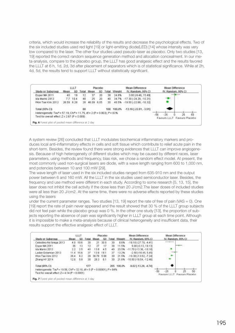
195
criteria, which would increase the reliability of the results and decrease the psychological effects. Two of the six included studies used red light [19] or light-emitting diode(LED) [14] whose intensity was verylow compared to the laser. The other four studies used pseudo-laser as placebo. Only two studies [13, 19] reported the correct random sequence generation method and allocation concealment. In our me-ta-analysis, compare to the placebo group, the LLLT has good analgesic effect and the results favored the LLLT at 6 h, 1d, 2d, 3d after placement of separators which is of statistical significance. While at 2h, 4d, 5d, the results tend to support LLLT without statistically significant.
A system review [26] concluded that LLLT modulates biochemical inflammatory markers and pro-duces local anti-inflammatory effects in cells and soft tissue which contribute to relief acute pain in the short-term. Besides, the review found there were strong evidences that LLLT can improve angiogene-sis. Because of high heterogeneity of different studies which may be caused by different races, laser parameters, using methods and frequency, bias risk, we chose a random effect model. At present, the most commonly used non-surgical lasers are diode, with a wave length ranging from 600 to 1,000 nm, and potencies between 10 and 100 mW [29].The wave length of laser used in the six included studies ranged from 635-910 nm and the output power between 6 and 160 mW. All the LLLT in the six studies used semiconductor laser. Besides, the frequency and use method were different in each study. According to some research [5, 13, 15], the laser does not inhibit the cell activity if the dose less than 20 J/cm2.The laser doses of included studies were all less than 20 J/cm2. At the same time, there were no adverse effects reported by these studies using the lasersunder the current parameter ranges. Two studies [13, 19] report the rate of free of pain (VAS = 0). One [19] report the rate of pain never appeared and the result showed that 30 % of the LLLT group subjectsdid not feel pain while the placebo group was 0 %. In the other one study [13], the proportion of sub-jects reporting the absence of pain was significantly higher in LLLT group at each time point. Although it is impossible to make a meta-analysis because of clinical heterogeneity and insufficient data, their results support the effective analgesic effect of LLLT.
six included studies, two [13, 19] of them were judged tohave a low risk of bias. Two studies [14, 20] were judgedto have an unclear risk of bias. Two studies [15, 29]were judged to have a high risk of bias .
Meta-analysis for mean score of painIn our included studies, if there were three or more stud-ies measured the pain score at the same time point, wewill make an analysis. Therefore, totally seven time pointsmeet the requirements: 2 hours, 6 hours, 24 hours, 2 days,3 days, 4 days, 5 days. Figs. 3, 4, 5, 6, 7, 8 and 9 showedthe comparison between LLLT and Placebo on pain reliefafter placing the separators at each time point. Because ofthe high heterogeneity, a random effect was selected.2 hours after the placement, the overall effect test showed
no significant different between the LLLT and placebo(P = 0.55). The mean difference was −3.24 and 95 %CI( −13.98 , 7.49) (Fig. 3).While for the time points of 6 h, 24 h, 2d, 3d, the overall
effects favored the LLLT and showed a statistical differencebetween the LLLT and placebo, because all of the P valuesof the tests were less than 0.05 (Figs 4, 5, 6 and 7).At 4th day and 5th day the overall effects showed there
was no statistical difference between the LLLT and theplacebo group (P = 0.06 at 4d and P = 0.15 at 5d).
The pain incidenceOne of the included studies reported the rate of pain neverappeared and never disappeared [19]. The result showedthat 30 % of the LLLTgroup subjects did not feel pain while
the placebo group was 0 %. In another study [13], theproportion of subjects reporting the absence of painwas significantly higher in LLLT group at each time point.Meta-analysis is not feasible because of inadequate data.
DiscussionPain caused by orthodontic treatment can affect patient’scompliance and change their eating habits [8], even for-cing them to terminate treatments [13]. Orthodontistshave been working on the controlling of pain. Al-though the NSAIDs had been proved effective on paincontrol, the side effects limited its clinical application[9–12]. Some researches [5, 13–20] consider LLLT as aneffective method to control orthodontic pain, thereforethis system review is to confirm this analgesic effect afterplacement of separators. Because many orthodontic oper-ations can induce pain, in order to reduce the heterogen-eity of clinical, we select the studies of using LLLT torelief pain after placing the separators.For the orthodontic treatment with fixed appliances, the
separators were used to create enough space for thebands[8]. After placement, whether separators or archwires, the periodontal ligament and the vessels wereunder pressure, causing the release of inflammatorymediators and inducing pain [2, 9].However, it is difficult to measure the pain precisely be-
cause pain is a subjective experience, the individual vari-ability of pain threshold and sensitivity can be influencedby physical and psychological effects [18, 19]. Besides,other factors, such as environmental, sociocultural, genetic
Fig. 6 Forest plot of pooled mean difference at 2 day
Fig. 7 Forest plot of pooled mean difference at 3 day
Shi et al. Head & Face Medicine (2015) 11:28 Page 7 of 9

196
According to the results of our meta-analysis, LLLT can reduce the pain caused by the placement of separators effectively without adverse effect under current evidence. Considering LLLT may increase the speed of tooth factors, and so on, can influence pain [15]. Therefore, from these viewpoints, the split-mouth design perhaps is the best choice. In our included studies, only one is splitmouth design. There are no objective measurements for pain. The VAS is one of the most common used tools tomeasure pain intensity at present [8, 16]. All of the six included studies in this review used this method. What’s more, in order to avoid the psychological effect, we need well designed clinical trials to evaluate the pain. Using placebo is one of our included criteria, which would increase the reliability of the results and decrease the psychological effects. Two of the six included studies used red light [19] or light-emit-ting diode(LED) [14] whose intensity was very low compared to the laser. The other four studies usedpseudo-laser as placebo. Only two studies [13, 19] reported the correct random sequence generation method and allocation concealment.In our meta-analysis, compare to the placebo group, the LLLT has good analgesic effect and the results favored the LLLT at 6 h, 1d, 2d, 3d after placement of separators which is of statistical significance. While at 2 h, 4d, 5d, the results tend to support LLLT without statistically significant.A system review [26] concluded that LLLT modulates biochemical inflammatory markers and produceslocal anti-inflammatory effects in cells and soft tissue which contribute to relief acute pain in the short-term. Besides, the review found there were strong evidences that LLLT can improve angiogenesis. Be-cause of high heterogeneity of different studies which may be caused by different races, laser parame-ters, using methods and frequency, bias risk, we chose a random effect model. At present, the most commonly used non-surgical lasers are diode, with a wave length ranging from 600 to 1,000 nm, and potencies between 10 and 100 mW [29]. The wave length of laser used in the six included studiesranged from 635-910 nm and the output power between 6 and 160 mW. All the LLLT in the six studies used semiconductor laser. Besides, the frequency and use method were different in each study. Accor-ding to some research [5, 13, 15], the laser does not inhibit the cell activity if the dose less than 20 J/cm2.The laser doses of included studies were all less than 20 J/cm2. At the same time, there were no
factors, and so on, can influence pain [15]. Therefore, fromthese viewpoints, the split-mouth design perhaps is thebest choice. In our included studies, only one is split-mouth design. There are no objective measurements forpain. The VAS is one of the most common used tools tomeasure pain intensity at present [8, 16]. All of the six in-cluded studies in this review used this method. What’smore, in order to avoid the psychological effect, we needwell designed clinical trials to evaluate the pain. Using pla-cebo is one of our included criteria, which would increasethe reliability of the results and decrease the psychologicaleffects. Two of the six included studies used red light [19]or light-emitting diode(LED) [14] whose intensity was verylow compared to the laser. The other four studies usedpseudo-laser as placebo. Only two studies [13, 19] reportedthe correct random sequence generation method and allo-cation concealment.In our meta-analysis, compare to the placebo group, the
LLLT has good analgesic effect and the results favored theLLLT at 6 h, 1d, 2d, 3d after placement of separators whichis of statistical significance. While at 2 h, 4d, 5d, theresults tend to support LLLT without statistically signifi-cant. A system review [26] concluded that LLLT modu-lates biochemical inflammatory markers and produceslocal anti-inflammatory effects in cells and soft tissuewhich contribute to relief acute pain in the short-term. Be-sides, the review found there were strong evidences thatLLLT can improve angiogenesis. Because of high hetero-geneity of different studies which may be caused by
different races, laser parameters, using methods and fre-quency, bias risk, we chose a random effect model. Atpresent, the most commonly used non-surgical lasersare diode, with a wave length ranging from 600 to1,000 nm, and potencies between 10 and 100 mW [29].The wave length of laser used in the six included studiesranged from 635-910 nm and the output power between 6and 160 mW. All the LLLT in the six studies used semi-conductor laser. Besides, the frequency and use methodwere different in each study. According to some research[5, 13, 15], the laser does not inhibit the cell activity if thedose less than 20 J/cm2.The laser doses of included studieswere all less than 20 J/cm2. At the same time, there wereno adverse effects reported by these studies using the lasersunder the current parameter ranges.Two studies [13, 19] report the rate of free of pain
(VAS = 0). One [19] report the rate of pain never appearedand the result showed that 30 % of the LLLT group subjectsdid not feel pain while the placebo group was 0 %. In theother one study [13], the proportion of subjects reportingthe absence of pain was significantly higher in LLLT groupat each time point. Although it is impossible to make ameta-analysis because of clinical heterogeneity and in-sufficient data, their results support the effective anal-gesic effect of LLLT.According to the results of our meta-analysis, LLLT can
reduce the pain caused by the placement of separators ef-fectively without adverse effect under current evidence.Considering LLLT may increase the speed of tooth
Fig. 8 Forest plot of pooled mean difference at 4 day
Fig. 9 Forest plot of pooled mean difference at 5 day
Shi et al. Head & Face Medicine (2015) 11:28 Page 8 of 9

197
adverse effects reported by these studies using the lasers under the current parameter ranges.Two studies [13, 19] report the rate of free of pain (VAS = 0). One [19] report the rate of pain never ap-peared and the result showed that 30 % of the LLLT group subjects did not feel pain while the placebo group was 0 %. In the other one study [13], the proportion of subjects reporting the absence of pain was significantly higher in LLLT group at each time point. Although it is impossible to make a meta-ana-lysis because of clinical heterogeneity and insufficient data, their results support the effective analgesiceffect of LLLT.According to the results of our meta-analysis, LLLT can reduce the pain caused by the placement of se-parators effectively without adverse effect under current evidence. Considering LLLT may increase the speed of tooth movement [22], in the field of orthodontics, LLLT may have broad application prospects. But different studies used different separators, different lasers and parameters, different method and frequency of laser, different test positions (mandible or maxilla or both), different design and different risk of bias, and these can lead to the high heterogeneity. Therefore, well designed RCTs are required to evaluate the analgesic effect of LLLT.
ConclusionUnder current studies and evidences, the results of our meta-analysis reveals that LLLT can reduce the pain caused by the placement of separators effectively at 6 h, 1d, 2d, 3d after the placement of the orthodontic separators without adverse effect reports. Besides, there is no evidence reveals that LLLT can bring forward the most painful day. These results indicate the good clinical application prospect. However, because of the high heterogeneity and the bias risk of included studies, well designedRCTs are required in the future.
Competing interestsThis work was supported by Clinical scientific research fund (No.320.6750.15029) from Wu Jieping Medical Foundation.
Authors’ contributionsSQ carried out the literature research and drafted the manuscript. The part of study selection, data extraction, risk of bias evaluation was finished by SQ and YS. For the part of data analysis, it was com-pleted by SQ and JFF. XJ is the Corresponding author and she undertook the work of design of this meta-analysis, coordination and helped to draft the manuscript. All authors read and approved the final manuscript.
AcknowledgmentsWe are grateful to Dra. Maria Ângela L. R. Esper (Institute for Ortodontia e Ortopedia Especializada), Bortolotti F (Institute for Department of Neurosciences, Section of Orthodontics, University of Naples “Federico II”, Italy, via Pansini), and A. Puigdollers (Department of Orthodontics and Dentofacial Or-thopedics, Dental School, International University of Catalunya, Campus Sant Cugat, Josep Truetas/n)), for giving us the data we need and answering our questions by E-mail.Received: 25 May 2015 Accepted: 10 August 2015
References1. Krishnan V. Orthodontic pain: from causes to management–a review. Eur J Orthod. 2007;29(2):170–9.2. Bergius M, Kiliaridis S, Berggren U. Pain in orthodontics. A review and discussion of the literature. J Orofac Orthop. 2000;61(2):125–37.3. Hoar S, Linnell KJ. Cognitive load eliminates the global perceptual bias for unlimited exposure dura-tions. Atten Percept Psychophys. 2013;75(2):210–5.4. Zawawi KH. Acceptance of orthodontic miniscrews as temporary anchorage devices. Patient Prefer Adherence. 2014 Jun 30;8:933–7.

198
5. Tortamano A, Lenzi DC, Haddad AC, Bottino MC, Dominguez GC, Vigorito JW. Low-level laser therapy for pain caused by placement of the first orthodontic archwire: a randomized clinical trial. Am J Orthod Dentofacial Orthop. 2009;136(5):662–7.6. Youssef M, Ashkar S, Hamade E, Gutknecht N, Lampert F, Mir M. The effect of low-level laser therapy during orthodontic movement: a preliminary study. Lasers Med Sci. 2008;23(1):27–33.7. Erdinç AM, Dinçer B. Perception of pain during orthodontic treatment with fixed appliances. Eur J Orthod. 2004;26(1):79–85.8. Bondemark L, Fredriksson K, Ilros S. Separation effect and perception of pain and discomfort from two types of orthodontic separators. World J Orthod. 2004;5(2):172–6.9. Polat O, Karaman AI. Pain control during fixed orthodontic appliance therapy. Angle Orthod. 2005;75(2):214–9.10. Xiaoting L, Yin T, Yangxi C. Interventions for pain during fixed orthodontic appliance therapy. A sys-tematic review. Angle Orthod. 2010;80(5):925–32.11. Salmassian R, Oesterle LJ, Shellhart WC, Newman SM. Comparison of the efficacy of ibuprofen and acetaminophen in controlling pain after orthodontic tooth movement. Am J Orthod Dentofacial Orthop.2009;135(4):516–21.12. Kyrkanides S, O’Banion MK, Subtelny JD. Nonsteroidal anti-inflammatory drugs in orthodontic tooth movement: metalloproteinase activity and collagen synthesis by endothelial cells. Am J Orthod Dentofa-cial Orthop. 2000;118(2):203–9.13. Nóbrega C, da Silva EM, deMacedo CR. Low-level laser therapy for treatment of pain associated with orthodontic elastomeric separator placement: a placebocontrolled randomized double-blind clinical trial. Photomed Laser Surg. 2013;31(1):10–6.14. Kim WT, Bayome M, Park JB, Park JH, Baek SH, Kook YA. Effect of frequent laser irradiation on orthodontic pain. A single-blind randomized clinical trial. Angle Orthod. 2013;83(4):611–6.15. Eslamian L, Borzabadi-Farahani A, Hassanzadeh-Azhiri A, Badiee MR, Fekrazad R. The effect of 810-nm low-level laser therapy on pain caused by orthodontic elastomeric separators. Lasers Med Sci.2014;29(2):559–64.16. Artés-Ribas M, Arnabat-Dominguez J, Puigdollers A. Analgesic effect of a low-level laser therapy (830 nm) in early orthodontic treatment. Lasers Med Sci. 2013;28(1):335–41.17. Fujiyama K, Deguchi T, Murakami T, Fujii A, Kushima K, Takano-Yamamoto T. Clinical effect of CO2 laser in reducing pain in orthodontics. Angle Orthod. 2008;78(2):299–303.18. He WL, Li CJ, Liu ZP, Sun JF, Hu ZA, Yin X, et al. Efficacy of low-level laser therapy in the manage-ment of orthodontic pain: a systematic review and meta-analysis. Lasers Med Sci. 2013;28(6):1581–9.19. Marini I, Bartolucci ML, Bortolotti F, Innocenti G, Gatto MR, Alessandri Bonetti G. The effect of diode superpulsed low-level laser therapy on experimental orthodontic pain caused by elastomeric separators: a randomized controlled clinical trial. Lasers Med Sci. 2013. [Epub ahead of print]20. Zhang HY, Yan Y, Deng HY, Liu HX. Clinical therapeutic evaluation of low-level laser in alleviating pain during orthodontic elastomeric separator placement. Journal of Chinese Practical Diagnosis and Therapy. 2014;28(3):256–8.21. Saygun I, Karacay S, Serdar M, Ural AU, Sencimen M, Kurtis B. Effects of laser irradiation on the release of basic fibroblast growth factor (bFGF), insulin like growth factor-1 (IGF-1), and receptor of IGF-1 (IGFBP3) from gingival fibroblasts. Lasers Med Sci. 2008;23(2):211–5.22. Pallotta RC, Bjordal JM, Frigo L, Leal Junior EC, Teixeira S, Marcos RL, et al. Infrared (810-nm) low-level laser therapy on rat experimental knee inflammation. Lasers Med Sci. 2012;27(1):71–8.23. Pinheiro AL, Gerbi ME. Photoengineering of bone repair processes. Photomed Laser Surg. 2006;24(2):169–78.24. Sousa MV, Scanavini MA, Sannomiya EK, Velasco LG, Angelieri F. Influence of low-level laser on the speed of orthodontic movement. Photomed Laser Surg. 2011;29(3):191–6.25. Fikácková H, Dostálová T, Navrátil L, Klaschka J. Effectiveness of low-level laser therapy in temporo-mandibular joint disorders: a placebo-controlled study. Photomed Laser Surg. 2007;25(4):297–303.26. Bjordal JM, Johnson MI, Iversen V, Aimbire F, Lopes-Martins RA. Low-level laser therapy in acute

199
pain: a systematic review of possible mechanisms of action and clinical effects in randomized place-bo-controlled trials. Photomed Laser Surg. 2006;24(2):158–68.27. Higgins JPT, Green S, editors. Cochrane handbook for systematic reviews of interventions [version 5.1.0]. The Cochrane Collaboration. 2011.28. Lim HM, Lew KK, Tay DK. A clinical investigation of the efficacy of low level laser therapy in reducing orthodontic postadjustment pain. Am J Orthod Dentofacial Orthop. 1995;108(6):614–22.29. Esper MA, Nicolau RA, Arisawa EA. The effect of two phototherapy protocols on pain control in orthodontic procedure–a preliminary clinical study. Lasers Med Sci. 2011;26(5):657–63.30. Abtahi SM, Mousavi SA, Shafaee H, Tanbakuchi B. Effect of low-level laser therapy on dental pain induced by separator force in orthodontic treatment. Dent Res J (Isfahan). 2013;10(5):647–51.
5.18 Does ultra-pulse CO(2) laser reduce the risk of enamel damage during debonding of cera-mic brackets?Ahrari F1, Heravi F, Fekrazad R, Farzanegan F, Nakhaei S.1Dental Materials Research Center, School of Dentistry, Mashhad University of Medical Sciences, Mashhad, Iran. [email protected]
AbstractThis study seeks to evaluate the enamel surface characteristics of teeth after debonding of ceramic brackets with or without laser light. Eighty premolars were bonded with either of the chemically retained or the mechanically retained ceramic brackets and later debonded conventionally or through a CO(2) laser (188 W, 400 Hz). The laser was applied for 5 s with scanning movement. After debonding, the adhesive remnant index (ARI), the incidence of bracket and enamel fracture, and the lengths, frequency, and directions of enamel cracks were compared among the groups. The increase in intrapulpal tempe-rature was measured in ten extra specimens. The data were analyzed with SPSS software. There was one case of enamel fracture in the chemical retention/conventional debonding group. When brackets were removed with pliers, incidences of bracket fracture were 45% for the chemical retention, and 15% for the mechanical retention brackets. No case of enamel or bracket fracture was seen in the laser-de-bonded teeth. A significant difference was observed in ARI scores among the groups. Laser debonding caused a significant decrease in the frequency of enamel cracks, compared to conventional debonding. The increase in intrapulpal temperatures was below the benchmark of 5.5 °C for all the specimens. Laser-assisted debonding of ceramic brackets could reduce the risk of enamel damage and bracket fracture, and produce the more desirable ARI scores without causing thermal damage to the pulp. However, some augmentations in the length and frequency of enamel cracks should be expected with all debonding methods.
https://www.ncbi.nlm.nih.gov/pubmed/21667137
5.19 Effect of 940 nm low-level laser therapy on osteogenesis in vitroJawad MM1, Husein A2, Azlina A3, Alam MK4, Hassan R4, Shaari R5. 1Universiti Sains Malaysia, School of Dental Sciences, Health Campus, Orthodontic Unit, Kota Bharu, Kelantan 16150, MalaysiaeAlnahrain University, Baghdad, Iraq.2Universiti Sains Malaysia, School of Dental Sciences, Health Campus, Conservative Department, Kota Bharu, Kelantan 16150, Malaysia.3Universiti Sains Malaysia, School of Dental Sciences, Health Campus, Kota Bharu, Kelantan 16150, Malaysia.4Universiti Sains Malaysia, School of Dental Sciences, Health Campus, Orthodontic Unit, Kota Bharu, Kelantan 16150, Malaysia.5Universiti Malaysia Kelantan, Clinical sciences, Faculty of Veterinary Medicine, Kota Bharu, Kelantan 16100, Malaysia.
AbstractBone regeneration is essential in medical treatment, such as in surgical bone healing and orthodon-

200
tics. The aim of this study is to examine the effect of different powers of 940 nm diode low-level laser treatment (LLLT) on osteoblast cells during their proliferation and differentiation stages. A human fetal osteoblast cell line was cultured and treated with LLLT. The cells were divided into experimental groups according to the power delivered and periods of exposure per day for each laser power. The (3-(4,5-di-methylthiazol-2yl)-2,5 diphenyl tetrazolium bromide) (MTT) assay was used to determine cell prolifera-tion. Both alkaline phosphatase and osteocalcin activity assays were assessed for cell differentiation. All treatment groups showed a significant increase in cell proliferation and differentiation compared to the control group. Regarding the exposure time, the subgroups treated with the LLLT for 6 min showed hi-gher proliferation and differentiation rates for the powers delivered, the 300-mW LLLTgroup significantly increased the amount of cell proliferation. By contrast, the 100 and 200 mW groups showed significant-ly greater amounts of cell differentiation. These results suggest that the use of LLLT may play an impor-tant role in stimulating osteoblast cells for improved bone formation.
https://www.ncbi.nlm.nih.gov/pubmed/24337495
5.20 Effect of a low-level laser on bone regeneration after rapid maxillary expansionCepera F1, Torres FC, Scanavini MA, Paranhos LR, Capelozza Filho L, Cardoso MA, Siqueira DC, Siqueira DF.1Methodist University of São Paulo, São Paulo, Brazil.
AbstractINTRODUCTIONIn this study, we evaluated the effects of a low-level laser on bone regeneration in rapid maxillary expan-sion procedures.
METHODSTwenty-seven children, aged 8 to 12 years, took part in the experiment, with a mean age of 10.2 years, divided into 2 groups: the laser group (n = 14), in which rapid maxillary expansion was performed in conjunction with laser use, and the no-laser group (n = 13), with rapid maxillary expansion only. The activation protocol of the expansion screw was 1 full turn on the first day and a half turn daily until achie-ving overcorrection. The laser type used was a laser diode (TWIN Laser; MMOptics, São Carlos, Brazil), according to the following protocol: 780 nm wavelength, 40 mW power, and 10 J/cm(2) density at 10 points located around the midpalatal suture. The application stages were 1 (days 1-5 of activation), 2 (at screw locking, on 3 consecutive days), 3, 4, and 5 (7, 14, and 21 days after stage 2). Occlusal radio-graphs of the maxilla were taken with the aid of an aluminum scale ruler as a densitometry reference at different times: T1 (initial), T2 (day of locking), T3 (3-5 days after T2), T4 (30 days after T3), and T5 (60 days after T4). The radiographs were digitized and submitted to imaging software (Image Tool; UTHSC-SA, San Antonio, Tex) to measure the optic density of the previously selected areas. To perform the statistical test, analysis of covariance was used, with the time for the evaluated stage as the covariable. In all tests, a significance level of 5% (P <0.05) was adopted.
RESULTSFrom the evaluation of bone density, the results showed that the laser improved the opening of the mid-palatal suture and accelerated the bone regeneration process.
CONCLUSIONSThe low-level laser, associated with rapid maxillary expansion, provided efficient opening of the midpala-tal suture and influenced the bone regeneration process of the suture, accelerating healing.Copyright © 2012 American Association of Orthodontists. Published by Mosby, Inc. All rights reserved.
https://www.ncbi.nlm.nih.gov/pubmed/22464526

201
5.21 Effect of a single dose of low-level laser therapy on spontaneous and chewing pain caused by elastomeric separatorsQamruddin I1, Alam MK2, Fida M3, Khan AG4.
1 Assistant professor and head, Orthodontic Department, Baqai Medical University, Karachi, Pakistan. Electronic address: [email protected] Senior lecturer, Orthodontic Unit, School of Dental Science, Universiti Sains Malaysia, Kelantan, Ma-laysia. Electronic address: [email protected] Director, Orthodontics Residency Program, Associate Professor, Section of Dental Surgery, Depart-ment of Surgery, Aga Khan University, Karachi, Pakistan.4 Lecturer, Orthodontic Department, Baqai Medical University, Karachi, Pakistan.
AbstractINTRODUCTIONThe aim of this study was to see the effect of a single dose of low-level laser therapy on spontaneous and chewing pain after the placement of elastomeric separators.
METHODSEighty-eight patients were randomly selected for this single-blind study. Elastomeric separators were placed mesial and distal to the permanent first molars in all quadrants. Both arches were divided into experimental and control sides. The experimental sides were treated withlow-level laser therapy on 3 points on the buccal mucosa for 20 seconds each, with a 940-nm gallium-aluminum-arsenic diode laser on continuous mode and power set at 200 mW. The other side received placebo laser therapy without turning on the laser. A numeric rating scale was used to assess the intensity of spontaneous and chewing pain for the next 7 days. The independent sample t test and repeated-measures analysis of variance with the post hoc Tukey test was used to analyze the results.
RESULTSSignificant differences were found in spontaneous and chewing pain among both groups (P <0.05).
CONCLUSIONSA single dose of low-level laser therapy can be an efficient modality to reduce the postoperative pain associated with the placement of elastomeric separators.Copyright © 2016 American Association of Orthodontists. Published by Elsevier Inc. All rights reserved.
https://www.ncbi.nlm.nih.gov/pubmed/26718379
5.22 Effect of frequent application of low-level laser therapy on corticotomized tooth move-ment in dogs: a pilot studyHan KH1, Park JH2, Bayome M3, Jeon IS4, Lee W5, Kook YA6.
1 Private Practitioner, Former Graduate Student, Graduate School of Clinical Dental Science, The Ca-tholic University of Korea, Seoul, Korea.2 Associate Professor and Chair, Postgraduate Orthodontic Program, Arizona School of Dentistry & Oral Health, A.T. Still University, Mesa, AZ; and Adjunct Professor, the Graduate School of Dentistry, Kyung Hee University, Seoul, Korea.3 Research Assistant Professor, Department of Dentistry, The Catholic University of Korea, Seoul, Korea; and Visiting Professor, Department of Postgraduate Studies, Universidad Autonóma del Para-guay, Asunción, Paraguay.4 Private Practitioner, Seoul, Korea.

202
5Professor, Oral and Maxillofacial Surgery, Department of Dentistry, Uijeongbu St Mary’s Hospital, Colle-ge of Medicine, The Catholic University of Korea, Uijeongbu, GyeongGi, Korea.6Professor, Department of Orthodontics, Seoul St Mary’s Hospital, College of Medicine, The Catholic University of Korea, Seoul, Korea. Electronic address: [email protected].
AbstractPURPOSEThe purposes of the present study were to evaluate the effects of frequent applications of low-level laser therapy (LLLT) on corticotomy-assisted tooth movement in a beagle dog model and to compare the effects in the mandible and maxilla.
MATERIALS AND METHODSIn 4 male beagles, the maxillary and mandibular second premolars were extracted. The third premolars were corticotomized and then protracted from the canines with a continuous force of 200 g. Daily LLLT (using an aluminum gallium indium phosphide [AlGaInP] diode) was applied at the buccal mucosa of the corticotomized premolars on 1 side only. The tooth movement was measured for 8 weeks. Fluoro-chromes were injected intravenously at the start of the experiment (T0) and after 2 (T2), 4 (T4), and 8 (T8) weeks to evaluate new bone formation on the tension sides. Histomorphometric and immunohisto-logic evaluations were performed.
RESULTSIn the mandible, the movement of the corticotomized premolars in the LLLT plus corticotomy group was less than that in the corticotomy-only group, although the difference was not statistically significant. In the maxilla, no significant differences between the 2 groups were found. Osteoclastic and proliferating cell activities and the amount of new bone formation were greater in the mandibular LLLT plus cortico-tomy group than in the corticotomy-only group.
CONCLUSIONSThe frequent application of LLLT showed no significant effect on the corticotomized tooth movement.Copyright © 2014 American Association of Oral and Maxillofacial Surgeons. Published by Elsevier Inc. All rights reserved.
https://www.ncbi.nlm.nih.gov/pubmed/24704036
5.23 Effect of frequent laser irradiation on orthodontic pain A single-blind randomized clinical trialWon Tae Kima; Mohamed Bayomeb; Jun-Beom Parkc; Jae Hyun Parkd; Seung-Hak Baeke;Yoon-Ah Kookf
ABSTRACTObjective To analyze the effect of low-level laser therapy (LLLT) on perception of pain afterseparator placement and compare it with perceptions of control and placebo groups using afrequent irradiation protocol.
Materials and MethodsEighty-eight patients were randomly allocated to a laser group, a lightemitting diode (LED) placebo group, or a control group. Elastomeric separators were placed on the first molars. In the laser and LED groups, first molars were irradiated for 30 seconds every 12 hours for 1 week using a portable device. Pain was marked on a visual analog scale at predetermined intervals. Repeated measure analysis of

203
variance was performed for statistical analysis.
ResultsThe pain scores of the laser group were significantly lower than those of the control groupup to 1 day. The pain scores in the LED group were not significantly different from those of the lasergroup during the first 6 hours. After that point, the pain scores of the LED group were notsignificantly different from those of the control.
ConclusionsFrequent LLLT decreased the perception of pain to a nonsignificant level throughoutthe week after separator placement, compared with pain perception in the placebo and controlgroups. Therefore, LLLT might be an effective method of reducing orthodontic pain. (Angle Orthod.2013;83:611–616.)
KEY WORDSLow-level laser therapy (LLLT); Light-emitting diode (LED); Orthodontic pain; Elastomeric separator; Placebo; Visual analog scale (VAS); Laser irradiation.
INTRODUCTIONPain or discomfort during orthodontic treatment is a major concern for clinicians and patients. It maydiscourage patients from seeking or continuing treatment.1 The incidence and severity of pain have been reported to be higher than those of extractions.2 Several patients have reported that orthodontic pain disturbed their sleep during the first week after initial wire placement.3 The peak of pain occurred approximately 24 hours after separator/initial wire placement and decreased over the next 6 to 8 days.2,4–7To control the pain, analgesics or anti-inflammatory agents have been prescribed, and the initial arch wire has often been limited to light force only.8,9 Preemptive ibuprofen significantly reduced the pain 2 hours after separator placement compared with a placebo. However, a significant amount of pain was reported at 24 hours.6 Another option, the use of nonsteroidal antiinflammatory drugs, is still controversial because of their potential influence on tooth movement and their adverse systemic side effects.10–13Recently, low-level laser therapy (LLLT) has been used to control pain because of its anti-inflammatory properties and regenerative effect on neurons.14–17These effects have been attributed to photobioactive reaction that stimulates the proliferation and differentiation of cells.18 Previous studies have shown that LLLT may increase the blood supply and promote recovery of dental tissue.19 In addition, several studies have shown that LLLT reduced ortho-dontic pain.20–22Other studies showed no significant reduction of pain with lasers compared to a placebo.16,23 Moreo-ver, laser therapy had no impact on time of pain initiation and maximum pain.21 Interestingly, in previous LLLT studies, clinicians directly performed a single-session laser irradiation in their office.17,21,24 Other studies recalled the participants to the office several times for laser application.20,25 This limitation might have decreased the efficacy of the therapy. It is hypothesized that a more frequent application of laser therapy during the pain/discomfort period might lead to a greater reduction in the perceptionof pain in orthodontic patients. However, no study has evaluated the effect of frequent LLLT.Currently, a compact, battery-operated, handheld device that produces a low-level aluminum-galliu-mindium- phosphide semiconductor (AlGaInP) laser has been developed for use by patients at home. These devices have been found to be safe and effective in the management of dentin hypersensiti-vity.26 The purpose of this study was to analyze the effect of a more frequent LLLT protocol on the perception of pain from immediately up to 7 days after separator placement and to compare it to the perception of pain in control and placebo groups.

204
MATERIALS AND METHODSOne hundred and twenty patients were assessed in a private clinic from May 2010 to January 2012. Ofthese, 88 patients (23 males, 65 females; mean age, 22.7 years) met the following inclusion criteria:complete eruption of the second molars and no open interproximal contacts of the first molar. In addi-tion, patients with previous orthodontic treatment, metabolic and periodontal diseases, or medication were excluded.The study was approved by the institutional review board of the Catholic University of Korea. Sample-size calculation was performed based on previous results.20Subjects were assigned randomly to three test groups: laser irradiation group (n 5 28), light-emittingdiode (LED) group as a placebo (n 5 30), and a control group receiving no irradiation (n 5 30). Subjectsassigned to the laser and LED groups were blinded to their assignment.A low-level medical semiconductor laser device with an AlGaInP diode, wavelength of 635 nm, energy of 10 mJ, field diameter of 5.6 mm, and output potency of 6 mW (Figure 1) and an LED device with wavelength of 635 nm and output of 12.9 mW of the same exterior design (Dental.M, M&H Co, Hwas-eong, Korea) were used. After placement of 2.1 mm elastomeric separators (Dentalastics Separators, Dentaurum, Ispringen, Germany) mesially and distally on both maxillary first molars, subjects in the laser and LED groups were taught to use the device and asked to apply it for 30 seconds on each area immediately then every 12 hours for 1 week with close contact between the tip and mucosa to irradiate the mesiobuccal, mesiolingual, distobuccal, and distolingual areas.The three groups were asked to mark pain severity on a questionnaire containing 11 copies of a 10 cmvisual analog scale (VAS) at 5 minutes, 1 hour, 6 hours, 12 hours and then at days 1, 2, 3, 4, 5, 6, and 7 after the separators were applied.
Statistical AnalysisStatistical evaluation was performed by SPSS 16.0 (SPSS Inc, Chicago, Ill). Two-way analysis of va-riance (ANOVA) was performed to compare age of subjects according to group and gender. Repeated measures ANOVA was conducted to evaluate the differences in pain scores at each time point as the within-subjects variable and between groups and gender as the between-subjects variables. Age was used as a covariant. The alpha level was 0.05.
RESULTSThere were no significant differences in age among the laser, LED placebo, and control groups (P 5 .069) or between genders (P 5 .094). Also, no significant interaction between groups and gender was found (P 5.30) (Table 1).
interaction between groups and gender was found (P5
.30) (Table 1).
There was a significant difference in pain perceptionamong the three groups (P 5 .004). The laser groupshowed a statistically significant decrease in painscores compared with the control group (P 5 .003).The placebo group demonstrated no significant differ-ences from the laser and control groups (P 5 .28 andP 5.26, respectively; Figure 2). However, gender hadno significant effect on scores of pain perception (P 5
.81; Figure 3). In addition, there was a borderlinesignificance of the interaction between the gender andgroups (P 5 .05).
The maximum level of pain was reached 1 day afterseparator placement. The laser group showed amaximum level of (26.6 6 6.3), which was significantlylower than those of the LED placebo and controlgroups (46.2 6 6.1 and 55.5 6 5.6, respectively)(Table 2 and Figure 2).
During the first 6 hours, the pain scores in theplacebo group were not significantly different fromthose of the laser group. Furthermore, at 12 and24 hours after placement of separators, the pain
scores of the placebo group were not significantlydifferent from those of the control group. From day 2 today 7 after separators placement, the pain scores of allthree groups decreased. Pain scores were still lower atday 2 in the laser group; however, the difference wasnot statistically significant. A decrease was seen in thecontrol and placebo groups, which reached a level thatwas not significantly different from the laser group(Table 2 and Figure 2).
In the laser group, there were no significantdifferences between pain scores at different timepoints. However, in the placebo group, the maximumpain score occurred at 1 day and was significantlyhigher than at 5 minutes, 1 hour, 6 hours, or day 6 or 7;whereas in the control group it was higher than at5 minutes and at days 5, 6, and 7 (Table 2).
Because of the borderline significance (P 5 .05) ofinteraction between groups and gender, the groupswere compared within each gender independently. Inmale subjects, the pain scores in the laser group werelower than those in the placebo and control groups,which showed a similar pain perception trend. Infemale subjects, pain scores in the placebo group
Table 1. Descriptive Data for the Laser, LED, and Control Groupsa
Total Male Female P Value*
Laser group 28 (20.79 6 5.15) 7 (18.14 6 4.18) 21 (21.67 6 5.23) .094
LED group 30 (22.20 6 7.19) 7 (22.71 6 10.19) 23 (22.04 6 6.30)
Control group 30 (25.10 6 6.07) 9 (21.78 6 4.89) 21 (26.52 6 6.06)
Total 88 (22.74 6 6.40) 23 (20.96 6 6.75) 65 (23.37 6 6.21)
P value{ .069
a Data presents number of subjects (mean age 6 standard deviation); two-way ANOVA comparing age according to gender (*) and group ({);no significant interaction between group and gender.
Figure 2. Comparison of mean pain scores on the VAS among the
laser, LED, and control groups over time.
Figure 3. Comparison of mean pain scores on the VAS between
male and female subjects over time.
EFFECT OF FREQUENT LLLT ON ORTHODONTIC PAIN 613
Angle Orthodontist, Vol 83, No 4, 2013
properties and regenerative effect on neurons.14–17
These effects have been attributed to photobioactivereaction that stimulates the proliferation and differen-tiation of cells.18 Previous studies have shown thatLLLT may increase the blood supply and promoterecovery of dental tissue.19 In addition, several studieshave shown that LLLT reduced orthodontic pain.20–22
Other studies showed no significant reduction of painwith lasers compared to a placebo.16,23 Moreover, lasertherapy had no impact on time of pain initiation andmaximum pain.21
Interestingly, in previous LLLT studies, cliniciansdirectly performed a single-session laser irradiation intheir office.17,21,24 Other studies recalled the participantsto the office several times for laser application.20,25 Thislimitation might have decreased the efficacy of thetherapy. It is hypothesized that a more frequentapplication of laser therapy during the pain/discomfortperiod might lead to a greater reduction in the per-ception of pain in orthodontic patients. However, nostudy has evaluated the effect of frequent LLLT.Currently, a compact, battery-operated, handhelddevice that produces a low-level aluminum-gallium-indium-phosphide semiconductor (AlGaInP) laser hasbeen developed for use by patients at home. Thesedevices have been found to be safe and effective in themanagement of dentin hypersensitivity.26
The purpose of this study was to analyze the effectof a more frequent LLLT protocol on the perception ofpain from immediately up to 7 days after separatorplacement and to compare it to the perception of painin control and placebo groups.
MATERIALS AND METHODS
One hundred and twenty patients were assessed ina private clinic from May 2010 to January 2012. Ofthese, 88 patients (23 males, 65 females; mean age,22.7 years) met the following inclusion criteria:complete eruption of the second molars and no openinterproximal contacts of the first molar. In addition,patients with previous orthodontic treatment, metabolicand periodontal diseases, or medication were exclud-ed. The study was approved by the institutional reviewboard of the Catholic University of Korea. Sample-sizecalculation was performed based on previous results.20
Subjects were assigned randomly to three testgroups: laser irradiation group (n 5 28), light-emittingdiode (LED) group as a placebo (n5 30), and a controlgroup receiving no irradiation (n 5 30). Subjectsassigned to the laser and LED groups were blindedto their assignment.
A low-level medical semiconductor laser device withan AlGaInP diode, wavelength of 635 nm, energy of10 mJ, field diameter of 5.6 mm, and output potency of
6 mW (Figure 1) and an LED device with wavelengthof 635 nm and output of 12.9 mW of the same exteriordesign (Dental.M, M&H Co, Hwaseong, Korea) wereused. After placement of 2.1 mm elastomericseparators (Dentalastics Separators, Dentaurum,Ispringen, Germany) mesially and distally on bothmaxillary first molars, subjects in the laser and LEDgroups were taught to use the device and asked toapply it for 30 seconds on each area immediatelythen every 12 hours for 1 week with close contactbetween the tip and mucosa to irradiate themesiobuccal, mesiolingual, distobuccal, and distolin-gual areas.
The three groups were asked to mark pain severityon a questionnaire containing 11 copies of a 10 cmvisual analog scale (VAS) at 5 minutes, 1 hour, 6 hours,12 hours and then at days 1, 2, 3, 4, 5, 6, and 7 afterthe separators were applied.
Statistical Analysis
Statistical evaluation was performed by SPSS 16.0(SPSS Inc, Chicago, Ill). Two-way analysis of variance(ANOVA) was performed to compare age of subjectsaccording to group and gender. Repeated measuresANOVA was conducted to evaluate the differences inpain scores at each time point as the within-subjectsvariable and between groups and gender as thebetween-subjects variables. Age was used as acovariant. The alpha level was 0.05.
RESULTS
There were no significant differences in age amongthe laser, LED placebo, and control groups (P 5 .069)or between genders (P 5 .094). Also, no significant
Figure 1. A compact, battery-operated, handheld AlGaInP laser
device for home use. (A) Disassembled. (B) Activated.
612 KIM, BAYOME, PARK, PARK, BAEK, KOOK
Angle Orthodontist, Vol 83, No 4, 2013

205
There was a significant difference in pain perception among the three groups (P 5 .004). The laser groupshowed a statistically significant decrease in pain scores compared with the control group (P 5 .003).The placebo group demonstrated no significant differences from the laser and control groups (P 5 .28 and P 5.26, respectively; Figure 2). However, gender had no significant effect on scores of pain percep-tion (P 5 .81; Figure 3). In addition, there was a borderline significance of the interaction between the gender and groups (P 5 .05).The maximum level of pain was reached 1 day after separator placement. The laser group showed amaximum level of (26.6 6 6.3), which was significantly lower than those of the LED placebo and controlgroups (46.2 6 6.1 and 55.5 6 5.6, respectively) (Table 2 and Figure 2).During the first 6 hours, the pain scores in the placebo group were not significantly different from those of the laser group. Furthermore, at 12 and 24 hours after placement of separators, the pain scores of the placebo group were not significantly different from those of the control group. From day 2 to day 7 after separators placement, the pain scores of all three groups decreased. Pain scores were still lower at day 2 in the laser group; however, the difference was not statistically significant. A decrease was seen in the control and placebo groups, which reached a level that was not significantly different from the laser group (Table 2 and Figure 2). In the laser group, there were no significant differences between pain scores at different time points. However, in the placebo group, the maximum pain score occurred at 1 day and was significantly higher than at 5 minutes, 1 hour, 6 hours, or day 6 or 7; whereas in the control group it was higher than at 5 minutes and at days 5, 6, and 7 (Table 2). Because of the border-line significance (P 5 .05) of interaction between groups and gender, the groups were compared within each gender independently. In male subjects, the pain scores in the laser group were lower than those in the placebo and control groups, which showed a similar pain perception trend.
interaction between groups and gender was found (P5
.30) (Table 1).
There was a significant difference in pain perceptionamong the three groups (P 5 .004). The laser groupshowed a statistically significant decrease in painscores compared with the control group (P 5 .003).The placebo group demonstrated no significant differ-ences from the laser and control groups (P 5 .28 andP 5.26, respectively; Figure 2). However, gender hadno significant effect on scores of pain perception (P 5
.81; Figure 3). In addition, there was a borderlinesignificance of the interaction between the gender andgroups (P 5 .05).
The maximum level of pain was reached 1 day afterseparator placement. The laser group showed amaximum level of (26.6 6 6.3), which was significantlylower than those of the LED placebo and controlgroups (46.2 6 6.1 and 55.5 6 5.6, respectively)(Table 2 and Figure 2).
During the first 6 hours, the pain scores in theplacebo group were not significantly different fromthose of the laser group. Furthermore, at 12 and24 hours after placement of separators, the pain
scores of the placebo group were not significantlydifferent from those of the control group. From day 2 today 7 after separators placement, the pain scores of allthree groups decreased. Pain scores were still lower atday 2 in the laser group; however, the difference wasnot statistically significant. A decrease was seen in thecontrol and placebo groups, which reached a level thatwas not significantly different from the laser group(Table 2 and Figure 2).
In the laser group, there were no significantdifferences between pain scores at different timepoints. However, in the placebo group, the maximumpain score occurred at 1 day and was significantlyhigher than at 5 minutes, 1 hour, 6 hours, or day 6 or 7;whereas in the control group it was higher than at5 minutes and at days 5, 6, and 7 (Table 2).
Because of the borderline significance (P 5 .05) ofinteraction between groups and gender, the groupswere compared within each gender independently. Inmale subjects, the pain scores in the laser group werelower than those in the placebo and control groups,which showed a similar pain perception trend. Infemale subjects, pain scores in the placebo group
Table 1. Descriptive Data for the Laser, LED, and Control Groupsa
Total Male Female P Value*
Laser group 28 (20.79 6 5.15) 7 (18.14 6 4.18) 21 (21.67 6 5.23) .094
LED group 30 (22.20 6 7.19) 7 (22.71 6 10.19) 23 (22.04 6 6.30)
Control group 30 (25.10 6 6.07) 9 (21.78 6 4.89) 21 (26.52 6 6.06)
Total 88 (22.74 6 6.40) 23 (20.96 6 6.75) 65 (23.37 6 6.21)
P value{ .069
a Data presents number of subjects (mean age 6 standard deviation); two-way ANOVA comparing age according to gender (*) and group ({);no significant interaction between group and gender.
Figure 2. Comparison of mean pain scores on the VAS among the
laser, LED, and control groups over time.
Figure 3. Comparison of mean pain scores on the VAS between
male and female subjects over time.
EFFECT OF FREQUENT LLLT ON ORTHODONTIC PAIN 613
Angle Orthodontist, Vol 83, No 4, 2013
In female subjects, pain scores in the placebo group showed a similar trend to those in the laser group(Figure 4).
DISCUSSIONThe aim of this study was to evaluate the effect of frequent laser irradiation on the perception of paincaused by orthodontic forces. The pain perception ranged widely depending on individualized painthresholds, age, and gender.2–5,27,28 In agreement with previous studies, pain reached amaximum level 24 hours after separator placement.

206
14,22,28 The maximum pain level in the laser group was similar to the initial pain level in the control group. The pain scores were decreased and were not significantly different among all time points. This might indicate the laser blunts the peak of the pain compared with the peak pain experienced by the placebo and control groups (Figure 2).It was presumed that a score of 0 to 30 score is the mild pain range, whereas a score of 31 to 69 ismoderate pain.29 In our study, the laser group’s pain was 20 scores lower than pain scores in the othergroups. This decrease that changes the level of pain from moderate to mild might be clinically signifi-cant.Less pain and faster recovery have been reported with the CO2 laser than with the Nd:YAG laser group,suggesting a longer-lasting analgesic effect with CO2 laser therapy.30 Meanwhile, Dilsiz et al.31 de-monstrated that the Nd:YAG laser was more effective in desensitizing teeth than the 685 nm diode laser. In our study, the AlGaInP diode laser was applied whereas in several studies a gallium-aluminum-arse-nide diode laser was used.19,24,32 Thus far, no study has compared the effect of different laser types with different treatment settings.In our study, the LED group showed lower pain scores than the control group during the first 6 hours, and then the scores increased and showed no significant difference from those of the control group.The decreased scores could be attributed to the placebo effect. Several mechanisms have beensuggested to explain the placebo effect, such as emotional modulation and reduction of subjectivestress during painful stimulation.33 In a previous study, pain reduction was reported by39% of the subjects after administration of analgesic placebo.34 However, the increased fear of pain after a previous placebo experience was found to reduce the Figure placebo analgesic effect.35 This might explain the increased pain scores in our study after the second LED application. In addition, evidence supported the hypothesis that placebo and treatment effects are not cumulative and that they have mutually exclusive mechanisms.36 Esper et al.23 showed that pain scores decreased significantly in the LED group between 2 and 120 hours compared with scores of the control and laser groups.
showed a similar trend to those in the laser group(Figure 4).
DISCUSSION
The aim of this study was to evaluate the effect offrequent laser irradiation on the perception of paincaused by orthodontic forces. The pain perceptionranged widely depending on individualized painthresholds, age, and gender.2–5,27,28
In agreement with previous studies, pain reached amaximum level 24 hours after separator place-ment.14,22,28 The maximum pain level in the laser groupwas similar to the initial pain level in the control group.The pain scores were decreased and were notsignificantly different among all time points. This mightindicate the laser blunts the peak of the pain comparedwith the peak pain experienced by the placebo andcontrol groups (Figure 2).
It was presumed that a score of 0 to 30 score is themild pain range, whereas a score of 31 to 69 ismoderate pain.29 In our study, the laser group’s painwas 20 scores lower than pain scores in the othergroups. This decrease that changes the level of painfrom moderate to mild might be clinically significant.
Less pain and faster recovery have been reported
with the CO2 laser than with the Nd:YAG laser group,
suggesting a longer-lasting analgesic effect with CO2
laser therapy.30 Meanwhile, Dilsiz et al.31 demonstrated
that the Nd:YAG laser was more effective in desensi-
tizing teeth than the 685 nm diode laser. In our study,
the AlGaInP diode laser was applied whereas in
several studies a gallium-aluminum-arsenide diode
laser was used.19,24,32 Thus far, no study has compared
the effect of different laser types with different
treatment settings.
In our study, the LED group showed lower pain
scores than the control group during the first 6 hours,
and then the scores increased and showed no
significant difference from those of the control group.
The decreased scores could be attributed to the
placebo effect. Several mechanisms have been
suggested to explain the placebo effect, such as
emotional modulation and reduction of subjective
stress during painful stimulation.33
In a previous study, pain reduction was reported by39% of the subjects after administration of analgesicplacebo.34 However, the increased fear of pain after aprevious placebo experience was found to reduce the
Figure 4. Comparison of mean pain scores on the VAS among laser, LED, and control groups over time according to gender. (A) Male subjects.
(B) Female subjects.
Table 2. Pain Perception Scores of the Laser, LED, and Control Groups at Each Time Pointa,b
5 min 1 h 6 h 12 h 1 d 2 d
Laser group 9.28 6 4.43 14.98 6 4.71 19.59 6 5.53 23.88 6 6.01 26.64 6 6.28 26.59 6 6.28
LED group 10.30 6 4.27 12.38 6 4.54 25.32 6 5.33 32.99 6 5.79 46.21 6 6.05 46.09 6 6.05
Control group 26.10 6 3.97 35.36 6 4.22 47.43 6 4.96 50.46 6 5.39 55.47 6 5.62 47.11 6 5.63
P value .008 .001 .001 .005 .004 NS
Multiple comparison C . L (0.020) and
LED (0.025)
C. L (0.006) and
LED (0.001)
C. L (0.001) and
LED (0.10)
C. L (0.005) C . L (0.003)
a Repeated measures ANOVA with Bonferroni post hoc for multiple comparison.b C indicates control; L, laser; LED, light-emitting diode; NS, nonsignificant.
614 KIM, BAYOME, PARK, PARK, BAEK, KOOK
Angle Orthodontist, Vol 83, No 4, 2013

207
They attributed their result to the high-potency and high-energy LED (0.1 W and 7 J, respectively)applied in their study. Consequently, this inconsistency between the investigations might be due to thedifferences in the properties of the LED and laser used. In our study, the pattern of the placebo effect over time seemed different between genders. The pain scores in male subjects in the placebo group followed the same pattern as the control group whereas in females, pain scores appeared to more clo-sely follow the trend in the laser group. In agreement, Saxon et al.37 reported that female patients were more affected by placebo treatment, although an effective response to the analgesic placebo treatment was demonstrated in males only.38 Another study demonstrated no gender dimorphism in relation to the placebo analgesic effect.39 Nevertheless, our results should be interpreted with caution due to the relatively small size of the male group.In concordance, no significant difference was reported in the discomfort level after placement of sepa-rators and initial arch wire, according to age and gender.7,28 A group of environmental, sociocultural,and genetic factors are responsible for these differences. 40–43 Therefore, the present study might have been affected by these individual variations. A future study applying a split-mouth method might be required to remove bias. Also, a study evaluating the effect of laser application on the pain produced by the initial wire is recommended. In addition, a comparison between the pain that resulted from sepa-rator placement and from arch wire change is warranted.
CONCLUSIONSN The laser group showed a significant main effect compared with the control group only. N The per-ception of pain was not significantly different based on age or gender. However, the effect of laserirradiation was more pronounced in male subjects. N The placebo treatment was more effective after the first irradiation than after the following sessions and was more effective in female subjects than in males. N Therefore, frequent low-level laser irradiation may be an effective way to reduce orthodontic pain during the first day after separator placement.
REFERENCES1. Oliver RG, Knapman YM. Attitudes to orthodontic treatment. Br J Orthod. 1985;12:179–188.2. Jones ML, Chan C. Pain in the early stages of orthodontic treatment. J Clin Orthod. 1992;26:311–313.3. Jones M, Chan C. The pain and discomfort experienced during orthodontic treatment: a randomized controlled clinical trial of two initial aligning arch wires. Am J Orthod Dentofacial Orthop. 1992;102:373–381.4. Jones ML. An investigation into the initial discomfort caused by placement of an archwire. Eur J Orthod. 1984;6:48– 54.5. Scheurer PA, Firestone AR, Burgin WB. Perception of pain as a result of orthodontic treatment with fixed appliances. Eur J Orthod. 1996;18:349–357.6. Steen Law SL, Southard KA, Law AS, Logan HL, Jakobsen JR. An evaluation of preoperative ibu-profen for treatment of pain associated with orthodontic separator placement. Am J Orthod Dentofacial Orthop. 2000;118:629–635.7. Wilson S, Ngan P, Kess B. Time course of the discomfort in young patients undergoing orthodontic treatment. Pediatr Dent. 1989;11:107–110.8. Bird SE, Williams K, Kula K. Preoperative acetaminophen vs ibuprofen for control of pain after ortho-
placebo analgesic effect.35 This might explain theincreased pain scores in our study after the secondLED application. In addition, evidence supported thehypothesis that placebo and treatment effects are notcumulative and that they have mutually exclusivemechanisms.36 Esper et al.23 showed that pain scoresdecreased significantly in the LED group between 2 and120 hours compared with scores of the control and lasergroups. They attributed their result to the high-potencyand high-energy LED (0.1 W and 7 J, respectively)applied in their study. Consequently, this inconsistencybetween the investigations might be due to thedifferences in the properties of the LED and laser used.
In our study, the pattern of the placebo effect overtime seemed different between genders. The painscores in male subjects in the placebo group followedthe same pattern as the control group whereas infemales, pain scores appeared to more closely followthe trend in the laser group. In agreement, Saxon et al.37
reported that female patients were more affected byplacebo treatment, although an effective response to theanalgesic placebo treatment was demonstrated inmalesonly.38 Another study demonstrated no gender dimor-phism in relation to the placebo analgesic effect.39
Nevertheless, our results should be interpreted withcaution due to the relatively small size of the male group.
In concordance, no significant difference wasreported in the discomfort level after placement ofseparators and initial arch wire, according to age andgender.7,28 A group of environmental, sociocultural,and genetic factors are responsible for these differ-ences.40–43 Therefore, the present study might havebeen affected by these individual variations. A futurestudy applying a split-mouth method might be requiredto remove bias. Also, a study evaluating the effect oflaser application on the pain produced by the initialwire is recommended. In addition, a comparisonbetween the pain that resulted from separator place-ment and from arch wire change is warranted.
CONCLUSIONS
N The laser group showed a significant main effectcompared with the control group only.
N The perception of pain was not significantly differentbased on age or gender. However, the effect of laserirradiation was more pronounced in male subjects.
N The placebo treatment was more effective after thefirst irradiation than after the following sessions andwas more effective in female subjects than in males.
N Therefore, frequent low-level laser irradiation may bean effective way to reduce orthodontic pain duringthe first day after separator placement.
REFERENCES
1. Oliver RG, Knapman YM. Attitudes to orthodontic treatment.Br J Orthod. 1985;12:179–188.
2. Jones ML, Chan C. Pain in the early stages of orthodontictreatment. J Clin Orthod. 1992;26:311–313.
3. Jones M, Chan C. The pain and discomfort experiencedduring orthodontic treatment: a randomized controlledclinical trial of two initial aligning arch wires. Am J OrthodDentofacial Orthop. 1992;102:373–381.
4. Jones ML. An investigation into the initial discomfort causedby placement of an archwire. Eur J Orthod. 1984;6:48–54.
5. Scheurer PA, Firestone AR, Burgin WB. Perception of painas a result of orthodontic treatment with fixed appliances.Eur J Orthod. 1996;18:349–357.
6. Steen Law SL, Southard KA, Law AS, Logan HL, JakobsenJR. An evaluation of preoperative ibuprofen for treatment ofpain associated with orthodontic separator placement.Am J Orthod Dentofacial Orthop. 2000;118:629–635.
7. Wilson S, Ngan P, Kess B. Time course of the discomfort inyoung patients undergoing orthodontic treatment. PediatrDent. 1989;11:107–110.
8. Bird SE, Williams K, Kula K. Preoperative acetaminophen vsibuprofen for control of pain after orthodontic separator place-ment. Am J Orthod Dentofacial Orthop. 2007;132:504–510.
9. Bernhardt MK, Southard KA, Batterson KD, Logan HL,Baker KA, Jakobsen JR. The effect of preemptive and/orpostoperative ibuprofen therapy for orthodontic pain.Am J Orthod Dentofacial Orthop. 2001;120:20–27.
10. Kyrkanides S, O’Banion MK, Subtelny JD. Nonsteroidalanti-inflammatory drugs in orthodontic tooth movement:metalloproteinase activity and collagen synthesis by endo-thelial cells. Am J Orthod Dentofacial Orthop. 2000;118:203–209.
11. Walker JB, Buring SM. NSAID impairment of orthodontictooth movement. Ann Pharmacother. 2001;35:113–115.
12. Gameiro GH, Pereira-Neto JS, Magnani MB, Nouer DF. Theinfluence of drugs and systemic factors on orthodontic toothmovement. J Clin Orthod. 2007;41:73–78; quiz 71.
3 d 4 d 5 d 6 d 7 d P Value Multiple Comparison
26.40 6 6.20 22.50 6 5.87 17.30 6 5.59 14.24 6 4.94 9.55 6 4.47 NS
36.78 6 5.98 30.30 6 5.65 25.08 6 5.38 21.37 6 4.76 15.94 6 4.31 .003 1 d . 5 min, 1 h, 6 h, 6 d, and 7 d
37.58 6 5.56 29.26 6 5.26 24.76 6 5.00 19.12 6 4.43 19.48 6 4.01 .004 1 d . 5 min, 5 d, 6 d, and 7 d
NS NS NS NS NS
a Repeated measures ANOVA with Bonferroni post hoc for multiple comparison.b C indicates control; L, laser; LED, light-emitting diode; NS, nonsignificant.
Table 2. Extended
EFFECT OF FREQUENT LLLT ON ORTHODONTIC PAIN 615
Angle Orthodontist, Vol 83, No 4, 2013

208
dontic separator placement. Am J Orthod Dentofacial Orthop. 2007;132:504–510.9. Bernhardt MK, Southard KA, Batterson KD, Logan HL, Baker KA, Jakobsen JR. The effect of preemptive and/or postoperative ibuprofen therapy for orthodontic pain. Am J Orthod Dentofacial Or-thop. 2001;120:20–27.10. Kyrkanides S, O’Banion MK, Subtelny JD. Nonsteroidal anti-inflammatory drugs in orthodontic tooth movement: metalloproteinase activity and collagen synthesis by endothelial cells. Am J Orthod Dentofa-cial Orthop. 2000;118: 203–209.11. Walker JB, Buring SM. NSAID impairment of orthodontic tooth movement. Ann Pharmacother. 2001;35:113–115.12. Gameiro GH, Pereira-Neto JS, Magnani MB, Nouer DF. The influence of drugs and systemic factors on orthodontic tooth movement. J Clin Orthod. 2007;41:73–78; quiz 71.13. Krishnan V, Davidovitch Z. The effect of drugs on orthodontic tooth movement. Orthod Craniofac Res. 2006; 9:163–171.14. Harazaki M, Isshiki Y. Soft laser irradiation effects on pain reduction in orthodontic treatment. Bull Tokyo Dent Coll. 1997;38:291–295.15. Harazaki M, Takahashi H, Ito A, Isshiki Y. Soft laser irradiation induced pain reduction in orthodontic treatment. Bull Tokyo Dent Coll. 1998;39:95–101.16. Lim HM, Lew KK, Tay DK. A clinical investigation of the efficacy of low level laser therapy in reducing orthodontic postadjustment pain. Am J Orthod Dentofacial Orthop. 1995;108:614–622.17. Turhani D, Scheriau M, Kapral D, Benesch T, Jonke E, Bantleon HP. Pain relief by single low-level laser irradiation in orthodontic patients undergoing fixed appliance therapy. Am J Orthod Dentofacial Orthop. 2006;130:371–377.18. Verschueren RC, Koudstaal J, Oldhoff J. The carbon dioxide laser; some possibilities in surgery. Acta Chir Belg. 1975;74:197–204.19. Abi-Ramia LB, Stuani AS, Stuani MB, Mendes Ade M. Effects of low-level laser therapy and ortho-dontic tooth movement on dental pulps in rats. Angle Orthod. 2010;80:116–122.20. Doshi-Mehta G, Bhad-Patil WA. Efficacy of low-intensity laser therapy in reducing treatment time and orthodontic pain: a clinical investigation. Am J Orthod Dentofacial Orthop. 2012;141:289–297.21. Tortamano A, Lenzi DC, Haddad AC, Bottino MC, Dominguez GC, Vigorito JW. Low-level laser therapy for pain caused by placement of the first orthodontic archwire: a randomized clinical trial. Am J Orthod Dentofacial Orthop. 2009;136:662–667.22. Fujiyama K, Deguchi T, Murakami T, Fujii A, Kushima K, Takano-Yamamoto T. Clinical effect of CO(2) laser in reducing pain in orthodontics. Angle Orthod. 2008;78: 299–303.23. Esper MA, Nicolau RA, Arisawa EA. The effect of two phototherapy protocols on pain control in orthodontic procedure—a preliminary clinical study. Lasers Med Sci. 2011;26:657–663.24. Bicakci AA, Kocoglu-Altan B, Toker H, Mutaf I, Sumer Z. Efficiency of low-level laser therapy in redu-cing pain induced by orthodontic forces. Photomed Laser Surg. 2012;30: 460–465.25. Youssef M, Ashkar S, Hamade E, Gutknecht N, Lampert F, Mir M. The effect of low-level laser the-rapy during orthodontic movement: a preliminary study. Lasers Med Sci. 2008;23:27–33. 26. Kook YA, Beak SH. A Comparative, Multi-Center, DoubleBlind, Randomized Clinical Trial to Assess the Safety andEfficacy of the ‘‘Laser Toothbrush,’’ a Low-Level Laser Therapy Toothbrush for the Treatment of Dentin Hypersensitivity. Seoul, Korea: The Catholic University of Korea, Seoul, St Mary’s Hospital and Seoul National University Dental Hospital; 2009.27. Brown DF, Moerenhout RG. The pain experience and psychological adjustment to orthodontic treat-ment of preadolescents, adolescents, and adults. Am J Orthod Dentofacial Orthop. 1991;100:349–356.28. Ngan P, Kess B, Wilson S. Perception of discomfort by patients undergoing orthodontic treatment. Am J Orthod Dentofacial Orthop. 1989;96:47–53.29. Garven A, Brady S, Wood S, et al. The impact of enrollment in a specialized interdisciplinary neuro-pathic pain clinic. Pain Res Manag. 2011;16:159–168.30. Lippert BM, Werner JA. Comparison of carbon dioxide and neodymium: yttrium-aluminum-garnet lasers in surgery of the inferior turbinate. Ann Otol Rhinol Laryngol. 1997;106: 1036–1042.

209
31. Dilsiz A, Canakci V, Ozdemir A, Kaya Y. Clinical evaluation of Nd:YAG and 685-nm diode laser the-rapy for desensitization of teeth with gingival recession. Photomed Laser Surg. 2009;27:843–848.32. Altan BA, Sokucu O, Ozkut MM, Inan S. Metrical and histological investigation of the effects of low-level laser therapy on orthodontic tooth movement. Lasers Med Sci. 2012;27:131–140.33. Aslaksen PM, Flaten MA. The roles of physiological and subjective stress in the effectiveness of a placebo on experimentally induced pain. Psychosom Med. 2008;70: 811–818.34. Levine JD, Gordon NC, Bornstein JC, Fields HL. Role of pain in placebo analgesia. Proc Natl Acad Sci U S A. 1979; 76:3528–3531.35. Lyby PS, Aslaksen PM, Flaten MA. Variability in placebo analgesia and the role of fear of pain—an ERP study. Pain. 2011;152:2405–2412.36. van Die MD, Bone KM, Burger HG, Teede HJ. Are we drawing the right conclusions from rando-mised placebocontrolled trials? A post-hoc analysis of data from a randomised controlled trial. BMC Med Res Methodol. 2009;9:41.37. Saxon L, Hiltunen AJ, Hjemdahl P, Borg S. Gender-related differences in response to placebo in benzodiazepine withdrawal: a single-blind pilot study. Psychopharmacology (Berl). 2001;153:231–237.38. Aslaksen PM, Bystad M, Vambheim SM, Flaten MA. Gender differences in placebo analgesia: event-related potentials and emotional modulation. Psychosom Med. 2011;73: 193–199.39. Averbuch M, Katzper M. Gender and the placebo analgesic effect in acute pain. Clin Pharmacol Ther. 2001;70:287–291.40. Salomons TV, Johnstone T, Backonja MM, Shackman AJ, Davidson RJ. Individual differences in the effects of perceived controllability on pain perception: critical role of the prefrontal cortex. J Cogn Neurosci. 2007;19:993–1003.41. Mogil JS. The genetic mediation of individual differences in sensitivity to pain and its inhibition. Proc Natl Acad Sci U S A. 1999;96:7744–7751.42. Moore R, Miller ML, Weinstein P, Dworkin SF, Liou HH. Cultural perceptions of pain and pain coping among patients and dentists. Community Dent Oral Epidemiol. 1986;14: 327–333.43. Diatchenko L, Slade GD, Nackley AG, et al. Genetic basis for individual variations in pain perception and the development of a chronic pain condition. Hum Mol Genet. 2005;14: 135–143.
5.24 Effect of laser phototherapy on the hyalinization following orthodontic tooth movement in rats
Habib FA1, Gama SK, Ramalho LM, Cangussú MC, dos Santos Neto FP, Lacerda JA, de Araújo TM, Pinheiro AL.1Centro de Ortodontia e Ortopedia Facial Prof. José Édimo Soares Martins, School of Dentistry, Federal University of Bahia, Salvador, BA, Brazil.
AbstractOBJECTIVEWe aimed to assess histologic changes after the use of laser phototherapy (LPT) during induced tooth movement with 40 g/F on young adult male rats.
BACKGROUND DATAHyalinization is a sterile necrosis at the pressure zone of the periodontal ligament observed during the initial stages of theorthodontic movement, and extensive hyaline areas might cause an important delay in the tooth movement. The use of LPT is considered an enhancement factor for bone repair, as it sti-mulates microcirculation as well as the cellular metabolism.
MATERIALS AND METHODSThirty animals were divided into two groups (n=15), named according to the time of animal death (7, 13, and 19 days). Half of the animals in each group were subjected to irradiation with infrared (IR) laser (β790 nm, round shaped beam, 40 mW, continuous wave (CW), diameter=2 mm (0.0314 cm(2)),

210
1.273 W/cm(2), time=2×112 sec+1×275 sec (total time 499 sec), 2×142.6/4.48 J+1×350/11 J, 635.2 J/cm(2)/20 J/ session), during orthodontic movement, the other half were used as nonirradiated controls. After animal death, specimens were sectioned, processed, and stained with hematoxylin and eosin (HE) and Sirius Red, and were used for semi-quantitative histologic analysis bylight microscopy. Data were statistically analyzed.
RESULTSWe demonstrated that LPT positively affected an important aspect of dental movement; the hyaliniza-tion. In the present study, we found a significant reduced expression of hyalinization after 19 days. On irradiated subjects, hyalinization was increased at day 7 with significant reduction at day 13.
CONCLUSIONSIt is possible to conclude that the use of laser light caused histologic alterations during the orthodontic movement characterized by increased formation of areas of hyalinization at early stages, and late reduc-tion when compared to nonirradiated animals.
https://www.ncbi.nlm.nih.gov/pubmed/22321059
5.25 Effect of LED-mediated photobiomodulation therapy on orthodontic tooth movement and root resorption in ratsEkizer A1, Uysal T, Güray E, Akkuş D.
1Department of Orthodontics, Faculty of Dentistry, Erciyes University, Kayseri, Turkey, [email protected].
AbstractThe aim of this experimental study was to evaluate the effects of light-emitting diode-mediated-pho-tobiomodulation therapy (LPT), on the rate oforthodontic tooth movement (TM) and orthodontically induced root resorption, in rats. Twenty male 12-week-old Wistar rats were separated into two groups (control and LPT) and 50 cN of force was applied between maxillary left molar and incisor with a coil spring. In the treatment group, LPT was applied with an energy density of 20 mW/cm(2) over a period of 10 consecutive days directly over the movement of the first molar teeth area. The distance between the teeth was measured with a digital caliper on days 0 (T0), 10 (T1), and 21 (T2) on dental cast models. The surface area of root resorption lacunae was measured histomorphometrically using digital photomi-crographs. Mann-Whitney U and Wilcoxon tests were used for statistical evaluation at p < 0.05 level. TM during two different time intervals (T1-T0 and T2-T1) were compared for both groups and a statistically significant difference was found in the LPT group (p = 0.016). The TM amount at the first time period (1.31 ± 0.36 mm) was significantly higher than the second time period (0.24 ± 0.23 mm) in the LPT group. Statistical analysis showed significant differences between two groups after treatment/observa-tion period (p = 0.017). The magnitude of movement in the treatment group was higher (1.55 ± 0.33 mm) compared to the control group (1.06 ± 0.35 mm). Histomorphometric analysis of root resorption, ex-pressed as a percentage, showed that the average relative root resorption affecting the maxillary molars on the TM side was 0.098 ± 0.066 in the LPT group and 0.494 ± 0.224 in the control group. Statistically significant inhibition of root resorption with LPT was determined (p < 0.001) on the TM side. The LPT method has the potential of accelerating orthodontic tooth movement and inhibitory effects on ortho-dontically induced resorptive activity.
https://www.ncbi.nlm.nih.gov/pubmed/23990217

211
5.26 Effect of low-level laser irradiation on proliferation of human dental mesenchymal stem cells; a systemic review.Borzabadi-Farahani A1
1Orthodontics, Department of Clinical Sciences and Translational Medicine, Univeristy of Rome Tor Ver-gata, Rome, Italy; Warwick Medical School, University of Warwick, Coventry, and Specialist Orthodontic Practice, London, United Kingdom. Electronic address: [email protected].
AbstractCONTEXTIdentification of factors that enhance the proliferation of human dental mesenchymal stem cells (DMSCs) is vital to facilitate tissue regeneration. The role of low-level laser irradiation (LLLI) on prolifera-tion of human DMSCs has not been well established.
OBJECTIVETo assess the effect of LLLI on proliferation of human DMSCs when applied in-vitro.
DATA SOURCESElectronic search of literature was conducted (2000-2016) on PubMed, Web of Science, and Scopus databases. Search terms included low-level light therapy, low-level laser irradiation, low-level light irra-diation, LLLT, humans, adolescent, adult, cells, cultured, periodontal ligament, dental pulp, stem cells, dental pulp stem cells, mesenchymal stem cells, periodontal ligament stem cell, deciduous teeth, cell proliferation, adult stem cells, radiation, and proliferation.
RESULTSThe literature search identified 165 studies with 6 being eligible for inclusion; all used diode lasers; 5 studies used InGaAIP diode lasers; 4 used 660nm, and the other two applied 810nm or 980nm wave-length LLLI. The distance between the DMSCs and the laser spot ranged between 0.5mm to 2mm. The time intervals of cell proliferation analysis ranged from 0h to 7days after LLLI. After 660nm LLLI, an increase in the DMSC’s proliferation was reported [DMSCs extracted from dental pulp of deciduous teeth (two irradiations, 3J/cm(2), 20mW was more effective than 40mW), adult teeth (two irradiations, 0.5 and 1.0J/cm(2), 30mW), and from adult periodontal ligament (two irradiations, 1.0J/cm(2) was more effective than 0.5J/cm(2), 30mW)]. Similarly, an increase in the proliferation of DMSCs extracted from dental pulp of adult teeth was reported after 810nm LLLI (7 irradiations in 7days, 0.1 and 0.2J/cm(2), 60mW) or 980nm LLLI (single irradiation, 3J/cm(2), 100mW). However, 660nm LLLI in one study did not increase the proliferation of DMSCs (single irradiation, energy densities of 0.05, 0.30, 7, and 42J/cm(2), 28mW).
CONCLUSIONThere is limited evidence that in-vitro LLLI (660/810/980nm, with energy densities of 0.1-3J/cm(2)) increases the proliferation of DMSCs. Considering the limited evidence and their method heterogeneity it is difficult to reach a firm conclusion. Further research is necessary to identify the optimal characteris-tics of the LLLI setting (wave length, energy density, power output, frequency/duration of irradiations, distance between the cells and the laser spot/probe) to increase proliferation of DMSCs, and assess its impact on replicative senescence, as well as determine feasibility of the use in the clinical setting.Copyright © 2016 Elsevier B.V. All rights reserved.
KEYWORDS660nm; 810nm; 980nm; Cell proliferation; Dental mesenchymal stem cells; Low-level laser irradiation
https://www.ncbi.nlm.nih.gov/pubmed/27475781

212
5.27 Effect of low-level laser therapy (LLLT) on orthodontic tooth movementGenc G1, Kocadereli I, Tasar F, Kilinc K, El S, Sarkarati B.1Department of Orthodontics, Faculty of Dentistry, Hacettepe University, Ankara, Turkey.
AbstractThe aim of this study is to evaluate the effects of low-level laser therapy (LLLT) on (1) the velocity of orthodontic tooth movement and (2) the nitric oxide levels in gingival crevicular fluid (GCF) during ortho-dontic treatment. The sample consisted of 20 patients (14 girls, six boys) whose maxillary first premolars were extracted and canines distalized. A gallium-aluminum-arsenide (Ga-Al-As) diode laser was applied on the day 0, and the 3rd, 7th, 14th, 21st, and 28th days when the retraction of the maxillary lateral in-cisors was initiated. The right maxillary lateral incisors composed the study group (the laser group), whe-reas the left maxillary lateral incisors served as the control. The teeth in the laser group received a total of ten doses of laser application: five doses from the buccal and five doses from the palatal side (two cervical, one middle, two apical) with an output power of 20 mW and a dose of 0.71 J /cm(2). Gingival crevicular fluid samples were obtained on the above-mentioned days, and the nitric oxide levels were analyzed. Bonferroni and repeated measures variant analysis tests were used for statistical analysis with the significance level set at p ≤ 0.05. The application of low-level laser therapy accelerated orthodontic tooth movement significantly; there were no statistically significant changes in the nitric oxide levels of the gingival crevicular fluid during orthodontic treatment.
https://www.ncbi.nlm.nih.gov/pubmed/22350425
5.28 Effect of low-level laser therapy after rapid maxillary expansion: a clinical investigation.Garcia VJ1, Arnabat J2, Comesaña R3, Kasem K4, Ustrell JM4, Pasetto S5, Segura OP5, ManzanaresCéspedes MC6, Carvalho-Lobato P6.
1Orthodontics Unit, Universitat de Barcelona, 5305, Planta 2, Pavelló de Govern, Health University Campus Bellvitge, C/ FeixaLlarga SN, 08907, L’Hospitalet de Llobregat, Barcelona, Spain. [email protected] and Maxilla-Facial Surgery Unit, Universitat de Barcelona, Planta 2, Pavelló de Govern, Health University Campus Bellvitge, C/ FeixaLlarga SN, 08907, L’Hospitalet de Llobregat, Barcelona, Spain.3Applied Physics Unit, EEI, Universida de Vigo, Lagoas-Marcosende, 36310, Vigo, Spain.4Orthodontics Unit, Universitat de Barcelona, 5305, Planta 2, Pavelló de Govern, Health University Campus Bellvitge, C/ FeixaLlarga SN, 08907, L’Hospitalet de Llobregat, Barcelona, Spain.5Diagnosis by Image Service, Vendrell Hospital, Carretera de Barcelona, s/n, 43700, El Vendrell, Tarra-gona, Spain.6Human Anatomy Unit, Universitat de Barcelona, 5304, Pavelló de Govern, Health University Campus Bellvitge, C/ FeixaLlarga SN, 08907, L’Hospitalet de Llobregat, Barcelona, Spain.
AbstractTo evaluate the effectiveness low-level laser therapy (LLLT) on the repair of the mid palatal suture, after rapid maxillary expansion (RME). A single-operator, randomized single-blind placebo-controlled study was performed at the Orthodontic Department at the Dental Hospital of Bellvitge. Barcelona University, Hospitalet de Llobregat, Spain. Thirty-nine children (range 6-12 years old), completed RME and were randomized to receive active LLLT (n = 20) or placebo (n = 19). The laser parameters and dose were 660 nm, 100 mW, CW, InGaAlP laser, illuminated area 0.26 cm(2), 332 mW/cm(2), 60 s to four points along midpalatal suture, and 30 s to a point each side of the suture. A total of seven applications were made on days 1, 7, 14, 28, 42, 56, and 70 of the retention phase RME. A cone beam computed tomography (CBCT) scan was carried out on the day of the first laser treatment, and at day 75, a second CBCT scan was performed. Two radiologists synchronized the slices of two scans to be assessed. P = 0.05 was considered to be statistically significant. At day 75 of the suture, the irradiated patients presented

213
a greater percentage of approximate zones in the anterior (p = 0.008) and posterior (p = 0.001) superior suture-and less approximation in the posterior superior suture (p = 0.040)-than the placebo group. LLLT appears to stimulate the repair process during retention phase after RME.
KEYWORDSCBCT; LLLT; Maxillary midpalatal suture; Rapid maxillary expansion
https://www.ncbi.nlm.nih.gov/pubmed/27236292
5.29 Effect of low-level laser therapy on Candida albicans growth in patients with denture sto-matitisMaver-Biscanin M1, Mravak-Stipetic M, Jerolimov V.1Department of Prosthodontics, Clinical Hospital Centre, Zagreb, Croatia. [email protected]
AbstractOBJECTIVEThe purpose of our report is to present the effect of low-level laser therapy on Candida albicans growth and palatal inflammation in two patients with denture stomatitis.
BACKGROUND DATAThe most common oral mucosal disorder in denture wearers is denture stomatitis, a condition that is usually associated with the presence of the yeast Candida albicans. Different treatment methods have been suggested to treat this symptom, none of which is proven to be absolutely effective.
METHODSTwo denture-wearing patients, both with palatal inflammation diagnosed as Newton type II denture stomatitis were treated with low-power semiconductor diode laser (BTL-2000, Prague, Czech Republic) at different wavelengths (685 and 830 nm) for 5 d consecutively. In both patients, palatal mucosa and acrylic denture base were irradiated in noncontact mode (probe distance of 0.5 cm from irradiated area) with different exposure times-5 min (830 nm, 3.0 J/cm2, 60 mW) and 10 min (685 nm, 3.0 J/cm2, 30 mW). The effect of laser light on fungal growth in vivo was evaluated after the final treatment using the swab method and semiquantitative estimation of Candida albicans colonies growth on agar plates. The severity of inflammation was evaluated using clinical criteria.
RESULTSAfter lowlevel laser treatment, the reduction of yeast colonies on the agar plates was observed and palatal inflammation was diminished.
CONCLUSIONLLLT is effective in the treatment of denture stomatitis. Further placebo controlled studies are in pro-gress.
https://www.ncbi.nlm.nih.gov/pubmed/15954824
5.30 Effect of low-level laser therapy on orthodontic tooth movement into bone-grafted alveo-lar defectsKim KA1, Choi EK2, Ohe JY3, Ahn HW4, Kim SJ5.
1 Clinical fellow, Department of Orthodontics, Dental Hospital, Kyung Hee University, Seoul, Korea.2 Postgraduate student, Department of Orthodontics, School of Dentistry, Kyung Hee University, Seoul, Korea.

214
3 Assistant professor, Department of Oral and Maxillofacial Surgery, School of Dentistry, Kyung Hee University, Seoul, Korea.4 Assistant professor, Department of Orthodontics, School of Dentistry, Kyung Hee University, Seoul, Korea.5 Associate professor, Department of Orthodontics, School of Dentistry, Kyung Hee University, Seoul, Korea. Electronic address: [email protected].
AbstractINTRODUCTIONThe objective of this study was to investigate the effect of low-level laser therapy (LLLT) on the rate of orthodontic tooth movement (OTM) into bone-grafted alveolar defects based on different healing states.
METHODSTen male beagles were randomly allocated to 3 groups: group C, OTM alone as a control; group G, OTM into the grafted defects; group GL, OTM into the grafted defects with LLLT. The maxillary second premolars were protracted into the defects for 6 weeks, immediately (G-0 and GL-0) and at 2 weeks (G-2 and GL-2) after surgery. The defects were irradiated with a diode laser (dose, 4.5 J/cm(2)) every other day for 2 weeks. The rates of OTM and alveolar bone apposition, and maturational states of the defects were analyzed by histomorphometry, microcomputed tomography, and histology.
RESULTSThe total amounts of OTM and new bone apposition rates were decreased by LLLT, with increased bone mineral density and trabecular maturation in the defects. Group GL-2 had the slowest movement with root resorption in relation to less woven bone in the hypermatured defect.
CONCLUSIONSLLLT significantly decreased the rate of OTM into the bone-grafted surgical defects by accelerating defect healing and maturation, particularly when the start of postoperative OTM was delayed.Copyright © 2015 American Association of Orthodontists. Published by Elsevier Inc. All rights reserved.
https://www.ncbi.nlm.nih.gov/pubmed/26432316
5.31 Effect of low-level laser therapy on pain following activation of orthodontic final archwires: a randomized controlled clinical trialDomínguez A1, Velásquez SA.
1Department of Orthodontics, Universidad del Valle, Cali, Colombia. [email protected]
AbstractOBJECTIVEThe purpose of this study was to evaluate the efficacy of GaAlAs laser light to reduce pain induced by post-adjustment orthodonticfinal archwire, compared with a placebo control group, and also to evaluate if there are differences in pain gradient when conventional brackets or self-ligating brackets are used for orthodontic treatment.
BACKGROUND DATAPrevious reports indicate that laser therapy is a safe and efficient alternative to alleviate pain caused in the initial stages of treatment, but there are no studies about its efficacy during the last stages of ortho-dontic treatment.

215
METHODSThe initial sample was 60 orthodontic patients from a private practice, treated by straight wire tech-nique, 30 of them with mini brackets Equilibrium(®) (Dentaurum, Ispringen, Germany) and 30 with self-ligation In-Ovation C(®) (GAC/Dentsply, Tokyo, Japan) slot 0.022 inch brackets. The archwires used in the final stage of orthodontic treatment were stainless steel 0.019×0.025 inch, slot 0.022 inch in both groups. In a design of divided mouth, the dental arches were randomly assigned to receive one dental arch irradiation with 830 nm 100mW therapeutic laser (Photon Lase II), for 22 sec (2.2 J, 80 J/cm(2)) along the vestibular surface and 22 sec (2.2 J, 80 J/cm(2)) along the palatal surface of the root in the randomly selected arch. The opposite dental arch received placebo treatment, with the laser light off. Pain was evaluated using a visual analog scale (VAS) after 2, 6, and 24 h, and 2, 3, and 7 days of application.
RESULTSThe time course of pain showed the same tendency in both groups, reaching a peak 24 h after the archwire activation. The application of laser therapy reduced pain for any period of time up to 7 days (p<0.00001) and for any kind of bracket.
CONCLUSIONSLow intensity laser application reduces pain induced by archwires used during the final stage of ortho-dontic treatment, without any interference regarding the kind of bracket, as reported by patients.
https://www.ncbi.nlm.nih.gov/pubmed/23240876
5.32 Effect of single-dose low-level helium-neon laser irradiation on orthodontic pain: a split-mouth single-blind placebo-controlled randomized clinical trialFarhad Sobouti1, Maziar Khatami2, Nasim Chiniforush3, Vahid Rakhshan4,5 and Mahsa Shariati6*
AbstractBackgroundPain is the most common complication of orthodontic treatment. Low-level laser therapy (LLLT) hasbeen suggested as a new analgesic treatment free of the adverse effects of analgesic medications. However, it is not studied thoroughly, and the available studies are quite controversial. Moreover, helium neon (He-Ne) laser has not been assessed before.
MethodsThis split-mouth placebo-controlled randomized clinical trial was performed on 16 male and 14 femaleorthodontic patients requiring bilateral upper canine retraction. The study was performed at a private clinic in Sari, Iran, in 2014. It was single blind: patients, orthodontist, and personnel were blinded of the allocations, but the laser operator (periodontist) was not blinded. Once canine retractor was activated, a randomly selected maxillary quarter received a single dose of He-Ne laser irradiation (632.8 nm, 10 mw, 6 j/cm2 density). The other quarter served as the placebo side, treated by the same device but powe-red off. In the first, second, fourth, and seventh days, blinded patients rated their pain sensed on each side at home using visual analog scale (VAS) questionnaires.There was no harm identified during or after the study. Pain changes were analyzed using two- and one-way repeated-measures ANOVA, Bonferroni, and t-test (β = 0.01, β > 0.99). This trial was not regis-tered. It was self-funded by the authors.
ResultsSixteen males and 11 females remained in the study (aged 12–21). Average pain scores sensed in all 4intervals on control and laser sides were 4.06 ± 2.85 and 2.35 ± 1.77, respectively (t-test P < 0.0001).

216
One-way ANOVA showed significant pain declines over time, in each group (P < 0.0001). Two-way ANOVA showed significant effects for LLLT (P < 0.0001) and time (P=<0.0001).
ConclusionsSingle-dose He-Ne laser therapy might reduce orthodontic pain caused by retracting maxillary canines.
KeywordsLow-level laser therapy (LLLT); Helium-neon (He-Ne) laser; Orthodontic pain; Canine retraction; Placebo;Visual analog scale (VAS); Laser irradiation.
BackgroundThe most common sequela of orthodontic treatment and one of its most significant problems is pain and discomfort [1–8]. Its intensity might be comparable with the highest ranked general pains such as wasp sting or spraining one’s ankle [1]. About 90 % of orthodontic patients find that orthodontic treat-ment is painful [9, 10]. Therefore, it is a critical deterrent to orthodontic treatment and a common cause of treatment discontinuation [1, 5–9, 11–14]. Despite its substantial clinical value, orthodontic pain is broadly neglected and underestimated [1, 7, 9, 14].Various methods have been proposed to relieve orthodontic pain. According to recent reviews, the most effective approach is the administration of non-steroidal anti-inflammatory drugs (NSAID) [7, 11, 15]. However, besides their adverse effects, these analgesics might disrupt the osteoclastic mechanisms responsible for tooth movement by inhibiting prostaglandins and thus reduce the efficacy of orthodon-tic treatment [7, 11, 15]. Moreover, over-the-counter NSAID doses might inhibit tooth movement while might not necessarily relieve pain [9,16]. Other methods for pain control include vibratory stimulation, transcutaneous electrical nerve stimulation, and chewing gum or plastic wafers [7, 11, 15]. However, the clinical application of such alternatives has been limited due to scant evidence, unclear influence, and poor tolerance [15]. Moreover, masticating firm objects might cause pain and discomfort [16].Owing to unique advantages in bio-stimulation, pain relief, therapeutic effects, and lack of adverse effects, low-level laser therapy (LLLT) has attracted increasing attention in recent years [7, 11, 15]. This method might be relatively safer than some traditional approaches [11]. The efficacy of LLLT in reducing orthodontic pain has been studied recently [17–23]. Three systematic reviews/ meta-analyses have been published in 2013 [24], 2014 [25], and 2015 [15], summarizing the emerging literature.Each of them independently concluded that the evidence is still lacking and further randomized clini-cal trials are necessary. This was mainly because of the rather small number of studies, controversial results, and methodological issues in almost all of them [15]. An issue with the methods was that most studies evaluated pain invoked by local separator placement [15], which cannot simulate common or-thodontic pain caused by real tooth movements. A few studies have induced a generalized orthodontic pain by activating archwires [18, 26]; nevertheless, this method disallows effective splitmouth designs with proper contrasts between the left/ right sides of the mouth. Evaluating subjective phenomena like pain is a challenge, since it varies considerably between patients and even between different times in a single patient [15]. The best approach for dealing with such situations is conducting a split-mouth design which eliminates both interindividual and intra-individual confounders and thus allows deriving stronger conclusions based on smaller samples [15]. A way to assess localized pain (which is more reliable) in a split-mouth setup is to evaluate the pain caused by canine retraction. However, due to the design difficulties, only three studies have evaluated the pain of canine retraction [27–29], on 12[29], 20 [27], and 30 patients [28]. Furthermore, all previous studies have evaluated aluminum-gallium lasers. There is no study on heliumneon (He-Ne) lasers. Therefore, we aimed to conduct this split-mouth clinical trial on the analgesic effect of a single-dose He-Ne laser irradiation on pain caused bycanine retraction. The null hypotheses were the absence of any differences between the pains felt at laser or placebo sides as well as the absence of any changes in pain levels over time.

217
MethodsThis single-blind split-mouth placebo-controlled randomized clinical trial was performed (in 2014, Sari,Iran) on 60 bilateral maxillary canines retracted in 30 orthodontic patients (16 males, 14 females).
Ethical considerations and potential harms The ethics were approved by the university’s research committee, in accordance with the Helsinki declaration. This trial was not registered. Subjects or their parents were thoroughly briefed written and orally. Subjects could leave the study at their wish in any stage. They signed written consent forms. The patients and the operator wore protective goggles. No harms were identified during the study, except for those being a routine part of the process of canine retraction (pain and discomfort).
Screening for potential subjectsThe patients were selected from attendees to a private orthodontic clinic in Sari, during 2013. The sub-jects were sequentially acquired until reaching the predetermined sample size.
Eligibility criteria and sampleThe inclusion criteria comprised the subjects’ willingness to participate, the indication for bilateral canine retraction (through the extraction of maxillary first premolars), the absence of any systemic diseases or mental disorders (e.g., anxiety disorders etc.), any history of medication intake as of 4 days before the treatment, any local or systemic condition affecting or inducing pain, as well as no history of previous orthodontic treatment of any kind. Patients were excluded if they did not return the completed ques-tionnaires, used any analgesics during the trial period, were not available at the scheduled phone call,and if the treatment was interrupted [23, 30–32].
RandomizationIn this split-mouth design, each patient had a treatment side (real laser therapy) and a placebo side (si-mulated laser therapy) simultaneously. These sides were randomly pre-assigned in each patient, based on a random number table, by a periodontist who was the only person knowing the allocations (and did all the laser irradiations).
BlindingThe patients, orthodontist, and personnel were blinded of the allocations. The results were coded. Du-ring the irradiation, personnel would leave the room, so only the periodontist would know the allocations (hence, single blind). Patients were not told of the experimental side. The placebo was the simulation of irradiation with the same duration but with the device turned off. Therefore, patients could not distin-guish the placebo/experimental sides. Since the data were coded, the statistician did not know the grouping as well.
Uniform treatment protocolsOrthodontic treatment plan included extraction of upper premolars for crowding correction or treatment of maxillary dental protrusion. Patients were treated using metal pre-adjusted brackets of slot 0.022 in. (MBT 3 M, Unitek, Monrovia, CA). After banding and bracket bonding, the stages of aligning and leveling were started. According to common treatment sequence, this treatment stage was done by nickel titanium archwires (Ormco, CA, USA) with diameters of 0.014, 0.016, and 0.018 in. After finishing the aligning and leveling stages, canine retraction began using 0.018-in-stainless steel wires contai-ning offset for canines, molar toe in and tip back in the mesial side of first molars. For more anchorage preservation, second molars were banded and engaged in wires in both sides. A closed power chain (3 M Unitek, USA) was used to apply forces of 150–175 g. Both sides were treated in the same session and immediately after each other. The side to begin the canine retraction with (left or right) was selected randomly as stated above. This randomization was absolutely independent of the randomization of the laser treatment side (left/right) and its order (being performed first or second). The force was standar-

218
dized between both sides and among all patients, using a force gauge. All the canine retraction (andlaser irradiation) procedures were performed at evening sessions (between 17 and 20 o’clock).
Laser irradiationAll the experiments were performed in a single location and in the evening. In the experimental side, la-ser irradiation was conducted as follows: A single dose of laser emission was applied immediately after the initiation of force exertion. The used laser was He-Ne of red color (632.8 nm) emitted at a 10-mW power and an energy density of 6 J/cm2. The tip diameter was 5 mm. From the tooth CEJ to the end of the root apex, irradiation was separately done from the buccal and palatal. During the irradiation, the tip was directed perpendicular to the long axis of the tooth. Since the thickness of alveolar bone is greater over the apical part of the root, the duration of irradiation was decided to be as twice longer in the apical one half of the root, compared to its coronal half. Therefore, radical apical and coronal halves were irradiated for 40 and 80 s, respectively (on each of buccal or lingual sides). The phototherapy of each root section (buccal/lingual in combination with coronal/apical) was performed by a slow up-and-down movement of the device tip in a gentle touch with soft tissue, within the predetermined duration. The amount of laser irradiated at each point was standardized by the constant speed of the device tip being moved on the desired root section/side.In the placebo side, the phototherapy was simulated [pretended] in terms of timing and every procedu-ral detail with the same equipment, however, turned off. The patient was unaware of the placebo and experimental sides as well as the order of performing laser/placebo treatments.
Pain measurementIn each patient, the pain was assessed on each side of the mouth using a visual analog scale (VAS). The patients were thoroughly instructed regarding filling VAS for left and right sides. A written instruction was as well given to them. The evaluations were done at home, on the first, second, fourth, and seventh days after imposing the force. Patients were called on their landline and/or mobile phones after 24, 48, 96, and 168 h after the treatment. On the phone, they were reminded of filling their VAS questionnaires.The VAS was converted to 10 distances of equal length, between the 11 scores of 0–10. The score zero meant the absence of any pain/discomfort. The score 10 meant any pain considered intolerable by the patient OR causing the patient to seek emergency visits OR waking them from sleep [30].
Statistical analysisDescriptive statistics for pain levels, as the outcome, were calculated. The sample size was predeter-mined based on a pilot study of 17 patients, to obtain powers greater than 90 %. It sufficed to provide post hoc test powers greater than 99 % (n = 216 measurements, β = 0.01, mean difference = 1.213 ± 1.326). The difference between the control and experimental groups was assessed using a paired t-test of the SPSS program (v 20.0, IBM, USA). Repeated-measures one- and two-way analyses ofvariance (ANOVA) and a Bonferroni post hoc test were used to assess the effects of treatment and time on pain.The level of significance was set at 0.01.
greater than 90 %. It sufficed to provide post hoc testpowers greater than 99 % (n = 216 measurements, α =0.01, mean difference = 1.213 ± 1.326). The difference be-tween the control and experimental groups was assessedusing a paired t-test of the SPSS program (v 20.0, IBM,USA). Repeated-measures one- and two-way analyses ofvariance (ANOVA) and a Bonferroni post hoc test wereused to assess the effects of treatment and time on pain.The level of significance was set at 0.01.
ResultsMore than 80 patients were assessed until 30 patientswere enrolled. The excluded patients did not meetthe inclusion criteria. Of the 30 included patients, 3girls were dropped out of the study because of con-suming analgesics or failure to answer the phone andfill the questionnaire on time. The remaining volun-teers (16 males and 11 females) aged 12–21 years(mean = 15.3).
Differences between pain sensed on placebo and lasersidesThe average pain scores sensed in all 4 intervals on con-trol and laser sides were 4.06 ± 2.85 and 2.35 ± 1.77,respectively. The paired t-test showed a significant differ-ence between the pain level senses on each side (P <0.0001). The paired t-test also detected significant differ-ences between the treatment/placebo groups, at each oftime intervals (Tables 1 and 2).
Pain changes over time
– Control groupThe one-way repeated-measures ANOVAshowed a significant overall time-dependentdecline in pain perceived in the placebo side(P < 0.0001). The Bonferroni test showedsignificant differences between each of theintervals (all P values ≤0.001).
– Experimental group
The time-dependent pain decrease was significantin the laser side as well (ANOVA P < 0.0001). Allpairwise comparisons were significant (all BonferroniP values ≤0.005).
Effect of treatment and time on painAccording to the two-way repeated-measures ANOVA,the effect of treatment (P < 0.0001) and time (P < 0.0001)were significant. The interaction of the variables “timeand treatment” was not significant (P = 0.022). Accord-ing to the Bonferroni post hoc test, all pairwise compari-sons were significant (all P values <0.001, Fig. 1,Table 1).
DiscussionPain is a part of all orthodontic treatments [1, 3, 9,14, 33], although its intensity, prevalence, and dur-ation are disputed [1–7, 9–14, 16, 33–37]. About90 % of patients experience pain during fixed ortho-dontic treatment [1–7, 11, 14]. In this study, all pa-tients firstly felt pain in the first 24 h, whichalthough decreased significantly, did not completelyeliminate within 1 week. This was in line with earlierstudies [1–3, 5, 6, 9, 16], most of which asserting thatthe pain peaks within the first 24 h and lasts for ashort period [2, 5, 11–14, 33–35], while some othersstate that it might last for a rather long duration[6, 16]. Although not completely understood, ortho-dontic pain is mainly attributed to the compression of
Table 2 Pairwise comparisons between laser and placebo-matched sides presented as mean pain difference in 27 patients(control pain minus experimental pain)
Day Groups N Mean SD 95 % CI P
1 Control–laser 27 2.04 1.60 1.40 2.67 <0.0001
2 Control–laser 27 1.48 1.31 0.96 2.00 <0.0001
4 Control–laser 27 0.93 0.92 0.56 1.29 <0.0001
7 Control–laser 27 0.41 0.75 0.11 0.70 0.0088
SD standard deviation, CI confidence interval for the pain difference
Table 1 Descriptive statistics for pain values
Day Treatment N Mean SD CV Min Q1 Med Q3 Max 95 % CI
1 Placebo 27 6.63 1.94 29.3 2.0 5.0 7.0 8.0 10.0 5.86 7.40
Laser 27 4.59 1.39 30.4 2.0 4.0 5.0 5.0 7.0 4.04 5.14
2 Placebo 27 5.22 0.93 17.9 3.0 5.0 5.0 6.0 7.0 4.85 5.59
Laser 27 3.74 1.26 33.6 1.0 3.0 4.0 5.0 6.0 3.24 4.24
4 Placebo 27 2.81 0.96 34.2 1.0 2.0 3.0 4.0 5.0 2.43 3.20
Laser 27 1.89 0.89 47.2 0.0 2.0 2.0 2.0 3.0 1.54 2.24
7 Placebo 27 1.59 0.93 58.4 0.0 1.0 2.0 2.0 3.0 1.22 1.96
Laser 27 1.19 0.83 70.3 0.0 1.0 1.0 2.0 3.0 0.86 1.52
SD standard deviation, CV coefficient of variation (%), Min minimum, Q1 25th percentile, Med median, Q3 75th percentile, Max maximum, CI confidence interval forthe mean
Sobouti et al. Progress in Orthodontics (2015) 16:32 Page 4 of 7

219
greater than 90 %. It sufficed to provide post hoc testpowers greater than 99 % (n = 216 measurements, α =0.01, mean difference = 1.213 ± 1.326). The difference be-tween the control and experimental groups was assessedusing a paired t-test of the SPSS program (v 20.0, IBM,USA). Repeated-measures one- and two-way analyses ofvariance (ANOVA) and a Bonferroni post hoc test wereused to assess the effects of treatment and time on pain.The level of significance was set at 0.01.
ResultsMore than 80 patients were assessed until 30 patientswere enrolled. The excluded patients did not meetthe inclusion criteria. Of the 30 included patients, 3girls were dropped out of the study because of con-suming analgesics or failure to answer the phone andfill the questionnaire on time. The remaining volun-teers (16 males and 11 females) aged 12–21 years(mean = 15.3).
Differences between pain sensed on placebo and lasersidesThe average pain scores sensed in all 4 intervals on con-trol and laser sides were 4.06 ± 2.85 and 2.35 ± 1.77,respectively. The paired t-test showed a significant differ-ence between the pain level senses on each side (P <0.0001). The paired t-test also detected significant differ-ences between the treatment/placebo groups, at each oftime intervals (Tables 1 and 2).
Pain changes over time
– Control groupThe one-way repeated-measures ANOVAshowed a significant overall time-dependentdecline in pain perceived in the placebo side(P < 0.0001). The Bonferroni test showedsignificant differences between each of theintervals (all P values ≤0.001).
– Experimental group
The time-dependent pain decrease was significantin the laser side as well (ANOVA P < 0.0001). Allpairwise comparisons were significant (all BonferroniP values ≤0.005).
Effect of treatment and time on painAccording to the two-way repeated-measures ANOVA,the effect of treatment (P < 0.0001) and time (P < 0.0001)were significant. The interaction of the variables “timeand treatment” was not significant (P = 0.022). Accord-ing to the Bonferroni post hoc test, all pairwise compari-sons were significant (all P values <0.001, Fig. 1,Table 1).
DiscussionPain is a part of all orthodontic treatments [1, 3, 9,14, 33], although its intensity, prevalence, and dur-ation are disputed [1–7, 9–14, 16, 33–37]. About90 % of patients experience pain during fixed ortho-dontic treatment [1–7, 11, 14]. In this study, all pa-tients firstly felt pain in the first 24 h, whichalthough decreased significantly, did not completelyeliminate within 1 week. This was in line with earlierstudies [1–3, 5, 6, 9, 16], most of which asserting thatthe pain peaks within the first 24 h and lasts for ashort period [2, 5, 11–14, 33–35], while some othersstate that it might last for a rather long duration[6, 16]. Although not completely understood, ortho-dontic pain is mainly attributed to the compression of
Table 2 Pairwise comparisons between laser and placebo-matched sides presented as mean pain difference in 27 patients(control pain minus experimental pain)
Day Groups N Mean SD 95 % CI P
1 Control–laser 27 2.04 1.60 1.40 2.67 <0.0001
2 Control–laser 27 1.48 1.31 0.96 2.00 <0.0001
4 Control–laser 27 0.93 0.92 0.56 1.29 <0.0001
7 Control–laser 27 0.41 0.75 0.11 0.70 0.0088
SD standard deviation, CI confidence interval for the pain difference
Table 1 Descriptive statistics for pain values
Day Treatment N Mean SD CV Min Q1 Med Q3 Max 95 % CI
1 Placebo 27 6.63 1.94 29.3 2.0 5.0 7.0 8.0 10.0 5.86 7.40
Laser 27 4.59 1.39 30.4 2.0 4.0 5.0 5.0 7.0 4.04 5.14
2 Placebo 27 5.22 0.93 17.9 3.0 5.0 5.0 6.0 7.0 4.85 5.59
Laser 27 3.74 1.26 33.6 1.0 3.0 4.0 5.0 6.0 3.24 4.24
4 Placebo 27 2.81 0.96 34.2 1.0 2.0 3.0 4.0 5.0 2.43 3.20
Laser 27 1.89 0.89 47.2 0.0 2.0 2.0 2.0 3.0 1.54 2.24
7 Placebo 27 1.59 0.93 58.4 0.0 1.0 2.0 2.0 3.0 1.22 1.96
Laser 27 1.19 0.83 70.3 0.0 1.0 1.0 2.0 3.0 0.86 1.52
SD standard deviation, CV coefficient of variation (%), Min minimum, Q1 25th percentile, Med median, Q3 75th percentile, Max maximum, CI confidence interval forthe mean
Sobouti et al. Progress in Orthodontics (2015) 16:32 Page 4 of 7
ResultsMore than 80 patients were assessed until 30 patients were enrolled. The excluded patients did not meet the inclusion criteria. Of the 30 included patients, 3 girls were dropped out of the study because of consuming analgesics or failure to answer the phone and fill the questionnaire on time. The remaining volunteers (16 males and 11 females) aged 12–21 years (mean = 15.3).Differences between pain sensed on placebo and laser sidesThe average pain scores sensed in all 4 intervals on control and laser sides were 4.06 ± 2.85 and 2.35 ± 1.77, respectively. The paired t-test showed a significant difference between the pain level senses on each side (P < 0.0001). The paired t-test also detected significant differences between the treatment/placebo groups, at each of time intervals (Tables 1 and 2).Pain changes over time – Control group The one-way repeated-measures ANOVA showed a significant overall time-dependent decline in pain perceived in the placebo side (P < 0.0001). The Bonferroni test showed significant differences between each of the intervals (all P values ≤0.001). – Experimental groupThe time-dependent pain decrease was significant in the laser side as well (ANOVA P < 0.0001). Allpairwise comparisons were significant (all Bonferroni P values ≤0.005).
Effect of treatment and time on painAccording to the two-way repeated-measures ANOVA, the effect of treatment (P < 0.0001) and time (P < 0.0001) were significant. The interaction of the variables “time and treatment” was not significant (P = 0.022). According to the Bonferroni post hoc test, all pairwise comparisons were significant (all P values <0.001, Fig. 1, Table 1).
DiscussionPain is a part of all orthodontic treatments [1, 3, 9, 14, 33], although its intensity, prevalence, and du-ration are disputed [1–7, 9–14, 16, 33–37]. About 90 % of patients experience pain during fixed ortho-dontic treatment [1–7, 11, 14]. In this study, all patients firstly felt pain in the first 24 h, which although decreased significantly, did not completely eliminate within 1 week. This was in line with earlierstudies [1–3, 5, 6, 9, 16], most of which asserting that the pain peaks within the first 24 h and lasts for ashort period [2, 5, 11–14, 33–35], while some others state that it might last for a rather long duration[6, 16]. Although not completely understood, orthodontic pain is mainly attributed to the compression of periodontal ligament under orthodontic forces [2, 7, 12, 14]. The immediate response to orthodontic forces characterizes by ischemia and PDL compression. After a few hours of prostaglandin release, the sensitivity of the pain receptors to noxious chemicals (e.g., histamine, bradykinin, acetylcholine, etc.) increases, marking the PDL hyperalgesia phase. This mechanism together with other phenomena (such as osteoclastic activity, neurogenic inflammation, and vasodilatation in the PDL) might cause pain [2, 3, 5, 7, 12, 14, 16,35].

220
Different methods proposed to reduce orthodontic pain are NSAID consumption, chewing plastic wafersor gum, vibratory and transcutaneous electrical stimulation, and a diet of softer foods [7, 11, 15, 16].It seems that fixed appliances might cause higher levels of pressure, tension, pain, and sensitivity of theteeth compared to removable appliances [13, 38]. However, the differences between the levels of pain treated with various fixed appliances such as with self-ligation, lingual, or conventional brackets weremostly not significant [33, 38, 39]. Recently, Invisalign approach has been suggested as a less painful method, although it has its own limitations [38]. Low-level laser therapy can be performed by He-Ne lasers. Irradiation with He-Ne laser at 632.8-nm wavelength and energy of 7.5 J/cm2 might reduce in-flammation and accelerate the healing [40]. In this study, a single dose of He-Ne laser was shown effec-tive in reducing the orthodontic pain sensed after beginning of tooth movement. There was no previous study on this particular type of laser, and all studies focused on laser wavelengths longer than ours. Therefore, we are limited to compare these results with other laser types. In this study, laser treatmentcontributed to about 12.1 % pain reduction in the laser side compared with the matched placebo side (1.21 out of 10 points). Our result was within the range reported in split-mouth studies [19, 26, 41] while it was smaller than the differences observed in parallel designs [18, 21, 42]. Of the few split-mouth stu-dies conducted in this regard, only two found a significant difference. In one of them, laser irradiation ac-counted for 36.7 % pain reduction (3.67 out of 10) [26], while in the other one, laser reduced orthodon-tic pain for a statistically significant main score of 6.4 % (0.64 score out of 10) favoring laser irradiation [19]. The other two split-mouth designs failed to find a significant difference with very small differences (0.6 % in favor of the placebo side [41] and 2.4 % in favor of laser [17]). On the other hand, all parallel designs showed significant differences between the laser and placebo groups, with differences ranging from 19.6 to 52.5 % all favoring laser groups [18, 21, 42–44]. The differences can be attributable to the highly different methodologies including the orthodontic technique applied, laser dosimetry and parame-ters, the number of laser irradiation sessions, the laser types used, sample sizes, age ranges, gender compositions, analgesic consumption, and many other factors [15]. Mechanisms responsible for the pain-reducing effect of LLLT are unclear [15]. Perhaps, because of having antiinflammatory and neural regenerative properties—as a probable result of photobioactive reaction which stimulates cell differentia-tion and proliferation—low-level laser therapy might be useful for pain control [20, 42–46]. Also, it might improve blood supply and enhance tissue recovery [42, 47]. Other factors contributing to the analgesic effect of LLLT might be the reactivation of enzymes targeted at pain-inductive factors, inhibiting nerve depolarization (C fibers in particular), ATP production, and prostaglandin reduction [15, 48]. Also, LLLT might alter nerve conduction by influencing the synthesis, release, and metabolism of encephalin and endorphins and many other neurochemicals [15, 49].
periodontal ligament under orthodontic forces [2, 7,12, 14]. The immediate response to orthodontic forcescharacterizes by ischemia and PDL compression. Aftera few hours of prostaglandin release, the sensitivity ofthe pain receptors to noxious chemicals (e.g., hista-mine, bradykinin, acetylcholine, etc.) increases, mark-ing the PDL hyperalgesia phase. This mechanismtogether with other phenomena (such as osteoclasticactivity, neurogenic inflammation, and vasodilatationin the PDL) might cause pain [2, 3, 5, 7, 12, 14, 16,35]. Different methods proposed to reduce orthodon-tic pain are NSAID consumption, chewing plastic wa-fers or gum, vibratory and transcutaneous electricalstimulation, and a diet of softer foods [7, 11, 15, 16].It seems that fixed appliances might cause higherlevels of pressure, tension, pain, and sensitivity of theteeth compared to removable appliances [13, 38].However, the differences between the levels of paintreated with various fixed appliances such as withself-ligation, lingual, or conventional brackets weremostly not significant [33, 38, 39]. Recently, Invisalignapproach has been suggested as a less painful method,although it has its own limitations [38].Low-level laser therapy can be performed by He-Ne la-
sers. Irradiation with He-Ne laser at 632.8-nm wavelengthand energy of 7.5 J/cm2 might reduce inflammation andaccelerate the healing [40]. In this study, a single dose ofHe-Ne laser was shown effective in reducing the ortho-dontic pain sensed after beginning of tooth movement.There was no previous study on this particular type oflaser, and all studies focused on laser wavelengths longerthan ours. Therefore, we are limited to compare these re-sults with other laser types. In this study, laser treatmentcontributed to about 12.1 % pain reduction in the laser sidecompared with the matched placebo side (1.21 out of
10 points). Our result was within the range reportedin split-mouth studies [19, 26, 41] while it wassmaller than the differences observed in parallel de-signs [18, 21, 42]. Of the few split-mouth studies con-ducted in this regard, only two found a significantdifference. In one of them, laser irradiation accountedfor 36.7 % pain reduction (3.67 out of 10) [26], whilein the other one, laser reduced orthodontic pain for astatistically significant main score of 6.4 % (0.64 scoreout of 10) favoring laser irradiation [19]. The othertwo split-mouth designs failed to find a significantdifference with very small differences (0.6 % in favorof the placebo side [41] and 2.4 % in favor of laser[17]). On the other hand, all parallel designs showedsignificant differences between the laser and placebogroups, with differences ranging from 19.6 to 52.5 %all favoring laser groups [18, 21, 42–44]. The differ-ences can be attributable to the highly different meth-odologies including the orthodontic technique applied,laser dosimetry and parameters, the number of laserirradiation sessions, the laser types used, sample sizes,age ranges, gender compositions, analgesic consump-tion, and many other factors [15]. Mechanisms re-sponsible for the pain-reducing effect of LLLT areunclear [15]. Perhaps, because of having anti-inflammatory and neural regenerative properties—as aprobable result of photobioactive reaction which stim-ulates cell differentiation and proliferation—low-levellaser therapy might be useful for pain control [20,42–46]. Also, it might improve blood supply and en-hance tissue recovery [42, 47]. Other factors contrib-uting to the analgesic effect of LLLT might be thereactivation of enzymes targeted at pain-inductive fac-tors, inhibiting nerve depolarization (C fibers in particular),ATP production, and prostaglandin reduction [15, 48].
Fig. 1 Pain levels on each side and at each evaluated day. Error bars represent 95 % confidence intervals
Sobouti et al. Progress in Orthodontics (2015) 16:32 Page 5 of 7

221
Limitations and strengthsThis study was limited by some factors. Pain is subjective, and numerous factors (such as sex, age, genetics, pain threshold, stress, emotional state, response to analgesics, sociocultural differences, past pain experiences, and the magnitude of the force applied) can affect it [1, 2, 4, 5, 7, 9, 11, 14–16, 30, 46, 32]. On the other hand, the sample size was based on a pilot study and the post hoc power was very high because of the specific design of the study, excluding the abovementioned confoundingvariables [30, 50]. Moreover, VAS is understandable by patients and is reliable, sensitive, and reprodu-cible [5, 11, 12, 14, 16, 46]. Still, standardizing the intolerable pain was virtually impossible, as patients might have different levels of tolerance to pain. However, this could favor the generalizability since it was similar to what happens in a clinical condition, as what is relevant to patient is not a pain which can necessarily keep them awake at night (as might be incorrectly considered as a standardized response), but a pain which can render that specific patient seek emergency treatment.Some studies did not exclude patients taking analgesics and only monitored the number of analgesics taken [19]. However, taking analgesics could disrupt the reliability and validity of the responses [30, 32]. Therefore, this and some other studies [21, 23] excluded such patients.Since there was no bias in delivering proper treatment towards the excluded patients and patients had voluntarily participated, they were unlikely secretly taking painkillers while falsely reporting the opposite.Therefore, the pain-related side effects might not be biased. It was possible that excluding patients consuming analgesics might skew the sample to more cooperating and psychologically prepared patients (and perhaps also to those with lower pains) [30]. However, including patients taking analge-sics would not help in improving the generalizability, since they would as well perceive lower pains and skew the results [30]. Finally, the inclusion of both genders and a rather broad range of ages favored the generalizability, as pain perception might differ between ages [7, 16] and between genders [1, 3, 7].The role of age in pain is debated, since the methodologies differ [3], and the correlation between pain threshold and age might be non-linear [7, 16]. There might be a linear negative correlation between general pain and age until the age 25 years [14, 16]. Nevertheless, in orthodontics, the relationship is not necessarily linear, and the most sensitive age might be between 13 and 16 years old [7, 14]. Some studies have observed more intense pains in older subjects [3, 14, 36] while some others have found no correlations between pain and age [12, 16, 33]. Besides sample and methodological differences, thisagain might be caused by a non-linear correlation pattern, with adolescence or another age range ha-ving lower pain thresholds compared to ages younger or older than it [7, 16]. With this in mind, enrolling subjects from different ages seem advantageous over pooling a narrow age range, since results of a study on pain in children might not be necessarily generalizable to pain perceived by adults and vice versa. Since, in this split-mouth design each subject was matched with himself/herself, such variations in patients’ demographics less likely confound the results, since the laser (treatment) sides were perfec-tly matched with their counterpart placebo quarters, in terms of age, gender, genetics, etc.
ConclusionsSingle-dose low-level laser therapy might reduce orthodontic pain caused by retracting maxillary canines. Regardless of the presence or absence of laser therapy, orthodontic pain might considerably decrease after a week, although not completely eliminated in this period.
Competing interestsThe authors declare that they have no competing interests.
Authors’ contributionsFS conceived and designed the study, selected the patients, performed the orthodontic treatments, interpreted the findings, and drafted the methods. NC searched the literature, designed the study, in-terpreted the findings, and drafted the methods. MK designed the study, selected the patients, perfor-med the laser irradiation, interpreted the findings, and drafted the methods. VR searched the literature, specified and performed the analyses, interpreted the findings, and drafted the article. MS searched the

222
literature and interpreted the findings. All authors read and approved the final manuscript.
Author details1Department of Orthodontics, Dental Faculty, Mazandaran University of Medical Sciences, Sari, Iran. 2Department of Periodontics, Dental Faculty, Mazandaran University of Medical Sciences, Sari, Iran. 3Laser Research Center of Dentistry, Dental Research Institute, Tehran University of Medical Sciences, Tehran, Iran. 4Iranian Tissue Bank and Research Center, Tehran University of Medical Sciences, Tehran, Iran. 5Department of Dental Anatomy and Morphology, Dental Branch, Islamic Azad University, Tehran, Iran. 6Craniomaxillofacial Surgery Research Center, Shariati Hospital, Tehran University of Medical Sciences, Postal Code: 14174North Kargar Ave., 16 Azar ST, Tehran, Iran.Received: 23 June 2015 Accepted: 13 September 2015
References1. Bergius M, Berggren U, Kiliaridis S. Experience of pain during an orthodontic procedure. Eur J Oral Sci. 2002;110:92–8.2. Tecco S, D’Attilio M, Tete S, Festa F. Prevalence and type of pain during conventional and self-ligating orthodontic treatment. Eur J Orthod. 2009;31:380–4.3. Scheurer PA, Firestone AR, Bürgin WB. Perception of pain as a result oforthodontic treatment with fixed appliances. Eur J Orthod. 1996;18:349–57.4. Firestone AR, Scheurer PA, Bürgin WB. Patients’ anticipation of pain and pain-related side effects, and their perception of pain as a result of orthodontic treatment with fixed appliances. Eur J Orthod. 1999;21:387–96.5. Erdinc AM, Dincer B. Perception of pain during orthodontic treatment with fixed appliances. Eur J Orthod. 2004;26:79–85.6. Miyawaki S, Yasuhara M, Koh Y. Discomfort caused by bonded lingual orthodontic appliances in adult patients as examined by retrospective questionnaire. Am J Orthod Dentofacial Orthop. 1999;115:83–8.7. Krishnan V. Orthodontic pain: from causes to management—a review. Eur J Orthod. 2007;29:170–9.8. Sandhu SS, Sandhu J. Orthodontic pain-an interaction between age and sex in early and middle adolescence. Angle Orthod. 2013;83:966–72.9. Krukemeyer AM, Arruda AO, Inglehart MR. Pain and orthodontic treatment. Angle Orthod. 2009;79:1175–81.10. Lew KK. Attitudes and perceptions of adults towards orthodontic treatment in an Asian community. Community Dent Oral Epidemiol. 1993;21:31–5.11. Xiaoting L, Yin T, Yangxi C. Interventions for pain during fixed orthodontic appliance therapy. A sys-tematic review. Angle Orthod. 2010;80:925–32.12. Scott P, Sherriff M, Dibiase AT, Cobourne MT. Perception of discomfort during initial orthodontic tooth alignment using a self-ligating or conventional bracket system: a randomized clinical trial. Eur J Orthod. 2008;30:227–32.13. Sergl HG, Klages U, Zentner A. Pain and discomfort during orthodontic treatment: causative factors and effects on compliance. Am J Orthod Dentofacial Orthop. 1998;114:684–91.14. Bergius M, Kiliaridis S, Berggren U. Pain in orthodontics. A review and discussion of the literature. J Orofac Orthop. 2000;61:125–37.15. Ren C, McGrath C, Yang Y. The effectiveness of low-level diode laser therapy on orthodontic pain management: a systematic review and meta-analysis. Lasers Med Sci. 2015;30:1881–93.16. Rakhshan H, Rakhshan V. Pain and discomfort perceived during the initial stage of active fixed or-thodontic treatment. Saudi Dent J. 2015;27:81–7.17. Eslamian L, Borzabadi-Farahani A, Hassanzadeh-Azhiri A, Badiee MR, Fekrazad R. The effect of 810-nm low-level laser therapy on pain caused by orthodontic elastomeric separators. Lasers Med Sci. 2014;29:559–64.

223
18. Tortamano A, Lenzi DC, Haddad ACSS, Bottino MC, Dominguez GC, Vigorito JW. Low-level laser therapy for pain caused by placement of the first orthodontic archwire: a randomized clinical trial. Am J Orthod Dentofacial Orthop. 2009;136:662–7.19. Artes-Ribas M, Arnabat-Dominguez J, Puigdollers A. Analgesic effect of a low-level laser therapy (830 nm) in early orthodontic treatment. Lasers Med Sci. 2013;28:335–41.20. Turhani D, Scheriau M, Kapral D, Benesch T, Jonke E, Bantleon HP. Pain relief by single low-level laser irradiation in orthodontic patients undergoing fixed appliance therapy. Am J Orthod Dentofacial Orthop. 2006;130:371–7.21. Nobrega C, da Silva EM, de Macedo CR. Low-level laser therapy for treatment of pain associated with orthodontic elastomeric separator placement: a placebo-controlled randomized double-blind clini-cal trial. Photomed Laser Surg. 2013;31:10–6.22. Marini I, Bartolucci ML, Bortolotti F, Innocenti G, Gatto MR, Alessandri BG. The effect of diode su-perpulsed low-level laser therapy on experimental orthodontic pain caused by elastomeric separators: a randomized controlled clinical trial. Lasers Med Sci. 2015;30:35–41.23. Abtahi SM, Mousavi SA, Shafaee H, Tanbakuchi B. Effect of low-level laser therapy on dental pain induced by separator force in orthodontic treatment. Dent Res J (Isfahan). 2013;10:647–51.24. He WL, Li CJ, Liu ZP, Sun JF, Hu ZA, Yin X, et al. Efficacy of low-level laser therapy in the manage-ment of orthodontic pain: a systematic review and meta-analysis. Lasers Med Sci. 2013;28:1581–9.25. Li FJ, Zhang JY, Zeng XT, Guo Y. Low-level laser therapy for orthodontic pain: a systematic review. Lasers Med Sci. 2014;30:1789–803.26. Dominguez A, Velasquez SA. Effect of low-level laser therapy on pain following activation of ortho-dontic final archwires: a randomized controlled clinical trial. Photomed Laser Surg. 2013;31:36–40.27. Heravi F, Moradi A, Ahrari F. The effect of low level laser therapy on the rateof tooth movement and pain perception during canine retraction. Oral Health Dent Manag. 2014;13:183–8.28. Doshi-Mehta G, Bhad-Patil WA. Efficacy of low-intensity laser therapy in reducing treatment time and orthodontic pain: a clinical investigation. Am J Orthod Dentofacial Orthop. 2012;141:289–97.29. Angelieri F, Sousa MVS, Kanashiro LK, Siqueira DF, Maltagliati LÁ. Effects of low intensity laser on pain sensitivity during orthodontic movement. Dental Press J Orthod. 2011;16:95–102.30. Haraji A, Rakhshan V. Chlorhexidine gel and less difficult surgeries might reduce postoperative pain, controlling for dry socket, infection, and analgesic consumption: a split-mouth controlled randomized clinical trial. J Oral Rehab. 2015;42:209–19.31. Amini F, Harandi S, Mollaei M, Rakhshan V. Effects of fixed orthodontictreatment using conventional versus metal-injection molding brackets on salivary nickel and chromium levels: a double-blind rando-mized clinical trial. Eur J Orthod. 2015. [Epub ahead of print].32. Haraji A, Rakhshan V, Khamverdi N, Khanzadeh-Alishahi H. Effects of intra-alveolar placement of 0.2 % chlorhexidine bioadhesive gel on dry socket incidence and postsurgical pain: a double-blind split-mouth randomized controlled clinical trial. J Orofac Pain. 2013;27:256–62.33. Ngan P, Kess B, Wilson S. Perception of discomfort by patients undergoing orthodontic treatment. Am J Orthod Dentofacial Orthop. 1989;96:47–53.34. Jones M, Chan C. The pain and discomfort experienced during orthodontic treatment: a ran-domized controlled clinical trial of two initial aligning arch wires. Am J Orthod Dentofacial Orthop. 1992;102:373–81.35. Fujita K. New orthodontic treatment with lingual bracket mushroom arch wire appliance. Am J Or-thod. 1979;76:657–75.36. Jones ML. An investigation into the initial discomfort caused by placement of an archwire. Eur J Orthod. 1984;6:48–54.37. Jones ML, Richmond S. Initial tooth movement: force application and pain—a relationship? Am J Orthod. 1985;88:111–6.38. Fujiyama K, Honjo T, Suzuki M, Matsuoka S, Deguchi T. Analysis of pain level in cases treated with Invisalign aligner: comparison with fixed edgewise appliance therapy. Prog Orthod. 2014;15:64.39. Wu AKY, McGrath C, Wong RWK, Wiechmann D, Rabie ABM. A comparison of pain experienced

224
by patients treated with labial and lingual orthodontic appliances. Eur J Orthod. 2010;32:403–7.40. Fahimipour F, Mahdian M, Houshmand B, Asnaashari M, Sadrabadi AN, Farashah SE, et al. The effect of He-Ne and Ga-Al-As laser light on the healing of hard palate mucosa of mice. Lasers Med Sci. 2013;28:93–100.41. Bicakci AA, Kocoglu-Altan B, Toker H, Mutaf I, Sumer Z. Efficiency of low-level laser therapy in redu-cing pain induced by orthodontic forces. Photomed Laser Surg. 2012;30:460–5.42. Kim WT, Bayome M, Park JB, Park JH, Baek SH, Kook YA. Effect of frequent laser irradiation on orthodontic pain. A single-blind randomized clinical trial. Angle Orthod. 2013;83:611–6.43. Harazaki M, Takahashi H, Ito A, Isshiki Y. Soft laser irradiation induced pain reduction in orthodontic treatment. Bull Tokyo Dent Coll. 1998;39:95–101.44. Harazaki M, Isshiki Y. Soft laser irradiation effects on pain reduction in orthodontic treatment. Bull Tokyo Dent Coll. 1997;38:291–5.45. Verschueren RC, Koudstaal J, Oldhoff J. The carbon dioxide laser; some possibilities in surgery. Acta Chir Belg. 1975;74:197–204.46. Sobouti F, Rakhshan V, Chiniforush N, Khatami M. Effects of laser-assisted cosmetic smile lift gingi-vectomy on postoperative bleeding and pain in fixed orthodontic patients: a controlled clinical trial. Prog Orthod. 2014;15:66.47. Abi-Ramia LB, Stuani AS, Stuani AS, Stuani MB, Mendes AM. Effects of low-level laser therapy and orthodontic tooth movement on dental pulps in rats. Angle Orthod. 2010;80:116–22.48. Mizutani K, Musya Y, Wakae K, Kobayashi T, Tobe M, Taira K, et al. A clinical study on serum pros-taglandin E2 with low-level laser therapy. Photomed Laser Surg. 2004;22:537–9.49. Sun G, Tuner J. Low-level laser therapy in dentistry. Dent Clin North Am. 2004;48:1061–76. viii.50. Kazemi M, Geramipanah F, Negahdari R, Rakhshan V. Active tactile sensibility of single-tooth implants versus natural dentition: a split-mouth double-blind randomized clinical trial. Clin Implant Dent Relat Res. 2014;16:947–55
5.33 Effect of the laser and light-emitting diode (LED) phototherapy on midpalatal suture bone formation after rapid maxilla expansion: a Raman spectroscopy analysisRosa CB1, Habib FA, de Araújo TM, Aragão JS, Gomes RS, Barbosa AF, Silveira L Jr, Pinheiro AL.
1Center of Biophotonics, School of Dentistry, Federal University of Bahia, Av. Araújo Pinho, 62, Canela, Salvador, BA, 40110-150, Brazil, [email protected].
AbstractThe aim of this study was to analyze the effect of laser or light-emitting diode (LED) phototherapy on the bone formation at the midpalatal suture after rapid maxilla expansion. Twenty young adult male rats were divided into four groups with 8 days of experimental time: group 1, no treatment; group 2, expan-sion; group 3, expansion and laser irradiation; and group 4, expansion and LED irradiation. In groups 3 and 4, light irradiation was in the first, third, and fifth experimental days. In all groups, the expansion was accomplished with a helicoid 0.020» stainless steel orthodonticspring. A diode laser (β780 nm, 70 mW, spot of 0.04 cm(2), t = 257 s, spatial average energy fluence (SAEF) of 18 J/cm(2)) or a LED (β850 nm, 150 mW ± 10 mW, spot of 0.5 cm(2), t = 120 s, SAEF of 18 J/cm(2)) were used. The samples were analyzed by Raman spectroscopy carried out at midpalatal suture and at the cortical area close to the suture. Two Raman shifts were analyzed: β 960 (phosphate hydroxyapatite) and β 1,450 cm(-1) (lipids and protein). Data was submitted to statistical analysis. Significant statistical difference (p ≤ 0.05) was found in the hydroxyapatite (CHA) peaks among the expansion group and the expansion and laser or LED groups. The LED group presented higher mean peak values of CHA. No statistical differences were found between the treated groups as for collagen deposition, although LED also presented higher mean peak values. The results of this study using Raman spectral analysis indicate that laser and LED light irradiation improves deposition of CHA in the midpalatal suture after orthopedic expansion.
https://www.ncbi.nlm.nih.gov/pubmed/23430219

225
5.34 Effectiveness of Er:YAG laser-aided fiberotomy and low-level laser therapy in alleviating relapse of rotated incisorsJahanbin A1, Ramazanzadeh B2, Ahrari F3, Forouzanfar A4, Beidokhti M5.
1 Associate professor, Dental Research Center, School of Dentistry, Mashhad University of Medical Sciences, Mashhad, Iran.2 Professor, Dental Research Center, School of Dentistry, Mashhad University of Medical Sciences, Mashhad, Iran.3 Assistant professor, Dental Research Center, School of Dentistry, Mashhad University of Medical Sciences, Mashhad, Iran. Electronic address: [email protected] Assistant professor, Dental Research Center, School of Dentistry, Mashhad University of Medical Sciences, Mashhad, Iran.5 Private practice, Mashhad, Iran.
AbstractINTRODUCTIONIn this study, we compared the effectiveness of laser-aided circumferential supracrestal fiberotomy (CSF) and low-level lasertherapy (LLLT) with conventional CSF in reducing relapse of corrected rotations.
METHODSThe study included 24 patients who were at the finishing stage of orthodontic treatment and had at least 1 maxillary incisor with 30° to 70° of rotation before starting therapy. The subjects were divided into 4 groups by treatment: conventional CSF, Er:YAG laser-aided CSF, LLLT, and control. After alginate impressions were taken, the archwire was sectioned from the experimental incisors, and they were allowed to relapse. The second impression was taken 1 month later, and the degree and percentage of relapse were calculated in photographs taken from the dental models. Gingival recession, pocket depth, and pain were also measured in the CSF groups.
RESULTSThe mean percentages of relapse were 9.7% in the conventional CSF, 12.7% in the Er:YAG laser-aided CSF, 11.7% in the LLLT, and 27.8% in the control groups. Relapse was significantly greater in the control than the experimental groups (P <0.05), which were not statistically different from each other. The changes in sulcus depth and gingival recession were small and not significantly different among the CSF groups (P >0.05), but pain intensity was greater in subjects who underwent conventional CSF (P = 0.003).
CONCLUSIONSEr:YAG laser-aided CSF proved to be an effective alternative to conventional CSF in reducing rotatio-nal relapse. LLLT with excessively high energy density was also as effective as the CSF procedures in alleviating relapse, at least in the short term.Copyright © 2014 American Association of Orthodontists. Published by Elsevier Inc. All rights reserved.
https://www.ncbi.nlm.nih.gov/pubmed/25439206
5.35 Effectiveness of non-conventional methods for accelerated orthodontic tooth movement: a systematic review and meta-analysisGkantidis N1, Mistakidis I2, Kouskoura T3, Pandis N3.
1 Department of Orthodontics and Dentofacial Orthopedics, University of Bern, Freiburgstrasse 7, CH-3010 Bern, Switzerland. Electronic address: [email protected] Department of Orthodontics, School of Health Sciences, Faculty of Dentistry, Aristotle University of

226
Thessaloniki, Thessaloniki, Greece.3 Department of Orthodontics and Dentofacial Orthopedics, University of Bern, Freiburgstrasse 7, CH-3010 Bern, Switzerland.
AbstractOBJECTIVESTo assess the available evidence on the effectiveness of accelerated orthodontic tooth movement through surgical and non-surgical approaches in orthodontic patients.
METHODSRandomized controlled trials and controlled clinical trials were identified through electronic and hand searches (last update: March 2014). Orthognathic surgery, distraction osteogenesis, and pharmacologi-cal approaches were excluded. Risk of bias was assessed using the Cochrane risk of bias tool.
RESULTSEighteen trials involving 354 participants were included for qualitative and quantitative synthesis. Eight trials reported on low-intensity laser, one on photobiomodulation, one on pulsed electromagnetic fields, seven on corticotomy, and one on interseptal bone reduction. Two studies on corticotomy and two on low-intensity laser, which had low or unclear risk of bias, were mathematically combined using the ran-dom effects model. Higher canine retraction rate was evident with corticotomy during the first month of therapy (WMD=0.73; 95% CI: 0.28, 1.19, p<0.01) and with low-intensity laser (WMD=0.42mm/month; 95% CI: 0.26, 0.57, p<0.001) in a period longer than 3 months. The quality of evidence supporting the interventions is moderate for laser therapy and low for corticotomy intervention.
CONCLUSIONSThere is some evidence that low laser therapy and corticotomy are effective, whereas the evidence is weak for interseptal bone reduction and very weak for photobiomodulation and pulsed electromagne-tic fields. Overall, the results should be interpreted with caution given the small number, quality, and heterogeneity of the included studies. Further research is required in this field with additional attention to application protocols, adverse effects, and cost-benefit analysis.
CLINICAL SIGNIFICANCEFrom the qualitative and quantitative synthesis of the studies, it could be concluded that there is some evidence that low laser therapy and corticotomy are associated with accelerated orthodontic tooth mo-vement, while further investigation is required before routine application.Copyright © 2014 Elsevier Ltd. All rights reserved.
KEYWORDSAccelerated tooth movement; Corticotomy; Low-level laser therapy; Meta-analysis; Orthodontics; Syste-matic review
5.36 Effects of a Low Level Laser on Periodontal Tissue in Hypofunctional TeethHidetaka Hayashi1*, Akiko Terao2, Ryo Kunimatsu3, Toshitsugu Kawata4
1 Hiroshima Shuccho Dental clinic, Hiroshima, Japan, 2 Postgraduate Student, Department of Orthodontics and Craniofacial Developmental Biology, Hiro-shima UniversityGraduate School of Biomedical Science, Hiroshima, Japan,3 Assistant Professor, Department of Orthodontics and Craniofacial Developmental Biology, HiroshimaUniversity Graduate School of Biomedical Science, Hiroshima, Japan, 4 Char and chief professor, De-partment of Orthodontics, Kanagawa Dental University, Kanagawa, Japan

227
AbstractMalocclusions, such as an open bite and high canines, are often encountered in orthodontic practice. Teeth without occlusal stimuli are known as hypofunctional teeth, and numerous atrophic changes have been reported in the periodontal tissue, including reductions in blood vessels in the periodontal ligament (PDL), heavy root resorption, and reduced bone mineral density (BMD) in the alveolar bone. Low Level Laser (LLL) has been shown to have a positive effect on bone formation and the vasculature. Although the recovery of hypofunctional teeth remains unclear, LLL is expected to have a positive influence on periodontal tissue in occlusal hypofunction. The aim of the present study was to elucidate the rela-tionship between LLL and periodontal tissue in occlusal hypofunction. Twenty-four male rats aged 5 weeks were randomly divided into control and hypofunctional groups. An anterior metal cap and bite plate were attached to the maxillary and mandibular incisors in the hypofunctional group to simulate occlusal hypofunction in the molars. LLL irradiation was applied to the maxillary first molar through the gingival sulcus in half of the rats. Rats were divided into four groups; control, control+LLL, hypofunctio-nal, and hypofunctional+LLL. Exposure to LLL irradiation was performed for 3 minutes every other day for 2 weeks. Animals were examined by Micro-CT at 5 and 7 weeks and were subsequently sacrificed. Heads were resected and examined histologically and immunohistologically. The hypofunctional group had obvious stricture of the PDL. However, no significant differences were observed in the PDL and alveolar bone between the hypofunctional+LLL and the control groups. In addition, the expression of basic fibroblast growth factor (bFGF) and vascular endothelial growth factor (VEGF)- positive cells were higher in the hy-pofunctional + LLL group than in the hypofunctional group. These results indicated that LLL enhanced the production of bFGF and VEGF in the periodontal tissue of hypofunctional teeth.
Citation: Hayashi H, Terao A, Kunimatsu R, Kawata T (2014) Effects of a Low Level Laser on Periodon-tal Tissue in Hypofunctional Teeth. PLoS ONE 9(6): e100066. doi:10.1371/journal.pone.0100066Editor: Francesco Cappello, University of Palermo, ItalyReceived February 15, 2014; Accepted May 21, 2014; Published June 13, 2014Copyright: 2014 Hayashi et al. This is an open-access article distributed under the terms of the Crea-tive Commons Attribution License, which permits unrestricted use, distribution, and reproduction in any medium, provided the original author and source are credited.Funding: This study was funded by a Grant-in-Aid for Scientific Research from the ministry of Educa-tion, Culture, Sports, Science and Technology of Japan (24890141). Hidetaka Hayashi provided sup-port in the form of salaries for author HH, but did not have any additional role in the study design, data collection and analysis, decision to publish, or preparation of the manuscript. The Ministry of Education, Culture, Sports, Science and Technology of Japan had no role in study design, data collection and analysis, decision to publish, or preparation of the manuscript.Competing Interests: HH is an employee of Hidetaka Hayashi. There are no patents, products in deve-lopment or marketed products to declare. This does not alter the authors’ adherence to all the PLOS ONE policies on sharing data and materials.* E-mail: [email protected]
IntroductionMalocclusions, such as an open bite, high canines, or underoccluded teeth, are often encountered in orthodontic practice.Teeth without occlusal stimuli are known as hypofunctional teeth, and numerous atrophic changes have been reported in the periodontal ligament (PDL) of these teeth [1,2]. The extent of root resorption was also shown to be significantly greater in hypofunctional teeth than in control teeth under normal occlu-sal conditions during orthodontic tooth movement in rats [3]. Moreover, root size and the structure of the PDL may be reduced because of disuse atrophy resulting from defects in occlusal function [4]. The effects of the loss of occlusal stimuli loss on alveolar bone formation have also been reported [5].Low Level Laser (LLL) has recently been shown to have a positive influence on diseases of the joints,

228
connective tissue, neuronal tissue, bone formation, and vasculature [6,7]. LLL was previously used to enhance bone healing after fractures [8,9], and to stimulate condylar growth [10]. Moreover, LLL may accelerate the process of fracture repair or increase bone mineral density (BMD) [11]. These findings suggest that low level laser therapy (LLLT) may have a positive effect on newly formed bone. The useof low-level diode lasers in periodontal therapy has recently been considered to improve wound healing in gingival tissue and accelerates gingival healing after LLLT in sites undergoing gingivectomy [12,13]. Due to their wavelength characteristics, low-level diode lasers are able to reach not only epithelial tissues, but also subepithelial tissues, and laser irradiation is also expected to induce the proliferation of osteoblasts [14] and periodontal ligament fibroblasts [15]. Numerous studies have reported structural changes in the PDL under hypofunctional conditions, and an association with cytokine growth factors that may affect the biological properties of hypofunctional teeth. Occlusal stimuli have been shown toregulate interleukin-1 beta and basic fibroblast growth factor (bFGF) [16].
such as vascular endothelial growth factor (VEGF) [17], bFGF,
and insulin-like growth factor-1 [18]. bFGF is known to promote
the proliferation of various cells associated with wound healing,
and plays important roles in the differentiation of mesenchymal
cells into fibroblasts and osteoblasts, angiogenesis, and formation
of the extracellular matrix [19,20]. VEGF is the primary mediator
of angiogenesis [21] and has various biological functions, such as
increasing vascular permeability [22]. It has also been shown to be
involved in bone remodeling [23,24]. Therefore, bFGF and VEGF
may play important roles in maintaining homeostasis in periodon-
tal tissue. Thus, we focused on bFGF and VEGF when we
evaluated the condition of the PDL in hypofunctional teeth.
The aim of the present study was to elucidate the relationship
between LLL and periodontal tissue in hypofunctional teeth, and
clarify the participation of bFGF and VEGF.
Materials and Methods
Ethics StatementAll experiments were approved by the Animal Experimentation
Committee at Hiroshima University, and conformed to the
ARRIVE guidelines for animal research [25] and Rules for
Animal Experiments of Hiroshima University.
Figure 1. Experimental model and itinerary. (A) In order to eliminate occlusal force at the molar region, an anterior bite plate and metal capwere attached to the mandibular and maxillary incisors, respectively. (B) X-ray images were obtained to confirm the hypofunctional condition (#). (C)Itinerary.doi:10.1371/journal.pone.0100066.g001
Effects of a Low Level Laser on Periodontal Tissue in Hypofunction
PLOS ONE | www.plosone.org 2 June 2014 | Volume 9 | Issue 6 | e100066

229
On the other hand, recent studies have indicated that LLL irradiation is able to stimulate the release of growth factors, such as vascular endothelial growth factor (VEGF) [17], bFGF, and insulin-like growth factor-1 [18]. bFGF is known to promote the proliferation of various cells associated with wound healing, and plays important roles in the differentiation of mesenchymal cells into fibroblasts and osteoblasts, angiogenesis, and formation of the extracellular matrix [19,20]. VEGF is the primary mediatorof angiogenesis [21] and has various biological functions, such as increasing vascular permeability [22]. It has also been shown to be involved in bone remodeling [23,24]. Therefore, bFGF and VEGF may play important roles in maintaining homeostasis in periodontal tissue. Thus, we focused on bFGF and VEGF when we evaluated the condition of the PDL in hypofunctional teeth.The aim of the present study was to elucidate the relationship between LLL and periodontal tissue in hypofunctional teeth, and clarify the participation of bFGF and VEGF.
Materials and MethodsEthics StatementAll experiments were approved by the Animal Experimentation Committee at Hiroshima University, and conformed to the ARRIVE guidelines for animal research [25] and Rules for Animal Experiments of Hiro-shima University.
AnimalsTwenty-four 5-week-old male Wistar rats (Charles River Labs,
Yokohama, Japan) were used. Rats were randomly divided into
hypofunctional and control groups. In the hypofunctional group,
an appliance consisting of a metal cap made of band material (3M
Unitek Co., Tokyo, Japan) and an anterior bite plate made of a
new ST lock base (Dentsply-Sankin, Tokyo, Japan) were bonded
with composite resin (Clearfil Majesty LV; Kuraray Co., Ltd.,
Kurashiki, Japan) onto the maxillary and mandibular incisors,
respectively [4] (Fig. 1A). The appliance was used for 2 weeks in
the hypofunctional group. LLL irradiation was applied to the
maxillary first molar through the gingival sulcus in half of the rats
(Lumix2TM HEPL, Fisioline s.r.l.; Verduno, Cuneo, Italy) (48.6 J;
frequency: 30 kHz). Rats were then divided into four groups;
control, control+LLL, hypofunctional, and hypofunctional+LLL.Exposure was performed for 3 minutes every other day for 2
weeks. Animals were subjected to Micro-CT (SkyScan1176;
SkyScan, Kontich, Belgium) at 5 and 7 weeks, and were then
scarified. Rats in the hypofunctional group were subjected to soft
X-ray radiography to confirm occlusal conditions at age 5 and 7
weeks (Fig. 1B). Next the heads were then resected and examined
histologically and immunohistologically. The experimental itiner-
ary is summarized in Figure 1C.
All animals were fed a powder diet (Rodent Diet CE-2; Japan
CLEA Inc., Tokyo, Japan), and water ad libitum under a 12-hour
light/dark environment at a constant temperature of 23uC. Ratswere weighed once a week during the experimental period.
Laser Device and IrradiationWe used a low-level diode laser (Lumix2TM HEPL, Fisioline
s.r.l.) that emits pulse waves at a wavelength of 904–910 nm with a
peak power of 45 W, maximum pulse repetition rate of 30 kHz,
and pulse duration of 200 ns.
LLL was applied with a probe (diameter, 8.0 mm) possessing a
free program mode around the maxillary first molar through the
gingival sulcus in the control+LLL and hypofunctional+LLLgroups (48.6 J; frequency: 30 kHz). Exposure was performed for
3 minutes every other day for 2 weeks.
Morphological Analysis of PDL Thickness and BMD of theMaxillary Alveolar Bone by Micro-CTThe thickness of the PDL at the distal palatal root of the
maxillary first molars was measured using of Micro-CT at 5 and 7
weeks of age. Image reconstruction on an appropriate cross-
section was performed using software (Nrecon; SkyScan), and the
thickness of the PDL on the buccal side of the distal palatal root of
the maxillary first molars was measured (Data Viewer; SkyScan)
(Fig. 2A). The BMD of the maxillary alveolar bone on the palatal
side was analyzed (500 mm3) (CT-An; SkyScan), and measurement
items are summarized in Figure 2B. The same researcher
performed all measurements. Measurements were repeated 3
times, and mean values were used.
Tissue PreparationAnimals were deeply anesthetized in diethyl ether, followed by
an intraperitoneal injection of chloral hydrate (400 mg/kg), and
were then were subjected to soft X-ray radiography to confirm the
occlusal condition prior to being sacrificed by means of
transcardiac perfusion with 4% paraformaldehyde. Maxillae were
immediately immersed in the same fixative solution overnight at
4uC. Tissue blocks were subsequently decalcified in 14% ethlylene
diamine tetraacetic acid (EDTA) at 4uC for 4–6 weeks and
prepared for the paraffin-embedded method. Serial sections of
5.0 mm in thickness were prepared along the frontal fault,
perpendicular to the long axis of the distal root of the maxillary
first molar. Sections were prepared for hematoxylin-eosin (H-E)
and immunohistochemical staining.
Figure 2. Measurement items in detail. (A) Micro-CT images of rat periodontal tissue. (B) Measurement items in detail. PDL thickness of the distalpalatal root. BMD in the alveolar bone on the palatal side (500 mm3). Counting bFGF- and VEGF-positive cells. The middle 1/3 of the buccal aspect ofthe distal palatal root was selected for observations. A rectangular area (3006400 mm) including PDL cells and alveolar bone lining cells was used formeasurements. B: buccal, P: palatal.doi:10.1371/journal.pone.0100066.g002
Effects of a Low Level Laser on Periodontal Tissue in Hypofunction
PLOS ONE | www.plosone.org 3 June 2014 | Volume 9 | Issue 6 | e100066
AnimalsTwenty-four 5-week-old male Wistar rats (Charles River Labs, Yokohama, Japan) were used. Rats were randomly divided into hypofunctional and control groups. In the hypofunctional group, an appliance consisting of a metal cap made of band material (3M Unitek Co., Tokyo, Japan) and an anterior bite plate made of a new ST lock base (Dentsply-Sankin, Tokyo, Japan) were bonded with composite resin (Clearfil Majesty LV; Kuraray Co., Ltd., Kurashiki, Japan) onto the maxillary andmandibular incisors, respectively [4] (Fig. 1A). The appliance was used for 2 weeks in the hypofunctional group. LLL irra-diation was applied to the maxillary first molar through the gingival sulcus in half of the rats (Lumix2TM

230
HEPL, Fisioline s.r.l.; Verduno, Cuneo, Italy) (48.6 J; frequency: 30 kHz). Rats were then divided into four groups; control, control+LLL, hypofunctional, and hypofunctional+LLL. Exposure was performed for 3 minutes every other day for 2 weeks. Animals were subjected to Micro-CT (SkyScan1176; SkyScan, Kontich, Belgium) at 5 and 7 weeks, and were then scarified. Rats in the hypofunctional group were subjected to soft X-ray radiography to confirm occlusal conditions at age 5 and 7 weeks (Fig. 1B). Next the heads were then resected and examined histologically and immunohistologically. The experimental itinerary is summarized in Figure 1C.All animals were fed a powder diet (Rodent Diet CE-2; Japan CLEA Inc., Tokyo, Japan), and water ad libitum under a 12-hour light/dark environment at a constant temperature of 23uC. Rats were weighed once a week during the experimental period.
Laser Device and IrradiationWe used a low-level diode laser (Lumix2TM HEPL, Fisioline s.r.l.) that emits pulse waves at a wavelen-gth of 904–910 nm with a peak power of 45 W, maximum pulse repetition rate of 30 kHz, and pulse duration of 200 ns. LLL was applied with a probe (diameter, 8.0 mm) possessing a free program mode around the maxillary first molar through the gingival sulcus in the control+LLL and hypofunctional+LLLgroups (48.6 J; frequency: 30 kHz). Exposure was performed for 3 minutes every other day for 2 weeks.Morphological Analysis of PDL Thickness and BMD of the Maxillary Alveolar Bone by Micro-CTThe thickness of the PDL at the distal palatal root of themaxillary first molars was measured using of Micro-CT at 5 and 7weeks of age. Image reconstruction on an appropriate crosssectionwas performed using software (Nrecon; SkyScan), and the thickness of the PDL on the buccal side of the distal palatal root of the maxillary first molars was measured (Data Viewer; SkyScan) (Fig. 2A). The BMD of the maxillary alveolar bone on the palatal side was analyzed (500 mm3) (CT-An; SkyScan), and measurement items are summarized in Figure 2B. The same researcher performed all measurements. Measurements were repeated 3 times, and mean values were used.
Tissue PreparationAnimals were deeply anesthetized in diethyl ether, followed by an intraperitoneal injection of chloral hydrate (400 mg/kg), and were then were subjected to soft X-ray radiography to confirm the occlu-sal condition prior to being sacrificed by means of transcardiac perfusion with 4% paraformaldehyde. Maxillae were immediately immersed in the same fixative solution overnight at 4uC. Tissue blocks were subsequently decalcified in 14% ethlylene diamine tetraacetic acid (EDTA) at 4uC for 4–6 weeks and prepared for the paraffin-embedded method. Serial sections of 5.0 mm in thickness were prepared along the frontal fault, perpendicular to the long axis of the distal root of the maxillary first molar. Sec-tions were prepared for hematoxylin-eosin (H-E) and immunohistochemical staining.
Immunohistochemical StainingAfter deparaffinization, sections from 7-week-old rats that included the root canal were treated with 3% hydrogen peroxide in absolute methanol to block endogenous peroxidase. Sections were immunos-tained with a 1:50 dilution of primary anti-rat bFGF rabbit polyclonal antibodies (Santa Cruz Biotechnolo-gy, Inc., CA), followed by the anti-rabbit secondary IgG antibody (Hystofine simple stain rat MAX-PO(R); Nichirei, Tokyo, Japan), and were immunostained with a 1:150 dilution of primary anti-rat VEGF chicken polyclonal antibodies (Abcam, Inc., CA), followed by an anti-chicken secondary IgG antibody (Abcam). Immunoreactive sites were finally visualized with 3, 39-diaminobenzidine (DAB). Counterstaining was performed using hematoxylin. Sections incubated without the primary antibody were used as anegative control.Number of bFGF- and VEGF-immunopositive PDL Cells The middle one-third buccal aspect of the PDL on the distal palatal root was photographed using an optical microscope (Biozero; Keyence). Quan-titative images were measured using image analysis software (BZ analyzer; Keyence). The number of bFGF- and VEGF-positive PDL cells was counted in a rectangular area (3006400 mm) (Fig. 2B). Three representative sections from each of the five samples of all groups were measured in a blinded manner.

231
Statistical AnalysisPDL thickness, BMD of the maxillary alveolar bone, and number of bFGF- and VEGF-immunopositive PDL cells were measured in each group, and characteristic values in the experimental groups were compared with those in the control groups. To determine the significance of differences among groups of rats, we performed a repeated one-way analysis of variance (ANOVA) and the Tukey-Kramer test using a Statview Confidence level of p,0.05.
ResultsBody WeightAll animals exhibited normal growth, and no significant difference was observed among the experimen-tal groups (data not shown).
Occlusal ConditionIn order to confirm occlusal contact in the molar region, soft Xray images were taken at 5 and 7 weeks of age before sacrifice in each group (Fig. 1B). Occlusal hypofunction was confirmed in the molars of all rats in the hypofunctional and hypofunctional+LLL groups.
Morphometric Findings from Micro-CT AnalysesNo significant difference was observed in PDL thickness between the four groups at 5 weeks of age (data are not shown).However, the PDL in the experimental groups exhibited morphological changes at 7 weeks of age (Fig. 3A). No significant difference was observed in PDL thickness between the control, hypofunctional+LLL, and control+LLL groups at 7 weeks of age. In contrast, PDL thickness was significantly smaller in the hypofunctional group than in the other groups (Fig. 3B). BMD in the Maxillary Alveolar Bone No signi-ficant differences were observed in BMD in the maxillary alveolar bone between the four groups at 5 weeks of age (data not shown). BMD in the maxillary alveolar bone for the four groups is shown in Figure 3C. BMD was significantly lower in the hypofunctional group than in the controls. On the other hand, BMD was higher in the hypofunctional+LLL group than in the hypofunctional group, and No signi-ficant difference was observed between the control and hypofunctional+LLL groups.
Histomorphometric FindingsFigure 4A shows H-E-stained distal palatal root sections from the four groups. In the hypofunctional group, changes in stricture on the PDL were observed, and PDL thickness was thinner than in the other groups. In addition, PDL thickness in the hypofunctional+ LLL group was similar to that in the control and control+ LLL groups.
Immunohistochemical Findings of bFGF and VEGFExpressionThe number of bFGF and VEGF immunopositive cells, particularly fibroblastic cells, cementum, and alveolar bone lining cells was significantly lower in the hypofunctional group than in the other groups (P,0.01; Fig. 4B, 4C). However, the number of bFGF and VEGF immunopositive cells was significantly higher in the hypofunctional+LLL group than in the hypofunctional group, and was similar to that in thecontrol group (P,0.01; Fig. 4B, 4C).
DiscussionThe present study was designed to investigate the influence of LLL on hypofunctional teeth. We deve-loped an experimental hypofunctional model in the molar region using a bite-raising appliance [4]. This method made it possible to simulate hypofunctional conditions in the molar region. In the present study, occlusal hypofunction using a bite-raising appliance resulted in changes in the periodontal tissue; PDLthickness and BMD in the maxillary alveolar bone were thinner and lower, respectively, than these in the controls. Numerous studies have reported structural changes in the PDL [1,2] and loss of BMD in alveo-

232

233
lar bone [5] with occlusal hypofunction. However, PDL thickness and BMD in the maxillary alveolarbone were thicker and higher, respectively, in the hypofunctional+ LLL group than in the hypofunctional group, and no significant difference was observed from the control group. Occlusal stimuli have been shown to affect periodontal tissue; root size and the structure of the PDL may be reduced because of disuse atrophy resulting from defects in occlusal function, and atrophy may recover after occlusal sti-muli are regained [4]. The effects of occlusal stimuli on alveolar bone formation have also been reported previously [5]. On the other hand, LLL may increase BMD [11]. Due to its wavelength characteristics, low-level diode lasers are able to reach periodontal tissues, and laser irradiation is expected to induce the proliferation of osteoblasts [14] and periodontal ligament fibroblasts [15]. Therefore, LLL affects periodontal tissue, leading to extensive changes in the PDL and increases in BMD in alveolar bone with occlusal hypofunction. The number of bFGF- and VEGF-immunopositive cells was significantly lower in the hypofunctional group, particularly for fibroblastic cells, cementum, and alveolar bone lining cells, than in the other groups. However, when LLL was applied, the number of bFGF- and VEGF-immunopo-sitive cells was significantly higher than that in the hypofunctional group, and levels were similar tothose in the control group. Recent in vitro studies indicated that LLL irradiation was able to stimulate

234
the release of growth factors, such as VEGF [17] and bFGF [18]. LLL has also been shown to have a positive effect on bone formation, and the vasculature [6,7]. On the other hand, occlusal stimuli regulate bFGF in the rat PDL [16]. PDL homeostasis is a complex mechanism involving inflammation, neovascu-larization, neurogenesis, bone formation and matrix remodeling. bFGF is a potent angiogenic factor, andangiogenesis may be involved in the periodontal regeneration promoted by bFGF. VEGF also increases vascular permeability [22] and is involved in bone remodeling [23,24]. Constitutive VEGF expression may contribute to periodontal tissue homeostasis by regulating local blood circulation and bone metabolism.Thus, bFGF and VEGF are essential for periodontal remodeling and vascular permeability in PDL. There-fore, the expression of bFGF and VEGF after LLL stimulation may in increase vascular permeability in the PDL of hypofunctional teeth with a reduction in blood vessels.Malocclusions, such as an open bite, are often encountered in orthodontic practice and that include hypofunctional teeth in orthodontic practice. Root resorption was shown to be more prominent in hypo-functional teeth than in normal teeth during orthodontic tooth movement [3,26]. Therefore, it is important for hypofunctional teeth with atrophied periodontal tissues to recover their physiological structure and function. The results of the present study suggests that the recovery of periodontal tissue in hypofunc-tional teeth is possible with LLL prior to orthodontic tooth movement, and this may reduce or prevent root resorption. In conclusion, occlusal hypofunction during the growth period may weaken periodontal tissue, leading to PDL stricture and decreased BMD in the alveolar bone crest; however, LLL irradia-tion to hypofunctional teeth led to a periodontal condition similar to that in normal teeth. bFGF- and VEGF-positive fibroblasts and odontoclasts were also observed in the PDL in the hypofunctional+LLL group. Thus, periodontal tissues, in terms of bFGF and VEGF, were enhanced by LLL, which stimulated the structure and function of periodontal tissues.
Author ContributionsConceived and designed the experiments: HH AT. Performed the experiments: HH AT. Analyzed the data: HH. Contributed reagents/ materials/analysis tools: HH AT. Wrote the paper: HH AT RK TK.
References1. Kronfeld R (1931) Histologic study of the influence of function on the human periodontal membrane. J Am Dent Assoc 18: 1242–1274.2. Tanaka AI, Soma JK (1998) Effect of hypofunction on the microvasculature in the periodontal ligament of the rat molar. Orthod Waves 57: 180–188.3. Sringkarnboriboon S, Matsumoto Y, Soma K (2003) Root resorption related to hypofunctional perio-dontium in experimental tooth movement. J Dent Res 82: 486–490.4. Motokawa M, Terao A, Karadeniz EI, Kaku M, Kawata T, et al. (2013) Effects of long-term occlusal hypofunction and its recovery on the morphogenesis of molar roots and the periodontium in rats. Angle Orthod 83: 597–604.5. Shimomoto Y, Chung CJ, Iwasaki-Hayashi Y, Muramoto T, Soma K (2007) Effects of occlusal stimuli on alveolar/jaw bone formation. J Dent Res 86: 47–51.6. Barolet D (2008) Light-emitting diodes (LEDs) in dermatology. Semin Cutan Med Surg 27: 227–238.7. Huang YY, Chen AC, Carroll JD, Hamblin MR (2009) Biphasic dose response in low level light the-rapy. Dose Response 7: 358–383.8. Bashardoust Tajali S, Macdermid JC, Houghton P, Grewal R (2010) Effects of low power laser irradia-tion on bone healing in animals: a meta-analysis. J Orthop Surg Res 5: 1.9. Kazem Shakouri S, Soleimanpour J, Salekzamani Y, Oskuie MR (2010) Effect of low-level laser the-rapy on the fracture healing process. Lasers Med Sci 25: 73–77.10. Seifi M, Maghzi A, Gutknecht N, Mir M, Asna-Ashari M (2010) The effect of 904 nm low level laser on condylar growth in rats. Lasers Med Sci 25: 61–65.11. Liu X, Lyon R, Meier HT, Thometz J, Haworth ST (2007) Effect of lower-level laser therapy on rabbit tibial fracture. Photomed Laser Surg 25: 487–494.12. Ozcelik O, Cenk Haytac M, Kunin A, Seydaoglu G (2008) Improved wound healing by low-level

235
laser irradiation after gingivectomy operations: a controlled clinical pilot study. J Clin Periodontol 35: 250–254.13. Amorim JC, de Sousa GR, de Barros Silveira L, Prates RA, Pinotti M, et al. (2006) Clinical study of the gingiva healing after gingivectomy and low-level laser therapy. Photomed Laser Surg 24: 588–594.14. Khadra M, Lyngstadaas SP, Haanaes HR, Mustafa K (2005) Determining optimal dose of laser therapy for attachment and proliferation of human oral fibroblasts cultured on titanium implant material. J Biomed Mater Res A 73: 55– 62.15. Kreisler M, Christoffers AB, Willershausen B, d’Hoedt B (2003) Effect of lowlevel GaAlAs laser irradiation on the proliferation rate of human periodontal ligament fibroblasts: an in vitro study. J Clin Periodontol 30: 353–358.16. Boonpratham S, Kanno Z, Soma K (2007) Occlusal stimuli regulate interleukin- 1 beta and FGF-2 expression in rat periodontal ligament. J Med Dent Sci 54: 71–77.17. Tuby H, Maltz L, Oron U (2006) Modulations of VEGF and iNOS in the rat heart by low level la-ser therapy are associated with cardioprotection and enhanced angiogenesis. Lasers Surg Med 38: 682–688.18. Saygun I, Karacay S, Serdar M, Ural AU, Sencimen M, et al. (2008) Effects of laser irradiation on the release of basic fibroblast growth factor (bFGF), insulin like growth factor-1 (IGF-1), and receptor of IGF-1 (IGFBP3) from gingival fibroblasts. Lasers Med Sci 23: 211–215.19. Gospodarowicz D (1990) Fibroblast growth factor. Chemical structure and biologic function. Clin Orthop Relat Res: 231–248.20. Faitova J (2004) [Fibroblast growth factor-2]. Cesk Fysiol 53: 92–101.21. Leung DW, Cachianes G, Kuang WJ, Goeddel DV, Ferrara N (1989) Vascular endothelial growth factor is a secreted angiogenic mitogen. Science 246: 1306– 1309.22. Senger DR, Galli SJ, Dvorak AM, Perruzzi CA, Harvey VS, et al. (1983) Tumor cells secrete a vascu-lar permeability factor that promotes accumulation of ascites fluid. Science 219: 983–985.23. Niida S, Kaku M, Amano H, Yoshida H, Kataoka H, et al. (1999) Vascular endothelial growth factor can substitute for macrophage colony-stimulating factor in the support of osteoclastic bone resorption. J Exp Med 190: 293–298.24. Aldridge SE, Lennard TW, Williams JR, Birch MA (2005) Vascular endothelial growth factor recep-tors in osteoclast differentiation and function. Biochem Biophys Res Commun 335: 793–798.25. Kilkenny C, Browne WJ, Cuthill IC, Emerson M, Altman DG (2012) Improving bioscience research reporting: the ARRIVE guidelines for reporting animal research. Osteoarthritis Cartilage 20: 256–260.26. Harris EF, Butler ML (1992) Patterns of incisor root resorption before and after orthodontic correc-tion in cases with anterior open bites. Am J Orthod Dentofacial Orthop 101: 112–119.
5.37 Effects of low-level laser therapy after Corticision on tooth movement and paradental remodeling
Kim SJ1, Moon SU, Kang SG, Park YG.1Department of Orthodontics, Oral Biology Research Institute, College of Dentistry, Kyung-Hee University, Seoul 130-701, Korea.
AbstractBACKGROUND AND OBJECTIVEBoth Corticision and low-level laser therapy (LLLT) are known to affect the rate of tooth movement. Our objective was to investigate the combined effects of Corticision and LLLT on the tooth movement rate and paradental remodeling in beagles.
STUDY DESIGN/MATERIALS AND METHODSThe maxillary second premolars (n = 24) of 12 beagles were randomly divided into four groups (n = 6 per group) based on the treatment modality: group A, only orthodontic force (control); group B, or-

236
thodontic force plus Corticision; group C,orthodontic force plus LLLT; group D, orthodontic force plus Corticision and LLLT.
RESULTSRatios of second premolar-to-canine movement were greater by 2.23-fold in group B and 2.08-fold in group C, but 0.52-fold lesser in group D than in group A. The peak velocity was observed at an earlier stage of tooth movement in group B but at a later stage in group C during the 8-week treatment period. At week 8, both tartrate-resistant acid phosphatase (TRAP)-positive osteoclasts on the compression side and proliferating cell nuclear antigen (PCNA)-positive osteoblasts on the tension side increased significantly (P<.05) in group C but decreased in group D. Histomorphometric analysis revealed that the mean apposition length of newly formed mineralized bone during the 8 weeks of treatment significantly increased in both group B (2.8-fold) and group C (2.2-fold). In group D, the labeling lines on lamina dura were thin and discontinuous, but intratrabecular remodeling and lamellation were found to be active.
CONCLUSIONPeriodic LLLT after Corticision around a moving tooth decreased the tooth movement rate and alveolar remodeling activity.
https://www.ncbi.nlm.nih.gov/pubmed/19639625
5.38 Effects of low-level laser therapy and epidermal growth factor on the activities of gingival fibroblasts obtained from young or elderly individuals
Pansani TN1, Basso FG2, Turrioni AP3, Soares DG1, Hebling J2, de Souza Costa CA4.
1 Department of Dental Materials and Prosthodontics, Araraquara School of Dentistry, University Esta-dual Paulista (UNESP), Araraquara, Brazil.2 Department of Orthodontics and Pediatric Dentistry, Araraquara School of Dentistry, University Esta-dual Paulista (UNESP), Araraquara, Brazil.3 Department of Pediatric Dentistry, School of Dentistry, University Federal Uberlândia (UFU), Uberlân-dia, Brazil.4 Department of Physiology and Pathology, Araraquara School of Dentistry, University Estadual Paulista (UNESP), Humaita, 1680. Centro, 14801903, Araraquara, SP, Brazil. [email protected].
AbstractThis study evaluated the effects of low-level laser therapy (LLLT) and epidermal growth factor (EGF) on fibroblasts obtained from young and elderly individuals. Gingival fibroblasts from young (Y) and elderly (E) individuals were seeded in wells of 24-well plates with Dulbecco’s modified Eagle’s medium (DMEM) containing 10 % of fetal bovine serum (FBS). After 24 h, the cells were irradiated (LASERTable-In-GaAsP-780 ± 3 nm, 25 mW, 3 J/cm2) or exposed to EGF (100 μM). After 72 h, cells were evaluated for viability, migration, collagen and vascular endothelial growth factor (VEGF) synthesis, and gene expres-sion of growth factors. Data were analyzed by Kruskal-Wallis and Mann-Whitney tests (β = 5 %). Y and E fibroblasts irradiated with laser or exposed to EGF showed increased viability and collagen synthesis. Enhanced cell migration was observed for Y fibroblasts after both treatments, whereas only the LLLT stimulated migration of E cells. VEGF synthesis was higher for Y and E cells exposed to EGF, while this synthesis was reduced when E fibroblasts were irradiated. Increased gene expression of VEGF was observed only for Y and E fibroblasts treated with LLLT. Regardless of a patient’s age, the LLLT and EGF applications can biostimulate gingival fibroblast functions involved in tissue repair.

237
KEYWORDSCell biology; Fibroblast(s); Growth factors; Laser(s); Wound healing
https://www.ncbi.nlm.nih.gov/pubmed/27677475
5.39 Effects of low-level laser therapy on orthodontics: rate of tooth movement, pain, and re-lease of RANKL and OPG in GCF
Domínguez A1, Gómez C, Palma JC.
1 Departamento de Estomatología IV, Facultad de Odontología, UCM, Madrid, Spain.
AbstractThe aim of the study was evaluate tooth movement, receptor activator of nuclear factor KB ligand (RANKL), osteoprotegerin (OPG), and RANKL/OPG ratio in gingival crevicular fluid (GCF) in compression side and pain level during initial orthodontic tooth treatment to determine the efficacy of low-level laser therapy (LLLT). Ten volunteers who required fixed appliance positioned from the upper first premolars to upper first molars were selected. For each patient, the upper first premolar of the quadrant 1 was chosen to be irradiated with a laser diode at 670 nm, 200 mW, and 6.37 W/cm(2), applied on the distal, buccal, and lingual sides during 9 min on days 0, 1, 2, 3, 4, and 7. The same procedure was applied in the first premolar of the contralateral quadrant inserting the tip but without laser emission. Samples of GCF from the compression side of the upper first premolars to distalize were collected at baseline and after 2, 7, 30, and 45 days posttreatment for determination of RANKL and OPG by enzyme-linked immunosorbent assay. In addition, tooth movement was assessed by scanning models and pain inten-sity was assessed using a visual analog scale. There was improvement in the parameters studied (pain, tooth movement, levels of RANKL in GCF, and RANKL/OPG ratio) in the laser group when compared to the control group, although differences were not statistically significant. The accumulated retraction of the upper premolar at 30 days was higher in the laser group, and this difference was statistically signi-ficant between groups. LLLT delivered in repeated doses (six times in the initial 2 weeks) leads in some extent to a slight orthodontical improvement.
https://www.ncbi.nlm.nih.gov/pubmed/24346335
5.40 Effects of low-intensity laser therapy on periodontal tissue remodeling during relapse and retention of orthodontically moved teeth
Kim SJ1, Kang YG, Park JH, Kim EC, Park YG.
1 Department of Orthodontics, Oral Biology Research Institute, Kyung Hee University School of Den-tistry, 1 Hoegi-dong, Dongdaemoon-Ku, Seoul, 130-701, Korea
AbstractThis study was designed to investigate the effects of low-intensity laser therapy (LILT) on periodontal ligament (PDL) remodeling during relapse and retention after the completion of orthodontic movement. The maxillary central incisors (n = 104) of the 52 rats were randomly divided into five groups according to the treatment modality: baseline control group without any intervention (n = 8); relapse group without retainer after tooth movement (n = 24); retention group with fixed retainer after tooth movement (n = 24); lased relapse group without retainer after tooth movement and LILT (n = 24); lased retention group with retainer after tooth movement and LILT (n = 24). LILT was daily performed using a gallium-alumi-num-arsenide diode laser in a biostimulation mode: wavelength of 780 nm, continuous waves at 70 mW output power, a preset low intensity of 1.75 W/cm(2) in contact mode, resulting in energy dose of

238
5 J/cm(2) per irradiation for 3 s. The animals were euthanized on days 1, 3, and 7 after removal of the orthodontic appliance. Real-time RT-PCR was performed for quantitative analysis of matrix metallo-proteinases mRNA expression. Immunoreactivities of collagen and tissue inhibitor of metalloproteinase were observed on the compression and tension sides. LILT significantly facilitated the expression of five tested MMP mRNAs in both relapse and retention groups. TIMP-1 immunoreactivity was inhibited by LILT in both groups, whereas Col-I immunoreactivity was increased by LILT only in the retention group. These results indicate that LILT would act differently on the stability after orthodontic treatment accor-ding to additional retainer wearing or not. LILT when combined with a retainer on the moved teeth may shorten the retention period by accelerating periodontal remodeling in the new tooth position, whereas, LILT on the moved teeth left without any retainer would rather increase the rate of relapse after treat-ment.
https://www.ncbi.nlm.nih.gov/pubmed/22814894
5.41 Effects of low-intensity laser therapy over mini-implants success rate in pigs
Garcez AS1, Suzuki SS, Martinez EF, Iemini MG, Suzuki H.
1 São Leopoldo Mandic Dental Research Center, Campinas, SP, Brazil, [email protected].
AbstractThe success rate of miniscrews when used as temporary orthodontic anchorage is relatively high, but some factors could affect its clinical success such as inflammation around the miniscrew. Low-inten-sity laser therapy has been widely used for biostimulation of tissue and wound healing specially for its anti-inflammatory effects. The purpose of this study was to evaluate the effect of low-intensity laser therapy over the miniscrew success rate. Five Landrace’s pigs received 50 miniscrews on the buccal side of the mandible and on the palate of the maxilla. All the miniscrews were immediately loaded with 250 gf. The laser group were irradiated with a 780-nm diode laser with 70 mWs for 1 min (dose = 34 J/cm(2)); the contralateral side was used as the control group. The miniscrews were photographed and analyzed clinically every week to determine their stability and presence of local inflammation. After 3 weeks, histological analysis and fluorescent microscopy were performed to compare the laser and the control side. Clinical results showed a success rate of 60% for the control group and 80% for the laser-treated group. The histological analysis and fluorescent microscopy demonstrated that the laser group had less inflammatory cells than the control group and the bone neoformation around the minis-crew was more intense. Low-intensity laser therapy increased the success rate of orthodontic minis-crews, probably due to anti-inflammatory effect and bone stimulation.
https://www.ncbi.nlm.nih.gov/pubmed/23929562
5.42 Effects of low-level laser irradiation on the pathogenicity of Candida albicans: in vitro and in vivo study.
Seyedmousavi S1, Hashemi SJ, Rezaie S, Fateh M, Djavid GE, Zibafar E, Morsali F, Zand N, Alinaghizadeh M, Ataie-Fash-tami L.11 Department of Medical Microbiology , Radboudumc, Nijmegen, The Netherlands .
AbstractOBJECTIVEThe purpose of this study was to evaluate the effects of low-level laser irradiation (LLLI) on the in vitro growth characteristics and in vivo pathogenicity of Candida albicans in a murine model in the absence of a photosensitizer.

239
BACKGROUND DATAC. albicans is an opportunistic commensal organism that causes a wide variety of diseases in human beings, ranging from superficial infections to life-threatening invasive candidiasis. The incidence of C. albicans infection is increasing, because of the greater frequency of acquired immunodeficiency condi-tions. A high recurrence rate has been reported for vulvovaginal and oralcandidiasis, despite the best available treatments. Therefore, the search for new treatment modalities seems quite rational.
METHODSCandida culture plates were exposed to common clinical energies of LLLI: 3, 5, 10, and 20 J at 685 nm (BTL Laser 5000, Medicinos Projektai, Czech Republic, Prague, max power output 50 mW) and 3, 5, 10, 30, and 50 J at 830 nm (BTL Laser 5000, Medicinos Projektai, Czech Republic, Prague, max power output 400 mW).
RESULTSFollowing LLLI with energies >10 J at both 685 and 830 nm wavelengths, statistically significant effects were observed in vitro on the turbidimetric growth kinetics of C. albicans and in vivo on the survival rate of infected mice (p value ≤ 0.05). Therefore, this energy could be considered a threshold for clinical investigation.
CONCLUSIONSTranslating our data into the clinical setting, it can be proposed that a direct laser-based approach wit-hout using a photosensitizing dye can significantly reduce the pathogenicity of Candida albicans. It can also be concluded that laser light at specific wavelengths could be a possible promising novel treatment for superficial and mucocutaneous C. albicans infections.
https://www.ncbi.nlm.nih.gov/pubmed/24905928
5.43 Effects of Low-Level Laser Therapy and Orthodontic Tooth Movement on Dental Pulps in Rats
Luciana Baptista Pereira Abi-Ramiaa; Andrea Sasso Stuanib; Adriana Sasso Stuanic;Maria Bernadete Sasso Stuanid; Alvaro de Moraes Mendese
ABSTRACTObjectives: To describe the microscopic pulpal reactions resulting from orthodontically induced tooth movement associated with low-level laser therapy (LLLT) in rats.Materials and Methods: Forty-five young male Wistar rats were randomly assigned to three groups. In group I (n 20), the maxillary right first molars were submitted to orthodontic movement with placement of a coil spring. In group II (n 20), the teeth were submitted to orthodontic movement plus LLLT at 4 seconds per point (buccal, palatal, and mesial) with a GaAlAs diode laser source (830 nm, 100 mW, 18 J/cm2). Group III (n 5) served as a control (no orthodontic movement or LLLT). Groups I and II were divided into four subgroups according to the time elapsed between the start of tooth movement and sacrifice (12 hours, 24 hours, 3 days, and 7 days).
Results Up until the 3-day period, the specimens in group I presented a thicker odontoblastic layer, no cell-free zone of Weil, pulp core with differentiated mesenchymal and defense cells, and a high concentration of blood vessels. In group II, at the 12- and 24-hour time points, the odontoblastic layer was disorganized and the cell-free zone of Weil was absent, presenting undifferentiated cells, intensive vascularization with congested capillaries, and scarce defense cells in the cell-rich zone. In groups I and II, pulpal res-ponses to the stimuli were more intense in the area underneath the region of application of the force or force/laser.

240
Conclusions: The orthodontic-induced tooth movement and LLLT association showed reversiblehyperemia as a tissue response to the stimulus. LLLT leads to a faster repair of the pulpal tissue due to orthodontic movement. (Angle Orthod. 2010;80:116–122.)
KEY WORDS: Orthodontics; Low-level laser therapy; Pulp; Rats
INTRODUCTION
a Graduate MS student, Department of Pediatric Dentistry and Orthodontics, Division of Orthodontics, State University of Rio de Janeiro, Rio de Janeiro, Brazil.
b PhD student, Department of Pediatric Dentistry and Orthodontics, Division of Orthodontics, State University of Rio de Janeiro, Rio de Janeiro, Brazil.
c PhD student, Department of Molecular Biology, School of Pharmacy, University of Sa˜o Paulo, Sa˜o Paulo, Brazil.
d Professor, Department of Pediatric Clinic, Preventive and Social Dentistry, School of Dentistry of Ribeira˜o Preto, Univer-sity of Sa˜o Paulo, Sa˜o Paulo, Brazil.
e Assistant Professor, Department of Pediatric Dentistry and Orthodontics, Division of Orthodontics, State University of Rio de Janeiro, Rio de Janeiro, Brazil.
Corresponding author: Ms Luciana Baptista Pereira Abi-Ramia, Department of Pediatric Dentistry and Orthodontics, Division of Orthodontics, State University of Rio de Janeiro, Rua Uruguai, 540/103, Tijuca, Rio de Janeiro, Rio de Janeiro 20510060, Brazil (e-mail: [email protected])Accepted: March 2009. Submitted: December 2008.2010 by The EH Angle Education and Research Foundation, Inc.
Orthodontically induced tooth movement produces alterations in periodontal tissues and in the pulp-dentinal complex.1–3 While some authors1 have suggested that the mechanical stimulus is per-manent and that the pulp loses its vitality, others2 have advocated that the orthodontic force has no long-term significant effect on the dental pulp. The vascular pulpal alterations caused by the orthodontic movement are related to the breathing rate, disturbances in the odontoblastic layer, pulpal obliteration, root resorption, and pulp necrosis.4 Stenvik and Mjo¨r5 have reported that intrusive forces applied to human premolars result in vascular disturbances such as a disturbance in the odontoblastic layerand root resorption. Root resorption can be reversible or a degenerative process that could result in necrosis, depending on the orthodontic force application.Studies on tooth movement in rats have demonstrated early pulp hyperemia6 and tissue alterations consistent with inflammatory processes,7,8 which are, however, reversible if the aggression does not exceed the physiologic limit of tolerance of the pulp tissue.Low-level laser therapy (LLLT) has shown positive effects on bone remodeling, optimizing the orthodon-tic treatment.9,10 LLLT has several biomodulating effects on cell functions, such as fibroblast prolife-ration, collagen synthesis, and organization of collagenous fibers.11–18 Furthermore, studies in vivo and in vitro have demonstrated that laser increases ATP levels15 and activates specific enzymes that accelerate tissue healing and repair, neovalcularization,11–15,18 and increases in leukocyte phagocytic activity.12,15 In addition, the laser effects are related to attenuation of painful symptoms in the diverse postoperative processes and mucosal lesions.11,18 Cruz et al9 demonstrated clinically that LLLT acce-lerates the orthodontic movement in humans. Kawasaki and Shimizu19 investigated the effects of low-power laser irradiation on bone remodeling during experimental tooth movement in rats and observed that the amount of bone formation and cell proliferation rate in the tension side as well as the number of osteoclasts in the pressure side were all significantly increased in the irradiated group when compared with the nonirradiated group. These findings suggest that LLLT can accelerate tooth movement accom-

241
panied by alveolar bone remodeling and thus reduce orthodontic treatment duration.The purpose of this study was to describe microscopically the pulpal reactions resulting from orthodon-tically induced tooth movement associated with LLLT in rats.
MATERIALS AND METHODSThe research protocol was approved by the Institutional Ethics in Animal Experimentation and Use Committee CEUA 05.1.666.53.6). Forty-five young male adult Wistar rats (Rattus norvegicus, albinus) weighing approximately 300 g were obtained from the Animal Care Facility of the School of Dentistry of Ribeira˜o Preto, University of Sa˜o Paulo, Brazil.The animals were randomly assigned to three groups according to the treatment protocol. In group I(n 20), the rats received a 5-mm-long closed nickeltitanium coil spring (Dental Morelli, Sa˜o Paulo, SP,Brazil), which was placed from the maxillary right incisor to the maxillary right first molar to provide orthodontically induced mesial tipping of the molar (Figure 1). A force of approximately 0.4 N was ap-plied. In group II (n 20), the teeth were submitted to orthodontic movement associated with LLLT using a gallium aluminum arsenide (GaAlAs) diode laser source (830 nm, 100 mW, 18 J/cm2; Thera Laser, DMC, Sao Carlos, SP, Brazil). LLLT was standardized as related by Saito and Shimizu20 at 4-second exposures per point of the orthodontically moved tooth (buccal, palatal, and mesial) and perpendicular to the tooth axis. Group III (n 5) served as a control (no orthodontic movement or LLLT). Groups I and II were divided into four subgroups according to the time elapsed between the start of tooth movement and euthanasia (12 hours, 24 hours, 3 days, and 7 days). The control animals were sacrificed at the beginning of the experiment. For installation of the orthodontic appliance and daily LLLT applications, the animals were anesthetized by intramuscular injections of a combination of ketamine hydrochloride (Ketamina , Agener–Unia˜o Quı´mica Farmaceˆ utica Nacional SA, Sa˜o Paulo, SP, Brazil) and xylazine hydrochloride (Coopazine , Coopers Brazil, Sa˜o Paulo, SP, Brazil) at a ratio of 1:2 respectively (1 mL/kg body weight).At the end of each period, the animals were sacrificed by an intraperitoneal injection of a lethal dose ofsodium pentobarbital. After sacrifice, the anatomic pieces were maintained in individual and sterilized receptacles and were fixed by formaldehyde 10% for 48 hours. The anatomic pieces were embedded in paraffin, and serial longitudinal 5- m-thick sections were obtained and stained with hematoxylin and eosin for histological analysis of tissue reactions. The specimens were examined with a light microscope (Axioplan, Zeiss, Oberoken, Germany) at 200<?> and 400<?>,and photomicrographs of representative areas were made for qualitative analysis of pulpal structures, cells, and blood vessels.
RESULTSIn group III (control), all specimens presented normal pulp tissue, with homogeneity of cell types, inter-cellular substance, fibers, nerves, and blood vessels.The pulp tissue exhibited all 4 structural layers identified at a histological level: odontoblastic layer, cell-free zone of Weil, cell-rich zone, and pulp core (Figure 2).The teeth that moved orthodontically in group I (tooth movement) showed pulp structure alterations consistent with an inflammatory process. In group II (tooth movement plus LLLT), the pulpal structures presented significant alterations in their components induced by laser irradiation, compared with groups I and III (control). Pulp responses were significantly more accentuated and seemed to be restricted to the area underneath the region submitted to orthodontic force and laser application.
In the earliest periods (up to 3 days), all specimens in group I presented odontoblasts juxtaposed to each other, presenting nuclei with extensive and diffused chromatin (hypertrophic) with appearance of an active cell and a more basophilic cytoplasm. Such characteristics conferred a greater volume to the odontoblastic layer and an apparently larger number of layers in comparison to the control group (Figure 3). The cellfree zone of Weil was absent, especially in the areas where the odontoblastic layer presented more accentuate alterations. In group II, the odontoblastic layer was completely disorganized in the earliest periods (12 hours and 24 hours; Figure 4). After 3 and 7 days, the cell-free zone of Weil

242
could be seen again in the coronal pulp. The odontoblastic layer had a normal appearance.The cell-rich zone was more pronounced at all periods in group I. It was possible to observe undiffer en-tiated mesenchymal cells, fibroblasts, and defense cells. At the 12- and 24-hour periods, the fibroblastnuclei were more distinguished from each other due to the great amount of amorphous ground subs-tance, suggesting an accumulation of interstitial liquid and edema, which made the pulp tissue less homogenous when compared with group III. Another interesting characteristic was the presence of small hemorrhagic areas in the pulp core, mainly in the root pulp, characterized by a large number of erythrocytes in this region (Figure 5). In group II, the fibroblast nuclei were intensely stained and pre-sented loose chromatin with granules of different sizes. The presence of 1 or more nucleoli was marke-dly evident. There were only few defense cells.
118 ABI-RAMIA, ANDREA STUANI, ADRIANA STUANI, M. STUANI, MENDES
Angle Orthodontist, Vol 80, No 1, 2010
Figure 2. Group III (control). Photomicrograph showing (a) the odontoblastic layer, (b) the cell-free zone of Weil, (c) the cell-rich zone, and (d)the pulp core (200�, HE).
Figure 3. Group I. Photomicrographs showing the odontoblastic layer maintaining its characteristic organization, exhibiting nuclei with extensiveand diffuse chromatin and a more basophilic cytoplasm, with more scattered cells (full arrows) and ramification of vessels full of erythrocytes,also in the odontoblastic layer (empty arrows; 200�, HE).
In the earliest periods (up to 3 days), all specimensin group I presented odontoblasts juxtaposed to eachother, presenting nuclei with extensive and diffusedchromatin (hypertrophic) with appearance of an activecell and a more basophilic cytoplasm. Such charac-teristics conferred a greater volume to the odontoblas-tic layer and an apparently larger number of layers incomparison to the control group (Figure 3). The cell-free zone of Weil was absent, especially in the areas
where the odontoblastic layer presented more accen-tuated alterations. In group II, the odontoblastic layerwas completely disorganized in the earliest periods(12 hours and 24 hours; Figure 4). After 3 and 7 days,the cell-free zone of Weil could be seen again in thecoronal pulp. The odontoblastic layer had a normal ap-pearance.
The cell-rich zone was more pronounced at all pe-riods in group I. It was possible to observe undiffer-

243
Vascularization was concentrated in the coronal pulp core in group I. A high concentration of blood
vessels was found close to the odontoblastic layer, which were wider than those in group III. There werea large number of blood cells, erythrocytes, and leukocytes inside the vessels, greatest at the earliest periods.In the other groups, the number of erythrocytes inside the vessels diminished. Defense cells, such asneutrophils, eosinophils, and monocytes, were also identified at all experimental periods close to the odontoblastic layer. The initial periods (12 and 24 hours) of group II also exhibited a concentrated vascu-larization in the coronal and root pulp cores; however, it was characterized by blood vessels of greater diameter, containing a large number of blood cells. Despite the presence of hyperemic vessels in the pulp region underlying the odontoblastic layer, there was no evidence of large hemorrhagic areas.One of the main differences between the lased and nonlased groups was the presence of an intense vascularization.In group II at 3 days, mild hemorrhage was observed close to the odontoblastic layer, and some erythro-cytes were seen invading this layer (Figure 6). Inside the vessels, there were a smaller number of blood cells compared with the earlier evaluation periods, but they were still congested with liquid substance.Groups I and II exhibited characteristics of normality at 7 days. All 4 structural layers were evident, with the same arrangement as that observed in the control animals (group III). The blood vessels of the pulp core contained fewer blood cells as compared with the initial periods of tooth movement.
119EFFECTS OF TOOTH MOVEMENT AND LLLT ON RAT PULP
Angle Orthodontist, Vol 80, No 1, 2010
Figure 4. Group II. Photomicrographs showing the disorganization of the odontoblastic layer (empty arrows) and hyperemic capillaries (fullarrows; 200�, HE).
Figure 5. Group I. Photomicrographs showing the cell-rich layer (empty arrows) and blood vessel proliferation (full arrows; 200� HE at left,and 400� HE at right).
entiated mesenchymal cells, fibroblasts, and defensecells. At the 12- and 24-hour periods, the fibroblastnuclei were more distinguished from each other dueto the great amount of amorphous ground substance,suggesting an accumulation of interstitial liquid andedema, which made the pulp tissue less homogenouswhen compared with group III. Another interestingcharacteristic was the presence of small hemorrhagicareas in the pulp core, mainly in the root pulp, char-acterized by a large number of erythrocytes in this re-gion (Figure 5). In group II, the fibroblast nuclei wereintensely stained and presented loose chromatin withgranules of different sizes. The presence of 1 or morenucleoli was markedly evident. There were only fewdefense cells.
Vascularization was concentrated in the coronalpulp core in group I. A high concentration of blood
vessels was found close to the odontoblastic layer,which were wider than those in group III. There werea large number of blood cells, erythrocytes, and leu-kocytes inside the vessels, greatest at the earliest pe-riods. In the other groups, the number of erythrocytesinside the vessels diminished. Defense cells, such asneutrophils, eosinophils, and monocytes, were alsoidentified at all experimental periods close to the odon-toblastic layer. The initial periods (12 and 24 hours) ofgroup II also exhibited a concentrated vascularizationin the coronal and root pulp cores; however, it wascharacterized by blood vessels of greater diameter,containing a large number of blood cells. Despite thepresence of hyperemic vessels in the pulp region un-derlying the odontoblastic layer, there was no evi-dence of large hemorrhagic areas.
One of the main differences between the lased and

244
DISCUSSIONSince the introduction of low-power lasers to the market, several studies have been conduc-ted13,18,20,21 to evaluate their therapeutic effects. These effects include pain relief, acceleration of tis-sue healing and repair, acceleration of new vascularization, wound closure, greater formation of granu-lation tissue, fibroblast and collagen fiber proliferation, increase of ATP synthesis, release of preformed histamine, decrease of intracellular pH and changes in cell proliferation and motility, phagocytosis, and immune response.12,15–17 LLLT has yielded important outcomes in orthodontics, with positive effects on bone remodeling. The findings of an in vivo19 study in which an orthodontic force was applied to rat molars to cause experimental tooth movement demonstrated a greater amount of newly formed bone, cell proliferation on the tension side, and a large number of osteoclasts on the pressure side. This indi-cates that low-power laser irradiation can accelerate tooth movement. In humans, this tissue response seems to be similar.9 The alterations occurring in the pulp tissue during experimentally induced tooth movement have been extensively investigated.1,2,5–8 However, the association of orthodontic tooth movement with LLLT might produce different effects on the pulp tissue, as the LLLT has advantageous effects for orthodontics, especially those related to the relief of the painful symptoms triggered by force application to a tooth.13,14,18,20,21 The literature has shown controversial results with respect to pul-pal alterations caused by orthodontic tooth movement, mainly those regarding revascularization,which may be explained by differences in the methodological designs.1,5–8 The analysis of the results of group II was complex because of the small number of studies addressing the pulpal responses resul-ting from LLLT application. Most studies refer to high-power laser irradiation, temperaturechanges,11,22 and the presence of free radicals in the tooth pulp.22 It is acknowledged that laser wavelength, total delivered energy, frequency, and dose, as well as the optical properties of the irra-diated tissues, are all directly related to cell response to laser therapy. 21 The histological findings of group II demonstrate significant pulp alterations resulting from LLLT compared with groups I and III. In orthodontic practice, LLLT has been used to potentiate tooth movement9,19 and to relieve the pain associated with the application of the orthodontic force.14,20 The alterations in the odontoblastic layer observed in group I are consistent with the consensual outcomes reported in previous studies.1,7,8 In the earliest periods of group II, this layer was completely disorganized, similar to group I. The pulp region that presented the greatest alteration was the mesial surface, accompanying the pres-
120 ABI-RAMIA, ANDREA STUANI, ADRIANA STUANI, M. STUANI, MENDES
Angle Orthodontist, Vol 80, No 1, 2010
Figure 6. Group II. Photomicrographs after being subjected to orthodontic force and laser application, showing vessel proliferation with pulphyperemia (3 days) and vessel proliferation in the 7-day period (200�, HE).
nonlased groups was the presence of an intense vas-cularization. In group II at 3 days, mild hemorrhagewas observed close to the odontoblastic layer, andsome erythrocytes were seen invading this layer (Fig-ure 6). Inside the vessels, there were a smaller num-ber of blood cells compared with the earlier evaluationperiods, but they were still congested with liquid sub-stance.
Groups I and II exhibited characteristics of normalityat 7 days. All 4 structural layers were evident, with thesame arrangement as that observed in the control an-imals (group III). The blood vessels of the pulp corecontained fewer blood cells as compared with the ini-tial periods of tooth movement.
DISCUSSION
Since the introduction of low-power lasers to themarket, several studies have been conducted13,18,20,21
to evaluate their therapeutic effects. These effects in-clude pain relief, acceleration of tissue healing and re-pair, acceleration of new vascularization, wound clo-sure, greater formation of granulation tissue, fibroblastand collagen fiber proliferation, increase of ATP syn-thesis, release of preformed histamine, decrease of in-tracellular pH and changes in cell proliferation and mo-tility, phagocytosis, and immune response.12,15–17
LLLT has yielded important outcomes in orthodon-tics, with positive effects on bone remodeling. Thefindings of an in vivo19 study in which an orthodonticforce was applied to rat molars to cause experimentaltooth movement demonstrated a greater amount ofnewly formed bone, cell proliferation on the tensionside, and a large number of osteoclasts on the pres-sure side. This indicates that low-power laser irradia-
tion can accelerate tooth movement. In humans, thistissue response seems to be similar.9
The alterations occurring in the pulp tissue duringexperimentally induced tooth movement have beenextensively investigated.1,2,5–8 However, the associa-tion of orthodontic tooth movement with LLLT mightproduce different effects on the pulp tissue, as theLLLT has advantageous effects for orthodontics, es-pecially those related to the relief of the painful symp-toms triggered by force application to a tooth.13,14,18,20,21
The literature has shown controversial results withrespect to pulpal alterations caused by orthodontictooth movement, mainly those regarding revasculari-zation, which may be explained by differences in themethodological designs.1,5–8
The analysis of the results of group II was complexbecause of the small number of studies addressing thepulpal responses resulting from LLLT application. Moststudies refer to high-power laser irradiation, tempera-ture changes,11,22 and the presence of free radicals inthe tooth pulp.22 It is acknowledged that laser wave-length, total delivered energy, frequency, and dose, aswell as the optical properties of the irradiated tissues,are all directly related to cell response to laser thera-py.21 The histological findings of group II demonstratesignificant pulp alterations resulting from LLLT com-pared with groups I and III. In orthodontic practice,LLLT has been used to potentiate tooth movement9,19
and to relieve the pain associated with the applicationof the orthodontic force.14,20
The alterations in the odontoblastic layer observedin group I are consistent with the consensual out-comes reported in previous studies.1,7,8 In the earliestperiods of group II, this layer was completely disor-ganized, similar to group I. The pulp region that pre-

245
sure side of the orthodontically induced tooth movement.7,8 The cell-free zone of Weil was absent in the initial periods of group I, which is in agreement with the results of previous studies,1,7,8 especially where there were more accentuated alterations of the odontoblastic layer. The cell-free zone of Weil was restored at the 7-day period with normalization of the odontoblastic layer. In group II, this layer could be observed after the third day.For all periods of group I, the cell-rich zone toward the pulp core was more intensive when compared with that of group III, presenting fibroblasts, undifferentiated mesenchymal cells, and defense cells. These findings are consistent with those of earlier studies.7,8 Alterations consistent with inflammatory process events were found in all evaluation periods. The increase in the number of blood cells, erythro-cytes, and leukocytes in the blood vessels and in the connective pulp tissue7,8 reveals a chemotactic reaction and the presence of tissue-irritating agents in this area.23 Cell migration and liquid accumula-tion in the pulp tissue are part of the inflammatory process, whose main triggering event is the alteration of pulpal blood flow.6–8 The initial hyperemia (12 hours and 24 hours) decreasedgradually up to the 3-day period. This increase in pulp vascularity and blood flow during orthodontic movement are alterations ascribed to the start of the inflammatory process6–8 as defensive reactions due to the alterations in tissue histophysiology arising from the mechanical stimuli.8 In group II, the cell-rich layer was abundant and thicker in comparison to groups I and III, presenting fibroblasts and undifferentiated mesenchymal cells with characteristics of high cellular activity. These findings corrobo-rate the reports on LLLT effects on fibroblast and collagen fiber proliferation.12,15–17 No defense cells or edema were found in any specimen of groupII. The findings of group II may be explained by LLLT’s capacity of promoting pain relief, faster tissue healing and repair and new vascularization, wound closure, greater formation of granulation tissue, and proliferation of fibroblasts and collagen fibers.15–17 During the 3-day period, the presence of some erythrocytes indicated a mild hemorrhage, with congested vessels containing plasmatic proteins. Hyperemia is the pulp response to an irritating stimulus and is associated with an attempt to avoid the establishment an inflammatory process and edema.23 The main difference between the laser-irradiated and nonirradiated groups was the intense vascularization, which may be justified by the acceleration of new vascularization induced by LLLT.17The results of this study show an optimization of the orthodontic treatment when associated with the use of LLLT. The results of the qualitative analysis suggest that from a clinical standpoint, the pain usually associated with the orthodontic treatment may be attenuated by the LLLT’s biomodulating effects. In addition, no inflammatory process was observed, and only an initial hyperemia developed as a tissue reaction to the mechanical stimulus, which indicates that the vital pulp has defense capacity. Stereological studies are required to analyze these data quantitatively.Although there have been several studies that have addressed the action of LLLT therapy on bone repair and osteogenesis, there are few reports on its effects on the pulp tissue. Further research is re-quired to develop more solid scientific bases for the clinical use of LLLT and to describe the mechanism action of lowpower lasers as there are only a few studies in this field and different methodologies have been employed.
CONCLUSIONS• Orthodontically induced tooth movement associated with LLLT produced an increase in the vasculari-zation, and this factor could accelerate pulp tissue repair.• Laser therapy is beneficial to orthodontic movement.
ACKNOWLEDGMENTThis study was supported by a grant-in-aid from FAPESP.
REFERENCES1. Anstendig HS, Kronman JH. A histological study of pulpal reaction to orthodontic tooth movement in dogs. Angle Orthod. 1972;42:50–55.

246
2. Kvinnsland S, Heyeraas K, Flord ES. Effect of experimental tooth movement on periodontal and pul-pal blood flow. Eur J Orthod. 1989;11:200–205.3. Reitan K. Clinical and histological observation on tooth movement during and after orthodontic treat-ment. Am J Orthod. 1967;53:721–745.4. Baume LJ. The biology of pulp and dentine. Monogr Oral Sci. 1980;8:1–246.5. Stenvik A, Mjo¨ r A. A pulp and dentine reactions to experimental tooth intrusion. Am J Orthod. 1970;57:370–385.6. Nixon CE, Saviano JA, King GJ, Keeling SD. Histomorphometric study of dental pulp during ortho-dontic tooth movement. J Endod. 1993;19:13–16.7. Santamaria M Jr, Milagres D, Stuani AS, Stuani MB, Ruellas AC. Initial changes in pulpal microvascu-lature during orthodontic tooth movement: a stereological study. Eur J Orthod. 2006;28:217–220.8. Santamaria M Jr, Milagres D, Iyomasa MM, Stuani MBS, Ruellas ACO. Initial pulp changes during orthodontic movement: histomorphological evaluation. Braz Dent J. 2007;18: 34–39.9. Cruz DR, Kohara EK, Ribeiro MS, Wetter NU. Effects of low-intensity laser therapy on the orthodontic movement velocity of human teeth: a preliminary study. Laser Surg Med. 2004;35:117–120.10. Kobayashi Y, Takagi H, Sakay H, Hashimoto F, Mataki S, Kobayashi K, Kato Y. Effects of local admi-nistration of osteocalcin on experimental tooth movement. Angle Orthod. 1998;68:259–266.11. Coluzzi DJ. An overview of laser wavelengths used in Dentistry. Dent Clin N Am. 2000;44:753–765.12. Demir H, Yaray S, Kirnap M, Yaray K. Comparison of the effects of laser and ultrasound treatments on experimental wound healing in rats. J Rehabil Res Dev. 2004;41:721– 728.13. Koichiro K, Shimizu N. Effect of low-energy laser irradiation on bone remodeling during experimental tooth movement in rats. Laser Surg Med. 2000;26:282–291.14. Lim HM, Lew KK, Tay DK. A clinical investigation of the efficacy of low level laser therapy in reducing orthodontic postadjustment pain. Am J Orthod Dentofacial Orthop. 1995;108:614–622.15. Lirani-Galva˜o AP, Jorgetti V, Silva OL. Comparative study of low-level laser therapy and low-intensity pulsed ultrasound effect on bone in rats. Photomed Laser Surg. 2006; 24:735–740.16. De Oliveira RF, Oliveira DA, Monteiro W, Zangaro RA, Magini M, Soares CP. Comparison between the effect of lowlevel laser therapy and low-intensity pulsed ultrasonic irradiation in vitro. Photomed Laser Surg. 2008;26:6–9.17. Sulewski JG. Historical survey of laser dentistry (lasers and light amplification in dentistry). Dent Clin North Am. 2000; 44:717–765.18. Ueda Y, Shimizu N. Effects of pulse frequency of low-level laser therapy (LLLT) on bone nodule for-mation in rat calvarial cells. J Clin Laser Med Surg. 2003;21:271–277.19. Kawasaki K, Shimizu N. Effects of low-energy laser irradiation on bone remodeling during experi-mental tooth movement in rats. Lasers Surg Med. 2000;26:282–291.20. Saito S, Shimizu N. Stimulatory effects of low-power laser irradiation on bone regeneration in midpa-latal surface during expansion in the rat. Am J Orthod Dentofacial Orthop. 1997; 111:525–532.21. Takeda Y. Irradiation effect of low-energy laser on alveolar bone after tooth extraction: experimental study in rats. Int J Oral Maxillofac Surg. 1988;17:388–391.22. Ma T, Marangoni RD, Flint W. In vitro comparison of debonding force and intrapulpal temperature changes during ceramic orthodontic bracket removal using a carbon dioxide laser. Am J Orthod Dento-facial Orthop. 1997;111:203–210.23. Ten Cate AR. Oral Histology: Development, Structure and Function. St Louis, Mo: CV Mosby; 1991.
5.44 Effects of Neodymium-Doped Yttrium Aluminium Garnet (Nd:YAG) Laser Irradiation on Bone Metabolism During Tooth Movement.Tsuka Y1, Fujita T1, Shirakura M1, Kunimatsu R1, Su SC1, Fujii E1, Tanimoto K1.1Department of Orthodontics, Applied Life Sciences, Hiroshima University Institute of Biomedical & Health Sciences, Hiroshima, Japan.Abstract

247
INTRODUCTIONThe aim of this study is to evaluate the effects of low-level neodymium-doped yttrium aluminium garnet (Nd:YAG) laser irradiation on orthodontic tooth movement and histological examination.
METHODSEleven male Wistar rats (aged 10 weeks) were included. To produce experimental tooth movement in rats, 10 g force was applied to maxillary first molars with nickel titanium closed coil springs. Right mo-lars were irradiated with Nd:YAG laser on days 0, 1, 2, 3, 7, 10, 14, 17, 21 and 24, while un-irradiated left molars were used as control. Distance between mesial side of second molar and distal side of first molar was measured on μCT image during tooth movement and the rats were sacrificed 4 weeks after the initiation of tooth movement.
RESULTSThe amount of tooth movement was significantly greater in the irradiation group (0.20 ± 0.06) than in the control group (0.14 ± 0.03) during the first week (P < 0.05). However, no statistically significant difference was found afterwards. There was a tendency of higher tartrate-resistant acid phosphatase (TRAP)-positive nuclei count in the pressure zones of the laser irradiation group, but it was not statis-tically significant. In immuno-histological examination, expressions of alkaline phosphatase (ALP) and receptor activator of nuclear factor kappa-B ligand (RANKL) were higher at the pressure site of the laser irradiation group than the control group, whereas there was no difference in osteoprotegerin (OPG) expression.
CONCLUSIONThe results suggest that low-level Nd:YAG laser may stimulate osteoclast and osteoblast activation and accelerate bone metabolism during tooth movement.
KEYWORDSLLLT; Movement; Nd:YAG laser; RANKL; Tooth
https://www.ncbi.nlm.nih.gov/pubmed/27330697
5.45 Effects of neuromuscular electrical stimulation, laser therapy and LED therapy on the masticatory system and the impact on sleep variables in cerebral palsy patients: a randomized, five arms clinical trial
Lilian Chrystiane Giannasi1,2*†, Miriam Yumi Matsui1†, Sandra Regina de Freitas Batista1, Cami-la Teixeira Hardt1†, Carla Paes Gomes1†, José Benedito Oliveira Amorim1†, Isabella de Carvalho Aguiar2, Luanda Collange2†, Israel dos Reis dos Santos2, Ismael Souza Dias2†, Cláudia Santos de Oliveira2, Luis Vicente Franco de Oliveira2 and Mônica Fernandes Gomes1
AbstractBACKGROUNDFew studies demonstrate effectiveness of therapies for oral rehabilitation of patients with cerebral palsy (CP), given the difficulties in chewing, swallowing and speech, besides the intellectual, sensory and so-cial limitations. Due to upper airway obstruction, they are also vulnerable to sleep disorders. This study aims to assess the sleep variables, through polysomnography, and masticatory dynamics, using elec-tromiography, before and after neuromuscular electrical stimulation, associated or not with low power laser (Gallium Arsenide- Aluminun, =780 nm) and LED (= 660 nm) irradiation in CP patients.Methods/design: 50 patients with CP, both gender, aged between 19 and 60 years will be enrolled in this study. The inclusion criteria are: voluntary participation, patient with hemiparesis, quadriparesis or diparetic CP, with ability to understand and respond to verbal commands. The exclusion criteria are: pa-

248
tients undergoing/underwent orthodontic, functional maxillary orthopedic or botulinum toxin treatment. Polysomnographic and surface electromyographic exams on masseter, temporalis and suprahyoid will be carry out in all sample. Questionnaire assessing oral characteristics will be applied. The sample will be divided into 5 treatment groups: Group 1: neuromuscular electrical stimulation; Group 2: laser the-rapy; Group 3: LED therapy; Group 4: neuromuscular electrical stimulation and laser therapy and Group 5: neuromuscular electrical stimulation and LED therapy. All patients will be treated during 8 consecutive weeks. After treatment, polysomnographic and electromiographic exams will be collected again.
Discussion This paper describes a five arm clinical trial assessing the examination of sleep quality and masticatoryfunction in patients with CP under non-invasive therapies.Trial registration: The protocol for this study is registered with the Brazilian Registry of Clinical Trials - ReBEC RBR-994XFS
DescriptorsCerebral Palsy. Stomatognathic System. Electromyography. Transcutaneous Electric Nerve Stimulation.Phototherapy. Sleep Disorders. Polysomnography.
BackgroundCerebral palsy (CP) refers to disorders of motor development, rising from the primary brain injury, are permanent and changeable character, causing secondary musculoskeletal abnormalities and limitations in activities [1]. Recent studies estimate the prevalence of this condition in 2,4 per 1000 children, which means a significant number of people with this kind of disturbances [2].Currently, the children with CP are classified according to their functional independence in gross motor function.Through Gross Motor Function Classification System (GMFCS) for CP [3], the classification is by age (0–2, 2–4, 4–6, e 6–12 years), in five functional levels. The system goal is to classify gross motor func-tion with emphasis on the movements of «sit» and «walk» [4-6]. Children who have motor function pro-blems similar to those classified at level 1, can generally walk without restrictions but tend to be limited in some of the more advanced motor skills.Children classified at level 5, are usually very limited in their ability to move even with the use of assis-tive [6]. It is known that the main alteration present in children with CP is the motor impairment, which causes several modifications rose from encephalopathy, with consequent changes in body biomecha-nics. In addition, the child may have intellectual disorders, sensitive, visual and hearing, which added to the motor changes, task constraints and the environment, have repercussions in different ways intheir functional performance [3,7-11].Besides the difficulties in locomotion previously described, as a result of the lack of motor coordination, orofacial alterations are also common in these individuals. These disorders are followed by pain, joint noise and irregular or deviated jaw function [12]. Individuals with spastic muscles present severely com-promised function due to a diminished range of motion, diminished voluntary strength, and increasedjoint stiffness [13]. In general, important functions such as mastication, speech and swallowing are com-promised, due to tongue thrusting, cheeks and lips incompetence, resulting in salivary incompetence, presence involuntary bite reflex, asymmetric positioning of the neck, making it difficult to maintain theposture of the head, as well as lack of dynamic balance of the masticatory muscles [14-16]. Some therapies may be suggested to treat muscular alteration in CP, such as electrical stimulation, LED the-rapy and laser therapy [17,18]. Neuromuscular electrical stimulation has been proposed as a potentially useful modality for muscle strengthening in children with CP. The electrical neuromuscular stimulation,it is the application of electrical stimulation of sufficient intensity to produce a visible muscle contraction which is applied to the motor point of the muscle, in order to promote muscle strengthening. However, none study that analyzed and compared their effectiveness in adult patients with CP was found so far. Considering the relation between the function of masticatory muscles and the craniofacial complex,

249
the electromyographyc analysis (EGM) is an important tool for the undestanding of muscular pattern when developmental and functional alterations are present [19]. Evaluating and treating patients with special needs requires a multidisciplinary approach. In this context, it is important to consider that indi-viduals with CP are also predisposed to sleep-disordered breathing, such as obstructive sleep apnea (OSA), which is one of the most common respiratory disorders. Besides, it could occur oxyhemoglobin desaturation, altered sleep-wake cycle, insomnia, disruption of sleep architecture, thus, resulting in hypoxia events during sleep. Patients with CP have a higher prevalence ranging between 50–60% of sleep-disordered breathing, when compared to individuals without CP. Health professionals should then consider the obstruction of the upper airway during wakefulness and sleep in these patients, since in most cases, OSA is not diagnosed [20-22]. In addition, sleep disorder leads to an impairment on mood, behavior, and neurocognitive function and, along with pre-existing problems in patients with PC, causes greater damage in their quality of life [20]. The measurement of sleep quality and the evaluation of sleepdisorders in patients with PC are very important for the assessment of these individuals holistically, and should be added to the protocol for treating these patients. There is much to clarify about the physiolo-gy of the impact of sleep and its disorders, both in normal subjects and in patients with special needs. Forward studies are needed to search for an effective treatment protocol for improvement of qualityof life of these individuals.
Aims and hypothesesThis study aims to assess the sleep variables and masticatory dynamics by means of PSG and EMG, respectively, prior and after neuromuscular electrical stimulation, associated or not with low power laser irradiation (Gallium Arsenide- Aluminun, = 780 nm) and LED (= 660 nm) in patients with cerebral palsy. It is expected that Laser e LED biostimulation will promote the morphophysiologic recovering of musclefibers and the decreasing of inflammatory process that will be observed through the achievement of muscular physiology within normal patterns established in this study. It is hypothesized that oxyhemo-globin desaturation, caused by pauses in breathing during sleep, can lead to harmful function in neuro-muscular system in individuals with CP. We also hypothesize that electrical stimulation, led therapy andlaser therapy will contribute to balance the muscular function, adjusting to physiologic patterns of mus-cular activity, in rest and isometric positions [17,18]. The sample will be divided according the rando-mization rules, in 5 groups with 10 patients (G1,G2,G3,G4,G5). In the G1 will be applied the electrical stimulation, in the G2 laser therapy, in the G3 led therapy, in the G4 the association of led therapy and electrical stimulation and in the G5 the association of laser therapy and electrical stimulation.
Methods/designThis is a randomized, five arms clinical trial [Figure 1] conducted according to the ethical standards established in the 1961 Declaration of Helsinki (as revised in Hong Kong in 1989 and in Edimburgh, Scotland in 2000). This study is registered with the World Health Organization Universal Trial Number (UTN) U1111-1123-7969, and Registro Brasileiro de Ensaios Clínicos (RBR-994xfs), and has beenapproved by the Human Research Ethics Committees of the Universidade Estadual Julio de Mesquita Filho, Sao Jose dos Campos, Brazil (process number 25000.058696/2010-74). All caregivers gave written, informed consent.
SubjectsAdult individuals between 19 and 60 years old with CP will be recruited from the Training Program in Dentistry for Persons with Disabilities, Department of Biosciences and Oral Diagnosis, School of Dentistry, São Paulo State University, Sao Jose dos Campos, SP, Brazil. It will be included hemiparetic, quadriparetic or diparetic CP subjects, with partially preserved cognitive function, ability to respond to verbal commands, and informed consent signed by patient or patient’s responsible to voluntary parti-cipate in the study. The exclusion criteria are patients underwent to orthodontic or functional maxillary orthopedic treatment and therapies to reduce spasticity (eg. botulinum toxin) at least 6 months before the study.

250
RandomizationAfter the evaluation of the eligibility criteria, the subjects will be randomly distributed into the five inter-vention groups. Randomization numbers will be generated using envelopes which will contain a card stipulating to which group the subject will be allocated. It will be used sealed and opaque envelopes to ensure confidentiality.
Sample sizeThe sample size, obtained by means of statistical power analysis revealed that with 10 subjects in each group, an 90% power to detect a clinically relevant difference would be present at the alpha level of 0.05.
Study interventionsClinical evaluation of oromotor functions Anamnesis will be obtained in order to assess chief complaint,onset, frequency, evolution of the problem, consulted professionals, treatments, results and prescription of drugs, medical and family history, parafunctional habits and psychogenic aspects. A specific part of the questionnaire will approach sleep breathing disorders, including snoring, choking during sleep, droo-ling, nightmare experience, movement during sleep and mood after waking. For the clinical examination,dental occlusion, tooth wear, tooth loss, Mallampati evaluation [23] and tonsils classification [24] will be evaluated. Subjects will also be classified according to the five levels of Gross Motor Function Classifi-cation System [3]. It is emphasized that oropharyngeal alterations presented in patients with CP will be reviewed by a speech therapist. A modified scale of orofacial motor function assessment for adults with CP, based on Santos [25], will be used, in order to evaluate oral motor function, by performing simplemovements such as coordination and performance of voluntary facial muscles, jaw protrusion and lateral movement, tongue movements, such as elevation and laterality, lip muscle strength (puff-out cheeks/maintain pressure), glossopharyngeal/hypoglossal motor activity and rapid coordinated jaw, lip, tongue and palatal movement. According to the ability of the patient to perform properly or not each movement, it will be applied the score 0 (inability to perform the movement) to 2 (ability to complete the movement). The sum of scores for each item will result in the total value of the patient´s oromotorfunction.Surface electromyography
electrical stimulation and in the G5 the association of lasertherapy and electrical stimulation.
Methods/designThis is a randomized, five arms clinical trial [Figure 1]conducted according to the ethical standards established inthe 1961 Declaration of Helsinki (as revised in Hong Kongin 1989 and in Edimburgh, Scotland in 2000). This study isregistered with the World Health Organization UniversalTrial Number (UTN) U1111-1123-7969, and RegistroBrasileiro de Ensaios Clínicos (RBR-994xfs), and has beenapproved by the Human Research Ethics Committees of theUniversidade Estadual Julio de Mesquita Filho, Sao Jose dosCampos, Brazil (process number 25000.058696/2010-74).All caregivers gave written, informed consent.
SubjectsAdult individuals between 19 and 60 years old with CPwill be recruited from the Training Program in Dentistryfor Persons with Disabilities, Department of Biosciencesand Oral Diagnosis, School of Dentistry, São Paulo StateUniversity, Sao Jose dos Campos, SP, Brazil.It will be included hemiparetic, quadriparetic or
diparetic CP subjects, with partially preserved cognitivefunction, ability to respond to verbal commands, andinformed consent signed by patient or patient’s respon-sible to voluntary participate in the study. The exclusioncriteria are patients underwent to orthodontic or functional
maxillary orthopedic treatment and therapies to reducespasticity (eg. botulinum toxin) at least 6 months before thestudy.
RandomizationAfter the evaluation of the eligibility criteria, the subjectswill be randomly distributed into the five interventiongroups. Randomization numbers will be generated usingenvelopes which will contain a card stipulating to whichgroup the subject will be allocated. It will be used sealedand opaque envelopes to ensure confidentiality.
Sample sizeThe sample size, obtained by means of statistical poweranalysis revealed that with 10 subjects in each group, an90% power to detect a clinically relevant differencewould be present at the alpha level of 0.05.
Study interventionsClinical evaluation of oromotor functionsAnamnesis will be obtained in order to assess chief com-plaint, onset, frequency, evolution of the problem, con-sulted professionals, treatments, results and prescriptionof drugs, medical and family history, parafunctionalhabits and psychogenic aspects.A specific part of the questionnaire will approach sleep
breathing disorders, including snoring, choking duringsleep, drooling, nightmare experience, movement during
Figure 1 Flowchart of the study protocol.
Giannasi et al. BMC Musculoskeletal Disorders 2012, 13:71 Page 3 of 8http://www.biomedcentral.com/1471-2474/13/71

251
For the EMG record, it will be used an eight-channel electromyography equipment (EMG-800 C, EMG System of Brazil Ltda, Sao Jose dos Campos, SP) [Figure 2], previously calibrated with amplification of 2000 times and 16-bit resolution. Six input channels will be used to assess the following muscles: channel 1 - anterior portion of right temporalis muscle, channel 2 - superficial portion of right masseter; channel 3 - anterior portion of left temporalis muscle; channel 4 - superficial portion of the left masseter,channel 5 - right suprahyoid muscles; and channel 6 - left suprahyoid muscles. The other two channels will be used for the force transducer and mandibular goniometer. Bipolar, small, passive, circular and disposable Ag/AgC surface electrodes (MeditraceW Kendall-LTP, Chicopee, MA) will be used for eva-luation of masticatory system activity. A reference electrode will be positioned in the patient´s right wrist to reduce undesirable interferences of the electromyographic signal. Volunteers will remain seated, with natural head position during sEMG exam.Electromyographic signals will be recorded after cleaning the skin with 70% alcohol to reduce skin impedance and to allow proper placement of surface electrodes. Surface electrodes will be bilaterally placed according to anatomical references and procedures guided by the direction of muscle fibers in three points, the anterior temporal muscle – 2 to 3 cm superior-posterior distant to the lateral corner of the eyes in the region of greatest evidence of muscle mass, no hair, parallel to the muscle fibers, but with its sensing surface perpendicularly oriented; the superficial portion of masseter – 1 to 2 cm above the gonial angle of the mandible, in the region of greatest evidence of muscle mass, with muscle fibers parallel to the surface, and supra-hyoid muscles – in the region of greatest evidence of muscle mass, parallel to the muscle fibers [26] [Figure 3].
Analysis of sEMG dataThe average data will be expressed in RMS (Root Mean Square) which qualitatively expresses the re-cord of electrical activity of muscles under study [27]. Protocol for electromyographic examination It will be used a mandibular force transducer (EMG System of Brazil Ltda, Sao Jose dos Campos, SP) [Figure 4] to record the maximum bite force, which consists of a mechanical device with sensors that record material deformations during the bite. This deformation is converted into kgf or Newton by means of EMGLab V1.1 software (EMG System of Brazil Ltda). In order to measure the mouth opening amplitude, it will be adopted a mandibular goniometer (EMG System do Brazil Ltda, Sao Jose dos Campos, SP) [Figure 5]. The electromyographic recordings will be performed in all phases of the study, described below, in the rest position, isometric position, bite force, using a transducer, and opening/closingwith the aid of the mandibular goniometer. Each EMG recording will last ten seconds with an interval of one minute and will be repeated three times at the same appointment [28].
Phases of electromyographic examThe sEMG, referring to the masseter, temporallis, suprahyoid, bite force and range of mandibular opening shall be provided in four distinct phases. Phase 1- initial data collection (Groups 1 to 5); Phase 2- treated groups (Groups 1 to 5) after 1 week of electrical stimulation with or without laser and LED therapy; Phase 3-treated groups (Groups 1 to 5) after 4 week of electrical stimulation with or without laser and LED therapy and Phase 4- treated groups (Groups 1 to 5) after 8 weeks after the last electri-cal stimulation with or without laser and LED therapy.Data obtained will be compared among each group to verify the effectiveness of the proposed thera-pies to improve the masticatory muscle activity in patients with CP.
Protocol for laser and LED therapyAfter the evaluation and diagnosis, patients will be randomly divided into 5 groups (n = 10). Groups 2 to 5 will be treated with lasertherapy or LED theraphy, combined or not with electricalstimulation twice a week for eight consecutive weeks, following the protocol of [29]. The craniofacial complex will be irradiated in 12 areas, being 1. posterior region of the temporomandibular joint (TMJ) with open mouth, reaching the auriculo temporal nerve; 2. area prior to the sigmoid notch, insertion area of the lateral pte-rygoid muscle (upper beam) at the neck of the condyle and disk; 3. articular interface between condyle

252
and fossa with open mouth; 4. angle of the jaw; 5. anterior temporal muscle; 6. middle portion of the temporal muscle; 7. posterior portion of the temporal muscle; 8. upper, middle and bottom of the sternocleidomastoid muscle; 9. anterior portion of occipitofrontal muscle; 10. posterior portion of the occipitofrontal muscle; 11. superficial portion of the masseter; and 12. supra-hyoid muscles. In groups 2 and 4, these anatomical structures will be irradiated with a laser diode of gallium arsenide and alu-minum-GaAlAs (TWIN Laser, Optics brand MM), emitting at a wavelength of 660 nm, with a constant power 40 mW, and a maximum beam diameter of 0.38 cm2.Groups 3 and 5 will be irradiated with a light emitting diode (LED), emitting a wavelength band of 630 ± 5 nm, with a constant power 40 mW, and the maximum laser beam diameter of 0.38 cm2. Both will be operated in continuous mode and should be used in contact with the target tissue, providing an irra-diance or intensity of 0.40 mW/cm2. The incidence of fluency range for each point of application will be of 12.0 J/cm2, and the irradiation time of 30 seconds for each predetermined point.
Protocol for neuromuscular electrical stimulation (NMES)NMES is a noninvasive technique, without systemic effects, is not addictive and has no undesirable sideeffects. This technique consists on the application of mild electrical stimulation through electrodes placed on the surface of muscles. It induces action potentials in motor nerve, causing activation of motor units [30]. Effects such as strengthening the stimulated muscles, facilitation of voluntary motor control [31] and decreased spasticity have been reported. Neuromuscular electrical stimulation (Neu-rodyn III) equipment will be used. In this study, a protocol will be applied based on Nunes [32] recom-mendations, which are sessions of 30 minutes (divided between the superficial portion of masseter, the anterior portion of temporalis muscle and supra-hyoid, according to the electromyogram diagnosis), 2 times per week for 8 weeks compatible with a total of NMES 16 sessions in patients of Groups 1, 4 and5. After 8 weeks of NMES training, both neural and muscular adaptations mediate the strength improve-ment .
Protocol for polysomnography
nerve; 2. area prior to the sigmoid notch, insertion area ofthe lateral pterygoid muscle (upper beam) at the neck ofthe condyle and disk; 3. articular interface between con-dyle and fossa with open mouth; 4. angle of the jaw; 5. an-terior temporal muscle; 6. middle portion of the temporalmuscle; 7. posterior portion of the temporal muscle; 8.
upper, middle and bottom of the sternocleidomastoidmuscle; 9. anterior portion of occipitofrontal muscle; 10.posterior portion of the occipitofrontal muscle; 11. super-ficial portion of the masseter; and 12. supra-hyoid mus-cles. In groups 2 and 4, these anatomical structures will beirradiated with a laser diode of gallium arsenide and
Figure 2 Electromyography equipment used in this study.
Figure 3 Electrodes placement.
Giannasi et al. BMC Musculoskeletal Disorders 2012, 13:71 Page 5 of 8http://www.biomedcentral.com/1471-2474/13/71

253
nerve; 2. area prior to the sigmoid notch, insertion area ofthe lateral pterygoid muscle (upper beam) at the neck ofthe condyle and disk; 3. articular interface between con-dyle and fossa with open mouth; 4. angle of the jaw; 5. an-terior temporal muscle; 6. middle portion of the temporalmuscle; 7. posterior portion of the temporal muscle; 8.
upper, middle and bottom of the sternocleidomastoidmuscle; 9. anterior portion of occipitofrontal muscle; 10.posterior portion of the occipitofrontal muscle; 11. super-ficial portion of the masseter; and 12. supra-hyoid mus-cles. In groups 2 and 4, these anatomical structures will beirradiated with a laser diode of gallium arsenide and
Figure 2 Electromyography equipment used in this study.
Figure 3 Electrodes placement.
Giannasi et al. BMC Musculoskeletal Disorders 2012, 13:71 Page 5 of 8http://www.biomedcentral.com/1471-2474/13/71
A full-night PSG [33] will be performed prior and after all therapies, using a digital system (Embla, A10 version 3.1.2 Flaga, Hs. Medical Devices, Iceland) at the Sleep Laboratory of University of Nove de Julho. All recording sensors will be attached to the patient in a non-invasive manner using tape or elastic bands. The following physiological variables will be monitored simultaneously and continuously: four channels for the electroencephalogram (EEG) (C3-A2, C4-A1, O1-A2, O2-A1), two channels for the electrooculogram (EOG) (EOG-Left-A2, EOG-Right-A1), four channels for the surface electromyogram(muscles of the submentonian region, anterior tibialis muscle, masseter region and seventh intercostalspace), one channel for an electrocardiogram (derivation V1 modified), airflow detection via two chan-nels through a thermocouple (one channel) and nasal pressure (one channel), respiratory effort of the thorax (one channel) and the abdomen (one channel) via x-trace belts, snoring (one channel) and body position (one channel) via EMBLA sensors, and arterial oxygen saturation (SaO2) and pulse rate via an EMBLA oximeter. All PSGs will be performed and sleep stages visually scored according to standar-dized criteria for investigating sleep. EEG arousals, sleep-related respiratory events and leg movementswill be scored in accordance with the criteria established by the American Academy of Sleep Medicine Manual for Scoring Sleep and Associated Events [34]. The patients will be instructed to remain as relaxed as possible and sleep naturally, as if at home. All signals will be recorded continuously. Throughout the night, all the subjects will be monitored by a technician experienced in polysomnogra-phy [33].
Quality controlIn order to ensure data quality, dentists in charge of EMG exam, as well as the speech-therapist in charge of oral movements and sleep technician in charge of the data acquisition of polysomnography will receive specific training. Periodic external monitoring will be performed to verify the adequate poly-somnnographic examination. The results of the preoperative and postoperative exams will be analysed by blinded evaluators.

254
Statistical analysis
aluminum-GaAlAs (TWIN Laser, Optics brand MM),emitting at a wavelength of 660 nm, with a constant power40 mW, and a maximum beam diameter of 0.38 cm2.Groups 3 and 5 will be irradiated with a light emittingdiode (LED), emitting a wavelength band of 630± 5 nm,with a constant power 40 mW, and the maximum laserbeam diameter of 0.38 cm2. Both will be operated incontinuous mode and should be used in contact with thetarget tissue, providing an irradiance or intensity of0.40 mW/cm2. The incidence of fluency range for each
point of application will be of 12.0 J/cm2, and the irradi-ation time of 30 seconds for each predetermined point.
Protocol for neuromuscular electrical stimulation (NMES)NMES is a noninvasive technique, without systemiceffects, is not addictive and has no undesirable sideeffects. This technique consists on the application ofmild electrical stimulation through electrodes placed onthe surface of muscles. It induces action potentials inmotor nerve, causing activation of motor units [30].Effects such as strengthening the stimulated muscles, fa-cilitation of voluntary motor control [31] and decreasedspasticity have been reported.Neuromuscular electrical stimulation (Neurodyn III)
equipment will be used. In this study, a protocol will beapplied based on Nunes [32] recommendations, which aresessions of 30 minutes (divided between the superficialportion of masseter, the anterior portion of temporalismuscle and supra-hyoid, according to the electromyogramdiagnosis), 2 times per week for 8 weeks compatible witha total of NMES 16 sessions in patients of Groups 1, 4 and5. After 8 weeks of NMES training, both neural and mus-cular adaptations mediate the strength improvement .
Protocol for polysomnographyA full-night PSG [33] will be performed prior and afterall therapies, using a digital system (Embla, A10 version3.1.2 Flaga, Hs. Medical Devices, Iceland) at the SleepLaboratory of University of Nove de Julho. All recordingsensors will be attached to the patient in a non-invasivemanner using tape or elastic bands. The followingphysiological variables will be monitored simultaneouslyand continuously: four channels for the electroenceph-alogram (EEG) (C3-A2, C4-A1, O1-A2, O2-A1), twochannels for the electrooculogram (EOG) (EOG-Left-A2,EOG-Right-A1), four channels for the surface electro-myogram (muscles of the submentonian region, anteriortibialis muscle, masseter region and seventh intercostalspace), one channel for an electrocardiogram (derivationV1 modified), airflow detection via two channels througha thermocouple (one channel) and nasal pressure (onechannel), respiratory effort of the thorax (one channel)and the abdomen (one channel) via x-trace belts, snor-ing (one channel) and body position (one channel) viaEMBLA sensors, and arterial oxygen saturation (SaO2)and pulse rate via an EMBLA oximeter. All PSGs will beperformed and sleep stages visually scored according tostandardized criteria for investigating sleep. EEG arou-sals, sleep-related respiratory events and leg movementswill be scored in accordance with the criteria establishedby the American Academy of Sleep Medicine Manualfor Scoring Sleep and Associated Events [34]. Thepatients will be instructed to remain as relaxed as pos-sible and sleep naturally, as if at home. All signals will
Figure 4 Mandibular force transducer equipment used in thisstudy.
Figure 5 Mandibular goniomenter equipment used in thisstudy.
Giannasi et al. BMC Musculoskeletal Disorders 2012, 13:71 Page 6 of 8http://www.biomedcentral.com/1471-2474/13/71
Data will be presented as means ± standard deviation, when applicable. For comparison of continuous variables prior and after polysomnography and specifics therapies, it will be used the paired Student t-test or Wilcoxon tests as appropriate. Comparisons between groups will be performed using Student t test or Mann–Whitney U according to the distribution. All tests will be 2 tailed, and p values of less than 0.05 will be assumed to represent statistical significance. All analyses will be performed using SPSS ver. 16.0.
DiscussionThis study will evaluate the effects of lower power laser, neuromuscular electrical stimulation, LED therapy, as well as neuromuscular electrical stimulation plus LED therapy and neuromuscular electrical stimulation plus laser therapy on the masticatory muscles activity in adults with CP, by means of surface electromyography. We believe that electrical stimulation may act in the modulation of muscle hyperacti-vity/hypoactivity, adjusting them to a level close to normality. Also, we expect the LED and laser mor-phophysiological favor recovery, which will be observed clinically by the absence or reduction of pain. In addition, polysomnography will be used to evaluate the sleep variables and to detect sleepbreathing disorders, and we believe that the sleep quality will be improved after therapies application.
Competing interestsThe authors declare that they have no competing interests.
Authors’ contributionsAll the authors contributed to the conception and design the study. LCG and MFG provided the idea for the study, established the hypothesis and wrote the original proposal. SRFB is a speech therapist of the patients and made a contribution to evaluate and collect the data of the oral motor function. LCG, MYM, CTH and CPG made a contribution to acquisition and interpretation of EMG data. IRS, EFO, and ISD made a contribution to acquisition and interpretation of PSG data. LCG and LC significantly contri-

255
buted to statistical analizys. SRN is a medical doctor, sleep specialist, made a contribution to literature research and shall make polysomnographics reports. ICA and JBOA were involved in critically revising the manuscript. CSO, LVFO and MFG supervised this study, participated in its design and coordination and, revised the manuscript that led to the final approval of the current submission. All authors read and approved the final manuscript.
AcknowledgementsThe Sleep Laboratory receives funding from the Nove de Julho University (UNINOVE) and EMG Labora-tory is supported by Sao Paulo State University (UNESP). This project is financially supported by Coor-dination for the Improvement of Higher Education Personnel/Postdoctoral National Program (CAPES/PNPD) - grant number 02495/09-0, for supporting this study. LVFO receive grant from the Conselho Nacional de Desenvolvimento Cientifico e Tecnologico (local acronym CNPq) (Research Productivity modality – PQID, process number 307618/2010-2). Received: 28 March 2012 Accepted: 25 April 2012Published: 15 May 2012
References1. Rosenbaum P, Paneth N, Leviton A, Goldstein M, Bax M, Damiano D, et al: A report: the definition and classification of cerebral palsy. Dev Med Child Neurol 2007, 109(Suppl):8–14.2. Hirtz D, Thurman DJ, Gwinn-Hardy K, Mohamed M, Chaudhuri AR, Zalutsky R: How common are the “common” neurologic disorders? Neurology 2007, 68:326–337.3. Palisano R, Rosenbaum P, Stephen W, Russell D, Wood E, Galuppi B: Gross motor function classifi-cation system for cerebral palsy. Dev Med Child Neurol 1997, 39:214–223.4. Morris C, Bartlett D: Gross Motor Function Classification System: impact and utility. Dev Med Child Neurol 2004, 46(1):60–65. Review.5. Pfeifer LI, Silva DB, Funayama CA, Santos JL: Classification of cerebral palsy: association between gender, age, motor type, topography and Gross Motor Function. Arq Neuropsiquiatr 2009, 67(4):1057–1061.6. Hiratuka E, Matsukura TS, Pfeifer LI: Cross-cultural adaptation of the Gross Motor Function Classifi-cation System into Brazilian-Portuguese (GMFCS). Rev Bras Fisioter 2010, 14(6):537–544.7. Dzienkowski RC, Smith KK, Dillow KA, Yucha CB: Cerebral palsy: a comprehensive review. Nurse Pract 1996, 21(2):45–48. 51–4, 57–9; quiz 60–1.8. Knox V, Evans AL: Evaluation of the functional effects of a course of Bobath therapy in children with cerebral palsy: a preliminary study. Dev Med Child Neurol 2002 Jul, 44(7):447–460.9. Awaad Y, Tayem H, Munoz S, Ham S, Michon AM, Awaad R: Functional assessment following in-trathecal baclofen therapy in children with spastic cerebral palsy. J Child Neurol 2003, 18(1):26–34.10. Kavcic A, Vodusek DB: A historical perspective on cerebral palsy as a concept and a diagnosis. Eur J Neurol 2005, 12(8):582–587. Review.11. Ramstad K, Jahnsen R, Skjeldal OH, Diseth TH: Mental health, health related quality of life and re-current musculoskeletal pain in children with cerebral palsy 8–18 years old. Disabil Rehabil 2012, [epub ahead of print].12. Ortega AOL, Guimarães AS, Ciamponi AL, Marie SKN: Frequency of temporomandibular disorder signs in individuals with cerebral palsy. J Oral Rehabil 2008, 35:191–195.13. Santos MTBR, Manzano FS, Chamlian TR, Masiero D, Jardim JR: Effect of spastic cerebral palsy on jaw-closing muscles during clenching. Special Care Dentistry 2010, 30:163–167.14. Troughton KEV, Hill AE: Relation between objectively measured feeding competence and nutrition in children with cerebral palsy. Dev Med Child Neurol 2001, 43:183–190.15. Vaughan CW, Neilson PD, O’Dwyer NJ: Motor control deficits of orofacial muscles in cerebral palsy. J Neurol Neurosurg Psychiatry 1988, 51(4):534–539.16. Furkim AM, Behlau MS, Weckx LLM: Avaliação clínica e videofluoroscópica da deglutição em crian-ças com paralisia cerebral tetraparética espástica. Arq Neuro-Psiquiatr 2003, 61(3A):611–616.17. Rizzi ÉC, Issa JP, Dias FJ, Leão JC, Regalo SC, Siéssere S, Watanabe IS, Iyomasa MM: Low-le-

256
vel laser intensity application in masseter muscle for treatment purposes. Photomed Laser Surg 2010, 28(2):31–35.18. Jadidi F, Wang K, Arendt-Nielsen L, Svensson P: Effects of different stimulus locations on inhibitory responses in human jaw-closing muscles. J Oral Rehabil 2011, 38(7):487–500.19. Dong Y, Wang XM, Wang MQ, Widmalm SE: Asymmetric muscle function in patients with develop-mental mandibular asymmetry. J Oral Rehabil 2008, 35(1):27–36.20. Hisiao KH, Nixon GM: The effect of treatment of obstructive sleep apnea on quality of life in children with cerebral palsy. Res Dev Disabil 2008, 29:133–140.21. Wilkinson DJ, Baikie G, Berkowitz RG, Reddihough DS: Awake upper airway obstruction in children with spastic quadriplegic cerebral palsy. J Paediatr Child Health 2006, 42:44–48.22. Fitzgerald DA, Follett J, Van Asperen PP: Assessing and managing lung disease and sleep disorde-red breathing in children with cerebral palsy. Paediatr Respir Rev 2009, 10:18–24.23. Friedman M, Tanyeri H, La Rosa M, Landsberg R, Vaidyanathan K, Pieri S, Caldarelli D: Clinical Pre-dictors of Obstructive Sleep Apnea. Laryngoscope 1999, 109(12):1901–1907.24. Nakata S, Noda A, Yanagi E, Suzuki K, Yamamoto H, Nakashima T: Tonsil size and body index are importante factors for efficacy of simple tonsillectomy in obstructive apnoea syndrome. Clin Otolaryngol 2006, 31:41–45.25. Santos MT, Manzano FS, Ferreira MC, Masiero D: Development of a novel orofacial motor function assessment scale for children with cerebral palsy. J Dent Child (Chic) 2005, 72(3):113–118.26. Vitti M, Basmajian JV: Integrated actions of masticatory muscles: Simultaneous EMG from eight intramuscular electrodes. Anat Rec 1977, 187(2):173–189.27. Basmajian JV, De Luca CJ: Muscle alive: their functions revealed by electromyography. 5th edition. Baltimore: Williams & Wilkins; 1985.28. Sforza C, Rosati R, De Menezes M, Musto F, Toma M: EMG analysis of trapezius and masticatory muscles: experimental protocol and data reproducibility. J Oral Rehabil 2011, 38:648–654.29. Lizarelli RFZ, Miguel FAC, Villa GEP, Filho EC, Pelino JEP, Bagnato VS: Clinical effects of low-in-tensity laser vs light-emitting diode therapy on dentin hypersensitivity. J Oral Laser Applications 2007, 7:167–174.30. Chae J, Sheffler L, Knutson J: Neuromuscular electrical stimulation for motor restoration in hemiple-gia. Top Stroke Rehabil 2008, 15(5):412–426.31. Baker LL: Electrical stimulation to increase functional activity. In: Clinical Electrotherapy. 3rd edition. New Jersey: Prentice Hall; 1999:355–410.32. Nunes LCBG, Quevedo AAF, Magdalon EC: Effects of neuromuscular electrical stimulation on tibialis anterior muscle of spastic hemiparetic children. Rev Bras Fisioter 2008, 12(4):317–323.33. Iber C, Ancoli-Israel S, Chesson A, Quan S: The AASM manual for the scoring of sleep and as-sociated events: rules, terminology and technical specifications. 1st edition. Westchester: American Academy of Sleep Medicine; 2007.34. Rechtschaffen A, Kales A: A manual of standardized terminology, techniques and scoring system for sleep stages of human subjects. Los Angeles: UCLA. Brain Information Service/Brain Research Insti-tute; 1968.
5.46 Effects of the pulse frequency of low-level laser therapy on the tooth movement speed of rat molars
Duan J1, Na Y, Liu Y, Zhang Y.1Department of Orthodontics, School of Stomatology, China Medical University, Shenyang, Liaoning, China.

257
AbstractOBJECTIVEThe purpose of this study was to compare the speed of the orthodontic tooth movement of rat molars under continuous wave (CW) and pulsed wave (PW) low-level laser therapy (LLLT).
BACKGROUND DATAIt remains unclear whether LLLT can increase the speed of tooth movement, and no consensus has been established regarding the appropriate parameters and experimental design of LLLT.
MATERIALS AND METHODSOrthodontic movement was induced in 40 rats with 10g coil springs. Rats were randomly assigned to five groups. In Group I, the maxillary left first molars were irradiated with CW by a gallium aluminum arsenide (GaAlAs) diode laser source (830 nm, 180 mW, 3.6 J/cm(2), and 0.9 W/cm(2) for 4 sec at three locations for 3 consecutive days). In Groups II, III, and IV, animals were irradiated with PW at 2, 4, and 8 Hz, respectively (50% duty cycle, average power of 90 mW, 3.6 J/cm(2), and 0.45 W/cm(2) for 8 sec at three locations for 3 consecutive days). Group V served as the control (no irradiation). The movement distance was measured on days 3, 7, and 14.
RESULTSAlthough there were no significant differences among the irradiation groups, significant differences were found between the control and irradiation groups starting from day 3.
CONCLUSIONSThe CW and PW treatments both led to faster orthodontic tooth movement compared with the control group.
https://www.ncbi.nlm.nih.gov/pubmed/23025701
5.47 Effects of two low-intensity laser therapy protocols on experimental tooth movement
Marquezan M1, Bolognese AM, Araújo MT.1Federal University of Rio de Janeiro, Rio de Janeiro, RJ, Brazil. [email protected]
AbstractOBJECTIVEThe purpose of this in vivo study was to determine the effect of two low-intensity laser therapy (LILT) protocols on macroscopic and microscopic parameters of experimental tooth movement.
MATERIALS AND METHODSTo induce experimental tooth movement in rats, 40 cN of orthodontic force was applied to the left first molars. Next, a gallium-aluminum-arsenide (Ga-Al-As) diode laser with a wavelength of 830 nm and power output of 100 mW was applied with fluence of 6000 J/cm(2) on the area around the moved tooth. Two different application protocols were used in the experimental groups: one with daily irradiation and another with irradiation during early stages. Macroscopic and microscopic analyses were performed at days 2 and 7 of tooth movement. The amount of tooth movement was measured with a caliper, and tartrate-resistant acid phosphatase and picrosirius staining were used to enable identification of osteo-clasts and immature collagen, respectively.
RESULTSThe amount of tooth movement did not differ between the irradiated and nonirradiated groups on days 2 and 7 of the experiment. On day 2, no difference was observed in the number of osteoclasts or the

258
percentage of immature collagen. On day 7, there was an increase in the number of osteoclasts after daily applications of LILT, while two applications produced no significant difference from control. The amount of immature collagen on the tension side significantly increased in the nonirradiated group and when LILT was applied for only 2 d, whereas it was shown to be inhibited by daily LILT applications (p < 0.05).
CONCLUSIONThe tested LILT protocols were unable to accelerate tooth movement. Even though the number of osteoclasts increased when LILT was applied daily, the repair at the tension zone was inhibited.
https://www.ncbi.nlm.nih.gov/pubmed/21142720
5.48 Effects of two types of low-level laser wave lengths (850 and 630 nm) on the orthodontic tooth movements in rabbits
Seifi M1, Shafeei HA, Daneshdoost S, Mir M.1Faculty of Dentistry, Orthodontics Department, Shaheed Beheshti University of Medical Science, Evin, Tehran, Iran. [email protected]
AbstractThe effects of low-level lasers on bone cellular activity, bone structures, bone healing, fibroblasts activity and inflammation process have already been investigated. Considering orthodontic tooth movement, which is a complicated inflammatory process involving simultaneous bone apposition and resorption, the aim of this controlled study is to investigate the quantitative effects of a pulsed 850 nm laser (Opto-dan) and a continuous 630 nm laser (KLO3) on the orthodontic tooth movement in rabbits. This ex-perimental study was conducted on 18 male albino rabbits divided into three equal groups of control, Optodan and KLO3. In all the groups, NiTi-closed coil springs were used on the first mandibular molars with 4-oz tension. The control group was not irradiated by laser, but the teeth in the laser groups were irradiated 9 days according to the periodontal therapeutic protocols. After 16 days, samples were sa-crificed. The distance between the distal surface of the first molar and the mesial surface of the second molar was measured with 0.05-mm accuracy. The data were subjected to the statistical tests of Kol-mogrov Smirnov and variance analysis. The mean orthodontic tooth movements of the first mandibular molars were 1.7 +/- 0.16 mm in control group, 0.69 +/- 0.16 mm in Optodan group and 0.86 +/- 0.13 mm in KLO3 group. There were statistically significant difference between the control and the two other laser-irradiated groups (P < 0.001). The findings of the present study imply that the amounts of ortho-dontic tooth movement, after low-level lasertherapy, are diminished. It could not be concluded that any low-level laser will reduce the speed of teeth movement in orthodontic treatments, and further studies with less or more energies may show different results.
https://www.ncbi.nlm.nih.gov/pubmed/17334676
5.49 Efficacy of low-level laser therapy for accelerating tooth movement during orthodontic treatment: a systematic review and meta-analysis
Ge MK1, He WL, Chen J, Wen C, Yin X, Hu ZA, Liu ZP, Zou SJ.
1 State Key Laboratory of Oral Diseases, Department of Orthodontics, West China Hospital of Stomato-logy, Sichuan University, 14 Section 3 South Ren Min Road, 610041, Chengdu, China.

259
AbstractThis review aimed to evaluate the efficacy of low-level laser therapy (LLLT) for accelerating tooth move-ment during orthodontic treatment. An extensive electronic search was conducted by two reviewers. Randomized controlled trials (RCTs) and quasi-RCTs concerning the efficacy ofLLLT for accelerating tooth movement during orthodontic treatment were searched in CENTRAL, Medline, PubMed, Em-base, China Biology Medicine Disc (CBM), China National Knowledge Infrastructure (CNKI), and Google Scholar. Six RCTs and three quasi-RCTs, involving 211 patients from six countries, were selected from 173 relevant studies. All nine articles were feasible for the systematic review and meta-analysis, five of which were assessed as moderate risk of bias, while the rest were assessed as high risk of bias. The mean difference and the 95 % confidence interval (95 % CI) of accumulative moved distance of teeth were observed among all the researches. The results showed that theLLLT could accelerate orthodontic tooth movement (OTM) in 7 days (mean difference 0.19, 95 % CI [0.02, 0.37], p = 0.03) and 2 months (mean difference 1.08, 95 % CI [0.16, 2.01], p = 0.02). Moreover, a relatively lower energy density (5 and 8 J/cm(2)) was seemingly more effective than 20 and 25 J/cm(2) and even higher ones.
https://www.ncbi.nlm.nih.gov/pubmed/24554452
5.50 Efficacy of low-intensity laser therapy in reducing treatment time and orthodontic pain: a clinical investigation
Doshi-Mehta G1, Bhad-Patil WA.1Department of Orthodontics and Dentofacial Orthopedics, Government Dental College and Hospital, Nagpur, Maharash-tra, India. [email protected]
AbstractINTRODUCTIONThe long duration of orthodontic treatment is a major concern for patients. A noninvasive method of accelerating tooth movement in a physiologic manner is needed. The aim of this study was to evaluate of the efficacy of low-intensity laser therapy in reducingorthodontic treatment duration and pain.
METHODSTwenty patients requiring extraction of first premolars were selected for this study. We used a randomly assigned incomplete block split-mouth design. Individual canine retraction by a nickel-titanium closed-coil spring was studied. The experimental side received infrared radiation from a semiconductor (alumi-nium gallium arsenide) diode laser with a wavelength of 810 nm. The laser regimen was applied on days 0, 3, 7, and 14 in the first month, and thereafter on every 15th day until complete canine retraction was achieved on the experimental side. Tooth movement was measured on progress models. Each patient’s pain response was ranked according to a visual analog scale.
RESULTSAn average increase of 30% in the rate of tooth movement was observed with the low-intensity laser therapy. Pain scores on the experimental sides were significantly lower compared with the control sides.
CONCLUSIONSLow-intensity laser therapy is a good option to reduce treatment duration and pain.Copyright © 2012 American Association of Orthodontists. Published by Mosby, Inc. All rights reserved.
https://www.ncbi.nlm.nih.gov/pubmed/22381489

260
5.51 Efficacy of low-level laser therapy in accelerating tooth movement, preventing relapse and managing acute pain during orthodontic treatment in humans: a systematic review
Mikael Sonesson1*, Emelie De Geer2, Jaqueline Subraian3 and Sofia Petrén1
AbstractBackgroundRecently low-level laser therapy (LLLT) has been proposed to improve orthodontic treatment. Theaims of this systematic review were to investigate the scientific evidence to support applications of LLLT: (a) to accelerate tooth movement, (b) to prevent orthodontic relapse and (c) to modulate acute pain, during treatment with fixed appliances in children and young adults.
MethodsTo ensure a systematic literature approach, this systematic review was conducted to Goodman’s fourstep model. Three databases were searched (Medline, Cochrane Controlled Clinical Trials Register and Scitation), using predetermined search terms. The quality of evidence was rated according to the GRADE system.
ResultsThe search identified 244 articles, 16 of which fulfilled the inclusion criteria: three on acceleration of tooth movement by LLLT and 13 on LLLT modulation of acute pain. No study on LLLT for prevention of relapse was identified. The selected studies reported promising results for LLLT; elevated acceleration of tooth movement and lower pain scores, than controls. With respect to method, there were wide varia-tions in type of laser techniques.
ConclusionsThe quality of evidence supporting LLLT to accelerate orthodontic tooth movement is very low andlow with respect to modulate acute pain. No studies met the inclusion criteria for evaluating LLLT to limit relapse. The results highlight the need for high quality research, with consistency in study design, to determine whether LLLT can enhance fixed appliance treatment in children and young adults.
KeywordsLow-level laser therapy, Orthodontics, Pain, Relapse, Tooth movement
BackgroundIt has recently been proposed that low intensity lasers, which interact with oral tissues, could improve orthodontic treatment by reducing treatment time, preventing relapse and modulating the pain of tooth movement.Low-level laser therapy (LLLT), also known as cold laser, is a type of irradiation that does not cause a temperature rise in the tissue [1]. The mechanism of action depends on the ability of subcellular pho-toreceptors to respond to visible red and near-infrared wavelengths. Stimulation of these receptors influences the electron transport chain, the respiratory chain and oxidation, expressed as an increase in the cellular metabolic processes [2]. There are various potential modes of action of LLLT on the inflam-matory process during orthodontic treatment, e.g. vasodilatation and induction of degranulation of mast cells, with release of proinflammatory substances to accelerate tissue healing. LLLT also increase osteoblastic and osteoclastic activity and stimulates collagen production [1]. The neuronal effect of laser therapy includes stabilization of membrane potential, inhibiting activation of the pain signal. Following laser irradiation, suppression of the pulpal response to painful stimulation has been shown in C-fibers[3]. Moreover, laser irradiation has been shown to decrease inflammatory mediators such as prosta-glandin E2, known to elicit painful sensations [4]. The aimof the present study was to investigate the scientific evidence to support the application of low-level laser therapy to (a) accelerate orthodontic

261
tooth movement, (b) prevent orthodontic relapse or (c) modulate acute pain of orthodontic treatment in children and young adults.MethodsTo ensure a systematic approach, the literature review was conducted according to Goodman’s model [5], which consists of the following steps:- Problem specification- Formulation of a plan for the literature search- Literature search and retrieval of publications- Data extraction, interpretation of data and evidence from the literature retrieved.
The title and abstract lists were independently assesse by the four authors (MS,EDG,JS,SP). Papers of potential relevance were selected. The full-text version was analyzed and assessed according to a preset protocol by the authors,on the basis of the initial inclusion criteria. Divergingopinions were solved in consensus. The literature selection followed the PRISMA-compliant selection process [6].
Problem specificationI. Is there evidence that LLLT is more effective than a control method in accelerating tooth movement inchildren and young adults during orthodontic treatment with fixed orthodontic appliances?II. Is there evidence that LLLT is more effective than a control method in preventing relapse after ortho-dontic treatment in children and young adults?III. Is there evidence that LLLT is more effective than a control method in modulating the acute pain oforthodontic treatment in children and adolescents?The search terms used in the problem specification were defined on the basis of the United States Na-tional Library of Medicineβs Medical Subject Headings (MeSH)prior to the literature search.
Formulation of a plan for the literature searchThree databases were searched to identify all relevant studies: Medline (via PubMed), The Cochrane Controlled Clinical Trials Register and Scitation. The entrez date was 27/11/2015. To ensure the most comprehensive search, no MeSH terms were used, in order to avoid exclusion of recently published studies without these terms. The search strategy is presented in Table 1. The search was assistedby the staff at the Library, Malmö University, Sweden.
Literature search and retrieval of publicationsInclusion criteria were determined prior to reading the retrieved abstracts, using the population interven-tion control outcome method (PICO), as presented in Table 2.Sample size calculations in two studies with sufficient power [7, 8], resulted in the decision to require a minimum of twenty subjects per group. Publications written in English or a Scandinavian language, addressing questions which seemed relevant to the specifications of the problem, were read in full andeither included for further analysis, or excluded. The reference lists of included studies were hand searched for additional publications.Data extraction, interpretation and evidence from the literature A data extraction protocol (not shown) was used to create an overview of the included studies. See Tables 3 and 4. The quality of the selected publications was assessed according to predetermined criteria for methodology and performance. The criteria of the checklist for clinical trial of The Swedish Council on Technology Assessment in Health Care (SBU), was modified and used (Appendix 1). Seven variables were analyzed; adequate selection, blinding, adequate interpretation of results, adequate reporting of attrition, adequate reporting of side effects, risks for conflict of interest and adequate study population. Each variable consisted of several subheadings. The results were summarized and resulted in a yes or a no for the field. One point was awarded for each variable, except for the variable double blinded which was awarded two points, as double blind studies are preferred. The quality of studies awarded six to eight points was denotedas high, three to five points as moderate, and up to two points as low. Quality of evidence was rated

262
according to Grading of Recommendations Assessment, Development and Evaluation (GRADE) gui-delines, as strong, moderate, low and very low. To investigate the risk of publication bias, a search was conducted in www.controlled-trials.com and www.clinicaltrials.gov to verify the number of ongoing studies in this field. No studies were to be found.
ResultsAccelerating tooth movementThe systematic search approach, further described in Fig. 1 yielded three studies [7, 9, 10]. One trial was from India, one from Iran, and one was from Turkey. One study was designed as a randomized controlled trial (RCT) and two as controlled clinical trials (CCT), Table 3. Two of the studies reported a si-gnificant increase in velocity of tooth movement [7, 9]. One study showed an increased velocity of tooth movement of approximately 30 % in the laser treatment group compared to the control group [9].Another study reported similar results after complete canine retraction: acceleration of 27 % in the maxilla and 31 % in the mandible [7]. Finally, one study reported no accelerated tooth movement [10], Table 3.
of tooth movement of approximately 30 % in the lasertreatment group compared to the control group [9].Another study reported similar results after completecanine retraction: acceleration of 27 % in the maxillaand 31 % in the mandible [7]. Finally, one study re-ported no accelerated tooth movement [10], Table 3.
Preventing relapseThe systematic search approach failed to identify anyrelevant study that matched the inclusion criteria, Fig. 2.
Modulating acute painThe systematic search approach yielded thirteen studies,Fig. 3. Two trials were from Brazil, one from Colombia,one from India, three from Iran, three from Japan, onefrom Korea, one from Singapore and one was from Spain.Ten studies were designed as RCT and three as CCT,Table 4. Eleven studies showed a statistically significantreduction in reported pain among the patients treatedwith LLLT [7, 8, 11–19]. Two studies [10, 20] found nodifferences in pain sensation, Table 4.
Quality evaluationAccelerating tooth movement After analysis, the qualityof the three studies of acceleration of tooth movementby laser irradiation was rated as moderate, Table 5.
One study [7] included a power analysis and had singleblinded subjects, but failed to report side effects. Thequality of two studies [9, 10] was downgraded becausethe subjects were not blinded and recruitment of theparticipants was not described. In addition, no poweranalysis or control of side effects was included. Qualityof evidence was rated according to GRADE guidelinesas very low Table 6.
Preventing relapse No quality analysis or quality of evi-dence rating according to GRADE was made.
Modulating acute pain In the quality analysis, twostudies [11, 19] were graded as low and ten studies [7,10, 12–18, 20] as moderate. Only one study [8] wasconsidered to be of high quality, Table 7.All subjects in the studies on pain were blinded to their
treatment; three studies used a double blind method[8, 17, 20]. Three studies included a power analysis tocalculate the number of subjects needed [7, 8, 13].However, one study [7] was on pain and treatmenttime and the sample size calculation was based ontreatment time. Quality of evidence was rated accor-ding to GRADE guidelines as low, Table 6.
Table 1 Search strategy
Tooth movement Orthodontic relapse Acute pain
Search block
#1 Orthodontics OR OrthodonticOR Fixed Appliance
Orthodontics OR OrthodonticOR Fixed Appliance
Orthodontics OR OrthodonticOR Fixed Appliance
#2 Laser OR Low level laser therapy OR LLLT Laser OR Low level laser therapy OR LLLT Laser OR Low level laser therapy OR LLLT
#3 Tooth movement OR VelocityOR Rate OR Speed
Relapse OR Recurrence OR Retention Pain OR Discomfort
#4 #1 AND #2 AND #3 #1 AND #2 AND #3 #1 AND #2 AND #3
Table 2 Inclusion/exclusion criteria
Tooth movement Orthodontic relapse Acute pain
Inclusion criteria
Study design – RCT, CCT –
Observation period – Unlimited –
Language – English, Scandinavian –
Population – Male/female, mean age 10–30 years,sample size ≥ 20/group
–
Intervention LLLT accelerate movement LLLT prevent relapse LLLT diminish acute pain
Control – Control or placebo –
Outcome Measurement in mm or per cent – Measurement in NRS or VAS
Exclusion criteria
Problem specification – Not addressed –
Research – Not original (editorial, review etc.), case series –
Sonesson et al. BMC Oral Health (2017) 17:11 Page 3 of 12
Preventing relapseThe systematic search approach failed to identify any relevant study that matched the inclusion criteria, Fig. 2.
Modulating acute painThe systematic search approach yielded thirteen studies, Fig. 3. Two trials were from Brazil, one from Colombia, one from India, three from Iran, three from Japan, one from Korea, one from Singapore and one was from Spain. Ten studies were designed as RCT and three as CCT, Table 4. Eleven studies showed a statistically significant reduction in reported pain among the patients treated with LLLT [7, 8, 11–19]. Two studies [10, 20] found no differences in pain sensation, Table 4.Quality evaluationAccelerating tooth movement After analysis, the quality of the three studies of acceleration of tooth movement by laser irradiation was rated as moderate, Table 5.One study [7] included a power analysis and had single blinded subjects, but failed to report side ef-fects. The quality of two studies [9, 10] was downgraded because the subjects were not blinded and re-cruitment of the participants was not described. In addition, no power analysis or control of side effects was included. Quality of evidence was rated according to GRADE guidelines as very low Table 6.Preventing relapse No quality analysis or quality of evidence rating according to GRADE was made.Modulating acute pain In the quality analysis, two studies [11, 19] were graded as low and ten studies [7, 10, 12–18, 20] as moderate. Only one study [8] was considered to be of high quality, Table 7.All subjects in the studies on pain were blinded to their treatment; three studies used a double blind method [8, 17, 20]. Three studies included a power analysis to calculate the number of subjects needed [7, 8, 13]. However, one study [7] was on pain and treatment time and the sample size calculation was based on treatment time. Quality of evidence was rated according to GRADE guidelines as low, Table 6.

263
of tooth movement of approximately 30 % in the lasertreatment group compared to the control group [9].Another study reported similar results after completecanine retraction: acceleration of 27 % in the maxillaand 31 % in the mandible [7]. Finally, one study re-ported no accelerated tooth movement [10], Table 3.
Preventing relapseThe systematic search approach failed to identify anyrelevant study that matched the inclusion criteria, Fig. 2.
Modulating acute painThe systematic search approach yielded thirteen studies,Fig. 3. Two trials were from Brazil, one from Colombia,one from India, three from Iran, three from Japan, onefrom Korea, one from Singapore and one was from Spain.Ten studies were designed as RCT and three as CCT,Table 4. Eleven studies showed a statistically significantreduction in reported pain among the patients treatedwith LLLT [7, 8, 11–19]. Two studies [10, 20] found nodifferences in pain sensation, Table 4.
Quality evaluationAccelerating tooth movement After analysis, the qualityof the three studies of acceleration of tooth movementby laser irradiation was rated as moderate, Table 5.
One study [7] included a power analysis and had singleblinded subjects, but failed to report side effects. Thequality of two studies [9, 10] was downgraded becausethe subjects were not blinded and recruitment of theparticipants was not described. In addition, no poweranalysis or control of side effects was included. Qualityof evidence was rated according to GRADE guidelinesas very low Table 6.
Preventing relapse No quality analysis or quality of evi-dence rating according to GRADE was made.
Modulating acute pain In the quality analysis, twostudies [11, 19] were graded as low and ten studies [7,10, 12–18, 20] as moderate. Only one study [8] wasconsidered to be of high quality, Table 7.All subjects in the studies on pain were blinded to their
treatment; three studies used a double blind method[8, 17, 20]. Three studies included a power analysis tocalculate the number of subjects needed [7, 8, 13].However, one study [7] was on pain and treatmenttime and the sample size calculation was based ontreatment time. Quality of evidence was rated accor-ding to GRADE guidelines as low, Table 6.
Table 1 Search strategy
Tooth movement Orthodontic relapse Acute pain
Search block
#1 Orthodontics OR OrthodonticOR Fixed Appliance
Orthodontics OR OrthodonticOR Fixed Appliance
Orthodontics OR OrthodonticOR Fixed Appliance
#2 Laser OR Low level laser therapy OR LLLT Laser OR Low level laser therapy OR LLLT Laser OR Low level laser therapy OR LLLT
#3 Tooth movement OR VelocityOR Rate OR Speed
Relapse OR Recurrence OR Retention Pain OR Discomfort
#4 #1 AND #2 AND #3 #1 AND #2 AND #3 #1 AND #2 AND #3
Table 2 Inclusion/exclusion criteria
Tooth movement Orthodontic relapse Acute pain
Inclusion criteria
Study design – RCT, CCT –
Observation period – Unlimited –
Language – English, Scandinavian –
Population – Male/female, mean age 10–30 years,sample size ≥ 20/group
–
Intervention LLLT accelerate movement LLLT prevent relapse LLLT diminish acute pain
Control – Control or placebo –
Outcome Measurement in mm or per cent – Measurement in NRS or VAS
Exclusion criteria
Problem specification – Not addressed –
Research – Not original (editorial, review etc.), case series –
Sonesson et al. BMC Oral Health (2017) 17:11 Page 3 of 12
Table 3 Summary of data of the included studies on tooth movement
Study Country Subjects(Laser/Placebo/Control)Age (yrs)Gender(M/F)
Studydesign
Orthodontictreatment
Placebo/Controlgroup
Measurement Results(laser group,LG, Controlgroup, C)
Type oflaser
Wavelength(nm)
Timeper point/Total timepertreatment
Frequencyof lasertreatment
Power(mW)
Dose(J/cm2)
Timeper point/Total timepertreatment
Frequencyof lasertreatment
Doshi-Meht[7] (2012)
India 20/2012–23 y8/12
SingleblindedRCT(Split mouth)
Maxillary andmandibularcanineretractionNiTi closed-coil spring
Placebo Digital caliperon model
Mean increasedtooth movementrate end of 3month: LG:Maxilla; 54 %.Mandible; 58 %Mean increasedtooth movementrate at completeretraction LG:Maxilla; 29 %,Mandible; 31 %
AlGaAs 800 10 s/1 min 40 s
Day 3, 7,and 14 inthe firstmonth.Thereafteron every15th dayuntilcompletecanineretractionon theexperimentalside, average4.5 month
0,25 5 10 s/Unclear
Day 3, 7,and 14in the firstmonth.Thereafteron every15th day untilcompletecanineretractionon theexperimentalside, average4.5 month.
Genc [9](2013)
Turkey 20/2017,8 y6/14
UnblindedCCT(Split mouth)
MaxillarycanineretractionNiTi closedcoil spring(mini-implant)
Control Digital calliper Tooth movementLG; 20–40 %faster than C.
GaAlAs 808 10 s/1 min 40 s
Day 0, 3, 7,14, 21, 28after activation
20 0,71 10 s/1 min 40 s
Day 0, 3, 7,14, 21, 28afteractivation
Heravi [10](2014)
Iran 20/2022.1 y3/17
SingleblindedCCT(Split mouth)
Maxillarycanineretraction
Control Computermeasurementson photos ofstudy models
No differencesbetween LG andC after 56 days.
GaAlAs 810 30 s/7 min 30 s
Day 4, 7, 11,15 and 28 inthe first monthafterActivation,Day 32, 25, 39,43 and56 in thesecond month
– – – –
Sonessonet
al.BMCOralH
ealth (2017) 17:11
Page4of
12
Table 4 Summary of data of the included studies on acute pain
Study Country Subjects(Laser/Placebo/Control)Age (yrs),Gender (M/F)
Studydesign
Orthodontictreatment
Placebo/controlgroup
Painmeasurement
Results (laser group, LG,laser side, LS, control,C, placebo, P)
Type oflaser
Wavelength(nm)
Power(mW)
Timeper point/time perlaser-treatment(second, s,minute, min)
Frequency oflaser treatment(day, d, week,wk, month, mo)
Lim [20](1995)
Singapore 39/3921–24 yNot reported
Doubleblindedplacebo, RCT(split mouth)
Elastomericseparators
Placebo VAS No difference in painsensation
GaAsAl 830 30 15, 30, 60 s/1 min 15 s–5 min
One session/dduring 5 d
Harazaki[19] (1997)
Japan 20/20/4411–34 y27/57
Single blindedRCT
Fixedappliance
Placebo andcontrol
NRS (1–5) Pain onset later in LGapprox. 3 h
HeNe 632,8 6 30 s/12–24 min
One
Harazaki[11] (1998)
Japan 20/2020,1 y11/23
Single blindedCCT
Fixedappliance
Placebo NRS (1–5) LG pain reductionrate: 48.4 %
HeNe 832.8 6 30 s/2–5 min
One, until painceased
Fujiyama[12] (2008)
Japan 60/60/3019,22 y18/42
Single blindedCCT (splitmouth)
Elastomericseparators
Control VAS Lower VAS separatorsday 4. VAS: LS 36.1, C 60.1
CO2 Notreported
2000 30 s/1 min
One
Tortamano[17] (2009)
Brazil 20/20/2012–18 y18/42
Double blindRCT
Fixed
appliance
Placebo andcontrol
NRS (1–5) Lower 1th day. LG: 1.95,Placebo: 1.7, C:2.05. endedearlier LG
GaAsAl 830 30 16 s/32–37 min30 s
One
Doshi-Mehta [7](2012)
India 20/2012–23 y8/12
Single blindedRCT(split mouth)
Upper, lowercanineretraction
Placebo Children’sVAS
Lower VAS day 3 and 30.Day 3: LG 0.8, C 3.2. Day30: LG 1.5, C 2.4
AlGaAs 800 0,7 30 s/Unclear Day 0, 3, 7, 14,every 15th din 4.5 mo.
Kim [13](2012)
Korea 28/30/3022,7 y23/65
Single blindedRCT
Elastomericseparators
Placebo andcontrol
VAS LG lower VAS up to day 1.Overall mean VAS: LG:19.7,C:35.64
AlGaInP 635 6 30 s/28 min
2 times/d for 1 wk
Artés-Ribas[15] (2012)
Spain 20/2026,4 y6/14
Single blindedRCT (splitmouth)
Elastomericseparators
Placebo VAS Overall mean VAS LG: 7.7,C:14.1
GaAlAs 830 100 20 s/3 min 20 s
One
Domínguez[14] (2013)
Colombia 60/6024,3 yNot reported
Single blindedRCT (splitmouth)
Fixedappliance
Placebo VAS Lower max pain on VAS.LG: 3.3, C: 6.9
GaAlAs 830 100 22 s/44 s One
Eslamían[16] (2013)
Iran 37/3724,97 y12/25
Single blindedRCT (splitmouth)
Elastomericseparators
Placebo VAS Lower VAS 6 h, 24 h, 30 h,day 3. VAS: LG:0.86, PG:1.10
AlGaAs 810 100 20 s/3 min 20 s
Two
Nóbrega[8] (2013)
Brazil 30/3017,5 y12/18
Double blindedRCT
Elastomericseparators
Placebo VAS LG Lower VASVAS: LG:0.42, PG:1.88
AlGaAS 830 40,6 25–50 s/2 min 5 s
One
Abtahi [18](2013)
Iran 29/2912–22 y24/5
Single blindedRCT (splitmouth)
Elastomericseparators
Placebo VAS Lower VAS day 2LG: 4.5, PG: 7.45
GaAs 904 200 7.5 s/30 s One session/d,5 d
Heravi [10](2014)
Iran 20/2022.1 y3/17
Single blindedCCT(Split mouth)
Maxillarycanineretraction
Control – No differences betweengroups after 56 days
GaAlAs 810 200 30 s/7 min 30 s
Day 4, 7, 11, 15, 281th mo.. Day 32, 25,39, 43, 56 2nd mo.
Sonessonet
al.BMCOralH
ealth (2017) 17:11
Page5of
12

264
Quality evaluationAccelerating tooth movement After analysis, the quality of the three studies of acceleration of tooth movement by laser irradiation was rated as moderate, Table 5.One study [7] included a power analysis and had single blinded subjects, but failed to report side effects. The quality of two studies [9, 10] was downgraded because the subjects were not blinded and recruitment of the participants was not described. In addition, no power analysis or control of side effects was included. Quality of evidence was rated according to GRADE guidelines as very low Table 6.Preventing relapse No quality analysis or quality of evidence rating according to GRADE was made.Modulating acute pain In the quality analysis, two studies [11, 19] were graded as low and ten studies [7, 10, 12–18, 20] as moderate. Only one study [8] was considered to be of high quality, Table 7.All subjects in the studies on pain were blinded to their treatment; three studies used a double blind method [8, 17, 20]. Three studies included a power analysis to calculate the number of subjects nee-ded [7, 8, 13]. However, one study [7] was on pain and treatment time and the sample size calculation was based on treatment time. Quality of evidence was rated according to GRADE guidelines as low, Table 6.
DiscussionThe present systematic review revealed that there is cur-rently inadequate evidence to support the application ofLLLT to prevent relapse. With respect to acceleration oftooth movement, the quality of evidence was very low.The quality of evidence that LLLT modulates the acutepain of orthodontic tooth movement was low.The aim of the present review was to identify studies of
higher quality. A limited number of studies were foundand only one [8] was of high quality. However, this is arelatively recent field of research and several of the studieswere published in periodicals, thus not included in thedatabases. Wider inclusion criteria, including studies inother languages than English or Scandinavian, might haveresulted in higher numbers of studies, thus better reflec-ting the scientific field. However, as inclusion was limitedto human studies of adequate sample size ensuring suffi-cient power, this review is of high clinical relevance. Usingthe strict guidelines of The Swedish Council on TechnologyAssessment in Health Care, the present review showsthat, there is inadequate scientific evidence supporting
application of LLLT to improve orthodontic treatmentwith respect to current indications.The main reason for exclusion of studies was that the
laser application investigated was not relevant to the sci-entific question specified for the present review. Otherapplications included e.g. measurements of casts andbonding of brackets. The inclusion criterion requiring aminimum of twenty subjects in the test group was basedon sample size calculations in two studies [7, 8]. In thequality analysis, the inclusion of dental students wasconsidered unacceptable because of the potential increasein the Hawthorne effect. However, such an assumptionabout the test subjects is ambiguous and could be regardedas an error in the selection method.
Accelerating tooth movementSeveral studies were excluded because they did not meetthe established inclusion criteria. The main reason forexclusion was that the stated objectives did not corres-pond with the specifications of the research question tobe addressed by the review. Four studies were excluded
Fig. 1 Flowchart showing studies included in LLLT and acceleration of tooth movement. a Identification. b Screening. c Eligibility. d Included
Sonesson et al. BMC Oral Health (2017) 17:11 Page 6 of 12

265
DiscussionThe present systematic review revealed that there is currently inadequate evidence to support the application of LLLT to prevent relapse. With respect to acceleration of tooth movement, the quality of evidence was very low. The quality of evidence that LLLT modulates the acute pain of orthodontic tooth movement was low. The aim of the present review was to identify studies of higher quality. A limited nu-mber of studies were found and only one [8] was of high quality. However, this is a relatively recent field of research and several of the studies were published in periodicals, thus not included in the databases. Wider inclusion criteria, including studies in other languages than English or Scandinavian, might haveresulted in higher numbers of studies, thus better reflecting the scientific field. However, as inclusion was limited to human studies of adequate sample size ensuring sufficient power, this review is of high clinical relevance. Using the strict guidelines of The Swedish Council on Technology Assessment in Health Care, the present review shows that, there is inadequate scientific evidence supporting applica-tion of LLLT to improve orthodontic treatment with respect to current indications.The main reason for exclusion of studies was that the laser application investigated was not relevant to the scientific question specified for the present review. Other applications included e.g. measure-ments of casts and bonding of brackets. The inclusion criterion requiring a minimum of twenty subjects in the test group was based on sample size calculations in two studies [7, 8]. In the quality analysis, the inclusion of dental students was considered unacceptable because of the potential increase in the Hawthorne effect. However, such an assumption about the test subjects is ambiguous and could be regarded as an error in the selection method.
Accelerating tooth movementSeveral studies were excluded because they did not meet the established inclusion criteria. The main reason for exclusion was that the stated objectives did not correspond with the specifications of the research question to be addressed by the review. Four studies were excluded because of inadequate sample size [21–24]; all but one [22] reported significant acceleration of tooth movement.One study showed that irradiated teeth, compared to control teeth, moved 34 % further during the same time interval and one study showed that LLLT accelerated the initial phase of canine retraction [21, 24]. Although there are few published investigations in this field, all studies included reported similar results, LLLT accelerates tooth movement by 30 %. Doshi-Mehta et al. [7] investigated both velocity and pain. Different exposure times and output power were used to promote analgesia or biostimulation. Given the differences between power and exposure in comparison with other studies, the possibility that the analgesic regimen affected biostimulation and vice versa, cannot be disregarded. Neither the included nor the excluded studies adequately addressed side effects of LLLT treatment. Theclinical advantages and disadvantages must be considered before LLLT becomes generally available for clinical application. Rapid tooth movement increases the risk of root resorption [25], yet only one study [7] used radiographs to monitor possible radiographic changes. It is important to monitor such side effects, even though this is not a primary effect of the irradiation, but an effect of its ability to accelerate tooth movement. Two studies [7, 9] stated that LLLT reduced orthodontic treatment time: according to the authors this could lead to further benefits for the patient as well as reduced costs. However, another study [10] showed that LLLT did not reduce treatment time. Thus to date the effects of LLLT on treat-ment time are unconfirmed.
Preventing relapseOne study [26], excluded because of the small sample size (n = 14), investigated the impact of LLLT on preventing relapse, by stimulating bone remodelling after closure of a median diastema, but there was no statistically significant difference between test and control groups. As no studies were included, the question of whether LLLT can prevent relapse remains undetermined. One reason for the limited num-ber of studies in the field might be the difficulty of study design: an extended follow-up time, preferably up to several years, is desirable. In addition, the long term side effects of using LLLT seems not to be investigated.

266
because of inadequate sample size [21–24]; all but one[22] reported significant acceleration of tooth move-ment. One study showed that irradiated teeth, comparedto control teeth, moved 34 % further during the sametime interval and one study showed that LLLT acceler-ated the initial phase of canine retraction [21, 24].Although there are few published investigations in this
field, all studies included reported similar results, LLLTaccelerates tooth movement by 30 %. Doshi-Mehta et al.[7] investigated both velocity and pain. Different exposuretimes and output power were used to promote analgesiaor biostimulation. Given the differences between powerand exposure in comparison with other studies, the possi-bility that the analgesic regimen affected biostimulationand vice versa, cannot be disregarded.Neither the included nor the excluded studies ad-
equately addressed side effects of LLLT treatment. Theclinical advantages and disadvantages must be consid-ered before LLLT becomes generally available for clinicalapplication. Rapid tooth movement increases the risk ofroot resorption [25], yet only one study [7] used radio-graphs to monitor possible radiographic changes. It is
important to monitor such side effects, even though thisis not a primary effect of the irradiation, but an effect ofits ability to accelerate tooth movement.Two studies [7, 9] stated that LLLT reduced orthodontic
treatment time: according to the authors this could leadto further benefits for the patient as well as reduced costs.However, another study [10] showed that LLLT did notreduce treatment time. Thus to date the effects of LLLTon treatment time are unconfirmed.
Preventing relapseOne study [26], excluded because of the small sample size(n = 14), investigated the impact of LLLT on preventingrelapse, by stimulating bone remodelling after closure of amedian diastema, but there was no statistically significantdifference between test and control groups. As no studieswere included, the question of whether LLLT can preventrelapse remains undetermined. One reason for the limitednumber of studies in the field might be the difficulty ofstudy design: an extended follow-up time, preferably up toseveral years, is desirable. In addition, the long term sideeffects of using LLLT seems not to be investigated.
Fig. 2 Flowchart showing studies included in LLLT and prevention of orthodontic relapse. a Identification. b Screening. c Eligibility. d Included
Sonesson et al. BMC Oral Health (2017) 17:11 Page 7 of 12
Nevertheless, given the increasing demand from patients for long-term treatment results, this field of research is likely to warrant more attention in future.
Modulating acute painFour studies [23, 27–29] on LLLT and modulation of pain during orthodontic treatment were excluded due to small sample sizes. All but one [28] of these studies reported reduced pain sensation in the LLLT group. One study [29] showed both less pain and a decrease in Prostaglandin E2 production and two studies [23, 27] showed lower pain prevalence when using LLLT.Of the included studies [7, 8, 11–19], all but one [12] had a placebo group. The placebo groups re-ceived only light from the laser device or were irradiated with a Light Emitting Diode, LED. Since all studies scored severity of perceived pain by VAS (Visual Analogue Scale) or NRS (Numeric Rate Scale), a placebo group must be considered preferable, as it excludes any response that could interfere with perception of pain. The means used to elicit pain differed in the studies, some using elastomeric sepa-rators and others fixed orthodontic appliances. None of the studies addressed the question of whether pain elicited by an elastomeric separator is as recalcitrant as that elicited by a fixed orthodontic ap-pliance. No correlation was discerned between the type of pain stimulus and the study results. Thus, as the method of pain induction seemed to have little impact on the result, it would be more clinicallyrelevant to measure pain associated with fixed appliance treatment rather than separators.In the studies on pain, the most frequently used method for measurement was VAS. In some studies[11, 17, 19] NRS was used instead. Only one study [7] used a childrenβs VAS. None of the studies addressed the question of whether the younger participants were able to comprehend the method being applied.

267
Nevertheless, given the increasing demand from patientsfor long-term treatment results, this field of research islikely to warrant more attention in future.
Modulating acute painFour studies [23, 27–29] on LLLT and modulation ofpain during orthodontic treatment were excluded due tosmall sample sizes. All but one [28] of these studies re-ported reduced pain sensation in the LLLT group. Onestudy [29] showed both less pain and a decrease in Pros-taglandin E2 production and two studies [23, 27] showedlower pain prevalence when using LLLT.
Of the included studies [7, 8, 11–19], all but one [12]had a placebo group. The placebo groups received onlylight from the laser device or were irradiated with a LightEmitting Diode, LED. Since all studies scored severity ofperceived pain by VAS (Visual Analogue Scale) or NRS(Numeric Rate Scale), a placebo group must be consideredpreferable, as it excludes any response that could interferewith perception of pain. The means used to elicit pain dif-fered in the studies, some using elastomeric separatorsand others fixed orthodontic appliances. None of the stud-ies addressed the question of whether pain elicited by anelastomeric separator is as recalcitrant as that elicited by a
Fig. 3 Flowchart showing studies included in LLLT and modulation of acute pain. a Identification. b Screening. c Eligibility. d Included
Table 5 Quality evaluation protocol showing total score for studies on tooth movement
Study Adequateselection
Singleblinded
Adequateassessmentof result
Adequatereport ofattrition
Adequatereport ofside effects
No conflictof interests
Adequate studypopulation
TOTAL
Doshi-Mehta [7] (2011) Yes Yes Yes Not reported No Not reported Yes Moderate
Genc [9] (2013) Yes No Yes Not reported No Not reported Yes Moderate
Heravi [10] (2014) Yes Yes Yes Not reported No Not reported Yes Moderate
Sonesson et al. BMC Oral Health (2017) 17:11 Page 8 of 12
Acute orthodontic pain lasts up to 7 days [30]. It is therefore of interest to note that in the study inves-tigating the severity of pain on day 30 [7], canine retraction did not start until day 21. In one study [19] the subjects rated their pain for 14 days: this must be considered an unnecessarily long follow-up time. Moreover, five subjects in the control group experienced pain until day 14: this is difficult toexplain and was not commented on by the authors. Three studies on pain [11, 17, 19] were double blinded. Blinding was not discussed in any of the studies; although in this context, the risk of operator bias is considered to be low, double blinding would have been preferable. An inherent risk associated with the split mouth method is that the desired effect may occur on the control side as well. This issue was not addressed in any of the studies. It is notable that none of the studies discussed sideeffects of laser treatment. Furthermore, no safety instructions appear to have been given to those ope-rating the quipment. LLLT is unlikely to cause side effects in the oral environment, but should always be

268
handled with care [31]. Although eleven out of thirteen studies reported significant modulation of acute orthodontic pain associated with application of LLLT, it was difficult to draw any conclusions because of the variation in study design. Some studies [11, 14] measured the most severe pain as the main outcome, whereas others [15, 19] focused on delayed pain or acute pain. Furthermore, the pain rating was generally low in both the placebo/control group and in the experimental group. A pain reduction of approximately one unit on the scale must unfortunately be considered to be of limited clinical relevance.
fixed orthodontic appliance. No correlation was discernedbetween the type of pain stimulus and the study results.Thus, as the method of pain induction seemed to havelittle impact on the result, it would be more clinicallyrelevant to measure pain associated with fixed appliancetreatment rather than separators.In the studies on pain, the most frequently used
method for measurement was VAS. In some studies[11, 17, 19] NRS was used instead. Only one study [7]used a childrenʼs VAS. None of the studies addressed
the question of whether the younger participants wereable to comprehend the method being applied.Acute orthodontic pain lasts up to 7 days [30]. It is
therefore of interest to note that in the study investigatingthe severity of pain on day 30 [7], canine retraction did notstart until day 21. In one study [19] the subjects rated theirpain for 14 days: this must be considered an unnecessarilylong follow-up time. Moreover, five subjects in the controlgroup experienced pain until day 14: this is difficult toexplain and was not commented on by the authors.Three studies on pain [11, 17, 19] were double blinded.
Blinding was not discussed in any of the studies; althoughin this context, the risk of operator bias is considered tobe low, double blinding would have been preferable. Aninherent risk associated with the split mouth method isthat the desired effect may occur on the control side aswell. This issue was not addressed in any of the studies.It is notable that none of the studies discussed side
effects of laser treatment. Furthermore, no safety instruc-tions appear to have been given to those operating theequipment. LLLT is unlikely to cause side effects in theoral environment, but should always be handled with care[31]. Although eleven out of thirteen studies reported sig-nificant modulation of acute orthodontic pain associatedwith application of LLLT, it was difficult to draw any con-clusions because of the variation in study design. Somestudies [11, 14] measured the most severe pain as themain outcome, whereas others [15, 19] focused on delayedpain or acute pain. Furthermore, the pain rating was gen-erally low in both the placebo/control group and in theexperimental group. A pain reduction of approximatelyone unit on the scale must unfortunately be considered tobe of limited clinical relevance.
Table 6 Quality of evidence that LLLT accelerates toothmovement and modulate acute pain
Acceleratingtooth movement
Modulationof acute pain
Studies 3 13
Subjects 60 333
Study design RCT, CCT RCT
Preliminary grade of evidence ⊕⊕⊕ ⊕⊕⊕⊕
Study qualitya 1 1
Inconsistencya 0 1
Indirectness/Relevancea 1 0
Imprecise datab 0 0
Risk of publication biasa 0 0
Large effectc 0 0
Dose-responsed 0 0
Confounding factorsd 0 0
Overall quality of evidence ⊕ Very low ⊕⊕ LowaFactors that can reduce the quality of the evidence (1 or 2 levels)bFactor that can reduce the quality of the evidence (1 level)cFactor that can increase the quality of the evidence (1 or 2 levels)dFactors that can increase the quality of the evidence (1 level)
Table 7 Quality evaluation protocol showing total score for studies on acute pain
Study Adequateselection
Singleblinded
Doubleblinded
Adequateassessmentof result
Adequatereport ofattrition
Adequatereport ofside effects
No conflictof interests
Adequate studypopulation
TOTAL
Lim [20] (1995) Yes No Yes Yes Not reported No Not reported No Moderate
Harazki [19] (1997) No Yes No No Not reported No Not reported Yes Low
Harazaki [11] (1998) No (CCT) Yes No No Not reported No Not reported Yes Low
Fujiyama [12] (2008) No (CCT) Yes No Yes Not reported No Not reported Yes Moderate
Tortamano [17] (2009) Yes No Yes Yes Not reported No Not reported Yes Moderate
Doshi-Metha [7] (2012) Yes Yes No Yes Not reported No Not reported Yes Moderate
Kim [13] (2012) Yes Yes No Yes Not reported No Not reported Yes Moderate
Artés-Ribas [15] (2012) Yes Yes No Yes Not reported No Not reported Yes Moderate
Dominguez [14] (2013) Yes Yes No Yes Not reported No Yes Yes Moderate
Eslamian [16] (2013) Yes Yes No Yes Not reported No Not reported Yes Moderate
Nóbrega [8] (2013) Yes No Yes Yes Yes No Yes Yes High
Abtahi [18] (2013) Yes Yes No Yes Not reported No Yes No Moderate
Heravi [10] (2014) Yes Yes No Yes Not reported No Not reported Yes Moderate
Sonesson et al. BMC Oral Health (2017) 17:11 Page 9 of 12
Because the studies used different definitions of pain frequency, intensity, onset and duration, these characteristics were not considered sepa-rately in the present review. The question arises as to how these aspects of pain perception might affect patient preferences. Would it be preferable to experience severe pain of short duration or mild pain over a longer period? The findings of pain modulation in the studies should be considered in the context of current knowledge about different perceptions of pain. As in all discussions of pain, the wide individual range in sensitivity needs to be taken into account. Several studies included in this review reported quite promising results for the application of LLLT to accelerate tooth movement and modulate acute pain. In addition, two systema-tic review were published recently, one metaana-lysis on the efficacy of LLLT for accelerating tooth movement and one on LLLT for orthodontic pain, indicating that LLLT might be a promising method to speed up the tooth movement and relieve pain during orthodontic reatment [32, 33]. However, the

269
previous reviews had different inclusion criteria partly identifying other studies compared to the present investigation, which makes it difficult to do any comparisons of the outcome.In this study, the laser regimens varied widely between the investigations and it is obvious that there is no consensus with respect to different lasers, frequencies and powers. Thus whether the relationship between the different laser parameters is a major determinant of effectiveness of LLLT in improving orthodontic treatment, is still open to speculation. The question of selection of laser regimen cannot be overemphasized. For instance, comparison of studies is difficult because of the confusion of concepts and terms. One example is the term dose, which can be referred to as J/cm2 or just Joules. Also, J/cm2 can be described as total time or per second. As it is unclear what J/cm2 refers to in the included studies, the term dose in Tables 4 and 5 is not further defined.
ConclusionsThe present systematic review reveals very low quality of evidence that LLLT accelerates orthodontic tooth movement and low quality of evidence that LLLT modulates acute orthodontic pain. No studies on LLLT to prevent orthodontic relapse met the inclusion criteria. These findings highlight the need for consistency in study design and conformity of laser method, to determine whether LLLT is an effective method for accelerating tooth movement, preventing orthodontic relapse or modulating the acute pain of orthodontic tooth movement in children and young adults.
Because the studies used different definitions of painfrequency, intensity, onset and duration, these characteris-tics were not considered separately in the present review.The question arises as to how these aspects of pain per-ception might affect patient preferences. Would it bepreferable to experience severe pain of short durationor mild pain over a longer period? The findings of painmodulation in the studies should be considered in thecontext of current knowledge about different perceptionsof pain. As in all discussions of pain, the wide individualrange in sensitivity needs to be taken into account.Several studies included in this review reported quite
promising results for the application of LLLT to acceleratetooth movement and modulate acute pain. In addition,two systematic review were published recently, one meta-analysis on the efficacy of LLLT for accelerating toothmovement and one on LLLT for orthodontic pain, indi-cating that LLLT might be a promising method to speedup the tooth movement and relieve pain during orthodon-tic treatment [32, 33]. However, the previous reviews haddifferent inclusion criteria partly identifying other studiescompared to the present investigation, which makes itdifficult to do any comparisons of the outcome.In this study, the laser regimens varied widely between
the investigations and it is obvious that there is no
consensus with respect to different lasers, frequenciesand powers. Thus whether the relationship between thedifferent laser parameters is a major determinant ofeffectiveness of LLLT in improving orthodontic treat-ment, is still open to speculation. The question of selec-tion of laser regimen cannot be overemphasized. Forinstance, comparison of studies is difficult because of theconfusion of concepts and terms. One example is the termdose, which can be referred to as J/cm2 or just Joules.Also, J/cm2 can be described as total time or per second.As it is unclear what J/cm2 refers to in the included stud-ies, the term dose in Tables 4 and 5 is not further defined.
ConclusionsThe present systematic review reveals very low quality ofevidence that LLLT accelerates orthodontic tooth move-ment and low quality of evidence that LLLT modulatesacute orthodontic pain. No studies on LLLT to preventorthodontic relapse met the inclusion criteria.These findings highlight the need for consistency in
study design and conformity of laser method, to deter-mine whether LLLT is an effective method for accelera-ting tooth movement, preventing orthodontic relapse ormodulating the acute pain of orthodontic tooth movementin children and young adults.
Table 8 Quality template, modified version from The Swedish Council on Technology Assessment in Health Care, SBU
A1. Review of shortcomings – any systematic errors (bias) Yes No Indefinite Not applicable
A1. Selection bias
a) Was an appropriate method for randomizing used? – – – –
b) If the study used any form of limitation within the process of randomizing(for example block, strata, minimizing), are the reasons for this adequate?
– – – –
c) Was the composition of the groups adequately analogous? – – – –
d) If there was any correction for imbalances in the baseline variables, was it performed in an adequate way? – – – –
Comments:
Concluding assessment: Low: Medium: High:
A2. Treatment bias
a) Were the study participants blinded? – – – –
b) Were the researchers blinded? – – – –
Comments:
Concluding assessment: Low: Medium: High:
A3. Assessment bias
a) Were the observers evaluating the results blinded to the type of intervention? – – – –
b) Were the observers reviewing the outcome impartial? – – – –
c) Was the outcome adequately defined? – – – –
d) Was the outcome identified/diagnosed with a validated method of measurement? – – – –
e) Were adequate statistical methods applied to reporting the outcome? – – – –
Comments:
Concluding assessment: Low: Medium High:
Appendix 1
Sonesson et al. BMC Oral Health (2017) 17:11 Page 10 of 12

270
AbbreviationsCCT, Controlled clinical trials; GRADE, Grading of recommendationsassessment, development and evaluation; J/cm2, Joules per square meter;LLLT, Low-level laser therapy; MeSH, Medical Subject Headings; NRS, Numericrate scale; PICO, Population intervention control outcome; RCT, Randomizedclinical trials; SBU, The Swedish Council on Technology Assessment in HealthCare; VAS, Visual analogue scale
AcknowledgementsThe authors are thankful to the Malmö University for their help with theliterature search.
FundingNo funding was obtained for this study.
Availability of data and materialsSince this is a systematic review the available data may be found by usingthe search strategy stated in Table 1.
Authors’ contributionsThe study was designed by MS, EDG, JS and SP. The data gathering wasmade by MS, EDG, JS and SP. Data analysis and writing was supervised byMS and SP who also, together with EDG and JS. All authors wrote the finaldraft and organized the preparation for the submitting and all authors readand approved the final manuscript.
Competing interestsThe authors declare that they have no competing interests. There was noexternal support or funding source associated with this review.
Consent for publication“Not applicable”.
Ethics approval and consent to participate“Not applicable”.
Author details1Department of Orthodontics, Faculty of Odontology, Malmö University, CarlGustavs väg 34, SE-205 06 Malmö, Sweden. 2Vasternorrland County Council,Sundsvall, Sweden. 3Orebro County Council, Orebro, Sweden.
Table 8 Quality template, modified version from The Swedish Council on Technology Assessment in Health Care, SBU (Continued)
A4. Failure bias
a) Was the statistical handling of attrition adequate? – – – –
b) Were the reasons for attrition analysed? – – – –
Comments:
Concluding assessment: Low: Medium: High:
A5. Reporting bias
a) Were side effects/complications measured in a systematic manner? – – – –
Comments:
Concluding assessment: Low: Medium: High:
A6. Conflicts of interest
a) Was there a risk of conflicts of interest or of influence from a financier? – – – –
Comments:
Concluding assessment: Low: Medium: High:
A7. Study population
a) Was the population from which the participants were sampled described and relevant? – – – –
b) Was the recruitment of participants acceptable? – – – –
c) Was the study population adequate? – – – –
d) Was the analysed population (ITT or PP) appropriate to the question to be addressed by the study? – – – –
Comments:
Concluding assessment: Low: Medium: High:
Appraisal
A1. Selection bias – – – –
A2. Treatment bias – – – –
A3. Assessment bias – – – –
A4. Failure bias – – – –
A5. Reporting bias – – – –
A6. Conflicts of interest bias – – – –
A7. Study population – – – –
Comments:
Concluding assessment of quality: Low: Medium: High:
Sonesson et al. BMC Oral Health (2017) 17:11 Page 11 of 12
AbbreviationsCCT, Controlled clinical trials; GRADE, Grading of recommendations assessment, development and evaluation; J/cm2, Joules per square meter; LLLT, Low-level laser therapy; MeSH, Medical Subject Hea-dings; NRS, Numeric rate scale; PICO, Population intervention control outcome; RCT, Randomizedclinical trials; SBU, The Swedish Council on Technology Assessment in Health Care; VAS, Visual ana-logue scale
AcknowledgementsThe authors are thankful to the Malmö University for their help with the literature search.
FundingNo funding was obtained for this study.
Availability of data and materialsSince this is a systematic review the available data may be found by using the search strategy stated in Table 1.

271
Authors’ contributionsThe study was designed by MS, EDG, JS and SP. The data gathering was made by MS, EDG, JS and SP. Data analysis and writing was supervised by MS and SP who also, together with EDG and JS. All authors wrote the final draft and organized the preparation for the submitting and all authors read and approved the final manuscript.
Competing interestsThe authors declare that they have no competing interests. There was no external support or funding source associated with this review.
Consent for publicationEthics approval and consent to participate
Author details1 Department of Orthodontics, Faculty of Odontology, Malmö University, Carl Gustavs väg 34, SE-205 06 Malmö, Sweden. 2Vasternorrland County Council, Sundsvall, Sweden. 3Orebro County Council, Orebro, Sweden.
References1. Chung H, Dai T, Sharma SK, Huang YY, Carroll JD, Hamblin MR. The nuts and bolts of low-level laser (light) therapy. Ann Biomed Eng. 2012;40:516–33.2. Johar K. Low level laser therapy. In: fundamentals of laser dentistry. New Delhi: Jaypee Brothers Medical Publisher (P) Ltd; 2011. p. 128–32.3. Wakabayashi H, Hamba M, Matsumoto K, Tachibana H. Effect of irradiation by semiconductor laser on responses evoked in trigeminal caudal neurons by tooth pulp stimulation. Lasers Surg Med. 1993;13:605–10.4. Bjordal JM, Lopes-Martins RA, Iversen VV. A randomised, placebo controlled trial of low level laser therapy for activated Achilles tendinitis with microdialysis measurement of peritendinous prostaglandin E2 concentrations. Br J Sports Med. 2006;40:76–80.5. Goodman CS. Report from the Swedish council on technology assessment in health care (SBU). literature searching and evidence interpretation for assessing health care practices. Int J Technol Assess Health Care. 1994;10:714–5.6. Moher D, Liberati A, Tetzlaff J, Altman DG, and The PRISMA Group. Preferred reporting items for systematic reviews and meta-analyses: the PRISMA Statement. PLoS Med. 2009;6, e1000097.7. Doshi-Mehta G, Bhad-Patil WA. Efficacy of low-intensity laser therapy in reducing treatment time and orthodontic pain: a clinical investigation. Am J Orthod Dentofacial Orthop. 2012;141:289–97.8. Nobrega C, da Silva EM, de Macedo CR. Low-level laser therapy for treatment of pain associated with orthodontic elastomeric separator placement: a placebo-controlled randomized double-blind clini-cal trial. Photomed Laser Surg. 2013;31:10–6.9. Genc G, Kocadereli I, Tasar F, Kilinc K, El S, Sarkarati B. Effect of low-level laser therapy (LLLT) on orthodontic tooth movement. Lasers Med Sci. 2013;28:41–7.10. Heravi F, Moradi A, Ahrari F. The effect of low level laser therapy on the rate of tooth movement and pain perception during canine retraction. Oral Health Dent Manag. 2014;13:183–8.11. Harazaki M, Takahashi H, Ito A, Isshiki Y. Soft laser irradiation induced pain reduction in orthodontic treatment. Bull Tokyo Dent Coll. 1998;39:95–101.12. Fujiyama K, Deguchi T, Murakami T, Fujii A, Kushima K, Takano-Yamamoto T. Clinical effect of CO2 laser in reducing pain in orthodontics. Angle Orthod. 2008;78:299–303.13. Kim WT, Bayome M, Park JB, Park JH, Baek SH, Kook YA. Effect of frequent laser irradiation on orthodontic pain. A single-blind randomized clinical trial. Angle Orthod. 2013;83:611–6.14. Dominguez A, Velasquez SA. Effect of low-level laser therapy on pain following activation of ortho-dontic final archwires: a randomized controlled clinical trial. Photomed Laser Surg. 2013;31:36–40.

272
15. Artes-Ribas M, Arnabat-Dominguez J, Puigdollers A. Analgesic effect of a low-level laser therapy (830 nm) in early orthodontic treatment. Lasers Med Sci. 2013;28:335–41.16. Eslamian L, Borzabadi-Farahani A, Hassanzadeh-Azhiri A, Badiee MR, Fekrazad R. The effect of 810-nm low-level laser therapy on pain caused by orthodontic elastomeric separators. Lasers Med Sci. 2013;29:559–64.17. Tortamano A, Lenzi DC, Haddad AC, Bottino MC, Dominguez GC, Vigorito JW. Low-level laser therapy for pain caused by placement of the first orthodontic archwire: a randomized clinical trial. Am J Orthod Dentofacial Orthop. 2009;136:662–7.18. Abtahi SM, Mousavi SA, Shafaee H, Tanbakuchi B. Effect of low-level laser therapy on dental pain induced by separator force in orthodontic treatment. J Dent Res. 2013;10:647–51.19. Harazaki M, Isshiki Y. Soft laser irradiation effects on pain reduction in orthodontic treatment. Bull Tokyo Dent Coll. 1997;38:291–5.20. Lim HM, Lew KK, Tay DK. A clinical investigation of the efficacy of low level laser therapy in reducing orthodontic postadjustment pain. Am J Orthod Dentofacial Orthop. 1995;108:614–22.21. Cruz DR, Kohara EK, Ribeiro MS, Wetter NU. Effects of low-intensity laser therapy on the orthodon-tic movement velocity of human teeth: a preliminary study. Lasers Surg Med. 2004;35:117–20.22. Limpanichkul W, Godfrey K, Srisuk N, Rattanayatikul C. Effects of low-level laser therapy on the rate of orthodontic tooth movement. Orthod Craniofac Res. 2006;9:38–43.23. Youssef M, Ashkar S, Hamade E, Gutknecht N, Lampert F, Mir M. The effect of low-level laser the-rapy during orthodontic movement: a preliminary study. Lasers Med Sci. 2008;23:27–33.24. Sousa MV, Scanavini MA, Sannomiya EK, Velasco LG, Angelieri F. Influence of low-level laser on the speed of orthodontic movement. Photomed Laser Surg. 2011;29:191–6.25. Proffit WR. The biological basis of orthodontic therapy. In: Proffit WR, Fields HW, Sarver DM, editors. Contemporary Orthodontics. 5th ed. St. Louis: Mosby Elsevier; 2013. p. 278–311.26. Zahra SE, Elkasi AA, Eldinb MS, Vandevska-Radunovicc V. The effect of low level laser therapy (LLLT) on bone remodelling after median diastema closure: A one year and half follow-up study. Orthod Waves. 2009;68:116–22.27. Turhani D, Scheriau M, Kapral D, Benesch T, Jonke E, Bantleon HP. Pain relief by single low-level laser irradiation in orthodontic patients undergoing fixed appliance therapy. Am J Orthod Dentofacial Orthop. 2009;130:371–7.28. Esper MA, Nicolau RA, Arisawa EA. The effect of two phototherapy protocols on pain control in orthodontic procedure—a preliminary clinical study. Lasers Med Sci. 2011;26:657–63.29. Bicakci AA, Kocoglu-Altan B, Toker H, Mutaf I, Sumer Z. Efficiency of low-level laser therapy in redu-cing pain induced by orthodontic forces. Photomed Laser Surg. 2012;30:460–95.30. Krishnan V. Orthodontic pain: from causes to management—a review.Eur J Orthod. 2007;29:170–9.31. Coluzzi DJ, Convissar RA. Laser fundamentals. In: Convissar RA, editor.Principles and practice of laser dentistry. 1st ed. St. Louis: Mosby Elsevier; 2011. p. 12–26.32. Ge MK, He WL, Chen J, Wen C, Yin X, Hu ZA, Liu ZP, Zou SJ. Efficacy of low-level laser therapy for accelerating tooth movement during orthodontic treatment: a systematic review and meta-analysis.Lasers Med Sci. 2015;30:1609–18.33. Li FJ, Zhang JY, Zeng XT, Guo Y. Low-level laser therapy for orthodontic pain: a systematic review. Lasers Med Sci. 2015;30:1789–803.
5.52 Efficacy of surgical and non-surgical interventions on accelerating orthodontic tooth movement: a systematic reviewKalemaj Z, DebernardI CL, Buti J.
AbstractPURPOSETo conduct a systematic review of randomised controlled trials (RCTs) evaluating the effect of surgical

273
and non-surgical procedures on the acceleration of orthodontic tooth movement (OTM) as an adjunct to orthodontic therapy (OT) in order to estimate the efficacy of these procedures and the benefit of their use in everyday orthodontic practice.
MATERIALS AND METHODSLiterature search was performed on PubMed, Scopus, Web of Science and Cochrane databases up to July 2014. Inclusion criteria were: (1) RCTs; (2) orthodontic therapy on permanent dentition; (3) applica-tion of adjunctive surgical or non-surgical procedures for accelerating OTM; (4) measurement of tooth movement. The primary outcome measure was tooth movement expressed as cumulative tooth mo-vement (CTM), rate of tooth movement (RTM) or time of tooth movement (TTM). Pain and discomfort, periodontal health, anchorage loss, bone and root changes, and undesired tooth movement were evaluated as secondary outcomes.
RESULTSLiterature research identified 184 studies. After screening of titles, abstracts and full-text studies, fifteen fulfilled the inclusion criteria and were included in this review. Six of the included studies investigated the effect of corticotomies, one of interseptal bone reduction, four of lowlevel laser therapy (LLLT), three of intraoral/extraoral devices releasing extracorporeal shock waves (ESWT), pulsed electromagnetic field (PEMF) and electrical current, respectively, and one of injected substances (relaxin) as an adjunct to OT. Three studies resulted of high methodological quality, six of medium, and six of low quality. Interseptal bone reduction was reported to increase RTM during the first 2 months (P = 0.002) and CTM at 3 mon-ths (P = 0.003). Studies investigating corticotomy reported significantly increased RTM (up to 2.3 times) during the first months after intervention, whereas results on TTM and CTM were quite controversial ranging from non-significant to highly significant (up to three times of TTM increase). The heterogeneity between studies investigating corticotomy could not allow for quantitative synthesis of the findings. Out of four studies investigating LLLT three reported positive effect on OT. Due to inadequate statistical analysis of data from original articles, results could not be summarised in meta-analyses. Effects of both electrical current devices and PEMF devices on CTM were reported to be larger on the experimental sides than on the control sides (P < 0.001). The other interventions were reported to be of no statistical or clinical relevance.
CONCLUSIONSIn the short term, corticotomy can accelerate OTM whereas long-term effects are questionable, thus no firm conclusions can be made on its efficacy and benefit of clinical use. There is some evidence that LLLT can slightly accelerate OTM but this result is not significant and the effect estimated is not clinical-ly relevant. The very limited research-based evidence suggesting beneficial effects of interseptal bone reduction, electrical current and PEMF on OTM does not allow for solid conclusions. More high quality clinical research is required in order to estimate the efficacy of adjunctive interventions on accelerating OTM and their potential clinical use.
https://www.ncbi.nlm.nih.gov/pubmed/25738176
5.53 Evaluation of the use of low-level laser therapy in pain control in orthodontic patients: A randomized split-mouth clinical trial
Rodrigo Duarte Fariasa; Luciane Quadrado Clossb; Sergio Augusto Quevedo Miguens Jrc
ABSTRACTObjectiveTo evaluate the effect of using low-level laser therapy (LLLT) to control pain and discomfort during ortho-

274
dontic treatment.
Materials and MethodsA randomized, split-mouth clinical trial was conducted with 30 volunteers in need of orthodontic treat-ment, of both genders, aged between 18 and 40 years, who were randomly divided into two groups. One hemiarch was considered the exposed group (EG) and the other, the placebo group (PG). Both groups had elastic separators placed mesially and distally to the first molars of the two hemiarches at different times. The EG received an AIGaAs diode LLLT (810 nm, 100 mW, 2J/cm2) application for 15 seconds per point (interdental papilla at the mesial, distal, and near the root apex) immediately after separator placement on the maxillary right side. The PG also had elastics placed around the maxillary right molars, but received only simulated LLLT application. The elastics were left in place for 5 days, and after a waiting period of 1 week, they were inserted on the left side in both groups; however, the order of laser application was changed. While the separator remained in place, the patient marked his degree of perceived discomfort on a Visual Analog Scale (VAS) at 5 minutes (T0), 24 hours (T1), and 120 hours (T2), after LLLT application.
ResultsA statistically significant difference was observed (P , .005) in reducing discomfort in the exposed group compared with the placebo group. This reduction of discomfort in the EG was observed at all time intervals.
ConclusionsA single AIGaAs diode LLLT application may be indicated for the control or reduction of pain in the early stages of orthodontic treatment. (Angle Orthod. 2016;86:193–198.)
KEY WORDSOrthodontics; Pain; Low level laser therapy; Clinical trial
INTRODUCTIONThe correction of malocclusion during orthodontic treatment, especially in the early stages, results in thepatient’s experiencing some degree of pain.1–7 The pain mechanism in orthodontic treatment is a result of compression forces, consequently leading to ischemia, inflammation, and edema in the periodontal tissues.4,8 In patients who experience a higher degree of pain, the orthodontist may recommend the use of pharmacological agents or nonpharmacological methods for pain relief, considering their pain sensitivity threshold or reported emotional condition. As a nonpharmacological method, low-level laser therapy (LLLT) has recently been used. It has analgesic properties and anti-inflammatory effects7,9–11 through increasing the local blood flow by reduction of prostaglandin levels E2 and inhibition of cy-cloxygenase-2.10–12 Some studies have already investigated the action of LLLT in pain reduction during orthodontic movement13– 15 and the placing of elastic separators.7,16–20 Some researchers have pointed out that the use of LLLT reduced the risk of incidence of pain by 24% compared with control or placebo groups.8 However, standardization of the type and use of LLLT needs to be establi-shed. The clinical results and efficacy of LLLT in reducing orthodontic pain are directly related to the type of laser, wavelength, energy density (J/cm2), time of application per point, and frequency.8,21 The risk of bias of studies included in the systematic reviews8,21 indicate that the use of LLLT for the reduction of pain or discomfort, at any stage of orthodontic treatment, presents limited clinical evidence. The aim of developing this study was to evaluate the effect of using LLLT on pain experienced by patients under-going elastic separation of the first molars during the early stages of orthodontic treatment, following the recommendations of the statement declaration.22
MATERIALS AND METHODSThis randomized, split-mouth clinical trialwas approved by the Ethics Committee on Research with

275
Human Beings of the Universidade Luterana do Brasil (ULBRA). Patients of both sexes, aged between 18 and 40 years, were selected from a private orthodontic clinic. All patients in need of orthodontic treatment were invited to participate in the study and were selected according to the following inclusion criteria: patients with healthy molars, presence of proximal contacts around the maxillary first perma-nent molars on both sides, and without periodontal disease. The latter was based on indexes of visible plaque, gingival bleeding, bleeding on probing, periodontal probing depth, and clinical attachment loss. Absence of periapical pathology was verified by periapical radiographs from the initial records. Exclusion criteria were as follows: excessive tooth crowding (.3 mm) as measured by misalignment of teeth on the study models according to the Dental Aesthetic Index, any systemic disease that would contraindicate the use of LLLT (eg, malignant neoplasias), metabolic diseases (eg, diabetes), chronicpain, neurological or psychiatric disorders, and use of medications (antibiotics, corticosteroids, bis-phosphonates, analgesics, anti-inflammatory agents, or contraceptives) taken up to 1 month before the selection exam. This information was answered by the patients in an anamnestic questionnaire.Exams and all research procedures were performed at the same clinic, and volunteering patients whoagreed to participate and sign an informed consent form were included in the study.After complying with these requirementss, all selected patients were randomly divided into two groups (n 5 30) with respect to the right and left first molars in each hemiarch in order to eliminate intersubjec-tvariability. One hemiarch was designated the exposed group (EG) and the other, the placebo group (PG). In the EG, effective applications of LLLT were performed; the PG received simulated laser appli-cations (Figure 1). To randomize the groups for comparison of placebo with intervention (simulation vs laser application), each patient took an envelope that had either the letter A (EG) or B (PG) printed on it. Patients who took the A envelope had the separators placed on the right side and the laser treatment applied. The elastics were left in place for 5 days. After 1 week, the same patient had the separators placed on the left side and received only the simulated laser application. For patients who took the letter B (PG) the same methodology was used, only the order of laser application was changed, that is, theLLLT was applied on the left side and the simulated application on the right side. Patients were blinded as to which group the letter or intervention belonged. A single operator performed all the clinical procedures, and was blinded to the groups and objectives of the study. Data collection and analysis was performed by a single investigator, who was also blinded to the study groups. Half-millimeter-thick elastic separators (American Orthodontics, Sheboygan, Wisc) that had been previously selected and measured by electronic caliper (Mitutoyo America, Aurora, Ill) were inserted at the mesial and distal of the right and left first molars with the aid of two pieces of dental floss. After placement of the separa-tors, the LLLT AIGaAs diode was used; the area of the spot tip of this tool was 0.028cm2. Laserirradiation was performed in continuous wave mode in accordance with the protocol of the Photon Lase Plus unit (DMC, Sa˜o Carlos, Sa˜o Paulo, Brazil): 810 nm (infrared) wave length, 100mW output power, 2J/cm2 energy density per point (6J total dose per tooth), with a single spot application in the imme-diate region corresponding to the buccal surfaces of the tooth at three points. One point was aimed at the interdental papilla from the mesial direction, one point was aimed from the distal, and another near the apex of the root. For the PG, the laser unit was switched off; however, the sound signal was maintained in order to be aware of the application time, thereby maintaining blinding of the operator and participants as to allocation of the group. The laser was applied to each group for the same amount of time (15 seconds per point), corresponding to a total of 45 seconds per tooth. The volunteers were instructed to quantify the discomfort or pain by means of a VAS, noting the intensity on a scale of zero to 10 according to the participant’ s self-perception. This evaluation was performed after separator placement at the following times: 5 minutes (T0), 24 hours (T1), and 120 hours (T2). The marking the patient made on the VAS was measured using a digital caliper (Mitutoyo America) and recorded in millimeters (100 mm). After data collection, 6.67% of the volunteers (n 5 2) had to be excluded from the analysis (Figure 1): One participant from the EG, who used the analgesic, removed the separators before the deadline (T2) due to excessive pain. Another (PG) did not attend the appointment for delivery of the form (VAS). For these reasons, the final sample consisted of 28 patients.

276
Statistical AnalysisAll data were tabulated and analyzed using the v.18.0 statistical software program (SPSS Inc, Chica-go, Ill). Categorical variables were described as frequencies and percentages and compared by using Fisher’s exact test. Quantitative variables with normal distribution were described by the mean and standard deviation and compared between groups by Student’s t test for independent samples. To ve-rify the distribution of pain in the groups, the Kolmogorov Smirnov test was used. Variables with asym-metrical distribution were described by the median and interquartile interval. Comparisons between the hemiarches and groups were performed using the Wilcoxon test. Comparisons between the different times were performed by the Friedman test, and differences by the Wilcoxon test. Bonferroni adjust-
participant from the EG, who used the analgesic,removed the separators before the deadline (T2) dueto excessive pain. Another (PG) did not attend theappointment for delivery of the form (VAS). For thesereasons, the final sample consisted of 28 patients.
Statistical Analysis
All data were tabulated and analyzed using the v.18.0statistical software program (SPSS Inc, Chicago, Ill).Categorical variables were described as frequenciesand percentages and compared by using Fisher’s exacttest. Quantitative variables with normal distributionwere described by the mean and standard deviationand compared between groups by Student’s t test forindependent samples. To verify the distribution of painin the groups, the Kolmogorov Smirnov test was used.Variables with asymmetrical distribution were de-scribed by the median and interquartile interval.Comparisons between the hemiarches and groupswere performed using the Wilcoxon test. Comparisonsbetween the different times were performed by theFriedman test, and differences by the Wilcoxon test.Bonferroni adjustment modified by Finner was used toadjust P values. The maximum level of significance of5% was adopted.
RESULTS
Of the 28 participants included in the data collectionand analysis, 14 belonged to the EG and 14 to the PG.In the EG, five were men (35.7%) and nine werewomen (64.3%), while in the PG, eight were males(57.1%) and six, females (42.9%). As regards gender,there was no statistically significant difference betweengroups (P 5 .449).
The mean (SD) age of the EG was 24.9 years (6 7)and of the PG, 22.8 years (6 5.3). In the two groups,there was no statistically significant difference betweenages (P 5 .378).
In Table 1, comparisons between the differentevaluation time intervals in all the hemiarches allocat-ed to the placebo group can be verified. A statisticallysignificant difference between the times (P 5 .001)was verified with regard to pain reduction.
When the hemiarches allocated to the EG werecompared with the various times of evaluation(Table 2), a statistically significant difference wasverified between the times (P , .001), with thisdifference being located between times T0 and T1(P 5 .030) and between T1 and T2 (P 5 .003).
When the EG was compared with the PG, there wasa statistically significant difference between groups at
Figure 1. Flowchart representing strategies and follow-up of the study.
LOW-LEVEL LASER IN PAIN CONTROL 195
Angle Orthodontist, Vol 86, No 2, 2016
ment modified by Finner was used to adjust P values. The maximum level of significance of5% was adopted.
RESULTSOf the 28 participants included in the data collection and analysis, 14 belonged to the EG and 14 to the PG. In the EG, five were men (35.7%) and nine were women (64.3%), while in the PG, eight were males (57.1%) and six, females (42.9%). As regards gender, there was no statistically significant difference between groups (P 5 .449).The mean (SD) age of the EG was 24.9 years (6 7) and of the PG, 22.8 years (6 5.3). In the two groups,there was no statistically significant difference betweenages (P 5 .378). In Table 1, comparisons between the different evaluation time intervals in all the he-miarches allocated to the placebo group can be verified. A statistically significant difference between the times (P 5 .001) was verified with regard to pain reduction. When the hemiarches allocated to the EG were compared with the various times of evaluation (Table 2), a statistically significant difference was verified between the times (P , .001), with this difference being located between times T0 and T1 (P 5 .030) and between T1 and T2 (P 5 .003). When the EG was compared with the PG, there was a statisti-

277
cally significant difference between groups at all time intervals (P , .001). The EG exhibited reduced pain at time intervals T0, T1, and T2 (P , .001) (Table 3).In Table 4, using the Wilcoxon test and Bonferroni adjustment modified by Finner, it can be seen that after 24 hours (T1), there was a decrease of pain in 13.89% of the EG, while in the PG there was a 44.39% increase.
DISCUSSIONIn our study, it was observed that pain increased 24 hours after insertion of the separators, and in both the exposed and placebo groups, pain regressed over time, showing uniformity of the groups and providing greater reliability of the posttherapy results.20 Similar studies have reported that pain is usually highest during the first 24 hours after application of orthodontic force. The frequency decreases to baseline levels in up to 7 days.23–26 However, the goal in this study was to verify the positive effect of LLLT in reducing pain in the EG at all times evaluated compared with the PG. Another important fact was that in the first 24 hours after application of LLLT, a 13.89% reduction in pain was promoted, while in the PG, there was a 44.39% increase in pain during the same period. Compared with other studies, we observed a significant reduction in pain levels when LLLT was applied.20,27,28 However, there was a large variation in the methodologies used, as well as the presence of methodological bias risk, as reported in a systematic review with meta-analysis. Therefore, it is difficult to compare the results ob-tained and described in the various studies.8,21 According to Li et al.,21 who considered the results ofpublished studies, the use of LLLT cannot yet be considered a standard treatment for orthodontic pain,because the various commercial laser systems differ both in technical specifications and in methods ofapplication, as well as in the study designs, which are limited and have risk of bias. Furthermore, stu-dies should be analyzed separately with regard to the origin of pain, whether it is caused by orthodontic movement or by the use of separators.If we consider only clinical trials that used elastic separators and the use of LLLT with a AIGaAs diode and the same technical and application parameters for pain redu tion, we could compare our results with those of the study of Eslamian et al.,20 who verified that LLLT (810 nm) was effective during the first 3 days after placement of the separators and made a substantialreduction in pain after the fifth day (120 h).Similarly, in the present study, LLLT (810 nm) was effective in pain reduction from the first 24 hours up to the fifth day (120 hours) after separator placement. The LLLT at a wavelength of 810 nm used in the study showed an analgesic action in all patients. Otherstudies have confirmed this effect using powers ranging from 650 nm to 910 nm, with an average of830 nm.8,13,19,29,30 With respect to the dose and wavelength, more profound penetration has been shown to occur with infrared radiation at 810 nm, with a possible effect on both cortical and alveolar bone tissue, and it was more effective than laser at wavelengths between 620 nm and 670 nm.18 Differently from other studies,16–20 pain control was obtained with only a single application of GaA-lAs diode LLLT immediately after separator placement, with a total time of 45 seconds per tooth. This method was effective mainly after the first 24 hours, considering the peak of pain, which showed a significant difference compared with the placebo group. As for the design, the study was conducted as a randomized, split-mouth clinical trial, preventing interindividual biological variation in pain perceptionof the participants.13,30 It must be taken in account that perception of pain intensity is variable for eachindividual. This bias occurs when the difference of the means are compared between the groups, asverified in other studies.18,19 Moreover, use of this design associated with masking of the participantsmay have reduced the Hawthorne effect.21 With reference to the means of inducing pain,
all time intervals (P , .001). The EG exhibited reducedpain at time intervals T0, T1, and T2 (P , .001)(Table 3).
In Table 4, using the Wilcoxon test and Bonferroniadjustment modified by Finner, it can be seen that after24 hours (T1), there was a decrease of pain in 13.89%of the EG, while in the PG there was a 44.39%increase.
DISCUSSION
In our study, it was observed that pain increased 24hours after insertion of the separators, and in both theexposed and placebo groups, pain regressed overtime, showing uniformity of the groups and providinggreater reliability of the posttherapy results.20 Similarstudies have reported that pain is usually highestduring the first 24 hours after application of orthodonticforce. The frequency decreases to baseline levels inup to 7 days.23–26
However, the goal in this study was to verify thepositive effect of LLLT in reducing pain in the EG at alltimes evaluated compared with the PG. Anotherimportant fact was that in the first 24 hours afterapplication of LLLT, a 13.89% reduction in pain waspromoted, while in the PG, there was a 44.39%increase in pain during the same period.
Compared with other studies, we observed a signif-icant reduction in pain levels when LLLT wasapplied.20,27,28 However, there was a large variation inthe methodologies used, as well as the presence ofmethodological bias risk, as reported in a systematicreview with meta-analysis. Therefore, it is difficult to
compare the results obtained and described in thevarious studies.8,21
According to Li et al.,21 who considered the results ofpublished studies, the use of LLLT cannot yet beconsidered a standard treatment for orthodontic pain,because the various commercial laser systems differboth in technical specifications and in methods ofapplication, as well as in the study designs, which arelimited and have risk of bias. Furthermore, studiesshould be analyzed separately with regard to the originof pain, whether it is caused by orthodontic movementor by the use of separators.
If we consider only clinical trials that used elasticseparators and the use of LLLT with a AIGaAs diodeand the same technical and application parameters forpain reduction, we could compare our results withthose of the study of Eslamian et al.,20 who verified thatLLLT (810 nm) was effective during the first 3 daysafter placement of the separators and made a sub-stantial reduction in pain after the fifth day (120 h).Similarly, in the present study, LLLT (810 nm) waseffective in pain reduction from the first 24 hours up tothe fifth day (120 hours) after separator placement.
The LLLT at a wavelength of 810 nm used in thestudy showed an analgesic action in all patients. Otherstudies have confirmed this effect using powersranging from 650 nm to 910 nm, with an average of830 nm.8,13,19,29,30
With respect to the dose and wavelength, moreprofound penetration has been shown to occur with
Table 3. Comparison of Hemiarches of the Exposed Group (EG)
and Placebo Group (PG) with the Different Time Intervals of
Evaluating Pain Perception (VAS)
Time Period/Groups
T0 T1 T2
EG PG EG PG EG PG
Median 6.67 25.70 11.26 47.16 4.34 13.80
P25a 0.00 5.44 2.37 19.80 0.00 1.82
P75 21.60 47.93 51.41 74.75 13.44 40.29
P ,.001 ,.001 ,.001
a P25 indicates 25th percentile; P75, 75th percentile.
Table 4. Difference Between Initial Time (T0) and Other Time
Intervals of Evaluating Pain Reduction in the Groups (Exposed
and Placebo)
Group (Time Period)
EGa (T1/T0) EG (T2/T0) PG (T1/T0) PG (T2/T0)
Mean 131.42 248.33 185.54 214.77
Median 213.89 256.13 44.39 247.57
Min 2100.00 2100.00 2100.00 2100.00
Max 1826.34 14.48 1360.50 427.13
a EG indicates exposed group (LLLT); PG, placebo group;
T0, 5 min; T1, 24 h; T2, 120 h.
Table 2. Comparison of Hemiarches of the Exposed Group (EG)
with the Different Time Intervals of Evaluation in Pain
Perception (VAS)
Times
VAS (mm) T0 T1 T2
Median 8.82 27.15 9.03
P25a 3.11 6.03 2.27 P , .001
P75 38.12 74.00 31.34
a T0 indicates 5 min; T1, 24 h; T2, 120 h; P25, 25th percentile;
P75, 75th percentile.
Table 1. Comparison of Hemiarches of the Placebo Group (PG)
with the Time Intervals of Evaluation in Pain Perception (VAS)
Times
VAS (mm) T0a T1 T2
Median 15.99 25.47 6.04
P25 1.07 6.45 0.00 (P 5 .001)
P75 34.73 58.54 23.04
a T0 indicates 5 min; T1, 24 h; T2, 120 h; P25, 25th percentile;
P75, 75th percentile.
196 FARIAS, CLOSS, MIGUENS
Angle Orthodontist, Vol 86, No 2, 2016

278
all time intervals (P , .001). The EG exhibited reducedpain at time intervals T0, T1, and T2 (P , .001)(Table 3).
In Table 4, using the Wilcoxon test and Bonferroniadjustment modified by Finner, it can be seen that after24 hours (T1), there was a decrease of pain in 13.89%of the EG, while in the PG there was a 44.39%increase.
DISCUSSION
In our study, it was observed that pain increased 24hours after insertion of the separators, and in both theexposed and placebo groups, pain regressed overtime, showing uniformity of the groups and providinggreater reliability of the posttherapy results.20 Similarstudies have reported that pain is usually highestduring the first 24 hours after application of orthodonticforce. The frequency decreases to baseline levels inup to 7 days.23–26
However, the goal in this study was to verify thepositive effect of LLLT in reducing pain in the EG at alltimes evaluated compared with the PG. Anotherimportant fact was that in the first 24 hours afterapplication of LLLT, a 13.89% reduction in pain waspromoted, while in the PG, there was a 44.39%increase in pain during the same period.
Compared with other studies, we observed a signif-icant reduction in pain levels when LLLT wasapplied.20,27,28 However, there was a large variation inthe methodologies used, as well as the presence ofmethodological bias risk, as reported in a systematicreview with meta-analysis. Therefore, it is difficult to
compare the results obtained and described in thevarious studies.8,21
According to Li et al.,21 who considered the results ofpublished studies, the use of LLLT cannot yet beconsidered a standard treatment for orthodontic pain,because the various commercial laser systems differboth in technical specifications and in methods ofapplication, as well as in the study designs, which arelimited and have risk of bias. Furthermore, studiesshould be analyzed separately with regard to the originof pain, whether it is caused by orthodontic movementor by the use of separators.
If we consider only clinical trials that used elasticseparators and the use of LLLT with a AIGaAs diodeand the same technical and application parameters forpain reduction, we could compare our results withthose of the study of Eslamian et al.,20 who verified thatLLLT (810 nm) was effective during the first 3 daysafter placement of the separators and made a sub-stantial reduction in pain after the fifth day (120 h).Similarly, in the present study, LLLT (810 nm) waseffective in pain reduction from the first 24 hours up tothe fifth day (120 hours) after separator placement.
The LLLT at a wavelength of 810 nm used in thestudy showed an analgesic action in all patients. Otherstudies have confirmed this effect using powersranging from 650 nm to 910 nm, with an average of830 nm.8,13,19,29,30
With respect to the dose and wavelength, moreprofound penetration has been shown to occur with
Table 3. Comparison of Hemiarches of the Exposed Group (EG)
and Placebo Group (PG) with the Different Time Intervals of
Evaluating Pain Perception (VAS)
Time Period/Groups
T0 T1 T2
EG PG EG PG EG PG
Median 6.67 25.70 11.26 47.16 4.34 13.80
P25a 0.00 5.44 2.37 19.80 0.00 1.82
P75 21.60 47.93 51.41 74.75 13.44 40.29
P ,.001 ,.001 ,.001
a P25 indicates 25th percentile; P75, 75th percentile.
Table 4. Difference Between Initial Time (T0) and Other Time
Intervals of Evaluating Pain Reduction in the Groups (Exposed
and Placebo)
Group (Time Period)
EGa (T1/T0) EG (T2/T0) PG (T1/T0) PG (T2/T0)
Mean 131.42 248.33 185.54 214.77
Median 213.89 256.13 44.39 247.57
Min 2100.00 2100.00 2100.00 2100.00
Max 1826.34 14.48 1360.50 427.13
a EG indicates exposed group (LLLT); PG, placebo group;
T0, 5 min; T1, 24 h; T2, 120 h.
Table 2. Comparison of Hemiarches of the Exposed Group (EG)
with the Different Time Intervals of Evaluation in Pain
Perception (VAS)
Times
VAS (mm) T0 T1 T2
Median 8.82 27.15 9.03
P25a 3.11 6.03 2.27 P , .001
P75 38.12 74.00 31.34
a T0 indicates 5 min; T1, 24 h; T2, 120 h; P25, 25th percentile;
P75, 75th percentile.
Table 1. Comparison of Hemiarches of the Placebo Group (PG)
with the Time Intervals of Evaluation in Pain Perception (VAS)
Times
VAS (mm) T0a T1 T2
Median 15.99 25.47 6.04
P25 1.07 6.45 0.00 (P 5 .001)
P75 34.73 58.54 23.04
a T0 indicates 5 min; T1, 24 h; T2, 120 h; P25, 25th percentile;
P75, 75th percentile.
196 FARIAS, CLOSS, MIGUENS
Angle Orthodontist, Vol 86, No 2, 2016
patients can experience acute pain immediately after placement of separators or express “medium pain” for 1–2 days. LLLT seemed to be effective in delaying pain onset, shortening pain duration, and reducing average pain intensity.8 One of the limitations of these studies is that they do not specify the origin of the subjects14 or they use a very specific population, such as dental students,7,19 so that the results do not present sufficient variety in pain response. Therefore, the conclusions obtained from these samples may not reflect the same results obtainable in the general population requiring orthodontictreatment, thereby increasing the risk of bias21 and reducing the reliability and external validity of thestudies. Another limitation observed in previous studies is sample size calculation. It should bedetermined based on the pain outcome size, but some studies do not justify that and use sample size as described in previous articles.14,20 The VAS used in this work, although subjective, is a widely used and reliable method to quantify pain levels over a period of time when one expects to observe a large variability between individuals.6,31 Other studies have evaluated the use of LLLT in reducing pain during the active stages of conventional treatment in which all the teeth have been banded, bracketed, and wired.13,15,17 While this methodology assesses the routine of conventional treatment, it may be biased with many variations with regard to different types of malocclusion and range of motion. In the present study, the technique of separating the teeth followed by immediate application of LLLT, as recom-mended in other randomized, controlled, and and in quasirandomized trials,29,32 was done to facilitatecomparisons of localized pain. Nevertheless, differences in methods were noted, since some investiga-tors used the first molars and others chose to use the first premolars of their volunteers.7,32
CONCLUSIONSN There was a statistically significant reduction in pain in the exposed side of patients receiving a singleapplication of AlGaAs diode LLLT (810nm) compared with the placebo or control side at all timeintervals evaluated.N The use of a single AlGaAs diode LLLT (810nm) is suggested as an effective therapeutic method tocontrol or reduce pain in the early stages of orthodontic treatment.
REFERENCES1. Kvam E, Bondevik O, Gjerdet NR. Traumatic ulcers and painin adults during orthodontic treatment. Community Dent OralEpidemiol. 1989;17:154–157.2. Bergius M, Kiliaridis S, Berggren U. Pain in orthodontics. A review and discussion of the literature. J Orofac Orthop. 2000;61:125–137.3. Polat O, Karaman AI. Pain control during fixed orthodontic appliance therapy. Angle Orthod. 2005;75:214–219.4. Krishnan V. Orthodontic pain: from causes to management— a review. Eur J Orthod. 2007;29:170–179.5. Polat O. Pain and discomfort after orthodontic appointments. Semin Orthod. 2007;13:292–300.6. Youssef M, Ashkar S, Hamade E, Gutknecht N, Lampert F, Mir M. The effect of low-level laser therapy during orthodontic movement: a preliminary study. Lasers Med Sci. 2008;23:27–33.

279
7. Lim HM, Lew KK, Tay DK. A clinical investigation of the efficacy of low level laser therapy in reducing orthodontic postadjustment pain. Am J Orthod Dentofacial Orthop. 1995;108:614–622.8. He WL, Li CJ, Liu ZP, et al. Efficacy of low-level laser therapy in the management of orthodontic pain: a systematic review and meta-analysis. Lasers Med Sci. 2013;28:1581–1589.9. Holmberg PF, Zaror SC, Fabres SR, Sandoval VP. Use the laser therapy in orthodonthic pain control. Rev Clin Periodoncia Implantol Rehabil Oral. 2011;4:114–116.10. Pallotta RC, Bjordal JM, Frigo L, et al. Infrared (810-nm) low-level laser therapy on rat experimental knee inflammation. Lasers Med Sci. 2012;27:71–78.11. Silveira PC, Silva LA, Freitas TP, Latini A, Pinho RA. Effects of low-power laser irradiation (LPLI) at different wavelengths and doses on oxidative stress and fibrogenesis parameters in an animal model of wound healing. Lasers Med Sci. 2011; 26:125–131.12. Sakurai Y, Yamaguchi M, Abiko Y. Inhibitory effect of lowlevel laser irradiation on LPS-stimulated prostaglandin E2 production and cyclooxygenase-2 in human gingival fibroblasts. Eur J Oral Sci. 2000;108:29–34.13. Doshi-Mehta G, Bhad-Patil WA. Efficacy of low-intensity laser therapy in reducing treatment time and orthodontic pain: a clinical investigation. Am J Orthod Dentofacial Orthop. 2012;141:289–297.14. Abtahi SM, Mousavi SA, Shafaee H, Tanbakuchi B. Effect of low-level laser therapy on dental pain induced by separator force in orthodontic treatment. Dent Res J (Isfahan). 2013; 10:647–651.15. Domı´nguez A, Vela´squez SA. Effect of low-level laser therapy on pain following activation of ortho-dontic final archwires: a randomized controlled clinical trial. Photomed Laser Surg. 2013;31:36–40.16. Esper MA, Nicolau RA, Arisawa EA. The effect of two phototherapy protocols on pain control in orthodontic procedure— a preliminary clinical study. Lasers Med Sci. 2011;26:657–663.17. Kim WT, Bayome M, Park JB, Park JH, Baek SH, Kook YA. Effect of frequent laser irradiation on orthodontic pain. A singleblind randomized clinical trial. Angle Orthod. 2013;83:611–616.18. No´brega C, da Silva EM, de Macedo CR. Low-level laser therapy for treatment of pain associated with orthodontic elastomeric separator placement: a placebo-controlled randomized double-blind clini-cal trial. Photomed Laser Surg. 2013;31:10–16.19. Marini I, Bartolucci ML, Bortolotti F, Innocenti G, Gatto MR, Alessandri Bonetti G. The effect of diode superpulsed lowlevel laser therapy on experimental orthodontic pain caused by elastomeric separators: a randomized controlled clinical trial. Lasers Med Sci. 2015;30:35–41.20. Eslamian L, Borzabadi-Farahani A, Hassanzadeh-Azhiri A, Badiee MR, Fekrazad R. The effect of 810-nm low-level laser therapy on pain caused by orthodontic elastomeric separators. Lasers Med Sci. 2014;29:559–564. 21. Li FJ, Zhang JY, Zeng XT, Guo Y. Low-level laser therapy for orthodontic pain: a systematic review. Lasers Med Sci. 2014. [Epub ahead of print]22. CONSORT. http://www.consort-statement.org/consort-state ment/23. Scheurer PA, Firestone AR, Burgin WB. Perception of pain as a result of orthodontic treatment with fixed appliances. Eur J Orthod. 1996;18:349–357.24. Fernandes LM, Ogaard B, Skoglund L. Pain and discomfort experienced after placement of a conventional or a superelastic NiTi aligning archwire. A randomized clinical trial. J Orofac Orthop. 1998;59:331–339.25. Grieve WG III, Johnson GK, Moore RN, Reinhardt RA, DuBois LM. Prostaglandin E (PGE) and in-terleukin-1 beta (IL-1 beta) levels in gingival crevicular fluid during human orthodontic tooth movement. Am J Orthod Dentofacial Orthop. 1994;105:369–374.26. Chow RT, David MA, Armati PJ. 830 nm laser irradiation induces varicosity formation, reduces mitochondrial membrane potential and blocks fast axonal flow in small and medium diameter rat dorsal root ganglion neurons: implications for the analgesic effects of 830 nm laser. J Peripher Nerv Syst. 2007;12:28–39.27. Jones M, Chan C. The pain and discomfort experienced during orthodontic treatment: a ran-domized controlled clinical trial of two initial aligning arch wires. Am J Orthod Dentofacial Orthop. 1992;102:373–381.28. Artes-Ribas M, Arnabat-Dominguez J, Puigdollers A. Analgesic effect of a low-level laser therapy

280
(830 nm) in early orthodontic treatment. Lasers Med Sci. 2013;28: 335–341.29. Kawasaki K, Shimizu N. Effects of low-energy laser irradiation on bone remodeling during experi-mental tooth movement in rats. Lasers Surg Med. 2000;26:282–291.30. Shirani AM, Gutknecht N, Taghizadeh M, Mir M. Low-level laser therapy and myofacial pain dysfunc-tion syndrome: a randomized controlled clinical trial. Lasers Med Sci. 2009; 24:715–720.31. Sriwatanakul K, Kelvie W, Lasagna L, Calimlim JF, Weis OF, Mehta G. Studies with different types of visual analog scales for measurement of pain. Clin Pharmacol Ther. 1983; 34:234–239.32. Fujiyama K, Deguchi T, Murakami T, Fujii A, Kushima K, Takano-Yamamoto T. Clinical effect of CO2 laser in reducing pain in orthodontics. Angle Orthod. 2008;78:299–303.
5.54 Evaluation of two protocols for low-level laser application in patients submitted to ortho-dontic treatment
Marquezan M1, Bolognese AM, Araújo MT.1Department of Orthodontics, UFRJ
AbstractINTRODUCTIONDifferent low-level laser (LLL) irradiation protocols have been tested to accelerate orthodontic tooth mo-vement (OTM). Nevertheless, divergent results have been obtained. It was suggested that the stimula-tory action of low level laser irradiation occurs during the proliferation and differentiation stages of bone cellular precursors, but not during later stages.
OBJECTIVEThe purpose of this study was to determine the effect of two protocols of LLL irradiation on experimental tooth movement: One with daily irradiations and another with irradiations during the early stages.
METHODSThirty-six rats were divided into control groups (CG1, CG2, CG3) and irradiated groups (IrG1, IrG2, IrG3) according to the presence of: experimental tooth movement, laser irradiation, type of laser irradiation protocol and date of euthanasia (3th or 8th day of experiment). At the end of experimental periods, a quantitative evaluation of the amount of OTM was made and the reactions of the periodontium were analyzed by describing cellular and tissue reactions and by counting blood vessels.
RESULTSThe amount of OTM revealed no significant differences between groups in the same experimental period (p < 0.05). Qualitative analysis revealed the strongest resorption activity in irradiated groups after seven days, especially when using the daily irradiation protocol. There was a higher number of blood vessels in irradiated animals than in animals without orthodontic devices and without laser irradiation (p < 0.05).
CONCLUSIONMoreover, angiogenesis was verified in some of the irradiated groups. The irradiation protocols tested were not able to accelerate OTM and root resorption was observed while they were applied.
KEYWORDSAngiogenesis inducing agents; Lasers; Low-level laser therapy; Tooth movement
https://www.ncbi.nlm.nih.gov/pubmed/23876967

281
5.55 Implantable Self-Powered Low-Level Laser Cure System for Mouse Embryonic Osteoblasts’ Proliferation and DifferentiationTang W1, Tian J1, Zheng Q1, Yan L2, Wang J2, Li Z1, Wang ZL1,3
1 Beijing Institute of Nanoenergy and Nanosystems, Chinese Academy of Sciences , Beijing 100083, China.2 School of Biological Science and Medical Engineering, Beihang University , Beijing 100191, China.3 School of Material Science and Engineering, Georgia Institute of Technology , Atlanta, Georgia 30332-0245, United States.
AbstractBone remodeling or orthodontic treatment is usually a long-term process. It is highly desirable to speed up the process for effective medical treatment. In this work, a self-powered low-level laser cure system for osteogenesis is developed using the power generated by the triboelectric nanogenerator. It is found that the system significantly accelerated the mouse embryonic osteoblasts’ proliferation and differentia-tion, which is essential for bone and tooth healing. The system is further demonstrated to be driven by a living creature’s motions, such as human walking or a mouse’s breathing, suggesting its practical use as a portable or implantable clinical cure for bone remodeling or orthodontic treatment.
Keywordsdifferentiation; embryonic osteoblast; low-level laser; proliferation; self-powered
https://www.ncbi.nlm.nih.gov/pubmed/26161869
5.56 Influence of low-intensity laser therapy on the stability of orthodontic mini-implants: a study in rabbitsMardônio Rodrigues Pinto,a Rogério Lacerda dos Santos,b Matheus Melo Pithon,c Mônica Tirre de Souza Araújo,d João Paulo Viana Braga,d and Lincoln Issamu Nojima,d Rio de Janeiro, Paraiba, and Bahia, Brazil
FEDERAL UNIVERSITY OF RIO DE JANEIRO, FEDERAL UNIVERSITY OF CAMPINA GRANDE, AND SOUTHWEST BAHIA STATE UNIVERSITY - UESB
ObjectiveThe objective of this study was to assess stability of different orthodontic mini-implants in the tibia of rabbits after low-intensity laser therapy.
Material and methodsThirty-two mini-implants were assessed, 16 were self-threading (Titanium Fix) and 16 self-perforating(INP). These were inserted into the tibia of rabbits and immediately loaded with a horizontal force of 200g uniting 2 miniimplants in each tibia. Then they were submitted to low-intensity laser therapy for 21 days. Sixteen male New Zealand breed rabbits were used, and divided into 2 groups of 8 animals each as follows: Groups INP and TF. In both groups, mini-implants were submitted to low-intensity laser the-rapy (right tibia) and their respective controls (left tibia) did not undergo laser therapy. After the animals were killed, blocks of bone tissue containing the mini-implants were removed so as to performmechanical pull-out tests.
ResultsThere was a statistically significant difference only between Group TF submitted to laser and all the other groups (P .05).

282
ConclusionsLow-intensity laser was capable of increasing stability of self-threading orthodontic mini-implants. (Oral Surg Oral Med Oral Pathol Oral Radiol 2013;115:e26-e30)
One of the goals in contemporary orthodontics is to find an ideal anchorage system that provides the desired orthodontic movement with maximum control and minimum loss of anchorage,1,2 thus redu-cing the dependence on patients’ cooperation with regard to the use of intra- and extraoral appliances and elastic mechanics. Mini-implants are increasingly being used as maximum anchorage for tooth movement in orthodontictreatments.1,2 Therefore, research has been conducted with the purpose of showing evidence of the applicability, characteristics, shapes, and stability of different types of orthodontic mini-implants.3One of the crucial aspects for the success of stabilityand maintenance of the appliances in the oral ca-vity is the quality and preservation of bone in the region that received the implant.3 Therefore, minimally invasive procedures for implant placement are fundamental for a favorable prognosis.4 On the other hand, studies using low-intensity laser for the purpose of alveolar bone repair have shownpromising results.5-9 Cell cultures and in vivo studies in rabbit tibias5,7 to investigate titanium implants have suggested that laser therapy may induce biostimulation and accelerate integration of dental im-plants into bone.5-9The use of low-intensity laser therapy in the recovery of soft tissues by the proliferation of repair cells has been shown to be capable of increasing vascularization, as well as having anti-inflammatory and anti-edema effects, depending on the dose of laser applied. These findings are in alignment with the idea that mechanisms by which laser therapy acts on soft tissue regeneration are similar to those of bone biostimulation. Therefore, the aim of this study was to assess stability of different orthodontic mini-implants in rabbit tibias after low-intensity laser therapy using the pullout test.
MATERIAL AND METHODSExperimental groups
Sixteen male New Zealand breed rabbits, aged approximately 4 months and weighing 2500 g, ob-tained from the vivarium at the Center for Health Sciences, the Federal University of Ceará, were used. The rabbits were randomly divided into 2 groups of 8 animals each (Group INP and Group TF). In both groups, miniimplants were submitted to low-intensity laser therapy (right tibia) and their respective controls (left tibia) did not undergo laser therapy. A total of 32 INP mini-implants (INP—Sistema deImplantes LTDA., São Paulo, São Paulo, Brazil), all with their specific characteristics, such as self-drillingtype, cylindrical screw design, 9-mm length, 6-mm body length, 4-mm screw length, 1.5-mm screw diameter, and Ti-6AI-4V alloy; and 32 TF mini-implants (TF—Titanium Fix, São José dos Campos, São Paulo, Brazil), all with their specific characteristics, such as self-threading type, cylindrical screw design, 9-mm length, 6-mm body length, 4-mm screw length, 1.5-mm screw diameter, and Ti-6AI-4V alloy, were used for study (Figure 1). Before insertion, the mini-implants were characterized and measured with the use of a profile projector (Nikon, Model 6, Tokyo, Japan).

283
Insertion and pull-out testThe rabbits were anesthetized with an injection of ketamine (Konig S.A., São Paulo, SP, Brazil) and xylazine (Konig S.A., São Paulo, SP, Brazil) administered intramuscularly. 10 After this, trichotomy of the surgical area and local asepsis with 4% chlorhexidine digluconate were performed (School of Pharmacy, Federal University of Rio de Janeiro, UFRJ, Rio de Janeiro, Brazil). An incision was made in planes in the direction of the long axis of the tibia, at a distance of approximately 4 mm from the implant site. Then, the orifices for implant placement were prepared with a helicoidal drill 1.2 mm in diameter (TF—Titanium Fix, São José dos Campos) mounted in a counter angle, at a speed of 2000 rotations per minute and abundant irrigation with a physiological saline solution. After perforation, the mini-implants were inserted into the tibia with the aid of an insertion key.Each animal received 4 mini-implants, 2 in each tibia with a distance of 10 mm between them, and these were immediately loaded. Load was applied by means of a nickel-titanium spring (Morelli, So-rocaba, São Paulo, Brazil) with a horizontal force of 200g, uniting the 2 mini-implants of each tibia. To prevent infection after surgery, tetracycline paste (tetracycline hydrochloride paste; School of Pharmacy,Federal University of Rio de Janeiro) was applied to the surgical site.The surgical loci were sutured with 4.0 suture wire (Ethicon, Johnson and Johnson, São Paulo, Brazil) and then the animals received an injection of sodium dipyrone (0.3 mL/100 g, Novalgina, São Paulo, Brazil). All procedures of this study were performed in accordance with the ethical and legal recommen-dations established for animal experimentation (Canadian Council on Animals Care, 1981). The animals were kept in individual cages at a temperature ranging from 22 to 26°C under a 12-hour light-dark cycle, under adequate conditions with appropriate rations and water ad libitum.The bone region perpendicular to the long axis of the mini-implants inserted into the right tibia of each animal received low-power density laser radiation (DMC Equipment, Whitening Laser Model II, São Carlos, São Paulo, Brazil) for 21 days, starting after surgery, with an interval of 48 hours between each laser application, totaling 10 sessions at the end of the experiment. The mini-implants inserted into the left tibia did not receive irradiation (control groups). Irradiation was performed in 2 points: externally and internally to the tibia, at a fluence of 90 J/cm2 for 25 seconds, resulting in energy of 2.5 J.After 21 days, the animals were killed with an overdose of ketamine and the tibias were surgically re-moved, dissected, and bone blocks containing the miniimplants were obtained. The samples were then stored in a saline solution at a temperature of 15°C for 15 days. After this period, the bone blocks were left at room temperature for mechanical assay.The pull-out test was performed in a universal testing machine (Emic DL 10.000, São José dos Pinhais, São Paulo, Brazil). A claw-shaped device was fabricated and mounted on the upper part of the ma-chine so that the mini-implant could be removed. Another device served as a base for both fixing the bone block and keeping the mini-implant in a perpendicular position during the tests, thus preventing the creation of momentum. Mechanical assay was performed at a crosshead speed of 0.5 mm/s for removing the mini-implant of the bone tissue. Load and displacement values were recorded as well as the maximum force (Fmax) (in N/cm2) for later evaluation.

284
Statistical analysisExperimental data were statistically analyzed with the use of SPSS software 13.0 (SPP Inc., Chicago, IL). The data were submitted to the analysis of variance to determine any statistical difference between the groups, followed by Tukey’s test. The results were statistically significant atPless than .05.
RESULTSThe mean pull-out force values among the mini-implants inserted ranged from 108.58 to 124.63 N for the groups of control animals, whereas in the animals treated with low-intensity laser therapy, means ranged from 124.63 to 177.39, which were higher values than those found in the control groups (Table I, Figure 2). There was a statistically significant difference between Group TF without laser therapy and Group INP without laser therapy (P .05) (Table I, Figure 2). The mean pull-out value showed a conside-rable increase in the groups submitted to laser therapy, and this was more significant in the self-threa-ding mini-implants.
DISCUSSIONStudies using low-intensity laser for the purpose of alveolar bone repair have shown promising re-sults.5-9 The mechanisms by which laser therapy acts on soft tissue regeneration11-13 are similar to those in bone biostimulation5-9; therefore, this therapy may influence the integration of dental implants into bone. The aim of this study was to assess stability of different orthodontic mini-implants in rabbit tibias after low-intensity laser therapy using the pull-out test.The pull-out test consists of extracting the miniimplant from osseous tissue in a perpendicular directionat a constant speed.14 This method, which is extensively used in several areas of medicine,15 has been increasingly used in orthodontics since the publication of an article by Huja et al.16In the present study, the pull-out test was performed after the animals were killed on the 21st day after the surgical procedure and laser therapy, to assess stability of mini-implants. According to some studies, this irradiation period would be sufficient to stimulate bone repair and bone healing in rab-bits.17,18 In the literature,5-9,11-13,19 there is a great difference in the choice of fluence and wave-length during irradiation of bone tissues using low-intensity laser. The energy and fluence used to repair bone tissue is higher than that used for soft tissues, in which the energy recommended must range from 2.5 to 3.4 J per application point and fluence of 90 to 120 J/cm2.6 Diode lasers are the types most frequently used in dental treatments and their active component is gallium arsenide-aluminum (GaAIAs) with wavelength ranging from 760 to 850 nm.6 In the present study, the wavelength used was 808 nm with fluence of 90 J/cm2 for 25 seconds for each application point and energy of 2.5 J.Each mini-implant received 2 laser applications perpendicular along its axis, one internally and one ex-ternally. Some studies6,13 have suggested that low-intensity laser (GaAIAs) significantly stimulates bone regeneration during rapid palatal expansion19 and in the mechanism of bone repair and osseointegra-tion of prosthetic implants. There is a lack of studies that investigate laser therapy with interlocking of orthodontic mini-implants within bone, however. Despite the nontractional force being applied tomini-implants, the values obtained during mechanical assay show “imbrication” between the screw-part of mini-implants and the osseous tissue in which they are inserted.2,17 To analyze the interlocking strength “imbrication” of mini-implants, the pull-out test was chosen.16 This method, initially used in various areas of medicine,20-22 has been used in orthodontic research since the study of Huja et al.16

285
As the pull-out test was performed in vitro, there is concern with regard to storage of the samples and time elapsed between when the animals were killed and mechanical tests. Earlier studies on pull-out force demonstrated a variation in force over time; that is, the interval between insertion and the pull-out test. Roe et al.,23 who tested 1-week samples stored at 20°C, reported lack of statistical differences when the test was carried out immediately after the animals were killed.Another study reported a decrease in pull-out force as storage time was extended from 4 to 8 weeks. In the present study, after the animals were killed, the samples were immediately dissected and stored in saline solution for 15 days at temperature of 15°C. The procedures followed were in accordance with those used in other studies on orthodontic mini-implants. 16,24 On the 15th day, the samples were left at room temperature to gradually unfreeze. To fix the bone fragment during the mechanical test, a metal device was made, which was mounted on a universal test machine. The bottom part of the devicewas made to keep the mini-implant perpendicularly positioned on the base without having to apply resin to the bone block, as suggested elsewhere.16,24 This decision was made because of the reduced size of the sample and the possibility of resin penetrating into the osseous tissue, which might mask the re-sults. In the present study, the animal experimental model in rabbits was chosen, because this has been widely used in mini-implant and low-intensity laser studies. 5,6,12The mean pull-out values found in the tests were higher for the groups submitted to low-intensity lasertherapy than in the nonirradiated groups, irrespective of the type of mini-implant; however, all the groupsshowed satisfactory results with regard to the stability of 0.3 to 4 N needed for orthodontic ancho-rage.16 A significantly higher pull-out test force was observed for the self-threading Titanium Fix mi-ni-implants submitted to laser therapy, however, probably because of the larger threadable contact area of these implants inserted into the bone structure. These findings also corroborate those of other studies5,8 showing that laser-irradiated implants present better osseointegration in comparison with no-nirradiated implants. It is possible that the higher pull-out value found in the self-threading mini-implants submitted to laser therapy may be associated with the increase in the number of bone trabeculae, as observed in the histologic analysis of the bone repair process of fractured rat tibias that received low-in-tensity laser therapy as reported by Trelles et al.13 Thus, the increase in the pull-out test force inducedby laser irradiation may not necessarily be related to the osseointegration condition of the implant, but to the differential repair of the lesioned structure that occurred in the process of mini-implant insertion.
CONCLUSIONSIt could be concluded that (1) low-intensity laser was capable of increasing stability of self-threading orthodontic mini-implants, and (2) all the types of miniimplants showed satisfactory stability for clinical use, irrespective of the use of laser therapy.
REFERENCES1. Favero LG, Pisoni A, Paganelli C. Removal torque of osseointegrated mini-implants: an in vivo evalua-tion. Eur J Orthod 2007;29:443-8.2. Inceoglu S, Ferrara L, McLain RF. Pedicle screw fixation strength: pullout versus insertional torque. Spine J 2004;4:513-8.3. Song YY, Cha JY, Hwang CJ. Mechanical characteristics of various orthodontic mini-screws in rela-tion to artificial cortical bone thickness. Angle Orthod 2007;77:979-85.4. Lim SA, Cha JY, Hwang CJ. Insertion torque of orthodontic miniscrews according to changes in shape, diameter and length. Angle Orthod 2008;78:234-40.5. Lopes CB, Pinheiro AL, Sathaiah S, Da Silva NS, Salgado MA. Infrared laser photobiomodulation (lambda 830 nm) on bone tissue around dental implants: a Raman spectroscopy and scanning electro-nic microscopy study in rabbits. Photomed Laser Surg 2007;25:96-101.6. Khadra M, Rønold HJ, Lyngstadaas SP, Ellingsen JE, Haanaes HR. Low-level laser therapy stimulates bone-implant interaction: an experimental study in rabbits. Clin Oral Implants Res 2004;15:325-32.7. Khadra M, Lyngstadaas SP, Haanaes HR, Mustafa K. Effect of laser therapy on attachment, prolifera-tion and differentiation of human osteoblast-like cells cultured on titanium implant material.

286
Biomaterials 2005;26:3503-9.8. Khadra M, Kasem N, Lyngstadaas SP, Haanaes HR, Mustafa K. Laser therapy accelerates initial attachment and subsequent behaviour of human oral fibroblasts cultured on titanium implant material. A scanning electron microscope and histomorphometric analysis. Clin Oral Implants Res 2005;16: 168-75.9. Khadra M, Kasem N, Haanaes HR, Ellingsen JE, Lyngstadaas SP. Enhancement of bone formation in rat calvarial bone defects using low-level laser therapy. Oral Surg Oral Med Oral Pathol Oral Radiol Endod 2004;97:693-700.10. Hobbs BA, Rolhall TG, Sprenkel TL, Anthony KL. Comparison of several combinations for anesthe-sia in rabbits. Am J Vet Res 1991;52:669-74.11. Karu TI, Pyatibrat LV, Kalendo GS, Esenaliev RO. Effects of monochromatic low-intensity light and laser irradiation on adhesion of HeLa cells in vitro. Lasers Surg Med 1996;18:171-7.12. Reddy GK, Stehno-Bittel L, Enwemeka CS. Laser photostimulation of collagen production in hea-ling rabbit Achilles tendons. Lasers Surg Med 1998;22:281-7.13. Trelles MA, Mayayo E, Miro L, Rigau J, Baudin G, Lapin R. Histamine and low power laser. J Bloo-dless Med Surg 1988;6:15-6.14. Santos RL, Pithon MM, Araújo MTS, Nojima MG, Nojima LI. Primary stability of orthodontic minis-crews inserted into femurs of osteopenic rats. Braz J Oral Sci 2010;9:464-9.15. Berzins A, Shah B, Weinans H, Sumner DR. Nondestructive measurements of implant-bone interface shear modulus and effects of implant geometry in pull-out tests. J Biomed Mater Res 1997;34:337-40.16. Huja SS, Litsky AS, Beck FM, Johnson KA, Larsen PE. Pull-out strength of monocortical screws placed in the maxillae and mandibles of dogs. Am J Orthod Dentofacial Orthop 2005;127: 307-13.17. Johansson C, Albrektsson T. Integration of screw implants in the rabbit: a one year follow up of removal torque of titanium implants. Int J Oral Maxillofac Implants 1987;2:69-75.18. Sennerby L, Thomsen P, Ericson LE. A morphometric and biomechanic comparison of titanium implants inserted in rabbit cortical and cancellous bone. Int J Oral Maxillofac Implants 1992;7:62-71.19. Saito S, Shimizu N. Stimulatory effects of low-power laser irradiation on bone regeneration in mid-palatal suture during expansion in the rat. Am J Orthod Dentofacial Orthop 1997;111:525-32.20. Abshire BB, McLain RF, Valdevit A, Kambic HE. Characteristics of pullout failure in conical and cylin-drical pedicle screws after full insertion and back-out. Spine J 2001;1:408-14.21. Heidemann W, Gerlach KL, Gröbel KH, Köllner HG. Influence of different pilot hole sizes on torque measurements and pullout analysis of osteosynthesis screws. J Craniomaxillofac Surg 1998;26:50-5.22. Johnston TL, Karaikovic EE, Lautenschlager EP, Marcu D. Cervical pedicle screws vs. lateral mass screws: uniplanar fatigue analysis and residual pullout strengths. Spine J 2006;6:667-72.23. Roe SC, Pijanowski GJ, Johnson AL. Biomechanical properties of canine cortical bone allografts: effects of preparation and storage. Am J Vet Res 1988;49:873-7.24. Shrout JD, Struckhoff GC, Parkin GF, Schnoor JL. Stimulation and molecular characterization of bacterial perchlorate degradation by plant-produced electron donors. Environ Sci Technol 2006;40:310-7.
5.57 Influence of low-level laser on bone remodeling during induced tooth movement in ratsEliziane Cossetina; Guilherme Jansonb; Maria Goretti F. de Carvalhoc; Rejane A. de Carvalhod;Jose´ Fernando Castanha Henriquese; Daniela Garibf
ABSTRACTObjectiveTo analyze the effect of low-level laser on bone remodeling during induced tooth movement in rats.
Materials and MethodsA diode laser (808 nm, 100 mW, 54 J on an area of 0.0028 cm2) was used. The application was conti-

287
nuous, punctual, and with contact. Forty-two 70-day-old Wistar rats had the maxillary left first molar moved using a force level of 25 g. In two experimental subgroups the movement was performed over 7 days and in three subgroups the movement occurred over 14 days. In the 7-day movement subgroups, one subgroup received laser irradiation on day 1 only; the other subgroup received laser irradiation on days 1, 3, and 5. In the 14-day movement subgroups, one subgroup received laser irradiation on day 1 only; the second on days 1, 3, and 5; and the third on days 1, 3, 5, 7, 9, 11, and 13. The control group was also divided into two subgroups, and movement occurred over two different periods of treatment (7 days and 14 days) without laser application; these were used as controls for the respective experimental subgroups. Inter-subgroup comparison was performed with Kruskal-Wallis, followed by Mann-Whitney and analysis of variance, followed by Tukey tests within the 7- and 14-day subgroups.
ResultsThe subgroup with three laser applications showed significantly greater osteoclastic activity and bone resorption than the other subgroups in the 7-day movement subgroups.
ConclusionsLow-level laser application significantly increased the osteoclastic but not the osteoblastic activity during the initial phases of tooth movement. In addition, the osteoclastic activity was dose-dependent. (Angle Orthod. 2013;83:1015–1021.)
Key WordsExperimental animal model; Laser therapy; Orthodontic movement
INTRODUCTIONLow-level laser (LLL) has demonstrated analgesic, anti-inflammatory, and biostimulatory effects.1 Among all methods studied to accelerate induced dental movement and consequently decrease ortho-dontic treatment time, low-level laser is minimally invasive, extremely simple, safe, and fast to apply.2 In spite of these advantages, studies on low-level laser have shown contradictory findings. Although some studies showed an increase in osteoclastic activity or tooth movement with low-level laser,3–5 others found no differences between irradiated and nonirradiated groups,6,7 and some concluded that the speed of tooth movement decreased in lased compared with nonlased samples.8Because of the aforementioned divergent results, the aim of this study was to analyze the influence oflow-level laser application on osteoclastic and osteoblastic activities and on degree of bone neoforma-tion during induced tooth movement in Wistar rats.

288
MATERIALS AND METHODSPermission to conduct this study was granted by the Ethics Committee in Animal Experimentation of Potiguar University (RN – Brazil). Forty-two 70-day-old female Wistar rats weighing 170–190 g were used for this experiment. During the experimental period, the animals remained inside appropriate cages at a constant temperature ranging between 23uC and 25uC, in a 12-hour light/dark environment and provided with food and water ad libitum. The animals were divided into two groups: the experimen-tal, or irradiated group (I), which had 30 rats, and the control group (C), which had 12 rats. The expe-rimental group was divided into five subgroups containing six rats each, according to laser irradiation frequency and duration of treatment (Table 1). In two experimental subgroups, movement was induced over 7 days, and in the remaining three subgroups, movement was induced over 14 days. In the two 7-day movement subgroups, one received laser irradiation on day 1 only, and the other was irradiated on days 1, 3, and 5. In the three 14-day movement subgroups, one received laser irradiation on day 1 only; the one on days 1, 3, and 5; and one on days 1, 3, 5, 7, 9, 11, and 13. The control group was also divided into two subgroups of six rats each. In these subgroups movement was also induced for two different periods of time (7 days and 14 days) but these rats received no laser application (Table 1). All procedures were carried out under general anesthesia, with 0.3 mL/ 100 g body weight intramuscu-lar injection of tiletamine chlorhydrate 125 mg/zolazepam chloridrate 125 g (Zoletil 50, Virbac, Sa˜oPaulo, Brazil). A modified model described by Heller and Nanda9 was used to move the maxillary left first molar in both groups. Tooth movement was performed by means of nickel titanium closed coil springs (Morelli, Sorocaba, Brazil) using both maxillary central incisors as anchorage.The closed coil spring characteristics were standardized at 0.25 mm of wire diameter, 0.76 mminternal diameter, and 7 mm total length. The coil was fixed to the teeth with a 0.25-mm stainless steel wire ligature. To calibrate the force magnitude, the spring was fixed to the first molar above the proximal contact point. The closed coil spring was stretched until a force of 25 g was achieved before fixation around both maxillary incisors. The teeth were covered by photocured resin around the ligature wire to improve coil spring retention (Figure 1). Gallium-aluminum-arsenide laser (Whitening Laser II – DMC, Sa˜o Carlos, SP, Brazil) was used to generate low-level laser irradiation. The wavelength was 808 nm (infrared laser), and a continuous emission regimen was used. The output power was set to 100 mW, the optic fiber diameter corresponded to 0.6 mm, and the energy density was 642 J/cm2/point (Table 2). Dosimetry was obtained by the following formula:
considering area (A) as p 3 R2 (radius of the optic fiber active point). Following the protocol used by Kawasaki and Shimizu,3 irradiation was applied in three points by the punctual method with 3 minutes of contact for each point, totaling 9 minutes. The application points were the buccal, palatal, and mesiocervical aspects of the first left maxillary molar. Laser was applied 1, 3, or 7 times in each animal during the experimental period, with 48-hour intervals, according to the subgroup (Table 1) with 48-hour

289
intervals, according to the subgroup.(Table 1). The animals were humanely killed in a carbonic gas chamber 7 or 14 days after force ap-plication. Their heads were submerged in 10% formaldehyde solution for 48 hours. After fixation, the samples were decalcified using 7.5% nitric acid for 5 days. The left maxillary hemi-arches were then divided and embedded in paraffin, sectioned with a rotary microtome of 4 mm in thickness, perpendicu-lar to the occlusal plane of the first molar up to the radicular pulp level. Finally, the samples were stained with hematoxylin and eosin. Histologic evaluation was performed using a binocular microscope (CX31, Olympus, Tokyo, Japan).
The blades were photographed using a digital camera (Olympus) connected to a computer. Two exami-ners who were blinded to the study groups performed the readings.The analyzed area corresponded to the inter-root region, especially the distal aspect of the mesial root,the mesial aspect of the distal root, and under the furcation. For interpretation, the same parameters were performed for histologic graduation in the experimental and control groups. The most evidentmanifestation of the cellular events in each specimen was recorded. The presence of active osteoblas-tic and osteoclastic cells and the amount of alveolar bone in the inter-root region were analyzed. Osteoblastic activities were interpreted by counting the young cells that had a cuboid shape in bone surfaces and that presented basophilic cytoplasm and polarized nucleus, arranged in palisade, in two fields of large magnification; these were classified as low (1 to 10 cells), moderate (11 to 25 cells), or intense (more than 25 active cells) (Figure 2). The osteoclasts were considered when their outline was irregular, filling the Howship’s lacuna, or near bone; the activity was registered as low (maximum of three osteoclasts per region), moderate (four to six cells), and intense (more than six, cells) (Figure 2). These analyses were performed in three inter-root regions (distal root, mesial root, and furcation). A 103 ma-gnification ocular lens placed on the right ocular with a micron graduated, 1 mm long ruler (1/0.01 mm of graduation) was used to measure the distance from the furcation wall to the nearest vertical alveolar bone present. The smallest measurement in each blade was recorded, and the greater the distance, the greater the bone loss (Figure 3).

290
Statistical AnalysesInter-subgroup comparisons of the magnitude of osteoclastic and osteoblastic cellular activity wereperformed with Kruskal-Wallis tests, followed by Mann-Whitney tests, for the animals in which move-ment was induced for 7 and for 14 days and their respective control subgroups. Analysis of variance and then Tukey tests were performed to compare the distances between the furcation and the inter-root alveolar bone within the 7- and 14-day movement subgroups and their respective control subgroups. Results were considered statistically significant at P , .05.
RESULTSAmong the 7-day tooth movement subgroups, the experimental subgroup that received three laser applications (I3ap7d) had a significantly greater osteoclastic cellular activity than the control group (C7d) and the experimental subgroup that received one laser application (I1ap7d) (Table 3). There was no inter-subgroup difference among the 14-day tooth movement subgroups (Table 4). Among the 7-day tooth movement subgroups, the experimental subgroup with three laser applications (I3ap7d) had a significantly greater amount of bone loss than the other groups (Figure 3; Table 5). There was no in-ter-subgroup difference among the 14-day tooth movement subgroups (Table 6).
DISCUSSIONDecalcification with 7.5% nitric acid in the determined time and concentration was adequate to preventstructural cellular changes.10 Cell marking was not necessary because it is perfectly possible for anexperienced examiner to count them by observing their characteristics as described in the methodolo-gy.11 The dosimetry used in this study was similar to the one proposed by Kawasaki and Shimizu,3 but the frequency of applications and force magnitude were distinct. Kawasaki and Shimizu used a daily frequency of laser application, whereas in this research the irradiations consisted of one, three, and seven applications within a 48-hour interval (Table 1). Twenty-five grams of force were applied instead of 10 g3 and this difference may have exacerbated the laser stimuli. Twenty-five grams of force for moving rat teeth is the amount often found in the literature.12 Therefore, the additional laser stimuli may be responsible for the differences between bone resorption and bone formation in our results.

291
Tissue changes were greater on the mesial aspect of the distal root than on the mesial aspect of themesial root. This finding was previously reported.13 Therefore, it was decided to analyze the region under the furcation of the mesial aspect of the distal root (compression) and of the distal aspect of the mesial root (tension), instead of the both aspects of the same root as other authors had done,3 enabling simultaneous visualization of the bone remodeling events (Figure 3).Our results showed that osteoclastic activity was influenced by laser phototherapy, demonstrating grea-ter stimulation with the increase of application frequency, in the 7-day experimental subgroups (Tables 3 and 4). The osteoclasts appeared in greater amounts in the 7-day period and decreased after 14 days of force application. These findings suggest that laser is capable of activating the pre-osteoclasts from the periodontal ligament to become mature but does not induce bone marrow cells to differentiate into new preosteoclasts fast enough. Fujita and colleagues14 also found stimulation only in the early stages. It seems that when the pre-osteoclast cells present in the ligament come to an end, the laser effect in the process of bone resorption is inexpressive. In this way, laser should ideally be recommended only at the initial period of force application, as demonstrated in our findings and according to the literature.15 It may be reasonable to assume that the effect of laser in stimulating the osteoclasts is dependent on the number of existing pre-osteoclasts, once the laser increases the speed in which these cells are activated, as has also been previously demonstrated.3 Osteoblasts were not significantly influenced by lowintensity laser in the dosage used in this study (Tables 3 and 4). The 7-day experimental subgroup that received three laser applications showed the numerically smallest osteoblastic activity. There was a similar response in the 14-day experimental subgroups. It has been previously demonstrated that laser does not have a significant effect on osteoblastic proliferation or activation, and is only beneficial to maintain cellular viability.16 On the other hand, a positive result of laser osteoblastic stimulation was found in other studies.17 These facts suggest that there is a limit of stimulation for osteoclasticand osteoblastic cells, or even an ideal dosage for each cell type that cannot be surpassed to achieve astimulatory response, as has already been demonstrated in the literature.18The result in this study was bone loss with consequent increase of the connective tissue area between the furcation wall and the inter-root alveolar bone during the experiment, which was more intense in the experimental subgroup that received three laser applications over 7 days (I3ap7d; Table 5). Theincrease of connective tissue area means that bone resorption was larger than bone apposition, or boneapposition was not fast enough to balance bone remodeling with the applied dosage in the first 7 daysof movement (Figure 3). Therefore, considering that bone remodeling for tooth movement depends on the synchronized activity of both cells, it is not practical that different ideal dosages are necessary to stimulate each cell type. The explanation for no significant difference among the 14-day subgroups (Table 6) is based on the early laser-stimulating effect already mentioned. After the seventh day, the laser activated the osteoclasts less intensively, allowing time for the non-irradiated group to achieve a bone resorption degree similar to that of the irradiated group. Considering that the number of osteoblasts during the 7- and 14-day periods of movement is relatively the same according to the literature19 and that the osteoclasts are stimulated only at the first period of experiment in irradiated group, a statistically significant difference is present only among the 7-day subgroups.

292
The literature is not clear about the differences between different cell responses and the ideal dosagefor each cell type. In any event, if there were a laser radiation modulation band for each cellular type, itwould be impractical to stimulate processes that involve many different and synchronized tissues, suchas tooth movement. After an accelerated movement by laser stimulation, relapse would be facilitated and greater anchorage reinforcement or greater retention time would be necessary. If this hypothesis isconfirmed, the correct indication for laser phototherapy would be regeneration of tissues with similar metabolic pattern.Studies involving epithelial, connective,20 and bone21 regeneration, or even dental implant osteointe-gration, 22 showed positive results in the irradiated groups. Conversely, studies on the effects of laser on tooth movement are divergent and inconclusive.3–8 In rats, low-level laser irradiation facilitated turnover of connective tissues during tooth movement and was dependent on dosage and frequency of laser application. 23 The dosage used in this study seemed to be stimulatory for the connective tissue cells, including osteoclasts, but inhibitory or less stimulatory for osteoblasts, consequent to the lack of synchrony in bone remodeling. Furthermore, in respect to tooth movement, synchrony between cel-lular events would be more important than the speed in which they occur.24 In other words, the ideal situation would be to increase the speed of the events, maintaining the physiological tissue organiza-tion. Considering these aspects, it would be interesting to test other laser dosages in tooth-movement stimulation.
CONCLUSIONSOsteoclastic activity was greatest in the 3-day laser irradiation administration subgroup, which receivedlaser irradiation on days 1, 3, and 5 in the first 7 days of movement. Therefore, osteoclastic activity wasdose-dependent. Osteoblastic activity was not influenced by laser irradiation.
REFERENCES1. Tortamano A, Lenzi DC, Haddad AC, Bottino MC, Dominguez GC, Vigorito JW. Low-level laser therapy for pain caused by placement of the first orthodontic archwire: a randomized clinical trial. Am J Orthod Dentofacial Orthop. 2009;136:662–667.2. Cruz DR, Kohara EK, Ribeiro MS, Wetter NU. Effects of lowintensity laser therapy on the orthodontic movement velocity of human teeth: a preliminary study. Lasers Surg Med. 2004; 35:117–120.3. Kawasaki K, Shimizu N. Effects of low-energy laser irradiation on bone remodeling during experimen-tal tooth movement in rats. Lasers Surg Med. 2000;26:282–291.4. Youssef M, Ashkar S, Hamade E, Gutknecht N, Lampert F, Mir M. The effect of low-level laser the-rapy during orthodontic movement: a preliminary study. Lasers Med Sci. 2008;23:27–33.

293
5. Yoshida T, Yamaguchi M, Utsunomiya T, et al. Low-energy laser irradiation accelerates the velocity of tooth movement via stimulation of the alveolar bone remodeling. Orthod Craniofac Res. 2009;12:289–298.6. Limpanichkul W, Godfrey K, Srisuk N, Rattanayatikul C. Effects of low-level laser therapy on the rate of orthodontic tooth movement. Orthod Craniofac Res. 2006;9:38–43.7. Kim SJ, Moon SU, Kang SG, Park YG. Effects of low-level laser therapy after corticision on tooth movement and paradental remodeling. Lasers Surg Med. 2009;41:524–533.8. Seifi M, Shafeei HA, Daneshdoost S, Mir M. Effects of two types of low-level laser wave lengths (850 and 630 nm) on the orthodontic tooth movements in rabbits. Lasers Med Sci. 2007;22:261–264.9. Heller IJ, Nanda R. Effect of metabolic alteration of periodontal fibers on orthodontic tooth movement. An experimental study. Am J Orthod. 1979;75:239–258.10. Mawhinney WH, Richardson E, Malcolm AJ. Control of rapid nitric acid decalcification. J Clin Pathol. 1984;37:1409–1413.11. Fonseca PD, de Lima FM, Higashi DT, et al. Effects of light emitting diode (LED) therapy at 940 nm on inflammatory root resorption in rats. Lasers Med Sci. 2013;28:49–55.12. Verna C, Hartig LE, Kalia S, Melsen B. Influence of steroid drugs on orthodontically induced root resorption. Orthod Craniofac Res. 2006;9:57–62.13. Ashizawa Y, Sahara N. Quantitative evaluation of newly formed bone in the alveolar wall surrounding the root during the initial stage of experimental tooth movement in the rat. Arch Oral Biol. 1998;43:473–484.14. Fujita S, Yamaguchi M, Utsunomiya T, Yamamoto H, Kasai K. Low-energy laser stimulates tooth movement velocity via expression of RANK and RANKL. Orthod Craniofac Res. 2008;11:143–155.15. Sousa MVS, Scanavini MA, Sannomiya EK, Velasco LG, Angelieri F. Influence of low level laser on the speed of orthodontic movement. Photomed Laser Surg. 2011;29: 191–196.16. Coombe AR, Ho CT, Darendeliler MA, et al. The effects of low level laser irradiation on osteoblastic cells. Clin Orthod Res. 2001;4:3–14.17. Ueda Y, Shimizu N. Pulse irradiation of low-power laser stimulates bone nodule formation. J Oral Sci. 2001;43:55–60.18. Karu T. Photobiology of low-power laser effects. Health Phys. 1989;56:691–704.19. Pavlin D, Dove SB, Zadro R, Gluhak-Heinrich J. Mechanical loading stimulates differentiation of periodontal osteoblasts in a mouse osteoinduction model: effect on type I collagen and alkaline phos-phatase genes. Calcif Tissue Int. 2000;67:163–172.20. Rezende SB, Ribeiro MS, Nunez SC, Garcia VG, Maldonado EP. Effects of a single near-infrared laser treatment on cutaneous wound healing: biometrical and histologic study in rats. J Photochem Photobiol B. 2007; 87:145–153.21. Weber JB, Pinheiro AL, de Oliveira MG, Oliveira FA, Ramalho LM. Laser therapy improves healing of bone defects submitted to autologous bone graft. Photomed Laser Surg. 2006;24:38–44.22. Dortbudak O, Haas R, Mailath-Pokorny G. Effect of lowpower laser irradiation on bony implant sites. Clin Oral Implants Res. 2002;13:288–292.23. Kim YD, Kim SS, Kim SJ, Kwon DW, Jeon ES, Son WS. Low-level laser irradiation facilitates fibronectin and collagen type I turnover during tooth movement in rats. Lasers Med Sci. 2010;25:25–31.24. Thilander B, Rygh P, Reitan K. Tissue reaction in orthodontics. In: Graber TM, Vanarsdall RL, eds. Orthodontics Current Principles and Techniques. St Louis, Mo: Mosby; 2000:128–39.
5.58 Influence of low-level laser on the speed of orthodontic movementSousa MV1, Scanavini MA, Sannomiya EK, Velasco LG, Angelieri F.
1 Department of Orthodontics, Dental School, São Paulo Methodist University, São Bernardo do Cam-po, São Paulo, Brazil. [email protected]

294
Abstract
INTRODUCTIONThis study evaluated the effect of low-level laser irradiation on the speed of orthodontic tooth movement of canines submitted to initial retraction.
METHODSTwenty-six canines were retracted by using NiTi spring (force of 150 g/side). Thirteen of those were irradiated with diode laser (780 nm, 20 mW, 10 sec, 5 J/cm(2)) for 3 days, and the other 13 were not ir-radiated and thus were considered the control group. Patients were followed up for 4 months, and nine laser applications were performed (three each month). The movement of the canines was evaluated through 3D casts, and the statistical analysis was performed with ANOVA and Tukey tests (p < 0.05). Periapical radiographs of the studied teeth were submitted to Levander, Malmgreen, and alveolar bone ridge analyses to evaluate tissue integrity and were compared with the Wilcoxon test (p < 0.05).
RESULTSA statistically significant increase in the movement speed of irradiated canines was observed in com-parison with nonirradiated canines in all evaluation periods. No statistically significant difference was observed in bone and root resorption of canines, whether irradiated or not.
CONCLUSIONThe diode laser used within the protocol guidelines increased the speed of tooth movement. This might reduce orthodontictreatment time.
https://www.ncbi.nlm.nih.gov/pubmed/21254890
5.59 Influence of low-level laser therapy on the rate of orthodontic movement: a literature reviewTorri S1, Weber JB.
1 Pontifícia Universidade Católica do Rio Grande do Sul (PUCRS) , Porto Alegre, RS, Brazil
Abstract
OBJECTIVEThe purpose of this study was to review low-level laser therapy (LLLT) protocols that have been used to date, and to indicate which parameters appear to be most effective to guide future research.
BACKGROUND DATAStudies assessing the influence of LLLT on the rate of orthodontic tooth movement have produced controversial results as a result of methodological differences.
METHODSThe MEDLINE(®) database (1975-2012) and the Cochrane library (subject 8) were reviewed. Clinical studies and animal experiments written in English and focusing on the effects of LLLT on the rate of orthodontic movement were browsed. Article selection was conducted by one reviewer and checked by a second investigator.
RESULTSA total of 109 articles were identified, of which 14 were selected for detailed analysis. Diode laser was

295
used in all studies with different energies, frequencies, and doses. In animal studies, the most common and effective energy input was 54 J per session daily; in humans, it was 2 J per session on the first days of each month, with 72-96 h intervals. Orthodontic force also influenced orthodontic movement. A force of 10 g/force seems to be indicated for moving molars in rats, versus 150 g for canines in humans.
CONCLUSIONSMost authors report positive effects of the use of LLLT on speed increase of orthodontic tooth move-ment when compared with control or placebo groups. Diode laser, especially gallium aluminum arse-nide, used continuously and in direct contact with the irradiated areas, was the most frequent protocol. Further studies are warranted to determine the best protocols with regard to energy, dose, and interven-tion schedule.
https://www.ncbi.nlm.nih.gov/pubmed/23883115
5.60 Infrared laser therapy after surgically assisted rapid palatal expansion to diminish pain and accelerate bone healingAbreu ME1, Viegas VN, Pagnoncelli RM, de Lima EM, Farret AM, Kulczynski FZ, Farret MM.
1 Department of Oral and Maxillofacial Surgery, Pontificia Universidade Católica do Rio Grande do Sul - PUCRS, Porto Alegre, Rio Grande do Sul, Brazil.
AbstractThe aim of this study was to illustrate how gallium arsenite aluminum diode laser (824 nm) irradiation can reduce postsurgical edema and discomfort and accelerate sutural osseous regeneration after surgically assisted rapid palatal expansion (SARPE). An adult patient with an 8-mm transverse maxillary discrepancy was treated with SARPE. Infrared laser therapy was started on the 7th postoperative day, with a total of eight sessions at intervals of 48 hours. The laser probe spot had a size of 0.2827 cm2 and was positioned in contact with the following (bilateral) points: infraorbital foramen, nasal alar, naso-palatine foramen, median palatal suture at the height of the molars, and transverse palatine suture distal to the second molars. The laser was run in continuous mode with a power of 100 mW and a fluency of 1.5 J/cm2 for 20 seconds at each point. Subsequently, an absence of edema and pain was obser-ved. Further, fast bone regeneration in the median palatal suture could be demonstrated by occlusal radiographs. These findings suggest that laser therapy can accelerate bone regeneration of the median palatal suture in patients who have undergone SARPE.© 2010 BY QUINTESSENCE PUBLISHING CO, INC.
https://www.ncbi.nlm.nih.gov/pubmed/20877738
5.61 Interventions for pain during fixed orthodontic appliance therapyLi Xiaotinga; Tang Yinb; Chen Yangxic
ABSTRACTObjectiveTo compare the different methods of pain control intervention during fixed orthodonticappliance therapy.
Materials and MethodsA computerized literature search was performed in MEDLINE (1966– 2009), The Cochrane Library (Issue 4, 2009), EMBASE (1984–2009), and CNKI (1994–2009) to collect randomized controlled trials (RCTs)

296
for pain reduction during orthodontic treatment. Data were independently extracted by two reviewers and a quality assessment was carried out. The Cochrane Collaboration’s RevMan5 software was used for data analysis. The Cochrane Oral Health Group’s statistical guidelines were followed.
ResultsTwenty-six RCTs were identified and six trials including 388 subjects were included.Meta-analysis showed that ibuprofen had a pain control effect at 6 hours and at 24 hours after archwire placement compared with the placebo group. The standard mean difference was 20.47 and 20.48, res-pectively. There was no difference in pain control between ibuprofen, acetaminophen, and aspirin. Other analgesics such as tenoxicam and valdecoxib had relatively lower visual analog scale (VAS) scores in pain perception. Low-level laser therapy (LLLT) was also an effective approach for pain relief with VAS scores of 3.30 in the LLLT group and 7.25 in the control group.
ConclusionsAnalgesics are still the main treatment modality to reduce orthodontic pain despite their side ef-fects. Some long-acting nonsteroidal anti-inflammatory drugs (NSAIDs) and cyclooxygenase enzyme (COX-2) inhibitors are recommended for their comparatively lesser side effects. Their preemptive use is promising. Other approaches such as LLLT have aroused researchers’ attention. (Angle Orthod. 2010;80:925–932.)
KEY WORDSPain; Orthodontic treatment; Fixed orthodontic appliance; Meta-analysis; Randomized clinical trials
INTRODUCTIONPain and discomfort are common clinical symptoms in orthodontic patients, especially 2 to 4 days after fixed orthodontic appliances are placed. It has even been suggested that orthodontic pain can discou-rage some patients from seeking treatment and might cause a number of patients to discontinuetreatment.1 After an orthodontic procedure, it is typical to experience pain and soreness 24 hours after placement of the appliance. The pain generally occurs after placement of the first archwire2–4 andsubsides after a week.5 Researchers attributed the initial and delayed pain response to hyperalgesia of the periodontal ligament. This hyperalgesia makes the periodontal ligament sensitive to released algogens such as histamine, bradykinin, prostaglandins, and serotonin.6 The increase in the levels of these mediators elicits a pain response following orthodontic force application. At present there is no universal recommendation on the use of analgesics in pain reduction. Nonsteroidal anti-inflammatory drugs (NSAIDs) such as ibuprofen and acetaminophen are commonly recommended.Their analgesic action has been explained by their ability to inhibit the synthesis of prostaglandins at thesite of the tissue injury. This is thought to be through inhibition of the cyclo-oxygenase enzymes COX-1 and COX-2.7 Because the use of analgesics has side effects, they are contraindicated in patients who are allergic to those drugs. To find alternatives for pain relief, researchers have looked for other new, but safer approaches, such as low-level laser therapy (LLLT).8–12 LLLT is a new internationally accep-ted designation and is defined as laser treatment in which the energy output is low enough so as not to cause a rise in the temperature of the treated tissue above 36.5uC or normal body temperature.13 Because of its lower energy output and intensity, its effects are mainly nonthermal and biostimulatory.The mechanism of laser analgesia is its antiinflammatory and regenerative effects on neurons and its conditioning effect on tooth enamel.14,15 Since the measurements of pain intensity are diverse, most of the studies have utilized a visual analog scale (VAS), which is designed to present the subject with a rating scale with minimum constraints16 to evaluate pain perception. The VAS is a line whose ends are anchored and measures the pain intensity by a gradated scale from 0 to 10. The subject is expected to mark a location on the line corresponding to the amount of pain experienced, considering 0 as no pain and 10 as unbearable pain intensity. The distance of the mark from the end of the scale is then taken to represent a ‘‘pain score.’’ Most subjects with pain understand the concept and can quickly make the

297
measurement.At present, there are some animal models established to evaluate pain relief and tooth movement through animal behavior.17,18 These procedures followed the Guidelines of Animal Research or were approved by the institutional review board of the universities. However, these studies have limitedclinical significance, are inconsistent and less pertinent than clinical studies, and offer results that can only be extrapolated to the human with great caution. Researchers tend to design more reasonable ethical human intervention experiments and to seek a relatively more efficient way to control orthodon-tic pain. Among these studies, randomized clinical trials (RCTs) and systematic reviews with meta-ana-lysis are believed to be the better way to provide more practical and reliable suggestions and informa-tion for clinical practice.19,20 The purpose of this systematic review is to compare the clinical outcome of different methods of pain intervention. Two questions are put forward: (1) Are medications still the main treatment modality to reduce orthodontic pain? (2) Are there any other new approaches proved to be more effective in pain control?
MATERIALS AND METHODSLiterature Search and Study SelectionA computerized literature search was performed using MEDLINE (1966–2009) (Table 1), The CochraneLibrary (Issue 4, 2009), EMBASE (1984–2009), and CNKI (1994–2009) with no language restriction.Randomized controlled trials and controlled clinical trials conducted in humans were identified. A num-ber of useful references and appropriate search strategies were received from the Cochrane Handbook for Systematic Reviews of Interventions.21 Two reviewers independently conducted the study selection using pilot-tested forms22 (Table 1). Titles and abstracts of all potential relevant studies were identified before retrieval of the full articles. Full articles were obtained if there was insufficient data in the title and abstract to make a clear decision.
Selection CriteriaThe inclusion and exclusion criteria are listed inTable 2. Two reviewers independently evaluated thequality of the searched articles to establish whether the studies met the inclusion criteria. Disagree-ments were resolved by discussion, and a third reviewer consulted where necessary. The articles in their reference lists were also scanned to be optimally identified. All studies meeting the inclusion crite-ria underwent validity assessment and data extraction. Studies rejected at this or subsequent stages were recorded in Figure 1, which describes the review retrieval flow from selection to meta-analysis.
and acetaminophen are commonly recommended.Their analgesic action has been explained by theirability to inhibit the synthesis of prostaglandins at thesite of the tissue injury. This is thought to be throughinhibition of the cyclo-oxygenase enzymes COX-1 andCOX-2.7
Because the use of analgesics has side effects, theyare contraindicated in patients who are allergic to thosedrugs. To find alternatives for pain relief, researchershave looked for other new, but safer approaches, suchas low-level laser therapy (LLLT).8–12 LLLT is a newinternationally accepted designation and is defined aslaser treatment in which the energy output is lowenough so as not to cause a rise in the temperature ofthe treated tissue above 36.5uC or normal bodytemperature.13 Because of its lower energy output andintensity, its effects are mainly nonthermal and biostim-ulatory. The mechanism of laser analgesia is its anti-inflammatory and regenerative effects on neurons andits conditioning effect on tooth enamel.14,15
Since the measurements of pain intensity arediverse, most of the studies have utilized a visualanalog scale (VAS), which is designed to present thesubject with a rating scale with minimum constraints16
to evaluate pain perception. The VAS is a line whoseends are anchored and measures the pain intensity bya gradated scale from 0 to 10. The subject is expectedto mark a location on the line corresponding to theamount of pain experienced, considering 0 as no painand 10 as unbearable pain intensity. The distance ofthe mark from the end of the scale is then taken torepresent a ‘‘pain score.’’ Most subjects with painunderstand the concept and can quickly make themeasurement.
At present, there are some animal models estab-lished to evaluate pain relief and tooth movementthrough animal behavior.17,18 These procedures fol-lowed the Guidelines of Animal Research or wereapproved by the institutional review board of theuniversities. However, these studies have limitedclinical significance, are inconsistent and less pertinentthan clinical studies, and offer results that can only beextrapolated to the human with great caution. Re-searchers tend to design more reasonable ethicalhuman intervention experiments and to seek a relativelymore efficient way to control orthodontic pain. Amongthese studies, randomized clinical trials (RCTs) andsystematic reviews with meta-analysis are believed tobe the better way to provide more practical and reliablesuggestions and information for clinical practice.19,20
The purpose of this systematic review is to comparethe clinical outcome of different methods of painintervention. Two questions are put forward: (1) Aremedications still the main treatment modality to reduceorthodontic pain? (2) Are there any other new
approaches proved to be more effective in paincontrol?
MATERIALS AND METHODS
Literature Search and Study Selection
A computerized literature search was performedusing MEDLINE (1966–2009) (Table 1), The Coch-rane Library (Issue 4, 2009), EMBASE (1984–2009),and CNKI (1994–2009) with no language restriction.Randomized controlled trials and controlled clinicaltrials conducted in humans were identified. A numberof useful references and appropriate search strategieswere received from the Cochrane Handbook forSystematic Reviews of Interventions.21 Two reviewersindependently conducted the study selection usingpilot-tested forms22 (Table 1). Titles and abstracts of allpotential relevant studies were identified before retriev-al of the full articles. Full articles were obtained if therewas insufficient data in the title and abstract to make aclear decision.
Selection Criteria
The inclusion and exclusion criteria are listed inTable 2. Two reviewers independently evaluated thequality of the searched articles to establish whether thestudies met the inclusion criteria. Disagreements wereresolved by discussion, and a third reviewer consultedwhere necessary. The articles in their reference listswere also scanned to be optimally identified. Allstudies meeting the inclusion criteria underwentvalidity assessment and data extraction. Studiesrejected at this or subsequent stages were recordedin Figure 1, which describes the review retrieval flowfrom selection to meta-analysis.
Methodologic Quality
According to the principles and procedures of ameta-analysis,23 two reviewers independently as-
Table 1. MEDLINE (Ovid) Search Strategy (Use ‘‘*’’ for Truncation)
Search History Results
1. (Explode) ORTHODONTICS 37,124
2. Orthodontic*.mp. 35,946
3. Orthodontic treatment*.mp. 6046
4. Initial archwire placement*.mp. 4
5. 1 or 2 or 3 or 4 41,515
6. (Explode) PAIN 243,022
7. Discomfort*.mp. 21,518
8. 6 or 7 260,828
9. Ibuprofen*.mp. 7812
10. (Low-level laser therapy* or LLLT*).mp. 439
11. 9 or 10 8251
12. 5 and 8 and 11 22
926 XIAOTING, YIN, YANGXI
Angle Orthodontist, Vol 80, No 5, 2010
sessed each selected study for methodologic quality,based on the criteria defined by Jadad et al.24,maximum score 5 and high/acceptable score $3(Table 3). All of the included studies should have‘‘acceptable’’ methodologic quality.
Data Extraction and Meta-analysis
Data were extracted from each study independentlyand entered into a computerized database. Theinformation extracted included the name of the firstauthor, year of publication, mean scores of experimentaland control groups, and standard deviation of experi-mental and control groups. Differences were resolved bydiscussion to reach consensus between the reviewers.
Meta-analysis was conducted with the help of Rev-Man 5 software provided by the Cochrane Collab-oration. Standard mean difference and 95% confidenceinterval (CI) were calculated using continuous data ofthe selected studies. Statistical tests of heterogeneitywere used to assess whether the observed variability instudy results was greater than that expected to occur bychance. The heterogeneity between studies wasassessed using a Q statistical test by examining thetype of participants, interventions, and outcomes ineach study. Meta-analyses were done only if there werestudies of similar comparisons reporting the sameoutcome measures.25,26
RESULTS
Study Selection and Data Summary
Characteristics of the trials
Of the eight qualified trials (Ngan et al.27, Polat andKaraman28, Polat’ et al.29, Young et al.30, Turhani et
Figure 1. Flow diagram of study selection and meta-analysis.
Table 2. Inclusion and Exclusion Criteria in the Analysis
Inclusion Criteria
1. All subjects began orthodontic treatment with at least one
archwire placement.
2. All subjects signed an informed consent before the research
procedures.
3. For the medical intervention, all subjects were healthy, with no
prophylactic antibiotic coverage required, were currently not
taking antibiotics or analgesics, and had no contraindications
to the use of nonsteroidal anti-inflammatory drugs (NSAIDs).
4. Follow-up periods were defined as short-term (eg, 2 hours,
6 hours, at night, 24 hours, 2 days, 3 days, 7 days).
5. The outcomes of pain perception were measured by either
visual analog scale (VAS) or a questionnaire for pain
perception.
Exclusion Criteria
1. The studies were not randomized control trials (RCTs) or
quasi-RCTs.
2. The studies were designed for pain management of tooth
extraction.
3. The studies were designed for pain control after orthodontic
separator placement.
4. The subjects had systemic disease or chronic pain or histories
of neurologic and psychiatric disorders.
5. The article could not be located.
INTERVENTION OF ORTHODONTIC PAIN 927
Angle Orthodontist, Vol 80, No 5, 2010

298
Methodologic QualityAccording to the principles and procedures of a meta-analysis,23 two reviewers independently a sessed each selected study for methodologic quality, based on the criteria defined by Jadad et al.24,maximum score 5 and high/acceptable score $3 (Table 3). All of the included studies should have‘‘acceptable’’ methodologic quality.
Data Extraction and Meta-analysisData were extracted from each study independently and entered into a computerized database. Theinformation extracted included the name of the first author, year of publication, mean scores of expe-rimental and control groups, and standard deviation of experimental and control groups. Differences were resolved by discussion to reach consensus between the reviewers. Meta-analysis was conducted with the help of Rev- Man 5 software provided by the Cochrane Collaboration. Standard mean diffe-rence and 95% confidence interval (CI) were calculated using continuous data of the selected studies. Statistical tests of heterogeneity were used to assess whether the observed variability in study results was greater than that expected to occur by chance. The heterogeneity between studies was assessed using a Q statistical test by examining the type of participants, interventions, and outcomes in each study. Meta-analyses were done only if there were studies of similar comparisons reporting the sameoutcome measures.25,26
RESULTSStudy Selection and Data SummaryCharacteristics of the trials Of the eight qualified trials (Ngan et al.27, Polat and Karaman28, Polat’ et al.29, Young et al.30, Turhani et al.10, Salmassian et al.31, Arantes et al.32, Tortamano et al.11), one trial (Polat et al.29) was excluded due to data duplication with authors’ other trial (Polat and Karaman28)
sessed each selected study for methodologic quality,based on the criteria defined by Jadad et al.24,maximum score 5 and high/acceptable score $3(Table 3). All of the included studies should have‘‘acceptable’’ methodologic quality.
Data Extraction and Meta-analysis
Data were extracted from each study independentlyand entered into a computerized database. Theinformation extracted included the name of the firstauthor, year of publication, mean scores of experimentaland control groups, and standard deviation of experi-mental and control groups. Differences were resolved bydiscussion to reach consensus between the reviewers.
Meta-analysis was conducted with the help of Rev-Man 5 software provided by the Cochrane Collab-oration. Standard mean difference and 95% confidenceinterval (CI) were calculated using continuous data ofthe selected studies. Statistical tests of heterogeneitywere used to assess whether the observed variability instudy results was greater than that expected to occur bychance. The heterogeneity between studies wasassessed using a Q statistical test by examining thetype of participants, interventions, and outcomes ineach study. Meta-analyses were done only if there werestudies of similar comparisons reporting the sameoutcome measures.25,26
RESULTS
Study Selection and Data Summary
Characteristics of the trials
Of the eight qualified trials (Ngan et al.27, Polat andKaraman28, Polat’ et al.29, Young et al.30, Turhani et
Figure 1. Flow diagram of study selection and meta-analysis.
Table 2. Inclusion and Exclusion Criteria in the Analysis
Inclusion Criteria
1. All subjects began orthodontic treatment with at least one
archwire placement.
2. All subjects signed an informed consent before the research
procedures.
3. For the medical intervention, all subjects were healthy, with no
prophylactic antibiotic coverage required, were currently not
taking antibiotics or analgesics, and had no contraindications
to the use of nonsteroidal anti-inflammatory drugs (NSAIDs).
4. Follow-up periods were defined as short-term (eg, 2 hours,
6 hours, at night, 24 hours, 2 days, 3 days, 7 days).
5. The outcomes of pain perception were measured by either
visual analog scale (VAS) or a questionnaire for pain
perception.
Exclusion Criteria
1. The studies were not randomized control trials (RCTs) or
quasi-RCTs.
2. The studies were designed for pain management of tooth
extraction.
3. The studies were designed for pain control after orthodontic
separator placement.
4. The subjects had systemic disease or chronic pain or histories
of neurologic and psychiatric disorders.
5. The article could not be located.
INTERVENTION OF ORTHODONTIC PAIN 927
Angle Orthodontist, Vol 80, No 5, 2010
al.10, Salmassian et al.31, Arantes et al.32, Tortamano etal.11), one trial (Polat et al.29) was excluded due to dataduplication with authors’ other trial (Polat and Kara-man28) and another (Turhani et al.10) was judged not tobe a double-blinded RCT with its Jadad Scale at 2.
Six eligible trials (Ngan et al.27, Polat and Karaman28,Young et al.30, Salmassian et al.31, Arantes et al.32,Tortamano et al.11), comprising 388 subjects, met theinclusion criteria. All trials were conducted at universitydental clinics and all trials declared that patients hadsigned the necessary consent informs. The datasummary of these eight trials and their Jadad Scaleare presented in Table 4.
Data analysis
Medicine
Ibuprofen vs control groups: meta-analysis
Ibuprofen was used as a representative NSAIDs onthe basis of its efficacy for postoperative relief of dentalpain. Acetaminophen was believed not to affect toothmovement, and aspirin was the traditional NSAID. Thequestion of whether ibuprofen had an advantage inpain relief compared to acetaminophen and aspirinneeds to be further studied. Totally, three trials were
included in this group. According to different controlgroups and inactive group, the meta-analysis wasdivided into three subgroups: (1) ibuprofen vs acet-aminophen (Polat and Karaman28 and Salmassian etal.31); (2) ibuprofen vs aspirin (Ngan et al.27 and Polatand Karaman28; (3) ibuprofen vs placebo (Ngan et al.27,Polat and Karaman28 and Salmassian et al.31). Meta-analyses of these three subgroups are summarized inTables 5–7.
In subgroup 1, at different time points within 7 days,the standard mean difference ranged between 0.20 and0.41, indicating the results slightly favored the controlgroup (acetaminophen). Though acetaminophen ap-peared to have a better effect on pain relief thanibuprofen, this difference did not reach statisticalsignificance with an overall P . .05. In subgroup 2,similar results appeared between ibuprofen and aspirin.
In subgroup 3, compared with the placebo, ibuprofenwas indicated to be more effective for pain relief at6 hours and at 24 hours when the initial archwire wasplaced. The standard mean differences were 20.47and 20.48 at 6 hours and 24 hours, respectively, andthe overall P values were all .01 (P , .05), showingthat the results favored the experimental group(ibuprofen) more than the control group (placebo).However, after 24 hours, the standard mean difference
Table 3. Methodological Quality Criteriaa of Jadad et al.24
1a Was the study described as randomized? Score 1 if yes
1b and 1c Was the method of randomization described and appropriate to conceal
allocation?
Score 1 if appropriate and 0 if not appropriate
2a Was the study described as double-blinded? Score 1 if yes
2b and 2c Was the method of double blinding described and appropriate to maintain double-
blinding?
Score 1 if appropriate and 0 if not appropriate
3 Was there a description of how withdrawals and dropouts were handled? Score 1 if yes
a Total score 5; high quality $3.
Table 4. Data Summary of Eight Qualified Trials and Their Jadad Scalea
Study N Age, y Appliance Interventions Pain Measure NNT
Jadad Criteria List
1a 1b 1c 2a 2b 2c 3 Total
Ngan et al.27 56 16.6 6 6.8 Begg/Edgewise Ibuprofen, aspirin VAS None 1 0 0 1 1 0 1 4
Polat and
Karaman28
120 Mean 15.3 NA Ibuprofen, acetaminophen,
naproxen sodium, aspirin
VAS None 1 0 0 1 1 0 1 4
Polat et al.29 60 Mean 16 NA Ibuprofen, naproxen sodium VAS None 1 0 0 1 0 0 1 3
Salmassian
et al.3160 12–18 NA Ibuprofen, acetaminophen VAS None 1 1 0 1 1 0 1 5
Arantes
et al.3236 16–25 Straight-wire
technique
Tenoxicam VAS NA 1 1 0 1 0 0 1 4
Young
et al.3056 18–54 NA Valdecoxib VAS NA 1 0 0 1 0 0 1 3
Turhani
et al.1076 Mean 23.1 Edgewise LLLT A modified
questionnaire
NA 1 1 0 0 0 0 0 2
Tortamano
et al.1160 12–18 Straight-wire
technique
LLLT A survey NA 1 1 0 1 1 0 0 4
a VAS indicates visual analog scale; LLLT, low-level laser therapy; NNT (number needed to treat).
928 XIAOTING, YIN, YANGXI
Angle Orthodontist, Vol 80, No 5, 2010

299
al.10, Salmassian et al.31, Arantes et al.32, Tortamano etal.11), one trial (Polat et al.29) was excluded due to dataduplication with authors’ other trial (Polat and Kara-man28) and another (Turhani et al.10) was judged not tobe a double-blinded RCT with its Jadad Scale at 2.
Six eligible trials (Ngan et al.27, Polat and Karaman28,Young et al.30, Salmassian et al.31, Arantes et al.32,Tortamano et al.11), comprising 388 subjects, met theinclusion criteria. All trials were conducted at universitydental clinics and all trials declared that patients hadsigned the necessary consent informs. The datasummary of these eight trials and their Jadad Scaleare presented in Table 4.
Data analysis
Medicine
Ibuprofen vs control groups: meta-analysis
Ibuprofen was used as a representative NSAIDs onthe basis of its efficacy for postoperative relief of dentalpain. Acetaminophen was believed not to affect toothmovement, and aspirin was the traditional NSAID. Thequestion of whether ibuprofen had an advantage inpain relief compared to acetaminophen and aspirinneeds to be further studied. Totally, three trials were
included in this group. According to different controlgroups and inactive group, the meta-analysis wasdivided into three subgroups: (1) ibuprofen vs acet-aminophen (Polat and Karaman28 and Salmassian etal.31); (2) ibuprofen vs aspirin (Ngan et al.27 and Polatand Karaman28; (3) ibuprofen vs placebo (Ngan et al.27,Polat and Karaman28 and Salmassian et al.31). Meta-analyses of these three subgroups are summarized inTables 5–7.
In subgroup 1, at different time points within 7 days,the standard mean difference ranged between 0.20 and0.41, indicating the results slightly favored the controlgroup (acetaminophen). Though acetaminophen ap-peared to have a better effect on pain relief thanibuprofen, this difference did not reach statisticalsignificance with an overall P . .05. In subgroup 2,similar results appeared between ibuprofen and aspirin.
In subgroup 3, compared with the placebo, ibuprofenwas indicated to be more effective for pain relief at6 hours and at 24 hours when the initial archwire wasplaced. The standard mean differences were 20.47and 20.48 at 6 hours and 24 hours, respectively, andthe overall P values were all .01 (P , .05), showingthat the results favored the experimental group(ibuprofen) more than the control group (placebo).However, after 24 hours, the standard mean difference
Table 3. Methodological Quality Criteriaa of Jadad et al.24
1a Was the study described as randomized? Score 1 if yes
1b and 1c Was the method of randomization described and appropriate to conceal
allocation?
Score 1 if appropriate and 0 if not appropriate
2a Was the study described as double-blinded? Score 1 if yes
2b and 2c Was the method of double blinding described and appropriate to maintain double-
blinding?
Score 1 if appropriate and 0 if not appropriate
3 Was there a description of how withdrawals and dropouts were handled? Score 1 if yes
a Total score 5; high quality $3.
Table 4. Data Summary of Eight Qualified Trials and Their Jadad Scalea
Study N Age, y Appliance Interventions Pain Measure NNT
Jadad Criteria List
1a 1b 1c 2a 2b 2c 3 Total
Ngan et al.27 56 16.6 6 6.8 Begg/Edgewise Ibuprofen, aspirin VAS None 1 0 0 1 1 0 1 4
Polat and
Karaman28
120 Mean 15.3 NA Ibuprofen, acetaminophen,
naproxen sodium, aspirin
VAS None 1 0 0 1 1 0 1 4
Polat et al.29 60 Mean 16 NA Ibuprofen, naproxen sodium VAS None 1 0 0 1 0 0 1 3
Salmassian
et al.3160 12–18 NA Ibuprofen, acetaminophen VAS None 1 1 0 1 1 0 1 5
Arantes
et al.3236 16–25 Straight-wire
technique
Tenoxicam VAS NA 1 1 0 1 0 0 1 4
Young
et al.3056 18–54 NA Valdecoxib VAS NA 1 0 0 1 0 0 1 3
Turhani
et al.1076 Mean 23.1 Edgewise LLLT A modified
questionnaire
NA 1 1 0 0 0 0 0 2
Tortamano
et al.1160 12–18 Straight-wire
technique
LLLT A survey NA 1 1 0 1 1 0 0 4
a VAS indicates visual analog scale; LLLT, low-level laser therapy; NNT (number needed to treat).
928 XIAOTING, YIN, YANGXI
Angle Orthodontist, Vol 80, No 5, 2010
and another (Turhani et al.10) was judged not to be a double-blinded RCT with its Jadad Scale at 2.Six eligible trials (Ngan et al.27, Polat and Karaman28, Young et al.30, Salmassian et al.31, Arantes et al.32, Tortamano et al.11), comprising 388 subjects, met the inclusion criteria. All trials were conducted at university dental clinics and all trials declared that patients had signed the necessary consent informs. The data summary of these eight trials and their Jadad Scale are presented in Table 4.
Data analysisMedicineIbuprofen vs control groups: meta-analysisIbuprofen was used as a representative NSAIDs on the basis of its efficacy for postoperative relief of dental pain. Acetaminophen was believed not to affect tooth movement, and aspirin was the traditional NSAID. The question of whether ibuprofen had an advantage in pain relief compared to acetaminophen and aspirin needs to be further studied. Totally, three trials were included in this group. According to different control groups and inactive group, the meta-analysis was divided into three subgroups: (1) ibu-profen vs acetaminophen (Polat and Karaman28 and Salmassian et al.31); (2) ibuprofen vs aspirin (Ngan et al.27 and Polat and Karaman28; (3) ibuprofen vs placebo (Ngan et al.27, Polat and Karaman28 and Salmassian et al.31). Metaanalyses of these three subgroups are summarized in Tables 5–7.In subgroup 1, at different time points within 7 days, the standard mean difference ranged between 0.20 and 0.41, indicating the results slightly favored the control group (acetaminophen). Though ace-taminophen appeared to have a better effect on pain relief than ibuprofen, this difference did not reach statistical significance with an overall P . .05. In subgroup 2, similar results appeared between ibuprofen and aspirin. In subgroup 3, compared with the placebo, ibuprofenwas indicated to be more effective for pain relief at 6 hours and at 24 hours when the initial archwire was placed. The standard mean diffe-rences were 20.47 and 20.48 at 6 hours and 24 hours, respectively, and the overall P values were all .01 (P , .05), showing that the results favored the experimental group (ibuprofen) more than the control group (placebo). However, after 24 hours, the standard mean difference still favored the ibuprofen group, but its effects had no statistically significant difference with the placebo (P . .05) (Figure 2).Valdecoxib. One RCT was obtained. Young et al.30 reported that the scores of VAS were 4.6, 6.6, 8.8,respectively, when measuring experienced discomfort in preemptive, postoperative, and placebo use. This suggests that preemptive analgesics might be an approach to prevent discomfort associated with initial archwire placement in healthy adults. Tenoxicam. One RCT was obtained. Arantes et al.32reported that pain intensity in the tenoxicam group was lower than in the placebo groups. The difference in pain intensity between the experimental and control groups was greatest at 12 hours when assessed after activation of orthodontic treatment.

300
still favored the ibuprofen group, but its effects had nostatistically significant difference with the placebo (P ..05) (Figure 2).
Valdecoxib. One RCT was obtained. Young et al.30
reported that the scores of VAS were 4.6, 6.6, 8.8,respectively, when measuring experienced discomfortin preemptive, postoperative, and placebo use. Thissuggests that preemptive analgesics might be anapproach to prevent discomfort associated with initialarchwire placement in healthy adults.
Tenoxicam. One RCT was obtained. Arantes et al.32
reported that pain intensity in the tenoxicam group waslower than in the placebo groups. The difference inpain intensity between the experimental and controlgroups was greatest at 12 hours when assessed afteractivation of orthodontic treatment.
Low-level Laser Therapy
One trial was included for this group, and there wassome evidence to support the use of LLLT for painreduction during fixed orthodontic appliance therapy.However, many diverse opinions existed concerningthis kind of clinical trial, such as duration of treatment,dosage (radiant power, frequency, energy density),and pain measure, which caused us to preclude ameta-analysis.
In a study by Tortamano et al.,11 the patients in theLLLT group had less oral pain and a lower intensity ofpain. The VAS score for the most painful day was 3.30
in the LLLT group compared with 7.25 in the controlgroup with no laser treatment, and 8.55 in the placebogroup with simulated laser treatment. Meanwhile, painceased on the third day in the LLLT group, but on thefifth day in the control and placebo groups. Thisindicated the efficacy of LLLT for pain control afterplacement of the first orthodontic archwire.
DISCUSSION
For treatment of pain induced by fixed orthodonticappliance, this systematic review found evidencefavoring medicine and low-level laser therapy for painrelief in the short term.
Few in vivo studies were found in the literaturesearch since pain is a subjective phenomenon that isdifficult to assess. Many variables come into play whenone attempts to measure and quantify it.33–35 It isdependent upon factors such as age, gender, individ-ual pain threshold, the magnitude of the force applied,present emotional state and stress, cultural differ-ences, and previous pain experiences.34 However, asclinical trials, especially well-designed randomizedclinical trials, provide more useful information andpractical suggestions, it is imperative to offer anupdate on the interventions of pain during fixedorthodontic appliance therapy, especially after initialarchwire placement.
Of six included trials, three reported using anorthodontic appliance, including edgewise, Begg, andstraight-wire technique. All of these appliances areconsidered conventional appliances compared with theself-ligating bracket systems. It is believed theseappliances result in similar pain experience, andtherefore their data are synthesized in this meta-analysis.
Since gastric ulceration, bleeding disorders, allergy,etc are among the common adverse effects inNSAIDs, orthodontic researchers and clinicians havedevoted themselves to finding much safer analgesicsfrom the many kinds of NSAIDs. At first, ibuprofen waschosen to be safe and effective. But clinical trials
Table 5. Meta-analysis Data Summary: Ibuprofen vs Acetami-
nophen
Time
Point
Standard
Mean
Difference
95% CIaTest for
Heterogeneity
Overall
Effect
Lower Upper x2 P Value P Value
2 Hours 0.33 20.11 0.78 0.62 .43 .14
6 Hours 0.21 20.23 0.65 0.01 .93 .34
24Hours 0.20 20.24 0.64 0.69 .41 .38
2 Days 0.21 20.23 0.65 0.05 .82 .36
3 Days 0.23 20.21 0.67 0.10 .75 .31
7 Days 0.41 20.04 0.85 0.13 .71 .07
a CI indicates confidence interval.
Table 6. Meta-analysis Data Summary: Ibuprofen vs Aspirin
Time
Point
Standard
Mean
Difference
95% CIaTest for
Heterogeneity
Overall
Effect
Lower Upper x2 P Value P Value
2 Hours 0.31 21.07 1.68 8.44 .004 .66
6 Hours 0.10 20.76 0.97 3.50 .06 .82
24Hours 0.10 20.35 0.56 0.17 .68 .66
2 Days 0.16 20.29 0.62 0.90 .34 .48
3 Days 20.12 20.58 0.34 1.94 .16 .60
7 Days 0.29 20.17 0.75 1.83 .18 .21
a CI indicates confidence interval.
Table 7. Meta-analysis Data Summary: Ibuprofen vs Placebo
Time
Point
Standard
Mean
Difference
95% CIaTest for
Heterogeneity
Overall
Effect
Lower Upper x2 P Value P Value
2 Hours 20.02 20.39 0.35 2.42 .30 .92
6 Hours 20.47 20.84 20.09 2.70 .26 .01
24Hours 20.48 20.86 20.11 4.46 .11 .01
2 Days 20.34 20.72 0.03 1.63 .44 .07
3 Days 20.34 20.71 0.03 0.96 .62 .07
7 Days 20.02 20.39 0.35 1.32 .52 .91
a CI indicates confidence interval.
INTERVENTION OF ORTHODONTIC PAIN 929
Angle Orthodontist, Vol 80, No 5, 2010
Low-level Laser TherapyOne trial was included for this group, and there was some evidence to support the use of LLLT for painreduction during fixed orthodontic appliance therapy.However, many diverse opinions existed concerning this kind of clinical trial, such as duration of treat-ment, dosage (radiant power, frequency, energy density), and pain measure, which caused us to preclude a meta-analysis. In a study by Tortamano et al.,11 the patients in the LLLT group had less oral pain and a lower intensity of pain. The VAS score for the most painful day was 3.30 in the LLLT group compared with 7.25 in the control group with no laser treatment, and 8.55 in the placebo group with simulated laser treatment. Meanwhile, pain ceased on the third day in the LLLT group, but on thefifth day in the control and placebo groups. This indicated the efficacy of LLLT for pain control afterplacement of the first orthodontic archwire.
DISCUSSIONFor treatment of pain induced by fixed orthodontic appliance, this systematic review found evidencefavoring medicine and low-level laser therapy for pain relief in the short term.Few in vivo studies were found in the literature search since pain is a subjective phenomenon that isdifficult to assess. Many variables come into play when one attempts to measure and quantify it.33–35 It is dependent upon factors such as age, gender, individual pain threshold, the magnitude of the force applied, present emotional state and stress, cultural differences, and previous pain experiences.34 However, as clinical trials, especially well-designed randomized clinical trials, provide more useful infor-mation and practical suggestions, it is imperative to offer an update on the interventions of pain during fixed orthodontic appliance therapy, especially after initial archwire placement.Of six included trials, three reported using an orthodontic appliance, including edgewise, Begg, andstraight-wire technique. All of these appliances are considered conventional appliances compared with the self-ligating bracket systems. It is believed these appliances result in similar pain experience, andtherefore their data are synthesized in this metaanalysis.Since gastric ulceration, bleeding disorders, allergy, etc are among the common adverse effects inNSAIDs, orthodontic researchers and clinicians have devoted themselves to finding much safer analge-
sics from the many kinds of NSAIDs. At first, ibuprofen was chosen to be safe and effective. But clinical trials revealed that the effect of ibuprofen on pain relief was limited.

301
revealed that the effect of ibuprofen on pain relief waslimited.
Also, there are still many controversies on the use ofNSAIDs because of their potential influence on toothmovement.36,37 Acetaminophen is preferred because itdoes not inhibit prostaglandin synthesis and has nodeleterious effects on tooth movement.38–40 Meta-analysis has revealed that there is no difference in painrelief between ibuprofen, acetaminophen, and aspirin.Although compared with a placebo, ibuprofen has abetter effect on pain control and there always exists theplacebo effect. This calls for properly performed double-blind trials to avoid this psychological effect. Recently,some long-acting NSAIDs such as tenoxicam and COX-2 inhibitors such as valdecoxib were studied, and theyhave proved to be more effective and convenient thanother analgesics. Recent research towards their pre-
emptive use as well as concentration on the idealdosage of those agents is promising.
Considering the side effects of analgesics, otherapproaches have been tested to reduce pain fromorthodontic procedures. Data have shown the efficacyof LLLT for pain control after placement of the firstarchwire.10,11 LLLT for pain relief is believed to benoninvasive and easy to administer, with no knownadverse tissue reactions. The reason for reducing itsclinical use would be the total time (32–37.5 minutes) forapplication to both dental arches. Also, LLLT should beapplied immediately after orthodontic appliance bond-ing in clinics.11 A well-designed double-blind trial isanother limitation. How could the laser therapy behandled between the experimental and control groupsso that the operators and patients are both blinded tothe difference? Face mask or glasses are suggested by
Figure 2. Ibuprofen (experimental) and placebo (control) groups for meta-analysis results, reported in standard mean difference (95% confidence
interval), show evidence favoring ibuprofen for pain reduction at 6 hours and at 24 hours after activation of fixed orthodontic treatment.
930 XIAOTING, YIN, YANGXI
Angle Orthodontist, Vol 80, No 5, 2010
Also, there are still many controversies on the use of NSAIDs because of their potential influence on tooth movement.36,37 Acetaminophen is preferred because it does not inhibit prostaglandin synthesis and has no deleterious effects on tooth movement.38–40 Metaanalysis has revealed that there is no difference in pain relief between ibuprofen, acetaminophen, and aspirin.Although compared with a placebo, ibuprofen has a better effect on pain control and there always exists the placebo effect. This calls for properly performed doubleblind trials to avoid this psychological effect. Recently, some long-acting NSAIDs such as tenoxicam and COX- 2 inhibitors such as valdecoxib were studied, and they have proved to be more effective and convenient than other analgesics. Recent research towards their preemptive use as well as concentration on the ideal dosage of those agents is promising. Considering the side effects of analgesics, other approaches have been tested to reduce pain from orthodontic procedures. Data have shown the efficacy of LLLT for pain control after place-ment of the first archwire.10,11 LLLT for pain relief is believed to be noninvasive and easy to administer, with no known adverse tissue reactions. The reason for reducing its clinical use would be the total time (32–37.5 minutes) for application to both dental arches. Also, LLLT should be applied immediately after orthodontic appliance bonding in clinics.11 A well-designed double-blind trial is another limitation. How

302
could the laser therapy be handled between the experimental and control groups so that the operators and patients are both blinded to the difference? Face mask or glasses are suggested by researchers in the included studies, but whether these approaches can be properly performed to eliminate experi-mental bias needs further investigation. Apart from medication and LLLT, many researchers have been exploring other effective ways for pain management during fixed orthodontic treatment. The use of vibratory stimulation to reduce orthodontic pain was first reported by Marie et al.,41 but on detailedanalysis it was found that once the discomfort sets in, most of the patients were not able to tolerate thevibrations. Bartlett et al.42 compared pretreatment and follow-up calls and the effects of each on painperception after initial archwire placement and found that a telephone call can reduce patients’ self-re-ported pain. Chewing gum or a plastic wafer was also suggested. Hwang et al.43 observed pain relief in the majority of patients after chewing wafers (56%), but the rest of the subjects reported increased discomfort. However, all of these suggested pain management methods were devoid of well-designed RCTs, and therefore were excluded from this systematic review. Because of the limited amount of com-parative evidence, there is an apparent need for high-quality RCTs to further investigate the effective-ness of these methods for interventions during fixed orthodontic appliance therapy. Orthodontic resear-chers and clinicians need to explore more effective treatment techniques, combinations, or approaches to evaluate and manage orthodontic pain experienced by patients.
CONCLUSIONSAnalgesics are still the main treatment modality to reduce orthodontic pain. However, the pharmacolo-gic actions as well as their side effects should be identified before prescribing these medications inroutine clinical practice.Some long-acting NSAIDs and COX-2 inhibitors are interestingly recommended for their comparativelyfewer side effects, and their preemptive use is promising.Other relatively safer approaches such as LLLT have aroused researchers’ attention. Up to now, there isstill limited evidence to suggest their benefit in the use of LLLT, vibratory stimulation, and other nonphar-macologic modalities.
ACKNOWLEDGMENTWe acknowledge Professor SHI Zong-dao for his guidance in conducting this systematic review and meta-analysis.
REFERENCES1. Oliver RG, Knapman YM. Attitudes to orthodontic treatment. Br J Orthod. 1985;12:179–188.2. Scheurer PA, Firestone AR, Burgin WB. Perception of pain as a result of orthodontic treatment with fixed appliances. Eur J Orthod. 1996;18:349–357.3. Soltis JE, Nakfoor PR, Bowman DC. Changes in ability of patients to differentiate intensity of forces of applied to maxillary central incisors during orthodontic treatment. J Dent Res. 1971;50:590–596.4. Jones ML. An investigation into the initial discomfort caused by placement of an archwire. Eur J Orthod. 1984; 6:48–54.5. Ngan P, Kess B, Wilson S. Perception of discomfort by patients undergoing orthodontic treatment. Am J Orthod Dentofacial Orthop. 1989;96:47–53.6. Krishnan V. Orthodontic pain: from causes to management— a review. Eur J Orthod. 2007;29:170–179.7. Vane JR. Inhibition of prostaglandin synthesis as a mechanism of action for aspirin-like drugs. Nat New Biol. 1971;231:237–239.8. Lim HM, Lew KK, Tay DK. A clinical investigation of the efficacy of low level laser therapy in reducing orthodontic postadjustment pain. Am J Orthod Dentofacial Orthop. 1995;108:614–622.9. Limpanichkul W, Godfrey K, Srisuk N, Rattanayatikul C. Effects of low-level laser therapy on the rate of orthodontic tooth movement. Orthod Craniofac Res. 2006;9:38–43.10. Turhani D, Scheriau M, Kapral D, Benesch T, Jonke E, Bantleon HP. Pain relief by single low-level

303
laser irradiation in orthodontic patients undergoing fixed appliance therapy. Am J Orthod Dentofacial Orthop. 2006;130: 371–377.11. Tortamano A, Lenzi DC, Haddad AC, Bottino MC, Dominguez GC, Vigorito JW. Low-level laser therapy for pain caused by placement of the first orthodontic archwire: a randomized clinical trial. Am J Orthod Dentofacial Orthop. 2009;136:662–667.12. Youssef M, Ashkar S, Hamade E, Gutknecht N, Lampert F, Mir M. The effect of low-level laser the-rapy during orthodontic movement: a preliminary study. Lasers Med Sci. 2008;23:27–33.13. Harris DM. Biomolecular mechanisms of laser biostimulation. J Clin Laser Med Surg. 1991;8:277–280.14. Okamoto H, Iwase T, Saito T, Morioka T. The possible application of He-Ne laser for dental plaque-control. J Jpn Soc Laser Med. 1993;14:3–7.15. Roberts-Harry DP. Laser etching of teeth for orthodontic bracket placement: a preliminary clinical study. Lasers Surg Med. 1992;12:467–470.16. Linacre JM. Visual analogue scales. Rasch Measurements Transactions. 1998;12:639.17. de Carlos F, Cobo J, Perillan C, Garcia MA, Arguelles J, Vijande M, Costales M. Orthodontic tooth movement after different coxib therapies. Eur J Orthod. 2007;29:596–599.18. Shibazaki T, Yozgatian JH, Zeredo JL, Gonzales C, Hotokezaka H, Koga Y, Yoshida N. Effect of ce-lecoxib on emotional stress and pain-related behaviors evoked by experimental tooth movement in the rat. Angle Orthod. 2009;79:1169–1174.19. Coward DD. Partial randomization design in a support group intervention study. West J Nurs Res. 2002;24:406–421.20. Torabinejad M, Bahjri K. Essential elements of evidencedbased endodontics: steps involved in conducting clinical research. J Endod. 2005;31:563–569.21. Higgins JPT, Green S. Cochrane handbook for systematic reviews of interventions 4.2.5. In: The Cochrane Library. Chichester, UK: John Wiley & Sons Ltd; 2005.22. Cicchetti DV. Assessing inter-rater reliability for rating scales: resolving some basic issue. Br J Psy-chiatry. 1976; 129:452–456.23. Torabinejad M, Bahjri K. Essential elements of evidencedbased endodontics: steps involved in conducting clinical research. J Endod. 2005;31:563–569.24. Jadad AR, Moore RA, Carroll D, Jenkinson C, Reynolds DJ, Gavaghan DJ, McQuay HJ. As-sessing the quality of reports of randomized clinical trials: is blinding necessary? Control Clin Trials. 1996;17:112.25. Egger M, Smith GD. Meta-analysis: potentials and promise. BMJ. 1997;315:1371–1374.26. Egger M, Smith GD, Phillips AN. Meta-analysis: principles and procedures. BMJ. 1997;315:1533–1537.27. Ngan P, Wilson S, Shanfeld J, Amini H. The effect of ibuprofen on the level of discomfort in patients undergoing orthodontic treatment. Am J Orthod Dentofacial Orthop. 1994;106:88–95.28. Polat O, Karaman AL. Pain control during fixed orthodontic appliance therapy. Angle Orthod. 2005;75:214–219.29. Polat O, Karaman AL, Durmus E. Effects of preoperative ibuprofen and naproxen sodium on ortho-dontic pain. Angle Orthod. 2005;75:791–796.30. Young AN, Taylor RW, Taylor SE, Linnebur SA, Buschang PH. Evaluation of preemptive valdecoxib therapy on initial archwire placement discomfort in adults. Angle Orthod. 2006;76:251–259.31. Salmassian R, Oesterle LJ, Shellhart WC, Newman SM. Comparison of the efficacy of ibuprofen and acetaminophen in controlling pain after orthodontic tooth movement. Am J Orthod Dentofacial Orthop. 2009;135:516–521.32. Arantes GM, Arantes VM, Ashmawi HA, Posso IP. Tenoxicam controls pain without altering ortho-dontic movement of maxillary canines. Orthod Craniofac Res. 2009;12: 14–19.33. Sergl HG, Klages U, Zentner A. Pain and discomfort during orthodontic treatment: causative factors and effects on compliance. Am J Orthod Dentofacial Orthop. 1998;114: 684–691.34. Bergius M, Kiliaridis S, Berggren U. Pain in orthodontics. A review and discussion of the literature. J

304
Orofac Orthop. 2000;61:125–137.35. Erdinc AM, Dincer B. Perception of pain during orthodontic treatment with fixed appliances. Eur J Orthod. 2004;26:79–85.36. Sandy JR, Harris M. Prostaglandin and tooth movement. Eur J Orthod. 1984;6:175–182.37. Kyrkanides S, O’Banion MK, Subtelny JD. Non-steroidal anti-inflammatory drugs in orthodontic tooth movement: metalloproteinase activity and collagen synthesis by endothelial cells. Am J Orthod Dentofacial Orthop. 2000;118: 203–209.38. Kehoe MJ, Cohen SM, Zarrinia K, Cowan A. The effect of acetaminophen, ibuprofen, and miso-prostol on prostaglandin E2 synthesis and the degree and rate of orthodontic tooth movement. Angle Orthod. 1996;66:339–350.39. Roche JJ, Cisneros GJ, Acs G. The effect of acetaminophen on tooth movement in rabbits. Angle Orthod. 1997;67: 231–236.40. Arias O, Marquez-Orozco MC. Aspirin, acetaminophen, and ibuprofen: their effects on orthodontic tooth movement. Am J Orthod Dentofacial Orthop. 2006;130:364–370.41. Marie SS, Powers M, Sheridan JJ. Vibratory stimulation as a method of reducing pain after ortho-dontic appliance adjustment. J Clin Orthod. 2003;37:205–208.42. Bartlett BW, Firestone AR, Vig KWL, Beck FM, Marucha PT. The influence of a structured telephone call on orthodontic pain and anxiety. Am J Orthod Dentofacial Orthop. 2005; 128:435–441.43. Hwang JY, Tee CH, Huang AT, Taft L. Effectiveness of thera-bite wafers in reducing pain. J Clin Orthod. 1994;28: 291–292.
5.62 Laser applications in oral surgery and implant dentistry.Deppe H1, Horch HH.1 Department of Oral and Craniomaxillofacial Surgery, Klinikum rechts der Isar, Ismaninger Strasse 22, 81675, München, Germany. [email protected]
AbstractLasers have been used for many years in oral surgery and implant dentistry. In some indications, laser treatment has become state of the art as compared to conventional techniques. This article is a comprehensive review of new laser applications in oral surgery and implant dentistry. One of the most interesting developments over the last years was the introduction of the 9.6-microm CO(2) laser. It has been shown in the recent literature that the use of this new device can preserve tissue with almost no adverse effects at the lightmicroscopic level. In contrast, modifications of approved CO(2) laser thera-pies of premalignant lesions resulted in higher recurrence rates than the conventional defocused laser technique. However, several studies indicate that other wavelengths such as Nd-YAG (lambda = 1,064 nm) or diode lasers (lambda = 810 nm) may be also of value in this field. In many other indications, the use of lasers is still experimental. Intraoperatively used photodynamic therapy or peri-implant care of ailing implants with the CO(2) laser seems to be more of value than conventional methods. However, further studies are required to assess standard protocols. Over the past years, research identified some new indications for laser treatment in oral surgery and implant dentistry. Moreover, well-known laserap-plications were defined as state of the art. Nevertheless, further studies are required for laser treatment in oral surgery and implant dentistry.
https://www.ncbi.nlm.nih.gov/pubmed/17268764
5.63 Laser-activated transforming growth factor-β1 induces human β-defensin 2: implications for laser therapies for periodontitis and peri-implantitis.Tang E1, Khan I1, Andreana S2, Arany PR1,3.
1 Cell Regulation and Control Unit, NIDCR, National Institutes of Health, Bethesda, MD, USA.2 Restorative and Implant Dentistry, School of Dental Medicine, University at Buffalo, Buffalo, NY, USA.

305
3 Oral Biology, School of Dental Medicine, University at Buffalo, Buffalo, NY, USA.
AbstractBACKGROUNDThere is increasing popularity of high-power lasers for surgical debridement and antimicrobial therapy in the management of peri-implantitis and periodontal therapy. Removal of the noxious foci would natu-rally promote tissue healing directly. However, there are also anecdotal reports of better healing around routine high-power laser procedures. The precise mechanisms mediating these effects remain to be fully elucidated. This work examines these low-dose laser bystander effects on oral human epithelial and fibroblasts, particularly focusing on the role of human β-defensin 2 (HBD-2 or DEFB4A), a potent factor capable of antimicrobial effects and promoting wound healing.
MATERIAL AND METHODSLaser treatments were performed using a near-infrared laser (810 nm diode) at low doses. Normal human oral keratinocytes and fibroblast cells were used and HBD-2 mRNA and protein expression was assessed with real time polymerase chain reaction, western blotting and immunostaining. Role of trans-forming growth factor (TGF)-β1 signaling in this process was dissected using pathway-specific small molecule inhibitors.
RESULTSWe observed laser treatments robustly induced HBD-2 expression in an oral fibroblast cell line com-pared to a keratinocyte cell line. Low-dose laser treatments results in activation of the TGF-β1 pathway that mediated HBD-2 expression. The two arms of TGF-β1 signaling, Smad and non-Smad are involved in laser-mediated HBD-2 expression.
CONCLUSIONSLaser-activated TGF-β1 signaling and induced expression of HBD-2, both of which are individually capable of promoting healing in tissues adjacent to high-power surgical laser applications. Moreover, the use of low-dose laser therapy itself can provide additional therapeutic benefits for effective clinical management of periodontal or peri-implant disease.© 2016 John Wiley & Sons A/S. Published by John Wiley & Sons Ltd.
KEYWORDSdefensins; lasers; low-level light/laser therapy; peri-implantitis; periodontitis; photobiomodulation therapy
https://www.ncbi.nlm.nih.gov/pubmed/27396269
5.64 Laser-Aided Circumferential Supracrestal Fiberotomy and Low-Level Laser Therapy Ef-fects on Relapse of Rotated Teeth in BeaglesSu-Jung Kima; Joo-Hee Paekb; Ki-Ho Parkc; Seung-Goo Kangd; Young-Guk Parke
ABSTRACTObjectiveTo investigate the effectiveness and periodontal side effects of laser circumferentialsupracrestal fiberotomy (CSF) and low-level laser therapy (LLLT) on orthodontically rotated teeth inbeagles.
Materials and MethodsEighteen mandibular incisors from nine dogs were divided into threegroups by treatment (n 5 6/group): A, orthodontic couple force application only (control); B, laser

306
CSF following orthodontic couple force application; and C, LLLT following orthodontic couple forceapplication. Both mandibular lateral incisors were rotated for 4 weeks, and the relapse tendency was observed for 4 weeks more without any retainers. The amount of relapse, sulcus depth, and gingival recession were measured at weeks 4 and 8. One-way analysis of variance (ANOVA) and Scheffe´ ’s post hoc test were used for data analysis. Tissue specimens were examined at week 8 under light microsco-py after hematoxylin-eosin (H&E) and Masson’s trichrome staining.
ResultsThe mean percentage of relapse was 41.29% in group A, 14.52% in group B, and 56.80% in group C (P , .001). Four weeks after laser CSF, the sulcus depth increased by 0.67 mm, but no gingival reces-sion was observed. There was no significant difference between groups A and C in terms of sulcus depth and gingival recession.
ConclusionsLaser CSF is an effective procedure to decrease relapse after tooth rotation, causing no apparent da-mage to the supporting periodontal structures, whereas LLLT on orthodontically rotated teeth without retainers appears to increase the relapse tendency. (Angle Orthod 2010;80:385–390.)
KEY WORDSDiode laser; Relapse; CSF; LLLT; Biostimulation
INTRODUCTIONStability of rotated teeth is a concern in orthodontic treatment. A significant cause of relapse is thought to be the gingival and transseptal fibers of the periodontium, which are stretched and twisted as the tooth is rotated.1–3To relieve the rotated tooth from forces exerted by the stretched fibers, circumferential supracrestal fiberotomy (CSF) was introduced.4,5 In simple CSF using a scalpel blade, intergingival, transgingival, transseptal, and semicircular fibers are transected. CSF appears to help the tissue remodel and de-crease relapse of orthodontically rotated teeth. The literature indicates that periodontal problems such as pocket formation, loss of attached gingiva, and gingival recession do not occur after CSF.6However, conventional CSF has some clinical drawbacks: poor patient acceptability as an invasive procedure, and it is feasible only on teeth with a healthy periodontium. This lack of patient acceptance despite its demonstrated advantage suggests that an alternative technique needs to be considered. In 1990, Frick and Rankine7 demonstrated that electrosurgery is as effective as the conventional proce-dure for CSF, yet causes less bleeding and infection. Laser offers numerous advantages compared withsurgery. It offers biostimulatory effects, coagulates blood vessels, seals lymphatics, and sterilizes thewound during ablation while maintaining a clear, clean, surgical field.8,9 There is markedly less blee-ding, minimal swelling, and less postoperative infection.10 Laser CSF is expected to prevent relapse of orthodontically rotated teeth in addition to the advantages offered by the properties of laser.Low-level laser therapy (LLLT) has biostimulatory effects such as stimulation of wound healing, collagensynthesis, and acceleration of bone remodeling during tooth movement.11–13 However, its effects on the relapse tendency of orthodontically rotated teeth have not been fully characterized.Therefore, this study was conducted to evaluate the effectiveness and periodontal side effects of laser CSF and LLLT on orthodontically rotated tooth. The specific aims were to compare the (1) amount of relapse, (2) sulcus depth, (3) gingival recession, and (4) connective tissue rearrangement.
blood vessels, seals lymphatics, and sterilizes thewound during ablation while maintaining a clear, clean,surgical field.8,9 There is markedly less bleeding,minimal swelling, and less postoperative infection.10
Laser CSF is expected to prevent relapse of ortho-dontically rotated teeth in addition to the advantagesoffered by the properties of laser.
Low-level laser therapy (LLLT) has biostimulatoryeffects such as stimulation of wound healing, collagensynthesis, and acceleration of bone remodeling duringtooth movement.11–13 However, its effects on therelapse tendency of orthodontically rotated teeth havenot been fully characterized.
Therefore, this study was conducted to evaluate theeffectiveness and periodontal side effects of laser CSFand LLLT on orthodontically rotated tooth. The specificaims were to compare the (1) amount of relapse, (2)sulcus depth, (3) gingival recession, and (4) connec-tive tissue rearrangement.
MATERIALS AND METHODS
Nine domestic male beagles, each weighing about10–13 kg and aged 12–18 months were used. Eighteenmandibular lateral incisors were divided into threegroups (n 5 6/group), as follows: A, orthodontic coupleforce only (control); B, orthodontic couple force withlaser CSF; and C, orthodontic couple force with LLLT.This project was approved by the Kyung Hee MedicalCenter Institutional Animal Care and Use Committee.
Experimental Tooth Movement
Both mandibular lateral incisors were selected asthe experimental teeth. Each dog was sedated withZoletil 50 (Virbac Lab, France; 0.25 mg/kg, IM). Ashallow groove was made on the facial enamel at thecrest of the marginal gingiva as reference for mea-surement of the amount of gingival recession. Anothergroove was made on the incisal edge, and shallow pitswere made on both second premolar cusp tips tomeasure the amount of rotation.
Orthodontic buttons (Ormco Corp, Orange, CA) werebonded to the labial and lingual surfaces of theexperimental teeth and the labial surfaces of themandibular canines with Superbond C&BR (Sun Med-
ical Co, Moriyama, Japan). Elastic chains were engagedbetween buttons to create rotational couple forces(Figure 1). Each of these elastic chains exerted forcesof around 50 gm. By grinding the incisal surfaces of themaxillary incisors, the experimental teeth were main-tained out of occlusion. Rotational movement wasaccomplished within 4 weeks. The appliances werechecked once a week to change the elastics.
After 4 weeks, the orthodontic couple forces wereremoved. No surgery was performed in group A, laserCSF was performed immediately after removing theorthodontic appliances in group B, and LLLT wascarried out every 3 days for 4 weeks on group C. Noretainer was used in any group. Rotational relapse ofthe experimental teeth were evaluated over a shortobservation period (4 weeks).
Laser Supracrestal Fiberotomy
A gallium-aluminum-arsenide (Ga-Al-As) diode laser(SoftLase Pro; Zap Lasers, LLC, Pleasant Hill, Calif)with an 808-nm wavelength and 0.4-mm fiber diameterwas used. Infiltration of 2% lidocaine (YuHan Co,Seoul, Korea) with 1:100,000 epinephrine providedlocal anesthesia during the procedure. Immediatelybefore laser CSF was performed, the sulcus depth ofeach tooth was measured. The maximum depth ofinsertion of the fiber tip was determined to be the sumof the sulcus depth and biologic width (,2 mm).14,15
The laser tip was inserted into the gingival sulcus tothe level of the alveolar bone crest, and the incisionwas extended around the tooth circumference with thesystem configured to the soft tissue cutting mode(continuous wave; 1.2 W). Postoperative care includedgentamicin (Daesung Co, Seoul, Korea; 0.1 ml/kg)injection for one day.
Low-Level Laser Therapy
The biostimulation mode (pulsed wave, 10 Hz,763 mW, 4.63–6.47 J/cm2) was used for irradiation,with the fiber tip held 2–3 mm away from the gingiva.The coronal and apical thirds of the mesiobuccal,distobuccal, mesiolingual, and distolingual sides of theroots (totaling 8 regions) were irradiated every 3 daysfor 30 seconds each for 4 weeks.
Figure 1. Photographs of orthodontic buttons with elastic chains. (A) Occlusal view. (B) Frontal view.
386 KIM, PAEK, PARK, KANG, PARK
Angle Orthodontist, Vol 80, No 2, 2010

307
MATERIALS AND METHODSNine domestic male beagles, each weighing about 10–13 kg and aged 12–18 months were used. Eighteen mandibular lateral incisors were divided into three groups (n 5 6/group), as follows: A, ortho-dontic couple force only (control); B, orthodontic couple force with laser CSF; and C, orthodontic couple force with LLLT. This project was approved by the Kyung Hee Medical Center Institutional Animal Care and Use Committee.
Experimental Tooth MovementBoth mandibular lateral incisors were selected as the experimental teeth. Each dog was sedated withZoletil 50 (Virbac Lab, France; 0.25 mg/kg, IM). A shallow groove was made on the facial enamel at thecrest of the marginal gingiva as reference for measurement of the amount of gingival recession. Anothergroove was made on the incisal edge, and shallow pits were made on both second premolar cusp tips to measure the amount of rotation. Orthodontic buttons (Ormco Corp, Orange, CA) were bonded to the labial and lingual surfaces of the experimental teeth and the labial surfaces of the mandibular canines with Superbond C&BR (Sun Medical Co, Moriyama, Japan). Elastic chains were engaged between but-tons to create rotational couple forces (Figure 1). Each of these elastic chains exerted forces of around 50 gm. By grinding the incisal surfaces of the maxillary incisors, the experimental teeth were maintainedout of occlusion. Rotational movement was accomplished within 4 weeks. The appliances were checked once a week to change the elastics. After 4 weeks, the orthodontic couple forces wereremoved. No surgery was performed in group A, laser CSF was performed immediately after removing the orthodontic appliances in group B, and LLLT was carried out every 3 days for 4 weeks on group C. No retainer was used in any group. Rotational relapse of the experimental teeth were evaluated over a short observation period (4 weeks).
Laser Supracrestal FiberotomyA gallium-aluminum-arsenide (Ga-Al-As) diode laser (SoftLase Pro; Zap Lasers, LLC, Pleasant Hill, Calif)with an 808-nm wavelength and 0.4-mm fiber diameter was used. Infiltration of 2% lidocaine (YuHan Co, Seoul, Korea) with 1:100,000 epinephrine provided local anesthesia during the procedure. Imme-diately before laser CSF was performed, the sulcus depth of each tooth was measured. The maximum depth of insertion of the fiber tip was determined to be the sumof the sulcus depth and biologic width (,2 mm).14,15 The laser tip was inserted into the gingival sulcus to the level of the alveolar bone crest, and the incision was extended around the tooth circumference with the system configured to the soft tissue cutting mode (continuous wave; 1.2 W). Postoperative care included gentamicin (Daesung Co, Seoul, Korea; 0.1 ml/kg) injection for one day.
Low-Level Laser TherapyThe biostimulation mode (pulsed wave, 10 Hz, 763 mW, 4.63–6.47 J/cm2) was used for irradiation,with the fiber tip held 2–3 mm away from the gingiva. The coronal and apical thirds of the mesiobuccal, distobuccal, mesiolingual, and distolingual sides of the roots (totaling 8 regions) were irradiated every 3 days for 30 seconds each for 4 weeks.
Amount of Relapse
We took alginate (GC Co, Tokyo, Japan) impres-sions on day 1, and at weeks 4 and 8. On the original,rotational, and relapse casts, a sharp pencil line wasdrawn on the groove of the incisal edge of theexperimental tooth, and dots were marked in theshallow pits of the mandibular second premolar cusptips. The line linking both these cusp tips was used asthe reference line.
Each cast was placed on a prosthetic surveyor (theposterior occlusal plane and the incisal plane werepositioned parallel to the floor) and photographed withconstant magnification on a Kaiser copy stand (KaiserFototechnik, Boston, Mass) (Figure 2). From thephotographs, the original, rotational, and relapseangles were measured by using lines superimposedon the reference line and incisal edge line (Figure 3).Double-determination measurements were performedby two investigators independently.
Pocket Depth and Gingival Recession
A periodontal probe was used to measure the pocketdepth and gingival height on day 1, and at weeks 4 and8. The pocket depths were recorded on the mesiolin-gual, lingual, distolingual, mesiolabial, labial, and dis-tolabial surfaces of the experimental teeth. The amountof gingival recession was measured from the shallowhorizontal groove in the facial enamel, corresponding tothe preoperative level of the free gingival margin.
Histologic Examination
All experimental animals were euthanized by directinjection of Zoletil 50 (50 mg/kg) (Virbac Laboratories,Carros France) into the heart at week 8. Tissue blockswere fixed in 10% neutral buffered formalin anddecalcified in 10% ethylenediaminetetraacetic acid(EDTA) solution. Paraffin blocks were sectioned
perpendicular to the long axis of the experimentalteeth, and the specimens were examined under lightmicroscopy following hematoxylin-eosin (H&E) andMasson’s trichrome staining.
Statistical Analysis
Data analysis was conducted using one-way anal-ysis of variance (ANOVA) and Scheffe’s post hoc test.A P value less than 0.05 was considered statisticallysignificant.
RESULTS
The degree of initial rotation and relapse, and rate ofrelapse are shown in Table 1. The mean (6 SD)degree of rotation after 4 weeks was 15.42u 6 2.60u ingroup A, 18u 6 4.98u in group B, and 17.25u 6 3.37u ingroup C, without statistically significant differences (P, .05). The mean degree of relapse was 6.42u6 1.72uin group A, 2.58u6 0.86u in group B, and 9.75u6 2.44uin group C. The mean percentages of relapse were41.29u 6 5.65%, 14.52u 6 3.59%, and 56.80u 610.98% in groups A, B, and C, respectively (Figure 4).The mean degree and mean percentage of relapsewere statistically different (P , .001).
Figure 5 shows the average sulcus depth. All groupsshowed increased sulcus depth at week 4, whichtended to decrease at week 8. In group B, the initialmean sulcus depth of 1.97 mm increased, reaching3.14 mm at week 4, subsequently decreasing to
Figure 2. The cast was placed on a prosthetic surveyor and
photographed on a Kaiser copy stand. (A) Kaiser copy stand. (B)
Prosthetic surveyor with dental cast.
Figure 3. Photographs of the dental casts. (A) Day 1. (B) Week 4.
Table 1. The Means of Rotational Movement of Teeth in Each
Group Tested by One-Way ANOVA and Scheffe’s Post Hoc
Comparison
Degree of
Rotation, uDegree of
Relapse, uRelapse,
%
Control 15.42 6 2.60 6.42 6 1.72 b 41.29 6 5.65b
Laser CSF 18.00 6 4.98 2.58 6 0.86 c 14.52 6 3.59c
LLLT 17.25 6 3.37 9.75 6 2.44 a 56.806 10.98a
F .741* 23.969*** 49.742***
CSF, circumferential supracretal fiberotomy; LLLT, low-level laser
therapy; F, degree of freedom.
M 6 SD; *P , .05; ***P , .001.a,b,c Order of mean value: a.b.c.
EFFECTS OF LASER CSF AND LLLT ON RELAPSE 387
Angle Orthodontist, Vol 80, No 2, 2010

308
Amount of Relapse
We took alginate (GC Co, Tokyo, Japan) impres-sions on day 1, and at weeks 4 and 8. On the original,rotational, and relapse casts, a sharp pencil line wasdrawn on the groove of the incisal edge of theexperimental tooth, and dots were marked in theshallow pits of the mandibular second premolar cusptips. The line linking both these cusp tips was used asthe reference line.
Each cast was placed on a prosthetic surveyor (theposterior occlusal plane and the incisal plane werepositioned parallel to the floor) and photographed withconstant magnification on a Kaiser copy stand (KaiserFototechnik, Boston, Mass) (Figure 2). From thephotographs, the original, rotational, and relapseangles were measured by using lines superimposedon the reference line and incisal edge line (Figure 3).Double-determination measurements were performedby two investigators independently.
Pocket Depth and Gingival Recession
A periodontal probe was used to measure the pocketdepth and gingival height on day 1, and at weeks 4 and8. The pocket depths were recorded on the mesiolin-gual, lingual, distolingual, mesiolabial, labial, and dis-tolabial surfaces of the experimental teeth. The amountof gingival recession was measured from the shallowhorizontal groove in the facial enamel, corresponding tothe preoperative level of the free gingival margin.
Histologic Examination
All experimental animals were euthanized by directinjection of Zoletil 50 (50 mg/kg) (Virbac Laboratories,Carros France) into the heart at week 8. Tissue blockswere fixed in 10% neutral buffered formalin anddecalcified in 10% ethylenediaminetetraacetic acid(EDTA) solution. Paraffin blocks were sectioned
perpendicular to the long axis of the experimentalteeth, and the specimens were examined under lightmicroscopy following hematoxylin-eosin (H&E) andMasson’s trichrome staining.
Statistical Analysis
Data analysis was conducted using one-way anal-ysis of variance (ANOVA) and Scheffe’s post hoc test.A P value less than 0.05 was considered statisticallysignificant.
RESULTS
The degree of initial rotation and relapse, and rate ofrelapse are shown in Table 1. The mean (6 SD)degree of rotation after 4 weeks was 15.42u 6 2.60u ingroup A, 18u 6 4.98u in group B, and 17.25u 6 3.37u ingroup C, without statistically significant differences (P, .05). The mean degree of relapse was 6.42u6 1.72uin group A, 2.58u6 0.86u in group B, and 9.75u6 2.44uin group C. The mean percentages of relapse were41.29u 6 5.65%, 14.52u 6 3.59%, and 56.80u 610.98% in groups A, B, and C, respectively (Figure 4).The mean degree and mean percentage of relapsewere statistically different (P , .001).
Figure 5 shows the average sulcus depth. All groupsshowed increased sulcus depth at week 4, whichtended to decrease at week 8. In group B, the initialmean sulcus depth of 1.97 mm increased, reaching3.14 mm at week 4, subsequently decreasing to
Figure 2. The cast was placed on a prosthetic surveyor and
photographed on a Kaiser copy stand. (A) Kaiser copy stand. (B)
Prosthetic surveyor with dental cast.
Figure 3. Photographs of the dental casts. (A) Day 1. (B) Week 4.
Table 1. The Means of Rotational Movement of Teeth in Each
Group Tested by One-Way ANOVA and Scheffe’s Post Hoc
Comparison
Degree of
Rotation, uDegree of
Relapse, uRelapse,
%
Control 15.42 6 2.60 6.42 6 1.72 b 41.29 6 5.65b
Laser CSF 18.00 6 4.98 2.58 6 0.86 c 14.52 6 3.59c
LLLT 17.25 6 3.37 9.75 6 2.44 a 56.806 10.98a
F .741* 23.969*** 49.742***
CSF, circumferential supracretal fiberotomy; LLLT, low-level laser
therapy; F, degree of freedom.
M 6 SD; *P , .05; ***P , .001.a,b,c Order of mean value: a.b.c.
EFFECTS OF LASER CSF AND LLLT ON RELAPSE 387
Angle Orthodontist, Vol 80, No 2, 2010
Amount of RelapseWe took alginate (GC Co, Tokyo, Japan) impressions on day 1, and at weeks 4 and 8. On the original,rotational, and relapse casts, a sharp pencil line was drawn on the groove of the incisal edge of theexperimental tooth, and dots were marked in the shallow pits of the mandibular second premolar cusptips. The line linking both these cusp tips was used as the reference line.Each cast was placed on a prosthetic surveyor (the posterior occlusal plane and the incisal plane werepositioned parallel to the floor) and photographed with constant magnification on a Kaiser copy stand (Kaiser Fototechnik, Boston, Mass) (Figure 2). From the photographs, the original, rotational, and relapse angles were measured by using lines superimposed on the reference line and incisal edge line (Figure 3). Double-determination measurements were performed by two investigators independently.Pocket Depth and Gingival Recession A periodontal probe was used to measure the pocket depth and gingival height on day 1, and at weeks 4 and 8. The pocket depths were recorded on the mesiolingual,lingual, distolingual, mesiolabial, labial, and distolabial surfaces of the experimental teeth. The amountof gingival recession was measured from the shallow horizontal groove in the facial enamel, correspon-ding to the preoperative level of the free gingival margin.
Histologic ExaminationAll experimental animals were euthanized by direct injection of Zoletil 50 (50 mg/kg) (Virbac Laborato-ries, Carros France) into the heart at week 8. Tissue blocks were fixed in 10% neutral buffered formalin and decalcified in 10% ethylenediaminetetraacetic acid (EDTA) solution. Paraffin blocks were sectionedperpendicular to the long axis of the experimental teeth, and the specimens were examined under lightmicroscopy following hematoxylin-eosin (H&E) and Masson’s trichrome staining.
Statistical AnalysisData analysis was conducted using one-way analysis of variance (ANOVA) and Scheffe´ ’s post hoc test.A P value less than 0.05 was considered statistically significant.
RESULTSThe degree of initial rotation and relapse, and rate of relapse are shown in Table 1. The mean (6 SD)degree of rotation after 4 weeks was 15.42u 6 2.60u in group A, 18u 6 4.98u in group B, and 17.25u 6 3.37u in group C, without statistically significant differences (P.05). The mean degree of relapse was 6.42u 6 1.72u in group A, 2.58u 6 0.86u in group B, and 9.75u 6 2.44u in group C. The mean percen-tages of relapse were 41.29u 6 5.65%, 14.52u 6 3.59%, and 56.80u 6 10.98% in groups A, B, and C, respectively (Figure 4). The mean degree and mean percentage of relapse were statistically different (P , .001). Figure 5 shows the average sulcus depth. All groups showed increased sulcus depth at week 4, which tended to decrease at week 8. In group B, the initial mean sulcus depth of 1.97 mm increased, reaching 3.14 mm at week 4, subsequently decreasing to 2.64 mm at week 8. Only in group B, pocket depth increased by 0.67 mm at week 8 compared with the initial level. No gingival recession was observed in any group (Figure 6). All groups showed gingival swelling due to wearing the orthodontic appliances for 4 weeks. However, after debonding, gingivitis subsided and the gingival height returned to its original level by week 8.

309
2.64 mm at week 8. Only in group B, pocket depthincreased by 0.67 mm at week 8 compared with theinitial level. No gingival recession was observed in anygroup (Figure 6). All groups showed gingival swellingdue to wearing the orthodontic appliances for 4 weeks.However, after debonding, gingivitis subsided and thegingival height returned to its original level by week 8.
Histologic Findings
Supracrestal fibers of the experimental teeth ingroup A had a fiber pattern similar in density andarrangement to comparable areas of the nonrotatedcentral incisors (Figure 7), showing mild waviness andthen constant thickness.
Slight infiltration of inflammatory cells was observedin group B. Histological studies failed to show bonenecrosis, sequestration, or destruction. Rearrange-ment of the fibrous structures was observed: theorganizational pattern resembled that of the nonro-tated teeth, in which large fiber bundles were seeninterconnected with thin fibers (Figure 8).
Group C specimens showed no differences fromgroup A specimens (Figure 9). As tooth relapse is aprogressive condition, the diurnal changes and thick-ness of the fiber bundles, as well as the phase of the
blood vessels, were similar to those of the controlgroup.
DISCUSSION
The results of this study indicate that laser CSFalleviates the relapse of orthodontically rotated teeth.Four weeks after surgery, the supracrestal fibers hadalready healed and were rearranged regularly. Therewas no sign of gingival recession, although theperiodontal pocket depth increased by about0.67 mm. The approximate 1-mm increase in peri-odontal pocket depth at week 4 could have beencaused by temporary hyperplastic gingivitis.
A short-wavelength laser (500 nm–1000 nm) isabsorbed by pigmented tissue or blood elements butless absorbed by water or hydroxyapatite. Becauselight energy from the diode laser (810–830 nm) ishighly absorbed by the soft tissues and poorlyabsorbed by teeth and bone, hard tissue damage isavoided.16 In this study, we were able to confirm thesafety of the diode laser CSF procedure because therewas no particular injury to teeth or bone.
Basically, by stimulating thrombocyte activation andblood vessel congealment, a laser can provide a senseof relief to both doctor and patient, as there is less
Figure 5. Graph of the changes of gingival sulcus depth. Figure 6. Graph of the changes of gingival height.
Figure 4. Percentage of relapse in the control, laser CSF, and LLLT groups. Asterisk indicates a significant difference between groups (P ,
.001). Values are mean 6 SD.
388 KIM, PAEK, PARK, KANG, PARK
Angle Orthodontist, Vol 80, No 2, 2010
Histologic FindingsSupracrestal fibers of the experimental teeth in group A had a fiber pattern similar in density andarrangement to comparable areas of the nonrotated central incisors (Figure 7), showing mild waviness and then constant thickness.Slight infiltration of inflammatory cells was observed in group B. Histological studies failed to show bonenecrosis, sequestration, or destruction. Rearrangement of the fibrous structures was observed: theorganizational pattern resembled that of the nonrotated teeth, in which large fiber bundles were seeninterconnected with thin fibers (Figure 8).Group C specimens showed no differences from group A specimens (Figure 9). As tooth relapse is aprogressive condition, the diurnal changes and thickness of the fiber bundles, as well as the phase of the blood vessels, were similar to those of the control group.
DISCUSSIONThe results of this study indicate that laser CSF alleviates the relapse of orthodontically rotated teeth.Four weeks after surgery, the supracrestal fibers had already healed and were rearranged regularly. There was no sign of gingival recession, although the periodontal pocket depth increased by about0.67 mm. The approximate 1-mm increase in periodontal pocket depth at week 4 could have beencaused by temporary hyperplastic gingivitis.A short-wavelength laser (500 nm–1000 nm) is absorbed by pigmented tissue or blood elements butless absorbed by water or hydroxyapatite. Because light energy from the diode laser (810–830 nm) ishighly absorbed by the soft tissues and poorly absorbed by teeth and bone, hard tissue damage isavoided.16 In this study, we were able to confirm the safety of the diode laser CSF procedure because there was no particular injury to teeth or bone.Basically, by stimulating thrombocyte activation and blood vessel congealment, a laser can provide a sense of relief to both doctor and patient, as there is less blood loss during and after surgery. It can also reduce tissue swelling by sealing the lymphatic vessels.9 For example, laser gingivectomy has advan-tages such as minimal bleeding and postoperative pain and no swelling.17 Surgical lasers typically have

310
(1) a central zone of carbonization surrounded by, (2) a zone of vaporization, coagulation, and protein denaturation, and (3) a stimulating zone. This may be one reason for the improved healing with laser surgery compared with traditional scalpel surgery. During laser curettage, sufficient hemostasis and significant reduction of the initial levels of periodontal pathogens are achieved.17 Although the proce-dures mentioned in the preceding are not identical to laser CSF, the treated part is composed of the same gingival fibers. In this experiment, gingival bleeding was sufficiently insignificant to be controlled with sterile gauze during laser CSF. It certainly seems that the bactericidal effect transferred by the laser within the periodontal pocket can reduce the risk of infection. However, for confirmation, furtherhistologic or cytologic studies are needed.
blood loss during and after surgery. It can also reducetissue swelling by sealing the lymphatic vessels.9 Forexample, laser gingivectomy has advantages such asminimal bleeding and postoperative pain and noswelling.17 Surgical lasers typically have (1) a centralzone of carbonization surrounded by, (2) a zone ofvaporization, coagulation, and protein denaturation,and (3) a stimulating zone. This may be one reason forthe improved healing with laser surgery compared withtraditional scalpel surgery. During laser curettage,sufficient hemostasis and significant reduction of theinitial levels of periodontal pathogens are achieved.17
Although the procedures mentioned in the precedingare not identical to laser CSF, the treated part iscomposed of the same gingival fibers. In this exper-iment, gingival bleeding was sufficiently insignificant tobe controlled with sterile gauze during laser CSF. Itcertainly seems that the bactericidal effect transferredby the laser within the periodontal pocket can reducethe risk of infection. However, for confirmation, furtherhistologic or cytologic studies are needed.
Because a laser beam is irradiated from the end of a0.4-mm-diameter laser tip, the sides of the tip cannotbe used for transection; therefore, an up-and-downstroking movement was initially required. As the diodelaser tip is made of relatively low-strength fiber, the tipcan occasionally break.
LLLT of orthodontically rotated teeth without retain-ers increased the rotational relapse of the teeth ingroup C compared with those in the control group.There was no significant difference between groups Aand C in terms of the orientation and extent ofstretching of the supracrestal fibers and the distributionof blood vessels.
The coherence of electromagnetic laser energyplays a role in biostimulation efficacy. The coherent
character of diode laser light is not lost after tissuepenetration but is split into small, coherent andpolarized islands called speckles. With these features,the energy can penetrate deep into the tissues,resulting in advantageous biostimulation effects.18
Published data indicate that low-level irradiation canenhance collagen production,19 as well as increase theproliferation rate20 and alter locomotor characteristicsin connective tissue cells.21 These findings are true forskin and embryonic fibroblasts, but little is known aboutthe effects on oral fibroblasts and particularly onhuman gingival fibroblasts.
The reason for orthodontic relapse of rotated teeth ispoorly understood, but it is often related to thepresence of the supragingival fiber group. Based onthe results obtained from light microscopic studies byEdwards,2 we assume that rotational movements arebrought about by stretching of collagen fibers. Hitherto,relapse was considered to be the effect of orthodon-tically stretched gingival collagen fibers, which pull thetooth toward its pretreatment position. However, usingscanning and transmission electron microscopy, Red-lich et al22 reported that the stretched gingival fiberswere torn, ripped, disorganized, and laterally spaced;an increased number of elastic fibers were also seennear the torn collagen fibers. This study suggests thatrelapse may not be due to the stretched collagen fibersbut rather it originates in changed elastic properties.
The fact that prolonged orthodontic retention orsurgical excision of supracrestal fibers is requiredsuggests that these fibers have low collagen-turnoveractivity and may be remodeled very slowly. Minkoffand Engstrom23 and Rippin24 stated that fibroblastactivity is lower in dentogingival regions than indentoalveolar regions. However, Deporter et al25 andProye and Polson26 have suggested that collagenturnover in the transseptal region is at least as high as,and possibly higher than, that in the periodontalligament.
As the histological and cytological causes of relapseare unclear, it is difficult to analyze the role of LLLT inthe increased rate of relapse found in this study. Wefocused on the amount of relapse occurring onorthodontically rotated teeth, and could not observethe histological and cytological changes in more detail
Figure 9. Group C. Thickness of the fiber bundle and the phase of
blood vessels are arranged similarly to the control group. (A) H&E
staining, 1003, (B) Masson’s Trichrome staining, 403, (C) 2003; R,
experimental tooth root; S, supracrestal fiber.
Figure 8. Group B. Rearrangement of fibrous structures had taken
place. (A) H&E staining, 1003, (B) Masson’s Trichrome staining,
403, (C) 2003; R, experimental tooth root; S, supracrestal fiber.
Figure 7. Group A. Supracrestal fibers disclosed a fiber pattern with
the nonrotated central incisors. (A) H&E staining, 1003, (B)
Masson’s Trichrome staining, 403, (C) 2003; R, experimental tooth
root; S, supracrestal fiber; N, nonrotated tooth.
EFFECTS OF LASER CSF AND LLLT ON RELAPSE 389
Angle Orthodontist, Vol 80, No 2, 2010
Because a laser beam is irradiated from the end of a 0.4-mm-diameter laser tip, the sides of the tip cannot be used for transection; therefore, an up-and-down stroking movement was initially required. As the diode laser tip is made of relatively low-strength fiber, the tip can occasionally break.LLLT of orthodontically rotated teeth without retainers increased the rotational relapse of the teeth ingroup C compared with those in the control group.There was no significant difference between groups A and C in terms of the orientation and extent ofstretching of the supracrestal fibers and the distribution of blood vessels.The coherence of electromagnetic laser energy plays a role in biostimulation efficacy. The coherentcharacter of diode laser light is not lost after tissue penetration but is split into small, coherent andpolarized islands called speckles. With these features, the energy can penetrate deep into the tissues,resulting in advantageous biostimulation effects.18 Published data indicate that low-level irradiation canenhance collagen production,19 as well as increase the proliferation rate20 and alter locomotor charac-teristics in connective tissue cells.21 These findings are true for skin and embryonic fibroblasts, but little is known about the effects on oral fibroblasts and particularly on human gingival fibroblasts.The reason for orthodontic relapse of rotated teeth is poorly understood, but it is often related to thepresence of the supragingival fiber group. Based on the results obtained from light microscopic studies by Edwards,2 we assume that rotational movements are brought about by stretching of collagen fibers. Hitherto, relapse was considered to be the effect of orthodontically stretched gingival collagen fibers, which pull the tooth toward its pretreatment position. However, using scanning and transmission elec-tron microscopy, Redlich et al22 reported that the stretched gingival fibers were torn, ripped, disorga-nized, and laterally spaced; an increased number of elastic fibers were also seen near the torn collagen fibers. This study suggests that relapse may not be due to the stretched collagen fibersbut rather it originates in changed elastic properties.
The fact that prolonged orthodontic retention orsurgical excision of supracrestal fibers is requiredsuggests that these fibers have low collagen-tur-nover activity and may be remodeled very slowly. Minkoff and Engstrom23 and Rippin24 stated that fibroblast activity is lower in dentogingival regions than in dentoalveolar regions. However, Deporter et al25 and Proye and Polson26 have suggested that

311
collagen turnover in the transseptal region is at least as high as, and possibly higher than, that in the periodontal ligament. As the histological and cytological causes of relapse are unclear, it is difficult to analyze the role of LLLT in the increased rate of relapse found in this study. We focused on the amount of relapse occurring on orthodontically rotated teeth, and could not observe the histological and cytolo-gical changes in more detail during laser irradiation. To find a more plausible explanation for the effect of LLLT on relapse tendency, further studies at the molecular level are needed.
CONCLUSIONSLaser CSF is an effective procedure to decrease relapse following tooth rotation, causing no apparentdamage to the supporting periodontal structures.LLLT of orthodontically rotated teeth without retainers appears to increase the relapse tendency.
ACKNOWLEDGMENTThis study was supported by the Kyung-Hee University Research fund in 2007 (20071630), Seoul, Korea.
REFERENCES1. Thompson HE. Orthodontic relapses analyzed in a study of connective tissue fibers. Am J Orthod. 1959;45:93–109.2. Edwards JG. A study of the periodontium during orthodontic rotation of teeth. Am J Orthod. 1968;54:441–461.3. Wiser GM. Resection of the supra-alveolar fibers and the retention of orthodontically rotated teeth. Am J Orthod. 1996;52:855.4. Edwards JG. A surgical procedure to eliminate rotational relapse. Am J Orthod. 1970;57:35–46.5. Crum RE, Andreason GF. The effect of gingival fiber surgery on the retention of rotated teeth. Am J Orthod. 1974;65: 626–638.6. Block RW, Kerns DG, Regennitter FJ, Kerns LL. The circumferential supracrestal fiberotomy. General Dent. 1998;55:48–54.7. Frick LL, Rankine CA. Comparison of electrosurgery with conventional fiberotomies on rotational relapse and gingival tissue in the dog. Am J Orthod Dentofacial Orthop. 1990;97: 405–412.8. Sarver DM, Yanosky M. Principles of cosmetic dentistry in orthodontics: part 2. Soft tissue laser tech-nology and cosmetic gingival contouring. Am J Orthod Dentofacial Orthop. 2005;127:85–90.9. Hall RR. The healing of soft tissues incised by a carbon dioxide laser. Br J Surg. 1971;58:222–225.10. Moritz A. Oral Laser Application. Chicago, Ill: Quintessence; 2006.11. Youssef M, Ashkar S, Hamade E, Gutknecht N, Lampert F, Mir M. The effect of low-level laser the-rapy during orthodontic movement: a preliminary study. Lasers Med Sci. 2008;23:27–33.12. Mester E, Mester AF, Mester A. The biomedical effects of laser application. Lasers Surg Med. 1985;5:31–39.13. Balboni GC, Brandi ML, Zonefrati L, Repice F. Effect of laser irradiation on collagen production by fibroblasts in vitro. Bull Assoc Anat. 1985;69:15–18.14. Tal H, Soldinger M, Dreiangel A, Pitaru S. Periodontal response to long-term abuse of the gingival by supracrestal amalgam restorations. J Clin Periodontol. 1989;16:654–659.15. Berglundh T, Marinello CP, Lindhe J, Thilander B, Liljenberg B. Periodontal tissue reactions to ortho-dontic extrusion. J Clin Periodontol. 1991;18:330–336.16. Kravitz ND, Kusnoto B. Soft-tissue lasers in orthodontics: an overview. Am J Orthod Dentofacial Orthop. 2008;133:S110– S114.17. Lioubavina-Hack N. Lasers in dentistry. 5. The use of lasers in periodontology. Ned Tijdschr Tand-heelkd. 2002;109:286– 292.18. Saito S, Shimizu N. Stimulatory effects of low-power laser irradiation on bone regeneration in midpa-latal suture during expansion in the rat. Am J Orthod Dentofacial Orthop. 1997; 111:525–532.19. Dortbudak O, Haas R, Mallath-Pokorny G. Biostimulation of bone marrow cells with a diode soft

312
laser. Clin Oral Implants Res. 2000;11:540–545.20. Medrado AR, Pugliese LS, Reis SR, Andrade ZA. Influence of low level laser therapy on wound healing and its biological action upon myofibroblasts. Lasers Surg Med. 2003;32: 239–244.21. Luger EJ, Rochkind S, Wollman Y, Kogan G. Effect of lowpower laser irradiation on the mechanical properties of bone fracture healing in rats. Lasers Surg Med. 1998;22:97–102.22. Redlich M, Rahamim E, Gaft A, Shoshan S. The response of supraalveolar gingival collagen to orthodontic rotation movement in dogs. Am J Orthod Dentofacial Orthop. 1996;110:247–255.23. Minkoff R, Engstrom TG. A long-term comparison of protein turnover in subcrestal vs. supracrestal fibers tracts in the mouse periodontium. Arch Oral Biol. 1979;24:817–824.24. Rippin JW. Collagen turnover of the periodontal ligament under normal and altered functional forces. I. Young rat molars. J Period Res. 1976;11:101–107.25. Deporter DA, Svoboda EL, Howley TP, Shiga A. A quantitative comparison of collagen phagocytosis in periodontal ligament and transseptal ligament of the rat periodontium. Am J Orthod. 1984;85:519–522.26. Proye MP, Polson AM. Repair in different zones of the periodontium after tooth reimplantation. J Periodontol. 1982; 53:379–389.
ErratumPlease see correction for: Laser-Aided Circumferential Supracrestal Fiberotomy and Low-Level Laser Therapy Effects on Relapse of Rotated Teeth in Beagles’’ 2010;80(2)385–390.Demographic information for Dr. Joo-Hee Paek should read: Student, Program of Medical Engineering, Kyung Hee University, Seoul, Korea
5.65 Low-level laser therapy enhances the stability of orthodontic mini-implants via bone for-mation related to BMP-2 expression in a rat modelOmasa S1, Motoyoshi M, Arai Y, Ejima K, Shimizu N.
1 Department of Orthodontics, Nihon University School of Dentistry, Chiyoda-ku, Tokyo, Japan.
AbstractOBJECTIVEThe aim of this study was to investigate the stimulatory effects of low-level laser therapy (LLLT) on the stability of mini-implants in rat tibiae.
BACKGROUND DATAIn adolescent patients, loosening is a notable complication of mini-implants used to provide anchorage in orthodontictreatments. Previously, the stimulatory effects of LLLT on bone formation were reported; here, it was examined whether LLLT enhanced the stability of mini-implants via peri-implant bone for-mation.
MATERIALS AND METHODSSeventy-eight titanium mini-implants were placed into both tibiae of 6-week-old male rats. The mini-im-plants in the right tibia were subjected to LLLT of gallium-aluminium-arsenide laser (830 nm) once a day during 7 days, and the mini-implants in the left tibia served as nonirradiated controls. At 7 and 35 days after implantation, the stability of the mini-implants was investigated using the diagnostic tool (Periotest). New bone volume around the mini-implants was measured on days 3, 5, and 7 by in vivo microfocus CT. The gene expression of bone morphogenetic protein (BMP)-2 in bone around the mini-implants was also analyzed using real-time reverse-transcription polymerase chain reaction assays. The data were statistically analyzed using Student’s t test.

313
RESULTSPeriotest values were significantly lower (0.79- to 0.65-fold) and the volume of newly formed bone was significantly higher (1.53-fold) in the LLLT group. LLLT also stimulated significant BMP-2 gene expres-sion in peri-implant bone (1.92-fold).
CONCLUSIONSLLLT enhanced the stability of mini-implants placed in rat tibiae and accelerated peri-implant bone for-mation by increasing the gene expression of BMP-2 in surrounding cells.
https://www.ncbi.nlm.nih.gov/pubmed/22404559
5.66 Low-level laser therapy stimulates mineralization via increased Runx2 expression and ERK phosphorylation in osteoblastsKiyosaki T1, Mitsui N, Suzuki N, Shimizu N.
1 Department of Orthodontics, Nihon University School of Dentistry, Chiyoda-ku, Tokyo, Japan.
AbstractOBJECTIVEThis study examined the effects of low-level laser therapy (LLLT) on osteoblasts via insulin-like growth factor I (IGF-I) signal transduction.
BACKGROUNDBecause orthodontic treatment is usually accompanied by bone formation, if bone formation can be promoted, the treatment and retention periods will be shorter. Recently, we reported the stimulatory effects of LLLT on bone formation. It was dependent on increased IGF-I, which plays an essential role in the anabolic regulation of bone metabolism. However, the signal transduction of IGF-I stimulated by LLLT was not elucidated.
MATERIALS AND METHODSMouse osteoblastic MC3T3-E1 cells were cultured with or without LLLT (0.96-3.82 J/cm(2)), and the expression of IGF-I and Runt-related transcription factor 2 (Runx2) and the phosphorylation of extracel-lular-signal-regulated kinase (ERK) were determined by using real-time PCR and Western blot analysis.
RESULTSLLLT at 1.91 J/cm(2) significantly increased the expression of IGF-I and Runx2 and of ERK phosphory-lation. Cyclolignan picropodophyllin (PPP; an IGF-I receptor inhibitor) partly inhibited the LLLT-induced expression of these factors. Moreover, when conditioned medium from the LLLT (1.91 J/cm(2)) cells was added to the MC3T3-E1 culture, the calcium content in the mineralized nodules increased significant-ly. PPP or noggin [a bone morphogenetic protein (BMP) antagonist] partly inhibited the LLLT-induced change in calcium content, and the addition of both PPP and noggin inhibited most of the LLLT-induced change in calcium content.
CONCLUSIONThese results suggest that LLLT stimulates in vitro mineralization through increased IGF-I and BMP pro-duction, through Runx2 expression and ERK phosphorylation in osteoblasts.
https://www.ncbi.nlm.nih.gov/pubmed/20649430

314
5.67 Low-level laser therapy stimulates mineralization via increased Runx2 expression and ERK phosphorylation in osteoblastsKiyosaki T1, Mitsui N, Suzuki N, Shimizu N.
1 Department of Orthodontics, Nihon University School of Dentistry, Chiyoda-ku, Tokyo, Japan.
AbstractOBJECTIVEThis study examined the effects of low-level laser therapy (LLLT) on osteoblasts via insulin-like growth factor I (IGF-I) signal transduction.
BACKGROUNDBecause orthodontic treatment is usually accompanied by bone formation, if bone formation can be promoted, the treatment and retention periods will be shorter. Recently, we reported the stimulatory effects of LLLT on bone formation. It was dependent on increased IGF-I, which plays an essential role in the anabolic regulation of bone metabolism. However, the signal transduction of IGF-I stimulated by LLLT was not elucidated.
MATERIALS AND METHODSMouse osteoblastic MC3T3-E1 cells were cultured with or without LLLT (0.96-3.82 J/cm(2)), and the expression of IGF-I and Runt-related transcription factor 2 (Runx2) and the phosphorylation of extracel-lular-signal-regulated kinase (ERK) were determined by using real-time PCR and Western blot analysis.
RESULTSLLLT at 1.91 J/cm(2) significantly increased the expression of IGF-I and Runx2 and of ERK phosphory-lation. Cyclolignan picropodophyllin (PPP; an IGF-I receptor inhibitor) partly inhibited the LLLT-induced expression of these factors. Moreover, when conditioned medium from the LLLT (1.91 J/cm(2)) cells was added to the MC3T3-E1 culture, the calcium content in the mineralized nodules increased signi-ficantly. PPP or noggin [a bone morphogenetic protein (BMP) antagonist] partly inhibited the LLLT-in-duced change in calcium content, and the addition of both PPP and noggin inhibited most of the LLLT-induced change in calcium content.
CONCLUSIONThese results suggest that LLLT stimulates in vitro mineralization through increased IGF-I and BMP production, through Runx2 expression and ERK phosphorylation in osteoblasts.
https://www.ncbi.nlm.nih.gov/pubmed/20649430
5.68 Low Level Laser Therapy: A Panacea for oral maladies.Kathuria V1, Dhillon JK2, Kalra G3.
1 Centre for Dental Education and Research, All India Institute of Medical Sciencese.2 Pedodontics & Preventive Dentistry, Maulana Azad Institute of Dental Sciences.3 Division of Pedodontics & Preventive Dentistry, Clove Pental Pvt Ltd.
AbstractAIMTo review the applications of low level laser therapy on various soft and hard oral tissues. A variety of therapeutic effects of Low Level Laser Therapy have been reported on a broad range of disorders. It has been found amenably practical in dental applicationsincluding soft as well as hard tissues of the oral cavity. LLLT has been found to be efficient in acceleration of wound healing, enhanced remodelling

315
and bone repair, regeneration of neural cells following injury, pain attenuation, endorphin release stimula-tion and modulation of immune system. The aforementioned biological processes induced by Low level lasers have been effectively applied in treating various pathological conditions in the oral cavity. With is article, we attempt to review the possible application of Low Laser Therapy in the field of dentistry.
KEYWORDSBiostimulation; Low Level Laser Therapy (LLLT); Photobiomodulation
https://www.ncbi.nlm.nih.gov/pubmed/26557737
5.69 Low-level laser effects on simulated orthodontic tension side periodontal ligament cellsHuang TH1, Liu SL, Chen CL, Shie MY, Kao CT.
1 Dental Department, Chung Shan Medical University Hospital. School of Dentistry, College of Oral Me-dicine, Chung Shan Medical University Taichung, Taiwan.
AbstractOBJECTIVEThe purpose of this study was to analyze proliferation, inflammation, and osteogenic effects on pe-riodontal ligament (PDL) cells after low-level laser therapy (LLLT) under simulated orthodontic tension conditions.
BACKGROUND DATALow-level lasers affect fibroblast proliferation and collagen synthesis and reduce inflammation. Few stu-dies have focused on the LLLT changes in the PDL caused by moving teeth.
MATERIALS AND METHODSA human PDL cell line was cultured in a -100 kPa tension incubator. The PDL cells were treated with a 670 nmlow-level diode laser, output power of 500 mW (continuous wave modus) for 2.5 or 5 sec, spot area 0.25 cm(2), corresponding to 1.25 and 2.5 J at an energy density of 5 or 10 J/cm(2), respectively. PDL cell viability was assayed by detecting the ability of the cells to cleave tetrazolium salt to formazan dye. Inflammation and osteogenic markers were analyzed by Western blot analysis.
RESULTSPDL cell viablity increased in the experimental group, based on the ability of the cells to cleave tetrazo-lium salt at day 7 (p<0.05). The experimental group showed no difference in PDL cellular morphology compared with the control group. The inflammation markers inducible NO synthase (iNOS), cyclooxyge-nase (COX)-2 and interleukin (IL)-1 showed stronger expression in 5 and 10 J/cm(2) therapy at days 1 and 5, but decreased in expression at day 7. The osteogenic marker osteocalcin (OC) expression level was significantly higher at day 7 (p<0.05) than in the control cells.
CONCLUSIONSLLLT significantly increased PDL cell proliferation, decreased PDL cell inflammation, and increased PDL OC activity under the tension conditions used in this study.
https://www.ncbi.nlm.nih.gov/pubmed/23327633

316
5.70 Low-level laser therapy and invisible removal aligners.Caccianiga G1, Crestale C2, Cozzani M2, Piras A2, Mutinelli S2, Lo Giudice A3, Cordasco G3.
1 School of Medicine and Surgery, University of Milano-Bicocca, Milan, Italy.2 School of Orthodontics, University of Cagliari, Cagliari, Italy.3 School of Orthodontics, University of Messina, Messina, Italy.
AbstractIt seems that Low Level Laser Therapy (LLLT) stimulates orthodontic tooth movements, increasing the alveolar bone turnover. The aim of this study is to evaluate how LLLT can influence the orthodontic treatment with invisible removal aligner. A sample of 21 subjects was divided into two groups, a laser group (10 patients) and a control group (11 patients). All subjects were instructed to wear each aligner 12 hours a day for 2 weeks. Laser external bio-stimulation was given in the laser group every second week. The laser group successfully finished the treatment, while at 3rd β 5th aligner the control group did not finish the treatment. Laser treatment seemed to be better than treatment without laser. LLLT combined with aligners is able to favour, in 12 hours, the same tooth movement obtained by wearing the aligner 22 hours a day, according to the traditional protocol. This aspect could be useful for those patients who prefer not to use the aligners during the day. LLLT makes invisible removal aligner treat-ment more comfortable also because during the day the patients have to wear the aligners less hours than the treatment without laser.
https://www.ncbi.nlm.nih.gov/pubmed/27469556
5.71 Low-level laser therapy effects in traumatized permanent teeth with extrusive luxation in an orthodontic patientIlker Go¨ ru¨ ra; Kaan Orhanb; Deniz C. Can-Karabulutc; Ayse Isıl Orhand; Adnan O¨ ztu¨ rke
ABSTRACT The aim of this case report was to present and evaluate the effect of low-level laser therapy on trau-matized permanent teeth with extrusive luxation in an orthodontic patient. The treatment and follow-up evaluation of two orally luxated maxillary permanent central incisors in a 19-year-old man is described. Detailed anamnesis was taken, and extraoral, intraoral, radiographic examinations and electrical and thermal pulpal tests were performed to determine the type of the luxation and the further treatment pro-tocol. Teeth were splinted with composite resin, and antibiotic therapy was prescribed. Low-level laser therapy was applied for 25 sessions. No root canal treatment was applied to the teeth. Continuation of the orthodontic treatment was restarted after 6 months. No sign of clinical or radiographic patholo-gy was detected after 2 years from the end of the treatment. Teeth were identified healthy and sound without any root canal intervention. Treatments with low-level laser applications may be evaluated as noninvasive alternative treatment options in comparison with endodontic treatment for teeth with extrusive luxation more than 2 mm, especially for those who have orthodontic treatment needs. (Angle Orthod. 2010;80:968–974.)
KEY WORDSLow-level laser therapy; Extrusive luxation; Orthodontic treatment
INTRODUCTIONThe emergency treatment of a traumatically injured tooth occurs frequently in a general dental prac-tice.1 Traumatic injury to a permanent central incisor is a common occurrence in childhood and adoles-cence.2 The maxillary central incisors are the most affected tooth in both primary and permanent denti-tion injuries.3 The maxillary arch is involved in a higher percentage of trauma cases (95.72%). The most common cause of injuries is falls (67.34%). In the primary dentition, the most common type of injury is

317
extrusive luxation (38.23%), and in the permanent dentition, fracture of enamel and dentin without pulpal involvement is most common (50.5%).3 The anterior teeth are both functionally and estheticallyimportant.4 Fracture of such teeth can affect the appearance of an individual and the ability to eat pro-perly.4 Epidemiological studies worldwide on traumatic injuries to anterior teeth in children show that theprevalence is relatively high, with more boys affected than girls.4 The incidence, predisposing factors,etiology, classification, clinical features, treatment modalities, and complications of traumatic injuries toanterior teeth in children have been studied.1,5–9 Risk of incisor injury was reported to be greater for children who have a prognathic maxilla, a history of trauma, greater overjet, and mandibular anterior spacing.10 Luxation injuries can be classified as intrusive, extrusive, and/or lateral; this categorization further facilitates the mode of splinting and repositioning used during treatment.11 Luxation traumatic injuries affect the hard tissues and may involve periodontal tissues in severe cases. Periodontal healing must be considered during treatment of traumatic injuries that result in total luxation of the teeth.11Complications that have been reported include pulpal necrosis, apical radiolucencies, partial or totalpulp calcification, root resorption, marginal periodontal bone breakdown, and arrested or disturbed root development.5 The incidence of pulp necrosis in permanent teeth with open apices after periodontal tissue injuries was reported as 8%, while in teeth in which the apical foramen is closed, this ratio wasreported as 38%.12 The frequency of pulpal necrosis for extrusive luxation was 26% according to the same study.12 Endodontic treatment is indicated when the clinical and radiographic symptoms of root canal infection—periapical pathology, external root resorption, fistula development, and sensitivity to percussion— are detected after dental trauma. There is need to review the various aspects of thissubject and update the treatment technique.4 Despite more than 30 years of experience with low-level laser therapy (LLLT) or biostimulation in dentistry, concerns remain as to its effectiveness as a treat-ment modality. 13 Controlled clinical studies have demonstrated that LLLT is effective for some specific applications.13 Although LLLT has a wide range of area of usage, no clinical studies regarding its use in dental traumatology, luxation, and its long-term follow-up have been reported. Thus, the purpose of this study was to present and evaluate the effect of LLLT on traumatized permanent teeth with extrusive luxation in an orthodontic patient.
tissue injuries was reported as 8%, while in teeth inwhich the apical foramen is closed, this ratio wasreported as 38%.12 The frequency of pulpal necrosisfor extrusive luxation was 26% according to the samestudy.12 Endodontic treatment is indicated when theclinical and radiographic symptoms of root canalinfection—periapical pathology, external root resorp-tion, fistula development, and sensitivity to percus-sion—are detected after dental trauma.
There is need to review the various aspects of thissubject and update the treatment technique.4 Despitemore than 30 years of experience with low-level lasertherapy (LLLT) or biostimulation in dentistry, concernsremain as to its effectiveness as a treatment modal-ity.13 Controlled clinical studies have demonstrated thatLLLT is effective for some specific applications.13
Although LLLT has a wide range of area of usage,no clinical studies regarding its use in dental trauma-tology, luxation, and its long-term follow-up have beenreported. Thus, the purpose of this study was topresent and evaluate the effect of LLLT on traumatizedpermanent teeth with extrusive luxation in an ortho-dontic patient.
CASE REPORT
A 19-year-old male patient applied to the OralDiagnosis and Radiology Department of DentistryFaculty following dental trauma (Figure 1a). An infor-mational consent form was obtained.
The patient reported that he had fallen during asoccer game and hit his upper anterior teeth on theground. During his detailed anamnesis, no loss ofconsciousness was detected. The patient reported thathe did not have any systemic diseases and was beingtreated with fixed orthodontic treatment due to hisClass 1 anterior open bite.
Swelling in the upper lip, tearing in the right corner ofthe upper lip, and a scratchlike injury on the left side ofthe lower jaw was detected during the extraoralexamination. No pathologic symptoms were detectedduring the examination of either the temporomandib-ular joint or the bone structures. During the intraoralexamination, bleeding in the gingival sulcus of theupper anterior incisor teeth was discovered. Teeth 21and 22 had been subjected to extrusion luxation,displaced slightly from their sockets, were highlymobilized, and were sensitive to percussion. On theother hand, although it was diagnosed that teeth 11and 12 did not show any displacement, they werefound mobile and sensitive to percussion. Subluxationwas the diagnosis for these teeth.
The pulpal status of the teeth needed to beassessed, and a negative response was obtainedfrom all of the examined incisor teeth for their vitalityusing electrical and thermal pulpal tests. An electricalpulp tester (Digitest, Parkell, NY) and solid carbondioxide (CO2 ice) were used for these vitality tests. Inaddition, no dental hard tissue fractures were detected.However, it was observed that the orthodontic fixedappliances of teeth 12, 21, and 22 were debondedafter the trauma. Widening was observed in theperiodontal space in the apical regions due to coronaldisplacement of teeth 21 and 22, although no rootfracture was detected in the relevant teeth in theradiographic examination (Figure 2a–c).
Initially, laceration located in the upper lip wassutured under local infiltration anesthesia. Teeth 21and 22, with extrusive displacement, were repositionedshortly after the injury with the help of finger pressure.A composite resin splint was applied canine to canineinvolving all teeth from 23 to 13 (Figure 1b). Oralhygiene instructions and antibiotic therapy wereprescribed.
Figure 1. First visit of the patient after trauma and the view of the splinted teeth.
EFFECT OF LOW-LEVEL LASER ON EXTRUSIVE LUXATION 969
Angle Orthodontist, Vol 80, No 5, 2010

318
CASE REPORTA 19-year-old male patient applied to the Oral Diagnosis and Radiology Department of DentistryFaculty following dental trauma (Figure 1a). An informational consent form was obtained. The patient reported that he had fallen during a soccer game and hit his upper anterior teeth on the ground. During his detailed anamnesis, no loss of consciousness was detected. The patient reported that he did not have any systemic diseases and was being treated with fixed orthodontic treatment due to his Class 1 anterior open bite. Swelling in the upper lip, tearing in the right corner of the upper lip, and a scratchlike injury on the left side of the lower jaw was detected during the extraoral examination. No pathologic symptoms were detected during the examination of either the temporomandibular joint or the bone structures. During the intraoral examination, bleeding in the gingival sulcus of the upper anterior incisor teeth was discovered. Teeth 21 and 22 had been subjected to extrusion luxation, displaced slightly from their sockets, were highly mobilized, and were sensitive to percussion. On the other hand, although it was diagnosed that teeth 11 and 12 did not show any displacement, they were found mobile and sensitive to percussion. Subluxation was the diagnosis for these teeth. The pulpal status of the teeth needed to be assessed, and a negative response was obtained from all of the examined incisor teeth for their vitality using electrical and thermal pulpal tests. An electrical pulp tester (Digitest, Parkell, NY) and solid carbon dioxide (CO2 ice) were used for these vitality tests. In addition, no dental hard tissue fractures were detected. However, it was observed that the orthodontic fixed appliances of teeth 12, 21, and 22 were debonded after the trauma. Widening was observed in the periodontal space in the apical regions due to coronal displacement of teeth 21 and 22, although no root fracture was detec-ted in the relevant teeth in the radiographic examination (Figure 2a–c). Initially, laceration located in the upper lip was sutured under local infiltration anesthesia. Teeth 21 and 22, with extrusive displacement, were repositioned shortly after the injury with the help of finger pressure.A composite resin splint was applied canine to canine involving all teeth from 23 to 13 (Figure 1b). Oralhygiene instructions and antibiotic therapy were prescribed. A decision was made to use a gallium-alu-minumarsenide (GaAlAs) diode, low-level laser system (RJ Lasers, Vienna, Austria) for the treatment. The unit had a contact probe with a focus dimension of 1 mm2 with an elliptical standard. The system delivered a 25- mW output that emits a wavelength of 655 nm. The irradiance used was 2.5 J per treat-ment site; that is, the deposited energy density was 2.5 J/cm2 per dental element, which was delivered in a continuous wave mode with contact on the region of the applied area, from the apical level of the buccal and palatal surfaces of the patient’s traumatized teeth, for 100 seconds each, in every session. LLLT was applied as six consecutive sessions initially. Later, nine sessions were applied with a 1-day interval between sessions. After a 15-day interval, 10 consecutive sessions were applied. The total number of sessions applied was 25 (Figure 3a,b). The operator and the patient wore laserprotectiveeyewear specific to the diode laser’s wavelength during the treatments. Sutures in the upper lip of the patient were removed after 1 week. The composite resin splint was taken off after the end of the third week and a decrease in mobility was detected.
A decision was made to use a gallium-aluminum-arsenide (GaAlAs) diode, low-level laser system (RJLasers, Vienna, Austria) for the treatment. The unithad a contact probe with a focus dimension of 1 mm2
with an elliptical standard. The system delivered a 25-mW output that emits a wavelength of 655 nm. Theirradiance used was 2.5 J per treatment site; that is,the deposited energy density was 2.5 J/cm2 per dentalelement, which was delivered in a continuous wavemode with contact on the region of the applied area,from the apical level of the buccal and palatal surfacesof the patient’s traumatized teeth, for 100 secondseach, in every session. LLLT was applied as sixconsecutive sessions initially. Later, nine sessionswere applied with a 1-day interval between sessions.After a 15-day interval, 10 consecutive sessions wereapplied. The total number of sessions applied was 25(Figure 3a,b). The operator and the patient wore laser-protective eyewear specific to the diode laser’s
wavelength during the treatments. Sutures in theupper lip of the patient were removed after 1 week.The composite resin splint was taken off after the endof the third week and a decrease in mobility wasdetected.
The patient was taken into follow-up, where clinicalinspections, vitality evaluations, and radiographicexaminations were performed for the relevantteeth in the control appointments after 1 month(Figure 4a,b), 3 months (Figure 5a,b), 6 months(Figure 6a,b), and 2 years (Figure 7a,b) from the endof the treatment.
After the end of the LLLT, positive responses wereobtained with the help of pulpal vitality tests for everyincisor teeth in which negative responses wereobtained just after the dental trauma. Furthermore,no color change in the crowns of the related teeth wasobserved. Symptoms such as sensitivity to percussionor spontaneous pain were not detected clinically, and
Figure 2. Occlusal and periapical radiographs of the patient.
Figure 3. Application of low-level laser therapy.
970 GORUR, ORHAN, CAN-KARABULUT, ORHAN, OZTURK
Angle Orthodontist, Vol 80, No 5, 2010

319
A decision was made to use a gallium-aluminum-arsenide (GaAlAs) diode, low-level laser system (RJLasers, Vienna, Austria) for the treatment. The unithad a contact probe with a focus dimension of 1 mm2
with an elliptical standard. The system delivered a 25-mW output that emits a wavelength of 655 nm. Theirradiance used was 2.5 J per treatment site; that is,the deposited energy density was 2.5 J/cm2 per dentalelement, which was delivered in a continuous wavemode with contact on the region of the applied area,from the apical level of the buccal and palatal surfacesof the patient’s traumatized teeth, for 100 secondseach, in every session. LLLT was applied as sixconsecutive sessions initially. Later, nine sessionswere applied with a 1-day interval between sessions.After a 15-day interval, 10 consecutive sessions wereapplied. The total number of sessions applied was 25(Figure 3a,b). The operator and the patient wore laser-protective eyewear specific to the diode laser’s
wavelength during the treatments. Sutures in theupper lip of the patient were removed after 1 week.The composite resin splint was taken off after the endof the third week and a decrease in mobility wasdetected.
The patient was taken into follow-up, where clinicalinspections, vitality evaluations, and radiographicexaminations were performed for the relevantteeth in the control appointments after 1 month(Figure 4a,b), 3 months (Figure 5a,b), 6 months(Figure 6a,b), and 2 years (Figure 7a,b) from the endof the treatment.
After the end of the LLLT, positive responses wereobtained with the help of pulpal vitality tests for everyincisor teeth in which negative responses wereobtained just after the dental trauma. Furthermore,no color change in the crowns of the related teeth wasobserved. Symptoms such as sensitivity to percussionor spontaneous pain were not detected clinically, and
Figure 2. Occlusal and periapical radiographs of the patient.
Figure 3. Application of low-level laser therapy.
970 GORUR, ORHAN, CAN-KARABULUT, ORHAN, OZTURK
Angle Orthodontist, Vol 80, No 5, 2010The patient was taken into follow-up, where clinical inspections, vitality evaluations, and radiographicexaminations were performed for the relevant teeth in the control appointments after 1 month(Figure 4a,b), 3 months (Figure 5a,b), 6 months (Figure 6a,b), and 2 years (Figure 7a,b) from the endof the treatment.After the end of the LLLT, positive responses were obtained with the help of pulpal vitality tests for everyincisor teeth in which negative responses were obtained just after the dental trauma. Furthermore,no color change in the crowns of the related teeth was observed. Symptoms such as sensitivity to per-cussion or spontaneous pain were not detected clinically, and no resorption was found either in the root or bone structure in radiographic examinations.The orthodontic treatment was restarted after 6 months. No sign of clinically or radiographically definedpathology concerning tooth structures was detected after 2 years from the end of the treatment. The teeth were identified as healthy.
DISCUSSIONProper management of permanent incisors includes careful diagnosis, continued reevaluation, and aconservative treatment approach.7 Both the location of the root fracture and pulpal vitality status playimportant roles in proper treatment decisions.7 Multidisciplinary care involving pediatric dentistry, ortho-dontics, or oral and maxillofacial surgery may be indicated.2 Because poor primary management ofdental trauma may have lifelong consequences for the young patient, it is important to provide appro-priate care to ensure an optimum short- and long-term outcome for injured teeth.2 Extrusion luxation in the maxillary left central and lateral incisor teeth and subluxation in the maxillary right central and lateral incisor teeth were detected in this case report, in which the patient demonstrated periodontal tissue injury. In case of extrusive luxation, pulpal tissues and the periodontal ligament are injured.When tooth mobility is increased, flexible splinting should be considered. Repositioning and splinting ofthe tooth are necessary.14 Treatment after the extrusive type of luxation in permanent teeth generally is composed of gently repositioning the tooth to its original position and then splinting the tooth for 2 to 3 weeks. Endodontic treatment is indicated before tooth resorption begins for teeth that do not respond to pulpal vitality tests after 2 to 3 weeks. Mild movements at the apical of the immature teeth with wide apical endings can prevent damage of vascular-neural structures, and revascularization can be ob-tained. In mature teeth with closed apices displaying prominent extrusion (more than 2 mm), the need for endodontic treatment is almost certain.15 A high incidence of pulpal necrosis was reported in the case of extrusive injuries.16 This result supports the supposition that when teeth have completed root development, alteration of their blood circulation will lead to loss of vitality.On the other hand, most injured teeth remain vital, and neurovasculization of pulpal tissues generallydoes not fail after subluxation injuries in permanent teeth. The incidence of pulpal necrosis in teeth with

320
no resorption was found either in the root or bonestructure in radiographic examinations.
The orthodontic treatment was restarted after 6months. No sign of clinically or radiographically definedpathology concerning tooth structures was detectedafter 2 years from the end of the treatment. The teethwere identified as healthy.
DISCUSSION
Proper management of permanent incisors includescareful diagnosis, continued reevaluation, and aconservative treatment approach.7 Both the locationof the root fracture and pulpal vitality status playimportant roles in proper treatment decisions.7 Multi-disciplinary care involving pediatric dentistry, ortho-dontics, or oral and maxillofacial surgery may beindicated.2 Because poor primary management of
dental trauma may have lifelong consequences forthe young patient, it is important to provide appropriatecare to ensure an optimum short- and long-termoutcome for injured teeth.2
Extrusion luxation in the maxillary left central andlateral incisor teeth and subluxation in the maxillaryright central and lateral incisor teeth were detected inthis case report, in which the patient demonstratedperiodontal tissue injury. In case of extrusive luxation,pulpal tissues and the periodontal ligament are injured.When tooth mobility is increased, flexible splintingshould be considered. Repositioning and splinting ofthe tooth are necessary.14
Treatment after the extrusive type of luxation inpermanent teeth generally is composed of gentlyrepositioning the tooth to its original position and thensplinting the tooth for 2 to 3 weeks. Endodontic
Figure 4. Periapical radiographic images after 1 month.Figure 5. Periapical radiographic images after 3 months.
EFFECT OF LOW-LEVEL LASER ON EXTRUSIVE LUXATION 971
Angle Orthodontist, Vol 80, No 5, 2010
closed apices was reported as 4% to 15% after subluxation injuries. No active treatment is necessaryafter this kind of injury. One to 2 weeks of tooth splitting is advised if a prominent mobility is detected.
treatment is indicated before tooth resorption beginsfor teeth that do not respond to pulpal vitality tests after2 to 3 weeks. Mild movements at the apical of theimmature teeth with wide apical endings can preventdamage of vascular-neural structures, and revascular-ization can be obtained. In mature teeth with closedapices displaying prominent extrusion (more than2 mm), the need for endodontic treatment is almostcertain.15 A high incidence of pulpal necrosis wasreported in the case of extrusive injuries.16 This resultsupports the supposition that when teeth havecompleted root development, alteration of their bloodcirculation will lead to loss of vitality.
On the other hand, most injured teeth remain vital,and neurovasculization of pulpal tissues generallydoes not fail after subluxation injuries in permanentteeth. The incidence of pulpal necrosis in teeth with
closed apices was reported as 4% to 15% aftersubluxation injuries. No active treatment is necessaryafter this kind of injury. One to 2 weeks of tooth splittingis advised if a prominent mobility is detected. Thenecessity of beginning an endodontic treatment hasbeen advised when pathological changes are detectedduring radiographic follow-up continuing for at least ayear.12,15 Color change in the crown, negative responseto vitality tests, and periapical radiolucency areclassical symptoms of pulpal necrosis. Spontaneouspain, sensitivity to percussion, or occlusion aresymptoms that can be related to pulp necrosis.
However, clinical and radiographic parametersshould also be evaluated during diagnosis, as pulpalnecrosis that develops after luxation injuries can beasymptomatic. The healing capacity of young pulpaltissue is much higher. A negative response can beobtained for electrical stimulants during initial exami-nations of most traumatized teeth. It was reported thatsensitivity is mostly regained in teeth with subluxationinjuries. A change from negative to positive responsesis also detected in teeth with luxation injuries, althoughmore rarely. A single negative response should not bedetected as a necrosis. Endodontic treatment shouldbe delayed at least until another clinical and/orradiographic symptom of necrosis is detected.12,15
Endodontic treatment is necessary after extrusiveluxation of a tooth with completed root formation.14
Healing of the periodontal ligament will determineprognosis. When a normal ligament is obtained duringhealing, the tooth can be preserved for a long period.When progressive replacement resorption (ankylosis)develops, most teeth can remain in position for about10 years. When inflammatory resorption develops, thetooth will be lost within a short time.14
The wound-healing mechanism for LLLT wasreported previously.17–19 Studies on wound healingand pain relief are highlighted to show the clinicalefficacy of laser therapy.18 In examining the effects ofLLLT on cell cultures in vitro, some articles report anincrease in cell proliferation and collagen production.19
Although there have been several studies that haveaddressed the action of LLLT on bone repair,osteogenesis,20–29 pulpal tissue,30,31 and the dentinrepair process,32 there are no reports on its effectson teeth displaying periodontal tissue injury duringorthodontic treatment.
It was decided that LLLT should be used in this caseas a supplementary treatment originally, in view ofthese reported developments and effectiveness onwound healing, bone repair, and osteogenesis. How-ever, the fast occurring healing observed in the patientcaused a change of mind, and the authors decided notto perform endodontic treatment and to continue withonly the LLLT treatment.
Figure 6. Periapical radiographic images after 6 months.
972 GORUR, ORHAN, CAN-KARABULUT, ORHAN, OZTURK
Angle Orthodontist, Vol 80, No 5, 2010

321
The necessity of beginning an endodontic treatment has been advised when pathological changes are detected during radiographic follow-up continuing for at least a year.12,15 Color change in the crown, negative response to vitality tests, and periapical radiolucency areclassical symptoms of pulpal necrosis. Spontaneous pain, sensitivity to percussion, or occlusion are symptoms that can be related to pulp necrosis. However, clinical and radiographic parameters should also be evaluated during diagnosis, as pulpal necrosis that develops after luxation injuries can be asymptomatic. The healing capacity of young pulpal tissue is much higher. A negative response can be obtained for electrical stimulants during initial examinations of most traumatized teeth. It was reported that sensitivity is mostly regained in teeth with subluxation injuries. A change from negative to positive responses is also detected in teeth with luxa-tion injuries, although more rarely. A single negative response should not be detected as a necrosis. Endodontic treatment should be delayed at least until another clinical and/or radiographic symptom of necrosis is detected.12,15 Endodontic treatment is necessary after extrusive luxation of a tooth with completed root formation.14 Healing of the periodontal ligament will determine prognosis. When a normal ligament is obtained during healing, the tooth can be preserved for a long period. When progres-sive replacement resorption (ankylosis) develops, most teeth can remain in position for about 10 years. When inflammatory resorption develops, the tooth will be lost within a short time.14 The wound-hea-ling mechanism for LLLT was reported previously.17–19 Studies on wound healing and pain relief are highlighted to show the clinical efficacy of laser therapy.18 In examining the effects of LLLT on cell cultures in vitro, some articles report an increase in cell proliferation and collagen production.19Although there have been several studies that have addressed the action of LLLT on bone repair,osteogenesis,20–29 pulpal tissue,30,31 and the dentin repair process,32 there are no reports on its effects on teeth displaying periodontal tissue injury during orthodontic treatment.It was decided that LLLT should be used in this case as a supplementary treatment originally, in view ofthese reported developments and effectiveness on wound healing, bone repair, and osteogenesis. However, the fast occurring healing observed in the patient caused a change of mind, and the authors decided not to perform endodontic treatment and to continue with only the LLLT treatment.Abi-Ramia et al.,30 who studied the effects of LLLT and orthodontic tooth movement on dental pulps inrats, reported that LLLT leads to a faster repair of the pulpal tissue due to orthodontic movement. Orthodontically induced tooth movement associated with LLLT produced an increase in the vasculariza-tion, and this factor could accelerate pulp tissue repair.30 Ozen et al.33 reported that LLLT appears to be more beneficial as it is noninvasive when reducing long-standing sensory nerve impairment. Howe-ver, therapeutic and patient-related factors should be discussed using data from longitudinal clinical studies.6 There is much to be learned about the mechanisms and how to properly use these cellular phenomena to reach treatment goals.34 Also, the importance of standard parameters is emphasized for the applications of low-intensity lasers in biology and medicine.18
Abi-Ramia et al.,30 who studied the effects of LLLTand orthodontic tooth movement on dental pulps inrats, reported that LLLT leads to a faster repair of thepulpal tissue due to orthodontic movement. Orthodon-tically induced tooth movement associated with LLLTproduced an increase in the vascularization, and thisfactor could accelerate pulp tissue repair.30 Ozen etal.33 reported that LLLT appears to be more beneficialas it is noninvasive when reducing long-standingsensory nerve impairment. However, therapeutic andpatient-related factors should be discussed using datafrom longitudinal clinical studies.6 There is much to belearned about the mechanisms and how to properlyuse these cellular phenomena to reach treatmentgoals.34 Also, the importance of standard parameters isemphasized for the applications of low-intensity lasersin biology and medicine.18
LLLT may result in long-term retention of many ofthese traumatized teeth. After 2 years, no clinical orradiographic pathology was detected in the teeth andin the surrounding tissues in this case (Figure 7a,b). Itmay be concluded that extruded mature permanentteeth can spontaneously heal, conserve their vitality,and continue their duty both functionally and estheti-cally without any surgical or endodontic management.Moreover, teeth can even overcome the orthodontictherapy needs after the LLLT treatment. In this case,orthodontic treatment was continued successfully after6 months. Findings of acceleration of new vasculari-zation,30 changes in cell proliferation,30 increases incellular proliferation of periodontal ligament cells,27 andhigher expression of fibroblast growth factors in theperiodontal tissue25 as reported in studies regardinglow-power laser irradiation–associated orthodontic
tooth movement might have played a role in thefindings of this current study.
It has been reported that teeth with severe periodontalinjury during orthodontic therapy and subsequent totalpulp obliteration have an increased risk of pulp necrosisduring additional orthodontic treatment stages.35 Ac-cording to the results of this study, LLLTmay be thoughtas an alternative treatment option and may have anadditional therapeutic effect for this kind of teeth withperiodontal injury during orthodontic treatment, sincetherewere no signs of pulpal and/or periapical pathologyduring treatment and at 2-year follow-up. Controlledclinical studies including a larger number of traumacases regarding this subject are needed.
CONCLUSION
N Treatments with low-level laser applications may beevaluated as noninvasive alternative treatment op-tions in comparison with endodontic treatment forteeth with extrusive luxation greater than 2 mm.
REFERENCES
1. Trassler HE. Aesthetic management of traumatized anteriorteeth. Dent Clin North Am. 1995;39:181–202.
2. Rodd HD, Murray AM, Yesudian G, Lewis BR. Decision-making for children with traumatized permanent incisors: aholistic approach. Dent Update. 2008;35:439–440.
3. Saroglu I, Sonmez H. The prevalence of traumatic injuriestreated in the pedodontic clinic of Ankara University, Turkey,during 18 months. Dent Traumatol. 2002;18:299–303.
4. Adekoya-Sofowora CA. Traumatized anterior teeth inchildren: a review of the literature. Niger J Med. 2001;10:151–157.
5. Jacobs SG. The treatment of traumatized permanentanterior teeth: case report & literature review. Part I—
Figure 7. Periapical radiographic image after 2 years and the intraoral view.
EFFECT OF LOW-LEVEL LASER ON EXTRUSIVE LUXATION 973
Angle Orthodontist, Vol 80, No 5, 2010

322
LLLT may result in long-term retention of many of these traumatized teeth. After 2 years, no clinical orradiographic pathology was detected in the teeth and in the surrounding tissues in this case (Figure 7a,b). It may be concluded that extruded mature permanent teeth can spontaneously heal, conserve their vitality, and continue their duty both functionally and esthetically without any surgical or endodon-tic management. Moreover, teeth can even overcome the orthodontic therapy needs after the LLLT treatment. In this case, orthodontic treatment was continued successfully after 6 months. Findings of acceleration of new vascularization, 30 changes in cell proliferation,30 increases in cellular proliferation of periodontal ligament cells,27 and higher expression of fibroblast growth factors in the periodontal tissue25 as reported in studies regarding low-power laser irradiation–associated orthodontictooth movement might have played a role in the findings of this current study. It has been reported that teeth with severe periodontal injury during orthodontic therapy and subsequent total pulp obliteration have an increased risk of pulp necrosis during additional orthodontic treatment stages.35 Accordingto the results of this study, LLLT may be thought as an alternative treatment option and may have anadditional therapeutic effect for this kind of teeth with periodontal injury during orthodontic treatment, since there were no signs of pulpal and/or periapical pathology during treatment and at 2-year flow-up. Controlled clinical studies including a larger number of trauma cases regarding this subject are needed.
CONCLUSIONTreatments with low-level laser applications may be evaluated as noninvasive alternative treatment op-tions in comparison with endodontic treatment for teeth with extrusive luxation greater than 2 mm.
REFERENCES1. Trassler HE. Aesthetic management of traumatized anterior teeth. Dent Clin North Am. 1995;39:181–202.2. Rodd HD, Murray AM, Yesudian G, Lewis BR. Decisionmaking for children with traumatized perma-nent incisors: a holistic approach. Dent Update. 2008;35:439–440.3. Sarog˘ lu I, So¨nmez H. The prevalence of traumatic injuries treated in the pedodontic clinic of Ankara University, Turkey, during 18 months. Dent Traumatol. 2002;18:299–303.4. Adekoya-Sofowora CA. Traumatized anterior teeth in children: a review of the literature. Niger J Med. 2001;10: 151–157.5. Jacobs SG. The treatment of traumatized permanent anterior teeth: case report & literature review. Part I— management of intruded incisors. Aust Orthod J. 1995;13: 213–218.6. Creugers NH. Resin-retained bridges in the treatment of traumatized dentition. Endod Dent Trauma-tol. 1993;9: 53–56.7. Benenati FW, Biggs JT. Management of traumatized permanent incisor teeth with horizontal root fractures. J Okla Dent Assoc. 1994;85:30–33.8. Orhan K, Orhan AI, Oz FT. Management of untreated traumatized permanent incisors with crown and root fractures: a case report. Quintessence Int. 2009;40: 647–654.9. So¨nmez H, Tunc¸ ES, Dalci ON, Saroglu I. Orthodontic extrusion of a traumatically intruded perma-nent incisor: a case report with a 5-year follow up. Dent Traumatol. 2008; 24:691–694.10. Kania MJ, Keeling SD, McGorray SP, Wheeler TT, King GJ. Risk factors associated with incisor injury in elementary school children. Angle Orthod. 1996;66:423–432.11. Schatz JP, Joho JP, Dietschi D. Treatment of luxation traumatic injuries: definition and classification in the literature. Pract Periodontics Aesthet Dent. 2000;12:781–786.12. Andreasen FM, Pedersen BV. Prognosis of luxated permanent teeth—the development of pulp necrosis. Endod Dent Traumatol. 1985;1:207–220.13. Walsh LJ. The current status of low level laser therapy in dentistry. Part 1. Soft tissue applications. Aust Dent J. 1997; 42:247–254.14. Vinckier F, Declerck D, Verhaeghe V, Vanassche K. Injuries to the permanent teeth—periodontal lesions. Rev Belge Med Dent. 1998;53:105–151.15. McDonald RE, Avery DR. Management of trauma to the teeth and supporting tissues. In: Dentistry

323
for the Child and Adolescent. 7th ed. St Louis, Mo: Mosby; 2000:522.16. Dumsha T, Hovland EJ. Pulpal prognosis following extrusive luxation injuries in permanent teeth with closed apexes. J Endod. 1982;8:410–412.17. Tune´ r J, Hode L. The Laser Therapy Handbook. Gragensberg,Sweden: Prima Books; 2004.18. Reddy GK. Photobiological basis and clinical role of lowintensity lasers in biology and medicine. J Clin Laser Med Surg. 2004;22:141–150.19. Posten W, Wrone DA, Dover JS, Arndt KA, Silapunt S, Alam M. Low-level laser therapy for wound healing: mechanism and efficacy. Dermatol Surg. 2005;31:334–340.20. Yoshida T, Yamaguchi M, Utsunomiya T, et al. Low-energy laser irradiation accelerates the velocity of tooth movement via stimulation of the alveolar bone remodeling. Orthod Craniofac Res. 2009;12:289–298.21. Fujita S, Yamaguchi M, Utsunomiya T, Yamamoto H, Kasai K. Low-energy laser stimulates tooth movement velocity via expression of RANK and RANKL. Orthod Craniofac Res. 2008;11:143–155.22. Seifi M, Shafeei HA, Daneshdoost S, Mir M. Effects of two types of low-level laser wave lengths (850 and 630 nm) on the orthodontic tooth movements in rabbits. Lasers Med Sci. 2007;22:261–264.23. Youssef M, Ashkar S, Hamade E, Gutknecht N, Lampert F, Mir M. The effect of low-level laser the-rapy during orthodontic movement: a preliminary study. Lasers Med Sci. 2008;23:27–33.24. Limpanichkul W, Godfrey K, Srisuk N, Rattanayatikul C. Effects of low-level laser therapy on the rate of orthodontic tooth movement. Orthod Craniofac Res. 2006;9:38–43.25. Zhu X, Chen Y, Sun X. A study on expression of basic fibroblast growth factors in periodontal tissue following orthodontic tooth movement associated with low power laser irradiation. Hua Xi Kou Qiang Yi Xue Za Zhi. 2002;20: 166–168.26. Sun X, Zhu X, Xu C, Ye N, Zhu H. Effects of low energy laser on tooth movement and remodeling of alveolar bone in rabbits. Hua Xi Kou Qiang Yi Xue Za Zhi. 2001;19:290–293.27. Kawasaki K, Shimizu N. Effects of low-energy laser irradiation on bone remodeling during experi-mental tooth movement in rats. Lasers Surg Med. 2000;26:282–291.28. Merli LA, Santos MT, Genovese WJ, Faloppa F. Effect of low-intensity laser irradiation on the pro-cess of bone repair. Photomed Laser Surg. 2005;23:212–215.29. Miloro M, Miller JJ, Stoner JA. Low-level laser effect on mandibular distraction osteogenesis. J Oral Maxillofac Surg. 2007;65:168–176.30. Abi-Ramia LBP, Stuani AS, Stuani AS, Stuani MBS, Mendes AM. Effects of low-level laser therapy and orthodontic tooth movement on dental pulps in rats. Angle Orthod. 2010;80:116–122.31. Matsui S, Tsujimoto Y, Matsushima K. Stimulatory effects of hydroxyl radical generation by Ga-Al-As laser irradiation on mineralization ability of human dental pulp cells. Biol Pharm Bull. 2007;30:27–31.32. Godoy BM, Arana-Chavez VE, Nu´n˜ez SC, Ribeiro MS. Effects of low-power red laser on de tine-pulp interface after cavity preparation. An ultrastructural study. Arch Oral Biol. 2007;52:899–903.33. Ozen T, Orhan K, Gorur I, Ozturk A. Efficacy of low level laser therapy on neurosensory recovery after injury to the inferior alveolar nerve. Head Face Med. 2006;2:3.34. Sun G, Tune´ r J. Low-level laser therapy in dentistry. Dent Clin North Am. 2004;48:1061–1076.35. Bauss O, Ro¨ hling J, Meyer K, Kiliaridis S. Pulp vitality inteeth suffering trauma during orthodontic therapy. Angle Orthod. 2009;79:166–171.
5.72 Low-level laser therapy effects on pain perception related to the use of orthodontic elas-tomeric separatorsRachel D’Aurea Furquim1, Renata Correa Pascotto2, José Rino Neto3, Jefferson Rosa Cardoso4, Adilson Luiz Ramos5
How to cite this article: Furquim RD, Pascotto RC, Rino Neto J, Cardoso JR, Ramos AL. Low-level laser therapy effects on pain perception related to the use of orthodontic elastomeric separators. Dental Press J Orthod. 2015 May- June;20(3):37-42.DOI: http://dx.doi.org/10.1590/2176-9451.20.3.037-042.oar

324
Submitted: February 10, 2014Revised and accepted: December 17, 2014
The authors report no commercial, proprietary or financial interest in the products or companies descri-bed in this article.
1 MSc in Integrated Dentistry, Universidade Estadual de Maringá, Maringá, Paraná, Brazil.2 Associate professor, Universidade Estadual de Maringá, Maringá, Paraná, Brazil.3 Associate Professor, Universidade de São Paulo, São Paulo, São Paulo, Brazil.4 Associate professor, Universidade Estadual de Londrina, Londrina, Paraná, Brazil.5 Associate professor, Universidade Estadual de Maringá, Maringá, Paraná, Brazil.
Contact address: Rachel D’Aurea FurquimAv. Dr. Luiz Teixeira Mendes, 2712. CEP - 87015-180 - Maringá - PR - BrazilE-mail: [email protected]
IntroductionSome patients refer to pre-banding orthodontic separation as a painful orthodontic procedure. Low-le-vel laser therapy (LLLT) has been reported to have local analgesic effect.
ObjectiveThe aim of this single-blind study was to investigate the perception of pain caused by orthodontic elas-tomeric separators with and without a single LLLT application (6J).
MethodsThe sample comprised 79 individuals aged between 13 and 34 years old at orthodontic treatment onset. Elastomeric separators were placed in first maxillary molars at mesial and distal surfaces and kept in place for three days. The volunteers scored pain intensity on a visual analogue scale (VAS) after 6 and 12 hours, and after the first, second and third days. One third of patients received laser appli-cations, whereas another third received placebo applications and the remaining ones were controls. Applications were performed in a split-mouth design. Thus, three groups (laser, placebo and control) were assessed.
ResultsNo differences were found among groups considering pain perception in all periods observed.
ConclusionThe use of a single-dose of LLLT did not cause significant reduction in orthodontic painperception. Overall pain perception due to orthodontic separator placement varied widely and was usually mild.
KeywordsOrthodontics. Laser therapy. Pain perception.
INTRODUCTIONPain is often associated with dental procedures. It has been reported that 28% of orthodontic patients consider discontinuing treatment due to fear of pain, while 39% of them claim it is the worst feature of orthodontic appliances.1 After placement of orthodontic accessories, such as elastomeric separators, archwires or activation loops, the affected areas undergo a painful process triggered by pressure and stress.2,3 Although pain is subjective and may vary among individuals, studies show that all patients, regardless of age, have reported some degree of pain during treatment.2,3 It has been observed that,

325
due to being mild to moderate and often transient pain,4 medications are not routinely prescribed in orthodontic practice, unless discomfort becomes intolerable.5 Moreover, medications can produce side effects and are contraindicated for allergic patients.6,7 Low-level laser therapy (LLLT) has been reported to reduce inflammation and pain by reducing prostaglandin and interleucine production;7 and has, the-refore, been proposed as an alternative analgesic in Dentistry.6-14 However, few clinical LLLT trials15have been performed with clear methods, significant samples, homogeneous groups and a placebo group. Furthermore, it is not clear to what extent the use of pre-banding elastomeric orthodontic sepa-rators is perceived by patients as painful. In light of the above, the aim of this study was to assesspain perception associated with elastomeric separators with and without a single application of 808-nm LLLT.
MATERIAL AND METHODSThis study was approved by Universidade Estadual de Maringá Institutional Review Board (0315.0.093.000-09) and all volunteers and legal guardians signed an informed consent form.Sample size calculation was performed with a confidence level of 95%, 5-mm margin of error, 8.1 mm standard deviation, and an infinite population.9 Although the results showed that each group should comprise 11 individuals, 25 subjects were initially assigned to each group, given the inclusion of the placebo group and the clinical nature of the research.The following inclusion criteria were applied: complete permanent dentition in the maxillary arch,except for third molars, and good systemic health. Patients who had undergone prior oral LLLT; those who presented with systemic problems, such as diabetes or metabolic diseases, which may interfere in the inflammatory process; pregnant or lactating patients; those who were using painkillers or anti-in-flammatory medications and/or presented with clear signs of periodontal disease, such as bleeding or signs of inflammation (pain, heat, swelling and redness) were excluded from the study. The initial sample comprised 100 patients and all of them had the following maxillary teeth separated with elastomeric separators (Morelli - Sorocaba, SP, Brazil): between the second premolar and first molar (mesial of first molar), and between the first molar and second molar (distal of first molar).6,12 Patients were randomly divided into four initial groups in which maxillary molars on both sides received elastomeric separators. Each group was approached differently, as follows: Group 1, LLLT applied on the left side and placebo on the right side (blind) (SOLce); Group 2, LLLT applied on the left side and control on the right side (aware) (SOLci); Group 3, control on the right side and placebo on the left side (blind) (SOce); Group 4, control on both sides (aware) (SOci). The term “blind” refers to the fact that patients were not aware ofthe procedure (placebo). In the group “orthodontic separation with laser application (blind)” (SOLce), LLLT was applied immediately after elastomeric separators placement in the maxillary left first molars. On the right side, placebo applications were performed, with the LLLT device producing beeps without firing the laser. Since the infrared laser used is not visible and protection glasses were on, patients couldnot detect any differences between the two applications. In the group “orthodontic separation with laserapplication (aware)” (SOLci), laser therapy was performed only on the left side, as in group 1; but thistime, patients were aware that the laser would be applied on one side, only. On the other side, no place-bo applications were performed. In the group “orthodontic separation (blind)” (SOce), recorded as group 3, no LLLT was applied. However, on the left side, placebo applications were performed as previously described. Patients did not receive laser applications on the other side. Thus, the psychological factor was assessed in terms of what extent to which it interferes in the pain process, inducing the patient into thinking that the side supposedly treated with some sort of therapy would hurt less. In the group “orthodontic separation (aware)” (SOci), recorded as group 4, the volunteers received neither placebo nor laser applications, thus fully characterizing it as the control group. Twenty-one subjects dropped out of the study or provided incorrect data: five of them reported severe pain (two from the SOLce group, one from the SOce group and two from the SOci group); and sixteen lacked complete data in one of the study periods (three from the SOce group and 13 from the SOci group). Therefore, final data distribution (n = 79) was as follows: SOLce (n = 23), SOLci (n = 25), SOce (n = 21) and SOci (n = 10). Considering the sample in terms of the sides assessed (n = 158), distribution was as follows: laser = 30.37% (n =

326
48), placebo = 27.48% (n = 44), control = 41.77% (n = 66).Applications were performed with a Whitening Lase II device (DMC Equipment Ltda., São Carlos, Brazil) which has two laser probes with distinct functions: a smaller laser probe for LLLT and a curved laserprobe for teeth bleaching. The laser therapy probe in infrared mode (AsGaAl) was used.A standard guide was used for all patients (after disinfection with 70% alcohol and protection with film paper in the foam area) based on the average size (13 mm) of the buccal roots of the maxillary first molar.16 The device was placed on the occlusal surface of teeth and supported between the marginal ridges of the teeth involved. The guide was fabricated so that the first application was performed 5 mm above the gingival papilla, approaching patient’s bone crest region. The total length of the guide was 12 mm, allowing three applications, 4 mm apart from each other, to be performed (Fig 1). The wavelength used was 808 nm, with a fluency of 80 J/cm2, as recommended by the manufacturer (DMC Equipment Ltda., São Carlos, Brazil), thereby totaling approximately 6 J of energy per tooth (1 x 60 s x 100 mW).The probe of the device remained in contact with the gingival tissue during applications. Elastomeric separators were placed and laser applications performed by the same previously trained and calibrated operator. Subsequently, all patients were instructed to rate their level of spontaneous pain on a visual analogue scale (VAS). Initial scores were assigned as soon as the patient arrived at the office and before any procedure was carried out. This initial score made it possible to judge whether or not the patient already felt some pain, which was not related to the separation procedure, in the teeth involved in the study. After separation, patients’ pain levels were recorded 6 hours, 12 hours and 1, 2 and 3 days following separation. The scores assigned by the patient on the visual analogue scales were measured with a caliper (Mitutoyo, Japan). A zero score, located on the left side of the scale, suggested no pain; while a 100 (100 mm) score, at the right end of the scale, suggested maximum pain. The center of the scale corresponded to a score equal to 50 and suggested moderate pain. This information was provi-ded to the subjects before they started assigning scores on their dental history cards, which patients took home. Data were tested for normality of distribution by means of the Shapiro-Wilk test. Should normal distribution not be found, data were presented using median and their quartiles (1st and 3rd). Pain perception was assessed by analysis of variance (ANOVA) for repeated measures. Mauchly’s sphe-ricity test was also applied and, whenever violated, technical corrections were performed by Green-house-Geisser test. Statistical significance was set at 5% and analyses were carried out by means of SPSS version 15.0.
© 2015 Dental Press Journal of Orthodontics Dental Press J Orthod. 2015 May-June;20(3):37-4239
original articleFurquim RD, Pascotto RC, Rino Neto J, Cardoso JR, Ramos AL
Figure 1 - Guide and scheme of laser applications used in the study. A) 10-second application in the mesio-cervical region; B) 10-second application in the mesio-medial region; C) 10-second application in the mesio-apical region. The three regions (cervical, medial and apical) also received laser applications distally, thereby totaling 60 seconds per tooth (6 J / tooth).
In the group “orthodontic separation (aware)” (SOci), recorded as group 4, the volunteers received neither placebo nor laser applications, thus fully charac-terizing it as the control group.
Twenty-one subjects dropped out of the study or provided incorrect data: five of them reported severe pain (two from the SOLce group, one from the SOce group and two from the SOci group); and sixteen lacked complete data in one of the study periods (three from the SOce group and 13 from the SOci group). Therefore, final data distribution (n = 79) was as fol-lows: SOLce (n = 23), SOLci (n = 25), SOce (n = 21) and SOci (n = 10).
Considering the sample in terms of the sides as-sessed (n = 158), distribution was as follows: laser = 30.37% (n = 48), placebo = 27.48% (n = 44), control = 41.77% (n = 66).
Applications were performed with a Whitening Lase II device (DMC Equipment Ltda., São Carlos, Brazil) which has two laser probes with distinct func-tions: a smaller laser probe for LLLT and a curved laser probe for teeth bleaching. The laser therapy probe in infrared mode (AsGaAl) was used.
A standard guide was used for all patients (after disin-fection with 70% alcohol and protection with film paper in the foam area) based on the average size (13 mm) of the buccal roots of the maxillary first molar.16 The device was placed on the occlusal surface of teeth and support-ed between the marginal ridges of the teeth involved. The guide was fabricated so that the first application was performed 5 mm above the gingival papilla, approach-ing patient’s bone crest region. The total length of the guide was 12 mm, allowing three applications, 4 mm apart from each other, to be performed (Fig 1).
The wavelength used was 808 nm, with a flu-ency of 80 J/cm2, as recommended by the manufac-turer (DMC Equipment Ltda., São Carlos, Brazil), thereby totaling approximately 6 J of energy per tooth (1 x 60 s x 100 mW).The probe of the device remained in contact with the gingival tissue during applications. Elastomeric separators were placed and laser applica-tions performed by the same previously trained and calibrated operator.
Subsequently, all patients were instructed to rate their level of spontaneous pain on a visual analogue scale (VAS). Initial scores were assigned as soon as the pa-tient arrived at the office and before any procedure was carried out. This initial score made it possible to judge whether or not the patient already felt some pain, which was not related to the separation procedure, in the teeth involved in the study. After separation, patients’ pain levels were recorded 6 hours, 12 hours and 1, 2 and 3 days following separation. The scores assigned by the patient on the visual analogue scales were measured with a caliper (Mitutoyo, Japan). A zero score, located on the left side of the scale, suggested no pain; while a 100 (100 mm) score, at the right end of the scale, sug-gested maximum pain. The center of the scale corre-sponded to a score equal to 50 and suggested moderate pain. This information was provided to the subjects be-fore they started assigning scores on their dental history cards, which patients took home.
Data were tested for normality of distribution by means of the Shapiro-Wilk test. Should normal distri-bution not be found, data were presented using median and their quartiles (1st and 3rd). Pain perception was as-sessed by analysis of variance (ANOVA) for repeated measures. Mauchly’s sphericity test was also applied
A B C
RESULTSPatients’ mean age was 23.4 ± 6.3 years for group SOLce (9 men and 14 women); 22.3 ± 4.1 years forgroup SOLci (8 men and 17 women); 23 ± 4.7 years for group SOce (6 men and 15 women) and 25.5 ± 7.8 years for group SOci (1 man and 9 women) (Table 1). Data frequency distribution for age and sex was performed in a similar manner (p > 0.05), confirming the homogeneity of the sample. Female pa-tients were predominant only in the control group (Table 1). This fact did not hinder comparison among the laser, placebo and control sides (Tables 2 and 3).All volunteers assigned zero to pain perception score at baseline. Among the 79 volunteers, 12.65% (n = 10) did not report any pain over all evaluated periods; and only 15.18% (n = 12) reported pain levels equal to or greater than 40 in at least one of the assessment periods. No statistical difference

327
was found (p = 0.16) between left and right sides in all periods compared across all groups (Table 2). Although the median was low, the pain peak perceived by patients occurred between 12 hours and 1 day (Tables 2 and 3). LLLT applications, placebo applications and control sides were compared during the scoring periods. The three situations showed no statistical difference (p = 0.32) in terms of pain level (Table 3).
DISCUSSIONCorroborating the results of previous studies,2,3 the pain caused by orthodontic procedures (separators or leveling archwires) reaches its peak 12 and 24 hours after placement (Table 3). However, in this study, pain perception, as shown in VAS scores, was highly variable, with a relatively low median. It is a known fact that separators cause pain. Despite reports by some people who do not feel any pain whatsoever,6 most authors report that, although pain intensity or location may vary, all patients eventually complain, which indicates that the procedures performed in orthodontic practice are always a nuisance.2,3,4,7 In the present study, 12.65% (n = 10) did not report any pain and only 15.18% (n = 12) reported pain levels equal to or greater than 40.If the five volunteers who dropped out of the study after reporting too much pain were to be included, this percentage would rise to 18% of the initial sample. Those distributions related to pain were similar among groups. Therefore, patients who claimed that the pain caused by orthodontic separation was relevant represented a minority of the sample. It is worth noting that the effects of LLLT could only be noted if the majority of subjects had perceived increased pain. Nevertheless, a detailed assessment of patients reporting pain greater than or equal to 40 on VAS, in at least one of the periods, revealed that six of them reported feeling greater pain on the laser side, compared to placebo or control, while six of them assigned lower scores to the laser side.
© 2015 Dental Press Journal of Orthodontics Dental Press J Orthod. 2015 May-June;20(3):37-4240
Low-level laser therapy effects on pain perception related to the use of orthodontic elastomeric separatorsoriginal article
Table 2 - Median and median quartiles (1st - 3rd) of the SOLce, SOLci, SOce, SOci groups in all periods analyzed, comparing left and right sides.
Md = median; (1st - 3rd) = first and third quartiles; F Greenhouse-Geisser test= 1.78; p = 0.16.
and, whenever violated, technical corrections were performed by Greenhouse-Geisser test. Statistical sig-nificance was set at 5% and analyses were carried out by means of SPSS version 15.0.
RESULTSPatients’ mean age was 23.4 ± 6.3 years for group
SOLce (9 men and 14 women); 22.3 ± 4.1 years for group SOLci (8 men and 17 women); 23 ± 4.7 years for group SOce (6 men and 15 women) and 25.5 ± 7.8 years for group SOci (1 man and 9 women) (Table 1).
Data frequency distribution for age and sex was per-formed in a similar manner (p > 0.05), confirming the homogeneity of the sample. Female patients were pre-dominant only in the control group (Table 1). This fact did not hinder comparison among the laser, placebo and control sides (Tables 2 and 3).
All volunteers assigned zero to pain perception score at baseline. Among the 79 volunteers, 12.65% (n = 10) did not report any pain over all evaluated periods; and only 15.18% (n = 12) reported pain levels equal to or greater than 40 in at least one of the assessment periods.
No statistical difference was found (p = 0.16) between left and right sides in all periods compared across all groups (Table 2). Although the median was low, the pain peak perceived by patients occurred between 12 hours and 1 day (Tables 2 and 3).
LLLT applications, placebo applications and con-trol sides were compared during the scoring periods. The three situations showed no statistical difference (p = 0.32) in terms of pain level (Table 3).
DISCUSSIONCorroborating the results of previous studies,2,3 the
pain caused by orthodontic procedures (separators or leveling archwires) reaches its peak 12 and 24 hours af-ter placement (Table 3). However, in this study, pain perception, as shown in VAS scores, was highly vari-able, with a relatively low median. It is a known fact that separators cause pain. Despite reports by some people who do not feel any pain whatsoever,6 most authors re-port that, although pain intensity or location may vary, all patients eventually complain, which indicates that the procedures performed in orthodontic practice are
Table 1 - Demographic analysis of group data.
*P < 0.05.
SOLce SOLci SOce SOci
(n = 23) (n = 25) (n = 21) (n = 10)
Age (years) (mean ± SD) 23.4 ± 6.3 22.3 ± 4.1 23 ± 4.7 25.5 ± 7.8
Sex
Male - n (%) 9 (39.1%) 8 (32%) 6 (28.6%) 1 (10%)
Female - n (%) 14 (60.9%) 17 (68%) 15 (71.4%) 9 (90%)*
SOLce SOLci SOce SOci
(n = 23) (n = 25) (n = 21) (n = 10)
Left side
(laser)
Right side
(placebo light)
Left side
(laser)
Right side
(no light)
Left side
(placebo light)
Right side
(no light)
Left side
(no light)
Right side
(no light)
Md (1st – 3rd) Md (1st – 3rd) Md (1st – 3rd) Md (1st – 3rd) Md (1st – 3rd) Md (1st – 3rd) Md (1st – 3rd) Md (1st – 3rd)
6 h 1.2 (0 – 12.4) 0.9 (0 – 11.8) 0 (0 – 8) 2.7 (0 – 21.8) 1.4 (0 – 19.9) 3.1 (0 – 12.6) 3.6 (0 – 12.9) 1.7 (0 – 12.2)
12 h 4.5 (0 – 23.3) 2.5 (0 – 16) 3 (0 – 10.8) 4.2 (0 – 11.2) 0.49 (0 – 22.7) 1.3 (0 – 9.3) 4.5 (0.8 – 7) 4.1 (0 – 7.2)
1 day 4.8 (0 – 18.3) 2.4 (0 – 16.1) 2.4 (0 – 23.6) 3.2 (0 – 26.7) 1.3 (0 – 24.8) 0.9 (0 – 19.5) 1.6 (0 – 4.8) 1.8 (0.5 – 6.5)
2 days 3.2 (0 – 11.8) 0 (0 – 12.5) 4.5 (0 – 10.7) 4 (0 – 17.8) 0 (0 – 11.7) 0.8 (0 – 12.8) 1.4 (0 – 6.2) 1.9 (1.1 – 4.3)
3 days 0 (0 – 6.3) 0 (0 – 3.3) 0.5 (0 – 9.1) 0.8 (0 – 13.7) 0 (0 – 8.4) 0 (0 – 5.8) 0 (0 – 5.4) 0.5 (0 – 3.1)
© 2015 Dental Press Journal of Orthodontics Dental Press J Orthod. 2015 May-June;20(3):37-4241
original articleFurquim RD, Pascotto RC, Rino Neto J, Cardoso JR, Ramos AL
always a nuisance.2,3,4,7 In the present study, 12.65% (n = 10) did not report any pain and only 15.18% (n = 12) reported pain levels equal to or greater than 40. If the five volunteers who dropped out of the study after reporting too much pain were to be included, this per-centage would rise to 18% of the initial sample. Those distributions related to pain were similar among groups. Therefore, patients who claimed that the pain caused by orthodontic separation was relevant represented a mi-nority of the sample. It is worth noting that the effects of LLLT could only be noted if the majority of subjects had perceived increased pain. Nevertheless, a detailed assessment of patients reporting pain greater than or equal to 40 on VAS, in at least one of the periods, re-vealed that six of them reported feeling greater pain on the laser side, compared to placebo or control, while six of them assigned lower scores to the laser side.
Although pain is seen as a subjective and, therefore, hard-to-assess variable, the use of visual analogue scales, as it was the case in this study, has been widely reviewed and is nowadays regarded as a reliable method.6,9,17 In comparison to other investigations on orthodontic pain perception, the present study disclosed lower VAS score values. Fujiyama et al12 reported higher scores that reached 80, 12 and 24 hours after placing separators and when no laser was applied; and 40 when it was ap-plied; however, no placebo group was used. Our study corroborates that pain registered in VAS scores varies from mild to moderate.18-23
It is worth noting that, as performed in a variety of other studies,6,7,11,12,18 volunteers were asked to score spon-taneous pain; however, other authors registered other situ-ations, such as biting, to which patients sometimes referred as being more painful than a spontaneous symptom.22,24
In the present study, a split-mouth, single-blind model was adopted and a placebo side was included, which al-lowed the authors to compare intrasubject pain perception with and without LLLT. Lim et al6 conducted a similar study with separators and found no difference between the placebo and laser sides. Additionally, their scores were similar to those found in the present study, which also shows considerable variability.6 Those data also corrobo-rate a recent study performed by Abtahi et al.18
Youssef et al,13 Tortamano et al,14 Turhani et al11 and Harazaki et al,7 for instance, applied laser in patients un-dergoing orthodontic treatment. The authors assessed pain during alignment and leveling or when performing canine retraction. Given that these procedures involve a higher number of teeth, they may enhance pain per-ception and underscore LLLT effects. Thus, it does not seem reasonable to compare these results with the present study which assessed pain perception in the presence of elastomeric separators.
A wide range of laser types, with different wave-lengths and energy doses, can be found in the lit-erature. AsGaAl diode laser, used in studies by Youssef et al,13 Tortamano et al14 and Lim et al,6 was also used in the present study. Moreover, Haraza-ki et al7 used HeNe laser whereas Fujiyama et al12 used CO2 laser. At lower wavelengths, for instance, 632.8 nm7 and 670 nm11, no difference, in terms of pain intensity, was reported between groups with or without laser applications. Nevertheless, the use of high-level laser, with wavelength of 808 nm, revealed statistically significant pain reduction in some studies.13,23 This was the wavelength used in the present study, following the manufacturer’s rec-ommendations. However, even the use of laser with wavelength at 830 nm has yielded discrepant results, with LLLT producing some analgesic effect,14 de-spite not being significant.6
According to the manufacturer’s instructions, we used, in this study, 6 J of energy in a single dose. Other similar studies used from 5 to 12 J of energy in single or daily applications. One single application seems more practical, as it does not rely on further ap-pointments and patient cooperation.19. Although the amount of energy probably influences the analgesic effect, some studies report LLLT efficacy19-22 or not6,18
with similar energy and frequency levels. Further stud-ies can clarify this point.
Table 3 - Median and median quartiles (1st - 3rd) of scores side by side with laser, placebo and control sides applications in all periods analyzed.
Md = median; (1st - 3rd) = first and third quartiles; F Greenhouse-Geisser test = 1.16; p = 0.32.
Laser (n = 44) Placebo (n = 44) Control (n = 66)
Md (1st – 3rd) Md (1st – 3rd) Md (1st – 3rd)
6 h 0.6 (0 – 8.3) 1.1 (0 – 8) 2.9 (0 – 14.8)
12 h 4.2 (0 – 13.6) 1.7 (0 – 17.7) 3.4 (0 – 10.7)
1 day 2.3 (0 – 18.6) 1.9 (0 – 22.3) 1.7 (0 – 19.8)
2 days 2.8 (0 – 11) 0 (0 – 11.6) 2.9 (0 – 12.9)
3 days 0 (0 – 6.4) 0 (0 – 6.5) 0.1 (0 – 6.2)

328
Although pain is seen as a subjective and, therefore, hard-to-assess variable, the use of visual analogue scales, as it was the case in this study, has been widely reviewed and is nowadays regarded as a re-liable method.6,9,17 In comparison to other investigations on orthodontic pain perception, the present study disclosed lower VAS score values. Fujiyama et al12 reported higher scores that reached 80, 12 and 24 hours after placing separators and when no laser was applied; and 40 when it was applied;however, no placebo group was used. Our study corroborates that pain registered in VAS scores variesfrom mild to moderate.18-23 It is worth noting that, as performed in a variety of other studies 6,7,11,12,18 volunteers were asked to score spontaneous pain; however, other authors registered other situations, such as biting, to which patients sometimes referred as being more painful than a spontaneous symptom.22,24 In the present study, a split-mouth, single-blind model was adopted and a placebo side was included, which allowed the authors to compare intrasubject pain perception with and without LLLT. Lim et al6 conducted a similar study with separators and found no difference between the placebo and laser sides. Additionally, their scores were similar to those found in the present study, which also shows considerable variability.6 Those data also corroborate a recent study performed by Abtahi et al.18 Youssef et al,13 Tortamano et al,14 Turhani et al11 and Harazaki et al,7 for instance, applied laser in patients undergoing orthodontic treatment. The authors assessed pain during alignment and leveling or when performing canine retraction. Given that these procedures involve a higher number of teeth, they may enhance pain perception and underscore LLLT effects. Thus, it doesnot seem reasonable to compare these results with the present study which assessed pain perception in the presence of elastomeric separators. A wide range of laser types, with different wavelengths and energy doses, can be found in the literature. AsGaAl diode laser, used in studies by Youssef et al,13 Tortamano et al14 and Lim et al,6 was also used in the present study. Moreover, Harazaki et al7 used HeNe laser whereas Fujiyama et al12 used CO2 laser. At lower wavelengths, for instance, 632.8 nm7 and 670 nm11, no difference, in terms of pain intensity, was reported between groups with or without laser applications. Nevertheless, the use of high-level laser, with wavelength of 808 nm, revealed sta-tistically significant pain reduction in some studies.13,23 This was the wavelength used in the present study, following the manufacturer’s recommendations. However, even the use of laser with wavelength at 830 nm has yielded discrepant results, with LLLT producing some analgesic effect,14 despitenot being significant.6 According to the manufacturer’s instructions, we used, in this study, 6 J of en-ergy in a single dose. Other similar studies used from 5 to 12 J of energy in single or daily applications. One single application seems more practical, as it does not rely on further appointmentsand patient cooperation.19. Although the amount of energy probably influences the analgesic effect, some studies report LLLT efficacy19-22 or not6,18 with similar energy and frequency levels. Further studies can clarify this point. A systematic review has recently reported that nonsteroidal anti-inflammatory drugs (NSAIDs), such as COX-2 selective inhibitor, are still the best choice to reduce pain during orthodontic treatment, despite potential side effects.15 Another recent study revealed that a single dose of Piroxi-cam, taken 60 minutes before separator placement, reduces pain.24 Since patients generally perceive pain as mild and transient, an analgesic regimen should only be adopted for less tolerant patients. However, should such regimen prove necessary, a single application of LLLT does not seem to provide a fully effective protocol for this purpose.
CONCLUSIONA single application (6 J) of LLLT (808 nm) did not produce significant effects on the perception of paincaused by orthodontic separation. Overall, pain arising from the use of orthodontic pre-banding elas-tomeric separators was low and transient, and discomfort was reported as relevant only by a minority of patients (18% in this study).
REFERENCES1. Oliver RG, Knapman YM. Attitudes to orthodontic treatment. Br J Orthod. 1985;12(4):179-88.2. Brown DF, Moerenhout RG. The pain experience and psychological adjustment to orthodontic treat-ment of preadolescents, adolescents, and adults. Am J Orthod Dentofacial Orthop. 1991;100(4):349-

329
3. Erdinç AM, Dinçer B. Perception of pain as a result of orthodontic treatment with fixed appliances. Eur J Orthod. 2004 Feb;26(1):79-85.4. Krekmanova L, Bergius M, Robertson A, Sabel N, Hafström C, Klingberg G, et al. Everyday- and dental-pain experiences in healthy Swedish 8-19 year olds: an epidemiological study. Int J Paediatr Dent. 2009;19(6):438-47.5. Erdinç AM, Dinçer B. Perception of pain during orthodontic treatment with fixed appliances. Eur J Orthod. 2004;26(1):79-85.6. Lim HM, Lew KK, Tay DK. A clinical investigation of the efficacy of low level laser therapy in reducing orthodontic postadjustment pain. Am J Orthod Dentofacial Orthop. 1995;108(6):614-22.7. Harazaki M, Isshiki Y. Soft laser irradiation effects on pain reduction in orthodontic treatment. Bull Tokyo Dent Coll. 1997;38(4):291-58. Shimizu N1, Yamaguchi M, Goseki T, Shibata Y, Takiguchi H, Iwasawa T, et al. Inhibition of prosta-glandin E2 and interleukin 1-beta production by low-power laser irradiation in stretched human perio-dontal ligament cell. J Dent Res. 1995;74(7):1382-8.9. Ngan P, Kess B, Wilson S. Perception of discomfort by patients undergoing orthodontic treatment. Am J Orthod Dentofacial Orthop. 1989;96(1):47-53.10. Saito S, Shimizu N. Stimulatory effects of low-power laser irradiation on bone regeneration in midpa-latal suture during expansion in the rat. Am J Orthod Dentofacial Orthop. 1997;111(5):525-32.11. Turhani D, Scheriau M, Kapral D, Benesch T, Jonke E, Bantleon HP. Pain relief by single low-level laser irradiation in orthodontic patients undergoing fixed appliance therapy. Am J Orthod Dentofacial Orthop. 2006;130(3):371-7.12. Fujiyama K, Deguchi T, Murakami T, Fujii A, Kushima K, Takano-Yamamoto T. Clinical effect of CO(2) laser in reducing pain in orthodontics. Angle Orthod. 2008;78(2):299-303.13. Youssef M, Ashkar S, Hamade E, Gutknecht N, Lampert F, Mir M. The effect of low-level laser the-rapy during orthodontic movement: a preliminary study. Lasers Med Sci. 2008 Jan;23(1):27-33. Epub 2007 Mar 15.14. Tortamano A, Lenzi DC, Haddad AC, Bottino MC, Dominguez GC, Vigorito JW. Low-level laser therapy for pain caused by placement of the first orthodontic archwire: a randomized clinical trial. Am J Orthod Dentofacial Orthop. 2009;136(5):662-7.15. Xiaoting L, Yin T, Yangxi C. Interventions for pain during fixed orthodontic appliance therapy. A sys-tematic review. Angle Orthod. 2010;80(5):925-32.16. Della-Serra O, Vellini FF. Anatomia dental. São Paulo: Artes Médicas; 1981.17. Aitken R. Measurement of feelings using visual analogue scale. Proc R Soc Med.1969;62(10):98918. Abtahi SM, Mousavi SA, Shafaee H, Tanbakuchi B. Effect of low-level laser therapy on dental pain induced by separator force in orthodontic treatment. Dent Res J (Isfahan). 2013;10(5):647-51.19. Kim WT, Bayome M, Park JB, Park JH, Baek SH, Kook YA. Effect of frequent laser irradiation on orthodontic pain. A single-blind randomized clinical trial. Angle Orthod. 2013;83(4):611-6.20. Artés-Ribas M, Arnabat-Dominguez J, Puigdollers A. Analgesic effect of a lowlevel laser therapy (830 nm) in early orthodontic treatment. Lasers Med Sci. 2013;28(1):335-41.21. Eslamian L, Borzabadi-Farahani A, Hassanzadeh-Azhiri A, Badiee MR, Fekrazad R. The effect of 810-nm low-level laser therapy on pain caused by orthodontic elastomeric separators. Lasers Med Sci. 2013;29(2):559-6422. Nóbrega C, Silva EM, Macedo CR. Low-level laser therapy for treatment of pain associated with orthodontic elastomeric separator placement: a placebocontrolled randomized double-blind clinical trial. Photomed Laser Surg. 2013;31(1):10-6.23. Doshi-Mehta G, Bhad-Patil W A. Efficacy of low-intensity laser therapy in reducing treatment time and orthodontic pain: a clinical investigation. Am J Orthod Dentofacial Orthop. 2012;141(3):289-97.24. Shetty S, Shenoy N, Shenoy KA, Unnikrishnan B, Mogra S. Comparison of effects of preoperative piroxicam and ibuprofen on pain after separator placement: a randomized controlled trial. J Orthod Res. 2013;1(2):57-61.

330
5.72 Low-level laser therapy for orthodontic pain: a systematic reviewLi FJ1, Zhang JY, Zeng XT, Guo Y.
1 Department of Orthodontics, School of Stomatology, Wuhan University, No.237, Luoyu Road, Hongshan District, Wuhan, 430079, China, [email protected].
AbstractThis review aimed to evaluate the clinical outcome of different lasers management on orthodontic pain. Cochrane Library (Issue 7, 2014) and MEDLINE (1966-2014.7) were searched to collect randomized controlled trials on lasers for orthodontic pain. Studies meeting the inclusion criteria were systemati-cally evaluated. The Cochrane Collaboration tools RevMan5.1.7 and GRADEpro 3.6 were used in this systematic review and meta-analysis. As a result, 11 randomized controlled trials (RCTs) studying on low-level laser therapy (LLLT) for orthodontic pain control were included. Meta-analysis and risk of bias assessment were implemented using RevMan5.1.7, and level of evidence assessments was measured by GRADEpro 3.6. In the outcome of the score of the most painful day, the comparison of laser versus placebo (pain associated with tooth movement) demonstrated that LLLT reduced the pain score signifi-cantly compared with placebo groups (MD = -4.39, 95% CI range -5.9--2.88, P < 0.00001). In the same way, the most painful day was significantly brought forward in laser versus control group (MD = -0.42, 95% CI range -0.74--0.10, P = 0.009). Furthermore, the outcome of the end of pain day showed a trend of pain termination earlier in laser versus control and placebo groups, but without statistical significance (MD = -1.37, 95% CI range -3.37-0.64, P = 0.18 and MD = -1.04, 95% CI range -4.22-2.15, P = 0.52). However, for the reason of downgrade factors, all the GRADE level of evidences of eight comparisons for three outcomes showed a very low quality. Therefore, for the methodological shortcomings and risk of bias of RCTs included, insufficient evidence was submitted to judge whetherLLLT was effective in relieving orthodontic pain. Further and more perfect researches should be done in order to recommend LLLT as a routine method for orthodontic pain. https://www.ncbi.nlm.nih.gov/pubmed/25258106
5.73 Low-level laser therapy for pain caused by placement of the first orthodontic archwire: a randomized clinical trialTortamano A1, Lenzi DC, Haddad AC, Bottino MC, Dominguez GC, Vigorito JW.
1 Department of Orthodontics, School of Dentistry, University of São Paulo, São Paulo, Brazil
AbstractINTRODUCTIONThe purpose of this study was to clinically evaluate the effect of low-level laser therapy (LLLT) as a me-thod of reducing pain reported by patients after placement of their first orthodontic archwires.
METHODSThe sample comprised 60 orthodontic patients (ages, 12-18 years; mean, 15.9 years). All patients had fixed orthodontic appliances placed in 1 dental arch (maxillary or mandibular), received the first archwire, and were then randomly assigned to the experimental (laser), placebo, or control group. This was a double-blind study. LLLT was started in the experimental group immediately after placement of the first archwire. Each tooth received a dose of 2.5 J per square centimeter on each side (buccal and lingual). The placebo group had the laser probe positioned into the mouth at the same areas overlying the dental root and could hear a sound every 10 seconds. The control group had no laser intervention. All patients received a survey to be filled out at home describing their pain during the next 7 days.

331
RESULTSThe patients in the LLLT group had lower mean scores for oral pain and intensity of pain on the most painful day. Also, their pain ended sooner. LLLT did not affect the start of pain perception or alter the most painful day. There was no significant difference in pain symptomatology in the maxillary or mandi-bular arches in an evaluated parameter.
CONCLUSIONSBased on these findings, we concluded that LLLT efficiently controls pain caused by the first archwire.
https://www.ncbi.nlm.nih.gov/pubmed/19892282
5.74 Low-level laser therapy for treatment of pain associated with orthodontic elastomeric separator placement: a placebo-controlled randomized double-blind clinical trialNóbrega C1, da Silva EM, de Macedo CR.
1 Department of Internal Medicine, School of Medicine, Federal University of São Paulo, UNIFESP, Bra-zil. [email protected]
AbstractOBJECTIVEThe objective of this study was to evaluate the effectiveness of the use of irradiation with a low-level laser therapy (LLLT), wavelength 830 nm, for treating pain inherent to tooth movement caused by ortho-dontic devices, simulated by positioning interdental elastomeric separators.
METHODSSixty orthodontic patients were randomly assigned to two groups: GA (ages 12-25 years; mean 17.1 years) was the control, and GB (ages 12-26 years; mean 17.9 years) the intervention group. All patients received elastomeric separators on the mesial and distal surfaces of one of the lower first molars, and immediately after insertion of the separators received irradiation as randomly indicated. The interven-tion group (GB) received irradiation with LLLT (aluminum gallium arsenide diode), by a single spot in the region of the radicular apex at a dose of 2 J/cm(2) and application along the radicular axis of the buccal surface with three spots of 1 J/cm(2) (wavelength 830 nm; infrared). Control group (GA) received irradiation with a placebo light in the same way. This was a double-blind study. All the patients received a questionnaire to be filled out at home describing their levels of pain 2, 6, and 24 h and 3 and 5 days after orthodontic separator placement, in situations of relaxed and occluded mouth.
RESULTSThe patients in the intervention group (LLLT) had lower mean pain scores in all the measures. The inci-dence of complete absence of pain (score=0) was significantly higher the intervention group.
CONCLUSIONSBased on this study, authors concluded that single irradiation with LLLT of wavelength 830 nm efficient-ly controlled the pain originating from positioning interdental elastomeric separators, to reproduce the painful sensation experienced by patients when fixed orthodonticdevices are used.
https://www.ncbi.nlm.nih.gov/pubmed/23153291

332
5.75 Low-level laser therapy stimulates bone metabolism and inhibits root resorption during tooth movement in a rodent model.Sayuri Suzuki S1, Silva Garcez A2, Suzuki H3, Ervolino E4, Moon W5, Simões Ribeiro M6.
1 CLA - Center for Laser and Applications, Nuclear and Energy Research Institute, IPEN-CNEN/SP, Brazil. [email protected] Department of Microbiology, São Leopoldo Mandic School and Dental Institute, Campinas, SP, Brazil.3 Department of Orthodontics, São Leopoldo Mandic School and Dental Institute, Campinas, SP, Brazil.4 Department of Basic Science and Embryology and Histology, Dental School of Araçatuba - UNESP, Brazil.5 Section of Orthodontics, UCLA School of Dentistry, Los Angeles, CA, USA.6 CLA - Center for Laser and Applications, Nuclear and Energy Research Institute, IPEN-CNEN/SP, Brazil.
AbstractThis study evaluated the biological effects of low-level laser therapy (LLLT) on bone remodeling, tooth displacement and root resorption, occurred during the orthodontic tooth movement. Upper first molars of a total of sixty-eight male rats were subjected to orthodontic tooth movement and euthanized on days 3, 6, 9, 14 and 21 days and divided as negative control, control and LLLT group. Tooth displa-cement and histomorphometric analysis were performed in all animals; scanning electron microscopy analysis was done on days 3, 6 and 9, as well as the immunohistochemistry analysis of RANKL/OPG and TRAP markers. Volumetric changes in alveolar bone were analyzed using MicroCT images on days 14 and 21. LLLT influenced bone resorption by increasing the number of TRAP-positive osteoclasts and the RANKL expression at the compression side. This resulted in less alveolar bone and hyaliniza-tion areas on days 6, 9 and 14. LLLT also induced less bone volume and density, facilitating significant acceleration of tooth movement and potential reduction in root resorption besides stimulating bone formation at the tension side by enhancing OPG expression, increasing trabecular thickness and bone volume on day 21. Taken together, our results indicate thatLLLT can stimulate bone remodeling re-ducing root resorption in a rat model. LLLT improves tooth movement via bone formation and bone resorption in a rat model.© 2016 WILEY-VCH Verlag GmbH & Co. KGaA, Weinheim.
KEYWORDSOPG; RANKL; TRAP; bone remodeling; microCT; near infrared laser; root resorption
https://www.ncbi.nlm.nih.gov/pubmed/27647761
5.76 Low-level laser therapy supported teeth extractions of two patients receiving IV zolendro-nate.Kan B1, Altay MA, Taşar F, Akova M.
1 Faculty of Dentistry, Department of Oral and Maxillofacial Surgery, Hacettepe University, Ankara, Tur-key. [email protected]
AbstractBRONJ (bisphosphonate-related osteonecrosis of jaws) is a frequently encountered disease, particularly in the maxillofacial region, and a consequence of bisphosphonate use. Treatment of BRONJ remains controversial, as efficiency of medical and surgical approaches as well as a combination of these methods with supportive treatments have not been clearly demonstrated in the literature. In recent years, laser usage alone or in combination with the main therapy methods, has become popular for the treatment of bisphosphonate-related osteo-necrosis of jaws. In this article, we present the successful

333
management of two dental patients who had high potentials for BRONJ development as a result of chemo and radiotherapy combined with IV zoledronic acid application. Multiple consecutive teeth ex-tractions followed with primary wound closure and LLLT applications were performed under high doses of antibiotics prophylaxis. Satisfactory wound healing in both the surrounding soft and hard tissues was achieved. LLLT application combined with atraumatic surgical interventions under antibiotics prophy-laxis is a preferable approach in patients with a risk of BRONJ development. Adjunctive effect of LLLT in addition to careful infection control on preventing BRONJ was reported and concluded.
https://www.ncbi.nlm.nih.gov/pubmed/20669038
5.77 Low-level laser use in dentistry.Parker S1.
130 East Parade, Harrogate, North Yorkshire, UK. [email protected]
AbstractThe use of laser light at power levels below that capable of direct tissue change (protein denaturation, water vaporisation and tissue ablation), has been advocated in diverse branches of medicine and vete-rinary practice, yet its acceptance in general dental practice remains low. However, the scope for using low-level laser light (LLLT) has emerged through many applications, either directly or indirectly tissue-re-lated, in delivering primary dental care. The purpose of this article is to explain the mechanisms of action and to explore the uses of this group of lasers in general dental practice.
https://www.ncbi.nlm.nih.gov/pubmed/17293815
5.78 Mechanical evaluation of the influence of low-level laser therapy in secondary stability of implants in mice shinbonesMaluf AP1, Maluf RP, Brito Cda R, França FM, de Brito RB Jr.
1 São Leopoldo Mandic Dental Research Institute, Campinas, SP, Brazil. [email protected]
AbstractThe present work evaluates mechanically the bone-implant attachment submitted or not to low-level laser therapy, with wavelength of 795 nm, in a continuous way, with power of 120 mW. The implant was placed in one of the shinbones of 24 mice, randomly distributed into two groups. The experimental group was submitted to six laser applications, divided into four points previously established, two lateral and two longitudinal, six times 8 J/cm2 with an interval of 2 days, totaling the dose of 48 J/cm2. The control group did not receive laser therapy. The interval between applications was 48 h and the irradia-tions began immediately after the end of the implant surgeries. The two groups were killed on the 14th day and a bone block of the area was removed where the implant was inserted. A torque machine was used to measure the torque needed for loosening the implants. A statistically significant difference was observed between the two groups. The experimental group presented larger difficulty for breaking up the implant interface with the bone block than the control group. It can be concluded that with the ani-mal model and the protocol of irradiation present in this study, the lasertherapy demonstrated capacity to increase the attachment bone implant.
https://www.ncbi.nlm.nih.gov/pubmed/20393769

334
5.78 Metrical and histological investigation of the effects of low-level laser therapy on ortho-dontic tooth movementAltan BA1, Sokucu O, Ozkut MM, Inan S.
1 Department of Orthodontics, Faculty of Dentistry, Cumhuriyet University, Sivas, Turkey. [email protected]
AbstractThe aim of this study was to evaluate the effects of 820-nm diode laser on osteoclastic and osteoblas-tic cell proliferation-activity and RANKL/OPG release during orthodontic tooth movement. Thirty-eight albino Wistar rats were used for this experiment. Maxillary incisors of the subjects were moved ortho-dontically by a helical spring with force of 20 g. An 820-nm Ga-Al-As diode laser with an output power of 100 mW and a fiber probe with spot size of 2 mm in diameter were used for laser treatment and irradiations were performed on 5 points at the distal side of the tooth root on the first, second, and 3rd days of the experiment. Total laser energy of 54 J (100 mW, 3.18 W/cm(2), 1717.2 J/cm(2)) was applied to group II and a total of 15 J (100 mW, 3.18 W/cm(2), 477 J/cm(2)) to group III. The experiment lasted for 8 days. The number of osteoclasts, osteoblasts, inflammatory cells and capillaries, and new bone formation were evaluated histologically. Besides immunohistochemical staining of PCNA, RANKL and OPG were also performed. No statistical difference was found for the amount of tooth movement in between the control and study groups (p > 0.05). The number of osteoclasts, osteoblasts, inflammatory cells, capillary vascularization, and new bone formation were found to be increased significantly in group II (p < 0.05). Immunohistochemical staining findings showed that RANKL immunoreactivity was stronger in group II than in the other groups. As to OPG immunoreactivity, no difference was found between the groups. Immunohistochemical parameters were higher in group III than in group I, while both were lower than group II. On the basis of these findings, low-level laser irradiation accelerates the bone remodeling process by stimulating osteoblastic and osteoclastic cell proliferation and function during orthodontic tooth movement.
https://www.ncbi.nlm.nih.gov/pubmed/21038101
5.79 Minimally Invasive Techniques to Accelerate the Orthodontic Tooth Movement: A Syste-matic Review of Animal StudiesIrfan Qamruddin,1 Mohammad Khursheed Alam,2 Mohd Fadhli Khamis,3 and Adam Husein4
1 Orthodontic Department, Baqai Medical University, P.O. Box 2407, Karachi, Pakistan2 Orthodontic Unit, School of Dental Sciences, Universiti Sains Malaysia, Health Campus, Kota Bharu, Kelantan, Malaysia3 Forensic Dentistry Unit, School of Dental Science, Universiti Sains Malaysia, Health Campus, Kota Bharu, Kelantan, Malaysia4 Prosthodontic Unit, School of Dental Sciences, Universiti Sains Malaysia, Health Campus, Kota Bha-ru, Kelantan, Malaysia
Correspondence should be addressed to Mohammad Khursheed Alam; [email protected] 29 September 2015; Accepted 6 December 2015Academic Editor: Andrea ScribanteCopyright © 2015 Irfan Qamruddin et al. This is an open access article distributed under the Creative Commons Attribution
License, which permits unrestricted use, distribution, and reproduction in any medium, provided the original work is properly cited.

335
Objective. To evaluate various noninvasive and minimally invasive procedures for the enhancement of orthodontic toothmovement in animals. Materials and Methods. Literaturewas searched usingNCBI (PubMed, PubMedCentral, and PubMedHealth),MedPilot (Medline, Catalogue ZB MED, CatalogueMe-dicine Health, and ExcerptaMedica Database (EMBASE)), and Google Scholar from January 2009 till 31 December 2014. We included original articles related to noninvasive and minimally invasive procedures to enhance orthodontic tooth movement in animals. Extraction of data and quality assessments were carried out by two observers independently. Results.The total number of hits was 9195 out of which just 11 fulfilled the inclusion criteria. Nine articles were good and 5 articles were moderate in quality. Low level laser therapy (LLLT) was among the most common noninvasive techniques whereas flapless corti-cision using various instruments was among the commonest minimally invasive procedures to enhancevelocity of tooth movement. Conclusions. LLLT, low intensity pulsed ultrasound (LIPUS), mechanical vibration, and flapless corticision are emerging noninvasive and minimally invasive techniques which need further researches to establish protocols to use them clinically with conviction.
IntroductionThe major concern of most of the patients going for orthodontic treatment is to improve their dento-facial esthetics while oral health benefits are secondary concerns [1]. However like other interventions orthodontic treatment with fixed appliances also poses some inherent complications and risks. These undesirable outcomes of the treatment are either due to excessive force exerted on the tooth in or-der to achieve movement or with difficulty in brushing and plaque accumulation around brackets [2, 3]. Irrespective of the reason, adverse effects of treatment are directly proportionate to the duration of treatment. Currently the duration of orthodontic treatment with fixed braces is 2 to 3 years on average [4, 5]; however the patient does not want more than 1.5 years [6]. Prolonged treatment duration is also detrimental to the productivity of a national healthcare systemand private practices [7]; therefore accele-rating the tooth movement and shortening the treatment duration have always been an issue of concern for patients as well as for orthodontists [8]. There are two basicways to reduce the treatment duration(Table 1). One approach is by making the treatment mechanics more efficient, for example, use of low friction and selfligating brackets [9, 10], preformed robotic archwires [11, 12], and use of microimplants [13, 14]. Another approach involves interventions to increase the velocity of orthodontic tooth movement by enhancing the bone remodeling. This intervention can be classified into three categories: (1) use of certain biochemical, (2) mechanical or physical stimulation of the alveolar bone which includes the use of cyclic vibration [15], magnets [16], or direct electrical current [16], and (3) surgical interventions toaccelerate tooth movement [17]. Local administration of biochemical such as dihydroxyvitaminD3 (1,25-(OH)2D3) [18], parathyroid hormone [19], prostaglandin E2 (PGE2) [20], or osteocalcin [21] hassystematic effects on body metabolism; therefore they are difficult to use for orthodontic tooth move-ment. Electric and pulsed electromagnetic field has no convincing evidence to be an effective modality for rapid movement [22]. Surgical procedures that enhance tooth movement involve alveolar corticoto-mies, rapid canine retraction, or dental distraction. These are highly invasive procedures associated with postoperative morbidity and harmful effects on periodontal tissues; thus the patient’s acceptance of theprocedure is low [23]. Hence the researchers are always looking for minimally invasive methods that enhance the orthodontic tooth movement and are also well accepted by the patients because ofminimal side effects and low cost. Low level laser therapy [24] has shown some evidence of being effec-tive in acceleration of tooth movement in humans and also been reviewed systematically [25].However the need is to bring the researcher’s attention towards all other techniques used in animal basedresearches on the subject so that there is further progress in the development of minimally invasive/n ninvasive techniques. Therefore the objective of this systematic review is to review all recently published animal studies involving noninvasive as well as minimally invasive procedures for acceleration of ortho-dontic tooth movement.
Materials and Methods2.1. Eligibility Criteria. Publications included in this study comprised research articles from the past six years, that is, from January 2009 till 31 December 2014. Eligibility criteria for inclusion were original in vivo researches on the noninvasive/minimally invasive modalities to enhance orthodontic tooth move-

336
ment in animals. Randomized clinical trials and human based researches were excluded from thesystematic review. Articles dealing with role of biochemical Table 2: Inclusion and exclusion criteria for the systematic review.Inclusion criteria Exclusion criteria Original research articles referring to noninvasive modalities or mini-mally invasive techniques to accelerate orthodontic tooth movement Animal studies Randomized clinical trials Articles dealing with highly invasive procedures Articles referring to use of biochemical or drugs to accelerate tooth movement Microimplants or frictionless brackets as a modality to reduce treatment du-ration Reviews, interviews, and discussions and cytokines were excluded from the study. Highly invasiveprocedures like Wilckodontics and periodontally assisted orthodontics were also excluded from this systematic review (Table 2).2.2. Information Resources and Search Strategy. Electronic database was searched in this study with related keyword combinations, using threemain search engines to track down the articles.Electronic databases searched are as follows:(i) NCBI databases:PubMed.PubMed Central.PubMed Health.(ii) MedPilot:Medline.Catalogue ZB MED.Catalogue Medicine Health.Excerpta Medica Database (EMBASE).(iii) Google Scholar.The main keyword used to search the literature was “orthodontic tooth movement”, which was searched in combination with the following terms:(i) Concerning enhancement of movement: accelerate, rapid, velocity.(ii) Concerning invasiveness: minimally invasive, non invasive.2.3. Data Extraction and Quality Assessment. Two authors independently searched the literature, sele-ted the studies, extracted the data, and assessed the risk of bias of the studies using ARRIVE (Animal Research: Reporting of In Vivo Experiments) guidelines [26]. Interobserver disagreements were resolved with discussions. The quality assessment of the included studies was performed by using ARRIVE gui-delines [26]. Maximum score of 20 was attributed to each study. Studies were evaluated and catego-rized as good (≥75%), moderate (56% to 74%), or poor (≤55%) quality based on the total score attained (Table 3).
2 BioMed Research International
Table 1: Methods to reduce orthodontic treatment duration.
More efficientmechanics
(i) Low friction mechanics(ii) Self-ligating brackets(iii) Preformed robotic archwires(iv) Microimplants
Enhance boneremodeling
(i) Biochemical(ii) Parathyroid hormone(iii) Parathyroid hormone(iv) Osteocalcin(v) Dihydroxyvitamin D3 (1,25-(OH)2D3)
Physicalstimulation
(i) Micropulse and cyclic vibration(ii) Low level laser therapy(iii) Low intensity pulsed ultrasound
Surgicalapproach
(i) Corticotomy(ii) Periodontally assisted osteogenic orthodontics(iii) Piezocision assisted orthodontics
includes the use of cyclic vibration [15], magnets [16], ordirect electrical current [16], and (3) surgical interventions toaccelerate tooth movement [17].
Local administration of biochemical such as dihydrox-yvitamin D3 (1,25-(OH)2D3) [18], parathyroid hormone[19], prostaglandin E2 (PGE2) [20], or osteocalcin [21] hassystematic effects on body metabolism; therefore they aredifficult to use for orthodontic tooth movement. Electric andpulsed electromagnetic field has no convincing evidence tobe an effective modality for rapid movement [22].
Surgical procedures that enhance tooth movementinvolve alveolar corticotomies, rapid canine retraction, ordental distraction. These are highly invasive proceduresassociated with postoperative morbidity and harmful effectson periodontal tissues; thus the patient’s acceptance of theprocedure is low [23].
Hence the researchers are always looking for minimallyinvasive methods that enhance the orthodontic tooth move-ment and are also well accepted by the patients because ofminimal side effects and low cost. Low level laser therapy [24]has shown some evidence of being effective in accelerationof tooth movement in humans and also been reviewed sys-tematically [25]. However the need is to bring the researcher’sattention towards all other techniques used in animal basedresearches on the subject so that there is further progressin the development of minimally invasive/noninvasive tech-niques. Therefore the objective of this systematic review isto review all recently published animal studies involvingnoninvasive as well as minimally invasive procedures foracceleration of orthodontic tooth movement.
2. Materials and Methods
2.1. Eligibility Criteria. Publications included in this studycomprised research articles from the past six years, thatis, from January 2009 till 31 December 2014. Eligibilitycriteria for inclusion were original in vivo researches onthe noninvasive/minimally invasive modalities to enhanceorthodontic tooth movement in animals. Randomized clini-cal trials and human based researches were excluded from thesystematic review. Articles dealing with role of biochemical
Table 2: Inclusion and exclusion criteria for the systematic review.
Inclusion criteria Exclusion criteria
Original research articlesreferring to noninvasivemodalities or minimallyinvasive techniques toaccelerate orthodontictooth movementAnimal studies
Randomized clinical trialsArticles dealing with highly invasiveproceduresArticles referring to use of biochemicalor drugs to accelerate tooth movementMicroimplants or frictionless bracketsas a modality to reduce treatmentdurationReviews, interviews, and discussions
and cytokines were excluded from the study. Highly invasiveprocedures like Wilckodontics and periodontally assistedorthodontics were also excluded from this systematic review(Table 2).
2.2. Information Resources and Search Strategy. Electronicdatabase was searched in this study with related keywordcombinations, using threemain search engines to track downthe articles.
Electronic databases searched are as follows:
(i) NCBI databases:
PubMed.PubMed Central.PubMed Health.
(ii) MedPilot:
Medline.Catalogue ZB MED.Catalogue Medicine Health.Excerpta Medica Database (EMBASE).
(iii) Google Scholar.
The main keyword used to search the literature was“orthodontic tooth movement”, which was searched in com-bination with the following terms:
(i) Concerning enhancement of movement: accelerate,rapid, velocity.
(ii) Concerning invasiveness: minimally invasive, noninvasive.
2.3. Data Extraction and Quality Assessment. Two authorsindependently searched the literature, selected the studies,extracted the data, and assessed the risk of bias of the studiesusing ARRIVE (Animal Research: Reporting of In VivoExperiments) guidelines [26]. Interobserver disagreementswere resolved with discussions.The quality assessment of theincluded studies was performed by using ARRIVE guidelines[26]. Maximum score of 20 was attributed to each study.Studies were evaluated and categorized as good (≥75%),moderate (56% to 74%), or poor (≤55%) quality based on thetotal score attained (Table 3).

337
BioMed Research International 3
Scre
enin
gIn
clude
dEl
igib
ility
Iden
tifica
tion Records identified through
database searching(n = 9195)
Records after duplicates were removed(n = 3450)
Additional records identified through other sources
(n = 0)
Records screened(n = 3450)
Records excluded
Did not meet inclusioncriteria
(n = 3377)
Full-text articles excluded, with reasons
RCTs, patents, hypothetical, case reports
(n = 62)
Full-text articles assessed for eligibility(n = 73)
Studies included in qualitative synthesis
(n = 11)
Studies included in quantitative synthesis
(meta-analysis)(n = 0)
Figure 1: PRISMA 2009 flow diagram. From [60]. For more information, visit http://www.prisma-statement.org/.
Table 3: Quality assessment scores of selected studies.
Procedure Good Moderate Poor≥75% 56% to 74% ≤55%
Minimally invasive [5] 4 1Noninvasive [5] 4 1Combination [1] 1
9 2
2.4. Statistical Analysis. Cohen’s kappa analysis was per-formed to assess the interobserver agreement to grade thequality of the studies, using SPSS version 20. The level ofagreement was evaluated by Landis and Koch criteria [27].Interrater agreement is near to perfect if the value of kappa is0.81–1, substantial if kappa is 0.61–0.80, moderate if kappa is0.41–0.60, fair if kappa is 0.21–0.40, and poor if kappa is lessthan 0.20.
3. Results
3.1. Study Selection. PRISMA guidelines were followed toscrutinize the articles as detailed in Table 4 and Figure 1.The total number of hits was 9195 in the databases: 8873 inGoogle Scholar, 43 in MedPilot, and 279 in NCBI search
resources. After adjusting the duplicates, 3450 hits werescrutinized for inclusion in the study. The majority of themwere excluded as they did not match the inclusion criteria,leaving 73 publications. After excluding randomized clinicaltrials, patents, case reports, and hypothetical articles, just 11original articles were remained which were included in thissystematic review.
Interobserver reliability for 20 criteria was 0.54 which is amoderate level of agreement. Cohen’s kappa for the majorityof the criteria from A to T showed absolute agreementexcept four criteria which showed moderate-to-good level ofinterrater agreement: A = 1, B = 0.45, C = 1, D = 0.76,E = 0.58, F = 0.88, G = 0.88, H = 1, I = 0.76, J = 0.87,K = 1, L = 1, M = 0.83, N = 0.90, O = 0.86, P = 0.94, Q = 1,R = 0.82, S = 0.92, and T = 0.90.
3.2. Study Characteristics. The selected articles could becategorized in two major categories: (A) studies focusingon noninvasive modalities and (B) studies involving mini-mally invasive modalities. Noninvasive procedures included5 articles and studies based onminimally invasive techniqueswere 5. One article combined both invasive and noninvasiveprocedure to enhance orthodontic tooth movement.
In noninvasive modalities 2 researches were based onthe use of low level laser therapy (LLLT) for acceleration of
2.4. Statistical Analysis. Cohen’s kappa analysis was performed to assess the interobserver agreement to grade the quality of the studies, using SPSS version 20. The level of agreement was evaluated by Landis and Koch criteria [27]. Interrater agreement is near to perfect if the value of kappa is 0.81–1, substantial if kappa is 0.61–0.80, moderate if kappa is 0.41–0.60, fair if kappa is 0.21–0.40, and poor if kappa is less than 0.20.
Results3.1. Study Selection. PRISMA guidelines were followed to scrutinize the articles as detailed in Table 4 and Figure 1. The total number of hits was 9195 in the databases: 8873 in Google Scholar, 43 in Me-dPilot, and 279 in NCBI search resources. After adjusting the duplicates, 3450 hits were scrutinized for inclusion in the study. The majority of them were excluded as they did not match the inclusion criteria,leaving 73 publications. After excluding randomized clinical trials, patents, case reports, and hypotheti-cal articles, just 11 original articles were remained which were included in this systematic review.Interobserver reliability for 20 criteria was 0.54which is a moderate level of agreement. Cohen’s kap-

338
pa for the majority of the criteria from A to T showed absolute agreement except four criteria which showed moderate-to-good level of interrater agreement: A = 1, B = 0.45, C = 1, D = 0.76, E = 0.58, F = 0.88, G = 0.88, H = 1, I = 0.76, J = 0.87, K = 1, L = 1, M = 0.83, N = 0.90, O = 0.86, P = 0.94, Q = 1, R = 0.82, S = 0.92, and T = 0.90.
3.2. Study Characteristics. The selected articles could be categorized in two major categories: (A) stu-dies focusing on noninvasive modalities and (B) studies involving minimally invasive modalities. Nonin-vasive procedures included 5 articles and studies based on minimally invasive techniques were 5. One article combined both invasive and noninvasive procedure to enhance orthodontic tooth movement. In noninvasive modalities 2 researches were based on the use of low level laser therapy (LLLT) for acce-leration of orthodontic tooth movement, 1 article evaluated mechanical vibration, and 2 involved low intensity pulsed ultrasound (LIPUS). One article studied the effect of LLLT with piezocision on velocity of tooth movement in animal model. In minimally invasive group, all researches involved flapless cortici-sion with slightly different approaches. Three researchers used piezoelectric knife, 1 author used laser assisted corticision, and 1 research evaluated flapless corticotomy using burs.4 BioMed Research International
Table 4: PRISMA 2009 Checklist.
Section/topic # Checklist item Reported onpage #
TitleTitle 1 Identify the report as a systematic review, meta-analysis, or both 1
Abstract
Structured summary 2Provide a structured summary including, as applicable, background; objectives; datasources; study eligibility criteria, participants, and interventions; study appraisal andsynthesis methods; results; limitations; conclusions and implications of key findings;systematic review registration number
1
IntroductionRationale 3 Describe the rationale for the review in the context of what is already known 3
Objectives 4 Provide an explicit statement of questions being addressed with reference toparticipants, interventions, comparisons, outcomes, and study design (PICOS) 3
Methods
Protocol and registration 5Indicate if a review protocol exists and if and where it can be accessed (e.g., Webaddress) and if available provide registration information including registrationnumber
Eligibility criteria 6Specify study characteristics (e.g., PICOS, length of follow-up) and reportcharacteristics (e.g., years considered, language, and publication status) used ascriteria for eligibility, giving rationale
3
Information sources 7 Describe all information sources (e.g., databases with dates of coverage, contact withstudy authors to identify additional studies) in the search and date last searched 3
Search 8 Present full electronic search strategy for at least one database, including any limitsused, such that it could be repeated 4
Study selection 9 State the process for selecting studies (i.e., screening, eligibility, included insystematic review and if applicable included in the meta-analysis) 4
Data collection process 10 Describe method of data extraction from reports (e.g., piloted forms, independently,in duplicate) and any processes for obtaining and confirming data from investigators 4
Data items 11 List and define all variables for which data were sought (e.g., PICOS, fundingsources) and any assumptions and simplifications made
Risk of bias in individual studies 12Describe methods used for assessing risk of bias of individual studies (includingspecification of whether this was done at the study or outcome level) and how thisinformation is to be used in any data synthesis
4
Summary measures 13 State the principal summary measures (e.g., risk ratio, difference in means)
Synthesis of results 14 Describe the methods of handling data and combining results of studies, if done,including measures of consistency (e.g., 𝐼𝐼2) for each meta-analysis
Risk of bias across studies 15 Specify any assessment of risk of bias that may affect the cumulative evidence (e.g.,publication bias, selective reporting within studies) 4
Additional analyses 16 Describe methods of additional analyses (e.g., sensitivity or subgroup analyses,metaregression), if done, indicating which were prespecified 4
Results
Study selection 17 Give numbers of studies screened, assessed for eligibility, and included in the review,with reasons for exclusions at each stage, ideally with a flow diagram 5
Study characteristics 18 For each study, present characteristics for which data were extracted (e.g., study size,PICOS, and follow-up period) and provide the citations
Risk of bias within studies 19 Present data on risk of bias of each study and if available any outcome levelassessment (see item 12) 6
Results of individual studies 20For all outcomes considered (benefits or harms), present, for each study: (a) simplesummary data for each intervention group, (b) effect estimates and confidenceintervals, ideally with a forest plot
Synthesis of results 21 Present results of each meta-analysis done, including confidence intervals andmeasures of consistency
Risk of bias across studies 22 Present results of any assessment of risk of bias across studies (see item 15) 6
Additional analysis 23 Give results of additional analyses, if done (e.g., sensitivity or subgroup analyses,metaregression [see item 16])

339
BioMed Research International 5
Table 4: Continued.
Section/topic # Checklist item Reported onpage #
Discussion
Summary of evidence 24Summarize the main findings including the strength of evidence for each mainoutcome; consider their relevance to key groups (e.g., healthcare providers, users,and policy makers)
6–10
Limitations 25 Discuss limitations at study and outcome level (e.g., risk of bias) and at review-level(e.g., incomplete retrieval of identified research, reporting bias)
Conclusions 26 Provide a general interpretation of the results in the context of other evidence andimplications for future research 10
Funding
Funding 27 Describe sources of funding for the systematic review and other support (e.g., supplyof data); role of funders for the systematic review Nil
From [60]. For more information, visit: http://www.prisma-statement.org/.
Table 5: Assessment of the included studies based on quality assessment tool.
Author Year Topic 1 2 3 4 5 6 7 8 9 10 11 12 13 14 15 16 17 18 19 20 Score
Altan et al. [40] 2012 LLLT ✓ ✓ ✓ ✓ ✓ ✓ ✓ ✓ ✓ ✓M ✓ ✓✓M ✓ ✓ M M ✓M
✓M ✓ 16
Shirazi et al. [39] 2013 LLLT ✓ ✓ ✓ ✓ ✓ ✓ ✓ ✓ ✓ ✓M✓M ✓ ✓ ✓ ✓ ✓ M ✓ ✓ M 17
Xue et al. [45] 2013 LIPUS ✓ ✓ ✓ ✓ ✓ ✓ ✓ ✓ ✓M✓M ✓ ✓ ✓ ✓ ✓
✓M✓M M ✓ 17
Al-Daghreer etal. [46] 2014 LIPUS ✓ ✓ ✓ ✓ ✓ ✓ ✓ M M ✓M ✓ ✓ ✓ M ✓ ✓M M ✓ ✓ ✓ 15
AlSayagh andSalman [50] 2014 Mechanical
vibration ✓ M ✓ M M ✓ ✓ M ✓ ✓M M M ✓ ✓ ✓ M M ✓ ✓ M 10
Kim et al. [42] 2009 LLLT andcorticision ✓ ✓ ✓ ✓ M ✓ ✓ ✓ ✓ ✓M ✓ ✓ ✓ ✓ ✓ ✓ M ✓ ✓ ✓ 18
Seifi et al. [58] 2012Laser assisted
flaplesscorticotomy
✓ ✓ ✓ ✓ M ✓M ✓ M M ✓M M ✓ M M ✓ M ✓ ✓ ✓ M 11
Kim et al. [61] 2013 Piezopuncture ✓ ✓ ✓ ✓ M ✓ ✓M ✓ ✓✓M ✓ ✓ ✓ ✓ ✓
✓M M ✓M ✓ ✓ 16
Safavi et al. [59] 2012 Flapless burdecortication ✓ ✓
M ✓ ✓ M ✓ ✓ ✓ M ✓M M M ✓ ✓ ✓ ✓ ✓ ✓ ✓ ✓ 15
Dibart et al. [55] 2013 Piezocision ✓ ✓ ✓ M ✓ ✓ ✓ ✓ M ✓M M ✓ M ✓ ✓ ✓M ✓ ✓ ✓ ✓ 15
Ruso et al. [56] 2013 Flaplessdecortication ✓ ✓
M ✓ ✓ ✓ ✓ ✓ ✓ M ✓M ✓ ✓ ✓ ✓ ✓ ✓ ✓ ✓ ✓ ✓ 18
orthodontic tooth movement, 1 article evaluated mechanicalvibration, and 2 involved low intensity pulsed ultrasound(LIPUS). One article studied the effect of LLLT with piezo-cision on velocity of tooth movement in animal model.
In minimally invasive group, all researches involvedflapless corticision with slightly different approaches. Threeresearchers used piezoelectric knife, 1 author used laserassisted corticision, and 1 research evaluated flapless cortico-tomy using burs.
3.3. Quality Assessment and Risk of Bias. The quality of thestudieswas assessed byARRIVEguidelines. Table 5 shows theassessment of the included studies.The quality of most of thestudies was good and none of the studies were categorized
as poor quality (Table 3). Low level laser therapy was themost common among noninvasive modalities (2 articles) asit was also used along with corticision in an article. Howeverflapless piezocision was among the commonest minimallyinvasive procedures to enhance orthodontic toothmovement(3 studies).
4. Discussion
4.1. Noninvasive Techniques
4.1.1. Low Level LaserTherapy. Low level laser therapy (LLLT)is also known as photobiomodulation or biostimulation thatinvolves the use of near infrared or low levels of red light
3.3. Quality Assessment and Risk of Bias. The quality of the studieswas assessed by ARRIVE guide-lines.Table 5 shows the assessment of the included studies.The quality of most of the studies was good and none of the studies were categorized as poor quality (Table 3). Low level laser therapy was the most common among noninvasive modalities (2 articles) as itwas also used alongwith corticision in an article. However flapless piezocision was among the commonest minimally invasive procedures to enhance orthodontic toothmovement (3 studies).
Discussion4.1. Noninvasive Techniques4.1.1. LowLevel LaserTherapy. Lowlevel laser therapy (LLLT) is also known as photobiomodulation or biostimulation that involves the use of near infrared or low levels of red light to treat a variety of ail-ments. It does not raise local tissue temperature by more than 1βC and therefore is referred toas “cold laser” or “low level laser” [8, 28]. Although the exact mechanism of therapeutic effects of LLLT is not well established yet, it has been observed that it has effects at the molecular, cellular, andtissue

340
levels.At the cellular level, there is strong evidence that LLLT acts onmitochondria [29]which results in increase in adenosine triphosphate (ATP) production [30] and the induction of transcription factors [31]. These transcription factors trigger protein synthesis leading to cell proliferation and migration. It also modulates the levels of cytokines, inflammatory mediators, and growth factors [32].Since LLLT accelerate bone regeneration and remodeling by increasing vascularization, promoting tra-becular osteoid tissue formation, and enhancing tissue metabolism [33], therefore it was thought to be beneficial also in acceleration of orthodontic tooth movement.6 BioMed Research International
Table 6: Use of LLLT to accelerate orthodontic tooth movement in animals.
Author name Sample Laser type Energy ResultsMovement inexperimentalgroup (mm)
Movement incontrol group
(mm)
Shirazi et al. [39] 30 rats divided into 2groups, 15 each
GaAlP diode660 nm
Continuouswave mode25mW660 nm
7.5 J/session5min/session after
every 48 hrsfor a total of 6
sessions
2.3-fold acceleration in toothmovement in laser irradiated
group
0.39 ± 0.07𝑃𝑃 𝑃 𝑃𝑃𝑃𝑃𝑃 0.11 ± 0.04
Altan et al. [40]
38 male Wistar ratsdivided into 4 groups:3 experimental groups
= 11 rats each, 1control group = 5 rats
GaAlAs820 nm
Continuousmode 100mW
One groupreceived
54 J/sessionThe other group
received15 J/session applieddaily for 8 days
No statistically significantresult
Notmentioned
Notmentioned
Kim et al. [42](combinationwith corticision)
12 beagle dogsMaxillary 2nd
premolars (𝑛𝑛 𝑛 𝑛𝑛)divided into 4 groups
(𝑛𝑛 𝑛 𝑛)Split mouth design
GaAlAs808 nm
Pulsed mode763mW
75mJ per pulse41.7 J/cm2/point
333.6 J/cm2/sessionApplied every 3rdday for 8 weeks
LLLT accelerated toothmovement 3.75-fold
Corticision accelerated toothmovement 3.76-fold
No significant difference intooth movement in LLLT +
corticision group
4.62 ± 0.25𝑃𝑃 𝑃 𝑃𝑃𝑃𝑃𝑃4.61 ± 0.30𝑃𝑃 𝑃 𝑃𝑃𝑃𝑃𝑃0.88 ± 0.19𝑃𝑃 𝑃 𝑃𝑃𝑃𝑃𝑃
0.23 ± 0.18
to treat a variety of ailments. It does not raise local tissuetemperature by more than 1∘C and therefore is referred toas “cold laser” or “low level laser” [8, 28]. Although theexact mechanism of therapeutic effects of LLLT is not wellestablished yet, it has been observed that it has effects at themolecular, cellular, and tissue levels. At the cellular level, thereis strong evidence that LLLT acts onmitochondria [29] whichresults in increase in adenosine triphosphate (ATP) produc-tion [30] and the induction of transcription factors [31].Thesetranscription factors trigger protein synthesis leading to cellproliferation and migration. It also modulates the levels ofcytokines, inflammatory mediators, and growth factors [32].Since LLLT accelerate bone regeneration and remodelingby increasing vascularization, promoting trabecular osteoidtissue formation, and enhancing tissue metabolism [33],therefore it was thought to be beneficial also in accelerationof orthodontic tooth movement.
In vitro studies involving rat osteoclast precursor cellsand osteoclasts have shown that laser irradiation inducesdifferentiation and activation of osteoclasts [34–38] throughexpression of RANK, MMP-9, cathepsin K, and 𝛼𝛼 (v) 𝛽𝛽3integrin.
In all of the articles included in this systematic review,diode laser was the source of LLLT including the one whichcombined LLLT with corticision; however the wavelength,frequency, energy input, and hence the results were slightlydifferent (Table 6) [39, 40]. Shirazi et al. [39] in their researchconcluded that LLLT can increase the velocity of toothmovement 2.3-fold and the laser light does not reflect tothe contralateral side as they found no difference in themovement on the contralateral side compared with the con-trol group. However Altan et al. [40] reported no differencebetween laser and control groups after application of high
energy density. The reason for insignificant results couldbe the use of higher energy density (54 J) used by Altanin his study, because the most effective range of LLLT forbiomodulation is reported to be 0.5–4 J/cm2 [41].
Kim et al. applied high energy density laser therapy andfound it equally effective in accelerating tooth movement ascorticision [42]. But the difference in their research fromother reviewed articles was the pulsed mode of laser therapyrather than the continuous mode.When both the procedures(LLLT and corticision)were performedon the same site, therewas decrease in the velocity of tooth movement. Althoughthe article was good in quality assessment, the sample size(𝑛𝑛 𝑛 𝑛 premolars in each group) was too small to reach anyconclusion.
4.1.2. Low Intensity Pulsed Ultrasound. Ultrasound is a soundwave having frequency above the limit of human ear percep-tion, which can be transmitted into biological tissues. It iswidely used in the field of medicine for diagnostic as well astherapeutic purpose [43]. LIPUS stimulation is being utilizedeffectively as therapeutic modality for bone regenerationand fracture healing; therefore it has been approved by theU.S. Food and Drug Administration (FDA) for healing offractured bone [8].
Although very limited studies have been conducted onthe effects of LIPUS on toothmovement, in vitro studies haveshown that LIPUS has anabolic effects on growth factors andother signaling factors production that results in differen-tiation of osteogenic cells and extracellular matrix [44]. Ina very recent study involving rat model, LIPUS acceleratedorthodontic tooth movement by 45% and promoted alveolarbone remodeling by stimulating the HGF/Runx2/BMP-2signaling pathway and RANKL expression [45].
In vitro studies involving rat osteoclast precursor cells and osteoclasts have shown that laser irradiation induces differentiation and activation of osteoclasts [34–38] through expression of RANK, MMP-9, ca-thepsin K, and β (v) β3 integrin. In all of the articles included in this systematic review, diode laser was thesource of LLLT including the one which combined LLLT with corticision; however the wavelength,frequency, energy input, and hence the results were slightly different (Table 6) [39, 40]. Shirazi et al. [39] in their research concluded that LLLT can increase the velocity of tooth movement 2.3-fold and the laser light does not reflect to the contralateral side as they found no difference in the movement on the contralateral side compared with the control group. However Altan et al. [40] reported no differencebetween laser and control groups after application of high energy density. The reason for insignificant results could be the use of higher energy density (54 J) used by Altan in his study, because the most ef-fective range of LLLT for biomodulation is reported to be 0.5–4 J/cm2 [41]. Kim et al. applied high ener-gy density laser therapy and found it equally effective in accelerating tooth movement as corticision [42]. But the difference in their research from other reviewed articles was the pulsed mode of laser therapyrather than the continuousmode.When both the procedures (LLLTand corticision)were performed on the samesite, there was decrease in the velocity of tooth movement. Although the article was good in qua-lity assessment, the sample size (β = 6 premolars in each group) was too small to reach any conclusion.
4.1.2. Low Intensity PulsedUltrasound. Ultrasound is a sound wave having frequency above the limit of human ear perception, which can be transmitted into biological tissues. It is widely used in the field of medicine for diagnostic as well as therapeutic purpose [43]. LIPUS stimulation is being utilizedeffectively as therapeutic modality for bone regeneration and fracture healing; therefore it has been ap-proved by the U.S. Food and Drug Administration (FDA) for healing of fractured bone [8].

341
Although very limited studies have been conducted on the effects of LIPUS on toothmovement, in vitro studies have shown that LIPUS has anabolic effects on growth factors and other signaling factors pro-duction that results in differentiation of osteogenic cells and extracellular matrix [44]. In a very recent stu-dy involving rat model, LIPUS accelerated orthodontic tooth movement by 45% and promoted alveolarbone remodeling by stimulating the HGF/Runx2/BMP-2 signaling pathway and RANKL expression [45].In this systematic review, 2 animal studies related to the role of LIPUS on orthodontic toothmove-mentwere reviewed (Table 7). The outcome of both the researches was different in spite of using the same specification of LIPUS. Xue et al. reported 55%, 37%, and 45% acceleration in toothmovementafter application of LIPUS for 5, 7, and 14 days, respectively; however Al-Daghreer et al. found no diffe-rence in tooth movement even after application for 4 weeks [45, 46].BioMed Research International 7
Table 7: Use of low intensity pulsed ultrasound and mechanical vibrations to accelerate tooth movement in animals.
Author SampleLIPUS andvibration
specificationDuration Results
Movement inexperimental
group
Movement incontrol group
Xue et al. [45]48 rats
divided into 6groups
Frequency1.5-MHz; intensity
30mW/cm2
Burst of 200 𝜇𝜇sfollowed by pause
of 800 𝜇𝜇s20min/day for 14
days
55%, 36%, and 45%acceleration in tooth
movement on days 5, 7, and14, respectively
1118 𝜇𝜇m ± notgiven 773 ± not
given
Al-Daghreer etal. [46]
10 beagle dogsSplit mouth design
Frequency1.5MHz; intensity
30mW/cm2
200 𝜇𝜇s20min/day for
4 weeks
No significant difference inthe amount of tooth
movement
0.79mm ±0.17𝑃𝑃 𝑃 𝑃𝑃𝑃𝑃
0.6mm ± 0.21
AlSayagh andSalman [50]
14 rabbits dividedinto 2 groups
(𝑛𝑛 𝑃 𝑛)Frequency 113Hz
1, 3, 5, 8, 10, 12, 15,17, and 19 days (9sessions of 10 mineach in 22 days)
Acceleration in orthodontictooth movement
3.73mm ±0.24
3.11mm ±0.07
In this systematic review, 2 animal studies related to therole of LIPUS on orthodontic toothmovement were reviewed(Table 7). The outcome of both the researches was differentin spite of using the same specification of LIPUS. Xue et al.reported 55%, 37%, and 45% acceleration in tooth movementafter application of LIPUS for 5, 7, and 14 days, respectively;however Al-Daghreer et al. found no difference in toothmovement even after application for 4 weeks [45, 46].
4.2. Mechanical Vibration. Low level mechanical vibrationhas profound effect on musculoskeletal morphology [47].Mechanical vibration signals can promote bone healing,enhance bone strength, and reduce the negative effect ofcatabolic process [48]. It was hypothesized that mechani-cal vibration may reduce the lag phase (hyalinization) oforthodontic tooth movement and can result in painless andrapid movement [49].
In this review just 1 article in relation to the use ofmechanical vibration for orthodontic tooth movement wasreviewed [50]. Shirazi et al. [39] used animal model to assessthe effect of mechanical vibration on incisor’s movement andreported favorable results but because of vague methodology,indistinct selection criteria, and moderate quality scores, itwas difficult for us to give remarks on the effect of mechanicalvibration on orthodontic tooth movement.
4.3. Minimally Invasive Techniques. Osteotomy and cortico-tomy to accelerate tooth movement is not new in orthodon-tics, introduced by Kole in 1959 [51]. His concept was tosegment the teeth containing alveolar bone with lingual andlabial osteotomy and move the whole segmented alveoluswith orthodontic forces. The technique was effective butrequired buccal as well as lingual full-thickness flaps followedby massive decortication of alveolar bone on buccal andlingual sides making the procedure very invasive and painful[52]. Thus the acceptance of the procedure was low andresearchers were always looking for less invasive methods.Since the rapid movement in the procedure is not due to enbloc movement of alveolus rather there is a mechanism ofaccelerated soft tissue and hard tissue remodeling “Regional
Acceleratory Phenomenon (RAP)” associated directly withthe severity of surgical procedure [53]. Osteoperforationsplaced even far from the tooth to be moved can increasethe rate of tooth movement, by increasing the level ofinflammatory cytokine expression and extensive osteoporoticchanges [54]. This led to the incessant development of lessinvasive approaches.
In this systematic review, five animal studies in relationto theminimally invasive technique to accelerate orthodontictooth movement were reviewed. Mucoperiosteal flap was notreflected in any of the researches and there was no massivedecortication of cortical bone, which made the proceduresless invasive. In two studies, piezosurgery unit was used toperform cuts on the buccal alveolar bone, mesial and distalto the tooth to be moved [55, 56]. However Xue et al. usedultrasonic piezotome to create multiple holes buccally andlingually [45]. Since the velocity of tooth movement is indirect proportion to the amount of surgical insult, Ruso etal. [56] found acceleration only by 135% which was thoughsignificantly greater than the conventional group but lesserthan the corticotomy induced acceleration reported earlier[57]. This was in accordance with the ultrasonic piezopunc-ture method used by Xue et al. [45] who suggested repeatedapplication at regular intervals to overcome the deficientRAP phase associated. On the other hand Teixeira et al. [54]concluded that greater increase in velocity of toothmovementcan be obtained if mechanical stimulation of alveolar bone ismaintained through constant orthodontic force, along withpiezosurgery. Seifi et al. [58] used Er,Cr;YSGG laser devicewith the energy range of 300mJ and pulse rates of 20Hzfor corticotomy. They found twofold acceleration in toothmovement, without any adverse effects on periodontal healthon the experimental side. Safavi et al. [59] used tungstencarbide bur in high torque slow speed surgical handpiece tomake holes in the buccal cortical plate. They found acceler-ated tooth movement in the first month of the experimentfollowed by lesser amount of movement in the third monthof experiment. The reason could be the formation of moremature lamellar bone after bur decortication as compared tothe control group (Table 8).
4.2. Mechanical Vibration. Low level mechanical vibration has profound effect on musculoskeletal morphology [47]. Mechanical vibration signals can promote bone healing, enhance bone strength, and reduce the negative effect of catabolic process [48]. It was hypothesized that mechanical vibration may reduce the lag phase (hyalinization) of orthodontic tooth movement and can result in painless and rapid movement [49]. In this review just 1 article in relation to the use of mechanical vibration for orthodontic tooth movement was reviewed [50]. Shirazi et al. [39] used animal model to assess the effect of mecha-nical vibration on incisor’s movement and reported favorable results but because of vaguemethodology,indistinct selection criteria, and moderate quality scores, it was difficult for us to give remarks on the effect ofmechanical vibration on orthodontic tooth movement.
4.3. Minimally Invasive Techniques. Osteotomy and corticotomy to accelerate tooth movement is not new in orthodontics, introduced by K¨ole in 1959 [51]. His concept was to segment the teeth containing alveolar bone with lingual and labial osteotomy and move the whole segmented alveolus with orthodon-tic forces. The technique was effective but required buccal as well as lingual full-thickness flaps followedby massive decortication of alveolar bone on buccal and lingual sides making the procedure very invasive and painful [52]. Thus the acceptance of the procedure was low and researchers were always looking for less invasive methods.Since the rapid movement in the procedure is not due to en bloc movement of alveolus rather there is a mechanism of accelerated soft tissue and hard tissue remodeling “Regional Acceleratory Phenomenon (RAP)” associated directly with the severity of surgical procedure [53]. Osteoperforationsplaced even far from the tooth to be moved can increase the rate of tooth movement, by increasing the level of inflammatory cytokine expression and extensive osteoporotic changes [54]. This led to the incessant development of less invasive approaches.

342
In this systematic review, five animal studies in relation to theminimally invasive technique to accele-rate orthodontic tooth movement were reviewed.Mucoperiosteal flap was not reflected in any of the researches and there was no massive decortication of cortical bone, which made the procedures less invasive. In two studies, piezosurgery unit was used toperform cuts on the buccal alveolar bone, mesial and distal to the tooth to be moved [55, 56]. However Xue et al. used ultrasonic piezotome to create multiple holes buccally and lingually [45]. Since the velocity of tooth movement is in direct proportion to the amount of surgical insult, Ruso et al. [56] found acceleration only by 135% which was though significantly greater than the conventional group but lesser than the corticotomy induced acceleration reported earlier [57]. This was in accordance with the ultrasonic piezopuncture method used by Xue et al. [45] who suggested repeated application at regular intervals to overcome the deficient RAP phase associated. On the other hand Teixeira et al. [54] concluded that greater increase in velocity of tooth-movement can be obtained if mechanical stimulation of alveolar bone is maintained through constant orthodontic force, along with piezosurgery. Seifi et al. [58] used Er,Cr;YSGG laser device with the energy range of 300mJ and pulse rates of 20Hz for corticotomy. They found twofold acceleration in tooth movement, without any adverse effects on periodontal health on the experimental side. Safavi et al. [59] used tungsten carbide bur in high torque slow speed surgical handpiece to make holes in the buccal cortical plate. They found accelerated tooth movement in the first month of the experiment followed by lesser amount of movement in the third month of experiment. The reason could be the formation of more mature lamellar bone after bur decortication as compared to the control group (Table 8).8 BioMed Research International
Table 8: Use of flapless corticotomy to accelerate orthodontic tooth movement in animals.
Author Sample Procedure Duration of study ResultsMovement inexperimentalgroup (mm)
Movement incontrol group
(mm)
Dibart et al. [55]
94 Sprague Dawleyrats divided into 4
groups:control = 3, toothmovement = 21,
piezocision = 35, andpiezocision + toothmovement = 35
Flaplesspiezocision 56 days Tooth movement accelerated
2-foldNot
mentionedNot
mentioned
Ruso et al. [56] 6 dogsSplit mouth design
Flaplesspiezocision andexpansion with
archwire
9 weeks followedby 2 weeks ofconsolidation
135% acceleration in toothmovement
21.9 ± 8.1∘𝑃𝑃 𝑃 𝑃𝑃𝑃𝑃 10.7±6∘
Kim et al. [42]10 dogs
Control (𝑛𝑛 𝑛 𝑛),experimental (𝑛𝑛 𝑛 𝑛)
Flaplesspiezopuncture 6 weeks
Tooth movement accelerated3.26- and 2.45-fold in maxillaand mandible, respectively
2.31 ± 0.82𝑃𝑃 𝑃 𝑃𝑃𝑃𝑃1.33 ± 0.28𝑃𝑃 𝑃 𝑃𝑃𝑃𝑃
0.72 ± 0.06 inmaxilla
0.51 ± 0.19 inmandible
Safavi et al. [59] 5 dogsSplit mouth design
Flapless burdecortication 3 months No significant difference in
tooth movement4.59 ± 2.45𝑃𝑃 𝑛 𝑃𝑃𝑃𝑛𝑃 4.88 ± 1.93
Seifi et al. [58] 8 rabbitsSplit mouth design
Flapless(Er-Cr:YSGG)laserassistedcorticotomy
21 days 1.77-fold acceleration intooth movement
1.65 ± 0.34𝑃𝑃 𝑛 𝑃𝑃𝑃𝑃𝑃 0.93 ± 0.28
5. Conclusion
It can be concluded from the study that LLLT and flaplesscorticotomy have some evidence of accelerating effect onorthodontic tooth movement; however there is no set pro-tocol found for the procedures yet. LIPUS and mechanicalvibrations are also emerging noninvasive modalities but dueto fewer studies, no evidence based conclusion can be drawn.
Abbreviations
LLLT: Low level laser therapyLIPUS: Low intensity pulsed ultrasound.
Conflict of Interests
The authors declare that they have no competing interests.
Authors’ Contribution
Irfan Qamruddin and Mohammad Khursheed Alam con-tributed equally.
Acknowledgment
This study was supported by USM RUI Grant 1001/PPSG/812154.
References
[1] M. B. Ackerman, Enhancement Orthodontics: Theory and Prac-tice, Wiley-Blackwell, 2007.
[2] N. F. Talic, “Adverse effects of orthodontic treatment: a clinicalperspective,”The Dental Journal, vol. 23, no. 2, pp. 55–59, 2011.
[3] P. Y.-W. Lau and R. W.-K. Wong, “Risks and complications inorthodontic treatment,”Hong Kong Dental Journal, vol. 3, no. 1,pp. 15–22, 2006.
[4] D. F. Fink and R. J. Smith, “The duration of orthodontictreatment,” American Journal of Orthodontics and DentofacialOrthopedics, vol. 102, no. 1, pp. 45–51, 1992.
[5] M. A. Fisher, R. M. Wenger, and M. G. Hans, “Pretreatmentcharacteristics associatedwith orthodontic treatment duration,”American Journal of Orthodontics and Dentofacial Orthopedics,vol. 137, no. 2, pp. 178–186, 2010.
[6] M. S. Sayers and J. T. Newton, “Patients’ expectations oforthodontic treatment: part 2-findings from a questionnairesurvey,” Journal of Orthodontics, vol. 34, no. 1, pp. 25–35, 2007.
[7] E. A. Turbill, S. Richmond, and J. L. Wright, “The time-factorin orthodontics: what influences the duration of treatments inNational Health Service practices?” Community Dentistry andOral Epidemiology, vol. 29, no. 1, pp. 62–72, 2001.
[8] M. M. Jawad, A. Husein, M. K. Alam, R. Hassan, and R.Shaari, “Overview of non-invasive factors (low level laser andlow intensity pulsed ultrasound) accelerating tooth movementduring orthodontic treatment,” Lasers in Medical Science, vol.29, no. 1, pp. 367–372, 2014.
[9] D. H. Damon, “The rationale, evolution and clinical applicationof the self-ligating bracket,” Clinical Orthodontics and Research,vol. 1, no. 1, pp. 52–61, 1998.
ConclusionIt can be concluded from the study that LLLT and flapless corticotomy have some evidence of acce-lerating effect on orthodontic tooth movement; however there is no set protocol found for the proce-dures yet. LIPUS and mechanical vibrations are also emerging noninvasive modalities but due to fewer studies, no evidence based conclusion can be drawn.
AbbreviationsLLLT: Low level laser therapyLIPUS: Low intensity pulsed ultrasound.

343
Conflict of InterestsThe authors declare that they have no competing interests.
Authors’ ContributionIrfan Qamruddin and Mohammad Khursheed Alam contributed equally.
AcknowledgmentThis study was supported by USM RUI Grant 1001/PPSG/ 812154.
References[1] M. B. Ackerman, Enhancement Orthodontics: Theory and Practice, Wiley-Blackwell, 2007.[2] N. F. Talic, “Adverse effects of orthodontic treatment: a clinical perspective,” The Dental Journal, vol. 23, no. 2, pp. 55–59, 2011.[3] P. Y.-W. Lau and R. W.-K. Wong, “Risks and complications in orthodontic treatment,” Hong Kong Dental Journal, vol. 3, no. 1, pp. 15–22, 2006.[4] D. F. Fink and R. J. Smith, “The duration of orthodontic treatment,” American Journal of Orthodon-tics and Dentofacial Orthopedics, vol. 102, no. 1, pp. 45–51, 1992.[5] M. A. Fisher, R. M. Wenger, and M. G. Hans, “Pretreatment characteristics associatedwith orthodon-tic treatment duration,” American Journal of Orthodontics and Dentofacial Orthopedics, vol. 137, no. 2, pp. 178–186, 2010.[6] M. S. Sayers and J. T. Newton, “Patients’ expectations of orthodontic treatment: part 2-findings from a questionnaire survey,” Journal of Orthodontics, vol. 34, no. 1, pp. 25–35, 2007.[7] E. A. Turbill, S. Richmond, and J. L. Wright, “The time-factor in orthodontics: what influences the duration of treatments in National Health Service practices?” Community Dentistry and Oral Epidemiolo-gy, vol. 29, no. 1, pp. 62–72, 2001.[8] M. M. Jawad, A. Husein, M. K. Alam, R. Hassan, and R. Shaari, “Overview of non-invasive factors (low level laser and low intensity pulsed ultrasound) accelerating tooth movement during orthodontic treatment,” Lasers in Medical Science, vol. 29, no. 1, pp. 367–372, 2014.[9] D. H. Damon, “The rationale, evolution and clinical application of the self-ligating bracket,” Clinical Orthodontics and Research, vol. 1, no. 1, pp. 52–61, 1998.[10] A. Scribante, M.-F. Sfondrini, S. Gatti, and P. Gandini, “Disinclusion of unerupted teeth by mean of self-ligating brackets: effect of blood contamination on shear bond strength,” Medicina Oral, Patologia Oral y Cirugia Bucal, vol. 18, no. 1, pp. e162– e167, 2013.[11] D. D. Oliveira, B. F. de Oliveira, and R. V. Soares, “Alveolar corticotomies in orthodontics: indica-tions and effects on tooth movement,” Dental Press Journal of Orthodontics, vol. 15, no. 4, pp. 144–157, 2010.[12] R. M¨uller-Hartwich, T. M. Pr¨ager, and P.-G. Jost-Brinkmann, “SureSmile—CAD/CAM system for orthodontic treatment planning, simulation and fabrication of customized archwires,”International Journal of Computerized Dentistry, vol. 10, no. 1, pp. 53–62, 2007.[13] M. Motoyoshi, M. Matsuoka, and N. Shimizu, “Application of orthodontic mini-implants in adoles-cents,” InternationalJournal of Oral andMaxillofacial Surgery, vol. 36,no. 8, pp. 695– 699, 2007.[14] J.-S. Kim, S.-M. Kang, K.-W. Seo et al., “Nanoscale bonding between human bone and titanium surfaces: osseohybridization,” BioMed Research International, vol. 2015, Article ID 960410, 10 pages, 2015.[15] C. H. Kau, “A radiographic analysis of tooth morphology following the use of a novel cyclical force device in orthodontics,” Head & Face Medicine, vol. 7, article 14, 2011.[16] J. Kolahi, M. Abrishami, and Z. Davidovitch, “Microfabricated biocatalytic fuel cells: a new approach to accelerating the orthodontic tooth movement,” Medical Hypotheses, vol. 73, no. 3, pp. 340–341, 2009.[17] M. T. Wilcko, W. M. Wilcko, J. J. Pulver, N. F. Bissada, and J. E. Bouquot, “Accelerated osteogenic orthodontics technique: a 1-stage surgically facilitated rapid orthodontic technique with alveolar aug-

344
mentation,” Journal of Oral and Maxillofacial Surgery, vol. 67, no. 10, pp. 2149–2159, 2009.[18] M. Kawakami and T. Takano-Yamamoto, “Local injection of 1,25-dihydroxyvitamin D3 enhanced bone formation for tooth stabilization after experimental tooth movement in rats,” Journal of Bone and-MineralMetabolism, vol. 22, no. 6, pp. 541–546, 2004.[19] S. Soma, M. Iwamoto, Y. Higuchi, and K. Kurisu, “Effects of continuous infusion of PTH on expe-rimental tooth movement in rats,” Journal of Bone and Mineral Research, vol. 14, no. 4, pp. 546–554, 1999.[20] K. Yamasaki, Y. Shibata, and T. Fukuhara, “The effect of prostaglandins on experimental tooth movement in monkeys (Macaca fuscata),” Journal of Dental Research, vol. 61, no. 12, pp. 1444–1446, 1982.[21] F. Hashimoto, Y. Kobayashi, S. Mataki, K. Kobayashi, Y. Kato, and H. Sakai, “Administration of osteocalcin accelerates orthodontic tooth movement induced by a closed coil spring in rats,” The Euro-pean Journal of Orthodontics, vol. 23, no. 5, pp. 535–545, 2001.[22] H. Long, U. Pyakurel, Y.Wang, L. Liao, Y. Zhou, andW. Lai, “Interventions for accelerating ortho-dontic tooth movement: a systematic review,” The Angle Orthodontist, vol. 83, no. 1, pp. 164–171, 2012.[23] B. Gantes, E. Rathbun, and M. Anholm, “Effects on the periodontium following corticotomy-facili-tated orthodontics. Case reports,” Journal of Periodontology, vol. 61, no. 4, pp. 234– 238, 1990.[24] M. K. Ge, W. L. He, J. Chen et al., “Efficacy of low-level laser therapy for accelerating tooth move-ment during orthodontic treatment: a systematic review and meta-analysis,” Lasers in Medical Science, vol. 30, no. 5, pp. 1609–1618, 2015.[25] N. Gkantidis, I. Mistakidis, T. Kouskoura, and N. Pandis, “Effectiveness of non-conventional me-thods for accelerated orthodontic tooth movement: a systematic review and metaanalysis,”Journal ofDentistry, vol. 42,no. 10,pp. 1300–1319, 2014.[26] C. Kilkenny,W. J. Browne, I. C. Cuthill,M. Emerson, and D. G. Altman, “Improving bioscience re-search reporting: the arrive guidelines for reporting animal research,” PLoS Biology, vol. 8, no. 6, Article ID e1000412, 2010.[27] J. R. Landis and G. G. Koch, “The measurement of observer agreement for categorical data,” Biometrics, vol. 33, no. 1, pp. 159–174, 1977.[28] H. Chung, T. Dai, S. K. Sharma, Y.-Y. Huang, J. D. Carroll, and M. R. Hamblin, “The nuts and bolts of low-level laser (light) therapy,” Annals of Biomedical Engineering, vol. 40, no. 2, pp. 516–533, 2012.[29] M. Greco, G. Guida, E. Perlino, E. Marra, and E. Quagliariello, “Increase in RNA and protein synthesis by mitochondria irradiated with helium-neon laser,” Biochemical and Biophysical Research Communications, vol. 163, no. 3, pp. 1428–1434, 1989.[30] T. Karu, “Primary and secondary mechanisms of action of visible to near-IR radiation on cells,” Journal of Photochemistry and Photobiology B: Biology, vol. 49, no. 1, pp. 1–17, 1999.[31] A. C.-H. Chen, P. R. Arany, Y.-Y. Huang et al., “Low-Level laser therapy activates NF-kB via gene-ration of reactive oxygen species in mouse embryonic fibroblasts,” PLoS ONE, vol. 6, no. 7, Article ID e22453, 2011.[32] T. I. Karu and S. F. Kolyakov, “Exact action spectra for cellular responses relevant to phototherapy,” Photomedicine and Laser Surgery, vol. 23, no. 4, pp. 355–361, 2005.[33] S. E. Zahra, A. A. Elkasi, M. S. Eldin, and V. Vandevska- Radunovic, “The effect of lowlevel laser therapy (LLLT) on bone remodelling after median diastema closure: a one year and half follow-up study,” OrthodonticWaves, vol. 68, no. 3, pp. 116–122, 2009.[34] M. Yamaguchi, M. Hayashi, S. Fujita et al., “Low-energy laser irradiation facilitates the velocity of tooth movement and the expressions of matrix metalloproteinase-9, cathepsin K, and alpha(v) beta(3) integrin in rats,” The European Journal of Orthodontics, vol. 32, no. 2, pp. 131–139, 2010.[35] S. Fujita, M. Yamaguchi, T. Utsunomiya, H. Yamamoto, and K. Kasai, “Low-energy laser stimulates tooth movement velocity via expression of RANK and RANKL,” Orthodontics & Craniofacial Research, vol. 11, no. 3, pp. 143–155, 2008.[36] N. Aihara, M. Yamaguchi, and K. Kasai, “Low-energy irradiation stimulates formation of oste clast-

345
like cells via RANK expression in vitro,” Lasers in Medical Science, vol. 21, no. 1, pp. 24–33, 2006.[37] Y. Ueda andN. Shimizu, “Effects of pulse frequency of low-level laser therapy (LLLT) on bone nodule formation in rat calvarial cells,” Journal of Clinical Laser Medicine & Surgery, vol. 21, no. 5, pp. 271–277, 2003.[38] M. M. Jawad, A. Husein, A. Azlina, M. K. Alam, R. Hassan, and R. Shaari, “Effect of 940 nm low-level laser therapy on osteogenesis in vitro,” Journal of Biomedical Optics, vol. 18, no. 12, Article ID 128001, 2013.[39] M. Shirazi,M. S. Ahmad Akhoundi, E. Javadi et al., “The effects of diode laser (660 nm) on the rate of tooth movements: an animal study,” Lasers in Medical Science, vol. 30, no. 2, pp. 713– 718, 2013.[40] B.A.Altan,O. Sokucu, M. M. Ozkut, and S. Inan, “Metrical and histological investigation of the effects of low-level laser therapy on orthodontic toothmovement,” Lasers inMedical Science, vol. 27, no. 1, pp. 131–140, 2012.[41] E. Mester, A. F. Mester, and A. Mester, “The biomedical effects of laser application,” Lasers in Sur-gery and Medicine, vol. 5, no. 1, pp. 31–39, 1985.[42] S.-J. Kim, S.-U. Moon, S.-G. Kang, and Y.-G. Park, “Effects of low-level laser therapy afterCortici-sion on toothmovement and paradental remodeling,” Lasers in Surgery andMedicine, vol. 41, no. 7, pp. 524–533, 2009.[43] E. Maylia and L. D. M. Nokes, “The use of ultrasonics in orthopaedics—a review,” Technology and Health Care, vol. 7, no. 1, pp. 1–28, 1999.[44] L. Claes and B.Willie, “The enhancement of bone regeneration by ultrasound,” Progress in Biophy-sics and Molecular Biology, vol. 93, no. 1–3, pp. 384–398, 2007.[45] H. Xue, J. Zheng, Z. Cui et al., “Low-intensity pulsed ultrasound accelerates tooth movement via activation of the BMP-2 signaling pathway,” PLoS ONE, vol. 8, no. 7,Article ID e68926, 2013.[46] S. Al-Daghreer, M. Doschak, A. J. Sloan et al., “Effect of lowintensity pulsed ultrasound on ortho-dontically induced root resorption in beagle dogs,” Ultrasound in Medicine & Biology, vol. 40, no. 6, pp. 1187–1196, 2014.[47] C. Rubin, A. S. Turner, S. Bain, C.Mallinckrodt, and K.McLeod, “Anabolism: low mechanical signals strengthen long bones,” Nature, vol. 412, no. 6847, pp. 603–604, 2001.[48] L. Xie, C. Rubin, and S. Judex, “Enhancement of the adolescent murine musculoskeletal system using low-level mechanical vibrations,” Journal of Applied Physiology, vol. 104, no. 4, pp. 1056–1062, 2008.[49] J. A. Bosio and D. Liu, “Moving teeth faster, better and painless. Is it possible?” Dental Press Jour-nal of Orthodontics, vol. 15, no. 6, pp. 14–17, 2010.[50] N. M. AlSayagh and D. K. A. Salman, “The effect of mechanical vibration on the velocity of ortho-dontic toothmovement,” International Journal of Enhanced Research in Science Technology & Enginee-ring, vol. 3, no. 1, pp. 284–291, 2014.[51] H. K¨ole, “Surgical operations on the alveolar ridge to correct occlusal abnormalities,” Oral Surgery, Oral Medicine, Oral Pathology, vol. 12, no. 5, pp. 515–529, 1959.[52] S.-J. Kim, Y.-G. Park, and S.-G. Kang, “Effects of corticision on paradental remodeling in ortho-dontic tooth movement,” The Angle Orthodontist, vol. 79, no. 2, pp. 284–291, 2008.[53] H. M. Frost, “The regional acceleratory phenomenon: a review,” Henry Ford Hospital Medical Jour-nal, vol. 31, article 3, 1983.[54] C. C. Teixeira, E. Khoo, J. Tran et al., “Cytokine expression and accelerated tooth movement,” Journal of Dental Research, vol. 89, no. 10, pp. 1135–1141, 2010.[55] S. Dibart, C. Yee, J. Surmenian et al., “Tissue response during Piezocision-assisted tooth move-ment: a histological study in rats,”TheEuropean Journal ofOrthodontics, vol. 36,no. 4, Article ID cjt079, pp. 457–464, 2013.[56] S. Ruso, P. M. Campbell, J. Rossmann, L. A. Opperman, R. W. Taylor, and P. H. Buschang, “Bone response to buccal tooth movements—with and without flapless alveolar decortication,” The European Journal of Orthodontics, vol. 36, no. 6, Article ID cjt057, pp. 613–623, 2013.[57] Y. A.Mostafa, M. M. S. Fayed, S.Mehanni,N.N. ElBokle, and A.M. Heider, “Comparison of corti-
346
cotomy-facilitated vs standard tooth-movement techniques in dogs with miniscrews as anchor units,” American Journal ofOrthodontics andDentofacialOrthopedics, vol. 136, no. 4, pp. 570–577, 2009.[58] M. Seifi, F. Younessian, and N. Ameli, “The innovated laser assisted flapless corticotomy to en-hance orthodontic tooth movement,” Journal of Lasers in Medical Sciences, vol. 3, no. 1, pp. 20–25, 2012.[59] S. M. Safavi, M. Heidarpour, S. S. Izadi, and M. Heidarpour, “Effects of flapless bur decortications on movement velocity of dogs’ teeth,” Dental Research Journal, vol. 9, no. 6, pp. 783–789, 2012.[60] D. Moher, A. Liberati, J. Tetzlaff, D. G. Altman, and The PRISMA Group, “Preferred reporting items for systematic reviews and meta-analyses: The PRISMA Statement,” PLoS Medicine, vol. 6, no. 7, Article ID e1000097, 2009.[61] Y.-S. Kim, S.-J. Kim, H.-J. Yoon, P.-J. Lee, W. Moon, and Y.-G. Park, “Effect of piezopuncture on tooth movement and bone remodeling in dogs,” American Journal of Orthodontics and Dentofacial Orthopedics, vol. 144, no. 1, pp. 23–31, 2013
5.80 Nonsurgical Methods for the Acceleration of the Orthodontic Tooth MovementAlmpani K, Kantarci A.
AbstractWhile acceleration of the orthodontic tooth movement by surgical techniques has been shown to be effective for decades, noninvasive and nonsurgical methods have always been preferred by both the clinicians and the patients. These techniques have ranged from application of biological molecules to innovative technologies such as resonance vibration, cyclic forces, light electrical currents, magnetic field forces, low-intensity laser irradiation and low-level light therapy. Endogenously produced biologicals have been tested based on their roles in the turnover of alveolar bone in response to orthodontic tooth movement as well as during wound healing. The premise behind this approach is that these exoge-nously applied compounds will mimic their counterparts produced in vivo. Meanwhile, technologies tested so far target these pathways for the acceleration of the orthodontic tooth movement. All these approaches have shown favorable outcomes with varying success. This chapter presents the current knowledge and a discussion over their limitations with an emphasis on the mechanism of action for each technique.© 2016 S. Karger AG, Basel.
https://www.ncbi.nlm.nih.gov/pubmed/26599121
5.81 Overview of non-invasive factors (low level laser and low intensity pulsed ultrasound) accelerating tooth movement during orthodontic treatmentJawad MM1, Husein A, Alam MK, Hassan R, Shaari R
1 Orthodontic Unit, School of Dental Sciences, Health Campus, Universiti Sains Malaysia, Kota Bharu, 16150, Kelantan, Malaysia, [email protected]
AbstractThe need for orthodontic treatment is increasing all the time. As the treatment is time consuming ranging from a year to several years, any method of reducing the period of treatment and increasing the quality of the tissue will be beneficial to patients. The use of non-invasive techniques such as low level laser therapy and low intensity pulsed ultasound in accelerating orthodontic tooth movement are promising. Thus, this overview study will help to generate more understanding about the background information and the possible applications of them in daily orthodontics, depending on previous literature searching for reviews and original research articles.
https://www.ncbi.nlm.nih.gov/pubmed/22986701

347
5.82 Overview of non-invasive factors (low level laser and low intensity pulsed ultrasound) accelerating tooth movement during orthodontic treatmentJawad MM1, Husein A, Alam MK, Hassan R, Shaari R
1 Orthodontic Unit, School of Dental Sciences, Health Campus, Universiti Sains Malaysia, Kota Bharu, 16150, Kelantan, Malaysia, [email protected]
AbstractThe need for orthodontic treatment is increasing all the time. As the treatment is time consuming ranging from a year to several years, any method of reducing the period of treatment and increasing the quality of the tissue will be beneficial to patients. The use of non-invasive techniques such as low level laser therapy and low intensity pulsed ultasound in accelerating orthodontic tooth movement are promising. Thus, this overview study will help to generate more understanding about the background information and the possible applications of them in daily orthodontics, depending on previous literature searching for reviews and original research articles.
https://www.ncbi.nlm.nih.gov/pubmed/22986701
5.83 Pain reduced by low-level laser therapy during use of orthodontic separators in early mixed dentitionStein S1, Korbmacher-Steiner H1, Popovic N2, Braun A3.
1 Department of Orthodontics, Philipps University Marburg, Marburg, Germany.2 Orthodontic Practice, Bad Soden am Taunus, Germany.3 Department of Operative Dentistry, Philipps University Marburg, Georg-Voigt-Str. 3, 35039, Marburg, Germany. [email protected].
AbstractOBJECTIVEThe purpose of this work was to investigate whether low-level laser therapy (LLLT) applied at a defined distance from the gingiva has a pain-reducing effect in young patients undergoing orthodontic separa-tion during the early mixed-dentition stage.
MATERIALS AND METHODSA total of 40 children in early mixed dentition (mean age 8.05 years) who required separation of molars were included. The study comprised a group of 20 patients whose treatment included laser application on the day of separation and a control group of 20 patients not receiving LLLT. All patients recorded their maximum pain intensities on the day of separation (day 1) and on the following 4 days.
RESULTSCompared to the control group, pain perception was significantly reduced (p < 0.05) in the LLLT group on day 1 and continued to be reduced on day 2. Equivalent pain levels were recorded in both groups on days 3-5.
CONCLUSIONGiven our findings of a pain-reducing effect in young patients undergoing orthodontic separation during the early mixed-dentition stage, LLLT is an interesting alternative option of providing analgesia even in very young patients.

348
KEYWORDSChildren; Low-level laser therapy; Orthodontics; Pain; Separation
https://www.ncbi.nlm.nih.gov/pubmed/26272170
5.84 Pain relief by single low-level laser irradiation in orthodontic patients undergoing fixed appliance therapyTurhani D1, Scheriau M, Kapral D, Benesch T, Jonke E, Bantleon HP.
1 Department of Oral and Maxillofacial Surgery, Medical University of Vienna, Vienna, Austria. [email protected]
AbstractINTRODUCTIONThe objective of this study was to analyze the effect of single low-level laser therapy (LLLT) irradiation on pain perception in patients having fixed appliance treatment.
METHODSSeventy-six patients (46 women, 30 men; mean age, 23.1 years) enrolled in this single-blind study were assigned to 2 groups. The patients in group 1 (G1; 38 patients, 13 men, 25 women; mean age, 25.1 years) received a single course of LLLT (Mini Laser 2075, Helbo Photodynamic Systems GmbH & Co KG, Linz, Austria; wavelength 670 nm, power output 75 mW) for 30 seconds per banded tooth. The patients in group 2 (G2; 38 patients, 17 men, 21 women; mean age, 21.0 years) received placebo laser therapy without active laser irradiation. Pain perception was evaluated at 6, 30, and 54 hours after LLLT by self-rating with a standardized questionnaire.
RESULTSMajor differences in pain perception were found between the 2 groups. The number of patients repor-ting pain at 6 hours was significantly lower in G1 (n = 14) than in G2 (n = 29) (P <.05), and the diffe-rences persisted at 30 hours (G1, n = 22; G2, n = 33) (P <.05). At 54 hours, no significant differences were seen between the number of patients reporting pain (G1, n = 20; G2, n = 25), although the wo-men had a different prevalence between G1 (n = 11) and G2 (n = 15) (P = .079). At 6, 30, and 54 hours, more than 90% of the subjects in both groups described the pain as «tearing.»
CONCLUSIONSLLLT immediately after multibanding reduced the prevalence of pain perception at 6 and 30 hours. LLLT might have positive effects in orthodontic patients not only immediately after multibanding, but also for preventing pain during treatment.
https://www.ncbi.nlm.nih.gov/pubmed/16979496
5.85 Photobiomodulation and implants: implications for dentistry.Tang E1, Arany P1.
1 Cell Regulation and Control Unit, National Institute of Dental and Craniofacial Research, National Insti-tutes of Health, Bethesda, MD, USA.

349
AbstractThe use of dental implants has become a mainstay of rehabilitative and restorative dentistry. With an impressive clinical success rate, there remain a few minor clinical issues with the use of implants such as peri-implant mucositis and peri-implantitis. The use of laser technology with implants has a fascina-ting breadth of applications, beginning from their precision manufacturing to clinical uses for surgical site preparation, reducing pain and inflammation, and promoting osseointegration and tissue regeneration. This latter aspect is the focus of this review, which outlines various studies of implants and laser therapy in animal models. The use of low level light therapy or photobiomodulation has demonstrated its efficacy in these studies. Besides more research studies to understand its molecular mechanisms, significant efforts are needed to standardize the clinical dosing and delivery protocols for laser therapy to ensure the maximal efficacy and safety of this potent clinical tool for photobiomodulation.
KEYWORDSDental implants; Low-level laser therapy; Peri-implantitis; Phototherapy
https://www.ncbi.nlm.nih.gov/pubmed/24455438
5.86 Photobiomodulation and LasersChiari S.
AbstractPhotobiomodulation is discussed to be a noninvasive method to accelerate orthodontic tooth move-ment. The stimulatory effect of low-level lasertherapy is well known and includes enhancement in tissue growth and tissue regeneration, resolvement of inflammation and pain. In recent research projects, the effect of laser therapy was tested regarding the stimulatory effect on bone remodeling with the potential to influence the tooth movement rate. The results are divers. The effect of laser regarding the reduc-tion of the postadjustment pain could be proved, but not all authors describe the acceleration of tooth movement. Depending on the protocol, low-level laser therapy with low dosage increases the amount of tooth movement while high dosage seems to result in inhibitory effects. In conclusion, future studies are necessary to find the right protocol delivering beneficial results regarding the influence on bone remode-ling and tooth movement to implement this therapy in daily orthodontic routine.© 2016 S. Karger AG, Basel.
https://www.ncbi.nlm.nih.gov/pubmed/26599125
5.87 Short-and Medium-Term Effects of Low Level Laser Therapy on Periodontal Status in Lin-gual Orthodontic Patients.Abellán R1, Gómez C2, Oteo MD1, Scuzzo G1, Palma JC1.
11 Departamento de Estomatología IV, Facultad de Odontología, UCM , Madrid, Spain .22 Departamento de Sistemas de Baja Dimensionalidad, Superficies y Materia Condensada, Instituto de Química Física Rocasolano , CSIC, Madrid, Spain .
AbstractOBJECTIVEThe purpose of this study was to evaluate the short- and medium-term effects of low-level laser therapy (LLLT) applied in repeated doses in adults with a healthy periodontium treated by lingual orthodontic appliances.

350
BACKGROUND DATAPlaque accumulation, in combination with difficulty in removing it in lingual orthodontic patients, can cause gingival inflammation.
METHODSTwelve orthodontic patients scheduled for fixed lingual orthodontic treatment were selected. Clinical measurements [visible plaque index (VPI), bleeding on probing (BOP), and probing depth (PD), and collection of gingival crevicular fluid (GCF)], which was used to measure the levels of interleukin-1beta (IL-1β) and tumor necrosis factor alpha (TNF-β), was performed before bonding the lingual device, and at the short-term (1, 2, 3 months) and medium-term (12 months) follow-up appointments. For each pa-tient, quadrant 1 or 2 was randomly chosen for irradiation by a diode laser (β = 670 nm, 190 mW, 6.05 W/cm(2), 60 sec/ tooth) (Laser Group) and the contralateral quadrant was used as the Control Group.
RESULTSIn both studied groups, a slight worsening of the periodontal condition was observed, which was evident at the 3rd month follow-up and which was mainly at the lingual side in the Control Group. The levels of IL-1β in the GCF were significantly increased in the Control Group compared with the Laser Group at the 2nd and 3rd months after bonding. At the 12th month follow-up, an improvement of the inflammation was observed in both groups in the study.
CONCLUSIONSLLLT showed short-term effects by preventing a substantial increase in IL-1β levels. At medium-term follow-up, LLLT diminished VPI, BOP, and PD scores.
https://www.ncbi.nlm.nih.gov/pubmed/27082031
5.88 Systematic literature review: influence of low-level laser on orthodontic movement and pain control in humansSousa MV1, Pinzan A, Consolaro A, Henriques JF, de Freitas MR.
11 Department of Orthodontics, Bauru Dental School, University of São Paulo , Brazil .
AbstractOBJECTIVEThe purpose of this study was to systematically review the literature to check the influence of low-level laser (LLL) on orthodonticmovement and pain control in humans, and what dose ranges are effective for pain control and increased speed of orthodontic movement.
METHODSComputerized and manual searches were conducted up to January 4, 2014 for clinical studies that addressed these objectives. The selection criteria required that these studies (1) be prospective controlled clinical trials (CCT) and randomized clinical trials (RCT); (2) only use LLL in both infrared and visible red wavelengths, a laser with emission of constant wave; (3) have all main parameters of dose described, or at least conditions for calculation of the energy, in Joules; and (4) be published in Portu-guese, English, or Spanish and be meta-analyses.
RESULTSSeven studies met the eligibility criteria for orthodontic movement/LLL and 11 studies met the inclusion

351
criteria for analgesia/LLL, totaling 18 prospective randomized studies that were selected for detailed analysis. The most common and effective energy input was the interval of 0.2-2.2 J per point/2-8 J per tooth at a frequency of application 1-5 days per month to accelerate the orthodontic movement. For pain control, the recommended energy per points varied from 1-2 J when only one tooth was irradiated to 0.5-2.25 J per point when all teeth in the dental arch were irradiated.
CONCLUSIONSLLL seems to have a demonstrated efficacy, but further studies are warranted to determine the best protocols with regard to energy and frequency.
https://www.ncbi.nlm.nih.gov/pubmed/25335088
5.89 The current status of low level laser therapy in dentistry. Part 1. Soft tissue applications.Walsh LJ1
1 Department of Dentistry, University of Queensland.
AbstractDespite more than 30 years of experience with low level laser therapy (LLLT) or ‘biostimulation’ in den-tistry, concerns remain as to its effectiveness as a treatment modality. Controlled clinical studies have demonstrated that while LLLT is effective for some specificapplications, it is not a panacea. This paper provides an outline of the biological basis of LLLT and summarizes the findings of controlled clinical stu-dies of the use of LLLT for specific soft tissue applications in dentistry. Areas of controversy where there is a pressing need for further research are identified.Low level laser therapy. [Aust Dent J. 1997]
https://www.ncbi.nlm.nih.gov/pubmed/9316312
5.90 The current status of low level laser therapy in dentistry. Part 2. Hard tissue applicationsWalsh LJ1.
1Department of Dentistry, University of Queensland. AbstractWhile most applications of low level laser therapy (LLLT) in dentistry are directed toward soft tissues, in recent years there has been increasing interest in tooth-related or hard tissue applications of LLLT. This report provides an overview of applications of LLLT in the treatment of dentine hypersensitivity and pain arising from the periodontal ligament, and describes the phenomenon of lethal laser photosensitization and its applications in the treatment of dental caries. Technical aspects of LLLT equipment and safety concerns are also discussed.
https://www.ncbi.nlm.nih.gov/pubmed/9409045
5.91 The effect of 810-nm low-level laser therapy on pain caused by orthodontic elastomeric separatorsEslamian L1, Borzabadi-Farahani A, Hassanzadeh-Azhiri A, Badiee MR, Fekrazad R.
1Dentofacial Deformities Research Center, Department of Orthodontics, School of Dentistry, Shahid

352
Beheshti University of Medical Sciences, Tehran, Iran
AbstractThe purpose of this study was to assess the effect of 810-nm (DMC Equipamentos, Sao Carlos, Brazil) continuous wave low-level laser therapy(LLLT) on the pain caused by orthodontic elastomeric separa-tors. Thirty-seven orthodontic patients (12 male and 25 female, aged 11-32 years, mean age = 24.97 years) participated in the study, including 20 subjects aged 18 years or more, and 17 under 18 years of age. Four elastomeric separators (Dentarum, Springen, Germany) were placed for the first perma-nent molars (distal and mesial), either for maxillary (22 patients) or mandibular (15 patients) arches; one quadrant was randomly selected and used as a placebo group (received no laser irradiation). After separator placement for each quadrant, patients received 10 doses (2 J/cm(2), 100 mW, 20 s) of laser irradiation on the buccal side (at the cervical third of the roots), for distal and mesial of the second premolars and first permanent molars, as well as distal of second permanent molars (five doses). The same procedure was repeated for the lingual or palatal side (five doses). After 24 h, patients returned to the clinic and received another 10 doses of laser irradiation on the same quadrant. Postseparation pain level recorded on a 10-cm visual analog scale for both jaws immediately (hour 0), and after 6, 24, 30 h, as well as on days 3, 4, 5, 6, and 7. Significant differences in the pain perception (PP) were found between the laser and placebo groups at 6, 24, 30 h, and day 3 of the experiment (P < 0.05). Friedman’s test of multiple comparisons revealed significant differences in the PP among various time intervals for laser (chi-square = 173.407, P = 0.000) and placebo (chi-square = 184.712, P = 0.000) groups. In both groups, pain was highest at 6 and 30 h after placing elastomeric separators. No gender differences were observed in both groups. More pain was recorded in the mandible (P < 0.05) at 24 (laser group) and 30 h (both groups) after starting the experiment. The PP was significantly higher (P < 0.05) for the group aged 18 years or more, only at days 3 [both groups] and 4 [laser group only] of the experiment. The 810-nm continuous wave LLLT significantly reduced the PP in the first 3 days after orthodontic se-paration. However, the mean postseparation PP in both groups was low and wide ranges of PP scores were observed.
https://www.ncbi.nlm.nih.gov/pubmed/23334785
5.92 The effect of diode superpulsed low-level laser therapy on experimental orthodontic pain caused by elastomeric separators: a randomized controlled clinical trialMarini I1, Bartolucci ML, Bortolotti F, Innocenti G, Gatto MR, Alessandri Bonetti G.
1 Department of Biomedical Sciences, Section of Orthodontics, University of Bologna, Italy, via san Vitale, 59 40125, Bologna, Italy.
AbstractThe aim of this study was to evaluate the efficacy of diode superpulsed low-level laser therapy (SLLLT) in reducing experimentally inducedorthodontic pain. Overall, 120 subjects (23.01 ± 1.39 years) were enrolled for a clinical trial. Subjects were randomly assigned to upper (U, N = 60) or lower (L, N = 60) jaw groups. All subjects received 4 elastomeric separators mesial and distal to the upper (U group) or lower (L group) right first molar and bicuspids. Each subject of the U and L groups was randomly assigned to laser (Ul, N = 20 and Ll, N = 20), placebo (Up, N = 20 and Lp, N = 20) or control (Uc, N = 20 and Lc, N = 20) sub-groups. Subjects in laser groups received a single GaAs diode SLLLT application (910 nm, 160 mW, beam diameter of 8 mm, applied for 340 s) immediately after placing orthodontic separators. Placebo groups received a simulated SLLLT and controls did not receive any therapy. All participants compiled a survey on pain duration and a 100-mm visual analogue scale immediately after the separa-tors placement and after 12, 24, 36, 48, 72, and 96 h. Pain intensity of laser groups was significantly lower compared to placebo and control groups (p = 0.0001). In the laser group, 70% of subjects felt

353
pain, while in the placebo and control groups all subjects felt pain (p = 0.0001). The end of pain occurred earlier in laser compared to placebo and control groups (p = 0.021). A single-diode SLLLT application appeared to be effective in reducing the intensity and duration of experimentally induced orthodontic pain and could be used in daily orthodontic practice.
https://www.ncbi.nlm.nih.gov/pubmed/23666533
5.93 The effect of light-emitting diode and laser on mandibular growth in ratsTarek El-Bialya; Adel Alhadlaqb; Nayef Felembanc; Jasper Yeungd; Amal Ebrahime; Ali H. Hassanf
ABSTRACTObjectiveTo evaluate the effect of a light-emitting diode (LED) and/or low-level laser (LLL) with orwithout the use of anterior bite jumping appliances (also known as functional appliances [FAs]) onmandibular growth in rats.
Materials and MethodsThirty-six 8-week-old male Sprague-Dawley rats weighing 200 g wereobtained from Charles River Canada (St. Constant, QC, Canada) and were divided into six groupsof six animals each. Groups were as follows: group 1: LLL; group 2: LLL + FA; group 3: LED; group4: LED + FA; group 5: FA; and group 6: control (no treatment). Mandibular growth was evaluated byhistomorphometric and micro computed tomographic (microCT) analyses.
ResultsThe LED and LED + FA groups showed an increase in all condylar tissue parameterscompared with other groups.
ConclusionThe LED-treated groups showed more mandibular growth stimulation compared withthe laser groups. (Angle Orthod. 2015;85:233–238.)
KEY WORDSLED; Laser; Functional appliance; Mandibular growth
INTRODUCTIONPatients with craniofacial underdeveloped lower jaws can have severe psychological and socialimpacts, especially in growing children, and it can lead to severe airway constriction with associated lifethreatening complications such as nonpositional obstructive sleep apnea.1,2 Previous studies have shown that the use of mandibular advancement devices, also known as functional appliances (FAs), can enhance mandibular forward position/projection and may stimulate mandibular forward growth.3,4On the other hand, other studies have recently shown a device success rate of only 54.8% (failuresbeing attributed mainly to patient noncompliance).5,6 Nonetheless, advancing the mandible with oralappliances was reported to be effective in the short term.5 The long-term efficacy of all of the above-mentioned treatment modalities is unknown. There is increasing evidence that compensatory growth occurs at the temporomandibular joint, and especially the mandibular condyle, in response to altered occlusal function in young, growing animals4,7 and can be stimulated by ultrasound.8–11 It has been also shown that FAs can provide a synergetic effect to the stimulatory effect of low-intensity pulsed ultrasound on mandibular growth.9 However, clinical application of ultrasound in patients required a year on average to obtain a clinically noticeable effect.10 Therefore, there is a need for an alternative nonin-vasive approach to stimulate mandibular growth with little or no potential side effects in a shorter period

354
of time. Photobiomodulation is a new approach that has been shown to have therapeutic effects on sti-mulating tissue regeneration and growth. Photobiomodulation uses lowlevel laser (LLL) or light-emitting diode (LED) light, which have been shown to produce stimulatory effects on fibroblastic and chondral proliferation.12,13 LED and LLL have also been used to accelerate tooth movement14,15 and to mini-mize orthodontically induced root resorption,15,16 as well as to promote fracture repair.17 The aims of this study were to evaluate any stimulatory effect of LLL or LED on mandibular growth and to determine if there is any synergetic effect between LLL or LED and FAs on mandibular growth stimulation. We hypothesized that LLL or LED can stimulate mandibular growth and that this stimulation can be further augmented with the use of functional appliances.
root resorption,15,16 as well as to promote fracturerepair.17
The aims of this study were to evaluate anystimulatory effect of LLL or LED on mandibular growthand to determine if there is any synergetic effectbetween LLL or LED and FAs on mandibular growthstimulation. We hypothesized that LLL or LED canstimulate mandibular growth and that this stimulationcan be further augmented with the use of functionalappliances.
MATERIALS AND METHODS
This experiment was approved by the University ofAlberta Health Sciences Animal Policy and WelfareCommittee. Thirty-six 8-week-old male Sprague-Daw-ley rats weighing 200 g were obtained from CharlesRiver Canada (St. Constant, QC, Canada) and weredivided into six groups of six animals each. Groups arepresented in Table 1, and they are as follows: group 1:LLL; group 2: LLL + FA; group 3 LED; group 4: LED +FA; group 5: FA; and group 6: control (no treatment).Both LLL and LED (Biolux Inc, Vancouver, BC, Canada)devices produced the same average intensity (10 mW/cm2, which is equal to 6 J/cm2). The wavelength forboth is 655 nm (infrared range). These parameters/conditions were selected based on previous studiesshowing that these LED/LLL parameters have astimulatory effect on different tissues.12,13
Experimental animals that received LLL or LED weretreated on one side (right side), while the left side wasused as a self-control, for 10 minutes per day for28 days while they were under a short period of gasanesthesia (2.5% isoflurane; Figure 1). The LED/LLLapplicator size (therapeutic areas) for each applicatorwas 15.5 mm in length and 8.5 mm in height, and therewas a total surface area of 1.3175 cm2. The light wasaccurately and consistently delivered to the therapeu-tic point using a custom-made strap that ensuredconsistent application of the light devices to thecondyles. The devices were calibrated for their outputbefore and after finishing the treatment, using a 10DPin (OSI Optoelectronics, Camarillo, Calif), and theoutput was consistent. Also, FAs were fitted andcemented to the animals’ lower jaw while the animals
were under gas anesthesia (2.5% isoflurane). Thethickness of the FAs was 5 mm, which allowed theanimals to keep their mouths open and repositionedthe mandibular condyles downward and forward. Toeliminate any effect of using a different diet on animals’weight in groups with an FA and groups with no FA, allanimals were fed a soft diet. After 28 days, all animalswere euthanized, and dissected mandibles were fixedin 10% formalin and scanned by x-ray microtomogra-phy (microCT) then were processed for histological/histomorphometric analysis.
MicroCT Imaging of the Dissected Mandibles
Mandibles were scanned using a microCT imager(Skyscan-1076, Skyscan NV, Belgium) at 18-mmresolution, using a tube voltage of 100 kVp, a currentof 100 mA, and a power of 10 W. Scan projections wereaveraged per step, through the 180u of rotation at 0.5u-step increments with 1180 milliseconds exposure time.
The raw image data were reconstructed at across-sectional threshold of 0.0–0.046 using NReconreconstruction software (version 1.4.4, Skyscan NV).Reconstructed images were loaded on the histomor-phometric image analysis software (CT-An, SkyscanNV) for the whole hemimandibular bone volume andbone mineral density. Using CT-Vol software, recon-structed images were rendered into three-dimensional(3D) representations for viewing. Regions of interestwere manually selected on the right and left sides ofthe whole mandibular condyles. cTAN software wasalso used to obtain the 3D analysis from thereconstructed 2D images. In each group, the mandib-ular condylar bone volume/tissue volume ratio (BV/TV%) was measured and compared between groups.
Histology and Histomorphometric Analysis
The mandibles were decalcified using Cal-EX II(Fisher Scientific, Ottawa, Canada), which was com-posed of 1.03 M/L formaldehyde and 2.56 M/L formic
Table 1. Experimental Groups Used in the Studya
Group Description
Group 1 LLL
Group 2 LLL + FA
Group 3 LED
Group 4 LED + FA
Group 5 FA
Group 6 Control
a LLL indicates low-level laser; FA, functional appliance; LED,
light-emitting diode.
Figure 1. LED and LLL applied to the animals while they are under
gas anesthesia.
234 EL-BIALY, ALHADLAQ, FELEMBAN, YEUNG, EBRAHIM, HABIB
Angle Orthodontist, Vol 85, No 2, 2015
MATERIALS AND METHODSThis experiment was approved by the University of Alberta Health Sciences Animal Policy and WelfareCommittee. Thirty-six 8-week-old male Sprague-Dawley rats weighing 200 g were obtained from Charles River Canada (St. Constant, QC, Canada) and were divided into six groups of six animals each. Groups are presented in Table 1, and they are as follows: group 1: LLL; group 2: LLL + FA; group 3 LED; group 4: LED + FA; group 5: FA; and group 6: control (no treatment). Both LLL and LED (Biolux Inc, Vancouver, BC, Canada) devices produced the same average intensity (10 mW/ cm2, which is equal to 6 J/cm2). The wavelength for both is 655 nm (infrared range). These parameters/ conditions were selected based on previous studies showing that these LED/LLL parameters have astimulatory effect on different tissues.12,13 Experimental animals that received LLL or LED weretreated on one side (right side), while the left side was used as a self-control, for 10 minutes per day for28 days while they were under a short period of gas anesthesia (2.5% isoflurane; Figure 1). The LED/LLL applicator size (therapeutic areas) for each applicator was 15.5 mm in length and 8.5 mm in height, and there was a total surface area of 1.3175 cm2. The light was accurately and consistently delivered to the therapeutic point using a custom-made strap that ensured consistent application of the light devices to the condyles. The devices were calibrated for their output before and after finishing the treatment, using a 10D Pin (OSI Optoelectronics, Camarillo, Calif), and the output was consistent. Also, FAs were fitted and cemented to the animals’ lower jaw while the animals were under gas anesthesia (2.5% isoflurane). The thickness of the FAs was 5 mm, which allowed the animals to keep their mouths open and repositioned the mandibular condyles downward and forward. To eliminate any effect of using a different diet on animals’ weight in groups with an FA and groups with no FA, allanimals were fed a soft diet. After 28 days, all animals were euthanized, and dissected mandibles were fixed in 10% formalin and scanned by x-ray microtomography (microCT) then were processed for histological/ histomorphometric analysis. MicroCT Imaging of the Dissected Mandibles Mandibles were scanned using a microCT imager (Skyscan-1076, Skyscan NV, Belgium) at 18-mm resolution, using a tube voltage of 100 kVp, a current of 100 mA, and a power of 10 W. Scan projections were averaged

355
per step, through the 180u of rotation at 0.5ustep increments with 1180 milliseconds exposure time.The raw image data were reconstructed at a cross-sectional threshold of 0.0–0.046 using NReconreconstruction software (version 1.4.4, Skyscan NV). Reconstructed images were loaded on the histo-morphometric image analysis software (CT-An, Skyscan NV) for the whole hemimandibular bone volume and bone mineral density. Using CT-Vol software, reconstructed images were rendered into three-di-mensional (3D) representations for viewing. Regions of interest were manually selected on the right and left sides of the whole mandibular condyles. cTAN software was also used to obtain the 3D analysis from the reconstructed 2D images. In each group, the mandibular condylar bone volume/tissue volume ratio (BV/ TV%) was measured and compared between groups. Histology and Histomorphometric Analysis The mandibles were decalcified using Cal-EX II (Fisher Scientific, Ottawa, Canada), which was composed of 1.03 M/L formaldehyde and 2.56 M/L formic acid, for about 2 weeks. The samples were processed into paraffin blocks. The condyles were sectioned at a 6-mm thickness and were stained with hematoxylin and eosin. Six samples were taken from each mandibular condyle; the slides were scanned and photographs were taken using a Leica fluorescent digital microscope with CCD digital camera (Leica, Wetzlar, Germany). The analysis of the images was performed using RS Image software 1.73 (Photometric, Roper Scientific Inc, Tucson, Ariz). Four adjacent high-power (403) microscopic fields (100 mm2 each) in each histology section were analyzed. Images were automatically corrected for brightness and contrast and then converted into eight-bit grayscale. The boundary of the cartilage layer was then identified, and the surface area of the total condylar layer was automatically counted in the selected microscopic fields with the use of image analysis software (Metamorph version 6.1r1). Man-dibular condylar layers (Figure 2) were identified. Total surface areas of the mandibular condylar layers were measured representing the readings from the six slides of each sample and were then averaged for each group. The measurements were then compared between groups.
acid, for about 2 weeks. The samples were processedinto paraffin blocks. The condyles were sectioned at a6-mm thickness and were stained with hematoxylin andeosin. Six samples were taken from each mandibularcondyle; the slides were scanned and photographswere taken using a Leica fluorescent digital micro-scope with CCD digital camera (Leica, Wetzlar,Germany). The analysis of the images was performedusing RS Image software 1.73 (Photometric, RoperScientific Inc, Tucson, Ariz). Four adjacent high-power(403) microscopic fields (100 mm2 each) in eachhistology section were analyzed. Images were auto-matically corrected for brightness and contrast and
then converted into eight-bit grayscale. The boundaryof the cartilage layer was then identified, and thesurface area of the total condylar layer was automat-ically counted in the selected microscopic fields withthe use of image analysis software (Metamorphversion 6.1r1). Mandibular condylar layers (Figure 2)were identified. Total surface areas of the mandibularcondylar layers were measured representing thereadings from the six slides of each sample and werethen averaged for each group. The measurementswere then compared between groups.
Statistical Analysis
All data are presented as mean 6 standard deviation(Table 2) and were analyzed with SPSS version 20.0software (Chicago, Ill). Independent Student’st-test and one-way analysis of variance with Tukey posthoc test were used for two-group and multiple-groupcomparisons, respectively. Statistical significance wasset at P , .05.
RESULTS
Figure 3 and Table 2 show a comparison of the totalsurface area of the mandibular cartilage in all groupsas measured by histomorphometric analysis in mm2. Itcan be seen that the LED group showed a significantincrease in total condylar cartilage layer compared withall groups. The LLL group also showed a significantincrease in condylar cartilage surface area compared
Figure 2. Rat mandibular condyle showing condylar cartilaginous
layers for histomorphometric analysis. (1) Fibrocartilage layer.
(2) Proliferative layer. (3) Hypertrophic layer. (4) Chondrocyte Layer.
(5) Subchondral bone.
Table 2. Statistical Comparisons between and among Different Groupsa
Total condylar surface area (in m2) as measured by
histomorphometric analysis
Percent bone volume as measured
by Micro CT analysis
Groups Average 6 SD P Groups Average 6 SD P
1 Vs 2 142.5 6 39 169 6 46 P.0.05 1 Vs 2 10.9 6 5.4 11.9 6 4.1 P.0.05
2 Vs 4 169 6 46 133.9 6 20 P.0.05 2 Vs 4 11.9 6 4.1 14.5 6 3.9 P.0.05
2 Vs 6 169 6 46 211 6 43 P.0.05 2 Vs 6 11.9 6 4.1 6.9 6 0.8 P,0.05
2 Vs 9 169 6 46 102 6 22 P,0.05 2 Vs 9 11.9 6 4.1 6.8 6 0.9 P.0.05
2 Vs 10 169 6 46 77 6 24 P,0.05 2 Vs 10 11.9 6 4.1 7.5 6 2.6 P.0.05
3 Vs 4 163.8 6 28 133.9 6 20 P.0.05 3 Vs 4 13.1 6 3.4 14.5 6 3.9 P.0.05
4 Vs 9 133.9 6 20 102 6 22 P.0.05 4 Vs 9 14.5 6 3.9 6.8 6 0.9 P,0.05
4 Vs 10 133.9 6 20 77 6 24 P,0.05 4 Vs 10 14.5 6 3.9 7.5 6 2.6 P,0.05
5 vs 6 107 6 14 211 6 43 P,0.05 5 Vs 6 4.8 6 1.3 6.9 6 0.8 P,0.05
6 Vs 7 211 6 43 70 6 11 P,0.05 6 Vs 8 6.9 6 0.8 14.9 6 1.8 P,0.005
6 Vs 8 211 6 43 163 6 37 P,0.05 6 Vs 9 6.9 6 0.8 6.8 6 0.9 P.0.05
6 Vs 9 211 6 43 102 6 22 P,0.05 6 Vs 10 6.9 6 0.8 7.5 6 2.6 P.0.05
6 Vs 10 211 6 43 77 6 24 P,0.05 7 Vs 8 13.9 6 4.2 14.9 6 1.8 P.0.05
7 Vs 8 70 6 11 163 6 37 P,0.05 7 Vs 9 13.9 6 4.2 6.8 6 0.9 P,0.05
7 Vs 9 70 6 11 102 6 22 P.0.05 7 Vs 10 13.9 6 4.2 87.5 6 2.6 P,0.05
7 Vs 10 70 6 11 77 6 24 P.0.05 8 Vs 9 14.9 6 1.8 6.8 6 0.9 P,0.005
8 Vs 9 163 6 37 102 6 22 P,0.05 8 Vs 10 14.9 6 1.8 7.5 6 2.6 P,0.005
9 Vs 10 102 6 22 77 6 24 P.0.05 9 Vs 10 6.8 6 0.9 7.5 6 2.6 P.0.05
a Mean and standard deviation of total condylar thickness in m2 as measured by histomorphometric analysis and bone volume/tissue volume %
as measured by microCT. Groups: 1, LLL (self-control); 2, LLL (Tx); 3, LLL + FA (self-control); 4, LLL + FA (Tx), 5, LED (self-control); 6, LED (Tx),
7, LED + FA (self-control); 8, LED + FA (Tx), 9, FA; 10, negative control. CT indicated computed tomography; LLL, low-level laser, LED, light-
emitting diode; FA, functional appliance.
EFFECT OF LED AND LASER ON MANDIBULAR GROWTH 235
Angle Orthodontist, Vol 85, No 2, 2015
Statistical AnalysisAll data are presented as mean 6 standard deviation (Table 2) and were analyzed with SPSS version 20.0 software (Chicago, Ill). Independent Student’s t-test and one-way analysis of variance with Tukey post hoc test were used for two-group and multiple-group comparisons, respectively. Statistical significance was set at P , .05.
RESULTSFigure 3 and Table 2 show a comparison of the total surface area of the mandibular cartilage in all groups as measured by histomorphometric analysis in mm2. It can be seen that the LED group showed a significant increase in total condylar cartilage layer compared with all groups. The LLL group also showed a significant increase in condylar cartilage surface area compared with the control or FA groups. The FA did not provide any synergetic effect to either LED or LLL. There were significant differences between treated sides (right side) and self-control sides (left sides) in both the LED and LED + FA treated groups. There was no significant difference between LLL or LLL + FA treatment or selfcontrolgroups. Figure 4 and Table 2 show a comparison of microCT analyses between groups. A similar

356
pattern exists as for the significant increase in BV/TV as evaluated by microCT analysis in the LED group compared with the other groups. Also, microCT analyses did not show any synergetic effect between FA or either LED or LLL treatment.
acid, for about 2 weeks. The samples were processedinto paraffin blocks. The condyles were sectioned at a6-mm thickness and were stained with hematoxylin andeosin. Six samples were taken from each mandibularcondyle; the slides were scanned and photographswere taken using a Leica fluorescent digital micro-scope with CCD digital camera (Leica, Wetzlar,Germany). The analysis of the images was performedusing RS Image software 1.73 (Photometric, RoperScientific Inc, Tucson, Ariz). Four adjacent high-power(403) microscopic fields (100 mm2 each) in eachhistology section were analyzed. Images were auto-matically corrected for brightness and contrast and
then converted into eight-bit grayscale. The boundaryof the cartilage layer was then identified, and thesurface area of the total condylar layer was automat-ically counted in the selected microscopic fields withthe use of image analysis software (Metamorphversion 6.1r1). Mandibular condylar layers (Figure 2)were identified. Total surface areas of the mandibularcondylar layers were measured representing thereadings from the six slides of each sample and werethen averaged for each group. The measurementswere then compared between groups.
Statistical Analysis
All data are presented as mean 6 standard deviation(Table 2) and were analyzed with SPSS version 20.0software (Chicago, Ill). Independent Student’st-test and one-way analysis of variance with Tukey posthoc test were used for two-group and multiple-groupcomparisons, respectively. Statistical significance wasset at P , .05.
RESULTS
Figure 3 and Table 2 show a comparison of the totalsurface area of the mandibular cartilage in all groupsas measured by histomorphometric analysis in mm2. Itcan be seen that the LED group showed a significantincrease in total condylar cartilage layer compared withall groups. The LLL group also showed a significantincrease in condylar cartilage surface area compared
Figure 2. Rat mandibular condyle showing condylar cartilaginous
layers for histomorphometric analysis. (1) Fibrocartilage layer.
(2) Proliferative layer. (3) Hypertrophic layer. (4) Chondrocyte Layer.
(5) Subchondral bone.
Table 2. Statistical Comparisons between and among Different Groupsa
Total condylar surface area (in m2) as measured by
histomorphometric analysis
Percent bone volume as measured
by Micro CT analysis
Groups Average 6 SD P Groups Average 6 SD P
1 Vs 2 142.5 6 39 169 6 46 P.0.05 1 Vs 2 10.9 6 5.4 11.9 6 4.1 P.0.05
2 Vs 4 169 6 46 133.9 6 20 P.0.05 2 Vs 4 11.9 6 4.1 14.5 6 3.9 P.0.05
2 Vs 6 169 6 46 211 6 43 P.0.05 2 Vs 6 11.9 6 4.1 6.9 6 0.8 P,0.05
2 Vs 9 169 6 46 102 6 22 P,0.05 2 Vs 9 11.9 6 4.1 6.8 6 0.9 P.0.05
2 Vs 10 169 6 46 77 6 24 P,0.05 2 Vs 10 11.9 6 4.1 7.5 6 2.6 P.0.05
3 Vs 4 163.8 6 28 133.9 6 20 P.0.05 3 Vs 4 13.1 6 3.4 14.5 6 3.9 P.0.05
4 Vs 9 133.9 6 20 102 6 22 P.0.05 4 Vs 9 14.5 6 3.9 6.8 6 0.9 P,0.05
4 Vs 10 133.9 6 20 77 6 24 P,0.05 4 Vs 10 14.5 6 3.9 7.5 6 2.6 P,0.05
5 vs 6 107 6 14 211 6 43 P,0.05 5 Vs 6 4.8 6 1.3 6.9 6 0.8 P,0.05
6 Vs 7 211 6 43 70 6 11 P,0.05 6 Vs 8 6.9 6 0.8 14.9 6 1.8 P,0.005
6 Vs 8 211 6 43 163 6 37 P,0.05 6 Vs 9 6.9 6 0.8 6.8 6 0.9 P.0.05
6 Vs 9 211 6 43 102 6 22 P,0.05 6 Vs 10 6.9 6 0.8 7.5 6 2.6 P.0.05
6 Vs 10 211 6 43 77 6 24 P,0.05 7 Vs 8 13.9 6 4.2 14.9 6 1.8 P.0.05
7 Vs 8 70 6 11 163 6 37 P,0.05 7 Vs 9 13.9 6 4.2 6.8 6 0.9 P,0.05
7 Vs 9 70 6 11 102 6 22 P.0.05 7 Vs 10 13.9 6 4.2 87.5 6 2.6 P,0.05
7 Vs 10 70 6 11 77 6 24 P.0.05 8 Vs 9 14.9 6 1.8 6.8 6 0.9 P,0.005
8 Vs 9 163 6 37 102 6 22 P,0.05 8 Vs 10 14.9 6 1.8 7.5 6 2.6 P,0.005
9 Vs 10 102 6 22 77 6 24 P.0.05 9 Vs 10 6.8 6 0.9 7.5 6 2.6 P.0.05
a Mean and standard deviation of total condylar thickness in m2 as measured by histomorphometric analysis and bone volume/tissue volume %
as measured by microCT. Groups: 1, LLL (self-control); 2, LLL (Tx); 3, LLL + FA (self-control); 4, LLL + FA (Tx), 5, LED (self-control); 6, LED (Tx),
7, LED + FA (self-control); 8, LED + FA (Tx), 9, FA; 10, negative control. CT indicated computed tomography; LLL, low-level laser, LED, light-
emitting diode; FA, functional appliance.
EFFECT OF LED AND LASER ON MANDIBULAR GROWTH 235
Angle Orthodontist, Vol 85, No 2, 2015
DISCUSSIONThis study evaluated the possible stimulatory effect of either LED or LLL on mandibular condylar growthwith or without FA in rats. Although both LLL and LED have the same wavelength and power output, LED seems to have a greater stimulatory effect on the mandibular condyle compared with LLL or a combination of FA with either LED or LLL. The difference between LLL or LED in stimulatory effect on mandibular growth could be due to the intensity attenuation of LLL while it passes through tissues over-lying the mandibular condyle, while LED might have maintained its original power until it reached the mandibular condyles. It has been reported previously that a laser beam scatters through the skin/muco-sa, which reduces its energy level to 3% to 6% of its original intensity.18 In comparison, it has been re-ported that LED irradiation has a low absorption coefficient in hemoglobin and water and, consequently, a high penetration depth in the irradiated tissue.19 Light-mediated photobiomodulation therapy usingLLL and/or LED has been shown to stimulate the intracellular production of adenosine triphosphate(ATP).20 The absorption of laser and LED photons by the respiratory chain enzyme cytochrome c oxidase is a response from increasing ATP production.19,20 It has been reported that the difference between LED radiation and LLL radiation is that the latter is a laser with the characteristic of coherency, whereas LED light is not coherent.20 Regardless of the coherent characteristics of LLL compared with LED, LED showed a better stimulatory effect on mandibular growth compared with LLL. The stimulatory effect of LLL or LED on mandibular growth could also be mediated by type I collagen stimulation. A pre-vious study showed that LLL can stimulate type I collagen during tooth movement.21Although previous studies showed that FAs stimulate mandibular growth through stimulation of differentextracellular matrix proteins, including type II collagen and SOX9,22 it seems that FAs and LED or LLL do not have a synergetic effect on similar growth factor or extracellular matrix protein expression. To be confirmed, this assumption requires future studies. The significant differences between treated sides (right side) and self-control sides (left side) in LED- or LLLtreated groups suggest that LED and/or LLL

357
intensities attenuate to a substimulatory level once these irradiations pass through the treated condyles (right condyles), and when they have reached the left side, this is no clinical or possibly cellular stimula-tion effect.
with the control or FA groups. The FA did not provideany synergetic effect to either LED or LLL. There weresignificant differences between treated sides (rightside) and self-control sides (left sides) in both the LEDand LED + FA treated groups. There was no significantdifference between LLL or LLL + FA treatment or self-control groups. Figure 4 and Table 2 show a compar-ison of microCT analyses between groups. A similarpattern exists as for the significant increase in BV/TVas evaluated by microCT analysis in the LED groupcompared with the other groups. Also, microCTanalyses did not show any synergetic effect betweenFA or either LED or LLL treatment.
DISCUSSION
This study evaluated the possible stimulatory effectof either LED or LLL on mandibular condylar growthwith or without FA in rats. Although both LLL and LEDhave the same wavelength and power output, LEDseems to have a greater stimulatory effect on themandibular condyle compared with LLL or a combina-tion of FA with either LED or LLL. The differencebetween LLL or LED in stimulatory effect on mandib-ular growth could be due to the intensity attenuation ofLLL while it passes through tissues overlying themandibular condyle, while LED might have maintainedits original power until it reached the mandibularcondyles. It has been reported previously that a laserbeam scatters through the skin/mucosa, which reduc-es its energy level to 3% to 6% of its original intensity.18
In comparison, it has been reported that LED irradiationhas a low absorption coefficient in hemoglobin andwater and, consequently, a high penetration depth in theirradiated tissue.19
Light-mediated photobiomodulation therapy usingLLL and/or LED has been shown to stimulate theintracellular production of adenosine triphosphate(ATP).20 The absorption of laser and LED photons bythe respiratory chain enzyme cytochrome c oxidase isa response from increasing ATP production.19,20 It hasbeen reported that the difference between LEDradiation and LLL radiation is that the latter is a laserwith the characteristic of coherency, whereas LED lightis not coherent.20 Regardless of the coherent charac-teristics of LLL compared with LED, LED showed abetter stimulatory effect on mandibular growth com-pared with LLL. The stimulatory effect of LLL or LEDon mandibular growth could also be mediated by type Icollagen stimulation. A previous study showed that LLLcan stimulate type I collagen during tooth movement.21
Although previous studies showed that FAs stimulatemandibular growth through stimulation of differentextracellular matrix proteins, including type II collagenand SOX9,22 it seems that FAs and LED or LLL do nothave a synergetic effect on similar growth factor orextracellular matrix protein expression. To be con-firmed, this assumption requires future studies. Thesignificant differences between treated sides (rightside) and self-control sides (left side) in LED- or LLL-treated groups suggest that LED and/or LLL intensitiesattenuate to a substimulatory level once these irradi-ations pass through the treated condyles (rightcondyles), and when they have reached the left side,this is no clinical or possibly cellular stimulation effect.
Figure 3. Comparison of the total surface area of the layers in the
mandibular condyles in all groups as measured by histomorphomet-
ric analysis in mm2. * P, .05; ** P, .01; *** P, .001. It can be seen
that LED shows a significant increase in the surface area of the
condylar layers.
Figure 4. Comparison of microCT analyses (BV/TV) between
groups. * P , .05; ** P , .01); *** P , .001. It can be seen that
the LLL/LED-treated groups when combined with FA showed a
statistically significant increase compared with FA or control groups.
236 EL-BIALY, ALHADLAQ, FELEMBAN, YEUNG, EBRAHIM, HABIB
Angle Orthodontist, Vol 85, No 2, 2015
We have confirmed this by measuring light penetration through tissues in our lab, and it has been shown to decline up to 40% from the instant intensity output at 2- mm depth through the tissue from the application surface (data not shown). The possible effects of light on mandibular growth could be due to cellular and subcellular stimulation. It has been previously reported that light stimulates mitochon-drial chromophores, photons, proton pumping, and ATP production.23,24 In addition, nitrous oxide (NO) production has been reported to be induced by photon absorption.25–27 Future studies may be needed to investigate such mechanisms in mandibular condylar cells.
CONCLUSIONSThe current study suggests that LED or LLL, when used with presented parameters, have a stimulatoryeffect on the mandibular surface area, as evaluated by histomorphometric analysis compared with notreatment or FAs.The current study did not support the hypothesis that a combination of more than one treatment mo-dality (LED, LLL, or FAs) can stimulate mandibular growth more than each treatment modality by itself when evaluated by histomorphomteric analysis. However, microCT evaluation showed an increase in bone volume with LED + FA treatment compared with each treatment modality alone or control groups.Further studies are needed at the cellular and subcellular level to explore possible different effectsof either LED or LLL 6 FA on cellular or intracellular signaling that might have led to different histomor-phometric and microCT output.
REFERENCES1. Chang ET, Shiao GM. Craniofacial abnormalities in Chinese patients with obstructive and positional sleep apnea. Sleep Med. 2008;9:403–410.2. Sunitha C, Kumar SA. Obstructive sleep apnea and its management. Ind J Dent Res. 2010;21:119–

358
3. Rakosi T, Graber TM, Petrovic AC. Dentofacial Orthopedics With Functional Appliances. 2nd ed. St Louis, Mo: Mosby- Year Book. 1997.4. Rabie ABM, She TT, Ha¨gg U. Functional appliance therapy accelerates and enhances condylar growth. Am J Orthod Dentofacial Orthop. 2003;123:40–48.5. Lam B, Sam K, Lam JC, Lai AY, Lam CL, Ip MS. The efficacy of oral appliances in the treatment of severe obstructive sleep apnea. Sleep Breath. 2011;15:195– 201.6. Tison C, Se´ bille-Elhage S, Ferri J. Mandibular advancement device: a 5-year long experience in obstructive sleep apnea/ hypopnea syndrome. Revue de stomatologie et de chirurgie maxillo-faciale. 2011;112:80–86.7. Petrovic A, Stutzmann J, Gasson N. The final length of the mandible: is it genetically predetermined? In: Carlson DS, ed. Craniofacial Biology. Monograph No. 10, Craniofacial Growth Series, Center for Human Growth and Development. Ann Arbor, Mich: University of Michigan; 1981:105–126.8. El-Bialy T, El-Shamy I, Graber TM. Growth modification of the rabbit mandible using therapeutic ultra-sound: is it possible to enhance functional appliance results? Angle Orthod. 2003;73:631–639.9. El-Bialy T, Hassan A, Albaghdadi T, Fouad HA, Maimani AR. Growth modification of the mandible with ultrasound in baboons: a preliminary report. Am J Orthod Dentofacial Orthop. 2006;130:435.e437–414.10. El-Bialy T, Hassan AH, Alyamani A, Albaghdadi T. Treatment of hemifacial microsomia by thera-peutic ultrasound and hybrid functional appliance: a non-surgical approach. Open Access Journal of Clinical Trials. 2010; 130:435.e437–414.11. Oyonarte R, Za´ rate M, Rodriguez F. Low-intensity pulsed ultrasound stimulation of condylar growth in rats. Angle Orthod. 2009;79:964–970.12. Almeida-Lopes L, Rigau J, Zngaro R, Guidugli-Neto J, Jaeger MM. Comparison of the low level laser therapy effects on cultured human gingival fibroblasts proliferation using different irradiance and same fluence. Lasers Surg Med. 2001;29:179–184.13. Schultz RJ, Krishnamurthy S, Thelmo W, Rodriguez J, Harvey G. Effects of varying intensities of laser energy on articular cartilage. Lasers Surg Med. 1985;5:577–588.14. Duan J, Na Y, Liu Y, Zhang Y. Effects of the pulse frequency of low-level laser therapy on the tooth movement speed of rat molars. Photomed Laser Surg. 2012;30:663–667.15. Ekizer A, Uysal T, Gu¨ ray E, Akkus¸ D. Effect of LEDmediated- photobiomodulation therapy on orthodontic tooth movement and root resorption in rats. Lasers Med Sci. In press.16. Fonseca PD, de Lima FM, Higashi DT, et al. Effects o light emitting diode (LED) therapy at 940 nm on inflammatory root resorption in rats. Lasers Med Sci. 2013;28:49–55.17. Yaakobi T, Maltz L, Oron U. Promotion of bone repair in the cortical bone of the tibia in rats by low energy laser (He-Ne) irradiation. Calcif Tissue Int. 1995;4:297–300.18. Luger EJ, Rochkind S, Wollman Y, Kogan G, Dekel S. Effect of low-power laser irradiation on the mechanical properties of bone fracture healing in rats. Lasers Surg Med. 1998;22: 97–102.19. Eells JT, Wong-Riley MT, VerHoeve J, et al. Mitochondrial signal transduction in accelerated wound and retinal healing by near-infrared light therapy. Mitochondrion. 2004; 4:559–567.20. Karu TI, Pyatibrat LV, Kolyakov SF, Afanasyeva NI. Absorption measurements of a cell monolayer relevant to phototherapy: reduction of cytochrome c oxidase under near IR radiation. J Photochem Photobiol B. 2005;81: 98–106.21. Kim YD, Kim SS, Kim SJ, Kwon DW, Jeon ES, Son WS. Low-level laser irradiation facilitates fibronectin and collagen type I turnover during tooth movement in rats. Lasers Med Sci. 2010;25:2522. Rabie AB, She TT, Harley VR. Forward mandibular positioning up-regulates SOX9 and type II colla-gen expression in the glenoid fossa. J Dent Res. 2003;82:725– 730.23. Morimoto Y, Arai T, Kikuchi M, Nakajima S, Nakamura H. Effect of low-intensity argon laser irradia-tion on mitochondrial respiration. Lasers Surg Med. 1992;15:191–199.24. Eells JT, Wong-Riley MT, VerHoeve J, et al. Mitochondrial signal transduction in accelerated wound and retinal healing by near-infrared light therapy. Mitochondrion. 2004;4:559– 567.25. Yu W, Naim JO, McGowan M, Ippolito K, Lanzafame RJ. Photomodulation of oxidative metabolism

359
and electron chain enzymes in rat liver mitochondria. Photochem Photobiol.1997;66:866–871.26. Karu TI. Primary secondary mechanisms of action of visible to near–IR radiation on cells. J Photo-chem Photobiol B. 1998;49:1–17.27. Vladimiorv IA, Klebanov GI, Borisenko GG, Osipov AN. Molecular and cellular mechanisms of the low intensity laser radiation effect. Biofizika. 2004;49:339–350.
5.94 The effect of low level laser on condylar growth during mandibular advancement in rab-bitsMostafa Abtahi1, Maryam Poosti2*, Nasrollah Saghravanian3, Kamran Sadeghi1 and Hooman Shafaee1
AbstractIntroductionIt has been shown that Low Level Laser (LLL) has a positive effect on bone formation. The aim ofthis study was to evaluate the effect of low level laser on condylar growth during mandibular advance-ment in rabbits.
Materials and methodsContinuous forward mandibular advancement was performed in fourteen male Albino rabbits with the mean age of 8 weeks and the mean weight of 1.5 ± 0.5 kg, with acrylic inclined planes. The rabbits were randomly assigned into two groups after 4 weeks. LLL (KLO3: wave length 630 nm) was irradiated at 3 points around the TMJ, through the skin in the first group. The exposure was performed for 3 mi-nutes at each point (a total of 9 minutes) once a day for 3 weeks. The control group was not exposed to any irradiation. The rabbits in both groups were sacrificed after two months and the histological evalua-tion of TMJ was performed to compare fibrous tissue, cartilage, and new bone formation in condylar region in both groups. Disc displacement was also detected in both groups. Student’s t-test, Exact Fisher and Chi square tests were used for the statistical analysis.
ResultsThe formation of fibrous tissue was significantly lower, while bone formation was significantly greater inlased group as compared with control group. The thickness of cartilage did not differ significantly between two groups.
ConclusionIrradiation of LLL (KLO3) during mandibular advancement in rabbits, increases bone formation incondylar region, while neither increase in the cartilage thickness nor fibrous tissues was observed.
KeywordsLow level laser, rabbit, bite jumping, condyle
IntroductionThe Class II malocclusion has been called the most frequent skeletal problem in the orthodontic prac-tice [1,2]. The solution can involve the use of functional or fixed orthodontic appliances, or both [3]. It has been claimed that the most frequent skeletal problem in Class II patients is mandibular retrognathia [4,5]. In the treatment of Class II malocclusion, capability to alter patients’ facial growth is of particular interest, namely by means of functional appliances [6,7]. The findings from animal and human studies have been accepted as evidence that functional appliances can stimulate condylar [8-10] or mandibular growth, [11,12] and are able to make changes in the underlying skeletal pattern of the patient. There-fore the success of Class II treatment with mandibular deficiency depends on the ability of functional appliances to encourage condylar growth. Quantitative histological studies have clarified the time-de-pendent nature of the adaptive response, indicating that the initial large changes in cartilaginous prolife-

360
ration are progressively diminished when restoration of functional equilibrium is obtained [13].The development of technologies capable of accentuating the growth potential of mandibular cartilage could allow our profession to predictably accelerate the growth phenomena of this tissue. One stimulus capable of improving this process is the application of low-intensity pulsed ultrasound [14,15].Recently, low-level laser was used to enhance bonehealing after fracture [16,17], after mandibular distraction osteogenesis,[18,19] and also for condylar growth stimulation [20]. The results suggest that Low level laser therapy(LLLT) had a positive effect on the percentage of newly formed bone. Better-qua-lity bone sites may allow early healing, thus shortening total treatment time.Considering the positive effects of LLLT on bone regeneration and the common tendency of shorteningtreatment period in orthodontics, the aim of the present study was to evaluate the effect of low level la-ser on condylar growth during mandibular advancement in rabbits. Our hypothesis was that LLLT could increase bone formation during mandibular advancement.
Materials and methodsThis study was approved by ethical committee of Mashhad University of Medical sciences. (Code: 88349). Fourteen male white Albino rabbits with the mean age of 8 weeks and the mean weight of 1.5 ± 0.5 kg were selected. All the animals had intact central incisors in the upper and lower arch. Under general anesthesia (intramascular injection of 1 ml Xylazine and Ketamin with 1:2 ratio) primary impres-sions were obtained from maxilla and after constructing special trays secondary impressions were taken and plaster models were made. Identical acrylic inclined planes were constructed for the anteriorteeth of rabbits, to serve as functional appliances and create continuous forward mandibular advance-ment. These appliances were bonded to upper central incisors by self cure composite.(Figure 1)Following bonding the bite jumper appliance, rabbits were randomly assigned into two groups of seven. In the first group 630 nm low level laser with 10 mw power and a probe diameter of 0.8 mm (KLO3 Mustang2000, Russia) possessing a continuous mode, was irradiated at 3 points around the TMJ, through the skin from the end of the 3rd week after bite jumping [21]. Exposure was performed for 3 minutes [22] at each point (a total of 9 minutes) once a day for 3 weeks [23].The control group was not exposed to any irradiation.After two months the rabbits in both groups were sacrificed by vital perfusion, the mandibles were dis-sected and fixed in formaldehyde 4%, decalcified in EDTA for 60 days and then embedded in paraffin. Serial sections from TMJ including condyle and glenoid fossa were cut sagitally with 4-5 μm diameter, and stained with hematoxylin and eosin(H&E) to determine the following criteria:1- Maximum thickness of condylar fibrous tissue. (The number of fibroblasts and collagen bundles were determined in tissue;extensive seperation of fibroblasts by abunant collagen was considered as Fibro-sis.)[24]2- Minimum thickness of condylar fibrous tissue3- Maximum thickness of condylar cartilage4- Minimum thickness of condylar cartilage5- Maximum thickness of condylar new bone6- Minimum thickness of condylar new bone7- Disc displacementThe sections were evaluated blindly under a light microscope (Leica BME) with ×100 magnification. Thephotograph of each section was taken and saved as a digital file, and then analyzed by Adobe Photo-shop CS2 software (Adobe System Incorporated, USA). (Figure 2). The bone interconnected to cartilage considered as new bone. The power calculation for different variables to confirm the reliability of the study was performed. After the normal distribution of data was confirmed by Kolmogrov-Smirnov test the data were analyzed by Student t-test, Exact Fisher and Chi square tests.
ResultsThe power calculation for different variables included a follow: maximum condylar fibrous:0.99, mi-

361
nimum condylar fibrous: 0.70, maximum condylar cartilage: 0.35, minimum condylar cartilage: 0.12, maximum new bone: 1, minimum n new bone: 1. The power of our study for bone formation and condylar cartilage wa above 80% which was completely acceptable. The results show that maximum and minimum fibrous tissue thickness in condylar region are statistically greater in control group as com-pared to lased group(p < 0.05), while maximum and minimum thickness of new condylar bone is statis-tically greater which shows more bone formation in the lased group (p < 0.01). There was no statistically significant difference found in the maximum and minimum of new cartilage formed in the condylar area (p > 0.05).(Table 1) Discussion In this study we clearly demonstrated the stimulatory effects of 630 nm low level KLO3 laser irradiation on bone formation in condylar region during mandibular advance-ment in rabbits. The data of this study suggests that newly formed bone was significantly increased by 3 weeks irradiation around TMJ during employing bite jumper appliance. Rabie et al have shown that the best response of TMJ to mandibular advancement and the highest level of bone formation in the glenoid fossa was detected on day 21, so we started our laser irradiation on the third week [21]. Histological examination showed no pathological changes such as bone resorption in condylar area, and lower fibrous tissue formation in lased group indicates lower inflammation established in this group. Statistically significant greater amounts of bone were observed in the experimental group which strongly indicates that application of LLL accelerates the maturation of new bone tissue. Miloro et al found that LLL accelerates the process of bone regeneration in the mandibles during the consolidation phase after distraction osteogenesis as compared with control animals [19].
stimulation [20]. The results suggest that Low level lasertherapy(LLLT) had a positive effect on the percentage ofnewly formed bone. Better-quality bone sites may allowearly healing, thus shortening total treatment time.Considering the positive effects of LLLT on bone
regeneration and the common tendency of shorteningtreatment period in orthodontics, the aim of the presentstudy was to evaluate the effect of low level laser oncondylar growth during mandibular advancement inrabbits. Our hypothesis was that LLLT could increasebone formation during mandibular advancement.
Materials and methodsThis study was approved by ethical committee of Mash-had University of Medical sciences. (Code: 88349). Four-teen male white Albino rabbits with the mean age of 8weeks and the mean weight of 1.5 ± 0.5 kg were selected.All the animals had intact central incisors in the upperand lower arch. Under general anesthesia (intramascularinjection of 1 ml Xylazine and Ketamin with 1:2 ratio)primary impressions were obtained from maxilla andafter constructing special trays secondary impressionswere taken and plaster models were made. Identicalacrylic inclined planes were constructed for the anteriorteeth of rabbits, to serve as functional appliances and cre-ate continuous forward mandibular advancement. Theseappliances were bonded to upper central incisors by selfcure composite.(Figure 1)Following bonding the bite jumper appliance, rabbits
were randomly assigned into two groups of seven. In thefirst group 630 nm low level laser with 10 mw power anda probe diameter of 0.8 mm (KLO3 Mustang2000,Russia) possessing a continuous mode, was irradiated at3 points around the TMJ, through the skin from the endof the 3rd week after bite jumping [21]. Exposure was per-formed for 3 minutes [22] at each point (a total of 9 min-utes) once a day for 3 weeks [23].
The control group was not exposed to any irradiation.After two months the rabbits in both groups were sacri-ficed by vital perfusion, the mandibles were dissected andfixed in formaldehyde 4%, decalcified in EDTA for 60 daysand then embedded in paraffin. Serial sections from TMJincluding condyle and glenoid fossa were cut sagitally with4-5 μm diameter, and stained with hematoxylin and eosin(H&E) to determine the following criteria:
1- Maximum thickness of condylar fibrous tissue.(The number of fibroblasts and collagen bundleswere determined in tissue;extensive seperation offibroblasts by abunant collagen was considered asFibrosis.)[24]2- Minimum thickness of condylar fibrous tissue3- Maximum thickness of condylar cartilage4- Minimum thickness of condylar cartilage5- Maximum thickness of condylar new bone6- Minimum thickness of condylar new bone7- Disc displacement
The sections were evaluated blindly under a lightmicroscope (Leica BME) with ×100 magnification. Thephotograph of each section was taken and saved as adigital file, and then analyzed by Adobe Photoshop CS2software (Adobe System Incorporated, USA). (Figure 2).The bone interconnected to cartilage considered as newbone. The power calculation for different variables toconfirm the reliability of the study was performed.After the normal distribution of data was confirmed
by Kolmogrov-Smirnov test the data were analyzed byStudent t-test, Exact Fisher and Chi square tests.
ResultsThe power calculation for different variables included afollow: maximum condylar fibrous:0.99, minimum condy-lar fibrous: 0.70, maximum condylar cartilage: 0.35, mini-mum condylar cartilage: 0.12, maximum new bone: 1,minimum n new bone: 1. The power of our study for boneformation and condylar cartilage wa above 80% which wascompletely acceptable.The results show that maximum and minimum fibrous
tissue thickness in condylar region are statistically greaterin control group as compared to lased group(p < 0.05),while maximum and minimum thickness of new condylarbone is statistically greater which shows more bone for-mation in the lased group (p < 0.01). There was no statis-tically significant difference found in the maximum andminimum of new cartilage formed in the condylar area(p > 0.05).(Table 1)
DiscussionIn this study we clearly demonstrated the stimulatoryeffects of 630 nm low level KLO3 laser irradiation on
Figure 1 bite jumper appliance.
Abtahi et al. Head & Face Medicine 2012, 8:4http://www.head-face-med.com/content/8/1/4
Page 2 of 5
bone formation in condylar region during mandibularadvancement in rabbits. The data of this study suggeststhat newly formed bone was significantly increased by 3weeks irradiation around TMJ during employing bitejumper appliance.Rabie et al have shown that the best response of TMJ
to mandibular advancement and the highest level of boneformation in the glenoid fossa was detected on day 21, sowe started our laser irradiation on the third week [21].Histological examination showed no pathological
changes such as bone resorption in condylar area, andlower fibrous tissue formation in lased group indicateslower inflammation established in this group. Statisti-cally significant greater amounts of bone were observedin the experimental group which strongly indicates thatapplication of LLL accelerates the maturation of newbone tissue.Miloro et al found that LLL accelerates the process of
bone regeneration in the mandibles during the consoli-dation phase after distraction osteogenesis as comparedwith control animals [19].Current theories suggest that transcription of certain
nuclear proteins, such as a rhodopsin-kinase enzymemay be photosensitive at certain wavelengths and thismay be responsible for the accelerated wound healingcapabilities of the LLL [25].
The results of Stein’s studies indicate that low-levellaser therapy has a biostimulatory effect on humanosteoblast-like cells [26] and it could promote
Figure 2 A: Maximum thickness of condylar fibrous tissue B: Minimum thickness of condylar fibrous tissue C: old bone D: Minimumthickness of condylar new bone E: Maximum thickness of condylar new bone F: hyperthrophic chondrocytes.
Table 1 Comparison of lased and control group condylesin different variables (mm)
Variable(mm) Group Mean STD Max Min P-value
Max thickness condylarfibrous tissue
L 1.40 0.46 0.90 2.10 0.00
C 2.99 0.89 2.10 4.50
Min thickness condylarfibrous tissue
L 0.59 0.25 0.25 1.10 0.014
C 1.02 0.43 0.60 1.90
Max thickness condylarcartilage
L 3.73 1.42 1.80 6.80 0.115
C 4.83 1.41 2.80 7.10
Min thickness condylarcartilage
L 1.41 0.91 0.75 4.10 0.413
C 1.74 0.73 0.90 3.10
Max thickness condylarnew bone
L 19.29 1.63 16.20 21.60 0.00
C 12.24 1.03 9.80 13.10
Min thickness condylar newbone
L 5.04 0.97 3.50 6.50 0.00
C 2.25 0.50 1.60 3.20
Abtahi et al. Head & Face Medicine 2012, 8:4http://www.head-face-med.com/content/8/1/4
Page 3 of 5

362
bone formation in condylar region during mandibularadvancement in rabbits. The data of this study suggeststhat newly formed bone was significantly increased by 3weeks irradiation around TMJ during employing bitejumper appliance.Rabie et al have shown that the best response of TMJ
to mandibular advancement and the highest level of boneformation in the glenoid fossa was detected on day 21, sowe started our laser irradiation on the third week [21].Histological examination showed no pathological
changes such as bone resorption in condylar area, andlower fibrous tissue formation in lased group indicateslower inflammation established in this group. Statisti-cally significant greater amounts of bone were observedin the experimental group which strongly indicates thatapplication of LLL accelerates the maturation of newbone tissue.Miloro et al found that LLL accelerates the process of
bone regeneration in the mandibles during the consoli-dation phase after distraction osteogenesis as comparedwith control animals [19].Current theories suggest that transcription of certain
nuclear proteins, such as a rhodopsin-kinase enzymemay be photosensitive at certain wavelengths and thismay be responsible for the accelerated wound healingcapabilities of the LLL [25].
The results of Stein’s studies indicate that low-levellaser therapy has a biostimulatory effect on humanosteoblast-like cells [26] and it could promote
Figure 2 A: Maximum thickness of condylar fibrous tissue B: Minimum thickness of condylar fibrous tissue C: old bone D: Minimumthickness of condylar new bone E: Maximum thickness of condylar new bone F: hyperthrophic chondrocytes.
Table 1 Comparison of lased and control group condylesin different variables (mm)
Variable(mm) Group Mean STD Max Min P-value
Max thickness condylarfibrous tissue
L 1.40 0.46 0.90 2.10 0.00
C 2.99 0.89 2.10 4.50
Min thickness condylarfibrous tissue
L 0.59 0.25 0.25 1.10 0.014
C 1.02 0.43 0.60 1.90
Max thickness condylarcartilage
L 3.73 1.42 1.80 6.80 0.115
C 4.83 1.41 2.80 7.10
Min thickness condylarcartilage
L 1.41 0.91 0.75 4.10 0.413
C 1.74 0.73 0.90 3.10
Max thickness condylarnew bone
L 19.29 1.63 16.20 21.60 0.00
C 12.24 1.03 9.80 13.10
Min thickness condylar newbone
L 5.04 0.97 3.50 6.50 0.00
C 2.25 0.50 1.60 3.20
Abtahi et al. Head & Face Medicine 2012, 8:4http://www.head-face-med.com/content/8/1/4
Page 3 of 5
Current theories suggest that transcription of certain nuclear proteins, such as a rhodopsin-ki-nase enzyme may be photosensitive at certain wavelengths and this may be responsible for the accelerated wound healing capabilities of the LLL [25].The results of Stein’s studies indicate that low-le-vel laser therapy has a biostimulatory effect on human osteoblast-like cells [26] and it could promote proliferation and maturation of human osteoblasts invitro [27]. Similar conclusions have been ob-tained byDörtbudak about the effect of soft diode lasers on osteoblastsderived mesenchymal cells [28].Liu believes that LLL may accelerate the process offracture repair or increases the callus volume
and bone mineral density, in the early stages of fracture healing [29]. Khadra et al claimed that the application of LLL with a GaAlAs diode laser device can promote bone healing and formation in ske-letal defects [30]. Future studies are warranted with larger numbers of animals. Also, further research is needed to determine the precise cellular and biochemical effects of LLL treatment on both hard and soft tissues.
ConclusionRegardng the findings of this study LLL may prove efficacious in allowing a shorter period of functional therapy. Irradiation of LLL (KLO3) during mandibular advancement in rabbits, increases bone formation in condylar region, while no increase in the cartilage thickness or fibrous tissues was observed. This would provide great benefit to patients, allowing them to avoid the burdens of a prolonged treatment
AcknowledgementsThis research was financially supported by research vice chancellor of Mashhad University of Medical Sciences.(Code: 88349)
Author details1Orthodontic Dept, School of Dentistry and Dental Research Center, Mashhad University of Medi-cal Sciences, Mashhad, 91735 Iran. 2Orthodontic Dept, School of Dentistry, Islamic Azad University, Tehran, Iran. 3Oral and maxillofacial Pathology Dept, School of Dentistry, Mashhad University of Medical Sciences, Mashhad, 91735 Iran.
Authors’ contributionsMA guided the practical parts of animal study and participated in study design, MP participated in the design of the study and prepared the manuscript, NS performed the histological study, KS accompli-shed the animal study, HS helped with the statistical analysis. All authors read andapproved the final manuscript.
Competing interestsThe authors declare that they have no competing interests. Received: 28 September 2011 Accepted: 23 February 2012 Published: 23 February 2012

363
References1. Sharma JN: Epidemiology of malocclusions and assessment of orthodontic treatment need for the population of eastern Nepal. World J Orthod 2009, 10(4):311-6.2. Celikoglu M, Akpinar S, Yavuz I: The pattern of malocclusion in a sample of orthodontic patients from Turkey. Med Oral Patol Oral Cir Bucal 2010, 15(5):e791-6.3. Remmer KR, Mamandras AH, Hunter WS, Way DC: Cephalometric changes associated with treat-ment using the activator, the Frankel appliance, and the fixed appliance. Am J Orthod 1985, 88:3634. McNamara JA Jr: Components of class II malocclusion in children 8-10 years of age. Angle Orthod 1981, 51:177-202.5. Staley R: Etiology and prevalence of malocclusion. In Textbook of Orthodontics. Edited by: Bishara S. Philadelphia: WB Saunders; 2001:83-96.6. Singh GD, Clark WJ: Localization of mandibular changes in patients with class II division 1 maloc-clusions treated with twin-block appliances: finite element scaling analysis. Am J Orthod Dentofacial Orthop 2001, 119(4):419-25.7. Clark W: Design and management of Twin Blocks: reflections after 30 years of clinical use. J Orthod 2010, 37(3):209-16.8. Elgoyhen JC, Moyers RE, McNamara JA Jr, Riolo ML: Craniofacial adaptation of protrusive function in young rhesus monkeys. Am J Orthod 1972, 62:469-480.9. Petrovic AG: Mechanisms and regulation of mandibular condylar growth. Acta Morphol Neerl Scand 1972, 10:25-34.10. Ruf S, Baltromejus S, Pancherz H: Effective condylar growth and chin position changes in activator treatment: a cephalometric roentgenographic study. Angle Orthod 2001, 71:4-11.11. McNamara JA Jr, Hinton RJ, Hoffman DL: Histologic analysis of temporomandibular joint adaptation to protrusive function in young adult rhesus monkeys (Macaca mulatta). Am J Orthod 1982, 82:28812. Rodrigues de Almeida M, Castanha Henriques JF, Rodrigues de Almeida R, Ursi W: Treatment effects produced by Frankel appliance in patients with class II, division 1 malocclusion. Angle Orthod 2002, 72:418-425.13. Kiliaridis S, Engstrom C, Thilander B: The relationship between masticatory function and craniofa-cial morphology. I. A cephalometric longitudinal analysis in the growing rat fed a soft diet. Eur J Orthod 1985, 7:273-283.14. El-Bialy T, El-Shamy I, Graber TM: Growth modification of the rabbit mandible using therapeutic ultrasound: is it possible to enhance functional appliance results? Angle Orthod 2003, 73:631-639.15. Oyonarte R, Zarate M, Rodriguez F: Low-intensity pulsed ultrasound stimulation of condylar growth in rats. Angle Orthod 2009, 79:964-970.16. Bashardoust Tajali S, Macdermid JC, Houghton P, Grewal R: Effects of low power laser irradiation on bone healing in animals: a meta-analysis. J Orthop Surg Res 2010, 5:1.17. Kazem Shakouri S, Soleimanpour J, Salekzamani Y, Oskuie MR: Effect of low-level laser therapy on the fracture healing process. Lasers Med Sci 2010, 25:73-77.18. Kreisner PE, Blaya DS, Gaiao L, Maciel-Santos ME, Etges A, Santana-Filho M, de Oliveira MG: His-tological evaluation of the effect of low-level laser on distraction osteogenesis in rabbit mandibles. Med Oral Patol Oral Cir Bucal 2010, 15:e616-618.19. Miloro M, Miller JJ, Stoner JA: Low-level laser effect on mandibular distraction osteogenesis. J Oral Maxillofac Surg 2007, 65:168-176.20. Seifi M, Maghzi A, Gutknecht N, Mir M, Asna-Ashari M: The effect of 904 nm low level laser on condylar growth in rats. Lasers Med Sci 2010, 25:61-65.21. Rabie AB, Zhao Z, Shen G, Hagg EU, Dr O, Robinson W: Osteogenesis in the glenoid fossa in res-ponse to mandibular advancement. Am J Orthod Dentofacial Orthop 2001, 119:390-400.22. Yamaguchi M, Hayashi M, Fujita S, Yoshida T, Utsunomiya T, Yamamoto H, Kasai K: Low-energy laser irradiation facilitates the velocity of tooth movement and the expressions of matrix metalloprotei-nase-9, cathepsin K, and alpha(v) beta(3) integrin in rats. Eur J Orthod 2010, 32:131-139.23. Rabie AB, Xiong H, Hagg U: Forward mandibular positioning enhances condylar adaptation in adult

364
rats. Eur J Orthod 2004, 26:353-358.24. Scardino M, Swaim S, Sartin E, Hoffman C, Oglivie G, Hanson R, Coolman SL, Davenport E: The effects of omega-3 fatty acid diet enrichment on wound healing. Veterinary Dermatology 1999, 10:28325. Bliziotes M, Murtagh J, Wiren K: Beta-adrenergic receptor kinase-like activity and beta-arrestin are expressed in osteoblastic cells. J Bone Miner Res 1996, 11:820-826.26. Stein E, Koehn J, Sutter W, Wendtlandt G, Wanschitz F, Thurnher D, Baghestanian M, Turhani D: Initial effects of low-level laser therapy on growth and differentiation of human osteoblast-like cells. Wien Klin Wochenschr 2008, 120:112-117.27. Stein A, Benayahu D, Maltz L, Oron U: Low-level laser irradiation promotes proliferation and diffe-rentiation of human osteoblasts in vitro. Photomed Laser Surg 2005, 23:161-166.28. Dortbudak O, Haas R, Mallath-Pokorny G: Biostimulation of bone marrow cells with a diode soft laser. Clin Oral Implants Res 2000, 11:540-545.29. Liu X, Lyon R, Meier HT, Thometz J, Haworth ST: Effect of lower-level laser therapy on rabbit tibial fracture. Photomed Laser Surg 2007, 25:487-494.30. Khadra M, Kasem N, Haanaes HR, Ellingsen JE, Lyngstadaas SP: Enhancement of bone formation in rat calvarial bone defects using lowlevel laser therapy. Oral Surg Oral Med Oral Pathol Oral Radiol Endod 2004, 97:693-700.
5.95 The effect of low level laser therapy on the rate of tooth movement and pain perception during canine retractionHeravi F1, Moradi A, Ahrari F.
1 Dental Research Center, School of Dentistry, Mashhad University of Medical Sciences, Vakilabad Boulevard, Mashhad, Iran, Tel: +98 511 8829502; Fax: +98 511 8829500; Mobile: +98-9151155802; e-mail: [email protected], [email protected].
AbstractAIMSThis study investigated the effect of an 810 nm gallium-aluminum-arsenide (Ga-Al-As) laser on tooth movement velocity and pain perception during canine retraction in orthodontic patients.
METHODSThis single blind study included 20 patients requiring upper first premolar extraction on both sides. One half of the upper arch was irradiated with a GaAlAs laser (810 nm, 200 mW, 10 points, 21.4 J/cm2/point) and the other half served as the placebo group. Irradiation was performed just after loading canine retraction forces and on days 3, 7, 11 15 over the first month. At the 28th day, the coil spring was adjusted and the same protocol was continued. The extension of tooth movement and the degree of mesiodistal inclination of canines were measured on the study models prepared at 0, 28 and 56 days. The patients were also asked to bite on plastic blocks to examine the degree of pain perceived on canines at both sides.
RESULTSThere was no significant difference either in the speed of canine movement or in its degree of mesio-distal inclination between the laser and placebo sides. The pain perception did not differ significantly between the two groups at any of the treatment appointments.
CONCLUSIONLow level laser therapy (LLLT), with the parameter settings used in this study, did not affect canine movement velocity and its degree of mesiodistal inclination and did not influence pain perceived by the patients.
https://www.ncbi.nlm.nih.gov/pubmed/24984620

365
5.96 The effect of low-level therapy during orthodontic movement : a preliminary study Youssef M1, Ashkar S, Hamade E, Gutknecht N, Lampert F, Mir M.
1 Dental School, Damascus University, Damascus, Syria.
AbstractIt has been emphasized that one of the most valuable treatment objectives in dental practice is to afford the patient a pain-free treatment. By the evolution of the laser applications, the dental committee aimed to achieve this goal without analgesic drugs and painful methods. Orthodontictreatment is one of these concerns, that one of the major components of patient to reject this treatment is the pain accompa-nied during the different treatment phases. Another great concern of the patient is not to get through prolonged periods of treatment. The aim of this study is to evaluate the effect of the low-level (GaAlAs) diode laser (809 nm, 100 mW) on the canine retraction during an orthodontic movement and to as-sess pain level during this treatment. A group of 15 adult patients with age ranging from 14 to 23 years attended the orthodontic department at Dental School, Damascus University. The treatment plan for these patients included extraction of the upper and lower first premolars because there was not enough space for a complete alignment or presence of biprotrusion. For each patient, this diagnosis was based on a standardorthodontic documentation with photographs, model casts, cephalometric, panorama, and superior premolar periapical radiographies. Theorthodontic treatment was initiated 14 days after the premolar extraction with a standard 18 slot edgewise brackets [Rocky Mountain Company (RMO)]. The canine retraction was accomplished by using prefabricated Ricketts springs (RMO), in both upper and lower jaws. The right side of the upper and lower jaw was chosen to be irradiated with the laser, whereas the left side was considered the control without laser irradiation. The laser was applied with 0-, 3-, 7-, and 14-day intervals. The retraction spring was reactivated on day 21 for all sides. The amount of canine retraction was measured at this stage with a digital electronic caliper (Myoto, Japan) and com-pared each side of the relative jaw (i.e., upper left canine with upper right canine and lower left canine with lower right canine). The pain level was prompted by a patient questionnaire. The velocity of canine movement was significantly greater in the lased group than in the control group. The pain intensity was also at lower level in the lased group than in the control group throughout the retraction period. Our findings suggest that low-level laser therapy can highly accelerate tooth movement during orthodontic treatment and can also effectively reduce pain level.
https://www.ncbi.nlm.nih.gov/pubmed/17361391
5.97 The effect of photobiomodulation on root resorption during orthodontic treatmentGhada Nimeri, Chung H Kau, Rachel Corona, Jeffery Shelly
Department of Orthodontics, University of Alabama, Birmingham, AL , USA
Abstract: Photobiomodulation is used to accelerate tooth movement during orthodontic treatments. The changes in root morphology in a group of orthodontic patients who received photobiomodulation were evaluated using the cone beam computed tomography technique. The device used is called OrthoPul-se, which produces low levels of light with a near infrared wavelength of 850 nm and an intensity of 60 mW/cm2 continuous wave. Twenty orthodontic patients were recruited for these experiments, all with class 1 malocclusion and with Little’s Irregularity Index (.2 mm) in either of the arches. Root resorption was detected by measuring changes in tooth length using cone beam computed tomography. These changes were measured before the orthodontic treatment and use of low-level laser therapy and after fi-nishing the alignment level. Little’s Irregularity Index for all the patients was calculated in both the maxilla and mandible and patients were divided into three groups for further analysis, which were then com-pared to the root resorption measurements. Our results showed that photobiomodulation did not cause root resorption greater than the normal range that is commonly detected in orthodontic treatments.

366
Furthermore, no correlation between Little’s Irregularity Index and root resorption was detected.Keywords: photobiomodulation, root resorption, accelerate tooth movement, orthodontics, cone beam computed tomography
IntroductionInnovations in orthodontics have occurred in the last decade through the continuous modification of wires and brackets. These improvements have not necessarily translated to shorter treatment, and most patients still have to go through a period of fixed orthodontic appliance treatment that lasts between 2 and 3 years. Advancements in therapeutic technologies have created new avenues, which clinicians may potentially use to reduce treatment time.Every clinician is confronted with the challenging question of how to reduce the duration of orthodontic treatment. Treatment duration is one of the drawbacks of facial orthodontic treatment, and the longer the patient is in treatment, the higher the risks and side effects, which include compliance with treat-ment, risk of caries, gingival inflammation, and root resorption. A number of attempts have been made, however, both preclinically and clinically, to try to achieve quicker results. These attempts can be cate-gorized into traditional orthodontic biomechanics (frictionless orthodontic systems), pharmacological, surgical, and device-assisted therapeutic (DAT) approaches.It is not within the scope of this article to describe in detail all the various approaches; only DAT will be described herein. A number of different DAT techniques have been used in an attempt to accelerate tooth movement.These techniques are pulsed electromagnetic field, cyclical forces, static magnetic field, resonance vibration, and, finally low-level laser therapy. Low-level laser therapy is a medical and veterinary treat-ment that uses low-level lasers or lightemitting diodes to alter cellular function. Low-level laser therapy is controversial in mainstream medicine; research to determine whether there is a demonstrable effect isongoing. Some authors have also described light therapy as photobiomodulation.1 Low-level laser therapy is a widely investigated technique. In short, the laser has a biostimulatory effect on bone rege-neration, which has been seen in the midpalatal suture during rapid palatal expansion.2 It also stimu-lates bone regeneration after bone fractures and in extraction sites.3,4 Some have found that it can accelerate tooth movement in rats,5 and clinical trials were undertaken in which different intensities of laser were used and different results were obtained,6–8 as discussed later. Low-level laser therapy can be a good technique for acceleration of tooth movement because it increases bone remodeling without side effects in the periodontium. In addition, low-level laser therapy has shown increased rates of ATP (adenosine-triphosphate) production. When teeth move very quickly during orthodontic treatment,there is a natural tendency to worry about change to root morphology and the reason for these acce-lerated changes are that the changes are not physiological. All orthodontic treatment will result in some kind of root resorption – it is an unavoidable side effect associated with orthodontic treatment.The severity of root resorption is unpredictable and depends on multiple factors, such as individual biological variables, genetics, and mechanical factors.9,10 These multiple factors might be related to root morphology, abnormalities, endodontic treatments, severity of malocclusion, and trauma.,Exa-mples of systemic and genetic factors are hormone deficiency, hypothyroidism, and hypopituitarism, as discussed by Weltman et al in 2010. The mechanical factors are those related to the orthodontic treatments, such as direction and magnitude of the applied force, treatment techniques, and type of appliances used.11–13 It has also been shown that treatment variables show different results; for exa-mple, overjet treatments show more resorption than overbite treatments.14 It has been shown that root resorption can range from 1.4 mm to more than 2 mm in maxillary incisors; however, adults showed more resorption than children in the mandibular segment only.14 Previously, periapical and panoramic radiographs have been the most used techniques for detection of root resorption. It has been shown that the resorption in the majority of teeth is less than 2.5 mm and differs in the range of 10% for diffe-rent teeth.15–18 Severe root resorption is classified by the Malmgren index as more than 4 mm and 1%–5% of the root length.19 Periapical radiographs are one of the most used techniques in measuring tooth length and estimating root resorption; however, with this technique, errors can occur due to

367
angulation error, linear error, and film bending. Furthermore, with two-dimensional radiographs, occur-rence of overlapping makes it difficult to identify some anatomic points,20 which is why using cone beam computed tomography (CBCT) systems can be considered to be more reliable. Three-dimensio-nal CBCT provides valuable information regarding bone thickness and morphology for the positioning of titanium implants, an important tool for surgical planning and detecting the location of an impacted tooth, asymmetries, and airway construction, and for measuring root resorption. In a study by Sher-rard et al, in which CBCT scans were made of seven fresh porcine heads and then compared with the actual tooth length after all the surrounding bone had been carefully removed, tooth length and root length measured by CBCT were not significantly different from the actual tooth length.21 The study also showed that the method error in evaluating root length was two times greater for periapical radiographsversus CBCT method.21 The aim of the present study was to evaluate changes in root morphology in a group of orthodontic patients who had received photobiomodulation using the cone beam computed tomography technique.
Materials and methodsPatient recruitmentTwenty subjects were recruited to participate in a study to accelerate tooth movement using low-level laser therapy. Their ages ranged from 11–32 years. Patients requiring orthodontic treatment who met the following inclusion criteria were invited to participate in the study: 1) permanent dentition; 2) expec-ted by the investigator to be compliant with device use; 3) class I malocclusion with irregularity score of .2 mm in either one of the dental arches; and 3) good oral hygiene, as determined by the investigating orthodontist. The exclusion criteria for thestudy were as follows: 1) any medical or dental condition that, in the opinion of the investigator, could negatively affect study results during the expected length of the study; 2) currently using any investigational drug or any other investigational device; 3) planning to re-locate or move during the treatment period; 4) allergic to acetaminophen (use of aspirin or nonsteroidal anti-inflammatory drugs is excluded for patients while on the study); 5) use of bisphosphonates (osteo-porosis drugs) during the study; and 6) pregnancy.
OrthoPulse deviceThe OrthoPulse uses photobiomodulation, a form of lowlevel light therapy, and is manufactured by Bio-lux Research Ltd (Vancouver, BC, Canada). The device is intended to provide stimulation for accelera-ting orthodontic movement of teeth according to the principles of photobiomodulation. The OrthoPulse produces low levels of light with a near infrared wavelength of 850 nm and an intensity of less than 100 mW/cm2 continuous wave. Industry-standard lightemitting diodes (LEDs) are used to produce the light, with arrays of emitters arranged on a series of treatment arrays to cover the target area of the alveolus of both the maxilla and mandible. The OrthoPulse consists of three main components:1. A small, handheld controller that houses the microprocessor, the menu-driven software, and the liquid crystal display (LCD) screen. The controller is programmable by the investigator for the number of treatment sessions and the session duration. The user interface indicates tothe patient the number of sessions completed and the remaining time in each session. The controller plugs into the power mains via a medically approved, certified power supply.2. A set of four extra-oral treatment arrays, each with a flexible printed circuit board and a set of LEDs mounted to a contoured heatsink and infrared-transmissible plastic lens, with conductive cables to the controller.3. A headset similar to an eyeglass-support structure, tobe worn by the patient on a daily or weekly basis, with attachment and adjustment mechanisms for positioning the treatment arrays in the appro-priate location for the given patient.
CBCT imaging deviceThe Kodak 9500 CBCT (Carestream Dental LLC, Atlanta, Georgia, USA) machine was used. This device was used for hard tissue acquisition, and a series of two dimensional projections was obtained.

368
Data from the projections were reconstructed using sophisticated algorithms, and the process resulted in the axial, coronal, and sagittal plane x-ray projections of the patient’s skull. The device allows radia-tion dose control through variable mA and kV settings. The CBCT scan of the patient was taken with a field of view of 18 × 25 cm and a voxel resolution of 0.3 mm. Each of the 599 slices was saved in a Dicom file format.
Measuring root resorption by CBCTTwo CBCT scans were taken (one at each time point) before orthodontic treatment (T1) and at the completion of the alignment or space closure phase of treatment (T2). The teeth that were investigated were from the first molar of the right side to the first molar of the left side in both arches. Either the first premolar or the second premolar was extracted during the treatment and excluded from themeasurements.Root morphologies were calculated by measuring the whole length of the tooth before orthodontic treatment minus the whole length after the end of the alignment level (T1 - T2). The whole length of each tooth was measured from the highest point at the crown to the highest point at the apex of the root (Figure 1). The difference between T1 and T2 represented a change in root morphology and was assumed to be root resorption.
Clinical, Cosmetic and Investigational Dentistry 2014:6 submit your manuscript | www.dovepress.com
Dovepress
Dovepress
3
Photobiomodulation and root resorbtion
drugs is excluded for patients while on the study); 5) use
of bisphosphonates (osteoporosis drugs) during the study;
and 6) pregnancy.
OrthoPulse deviceThe OrthoPulse uses photobiomodulation, a form of low-
level light therapy, and is manufactured by Biolux Research
Ltd (Vancouver, BC, Canada). The device is intended to
provide stimulation for accelerating orthodontic movement
of teeth according to the principles of photobiomodulation.
The OrthoPulse produces low levels of light with a near
infrared wavelength of 850 nm and an intensity of less than
100 mW/cm2 continuous wave. Industry-standard light-
emitting diodes (LEDs) are used to produce the light, with
arrays of emitters arranged on a series of treatment arrays
to cover the target area of the alveolus of both the maxilla
and mandible.
The OrthoPulse consists of three main components:
1. A small, handheld controller that houses the micropro-
cessor, the menu-driven software, and the liquid crystal
display (LCD) screen. The controller is programmable
by the investigator for the number of treatment sessions
and the session duration. The user interface indicates to
the patient the number of sessions completed and the
remaining time in each session. The controller plugs into
the power mains via a medically approved, certified power
supply.
2. A set of four extra-oral treatment arrays, each with a
flexible printed circuit board and a set of LEDs mounted
to a contoured heatsink and infrared-transmissible plastic
lens, with conductive cables to the controller.
3. A headset similar to an eyeglass-support structure, to
be worn by the patient on a daily or weekly basis, with
attachment and adjustment mechanisms for positioning
the treatment arrays in the appropriate location for the
given patient.
CBCT imaging deviceThe Kodak 9500 CBCT (Carestream Dental LLC, Atlanta,
Georgia, USA) machine was used. This device was used
for hard tissue acquisition, and a series of two dimensional
projections was obtained. Data from the projections were
reconstructed using sophisticated algorithms, and the process
resulted in the axial, coronal, and sagittal plane x-ray projec-
tions of the patient’s skull. The device allows radiation dose
control through variable mA and kV settings. The CBCT scan
of the patient was taken with a field of view of 18 × 25 cm
and a voxel resolution of 0.3 mm. Each of the 599 slices was
saved in a Dicom file format.
Measuring root resorption by CBCTTwo CBCT scans were taken (one at each time point)
before orthodontic treatment (T1) and at the completion
of the alignment or space closure phase of treatment (T2).
The teeth that were investigated were from the first molar
of the right side to the first molar of the left side in both
arches. Either the first premolar or the second premolar
was extracted during the treatment and excluded from the
measurements.
Root morphologies were calculated by measuring the
whole length of the tooth before orthodontic treatment minus
the whole length after the end of the alignment level (T1 - T2). The whole length of each tooth was measured from the
highest point at the crown to the highest point at the apex
of the root (Figure 1). The difference between T1 and T2
represented a change in root morphology and was assumed
to be root resorption.
22.3 mm
24.7 mm
Figure 1 Measuring the changes in root length by cone beam computed tomo-graphy.Notes: Tooth length measurements were carried out by using a cone beam computed tomography radiograph. The measurements were taken from the highest incisal edge of the tooth to the highest point of the apex. as shown here, the upper-left central incisor is about 24.7 mm, and the lower left central is 22.3 mm.
Orthodontic mechanics used and durationTraditional orthodontic brackets and wires were used for all subjects participa-ting in the study. The wire sequencesfor each of these sites were standardized to an initial round alignment wire (014 Niti, 16 × 22 Niti) and final rectangularalignment wire of at least 18 × 25 stainless steel. The following parameters were included in the analysis: 1) root morphology; 2) difference in root length before and after treatment; 3) correla-tion of Little’s Irregularity Index (LII) with root resorption and alignment rate; and 4) root differences with cutoffs between 0.40 to 0.60 mm. LII and alignment rate measurements LII was used to measure distances between the anatomicalcontact points between the anterior six teeth on the maxilla and mandible in the horizontal occlusal plane. The sum ofthese points for each patient for the maxilla and mandible was considered to be crowding. LII was measured beforethe orthodontic treatment, then at the different intervals during the orthodontic treatment, and, finally, at the endof the treatment. The alignment rate for the maxilla and mandible for each patient was calculated by dividing the LII before treatment by the number of weeks it took

369
to reach LII =0.
StatisticsStata software (v 12; StataCorp LP, College Station, TX, USA) was used to conduct the analyses in this study, and paired t-test was used for the mean of root resorption measurements.The Wilcoxon rank-sum or Mann–Whitney test were used for LII and alignment measurement. P,0.05 indicated statistical significance.
ResultsSubjectsTwenty subjects were recruited for this study, comprising 15 females and five males, of whom 13 were aged between 11 and 15 years and seven between 22 and 36 years.Root morphology measurementsat T2 compared to T1 The changes in root morphology for the 20 patients were analyzed. The mean length of tooth was measured first before treatment as T1 and at the end of alignment and levelling pro-cess as T2. The mean changes in root morphology were calculated as T1 - T2 for teeth in the maxilla and the mandible (Tables 1 and 2). The mean values for tooth length in the maxilla before the treatment (T1) ranged from 17.02–26.69 mm and the mean values for tooth length in the maxilla after treatment (T2) ranged from 16.87–25.71 mm (Table 1). The mean values for tooth length in the mandible before treatment (T1) ranged from 19.51–25.73 mm and the mean values for tooth length in the mandible after treatment (T2) ranged from 19.17–25.41 mm (Table 2). The mean root resorption for all the maxillary roots (T1 - T2) ranged from 0.15–0.75 mm (Table 1). There was, however, an increase in the length (elongation) ranging from -0.01 to -2.03 mm in the first and second premolars and upper canines (Table 1). In addition, the mean root resorption for the mandibular roots (T1 - T2) ranged from0.32–1.19 mm (Table 2).
Clinical, Cosmetic and Investigational Dentistry 2014:6submit your manuscript | www.dovepress.com
Dovepress
Dovepress
4
nimeri et al
Orthodontic mechanics used and durationTraditional orthodontic brackets and wires were used for
all subjects participating in the study. The wire sequences
for each of these sites were standardized to an initial round
alignment wire (014 Niti, 16 × 22 Niti) and final rectangular
alignment wire of at least 18 × 25 stainless steel. The fol-
lowing parameters were included in the analysis: 1) root
morphology; 2) difference in root length before and after
treatment; 3) correlation of Little’s Irregularity Index (LII)
with root resorption and alignment rate; and 4) root differ-
ences with cutoffs between 0.40 to 0.60 mm.
lII and alignment rate measurementsLII was used to measure distances between the anatomical
contact points between the anterior six teeth on the maxilla
and mandible in the horizontal occlusal plane. The sum of
these points for each patient for the maxilla and mandible
was considered to be crowding. LII was measured before
the orthodontic treatment, then at the different intervals
during the orthodontic treatment, and, finally, at the end
of the treatment. The alignment rate for the maxilla and
mandible for each patient was calculated by dividing the
LII before treatment by the number of weeks it took to
reach LII =0.
statisticsStata software (v 12; StataCorp LP, College Station, TX,
USA) was used to conduct the analyses in this study, and
paired t-test was used for the mean of root resorption
measurements. The Wilcoxon rank-sum or Mann–Whitney
test were used for LII and alignment measurement. P,0.05
indicated statistical significance.
ResultssubjectsTwenty subjects were recruited for this study, comprising
15 females and five males, of whom 13 were aged between
11 and 15 years and seven between 22 and 36 years.
root morphology measurements at T2 compared to T1The changes in root morphology for the 20 patients were
analyzed. The mean length of tooth was measured first
before treatment as T1 and at the end of alignment and
levelling process as T2. The mean changes in root morphol-
ogy were calculated as T1 - T2 for teeth in the maxilla and
Table 1 root resorption measurements of upper teeth at T2 compared to T1
Upper teeth Mean T1 (mm)
Mean T2 (mm)
Mean T1 - T2 (mm)
Standard deviation
right 1st palatal molar
19.62 19.16 0.46 0.97
Mesiobuccal root 18.62 18.20 0.42 0.94Distobuccal root 17.02 16.87 0.15 0.64right 2nd buccal premolar
20.35 20.1 0.25 0.87
Palatal root 20.08 20.70 -0.62 1.75
right 1st buccal premolar
20.09 19.86 0.23 1.66
Palatal 19.07 19.70 -0.63 2.87
right canine root 26.69 24.66 -2.03 0.99
right lateral root 22.20 21.50 0.70 1.25right central root 22.74 22.01 0.74 1.26left central root 23.12 22.74 0.38 0.97left lateral root 22.70 21.95 0.75 1.30left canine 24.85 25.71 -0.75 1.25
left 1st buccal premolar
19.84 19.85 -0.01 1.16
Palatal 19.81 19.20 0.60 2.40left 2nd buccal premolar
21.04 20.85 0.19 0.68
Palatal 20.82 20.87 -0.05 0.50
left 1st palatal molar
19.61 19.38 0.23 0.90
Mesiobuccal 18.70 18.09 0.61 0.76Distobuccal 17.27 16.96 0.30 1.26
Notes: Mean tooth length was measured first before the orthodontic treatment (T1) then at the end of the alignment and levelling process (T2). The mean root resorption was calculated as T1 - T2 for teeth in the maxilla.
Table 2 root resorption measurements of lower teeth at T2 compared to T1
Lower teeth Mean T1 (mm)
Mean T2 (mm)
Mean T1 - T2 (mm)
Standard deviation
right 1st distal molar 20.46 20.01 0.45 0.87right 1st mesial molar 19.51 18.76 0.75 1.28right 2nd premolar 21.52 21.13 0.39 0.93right 1st premolar 21.08 20.20 0.88 0.87right canine 25.73 25.41 0.32 0.70right lateral 22.68 21.91 0.77 1.40right central 21.38 20.71 0.67 1.24left central 21.37 20.18 1.19 1.32left lateral 22.45 21.28 1.17 0.77left canine 25.26 24.49 0.77 0.92left 1st premolar 20.6 19.94 0.66 0.51left 2nd premolar 20.79 20.36 0.43 0.95left 1st distal molar 20.82 19.97 0.85 0.85left 1st mesial molar 19.93 19.17 0.76 1.23
Notes: Mean tooth length was measured first before the orthodontic treatment (T1) then at the end of the alignment and levelling process (T2). The mean root resorption was calculated as T1 - T2 for teeth in the mandible.

370
Correlation between LII and root resorptionLII was calculated for both maxilla and mandible for all the patients; for the maxilla, this ranged from 1–20.61 mm and, for the mandible, 1–16.61 mm, which illustrates the degree of crowding (Table 3). The alignment rate for the maxilla ranged from 0.05–2.48 mm/week and, for the mandible, 0.12–2.26 mm/w (Table 3). The mean alignment rate for the maxilla was calculated and was 1.03 mm/w, and, for the mandible, 0.92 mm/w. The mean LII values for all the patients were divided into three groups: group 1, LII ,6 mm; group 2, LII 6–10 mm; and group 3, LII .10 mm. The mean resorptions for each of thethree LII groups were compared. No significant differences were found between the mean value of the root resorption in each of the three LII groups; in other words, there was no correlation detected between LII and root resorption (Table 4).
Analysis of the level of root resorption at 0.40–0.60 mmStatistical analysis of the level of root resorption at 0.40–0.60 mm was performed. A statistically signifi-cant amount of root resorption was found at the level between 0.40 and 0.50 mm, andnonsignificance was found at values above 0.50 mm (Table 5). These results indicate that the changes in the root lengths at the end of the treatment were not statistically significant at values higher than 0.50 mm and these values were considered to be within the clinically acceptable limits.
DiscussionPreviously, studies of animal experiments showed that the laser wavelength of 800 nm and output power of 0.25 mW indicated significant stimulation of bone metabolism and rapid ossification.4,5 It has also been shown that low-level laser therapy accelerated tooth movement 1.5-fold in rat experiments. 22 In a recent clinical trial study by Doshi-Mehta and Bhad-Patil in 2012,7 tooth movement accelera-tion 1.3-fold higher was detected. The authors used a laser wavelength of 800 nm, a continuous wave mode, an output of 0.25 mW, and exposure of 10 seconds. In our study, we also detected acceleration in the alignment level, using a wavelength of 850 nm and intensity of less than 100 mW/cm2 conti-nuous wave. The mean rate of alignment for maxilla was 1.03 mm/w and for mandibles the rate was 0.92 mm/w. This alignment rate is higher than the rate obtained by others,23,24 and also higher than previous results obtained by Kau with the use of cyclic force-genera-ting therapy.25
Clinical, Cosmetic and Investigational Dentistry 2014:6 submit your manuscript | www.dovepress.com
Dovepress
Dovepress
5
Photobiomodulation and root resorbtion
Table 3 alignment rate and little’s Irregularity Index (lII) in maxilla and mandible for each patient
Patient code LII maxilla (mm) Alignment rate maxilla (mm/w)
LII mandible (mm) Alignment rate mandible (mm/w)
01 10.16 0.68 10.25 0.6804 7.71 1.28 7.11 1.1806 11.72 1.06 3.43 0.3107 9.93 2.48 6.4 1.6008 7.33 0.56 4.84 0.3709 20.61 0.79 8.95 0.3412 8.08 1.34 0 013 3.35 1.67 2.5 1.2516 0 0 1 0.1217 12.24 2.04 13.61 2.2618 8.93 0.44 4.86 0.2421 4.91 0.54 13.42 1.4922 9.63 0.74 16.61 1.2723 0 0 2.94 1.4724 15.34 0.90 12.12 0.7127 8.81 1.46 7.61 1.2628 8.9 0.36 9.25 0.6631 7.1 0.50 8.82 0.6332 6.23 0.69 8.76 0.9735 1 0.05 12.15 0.71
Note: The mean alignment rate for the maxilla and for the mandible was calculated by dividing the lII rate by the number of weeks to reach lII =0.
the mandible (Tables 1 and 2). The mean values for tooth
length in the maxilla before the treatment (T1) ranged from
17.02–26.69 mm and the mean values for tooth length in the
maxilla after treatment (T2) ranged from 16.87–25.71 mm
(Table 1). The mean values for tooth length in the mandible
before treatment (T1) ranged from 19.51–25.73 mm and the
mean values for tooth length in the mandible after treatment
(T2) ranged from 19.17–25.41 mm (Table 2).
The mean root resorption for all the maxillary roots
(T1 - T2) ranged from 0.15–0.75 mm (Table 1). There was,
however, an increase in the length (elongation) ranging
from -0.01 to -2.03 mm in the first and second premolars
and upper canines (Table 1). In addition, the mean root
resorption for the mandibular roots (T1 - T2) ranged from
0.32–1.19 mm (Table 2).
Correlation between lII and root resorptionLII was calculated for both maxilla and mandible for all the
patients; for the maxilla, this ranged from 1–20.61 mm and,
for the mandible, 1–16.61 mm, which illustrates the degree
of crowding (Table 3). The alignment rate for the maxilla
ranged from 0.05–2.48 mm/week and, for the mandible,
0.12–2.26 mm/w (Table 3). The mean alignment rate for
the maxilla was calculated and was 1.03 mm/w, and, for the
mandible, 0.92 mm/w.
The mean LII values for all the patients were divided into
three groups: group 1, LII ,6 mm; group 2, LII 6–10 mm; and
group 3, LII .10 mm. The mean resorptions for each of the
three LII groups were compared. No significant differences
were found between the mean value of the root resorption in
each of the three LII groups; in other words, there was no cor-
relation detected between LII and root resorption (Table 4).
analysis of the level of root resorption at 0.40–0.60 mmStatistical analysis of the level of root resorption at 0.40–0.60 mm
was performed. A statistically significant amount of root resorp-
tion was found at the level between 0.40 and 0.50 mm, and
nonsignificance was found at values above 0.50 mm (Table 5).
These results indicate that the changes in the root lengths at the
end of the treatment were not statistically significant at values
higher than 0.50 mm and these values were considered to be
within the clinically acceptable limits.
DiscussionPreviously, studies of animal experiments showed that the
laser wavelength of 800 nm and output power of 0.25 mW
indicated significant stimulation of bone metabolism and
rapid ossification.4,5 It has also been shown that low-level laser
therapy accelerated tooth movement 1.5-fold in rat experi-
ments.22 In a recent clinical trial study by Doshi-Mehta and

371
Clinical, Cosmetic and Investigational Dentistry 2014:6submit your manuscript | www.dovepress.com
Dovepress
Dovepress
6
nimeri et al
Table 5 Significance of root resorption at 0.4–0.6 mm
Root resorption (mm) P-value Significance
0.40 0.000 s0.42 0.000 s0.44 0.001 s0.46 0.003 s0.48 0.011 s0.50 0.033 s0.52 0.086 ns0.54 0.194 ns0.56 0.379 ns0.58 0.646 ns
Note: statistical analysis of the level of root resorption at 0.40 mm to 0.60 mm was performed. Statistical significance was at P,0.05.Abbreviations: NS, not significant; S, significant.
Table 4 Correlation between little’s Irregularity Index (lII) and root resorption
LII comparison Mean root resorption (A) Mean root resorption (B) Differences in root resorption between the three LII groups (A – B)
P-value
groups 1 and 2 lII ,6 mm: mean root resorption =0.61 mm
lII .6 to ,10 mm: mean root resorption =0.57 mm
0.04 mm 0.722
groups 1 and 3 lII ,6 mm: mean root resorption =0.61 mm
For lII .10 mm: mean root resorption =0.63 mm
-0.02 mm 0.854
groups 2 and 3 For lII .6 to ,10 mm: mean root resorption =0.57 mm
For lII .10 mm: mean root resorption =0.63 mm
-0.06 mm 0.660
Notes: The mean values for lII for all patients were divided into three groups: group 1, lII ,6 mm; group 2, LII 6–10 mm; group 3, LII .10 mm. The mean resorptions for each of the three lII groups were compared by subtracting one group from the other (a – B). P-value ,0.05.
Bhad-Patil in 2012,7 tooth movement acceleration 1.3-fold
higher was detected. The authors used a laser wavelength of
800 nm, a continuous wave mode, an output of 0.25 mW,
and exposure of 10 seconds. In our study, we also detected
acceleration in the alignment level, using a wavelength of
850 nm and intensity of less than 100 mW/cm2 continuous
wave. The mean rate of alignment for maxilla was 1.03 mm/w
and for mandibles the rate was 0.92 mm/w. This alignment
rate is higher than the rate obtained by others,23,24 and also
higher than previous results obtained by Kau with the use of
cyclic force-generating therapy.25
There have been some contradictory results related to
low-level laser therapy in the literature, where no effect of
acceleration was detected, as shown by Limpanichkul et al
in 2006.6 The reason for that might have been due to the
different energy density used in the experiment, hence why
it is very important to use the right optimum wavelength
and intensity of laser in tooth movement experiments. The
mechanism of acceleration of tooth movement by low-level
laser therapy has been investigated previously and is found
to be related to the activation of mitochondrial respiratory
chain components that promote cellular proliferation by the
activation of cytochrome c oxidase and the production of
ATP.26,27 It has also been shown that low-level laser therapy
accelerates tooth movement via RANK/RANKL expres-
sion, which was detected in rat experiments at an early stage
(days 2 and 3) in the laser-irradiated group.22
In this study, we focused on the evaluation of the
morphological changes of roots after the use of photobio-
modulation by CBCT. In our experiments, the mean root
resorption was measured by the subtraction of tooth length
after orthodontic and low-level laser therapy from the tooth
length before treatment. The changes in root length in the
maxillary roots ranged between 0.15 and 0.75 mm and, for
the mandibular roots, between 0.32 and 1.19 mm (Tables 1
and 2). In the maxilla and mandible, the most resorbed
teeth were the lateral followed by the central incisor. These
results correlate with the results shown by Sameshima and
Sinclair in 2001,28 who analyzed records obtained from
six private offices of 868 patients treated with full, fixed-
edgewise appliances.
It has also been shown in the literature that the majority
of root resorption associated with orthodontic treatment is
minor, averaging between 0.4 and 1.5 mm.18,29,30 The risk of
root resorption is due to multiple factors, such as genetics,
tooth root shape, mechanics applied, and duration of
treatment. Laterals and centrals are the most affected teeth,
but the reason for this is not known. One possible explana-
tion is that laterals are known to have the most abnormal
root shapes. It has been shown that an erupting canine can
resorb the lateral incisor from the palatal side without being
detected by conventional radiographs.31
In this study, we detected root elongation in the maxil-
lary premolars and canines. This finding can be due either
to a real increase in root length, as shown by others,18,32 or
an error during measurement.33,34 The latter possibility can
be excluded since we used CBCT images, which showed
more accurate results compared to the periapical and
panoramic radiographs. It has been shown that periapical
radiographs have led to underestimation of the loss of root
There have been some contradictory results related to low-level laser therapy in the literature, where no effect of acceleration was detected, as shown by Limpanichkul et al in 2006.6 The reason for that might have been due to thedifferent energy density used in the experiment, hence why it is very important to use the right optimum wavelength and intensity of laser in tooth movement experiments. The mechanism of acceleration of tooth movement by low-levellaser therapy has been investigated previously and is found to be related to the activation of mitochondrial respiratory chain components that
promote cellular proliferation by the activation of cytochrome c oxidase and the production of ATP.26,27 It has also been shown that low-level laser therapy accelerates tooth movement via RANK/RANKL ex-pression, which was detected in rat experiments at an early stage (days 2 and 3) in the laser-irradiated group.22 In this study, we focused on the evaluation of the morphological changes of roots after the use of photobiomodulation by CBCT. In our experiments, the mean root resorption was measured by the subtraction of tooth length after orthodontic and low-level laser therapy from the tooth length before treatment. The changes in root length in the maxillary roots ranged between 0.15 and 0.75 mm and, forthe mandibular roots, between 0.32 and 1.19 mm (Tables 1 and 2). In the maxilla and mandible, the most resorbed teeth were the lateral followed by the central incisor. These results correlate with the results shown by Sameshima and Sinclair in 2001,28 who analyzed records obtained from six private offices of 868 patients treated with full, fixededgewise appliances.It has also been shown in the literature that the majority of root resorption associated with orthodontic treatment is minor, averaging between 0.4 and 1.5 mm.18,29,30 The risk of root resorption is due to multiple factors, such as genetics, tooth root shape, mechanics applied, and duration of treatment.Laterals and centrals are the most affected teeth, but the reason for this is not known. One possible explanation is that laterals are known to have the most abnormal root shapes. It has been shown that an erupting canine can resorb the lateral incisor from the palatal side without being detected by conven-tional radiographs.31 In this study, we detected root elongation in the maxillary premolars and canines. This finding can be due either to a real increase in root length, as shown by others,18,32 or an error during measurement.33,34 The latter possibility can be excluded since we used CBCT images, which showed more accurate results compared to the periapical and panoramic radiographs. It has been shown that periapical radiographs have led to underestimation of the loss of root resorption, while pano-ramic radiographs have shown 20% overestimation.35We believe, regarding the first possibility, that root elongation is a real increase in root length. Elongation of teeth after orthodontic treatment was not surprising, since the majority of our patients (about 70%)

372
were aged 11–14 years and had immature teeth with open apical roots. It has been shown that roots that were incompletely developed before treatment reached a significantly greater length than those that were fully developed at the beginning of the treatment. It has also been suggested that incom-plete root formation means a higher resistance to root resorption in posterior teeth.36 Orthodontic treatment should be undertaken in younger ages due to the advantage of immature teeth.16 LII is a method by which to measure anterior arch crowding. It is the summation of the distances of the tooth contact points along the occlusal axis. Rotation irregularities and displacement can be reflected by the inter-contact positions. The greater the Little’s Index, the more anterior crowding is detected and the longer it takes for the completion of the treatment.We used LII measurements to calculate the alignment rate in maxilla and mandibles for each patient after low-level laser treatments. The alignment rate for the maxilla ranged between 0.05 and 2.48 mm/w and mandible rates ranged between 0.12 and 2.26 mm/w (Table 3). The mean alignment rate for the maxilla was 1.03 mm/w and, for the mandible, 0.92 mm/w. These alignment rates are much higher thanthose previously published. Wahab et al undertook a study in which conventional ligating brackets (CLB) and self-ligating brackets (SLB) were compared for 120 days in extractioncases.23 The alignment rate calculated for the maxilla using CLB was 0.73 mm/w and, using SLB, 0.50 mm/w. A similar experiment has been performed, and the alignment rate calculated in 20 weeks was 0.41 mm/w by CLB and 0.38 mm/w by SLB.37 Non-extraction cases have shown similar results,and the alignment rate ranged between 0.44 and 0.52 mm/w, as shown by Miles and Weyant38and Pandis et al in 2010.24 Thus, photobiomodulation showed a higher alignment rateand accelerated tooth movement than others. For the comparison of LII groups and root resorptionwe calculated the LII on the cast models for all the patients and divided them into the three groups des-cribed previously. The mean root resorption for each group was calculated and compared to the others. There was no correlation between LII and root resorption (Table 4). This shows that root resorption was not affected by the amount of crowding nor by how much teeth were moved, since teeth withLII .10 mm showed similar root resorption as teeth with a lower LII.Significance of root resorption was found at the range of 0.40–0.50 mm (Table 5). This value is in the range of normal root resorption, as shown in the literature.13 In our experiments, we found that 70% of our patients had root resorption (.0–1 mm), and these results are comparable to those obtained by Lund et al in 2012.29 The main limitation of this study was the lack of a control group; however, our results are comparable to those found in the literature.1
ConclusionThe Orthopulse photobiomodulation device can be used clinically for acceleration of tooth movement. Low-level laser therapy did not cause more root resorption than the normal range that is commonly detected by orthodontic treatments. There was no correlation between LII and root resorption.No clinically significant changes between root lengths were detected above 0.5 mm.
DisclosureDr Kau is principal investigator on a clinical trial sponsored by Biolux Research Limited. The other au-thors have no conflicts of interest to disclose.
References1. Kau CH, Kantarci A, Shaughnessy T, Vachiramon A, Santiwog P, de la Fuente A, et al. Photobiomo-dulation accelerates orthodontic alignment in the early phase of treatment. Prog Orthod. 2013;14:30.2. Saito S, Shimizu N. Stimulatory effects of low-power laser irradiation on bone regeneration in midpa-latal suture during expansion in the rat. Am J Orthod Dentofacial Orthop. 1997;111:525–532.3. Trelles MA, Mayayo E. Bone fracture consolidates faster with low-power laser. Lasers Surg Med. 1987;7:36–45.4. Takeda Y. Irradiation effect of low-energy laser on alveolar bone after tooth extraction. Experimental study in rats. Int J Oral Maxillofac Surg. 1988;17:388–391.

373
5. Kawasaki K, Shimizu N. Effects of low-energy laser irradiation on bone remodeling during experimen-tal tooth movement in rats. Lasers Surg Med. 2000;26:282–291.6. Limpanichkul W, Godfrey K, Srisuk N, Rattanayatikul C. Effects of low-level laser therapy on the rate of orthodontic tooth movement. Orthod Craniofac Res. 2006;9:38–43.7. Doshi-Mehta G, Bhad-Patil WA. Efficacy of low-intensity laser therapy in reducing treatment time and orthodontic pain: a clinical investigation. Am J Orthod Dentofacial Orthop. 2012;141:289–297.8. Youssef M, Ashkar S, Hamade E, Gutknecht N, Lampert F, Mir M. The effect of low-level laser therapy during orthodontic movement: a preliminary study. Lasers Med Sci. 2008;23:27–33.9. Weltman B, Vig KW, Fields HW, Shanker S, Kaizar EE. Root resorption associated with orthodontic tooth movement: a systematic review. Am J Orthod Dentofacial Orthop. 2010;137:462–476.10. Linge L, Linge BO. Patient characteristics and treatment variables associated with apical root re-sorption during orthodontic treatment. Am J Orthod Dentofacial Orthop. 1991;99:35–43.11. Weiland F. Constant versus dissipating forces in orthodontics: the effect on initial tooth movement and root resorption. Eur J Orthod. 2003;25:335–342.12. Harris DA, Jones AS, Darendeliler MA. Physical properties of root cementum: part 8. Volumetric analysis of root resorption craters after application of controlled intrusive light and heavy orthodontic forces: a microcomputed tomography scan study. Am J Orthod Dentofacial Orthop. 2006;130:639–647.13. Chan E, Darendeliler MA. Physical properties of root cementum: part 7. Extent of root resorption under areas of compression and tension. Am J Orthod Dentofacial Orthop. 2006;129:504–510.14. Sameshima GT, Sinclair PM. Predicting and preventing rootresorption: part I. Diagnostic factors. Am J Orthod Dentofacial Orthop. 2001;119:505–510.15. Blake M, Woodside DG, Pharoah MJ. A radiographic comparison of apical root resorption after or-thodontic treatment with the edgewise and Speed appliances. Am J Orthod Dentofacial Orthop. 1995; 108:76–84.16. Mavragani M, Bøe OE, Wisth PJ, Selvig KA. Changes in root length during orthodontic treatment: advantages for immature teeth. Eur J Orthod. 2002;24:91–97.17. Brezniak N, Wasserstein A. Root resorption after orthodontic treatment: part 2. Literature review. Am J Orthod Dentofacial Orthop. 1993;103:138–146.18. Linge BO, Linge L. Apical root resorption in upper anterior teeth. Eur J Orthod. 1983;5:173–183.19. Levander E, Malmgren O. Evaluation of the risk of root resorption during orthodontic treatment: a study of upper incisors. Eur J Orthod. 1988;10:30–38.20. Westphalen VP, Gomes de Moraes I, Westphalen FH, Martins WD, Souza PH. Conventional and di-gital radiographic methods in the detection of simulated external root resorptions: a comparative study.Dentomaxillofac Radiol. 2004;33:233–235.21. Sherrard JF, Rossouw PE, Benson BW, Carrillo R, Buschang PH. Accuracy and reliability of tooth and root lengths measured on conebeam computed tomographs. Am J Orthod Dentofacial Orthop. 2010; 137:100–108.22. Fujita S, Yamaguchi M, Utsunomiya T, Yamamoto H, Kasai K. Low-energy laser stimulates tooth movement velocity via expression of RANK and RANKL. Orthod Craniofac Res. 2008;11:143–155.23. Wahab RM, Idris H, Yacob H, Ariffin SH. Comparison of self- and conventional-ligating brackets in the alignment stage. Eur J Orthod. 2012;34:176–181.24. Pandis N, Polychronopoulou A, Eliades T. Active or passive self-ligating brackets? A randomized controlled trial of comparative efficiency in resolving maxillary anterior crowding in adolescents. Am J Orthod Dentofacial Orthop. 2010;137:12. e1–e6; discussion 12–13.25. Kau CH. A radiographic analysis of tooth morphology following the use of a novel cyclic force device in orthodontics. Head Face Med. 2011;9:7–14.26. Karu TI. Mitochondrial signaling in mammalian cells activated by red and near-IR radiation. Photo-chem Photobiol. 2008;84:1091–1099.27. Eells JT, Henry MM, Summerfelt P, et al. Therapeutic photobiomodulation for methanol-induced

374
retinal toxicity. Proc Natl Acad Sci U S A. 2003;100:3439–3444.28. Sameshima GT, Sinclair PM. Predicting and preventing root resorption: part II. Treatment factors. Am J Orthod Dentofacial Orthop. 2001; 119:511–515.29. Lund H, Gröndahl K, Hansen K, Gröndahl HG. Apical root resorption during orthodontic treatment. A prospective study using cone beam CT. Angle Orthod. 2012;82:480–487.30. Makedonas D, Lund H, Hansen K. Root resorption diagnosed with cone beam computed tomo-graphy after 6 months and at the end of orthodontic treatment with fixed appliances. Angle Orthod. 2013;83(3):389–393.31. Brin I, Becker A, Zilberman Y. Resorbed lateral incisors adjacent to impacted canines have normal crown size. Am J Orthod Dentofacial Orthop. 1993;104:60–66.32. Mavragani M, Vergari A, Selliseth NJ, Bøe OE, Wisth PL. A radiographic comparison of apical root resorption after orthodontic treatment with a standard edgewise and a straight-wire edgewise tech-nique. Eur J Orthod. 2000;22:665–674.33. Mirabella AD, Artun J. Prevalence and severity of apical root resorption of maxillary anterior teeth in adult orthodontic patients. Eur J Orthod. 1995;17:93–99.34. Baumrind S, Korn EL, Boyd RL. Apical root resorption in orthodontically treated adults. Am J Or-thod Dentofacial Orthop. 1996;110:311–320.35. Sameshima GT, Asgarifar KO. Assessment of root resorption and root shape: periapical vs panora-mic films. Angle Orthod. 2001;71: 185–189.36. Hendrix I, Carels C, Kuijpers-Jagtman AM, Van ’T Hof M. A radiographic study of posterior apical root resorption in orthodontic patients. Am J Orthod Dentofacial Orthop. 1994;105:345–349.37. Ong E, McCallum H, Griffin MP, Ho C. Efficiency of self-ligating vs conventionally ligated brackets during initial alignment. Am J Orthod Dentofacial Orthop. 2010;138(2):138. e1–e7; discussion 138–139.38. Miles P, Weyant R. Porcelain brackets during initial alignment: are self-ligating cosmetic brackets more efficient? Aust Orthod J. 2010;26:21–26.
5.98 The effect of two phototherapy protocols on pain control in orthodontic procedure--a preliminary clinical studyEsper MA1, Nicolau RA, Arisawa EA.
1Centro de Laserterapia e Fotobiologia, Instituto de Pesquisa e Desenvolvimento, Universidade do Vale do Paraíba, Av. Shishima Hifumi, 2911 - Bairro Urbanova, 12244-000, São José dos Campos, SP, Brazil. [email protected]
AbstractPhototherapy with low-level coherent light (laser) has been reported as an analgesic and anti-inflamma-tory as well as having a positive effect in tissue repair in orthodontics. However, there are few clinical studies using low-level LED therapy (non-coherent light). The aim of the present study was to analyze the pain symptoms after orthodontic tooth movement associated with and not associated with cohe-rent and non-coherent phototherapy. Fifty-five volunteers (mean age = 24.1 ± 8.1 years) were randomly divided into four groups: G1 (control), G2 (placebo), G3 (protocol 1: laser, InGaAlP, 660 nm, 4 J/cm(2), 0.03 W, 25 s), G4 (protocol 2: LED, GaAlAs, 640 nm with 40 nm full-bandwidth at half-maximum, 4 J/cm(2), 0.10 W, 70 s). Separators were used to induce orthodontic pain and the volunteers pain levels were scored with the visual analog scale (VAS) after the separator placement, after the therapy (place-bo, laser, or LED), and after 2, 24, 48, 72, 96, and 120 h. The laser group did not have statistically significant results in the reduction of pain level compared to the LED group. The LED group had a signi-ficant reduction in pain levels between 2 and 120 h compared to the control and the laser groups. The LED therapy showed a significant reduction in pain sensitivity (an average of 56%), after the orthodontic tooth movement when compared to the control group.
https://www.ncbi.nlm.nih.gov/pubmed/21626017

375
5.99 The effectiveness of low-level diode laser therapy on orthodontic pain management: a systematic review and meta-analysisChong Ren1 & Colman McGrath1 & Yanqi Yang1
Abstract To assess the effectiveness of diode low-level laser therapy (LLLT) for orthodontic pain control, a syste-matic and extensive electronic search for randomised controlled trials (RCTs) investigating the effects of diode LLLTon orthodontic pain prior to November 2014 was performed using theCochrane Library (Issue 9, 2014), PubMed (1997), EMBASE (1947) andWeb of Science (1956). The Co-chrane tool for risk of bias evaluation was used to assess the bias risk in the chosen data. A meta-ana-lysis was conducted using RevMan 5.3. Of the 186 results, 14 RCTs, with a total of 659 participantsfrom 11 countries, were included. Except for three studies assessed as having a ‘moderate risk of bias’, the RCTs were rated as having a ‘high risk of bias’. The methodological weaknesses were mainly due to ‘blinding’ and ‘allocation concealment’. The meta-analysis showed that diode LLLT significantly reduced orthodontic pain by 39 % in comparison with placebo groups (P=0.02). Diode LLLT was shown tosignificantly reduce the maximum pain intensity among parallel-design studies (P=0.003 versus placebo groups; P= 0.000 versus control groups). However, no significant effects were shown for split-mouth-design studies (P=0.38 versus placebo groups). It was concluded that the use of diode LLLT for ortho-dontic pain appears promising. However, due to methodological weaknesses, there was insufficient evidence to support or refute LLLT’s effectiveness. RCTs with better designs and appropriate sample power are required to provide stronger evidence for diode LLLT’s clinical applications.
Keywords Low-level laser therapy . Diode laser .Orthodontic pain . Systematic review
IntroductionPain and discomfort have long been among the most significant side effects of orthodontic treatment. An extensive prevalence of pain, ranging from70%in Caucasian populations to 95%in Asian populations, has been reported in a large variety of orthodontic treatment modalities, including fixed and removableappliance therapy, separator and band placement, orthopaedic force application and even bracket de-bonding [1]. It has been well documented that orthodontic pain has a negative effect on patients’ quality of life. About half of patients have reported difficulties in physiological abilities such as chewing and biting following orthodontic treatment [2]. A longitudinal prospective study conducted by Zhang et al. showed that the oral healthrelated quality of life (OHQoL) of adolescents significantly deteriorated du-ring fixed appliance treatment, with major manifestations in physical symptoms and functional limitations [3]. Liu et al. reported a similar finding among adult orthodontic patients [4]. Furthermore, surveys have shown that pain experience is a key barrier to the completion of treatment processes by orthodontic patients [5]. Despite the frequency of pain experience, insufficient evidence regarding the exact under-lying mechanism has been obtained. Existing evidence shows that the application of orthodontic forces creates compression and tension zones in the periodontal ligament followed by a cascade of reactions: changes in blood flow, the release of inflammatory cytokines (prostaglandins, substance P, histamine, encephalin, leukotrienes, etc.), the stimulation of afferent A-delta and C nerve fibres, the release of neu-ropeptides and hyperalgaesia [6, 7]. Pain symptoms can be influenced by various factors, such as age, gender, psychological state, pain experience and cultural background, yet they progress in a similar pattern after the placement of orthodontic appliances [1]. Symptoms normally appear several hours af-ter the force application, peak after 18–36 h and gradually decline to the baseline level within 7 days [8, 9]. Several treatment strategies have been suggested for the management of orthodontic pain, among which analgesics remain the major option. Non-steroidal anti-inflammatory drugs (NSAIDs) have been

376
proven to be effective in pain control by inhibiting the cyclooxygenase enzyme system, leading to de-creased synthesis of prostaglandins [10, 11]. However, the hindering of subsequent osteoclastic activity,causing reduced tooth movement rate, is a major concern for NSAIDs [12]. Moreover, common adverse effects, such as allergies, gastric ulcers and bleeding disorders, prevent the wide use of NSAIDs in clinical practice [10, 11]. Apart from medication, other methods, such as vibratory stimulation, chewing gum or a plastic wafer and transcutaneous electrical nerve stimulation, have been recommended for pain management [8, 13, 14]. However, the clinical application of such alternatives has been limited due to poor tolerance, unclear effects and scant evidence. In recent years, low-level laser therapy (LLLT) hasattracted increasing attention because of its unique advantages in analgesia, bio-stimulation and lack of adverse effects [15–18]. In contrast to high-powered surgical lasers, lowlevel lasers, also known as soft or low-intensity lasers, are classified as therapeutic lasers [17–19]. LLLT is defined as laser therapy with a low-energy output to keep the temperature of the treated tissue below 36.5 °C or normal body tem-perature [19]. Thus, compared to the utilisation of high-intensity lasers in cutting, ablation and thermal coagulation of tissue, low-level lasers have been demonstrated to have a non-thermal and biomodula-tive effect on the respiratory chain system within the membranes of mitochondria, triggering increasedproduction of ATP, the ‘energy currency’ for cells [20]. This explains why LLLT have been shownto benefit wound healing and accelerate orthodontic tooth movement [15, 21]. Another important application of LLLT is for pain relief [16, 17]. However, the underlying mechanism remains unclear. LLLT has been reported to modify nerve conduction by affecting the synthesis, release and metabolism of various neurochemicals, including endorphins and encephalin [18]. It has also been postulated that the effects of LLLT on pain relief can be attributed to four aspects: inhibitory effects on nerve de-polarisa-tion (especially C fibres), the reactivation of enzymes targeted at pain-inductive factors, the production of energy molecules (ATP) and the reduction of prostaglandin levels [22, 23]. Several types of low-level lasers have been found to have analgesic effects on pain caused by orthodontic mechanicalstimuli, including the helium-neon laser, the carbon dioxide laser and the diode laser [24–26]. Intro-duced in 1980s, the relatively compact and low-cost diode laser, also known as a semi-conductive laser, has become the most widely used laser in dentistry. Based on its wavelength in the red and nearinfrared region (600–1,000 nm), diode lasers can penetrate into deep tissues, promising desired effects on orthodontic pain control [18]. Moreover, diode laser devices offer greater optical efficiencies compared to its gas laser counterparts [17]. Two major types of low-level diode lasers, the GaAlAs laser(wavelength 780–890 nm) and the InGaAlP laser (wavelength 630–700 nm), have been used for ortho-dontic pain management [18]. In spite of the implicit merits of low-level diode lasers observed in a large number of clinical cases and trials, there is still no consensus on its exact analgesic effects becauseof inconsistent laser parameters, complex placebo effects and large inter-subject variations contributing to conflicting outcomes [17–19]. Although a few efforts have been made to assess the effect of LLLT on orthodontic pain management [27, 28], little attention has been paid to the specific effects of the most popular diode laser. Thus, a systematic review is essential for evidence-based clinical research and practice. This systematic review evaluated the effectiveness of diode LLLT on the management of pain induced by mechanical stimuli for orthodontic tooth movement based on outcomes from randomised controlled trials (RCTs).
Materials and methods This systematic review was performed with reference to the Cochrane Handbook for the Systematic Review of Interventions and the Preferred Reporting Items for Systematic Reviews and Meta-Analysis (PRISMA) [29, 30].
Search strategy An extensive literature research was conducted with the Cochrane Library (Issue 9, 2014), PubMed (1997), EMBASE (1947) andWeb of Science (1956) for RCTs investigating the effect of diode LLLT on orthodontic pain without language limitations prior to November 2014. The reference lists of the retrie-ved articles were also reviewed. No additional hand searching of journals was performed. The search

377
terms for orthodontic treatments consisted of ‘orthodontic’, ‘tooth movement’, ‘separator placement’, ‘archwire placement’, ‘canine retraction’ and ‘fixed appliance’; the search terms for the symptoms un-der investigation consisted of ‘pain’, ‘discomfort’ and ‘analgesia’, and these terms were combined withsynonyms for LLLT, including ‘laser’, ‘laser therapy’, ‘laser irradiation’, ‘phototherapy’, ‘low-level laser’, ‘low-intensity laser’, ‘low-output laser’, ‘soft laser’, ‘semiconductor laser’, ‘diode laser’, ‘GaAlAs laser’ and ‘InGaAlP laser’.
Eligible CriteriaInclusion criteria are as follows:1. The studies were RCTs examining the efficacy of diode LLLT on orthodontic pain control.2. The participants received orthodontic treatment with mechanical forces directly exerted on the pe-riodontal ligaments of the teeth (e.g. fixed appliance therapy, separator placement, etc.) There were no limitations on the age, gender, ethnicity and socio-economic status of the participants.3. The participants were allocated to an experimental group or placebo/control group. The experimental group was treated with a low-level diode laser. The placebo group received a pseudo-laser application in identical settings without laser activation. No laser treatment was conducted on the control group.4. The outcome variables included the prevalence, time course and intensity of pain assessed by means of a visual analogue scale (VAS) and/or questionnaires.
Exclusion criteria are as follows:1. The literature was characterised as review articles, case reports, descriptive studies, opinion articles, abstracts, animal experiments or in vitro studies.2. The participants had any systematic or dental diseases or were under medication that may have affected orthodontic tooth movement or pain perception.
Two reviewers screened the titles and abstracts of the studies independently. Subsequently, full-text reports were retrieved for all of the articles judged as potentially eligible or unclear due to insufficient information for a detailed evaluation. Cohen’s kappa test was used to assess the inter-reviewer reliability of the study selection, assuming 0.6 as an acceptable threshold value. Disagreements on the eligibility of studies were resolved by discussion between the two reviewers.
Assessment of risk of biasThe assessment of the bias risk was conducted in accordance with the Cochrane Tool for risk of bias assessment [29]. The methodological quality of each included study was judged with respect to the risk status (‘low’, ‘unclear’ and ‘high’) in seven domains, covering bias in selection, performance, detection,attrition, reporting and other aspects. The comprehensive methodological quality of a study was classi-fied as low risk of bias (six domains assessed as ‘low risk’), moderate risk of bias (one or more domains assessed as ‘unclear risk’) and high risk of bias (one or more domains assessed as ‘high risk’).
Extraction of dataThe following information was extracted from the included studies: the randomisation method, allocation concealment, blinding, study design, demographic features, sample size, lost to follow-up, orthodontic treatment approach, laser parameters and regimen, outcome measurements, adverse effects, assess-ment interval and follow-up duration.
Statistical analysisThe meta-analysis was conducted using RevMan 5.3. The mean difference (MD) with a 95 % confi-dence interval (CI) was adopted for continuous data, such as the VAS score and time course of pain. To assess the intervention effect on the maximum and mean pain intensity, the generic inverse variancemethod was applied to the combined data from studies with parallel designs and split-mouth designs [31]. In cases for which the standard error (SE) of the effect estimate was not available or not calculable

378
from the raw data, the method of variance imputation was used to estimate the variance values[29]. Because one study only presented the MD and SE of a paired comparison, the generic inverse variance method was also applied to estimate the effect on the termination of pain[32]. The intervention effect based on a dichotomous outcome (prevalence of pain) was measured by the relative risk (RR) with a 95% CI. The heterogeneity of the data was assessed by I2 statistics at β=0.10. A random-effects model was applied if substantial heterogeneity was detected (I2>50 %), otherwise a fixed-effects model was used. The statistically significant level for the hypothesis test was set at β=0.05 for two-tailed z tests. A subgroup analysis was conducted with respect to differentstudy designs (split-mouth or parallel design), if possible.
ResultsSearch resultsInitially, 186 studies were identified through the electronic search, of which 99 studies remained after re-moving duplicates. During the first stage, 76 studies were excluded based on the evaluation of the titles and abstracts (inter-reviewer agreement, kappa=0.91). In the second stage, after screening the full-text articles of the remaining 23 studies, a total of 14 eligible studies were included for the systematic review (interreviewer agreement, kappa=0.94) [36–49]. The whole selection process is shown in Fig. 1.
Characteristics of included studiesThe included studies were conducted in 11 countries with sample sizes varying from 12 to 120 and participants’ ages ranging from 11 to 33 years. Among the 14 studies, 9 used a split-mouth design, whereas the rest used a parallel design. The most commonly used model to trigger orthodontic pain appeared to be separator placement, followed by canine retraction and archwire placement (Table 1). The majority of studies used a GaAlAs diode laser, with a wavelength between 800 and 830 nm. Howe-ver, the output power and energy varied greatly among studies (0.18–9 J per treatment point). The application methods of the diode laser were also diversified among the studies. Most studies irradiated several points along or surrounding the root with direct contact between the laser tips and the alveolar mucosa. A single-application method was observed in about half of studies, whereas formultiple-appli-cation approach, additional irradiations were typically applied within 1 week after the orthodontic treat-ment (Table 2).With regard to the evaluation method, almost all of the studies used a VAS for measuring pain intensity. Several studies also used selfdesigned questionnaires to investigate the time course of pain. The most frequently applied follow-up period was 7 days after the force application, which coinci-ded with the commonly reported progress pattern of pain (Table 1). Assessment of methodology qualityThe results of the methodological quality assessment were shown in Figs. 2a, b. Of the 14 included studies, only 3 were assessed as having a moderate risk of bias, whereas the rest all implied a high risk of methodological drawbacks [32, 38, 43]. Among all seven domains, ‘blinding of key personnel’accounted for the principal risk factor affecting methodology quality. Only four studies reported that a double-blind method was used to prevent participants and key personnel from perceiving the assign-ment to diode LLLT or placebo (control) [32, 41, 43, 44]. One study failed to explicitly mention theblinding measure adopted in the experiment and assessment process [38]. However, the majority of studies applied a single-blind method, in which the participant was blinded and the operator who performed the intervention was aware of the grouping information. Although all of the studies were presented as randomised, one study used an inadequate sequence generation method [33]. The most commonly used randomisation methods were based on computer programs [37, 38, 43] and random number tables [36, 44]. Three trials used block randomisation to ensure a balance in the assignments to the experimental or placebo (control) groups [32, 39, 43]. One study used the Latin Square method for randomisation [41]. Another key risk factor was that most studies failed to state which method they used to conceal the allocation sequence, except four studies [32, 37, 41, 43].Moreover, one study presented incomplete outcome data without adequately addressing the missing information [44]. Apart from these clearly defined categories of bias risk, one trial recruited participants among dental students, limiting the generalisation of the conclusion to the entire population [41].

379
The laser was applied by the participants instead of by a welltrained clinician, suggesting a risk of bias induced by a potential inconsistency in intervention [42]. None of these included studies provided suffi-cient information for the judgement of ‘selective outcome reporting’.
the full-text articles of the remaining 23 studies, a total of 14eligible studies were included for the systematic review (inter-reviewer agreement, kappa=0.94) [36–49]. The whole selec-tion process is shown in Fig. 1.
Characteristics of included studies
The included studies were conducted in 11 countries with sam-ple sizes varying from 12 to 120 and participants’ ages rangingfrom 11 to 33 years. Among the 14 studies, 9 used a split-mouthdesign, whereas the rest used a parallel design. The most com-monly used model to trigger orthodontic pain appeared to beseparator placement, followed by canine retraction andarchwire placement (Table 1). The majority of studies used aGaAlAs diode laser, with a wavelength between 800 and830 nm. However, the output power and energy varied greatlyamong studies (0.18–9 J per treatment point). The applicationmethods of the diode laser were also diversified among thestudies. Most studies irradiated several points along or sur-rounding the root with direct contact between the laser tipsand the alveolar mucosa. A single-application method was ob-served in about half of studies, whereas for multiple-applicationapproach, additional irradiations were typically applied within1 week after the orthodontic treatment (Table 2). With regard tothe evaluation method, almost all of the studies used a VAS formeasuring pain intensity. Several studies also used self-designed questionnaires to investigate the time course of pain.The most frequently applied follow-up period was 7 days afterthe force application, which coincided with the commonly re-ported progress pattern of pain (Table 1).
Assessment of methodology quality
The results of the methodological quality assessment wereshown in Figs. 2a, b. Of the 14 included studies, only 3 wereassessed as having a moderate risk of bias, whereas the rest allimplied a high risk of methodological drawbacks [32, 38, 43].Among all seven domains, ‘blinding of key personnel’accounted for the principal risk factor affecting methodologyquality. Only four studies reported that a double-blind methodwas used to prevent participants and key personnel from per-ceiving the assignment to diode LLLT or placebo (control)[32, 41, 43, 44]. One study failed to explicitly mention theblinding measure adopted in the experiment and assessmentprocess [38]. However, the majority of studies applied asingle-blind method, in which the participant was blindedand the operator who performed the intervention was awareof the grouping information. Although all of the studies werepresented as randomised, one study used an inadequate se-quence generation method [33]. The most commonly usedrandomisation methods were based on computer programs[37, 38, 43] and random number tables [36, 44]. Three trialsused block randomisation to ensure a balance in the assign-ments to the experimental or placebo (control) groups [32, 39,43]. One study used the Latin Square method forrandomisation [41]. Another key risk factor was that moststudies failed to state which method they used to conceal theallocation sequence, except four studies [32, 37, 41, 43].Moreover, one study presented incomplete outcome datawithout adequately addressing the missing information [44].Apart from these clearly defined categories of bias risk, onetrial recruited participants among dental students, limiting the
Fig. 1 PRISMA flow diagram ofthe study inclusion process
1884 Lasers Med Sci (2015) 30:1881–1893
Table 1 Characteristics of included studies
Study ID No. (M/F) Country Age in mean±SD(range)
Study design Grouping method Orthodontic treatment Evaluation method Evaluation interval
Eslamian et al. [33] 37 (12/25) Iran 24.97 (11–32) Split-mouth I: N=37P: N=37
Separator placement VAS Pre-LLLT, 6, 24, and 30 h, day 3,4, 5, 6 and 7 post-LLLT
Heravi et al. [34] 20 (3/17) Iran 22.1±5.3 (15–31) Split-mouth I: N=20P: N=20
Canine retraction VAS Day 0, 4, 7, 11, 15, 28, 32, 35,39, 43 and 56 (pre-LLLT)
Abtahi et al. [35] 29 (24/5) Iran 15.03 (12–22) Split-mouth I: N=29P: N=29
Separator placement VAS Pre- and post-LLLT for 5 days
Artés-Ribas et al. [36] 20 (6/14) Spain 26.4 (19–33.8) Split-mouth I: N=20P: N=20
Separator placement VAS Pre-LLLT, 5 min, 6 h, 24 h, 48 hand 72 h post-LLLT
Domínguez andVelásquez [37]
59 (19/40) Colombia 24.3±3 Split-mouth Self-ligation groupI: N=29, P: N=29Straight-wire groupI: N=30, P: N=30
Archwire placement(0.019 × 0.025 in.
stainless steel)
VAS 2 h, 6 h, 24 h, day 2, 3 and 7post-LLLT
Bicakci et al. [38] 19 (8/11) Turkey 13.9 (13.5–14.5) Split-mouth I: N=19P: N=19
Molar band placement VAS 5 min, 1 h and 24 h after placement
Doshi-Mehta andBhad-Patil [39]
20 (8/12) India 12–23 Split-mouth I: N=30P: N=30
Canine retraction VAS Day 1 after placement, Day 3 and 30
Angelieri et al. [40] 12 Brazil 12.66 Split-mouth I: N=12P: N=12
Canine retraction VAS 12, 24, 48 and 72 h post-LLLTand repeat in the 2nd month
Lim et al. [41] 39 Singapore 21-24 Split-mouth 15 s group: N=3930 s group: N=3960 s group: N=39P: N=39
Separator placement VAS Day 0 (pre-separation, pre-LLLT andpost-LLLT); day 2, 3, 4 and 5(pre-LLLT and post-LLLT)
Kim et al. [42] 88 (23/65) Korea 22.7 Parallel I: N=28P: N=30B: N=30
Separator placement VAS 5 min, 1, 6, 12 h and day 1, 2, 3, 4, 5,6 and 7 after placement
Marini et al. [32] 120 (64/56) Italy 23.01±1.39 Parallel I: N=40P: N=40B: N=40
Separator placement VASA modified version of
Harazaki’s questionnaire
Immediately and 12, 24, 36, 48, 72and 96 h after placement
Nobrega et al. [43] 60 (22/38) Brazil 12–26 Parallel I: N=30P: N=30
Separator placement VAS 2, 6 and 24 h, day 3 and 5 afterplacement
Tortamano et al. [44] 60 (18/42) Japan 15.9 (12–18) Parallel I: N=20P: N=20B: N=20
Archwire placement(0.014 in. stainless steel)
VASHarazaki’s questionnaire
Over the next 7 days
Turhani et al. [45] 76 (30/46) Austria 23.1 Parallel I: N=38P: N=38
Archwire placement(0.016 in. stainless steel)
Self-designed questionnaire 6, 30 and 54 h after treatment
No. number of participants, M male, F female, I intervention group, P placebo group, B blank control group, VAS visual analogue scale
Lasers
Med
Sci(2015)30:1881–1893
1885

380
Table 2 The parameters and regimen of diode laser applied in included studies
Study ID Type of laser Wavelength Output/energy (density) Total dose per point (tooth) Time of exposure Method of application Frequency of laser treatment
Eslamian et al. [33] GaAlAs laser,continuous mode
810 nm 100 mW, 2 J/cm2 2 J/point, 20 J/tooth 200 s/tooth Perpendicular to the long axisof the teeth on 5 points of thebuccal and lingual side(10 points/tooth)
Immediately after separatorplacement and 24 h later
Heravi et al. [34] GaAlAs laser,continuous mode
810 nm 200 mW, 21.4 J/cm2 6 J/point, 60 J/tooth 300 s/tooth Perpendicular in contact withthe mucosa on 5 points of thebuccal and lingual side(10 points/tooth)
Day 0, 4, 7, 11, 15, 28, 32, 35,39, 43 and 56
Abtahi et al. [35] GaAs laser, highpulse mode
904 nm 200 mW 1.5 J/point, 6 J/tooth 30 s/tooth Perpendicular in contact withthe gum on 2 points of thevestibular and lingual side(4 points/tooth)
Immediately after separationand once daily for thefollowing 4 days
Artés-Ribas et al. [36] GaAlAs laser,continuous mode
830 nm 100 mW, 5 J/cm2 2 J/point, 12 J/tooth 120 s/tooth In contact with the mucosa on3 points of the buccal andpalatal side (6 points/tooth)
Single application (immediatelyafter separator placement)
Domínguez andVelásquez [37]
GaAlAs laser,continuous mode
830 nm 100 mW, 80 J/cm2 2.2 J/area, 4.4 J/ tooth 44 s/tooth Scanned 1 mm from the mucosaalong the vestibular and palatalsurface of the root (2 areas/tooth)
Single application
Bicakci et al. [38] GaAlAs laser,continuous mode
820 nm 50 mW, 7.96 J/cm2 0.25 J/point, 1 J/ tooth 20 s/tooth In direct contact on 4 points aroundthe tooth (4 points/tooth)
Immediately after bandplacement and 24 h later
Doshi-Mehta andBhad-Patil [39]
GaAlAs laser,continuous mode
800 nm 100 mW 0.8 J/point, 8 J/tooth 80 s/tooth In direct contact on 5 points ofthe buccal and lingual side(10 points/tooth)
Day 0, 3, 7 and 14 in the1st month, every 15th dayuntil complete canineretraction on the laser side
Angelieri et al. [40] ArGaAl laser 780 nm 20 mW, 5 J/cm2 0.2 J/point, 2 J/tooth 100 s /tooth Perpendicular in contact with themucosa on 5 points of the buccaland lingual side (10 points/tooth)
Immediately after springactivation, day 3 and 7
Lim et al. [41] GaAsA1 laser,continuous mode
830 nm 30 mW 0.45, 0.9, 1.8 J/tooth 15, 30 and 60s/tooth
Applied onto the buccal mucosaoverlying the middle third ofthe root (1 point/tooth)
Immediately after separatorplacement and thefollowing 4 days
Kim et al. [42] AlGaInP laser 635 nm 6 mW, 10 mJ 0.18 J/point, 0.72 J/tooth 120 s/tooth In direct contact with the mucosaon 2 areas of the buccal andlingual side (4 points/tooth)
Immediately after separatorplacement and every12 h for 1 week
Marini et al. [32] GaAs laser,superpulse mode
910 nm 160 mW 9 J/point, 18 J/tooth 113 s/tooth Applied on the cervical thirdof buccal and lingual gingiva(2 points/tooth)
Single application (immediatelyafter separator placement)
Nobrega et al. [43] GaAsA1 laser 830 nm 40.6 mW, 1 or 2 J/cm2 5 J/tooth 125 s/tooth Applied on root apex and alongthe root axis on the buccal side(4 points/tooth)
Single application (immediatelyafter separator placement)
Tortamano et al. [44] GaAlAs laser,continuous mode
830 nm 30 mW, 0.5 J/cm2 0.48 J/point, 4.8 J/tooth 160 s/ tooth Applied on 5 areas of the buccaland lingual mucosa overlyingthe dental root (10 points/tooth)
Single application (immediatelyafter archwire placement)
Turhani et al. [45] Diode lasercontinuous mode
670 nm 75 mW, 4.2 J/cm2 2.25 J/tooth 30 s/tooth At a distance of 5 to 8 mm witha right angle to the mucosa atthe level of the biomechanicalcentre of resistance (1 point/tooth)
Single application (immediatelyafter archwire placement)
1886Lasers
Med
Sci(2015)30:1881–1893
Effect of diode LLLT on orthodontic pain controlPrevalence of painTwo studies reported the detailed number of participants experiencing pain after orthodontic treatment, enabling a synthesising of the data by meta-analysis. The effect of the intervention was presented with a forest plot (Fig. 3). It was shown that diode LLLT reduced the prevalence of orthodontic painby 39 % at a significant level compared with the placebo group (RR=0.61, 95 % CI range: 0.41 to 0.92, P=0.02; β2= 2.84, P=0.09, I2=65 %).
End of painThe time course of pain was investigated in two studies via questionnaires modified from that used by Harazaki, providing continuous data for the meta-analysis of the endpoint of pain (Fig. 4a, b). A forest plot revealed that pain subsided significantly earlier in the laser-irradiated group compared with the placebo group (MD=−2.28, 95 % CI range −2.75 to −1.81, P<0.00001), with insignificant heterogeneity in the data (β2=1.15, P=0.28, I2=13 %). The comparison of the laser-treated versus control groups showed a similar pattern (MD=−2.12, 95 % CI range −2.59 to −1.64, P<0.00001;β2=0.47, P=0.49, I2=0).
Pain intensityAdequate continuous data concerning the most severe painlevel measured with a VAS score was avai-

381
lable in six studies, which were further divided into two subgroups according to different study designs (split-mouth and parallel designs) for meta-analysis (Fig. 5a). The assessment of split-mouth design studies showed that compared to placebo groups, the maximum pain intensity slightly decreased as a result of diode LLLT, but the result was not statistically significant (MD= −1.29, 95 % CI range −4.20 to 1.61, P=0.38; β2=491.62, P<0.00001, I2=100 %). In contrast, diode LLLT was shown to significantly re-duce the peak pain level by 3.27 compared with placebo groups in parallel-design studies (MD=−3.27,95 % CI range −5.40 to −1.15, P=0.003; β2=34.70, P<0.00001, I2=94 %). However, no significant difference was detected among subgroups (β2=1.16, P=0.28, I2= 14 %). Only the parallel-design stu-dies provided adequate data for comparisons with control groups (Fig. 5b). Diode LLLT demonstrated a statistically significant advantage in reducing the maximum pain intensity (MD=−3.25, 95 % CI range −4.25 to −2.26, P<0.00001; β2=2.85, P=0.09, I2= 65 %).Only two studies calculated the mean pain intensity experienced by participants during follow-ups (Fig. 5c). One study used a split-mouth design, whereas the other applied a parallel design. Both studies showed a significant reduction of the mean pain intensity in the groups treated with diode LLLT com-pared with the placebo groups (MD=−0.64, 95 % CI range −0.70 to −0.58, P<0.00001, for the split-mouth design study; MD= −2.05, 95 % CI range −2.54 to −1.56, P<0.00001, for the parallel-design study). However, only a marginal difference was detected in the overall assessment, slightly favouring the diode LLLT group (MD=−1.32, 95 % CI range −2.70 to 0.05, P=0.06; β2=31.18, P<0.00001,I2=97 %).
Adverse eventsAll of the included studies described that both the patients and therapists wore specially designed pro-tective goggles to avoid potential harm of irradiation to their eyes. No adverse eventswere reported.
DiscussionAfter an extensive search and careful selection, a total of 14 RCTs with divergent study methodologies and laser dosimetry were included in a qualitative review. The assessment of methodologyquality showed a high risk of bias in 11 RCTs, indicating a notable under-grading of the quality of the existing evidence. A quantitative analysis was conducted to evaluate the effects of diode LLLTs on the prevalence, time course and intensity of orthodontic pain. Diode LLLT was shown to be beneficial to the reduction of pain prevalence and to the termination of pain, which agreed with the conclusions of pre-vious systematic reviews on the analgesic effects of LLLT [27, 28]. Nevertheless, LLLT’s effectiveness in decreasing pain intensity was clouded by the differences in the study designs. Notably, there was exten-sive methodological weakness and substantial heterogeneity across almost all domains of meta-analy-sis. Thus, there was insufficient evidence to draw a conclusion on whether diode LLLT was an effectivetreatment strategy for orthodontic pain control.In general, there were three major factors contributing to the weakness of the existing evidence: study methodology, individual variation and laser dosimetry.
Effects of study methodology on outcome
The presence of paired or multiple organs (arches, quadrants,teeth) in oral cavities suggests a split-mouth design, in whichalternative treatments (no less than two interventions) are ap-plied to different sections (teeth, tooth surfaces) of the samepatient’s mouth [46]. Compared with parallel designs, inwhich each individual only receives one intervention, split-mouth designs can achieve meaningful results with a relative-ly smaller sample size. In addition, the effects of inter-subjectvariation can be minimised when the individual is self-matched or self-controlled [31, 46]. This characteristic makessplit-mouth designs particularly appropriate for studiesassessing highly subjective outcomes, such as pain perception.The decision on whether to choose a split-mouth design de-pends on the nature of the disease and treatment effect [46].Low-level laser therapy appears to have a localised effect onorthodontic pain, which is a relatively stable and uniformlydistributed symptom [18]. Only a few studies have reportedsystematic effects of LLLT on wound healing; however, theevidence was limited by unclarified mechanisms [47]. Thus,we consider that the application of split-mouth designs tostudies investigating orthodontic pain is justified and advan-tageous compared to parallel-design studies. The high hetero-geneity among the included studies was due to different studydesigns, to a great extent. According to the recommendationsby Lesaffre et al. and the Cochrane Oral Health group, split-mouth and parallel-arm studies should be assessed andinterpreted separately [29, 31]. However, limited evidencewas found in previous reviews addressing the association be-tween study designs and effect estimates. Therefore, weassessed the effects of diode LLLT by analysing these twotypes of designs independently. The subgroup analysis re-vealed differences in diode LLLT’s effects on pain intensity,with studies of split-mouth design showing less statisticallysignificant effects. However, the difference failed to reach a
significant level, in accordance with the conclusion of Smaïl-Faugeron et al. [48].
It is noteworthy that the quality of the evidence wasgreatly affected by defections in methodology and in-consistencies in laser dosimetry among the limited num-ber of studies. Most studies were implemented withouteffective blinding of the intervention operators and out-come assessors. Moreover, appropriate measures toavoid foreseeing the intervention method were neglectedin the majority of the studies. Besides, one study with asplit-mouth design adopted an inadequate method ofrandomisation [33], whereas another five studies didnot describe the method explicitly [34, 35, 40, 42,45]. Methodological drawbacks existed extensively inboth study designs, affecting the reliability of theconclusions.
In addition, the orthodontic mechanical stimuli used totrigger pain varied among the included studies. The placementof a separator was applied most frequently as a model to stim-ulate orthodontic pain. However, there can be differences inpain response and intensity between that induced by a separa-tor (single tooth) and by an archwire (entire arch). Moreover,the laser dosimetry and application method also differed ac-cording to various experimental models, affecting the compa-rability among studies. Thus, future research is advisable toadopt a common model for assessing diode LLLT’s effects onorthodontic pain that is closer to the real circumstances duringorthodontic tooth movement.
Consisting of a marked horizontal line from 0 cm (no pain)to 10 cm (worst pain possible), the VAS is recognised as asensitive and reliable instrument for evaluating an individual’ssubjective feeling of pain level quantitatively, superior to theverbal categorical rating scale (VRS) [49]. Almost all of theincluded studies applied the VAS to assess orthodontic pain,ensuring the reliability and comparability of outcomes. Sever-al studies with parallel designs also incorporated question-naires, which helps in understanding the effect of diode LLLTon the progression pattern of pain. However, there were noacknowledged guidelines on the questionnaire design and lim-ited data could be extracted from studies with a split-mouthdesign, disqualifying the synthesis of the outcomes.
�Fig. 2 a Risk of bias summary: review authors’ judgments about eachrisk of bias item for each included study. b Risk of bias graph: reviewauthors’ judgments about each risk of bias item presented as percentagesacross all included studies
Fig. 3 Comparison: laser versus placebo, outcome: prevalence of pain (studies with parallel design)
Lasers Med Sci (2015) 30:1881–1893 1889

382
1888 Lasers Med Sci (2015) 30:1881–1893

383
Effects of study methodology on outcomeThe presence of paired or multiple organs (arches, quadrants, teeth) in oral cavities suggests a split-mouth design, in which alternative treatments (no less than two interventions) are applied to different sections (teeth, tooth surfaces) of the same patient’s mouth [46]. Compared with parallel designs, in which each individual only receives one intervention, splitmouth designs can achieve meaningful results with a relatively smaller sample size. In addition, the effects of inter-subject variation can be minimised when the individual is selfmatched or self-controlled [31, 46]. This characteristic makes split-mouth de-signs particularly appropriate for studies assessing highly subjective outcomes, such as pain perception.The decision on whether to choose a split-mouth design depends on the nature of the disease and treatment effect [46].Low-level laser therapy appears to have a localised effect on orthodontic pain, which is a relatively stable and uniformly distributed symptom [18]. Only a few studies have reported systematic effects of LLLT on wound healing; however, the evidence was limited by unclarified mechanisms [47]. Thus,we consider that the application of split-mouth designs to studies investigating orthodontic pain is justified and advantageous compared to parallel-design studies. The high heterogeneity among the in-cluded studies was due to different study designs, to a great extent. According to the recommendations by Lesaffre et al. and the Cochrane Oral Health group, splitmouth and parallel-arm studies should be assessed and interpreted separately [29, 31]. However, limited evidence was found in previous reviews addressing the association between study designs and effect estimates. Therefore, we assessed the effects of diode LLLT by analysing these two types of designs independently. The subgroup analysis revealed differences in diode LLLT’s effects on pain intensity, with studies of split-mouth design showing less statistically significant effects. However, the difference failed to reach a significant level, in accor-dance with the conclusion of Smaïl- Faugeron et al. [48].It is noteworthy that the quality of the evidence was greatly affected by defections in methodology and inconsistencies in laser dosimetry among the limited number of studies. Most studies were implemented without effective blinding of the intervention operators and outcome assessors. Moreover, appropriate measures to avoid foreseeing the intervention method were neglected in the majority of the studies. Besides, one study with a split-mouth design adopted an inadequate method of randomisation [33], whereas another five studies did not describe the method explicitly [34, 35, 40, 42, 45]. Methodological drawbacks existed extensively in both study designs, affecting the reliability of the conclusions.In addition, the orthodontic mechanical stimuli used to trigger pain varied among the included studies. The placement of a separator was applied most frequently as a model to stimulate orthodontic pain.However, there can be differences in pain response and intensity between that induced by a separator(single tooth) and by an archwire (entire arch). Moreover, the laser dosimetry and application method also differed according to various experimental models, affecting the comparability among studies. Thus, future research is advisable to adopt a common model for assessing diode LLLT’s effects onorthodontic pain that is closer to the real circumstances during orthodontic tooth movement.Consisting of a marked horizontal line from 0 cm (no pain) to 10 cm (worst pain possible), the VAS is recognised as a sensitive and reliable instrument for evaluating an individual’s subjective feeling of pain level quantitatively, superior to the verbal categorical rating scale (VRS) [49]. Almost all of the included studies applied the VAS to assess orthodontic pain, ensuring the reliability and comparability of outco-mes. Several studies with parallel designs also incorporated questionnaires, which helps in understan-ding the effect of diode LLLT on the progression pattern of pain. However, there were noacknowledged guidelines on the questionnaire design and limited data could be extracted from studies with a split-mouth design, disqualifying the synthesis of the outcomes.
Effects of laser dosimetry on outcomeAnother important issue in this field is that there is no current consensus on the optimal parameters of diode low-level lasers. The efficacy of diode LLLT can be determined by a combination of multiple factors, including the light source, wavelength, spot-size, mean output measured in watts, energy mea-sured in Joules, mode of operation (continuous wave or pulsed), application interval and frequency [19].

384
It is recognised that a therapeutic window for diode LLLT exists. Irradiation energy exceeding this range will cause photobioinhibitory effects, whereas an extremely low dosage is not sufficient to trigger the desired biological effects. However, the exact dose range remains controversial, since there is a great variation in study designs and laser parameters among previous research [32–45]. Kert and Rose re-commended a treatment strategy of applying a diode low-level laser in a continuous mode, with energy between 0.5 and 10 J per treatment point and in contact with the tissue surface for deeper effects [19]. Some researchers have also suggested using 2–4 J per treatment point with multiple applications at the beginning of the treatment [18]. Among the included studies, the parameters of the diode laser varied greatly with respect to the wavelength (635–910 nm), output power (6–200 mW), energy (0.18–9 J per treatment point), application method (treatment points and contact mode) and treatment interval. This can partly explain the significant heterogeneity among studies during the assessment of the intervention effects. However, subgroup analysis and meta-regression to compare the effects of diode LLLT with different parameters was disqualified due to the confounding heterogeneity in dosimetry and insufficient numbers of studies. It should be noted that there was no standard in the reporting of laser parameters among the studies. Important information such as beam size and energy density was missing in several studies, making comparisons and generalisations difficult.
Effects of individual variation on outcomeFurthermore, considerable inter-subject variation may have contributed to the conflicting results. It has been reported that the perception of orthodontic pain can be affected by various factors such as age, gender, emotional status, past pain experience and so on [1]. Turhani et al. reported a smaller differencein pain intensity between laser and placebo groups among patients over 18 years old compared with those under 19. They also found that women appeared to recover more quickly than men under laser therapy, suggesting variations in the effects of diode LLLTamong different populations [57].Considering the wide age range (11–33 years old) and gender distribution among the included studies, there were substantial differences in the selection of the study sample. However, instead of assessing the analgesic effect of diode LLLT separately based on group characteristics, most studies pooled all data and analysed the overall effect. In addition, it is necessary to conduct sample size calculations based on data provided by the pilot study or previous literature to ensure sufficient test power.
Suggestions to future researchIn view of the weakness of the current evidence, the following strategies are suggested to improve the overall quality of related clinical trials. First, well-designed RCTs should be conducted with reference to Cochrane’s risk of bias assessment criteria. Adequate randomisation methods, effective allocation concealment and blinding measures should be adopted in the design of a RCT to ensure outcome reliability and minimise placebo effects. Moreover, an appropriate method of addressing missing data should be explicitly described. Second, splitmouth designs should be recommended on the premise that no carry-over effects of diode LLLTs in orthodontic pain relief are verified. However, stricter require-ments on study and statistical methodology are expected in RCTs of this design. Apart from the exami-nation of pain intensity, more attention should be paid to the effects of diode LLLTon the progressionpattern of pain, based on questionnaires designed according to pre-specified standards. Third, a consensus should be made on the range of potentially effective dosimetry of diode LLLTs, followed by a test of its effectiveness in vitro and subsequently in vivo. It is essential to report the laser parameters in adherence to recognised criteria, as suggested by some researchers and organisations [50]. Additional-ly, appropriate sample selections and assessment methods should be taken into account when investi-gating diode LLLT’s analgesic effects on a specific target population.

385
Effects of laser dosimetry on outcome
Another important issue in this field is that there is no currentconsensus on the optimal parameters of diode low-level lasers.The efficacy of diode LLLT can be determined by a combina-tion of multiple factors, including the light source, wave-length, spot-size, mean output measured in watts, energy mea-sured in Joules, mode of operation (continuous wave orpulsed), application interval and frequency [19]. It isrecognised that a therapeutic window for diode LLLT exists.Irradiation energy exceeding this range will causephotobioinhibitory effects, whereas an extremely low dosageis not sufficient to trigger the desired biological effects. How-ever, the exact dose range remains controversial, since there isa great variation in study designs and laser parameters amongprevious research [32–45]. Kert and Rose recommended atreatment strategy of applying a diode low-level laser in acontinuous mode, with energy between 0.5 and 10 J per treat-ment point and in contact with the tissue surface for deepereffects [19]. Some researchers have also suggested using 2–4 Jper treatment point with multiple applications at the beginningof the treatment [18]. Among the included studies, the param-eters of the diode laser varied greatly with respect to the wave-length (635–910 nm), output power (6–200 mW), energy(0.18–9 J per treatment point), application method (treatmentpoints and contact mode) and treatment interval. This canpartly explain the significant heterogeneity among studies dur-ing the assessment of the intervention effects. However, sub-group analysis and meta-regression to compare the effects ofdiode LLLTwith different parameters was disqualified due tothe confounding heterogeneity in dosimetry and insufficientnumbers of studies. It should be noted that there was no stan-dard in the reporting of laser parameters among the studies.Important information such as beam size and energy density
was missing in several studies, making comparisons and gen-eralisations difficult.
Effects of individual variation on outcome
Furthermore, considerable inter-subject variation may havecontributed to the conflicting results. It has been reported thatthe perception of orthodontic pain can be affected by variousfactors such as age, gender, emotional status, past pain expe-rience and so on [1]. Turhani et al. reported a smaller differ-ence in pain intensity between laser and placebo groupsamong patients over 18 years old compared with those under19. They also found that women appeared to recover morequickly than men under laser therapy, suggesting variationsin the effects of diode LLLTamong different populations [57].Considering the wide age range (11–33 years old) and genderdistribution among the included studies, there were substantialdifferences in the selection of the study sample. However,instead of assessing the analgesic effect of diode LLLT sepa-rately based on group characteristics, most studies pooled alldata and analysed the overall effect. In addition, it is necessaryto conduct sample size calculations based on data provided bythe pilot study or previous literature to ensure sufficient testpower.
Suggestions to future research
In view of the weakness of the current evidence, the followingstrategies are suggested to improve the overall quality of re-lated clinical trials. First, well-designed RCTs should be con-ducted with reference to Cochrane’s risk of bias assessmentcriteria. Adequate randomisationmethods, effective allocationconcealment and blinding measures should be adopted in thedesign of a RCT to ensure outcome reliability and minimise
a
b
Fig. 4 aComparison: laser versus placebo, outcome: end of pain (studies with parallel design). bComparison: laser versus control, outcome: end of pain(studies with parallel design)
1890 Lasers Med Sci (2015) 30:1881–1893
placebo effects. Moreover, an appropriate method of address-ing missing data should be explicitly described. Second, split-mouth designs should be recommended on the premise that nocarry-over effects of diode LLLTs in orthodontic pain reliefare verified. However, stricter requirements on study and sta-tistical methodology are expected in RCTs of this design.Apart from the examination of pain intensity, more attentionshould be paid to the effects of diode LLLTon the progressionpattern of pain, based on questionnaires designed according to
pre-specified standards. Third, a consensus should bemade onthe range of potentially effective dosimetry of diode LLLTs,followed by a test of its effectiveness in vitro and subsequentlyin vivo. It is essential to report the laser parameters in adher-ence to recognised criteria, as suggested by some researchersand organisations [50]. Additionally, appropriate sample se-lections and assessment methods should be taken into accountwhen investigating diode LLLT’s analgesic effects on a spe-cific target population.
a
b
c
Fig. 5 a Comparison: laser versus placebo, outcome: maximum painintensity, subgroup analysis: split-mouth versus parallel design. bComparison: laser versus control, outcome: maximum pain intensity
(studies with parallel design). c Comparison: laser versus placebo,outcome: mean pain intensity, subgroup analysis: split-mouth versusparallel design
Lasers Med Sci (2015) 30:1881–1893 1891

386
ConclusionThere is insufficient evidence to support or refute the effectiveness of diode LLLT for orthodontic pain management.Despite the extensive methodological weakness and significant heterogeneity of existing evidence, diode LLLT has demonstrated benefits in reducing the prevalence of and inducing the earlier termination of orthodontic pain; diode LLLTs also exhibit some effects on decreasing pain intensity. Further researchwith a better study design, appropriate sample power and controlled laser dosimetry is required to pro-vide more reliable evidence for the clinical application of diode LLLT.
Acknowledgement This research was supported by Health and MedicalResearch Fund of Hong Kong (01121056). We thank Ms. H. Yu for her help in this research.Conflict of interest None declared Open Access This article is distributed under the terms of the Crea-tive Commons Attribution License which permits any use, distribution, and reproduction in any medium, provided the original author(s) and the source are credited.
References1. Polat Ö (2007) Pain and discomfort after orthodontic appointments. Semin Orthod 13(4):292–300. doi:10.1053/j.sodo.2007.08.0102. Ngan P, Kess B, Wilson S (1989) Perception of discomfort by patients undergoing orthodontic treat-ment. Am J Orthod Dentofacial Orthop 96(1):47–533. Zhang M, McGrath C, Hägg U (2008) Changes in oral healthrelated quality of life during fixed orthodontic appliance therapy. Am J Orthod Dentofacial Orthop 133(1):25–29. doi:10.1016/j. aj do.2007.01.0244. Liu Z, McGrath C, Hägg U (2011) Changes in oral health-related quality of life during fixed orthodon-tic appliance therapy: an 18- month prospective longitudinal study. Am J Orthod Dentofacial Orthop 139(2):214–219. doi:10.1016/j.ajodo.2009.08.0295. Brown DF, Moerenhout RG (1991) The pain experience and psychological adjustment to orthodontic treatment of preadolescents, adolescents, and adults. Am J Orthod Dentofacial Orthop 100(4): 3493566. Shanfeld J, Jones JL, Laster L, Davidovitch Z (1986) Biochemical aspects of orthodontic tooth movement I. Cyclic nucleotide and prostaglandin concentrations in tissues surrounding orthodontically treated teeth in vivo. Am J Orthod Dentofacial Orthop 90(2):139–1487. Vandevska-Radunovic V (1999) Neural modulation of inflammatory reactions in dental tissues incident to orthodontic tooth movement. A review of the literature. Eur J Orthod 21(3):231–2478. Roth PM, Thrash WJ (1986) Effect of transcutaneous electrical nerve stimulation for controlling pain associated with orthodontic tooth movement. Am J Orthod Dentofacial Orthop 90(2):132–1389. JonesML(1984) An investigation into the initial discomfort caused by placement of an archwire. Eur J Orthod 6(1):48–5410. Bird SE, Williams K, Kula K (2007) Preoperative acetaminophen vs ibuprofen for control of pain after orthodontic separator placement. Am J Orthod Dentofacial Orthop 132(4):504–51011. Polat O, Karaman AI, Durmus E (2005) Effects of preoperativeibuprofen and naproxen sodium on orthodontic pain. Angle Orthod 75(5):791–79612. Walker JB, Buring SM (2001) NSAID impairment of orthodontic tooth movement. Ann Pharmaco-ther 35(1):113–115 13. Marie SS, Powers M, Sheridan JJ (2003) Vibratory stimulation as a method of reducing pain after orthodontic appliance adjustment. J Clin Orthod 37(4):205–20814. Mohri Y, Fumoto M, Sato-Suzuki I, Umino M, Arita H (2005)Prolonged rhythmic gum chewing suppresses nociceptive response via serotonergic descending inhibitory pathway in humans. Pain 118(1):35–4215. Hopkins JT, McLoda TA, Seegmiller JG, David Baxter G (2004) Low-level laser therapy facilitates superficial wound healing in humans: a triple-blind, sham-controlled study. J Athl Train 39(3): 223–22916. Bjordal JM, Johnson MI, Iversen V, Aimbire F, Lopes-Martins RA (2006) Low-level laser therapy in acute pain: a systematic review of possible mechanisms of action and clinical effects in randomized

387
placebo-controlled trials. Photomed Laser Surg 24(2):158–16817. Walsh LJ (1997) The current status of low level laser therapy in dentistry, Part 1. Soft tissue applica-tions. Aust Dent J 42(4):247–25418. Sun G, Tunér J (2004) Low-level laser therapy in dentistry. Dent Clin N Am 48(4):1061–107619. Kert J, Rose L (1989) Clinical laser therapy—low level laser therapy. Scandinavian Medical Laser Technology, Ballerup20. Passarella S, Casamassima E, Molinari S, Pastore D, Quagliariello E, Catalano IM, Cingolani A (1984) Increase of proton electrochemical potential and ATP synthesis in rat liver mitochondria irradiatedin vitro by helium-neon laser. FEBS Lett 175(1):95–9921. da Silva Sousa MV, Scanavini MA, Sannomiya EK, Velasco LG, Angelieri F (2011) Influence of low-le-vel laser on the speed of orthodontic movement. Photomed Laser Surg 29(3):191–196. doi: 10.1089/pho.2009.265222. Bolognani L, Volpi N (1992) Low power laser-enzyme interaction.Published Proceedings of the 3rd World Congress of International Soc, for Low Power Laser Application in Medicine and Surgery: 213–22223. Mizutani K, Musya Y, Wakae K, Kobayashi T, Tobe M, Taira K, Harada T (2004) A clinical study on serum prostaglandin E2 with low-level laser therapy. Photomed Laser Surg 22(6):537–53924. Harazaki M, Isshiki Y (1997) Soft laser irradiation effects on pain reduction in orthodontic treatment. Bull Tokyo Dent Coll 38(4): 291–29525. Fujiyama K, Deguchi T,Murakami T, Fujii A, Kushima K, Takano- Yamamoto T (2008) Clinical effect of CO2 laser in reducing pain in orthodontics. Angle Orthod 78(2):299–303. doi:10.2319/033007- 153.126. Youssef M, Ashkar S, Hamade E, Gutknecht N, Lampert F, Mir M(2008) The effect of low-level laser therapy during orthodontic movement: a preliminary study. Lasers Med Sci 23(1):27–3327. He WL, Li CJ, Liu ZP, Sun JF, Hu ZA, Yin X, Zou SJ (2013) Efficacy of low-level laser therapy in the management of orthodontic pain: a systematic review and meta-analysis. Lasers Med Sci28(6):1581–1589. doi:10.1007/s10103-012-1196-y28. Li FJ, Zhang JY, Zeng XT, Guo Y (2014) Low-level laser therapy for orthodontic pain: a systematic review. Lasers Med Sci 1–1529. Higgins JPT, Green S (2009) Cochrane handbook for systematic reviews of interventions. Wiley, Chichester30. Moher D, Liberati A, Tetzlaff J, Altman DG, PRISMA Group (2010) Preferred reporting items for systematic reviews and metaanalyses: the PRISMA statement. Int J Surg 8(5):336–341. doi:10. 1016/j.ijsu.2010.02.00731. Lesaffre E, Philstrom B, Needleman I, Worthington H (2009) The design and analysis of split mouth studies: what statisticians and clinicians should know. Stat Med 28(28):3470–3482. doi:10.1002/ sim.363432. Marini I, Bartolucci M L, Bortolotti F, Innocenti G, Gatto MR, Alessandri Bonetti G (2013) The effect of diode superpulsed lowlevel laser therapy on experimental orthodontic pain caused by elastomeric separators: a randomized controlled clinical trial. Lasers Med Sci:1–733. Eslamian L, Borzabadi-Farahani A, Hassanzadeh-Azhiri A, Badiee MR, Fekrazad R (2014) The effect of 810-nm low-level laser therapy on pain caused by orthodontic elastomeric separators. Lasers Med Sci 29(2):559–564. doi:10.1007/s10103-012-1258-134. Heravi F, Moradi A, Ahrari F (2014) The effect of low level laser therapy on the rate of tooth move-ment and pain perception during canine retraction. Oral Health Dent Manag 13(2):183–18835. Abtahi SM, Mousavi SA, Shafaee H, Tanbakuchi B (2013) Effect of low-level laser therapy on dental pain induced by separator force in orthodontic treatment. Dent Res J (Isfahan) 10(5):647–65136. Artés-Ribas M, Arnabat-Dominguez J, Puigdollers A (2013) Analgesic effect of a low-level laser therapy (830 nm) in early orthodontic treatment. Lasers Med Sci 28(1):335–341. doi:10. 1007/s10103-012-1135-y37. Domínguez A, Velásquez SA (2013) Effect of low-level laser therapy on pain following activation of

388
orthodontic final archwires: a randomized controlled clinical trial. Photomed Laser Surg 31(1): 36–40. doi:10.1089/pho.2012.336038. Bicakci AA, Kocoglu-Altan B, Toker H, Mutaf I, Sumer Z (2012) Efficiency of low-level laser therapy in reducing pain induced by orthodontic forces. Photomed Laser Surg 30(8):460–465. doi:10. 1089/pho.2012.324539. Doshi-Mehta G, Bhad-Patil WA (2012) Efficacy of low-intensity laser therapy in reducing treatment time and orthodontic pain: a clinical investigation. Am J Orthod Dentofacial Orthop 141(3):289–297. doi:10.1016/j.ajodo.2011.09.00940. Angelieri F, SousaMV, Kanashiro LK, Siqueira DF, Maltagliati LÁ (2011) Effects of low intensity laser on pain sensitivity during orthodontic movement. Dental Press J Orthod 16(4):95–10241. Lim HM, Lew KK, Tay DK (1995) A clinical investigation of the efficacy of low level laser therapy in reducing orthodontic postadjustment pain. Am J Orthod Dentofacial Orthop 108(6): 614–62242. KimWT, BayomeM, Park JB, Park JH, Baek SH, Kook YA (2013) Effect of frequent laser irradiation on orthodontic pain: a singleblind randomized clinical trial. Angle Orthod 83(4):611–616. doi:10.2319/082012-665.143. Nóbrega C, da Silva EM, de Macedo CR (2013) Low-level laser therapy for treatment of pain asso-ciated with orthodontic elastomeric separator placement: a placebo-controlled randomized doubleblind clinical trial. Photomed Laser Surg 31(1):10–16. doi:10.1089/pho.2012.333844. Tortamano A, Lenzi DC, Haddad AC, Bottino MC, Dominguez GC, Vigorito JW (2009) Low-level laser therapy for pain caused by placement of the first orthodontic archwire: a randomized clinicaltrial. Am J Orthod Dentofacial Orthop 136(5):662–667. doi:10. 1016/j.ajodo.2008.06.02845. Turhani D, Scheriau M, Kapral D, Benesch T, Jonke E, Bantleon HP (2006) Pain relief by single low-level laser irradiation in orthodontic patients undergoing fixed appliance therapy. Am J Orthod Den-tofacial Orthop 130(3):371–37746. Antczak-Bouckoms AA, Tulloch JF, Berkey CS (1990) Splitmouth and cross-over designs in dental research. J Clin Periodontol 17(7):446–45347. Rochkind S, Rousso M, Nissan M, Villarreal M, Barr-Nea L, Rees DG (1989) Systemic effects of low-power laser irradiation on the peripheral and central nervous system, cutaneous wounds, and burns. Lasers Surg Med 9(2):174–18248. Smaïl-Faugeron V, Fron-Chabouis H, Courson F, Durieux P (2014) Comparison of intervention effects in split-mouth and parallel-arm randomized controlled trials: a meta-epidemiological study. BMC Med Res Methodol 14(1):64. doi:10.1186/1471-2288-14-6449. Breivik H, Borchgrevink PC, Allen SM et al (2008) Assessment of pain. Br J Anaesth 101(1):17–2450. Jenkins PA, Carroll JD (2011)How to report low-level laser therapy(LLLT)/photomedicine dose and beam parameters in clinical and laboratory studies. Photomed Laser Surg 29(12):785–787. doi:10. 1089/pho.2011.9895
5.100 The effectiveness of low-level laser therapy in accelerating orthodontic tooth move-ment: a meta-analysisLong H1, Zhou Y, Xue J, Liao L, Ye N, Jian F, Wang Y, Lai W.
1Department of Orthodontics, State Key Laboratory of Oral Diseases, West China Hospital of Stomato-logy, Sichuan University, No. 14, Section 3, Ren Min South Road, Chengdu, 610041, Sichuan, China.
AbstractLow-level laser therapy is claimed to accelerate bone remodeling. The aim of this meta-analysis was to critically appraise current evidence and to determine the effectiveness of low-level laser therapy in accelerating orthodontic tooth movement. PubMed, Web of Knowledge, Embase, CENTRAL, ProQuest Dissertations &Theses, and SIGLE were electronically searched from Jan 1990 to Jun 2013. Article screening, data extraction, assessment of risk of bias and evaluation of evidence quality through

389
GRADE were conducted independently and in duplicate by two reviewer authors. Outcome of inte-rest in this meta-analysis was accumulative moved distance (AMD). Meta-analyses were performed in Comprehensive Meta-Analysis Version 2.2.064 (Biostat, Englewood, NJ, USA). Finally, five studies were included in this meta-analysis. The meta-analysis revealed that the pooled difference in mean (DM) was 0.33 [95 % CI: (0.03-0.64)], 0.76 [95 % CI: (-0.14, 1.65)] and 0.43 [95 % CI: (-0.05, 0.91)] for AMD within 1 month, AMD within 2 months and AMD within 3 months, respectively. However, significant heterogeneities and instability of the pooled results were detected. Moreover, publication bias was found for AMD within 3 months. The subgroup analysis on the wavelength of 780 nm revealed that the pooled DM of AMD were 0.54 (95 % CI = 0.18-0.91), 1.11 (95 % CI = 0.91-1.31) and 1.25 (95 % CI = 0.68-1.82) for 1, 2, and 3 months, respectively. For the output power of 20 mW, the subgroup analysis showed that the pooled DM of AMD was 0.45 (95 % CI = 0.26-0.64), 1.11 (95 % CI = 0.91-1.31), and 1.25 (95 % CI = 0.68-1.82) for 1, 2, and 3 months, respectively. Weak evidence suggests that low-level laser irradia-tions at the wavelength of 780 nm, at the fluence of 5 J/cm(2) and/or the output power of 20 mW could accelerateorthodontic tooth movement within 2 months and 3 months. However, we cannot determine its effectiveness within 1 month due to potential measurement errors.
https://www.ncbi.nlm.nih.gov/pubmed/24326745
5.101 The effects of CO2 laser with or without nanohydroxyapatite paste in the occlusion of dentinal tubules.Al-Maliky MA1, Mahmood AS2, Al-Karadaghi TS2, Kurzmann C1, Laky M3, Franz A4, Moritz A5
1 Division of Conservative Dentistry and Periodontology, Bernhard Gottlieb University Clinic of Dentistry, Medical University of Vienna, Sensengasse 2a, 1090 Vienna, Austria.2 Department of Biomedical Applications, Institute of Laser for Postgraduate Studies, University of Bag-hdad, Aljadriya Campus, Baghdad, Iraq.3 Division of Dental Student Training and Patient Care, Bernhard Gottlieb University Clinic of Dentistry, Medical University of Vienna, Sensengasse 2a, 1090 Vienna, Austria.4 Central Research Unit and Division of Conservative Dentistry, Bernhard Gottlieb University Clinic of Dentistry, Medical University of Vienna, Sensengasse 2a, 1090 Vienna, Austria.5 Division of Conservative Dentistry and Periodontology, Bernhard Gottlieb University Clinic of Dentistry, Medical University of Vienna, Sensengasse 2a, 1090 Vienna, Austria ; Division of Dental Student Training and Patient Care, Bernhard Gottlieb University Clinic of Dentistry, Medical University of Vienna, Sensen-gasse 2a, 1090 Vienna, Austria.
AbstractThe aim of this study was to evaluate a new treatment modality for the occlusion of dentinal tubules (DTs) via the combination of 10.6 μm carbon dioxide (CO2) laser and nanoparticle hydroxyapatite paste (n-HAp). Forty-six sound human molars were used in the current experiment. Ten of the molars were used to assess the temperature elevation during lasing. Thirty were evaluated for dentinal permea-bility test, subdivided into 3 groups: the control group (C), laser only (L-), and laser plus n-HAp (L+). Six samples, two per group, were used for surface and cross section morphology, evaluated through scanning electron microscope (SEM). The temperature measurement results showed that the maximum temperature increase was 3.2 °C. Morphologically groups (L-) and (L+) presented narrower DTs, and al-most a complete occlusion of the dentinal tubules for group (L+) was found. The Kruskal-Wallis nonpa-rametric test for permeability test data showed statistical differences between the groups (P < 0.05). For intergroup comparison all groups were statistically different from each other, with group (L+) showing significant less dye penetration than the control group. We concluded that CO2 laser in moderate power density combined with n-HAp seems to be a good treatment modality for reducing the permeability of dentin.
https://www.ncbi.nlm.nih.gov/pubmed/25386616

390
5.102 The effects of low-level laser therapy on orthodontically induced root resorptionAltan AB1, Bicakci AA2, Mutaf HI3, Ozkut M4, Inan VS4.
1 Department of Orthodontics, Faculty of Dentistry, Kocaeli University, Kocaeli, Turkey. [email protected] Department of Orthodontics, Faculty of Dentistry, Gaziosmanpaβa University, Tokat, Turkey.3 Department of Orthodontics, Faculty of Dentistry, Cumhuriyet University, Sivas, Turkey.4 Department of Histology & Embryology, Faculty of Medicine, Celal Bayar University, Manisa, Turkey.
AbstractThe aim of this study was to evaluate the preventive and/or reparative effects of low-level laser therapy (LLLT) on orthodontically induced inflammatory root resorption (OIIRR) in rats. Thirty rats were divided into four groups (short-term control (SC), short-term laser (SL), long-term control (LC), long-term laser (LL)). In all groups, the left first molar was moved mesially for 11 days. At the end of this period, the rats in groups SC and SL were killed in order to observe the resorption lacunas and to evaluate whether LLLT had any positive effect on root resorption. The groups LC and LL were remained for a healing pe-riod of 14 days in order to observe spontaneous repair of the resorption areas and investigate whether LLLT had reparative effects on root resorption. A Ga-Al-As diode laser (Doris, CTL-1106MX, Warsaw, Poland) with a wavelength of 820 nm was used. In SL group, the first molars were irradiated with the dose of 4.8 J/cm2 (50 mW, 12 s, 0.6 J) on every other day during force application. In LL group, the irradiation period was started on the day of appliance removal and the first molars were irradiated with the dose of 4.8 J/cm2 on every other day for the next 14 days. LLLT significantly increased the nu-mber of osteoblasts and fibroblasts, and inflammatory response in SL group in comparison with SC group (P = .001). The amount of resorption did not represent any difference between the two groups (P = .16). In LL group, LLLT significantly increased the number of fibroblasts and decreased the amount of resorption in comparison with LC group (P = .001; P = .02). Both parameters indicating the reparative and the resorptive processes were found to be increased by LLLT applied duringorthodontic force load. LLLT applied after termination of the orthodontic force significantly alleyed resorption and enhanced/accelerated the healing of OIIRR. LLLT has significant reparative effects on OIIRR while it is not possible to say that it definitely has a preventive effect.
KEYWORDSLow-level laser therapy; OIIRR; Orthodontic force
https://www.ncbi.nlm.nih.gov/pubmed/25633918
5.103 The influence of low-level laser on orthodontic relapse in ratsTanya J. Franzen*,‡, Sherif E. Zahra**,‡, Abbadi El-Kadi** and Vaska Vandevska-Radunovic*
*Department of Orthodontics, Institute of Clinical Dentistry, University of Oslo, Oslo, Norway and **De-partment of Orthodontics, Suez Canal University, Ismailia, Egypt‡These authors have contributed equally to this work.
Correspondence to: Tanya J. Franzen, Department of Orthodontics, Institute of Clinical Dentistry, Uni-versity of Oslo, PO Box 1109 Blindern, N-0317 Oslo, Norway. E-mail: [email protected]
SummaryObjectivesThis study evaluated the effect of low-level laser therapy (LLLT) on the tendency of rat molars to relapse following orthodontic tooth movement (OTM).

391
Material and methodsMaxillary rat molars were moved mesially for 10 days. Animals wererandomly assigned to group I (non-irradiated) or II (irradiation with LLLT). Appliances wereremoved, and the molars allowed to relapse for 1, 3, 5, 7, 14, or 21 days; rats in group II receivedLLLT according to a protocol. Bone density of periapical alveolar bone was measured usingradiographs and Digora software. Dental supporting structures were examined histologically withhaematoxylin and eosin and tartrate-resistant acid phosphatase.
ResultsIn both groups, first molar relapse was rapid 1 day after the end of active treatment; by21 days percentage relapse was measured as 86.11 per cent in group I, and 72.22 per cent in groupII. Osteoclast number was highest at the end of active OTM, and thereafter successively decreasedduring the relapse phase in both groups. Decrease in number, and redistribution of osteoclastsoccurred more rapidly in the non-irradiated than the LLLT group. Whilst molar relapse was generallyless and osteoclast numbers generally higher in group II compared to group I, the differences werenot significant. There was no significant difference in bone density between the two groups.Conclusions: These results indicate that LLLT may reduce the relapse tendency, possibly due in partto bone formation in previous tension areas, and to redistribution of osteoclasts following removalof orthodontic force. The role of LLLT in the prevention of orthodontic relapse requires further study.
IntroductionThe biological mechanism of relapse of orthodontically moved teeth appears to be similar to that of or-thodontic tooth movement (OTM), and relapse will occur rapidly in the absence of sufficient retention (1,2). In rats, the mean relapse 1 day after removal of orthodontic appliances ranged from 62.5 per cent to 73.3 per cent, with the rate of relapse decreasing gradually over time (1, 3, 4). In humans, Edman Tynelius et al. (5) stated that the major part of relapse took place during the first year of retention, whilst Kuijpers–Jagtman (6) reported that almost 50 per cent of the relapse occurred within the first 2 years of retention. This tendency toward rapid relapse has generated the interest to develop methods to reduce or prevent this undesirable change. Various systemically and locally administrated pharmacologic agentshave been reported to reduce the amount of relapse in animal models, including bisphosphonate (7), osteoprotegerin (8, 9), simvastatin (10), relaxin (11), and bone morphogenetic proteins (12). The mechanisms of action are varied, but relapse is ultimately decreased by modification of the remodelling process of the dental supporting tissues. Any technique which could alter the normal biologic process following relapse could possibly be used as an adjunct to retention. Low-level laser therapy (LLLT) has been used widely in dentistry; it is a noninvasive tool with various reported bio-stimulatory effects and could therefore be utilized to aid retention (13).Following a literature review of the influence of LLLT on OTM in both animal and human studies, Torri and Weber (14) found that most authors report that LLLT increases the rate of OTM. Similarly, using meta-analysis of randomized control trials of LLLT use on human subjects, Ge et al. (15) concluded that LLLT could accelerate OTM. Previous studies have observed that LLLT may stimulate the velocity of OTM via increased expression of several key molecules such as RANK and RANKL (16, 17), M-CSF and c-fms (18), MMP-9, cathepsin K, and alpha(v) beta(3) integrin (19). As a result of these molecular reac-tions, the effects of LLLT biostimulation may be increased bone remodelling, with increased collagen synthesis, bone formation and mineralization, cellular proliferation and differentiation, and angiogenesis (13, 17, 20, 21). Despite the common finding of increased OTM, Goulart et al. (22) observed that a highdose of laser irradiation retarded OTM, and it has been suggested that LLLT may inhibit relapse due to accelerated bone regeneration (13, 23). However, Kim et al. (24) concluded that LLLT would only aid retention if a retainer was in place during the irradiation therapy, otherwise rate of relapse would be accelerated. This investigation aimed to examine the effect of LLLT on orthodontic relapse tendencies in a rodent model. It was hypothesized that the biostimulatory effects of LLLT on the dental supportingtissues may minimize relapse after OTM.

392
Material and methodsAnimals and experimental procedureA total of 65 male 6-week-old Wistar rats (HanTac:WH, Taconic, Ry, Denmark), body weight 180–200 g were used. The animals were housed in the Laboratory Animal Unit at The Norwegian institute of Public Health according to a protocol approved by the Norwegian Animal Research Authority, in compliance with the Animal Welfare Act. Body weight of each rat was monitored throughout the experimentalperiod; no significant weight loss was recorded. Four animals were excluded from the study due to appliance complications (final number = 61 rats).The rats were randomly assigned to group I (non-irradiated) or group II (irradiation with LLLT). The maxil-lary right first molars of all rats were moved mesially for 10 days using a chrome alloy closed coil spring (0.008 × 0.030, Ormco, California, USA) ligated to the mesial aspect of the first molar and an incisor band (Figure 1). Activation force was approximately 0.5 N, with no reactivation during treatment. Force magnitude was calibrated by a Correx dynamometer (Haag-Streit, Bern, Switzerland).Appliance placement was performed under anaesthesia by intraperitoneal injection of Ketalar 10 mg/ml (Pfizer AS, Lysaker, Norway)/Midazolam 5 mg/ml (Alpharma, Actavis Norway AS, Skøyen, Norway), at a dose of 100 mg/kg body weight/5 mg/kg body weight. All other procedures in the investigation were performed under isoflurane inhalation anaesthesia (Forene, Abbot Scandinavia AB, Sweden). After 10 days of experimental OTM, appliances were removed, and tooth movement determined using a feeler gauge (Mitutoyo Co.Kawasaki, Japan) with a minimum measurable distance of 0.05 mm.Measurements were performed twice by one operator, with no observed variation in the recordings.In group I, six rats were sacrificed immediately following appliance removal (I: A0). The remaining ani-mals were killed 1 (I:R1) (n = 4), 3 (I:R3) (n = 4), 5 (I:R5) (n = 6), 7 (I:R7) (n = 5), 14 (I:R14) (n = 5), and 21 (I:R21) (n =5) days following appliance removal. In this group, a split-mouth design was employed, the right half of the maxilla of each animal was experimental and the contralateral sides (left) served as the control group (C). In group II, the rats were irradiated with LLLT according to varying protocols, and were sacrificed at the same time points as group I (Figure 2). All group II rats received LLLT on the day of appliance removal, and 1 day later six rats were killed (II:R1) (n = 6). Rats killed after 3 (II:R3) (n = 4), 5 (II:R5) (n = 4) and 7 (II:R7) (n = 5) days were irradiated every second day following end of OTM, whilst those killed after 14 (II:R14) (n = 4) and 21 (II:R21) (n = 3) days were irradiated every third day. Thus, the rats in group II, sacrificed at 1, 3, 5, 7, 14, and 21 days received one, two, three, four, five, and seven doses of irradiation, respectively. Animals were killed by intracardiac perfusion with 10 per cent formalin, following isoflurane inhalation anaesthesia. Measurement of tooth movement was performed again in all rats on the day of sacrifice.
laser therapy (LLLT) has been used widely in dentistry; it is a non-invasive tool with various reported bio-stimulatory effects and could therefore be utilized to aid retention (13).
Following a literature review of the influence of LLLT on OTM in both animal and human studies, Torri and Weber (14) found that most authors report that LLLT increases the rate of OTM. Similarly, using meta-analysis of randomized control trials of LLLT use on human subjects, Ge et al. (15) concluded that LLLT could acceler-ate OTM. Previous studies have observed that LLLT may stimulate the velocity of OTM via increased expression of several key mol-ecules such as RANK and RANKL (16, 17), M-CSF and c-fms (18), MMP-9, cathepsin K, and alpha(v) beta(3) integrin (19). As a result of these molecular reactions, the effects of LLLT biostimulation may be increased bone remodelling, with increased collagen synthesis, bone formation and mineralization, cellular proliferation and dif-ferentiation, and angiogenesis (13, 17, 20, 21). Despite the common finding of increased OTM, Goulart et al. (22) observed that a high dose of laser irradiation retarded OTM, and it has been suggested that LLLT may inhibit relapse due to accelerated bone regeneration (13, 23). However, Kim et al. (24) concluded that LLLT would only aid retention if a retainer was in place during the irradiation therapy, otherwise rate of relapse would be accelerated.
This investigation aimed to examine the effect of LLLT on orthodontic relapse tendencies in a rodent model. It was hypoth-esized that the biostimulatory effects of LLLT on the dental sup-porting tissues may minimize relapse after OTM.
Material and methods
Animals and experimental procedureA total of 65 male 6-week-old Wistar rats (HanTac:WH, Taconic, Ry, Denmark), body weight 180–200 g, were used. The animals were housed in the Laboratory Animal Unit at The Norwegian institute of Public Health according to a protocol approved by the Norwegian Animal Research Authority, in compliance with the Animal Welfare Act. Body weight of each rat was monitored throughout the experi-mental period; no significant weight loss was recorded. Four animals were excluded from the study due to appliance complications (final number = 61 rats).
The rats were randomly assigned to group I (non-irradiated) or group II (irradiation with LLLT). The maxillary right first molars of all rats were moved mesially for 10 days using a chrome alloy closed coil spring (0.008 × 0.030, Ormco, California, USA) ligated to the mesial aspect of the first molar and an incisor band (Figure 1). Activation force was approximately 0.5 N, with no reactivation during treatment. Force magnitude was calibrated by a Correx dynamometer (Haag-Streit, Bern, Switzerland). Appliance placement was performed under anaesthesia by intra-peritoneal injection of Ketalar 10 mg/ml (Pfizer AS, Lysaker, Norway)/Midazolam 5 mg/ml (Alpharma, Actavis Norway AS, Skøyen, Norway), at a dose of 100 mg/kg body weight/5 mg/kg body weight. All other procedures in the investigation were per-formed under isoflurane inhalation anaesthesia (Forene, Abbot Scandinavia AB, Sweden).
After 10 days of experimental OTM, appliances were removed, and tooth movement determined using a feeler gauge (Mitutoyo Co., Kawasaki, Japan) with a minimum measurable distance of 0.05 mm. Measurements were performed twice by one operator, with no observed variation in the recordings.
In group I, six rats were sacrificed immediately following appli-ance removal (I: A0). The remaining animals were killed 1 (I:R1)
(n = 4), 3 (I:R3) (n = 4), 5 (I:R5) (n = 6), 7 (I:R7) (n = 5), 14 (I:R14) (n = 5), and 21 (I:R21) (n =5) days following appliance removal. In this group, a split-mouth design was employed, the right half of the maxilla of each animal was experimental and the contralateral sides (left) served as the control group (C).
In group II, the rats were irradiated with LLLT according to vary-ing protocols, and were sacrificed at the same time points as group I (Figure 2). All group II rats received LLLT on the day of appliance removal, and 1 day later six rats were killed (II:R1) (n = 6). Rats killed after 3 (II:R3) (n = 4), 5 (II:R5) (n = 4) and 7 (II:R7) (n = 5) days were irradiated every second day following end of OTM, whilst those killed after 14 (II:R14) (n = 4) and 21 (II:R21) (n = 3) days were irradiated every third day. Thus, the rats in group II, sacrificed at 1, 3, 5, 7, 14, and 21 days received one, two, three, four, five, and seven doses of irradiation, respectively.
Animals were killed by intracardiac perfusion with 10 per cent formalin, following isoflurane inhalation anaesthesia. Measurement of tooth movement was performed again in all rats on the day of sacrifice.
Figure 1. Appliance in situ in rat model. Experimental tooth movement was achieved by mesial movement of the upper right first molar by approximately 0.5 N activation of a closed coil spring.
European Journal of Orthodontics, 2015, Vol. 37, No. 1112
by guest on October 10, 2016
Dow
nloaded from
Laser exposureA photon-plus, gallium-aluminium-arsenide (GaAlAs) diode laser device (Rønvig Dental AS, Daugaard, Denmark) was used, providing a continuous wavelength of 830 nm and a power out-put of 75 mW. The laser beam was delivered by a probe (18 mm diameter), with spot size 0.13 cm2, and intact power density of approximately 550 mW/cm2. The probe was in light contact with the first molar from the occlusal and lingual sides due to acces-sibility. Each animal received 3 J/session. Exposure time was 17 seconds, providing an energy density of approximately 23 J/cm2. These conditions were determined based on previous experiments which demonstrated accelerated bone remodelling in bone defects in rats following laser irradiation at energy densities of approxi-mately 20–25 J/cm2 (21, 25).
Histological preparationPreparation was performed as outlined by Franzen et al. (1). Briefly, following perfusion the maxillae were removed, post-fixed in 10 per cent formalin, demineralized in 10 per cent ethylene diamine tetra-acetate, then embedded in paraffin for histological analysis. Parasagittal sections parallel to the long axis of the first molars were cut at 7 µm and mounted on 3-aminopropyltriethoxysilane coated glass slides. The slide displaying the greatest length of the mesio-palatal root and four adjacent slides were alternatively stained with haematoxylin & eosin (H&E) and tartrate resistant acid phosphatase (TRAP) (in total 12 H&E sections and 15 TRAP sections per animal). The TRAP staining procedure followed the protocol outlined by Brudvik and Rygh (26) using 1 per cent aqueous green counterstain.
Histological analysisDental supporting structures of the molars were evaluated in the light microscope. Under high magnification (×100) osteoclasts were counted on the most mesial and most distal roots of the first molars. Cells were considered to be osteoclasts if they were TRAP-positive, multinucleated, and were located on the bone surface or residing in Howship’s lacunae. Cell counts for each section were blindly per-formed by two operators, following inter-operator calibration. The final count was designated to be the mean of these counts.
Bone density—densitometric analysisPrior to demineralization, standardized radiographs of the right and left maxillary molars of all rats were taken at a focus-film dis-tance of 40 cm, with focus perpendicular to the film-object plane, using a Trophy ETX X-ray machine (Trophy Radiologie, Croissy Beaubourg, France), operating at 70 kV, 10 mA for 0.6 seconds. The bone density was evaluated at two periapical areas; mesial and distal to the distal root of the first molar. Mean bone density was measured using Digora software, version 1.51 (Soredex Corporation, Tuusula, Finland). A high definition window mode was chosen in order to delineate the outline of the roots of the first molar. Images were ana-lysed and the mean bone density was measured using Hounsfield units (HU).
Statistical analysisData are presented as mean values ± SD. Relapse was calculated as a percentage per group. Group values were evaluated by independent or paired t tests, or one-way analysis of variance where appropriate. Results were considered statistically significant at the P < 0.05 level. Statistical analysis was carried out using the Sigmaplot 12 program (Systat Software Inc., San Jose, California, USA).
Results
OTM and relapseFollowing 10 days of orthodontic force application, all treated first molars demonstrated measurable mesial tooth movement, whilst no tooth movement of the untreated contralateral first molars was detected. The mean OTM for group I was 0.19 ± 0.10 mm, and for group II was 0.15 ± 0.09 mm.
In both groups all appliance-treated molars experienced relapse in a distal direction (Figure 3A); relapsing rapidly 1 day after the end of active treatment (group I: 62.5 ± 14.43%; group II: 54.17 ± 10.21%), with a subsequent reduction in relapse rate (µmd−1) (Figure 3B). By 21 days, the first molars in group I had relapsed a mean 86.11 ± 12.73% of their achieved OTM and those in group II had relapsed 72.22 ± 25.46%. Whilst the molars in group II relapsed less than those in group I at each experimental time point, the differ-ences were not significant.
Figure 2. Irradiation schedule.
T. J. Franzen et al. 113
by guest on October 10, 2016
Dow
nloaded from

393
Laser exposureA photon-plus, gallium-aluminium-arsenide (GaAlAs) diode laser device (Rønvig Dental AS, Daugaard, Denmark) was used, providing a continuous wavelength of 830 nm and a power output of 75 mW. The laser beam was delivered by a probe (18 mm diameter), with spot size 0.13 cm2, and intact power den-sity of approximately 550 mW/cm2. The probe was in light contact with the first molar from the occlusal and lingual sides due to accessibility. Each animal received 3 J/session. Exposure time was 17 seconds, providing an energy density of approximately 23 J/cm2. These conditions were determined based on previous experiments which demonstrated accelerated bone remodelling in bone defects in rats fol-lowing laser irradiation at energy densities of approximately 20–25 J/cm2 (21, 25).
Histological preparationPreparation was performed as outlined by Franzen et al. (1). Briefly, following perfusion the maxillae were removed, post-fixed in 10 per cent formalin, demineralized in 10 per cent ethylene diamine tetra-ace-tate, then embedded in paraffin for histological analysis. Parasagittal sections parallel to the long axis of the first molars were cut at 7 μm and mounted on 3-aminopropyltriethoxysilane coated glass slides. The slide displaying the greatest length of the mesiopalatal root and four adjacent slides were alternatively stained with haematoxylin & eosin (H&E) and tartrate resistant acid phosphatase (TRAP) (in total 12 H&E sections and 15 TRAP sections per animal). The TRAP staining procedure followed the protocol outlined by Brudvik and Rygh (26) using 1 per cent aqueous green counterstain.
Histological analysisDental supporting structures of the molars were evaluated in the light microscope. Under high magni-fication (×100) osteoclasts were counted on the most mesial and most distal roots of the first molars. Cells were considered to be osteoclasts if they were TRAP-positive, multinucleated, and were located on the bone surface or residing in Howship’s lacunae. Cell counts for each section were blindly perfor-med by two operators, following inter-operator calibration. The final count was designated to be the mean of these counts.
Bone density—densitometric analysisPrior to demineralization, standardized radiographs of the right and left maxillary molars of all rats were taken at a focus-film distance of 40 cm, with focus perpendicular to the film-object plane, using a Tro-phy ETX X-ray machine (Trophy Radiologie, Croissy Beaubourg, France), operating at 70 kV, 10 mA for 0.6 seconds. The bone density was evaluated at two periapical areas; mesial and distal to the distal root of the first molar. Mean bone density was measured using Digora software, version 1.51 (Soredex Cor-poration, Tuusula, Finland). A high definition window mode was chosen in order todelineate the outline of the roots of the first molar. Images were analysed and the mean bone density was measured using Hounsfield units (HU).
Statistical analysisData are presented as mean values ± SD. Relapse was calculated as a percentage per group. Group values were evaluated by independent or paired t tests, or one-way analysis of variance where appro-priate. Results were considered statistically significant at the P < 0.05 level. Statistical analysis was carried out using the Sigmaplot 12 program (Systat Software Inc., San Jose, California, USA).
ResultsOTM and relapseFollowing 10 days of orthodontic force application, all treated first molars demonstrated measurable mesial tooth movement, whilst no tooth movement of the untreated contralateral first molars wasdetected. The mean OTM for group I was 0.19 ± 0.10 mm, and for group II was 0.15 ± 0.09 mm.In both groups all appliance-treated molars experienced relapse in a distal direction (Figure 3A); relap-sing rapidly 1 day after the end of active treatment (group I: 62.5 ± 14.43%; group II: 54.17 ± 10.21%),

394
with a subsequent reduction in relapse rate (μmd−1) (Figure 3B). By 21 days, the first molars in group I had relapsed a mean 86.11 ± 12.73% of their achieved OTM and those in group II had relapsed 72.22 ± 25.46%. Whilst the molars in group II relapsed less than those in group I at each experimental time point, the differences were not significant.
Osteoclast cell countNo statistically significant differences in osteoclast numbers were found between the mesial and distal roots when comparing groups I and II (Figure 4A and 4B). The irradiated samples mirrored the pattern of the osteoclast number count seen in the non-irradiated samples following appliance removal. In both groups an increase in osteoclast number following OTM was noted; this decreased over the experi-mental time period following appliance removal. Although not significant, the irradiated molars displayed increased numbers of osteoclasts in nearly all experimental groups compared to the nonirradiated molars. After active tooth movement, the number of osteoclasts found along the mesial roots were generally higher than that found in the distal roots in both groups I and II; the only significant differences between mesial and distal roots were seen in I:R5, II:R3, II:R7 and II:R14 (Figure 4C and 4D).
Histological examinationNon-irradiated group IIn the control group (C), TRAP-positive multinucleated cells were seen in Howship’s lacunae along the alveolar bone wall opposite the distal aspects of all molar roots, suggesting physiological distal drift.Following ten days of active OTM (A0) TRAP-positive cells were located on the bone surfaces corres-ponding to pressure areas along the mesio-coronal half to two-thirds of the mesial and distal rootsand the disto-apical half of the mesial roots of the first molar, also opposite the mesial aspects of the second and third molars. Some TRAP-positive multinucleated cells were observed in remnants ofhyalinized tissue and in root resorption lacunae on root dentine.There was stretching of transseptal fibres and elongation of periodontal ligament (PDL) fibres in tension areas. One day after appliance removal the histological appearance was similar to group A0,however, multinucleated cells were now seen on the alveolar bone surface facing both the mesial and the distal aspects of the first molar roots .In groups I:R3, I:R5 and I:R7 relapse of the first molars was evident as new bone formation was so-metimes seen in previous compression areas, and osteoclasts were identified along the bone wall in previous tension areas, although in some cases they could still be observed in previous pressure areas. Distal drift of the second and third molars was denoted by TRAP-positive cells on the alveolar bone sur-face opposite the distal aspects of the roots. After 7 days, the transseptal fibres appeared to be reorga-nized and appearance was comparable to the control group. In groups I:R14 and I:R21 TRAP-positive cells were now mostly observed opposite the distal surfaces of the roots of the three maxillary right molars (Figure 5A) and there was bone apposition facing the mesial surfaces of the first molar roots. PDL fibres and cells were still irregularly arranged in previous pressure areas.
Irradiated group IIThe histological picture observed in the experimental groups was similar to that of the non-irradiated groups, however there appeared to be both an increased number of osteoclasts and a lag in distribu-tion pattern of TRAP-positive osteoclasts. In groups II:R1 and II:R3, around the first molar most osteo-clastic activity was seen on the mesial side of the mesial root, although scattered TRAP-positive cells were seen on distal sides of all molar roots. Subsequently, through to 21 days post-appliance removal, TRAP-positive cells were still found on the alveolar bone wall opposite the mesial sides of the first molar mesial roots (Figure 5B), although little evidence of new bone formation was observed in these areas. As the experimental time period increased, more TRAP-positive cells were located on the distal sidesof the first molar roots, additionally, new bone formation was seen here. This indicated a delay in reloca-tion of bone resorbing cells, and an increase in bone formation in previous tension areas in the LLLTtreated maxillae as compared to the non-irradiated group.

395
Bone densityThe changes in mean bone density of either the mesial or distal sides of the distal roots in the non-irra-diated group were not significantly different to those in the irradiated group (Figure 6A and 6B).Moreover, there was no discernible pattern to the changes in bone density over the experimental time period.
DiscussionClinical and experimental studies have confirmed that long-term stability of orthodontically moved teeth requires the coordination of favourable tissue remodelling, growth development, good treatment result,and a suitable retention protocol. Any additional method that could be utilized to decrease the relapse of orthodontically treated teeth should be developed on the basis of a comprehensive understanding of both the relapse process itself and the effects of the method in question on the dental supporting tissues after cessation of active orthodontic treatment. This investigation therefore examined the effects of LLLT on the relapse potential of orthodontically moved teeth, with particular attentionto osteoclast distribution during the relapse period.
Osteoclast cell countNo statistically significant differences in osteoclast numbers were found between the mesial and distal roots when comparing groups I and II (Figure 4A and 4B). The irradiated samples mirrored the pattern of the osteoclast number count seen in the non-irradiated samples following appliance removal. In both groups an increase in osteoclast number following OTM was noted; this decreased over the experimental time period following appliance removal. Although not significant, the irradiated molars displayed increased numbers of osteoclasts in nearly all experimental groups compared to the non-irradiated molars. After active tooth movement, the number of oste-oclasts found along the mesial roots were generally higher than that found in the distal roots in both groups I and II; the only significant differences between mesial and distal roots were seen in I:R5, II:R3, II:R7 and II:R14 (Figure 4C and 4D).
Histological examination
Non-irradiated group IIn the control group (C), TRAP-positive multinucleated cells were seen in Howship’s lacunae along the alveolar bone wall opposite the distal aspects of all molar roots, suggesting physiological distal drift. Following ten days of active OTM (A0) TRAP-positive cells were located on the bone surfaces corresponding to pressure areas along the mesio-coronal half to two-thirds of the mesial and distal roots and the disto-apical half of the mesial roots of the first molar, also opposite the mesial aspects of the second and third molars. Some TRAP-positive multinucleated cells were observed in remnants of hyalinized tissue and in root resorption lacunae on root dentine. There was stretching of transseptal fibres and elongation of peri-odontal ligament (PDL) fibres in tension areas. One day after appli-ance removal the histological appearance was similar to group A0, however, multinucleated cells were now seen on the alveolar bone surface facing both the mesial and the distal aspects of the first molar roots .
In groups I:R3, I:R5 and I:R7 relapse of the first molars was evi-dent as new bone formation was sometimes seen in previous com-pression areas, and osteoclasts were identified along the bone wall in previous tension areas, although in some cases they could still be observed in previous pressure areas. Distal drift of the second
and third molars was denoted by TRAP-positive cells on the alveolar bone surface opposite the distal aspects of the roots. After 7 days, the transseptal fibres appeared to be reorganized and appearance was comparable to the control group. In groups I:R14 and I:R21 TRAP-positive cells were now mostly observed opposite the distal surfaces of the roots of the three maxillary right molars (Figure 5A) and there was bone apposition facing the mesial surfaces of the first molar roots. PDL fibres and cells were still irregularly arranged in previous pressure areas.
Irradiated group IIThe histological picture observed in the experimental groups was similar to that of the non-irradiated groups, however there appeared to be both an increased number of osteoclasts and a lag in distribu-tion pattern of TRAP-positive osteoclasts. In groups II:R1 and II:R3, around the first molar most osteoclastic activity was seen on the mesial side of the mesial root, although scattered TRAP-positive cells were seen on distal sides of all molar roots. Subsequently, through to 21 days post-appliance removal, TRAP-positive cells were still found on the alveolar bone wall opposite the mesial sides of the first molar mesial roots (Figure 5B), although little evidence of new bone forma-tion was observed in these areas. As the experimental time period increased, more TRAP-positive cells were located on the distal sides of the first molar roots, additionally, new bone formation was seen here. This indicated a delay in relocation of bone resorbing cells, and an increase in bone formation in previous tension areas in the LLLT treated maxillae as compared to the non-irradiated group.
Bone densityThe changes in mean bone density of either the mesial or distal sides of the distal roots in the non-irradiated group were not signifi-cantly different to those in the irradiated group (Figure 6A and 6B). Moreover, there was no discernible pattern to the changes in bone density over the experimental time period.
Discussion
Clinical and experimental studies have confirmed that long-term stabil-ity of orthodontically moved teeth requires the coordination of favour-able tissue remodelling, growth development, good treatment result,
A BA
Figure 3. A) Percentage relapse ± SD, and B) relapse rate ± SD of experimental groups of maxillary first molars in the non-irradiated group I or LLLT-irradiated group II, at 0, 1, 3, 5, 7, 14, and 21 days following appliance removal. Day 0 corresponds to the 10 day active group (A0).
European Journal of Orthodontics, 2015, Vol. 37, No. 1114
by guest on October 10, 2016
Dow
nloaded from
Although LLLT reduced the percentage of relapse, it was nonsignificant and relapse still occurred rapidly following the removal of orthodontic forces. In both non-irradiated and irradiated groups first molar relapse was rapid 1 day after the end of active treatment; and by 21 days percentage relapse was measured to be 86.11 per cent in the non-irradiated group, and 72.22 per cent in the irradiated group. The causal mechanisms of orthodontic relapse remain relatively unclear, however it would seem to be a complex multifactorial process. Remodelling of the PDL and surrounding alveolar bone is an important element in the relapse process (1, 3, 4). Other potential factors may be normalization of the periodontal vasculature following orthodontic force (27), increase in elasticity of the gingiva that is being retracted and compressed in the direction of the tooth movement (28), and stretching of transseptal fibres (29, 30). However, collagen turnover is high within transseptal fibres and the PDL (31) and and therefore stretching of the transseptal fibres is not considered to be an important aetiological factor. Our results show that the number of osteoclasts was highest at the end of active treatment and subsequently de-creased during the relapse phase in both groups. Fall in number, and redistribution of osteoclasts from the mesial to the distal surfaces of the first molar roots occurred more rapidly in the non-irradiated group than the LLLT group, although differences were not significant.

396
and a suitable retention protocol. Any additional method that could be utilized to decrease the relapse of orthodontically treated teeth should be developed on the basis of a comprehensive understanding of both
the relapse process itself and the effects of the method in question on the dental supporting tissues after cessation of active orthodontic treat-ment. This investigation therefore examined the effects of LLLT on the
A B
DC
Figure 4. Number of osteoclasts (mean ± SD) located on the alveolar bone surface of the mesial and distal roots of maxillary first molars in the non-irradiated group I or LLLT irradiated group II at 0, 1, 3, 5, 7, 14, and 21 days following appliance removal. Groups C and A0 were not irradiated, but have been included in figure D to allow for comparision to the irradiated rats in group II. A, B significant differences between A0 and relapse periods of both groups are denoted by *P < 0.05. C, D significant differences between mesial and distal roots at each time period are denoted by #P< 0.05.
Figure 5. A, B Periodontal tissues of the maxillary first molar mesial root 14 days after appliance removal (R14). A non-irradiated group I (I:R14), B irradiated group II (II:R14). Tartrate-resistant acid phosphatase (TRAP)-positive cells (*) lining the alveolar bone surfaces (ab); A opposite the distal surface of the root (d) in the non-irradiated group, and B opposite both the mesial and distal surfaces in the irradiated group. Large arrow, direction of orthodontic force. A, B TRAP, bar 500µm. 1, 2, and 3 TRAP, bar 100µm.
T. J. Franzen et al. 115by guest on O
ctober 10, 2016D
ownloaded from

397
LLLT does not appear to prevent the biological relapse process, but delays redistribution of the TRAP-positive cells along the alveolar bone surfaces, and possibly increases the rate of bone formation. During OTM, some effects of LLLT are stimulation of osteoclast proliferation in the pressure side, and in-creased bone formation and rate of cellular proliferation in the tension side (16, 20). In this study irradia-tion was administered to all animals in group II on the day of appliance removal, and osteoclasts were possibly stimulated to proliferate in pressure areas formed during active OTM, as seen by the relativelyhigher TRAP-positive cell count on the mesial roots in the irradiated group. This would suggest that a LLLT regime used as a therapeutic aid to resist relapse should be started earlier, perhaps prior to de-bonding of appliances in order to maintain osteoclast presence on the former pressure sides and bone formation on the former tension sides.Once orthodontic force has been removed, rat molars start to relapse distally. A component of this distal movement is the resumption of physiological distal drift (32). LLLT could possibly stimulate further osteoclast proliferation on the distal surfaces, which could lead to relapse; however, the osteoclasts observed on the distal surfaces during relapse in the irradiated groups in this investigation mayonly be a result of the resumed drift process. Moreover, stimulated bone formation on the former tension sides may counteract the effect of any osteoclast proliferation in these areas. Yoshida et al. (33) reported that there was a temporal decrease in bone mineral density (BMD) on the tension side during OTM, withan increase in the amount of OTM in both irradiated and non-irradiated rats. After 7, 14, and 21 days the BMD was significantly greater in irradiated rats compared to the non-irradiated control group. This was suggested to be due to LLLT stimulation of osteogenesis at tension sites balancing osteoclastoge-nesis and bone resorption at pressure and tension sites. In this investigation no significant difference in bone density between the non-irradiated and the irradiated groups was seen. The employed method of densitometric analysis may not be sensitive enough, possibly due to the small sample area, whichmay have biased the results, additionally, it is only a two-dimensional measurement, therefore the inter-pretation of bone density in this study should be given limited weight.The action of LLLT on OTM has been investigated relatively frequently, even so, the principal mecha-nisms of action have yet to be clearly determined. Relatively few studies however, have beencarried out on the effect of LLLT on orthodontic relapse. Kim et al. (24) studied the effects of LLLT on relapse and retention of rat molars and concluded that LLLT administered with retention facilitated colla-gen synthesis contributing to faster repair of damaged PDL tissue and better retention, whilst irradiation performed without retention in place would lead to an increased rate of relapse due to increased cata-bolic metabolism of collagen. The results of the present investigation partially concur with these findings;and it would appear that orthodontically moved teeth should be stabilized by a retainer whilst a period of rapid remodelling is taking place. LLLT application could aid in the remodelling process by increased bone formation and reduction of redistribution of osteoclasts from previous pressure areas, ultimately resulting in less relapse. LLLT is characterized by many parameters including wavelength, total irradiated time, intensity, and energy density. These parameters must be defined and their effects on relapse stu-died before LLLT can be implemented as a biologic device to aid regulation of the orthodontic relapse tendency.
ConclusionThe results of this study indicate that LLLT may reduce the relapse tendency, possibly due in part to bone formation in previous tension areas, and to a delay in redistribution of osteoclasts followingremoval of orthodontic force. However, LLLT appears only to decrease orthodontic relapse and not inhibit it, therefore immediate conventional retention must also be employed. More research is required on a molecular and cellular level, and irradiation parameters must be developed before LLLT can be advocated as a biologic tool to reduce the orthodontic relapse tendency.
FundingFaculty of Dentistry, University of Oslo, and the Ministry of Higher Education, Egypt.Acknowledgements

398
The authors wish to thank the National Lab Animal Centre, Shabaz Yousefi, Marwan Khadra, and Jan Unneberg for technical support.
References1. Franzen, T. J., Brudvik, P. and Vandevska-Radunovic, V. (2013) Periodontal tissue reaction during orthodontic relapse in rat molars. European Journal of Orthodontics, 35, 152–159.2. Reitan, K. (1969) Principles of retention and avoidance of posttreatment relapse. American Journal of Orthodontics, 55, 776–790.3. Yoshida, Y., Sasaki, T., Yokoya, K., Hiraide, T. and Shibasaki, Y. (1999) Cellular roles in relapse pro-cesses of experimentally-moved rat molars. Journal of Electron Microscopy, 48, 147–157.4. Franzen, T. J., Monjo, M., Rubert, M. and Vandevska-Radunovic V. Expression of bone markers, and micro-CT analysis of alveolar bone during orthodontic relapse. Orthodontics and Craniofacial Research. First published on Jun 15, 2014, 10:1111/ocr.12050.5. Edman Tynelius, G., Bondemark, L. and Lilja-Karlander E. (2013) A randomized controlled trial of three orthodontic retention methods in Class I four premolar extraction cases – stability after 2 years in retention. Orthodontics & Craniofacial Research, 16, 105–115.6. Kuijpers-Jagtman, A. M. (2002) Repair and revision 8. Relapse of lower incisors: retreatment?. Ne-derlands Tijdschrift Voor Tandheelkunde, 109, 42–46.7. Kim, T. W., Yoshida, Y., Yokoya, K. and Sasaki, T. (1999) An ultrastructural study of the effects of bisphosphonate administration on osteoclastic bone resorption during relapse of experimentally moved rat molars. American Journal of Orthodontics and Dentofacial Orthopedics, 115, 645–653.8. Zhao, N., et al. (2012) Local osteoprotegerin gene transfer inhibits relapse of orthodontic tooth move-ment. American Journal of Orthodontics and Dentofacial Orthopedics, 141, 30–40.9. Hudson, J. B., et al. (2012) Local delivery of recombinant osteoprotegerin enhances postorthodontic tooth stability. Calcified Tissue International, 90, 330–342.10. Han, G., et al. (2010) Effects of simvastatin on relapse and remodeling of periodontal tissues after tooth movement in rats. American Journal of Orthodontics and Dentofacial Orthopedics, 138, 550.e1–7; discussion 550.11. Hirate, Y., Yamaguchi, M. and Kasai, K. (2012) Effects of relaxin on relapse and periodontal tissue remodeling after experimental tooth movement in rats. Connective Tissue Research, 53, 207–219.12. Hassan, A. H., Al-Hubail, A. and Al-Fraidi, A. A. (2010) Bone inductive proteins to enhance postor-thodontic stability. The Angle Orthodontist, 80,1051–1060.13. Saito, S. and Shimizu, N. (1997) Stimulatory effects of low-power laser irradiation on bone regene-ration in midpalatal suture during expansion in the rat. American Journal of Orthodontics and Dentofa-cial Orthopedics, 111, 525–532.14. Torri, S. and Weber, J. B. (2013) Influence of low-level laser therapy on the rate of orthodontic mo-vement: a literature review. Photomedicine and Laser Surgery, 31, 411–421.15. Ge, M. K., He, W. L., Chen, J., Wen, C., Yin, X., Hu, Z. A., Liu, Z. P. and Zou, S. J. (2014) Efficacy of low-level laser therapy for accelerating tooth movement during orthodontic treatment: a systema-tic review and metaanalysis. Lasers in Medical Science. First published on Feb 20, 2014, 10.1007/s10103-014-1538-z.16. Fujita, S., Yamaguchi, M., Utsunomiya, T., Yamamoto, H. and Kasai, K. (2008) Low-energy laser stimulates tooth movement velocity via expression of RANK and RANKL. Orthodontics & Craniofacial Research, 11, 143–155.17. Altan, B. A., Sokucu, O., Ozkut, M. M. and Inan, S. (2012) Metrical and histological investigation of the effects of low-level laser therapy on orthodontic tooth movement. Lasers in Medical Science, 27, 131–140.18. Yamaguchi, M., Fujita, S., Yoshida, T., Oikawa, K., Utsunomiya, T., Yamamoto, H. and Kasai, K. (2007) Low-energy laser irradiation stimulates the tooth movement velocity via expression of C-MSF and c-fms. Orthodontic Waves, 66, 139–148.19. Yamaguchi, M., et al. (2010) Low-energy laser irradiation facilitates the velocity of tooth movement

399
and the expressions of matrix metalloproteinase- 9, cathepsin K, and alpha(v) beta(3) integrin in rats. European Journal of Orthodontics, 32, 131–139.20. Kawasaki, K. and Shimizu, N. (2000) Effects of low-energy laser irradiation on bone remodeling during experimental tooth movement in rats. Lasers in Surgery and Medicine, 26, 282–291.21. Khadra, M., Kasem, N., Haanaes, H. R., Ellingsen, J. E. and Lyngstadaas, S. P. (2004) Enhance-ment of bone formation in rat calvarial bone defects using low-level laser therapy. Oral surgery, Oral Medicine, Oral Pathology, Oral Radiology, and Endodontics, 97, 693–700.22. Goulart, C. S., Nouer, P. R., Mouramartins, L., Garbin, I. U. and de Fátima Zanirato Lizarelli, R. (2006) Photoradiation and orthodontic movement: experimental study with canines. Photomedicine and Laser Surgery, 24, 192–196.23. Zahra, S. E., Elkasi, A. E., Eldin, M. S. and Vandevska-Radunovic, V. (2009) The effect of low level laser therapy (LLLT) on bone remodelling after median diastema closure: a one year and half follow-up study. Orthodontic Waves, 68, 116–122.24. Kim, S. J., Kang, Y. G., Park, J. H., Kim, E. C. and Park Y, G. (2013) Effects of low-intensity laser therapy on periodontal tissue remodeling during relapse and retention of orthodontically moved teeth. Lasers in Medical Science, 28, 325–333.25. Takeda, Y. (1988) Irradiation effect of low-energy laser on alveolar bone after tooth extraction. Expe-rimental study in rats. International Journal of Oral and Maxillofacial Surgery, 17, 388–391.26. Brudvik, P. and Rygh, P. (1993) The initial phase of orthodontic root resorption incident to local com-pression of the periodontal ligament. European Journal of Orthodontics, 15, 249–263.27. Murrell, E. F., Yen, E. H. and Johnson, R. B. (1996) Vascular changes in the periodontal ligament after removal of orthodontic forces. American Journal of Orthodontics and Dentofacial Orthopedics, 110, 280–286.28. Redlich, M., Shoshan, S. and Palmon, A. (1999) Gingival response to orthodontic force. American Journal of Orthodontics and Dentofacial Orthopedics, 116, 152–158.29. Reitan, K. (1967) Clinical and histologic observations on tooth movement during and after orthodon-tic treatment. American Journal of Orthodontics, 53, 721–745.30. Parker, G. R. (1972) Transseptal fibers and relapse following bodily retration of teeth: a histologic study. American Journal of Orthodontics, 61, 331–344.31. Row, K. L. and Johnson, R. B. (1990) Distribution of 3H-proline within transseptal fibers of the rat following release of orthodontic forces. The American Journal of Anatomy, 189, 179–188.32. King, G. J., Keeling, S. D., McCoy, E. A. and Ward, T. H. (1991) Measuring dental drift and ortho-dontic tooth movement in response to various initial forces in adult rats. American Journal of Orthodon-tics and Dentofacial Orthopedics, 99, 456–465.33. Yoshida, T., et al. (2009) Low-energy laser irradiation accelerates the velocity of tooth movement via stimulation of the alveolar bone remodeling. Orthodontics & Craniofacial Research, 12, 289–298.
5.104 The Role of Low-Level Laser in Periodontal SurgeriesFarhad Sobouti1, Maziar Khatami2, Mohaddase Heydari2*, Maryam Barati3
1 Orthodontic Department, Dental Faculty, Mazandaran University of Medical Sciences, Mazandaran, Iran.2 Periorthodontic Department,Dental Faculty, Mazandaran University of Medical Sciences, Mazandaran, Iran.3 Laser Application in Medical Sciences Research Center, Shahid Beheshti University of Medical Sciences, Tehran, Iran.
AbstractTreatment protocols with low-level Laser (also called ‘soft laser therapy) have been used in health care systems for more than three decades. Bearing in mind the suitable sub-cellular absorption and the cellular-vascular impacts, low-level laser may be a treatment of choice for soft tissues. Low-level lasers

400
have played crucial and colorful roles in performing periodontal surgeries. Their anti-inflammatory and painless effects have been variously reported in in-vitro studies. In this present review article, searches have been made in Pub Med, Google Scholar, and Science Direct, focusing on the studies which included low-level lasers, flap-periodontal surgeries, gingivectomy, and periodontal graft. The present study has sought to review the cellular impacts of low-level lasers and its role on reducing pain and inflammation following soft tissue surgical treatments.
KeywordsLLLT; periodontal diseases; laser
IntroductionLiterarily speaking, the term ‘LASER’ means light amplification by stimulated and excited emission, orempowerment of excited light distribution. Photon radiation is excited by atoms, which then results in the release of the next photons, and finally ends up in the generation of a strip of homologous, mo-no-color, and parallel light, which is named ‘LASER’. The basic theory of laser was first explained in anarticle by Albert Einstein1; however, it took relatively a long time till industry and technology could provide the grounds for manufacturing the first laser tool. Based on the theories put forth by Einstein, the first instrument for producing laser beams was invented by Maiman2. In the mid 1960s, laser was used for its coagulating effects in retina. In fact, the ophthalmologists were the pioneers in utilizing laser. Since then, much improvement was observed in laser use. In 1964, and for the first time, Goldman used laser in dentistry for the treatment of dental caries. The advantages of laser utilization in repair treatments included reduction of patient’s physical and mental stresses due to a decrease in noise and vibration, increasing efficiency, as well as betterment in the results because of decontamination, homeostatic and ablative effects3,4. Dental lasers classification have been shown according to output, type of active medium and oscillating mode in Table 1.
Low-Level LaserLow-level laser treatment, also called ‘Soft Laser Therapy’ has been used for more than three decades in the health system. It was first introduced by Mester and his colleagues. They pointed out that laser application with 1j/cm4 would result in lesion repair in mice 5. Lowlevel laser is a red light or infrared light whose wave length has a low absorption power in water and is capable of penetrating into soft and hard tissues in a depth of 3mm-15mm. Low-level laser application mechanism is complex; howe-ver, the most important absorption parameter of red light or infrared light is in the sub-cellular photo-re-ceptors, especially the electron transfer in the respiratory chain of the mitochondria membrane. Lightabsorption by respiratory chain components results in its short time activation and NADH oxidation. This oxidative phosphorylation causes a change in the mitochondrial and cellular cytoplasm revival. The electron transfer chain through enhancement of ATP, increase in electrical potential of mitochondrial membrane, activation of the nucleus and its synthesis result into an increase in the driving force to the cells. Generally speaking, the impacts of low-level laser is through its non-heating effects 6 which result into the stimulation of fibroblast reproduction; and in in-vivo and in-vitro experiments, it has been shown that low-level laser is capable of speeding up the repair process 7,8. On the other hand, low-level laser has been suggested as a method for post-op pain reduction; the involved probable mechanisms in pain reduction include stability of nerve cell membrane, enhancing cell revival systems, ATP production increase, etc. Low-Level Laser in Periodontal Surgeries
46 Journal of Lasers in Medical Sciences Volume 6 Number 2 Spring 2015
photo-receptors, especially the electron transfer in the respiratory chain of the mitochondria membrane. Light absorption by respiratory chain components results in its short time activation and NADH oxidation. This oxidative phosphorylation causes a change in the mitochondrial and cellular cytoplasm revival. The electron transfer chain through enhancement of ATP, increase in electrical potential of mitochondrial membrane, activation of the nucleus and its synthesis result into an increase in the driving force to the cells.
Generally speaking, the impacts of low-level laser is through its non-heating effects 6 which result into the stimulation of fibroblast reproduction; and in in-vivo and in-vitro experiments, it has been shown that low-level laser is capable of speeding up the repair process 7,8. On the other hand, low-level laser has been suggested as a method for post-op pain reduction; the involved probable mechanisms in pain reduction include stability of nerve cell membrane, enhancing cell revival systems, ATP production increase, etc.
Low-Level Laser Impact Mechanism on Inflammation
Low-level laser is capable of reducing inflammation and appearance of MMP8 (Matrix Metallopeptidase8) following scaling. It can also prevent plasminogen increased activity, and prostaglandin synthesis. Studies have shown that low-level laser may lower IL-1ß, and this effect depends on radiation duration. In the meantime, it can reduce IFN-γ, while having stimulating effect in the production of PDGF and TGF-ß. All these changes would result in anti-inflammatory effect of low-level laser, and can justify its impact on wound repair. Lasers with wavelength of 670nm along with typical periodontal treatment result in betterment of treatment outcomes, as well as stability in treatment time 9. Thus, the laser’s anti-inflammatory effect does not originate from just one method; rather different mechanisms are involved in such a process. In brief, low-level laser affects COX2,
IL-1ß, MMP-8, PDGF, TGF-ß, bFGF, and plasminogen expressions 10-13.
Low-Level Laser Impact Mechanism on Repair
Numerous processes including inflammation, migration, reproduction, and differentiation are necessary in successful repair. Many studies have reported that low-level laser, with a specified wavelength results in fibroblast reproduction. In higher densities, no reproductive effects are observed. By moderating the inflammatory reactions, low-level lasers will start the proliferation phase sooner, and therefore it will increase collagenous fibers 14. In many experimental and clinical studies which emphasized on speeding up the repair process, cell reproduction has been reported as the reason for laser impact.
Low-level laser may result into vasodilation, and local blood circulation, as well as relaxing the soft vascular muscles. This vascular dilation is responsible for blood perfusion, and an increase in the immunity cell migration to the tissues, these two effects can speed up repair. On the other hand, low-level laser can activate vessels by affecting the mast cells 15. There is clear evidence proving that 820nm, 940nm, and 660nm lasers can stimulate mast cell degranulation16 and thus the result of the release of pre-inflammatory TNF-α factor may stimulate the diffusion of leucocytes in the tissue; and on the other hand, the protease released from mast cells can change the basic membrane and facilitate leucocytes penetration into the tissue 17. Low-level laser activates lymphocytes and speeds up their reproduction. The impacts of low-level laser on fibroblasts include fibroblast reproduction increase and maturation, fibroblast conversion into myofibroblast, reduction in reproduction of E2 prostaglandin, and an increase in fibroblast growth factor (bFGF)18. The very vital point here is that such impacts on the skin, buccal and gingival mucosa may be observed under low-level laser doses; while high doses result into reduction of fibroblast reproduction and growth factor release 18.
Example Type Standard Low-output diodes, Helium-Neon Low-output, soft, or therapeuticOutput Energy
High-output, hard, or surgical Diodes, CO2, Nd:YAG, Er:YAG, Er,Cr:YSGGHigh-output, hard, or surgical
Nd:YAG, Er:YAG, Er,Cr:YSGG, KTPSolid Mediator
Gas HeNe, Argon, CO2Gas
Excimer F2, ArF, KrCl, XeClExcimer
Diode GaAlAs, InGaAsDiode
Depending on the utilization methodContinuous Oscillating Model
Depending on the utilization methodPulse
Table 1. Laser classification according to output power, active mediator and mode of oscillating.

401
Low-Level Laser Impact Mechanism on InflammationLow-level laser is capable of reducing inflammation and appearance of MMP8 (Matrix Metallopepti-dase8) following scaling. It can also prevent plasminogen increased activity, and prostaglandin syn-thesis. Studies have shown that low-level laser may lower IL-1ß, and this effect depends on radiation duration. In the meantime, it can reduce IFN-β, while having stimulating effect in the production of PDGF and TGF-ß. All these changes would result in anti-inflammatory effect of low-level laser, and can justify its impact on wound repair. Lasers with wavelength of 670nm along with typical periodontal treatment result in betterment of treatment outcomes, as well as stability in treatment time 9. Thus, the laser’santi-inflammatory effect does not originate from just one method; rather different mechanisms are invol-ved in such a process. In brief, low-level laser affects COX2, IL-1ß, MMP-8, PDGF, TGF-ß, bFGF, and plasminogen expressions 10-13.
Low-Level Laser Impact Mechanism on RepairNumerous processes including inflammation, migration, reproduction, and differentiation are necessaryin successful repair. Many studies have reported that lowlevel laser, with a specified wavelength results in fibroblast reproduction. In higher densities, no reproductive effects are observed. By moderating the inflammatory reactions, low-level lasers will start the proliferation phase sooner, and therefore it will in-crease collagenous fibers 14. In many experimental and clinical studies which emphasized on speeding up the repair process, cell reproduction has been reported as the reason for laser impact. Low-level laser may result into vasodilation, and local blood circulation, as well as relaxing the soft vascularmuscles. This vascular dilation is responsible for blood perfusion, and an increase in the immunity cell migration to the tissues, these two effects can speed up repair. On the other hand, low-level laser can activate vessels by affecting the mast cells 15. There is clear evidence proving that 820nm, 940nm, and 660nm lasers can stimulate mast cell degranulation16 and thus the result of the release of pre-inflam-matory TNF-β factor may stimulate the diffusion of leucocytes in the tissue; and on the other hand, the protease released from mast cells can change the basic membrane and facilitate leucocytes penetration into the tissue 17. Low-level laser activates lymphocytes and speeds up their reproduction. The im-pacts of low-level laser on fibroblasts include fibroblast reproduction increase and maturation, fibroblast conversion into myofibroblast, reduction in reproduction of E2 prostaglandin, and an increase in fibroblast growth factor (bFGF)18. The very vital point here is that such impacts on the skin, buccal and gingival mucosa may be observed under low-level laser doses; while high doses result into reduction offibroblast reproduction and growth factor release 18. The effects of low-level laser on macrophages include the following: increase in phagocytic activity, increase in fibroblast growth factor secretion, ab-sorption increase, and fibrin breakdown due to phagocytic activity in the first phase of speedy and early epithelialization tissue repair, increase in fibroblast activity, as well as faster diffusion of leucocytes.
Low-Level Laser Impact on PainPain control following an operation is a necessary part of periodontal treatment. This pain results from tissue trauma and the release of inflammatory mediators, which reaches its highest pick following the removal of local anesthesia. Low-level laser has been suggested as a pain-control protocol, which has more advantages over oral pain relievers and anti-inflammatory non-steroidal drugs; the reason is that the treatment protocol of the anti-inflammatory effect of this kind of laser overlaps with its potential in the advancement of wound repair. The anti-pain mechanism of low-level laser is not yet clear; however, numerous studies have pointed out the physiological changes from light interference with various cells as the cause. The offered mechanisms include: stability of the lipid double membrane and its proteins, the enhancement of revival system and the increase in ATP production. Low-level laser can modify the inflammatory process in a dose-related mechanism; and thus it can reduce the inflammatory pain. In acute pains, premium outcome is reached when the low-level laser is prescribed within the first 72 hours following the operation 19.

402
Low-Level Laser and GingivectomyGingivectomy is used to remove the supra-bony periodontal pockets, or the pockets not extending from the muco-gingival junction. Moreover, among other gingivectomy applications, one may refer to removing sick tissue for prosthetic or aesthetic purposes; or even in order to restore the normal gingi-val structure. Following gingivectomy, an open wound is formed whose repair may take more than five weeks; the period in which the patient may experience pain due to the open wound and secondary repair. Therefore, there have been studies through which drugs, antibiotics, and amino acids areused to reduce pain and speed up repair 20,21.In a split mouth randomized clinical trial, “Clinical Study of the Gingiva Healing after Gingivectomy andLow-Level Laser therapy”, Amorim et al.22 studied 20 patients. The patients had two-sided increased gingival volume on premolar teeth. Soon after gingivectomy was performed in the test group, low-le-vel laser was used for 80 seconds onto the target area; 24 hours later, and also three and seven days post-op, low-level laser was used again. The parameters used in the study included Diode laser with a wavelength of 685nm, and a power of 50mW in continuous mode. Following all surgeries, periodontal dressings were used; the dressings were renewed 24 hours, three and seven days post-op. Photogra-phic images were taken 3, 7, 14, 21, and 45 days following surgeries. The photographs were reviewed by three skillful periodontists based on the tissue color and contour, as well as the clinical condition of the wound repair. Moreover, in order to have biometrical assessment, a reference composite was inserted at the medial section of the buccal plane, and its distance with gingival margin and the pocket depth, as well as the keratinized gingival distance were calculated. After the third day post-op, theclinical visits showed better wound repair in the laser group; furthermore, the biometric assessments revealed more improvements in the laser group on days 21 and 28. In general, it was concluded that the application of low-level laser along with gingivectomy would result in improved conditions and faster repair 22. Ozcelik, et al.23 conducted a pilot study on wound healing by lowlevel laser irradiation after gingivectomy operations. In this split mouth randomized controlled clinical trial, 20 patients with an in-creased two-sided gingival volume in at least six teeth participated. Following surgery and homeostasis in the test group, low-level laser was radiated to the target points for five minutes, and then every dayfor one week. A specifically designed cast was made for each patient for preventing laser radiation diffusion onto the adjacent tissues. The applied laser parameters were a wavelength of 588nm and a power of 120mW in continuous mode. Dressing was not used following periodontal operation. All operations were performed by the same periodontist. The patients were prescribed to take Sodium Naproxen for pain relief. After each laser application, Mira-2-tone detector solution was usedto determine the presence, or absence of epithelium, and lack of keratinization. The comparison of test and laser application was performed using Image Analysis Software. Soon after the surgery, no signi-ficant difference was found between the two groups for color; however, after 3, 7, and 15 days, the laser-applied group had fewer colored areas (p<0.001). Finally, it was concluded that the application of low-level laser would result into an increased epithelialization and healing in wound repair The effects of low-level laser on macrophages include the following: increase in phagocytic activity, increase in fibroblast growth factor secretion, absorption increase, and fibrin breakdown due to phagocytic activity in the first phase of speedy and early epithelialization tissue repair, increase in fibroblast activity, as well as faster diffusion of leucocytes.
Low-Level Laser Impact on PainPain control following an operation is a necessary part of periodontal treatment. This pain results from tissue trauma and the release of inflammatory mediators, which reaches its highest pick following the removal of local anesthesia. Low-level laser has been suggested as a pain-control protocol, which has more advantages over oral pain relievers and anti-inflammatory non-steroidal drugs; the reason is that the treatment protocol of the anti-inflammatory effect of this kind of laser overlaps with its potential in the advancement of wound repair. The anti-pain mechanism of low-level laser is not yet clear; however, numerous studies have pointed out the physiological changes from light interference with various cells as the cause. The offered mechanisms include: stability of the lipid double membrane and its proteins,

403
the enhancement of revival system and the increase in ATP production. Low-level laser can modifythe inflammatory process in a dose-related mechanism; and thus it can reduce the inflammatory pain. In acute pains, premium outcome is reached when the low-level laser is prescribed within the first 72 hours following the operation 19.
Low-Level Laser and GingivectomyGingivectomy is used to remove the supra-bony periodontal pockets, or the pockets not extending fromthe muco-gingival junction. Moreover, among other gingivectomy applications, one may refer to re-moving sick tissue for prosthetic or aesthetic purposes; or even in order to restore the normal gingival structure. Following gingivectomy, an open wound is formed whose repair may take more than five weeks; the period in which the patient may experience pain due to the open wound and secondary repair. Therefore, there have been studies through which drugs, antibiotics, and amino acids are used to reduce pain and speed up repair 20,21. In a split mouth randomized clinical trial, “Clinical Study of the Gingiva Healing after Gingivectomy and Low-Level Laser therapy”, Amorim et al.22 studied 20patients. The patients had two-sided increased gingival volume on premolar teeth. Soon after gingivec-tomy was performed in the test group, low-level laser was used for 80 seconds onto the target area; 24 hours later, and also three and seven days post-op, low-level laser was used again. The parameters used in the study included Diode laser with a wavelength of 685nm, and a power of 50mW in conti-nuous mode. Following all surgeries, periodontal dressings were used; the dressings were renewed 24 hours, three and seven days post-op. Photographic images were taken 3, 7, 14, 21, and 45 daysfollowing surgeries. The photographs were reviewed by three skillful periodontists based on the tissue color and contour, as well as the clinical condition of the wound repair. Moreover, in order to have bio-metrical assessment, a reference composite was inserted at the medial section of the buccal plane, and its distance with gingival margin and the pocket depth, as well as the keratinized gingival distance were calculated. After the third day post-op, the clinical visits showed better wound repair in the laser group; furthermore, the biometric assessments revealed more improvements in the laser group on days 21 and28. In general, it was concluded that the application of low-level laser along with gingivectomy would result in improved conditions and faster repair 22. Ozcelik, et al.23 conducted a pilot study on wound healing by lowlevel laser irradiation after gingivectomy operations. In this split mouth randomized controlled clinical trial, 20 patients with an increased two-sided gingival volume in at least six teeth parti-cipated. Following surgery and homeostasis in the test group, low-level laser was radiated to the target points for five minutes, and then every day for one week. A specifically designed cast was made for each patient for preventing laser radiation diffusion onto the adjacent tissues. The applied laser parame-ters were a wavelength of 588nm and a power of 120mW in continuous mode. Dressing was not used following periodontal operation. All operations were performed by the same periodontist. The patients were prescribed to take Sodium Naproxen for pain relief. After each laser application, Mira-2-tone de-tector solution was used to determine the presence, or absence of epithelium, and lack of keratinization. The comparison of test and laser application was performed using Image Analysis Software. Soon after the surgery, no significant difference was found between the two groups for color; however, after 3, 7, and 15 days, the laser-applied group had fewer colored areas (p<0.001). Finally, it was concluded that the application of low-level laser would result into an increased epithelialization and healing in wound repair following gingivectomy and gingivoplasty 23. In 2014, Sobouti et al showed faster and painless wound healing by Diode low-level laser after gingivectomy in patients with fixed orthodontics for aesthe-tic purposes in comparison with those for whom surgical knife was used24.
Low-Level Laser and Periodontal FlapsGingival recession is a ubiquitous finding in periodontal visits. Anytime such a recession ends up inroot sensitivity, aesthetic problems, and caries, a treatment protocol has to be followed. There are nu-merous ways for the treatment of gingival recession, one of which is Coronally Advanced Flap (CAF) 25. Numerous models have been suggested to increase the CAF potential as a treatment protocol, one of which is the low-level laser.

404
Ozturan, et al. 26 conducted a study on Coronally advanced flap adjunct with low intensity laser the-rapy. In this split mouth study, 10 patients with 74 symmetrical gingival recession of Miller’s Classes I and II were recruited. The patients had at least two buccal gingival recessions of Miller’s Classes I and II which had been adjacent to each other and had occurred due to traumatic brushing. The clinical parameters which were calculated included the depth and width of the gingival recesion, probe depth, keratinized gingival thickness, and joint commissure, prior to surgery and 12 months post-op. After CAF, and before suturing, laser was radiated to the targeted area.The laser parameters used includeda wavelength of 588nm, with a power of 120mW, continuous mode, and 5 minutes radiation duration.Following suturing, the targeted area was radiated with laser. No dressing was used. The patients underwent laser therapy everyday for 5 minutes for 7 days. In the control group, following CAF surgery, laser (in swicthed off form) was used to blind the patients’ mind. Significant differences were found for the width, and depth of the gingival recession, keratinized gingival thickness, and finally clinical attach-ment level (p=0.018, p=0.009, p= 0.015, and p= 0.014, respectively); and complete root coating in the test group (n=7.70%) was more than that of the control group (n=3.30 %). Considering the studylimitations, including low sample volume, lack of study of aesthetical aspects, and lack of potential for daily laser radiation protocols, the authors suggested that laser application following CAF may enhance treatment prognoses 26. Periodontal disease is not a painful process, but the studies have shown that 30% of the patients suffer pain, especially following the first week of periodontal post-op27,28.Sanz-Moliner, et al.29 performed a study on the effect of a 810 nm Diode Laser on postoperative painand tissue response following modified Widman Flap (MWF) Surgery in Humans. In this split mouth radomized controlled clinical trial, 13 patients were studied. In the test group, following performance of modified Widman flap (MWF), Aluminum-Galium-Zinc-Arsenide Diode laser with 810 nm, and a power of 1W was continuously radiated; the radiation was done for 10 seconds, and after that it was stopped for 30 seconds. Following the termination of radiation, again the laser was radiated, but this time with a power of 0,1W. In the control group, after MWF performance, the switched-off laser was radiated tothe targeted area to make patients believe it was working. The time span between two surgeries was 3 weeks, and all surgeries were performed by the same person. After the operations, the patients were prescribed Ibuprofen (200mg) every 8 hrs for pain relief. They were asked to document their pain level every night for a week based on the ‘Modified Visual Analogue Scale’ (from 0-10) and write down the number of sedative tablets taken. Tissue response was also documented in physical examination as a secondary variable, considering color and tissue edema. Significant differences were found between the two groups for tissue edema (p=0.041), the dose of sedative drug taken (p<0.001), and postop pain (p<0.001); however, no significant difference was found for the tissue color (p=0.98). Moreover, thepatients reported more pain after the second surgery. The authors finally concluded that the application of Diode laser 810nm along with MWF would result into pain reduction and post-op edema, so that the laser application can be useful as a complement to surgery 29.
Low-Level Laser and Free Gingival GraftFree gingival graft is one of the most prevalent treatments of gingival augmentation. The treatmenthas got various applications, including increasing keratinized gingival width30,31; increasing the vesti-bule depth32; reducing gingival erosion33; and replacement of pigmented gingival 34. Graft includes epithelium and a thin layer of connective tissue, which would result into an open wound being healed between 2-4 weeks35. This condition may cause discomfort and tissue damage during and after ope-ration36,37. In 2009, Almeida et al. conducted the folowing study: “Utilization of low-intensity laser du-ring healing of free gingival grafts”. In this “split mouth” randomized clinical trial, 10 patients who needed double-sided gingival graft in the mandible underwent surgery by the same surgeon in one month. In the test group, following grafting, Diode Aluminum-Galium-Arsenide laser with a wavelength of 780nm (infrared) for anti-pain effect, and a wavelength of 660nm (red) for fast repair effect was used. The laser parameters used included a power of 40mW, with an energy dose of 10 j/cm² which was continously emmited onto each side. Laser was used twice, immediately after surgery and 48 hrs post-op. Inthe control group, following the free gingival graft, a switched-off laser was used to make them believe it

405
was working. Photographic images were produced at 7, 15, 30, and 60 days post-op. All photographs were studied by 5 skillful periodontists for their morphology, texture, and shade. The patients were asked to record their pain on a scale from 0-10 on the Visual Analogue Scale 3 hrs, 24 hrs, and 7 days post-op. No significant differences were found between the two groups, and it was concluded thatlow-level laser would not be useful in pain reduction and wound healing38. Moslemi, et al.39 in their “split mouth” randomized clinical trial “Evaluation of the effect of 660nm low power laser on pain and healing in palatal donor site: a randomized controlled clinical trial”, benefited from the participation of 12 patients; so that in the test group, following the free gingival graft ops, the Diode laser with 660 nm and a power of 200 mW was applied to the targeted site for 32 seconds, which was repeated on days 1, 2, 4, and 7 post-op. In the same way, in the control group, the switched-off laser was used. In order to evaluate the amount of epithelialization, H2O2, and for clinical repair observations, photographic imageswere used. The amount of the sedative drugs taken were recorded to assess the pain scale. In day 14, the palatal wound in the laser-applied group was significantly better healed than the control group regarding clinical repair and epithelialization; and in day 21, the epithelialization amount was significantly much better in the laser-applied group than the control group. However, the two groups showed no significant differences for the sedative drug used and bleeding. The authors concluded that low-levellaser may heal the wound in the palatal graft site 39. In a systematic, review article, Bjordal et al.40mentioned that low-level laser may be able to relieve pain through reducing biochemical markers, oxida-tive stress, and edema; such a relation is dose-related (the active dose ranging from 0.3 to 19 j/cm² with an average dose of 7.5 j/cm². The authors concluded that anti-pain efect of low-level laser with a high radiation density in the first 72 hours post-op may be more effective, and the lower laser doses have to be continued for faster pain relief 40. The previous studies have revealed that laser in low densities would result into faster relief, while higher doses would reduce fibroblast reproduction and growth factorrelease reduction18. Bearing in mind that low-level laser radiation depends on various parameters such wavelength used, power, energy density, radiation duration, radiation model, and the distance from the site under radiation, the differences in research results may be attributed to such parameters. It seems that numerous future studies with adequate samples and various parameters have to be conducted, so more comprehensive conclusions of the low-level laser effect following periodontal surgery would be obtained.
References1. Einstein A. Zur Quantentheorie der Strahlung. Phys Z.1917; 18:121–82. Maiman TH. Stimulated optical radiation by ruby. Nature. 1960; 187:493-4.3. Goldman L, Hornby P, Meyer R, Goladman B. Impact of the lasers on dental caries. Na-ture.1964;203:417.4. Ishikawa I, Aoki A, Takasaki AA, Mizutani K, Sasaki KM, Izumi Y. Application of lasers in periodontics: true innovation or myth? Periodontol 2000. 2009;50:90-126.5. Mester E, Korényi-Both A, Spiry T, Tisza S. The effect of laser irradiation on the regeneration of mus-cle fibers (preliminary report). Z Exp Chir. 1975;8(4):258-62.6. Lanzafame RJ, Stadler I, Coleman J, Haerum B, Oskoui P, Whittaker M, et al. Temperature-controlled 830-nm low-level laser therapy of experimental pressure ulcers. Photomed Laser Surg. 2004; 22(6): 483-8.7. Woodruff LD, Bounkeo JM, Brannon WM, Dawes KS, Barham CD, Waddell DL, et al. The efficacy of laser therapy in wound repair: a meta-analysis of the literature. Photomed Laser Surg. 2004; 22(3): 241-7.8. Conlan MJ, Rapley JW, Cobb CM. Biostimulation of wound healing by low-energy laser irradiation. A review. J Clin Periodontol. 1996; 23(5): 492-6.9. Pejcic A, Kojovic D, Kesic L, Obradovic R. The effects of low level laser irradiation on gingival inflam-mation. Photomed Laser Surg. 2010; 28(1): 69-74.10. Qadri T, Miranda L, Tunér J, Gustafsson A. The shortterm effects of low-level lasers as adjunct the-rapy in the treatment of periodontal inflammation. J Clin Periodontol. 2005; 32(7): 714-9.

406
11. Yilmaz S, Kuru B, Kuru L, Noyan U, Argun D, Kadir T. Effect of gallium arsenide diode laser on hu-man periodontal disease: a microbiological and clinical study. Lasers Surg Med. 2002; 30(1): 60-6.12. Ribeiro IW, Sbrana MC, Esper LA, Almeida AL. Evaluation of the effect of the GaAlAs laser on sub-gingival scaling and root planing. Photomed Laser Surg. 2008, 26(4): 387-91.13. Qadri T, Bohdanecka P, Tunér J, Miranda L, Altamash M, Gustafsson A. The importance of cohe-rence length in laser phototherapy of gingival inflammation: a pilot study. Lasers Med Sci. 2007; 22(4): 245-51.14. Yasukawa A, Hrui H, Koyama Y, Nagai M, Takakuda K. The effect of low reactive-level laser therapy (LLLT) with helium-neon laser on operative wound healing in a rat model. J Vet Med Sci. 2007; 69(8): 799-806.15. Walsh LJ. Ultraviolet B irradiation of skin induces mast cell degranulation and release of tumour necrosis factor-alpha. Immunol Cell Biol. 1995; 73(3): 226-33.16. el Sayed SO, Dyson M. A comparison of the effect of multiwavwlenght light produced by a cluster of semiconductor diodes and of each individual diode on mast cell number and degranulation in intact and injured skin. Laser Surg Med. 1990; 10(6):559-68.17. Walsh LJ, Kaminer MS, Lazarus GS, Lavker RM, Murphy GF. Role of laminin in localization of hu-man dermal mast cells. Lab Invest. 1991; 65(4): 433-40.18. Yu W, Naim JO, Lanzafame RJ. The effect of laser irradiation on the release of bFGF from 3T3 fibroblasts. Photochem Photobiol. 1994; 59(2): 167-70.19. de Paula Eduardo C, de Freitas PM, Esteves-Oliveira M, Aranha AC, Ramalho KM, Simões A, et al. Laser phototherapy in the treatment of periodontal disease. A review. Lasers Med Sci. 2010; 25(6): 781-92.20. Deβim Z, Celebi N, Sayan H, Babül A, Erdoβan D, Take G. An investigation on skin wound healing in mice with a taurine-chitosan gel formulation. Amino Acids. 2002; 22(2): 187-98.21. Sigusch B, Beier M, Klinger G, Pfister W, Glockmann E. A 2-step non-surgical procedure and syste-mic antibiotics in the treatment of rapidly progressive periodontitis. J Periodontol. 2001; 72(3): 275-83.22. Amorim JC, de Sousa GR, de Barros Silveira L, Prates RA, Pinotti M, Ribeiro MS.. Clinical Study of the Gingiva Healing after Gingivectomy and Low-Level Laser Therapy. Photomed Laser Surg. 2006; 24(5): 588-94.23. Ozcelik O, Cenk Haytac M, Kunin A, Seydaoglu G. Improved wound healing by low-level laser irradiation after gingivectomy operations: a controlled clinical pilot study. J Clin Periodontol. 2008; 36(3): 250-4.24. Sobouti F, Rakhshan V, Chiniforush N, Khatami M. Effects of laser-assisted cosmetic smile lift gingi-vectomy on postoperative bleeding and pain in fixed orthodontic patients: a controlled clinical trial. Prog Orthod. 2014;15(1):66.25. Cairo F, Pagliaro U, Nieri M. Treatment of gingival recession with coronally advanced flap proce-dures: a systematic review. J Clin Periodontol. 2008; 35(8 Suppl): 136-62.26. Ozturan S, Durukan SA, Ozcelik O, Seydaoglu G, Haytac MC. Coronally advanced flap adjunct with low intensity laser therapy: a randomized controlled clinical pilot study. J Clin Periodontol. 2011; 38(11): 1055-62.27. Matthews DC, McCulloch CA. Evaluating patient perceptions as short-term outcomes of periodon-tal treatment: a comparison of surgical and non-surgical therapy. J Periodontol. 1993; 64(10): 990-7.28. Curtis JW Jr, McLain JB, Hutchinson RA. The incidence and severity of complications and pain following periodontal surgery. J Periodontol. 1985; 56(10): 597-601.29. Sanz-Moliner JD, Nart J, Cohen RE, Ciancio SG. The Effect of a 810 nm Diode Laser on Postope-rative Pain and Tissue Response Following Modified Widman Flap Surgery: A Pilot Study in Humans. J Periodontol. 2013;84(2):152-8.30. Nabers JM. Free gingival grafts. Periodontics. 1966; 4: 243-5.31. Visser H, Mausberg R. Free Gingival Grafts Using a C02 Laser: Results of a Clinical Study. J Clin Laser Med Surg. 1996;14(2):85-8.32. Pfeifer J. The growth of gingival tissue over denuded bone. J Periodontol. 1963; 34: 10-6.

407
33. Mlinek A, Smukler H, Buchner A. The use of free gingival grafts for the coverage of denuded roots. J Periodontol. 1973; 44(4): 248-54.34. Dello Russo NM. Esthetic use of a free gingival autograft to cover an amalgam tattoo: report of case. J Am Dent Assoc. 1981. 102(3): 334-5.35. Farnoush, A., Techniques for the protection and coverage of the donor sites in free soft tissue grafts. J Periodontol. 1978; 49(8): 403-5.36. Griffin TJ, Cheung WS, Zavras AI, Damoulis PD. Postoperative complications following gingival aug-mentation procedures. J Periodontol. 2006; 77(12): 2070-9.37. Wessel JR, Tatakis DN., Patient outcomes following subepithelial connective tissue graft and free gingival graft procedures. J Periodontol. 2008; 79(3): 425-30.38. Almeida AL, Esper LA, Sbrana MC, Ribeiro IW, Kaizer RO. Utilization of low-intensity laser during healing of free gingival grafts. Photomed Laser Surg. 2009; 27(4): 561-4.39. Moslemi N, Heidari, M, Fekrazad R, Nokhbatolfoghahaie H, Yaghobee S, Shamshiri A, et al. Eva-luation of the effect of 660nm low power laser on pain and healing in palatal donor site: a randomized controlled clinical trial. J Dent Med-Tehran Univ Med Sci, 2014. 27(1): 71-7.40. Bjordal JM, Johnson MI, Iversen V, Aimbire F, Lopes- Martins RA. Low-level laser therapy in acute pain: a systematic review of possible mechanisms of action and clinical effects in randomized plac-bo-controlled trials. Photomed Laser Surg. 2006; 24(2): 158-68.
5.105 Tooth extractions in high-risk patients under bisphosphonate therapy and previously affected with osteonecrosis of the jaws: surgical protocol supported by low-level laser therapy.Vescovi P1, Giovannacci I, Merigo E, Meleti M, Manfredi M, Fornaini C, Nammour S.
1 Center of Oral Laser Surgery and Oral Medicine, Dental School, Department of Biomedical, Biotech-nological and Translational Sciences, University of Parma, Italy †Université de Liège, Liège, Belgium.
AbstractTrauma during dental surgery is a predisposing factor for medication-related osteonecrosis of the jaws (MRONJ). There are no specific guidelines for the management of dental extractions in patients under bisphosphonate therapy (BPT). The authors proposed in 2013 a successful protocol for tooth extrac-tions in patients under BPT supported by Nd:YAG low-level laser therapy (LLLT). The aim of this study was to validate the safety and efficacy of this protocol reporting the data related to its application in a particular category of patients under BPT at high risk for MRONJ and who were previously affected with MRONJ. Eighty-two tooth extractions were performed in 36 patients previously affected with MRONJ. Antibiotic treatment was administered 3 days before and 2 weeks after tooth extractions. Patients were additionally treated with Nd:YAG LLLT, 5 applications of 1 minute each. Patients were evaluated 3 days and once a week for 2 months after the extractions and every time they received LLLT. In a total of 82 extractions, minimal bone exposure was observed in 2 cases, treated with Er:YAG laser vaporization and then completely healed. The data confirmed that laser biostimulation is a reliable technique that can be considered in the surgical protocol for patients under BPT.
https://www.ncbi.nlm.nih.gov/pubmed/25915674
5.106 Tooth movement after infrared laser phototherapy: clinical study in rodentsGama SK1, Habib FA, Monteiro JS, Paraguassú GM, Araújo TM, Cangussú MC, Pinheiro AL.
1 Centro de Ortodontia e Ortopedia Facial Prof. José Édimo Soares Martins, School of Dentistry, Fede-ral University of Bahia (UFBA), Salvador, BA, Brazil.

408
AbstractOBJECTIVESThe aim of this research was to investigate the influence of low-power laser on tooth movement in rats.
BACKGROUNDTooth movement is closely related to the process of bone remodeling. The biologic result, with the application of a force to the tooth, is bone absorption on the pressure side and neoformation on the traction side of the alveolar bone. The laser photobiomodulation is capable of providing an increase in cellular metabolism, blood flow, and lymphatic drainage.
METHODSThirty young-adult male Wistar rats weighing between 250 and 300 g were divided into two groups, control and experimental, containing 15 animals each. The animals received orthodontic devices ca-librated to release a force of 40 g/F, with the purpose of moving the first upper molar mesially. Low-in-tensity laser, wavelength 790 nm, was used in the experimental group; the dose was 4.5 J/cm(2) per point, mesial and distal, on the palatal side, 11 J/cm(2) on the buccal side, and this procedure was repeated every 48 h, totaling nine applications. The active movement was clinically evaluated after 7, 13, and 19 days.
RESULTS AND CONCLUSIONThe results showed no statistically significant difference, p = 0.079 (T0-T7), p = 0.597 (T7-T13), and p = 0.550 (T13-T19) between the laser and control groups on the amount of tooth movement in the diffe-rent times evaluated. It may be concluded that laser phototherapy, with the parameters in the present study, did not significantly increase the amount of tooth displacement during induced orthodonticmove-ment in rodents.
https://www.ncbi.nlm.nih.gov/pubmed/20932152
5.107 Tooth Movement Alterations by Different Low Level Laser Protocols: A Literature ReviewMassoud Seifi1, Elahe Vahid-Dastjerdi2*
1 Laser Application in Medical Sciences Research Center, Shahid Beheshti University of Medical Sciences, Tehran, Iran2 Department of Orthodontics, Shahid Beheshti University of Medical Sciences, Tehran, Iran
AbstractLow-Level Laser Therapy (LLLT) provides several benefits for patients receiving orthodontic treatment. According to some literatures, Orthodontic Tooth Movement (OTM) can be enhanced but some inves-tigators have reported contradictory results. This article reviews the literature regarding the different aspects of the use of LLLT on OTM and its alterations. The general data regarding the study design, sample size, wavelength (nm), power (mW), and duration were extracted and recorded independently. Electronic databases of PubMed and ScienceDirect from January 2009 to August 2014 were searched. Also Google Scholar and grey literature was searched for relevant references. Some investigators found that the amount of tooth movement in the Low-Energy Laser Irradiation (LELI) group was significantly greater than in the nonirradiation group by the end of the experimental period. Low-level laser irradiation accelerates the bone remodeling process by stimulating osteoblastic and osteoclastic cell proliferationand function during orthodontic tooth movement. But some researchers have reported that no statis-tical differences in the mean rate of tooth movement were noted between low energy and high energy experimental sides and their controls. Some evidence shows that low-level laser irradiation accelerates the bone remodeling process and some evidence shows that LLLT has not effect on OTM. In some

409
investigations no statistical differences in the mean rate of tooth movement can be seen between low energy and high energy experimental sides and their controls. It has been shown by authors that laser irradiation can reduce the amount of OTM and a clinical usage for the inhibitory role of low level laser irradiation is enforcing the anchorage unit.
Keywordslaser therapies, low-level; movement, tooth; orthodontics.5.105
IntroductionOrthodontics has experienced a noticeable breakthrough with the introduction of diode lasers.The conservative nature of these lasers has created a platform for orthodontic tooth movement (OTM) control (enhancement or diminishing OTM as anchorage units) trend.1 On the other hand, in the ma-jority of the animal experiments on orthodontic tooth movement, the amount of force is not mentioned or measured at all. If it is measured at start, force will decay within the experiment (application of force by elastomeric materials in onequarter of publication) period. To compare humans and rats, an esti-mation of root surface areas may give an indication of force magnitude to be used. A human molar is approximately 50 times larger than a rat molar, which means that the effect of a 20 centi-Newton (cN) force on a rat molar is comparable with a force of 1000 cN (equal to 1 Kilogram) on a human molar. It is surprising to note that 80 percent of the reported studies used forces over 20 cN or forces of unknown magnitudes on rats and in only 20 percent of the studies, forces of 20 cN or less were applied.2 Apart from the magnitude of force, the protocols of laser beam irradiations are variant too. Youssef et al. have reported a significant increase in movement rate for the irradiated canines when compared to the control group i.e. four times more. They treated 15 patients (age between 14 and 23) by four bicuspid extraction and studied the tooth movement in both maxilla and mandible. The split-mouth design was used for the study and the laser type was a semiconductor Gallium Aluminum Arsenide (GaAlAs) laser with 809 nm wavelength operated at 100-mW output according to the manufacturer’s recommendation (Quanta, Italy).The laser beam was delivered to the tissue by a special handpiece. The tip of the handpiece was held in contact with the tissue during application. The areas chosen to be irradiated were the lingual and buc-cal PDL of the canines. These areas were divided into: cervical, middle, apical. The cervical area was lased for 10 s. The middle area was lased for 20 s. The apical area was lased for 10 s. The total energy density (dose) at each application was 8 J (2×40 s×100 mW). The laser was applied using intervals of 0, 3, 7 and 14 d. The retraction coil was activated on day 21 for both sides and both jaws. 3 Cruz et al. published a research on “Effects of Low-Intensity Laser Therapy on the Orthodontic Movement Velocityof Human Teeth” and concluded that a 33% increase in the rate of orthodontic tooth movement can oc-cur. The equipment used in their study was a Gallium Aluminum Arsenide (GaAlAs) semiconductor diode laser emitting infrared radiation at 780 nm, operating in continuous wave mode with a cylindrical quartz tip of 4 mm2 surface. The sample of Cruz et al. study consisted of 11 patients who received a 150 gram maxillary canine retraction force bilaterally for 2 month as split-mouth technique, one side was irradiated and the other side served as a control. Irradiation standards were wavelength 780 nm, power 20mW, energy flow 2 J, energy density 5 J/cm2, and total dose 8 J. 4 Sousa et al. studied the “Influence of low-level laser on the speed of orthodontic movement”. Twenty-six canines were retracted using NiTi spring (force of 150 g/ side). Thirteen of those were irradiated with diode laser (780 nm, 20 mW, 10 sec, 5 J/cm2) for 3 days, and the other 13 were not irradiated and thus were considered the control group. Patients were followed up for 4 months, and nine laser applications were performed (three eachmonth). The authors concluded that the diode laser used within the protocol guidelines increased the speed of tooth movement. 5 Camacho et al. in a prospective cohort study, started at 5 mm crowding non-extraction and finished with a sample of 45 patients between 20 and 30 years old. The experimen-tal group was irradiated at each appointment 1 mm away from the mucosa on the buccal and palatal sides, following the long axis of the tooth for 22 s on each surface. The control group did not receive laser irradiation. The measurement unit used was days of treatment, the dosage and parameters

410
of irradiation were: 830 nm, 100 mW, energy density 80 J/cm2, an active laser point of 0.028 cm2 and the energy was 2.2 J. These parameters allowed a reduction of 30% in the Low-Level Laser Irradiation (LLLI) treated group during the total treatment time.6 Limpanichkul et al. also studied the effect of LLLT on the rate of canine retraction with different standard (860 nm, 100 mW, 25 J/cm2, 18.4 J around the experimental tooth (buccal mucosa, distal and palatal) four times over a month for a total dose of 294.4 J) and concluded that there was no significant difference of means of the canine distal movement between the LLLT side and the placebo side for any time periods. They also interpret the equality of the OTM on both side as: “the energy density of LLLT (GaAlAs) at the surface level in this study (25 J/cm2) was probably too low to express either stimulatory effect or inhibitory effect on the rate of orthodontic tooth movement”. 7 Seifi et al. have reported diminished OTM following application of Low level laser therapy in experimental study i.e. in vivo of using Optodan® (Russian patent No 2014107 and certifiedby the Russian Ministry of Health) and KLO3® lasers (probe model=KLO3; http://www.magicray.ru/ENG/ outfit/mustang.html). The members of the control group were not irradiated, while those in laser groups received the following doses within 9 days: 850-nm laser (Central Institute of Dentistry, Rus-sia) applied with a power of 5 mW (repetition rate=3,000 Hz, pulse duration=100 ns) and continuous 630-nm laser (probe model =KLO3®, “Magic Ray” Moscow Center of Laser Medicine, Russia) set on 10mW. During irradiation, the tips of the probes were placed on the lingual side of the teeth in contactwith oral mucosa. The 850-nm laser was applied for 3 min per day, and teeth in the 630-nm laser groupreceived 630 nm energy for 5 min. The total amount of energy in the infrared and red laser groups were 8.1 and 27 J, respectively. The mean value of first-molar teeth movement in control group was calculated in millimeter as 1.7 ± 0.16; in 850-nm laser group, 0.69 ± 0.16 mm; and in the 630-nm laser group, 0.86 ± 0.13 mm that can be interpreted as reduced OTM following laser irradiation.1 By consi-dering the aforementioned articles and the variation in tissue response, in spite of the existence ofsimilarity between protocols; a trend exist from 400% increase of tooth movement 3, to 30-33% in-crease 4-6, to no significant effect7, and to 50-60% diminished OTM.1 As a result of differences, assessing the effects of low level laser therapy on the rate of orthodontictooth movement have produced controversial results. Diode lasers have been used in different studies with different energies, frequencies, and doses. To eliminate the intervening factors and problems with matching the clinical cases, authors decided to review in vivo studies with predetermined inclusion criteria.
Low Level Laser and Tooth Movement
3Journal of Lasers in Medical Sciences Volume 6 Number 1 Winter 2015
variation in tissue response, in spite of the existence of similarity between protocols; a trend exist from 400% increase of tooth movement 3, to 30-33% increase 4-6, to no significant effect7, and to 50-60% diminished OTM.1
As a result of differences, assessing the effects of low level laser therapy on the rate of orthodontic tooth movement have produced controversial results. Diode lasers have been used in different studies with different energies, frequencies, and doses. To eliminate the intervening factors and problems with matching the clinical cases, authors decided to review in vivo studies with predetermined inclusion criteria.
Methods
Inclusion criteria for included studies
Studies in any language that evaluate or compare interventions for low level laser therapy and orthodontic tooth movement in animals or in vivo research from January 2009 to August 2014 were included.
Exclusion criteria
Clinical studies with different protocols for laser low level laser therapy (LLLT) irradiation, in vitro studies, High-intensity laser therapy, and hard tissue laser therapies were not evaluated in this survey.
Data extraction and Analysis
The general data regarding the study design, sample size, wavelength (nm), power (mW), and duration were extracted and recorded independently. Electronic databases of PubMed and ScienceDirect from January 2009 to August 2014 were searched. Also Google Scholar and grey literature was searched for relevant references (Figure 1).
Description of studies and interventions
Low level laser therapy (LLLT), in in vivo rat experiments; stimulates bone regeneration in the midpalatal suture during expansion, increases the amount of tooth movement, and LLLT irradiation facilitates the turnover of connective tissues with acceleration of bone remodeling process by stimulating osteoblast and osteoclast cell proliferation and function during orthodontic tooth movement.8-10 On the other hand, authors found strong methodologies against the above
mentioned articles, likewise what was seen in clinical articles of the introduction section.11 The biostimulating effects of low level laser therapy have been shown in different studies but the varying experimental designs and results have produced many controversial issues (Table 1). Interpretation of these results is complicated by the fact that the laser parameters in each study differed greatly according to the number of applications, the time separating each application, the length of the experiment, laser wavelength, power output, mode of delivery, power density, and energy density. The experimental outcomes are further complicated because experiments were conducted on different subject models i.e. culture, rats, rabbits, dogs, and humans. The parameters used in these studies demonstrate great variability.
Yoshida et al. studied the Low-energy laser irradiation and showed that it accelerates the velocity of tooth movement via stimulation of the alveolar bone remodeling. 8 They detected a space between the first and second molars because the first molar was moved mesially. In contrast, there was no space between the second and third molars. The amount of tooth movement was significantly greater in the low-energy laser irradiation (LELI) group on days 3 (1.4-fold), 7 (1.19-fold), 14 (1.26-fold), and 21 (1.34-fold) than in the non-irradiation group.8 Yamaguchi et al. investigated the role of low-energy laser irradiation on facilitation of the OTM velocity and the expressions of matrix metalloproteinase-9, cathepsin K, and alpha (v)
Search of Electronic databases and websites plus Google Scholar and grey literature:
PubMed (17), ScienceDirect (57), and Google Scholar (1)
Primary selection by reviewing titles and abstracts:
33+1�
13 studies were included following considering the details of the studies
5 studies were included in final survey�
Figure 1. Systematic search for literature survey (Flow Chart)
MethodsInclusion criteria for included studiesStudies in any language that evaluate or com-pare interventions for low level laser therapy and orthodontic tooth movement in animals or in vivo research from January 2009 to August 2014 were included.
Exclusion criteriaClinical studies with different protocols for laser lowlevel laser therapy (LLLT) irradiation, in vitro studies, High-intensity laser therapy, and hard tissue laser therapies were not evaluated in this survey.
Data extraction and AnalysisThe general data regarding the study design, sample size, wavelength (nm), power (mW), and duration were extracted and recorded inde-

411
pendently. Electronic databases of PubMed and ScienceDirect from January 2009 to August 2014 were searched. Also Google Scholar and grey literature was searched for relevant references (Figure 1).
Description of studies and interventionsLow level laser therapy (LLLT), in in vivo rat experiments; stimulates bone regeneration in the midpalatal suture during expansion, increases the amount of tooth movement, and LLLT irradiation facilitatesthe turnover of connective tissues with acceleration of bone remodeling process by stimulating osteoblast and osteoclast cell proliferation and function during orthodontic tooth movement.8-10 On the other hand, authors found strong methodologies against the above mentioned articles, likewise what was seen in clinical articles of the introduction section.11 The biostimulating effects of low level laser therapy have been shown in different studies but the varying experimental designs and results have pro-duced many controversial issues (Table 1). Interpretation of these results is complicated by the fact that the laser parameters in each study differed greatly according to the number of applications, the timeseparating each application, the length of the experiment, laser wavelength, power output, mode of de-livery, power density, and energy density. The experimental outcomes are further complicated because experiments were conducted on different subject models i.e. culture, rats, rabbits, dogs, and humans. The parameters used in these studies demonstrate great variability. Yoshida et al. studied the Low-ener-gy laser irradiation and showed that it accelerates the velocity of tooth movement via stimulation of the alveolar bone remodeling. 8 They detected a space between the first and second molars because the first molar was moved mesially. In contrast, there was no space between the second and thirdmolars. The amount of tooth movement was significantly greater in the low-energy laser irradiation (LELI) group on days 3 (1.4-fold), 7 (1.19-fold), 14 (1.26-fold), and 21 (1.34-fold) than in the non-irradiation group.8 Yamaguchi et al. investigated the role of low-energy laser irradiation on facilitation of the OTM velocity and the expressions of matrix metalloproteinase-9, cathepsin K, and alpha (v) beta3 integrin in rats.9 A Ga-Al-As diode laser was used to irradiate the area around the moving tooth and, after 7days, the amount of tooth movement was measured. To determine the amount of tooth movement, plaster models of the maxillae were made using a silicone impression material before (day 0) and after tooth movement (days 1, 2, 3, 4, and 7). The models were scanned using a contacttype three-dimen-sional (3-D) measurement apparatus. They concluded that in the laser-irradiated group, the amount of tooth movement was significantly greater than that in the non-irradiated group at the end of the experi-ment (P < 0.05) and low-energy laser irradiation enhances the velocity of tooth movement.9 Rowan et al. conducted a research on the effect of two energy densities and dose applications of low level laser therapy on orthodontic tooth movement. Twentyfour male Wistar rats were divided into two groups of 12 rats each. Animals were randomly assigned to a low laser group, with an energy density of 5 J/cm² and total dose of 2.38 J, or a high laser group, with an energy density of 50 J/cm² and total dose of 23.84 J. Closed-coil springs delivered a force of 10 g to the right and left first molars. An 810 nm diode laser functioning in continuous wave mode with a power output of 100 mW delivered the laser doses. LLLT applications were delivered nine times over 22 days. Tooth movement measurements were taken with digital calipers at four time periods. Significant tooth movement was observed on all sides between each of the three time period, with greater movement recorded in the initial and third periods compared to the second. No statistical differences in the mean rate of tooth movement were noted between low and high experimental sides and their controls. Using a conventional surgical laser frequently found in orthodontic offices to deliver two low level laser doses; does not influence the rate of orthodontic tooth movement in rats.11Altan et al. studied the metrical and histological effects of low-level laser therapy on orthodontic toothmovement. Thirty-eight albino Wistar rats were used for the experiment. Maxillary incisors of the sub-jects were moved orthodontically by a helical spring with 20 g force. An 820-nm Ga-Al-As diode laser with an output power of 100 mW and a fiber probe with spot size of 2 mm in diameter were used for laser treatment and irradiations were performed on 5 points at the distal side of the tooth root on the first, second, and 3rd days of the experiment. Total laser energy of 54 J (100 mW, 3.18 W/ cm2, 1717.2 J/cm2) was applied to group II and a total of 15 J (100 mW, 3.18 W/cm2, 477 J/cm2) to group III. The

412
experiment lasted for 8 days. The number of osteoclasts, osteoblasts, inflammatory cells, capillaries, and new bone formation were evaluated histologically. On the basis of these findings, low-level laser irradiation accelerates the bone remodeling process by stimulating osteoblastic and osteoclastic cell proliferation and function during orthodontic tooth movement.10
Low Level Laser and Tooth Movement
4 Journal of Lasers in Medical Sciences Volume 6 Number 1 Winter 2015
beta3 integrin in rats.9 A Ga-Al-As diode laser was used to irradiate the area around the moving tooth and, after 7 days, the amount of tooth movement was measured. To determine the amount of tooth movement, plaster models of the maxillae were made using a silicone impression material before (day 0) and after tooth movement (days 1, 2, 3, 4, and 7). The models were scanned using a contact-type three-dimensional (3-D) measurement apparatus. They concluded that in the laser-irradiated group, the amount of tooth movement was significantly greater than that in the non-irradiated group at the end of the experiment (P < 0.05) and low-energy laser irradiation enhances the velocity of tooth movement.9
Rowan et al. conducted a research on the effect of two energy densities and dose applications of low level laser therapy on orthodontic tooth movement. Twenty-four male Wistar rats were divided into two groups of 12 rats each. Animals were randomly assigned to a low laser group, with an energy density of 5 J/cm² and total dose of 2.38 J, or a high laser group, with an energy density of 50 J/cm² and total dose of 23.84 J. Closed-coil springs delivered a force of 10 g to the right and left first molars. An 810 nm diode laser functioning in continuous wave mode with a power output of 100 mW delivered the laser doses. LLLT applications were delivered nine times over 22 days. Tooth movement measurements were taken with digital calipers at four time periods. Significant tooth movement was observed on all sides between each of the three time period, with greater movement recorded in the initial and third periods compared to the second. No statistical differences in the mean rate of tooth movement were noted between low and high experimental sides and their controls. Using a conventional surgical laser frequently found in orthodontic offices to deliver two low level laser doses; does not influence the rate of
orthodontic tooth movement in rats.11
Altan et al. studied the metrical and histological effects of low-level laser therapy on orthodontic tooth movement. Thirty-eight albino Wistar rats were used for the experiment. Maxillary incisors of the subjects were moved orthodontically by a helical spring with 20 g force. An 820-nm Ga-Al-As diode laser with an output power of 100 mW and a fiber probe with spot size of 2 mm in diameter were used for laser treatment and irradiations were performed on 5 points at the distal side of the tooth root on the first, second, and 3rd days of the experiment. Total laser energy of 54 J (100 mW, 3.18 W/cm2, 1717.2 J/cm2) was applied to group II and a total of 15 J (100 mW, 3.18 W/cm2, 477 J/cm2) to group III. The experiment lasted for 8 days. The number of osteoclasts, osteoblasts, inflammatory cells, capillaries, and new bone formation were evaluated histologically. On the basis of these findings, low-level laser irradiation accelerates the bone remodeling process by stimulating osteoblastic and osteoclastic cell proliferation and function during orthodontic tooth movement.10
Shirazi et al. published an article entitled: “The effects of diode laser (660 nm) on the rate of tooth movements: an animal study”. The aim of the study was to evaluate the effects of Indium Gallium Aluminum Phosphorus (InGaAlP) laser with a wavelength of 660 nm on the rate of tooth movement and histological status. Thirty male Wistar rats 7 weeks old were selected for the study. The rats were randomly divided into two groups of 15 each to form the experimental (laser-irradiated) and control (non-irradiated) groups. The control group received unilateral orthodontic appliance design (one quadrant), but the laser-irradiated group received split-mouth design, with orthodontic appliance on both sides and laser irradiation on one side only (group b) and on the contralateral side
First Author Name
Year of Publication
Wave Length (nm)
PowerSample
SizeResults
Yoshida et al.8 2009 810 100 60 The amount of tooth movement in the Low-Energy Laser Irradiation (LELI) group was significantly greater than in the non-irradiation group by the end of the experimental period.
Yamaguchi et al.9 2010 810 100 50 Low-energy laser irradiation facilitates the velocity of tooth movement and MMP-9, cathepsin K, and integrin subunits of alpha (v) b3 expressions in rats.
Rowan 11 2010 810 100 24 No statistical differences in the mean rate of tooth movement were noted between low and high experimental sides and their controls.
Altan et al.10 2012 820 100 38 low-level laser irradiation accelerates the bone remodeling process by stimulating osteoblastic and osteoclastic cell proliferation and function during orthodontic tooth movement.
Shirazi et al.12 2013 660 25 30 The results suggested that low-level laser can accelerate the rate of bone remodeling.
Table 1. General information of the five in vivo included studies
Shirazi et al. published an article entitled: “The effects of diode laser (660 nm) on the rate of tooth move-ments: an animal study”. The aim of the study was to evaluate the effects of Indium Gallium Aluminum Phosphorus (InGaAlP) laser with a wavelength of 660 nm on the rate of tooth movement and histolo-gical status. Thirty male Wistar rats 7 weeks old were selected for the study. The rats were randomly divided into two groups of 15 each to form the experimental (laser-irradiated) and control (nonirradiated)groups. The control group received unilateral orthodontic appliance design (one quadrant), but thelaser-irradiated group received split-mouth design, with orthodontic appliance on both sides and laser irradiation on one side only (group b) and on the contralateral side (group c). The diode laser (660 nm) was irradiated with an output power of 25 mW in continuous mode for a total time of 5 min in the laser-irradiated group. After 14 days of orthodontic tooth movement, the amount of tooth movements was measured. In the laser irradiated group, the amount of tooth movement was significantly greater than that of the non-irradiated group (2.3-fold), but there was no significant difference between the nonirradiated and indirectly irradiated groups.12 (Table 1) The transduction of force into a meaningful cellular response is one of the most intriguing aspect of tissue reaction in OTM. The behavior of all eu-karyotic cells is modulated by internal signaling systems which translate a wide array of external stimuli such as hormones or mechanical forces, into a very narrow range of internal signals (second messen-gers). Classically, the second messenger associated with mechanical force transduction is adenosine 3β5β cyclic monophosphate (cAMP).13 There is some evidence to support the theory that laser caninhibit Prostaglandin E release and subsequent joint pain i.e. it decreases the blood level of PGE2 and controls pain. On the other hand, PGE2 is released during tooth movement and it acts as a primary messenger. By contrasting these two evidences, authors believe that laser may have an inhibitory role in the phenomenon of OTM from a theoretical perspective.
ConclusionSome evidence shows that low-level laser irradiation accelerates the bone remodeling process by stimulating osteoblastic and osteoclastic cell proliferation and function during orthodontic tooth move-ment. The resultant tissue reaction leads to accelerated orthodontic tooth movement. Some evidence shows that LLLT has not effect on OTM. No statistical differences in the mean rate of tooth movement

413
can be seen between low energy and high energy experimental sides and their controls. These finding rejects the theory that inhibition of tooth movement by laser is due to entering inhibition zone of the Arndt-Schulz curve or biostimulation is not enough. Authors have shown that laser irradiation can re-duce the amount of OTM 1 and a clinical usage for the inhibitory role is enforcing the anchorage unit. In addition to the mentioned property, according to the findings of the selected articles; biostimulation can reinforce the bone around the miniscrews as absolute anchorage.
References1. Seifi M, Shafeei HA, Daneshdoost S, Mir M. Effects of two types of low-level laser wave lengths (850 and 630 nm) on the orthodontic tooth movements in rabbits. Lasers Med Sci. 2007;22(4):261-4.2. Ren Y, Maltha JC, Kuijpers-Jagtman AM. The rat as a model for orthodontic tooth movement--a criti-cal review and a proposed solution. Eur J Orthod. 2004;26(5):483-90.3. Youssef M, Ashkar S, Hamade E, Gutknecht N, Lampert F, Mir M. The effect of low-level laser therapy during orthodontic movement: a preliminary study. Lasers Med Sci. 2008;23(1):27-33.4. Cruz DR, Kohara EK, Ribeiro MS, Wetter NU. Effects of low-intensity laser therapy on the orthodontic movement velocity of human teeth: a preliminary study. Lasers Surg Med. 2004;35(2):117-20.5. Sousa MV, Scanavini MA, Sannomiya EK, Velasco LG, Angelieri F. Influence of low-level laser on thespeed of orthodontic movement. Photomed Laser Surg.2011;29(3):191-6.6. Camacho AD, Cujar SA. Dental movement acceleration: Literature review by an alternative scientific evidence method. World J Methodol. 2014;4(3):151-62.7. Limpanichkul W, Godfrey K, Srisuk N, Rattanayatikul C. Effects of low-level laser therapy on the rate of orthodontic tooth movement. Orthod Craniofac Res. 2006;9(1):38-43.8. Yoshida T, Yamaguchi M, Utsunomiya T, Kato M, Arai Y, Kaneda T, et al. Low-energy laser irradiation accelerates the velocity of tooth movement via stimulation of the alveolar bone remodeling. Orthod Cra-niofac Res. 2009 Nov;12(4):289-98. doi: 10.1111/j.1601-6343.2009.01464.x.Erratum in: Orthod Craniofac Res. 2010 Feb;13(1):68. PubMed PMID: 19840281.9. Yamaguchi M, Hayashi M, Fujita S, Yoshida T, Utsunomiya T, Yamamoto H, et al. Low-energy laser irradiation facilitates the velocity of tooth movement and the expressions of matrix metalloproteinase-9, cathepsin K, and alpha(v) beta(3) integrin in rats. Eur J Orthod. 2010;32(2):131-9.10. Altan BA, Sokucu O, Ozkut MM, Inan S. Metrical and histological investigation of the effects of low-level laser therapy on orthodontic tooth movement. Lasers Med Sci. 2012;27(1):131-40.11. Rowan RC. The effect of two energy density and dose applications of low level laser therapy on orthodontic tooth movement., Graduate School/Saint Louis University.201012. Shirazi M, Ahmad Akhoundi MS, Javadi E, Kamali A, Motahhari P, Rashidpour M, et al. The effects of diode laser (660 nm) on the rate of tooth movements: an animal study. Lasers Med Sci. 2013.13. Sandy JR, Farndale RW. Second messengers: regulators of mechanically-induced tissue remodel-ling. Eur J Orthod. 1991;13(4):271-8.
5.108 Tooth movement in orthodontic treatment with low-level laser therapy: a systematic review of human and animal studiesCarvalho-Lobato P1, Garcia VJ, Kasem K, Ustrell-Torrent JM, Tallón-Walton V, Manzanares-Céspedes MC.
11 Human Anatomy and Embryology Unit, HUBc, University of Barcelona , Barcelona, Spain.
AbstractOBJECTIVEThis review attempts to organize the existing published literature regarding tooth movement in ortho-dontic treatment when low-levellaser therapy (LLLT) is applied.

414
BACKGROUND DATAThe literature discusses different methods that have been developed to motivate the remodeling and decrease the duration of orthodontic treatment. The application of LLLT has been introduced to favor the biomechanics of tooth movements. However there is disagreement between authors as to whether LLLT reduces orthodontic treatment time, and the parameters that are used vary.
MATERIALS AND METHODSStudies in humans and animals in which LLLT was applied to increase the dental movement were reviewed. Three reviewers selected the articles. The resulting studies were analyzed according to the parameters used in the application of laser and existing changes clinically and histopathologically.
RESULTSOut of 84 studies, 5 human studies were selected in which canine traction had been performed after removing a premolar, and 11 studies in rats were selected in which first premolar traction was realized. There were statistically significant changes in four human studies and eight animal studies.
CONCLUSIONSVarying the wavelength with a reasonable dose in the target zone leads to obtaining the desired biolo-gical effect and achieving a reduction of the orthodontic treatment time, although there are studies that do not demonstrate any benefit according to their values.
https://www.ncbi.nlm.nih.gov/pubmed/24628587
5.109 Use of laser in orthodontics: applications and perspectivesFornaini C 1,2, Merigo E 1, Vescovi P 1, Lagori G 1 and Rocca JP 2
1: Oral Medicine and Laser-Assisted Surgery Unit, Faculty of Medicine, University of Parma,Viale Antonio Gramsci, 14, 43126 Parma, Italy
2: Faculty of Odontology, University Hospital “St. Roch”, University of Nice-Sophia Antipolis, 5,rue Pierre Devoluy, 06006 Nice, France
Laser technology got in these years a more and more important role in modern dentistry and,recently, also in orthodontics was proposed the utilization of laser devices.The aim of this work is to describe the utilization of this technology both in soft and hard oral tissuesto improve orthodontic treatment. Several cases, with different wavelengths (532, 810, 980,1064, 2940 and 10600 nm) and in different times of the treatment (before, during and after) arepresented. All the cases reported showed, according to the literature, that the use of the laser related to orthodontic treatment offers several advantages when compared with conventional methods. In the softtissues surgery it allows to reduce or eliminate the use of anesthetic injection, to avoid use of sutures and to bond bracket in dry enamel; associated with orthophosphoric acid, it gives a stronger adhe-sion of the brackets to the enamel and, in the case of porcelain brackets, it detaches them without damages; at low power (LLLT) it permits to control the pain of the first period after bonding and, by increasing the speed of teeth movement in the bone, reduces the time of the treatment.
Key wordsOrthodontics • Laser, Er:YAG • Nd:YAG • KTP • CO2 • Diode
IntroductionLaser technology is used in dentistry since 1988 in the surgery of the soft tissues 1) and, since 1990 it is employed in conservative dentistry as alternative to the rotating instruments. 2) Several works, per-

415
formed by a questionnaire from the patients, demonstrated that, in term of satisfactory, it represents an effective technique which may improve the cooperation and diminish the fear associated to the dental office 3), particularly in pediatric patients. This is the main of the reasons that suggest its application in orthodontics, where cooperation and good relationship patient/operator are strictly necessary for a full success of the treatment. In fact, the possibility to eliminate the use of the anesthetic injection, the rapidity of the intervention, the avoiding of the sutures and the absence of post-op discomfort are very appreciated by patients. Regarding the choice of the wavelength, while several Authors have pro-posed one of them as the best for the use in dentistry, our opinion is that it doesn’t exist up to date the ideal dental laser, each wavelength having advantages and disadvantages if related to the others, and that the success in the treatment depends largely from the ability and know-how of the operator for a specific wavelength. This clinical work wants to demonstrate, by showing some clinical cases, how it is possible to improve the orthodontic treatment using all the laser wavelengths normally employed in den-tistry. It must be underlined the importance to respect the correct parameters and to observe the safety rules, in order to protect the patients from the side effects and to avoid the possibility of incidents.
Materials and MethodsCase report 1: Upper vestibular frenectomy by KTPA 9-year-old male patient came to our clinics in order to check his dental occlusion. At oral examina-tion, the only problem evidenced was the presence of a very large inter-incisive diastema associated to a pathological insertion of upper vestibular frenum, positive to the traction test. It was decided for laser surgical intervention in order to correct the anomaly. A topical anesthetics was applied on the mucosa (Fig. 1) and KTP laser (LaseMar 500, Eufoton, Italy, l=532nm) was used with these parameters: 1W CW, 320μm optical fiber, contact mode.The duration was of 71 sec and suture was not requested, due to the perfect control of bleeding (Fig.2). The patient referred he had not pain. No drugs were prescribed. The one week after check showed agood healing process with fibrin organization (Fig. 3). Two years after it was observed a spontaneouspartial closure of the diastema (fig. 4), and four years after the space was completely closed with a gooderuption of definitive denture (Fig. 5). Case report 2: Lingual frenectomy by CO2 A 12-year-old female patient was sent to our clinics by a speech therapist because, due to the lingual frenum shortness, she was no able to make the exercises in order to re-educate her dysfunctional deglutition. At the clinical observation, it was noticed she had a 2nd class ankyloglossia of the Kotlow classification (Fig. 6).It was decided for a surgical intervention by using a CO2 laser (Miran 25, Mediclase, Israel) (Fig. 7).The choose of this wavelength was based on the consideration that in this area there are several impor-tant anatomical structures (glands, veins, arteries, nerves) and this laser, due to its poor penetration indepth, may be consider very safe. The intervention didn’t request injection but only topic anesthetics and sutures and had a duration of 110 sec. The parameters used were: 10600nm, 5.75 W, 140 Hz, 400 4sec pulse duration. Just after intervention (Fig. 8) it was noticed that the tongue was able to protrude over the lower lip (Fig. 9) and the patient was instructed to repeat this exercise also in the postoperative days, in order to avoid the risk of relapse. The patient referred that, during all the intervention, she felt no pain nor discomfort.
116
available at www.jstage.jst.go.jp/browse/islsmORIGINAL ARTICLES
Fornaini C
Materials and Methods
Case report 1: Upper vestibular frenectomy byKTP
A 9-year-old male patient came to our clinics in orderto check his dental occlusion. At oral examination, theonly problem evidenced was the presence of a verylarge inter-incisive diastema associated to a pathologi-cal insertion of upper vestibular frenum, positive to thetraction test. It was decided for laser surgical interven-tion in order to correct the anomaly. A topical anes-thetics was applied on the mucosa (Fig. 1) and KTPlaser (LaseMar 500, Eufoton, Italy, l=532nm) was usedwith these parameters: 1W CW, 320µm optical fiber,contact mode. The duration was of 71 sec and suture was notrequested, due to the perfect control of bleeding (Fig.2). The patient referred he had not pain. No drugs
were prescribed. The one week after check showed agood healing process with fibrin organization (Fig. 3). Two years after it was observed a spontaneouspartial closure of the diastema (fig. 4), and four yearsafter the space was completely closed with a gooderuption of definitive denture (Fig. 5).
Case report 2: Lingual frenectomy by CO2
A 12-year-old female patient was sent to our clinics bya speech therapist because, due to the lingual frenumshortness, she was no able to make the exercises inorder to re-educate her dysfunctional deglutition. Atthe clinical observation, it was noticed she had a 2ndclass ankyloglossia of the Kotlow classification (Fig. 6).It was decided for a surgical intervention by using aCO2 laser (Miran 25, Mediclase, Israel) (Fig. 7). The choose of this wavelength was based on theconsideration that in this area there are several impor-tant anatomical structures (glands, veins, arteries,
Fig. 1: Pre-operative aspect with topic anesthetics. Fig. 2: Post-operative aspect.
Fig. 3: One week after Fig. 4: Two years after

416
116
available at www.jstage.jst.go.jp/browse/islsmORIGINAL ARTICLES
Fornaini C
Materials and Methods
Case report 1: Upper vestibular frenectomy byKTP
A 9-year-old male patient came to our clinics in orderto check his dental occlusion. At oral examination, theonly problem evidenced was the presence of a verylarge inter-incisive diastema associated to a pathologi-cal insertion of upper vestibular frenum, positive to thetraction test. It was decided for laser surgical interven-tion in order to correct the anomaly. A topical anes-thetics was applied on the mucosa (Fig. 1) and KTPlaser (LaseMar 500, Eufoton, Italy, l=532nm) was usedwith these parameters: 1W CW, 320µm optical fiber,contact mode. The duration was of 71 sec and suture was notrequested, due to the perfect control of bleeding (Fig.2). The patient referred he had not pain. No drugs
were prescribed. The one week after check showed agood healing process with fibrin organization (Fig. 3). Two years after it was observed a spontaneouspartial closure of the diastema (fig. 4), and four yearsafter the space was completely closed with a gooderuption of definitive denture (Fig. 5).
Case report 2: Lingual frenectomy by CO2
A 12-year-old female patient was sent to our clinics bya speech therapist because, due to the lingual frenumshortness, she was no able to make the exercises inorder to re-educate her dysfunctional deglutition. Atthe clinical observation, it was noticed she had a 2ndclass ankyloglossia of the Kotlow classification (Fig. 6).It was decided for a surgical intervention by using aCO2 laser (Miran 25, Mediclase, Israel) (Fig. 7). The choose of this wavelength was based on theconsideration that in this area there are several impor-tant anatomical structures (glands, veins, arteries,
Fig. 1: Pre-operative aspect with topic anesthetics. Fig. 2: Post-operative aspect.
Fig. 3: One week after Fig. 4: Two years after
nerves) and this laser, due to its poor penetration indepth, may be consider very safe. The interventiondidn’t request injection but only topic anesthetics andsutures and had a duration of 110 sec. The parametersused were: 10600nm, 5.75 W, 140 Hz, 400 4sec pulseduration. Just after intervention (Fig. 8) it was noticed
that the tongue was able to protrude over the lower lip(Fig. 9) and the patient was instructed to repeat thisexercise also in the postoperative days, in order toavoid the risk of relapse. The patient referred that, during all the interven-tion, she felt no pain nor discomfort.
Lasers and Orthodontics 117
available at www.jstage.jst.go.jp/browse/islsm ORIGINAL ARTICLES
Fig. 6: Vision of the anchyloglossia Fig. 7: During intervention
Fig. 5: Four years after
Fig. 8: Post-operative aspect Fig. 9: Protrusion of the tongue
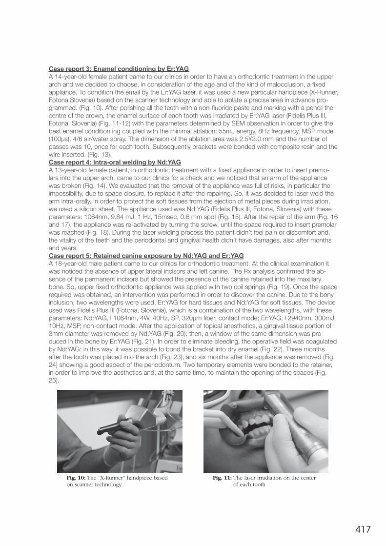
417
Case report 3: Enamel conditioning by Er:YAGA 14-year-old female patient came to our clinics in order to have an orthodontic treatment in the upperarch and we decided to choose, in consideration of the age and of the kind of malocclusion, a fixed appliance. To condition the email by the Er:YAG laser, it was used a new particular handpiece (X-Runner,Fotona,Slovenia) based on the scanner technology and able to ablate a precise area in advance pro-grammed. (Fig. 10). After polishing all the teeth with a non-fluoride paste and marking with a pencil the centre of the crown, the enamel surface of each tooth was irradiated by Er:YAG laser (Fidelis Plus III, Fotona, Slovenia) (Fig. 11-12) with the parameters determined by SEM observation in order to give the best enamel condition ing coupled with the minimal ablation: 55mJ energy, 8Hz frequency, MSP mode (100μs), 4/6 air/water spray. The dimension of the ablation area was 2.5¥3.0 mm and the number of passes was 10, once for each tooth. Subsequently brackets were bonded with composite resin and the wire inserted. (Fig. 13).Case report 4: Intra-oral welding by Nd:YAGA 13-year-old female patient, in orthodontic treatment with a fixed appliance in order to insert premo-lars into the upper arch, came to our clinics for a check and we noticed that an arm of the appliance was broken (Fig. 14). We evaluated that the removal of the appliance was full of risks, in particular the impossibility, due to space closure, to replace it after the repairing. So, it was decided to laser weld the arm intra-orally. In order to protect the soft tissues from the ejection of metal pieces during irradiation, we used a silicon sheet. The appliance used was Nd:YAG (Fidelis Plus III, Fotona, Slovenia) with these parameters: 1064nm, 9.84 mJ, 1 Hz, 15msec, 0.6 mm spot (Fig. 15). After the repair of the arm (Fig. 16 and 17), the appliance was re-activated by turning the screw, until the space required to insert premolar was reached (Fig. 18). During the laser welding process the patient didn’t feel pain or discomfort and, the vitality of the teeth and the periodontal and gingival health didn’t have damages, also after months and years. Case report 5: Retained canine exposure by Nd:YAG and Er:YAGA 18-year-old male patient came to our clinics for orthodontic treatment. At the clinical examination itwas noticed the absence of upper lateral incisors and left canine. The Rx analysis confirmed the ab-sence of the permanent incisors but showed the presence of the canine retained into the maxillary bone. So, upper fixed orthodontic appliance was applied with two coil springs (Fig. 19). Once the space required was obtained, an intervention was performed in order to discover the canine. Due to the bony inclusion, two wavelengths were used, Er:YAG for hard tissues and Nd:YAG for soft tissues. The device used was Fidelis Plus III (Fotona, Slovenia), which is a combination of the two wavelengths, with these parameters: Nd:YAG, l 1064nm, 4W, 40Hz, SP, 320μm fiber, contact mode; Er:YAG, l 2940nm, 300mJ, 10Hz, MSP, non-contact mode. After the application of topical anesthetics, a gingival tissue portion of 3mm diameter was removed by Nd:YAG (Fig. 20); then, a window of the same dimension was pro-duced in the bone by Er:YAG (Fig. 21). In order to eliminate bleeding, the operative field was coagulated by Nd:YAG: in this way, it was possible to bond the bracket into dry enamel (Fig. 22). Three months after the tooth was placed into the arch (Fig. 23), and six months after the appliance was removed (Fig. 24) showing a good aspect of the periodontum. Two temporary elements were bonded to the retainer, in order to improve the aesthetics and, at the same time, to maintain the opening of the spaces (Fig. 25).
118
available at www.jstage.jst.go.jp/browse/islsmORIGINAL ARTICLES
Case report 3: Enamel conditioning by Er:YAG
A 14-year-old female patient came to our clinics inorder to have an orthodontic treatment in the upperarch and we decided to choose, in consideration of theage and of the kind of malocclusion, a fixed appliance.To condition the email by the Er:YAG laser, it wasused a new particular handpiece (X-Runner,
Fotona,Slovenia) based on the scanner technology andable to ablate a precise area in advance programmed.(Fig. 10). After polishing all the teeth with a non-fluo-ride paste and marking with a pencil the centre of thecrown, the enamel surface of each tooth was irradiatedby Er:YAG laser (Fidelis Plus III, Fotona, Slovenia)(Fig. 11-12) with the parameters determined by SEMobservation in order to give the best enamel condition-
S. Kawata, et al.
Fig. 10: The “X-Runner” handpiece based on scanner technology
Fig. 11: The laser irradiation on the center of each tooth
Fig. 12: The laser irradiation completed Fig. 13: The brackets bonded and the upper wire inserted.
Fig. 14: The appliance with a broken arm Fig. 15: Nd:YAG laser irradiation

418
118
available at www.jstage.jst.go.jp/browse/islsmORIGINAL ARTICLES
Case report 3: Enamel conditioning by Er:YAG
A 14-year-old female patient came to our clinics inorder to have an orthodontic treatment in the upperarch and we decided to choose, in consideration of theage and of the kind of malocclusion, a fixed appliance.To condition the email by the Er:YAG laser, it wasused a new particular handpiece (X-Runner,
Fotona,Slovenia) based on the scanner technology andable to ablate a precise area in advance programmed.(Fig. 10). After polishing all the teeth with a non-fluo-ride paste and marking with a pencil the centre of thecrown, the enamel surface of each tooth was irradiatedby Er:YAG laser (Fidelis Plus III, Fotona, Slovenia)(Fig. 11-12) with the parameters determined by SEMobservation in order to give the best enamel condition-
S. Kawata, et al.
Fig. 10: The “X-Runner” handpiece based on scanner technology
Fig. 11: The laser irradiation on the center of each tooth
Fig. 12: The laser irradiation completed Fig. 13: The brackets bonded and the upper wire inserted.
Fig. 14: The appliance with a broken arm Fig. 15: Nd:YAG laser irradiation
119
available at www.jstage.jst.go.jp/browse/islsm ORIGINAL ARTICLES
ing coupled with the minimal ablation: 55mJ energy,8Hz frequency, MSP mode (100µs), 4/6 air/water spray.The dimension of the ablation area was 2.5¥3.0 mmand the number of passes was 10, once for each tooth.Subsequently brackets were bonded with compositeresin and the wire inserted. (Fig. 13).
Case report 4: Intra-oral welding by Nd:YAG
A 13-year-old female patient, in orthodontic treatmentwith a fixed appliance in order to insert premolars intothe upper arch, came to our clinics for a check and wenoticed that an arm of the appliance was broken (Fig.14). We evaluated that the removal of the appliancewas full of risks, in particular the impossibility, due tospace closure, to replace it after the repairing. So, itwas decided to laser weld the arm intra-orally. In orderto protect the soft tissues from the ejection of metalpieces during irradiation, we used a silicon sheet. Theappliance used was Nd:YAG (Fidelis Plus III, Fotona,Slovenia) with these parameters: 1064nm, 9.84 mJ, 1Hz, 15msec, 0.6 mm spot (Fig. 15). After the repair of the arm (Fig. 16 and 17), the
appliance was re-activated by turning the screw, untilthe space required to insert premolar was reached(Fig. 18). During the laser welding process the patient did-n’t feel pain or discomfort and, the vitality of the teethand the periodontal and gingival health didn’t havedamages, also after months and years.
Case report 5: Retained canine exposure byNd:YAG and Er:YAG
A 18-year-old male patient came to our clinics fororthodontic treatment. At the clinical examination itwas noticed the absence of upper lateral incisors andleft canine. The Rx analysis confirmed the absence ofthe permanent incisors but showed the presence of thecanine retained into the maxillary bone. So, upperfixed orthodontic appliance was applied with two coilsprings (Fig. 19). Once the space required wasobtained, an intervention was performed in order todiscover the canine. Due to the bony inclusion, twowavelengths were used, Er:YAG for hard tissues andNd:YAG for soft tissues. The device used was Fidelis
Lasers and Orthodontics
Fig. 16: The arm after laser welding Fig. 17: Particular of the welded arm
Fig. 18: The appliance after activation of the screw Fig. 19: Appliance applied with two coil springs activated.

419
120
available at www.jstage.jst.go.jp/browse/islsmORIGINAL ARTICLES
Fornaini C
Plus III (Fotona, Slovenia), which is a combination ofthe two wavelengths, with these parameters: Nd:YAG,l 1064nm, 4W, 40Hz, SP, 320µm fiber, contact mode;Er:YAG, l 2940nm, 300mJ, 10Hz, MSP, non-contactmode. After the application of topical anesthetics, agingival tissue portion of 3mm diameter was removedby Nd:YAG (Fig. 20); then, a window of the samedimension was produced in the bone by Er:YAG (Fig.21). In order to eliminate bleeding, the operative field
was coagulated by Nd:YAG: in this way, it was possi-ble to bond the bracket into dry enamel (Fig. 22). Three months after the tooth was placed into thearch (Fig. 23), and six months after the appliance wasremoved (Fig. 24) showing a good aspect of the peri-odontum. Two temporary elements were bonded tothe retainer, in order to improve the aesthetics and, atthe same time, to maintain the opening of the spaces(Fig. 25).
Fig. 20: Nd:Yag incision of the mucosa Fig. 21: Er:YAG vaporization of the bone
Fig. 22: Bracket bonded to the canine Fig. 23: Canine inserted into upper dental arch
Fig. 24: After debonding Fig. 25: Retainer with two composite lateral incisors bonded
Case report 6: Gingival hypertrophia surgery by DiodeA 14-year-old female patient, at the end of fixed orthodontic treatment, developed a gingival hypertro-phia in the upper arch (Fig. 26) probably related to the fast closure of the spaces associated to a poor oral hygiene due to the bleeding during teeth brushing. Just after the removal of the appliance a topic anesthetics was applied to the gum (Fig. 27) and a gengivectomy was done, associated to the elimina-tion of the interdental papilla (Fig. 28).The appliance used was XD-2 (Fotona, Slovenia), with these parameters: l 808nm, 3W CW, 320μm fiber, contact mode. The intervention had a duration of 375 sec. and the patient didn’t feel any kind of pain (Fig. 29). Five days after the healing process was completed (Fig. 30).

420
DiscussionLaser may be used before the beginning of the orthodontic treatment, during each of its steps and after the removal of the appliances. 4) Before the therapy, its use is related to the oral soft tissues surgery, in particular to normalize anomalies of upper vestibular and lingual frenulum. 5) The advantages of its utilization consist on the possibility of reducing or avoiding the use of anesthetics, especially important in pediatric patients, the bloodless surgical field and reducing postoperative pain. 6)
121
available at www.jstage.jst.go.jp/browse/islsm ORIGINAL ARTICLES
Case report 6: Gingival hypertrophia surgery byDiode
A 14-year-old female patient, at the end of fixed ortho-dontic treatment, developed a gingival hypertrophia inthe upper arch (Fig. 26) probably related to the fastclosure of the spaces associated to a poor oral hygienedue to the bleeding during teeth brushing. Just afterthe removal of the appliance a topic anesthetics wasapplied to the gum (Fig. 27) and a gengivectomy wasdone, associated to the elimination of the interdentalpapilla (Fig. 28). The appliance used was XD-2 (Fotona, Slovenia),with these parameters: l 808nm, 3W CW, 320µm fiber,contact mode. The intervention had a duration of 375sec. and the patient didn’t feel any kind of pain (Fig.29). Five days after the healing process was completed(Fig. 30).
Discussion
Laser may be used before the beginning of the ortho-dontic treatment, during each of its steps and after the
removal of the appliances. 4)
Before the therapy, its use is related to the oralsoft tissues surgery, in particular to normalize anom-alies of upper vestibular and lingual frenulum. 5)
The advantages of its utilization consist on thepossibility of reducing or avoiding the use of anesthet-ics, especially important in pediatric patients, thebloodless surgical field and reducing postoperativepain. 6)
Lasers and Orthodontics
Fig. 26: Aspect of the teeth just before debonding
Fig. 27: Aspect of the teeth just after debonding with topic anesthetics Fig. 28: During intervention
Fig. 29: Just after intervention Fig. 30: Five days after.

421
Moreover, suturing is generally not required and the biostimulating effect enhances the healing processtogether with the antimicrobial properties of laser energy. 7) Upper vestibular frenectomy is indicated, if related to inter-incisive diastema, when it is attached to the papillar gingiva (positive traction test) 8,9) and when the distance between central incisors is larger than 6-8 mm. The intervention, beyond the elimination of frenulum insertion, must also cut the inter-incisive fibers until to reach the periostium, in order to allow the space closure. In some cases, we noticed that if the intervention is done attending to these principles and choosing a correct timing during denture development, it may be sufficient, wit-hout a classical orthodontic treatment and without appliance wearing, to reach a good result. Another condition, without presence of diastema, is often associated to a short upper lip with gummy smile, and also in this case the surgery, associated to functional re-education by speech therapist, may correct the defect. 6) The presence of an abnormal short lingual frenulum, also called “anchyloglossia” or “tongue-tie” is a condition in which the tip of the tongue cannot protrude over the incisors 10) and it may be re-lated to several kinds of malocclusion such as total open bite, caused by interdental lingual interposition, or third class relation with a push on the mandibular arch. 11) A classification of this condition, based on the measure of the distance between frenulum insertion and tongue tip while patient is touching the palate with mouth opened, allows to distinguish four classes of importance. 12) These anomalies have a great importance in the functional re-education of deglutition and phonation and, sometimes, speech therapists send these patients to our clinics because they cannot do the exercises due to the tongue movement limitation. 13) In the cases when surgical intervention is indicated, the use of laser makes the therapy safe, effective and perfect. 14) Recently, several Authors have described a relation between an-chyloglossia and postural diseases and lingual frenulectomy has been proposed to improve physiothe-rapic treatment. 15) In this case, another more advantage of the use of laser is given by the possibilityto mobilize the tongue just after the intervention, due to the absence of suture: it is important to de-crease the probability of relapse. 16) The employment of laser, associated to the orthophosphoric acid etching, to enhance the strength adhesion of composite resins has been proposed by several Authors in conservative dentistry and also for bracket bonding in orthodontics. 17) The most used wavelengths are Er:YAG and Er,Cr:YSGG but also 214, 810 and 1064nm have been described.The advantage, by using a plastic template or the new “scanner handpiece” (X-Runner, Fotona,Slovenia), is also to prepare a very small surface of enamel, exactly of the same dimension of the bracket. Several studies, based both on traction and microleackage tests, showed the best values wereobtained with the samples irradiated by Er:YAG beam before the acid etching. 18) Moreover, other Au-thors underlined the result, by using laser to prepare enamel surface, to make this more resistant to the decay 19); the reason consists on the modification of the hydroxyapatite crystals and it is very important in the prevention of the decalcification zones around brackets, particularly in patients with a scanty oral hygiene. 20) Some effects of the coherent light, described for the first time by Mester in 1967 and today called “LLLT” consist in biostimulation and pain reduction in the irradiated area. These are defined as “photochemical effects” and are produced with Energy Densities up to 10J/cm2. In orthodontics the use of softlaser has been described in order to reduce the pain which is often present in the first weeks of treatment 21) and also to increase the speed of dental movement, so reducing the treatment time. 22) Generally, the wavelengths used for this kind of laser irradiation are in the visible portion (around 600nm) and infrared (from 800 to 1000nm), sometimes also associated, produced by diode devices.Recently, it was described the possibility to employ the Nd:YAG fibroptic delivered laser, normallyused in dental office for soft tissues surgery, to weld the metallic parts of broken appliances. 23) The ad-vantages of this technique are consist in the time reduction, the avoid of the impression and the mainte-nance of the integrity of acrylic portions, even close to welded area. By this device it was also showed it is possible to weld intra-orally the appliances without the necessity to remove them from the mouth. 24)The inclusion of one or more permanent teeth is a frequent pathology in clinical practice and, after thethird molar, the canine is the most element interested (between 0.92 and 4.3%), being palatal in 54%,vestibular in 32% and median in 12% . 25) The orthodontic-surgery combined treatment allows, in most of the cases, to replace the tooth into the arch without damages and, particularly by the periodontal point of view, the success seems to be related to the possibility to make a minimal radical surgery. 26)

422
The advantages of the laser utilization, beyond this aspect, regard the good pain control and the in-crease of the bracket adhesion strength even in the case of the bonding just after intervention, due to the dry enamel surface for the bleeding absence. When the inclusion is only mucous, all the wavelen-gths normally employed in dentistry may be utilized, except for the CO2 and the Erbium family laserswhich, due to the affinity for the water, might damage the enamel of the tooth. In the case of bony re-tention, two lasers must be used, one of the erbium family, for bone cutting, and another well absorbed by haemoglobine, to produce coagulation. Several orthodontists today use, for aesthetic reasons,porcelain brackets instead of metallic ones and this may represent a problem when, at the end of thetreatment, appliances must be removed. Laser may be useful in this step, because the energy by it emitted is able to soften the adhesive resin 27), so preventing the risk of the bracket fracture, ranging from 10 to 35%. Some “in vitro” studies, recording temperature by IR camera, demonstrated that the safer wavelengths is diode but also others had been proposed. 28) One of the problems related to orthodontic fixed appliances wearing, is represented by gingival overgrowth, in particular when spaces are fast closed in patients who don’t attend a good oral hygiene. Some studies described the presence of hyperplastic gingivitis after two months from the treatment beginning and for the whole duration of it 29), others evidenced the worse of the OH index 30), others proposed the removing of the gingival pa-pilla in the closure areas to favorite the new formation of normal connective tissue 31). The intervention of papillectomy may be done just after the removal of the appliance, it don’t require anesthetic injection and can be performed by every wavelength even if, for Erbium lasers and CO2 it is necessary to protect the teeth surfaces which may be damaged by the beam. Two-three days after the intervention the as-pect of the gum is almost in the normality and seven days after healing process is totally completed.32)
ConclusionLaser technology represents an important help which may be used before, after and during all the steps of orthodontic treatment. It may improve the success of the therapy, diminish the discomfort of the patients, increase their cooperation, reduce the duration of the treatment and the pain produced by the devices.
References1: Frame JW, Morgan D, Rhys Evans PH. Tongue resection with the CO2 laser: the effects of pastradiotherapy on postoperative complications. Br J Oral Maxillofac Surg. 1988 Dec;26(6):464-712: Keller U, Hibst R. [Ablative effect of an Er:YAG laser on enamel and dentin]. Dtsch Zahnarztl Z. 1989 Aug;44(8):600-23: Fornaini C, Riceputi D, Lupi-Pegurier L, Rocca JP. Patient responses to Er:YAG laser when used forconservative dentistry. Lasers Med Sci. 2011 Oct 264: Genovese MD, Olivi G. Use of laser technology in orthodontics: hard and soft tissue laser treatments.Eur J Paediatr Dent. 2010 Mar;11(1):44-85: Kravitz ND, Kusnoto B. Soft-tissue lasers in orthodontics: an overview. Am J Orthod DentofacialOrthop. 2008 Apr;133(4 Suppl):S110-46: Fornaini C, Rocca JP, Bertrand MF, Merigo E, Nammour S, Vescovi P. Nd:YAG and diode laser inthe surgical management of soft tissues related to orthodontic treatment. Photomed Laser Surg. 2007Oct;25(5):381-927: Gracco A, Tracey S, Lombardo L, Siciliani G. Soft tissue laser in orthodontics. Prog Orthod.2011;12(1):66-72.8: Edwards J. GB. The diastema, the frenum, the frenectomy: a clinical study. Am J Orthod. 1977May, 71(5):489-5089: Antoni R, De Angelis D, Gravina GM, Accivile E. The superior median frenulum. Surgical-orthodontictreatment of a recurrence. Clin Ter. 1989 Jul 31;130(2):95-10010: Hall DM, Renfrew MJ. Tongue tie. Arch Dis Child. 2005 Dec;90(12):1211-5.11: Dahan J. Ankyloglossia and its effect on the development of anomalies in the position of the teethand jaws. Diagnosis and therapy. Rev Fr Odontostomatol. 1969 Feb;16(2):213-22

423
12: Kotlow LA. Ankyloglossia (tongue-tie): a diagnostic and treatment quandary. Quintessence Int. 1999Apr;30(4):259-6213: Chaubal TV, Dixit MB. Ankyloglossia and its management. J Indian Soc Periodontol. 2011 Jul;15(3):270-214: Fiorotti RC, Bertolini MM, Nicola JH, Nicola EM. Early lingual frenectomy assisted by CO2 laser helps prevention and treatment of functional alterations caused by ankyloglossia. Int J Orofacial Myology. 2004 Nov;30:64-7115: Pelosi A Interferenze Orali nelle Sindromi Cranio- Mandibolo-Cervicali e Posturali. Castello Edi-tore, 200816: Puthussery FJ, Shekar K, Gulati A, Downie IP. Use of carbon dioxide laser in lingual frenectomy. Br JOral Maxillofac Surg. 2011 Oct;49(7):580-1. Epub 2010 Aug 2117: Kim JH, Kwon OW, Kim HI, Kwon YH. Effectiveness of an Er:YAG laser in etching the enamel surface for orthodontic bracket retention. Dent Mater J. 2005 Dec;24(4):596-60218: Bertrand MF, Semez G, Leforestier E, Muller-Bolla M, Nammour S, Rocca JP. Er:YAG laser cavitypreparation and composite resin bonding with a single-component adhesive system: relationshipbetween shear bond strength and microleakage. Lasers Surg Med. 2006 Jul;38(6):615-2319: Hicks MJ, Flaitz CM and Westerman GH, Argon laser irradiation in root surface caries: in vitrostudy examines laser’s effects. J Am Dent Assoc, 1994 (125) :401-40720: Hossain M, Kimura Y, Nakamura Y, Yamada Y, Kinoshita JI, Matsumoto K. A study on acquiredacid resistance of enamel and dentin irradiated by Er,Cr:YSGG laser. J Clin Laser Med Surg. 2001 Jun;19(3):159-6321: Tortamano A, Lenzi DC, Haddad AC, Bottino MC, Dominguez GC, Vigorito JW. Low-level laser therapy for pain caused by placement of the first orthodontic archwire: a randomized clinical trial. Am JOrthod Dentofacial Orthop. 2009 Nov;136(5):662-722: Sousa MV, Scanavini MA, Sannomiya EK, Velasco LG, Angelieri F. Influence of low-level laser on thespeed of orthodontic movement. Photomed Laser Surg. 2011 Mar;29(3):191-6.23: Fornaini C, Bertrand C, Bonanini M, Rocca JP, Nammour S. Welding in the dental office by fiber-delivered laser: a new technique. Photomed Laser Surg. 2009 Jun;27(3):417-2324: Fornaini C, Vescovi P, Merigo E, Rocca JP, Mahler P, Bertrand C, Nammour S. Intraoral metal laserwelding: a case report. Lasers Med Sci. 2010 Mar;25(2):303-725: Crescini A, Nieri M, Buti J, Baccetti T, Pini Prato GP. Orthodontic and periodontal outcomes oftreated impacted maxillary canines. Angle Orthod. 2007 Jul;77(4):571-726: Kohavi D, Becker A, Zilberman Y. Surgical exposure,orthodontic movement, and final tooth position as factors in periodontal breakdown of treatedpalatally impacted canines. Am J Orthod. 1984 Jan;85(1):72-727: Oztoprack MO, Nalbantgil D, Erdem AS, Tozlu M, Arun T. Debonding of ceramic brackets by a newscanning laser method. Am J Orthod Dentofacial Orthop. 2010 Aug;138(2):195-20028: Dostalova T, Jelinkova H, Sulc J, Nemec M, Jelinek M, Fibrich M, Michalik P, Miyagi M, Seydlova M.Ceramic bracket debonding by Tm:YAP laser irradiation. Photomed Laser Surg. 2011 Jul;29(7):477- 84.29: Zachrisson S, Zachrisson BU. Gingival condition associated with orthodontic treatment. Angle Orthod. 1972 Jan;42(1):26-3430: Lampert F, Unterer S. [The effect of various orthodontic treatment technics on the gingiva].

424
DtschZahnarztl Z. 1977 Jan;32(1):2231: Kurol J, Rönnerman A, Heyden G. Long-term gingival conditions after orthodontic closure of extraction sites. Histological and histochemical studies. Eur J Orthod. 1982 May;4(2):87-9232: Gontiya G, Bhatnagar S, Mohandas U, Galgali SR. Laser-assisted gingivectomy in pediatric pa-tients: a novel alternative treatment. J Indian Soc Pedod Prev Dent. 2011 Jul-Sep;29(3):264-9
5.110 Use of laser technology in orthodontics: hard and soft tissue laser treatmentsGenovese MD, Olivi G.
AbstractAIMModern technology has perfected a new instrument that has become almost indispensable in mo-dern dentistry, in accordance with the philosophy of minimally invasive therapy: the laser. The aim of this work is to evaluate the effectiveness and efficacy of laser technology to solve mucogingival pro-blems associated with orthodontic treatment. Some laser wavelengths work both on hard and soft tissues (2780 nm, 2940 nm), other lasers, such as the 810 nm diode, have a very good surgical and haemostatic action on soft tissues and an important analgesic and biostimulating effect that can help the healing of both TMJ painful symptoms as well as the pain following active orthodontic treatment. Several cases connected to orthodontic therapy are presented.
MATERIALS AND METHODSDifferent laser systems (diode laser at 810 nm; Er,Cr:YSGG laser at 2780 nm; Erbium:YAG laser at 2940 nm) were used, both for soft tissue surgery and enamel etching, and for biostimulating effect. These wavelengths were used with different parameters for each case, according to international current studies in view of minimally invasive therapy.
RESULTSThe cases reported showed very quick and good healing of the laser treated tissues. These treat-ments, necessary for the orthodontictherapy or for its completion, become extremely simple, safe and rapid and the orthodontic specialist can perform them himself.
CONCLUSIONThe laser technique is very effective in many operative and surgical procedures during orthodontic therapy. Further studies are however necessary to set the treatment protocols in orthodontic biosti-mulation.
https://www.ncbi.nlm.nih.gov/pubmed/20359282
5.111 Does low-level therapy decrease swelling and pain resulting from orthognathic surgery? Gasperini G, Rodrigues de Siqueira IC, Rezende Costa L – 2014
5.112 Brevet Biolux – 2007
5.113 Dossier clinique et scientifique OrhtoPulse (Biolux) – 2015

425
11. DOULEUR ARTICULATION TEMPORO-MANDIBULAIRE MAXILLOFACIALE
11.1 Efficacy of red and infrared lasers in treatment of temporomandibular disorders – a double-blind, randomized, parallel clinical trial. Pereira TS, Flecha OD, Guimaraes RC, de Oliveira D, Botelho AM, Ramos Gloria JC, Aguiar Tavano KT – 2014
11.2 Evaluation of pain, jaw movements, and psychosocial factors in elderly individuals with temporomandibular disorders under laser phototherapy.Rodrigues JH, Marques MM, Biasotto-Gonzalez DA, Moreira MS, Bussadori SK, Mesquita-Ferrari RA – 2013
11.3 Evaluation of anti-nociceptive and anti-inflammatory activity of low-level laser therapy on temporomandibular joint inflammation in rodents. Barretto SR, de Melo GC, Dos Santos JC, de Oliveira MG, Pereira-Filho RN, Alves AV, Ribeiro MA, Lima-Verde IB, Quintans Junior LJ, de Albuquerque-Junior RL, Bonjardim LR – 2013
11.4 Comparative clinical study of light analgesic effect on temporomandibular disorder (TMD) using red and infrared led therapy.Panhoca VH, de Fatima Zanirato Lizarelli R, Nunez SC, Pizzo RC, Grecco C, Paolillo FR, Bagnato VS – 2013
11.5 The efficacy of low-level laser therapy for the treatment of myogenous temporomandibular joint disorder.Ahrari F, Mafani AS, Ghafouri ZS, Turner J – 2013
11.6 Low level laser therapy as an adjunctive technique in the management of temporomandibular disorders.Da Silva MA, Botelho AL, Turim CV, da Silva AM – 2012
11.7 Evaluation of low-level laser therapy effectiveness on the pain and masticatory performance of patients with myofascial pain. de Moraes Maia ML, Ribeiro MA, Maia LG, Stuginski-Barbosa J, Costa YM, Porporatti AL, Conti PC, Bonjardim LR – 2012
11.8 Effectiveness of physiotherapy and GaAIAs laser in the management of temporomandibular joint disorders.Dostalova T, Hlinkova P, Kasparova M, Rehacek A, Vavrickova L, Navratil L – 2012
11.9 Evaluation of low-level laser therapy in patients with acute and chronic temporomandibular disorders.Salmos-Brito JA, de Menezes RF, Teixeira CE, Gonzaga RK, Rodrigues BH, Braz R, Bessa-Nogueira RV, de Martinez Gerbi ME – 2012
11.10 Efficacy evaluation of low-level laser therapy on temporomandibular disorder.Wang X, Yang Z, Zhang W, Yi X, Liang C, Li X – 2011
11.11 Management of myofascial pain: low-level laser therapy versus occlusal splints.Oz S, Gokcen-Rohling B, Saruhanoglu A, Tuncer EB – 2010
11.12 Measurements of jaw movements and TMJ pain intensity in patients treated with

426
GaAIAs laser.Mazzetto MO, Hotta TH, Pizzo RC – 2010
11.13 Effects of superpulsed low-level laser therapy on temporomandibular joint pain.Marini I, Gatto MR, Bonetti GA – 2010
11.14 Lasertherapy efficacy in temporomandibular disorders: control study.Santos T de S, Piva MR, Ribeiro MH, Antunes AA, Melo AR, Silva ED - 2010
11.15 Wavelength effect in temporomandibular joint pain: a cinical experience.Carvalho CM, de Lacerda JA, Dos Santos Neto FP, Cangussu MC, Marques AM, Pinheiro AL – 2009
11.16 Low intensity laser therapy in temporomandibular disorder: a phase II double-blind study.Carrasco TG, Mazzetto MO, Mazzetto RG, Mestriner W Jr – 2008
11.17 Effectiveness of low-level laser therapy in temporomandibular joint disorders: a placebo-controlled study.Fikackova H, Dostalova T, Navratil L, Klaschka J – 2007
11.18 Low intensity laser application in temporomandibular disorders: a phase I double-blind study.Mazzeto MO, Carrasco TG, Bodinelo EF, de Andrade Pizzo RC, Mazzetto RG – 2007
11.19 Evaluation of low-level laser therapy in the treatment of temporomandibular disorders. Cetiner S, Kahraman SA, Yucetas S – 2006
11.20 Arthralgia of the temporomandibular joint and low-level laser therapy.Fikackova H, Dostalova T, Vosicka R, Peterova V, Navratil L, Lesak J – 2006
11.21 Management of mouth opening in patients with temporomandibular disorders through low-level laser therapy and transcutaneous electrical neural stimulation.Nunez SC, Garcez AS, Suzuki SS, Ribeiro MS – 2006
11.22 Laser application effects on the bite strength of the masseter muscle, as an orofacial paintreatment.De Medeiros JS, Vieira GF, Nishimura PY – 2005
11.23 A systematic review of low level laser therapy with location- specific doses.Bjordal JM, Couppe C, Chow RT, Tuner J, Ljunggren EA – 2003
11.24 Low-level laser therapy is an important tool to treat disorders of the maxillofacial region.Pinheiro AL, Cavalcanti ET, Pinheiro TI, Alves MJ, Miranda ER, De Quevedo AS, Manzi CT, Vieira AL – 1998
11.25 Treatment of persistent idiopathic facial pain (PIFP) with a low-level energy diode laser.Yang HW, Huang YF – 2011
11.26 Can low reactive-level laser therapy be used in the treatment of neurogenic facial pain? A double-blind, placebo controlled investigation of patients with terminal neuralgia.

427
Arne Eckerdal, H Lehmann Bastian – 1996
11.27 Laser therapy for pain of trigeminal neuralgia.J.B. Walker
12. DENSITÉ OSSEUSE PRÉIMPLANTAIRE
12.1 Combined New Technologies to Improve Dental Implant Success -- Quantitative Ultra-sound Evaluation of NIR-LED PhotobiomodulationJerry Bouquot, Peter Brawn
Background Dental implants must be placed in healthy bone for successful osteointegration and stability. Low bone density (LBD) and ischemically damaged, desiccated bone both have a poor abi-lity to remodel and are, therefore, contraindications for implants. Readily available diagnostic imaging devices, including dental radiographs, lack the ability to adequately identify such bone. However, the new technology of through-transmission or quantitative ultrasound (QUS) is specifically cleared by the FDA to safely identify LBD and dehydrated bone and has a very low (<3%) false positive rate.Near-infrared light emitting diode (NIR-LED) therapy or photobiomodulation has been shown in cultured cells and animal models to stimulate bone healing and production. The present investigation uses QUS to determine the efficacy of in-vivo NIR-LED phototherapy to increase bone density and/or hydration of abnormal alveolar bone.
Methods 68 patients received LED therapy (OsseoPulse, version 1.0, Biolux Research Ltd.,Vancouver, Canada; 15 minutes daily for 3 months) to 294 QUS positive edentulous alveolar sites of LBD/desiccation. Before and after QUS scans were graded blindly by two independent observers (5-point scale: 0 = normal bone, 4 = most severe), after calibration, and compared using matched pair analysis.
Results After NID-LED photomodulation the average grade improved from 2.43 to 1.33(44.3% improvement), with 42% of sites returning to completely normal bone and 18.4%returning to grade 1. The mean difference (improvement of bone quality) of -1.11 was verystatistically significant (matched pair analysis: Std error 0.06914; t-Ratio -15.9896; DF 293;prob less than 0.0001; 95% confidence interval 0.558-1.242).
Conclusion NID-LED therapy seems to hold good potential for improving alveolar bone priorto implant placement, but long-term improvement must be evaluated, as must actual implantstability.
University of Texas Dental Branch at Houston, Houston, Texas; private practice, Nanaimo, British Columbia, Canada
12.2 A histologic comparison of light emitting diode phototherapy-treated hydroxyapaitegrafted extraction sockets.Brawn P, Kwong-Hing A – 2007 16(2):204-11
Case Study After bilateral extraction of periodontally involved lower molars an investigationalOsseoPulseTM was used daily for 21 days on the treated side after grafting both sockets withHydroxyapatite (HA) Osteograf LD300. Bone regeneration of the OsseoPulseTM treated and nontreated socket graft was compared. Histologic evaluations showed enhanced bone formation and faster particle resorption associated with the OsseoPulseTM treated socket graft compared with

428
the untreated socket.
Conclusion In this bilateral case study the accelerated bone healing in the OsseoPulseTM treated HA socket graft may provide faster implant placement compared to untreated treated socket grafts.
13. IMPLANTOLOGIE
13.1 Accelerated implant stability after LED photobiomodulation.P. Brawn, A. Kwong-Hing, S. Boeriu, CM Clokie – 2008
13.2 Low-level laser therapy for implants without initial stability.Campanha BP, Galina C. Geremia T, Drumond Loro RC, Valiati R, Hubler R, Gerhardt de Oliveira M – 2009Photomedicine and Laser Surgery 2009 00(00):1-5
Objective This study evaluated the effect of low-level infrared laser on removal torque values of implants with poor initial stability inserted in rabbit tibias.
Background Data It is important to nalyse the effects of laser radiation on bone repair when low-quality bone and implants with poor initial stability are used.
Materials and Methods Thirty male white New Zealand rabbits (Oryctolagus Cuniculus) about2mo old and weighing 1.5–2.0kg were used. Machined implants with poor initial stability were inserted in the tibia of each animal. Animals were randomly divided into two groups: laser irradiated and laser nonirradiated. Each group was further divided into three subgroups, according to the day the animals were killed: 15, 30, or 45d. Torque values were measured with an axial digital torquemeter that applied counter-torque. The Student’s t-test was used to calculate means and standard deviations for the comparisons between laser and control groups.
Results A significant increase (p=0.050) in removal torque values was found in the group of laser-

429
irradiated implants at 15 and 30d when compared with the control groups. At 45d, no significant differences were found.
Conclusion In this study, low-level laser therapy promoted the osseointegration of implants with poor initial stability, particularly in the initial stages of bone healing.
13.3 Combined technologies to improve dental implant success –quantitative ultrasound evaluation of NIR-LED photobiomodulation.Jerry Bouquot, Peter Brawn – 2008
13.4 Determining optimal dose of laser therapy for attachment and proliferation of human oral fibroblasts cultured on titanium implant material.Maawan Khadra, Ståle P. Lyngstadaas, Hans R. Haanæq, Kamal Mustafa – 2004
13.5 OsseoPulse dossier clinique et scientifique – 2011

430
13.6 OsseoPulse Science Technology Presentation
14. PULPE VITALE DES DENTS PRIMAIRES
14.1 Clinical and radiographic outcomes of the use of low-level laser therapy in vital pulp of primary teeth.Ana Paula Fernandes, Natalino Lourenço Neto, Nádia Carolina Teixeira Marques, Ana Beatriz Silveira Moretti, Vivien Thiemy Sakai, Thiago Cruvinel Silva, Maria AP Aparecida Andrea De Moreira Machado,Thais Marchini Oliveira – 2015

431
15. REPARATION DE LA STRUCTURE DENTAIRE
15.1 Effets de la thérapie LLLT sur l’ultrastructure de l’interface de la pâte dentaire après la préparation des cavités de classe I.Godoy BM, Arana-Chavez VE, Núñez SC, Ribeiro MS – 2007
D. Recherche sur les mécanismes d’action du LLLT (in vivo et in vitro)
1. Etude in vitro
1.1 Low level laser irradiation precondition to create friendly milieu of infracted myocardium and enhance early survival of transplanted bone marrow cells.Zhang H, Hou JF, Wang W, Wei YJ, Hu S – 2009 J of Cell and Mol Med 2009 Sep 1.
Abstract We hypothesized that low-level laser irradiation (LLLI) precondition prior to cell transplantation might remodel the hostile milieu of infracted myocardium and subsequently enhance early survival and therapeutic potential of implanted bone-marrow mesenchymal stem cells (BMSCs). Therefore, in this study we wanted to address: (1) whether LLLI pretreatment change the local cardiac micro-environment after MI; and (2) whether the LLLI preconditions enhance early cell survival and thus improve therapeutic angiogenesis and heart function.Myocardial infarction was induced by left anterior descending artery ligation in female rats. A 635 nm, 5 mW diode laser was performed with energy density of 0.96 J/cm(2) for 150 seconds for the purpose of myocardial precondition. Three weeks later, qualified rats were randomly received with LLLI precondition (n=26) or without LLLI precondition (n=27) for LLLI precondition study. Rats received thoracotomy without coronary ligation was served as sham group (n=24). For the following cell survival study, rats were randomly received serum-free culture media injection (n=8), LLLI precondition and culture media injection (n=8), 2 millions male BMSCs transplantation without LLLI pretreatment (n=26) and 2 millions male BMSCs transplantation with LLLI precondition (n=25). Vascular endothelial growth factor (VEGF), glucose-regulated protein 78 (GRP78), superoxide dismutase (SOD) and malondialdehyde (MDA) in the infarcted myocardium were evaluated by Western blotting, real-time polymerase chain reaction (real-time PCR) and colorimetry, respectively, at 1 hour, 1 day and 1 week after laser irradiation. Cell survival was assayed with quantitative real-time PCR to identify Y chromosome gene and apoptosis was assayed with TUNEL staining. Capillary density, myogenic differentiation and left ventricular function were tested by immunohistochemistry and echocardiography, respectively, at 1 week. After LLLI precondition, increased VEGF andGRP78 expression, as well as the enhanced SOD activity and inhibited MDA production, was observed. Compared with BMSCs transplantation and culture media injection group, although there was no difference in the improved heart function and myogenic differentiation, LLLI precondition significantly enhanced early cell survival rate by 2-fold, decreased the apoptotic percentage of implanted BMSCs in infarcted myocardium and thus increased the number of newly formed capillaries. Taking together, LLLI precondition could be a novel non-invasive approach for intraoperative cell transplantation to enhance cell early survival and therapeutic potential.

432
1.2 Effect of low-level laser therapy on typical oral microbial biofilms.Fernanda G. Basso, Camila F. Oliveira, Amanda Fontana, Cristina Kurachi, Vanderlei S. Bagnato, Denise M.P. Spolidório, Josimeri Hebling, Carlos A. De Souza Costa – 2011
1.3 The effects of low-level laser irradiation on osteoblastic cells.Coombe AR, Ho CT, Darendeliler MA, Hunter N, Philips JR, Chapple CC, Yum LW – Clin Orthod Res. 2001 Feb;4(1):3-14.
Abstract Low level laser therapy has been used in treating many conditions with reports of multiple clinical effects including promotion of healing of both hard and soft tissue lesions. Low level laser therapy as a treatment modality remains controversial, however. The effects of wavelength, beam type, energy output, energy level, energy intensity, and exposure regime of low level laser therapy remain unexplained. Moreover, no specific therapeutic window for dosimetry and mechanism of action has been determined at the level of individual cell types. The aim of this study was to investigate the effects of low level laser irradiation on the human osteosarcoma cell line, SAOS-2. The cells were irradiated as a single or daily dose for up to 10 days with a GaAlAs continuous wave diode laser (830 nm, net output of 90 mW, energy levels of 0.3, 0.5, 1, 2, and 4 Joules). Cell viability was not affected by laser irradiation, with the viability being greater than 90% for all experimental groups. Cellular proliferation or activation was not found to be significantly affected by any of the energy levels and varying exposure regimes investigated. Low level laser irradiation did result in a heat shock response at an energy level of 2 J. No significant early or late effects of laser irradiation on protein expression and alkaline phosphatase activity were found. Investigation of intracellular calcium concentration revealed a tendency of a transient positive change after irradiation. Low level laser irradiation was unable to stimulate the osteosarcoma cells utilized for this research at agross cell population level. The heat shock response and increased intracellular calcium indicate that the cells do respond to low level laser irradiation. Further research is required, utilizing different cell and animal models, to more specifically determine the effects of low level laser irradiation at a cellular level. These effects should be more thoroughly investigated before low level laser therapy can be considered as a potential accelerator stimulus for orthodontic tooth movement.
1.4 Phototherapy promotes attachment and subsequent proliferation of human osteoblast-like cells.M. Khadran N. Kasem, P. Brawn

433
2. Etude In Vivo
2.1 Resonance frequency analysis of orthodontics miniscrews subjected to light-emitting diode photobiomodulation.Tancan Uysal, Abdullah Ekizer, Huseyin Akcay, Osman Etoz, Enis Guray – 2010
3. Etudes Raphaël
3.1 Recherche bibliographique n°9 – Bénéfices de l’ATP38

434
4. Osteoblastes Humaines
4.1 Phototherapy promotes attachement and subsequent proliferation of human osteoblast-like cells M. Khadra, N. Kasemn, P. Brawn
5. Cicatrisation Osseuse
5.1 Increase of bone volume by a nanosecond pulsed laser irradiation is caused by a decreased osteoclast number and an activated osteoblasts.Tadashi Ninomiya, Akihiro Hosoya, Hiroaki Nakamura, Kazuo Sano, Tsuyoshi Nishisaka, Hidehiro Ozawa – 2007

435
5.2 Effect of low-level laser therapy on proliferation and differentiation of the cells contributing in bone regeneration. Reza Amid, Mahdi Kadkhodazadeh, Mitra Ghazizadeh Ahsaie, Arian Hakakzadeh – 2014
5.3 Enhancement of bone formation in rat calcarial bone defects using low-level laser therapy.Maawan Khadra, Nesrin Kasem, Hans R. Haanæs, Jan Ellingsen, Ståle P. Lyngstadaas – 2004
5.4 Une série de cas de 589 extractions dentaires chez les patients sous traitement de bisphosphonates. Proposition d’un protocole clinique soutenu par la thérapie LLLT Vescovi P, Meleti M, Merigo E, Manfredi M, Fornaini C, Guidotti R, Nanmour S – 2013

436
ConclusionNotre expérience soutient l’hypothèse que l’association d’un traitement antibiotique et la thérapie LLLT peut être efficace dans la prévention de l’ostéoporose suite à des extractions dentaires chez les patients sous bisphosphonate.
RéférenceMed Oral Patol Oral Cir Bucal. 2013 Jul 1; 18(4):e680-5.Case series of 589 tooth extractions in patients under bisphosphonates therapy. Proposal of a clinical protocol supported by Nd: YAG low-level laser therapy.Vescovi P1, Meleti M, Merigo E, Manfredi M, Fornaini C, Guidotti R, Nammour S.
Author information1Department of Biomedical, Biotechnological and Translational Sciences – S.Bi.Bi.T, University of Parma, Italy. [email protected]
5.5 Etude sur le contrôle de la douleur chez les patients atteints d’ostéonécrose induite par bisphosphonate en utilisant la thérapie LLLT : résultats préliminaires.Romeo U, Galanakis A, Marias C, Vecchio AD, Tenore G, Palaia G, Vescovi P, Polimeni A – 2010
ConclusionCette étude pilote suggère que la thérapie LLLT peut être une technique valable pour soutenir le traitement de la douleur chez les patients atteints d’ostéonécrose induite par bisphosphonate.
RéférencePhotomed Laser Surg. 2011 Jul;29(7):447-52. doi: 10.1089/pho.2010.2835. Epub 2011 Jan 16.Observation of pain control in patients with bisphosphonate-induced osteonecrosis using low level laser therapy: preliminary results.Romeo U1, Galanakis A, Marias C, Vecchio AD, Tenore G, Palaia G, Vescovi P, Polimeni A.
Author information1Department of Oral Sciences, «Sapienza» University of Rome, Italy. [email protected]
5.6 Effet de la thérapie LLLT sur l’ostéonécrose des mâchoires induite par bisphosphonate : résultats préliminaires d’une étude prospective.Scoletta M, Arduino PG, Reggio L, Dalmasso P, Mozzati M – 2010
ConclusionCette étude suggère que la thérapie LLLT semble être une modalité prometteuse du traitement pour les patients atteints d’ostéonécrose suite au traitement avec du bisphosphonate, à condition que l’efficacité clinique est sûr et bien toléré, en particulier par les patients qui ont besoin d’un traitement conservateur.
RéférencePhotomed Laser Surg. 2010 Apr;28(2):179-84. doi: 10.1089/pho.2009.2501.Effect of low-level laser irradiation on bisphosphonate-induced osteonecrosis of the jaws: preliminary results of a prospective study.Scoletta M1, Arduino PG, Reggio L, Dalmasso P, Mozzati M.

437
Author information1Oral Surgery Unit, Dentistry Section, Department of Clinical Physiopathology, University of Turin, Turin, Italy.
5.7 Evaluation grâce à la spectroscopie Raman proche infrarouge (NIRS), l’incorporation d’hydroxyapatite de calcium (CHA ; environ 960 cm) sur la cicatrisation osseuse autour des implants dentaires soumis ou non à la thérapie LLLT.Lopes CB, Pinheiro AL, Sathaiah S, Duarte J, Cristinamartins M – 2005
ConclusionIl est conclu que la thérapie LLLT améliore la guérison osseuse, et cela peut être évalué en toute sécurité par spectroscopie Raman.
RéférencePhotomed Laser Surg. 2005 Feb; 23(1):27-31.Infrared laser light reduces loading time of dental implants: a Raman spectroscopic study.Lopes CB1, Pinheiro AL, Sathaiah S, Duarte J, Cristinamartins M.
Author information1IP&D and Department of Dentistry, FCS, UNIVAP, S. J. Campos, São Paulo, Brazil.
5.10 Efficacy of laser therapy in the management of bisphosphonate related osteonecrosis of the jaw (BRONJ) : a systematic reviewJB. Weber, RS. Camilotti, ME. Ponte

Biotech Dental
305, Al lées de Craponne13300 Salon-de-Provence - France
Tel. : +33 (0)4 90 44 60 60Fax : +33 (0)4 90 44 60 [email protected]
Fabriqué par : Swiss Bio Inov - Distribué par : Biotech Dental. Dispositif médical de classe IIa. Technologie pour applications médicales : antalgique, anti-inflammatoire, cicatrisation. CE0459.
Doit être utilisé par des professionnels de santé qualifiés et formés. Des lunettes de protection doivent obligatoirement être portées par le praticien et le patient. Contre-indiqué aux patients porteurs d’un pacemaker, aux patients épileptiques, aux femmes enceintes et sur une population pré-pubère.
Visuels non contractuels. Non remboursé par la Sécurité Sociale. Lire attentivement les instructions figurants dans la notice.Biotech Dental - S.A.S au capital de 24 866 417 € - RCS Salon de Provence : 795 001 304 - SIRET : 795 001 304 00018 - N° TVA : FR 31 79 500 13 04.
Ne pas jeter sur la voie publique. Imprimerie VALLIERE - 163, Avenue du Luxembourg - ZAC des Molières - 13140 Miramas - France.
Copyright © 2022 FDOKUMEN