Bone morphogenic protein antagonist Drm/gremlin is a novel proangiogenic factor
28
doi:10.1182/blood-2006-06-032276 Prepublished online October 31, 2006; Franco Cotelli, Domenico Ribatti and Marco Presta Francesco Peri, Antonello Pessi, Laura Orsatti, Fabio Talamo, Vincent Castronovo, David Waltregny, Helena Stabile, Stefania Mitola, Emanuela Moroni, Mirella Belleri, Stefania Nicoli, Daniela Coltrini, pro-angiogenic factor The bone morphogenic protein antagonist Drm/gremlin is a novel (2494 articles) Hemostasis, Thrombosis, and Vascular Biology (2252 articles) Free Research Articles (564 articles) Chemokines, Cytokines, and Interleukins Articles on similar topics can be found in the following Blood collections http://bloodjournal.hematologylibrary.org/site/misc/rights.xhtml#repub_requests Information about reproducing this article in parts or in its entirety may be found online at: http://bloodjournal.hematologylibrary.org/site/misc/rights.xhtml#reprints Information about ordering reprints may be found online at: http://bloodjournal.hematologylibrary.org/site/subscriptions/index.xhtml Information about subscriptions and ASH membership may be found online at: digital object identifier (DOIs) and date of initial publication. the indexed by PubMed from initial publication. Citations to Advance online articles must include final publication). Advance online articles are citable and establish publication priority; they are appeared in the paper journal (edited, typeset versions may be posted when available prior to Advance online articles have been peer reviewed and accepted for publication but have not yet Copyright 2011 by The American Society of Hematology; all rights reserved. 20036. the American Society of Hematology, 2021 L St, NW, Suite 900, Washington DC Blood (print ISSN 0006-4971, online ISSN 1528-0020), is published weekly by For personal use only. by guest on March 10, 2014. bloodjournal.hematologylibrary.org From For personal use only. by guest on March 10, 2014. bloodjournal.hematologylibrary.org From
Transcript of Bone morphogenic protein antagonist Drm/gremlin is a novel proangiogenic factor

doi:10.1182/blood-2006-06-032276Prepublished online October 31, 2006;
Franco Cotelli, Domenico Ribatti and Marco PrestaFrancesco Peri, Antonello Pessi, Laura Orsatti, Fabio Talamo, Vincent Castronovo, David Waltregny, Helena Stabile, Stefania Mitola, Emanuela Moroni, Mirella Belleri, Stefania Nicoli, Daniela Coltrini, pro-angiogenic factorThe bone morphogenic protein antagonist Drm/gremlin is a novel
(2494 articles)Hemostasis, Thrombosis, and Vascular Biology � (2252 articles)Free Research Articles �
(564 articles)Chemokines, Cytokines, and Interleukins �Articles on similar topics can be found in the following Blood collections
http://bloodjournal.hematologylibrary.org/site/misc/rights.xhtml#repub_requestsInformation about reproducing this article in parts or in its entirety may be found online at:
http://bloodjournal.hematologylibrary.org/site/misc/rights.xhtml#reprintsInformation about ordering reprints may be found online at:
http://bloodjournal.hematologylibrary.org/site/subscriptions/index.xhtmlInformation about subscriptions and ASH membership may be found online at:
digital object identifier (DOIs) and date of initial publication. theindexed by PubMed from initial publication. Citations to Advance online articles must include
final publication). Advance online articles are citable and establish publication priority; they areappeared in the paper journal (edited, typeset versions may be posted when available prior to Advance online articles have been peer reviewed and accepted for publication but have not yet
Copyright 2011 by The American Society of Hematology; all rights reserved.20036.the American Society of Hematology, 2021 L St, NW, Suite 900, Washington DC Blood (print ISSN 0006-4971, online ISSN 1528-0020), is published weekly by
For personal use only. by guest on March 10, 2014. bloodjournal.hematologylibrary.orgFrom For personal use only. by guest on March 10, 2014. bloodjournal.hematologylibrary.orgFrom
1
The bone morphogenic protein antagonist Drm/gremlin is a novel pro-
angiogenic factor
Helena Stabile1, Stefania Mitola1, Emanuela Moroni1, Mirella Belleri1, Stefania Nicoli1, Daniela
Coltrini2, Francesco Peri3, Antonello Pessi4, Laura Orsatti4, Fabio Talamo4, Vincent Castronovo5,
David Waltregny6, Franco Cotelli7, Domenico Ribatti8, and Marco Presta1
1Unit of General Pathology and Immunology and 2Unit of Histology, Department of Biomedical Sciences and Biotechnology, University of Brescia, 25123 Brescia; 3Department of Biotechnology and Biosciences, University of Milano-Bicocca, 20126 Milan; 4Istituto di Ricerche di Biologia Molecolare P. Angeletti, 00040 Pomezia, Rome; 5Metastasis Research Laboratory and 6Department of Pathology, Center of Experimental Cancer Research, University of Liège, Belgium; 7Department of Biology, University of Milano, 20133 Milan; 8Department of Human Anatomy and Histology, University of Bari, 70124 Bari, Italy.
Running title: Drm/gremlin as an angiogenic factor
Correspondence to: Marco Presta General Pathology, Department of Biomedical Sciences and Biotechnology, University of Brescia, Viale Europa 11 25123 Brescia, Italy Email: [email protected].
This work was supported by grants from MIUR (Centro di Eccellenza IDET, FIRB 2001, Cofin
2004), Fondazione Berlucchi, Istituto Superiore Sanità (Progetto Oncotecnologico), and AIRC to
M.P.
Author contributions: H.S., S.M., F.T., and M.P. designed research; H.S., S.M., E.M., M.B., S.N.,
D.C., L.O., V.C., D.W., and D.M. performed research; H.S., S.M., F.P., A.P., F.C., and M.P
analyzed data; and S.M. and M.P. wrote the paper.
Blood First Edition Paper, prepublished online October 31, 2006; DOI 10.1182/blood-2006-06-032276
Copyright © 2006 American Society of Hematology
For personal use only. by guest on March 10, 2014. bloodjournal.hematologylibrary.orgFrom

2
ABSTRACT
Angiogenesis plays a key role in various physiological and pathological conditions, including tumor
growth. Drm/gremlin, a member the Dan family of bone morphogenic protein (BMP) antagonists, is
commonly thought to affect different processes during growth, differentiation, and development by
heterodimerizing various BMPs. Here we identify Drm/gremlin as a novel pro-angiogenic factor
expressed by endothelium. Indeed, Drm/gremlin was purified to homogeneity from the conditioned
medium of transformed endothelial cells using an endothelial cell sprouting assay to follow protein
isolation. Accordingly, recombinant Drm/gremlin stimulates endothelial cell migration and invasion
in fibrin and collagen gels, binds with high-affinity to various endothelial cell types, and triggers
tyrosine phosphorylation of intracellular signaling proteins. Also, Drm/gremlin induces
neovascularization in the chick embryo chorioallantoic membrane. BMP4 does not affect
Drm/gremlin interaction with endothelium and both molecules exert a pro-angiogenic activity in
vitro and in vivo when administered alone or in combination. Finally, Drm/gremlin is produced by
the stroma of human tumor xenografts in nude mice and it is highly expressed in endothelial cells of
human lung tumor vasculature when compared to non-neoplastic lung. Our observations point to a
novel, previously unrecognized capacity of Drm/gremlin to interact directly with target endothelial
cells and to modulate angiogenesis.
For personal use only. by guest on March 10, 2014. bloodjournal.hematologylibrary.orgFrom

3
INTRODUCTION
Gremlin, also known as Drm (Down-regulated by v-mos), belongs to the Dan family of
cysteine knot secreted proteins 1,2. Drm/gremlin exerts a potent bone morphogenic protein (BMP)
antagonist activity by binding BMP2, BMP4, and BMP7 and preventing their interaction with cell
surface receptors 3. This capacity is thought to be responsible for the pattern-inducing activity of
Drm/gremlin during embryonic development 4 and to play a role in human diseases 5. On the other
hand, intracellular, BMP-independent mechanism(s) of action 6 may mediate the ability of
Drm/gremlin to suppress transformation and tumorigenesis under certain experimental conditions 7.
Also, Drm/gremlin interacts with Slit proteins and acts as a negative regulator of monocyte
chemotaxis, thus suggesting a role for this protein in inflammation and immunity 8. Taken together
these observations indicate that Drm/gremlin may exert multiple functions in different physio-
pathological conditions via BMP-dependent and BMP-independent mechanisms of action 1-5.
Nevertheless, the possibility that Drm/gremlin may exert a direct effect on target cells has never
been explored.
Angiogenesis, the process of new blood vessel formation from pre-existing ones, plays a key
role in various physiological and pathological conditions, including inflammation and tumor growth 9. The local, continuous release of angiogenic growth factors and/or alterations of the production of
natural angiogenic inhibitors 10 are responsible for the uncontrolled endothelial cell proliferation
that takes place during tumor neovascularization and in angiogenesis-dependent diseases 11.
Numerous inducers of angiogenesis have been identified, including members of the vascular
endothelial growth factor (VEGF) 12,13 and of the fibroblast growth factor (FGF) families 14. These
angiogenic growth factors induce a complex “pro-angiogenic phenotype” in endothelial cells that
recapitulates several aspects of the in vivo angiogenesis process (summarized in 15).
To elucidate the molecular determinants of endothelial cell activation during angiogenesis,
we originated a stable mouse aortic endothelial (MAE) cell line transfected with a human FGF2
cDNA (FGF2-T-MAE cells) 16,17. Transfectants are characterized by the overexpression of
numerous genes implicated in the modulation of cell growth, differentiation, cell adhesion, and
stress/survival 18. FGF2-T-MAE cells are angiogenic and cause the formation of opportunistic
vascular lesions by recruiting endothelial cells of the host 16,19. Accordingly, FGF2-T-MAE cells
release an endothelial cell motogen that appears to be distinct from other well-characterized
angiogenic growth factors, including FGF2 and VEGF 19.
Here we describe the purification of this factor from the conditioned medium (CM) of
FGF2-T-MAE cells and its identification as the Drm/gremlin protein. Our data demonstrate for the
For personal use only. by guest on March 10, 2014. bloodjournal.hematologylibrary.orgFrom

4
first time that Drm/gremlin plays a BMP-independent role in the angiogenic process by binding
endothelial cell surface, thus activating intracellular signaling and cell motility. Accordingly,
Drm/gremlin induces new vessel growth in the chick embryo chorioallantoic membrane. Also, the
expression of Drm/gremlin in the endothelium of human lung tumor specimens points to a role for
this protein in blood vessel development in human cancers.
MATERIALS AND METHODS
Cell cultures
Immortalized Balb/c MAE cells were obtained from R. Auerbach (University of Wisconsin,
Madison, WI) and grown in Dulbecco’s modified minimal essential medium (DMEM, Gibco Life
Technologies) added with 10% FCS (Integro). FGF2-T-MAE cells were grown in DMEM
supplemented with 4 mM glutamine (Gibco) and 10% FCS. Bovine aortic endothelial cells (BAE
cells) and normal subcutaneous microvascular endothelial cells (SIE cells) 20 (both provided by A.
Vecchi, Istituto Mario Negri, Milan, Italy) were cultured in DMEM supplemented with 10% heat-
inactivated donor calf serum. Human umbilical vein endothelial cells (HUVE cells) were cultured in
EGM-2 medium (Clonetics).
Drm/gremlin purification
Conditioned medium (CM) was prepared by incubating confluent FGF2-T-MAE cell cultures
grown in 10 cm-dishes with 8 mL of serum-free DMEM for 2-3 days. 21 L of CM were precipitated
with 70% of ammonium sulfate. Then, the protein precipitate was dissolved in 40 mL of 25 mM
MES/NaOH (pH 6.5) plus 1.0 mM PMSF and dialyzed against the same buffer. The dialyzed
protein fraction (2 mg/mL) was applied onto a 80 mL Sp-Sepharose Fast Flow column (Amersham
Bioscience) pre-equilibrated with 50 mM MES/NaOH (pH 6.5). The column was extensively
washed with 50 mM MES/NaOH (pH 7.5) and eluted at a flow rate of 30 mL/hour with a linear 0-
1.0 M NaCl gradient at 4°C. The eluate was collected in 10 mL fractions and aliquots from each
fraction were assayed for their ability to stimulate MAE cell sprouting in three-dimensional fibrin
gel 19. The biologically active fractions eluted at 0.8-0.9 M NaCl (see Figure 1b) were pooled
together (10 mg of total protein) and concentrated 10 times with a 30 kDa cut-off ultrafiltration
system (Amicon). Proteins were then loaded onto a 10 mL heparin-Sepharose column equilibrated
in 10 mM Mes/NaOH (pH 7.0) and eluted with a linear 0-1.0 M NaCl gradient at 4°C at a flow rate
of 1.0 mL/minute. The major peak of activity eluted at 1.0 M NaCl (Figure 1c). Pooled fractions,
containing 250-500 µg of total proteins, were concentrated with a 50 kDa cut-off centrifugal
For personal use only. by guest on March 10, 2014. bloodjournal.hematologylibrary.orgFrom

5
concentrator (Centriplus, Millipore) and freeze-dried. Next, the sample was dissolved in 1.0 %
trifluoroacetic acid and loaded onto a 1.0 mL C4 Symmetry 300TM column (Waters) equilibrated
with 0.1% trifluoroacetic acid (1.0 mL/minute) for the initial 30 minutes. Bound material was eluted
at 1.0 mL/minute using a 40 minute linear 0-50% acetonitrile gradient in 0.1% trifluoroacetic acid
(Figure 1d). Collected fractions were freeze-dried, resuspended in 25 mM Mes/NaOH (pH 7.0),
assessed for biological activity and MS identification.
Mass spectrometry
Peptide mixtures from tryptic or CNBr digestions of biologically active HPLC fractions were
desalted and concentrated using C18 Zip Tips™ (Millipore) and analyzed by MALDI-ToF MS
(Voyager DE-sSTR, Applied Biosystems) using a 337 nm-wavelength laser for desorption and the
reflectron mode of analysis. Mass spectra of digested peptides were searched against the FASTA
database held by the National Center for Biotechnology Information (NCBI) using the PROWL
ProFound search engine 21 (available at www.prowl.rockefeller.edu) using an unbiased all taxa
search. For amino acid sequencing, the peptide mixtures were also analyzed by MS/MS using a Q-
q-Tof hybrid system (Q-Star XL, Applied Biosystems) equipped with a nanospray ion source. In
particular, the doubly charged ion of the phosphopeptide (65-86) at m/z 1225.5287 was selected to
sequence and identify the Ser-77 phosphorylation site.
Three-dimensional gel and migration assays
MAE cell aggregates were embedded in fibrin gel 17. Then, culture medium containing the
chromatographic fraction to be tested or murine rDrm (R&D Systems) was added on the top of the
gel in the presence of 10 µg/mL aprotinin to prevent the dissolution of the substrate. Formation of
radially growing cell sprouts was observed during the next 24-72 hours. Sprouts were photographed
at x40 magnification (Olympus IX51 inverted microscope with Camedia C-4040 digital camera)
and quantified by computerized analysis of the digitalized images. Three-dimensional gels of
reconstituted rat tail tendon type I collagen fibrils (Boehringer Mannheim Italia) were prepared as
described 17. Then, BAE cells were seeded on the top of collagen gel (80,000 cells/cm2) and
allowed to reach confluence. Cell cultures were then treated with fresh medium containing rDrm
plus 10% FCS. After 24 hours, cells were photographed at x100 magnification (Olympus IX51
inverted microscope) and endothelial cells invading the gel, in a plane of focus beneath the cell
monolayer surface, were quantified by computerized analysis of the digitalized images. To study
migration, 50,000 MAE cells resuspended in DMEM plus 0.1% heat-inactivated FCS were seeded
in the upper compartment of a Boyden chamber containing a gelatin-coated polycarbonate
For personal use only. by guest on March 10, 2014. bloodjournal.hematologylibrary.orgFrom

6
membrane filter (Nucleopore, Whatman, 8 µm pores). rDrm and/or BMP4 (R&D Systems) were
dissolved in the same medium and placed in the lower chamber. Then, cells were allowed to
migrate for 4 hours at 37°C. Migrated cells at the bottom surface of the filter were stained (Diff-
Quick, DADE Behring) and counted at x 250 magnification (5 fields/sample in triplicate).
125I-Drm binding and cross-linking to endothelial cells.
rDrm (5 µg) was dissolved in 200 µL of PBS, transferred into iodogen-coated tubes (Pierce), and
incubated for 5 minutes at 4°C with 0.2 mCi 125I (Amersham). The reaction products were separated
on a size-exclusion Sephadex-G10 column. For 125I-rDrm binding experiments, SIE, MAE, and
HUVE cells were plated in 24-well dishes at 70,000 cells/cm2. After 24 hours, cells were washed
three times with ice-cold PBS and incubated for 2 hours at 4°C in binding medium (serum free
medium containing 0.15% gelatin, 20 mM Hepes, pH 7.5) with increasing concentrations of 125I-
Drm in the absence or in the presence of 100-fold excess of unlabeled ligand. After a PBS wash,
cells were washed twice with 2.0 M NaCl in 20 mM HEPES buffer (pH 7.5) to elute 125I-rDrm
bound to low-affinity sites. Next, 125I-rDrm bound to high-affinity sites was eluted with 2.0 M NaCl
in 20 mM sodium acetate (pH 4.0) 22. Low-affinity and high-affinity binding data were analysed by
Scatchard plot using the Prism4 software (GraphPad).
For cross-linking experiment, confluent SIE and HUVE cells were incubated for 10 min at room
temperature in binding medium with 5.0 nM 125I-rDrm in the absence or presence of a 100-fold
molar excess of unlabeled rDrm or BMP4. Then, 1.0 mM bis[sulphosuccinamide]suberate (Pierce)
was added. The cross-linking reaction was allowed to proceed at 4°C for 2 hours. After a 2.0 M
NaCl wash in 20 mM HEPES buffer (pH 7.5), cells were lysed and proteins were separated by 6%
SDS-PAGE under reducing conditions. Complexes were visualized by autoradiography of the gel.
In a second set of experiments, 107 confluent SIE cells were incubated under the same experimental
conditions with 5.0 nM unlabeled rDrm. After cross-linking, cells were lysed in lysis buffer [50
mM Tris-HCl buffer (pH 7.4) containing 150 mM NaCl, 1% Triton X-100, and protease and
phosphatase inhibitors (50 µg/mL pepstatin, 50 µg/mL leupeptin, 10 µg/mL aprotinin, 1.0 mM
Na3VO4, all from Sigma)] and the whole sample (1.0 mg of protein) was incubated with anti-Drm
antibody (R&D Systems). Immunocomplexes were precipitated by overnight incubation at 4°C with
Protein G-Sepharose beads and analyzed by Western blotting under reducing conditions using the
same anti-Drm antibody. The lysate of cells in which rDrm incubation was omitted was used as a
negative control.
Intracellular signaling
For personal use only. by guest on March 10, 2014. bloodjournal.hematologylibrary.orgFrom

7
Confluent SIE cells were made quiescent by a 20 hour-starvation in 2% FCS. After stimulation with
rDrm (50 ng/mL), cells were lysed and 20 µg aliquots were analyzed by 6% or 10% SDS-PAGE
followed by Western blotting with antibodies against pFAK, pPaxillin, pERK1/2, or Jak2 (Santa
Cruz). For 2D-PAGE, samples were solubilized in 8.0 M urea, 50 mM DTT, 4% CHAPS, 0.2%
carrier ampholytes (pH 3-10), 0.0002% Bromophenol blue. Proteins were separated on pH 3-10 NL
IPG strips (BioRad) according to the manufacturer's instructions. The second dimension was
performed on 10% SDS-PAGE and proteins were probed with anti-pTyr antibody (Santa Cruz) in a
Western blot. The same antibody was used to decorate rDrm-stimulated, paraformaldehyde-fixed
SIE cells.
Drm/gremlin transfection in COS cells
COS cells were transfected with the pMEXneo expression vector harboring the rat Drm cDNA
(pMEX-DRM) or with the empty vector (both provided by D.G. Blair, NCI-Frederick, MA, USA)
to generate stable transfectants (mock-COS cells and Drm-COS cells, respectively) as described 23.
Chicken embryo chorioallantoic membrane assay
Gelatin sponges containing vehicle, 50 or 100 ng of rDrm or BMP4 or both (n=10-20 eggs/group)
were placed on the CAM of fertilized chicken eggs at day 8 of incubation 24. In a parallel
experiment, mock-COS cells and Drm-COS cells were implanted via a gelatin sponge (18,000 cells
per sponge) on the top of the CAM at day 8 (n=20 eggs per group). At day 12, blood vessels
entering the sponge within the focal plane of the CAM were counted at x50 magnification. When
indicated, CAMs were processed for light microscopy and microvessel density was evaluated by a
planimetric method 24.
Drm/gremlin expression in tumor xenografts
Drm/gremlin transcripts were evaluated by RT-PCR analysis in human endometrial
adenocarcinoma HEC-1-B-derived xenografts 25 (provided by R. Giavazzi, Mario Negri Institute,
Bergamo, Italy) using the following specie-specific primers: murine Drm/gremlin
[(+)CTCAGCACAATGACTCCGAGC; (-)ATCCAAGTCGATGGATATGCAA]; human
Drm/gremlin [(+)GTATGAGCCGCACAGCCTACA; (-)CTCGCTTCAGGTATTTGCGCT]. After
PCR reaction, 5 µL aliquots were separated on a 1.5% agarose gel and visualized by ethidium
bromide staining. Also, paraffin-embedded tumor samples were stained with anti-Drm/gremlin
antibody as detailed below.
For personal use only. by guest on March 10, 2014. bloodjournal.hematologylibrary.orgFrom

8
Human lung tumor collection and immunohistochemistry
Ten samples of formalin-fixed and paraffin-embedded human lung cancers (5 adenocarcinomas and
5 squamous cell carcinomas) were obtained from L. de Leval (Department of Pathology, Liège
University Hospital, Belgium). Five µm paraffin sections were stained with a goat polyclonal anti-
Drm/gremlin antibody (R&D Systems) using the immunoperoxidase ABC Vectastain Kit (Vector
Laboratories, Inc, Burlingame, CA). Antigen retrieval was performed by heating slides in a water-
bath at 95°C for 40 minutes in 10 mM citrate buffer (pH 6.0). After blocking the endogenous
peroxidase activity with 0.3% H2O2 in methanol for 30 minutes, slides were sequentially incubated
with normal horse serum (1:20) for 30 minutes and with the anti-gremlin antibody (1:30) overnight
at 4°C. Slides were then incubated with biotinylated anti-goat antibody (Vector) followed by the
avidin-biotin-peroxidase complex. Hematoxylin counterstained sections were reviewed by two
independent observers and immunostaining intensity in cancer cells and in endothelial cells was
scored as it follows: -, negative; +, weak staining; ++, strong staining.
Data representation
Data are expressed as mean ± S.D. Statistical analyses were done using Student's t-test. The
significance level was set at P < 0.01.
RESULTS AND DISCUSSION
Drm/gremlin purification
Preliminary observations had shown that FGF2-T-MAE cells release an uncharacterized heparin-
binding, heat-stabile, proteinaceous factor responsible for the capacity of their CM to induce
endothelial cell sprouting in a fibrin gel 19. On this basis, we thought to purify this protein
sequentially using ammonium sulphate precipitation, cation exchange chromatography, heparin-
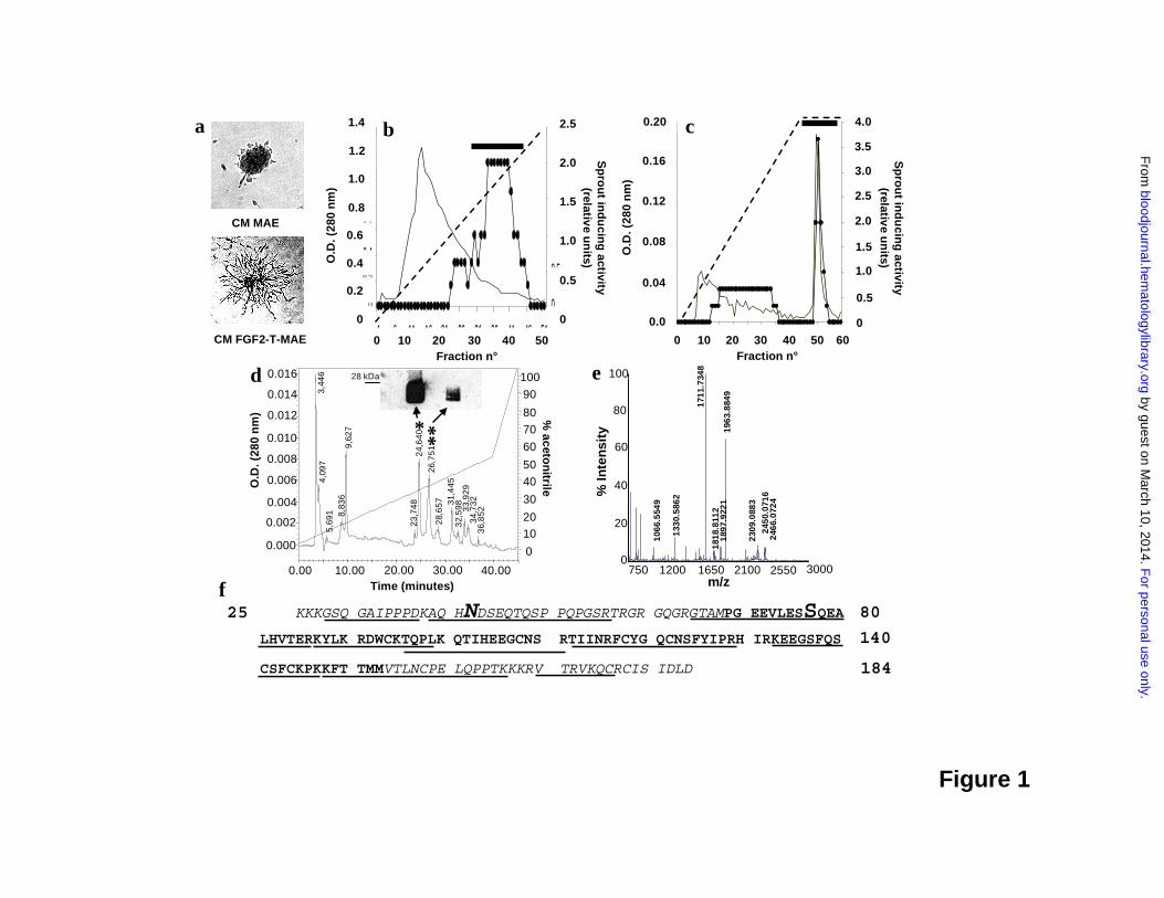
Sepharose affinity chromatography, and reversed-phase HPLC (Figure 1). The bioactivity of eluted
fractions was monitored by stimulation of MAE cell sprouting in three-dimensional fibrin gel (Fig.
1a). Most of the endothelial cell sprouting-inducing activity present in the serum-free CM of FGF2-
T-MAE cells precipitates in 70% ammonium sulphate and binds to a SP-Sepharose cation exchange
column from where it is recovered in the 0.8-0.9 M NaCl eluate (Figure 1b). Subsequent
fractionation by heparin-Sepharose chromatography reveals a major peak of activity eluting at 1.0
M NaCl (Figure 1c). Further purification by reversed-phase HPLC yields two biologically active
peaks (retention time of 24.6 and 26.7 minutes, Figure 1d). Peptide mass fingerprinting analysis of
both peaks by MALDI-ToF mass spectrometry (MS) (Figure 1e) identifies the purified endothelial
For personal use only. by guest on March 10, 2014. bloodjournal.hematologylibrary.orgFrom

9
cell motogen as the murine Drm gene product. The identification was confirmed by nano-ESI-
MS/MS sequencing of the peptides generated by tryptic and CNBr digestion of the purified protein
and by Western blotting of the bioactive HPLC fractions (Figure 1d, inset).
Nano-ESI-MS/MS analysis also shows that purified Drm undergoes post-translational
modifications, including maturation, glycosylation, and phosphorylation, in keeping with previous
observations in Drm-transfected COS cells 23. Indeed, purified Drm lacks the leader sequence for
secretion (amino acid sequence starting from Lys-25) and carries one N-glycosylation at Asn-42
and one phosphorylation at Ser-77 (Figure 1f). These post-translational modifications explain the
presence of more Drm forms after HPLC and Western blot analysis of the bioactive fractions
(Figure 1d).
Drm/gremlin as an endothelial cell motogen
To confirm the ability of Drm/gremlin to induce a motogenic response in endothelial cells,
increasing concentrations of recombinant murine Drm (rDrm) were tested for the capacity to
stimulate the sprouting of MAE cell aggregates in fibrin gel, the chemotactic response of MAE cells
in a Boyden chamber assay, and the invasion of type I collagen gel by BAE cells (Figure 2a). In all
the assays, rDrm stimulates endothelial cell motility with a maximal response at approximately 50
ng/mL. In keeping with its motogenic activity, rDrm stimulates tyrosine phosphorylation of focal
adhesion contacts (Figure 2a, inset) and phosphorylation of focal adhesion kinase (FAK) (see
below) in murine dermal microvascular endothelial (SIE) cells. Taken together, the data identify
Drm/gremlin as a motility factor for endothelial cells of different origin.
Drm/gremlin binding to endothelial cells
The capacity of rDrm to stimulate endothelial cell motility and to induce rapid intracellular
phosphorylation events points to the previously unrecognized possibility that Drm/gremlin may
interact directly with endothelial cells. Indeed, Scatchard plot analysis of the binding data of 125I-
rDrm to different endothelial cell types [including SIE, MAE, and HUVE cells] revealed one class
of high-affinity binding sites with a Kd of 5.1 ± 0.3 nM and a Bmax of 7,000 ± 1,700 binding
sites/cell (Figure 2b). As observed for other heparin-binding angiogenic factors 26, 125I-rDrm
interacts also with one class of low-affinity binding sites on surface of the different endothelial cell
types (from 80,000 to 250,000 binding sites/cell) with a Kd ranging between 30 nM and 300 nM.
The capacity of free heparin to fully prevent this binding (data not shown) is consistent with the
hypothesis that Drm/gremlin may engage a low-affinity interaction with cell surface heparan-sulfate
proteoglycans 27.
For personal use only. by guest on March 10, 2014. bloodjournal.hematologylibrary.orgFrom

10
Cross-linking of rDrm to the cell surface followed by immunoprecipitation with anti-Drm
antibody shows that rDrm binds to a cell membrane protein, originating a Drm-immunoreactive
protein complex with a molecular mass of approximately 250 kDa (Figure 2b, inset). Remarkably, 125I-rDrm/receptor complex formation is prevented by a molar excess of unlabeled rDrm but not of
the Drm/gremlin ligand BMP4 (Figure 2c), indicating that BMP interaction does not hamper the
receptor-binding capacity of rDrm.
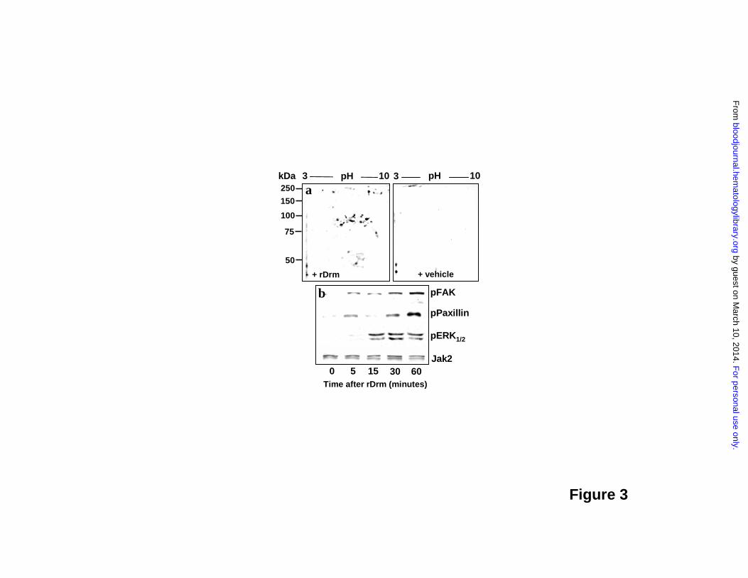
In keeping with the presence of a cell surface receptor, rDrm causes the rapid appearance of
a composite tyrosine phosphorylation pattern in endothelial SIE cells (Figure 3a). As anticipated for
a cell motility factor (see above), paxillin, FAK, and mitogen activated protein kinase ERK1/2 are
targets of rDrm-activated signaling (Figure 3b). Taken together, the data demonstrate for the first
time the capacity of Drm/gremlin to exert a direct, productive interaction with endothelial cells by
engaging cell surface receptor(s) whose identification will require further investigation.
Angiogenic activity of Drm/gremlin
To assess the impact of Drm/gremlin on blood vessel formation in vivo, rDrm (100 ng/egg) was
delivered on the top of the chick embryo chorioallantoic membrane (CAM) at day 8 of development
via a gelatine sponge implant 24. A potent angiogenic response was observed around the rDrm-
implants when compared to vehicle-treated embryos (Figure 4a,b), the number of macroscopic
blood vessels converging towards the gelatin sponge being significantly higher (p< 0.01) in rDrm
than in vehicle implants (Table 1). Histological morphometric analysis of microvessel density
within the gelatin implants confirmed the proangiogenic activity of rDrm (Figure 4c,d and Table 1).
To substantiate these observations, we transfected COS cells with an expression vector harboring
the rat Drm cDNA. Transfectants produce and release significant amounts of the recombinant
protein (Supplementary Figure 1). Accordingly, a potent angiogenic response was observed in chick
embryo CAMs implanted with Drm-transfectants when compared to mock-transfected cells
(Supplementary Figure 1 and Table 1).
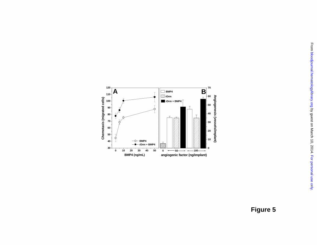
Effect of BMP4 on the angiogenic activity of Drm/gremlin
Our data indicate that rDrm binds to endothelial cells in a BMP-independent manner (see above).
On the other hand, previous observations had shown that BMPs are endowed with angiogenic
activity 28,29. Accordingly, the Drm/gremlin ligand BMP4 is a chemoattractant for MAE cells and
causes neovascularization in the chick CAM (Figure 5a,b). On this basis, we investigated the effect
of BMP4 on the angiogenic activity of rDrm in vitro and in vivo. As shown in Figure 5a,
stimulation of MAE cells with a sub-optimal concentration of rDrm (5.0 ng/mL) in the presence of
For personal use only. by guest on March 10, 2014. bloodjournal.hematologylibrary.orgFrom
11
increasing concentrations of BMP4 resulted in an increased chemotactic response. Similarly, the
simultaneous delivery of rDrm and BMP4 on the chick CAM triggered a more potent angiogenic
response when compared to that induced by rDrm alone (Figure 5b). Taken together, these
observations support the hypothesis that Drm/gremlin exerts its angiogenic activity on endothelium
via a BMP-independent mechanism of action.
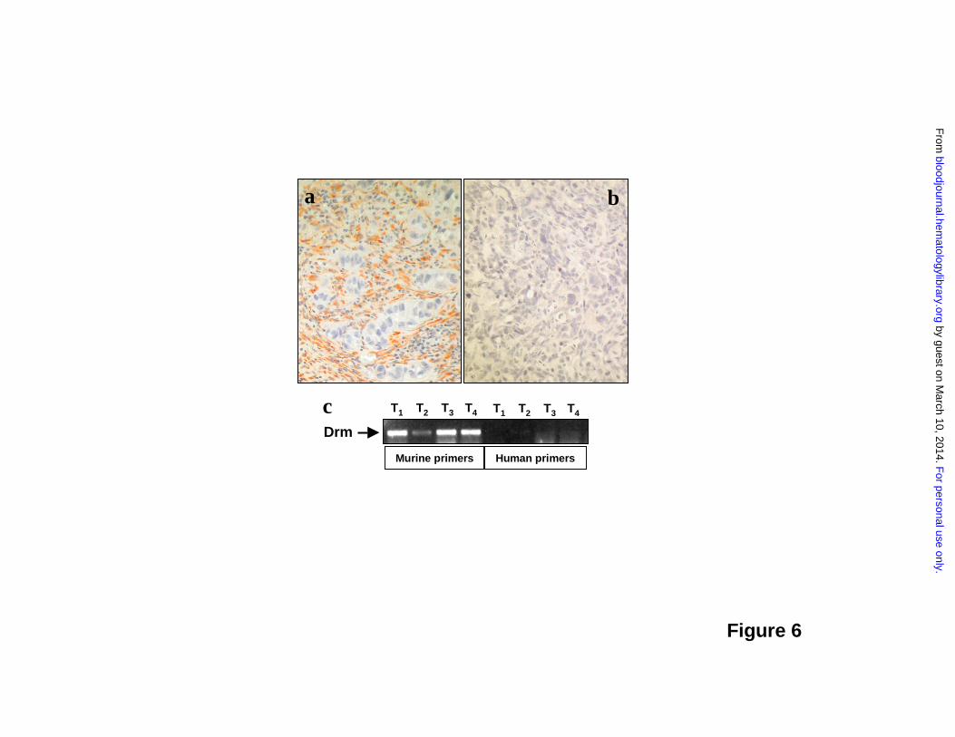
Drm/Gremlin expression in tumor vasculature
Recent observations have shown that Drm/gremlin is upregulated in human cancers 30. To gain
insights about a possible role of Drm/gremlin in tumor vascularization, we performed
immunohistochemical analysis of experimental and human tumors. Highly vascularized tumor
xenografts generated by s.c. injection in nude mice of human adenocarcinoma HEC-1-B-derived
cells 25 showed a strong Drm immunoractivity in the tumor stroma whereas tumor parenchyma was
negative (Figure 6). RT-PCR analysis of total RNA from these lesions using specie-specific primers
confirmed the murine stromal origin of Drm/gremlin and the lack of human Drm/gremlin transcripts
(Figure 6).
The cancer profiling DNA microarray database Oncomine 2.0 contains publicly available gene
expression sets generated from 60 tumor panels covering 20 different human malignancies 31.
Database mining reveals that Drm is significantly overexpressed in lung adenocarcinoma and
squamous cell carcinoma specimens when compared to normal lung (P<0.001). Accordingly,
immunostaining of 10 lung cancer samples (Table 2) demonstrated that endothelial cells of vessels
infiltrating or adjacent to tumor cells strongly express Drm/gremlin (Figure 7a-c). Cancer cells
lacked Drm/gremlin expression in seven cases and showed a weak cytoplasmic immunostaining in
the remaining three cases (Table 2). In all cases, endothelium of the non-neoplastic lung tissue
adjacent to tumor lesions showed no or very weak immunoreactivity (Figure 7d).
Conclusions
Our results show for the first time the capacity of Drm/gremlin to induce a motile and invasive
phenotype in endothelial cells in vitro and a potent angiogenic response in vivo. This reflects the
previously unrecognized ability of Drm/gremlin to interact with high affinity binding sites on
endothelial cells in a BMP-independent manner and to trigger a complex intracellular signaling
cascade.
Drm/gremlin exerts a BMP antagonist activity by binding BMP2, BMP4, and BMP7 3.
Previous observations had shown that BMP2 and BMP4 are endowed with angiogenic activity 28,29.
Here, we have confirmed these observations by showing that BMP4 exerts a significant angiogenic
For personal use only. by guest on March 10, 2014. bloodjournal.hematologylibrary.orgFrom

12
response in the chick embryo CAM. On the other hand, BMP4 does not hamper Drm/gremlin
interaction with endothelial cell receptors and the simultaneous exposure of endothelium to both
rDrm and BMP4 results in a potentiated chemotactic and angiogenic response in vitro and in vivo,
respectively. Our observations support the hypothesis that Drm/gremlin exerts its angiogenic
activity on endothelium via a BMP-independent mechanism of action. Further experiments are
required to fully dissect the complex interplay among Drm/gremlin, BMPs, and their cognate
receptors.
Drm/gremlin is overexpressed in human cancers 30. We have found that human tumor
xenografts in nude mice express Drm. Drm transcripts are synthesized by cells of the host within
the tumor, as shown by RT-PCR analysis using specific murine primers, and Drm/gremlin protein
accumulates in the stroma of the tumor. Also, we have observed a strong Drm/gremlin
immunoreactivity in endothelial cells of human lung tumor samples, absent in adjacent non-
neoplastic lung tissue. Recombinant FGF2 upregulates Drm expression in endothelial cells of
different origin (M. Presta, unpublished observations). This is in keeping with the capacity of
FGF2, as well as of BMP2 and platelet-derived growth factor, to induce Drm/gremlin production in
rat osteoblasts 32 and with the role of Drm/gremlin in the sonic hedgehog/FGF feedback loop in
vertebrate limb bud 33. The possibility that Drm/gremlin produced by tumor cells and/or by growth
factor-activated endothelium may contribute to neovascularization in experimental and human
neoplasms will deserve further investigation.
Secreted Drm/gremlin binds to various BMPs 3. Also, Drm/gremlin has been shown to bind
Slit proteins and to act as inhibitor of monocyte chemotaxis 8. Moreover, a possible intracellular,
BMP-independent mechanism of action has been hypothesized for this protein 6. Our observations
point out the previously unrecognized capacity of Drm/gremlin to interact with signaling receptor(s)
in endothelial cells with possible implications in angiogenesis. Thus, Drm/gremlin may exert
multiple functions by interacting with different partners. The ability of Drm/gremlin to act directly
on target endothelial cells via a receptor-mediated mechanism of action sheds a new light on its
putative role in embryogenesis and human diseases, including cancer.
ACKNOWLEDGEMENTS
For personal use only. by guest on March 10, 2014. bloodjournal.hematologylibrary.orgFrom

13
We thank M.L. Massardi and S. Palermo for technical assistance, and M. Rusnati and P. Dell’Era
(University of Brescia) and A. Scarpa (University of Verona) for helpful discussion and criticisms.
For personal use only. by guest on March 10, 2014. bloodjournal.hematologylibrary.orgFrom
14
Table 1. Chick embryo CAM assay: macroscopic and microscopic assessment of Drm-induced
neovascularization.
Treatment
n
Blood vessels a
(mean ± SD)
Intersection pointsb
(mean ± SD)
Microvessel density c
(%)
vehicle 10 6 ± 3 0 0.0
rDrm 20 27 ± 4* 23 ± 3 16.0
Mock-COS cells 20 10 ± 3 5 ± 2 3.5
Drm-COS cells 20 28 ± 3* 24 ± 2 16.6
Gelatin sponges containing vehicle, 100 ng of rDrm, mock-transfected or Drm-transfected COS
cells (18,000 cells per sponge) were implanted on top of chick embryo CAMs at day 8. The
angiogenic response was assessed macroscopically at day 12 by counting the number of blood
vessels entering the sponge a and histologically by a planimetric method of ‘point counting’ b,c as
described 24. *P < 0.001 vs vehicle and Mock-COS cells.
For personal use only. by guest on March 10, 2014. bloodjournal.hematologylibrary.orgFrom

15
Table 2. Immunohistochemical analysis of Drm/gremlin expression in human lung tumors.
N° of tumors with the indicated
Drm/gremlin immunostaining intensity
Lung tumors
cancer cell
immunostaining
- + ++
endothelial cell
immunostaining
- + ++
Adenocarcinomas (n=5)
4 1 0
0 0 5
Squamous cell carcinomas (n=5)
3 2 0
0 1 4
Paraffin-embedded human lung cancer specimens were analyzed for Drm/gremlin expression by
immunostaining. Cancer cells and endothelial cells of each specimen were scored for Drm/gremlin
immunoreactivity on an arbitrary scale: -, negative; +, weak staining; ++, strong staining.
For personal use only. by guest on March 10, 2014. bloodjournal.hematologylibrary.orgFrom

16
FIGURE LEGENDS
Figure 1. Drm/gremlin purification from the CM of FGF2-T-MAE cells. a, FGF2-T-MAE cell
CM (bottom), but not MAE cell CM (top), stimulates sprouting of MAE cell aggregates in fibrin
gel. This assay was used to follow the purification of FGF2-T-MAE cell CM by cation exchange
(b) and heparin-Sepharose (c) chromatography (horizontal bar, pooled bioactive fractions), and by
reverse-phase HPLC (d). Bioactive HPLC peaks (*,**) were probed with anti-Drm/gremlin
antibodies in a Western blot (inset in d) and identified by MALDI-ToF peptide mass fingerprinting
(peak* in e; peak**, not shown). f, Amino acid sequence of Drm/gremlin. Tryptic (underlined) or
CNBr (italic) digestion peptides were sequenced by nano-ESI-MS/MS. N-glycosilation and Ser-
phosphorylation sites are highlighted.
Figure 2. rDrm interaction with endothelial cells. a, rDrm induces MAE cell sprouting in fibrin
gel (dashed bars), MAE cell migration in a Boyden chamber (gray bars), and BAE cell invasion in
type I collagen gel (black bars); inset, tyrosine phosphorylation of focal adhesion contacts (arrows)
in SIE cells stimulated for 10 minutes with 100 ng/mL rDrm. b, Binding of 125I-rDrm to SIE cells;
inset, cross-linking of unlabeled rDrm to SIE cells followed by immunoprecipitation and Western
blotting with anti-Drm antibody. c, Cross-linking of 125I-rDrm to SIE cells with or without a 100
fold-molar excess of unlabeled rDrm or BMP4. 125I-rDrm complexes were visualized by
autoradiography of the SDS-PAGE gel.
Figure 3. rDrm-induced intracellular signaling. a, 2D electrophoresis of control and rDrm-
treated SIE cell extracts decorated with anti-phospho-Tyr antibody. b, FAK, paxillin, and ERK1/2
phosphorylation in rDrm-treated SIE cells. Anti-Jak2 antibody was used as loading control.
Figure 4. Pro-angiogenic activity of rDrm. Chick embryo CAM implants were loaded with
vehicle (a) or 100 ng of rDrm (b). Note the numerous blood vessels converging versus the rDrm
implant. c,d, Toluidine blue staining of histological sections of CAM treated with vehicle (c) or 100
ng of rDrm (d). Numerous capillaries filled with nucleated avian erythrocytes are present among the
trabeculae of the rDrm-treated sponge (arrows in d).
Figure 5. Effect of BMP4 on the angiogenic activity of Drm/gremlin. a, The chemotactic activity
of a sub-optimal concentration of rDrm (5.0 ng/mL) was tested on MAE cells in a Boyden chamber
For personal use only. by guest on March 10, 2014. bloodjournal.hematologylibrary.orgFrom

17
assay in the presence of increasing concentrations of BMP4 (�); in parallel, the chemotactic activity
of BMP4 alone was tested on the same cells (�). b, Chick embryo CAM implants were loaded with
vehicle, BMP4, rDrm, or rDrm plus BMP4 (both molecules were tested alone or in combination at
50 or 100 ng/implant). After 4 days, blood vessels converging versus the implant were counted.
Figure 6. Drm/gremlin expression in tumor xenografts. a, Drm/gremlin immunostaining of
tumor xenografts originated in nude mice by s.c. injection of human endometrial adenocarcinoma
HEC-1-B-derived cells. Note the Drm/gremlin immunoreactivity in tumor stroma, absent in tumor
parenchyma. b, Negative control in which the primary antibody was omitted. Original
magnification: x200. c, RT-PCR analysis of four tumor xenografts (T) using murine and human
specific Drm/gremlin primers.
Figure 7. Drm/gremlin immunohistochemistry in human lung cancers. Endothelial cells of
tumor vessels (arrows) show a strong Drm/gremlin immunoreactivity in human lung squamous cell
carcinoma (a) and adenocarcinoma (b, c). Squamous tumor cells are weakly immunoreactive (a).
No or very weak immunoreactivity is detected in non-neoplastic lung tissue adjacent to the tumor
(d). Original magnification: x200 (a, d); x400 (b, c).
REFERENCES
1. Pearce JJ, Penny G, Rossant J. A mouse cerberus/Dan-related gene family. Dev Biol.
1999;209:98-110
2. Vitt UA, Hsu SY, Hsueh AJ. Evolution and classification of cystine knot-containing
hormones and related extracellular signaling molecules. Mol Endocrinol. 2001;15:681-694
3. Balemans W, Van Hul W. Extracellular regulation of BMP signaling in vertebrates: a
cocktail of modulators. Dev Biol. 2002;250:231-250
4. Khokha MK, Hsu D, Brunet LJ, Dionne MS, Harland RM. Gremlin is the BMP
antagonist required for maintenance of Shh and Fgf signals during limb patterning. Nat Genet.
2003;34:303-307
5. Lappin DW, McMahon R, Murphy M, Brady HR. Gremlin: an example of the re-
emergence of developmental programmes in diabetic nephropathy. Nephrol Dial Transplant.
2002;17 Suppl 9:65-67
For personal use only. by guest on March 10, 2014. bloodjournal.hematologylibrary.orgFrom
18
6. Chen B, Athanasiou M, Gu Q, Blair DG. Drm/Gremlin transcriptionally activates
p21(Cip1) via a novel mechanism and inhibits neoplastic transformation. Biochem Biophys Res
Commun. 2002;295:1135-1141
7. Topol LZ, Marx M, Laugier D, Bogdanova NN, Boubnov NV, Clausen PA, Calothy G,
Blair DG. Identification of drm, a novel gene whose expression is suppressed in transformed cells
and which can inhibit growth of normal but not transformed cells in culture. Mol Cell Biol.
1997;17:4801-4810
8. Chen B, Blair DG, Plisov S, Vasiliev G, Perantoni AO, Chen Q, Athanasiou M, Wu JY,
Oppenheim JJ, Yang D. Cutting edge: bone morphogenetic protein antagonists Drm/Gremlin and
Dan interact with Slits and act as negative regulators of monocyte chemotaxis. J Immunol.
2004;173:5914-5917
9. Carmeliet P, Jain RK. Angiogenesis in cancer and other diseases. Nature. 2000;407:249-
257
10. Hanahan D, Christofori G, Naik P, Arbeit J. Transgenic mouse models of tumour
angiogenesis: the angiogenic switch, its molecular controls, and prospects for preclinical therapeutic
models. Eur J Cancer. 1996;32A:2386-2393
11. Folkman J. Angiogenesis in cancer, vascular, rheumatoid and other disease. Nat Med.
1995;1:27-31
12. Ferrara N. VEGF and the quest for tumour angiogenesis factors. Nat Rev Cancer.
2002;2:795-803
13. Carmeliet P. VEGF as a key mediator of angiogenesis in cancer. Oncology. 2005;69
Suppl 3:4-10
14. Presta M, Urbinati C, Dell'era P, Lauro GM, Sogos V, Balaci L, Ennas MG, Gremo F.
Expression of basic fibroblast growth factor and its receptors in human fetal microglia cells. Int J
Dev Neurosci. 1995;13:29-39
15. Javerzat S, Auguste P, Bikfalvi A. The role of fibroblast growth factors in vascular
development. Trends Mol Med. 2002;8:483-489
16. Sola F, Gualandris A, Belleri M, Giuliani R, Coltrini D, Bastaki M, Tosatti MP, Bonardi
F, Vecchi A, Fioretti F, Ciomei M, Grandi M, Mantovani A, Presta M. Endothelial cells
overexpressing basic fibroblast growth factor (FGF-2) induce vascular tumors in immunodeficient
mice. Angiogenesis. 1997;1:102-116
17. Gualandris A, Rusnati M, Belleri M, Nelli EE, Bastaki M, Molinari-Tosatti MP,
Bonardi F, Parolini S, Albini A, Morbidelli L, Ziche M, Corallini A, Possati L, Vacca A, Ribatti D,
For personal use only. by guest on March 10, 2014. bloodjournal.hematologylibrary.orgFrom
19
Presta M. Basic fibroblast growth factor overexpression in endothelial cells: an autocrine
mechanism for angiogenesis and angioproliferative diseases. Cell Growth Differ. 1996;7:147-160
18. Dell'Era P, Coco L, Ronca R, Sennino B, Presta M. Gene expression profile in
fibroblast growth factor 2-transformed endothelial cells. Oncogene. 2002;21:2433-2440
19. Ribatti D, Gualandris A, Belleri M, Massardi L, Nico B, Rusnati M, Dell'Era P, Vacca
A, Roncali L, Presta M. Alterations of blood vessel development by endothelial cells
overexpressing fibroblast growth factor-2. J Pathol. 1999;189:590-599
20. Dong QG, Bernasconi S, Lostaglio S, De Calmanovici RW, Martin-Padura I, Breviario
F, Garlanda C, Ramponi S, Mantovani A, Vecchi A. A general strategy for isolation of endothelial
cells from murine tissues. Characterization of two endothelial cell lines from the murine lung and
subcutaneous sponge implants. Arterioscler.Thromb.Vasc.Biol. 1997;17:1599-1604
21. Zhang W, Chait BT. ProFound: an expert system for protein identification using mass
spectrometric peptide mapping information. Anal Chem. 2000;72:2482-2489
22. Coltrini D, Rusnati M, Zoppetti G, Oreste P, Grazioli G, Naggi A, Presta M. Different
effects of mucosal, bovine lung and chemically modified heparin on selected biological properties
of basic fibroblast growth factor. Biochem J. 1994;303:583-590
23. Topol LZ, Bardot B, Zhang Q, Resau J, Huillard E, Marx M, Calothy G, Blair DG.
Biosynthesis, post-translation modification, and functional characterization of Drm/Gremlin. J Biol
Chem. 2000;275:8785-8793
24. Ribatti D, Gualandris A, Bastaki M, Vacca A, Iurlaro M, Roncali L, Presta M. New
model for the study of angiogenesis and antiangiogenesis in the chick embryo chorioallantoic
membrane: the gelatin sponge/chorioallantoic membrane assay. J Vasc Res. 1997;34:455-463
25. Giavazzi R, Sennino B, Coltrini D, Garofalo A, Dossi R, Ronca R, Tosatti MP, Presta
M. Distinct role of fibroblast growth factor-2 and vascular endothelial growth factor on tumor
growth and angiogenesis. Am J Pathol. 2003;162:1913-1926
26. Iozzo RV, San Antonio JD. Heparan sulfate proteoglycans: heavy hitters in the
angiogenesis arena. J.Clin.Invest. 2001;108:349-355
27. Presta M, Leali D, Stabile H, Ronca R, Camozzi M, Coco L, Moroni E, Liekens S,
Rusnati M. Heparin derivatives as angiogenesis inhibitors. Curr Pharm Des. 2003;9:553-566
28. Nimmagadda S, Geetha Loganathan P, Huang R, Scaal M, Schmidt C, Christ B. BMP4
and noggin control embryonic blood vessel formation by antagonistic regulation of VEGFR-2
(Quek1) expression. Dev Biol. 2005;280:100-110
29. Langenfeld EM, Langenfeld J. Bone morphogenetic protein-2 stimulates angiogenesis
in developing tumors. Mol Cancer Res. 2004;2:141-149
For personal use only. by guest on March 10, 2014. bloodjournal.hematologylibrary.orgFrom
20
30. Namkoong H, Shin SM, Kim HK, Ha SA, Cho GW, Hur SY, Kim TE, Kim JW. The
bone morphogenetic protein antagonist gremlin 1 is overexpressed in human cancers and interacts
with YWHAH protein. BMC Cancer. 2006;6:74
31. Rhodes DR, Yu J, Shanker K, Deshpande N, Varambally R, Ghosh D, Barrette T,
Pandey A, Chinnaiyan AM. ONCOMINE: a cancer microarray database and integrated data-mining
platform. Neoplasia. 2004;6:1-6
32. Pereira RC, Economides AN, Canalis E. Bone morphogenetic proteins induce gremlin,
a protein that limits their activity in osteoblasts. Endocrinology. 2000;141:4558-4563
33. Zuniga A, Haramis AP, McMahon AP, Zeller R. Signal relay by BMP antagonism
controls the SHH/FGF4 feedback loop in vertebrate limb buds. Nature. 1999;401:598-602
For personal use only. by guest on March 10, 2014. bloodjournal.hematologylibrary.orgFrom
CM MAE
CM FGF2-T-MAE
a
0
0.2
0.4
0.60.8
1
1.2
1.4
1 6 11 16 21 26 31 36 41 46 51
0
0.5
1
1.5
2
2.5
0
1.0
1.2
1.4
0.8
0.6
0.4
0.2
0 10 20 30 40 50O
.D. (
280
nm
)
Fraction n°
2.5
2.0
1.5
1.0
0.5
0
Sp
rou
t ind
ucin
g activity
(relative un
its)
b
0
0 .04
0 .08
0 .12
0 .16
0.2
1 6 11 16 21 2 31 3 41 4 51 560
0 .5
1
1.5
2
2 .5
3
3 .5
40.20
0.12
0.08
0.16
0.04
0.0
10 20 30 40 50 600
O.D
. (28
0 n
m)
Fraction n°
0
1.5
0.5
1.0
2.0
2.5
3.0
3.5
4.0
Sp
rou
t ind
ucin
g activity
(relative un
its)
c
d
0.000
0.002
0.004
0.006
0.008
0.010
0.012
0.014
0.016
0
1020
30
40
50
60
70
80
90
100
0.00 10.00 40.00
3,44
64,
097
5,69
1 8,83
69,
627
23,7
4824
,640
26,7
5128
,657 31
,445
32,5
98 33,9
2934
,732
36,8
52
O.D
. (28
0 n
m) %
aceton
itrile
Time (minutes)20.00 30.00
28 kDa
* **
1001711.73481711.7348
750 1200 2100 2550 30000
20
40
100
1963
.884
9
%In
ten
sity
1711
.734
8
2450
.071
6
1818
.811
2
1330
.586
2
1066
.554
9
2466
.072
4
1897
.922
1
2309
.088
3
m/z
80
60
1650
e
KKKGSQ GAIPPPDKAQ HNDSEQTQSP PQPGSRTRGR GQGRGTAMPG EEVLESSQEALHVTERKYLK RDWCKTQPLK QTIHEEGCNS RTIINRFCYG QCNSFYIPRH IRKEEGSFQS
CSFCKPKKFT TMMVTLNCPE LQPPTKKKRV TRVKQCRCIS IDLD
80
140
184
25f
Figure 1
F
or personal use only. by guest on M
arch 10, 2014. bloodjournal.hem
atologylibrary.orgF
rom
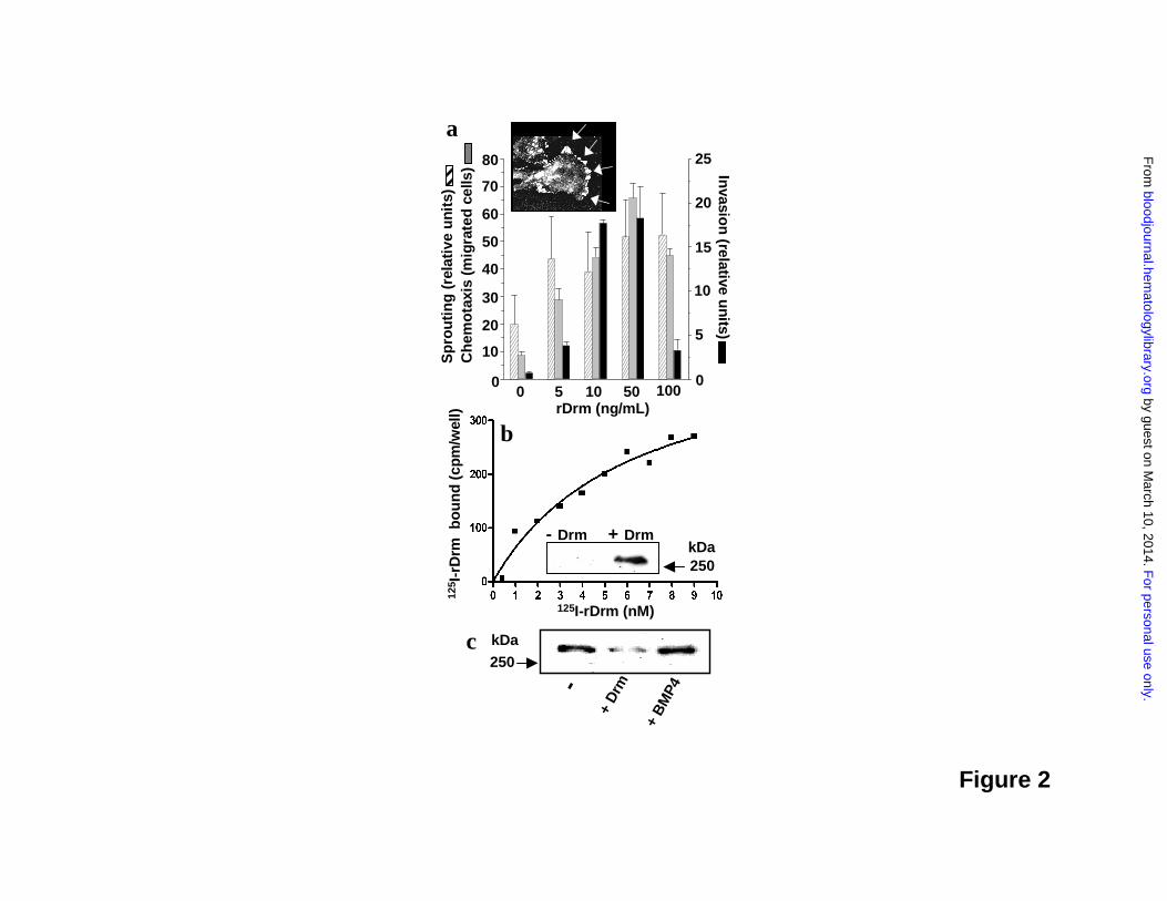
Figure 2
a
+ Drm- Drm
b
250kDa
125I-rDrm (nM)
c+
BM
P4
+ D
rm-
250kDa
125 I
-rD
rm b
ou
nd
(cpm
/wel
l)
rDrm (ng/mL)0 5 10 50 100
80
70
60
25
40
30
50
20
10
0
20
15
10
5
0
Sp
rou
tin
g (
rela
tive
uni
ts)
Ch
emo
taxi
s(m
igra
ted
cells
) Invasio
n(relative u
nits)
F
or personal use only. by guest on M
arch 10, 2014. bloodjournal.hem
atologylibrary.orgF
rom
pH3 10pH
+ rDrm + vehicle
3 10250
100
75
50
150
kDa
a
pPaxillin
Jak2
pERK1/2
pFAK
0 5 15 30 60Time after rDrm (minutes)
b
Figure 3
F
or personal use only. by guest on M
arch 10, 2014. bloodjournal.hem
atologylibrary.orgF
rom

Figure 4
vehicle rDrm
a b
c d
* *
F
or personal use only. by guest on M
arch 10, 2014. bloodjournal.hem
atologylibrary.orgF
rom
0
10
20
30
40
50
60
70
BrDrm + BMP4
rDrm
BMP4
100500
angiogenic factor (ng/implant)
Angiogenesis (vessels/im
plant)
0 10 20 30 40 5030
40
50
60
70
80
90
100
110
120
A
Che
mot
axis
(mig
rate
d ce
lls)
BMP4 (ng/mL)
BMP4 rDrm + BMP4
Figure 5
F
or personal use only. by guest on M
arch 10, 2014. bloodjournal.hem
atologylibrary.orgF
rom
Human primersMurine primers
c T1 T2 T3 T4
Drm
T1 T2 T3 T4
Figure 6
a b
F
or personal use only. by guest on M
arch 10, 2014. bloodjournal.hem
atologylibrary.orgF
rom

Figure 7
F
or personal use only. by guest on M
arch 10, 2014. bloodjournal.hem
atologylibrary.orgF
rom