Boc and Gas1 Each Form Distinct Shh Receptor Complexes with Ptch1 and Are Required for Shh-Mediated...
25
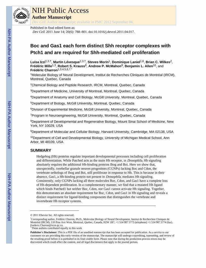
Boc and Gas1 each form distinct Shh receptor complexes with Ptch1 and are required for Shh-mediated cell proliferation Luisa Izzi 1,3,† , Martin Lévesque 1,3,† , Steves Morin 1 , Dominique Laniel 1,6 , Brian C. Wilkes 2 , Frédéric Mille 1,3 , Robert S. Krauss 8 , Andrew P. McMahon 9 , Benjamin L. Allen 10 , and Frédéric Charron 1,3,4,5,6,7,* 1 Molecular Biology of Neural Development, Institut de Recherches Cliniques de Montréal (IRCM), Montreal, Quebec, Canada 2 Chemical Biology and Peptide Research, IRCM, Montreal, Quebec, Canada 3 Department of Medicine, University of Montreal, Montreal, Quebec, Canada 4 Department of Anatomy and Cell Biology, McGill University, Montreal, Quebec, Canada 5 Department of Biology, McGill University, Montreal, Quebec, Canada 6 Division of Experimental Medicine, McGill University, Montreal, Quebec, Canada 7 Program in Neuroengineering, McGill University, Montreal, Quebec, Canada 8 Department of Developmental and Regenerative Biology, Mount Sinai School of Medicine, New York, NY 10029, USA 9 Department of Molecular and Cellular Biology, Harvard University, Cambridge, MA 02138, USA 10 Department of Cell and Developmental Biology, University of Michigan Medical School, Ann Arbor, MI 48109, USA SUMMARY Hedgehog (Hh) proteins regulate important developmental processes including cell proliferation and differentiation. While Patched acts as the main Hh receptor, in Drosophila, Hh signaling absolutely requires the additional Hh-binding proteins Ihog and Boi. Here we show that, unexpectedly, cerebellar granule neuron progenitors (CGNPs) lacking Boc and Cdon, the vertebrate orthologs of Ihog and Boi, still proliferate in response to Hh. This is because in their absence, Gas1, a Hh-binding protein not present in Drosophila, mediates Hh signaling. Consistently, only CGNPs lacking all three molecules Boc, Cdon, and Gas1 have a complete loss of Hh-dependent proliferation. In a complementary manner, we find that a mutated Hh ligand which binds Patched1 but neither Boc, Cdon, nor Gas1 cannot activate Hh signaling. Together, this demonstrates an absolute requirement for Boc, Cdon, and Gas1 in Hh signaling and reveals a distinct requirement for ligand-binding components that distinguishes the vertebrate and invertebrate Hh receptor systems. © 2011 Elsevier Inc. All rights reserved. * Corresponding author, Frédéric Charron, Ph.D., Molecular Biology of Neural Development, Institut de Recherches Cliniques de Montréal (IRCM), 110 Pine Ave West, Montreal, Quebec, Canada, H2W 1R7, +1 514 987 5773 (telephone) +1 514 987 5774 (fax), [email protected]. † These authors contributed equally to this work Publisher's Disclaimer: This is a PDF file of an unedited manuscript that has been accepted for publication. As a service to our customers we are providing this early version of the manuscript. The manuscript will undergo copyediting, typesetting, and review of the resulting proof before it is published in its final citable form. Please note that during the production process errors may be discovered which could affect the content, and all legal disclaimers that apply to the journal pertain. NIH Public Access Author Manuscript Dev Cell. Author manuscript; available in PMC 2012 September 04. Published in final edited form as: Dev Cell. 2011 June 14; 20(6): 788–801. doi:10.1016/j.devcel.2011.04.017. NIH-PA Author Manuscript NIH-PA Author Manuscript NIH-PA Author Manuscript
-
Upload
independent -
Category
Documents
-
view
2 -
download
0
Transcript of Boc and Gas1 Each Form Distinct Shh Receptor Complexes with Ptch1 and Are Required for Shh-Mediated...
Boc and Gas1 each form distinct Shh receptor complexes withPtch1 and are required for Shh-mediated cell proliferation
Luisa Izzi1,3,†, Martin Lévesque1,3,†, Steves Morin1, Dominique Laniel1,6, Brian C. Wilkes2,Frédéric Mille1,3, Robert S. Krauss8, Andrew P. McMahon9, Benjamin L. Allen10, andFrédéric Charron1,3,4,5,6,7,*
1Molecular Biology of Neural Development, Institut de Recherches Cliniques de Montréal (IRCM),Montreal, Quebec, Canada2Chemical Biology and Peptide Research, IRCM, Montreal, Quebec, Canada3Department of Medicine, University of Montreal, Montreal, Quebec, Canada4Department of Anatomy and Cell Biology, McGill University, Montreal, Quebec, Canada5Department of Biology, McGill University, Montreal, Quebec, Canada6Division of Experimental Medicine, McGill University, Montreal, Quebec, Canada7Program in Neuroengineering, McGill University, Montreal, Quebec, Canada8Department of Developmental and Regenerative Biology, Mount Sinai School of Medicine, NewYork, NY 10029, USA9Department of Molecular and Cellular Biology, Harvard University, Cambridge, MA 02138, USA10Department of Cell and Developmental Biology, University of Michigan Medical School, AnnArbor, MI 48109, USA
SUMMARYHedgehog (Hh) proteins regulate important developmental processes including cell proliferationand differentiation. While Patched acts as the main Hh receptor, in Drosophila, Hh signalingabsolutely requires the additional Hh-binding proteins Ihog and Boi. Here we show that,unexpectedly, cerebellar granule neuron progenitors (CGNPs) lacking Boc and Cdon, thevertebrate orthologs of Ihog and Boi, still proliferate in response to Hh. This is because in theirabsence, Gas1, a Hh-binding protein not present in Drosophila, mediates Hh signaling.Consistently, only CGNPs lacking all three molecules Boc, Cdon, and Gas1 have a complete lossof Hh-dependent proliferation. In a complementary manner, we find that a mutated Hh ligandwhich binds Patched1 but neither Boc, Cdon, nor Gas1 cannot activate Hh signaling. Together,this demonstrates an absolute requirement for Boc, Cdon, and Gas1 in Hh signaling and reveals adistinct requirement for ligand-binding components that distinguishes the vertebrate andinvertebrate Hh receptor systems.
© 2011 Elsevier Inc. All rights reserved.*Corresponding author, Frédéric Charron, Ph.D., Molecular Biology of Neural Development, Institut de Recherches Cliniques deMontréal (IRCM), 110 Pine Ave West, Montreal, Quebec, Canada, H2W 1R7, +1 514 987 5773 (telephone) +1 514 987 5774 (fax),[email protected].†These authors contributed equally to this work
Publisher's Disclaimer: This is a PDF file of an unedited manuscript that has been accepted for publication. As a service to ourcustomers we are providing this early version of the manuscript. The manuscript will undergo copyediting, typesetting, and review ofthe resulting proof before it is published in its final citable form. Please note that during the production process errors may bediscovered which could affect the content, and all legal disclaimers that apply to the journal pertain.
NIH Public AccessAuthor ManuscriptDev Cell. Author manuscript; available in PMC 2012 September 04.
Published in final edited form as:Dev Cell. 2011 June 14; 20(6): 788–801. doi:10.1016/j.devcel.2011.04.017.
NIH
-PA Author Manuscript
NIH
-PA Author Manuscript
NIH
-PA Author Manuscript
INTRODUCTIONHh proteins are key molecules for diverse tissue patterning processes in both invertebratesand vertebrates. For example, in Drosophila, Hh is crucial for the development of asegmented body plan and the patterning of imaginal tissues, whereas in vertebrates Sonichedgehog (Shh) functions to pattern limb buds and promote cell fate specification,proliferation, and axon guidance in the central nervous system (Charron and Tessier-Lavigne, 2005; Dessaud et al., 2008; Ingham and Placzek, 2006; Jiang and Hui, 2008). Shhis synthesized as a 45 kDa pro-protein and post-translational modifications generate abiologically active 19 kDa N-terminal fragment. Shh initiates signaling by binding the 12-pass transmembrane protein Patched1 (Ptch1). Upon Shh binding, the inhibition exerted byPtch1 on the 7-pass transmembrane protein Smoothened (Smo) is relieved, eliciting asignaling cascade which ultimately leads to Gli-mediated transcription.
In addition to Ptch1, several membrane-associated proteins are thought to function asaccessory receptors that promote Shh signaling. The related molecules Cdon (cell-adhesion-molecule-related/downregulated by oncogenes) and Boc (biregional Cdon-binding protein)positively regulate Shh signaling, promoting Shh-dependent cell fate specification and axonguidance (Okada et al., 2006; Tenzen et al., 2006; Yao et al., 2006; Zhang et al., 2006).They are single-pass transmembrane proteins of the Immunoglobulin (Ig) superfamily andcontain an extracellular region consisting of four (Boc) or five (Cdon) Ig repeats and threeFibronectin type 3 repeats (FNIII). In Drosophila, Ihog (Interference hedgehog) and Boi(Brother of Ihog), the orthologs of Boc and Cdon, are redundant with one another and areabsolutely required for Hh-dependent patterning (Camp et al., 2010; Zheng et al., 2010).However, whether Boc and Cdon are absolutely essential for Shh signaling in vertebrates isunknown.
Furthermore, additional membrane-associated proteins have been shown to positivelymodulate Shh signaling in vertebrates. Growth Arrest Specific 1 (Gas1), a GPI-linkedprotein bearing no structural resemblance to Boc and Cdon and with no orthologues inDrosophila, binds Shh and regulates ventral specification of neural progenitors during neuraltube development by promoting Shh signaling (Allen et al., 2007; Martinelli and Fan, 2007a,b). Interestingly, analysis of Gas1 mutant mice shows that it is a positive regulator ofcerebellar size and CGNP proliferation (Liu et al., 2001), two processes that are normallydriven by Shh during early postnatal development. However, whether Gas1 acts in a Shh-dependent manner in CGNP proliferation has yet to be determined.
Thus, Boc, Cdon and Gas1 have each been proposed to function as positive modulators ofShh signaling (Allen et al., 2007; Martinelli and Fan, 2007a; Okada et al., 2006; Seppala etal., 2007; Tenzen et al., 2006). We hypothesised that they are obligate Shh receptors withPtch1, and not simply modulators of the signaling cascade. If this model were correct,simultaneous disruption of all three molecules would completely abrogate Shh signaling.We tested this by investigating the proliferation of CGNPs in response to Shh in thedeveloping cerebellum. We found that CGNPs express Boc and Gas1, but not Cdon.Interestingly, the cerebellum is smaller in Boc−/− mice, and Boc−/− CGNPs have lowerproliferation than wild-type CGNPs in response to Shh. Similarly, Gas1−/− CGNPs are alsoless responsive to Shh, while Gas1−/−;Boc−/− CGNPs are completely unable to proliferate inresponse to Shh. We further demonstrated that Boc and Gas1 interact with Ptch1 and formdistinct receptor complexes. Finally, we generated a Shh mutant protein that binds Ptch1 butnot Boc, Cdon nor Gas1 and found that this molecule could not elicit Shh-dependentsignaling and CGNP proliferation. Together, our data indicates that Boc, Cdon and Gas1 arenecessary components of the Shh receptor complex and are essential for Shh signaltransduction in vertebrates. While Ihog and Boi are necessary to mediate Hh signaling in
Izzi et al. Page 2
Dev Cell. Author manuscript; available in PMC 2012 September 04.
NIH
-PA Author Manuscript
NIH
-PA Author Manuscript
NIH
-PA Author Manuscript
Drosophila (Camp et al., 2010; Zheng et al., 2010), we show that Gas1 is an additionalcomponent of the Hh receptor system in vertebrates that can compensate for the absence ofBoc and Cdon.
RESULTSBoc, but not Cdon, is expressed in proliferating CGNPs of the cerebellum
To investigate the receptor requirements for the proliferative effect of Shh in CGNPs, wefirst analyzed Boc and Cdon expression in the developing cerebellum. CGNPs arise from therhombic lip (RL) between embryonic day (E) 13.5–14.5 and migrate anteriorly over thecerebellar anlage, forming the highly proliferative external germinal layer (EGL) (Rousseland Hatten, 2011). Starting at E17.5 and continuing during early postnatal development,Purkinje cells (PCs) lining the EGL stimulate CGNP proliferation by secreting Shh(Dahmane and Ruiz i Altaba, 1999; Kenney and Rowitch, 2000; Wallace, 1999; Wechsler-Reya and Scott, 1999). Following a proliferative burst, CGNPs stop dividing, differentiateinto granule neurons, migrate inwards past the PC layer and populate the internal granularlayer (IGL).
We first examined Boc and Cdon expression in the cerebellar anlage of E14.5 mouseembryos. Immunostainings of sagittal sections showed that while Boc was expressed in thepresumptive EGL, RL and the ventricular zone of the roof of the 4th ventricle, Cdonexpression was restricted to the RL (Fig. 1A). At E18.5, a stage at which CGNPs proliferatein response to Shh, we detected Boc expression in the EGL and, albeit at lower level, in thePC layer of the developing cerebellum. In contrast, Cdon expression was limited to the tip ofthe RL.
Analysis of post-natal day (P) 6 Boc+/- and Cdon+/- gene-targeted mice encoding a β-galactosidase (β-Gal)-neomycin reporter gene fusion (β-geo) (Okada et al., 2006) revealedstrong β-Gal activity in Boc+/- cerebellum, but was limited to the choroid plexus of Cdon+/-
cerebellum. Immunostainings confirmed this expression pattern and revealed that Boclocalized to cells expressing Lim1, a marker for CGNPs and PCs (Fig. 1B,C). Interestingly,while highest levels of Boc were detected in the outer proliferative region of the EGL(Lim1+, Pax6+ and TAG1− cells), lower levels were observed in differentiated migratorygranule cells (TAG1+ cells) and in PCs (Calbindin+ cells). These results show that Boc, butnot Cdon, is highly expressed in proliferating CGNPs of the cerebellum.
Boc is important, but not absolutely required, for Shh-mediated CGNP proliferationTo investigate the role of Boc in cerebellum development, we examined the grossmorphology of Boc−/− cerebella. While Boc−/− mice are viable and cannot be distinguishedfrom their littermates, their cerebellum is smaller than Boc+/- or WT animals (Fig. 2A anddata not shown). Boc−/− cerebella were 14.3±0.05% (p<0.001) lighter than that of Boc+/-
cerebella (Fig. 2B). When the mass of the cerebellum was normalized to the body weight(p<0.001), the relative cerebellar mass was still reduced, indicating that this difference is notdue to an overall decrease in total body weight (Fig. 2B). The cerebellum and IGL surfaceareas measured from sagittal sections of Boc−/− adult mice were also reduced whencompared to Boc+/- animals (Fig. 2C,D; p<0.001 and 0.05, respectively). Although the IGLsurface area is diminished in adult mice, migration of granule neurons and cerebellumfoliation did not appear to be affected in Boc−/− mice.
The decrease in cerebellum size in the absence of Boc could be due, at least in part, toreduced cell proliferation and/or enhanced cell death. TUNEL staining showed nosignificant difference in the number of apoptotic cells between Boc−/− and Boc+/- cerebella(Fig. 2E,F). In contrast, measurement of BrdU incorporation in the EGL of Boc−/− and
Izzi et al. Page 3
Dev Cell. Author manuscript; available in PMC 2012 September 04.
NIH
-PA Author Manuscript
NIH
-PA Author Manuscript
NIH
-PA Author Manuscript
Boc+/- mice showed that 40±1% of Boc+/- CGNPs were actively dividing, compared to only30±3% of Boc−/− CGNPs (p<0.05) (Fig. 2G,H). Phospho-histone H3 (pH3) staining alsoshowed a significant reduction in the number of mitotic pH3-labeled cells per mm2 of EGLin Boc−/− mice compared to Boc+/- mice (p<0.05) (Fig. 2H). Together, these in vivo dataindicate that Boc plays a role in CGNP proliferation.
Since Boc modulates Shh signaling (Okada et al., 2006; Tenzen et al., 2006; Zhang et al.,2006), we next tested whether Boc mediates Shh-induced CGNP proliferation. We culturedCGNPs purified from Boc−/−, Boc+/- and Boc+/+ mice in the presence of varyingconcentrations of recombinant Shh (ShhN) (Fig. 2I). While Shh treatment induced theproliferation of WT CGNPs over 6 fold compared to unstimulated CGNPs, Shh stimulationincreased Boc−/− CGNP proliferation only about 3 fold. Significant differences in theproliferation of Boc+/+, Boc+/- and Boc−/− CGNPs was observed at all concentrations ofShhN used (Fig. 2I), indicating that Boc promotes proliferation of CGNPs in a gene copy-number dependent manner. Together with our in vivo data, these results indicate that Boc−/−
mice have a smaller cerebellum due to a decrease in Shh-dependent CGNP proliferation andthat Boc acts cell-autonomously in CGNPs to regulate their proliferation.
Gas1 is important, but not absolutely required, for Shh-mediated CGNP proliferationWhilst inactivation of Boc in CGNPs, which do not express Cdon, lead to a partial decreasein their proliferation, it did not abolish their response to Shh. Moreover, CGNP proliferationis not further decreased when Cdon is inactivated in Boc−/− mice (Fig. S1). These results arenot consistent with a model where Boc and Cdon act like their Drosophila orthologues Ihogand Boi and are absolutely required for Hh signaling in vertebrates (Camp et al., 2010;Zheng et al., 2010). This raises the possibility that, unlike Drosophila, additional or differentShh binding molecules (other than Ptch1, Boc and Cdon) are required for vertebrate cells torespond to Shh.
Given that Gas1 binds Shh and modulates Shh signaling (Allen et al., 2007; Martinelli andFan, 2007a, b; Seppala et al., 2007), we hypothesized that Gas1 may be this additionalreceptor. We first characterized the expression pattern of Gas1 in the developing cerebellum.Immunofluorescence stainings showed that Gas1 is restricted to the presumptive EGL of thecerebellar primordium at E14.5 and continues to be expressed in the EGL at E18.5 (Fig.3A). At P6, like Boc, Gas1 localizes to Lim1+ cells in the EGL (Fig. 3B). Gas1 staining ismost intense in the outer proliferative layer of the EGL (Lim1+, Pax6+, TAG1− cells) andwas not detected in TAG1+ migratory granule neurons and in Calbindin+ PCs (Fig. 3C).
To determine whether Boc and Gas1 are co-expressed in CGNPs, we performedimmunostainings on consecutive sections of cerebellum from Math1-Cre; mTmG E18.5mice, where the CGNPs express GFP following Cre-mediated recombination. We used thisstrategy instead of double immunostainings as both anti-Boc and anti-Gas1 antibodies areproduced in the same species. We found that both Boc and Gas1 co-localize with GFP+
cells, indicating that Gas1 and Boc are co-expressed in the same CGNPs (Fig. 3D).
Although the gross morphology of Gas1−/− cerebella appears normal, they are smaller insize compared to control cerebellum and have decreased proliferation in the outer EGL (Liuet al., 2001). While this phenotype is reminiscent of that of Boc−/− cerebella, no direct linkhas been made between the phenotype and the ability of Gas1−/− CGNPs to respond to Shh.To directly test this, we performed proliferation assays on purified CGNPs from Gas1−/−
mice and control littermates. Our results show that Gas1 is essential for normal CGNPproliferation in response to Shh (Fig. 4C). Interestingly, the mutation of Gas1, similarly tothe mutation of Boc, is not sufficient to abrogate the response of CGNPs to Shh.
Izzi et al. Page 4
Dev Cell. Author manuscript; available in PMC 2012 September 04.
NIH
-PA Author Manuscript
NIH
-PA Author Manuscript
NIH
-PA Author Manuscript
Shh-dependent proliferation is completely lost in Gas1−/−;Boc−/− CGNPsTo determine whether Boc and Gas1 might have partially redundant functions in Shh-dependent CGNP proliferation, we examined the cerebellum of E18.5 Gas1−/−;Boc−/−
embryos, since these animals die at birth. Hematoxylin-eosin staining of Gas1−/−;Boc−/−
cerebella revealed a significant loss of the EGL compared to controls (Fig. 4A). AlthoughGas1+/-;Boc−/− and Gas1−/−;Boc−/− cerebella showed no significant difference in the cross-sectional area of the whole cerebellum, the overall area of Gas1−/−;Boc−/− EGL was reducedby about 30% compared to controls (p<0.001) (Fig. 4B). Quantitation of the EGL along thepostero-anterior axis showed that the difference in EGL thickness is greatest towards theanterior pole of the cerebellum (Fig. 4B and S2D). Marker analysis showed that Lim1 andPax6 were properly expressed in the EGL of Gas1−/−;Boc−/− embryos compared to controls(Fig. S2A,B), thus, CGNPs are specified and localize normally. Furthermore, Cdonexpression was not changed in the absence of Gas1 and Boc (Fig. S2C). However, theproliferation of Gas1−/−;Boc−/− CGNPs was severely decreased compared to Gas1+/-;Boc−/−
CGNPs (p<0.001) (Fig. 4B). Moreover, the number of pH3+ cells per µm2 of EGL surfacearea was lower in Gas1−/−;Boc−/− than Gas1+/-;Boc−/− animals (p<0.05) (Fig. 4B),demonstrating that the decrease in pH3+ cells in the EGL is not simply due to a totaldecrease in EGL area. These results indicate that Gas1 and Boc account for a large part ofCGNP proliferation at this stage in vivo.
In addition to Shh, Insulin Growth Factor (IGF) and Notch signaling also promote CGNPproliferation (Corcoran et al., 2008; Solecki et al., 2001). Residual CGNP proliferation isobserved in other mutant cerebella that lack Shh signaling (Corrales et al., 2004), thus, theproliferation observed in the EGL of Gas1−/−;Boc−/− cerebellum is probably independent ofShh signaling. To test whether Gas1−/−;Boc−/− cells have completely lost Shhresponsiveness, we cultured CGNPs purified from E18.5 Gas1+/+;Boc−/− andGas1−/−;Boc−/− cerebella with various ShhN concentrations. We found that whileGas1+/+;Boc−/− CGNPs proliferate in vitro in response to Shh, Gas1−/−;Boc−/− CGNPs showno enhanced proliferation in response to Shh (Fig. 4D). Importantly, the proliferativeresponse of Gas1−/−;Boc−/− CGNPs to IGF-I, another factor able to stimulate CGNPproliferation, remained similar to that of control cells (Fig. 4E). Furthermore, treatment withpurmorphamine, a Smo agonist, induced the proliferation of Gas1−/−;Boc−/− CGNPs(p<0.01) (Fig. 4F), indicating that Boc and Gas1 function upstream of Smo. Together, ourdata indicates that the presence of either Gas1 or Boc is absolutely required for Shh topromote CGNP proliferation. Given that Shh signaling in the cerebellum begins only atE17.5 and that Shh signaling plays an even more important role in CGNP proliferation afterbirth than at E18.5 (Corrales et al., 2004; Flora et al., 2009; Lewis et al., 2004), weanticipate that the EGL of Gas1−/−;Boc−/− mice would be much more severely reduced post-natally. However, because Gas1−/−;Boc−/− mice die at birth, conditional alleles will berequired to directly test this.
To test whether the lack of a proliferative response of Gas1−/−;Boc−/− CGNPs to Shh in vitrois consistent with loss of Shh signaling in vivo, we examined the expression of Gli1, a Shhtranscriptional target (Corrales et al., 2004), by RNA in situ hybridization. While controlcerebella had intense Gli1 signal in the EGL, Gli1 expression was not detected inGas1−/−;Boc−/− cerebella (Fig. 4A), confirming the inactivation of Shh signaling inGas1−/−;Boc−/− cerebella.
Boc and Gas1 interact with Ptch1 and form distinct receptor complexesWe next investigated the molecular mechanism by which Boc and Gas1 act and, morespecifically, whether they associate with Ptch1 to constitute the Shh receptor complex. Wefound that Boc and Gas1 can each co-immunoprecipitate with Ptch1, indicating that Boc and
Izzi et al. Page 5
Dev Cell. Author manuscript; available in PMC 2012 September 04.
NIH
-PA Author Manuscript
NIH
-PA Author Manuscript
NIH
-PA Author Manuscript
Gas1 can physically interact with Ptch1 (Fig. 5A). Importantly, these interactions arespecific to Ptch1, as both Dispatched-1 (Disp1) and Smo, two multi-span transmembraneproteins also involved in Shh signaling, failed to interact with either Boc or Gas1 (Fig. S3).Furthermore, the addition of Shh did not modify the ability of Ptch1 to interact with Boc,suggesting that their interaction is constitutive (Fig. 5B).
Mapping studies showed that the second large extracellular loop of Ptch1 (L2), which isnecessary for binding to Shh (Marigo et al., 1996), was not required for the interaction withBoc. Ptch1ΔL2-HA, a Ptch1 construct where L2 is deleted, interacted with Boc to an extentsimilar to full length Ptch1-HA (Fig. 5C). This is consistent with the binding of Shh to Ptch1not being necessary for Ptch1 to interact with Boc. We next mapped the domain(s) of Bocmediating its interaction with Ptch1. BocΔCyto-GFP, a mutant lacking the cytoplasmicdomain of Boc, interacted with Ptch1 as strongly as full-length Boc-GFP (Fig. 5D),indicating that the cytoplasmic domain is not required for its association with Ptch1.
To further characterize the region of Boc that interacts with Ptch1, we performed bindingassays with various derivatives of Boc-Fc fusion proteins encompassing the Bocextracellular domain and cells expressing Ptch1-GFP. Deletion analysis of the Bocextracellular domain revealed that removal of the FNIIIc domain (mutant Boc FNIII(ab)),shown to be required and sufficient for Shh binding (Okada et al., 2006), only marginallyaffected Ptch1 binding, while truncation of both the FNIIIa and FNIIIb domains (mutantFNIII(c)) abolished it almost entirely (Fig. 5G). Boc-Fc constructs containing either theFNIIIa or FNIIIb domains alone bound to Ptch1 at levels that were about 60% of that of Bocecto-Fc. Together our data indicate that the Boc FNIIIa and FNIIIb domains are required andsufficient to mediate its interaction with Ptch1. In addition, the Boc FNIIIc domain, which isnecessary for Shh binding, is not required for the Boc-Ptch1 interaction, further supporting aShh-independent interaction between Boc and Ptch1.
We next tested whether Boc interacts with Gas1 and did not detect an interaction betweenBoc and Gas1 either in the absence or presence of Ptch1 (Fig. 5E, top panel, lanes 5–6),despite detecting a strong interaction between Boc and Ptch1 (Fig. 5E, middle panel, lane 6).These experiments suggest that Boc/Ptch1 complexes do not contain detectable amounts ofGas1 and that Boc, Ptch1 and Gas1 are unlikely to form a tripartite complex.
To further confirm these results, we performed the complementary experiment and lookedfor the presence of Boc in Gas1/Ptch1 complexes. Lysates of cells transfected with Ptch1-GFP, Boc-Flag and Gas1 were first immunoprecipitated with anti-Gas1 antibodies and,despite detecting a strong interaction between Gas1 and Ptch1 (Fig. 5F, IP#1 middle panel,lane 6), we did not detect an interaction between Boc and Gas1 in the absence nor presenceof Ptch1 (Fig. 5F, top panel lanes 5–6). To confirm that Boc is indeed able to interact withPtch1 in these lysates and test whether both Boc/Ptch1 and Gas1/Ptch1 complexes arepresent in the same cell lysates, we recovered the supernatants from the anti-Gas1immunoprecipitation (IP#1) and subjected them to a second immunoprecipitation, this timewith anti-Flag antibodies to immunnoprecipitate Boc (Fig. 5F; see Fig. S4 for a schematic).We found that the Ptch1-GFP remaining in the supernatant efficiently co-immunoprecipitated with Boc (Fig. 5F, IP#2 middle panels, lane 6). Together, our dataindicates that while Boc and Gas1 can both interact with Ptch1, it is unlikely that Boc, Gas1and Ptch1 form a tripartite complex. Moreover, these results suggest that the Boc-Ptch1 andthe Gas1-Ptch1 complexes are distinct molecular entities.
Binding of Shh to Ptch1 is not sufficient to activate Shh signalingOur results indicate that Boc and Gas1 are required for Shh-mediated CGNP proliferationand that they form independent complexes with Ptch1. While Boc and Gas1 are essential
Izzi et al. Page 6
Dev Cell. Author manuscript; available in PMC 2012 September 04.
NIH
-PA Author Manuscript
NIH
-PA Author Manuscript
NIH
-PA Author Manuscript

components of these receptor complexes, they could function as partners of Ptch1, but notnecessarily as receptors that bind to Shh. To determine whether the binding of Shh to Gas1and/or Boc (and Cdon) is required for a Shh response, we generated a mutant Shh proteinunable to bind Boc/Cdon/Gas1 but retaining the ability to bind Ptch1. If this mutant Shhmolecule with altered specificity no longer activates signaling, it would suggest that Shhbinding to Boc/Cdon/Gas1 is required for pathway activation. Conversely, if this mutantform of Shh activates the pathway, it would support a model where binding to Ptch1 alone issufficient for Shh signaling.
The amino acids responsible for mediating the interaction between Shh and Boc/Cdon havebeen identified from co-crystal structures of Shh and the third FNIII domain of Cdon andBoc (Fig. 6A,B) (Kavran et al., 2010; McLellan et al., 2008). Although similar structuraldata is unavailable for Shh in complex with Ptch1, mutagenesis of Shh surface amino acidshas identified residues required and residues dispensable for the binding of Shh to Ptch1(Fig. 6C) (Bosanac et al., 2009). Since Shh E90 is a contact amino acid between Shh andCdon/Boc (McLellan et al., 2008) that is not required for binding to Ptch1 (Fig. 6A,C)(Bosanac et al., 2009), we predicted that a mutation at this site might affect binding to Bocand Cdon, but not to Ptch1. In contrast, Shh R154 is a contact amino acid between Shh andBoc/Cdon (McLellan et al., 2008) that is also required for Ptch1 binding (Fig. 6B,C)(Bosanac et al., 2009); thus, a R154 mutation is expected to affect binding of Shh to Boc,Cdon and Ptch1.
We introduced mutations of these residues into alkaline-phosphatase (AP)–tagged ShhN(ShhN-AP) and tested their binding to Boc, Cdon, Gas1, and Ptch1 (Fig. 6D,H and TableS1). Consistent with our structural predictions, ShhN-AP R154E was unable to bind to Boc,Cdon and Ptch1. Also in agreement with our predictions, ShhN-AP E90A did not bind toBoc and Cdon, but retained the ability to bind Ptch1, with a dissociation constant notsignficantly different (p>0.05) from that of WT ShhN (Fig. 6H and Table S1). We alsoassessed the binding of our Shh mutants to Gas1 and found that they behaved similarlytowards Gas1 as they did with Boc and Cdon: ShhN-AP E90A and R154E were both unableto bind Gas1. Thus, according to the binding characteristics of our Shh mutants, somecommon amino acids may mediate the interaction of Shh with Gas1, Boc, and Cdon, afinding consistent with previous reports (Kavran et al., 2010; McLellan et al., 2008)
We next examined the effect of the E90A and R154E mutations on Shh signal transduction.We introduced these mutations into untagged ShhN and recombinant proteins were purified(Fig. 6D). To measure the signaling activity of the ShhN E90A and R154E mutants, weperformed transcription reporter assays using cells stably transfected with a Gli-luciferasereporter plasmid. While WT ShhN activated Shh-mediated transcription in a concentration-dependent manner, ShhN E90A and R154E mutants were unable to do so (Fig. 6E). We nexttested the ability of our Shh mutants to promote the osteoblastic differentiation of C3H10T½ cells and neither ShhN E90A nor R154E were able to induce alkaline phosphataseexpression, a marker of differentiation (Fig. 6F). Finally, we assayed the ability of thesealtered-specificity Shh ligands to induce CGNP proliferation. We found that while WTShhN activated Shh-mediated proliferation in a dose-dependent manner, both ShhN E90Aand R154E mutants were unable to induce proliferation (Fig. 6G). Together, these data showthat ShhN E90A, which interacts with Ptch1 but not with Boc, Cdon, and Gas1, fails toinduce Shh signaling and Shh-dependent cellular responses. This indicates that binding ofShh to Ptch1 alone is not sufficient to activate Shh signaling, suggesting that binding to Boc,Cdon, or Gas1 cell surface proteins is absolutely required for Shh-dependent signaltransduction to occur.
Izzi et al. Page 7
Dev Cell. Author manuscript; available in PMC 2012 September 04.
NIH
-PA Author Manuscript
NIH
-PA Author Manuscript
NIH
-PA Author Manuscript
DISCUSSIONIn this study, we used three complementary approaches to establish the receptorrequirements for Shh signaling in vertebrates. In one approach, we generated an altered-specificity Shh ligand (ShhN E90A) that cannot bind Boc, Cdon, and Gas1, but still bindsPtch1. This ligand was unable to induce Shh-dependent signaling, Shh-dependentosteoblastic differentiation, and CGNP proliferation, indicating that binding to Ptch1 aloneis not sufficient to induce Shh signaling. In a second approach, we used gene inactivation ofBoc and/or Gas1, and examined the ability of CGNPs, which already do not express Cdon,to respond to Shh. While inactivation of either Boc or Gas1 caused a partial decrease in theability of CGNPs to respond to Shh, the inactivation of both completely abolished theresponse. Thus, Boc and Gas1 are absolutely required for CGNPs to respond to Shh,consistent with our finding that a Shh mutant unable to bind Boc and Gas1 cannot activateShh signaling in CGNPs. Further supporting the idea that Boc and Gas1 are Shh receptorsfunctioning upstream of Smo, we showed that a Smo agonist rescued the proliferation defectof Gas1;Boc mutant CGNPs. Thirdly, we used a biochemical approach to show that Boc andGas1 interact with Ptch1. The interaction between Boc and Ptch1 appears to be constitutiveas it is not modulated by Shh and it occurs independently of the Shh-binding domains in Bocand Ptch1. We also observed that Boc/Ptch1 complexes do not contain detectable amountsof Gas1 (and that Gas1/Ptch1 complexes do not contain detectable amounts of Boc),suggesting that Boc/Ptch1 and Gas1/Ptch1 complexes are distinct entities.
Together, these results lead us to propose the following model. Boc, Cdon, and Gas1 interactwith Ptch1 to form Boc/Ptch1, Cdon/Ptch1 or Gas1/Ptch1 complexes (Fig. 7), with the Boc/Ptch1 interaction being mediated by the FNIIIa and FNIIIb domains of Boc. Thecombination of receptor complexes present in a cell would depend on the expression of Boc,Cdon, and Gas1. In the presence of Shh, the ligand binds to Boc/Ptch1, Cdon/Ptch1, andGas1/Ptch1 receptor complexes, leading to the de-repression of Smo, which in turn activatesa series of signal transduction events that result in Gli-mediated transcription. In the case ofBoc and Cdon, the interaction with Shh is direct and is mediated by the third FNIII (FNIIIc)domain of Boc and Cdon (McLellan et al., 2008; Okada et al., 2006; Tenzen et al., 2006;Yao et al., 2006).
Gas1 is an essential vertebrate Shh binding proteinPrevious studies have shown that forced expression of Boc, Cdon, or Gas1 can potentiateShh signaling and that inactivation of Cdon or Gas1 can decrease Shh signaling in the neuraltube (Allen et al., 2007; Martinelli and Fan, 2007a; Tenzen et al., 2006). Although thesestudies suggested a modulatory role for Boc, Cdon, and Gas1 in Shh signaling, whetherPtch1 alone is sufficient in the absence of any of these receptors remained an open question.Our study, together with a companion study by Allen et al. showing that embryos mutant forBoc, Cdon, and Gas1 completely lack Shh signaling in the neural tube, demonstrate that invertebrates these receptors do not function solely as auxiliary Shh receptors but areabsolutely required for Shh signaling in vivo.
In Drosophila, Ihog and Boi, the orthologs of Boc and Cdon, are absolutely required for Hhsignaling (Camp et al., 2010; Zheng et al., 2010). If Hh receptor requirements were entirelyconserved, it would have been expected that vertebrate cells lacking Boc and Cdon wouldnot be able to respond to Shh. However, the inactivation of Boc in CGNPs (which do notexpress Cdon) leads to a partial decrease in their proliferative response to Shh. Moreover,inactivation of Cdon in Boc−/− cerebella did not further decrease CGNP proliferationcompared to inactivation of Boc alone (Fig. S1). Hence, unlike Drosophila which absolutelyrequire either Ihog or Boi for Hh signaling, vertebrate cells still respond to Shh in absence ofBoc and Cdon. These results highlight a fundamental difference between Drosophila and
Izzi et al. Page 8
Dev Cell. Author manuscript; available in PMC 2012 September 04.
NIH
-PA Author Manuscript
NIH
-PA Author Manuscript
NIH
-PA Author Manuscript
vertebrate Hh signaling and raised two possible mechanisms for Hh reception in vertebrates:Either Ptch1 is sufficient for Shh signaling or, unlike Drosophila, additional Shh bindingmolecules enable vertebrate cells to respond to Shh. Our data, together with results fromAllen et al. (Allen et al., submitted), support a model where Gas1 acts with Boc and Cdon asessential Shh receptors. We showed that the combined inactivation of Boc, together withGas1, completely abolishes the ability of CGNPs to respond to Shh, and that a mutant Shhligand which binds Ptch1 (but not Boc, Cdon nor Gas1) is insufficient to activate Shhsignaling. Thus, Gas1 – which is not present in the Drosophila genome – is an essentialvertebrate Shh binding protein.
Other differences also exist between Hh signaling in invertebrates and vertebrates. Thecrystal structure of Hh in complex with Ihog, and Shh in complex with Cdon show thatwhile Shh interacts with the third FNIII domain of Cdon and Boc (McLellan et al., 2008;Okada et al., 2006; Tenzen et al., 2006), Hh interacts with the non-orthologous first FNIIIdomain of Ihog and Boi (McLellan et al., 2006; Yao et al., 2006). In addition, the mode ofHh binding by the FNIII domains is not conserved between Drosophila and vertebrates(McLellan et al., 2008).
Previous studies in Drosophila S2 cells have shown that expression of Ihog results in adramatic relocalization of Ptc to the cell suface (Zheng et al., 2010). Surprisingly, neitherBoc nor Gas1 expression resulted in an increased relocalization of Ptch1 to the surface in atleast two mammalian cell types (Fig. S5A,B). Surface biotinylation experiments also led tothe same conclusion (Fig. S5C). While it is possible that tagging Ptch1 with GFP mayinterfere with its relocalization, we think that this is unlikely to occur, as Ptch1-GFP appearsproperly targeted to the cell surface given that we were able to detect an interaction betweenBoc and Ptch1-GFP in our cell surface binding assays (Fig. 5G). Thus, in contrast to whatwas observed for Ihog and Ptc, Boc and Gas1 do not appear to play a major role inrelocalizing Ptch1 to the cell surface. Taken together, these results highlight importantdifferences between Drosophila and vertebrate Hh signal reception.
Another fundamental difference is the requirement for the primary cilium for vertebrate Hhsignaling (Eggenschwiler and Anderson, 2007). While Boc and Gas1 do not relocalize Ptch1to the cell surface, it will be interesting to determine whether they contribute to itslocalization at the primary cilium (Corbit et al., 2005; Rohatgi et al., 2007).
Another interesting subject for future investigation is whether posttranslationalmodifications and multimerization of Shh (Dessaud et al., 2008) affect its interaction withthe receptor complexes described in this study.
Are there specialized functions for Boc, Cdon, and Gas1?Although Boc and Gas1 seemed to act redundantly in our CGNP proliferation experiments,it is possible that, in other cellular contexts, Boc, Gas1, or Cdon might impart additionalnon-redundant functions to the Shh signaling pathway. For example, Boc is required forShh-mediated axon guidance (Fabre et al., 2010; Okada et al., 2006). Shh guides axonsthrough a Src-family kinase (SFKs) dependent and Gli-independent pathway, where SFKscouple Shh signaling to cytoskeletal changes that elicit axon turning (Charron et al., 2003;Yam et al., 2009). It is possible that axon guidance by Shh is a specific function of Boc,since inactivation of Cdon did not disrupt axon guidance (Fabre et al., 2010; Okada et al.,2006). Thus, it is likely that different Shh receptor complexes, besides eliciting a canonicalShh signal transduction cascade, also impart additional functions. We speculate that theacquisition of novel functions for the vertebrate Shh signaling pathway during evolutionmight have paralleled the appearance of additional Shh binding proteins, such as Gas1.
Izzi et al. Page 9
Dev Cell. Author manuscript; available in PMC 2012 September 04.
NIH
-PA Author Manuscript
NIH
-PA Author Manuscript
NIH
-PA Author Manuscript
A role for Boc, Cdon and Gas1 receptors in Hh signaling pathway-dependent pathologiesMany lines of evidence suggest that disruption of Hh proteins binding to Boc, Cdon, andGas1 have pathological outcomes in humans. It is noteworthy that mutations of Shh aminoacids important for binding to Boc, Cdon, and Gas1 have been identified inholoproencephaly (HPE) and in brachydactyly (McLellan et al., 2008). Moreover, mutationsin GAS1 have been identified in HPE patients (Ribeiro et al., 2010) and, in mouse,inactivation of Cdon and Gas1 lead to HPE (Seppala et al., 2007; Zhang et al., 2006). Inaddition, while Boc−/− mice do not display HPE, inactivation of Boc in a Cdon mutantbackground enhances the severity of the HPE phenotype in a dosage-dependent manner(Zhang et al., 2010). Thus, the identification and molecular understanding of the exactcomponents and interactions found in the vertebrate Shh receptor complexes might help ourunderstanding of pathologies associated with defective Shh signaling. Additionally, becausewe show that, in addition to cell fate specification, these complexes are also involved in thecontrol of cellular proliferation, our results provide new avenues for the treatment of Hhpathway-dependent cancers (Scales and de Sauvage, 2009).
EXPERIMENTAL PROCEDURESSee Supplementary material for additional procedures.
MiceBoc and Cdon (Okada et al., 2006; Zhang et al., 2010) and Gas1 (Martinelli and Fan, 2007a)mice were described previously.
Plasmids and reagentsRecombinant ShhN C24II and IGF-I were from R&D Systems. pEGFP-mCdon, pEGFP-mBoc and pCA-gap-EGFP were previously described (Okada et al., 2006). pEGFP-mPtch1,mGli1 and pcDNA3-mGas1 were kindly provided by C.C. Hui and V. Wallace. pEGFP-Smo was kindly provided by P. Beachy.(Okada et al., 2006)
Histology and immunohistochemistryβ-gal activity detection and immunochemistry on sections were performed according toprotocols described previously (Charron et al., 2003; Fabre et al., 2010; Okada et al., 2006).Antibody dilutions: rabbit anti-mouse Lim1 (1:500, T. Jessell lab), rabbit anti-mouse Pax6(1:100, Chemicon), rabbit anti-mouse Calbindin (1:200, Chemicon), goat anti-mouse TAG1(1:400), rabbit anti-GFP (1:1000, Invitrogen), goat anti-mouse Boc (1:100, R&D), goat anti-mouse Cdon (1:250, R&D), goat anti-mouse Gas1 (1:200, R&D). Prior to performingimmunostainings with anti-Boc antibody, sections were subjected to antigen retrieval for 1hrat 98°C in a sodium citrate buffer (10 mM sodium citrate, 0.05% Tween 20, pH 6.0), cooledat RT for 20 minutes and washed extensively with PBS.
Isolation of CGNPs and in vitro proliferation assaysCGNPs were isolated from either E18.5 or P4 mouse or rat cerebella using a modifiedprotocol previously described (Wechsler-Reya and Scott, 1999). Briefly, isolated cerebellawere cut in small pieces and treated with 0.25% trypsin and DNaseI. Following trituration,single cell suspensions were centrifuged through a 30% to 65% Percoll step gradient. Cellsharvested at the 30% interphase were resuspended in Neurobasal supplemented with B27,0.5 mM L-Glutamine and Pen/Strep and plated in 96-well plates pre-coated with 100 µg/mlpoly-D-Lysine. For CGNP proliferation assays with IGF-I, cells were resuspended inNeurobasal supplemented with 0.06% D-glucose, 100 µg/ml apo-transferrin, 16 µg/mlputrescine, 30 nM sodium selenite, 20 ng/ml progesterone and 1 mg/ml BSA. For 3H-
Izzi et al. Page 10
Dev Cell. Author manuscript; available in PMC 2012 September 04.
NIH
-PA Author Manuscript
NIH
-PA Author Manuscript
NIH
-PA Author Manuscript
thymidine incorporation assays, cells were seeded at 4×10⁵ cells/well in a 96-well plate intriplicate and treated with ShhN C24II for 48h. CGNPs were pulsed with 1 µCi/ml 3H-thymidine (PerkinElmer) for the last 12h. Incorporation was measured using the Filtermateharvester (PerkinElmer) and TopCount NXT beta counter (Packard). Alternatively, CGNPswere seeded at 2.5×10⁴ cells/well of a 96 well/plate and cultured as described above.Following 48h in culture, cells were fixed with 4% PFA, blocked with 10% PHT andimmunostained with mouse anti-Ki67 antibody (1:100, Becton-Dickinson).
Statistical MethodsUnless otherwise noted, all data are expressed as mean ± SEM. The statistical tests used tomeasure differences are indicated in the appropriate legends. Statistical significances areindicated as follows: *p < 0.05; **p < 0.01; ***p < 0.001; n.s., not significant.
Supplementary MaterialRefer to Web version on PubMed Central for supplementary material.
AcknowledgmentsWe are grateful to M. Cayouette, C. Jolicoeur, A. Kania, P. Fabre, E. Diaz, K. Tsanov and J.J. Guo for providingadvice and/or reagents, S. McConnell and A. Okada for Boc and Cdon mice, and D. Leahy for providing the Shh-Cdon crystallographic data prior to publication. We thank J. Barthe, J. Cardin and F. Bourque for expert technicalassistance. We thank D. van Meyel, P.T. Yam, M. Cayouette and A. Kania for critical comments on the manuscript.This work was supported by a grant from the Canadian Cancer Society Research Institute (CCSRI). BCW issupported by NIH grant DA-004443 to P. Schiller. APM is supported by NIH grant NS-033642. LI and ML aresupported by Fonds de Recherche en Santé du Québec (FRSQ) post-doctoral training awards. FM is a ResearchFellow of The Terry Fox Foundation. FC was a Canadian Institutes of Health Research (CIHR) New Investigatorand is now an FRSQ Research Scientist.
REFERENCESAllen BL, Tenzen T, McMahon AP. The Hedgehog-binding proteins Gas1 and Cdo cooperate to
positively regulate Shh signaling during mouse development. Genes Dev. 2007; 21:1244–1257.[PubMed: 17504941]
Bosanac I, Maun HR, Scales SJ, Wen X, Lingel A, Bazan JF, de Sauvage FJ, Hymowitz SG, LazarusRA. The structure of SHH in complex with HHIP reveals a recognition role for the Shh pseudoactive site in signaling. Nat Struct Mol Biol. 2009; 16:691–697. [PubMed: 19561609]
Camp D, Currie K, Labbe A, van Meyel DJ, Charron F. Ihog and Boi are essential for Hedgehogsignaling in Drosophila. Neural Dev. 2010; 5:28. [PubMed: 21044292]
Charron F, Stein E, Jeong J, McMahon AP, Tessier-Lavigne M. The morphogen sonic hedgehog is anaxonal chemoattractant that collaborates with netrin-1 in midline axon guidance. Cell. 2003;113:11–23. [PubMed: 12679031]
Charron F, Tessier-Lavigne M. Novel brain wiring functions for classical morphogens: a role asgraded positional cues in axon guidance. Development. 2005; 132:2251–2262. [PubMed:15857918]
Corbit KC, Aanstad P, Singla V, Norman AR, Stainier DY, Reiter JF. Vertebrate Smoothenedfunctions at the primary cilium. Nature. 2005; 437:1018–1021. [PubMed: 16136078]
Corcoran RB, Bachar Raveh T, Barakat MT, Lee EY, Scott MP. Insulin-like growth factor 2 isrequired for progression to advanced medulloblastoma in patched1 heterozygous mice. Cancer Res.2008; 68:8788–8795. [PubMed: 18974121]
Corrales JD, Rocco GL, Blaess S, Guo Q, Joyner AL. Spatial pattern of sonic hedgehog signalingthrough Gli genes during cerebellum development. Development. 2004; 131:5581–5590. [PubMed:15496441]
Dahmane N, Ruiz i Altaba A. Sonic hedgehog regulates the growth and patterning of the cerebellum.Development. 1999; 126:3089–3100. [PubMed: 10375501]
Izzi et al. Page 11
Dev Cell. Author manuscript; available in PMC 2012 September 04.
NIH
-PA Author Manuscript
NIH
-PA Author Manuscript
NIH
-PA Author Manuscript
Dessaud E, McMahon AP, Briscoe J. Pattern formation in the vertebrate neural tube: a sonic hedgehogmorphogen-regulated transcriptional network. Development. 2008; 135:2489–2503. [PubMed:18621990]
Eggenschwiler JT, Anderson KV. Cilia and developmental signaling. Annu Rev Cell Dev Biol. 2007;23:345–373. [PubMed: 17506691]
Fabre PJ, Shimogori T, Charron F. Segregation of ipsilateral retinal ganglion cell axons at the opticchiasm requires the Shh receptor Boc. J Neurosci. 2010; 30:266–275. [PubMed: 20053908]
Flora A, Klisch TJ, Schuster G, Zoghbi HY. Deletion of Atoh1 disrupts Sonic Hedgehog signaling inthe developing cerebellum and prevents medulloblastoma. Science. 2009; 326:1424–1427.[PubMed: 19965762]
Ingham PW, Placzek M. Orchestrating ontogenesis: variations on a theme by sonic hedgehog. Nat RevGenet. 2006; 7:841–850. [PubMed: 17047684]
Jiang J, Hui CC. Hedgehog signaling in development and cancer. Dev Cell. 2008; 15:801–812.[PubMed: 19081070]
Kavran JM, Ward MD, Oladosu OO, Mulepati S, Leahy DJ. All Mammalian Hedgehog ProteinsInteract with Cell Adhesion Molecule, Down-regulated by Oncogenes (CDO) and Brother of CDO(BOC) in a Conserved Manner. J Biol Chem. 2010; 285:24584–24590. [PubMed: 20519495]
Kenney AM, Rowitch DH. Sonic hedgehog promotes G(1) cyclin expression and sustained cell cycleprogression in mammalian neuronal precursors. Mol Cell Biol. 2000; 20:9055–9067. [PubMed:11074003]
Lewis PM, Gritli-Linde A, Smeyne R, Kottmann A, McMahon AP. Sonic hedgehog signaling isrequired for expansion of granule neuron precursors and patterning of the mouse cerebellum. DevBiol. 2004; 270:393–410. [PubMed: 15183722]
Liu Y, May NR, Fan CM. Growth arrest specific gene 1 is a positive growth regulator for thecerebellum. Dev Biol. 2001; 236:30–45. [PubMed: 11456442]
Marigo V, Davey RA, Zuo Y, Cunningham JM, Tabin CJ. Biochemical evidence that patched is theHedgehog receptor. Nature. 1996; 384:176–179. [PubMed: 8906794]
Martinelli DC, Fan CM. Gas1 extends the range of Hedgehog action by facilitating its signaling. GenesDev. 2007a; 21:1231–1243. [PubMed: 17504940]
Martinelli DC, Fan CM. The role of Gas1 in embryonic development and its implications for humandisease. Cell Cycle. 2007b; 6:2650–2655. [PubMed: 17726382]
McLellan JS, Yao S, Zheng X, Geisbrecht BV, Ghirlando R, Beachy PA, Leahy DJ. Structure of aheparin-dependent complex of Hedgehog and Ihog. Proc Natl Acad Sci USA. 2006; 103:17208–17213. [PubMed: 17077139]
McLellan JS, Zheng X, Hauk G, Ghirlando R, Beachy PA, Leahy DJ. The mode of Hedgehog bindingto Ihog homologues is not conserved across different phyla. Nature. 2008; 455:979–983.[PubMed: 18794898]
Okada A, Charron F, Morin S, Shin DS, Wong K, Fabre PJ, Tessier-Lavigne M, McConnell SK. Bocis a receptor for sonic hedgehog in the guidance of commissural axons. Nature. 2006; 444:369–373. [PubMed: 17086203]
Ribeiro LA, Quiezi RG, Nascimento A, Bertolacini CP, Richieri-Costa A. Holoprosencephaly andholoprosencephaly-like phenotype and GAS1 DNA sequence changes: Report of four Brazilianpatients. Am J Med Genet A. 2010; 152 A:1688–1694. [PubMed: 20583177]
Rohatgi R, Milenkovic L, Scott MP. Patched1 regulates hedgehog signaling at the primary cilium.Science. 2007; 317:372–376. [PubMed: 17641202]
Roussel MF, Hatten ME. Cerebellum development and medulloblastoma. Curr Top Dev Biol. 2011;94:235–282. [PubMed: 21295689]
Scales SJ, de Sauvage FJ. Mechanisms of Hedgehog pathway activation in cancer and implications fortherapy. Trends Pharmacol Sci. 2009; 30:303–312. [PubMed: 19443052]
Seppala M, Depew MJ, Martinelli DC, Fan CM, Sharpe PT, Cobourne MT. Gas1 is a modifier forholoprosencephaly and genetically interacts with sonic hedgehog. J Clin Invest. 2007; 117:1575–1584. [PubMed: 17525797]
Izzi et al. Page 12
Dev Cell. Author manuscript; available in PMC 2012 September 04.
NIH
-PA Author Manuscript
NIH
-PA Author Manuscript
NIH
-PA Author Manuscript
Solecki DJ, Liu XL, Tomoda T, Fang Y, Hatten ME. Activated Notch2 signaling inhibitsdifferentiation of cerebellar granule neuron precursors by maintaining proliferation. Neuron. 2001;31:557–568. [PubMed: 11545715]
Tenzen T, Allen BL, Cole F, Kang JS, Krauss RS, McMahon AP. The cell surface membrane proteinsCdo and Boc are components and targets of the Hedgehog signaling pathway and feedbacknetwork in mice. Dev Cell. 2006; 10:647–656. [PubMed: 16647304]
Wallace VA. Purkinje-cell-derived Sonic hedgehog regulates granule neuron precursor cellproliferation in the developing mouse cerebellum. Curr Biol. 1999; 9:445–448. [PubMed:10226030]
Wechsler-Reya RJ, Scott MP. Control of neuronal precursor proliferation in the cerebellum by SonicHedgehog. Neuron. 1999; 22:103–114. [PubMed: 10027293]
Yam PT, Langlois SD, Morin S, Charron F. Sonic hedgehog guides axons through a noncanonical,Src-family-kinase-dependent signaling pathway. Neuron. 2009; 62:349–362. [PubMed: 19447091]
Yao S, Lum L, Beachy P. The ihog cell-surface proteins bind Hedgehog and mediate pathwayactivation. Cell. 2006; 125:343–357. [PubMed: 16630821]
Zhang W, Hong M, Bae GU, Kang JS, Krauss RS. Boc modifies the holoprosencephaly spectrum ofCdo mutant mice. Dis Model Mech. 2010
Zhang W, Kang JS, Cole F, Yi MJ, Krauss RS. Cdo functions at multiple points in the SonicHedgehog pathway, and Cdo-deficient mice accurately model human holoprosencephaly. DevCell. 2006; 10:657–665. [PubMed: 16647303]
Zheng X, Mann RK, Sever N, Beachy PA. Genetic and biochemical definition of the Hedgehogreceptor. Genes Dev. 2010; 24:57–71. [PubMed: 20048000]
Izzi et al. Page 13
Dev Cell. Author manuscript; available in PMC 2012 September 04.
NIH
-PA Author Manuscript
NIH
-PA Author Manuscript
NIH
-PA Author Manuscript
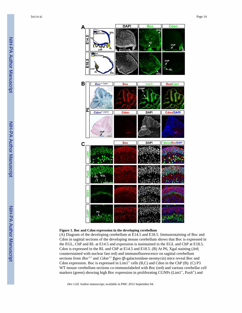
Figure 1. Boc and Cdon expression in the developing cerebellum(A) Diagram of the developing cerebellum at E14.5 and E18.5. Immunostaining of Boc andCdon in sagittal sections of the developing mouse cerebellum shows that Boc is expressed inthe EGL, ChP and RL at E14.5 and expression is maintained in the EGL and ChP at E18.5.Cdon is expressed in the RL and ChP at E14.5 and E18.5. (B) At P6, Xgal staining (left,counterstained with nuclear fast red) and immunofluorescence on sagittal cerebellumsections from Boc+/- and Cdon+/- βgeo (β-galactosidase-neomycin) mice reveal Boc andCdon expression. Boc is expressed in Lim1+ cells (B,C) and Cdon in the ChP (B). (C) P3WT mouse cerebellum sections co-immunolabeled with Boc (red) and various cerebellar cellmarkers (green) showing high Boc expression in proliferating CGNPs (Lim1+, Pax6+) and
Izzi et al. Page 14
Dev Cell. Author manuscript; available in PMC 2012 September 04.
NIH
-PA Author Manuscript
NIH
-PA Author Manuscript
NIH
-PA Author Manuscript
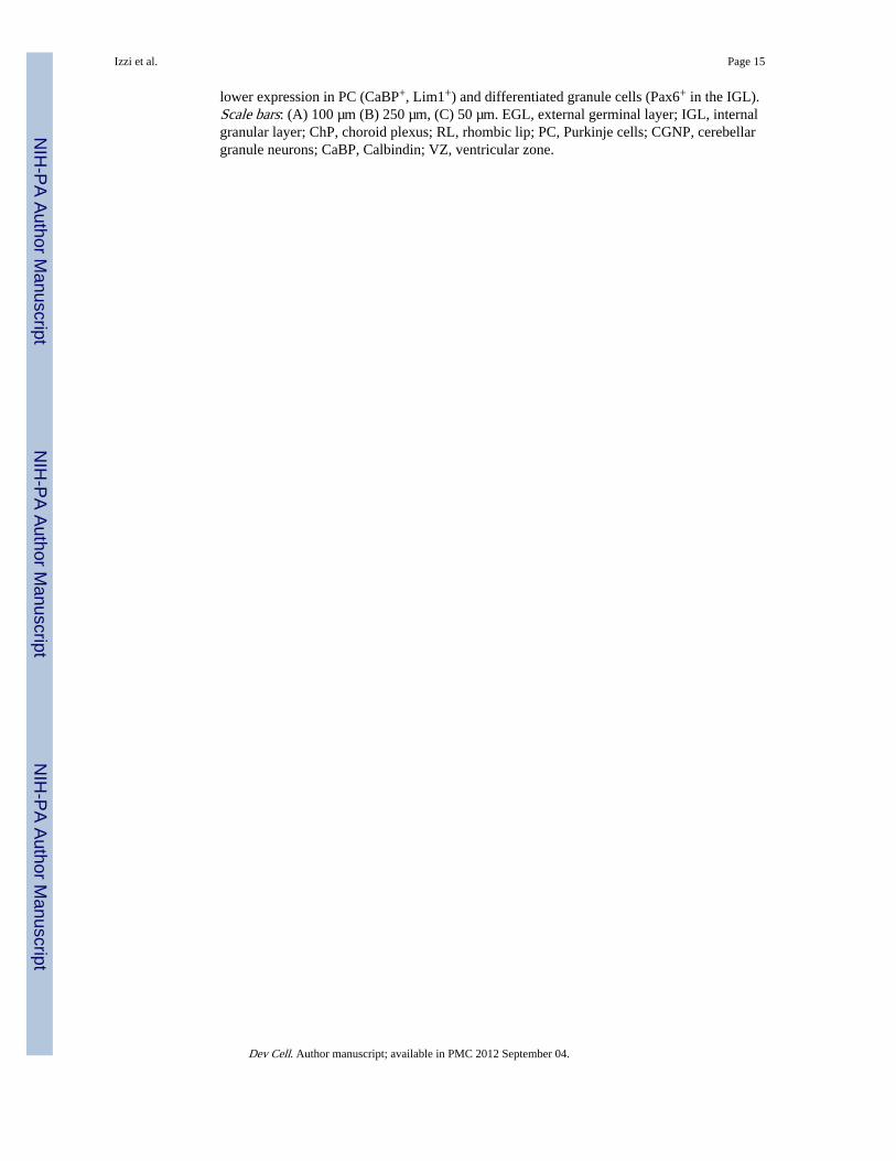
lower expression in PC (CaBP+, Lim1+) and differentiated granule cells (Pax6+ in the IGL).Scale bars: (A) 100 µm (B) 250 µm, (C) 50 µm. EGL, external germinal layer; IGL, internalgranular layer; ChP, choroid plexus; RL, rhombic lip; PC, Purkinje cells; CGNP, cerebellargranule neurons; CaBP, Calbindin; VZ, ventricular zone.
Izzi et al. Page 15
Dev Cell. Author manuscript; available in PMC 2012 September 04.
NIH
-PA Author Manuscript
NIH
-PA Author Manuscript
NIH
-PA Author Manuscript
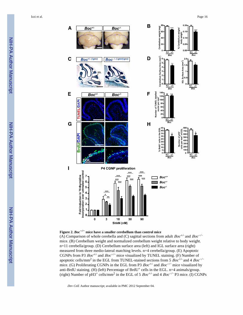
Figure 2. Boc−/− mice have a smaller cerebellum than control mice(A) Comparison of whole cerebella and (C) sagittal sections from adult Boc+/- and Boc−/−
mice. (B) Cerebellum weight and normalized cerebellum weight relative to body weight.n=11 cerebella/group. (D) Cerebellum surface area (left) and IGL surface area (right)measured from three medio-lateral matching levels. n=4 cerebella/group. (E) ApoptoticCGNPs from P3 Boc+/- and Boc−/− mice visualized by TUNEL staining. (F) Number ofapoptotic cells/mm2 in the EGL from TUNEL-stained sections from 5 Boc+/- and 4 Boc−/−
mice. (G) Proliferating CGNPs in the EGL from P3 Boc+/- and Boc−/− mice visualized byanti-BrdU staining. (H) (left) Percentage of BrdU+ cells in the EGL. n=4 animals/group.(right) Number of pH3+ cells/mm2 in the EGL of 5 Boc+/- and 4 Boc−/− P3 mice. (I) CGNPs
Izzi et al. Page 16
Dev Cell. Author manuscript; available in PMC 2012 September 04.
NIH
-PA Author Manuscript
NIH
-PA Author Manuscript
NIH
-PA Author Manuscript
purified from Boc+/+, Boc+/- and Boc−/− mice cerebella at P4 were cultured in the presenceof 0–90 nM ShhN. Proliferating cells were measured by 3H-thymidine incorporation. Data isrepresented as fold induction in CGNP proliferation compared to untreated cells. Scale bars:(A) 2mm, (C) 1mm, (E,G) 250 µm. IGL, internal granular layer; EGL, external germinallayer. p valules measured from Student’s t-test (B,D,F,H) and two-way ANOVA (I).
Izzi et al. Page 17
Dev Cell. Author manuscript; available in PMC 2012 September 04.
NIH
-PA Author Manuscript
NIH
-PA Author Manuscript
NIH
-PA Author Manuscript

Figure 3. Gas1 expression in the developing cerebellum(A) Immunostaining of sagittal sections showing Gas1 expression in the RL and EGL of thedeveloping cerebellum at E14.5 and E18.5. (B,C) P4-6 WT cerebellum sectionsimmunostained for Gas1 (red) and various cerebellar cell markers (green). Gas1 is highlyexpressed by proliferating CGNPs (Lim1+ cells; Pax6+ in EGL). (D) Immunostaining ofE18.5 cerebellum sections from Math1-Cre;mTmG mice show that Boc and Gas1 areexpressed in Math1+,GFP+ CGNPs in the EGL. EGL, external germinal layer; IGL, internalgranular layer; PC, Purkinje cells; RL, rhombic lip; VZ, ventricular zone; ChP, choroidplexus. Scale bars: (A, C and D) 100 µm, (B) 500 µm.
Izzi et al. Page 18
Dev Cell. Author manuscript; available in PMC 2012 September 04.
NIH
-PA Author Manuscript
NIH
-PA Author Manuscript
NIH
-PA Author Manuscript

Figure 4. Shh-dependent proliferation is completely lost in Gas1−/−;Boc−/− CGNPs(A) Haematoxylin-eosin staining on sagittal sections of E18.5 cerebellum revealing a thinnerEGL in Gas1−/−;Boc−/− cerebella than control. Anti-pH3 immunostaining of sagittal sectionsof Gas1+/-;Boc−/− and Gas1−/−;Boc−/− cerebella counterstained with DAPI. RNA in situhybridization showing the loss of expression of the Shh transcriptional target Gli1 inGas1−/−;Boc−/− cerebella at E18.5. (B) Quantification of: cerebellum surface area, EGLsurface area, pH3+ cells in EGL, pH3+ cells per µm2, and EGL thickness along the postero-anterior axis, n=4 animals/group. (C) CGNPs purified from Gas1+/+;Boc+/+ (n=3),Gas1+/-;Boc+/+ (n=4) and Gas1−/−;Boc+/+ (n=4) mice cerebella at E18.5 were cultured with0, 3, 10, 30 nM ShhN. Proliferating cells were visualized by immunostaining with an anti-
Izzi et al. Page 19
Dev Cell. Author manuscript; available in PMC 2012 September 04.
NIH
-PA Author Manuscript
NIH
-PA Author Manuscript
NIH
-PA Author Manuscript

Ki67 antibody. Data is represented as fold CGNP proliferation over untreated control (C, Dand E) or DMSO control (F). (D) Similar to (C) but CGNPs were purified fromGas1+/+;Boc−/− (n=3), Gas1+/-;Boc−/− (n=3) and Gas1−/−;Boc−/− (n=3) mice cerebella atE18.5. (E) CGNPs were purified from control (Ctl; Gas1+/+;Boc−/− and Gas1+/-;Boc−/−)(n=3) and Gas1−/−;Boc−/− (n=3) mice cerebella at E18.5 and treated with 0, 20, 50 or 100ng/ml of IGF-I. (F) Similar to (E) but CGNPs were treated with either DMSO, 0.150 µMpurmorphamine, or 30 nM ShhN. p values measured from Student’s t-test (B), two-wayANOVA (C, E), and ANOVA (D, F). EGL, external germinal layer. Scale bars: top tworows=500 µm, bottom two rows=100 µm.
Izzi et al. Page 20
Dev Cell. Author manuscript; available in PMC 2012 September 04.
NIH
-PA Author Manuscript
NIH
-PA Author Manuscript
NIH
-PA Author Manuscript
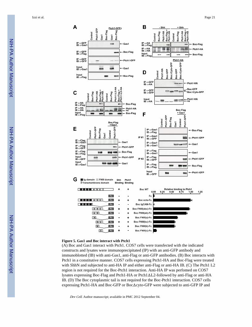
Figure 5. Gas1 and Boc interact with Ptch1(A) Boc and Gas1 interact with Ptch1. COS7 cells were transfected with the indicatedconstructs and lysates were immunoprecipitated (IP) with an anti-GFP antibody andimmunoblotted (IB) with anti-Gas1, anti-Flag or anti-GFP antibodies. (B) Boc interacts withPtch1 in a constitutive manner. COS7 cells expressing Ptch1-HA and Boc-Flag were treatedwith ShhN and subjected to anti-HA IP and either anti-Flag or anti-HA IB. (C) The Ptch1 L2region is not required for the Boc-Ptch1 interaction. Anti-HA IP was perfomed on COS7lysates expressing Boc-Flag and Ptch1-HA or Ptch1ΔL2-followed by anti-Flag or anti-HAIB. (D) The Boc cytoplasmic tail is not required for the Boc-Ptch1 interaction. COS7 cellsexpressing Ptch1-HA and Boc-GFP or BocΔcyto-GFP were subjected to anti-GFP IP and
Izzi et al. Page 21
Dev Cell. Author manuscript; available in PMC 2012 September 04.
NIH
-PA Author Manuscript
NIH
-PA Author Manuscript
NIH
-PA Author Manuscript

either anti-HA or anti-GFP IB. (E, F) Ptch1 forms receptor complexes with either Boc orGas1 but not both. Boc-Flag was co-transfected with Gas1 with or without Ptch1-GFP inCOS7 cells. (E) Lysates were IP with anti-Flag antibodies, followed by anti-Gas1 or anti-GFP IB. (F) Lysates were first immunoprecipated (IP #1) with anti-Gas1 antibodies.Supernatants from IP#1 were subjected to a second immunoprecipitation (IP #2) with anti-Flag antibodies. Both anti-Gas1 (IP #1) and anti-Flag (IP #2) immunoprecipitates were IBwith anti-Gas1, anti-Flag and anti-GFP antibodies. See Fig. S4 for a schematic of thisexperiment. (A–F) Protein expression inputs were verified by IB with the indicatedantibodies. ns, non-specific. (G, left) Diagram of WT Boc, Boc-Fc and Boc-Fc mutantproteins. (G, right) The Boc-Ptch1 interaction is mediated by the Boc FNIII(ab) domains.COS7 cells expressing Ptch1-GFP were incubated with conditioned mediated containingBoc-Fc proteins. Bound proteins were labeled with HRP-conjugated anti-Fc antibody andperoxidase activity measured.
Izzi et al. Page 22
Dev Cell. Author manuscript; available in PMC 2012 September 04.
NIH
-PA Author Manuscript
NIH
-PA Author Manuscript
NIH
-PA Author Manuscript

Figure 6. A Shh mutant which binds Ptch1 but which fails to bind Boc, Cdon and Gas1 does notinduce Shh signaling(A, B, C) Structural representation of ShhN. Residues colored in light, medium and darkgreen represent amino acids contacting Boc/Cdon FNIII(c) domain. E90 is represented inlight green (A). R154 is represented in dark green (B). (C) Amino acids important for Ptch1binding are colored in red while residues dispensible for the Ptch1-Shh interaction areillustrated in blue. (D) Anti-ShhN immunoblots showing the expression of purified ShhNWT, E90A, and R154E in comparison to ShhN C24II (top panel). Anti-ShhN immunoblotshowing conditioned medium (CM) containing ShhN-AP WT, E90A, and R154E incomparison to ShhN C24II (bottom panel). (E) Stimulation of Gli-luciferase reporter activity
Izzi et al. Page 23
Dev Cell. Author manuscript; available in PMC 2012 September 04.
NIH
-PA Author Manuscript
NIH
-PA Author Manuscript
NIH
-PA Author Manuscript
in C3H 10T1/2 cells treated with 17, 50 and 150 nM ShhN WT, E90A or R154E mutants.(F) Differentiation of C3H 10T1/2 cells treated with 12.5, 25 and 50 nM ShhN WT, E90A orR154E mutants. (E–F) Each condition was performed in duplicate, n=3. (G) Induction in ratP4 CGNP proliferation upon treatment with 6.25, 12.5, 25 and 50 nM of ShhN WT, E90A orR154E mutants. Proliferation was measured by 3H-thymidine incorporation. Each conditionwas performed in triplicate, n=3. (E–G) Two-way ANOVA comparing the activity of ShhNE90A to ShhN WT was performed. (H) Table summarizing binding of ShhN mutants toPtch1, Boc, Cdon and Gas1 and their functional activity.
Izzi et al. Page 24
Dev Cell. Author manuscript; available in PMC 2012 September 04.
NIH
-PA Author Manuscript
NIH
-PA Author Manuscript
NIH
-PA Author Manuscript
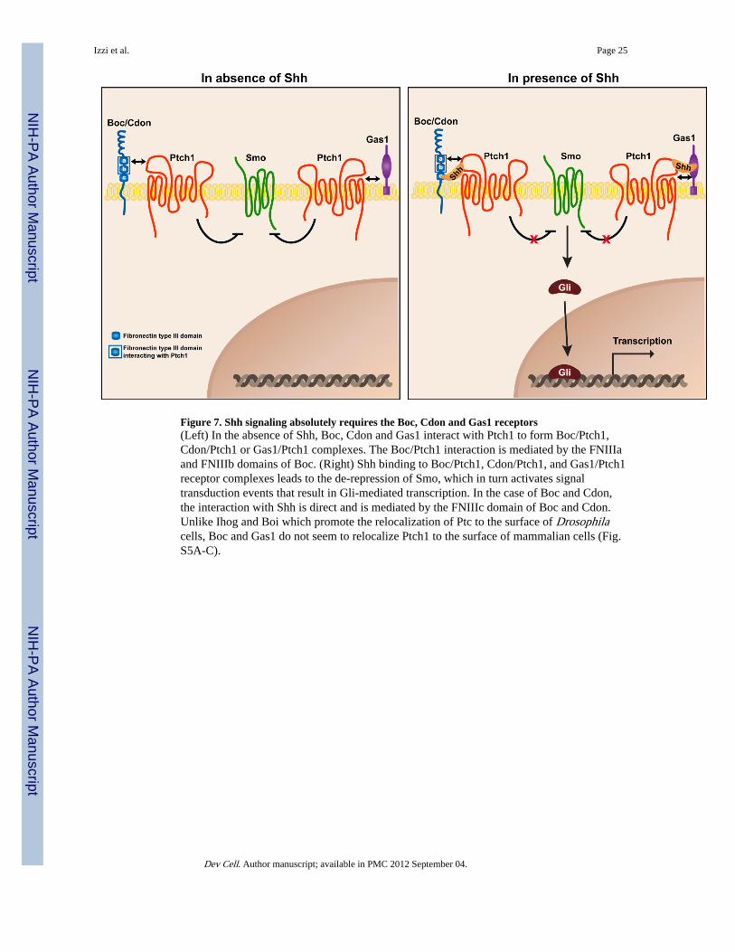
Figure 7. Shh signaling absolutely requires the Boc, Cdon and Gas1 receptors(Left) In the absence of Shh, Boc, Cdon and Gas1 interact with Ptch1 to form Boc/Ptch1,Cdon/Ptch1 or Gas1/Ptch1 complexes. The Boc/Ptch1 interaction is mediated by the FNIIIaand FNIIIb domains of Boc. (Right) Shh binding to Boc/Ptch1, Cdon/Ptch1, and Gas1/Ptch1receptor complexes leads to the de-repression of Smo, which in turn activates signaltransduction events that result in Gli-mediated transcription. In the case of Boc and Cdon,the interaction with Shh is direct and is mediated by the FNIIIc domain of Boc and Cdon.Unlike Ihog and Boi which promote the relocalization of Ptc to the surface of Drosophilacells, Boc and Gas1 do not seem to relocalize Ptch1 to the surface of mammalian cells (Fig.S5A-C).
Izzi et al. Page 25
Dev Cell. Author manuscript; available in PMC 2012 September 04.
NIH
-PA Author Manuscript
NIH
-PA Author Manuscript
NIH
-PA Author Manuscript